EP4516806A2 - Proteines g - Google Patents
Proteines g Download PDFInfo
- Publication number
- EP4516806A2 EP4516806A2 EP24206246.1A EP24206246A EP4516806A2 EP 4516806 A2 EP4516806 A2 EP 4516806A2 EP 24206246 A EP24206246 A EP 24206246A EP 4516806 A2 EP4516806 A2 EP 4516806A2
- Authority
- EP
- European Patent Office
- Prior art keywords
- subunit
- mutant
- gpcr
- binding
- protein
- Prior art date
- Legal status (The legal status is an assumption and is not a legal conclusion. Google has not performed a legal analysis and makes no representation as to the accuracy of the status listed.)
- Pending
Links
Images






















Classifications
-
- C—CHEMISTRY; METALLURGY
- C12—BIOCHEMISTRY; BEER; SPIRITS; WINE; VINEGAR; MICROBIOLOGY; ENZYMOLOGY; MUTATION OR GENETIC ENGINEERING
- C12N—MICROORGANISMS OR ENZYMES; COMPOSITIONS THEREOF; PROPAGATING, PRESERVING, OR MAINTAINING MICROORGANISMS; MUTATION OR GENETIC ENGINEERING; CULTURE MEDIA
- C12N9/00—Enzymes; Proenzymes; Compositions thereof; Processes for preparing, activating, inhibiting, separating or purifying enzymes
- C12N9/14—Hydrolases (3)
-
- A—HUMAN NECESSITIES
- A61—MEDICAL OR VETERINARY SCIENCE; HYGIENE
- A61P—SPECIFIC THERAPEUTIC ACTIVITY OF CHEMICAL COMPOUNDS OR MEDICINAL PREPARATIONS
- A61P35/00—Antineoplastic agents
-
- C—CHEMISTRY; METALLURGY
- C07—ORGANIC CHEMISTRY
- C07K—PEPTIDES
- C07K14/00—Peptides having more than 20 amino acids; Gastrins; Somatostatins; Melanotropins; Derivatives thereof
- C07K14/435—Peptides having more than 20 amino acids; Gastrins; Somatostatins; Melanotropins; Derivatives thereof from animals; from humans
- C07K14/46—Peptides having more than 20 amino acids; Gastrins; Somatostatins; Melanotropins; Derivatives thereof from animals; from humans from vertebrates
- C07K14/47—Peptides having more than 20 amino acids; Gastrins; Somatostatins; Melanotropins; Derivatives thereof from animals; from humans from vertebrates from mammals
- C07K14/4701—Peptides having more than 20 amino acids; Gastrins; Somatostatins; Melanotropins; Derivatives thereof from animals; from humans from vertebrates from mammals not used
- C07K14/4722—G-proteins
-
- C—CHEMISTRY; METALLURGY
- C07—ORGANIC CHEMISTRY
- C07K—PEPTIDES
- C07K14/00—Peptides having more than 20 amino acids; Gastrins; Somatostatins; Melanotropins; Derivatives thereof
- C07K14/435—Peptides having more than 20 amino acids; Gastrins; Somatostatins; Melanotropins; Derivatives thereof from animals; from humans
- C07K14/705—Receptors; Cell surface antigens; Cell surface determinants
-
- C—CHEMISTRY; METALLURGY
- C07—ORGANIC CHEMISTRY
- C07K—PEPTIDES
- C07K14/00—Peptides having more than 20 amino acids; Gastrins; Somatostatins; Melanotropins; Derivatives thereof
- C07K14/435—Peptides having more than 20 amino acids; Gastrins; Somatostatins; Melanotropins; Derivatives thereof from animals; from humans
- C07K14/705—Receptors; Cell surface antigens; Cell surface determinants
- C07K14/70571—Receptors; Cell surface antigens; Cell surface determinants for neuromediators, e.g. serotonin receptor, dopamine receptor
-
- C—CHEMISTRY; METALLURGY
- C07—ORGANIC CHEMISTRY
- C07K—PEPTIDES
- C07K14/00—Peptides having more than 20 amino acids; Gastrins; Somatostatins; Melanotropins; Derivatives thereof
- C07K14/435—Peptides having more than 20 amino acids; Gastrins; Somatostatins; Melanotropins; Derivatives thereof from animals; from humans
- C07K14/705—Receptors; Cell surface antigens; Cell surface determinants
- C07K14/72—Receptors; Cell surface antigens; Cell surface determinants for hormones
- C07K14/723—G protein coupled receptor, e.g. TSHR-thyrotropin-receptor, LH/hCG receptor, FSH receptor
-
- C—CHEMISTRY; METALLURGY
- C12—BIOCHEMISTRY; BEER; SPIRITS; WINE; VINEGAR; MICROBIOLOGY; ENZYMOLOGY; MUTATION OR GENETIC ENGINEERING
- C12Y—ENZYMES
- C12Y306/00—Hydrolases acting on acid anhydrides (3.6)
- C12Y306/05—Hydrolases acting on acid anhydrides (3.6) acting on GTP; involved in cellular and subcellular movement (3.6.5)
- C12Y306/05001—Heterotrimeric G-protein GTPase (3.6.5.1)
-
- G—PHYSICS
- G01—MEASURING; TESTING
- G01N—INVESTIGATING OR ANALYSING MATERIALS BY DETERMINING THEIR CHEMICAL OR PHYSICAL PROPERTIES
- G01N33/00—Investigating or analysing materials by specific methods not covered by groups G01N1/00 - G01N31/00
- G01N33/48—Biological material, e.g. blood, urine; Haemocytometers
- G01N33/50—Chemical analysis of biological material, e.g. blood, urine; Testing involving biospecific ligand binding methods; Immunological testing
- G01N33/53—Immunoassay; Biospecific binding assay; Materials therefor
- G01N33/566—Immunoassay; Biospecific binding assay; Materials therefor using specific carrier or receptor proteins as ligand binding reagents where possible specific carrier or receptor proteins are classified with their target compounds
-
- G—PHYSICS
- G01—MEASURING; TESTING
- G01N—INVESTIGATING OR ANALYSING MATERIALS BY DETERMINING THEIR CHEMICAL OR PHYSICAL PROPERTIES
- G01N2500/00—Screening for compounds of potential therapeutic value
- G01N2500/02—Screening involving studying the effect of compounds C on the interaction between interacting molecules A and B (e.g. A = enzyme and B = substrate for A, or A = receptor and B = ligand for the receptor)
-
- G—PHYSICS
- G01—MEASURING; TESTING
- G01N—INVESTIGATING OR ANALYSING MATERIALS BY DETERMINING THEIR CHEMICAL OR PHYSICAL PROPERTIES
- G01N2500/00—Screening for compounds of potential therapeutic value
- G01N2500/04—Screening involving studying the effect of compounds C directly on molecule A (e.g. C are potential ligands for a receptor A, or potential substrates for an enzyme A)
Definitions
- the present invention relates to mutant G proteins, and particularly to mutant alpha subunits of a heterotrimeric G protein. It also relates to products comprising such mutants, uses of the mutants, and methods involving such mutants.
- G proteins bind guanine nucleotides and act as molecular switches in a number of signalling pathways by interconverting between a GDP-bound inactive and a GTP-bound active state. They consist of two major classes: monomeric small G proteins and heterotrimeric G proteins. While small G proteins and the alpha subunit (G ⁇ ) of heterotrimeric G proteins both contain a GTPase domain (G-domain), G ⁇ contains an additional helical domain (H-domain) and also forms a complex with G beta (G ⁇ ) and G gamma (G ⁇ ) subunits. Although they undergo a similar signalling cycle, their activation differs in one important aspect.
- GEFs guanine nucleotide exchange factors
- GPCRs membrane-bound G protein coupled receptors
- GPCRs constitute a very large family of proteins that control many physiological processes and are the targets of many effective drugs. Thus, they are of considerable pharmacological importance. Reference is made particularly to Overington et al (2006) Nature Rev. Drug Discovery 5, 993-996 which indicates that over a quarter of present drugs have a GPCR as a target. A list of GPCRs is given in Foord et al (2005) Pharmacol Rev. 57, 279-288 , which is incorporated herein by reference.
- Atomic resolution mapping of the ligand-binding pocket is of significant importance for the design of drugs to modulate GPCR activity.
- methodology to crystallise receptors in their high-affinity agonist-bound conformation is a key prerequisite for efficient structure-based design of agonist compounds.
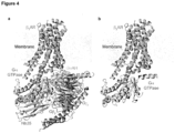
- three approaches have accomplished this: first, the C-terminal peptide of transducin was crystallised in complex with both opsin 36 and metarhodopsin II 37 ; second, a camelid antibody (Nb80), which induces the high-affinity agonist-bound state, has been crystallized in complex with ⁇ 2 AR 38 ; third, heterotrimeric Gs has been crystallised in complex with ⁇ 2 AR 10 .
- the Nb80- ⁇ 2 AR complex was solved at 3.5 ⁇ resolution, and also exhibited good electron density around the ligand-binding pocket 38 .
- the conformational changes induced by Nb80 are smaller than those observed in the ⁇ 2 AP-Gs complex 10 , suggesting that this structure may also represent an intermediate conformation.
- Nb80 was derived specifically to bind ⁇ 2 AP and is therefore likely to efficiently couple to other closely related GPCRs ( eg ⁇ 1 AP), new nanobodies likely need to be raised against more distantly related receptors.
- the ⁇ 2 AP-Gs complex was solved at 3.2 ⁇ resolution, however, in this structure electron density around the ligand-binding pocket was very poor.
- G ⁇ s The separate GTPase and helical domains of the stimulatory G protein (G ⁇ s) have been previously transfected into COS-7 cells 41 . However, individually, neither protein increased cellular cAMP production. Also, there was no investigation of the ability of the GTPase to bind a GPCR.
- G ⁇ subunits have also been shown to interact with GPCRs in a ⁇ -independent manner, and to undergo nucleotide exchange in the presence of a large excess of receptor, albeit with far slower kinetics than the holoenzyme 43, 45, 46 .
- all of the constructs contained an intact helical domain.
- MEGA domains can be considered to be minimal versions of the G ⁇ subunit, lacking part or all of the helical domain, which can couple to GPCRs even in the absence of the ⁇ dimer.
- MEGA domains can be considered to be minimal versions of the G ⁇ subunit, lacking part or all of the helical domain, which can couple to GPCRs even in the absence of the ⁇ dimer.
- An alternative description of MEGA domains is mini G proteins, and both definitions can be used to describe the mutant G proteins of the invention.
- a first aspect of the invention provides a mutant of a parent heterotrimeric G protein alpha (G ⁇ ) subunit, which mutant (i) lacks at least one helix of the helical domain of the parent G ⁇ subunit; (ii) is capable of binding to a GPCR in the absence of a heterotrimeric G protein beta (G ⁇ ) subunit and a heterotrimeric G protein gamma (G ⁇ ) subunit; and (iii) has an amino acid sequence that contains one or more mutations compared to the amino acid sequence of the parent heterotrimeric G ⁇ subunit, which mutations are selected from a deletion, a substitution and an insertion.
- heterotrimeric G protein By a heterotrimeric G protein, we include the meaning of a protein that is made of three subunits, a guanyl-nucleotide binding alpha subunit (G ⁇ ), a beta subunit (G ⁇ ) and a gamma subunit (G ⁇ ). Such heterotrimeric proteins transduce signals from a GPCR to a downstream effector as described above.
- G ⁇ subunits of any heterotrimeric G protein may be used in the practice of the present invention. Typically, G ⁇ subunits are between 350 and 400 amino acids in length and have molecular weights in the range 40-45 kDa.
- G s and G i families regulate adenylyl cyclase activity, while G q activates phospholipase C ⁇ and G 12/13 can activate small GTPase families.
- yeast are known to use the GPCR/G protein pathway; mating factor signal transduction in Saccharomyces cerevisiae is mediated by G protein alpha 1 subunit (GP-1).
- GP-1 G protein alpha 1 subunit
- Urano et al also review G protein signalling in plants studied mainly in two model organisms Arabidopsis thaliana and rice ( Oryza sativa ) ( Urano et al Open Biol. 2013 Mar; 3(3): 120186 ). All such G ⁇ subunits are included in the scope of the invention.
- G ⁇ subunits and their classification are well known in the art, and can be found at http://www.ebi.ac.uk/interpro/entry/IPR001019, and in Flock et al 2015 (Nature 524: 173 ) and Anantharanman et al, 2011 (Gene 475: 63-78 ).
- amino acid sequences (and the nucleotide sequences of the cDNAs which encode them) of many G ⁇ subunits are readily available, for example by reference to GenBank or UniProt. In particular, Flock et al, 2015 (Nature 524: 173 ) gives the human gene IDs for all human G ⁇ paralogues from UniProt (http://www.uniprot.org/uniprot). It should be noted, also, that because the sequence of an increasing number of genomes are complete, the amino acid sequences of G ⁇ subunits can be deduced therefrom.
- the G ⁇ may be derived from any source, it is particularly preferred if it is from a eukaryotic source. It is particularly preferred if it is derived from an animal (eg vertebrate) source such as a mammal or a bird. It is particularly preferred if the G ⁇ subunit is derived from rat, mouse, rabbit or dog or non-human primate or man, or from chicken or turkey. For the avoidance of doubt, we include within the meaning of "derived from” that a cDNA or gene was originally obtained using genetic material from the source, but that the protein may be expressed in any host cell subsequently.
- a eukaryotic G ⁇ (such as an avian or mammalian G ⁇ ) may be expressed in a prokaryotic host cell, such as E. coli, but be considered to be avian- or mammalian-derived, as the case may be.
- G ⁇ subunits comprise two domains: a GTP-binding domain and a helical domain.
- the GTP-binding domain is homologous to Ras-like small GTPases, and includes switch regions I and II, which change conformation during activation.
- the switch regions are loops of alpha-helices with conformations sensitive to guanine nucleotides.
- the helical domain of a G ⁇ subunit we include the meaning of the helical insertion domain that is inserted into the GTP-binding domain before switch region I and is unique to heterotrimeric G proteins. This helical domain functions to sequester the guanine nucleotide at the interface with the GTP-binding domain and must be displaced to enable nucleotide dissociation.
- Flock et al (2015) have performed a structural and sequence alignment of G ⁇ subunits from diverse organisms and have demonstrated that the helical domain of a G ⁇ subunit comprises six alpha helices, denoted Helix A, Helix B, Helix C, Helix D, Helix E and Helix F.
- the helical domain may be regarded as the region between the first amino acid residue of Helix A and the final amino acid residue of Helix F within the amino acid sequence of a G ⁇ subunit.
- the helical domain may also be considered to extend beyond these alpha helices to encompass the surrounding loop regions, ie the loop before Helix A and the loop after Helix F, such that the boundaries of the helical domain are not absolute.
- the mutant G ⁇ subunit lacks at least one of alpha helices A, B, C, D, E or F of the parent heterotrimeric G ⁇ subunit, such as at least two, three, four, five or all six of alpha helices A, B, C, D, E or F.
- the mutant G ⁇ subunit typically also lacks the intervening loops.
- the mutant G ⁇ subunit would also typically lack the loop connecting Helix A to Helix B, and so on.
- the mutant G ⁇ subunit lacks alpha helices A, B, C, D and E of the helical domain of the parent G ⁇ subunit, and the intervening loop regions.
- the mutant G ⁇ subunit lacks alpha helices A, B, C, D, E and F of the helical domain of the parent G ⁇ subunit, and the intervening loop regions.
- An analogous helix in the second G ⁇ subunit can be identified by searching for a similar amino acid sequence that defines Helix A in the sequence of one of the human G ⁇ subunit paralogues, for example, by sequence alignment.
- computer based algorithms are widely available in the art that can be used to predict the presence of protein motifs based on an amino acid sequence. Based upon the relative position of a particular alpha helix within the amino acid sequence and its position relative to other motifs and alpha helices, an analogous helix can readily be identified.
- CGN G ⁇ numbering
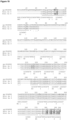
- Loops are labelled in lowercase letters of their flanking secondary structure elements (SSE); for example, s6h5 refers to the loop connecting strand S6 with helix H5 (see Figure 25 ).
- SSE flanking secondary structure elements
- s6h5 refers to the loop connecting strand S6 with helix H5 (see Figure 25 ).
- a CGN mapping webserver is available at http://mrc-Imb.cam.ac.uk/CGN.
- the CGN can be used to identify the boundaries of each of Helices A-F in any G ⁇ subunit.
- the first residue of Helix A (H.HA) in G ⁇ s is Asp85 H.HA.1 and the final residue of Helix F (H.HF) in G ⁇ s is Arg199 H.HF.6 .
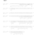
- the mutant G ⁇ subunit may lack the helical domain of the parent G ⁇ subunit corresponding to the region defined by amino acid residue Asp85 to amino acid residue Arg199 of the long isoform of human G ⁇ s as set out in Figure 1 and Figure 25 .
- corresponding region we include the meaning of the region in the amino acid sequence of a second G ⁇ subunit which aligns to the region in a first G ⁇ subunit (eg the region defined by amino acid Asp85 to amino acid residue Arg199 of the long isoform of human G ⁇ s), when the first and second G ⁇ subunits are compared by alignment, for example by using MacVector and Clustal W.
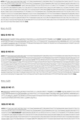
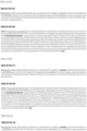
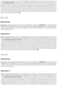
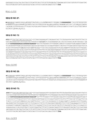
- Figure 25 shows an alignment of all of the human G ⁇ subunits, from which the region corresponding to the region defined by amino acid Asp85 to amino acid residue Arg199 of the long isoform of human G ⁇ s, in other human G ⁇ subunits can be identified. It will be appreciated that regions in other human G ⁇ subunits corresponding to different regions in the long isoform of human G ⁇ subunit may also be identified.
- the mutant G ⁇ subunit lacks a region of the helical domain of the parent heterotrimeric G ⁇ subunit corresponding to amino acid residues 70 to 193, 71 to 193, 85 to 193, or 85 to 199 according to the numbering of the long isoform of human G ⁇ -s subunit as set out in Figure 1 .
- mutant G ⁇ subunit of the invention may be considered to be an isolated GTPase domain or a G ⁇ subunit without its helical domain.
- Whether or not a given mutant G ⁇ subunit lacks at least one helix of the helical domain of the parent G ⁇ subunit can be determined by the skilled person, for example by aligning the amino acid sequence of the mutant G ⁇ subunit with the amino acid sequence of the parent G ⁇ subunit and assessing whether or not the amino acid sequence corresponding to the at least one helix of the helical domain of the parent G ⁇ subunit is present in the amino acid sequence of the mutant G ⁇ subunit. A similar analysis may be performed at the nucleotide sequence level.
- the mutant G ⁇ subunit of the invention is able to bind to a GPCR in a ⁇ independent manner.
- the mutant G ⁇ subunit of the invention should bind to a GPCR with a similar affinity (that is to say typically within 1-3 fold) as the parent G ⁇ subunit binds to the same GPCR when the parent G ⁇ subunit binds in combination with the ⁇ subunit.
- the mutant G ⁇ subunit should bind to a GPCR with a similar affinity as the parent heterotrimeric G protein binds to the same GPCR.
- binds to a GPCR we include the meaning of binding to a GPCR when bound by its agonist.
- Various methods may be used to determine binding between a GPCR and a test compound including, for example, size exclusion chromatography, enzyme linked immunosorbent assays (ELISA), surface plasmon resonance assays, chip-based assays, immunocytofluorescence, yeast two-hybrid technology and phage display which are common practice in the art and are described, for example, in Plant et al (1995) Analyt Biochem, 226(2), 342-348 and Sambrook et al (2001) Molecular Cloning A Laboratory Manual. Third Edition. Cold Spring Harbor Laboratory Press, Cold Spring Harbor, New York .
- Other methods of detecting binding between a test compound and the GPCR include ultrafiltration with ion spray mass spectroscopy/HPLC methods or other physical and analytical methods.
- Fluorescence Energy Resonance Transfer methods, for example, well known to those skilled in the art, may be used, in which binding of two fluorescent labelled entities may be measured by measuring the interaction of the fluorescent labels when in close proximity to each other. Yet further methods are described in WO2009/081136 .
- the mutant G ⁇ subunit is capable of functionally binding to a GPCR in the absence of a G ⁇ subunit and a G ⁇ subunit.
- functional binding we include the meaning that the mutant G ⁇ subunit is able to bind to a GPCR expressed on the surface of a cell such that it can transduce a signal from the GPCR (eg having bound to a modulator, such as a ligand of the receptor) into the cell via components of a signalling pathway of the cell.
- Another term for such functional binding is coupling.
- heterotrimeric G proteins couple with GPCRs and effectively transduce a signal from the GPCR to downstream effectors.
- binding of ligands such as hormones and neurotransmitters to a GPCR activates the receptor by causing a conformational change, which in turn activates the bound G protein on the intracellular-side of the membrane.
- the activated receptor promotes the exchange of bound GDP for GTP on the G ⁇ subunit.
- GTP binding changes the conformation of switch regions within the G ⁇ , which allows the bound trimeric G protein (inactive) to be released from the receptor, and to dissociate into active G ⁇ subunit (GTP-bound) and a ⁇ dimer.
- the G ⁇ subunit and the ⁇ dimer go on to activate distinct downstream effectors, such as adenylyl cyclase, phosphodiesterases, phospholipase C, and ion channels.
- effectors in turn regulate the intracellular concentrations of secondary messengers, such as cAMP, cGMP, diacylglycerol, sodium or calcium cations, which ultimately lead to a physiological response, usually via the downstream regulation of gene transcription.
- secondary messengers such as cAMP, cGMP, diacylglycerol, sodium or calcium cations
- the cycle is completed by the hydrolysis of alpha subunit-bound GTP to GDP, resulting in the reassociation of the alpha and beta/gamma subunits and their binding to the receptor, which terminates the signal.
- the mutant G ⁇ subunit is capable of binding to a GPCR in the absence of a G ⁇ subunit and a G ⁇ subunit, such that the G ⁇ subunit can be activated by the GPCR, for example as manifest by the generation of, or an increase in the basal level of, a G ⁇ protein signal.
- a G ⁇ protein signal we include the meaning of any downstream signal that is normally associated with signal transduction via the particular G ⁇ subunit in question, and so can be used as a marker for activation of the G ⁇ subunit.
- the signal may be any of the signals described herein and may be assayed using any suitable technique in the art.
- Functional binding may be assessed in a cellular assay, wherein a GPCR that is capable of a transducing a signal into the cell is contacted with the mutant G ⁇ subunit, and following stimulation of the GPCR ( eg with a ligand), the G ⁇ protein signal assessed.
- the cell co-expresses the mutant G ⁇ subunit and GPCR so as to enhance the signal, and even more preferably, expression of GPCRs or the mutant G ⁇ subunit is controlled by an inducible promoter, numerous examples of which have been described in the art and are generally available.
- any assay format that allows a G ⁇ protein signal to be measured following the binding of G ⁇ to its GPCR, and once the GPCR has been stimulated, can be used.
- the signal is detectable.
- the signal may correspond to the guanyl-nucleotide binding status of the G ⁇ subunit (eg GDP-bound or GTP-bound) which may be assessed biochemically on purified protein or enriched protein fractions, for example where the amount of radiolabelled GTPyS is detected.
- the signal may correspond to the GTPase activity of the G ⁇ subunit, or it may correspond to levels of secondary messengers, or it may reflect the status of downstream effectors (eg phosphorylation status or activity of cellular proteins).
- the use of a reporter gene can provide a readout, wherein expression of the reporter gene is controlled by the signal transduced through the G ⁇ subunit.
- Yet another method is to measure the affinity of the GPCR for its ligand. Still another method if to measure a phenotype of the cell known to be regulated by signalling through the G ⁇ subunit (eg cell growth or morphology). Further details are given below.
- the mutant G ⁇ subunit functionally binds to its GPCR in a ⁇ independent manner with a similar or greater potency than it does in the presence of the ⁇ dimer.
- the G ⁇ protein signal generated when the G ⁇ subunit binds to the GPCR in the presence and absence of a ⁇ dimer are within 5-10 fold of each other, such as within 2-3 fold.
- the G ⁇ protein signal generated when the G ⁇ subunit binds to the GPCR in the absence of a ⁇ dimer would be not more than 5 times weaker than the G ⁇ protein signal generated when the G ⁇ subunit binds to the GPCR in the presence of a ⁇ dimer.
- G ⁇ protein signal generated from functional binding between the G ⁇ and the GPCR eg activation of G ⁇
- G ⁇ subunits of the G ⁇ s type mediate signal transduction to effectors that stimulate the production of cyclic AMP (cAMP) within the cell.
- G ⁇ subunits of the G ⁇ i type mediate signal transduction to effectors that inhibit the production of cyclic AMP within the cell.
- G ⁇ subunit activates a phospholipase C (PLC) pathway resulting in the hydrolysis of phosphoinositides to generate two classes of different second messengers, namely diacylglycerol (DAG) and inositol phosphates.
- DAG diacylglycerol
- Diacylglycerol activates certain protein kinase Cs (PKCs) and certain inositol phosphates stimulate the mobilisation of calcium from intracellular stores.
- PKCs protein kinase Cs
- a wide variety of intracellular effectors have been identified as being under the control of G ⁇ subunits including cAMP, cGMP, phosphodiesterases, phospholipase C and phospholipase A2.
- G ⁇ activation is able to modulate a range of ion channels and is able to inhibit certain voltage sensitive calcium transients, as well as stimulating cardiac potassium channels.
- the skilled person will be able to select an appropriate assay for a given G ⁇ subunit (see, for example, Neves et al (2002) Science 296: 1636-1639 ; and Cabrera-Vera et al (2013) Endocrine Reviews 24: 765-781 ).
- the GTPase enzymatic activity by G ⁇ subunits can be measured, for example in plasma membrane preparations, by determining the breakdown of ⁇ 32P GTP using techniques that are well known in the art (eg see Signal Transduction: A Practical Approach: G Milligan, Ed. Oxford University Press, Oxford, Engl and).
- inositol lipids can be extracted and analysed using standard lipid extraction techniques. DAG can also be measured using thin-layer chromatography. Water soluble derivatives of all three inositol lipids (IP1, IP2, IP3) can also be quantified using radiolabelling techniques or HPLC. DAG can also be produced from phosphatidyl choline. The breakdown of this phospholipid in response to G ⁇ activation can also be measured using radiolabelling techniques.
- the mobilisation of intracellular calcium or the influx from calcium from outside of the cell can be measured using standard techniques.
- the choice of the appropriate calcium indicator, fluorescent, bioluminescent, metallochromic or calcium sensitive microelectrodes depends on the cell type and the magnitude and time constant of the event under study ( Borle (1990) Environ Health Perspect 84: 45-56 ).
- cells could be loaded with the calcium sensitive fluorescent dye fura-2 or indo-1, using standard methods, and any change in calcium measured using a fluorometer.
- suitable assays include: calcium mobilisation (Gonzalez JE, Maher MP. Cellular fluorescent indicators and voltage/ion probe reader (VIPR) tools for ion channel and receptor drug discovery. Receptors Channels. 2002;8(5-6):283-95, Dupriez VJ, Maes K, Le Poul E, Burgeon E, Detheux M . Aequorin-based functional assays for G-protein-coupled receptors, ion channels, and tyrosine kinase receptors. Receptors Channels.
- VIP voltage/ion probe reader
- G proteins such as measuring GTPase activity ( Jameson EE, Roof RA, Whorton MR, Mosberg HI, Sunahara RK, Neubig RR, Kennedy RT.Real-time detection of basal and stimulated G protein GTPase activity using fluorescent GTP analogues. J Biol Chem.
- a preferred way of assessing functional binding between a mutant G ⁇ subunit and a GPCR is by measuring the affinity of the GPCR for its ligand. In this way, functional binding can be characterised by an increase in the affinity of a GPCR for its agonist when the GPCR is bound to the G ⁇ subunit compared to the affinity of the GPCR for its agonist when the GPCR is not bound to the G ⁇ subunit.
- Such an increase in affinity can be measured using any suitable technique in the art, including competitive binding assays, optionally where one or both of the competing ligands are detectably labelled.
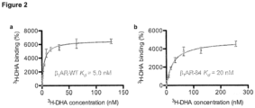
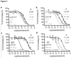
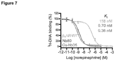
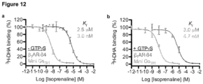
- An example of such an assay is described in the Examples, which measures competition between the antagonist 3H-dihydroalprenolol (3H-DHA) and the agonist isoprenaline for binding to a beta adrenergic receptor in the presence and absence of G ⁇ s.
- Such agents include antibodies (eg nanobodies) or other proteins whose function mimics that of the natural agonist. Specific examples are Nanobody 35 (Ref 40) and Nanobody 80 (Ref 38), and further examples are provided in WO2012007593 , WO2015121092 , and WO2014122183 . It will be appreciated that other such antibodies/nanobodies can be selected, for example by injecting purified GPCRs or GPCRs overexpressed in whole cells into mice or llamas, selecting antibodies/nanobodies that bind to the GPCR, and selecting those antibodies/nanobodies that activate the GPCR in whole cells ( eg by screening for increased production of a downstream effect of the GPCR activated signalling pathway).
- binding of a G protein to a GPCR is known to increase the affinity of the GPCR for an agonist
- binding of the mutant G ⁇ subunit in the absence of a G ⁇ subunit and a G ⁇ subunit, to a GPCR increases the affinity of the GPCR for the agonist by at least 1-fold, 2-fold, 3-fold, 4-fold or 5-fold.
- the binding of the mutant G ⁇ subunit in the absence of a G ⁇ subunit and a G ⁇ subunit, to a GPCR increases the affinity of the GPCR for the agonist by at least 10-fold, 50-fold or 100-fold.
- G proteins may decrease the affinity of GPCRs for their antagonists, in which case functional binding between a mutant G ⁇ subunit and a GPCR may be assessed by measuring this decrease in affinity. Similar assays to those described above for measuring an increase in affinity for agonist may be used to measure a decrease in affinity for antagonists.
- binding of a G protein to a GPCR is known to decrease the affinity of the GPCR for an antagonist, typically, binding of the mutant G ⁇ subunit in the absence of a G ⁇ subunit and a G ⁇ subunit, to a GPCR decreases the affinity of the GPCR for the antagonist by at least 1-fold, 2-fold, 3-fold, 4-fold or 5-fold.
- binding of the mutant G ⁇ subunit in the absence of a G ⁇ subunit and a G ⁇ subunit, to a GPCR decreases the affinity of the GPCR for the antagonist by at least 10-fold, 50-fold, 100-fold or 150-fold.
- the interaction between a GPCR and a heterotrimeric G protein induces conformational changes in both the G protein and receptor.
- functional binding of the mutant G ⁇ subunit in the absence of a G ⁇ subunit and a G ⁇ subunit, to a GPCR would be expected to induce one or more of these conformational changes that are evident when the parent G ⁇ subunit binds to the GPCR together with the G ⁇ and G ⁇ subunits.
- binding of the mutant G ⁇ subunit is expected to make the GPCR adopt its G protein-bound conformation.
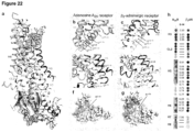
- binding of the mutant G ⁇ subunit in the absence of a G ⁇ subunit and a G ⁇ subunit, to a GPCR results in the movement of the cytoplasmic end of transmembrane helix 6 of the GPCR by 10 ⁇ or more away from the core of the receptor, such as by at least 11 ⁇ , 12 ⁇ , 13 ⁇ , 14 ⁇ , 15 ⁇ or 16 ⁇ away from the core of the receptor. It will be appreciated, therefore, that this provides yet another way of determining whether the G ⁇ subunit functionally binds to or couples to a GPCR.
- Various methods to probe protein structure are known in the art and any suitable method may be used. For example any structural biology technique such as x-ray crystallography may be used. Other methods include electron microscopy, NMR, direct measurement by epr spectroscopy or FRET.
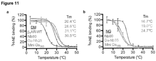
- Yet another way of assessing functional binding or coupling between a mutant G ⁇ subunit and a GPCR is to assess the stability of the GPCR/agonist/mutant G ⁇ subunit complex under denaturing conditions and compare it to the stability of the GPCR/agonist complex under denaturing conditions. If the stability (eg thermostability) of the GPCR/agonist/mutant G ⁇ subunit complex is greater than the stability of the GPCR/agonist complex, this would be indicative of functional binding. It will be appreciated that when the thermostability of a GPCR/G protein complex is measured, the temperature of the experiment should be performed within the temperature range tolerated by the mutant G ⁇ subunit ( eg to ensure that it is possible to detect ligand binding).
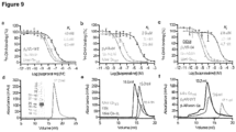
- Examples of assays that may be used to assess whether a mutant G ⁇ subunit functionally binds to or couples to a GPCR are also described in Example 5, and include (i) an agonist affinity shift assay, (ii) a thermostability assay, (iii) fluorescence-detection size exclusion chromatography (FSEC), (iv) fluorescence-based saturation binding analysis, and (v) size exclusion chromatography (SEC).
- FSEC fluorescence-detection size exclusion chromatography
- SEC size exclusion chromatography
- the mutant G ⁇ subunit has increased stability under denaturing conditions compared to its parent G ⁇ subunit and/or is expressed at a higher level than its parent G ⁇ subunit, when expressed in a cell.
- the mutant G ⁇ subunit when compared to its parent G ⁇ subunit, contains one or more mutations that increases the stability of the mutant G ⁇ subunit under denaturing conditions and/or increases the expression level of the mutant G ⁇ subunit, when expressed in a cell, compared to its parent G ⁇ subunit.
- the mutant G ⁇ subunit may be one which has increased stability to any denaturant or denaturing condition such as to any one or more of heat, a detergent, a chaotropic agent or an extreme of pH.
- the mutant G ⁇ may have an extended lifetime, relative to its parent, under destabilising conditions.
- thermostability In relation to an increased stability to heat (ie thermostability), this can readily be determined by measuring binding to a known binding partner (eg GPCR) or by using spectroscopic methods such as fluorescence, CD or light scattering at a particular temperature.
- a binding partner eg GPCR
- the ability of the G ⁇ subunit to bind that binding partner (eg GPCR) at a particular temperature may be used to determine thermostability of the mutant. It may be convenient to determine a "quasi T m " ie the temperature at which 50% of the G ⁇ subunit is inactivated under stated conditions after incubation for a given period of time (eg 30 minutes).
- thermostability can be assessed by measuring stability at a given temperature as a function of time. For example, the length of time at a given temperature by which the level of binding partner (eg GPCR) binding falls to 50% of the level of binding partner (eg GPCR) binding at time zero may be determined ( Shibata et al, 2009 J Mol Biol ) . In either case however, it is appreciated that temperature is the denaturant.
- level of binding partner eg GPCR
- the G ⁇ subunit is incubated for a defined time in the presence of a test detergent or a test chaotropic agent and the stability is determined using, for example, binding partner binding or a spectroscopic method as discussed above.
- a typical test pH would be chosen (eg in the range 4.5 to 5.5 (low pH) or in the range 8.5 to 9.5 (high pH).
- the mutant G ⁇ subunits are stable in the presence of such detergents.
- the order of "harshness" of certain detergents is DDM, C 11 ⁇ C 10 ⁇ C 9 ⁇ C 8 maltoside or glucoside, lauryldimethylamine oxide (LDAO) and SDS. It is particularly preferred if the mutant G ⁇ subunit is more stable to any of C 9 maltoside or glucoside, C 8 maltoside or glucoside, LDAO and SDS, and so it is preferred that these detergents are used for stability testing.
- the mutant G ⁇ has increased thermostability compared to its parent protein. It will be appreciated that heat is acting as the denaturant, and this can readily be removed by cooling the sample, for example by placing on ice. It is believed that thermostability may also be a guide to the stability to other denaturants or denaturing conditions. Thus, increased thermostability is likely to translate into stability in denaturing detergents, especially those that are more denaturing than DDM, eg those detergents with a smaller head group and a shorter alkyl chain and/or with a charged head group.
- the denaturing condition When an extreme of pH is used as the denaturing condition, it will be appreciated that this can be removed quickly by adding a neutralising agent. Similarly, when a chaotrope is used as a denaturant, the denaturing effect can be removed by diluting the sample below the concentration in which the chaotrope exerts its chaotropic effect.
- the mutant G ⁇ subunit may be one that is expressed at a higher level in a cell than its parent G ⁇ subunit.
- the mutant G ⁇ subunit is expressed in a cell at a level than is at least 1-fold greater than the level of its parent G ⁇ subunit when expressed in the cell under the same conditions, such as at least 2-fold, 3-fold, 4-fold or 5-fold greater, and more preferably at least 10-fold or 50-fold greater.
- Suitable expression systems are described in further detail below and in the Examples, and include constitutive or inducible expression systems in bacteria or yeasts, virus expression systems such as baculovirus, semliki forest virus and lentiviruses, or transient transfection in insect or mammalian cells. Methods for assessing protein expression are well known in the art and include techniques such as ELISA, SDS-PAGE analysis, western blotting, gel filtration and HPLC.
- mutant G ⁇ subunits of the first aspect of the invention may be expressed at a lower level in a cell and/or be less stable under denaturing conditions than their parent G ⁇ subunits, but such mutant G ⁇ subunits may form complexes with a GPCR, which are more stable under denaturing conditions than complexes formed between their parent G ⁇ subunits and a GPCR.
- GPCRs are thought to exist in multiple distinct conformations which are associated with different pharmacological classes of ligand such as agonists and antagonists, and to cycle between these conformations in order to function ( Kenakin T. (1997) Ann N Y Acad Sci 812, 116-125 ) . Switching between conformations contributes to the difficulty in obtaining crystal structures of receptors. Therefore, the ability to stabilise a particular conformation is highly desirable for crystallisation studies. As discussed in the Examples, the inventors have found that the mutant G ⁇ subunits described herein increase the thermostability of GPCRs in their agonist bound form.
- the mutant G ⁇ subunit of the first aspect of the invention is able to stabilise a particular conformation of the GPCR upon binding to the GPCR.
- stabilise a particular conformation we include the meaning that the conformation is stabilised under denaturing conditions.
- the conformation has an extended lifetime as manifest by retention of ligand binding ability under denaturing conditions.
- the particular conformation is an agonist conformation.
- Methods for assessing stability under denaturing conditions include those outlined above and are well known in the art (see, for example, WO2008/114020 ). Briefly, they may include subjecting the GPCR to denaturing conditions, either in the absence or presence of ligand, and then measuring retention of ligand binding.
- the GPCR When the GPCR is subjected to the denaturing conditions in the absence of ligand, the GPCR is then contacted with ligand to assess to what extent the GPCR can still bind to ligand.
- ligand eg agonist for agonist conformation, and so on. Since the conformation that is being stabilised is typically an agonist conformation, the ligand used for measuring stability is typically an agonist although it will be appreciated that partial agonists may also be used.
- the mutant G ⁇ subunits of the first aspect of the invention should retain the ability to bind to nucleotides (eg guanine nucleotides such as GDP, GTP or nucleotide derivatives such as GTP ⁇ S or GppNp) as do their parent G ⁇ subunits.
- nucleotides eg guanine nucleotides such as GDP, GTP or nucleotide derivatives such as GTP ⁇ S or GppNp
- the mutant G ⁇ subunits should retain the ability to bind to G ⁇ and/or G ⁇ subunits as do their parent G ⁇ subunits. Binding to nucleotides may stabilise the mutant G ⁇ subunit, and so the retention of these abilities is particularly desirable for structural analysis of G protein-GPCR complexes ( eg by cryo-electron microscopy) as described further below.
- mutant G ⁇ subunits that do not retain one or both of these activities are still encompassed by the mutant G ⁇ subunits of the invention.
- the switch I region of the G ⁇ subunit is composed of the loop between the helical domain and the beta strand 2 of the GTPase domain (CGN: G.hfs2), but also overlaps with Helix F from the helical domain.
- Switch I can be defined using the CGN system as the region located between the first amino acid residue of Helix F (H.HF) and the first amino acid residue of beta strand 2 (G.S2) ( eg in human G ⁇ s, this would be D194 H.HF.1 -I207 G.S2.1 including the two specified amino acid residues).
- the mutant G ⁇ subunit retains the switch I region of its parent G ⁇ subunit, eg the region corresponding to amino acid residues Asp 194 to Ile 207 according to the numbering of the long isoform of human G ⁇ s as set out in Figure 1 . It will be understood that this embodiment may correspond to the embodiment where the region corresponding to Helices A-E of the helical domain of the parent G ⁇ subunit is deleted. The inventors have found that in the absence of other mutations that increase stability and/or expression levels of the mutant G ⁇ subunit compared to its parent G ⁇ subunit, the switch I region is important for achieving expression.
- the switch I region may be truncated or deleted entirely.
- the mutant G ⁇ subunit is one where the switch I region (eg the region corresponding to amino acid residues Asp 194 to Ile 207 according to the numbering of the long isoform of human G ⁇ s as set out in Figure 1 ) or part thereof of the parent G ⁇ subunit is deleted.
- the part of the switch I region that is deleted comprises a stretch of consecutive amino acid residues that has been deleted.
- switch I region of the parent heterotrimeric G protein alpha subunit is replaced by a switch I region of a small GTPase.
- Switch I regions from small GTPases can be readily identified, for example by structural and sequence alignment as described herein.
- the helical domain, switch I region and the linker 1 region that links the GTPase domain to the N terminus of the helical domain, of the parent G ⁇ domain are all deleted.
- the linker 1 region (CGN: G.s1h1) varies in length between different G ⁇ subunits but can be defined using the CGN system as the region between the first and last residues of the helix 1/Helix A loop (G.h1ha), for example human G ⁇ s V65 G.h1ha.1 - S84 G.h1ha.20 (including the two specified residues).
- it can be defined as the region located between the final residue of helix 1 (G.H1) and the first residue of the helix A (H.HA), for example human G ⁇ s H64 G.H1.12 -D85 H.HA.1 (excluding the two specified residues).
- the mutant G ⁇ subunit may be one in which the region of the parent G ⁇ subunit that corresponds to amino acid residues 65 to 207 according to the numbering of the long isoform of human G ⁇ -s subunit as set out in Figure 1 , is deleted.
- the mutant G ⁇ subunit may be one in which the region of the parent G ⁇ subunit that corresponds to amino acid residues 66 to 207, 67 to 207, 68 to 207, 69 to 207, 70 to 207, 65 to 206, 65 to 205, 65 to 204, 65 to 203, or 65 to 202 according to the numbering of the long isoform of human G ⁇ -s subunit as set out in Figure 1 , is deleted.
- the mutant G ⁇ subunit is one in which the region of the parent G ⁇ subunit that corresponds to amino acid residues 65 to 203 according to the numbering of the long isoform of human G ⁇ -s subunit as set out in Figure 1 , is deleted.
- this region may be defined as the region between the final residue of helix 1 (G.H1) and the amino acid residue three residues N-terminal to the first amino acid residue of beta sheet (G.S2), for example human GaS H64 G.H1.12 -T204 G.hfs2.5 (including the two specified residues). It is especially preferred if this region is replaced with an amino acid linker as described further below.
- Preferred examples include linkers of eight amino acids in length such as GGSGGSGG or GGGGGGGG.
- the mutant G ⁇ subunit may be one in which the region of the parent G ⁇ subunit that corresponds to amino acid residues 60 to 207, 61 to 207, 62 to 207, 63 to 207, 64 to 207, 65 to 208, 65 to 209, 65 to 210, 65 to 211 or 65 to 212 according to the numbering of the long isoform of human G ⁇ -s subunit as set out in Figure 1 , is deleted.
- the helical domain and the linker 1 region that links the GTPase domain to the N terminus of the helical domain, of the parent G ⁇ subunit are deleted, but the switch I region of the parent G ⁇ subunit is deleted or else replaced with a switch I region from a small GTPase.
- the mutant G ⁇ subunit of the first aspect of the invention has an N-terminally truncated amino acid sequence when compared to the parent G ⁇ subunit.
- up to 10 amino acids may be deleted from the N-terminus, such as up to 9, 8, 7, 6, 5, 4, 3, or 2 amino acids, or 1 amino acid, may be deleted from the N-terminus.
- up to 15, 20, 25, 30, 35 or 40 amino acids may be deleted from the N-terminus.
- the deletion from the N-terminus is between 5 and 20 amino acids.
- N-terminus Deletion of the N-terminus is believed to be particularly favourable for crystallisation purposes, and so if the mutant G ⁇ subunit is to be crystallised it may be desirable to delete up to 40 amino acids from the N-terminus of its parent G ⁇ subunit. In contrast, in the case where binding to a ⁇ dimer is desirable, the N-terminus should not be deleted, or only up to the 5 N-terminal residues be deleted ( eg the N-terminal 1, 2, 3, 4 or 5 residues).
- the N-terminal 5 amino acids of the parent G ⁇ subunit are deleted, or the N-terminal 20 amino acids of the parent G ⁇ subunit are deleted or the N-terminal 21 amino acids from the parent G ⁇ subunit are deleted or the N-terminal 25 amino acids from the parent G ⁇ subunit are deleted.
- the mutant G ⁇ subunit is one where all of the amino acid residues N-terminal of the amino acid residue Ile/Leu HN43 , as shown in Figure 29 , are deleted.
- the mutant G ⁇ subunit is a mutant G ⁇ s subunit, this corresponds to deleting the first 25 amino acids corresponding to the first 25 amino acids of human G ⁇ s according to the numbering of human G ⁇ s as shown in Figure 29 .
- mutant G ⁇ subunit may comprise any of the N-terminal truncations defined herein in combination with any of the deletions of the helical domain, linker 1 region and switch I region mentioned above.
- mutant G ⁇ subunit may comprise any of the N-terminal truncations defined herein and be a mutant G ⁇ subunit in which the region of the parent G ⁇ subunit that corresponds to amino acid residues 65 to 203 according to the numbering of the long isoform of human G ⁇ -s subunit as set out in Figure 1 , is deleted.
- the mutant G ⁇ subunit is one which, when compared to the parent G ⁇ subunit, contains one or more mutations in the switch I region, and in a more specific embodiment, the mutant G ⁇ subunit is one which, when compared to the parent G ⁇ subunit, has a different amino acid at a position which corresponds to any one or more of the following positions according to the numbering of the long isoform of human G ⁇ -s subunit as set out in Figure 1 : Leu 197 and Cys 200. Using the CGN system, these amino acid residues are identified as L197 H.HF.4 and C200 G.hls2.1 .
- amino acid residue at position L197 H.HF.4 is a leucine, it is preferably substituted for an alanine residue, and when the amino acid residue at position C200 G.hfs2.1 is a cysteine, it is preferably substituted for a serine residue.
- the mutant G ⁇ subunit is one wherein the switch III region of the parent heterotrimeric G protein alpha subunit, or part thereof, is deleted.
- the switch III region can be defined using the CGN system as the region located between the last residue of beta sheet 4 (G.S4) and the first residue of helix 3 (G.H3), for example human G ⁇ s Ala249 G.S4.7 to Arg265 G.H3.1 (excluding the two specified residues).
- the mutant G ⁇ subunit may be one in which the region of the parent G ⁇ subunit that corresponds to amino acid residues Ser 250 to Asn 264 according to the numbering of the long isoform of human G ⁇ -s subunit as set out in Figure 1 , or part thereof, is deleted.
- the part of the switch III region that is deleted comprises a stretch of consecutive amino acid residues that has been deleted.
- the inventors have found that deleting the stretch of amino acids corresponding to Asn254 G.s4h3.5 to Thr263 G.s4h3.14 provides a mutant G ⁇ subunit with particularly good properties (eg in terms of coupling to a GPCR and improved stability/expression levels), and so in a specific embodiment, the mutant G ⁇ subunit is one in which the region of the parent G ⁇ subunit that corresponds to amino acid residues 254 to 263 according to the numbering of the long isoform of human G ⁇ -s subunit as set out in Figure 1 , is deleted.
- the mutant G ⁇ subunit may be one in which the region of the parent G ⁇ subunit that corresponds to amino acid residues 255 to 263, 256 to 263, 257 to 263, 258 to 263, 259 to 263, 254 to 262, 254 to 261, 254 to 260, 254 to 259, or 254 to 258 according to the numbering of the long isoform of human G ⁇ -s subunit as set out in Figure 1 , is deleted.
- the mutant G ⁇ subunit may be one in which the region of the parent G ⁇ subunit that corresponds to amino acid residues 253 to 263, 252 to 263, 251 to 263, 250 to 263, 249 to 263, 254 to 264, 254 to 265, 254 to 266, 254 to 267, or 254 to 268 according to the numbering of the long isoform of human G ⁇ -s subunit as set out in Figure 1 , is deleted.
- the mutant G ⁇ subunit may be one wherein the switch III region of the parent G ⁇ subunit, or part thereof, is deleted as defined above, and may comprise any of the N-terminal truncations defined herein, and any of the deletions of the helical domain, linker 1 region and switch I region mentioned above.
- the mutant G ⁇ subunit may comprise any of the N-terminal truncations defined herein, and be a mutant G ⁇ subunit in which the regions of the parent G ⁇ subunit that correspond to amino acid residues 65 to 203, and amino acids 254 to 263, according to the numbering of the long isoform of human G ⁇ -s subunit as set out in Figure 1 , are deleted.
- the mutant G ⁇ subunit is one wherein the switch II region of the parent heterotrimeric G protein alpha subunit, or part thereof, is deleted.
- the switch II region can be defined using the CGN system as the region between the last amino acid residue of beta sheet 3 (G.S3) and the first amino acid residue of beta sheet 4 (G.S4), for example G ⁇ s Val224 G.S3.8 to Ala243 G.S4.1 (excluding the two specified residues).
- the mutant G ⁇ subunit may be one in which the region of the parent G ⁇ subunit that corresponds to amino acid residues Gly 225 to Thr 242 according to the numbering of the long isoform of human G ⁇ -s subunit as set out in Figure 1 , or part thereof, is deleted.
- the mutant G ⁇ subunit is one in which the region of the parent G ⁇ subunit that corresponds to amino acid residues 227 to 230 according to the numbering of the long isoform of human G ⁇ -s subunit as set out in Figure 1 , is deleted.
- the switch II region or part thereof eg the region that corresponds to amino acid residues 227 to 230 according to the numbering of the long isoform of the human G ⁇ -s subunit as set out in Figure 1
- a linker sequence as described further below; however, such a linker is not essential.
- the mutant G ⁇ subunit may be one in which the region of the parent G ⁇ subunit that corresponds to amino acid residues 226 to 242, 227 to 242, 228 to 242, 229 to 242, 230 to 242, 225 to 241, 225 to 240, 225 to 239, 225 to 238, or 225 to 237 according to the numbering of the long isoform of human G ⁇ -s subunit as set out in Figure 1 , is deleted.
- the mutant G ⁇ subunit may be one in which the region of the parent G ⁇ subunit that corresponds to amino acid residues 224 to 242, 223 to 242, 222 to 242, 221 to 242, 220 to 242, 225 to 243, 225 to 244, 225 to 245, 225 to 246 or 225 to 247 according to the numbering of the long isoform of human G ⁇ -s subunit as set out in Figure 1 , is deleted.
- mutant G ⁇ subunit may be one wherein the switch II region of the parent G ⁇ subunit, or part thereof, is deleted as defined above, and may comprise any of the N-terminal truncations defined herein, and any of the deletions of the helical domain, linker 1 region, switch I region, and switch III region mentioned above.
- the mutant G ⁇ subunit may comprise any of the N-terminal truncations defined herein, and be a mutant G ⁇ subunit in which the regions of the parent G ⁇ subunit that correspond to amino acid residues 65 to 203, and amino acids 227 to 230, according to the numbering of the long isoform of human G ⁇ -s subunit as set out in Figure 1 , are deleted, optionally wherein the region of the parent G ⁇ subunit that corresponds to amino acid residues 254 to 263 is also deleted.
- the linker is a peptide. Suitable linker peptides are those that typically adopt a random coil conformation, for example the peptide may contain glycine or serine or a mixture of glycine plus serine residues.
- the linker contains between 2 and 50 amino acid residues, more preferably between 2 and 30, and still more preferably between 3 and 20 such as between 3 and 8. Examples of suitable linkers are provided in Table 2 below. It is preferred if any of the helical domain, switch I and switch II regions are replaced by a linker. The requirement for a linker may depend upon the size of the deletion. For example, small deletions such as in the region of ten amino acids or less may not require a linker and the two portions created by the deletion may be joined together directly. However, larger deletions such as in the region of more than ten amino acids will generally require a linker.
- the inventors have identified various mutations that increase the stability of the mutant G ⁇ subunit under denaturing conditions and/or increase the expression of the mutant G ⁇ subunit, when expressed in a cell, compared to its parent G ⁇ subunit, and so it is preferred if the mutant G ⁇ subunit of the first aspect of the invention comprises one or more mutations that increase such stability and/or expression. Examples of such mutations are listed in Tables 2-5 below, and so the mutant G ⁇ subunit may comprise any one or more of the mutations listed in Tables 2-5 below.
- the mutant G ⁇ subunit is one which, when compared to the parent G ⁇ subunit, has a different amino acid at a position which corresponds to any one or more of (eg 2, 3, 4, 5, 6, 7, 8, 9, 10, 11, 12, 13, 14, 15, 16 or 17 of) the following positions according to the numbering of the long isoform of human G ⁇ -s subunit as set out in Figure 1 : Val 36, His 41, Ala 48, Gly 49, Glu 50, Met 60, Leu 63, Leu 197, Lys 200, Arg 201, Phe 208, Asn 218, Gly 226, Glu 230, Ala 249, Ser 252, Leu 272, Ile 372, Val 375.
- each of the amino acids is replaced by the particular amino acid residue indicated in Tables 2-5.
- the valine at position 36 is replaced by an aspartate
- the histidine at position 41 is replaced by a isoleucine or a valine, and so on.
- they may be replaced by any other amino acid provided that the mutant G ⁇ subunit is capable of binding to a GPCR in the absence of a G ⁇ and a G ⁇ subunit.
- the amino acid used to replace the given amino acid at a particular position is typically a naturally occurring amino acid, typically an "encodeable" amino acid, it may be a non-natural amino acid (in which case the protein is typically made by chemical synthesis or by use of non-natural amino-acyl tRNAs).
- An "encodeable” amino acid is one which is incorporated into a polypeptide by translation of mRNA. It is also possible to create non-natural amino acids or introduce non-peptide linkages at a given position by covalent chemical modification, for example by post-translational treatment of the protein or semisynthesis. These post-translational modifications may be natural, such as phosphorylation, glycosylation or palmitoylation, or synthetic or biosynthetic.
- the mutant G ⁇ subunit is a mutant G ⁇ s subunit with an amino acid sequence which, when compared to the amino acid sequence of the parent G ⁇ subunit, has one or more of more of the following mutations according to the numbering of the long isoform of human G ⁇ -s subunit as set out in Figure 1 : V36D, H41I or H41V, A48L, G49D, E50N, M60A or M60C, L63Y or L63R or L63K, L297A, C200S, R201A, F208N, N218K, G226A, E230A, A249D or A249E, S252D or S252E, L272D or L272E, I372A or I372C, and V375I.
- the mutant G ⁇ subunit is one which, when compared to the parent G ⁇ subunit, has an N-terminal truncation of 5-20 or 5-25 amino acid residues in length, a deletion of the switch III region, and has a different amino acid at a position which corresponds to one or more of ( eg 2, 3, 4, or 5 of) the following positions according to the numbering of the long isoform of human G ⁇ -s subunit as set out in Figure 1 : His 41, Leu 197, Cys 200, Ala 249, and Leu 272.
- the mutant G ⁇ subunit is one which, when compared to the parent G ⁇ subunit, has an N-terminal truncation of 5-20 or 5-25 amino acid residues in length, a deletion of the switch III region, and has a different amino acid at a position which corresponds to any one or more of ( eg at least 2, 3, 4, 5, 6, 7 or 8 of) the following positions according to the numbering of the long isoform of human G ⁇ -s subunit as set out in Figure 1 : Gly 49, Glu 50, Leu 63, Ala 249, Ser 252, Leu 272, Ile 372 and Val 375.
- the mutant G ⁇ subunit is one which, when compared to the parent G ⁇ subunit, has a different amino acid at a position which corresponds to one or more of the following positions according to the numbering of the long isoform of human G ⁇ -s subunit as set out in Figure 1 : Gly 49, Glu 50, Gly 226 and Ser 252.
- the mutant G ⁇ subunit is one which, when compared to the parent G ⁇ subunit, has (i) a deletion of all amino acid residues N-terminal of Ile/Leu HN43 ; (ii) a deletion of the region between the final residue of helix 1 (G.H1) and the amino acid residue three residues N-terminal to the first amino acid residue of beta sheet 2 (G.S2), optionally wherein the region is replaced by an amino acid linker (eg an 8 amino acid linker such as GGSGGSGG or GGGGGGGG; (iii) a deletion of ten amino acid residues between Tyr S4H3.4 and Asn/Ser S4H3.15 ; and (iv) a different amino acid at a position which corresponds to any one or more of ( eg at least 1, 2, 3, 4, 5, 6 or 7 of) the following positions Gly49 SIH1.3 , Glu50 SIH1.4 , Ala249 S4.7 , Ser252 S4H3.3 , Le
- the mutant G ⁇ subunit of the first aspect of the invention has at least 20% sequence identity to the amino acid sequence of the long isoform of human G ⁇ -s subunit as set out in Figure 1 (SEQ ID NO: 91), such at least 30%, 40%, 50%, 60% or 70% sequence identity, and more typically at least 75%, 80%, 85%, 90%, 95%, 96%, 97%, 98% or 99% sequence identity.
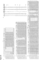
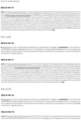
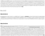
- the mutant G ⁇ subunit is one that has at least 20% sequence identity to any of the amino acid sequences in Figure 26 , corresponding to SEQ ID Nos: 1-45, for example at least 30%, 40%, 50%, 60% or 70% sequence identity, and more preferably at least 75%, 80%, 85%, 90%, 91%, 92%, 93%, 94%, 95%, 96%, 97%, 98%, or 99% sequence identity.
- the mutant G ⁇ subunit is one that comprises any of the amino acid sequences in Figure 26 , corresponding to SEQ ID Nos: 1-45.
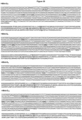
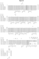
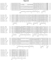
- the mutant G ⁇ subunit is one that has at least 20% sequence identity to any of the amino acid sequences in any of Figures 29 , 35 , 36 , 37 , 38 and 40 for example at least 30%, 40%, 50%, 60% or 70% sequence identity and more preferably at least 75%, 80%, 85%, 90%, 91%, 92%, 93%, 94%, 95%, 96%, 97%, 98% or 99% sequence identity.
- the mutant G ⁇ subunit is one that comprises any one of the amino acid sequence in any of Figures 29 , 35 , 36 , 37 , 38 and 40 .
- the percent sequence identity between two polypeptides may be determined using any suitable computer program, for example the GAP program of the University of Wisconsin Genetic Computing Group and it will be appreciated that percent identity is calculated in relation to polypeptides whose sequence has been aligned optimally.
- the alignment may alternatively be carried out using the Clustal W program ( Thompson et al., 1994 Nucleic Acids Res. 22(22):4673-80 ).
- the parameters used may be as follows: Fast pairwise alignment parameters: K-tuple(word) size; 1, window size; 5, gap penalty; 3, number of top diagonals; 5. Scoring method: x percent. Multiple alignment parameters: gap open penalty; 10, gap extension penalty; 0.05. Scoring matrix: BLOSUM.
- the mutant G ⁇ subunit of the first aspect of the invention is one which when compared to the parent G ⁇ subunit, comprises one or more dominant negative mutations. Any such dominant negative mutation known in the art may be incorporated into the mutant G ⁇ subunit of the invention, and some specific examples are included below.
- the mutant G ⁇ subunit is one which, when compared to the parent G ⁇ subunit, has a different amino acid at a position which corresponds Ser 54 according to the numbering of the long isoform of human G ⁇ -s subunit as set out in Figure 1 .
- the mutant G ⁇ subunit preferably comprises the mutation S54N
- the mutant G ⁇ subunit is a G ⁇ t subunit
- the mutant preferably comprises the mutation S43N.
- the N338D mutation located within the NKXD motif was identified as a dominant negative mutant in the yeast G protein Gpa1 58 .
- the Gpa1 N338D mutant appeared to form an irreversible empty binding pocket complex with the receptor, which was resistant to dissociation by guanine nucleotides 58 .
- the authors noted that the mutant protein was thermally labile and that receptor binding provided protection against denaturation 58 .
- Simon and colleagues reported that the D273N mutation, also located within the NKXD motif resulted in nucleotide-depleted G ⁇ o, G ⁇ 11 and G ⁇ 12 subunits that exhibit a receptor-sequestering dominant negative phenotype ( ⁇ independent) 62-64 .
- the mutant G ⁇ subunit is one which, when compared to the parent G ⁇ subunit, has one or more different amino acids within the NKXD motif, optionally wherein Asn of the NKXD motif is replaced with Asp and/or wherein Asp of the NKXD motif is replaced with Asn.
- the NKXD motif belongs to a group of G-box motifs that are highly conserved in all G proteins, and are described in detail in the scientific literature.
- the CGN code for the NKXD motif in Gas is N292 G.S5.7 , K293 Gshg.1 , Q294 G.HG.1 and D295 G.HG.2 .
- the mutant G ⁇ subunit is a yeast Gpa1
- the mutant preferably comprises the mutation N338D
- the mutant G ⁇ subunit is any of G ⁇ o, G ⁇ 11 or G ⁇ 12
- the mutant preferably comprises the mutation D273N.
- the mutant G ⁇ subunit of the first aspect of the invention is one which, when compared to the parent G ⁇ subunit, has a different amino acid at a position which corresponds to Gln 227 according to the numbering of G ⁇ s as set out in Figure 1 , and optionally has a different amino acid at a position which corresponds to Asp 295 according to the numbering of G ⁇ s as set out in Figure 1 .
- the mutant G ⁇ subunit is a G ⁇ o
- the mutant preferably comprises the mutation Q205L, and optionally also comprises the mutation D273N.
- the mutant G ⁇ subunit is one which, when compared to the parent G ⁇ subunit, has one or more different amino acids at a position which corresponds to one or more, or all of, of the following positions according to the numbering of the long isoform of human G ⁇ -s subunit as set out in Figure 1 : Gly 226, Glu 268 and Ala 366.
- the mutant G ⁇ subunit preferably comprises one or more, or all of, the mutations G226A, E268A and A366S.
- Pereira & Cerione reported a mutation in the switch III region, which produced a receptor-sequestering dominant negative phenotype 59 .
- the R238E mutation of G ⁇ t (chimera 6 44 ) was found to exist in a nucleotide deficient state.
- the mutant was also reported to be less thermally labile than other nucleotide deficient G ⁇ mutants, possibly because it adopted a partially active conformation 59 .
- the same mutation in G ⁇ s also failed to produce a dominant negative phenotype.
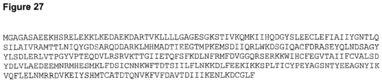
- the mutant G ⁇ subunit is a mutant G ⁇ t subunit (chimera 6) which, when compared to the parent G ⁇ subunit, has a different amino acid at a position which corresponds to Arg 238 according to the numbering of the G ⁇ t subunit (chimera 6) as set out in Figure 27 .
- this triple mutant was thermally unstable 65 . Therefore two of these mutations (G226A and A366S) were combined with additional mutations in order to produce a more stable dominant negative mutant 68 .
- the ⁇ 3/ ⁇ 5 loop of G ⁇ s was replaced with the corresponding region from G ⁇ i2 48 , as described in the previous section. This construct, containing a total of seven mutations, was reported to have significantly improved thermal stability 48 .
- mutant G ⁇ subunits of the first aspect of the invention include mutations that are known to increase the affinity of a G ⁇ subunit for a GPCR.
- mutant G ⁇ subunit may be one which, when compared to the parent G ⁇ subunit, comprises one or more mutations known to increase the affinity of a G ⁇ subunit for a GPCR. Any such affinity-increasing mutation known in the art may be incorporated into the mutant G ⁇ subunit of the invention, and some specific examples are included below.
- mutants displayed increased levels of nucleotide exchange, and both were able to interact with rhodopsin, however the receptor was unable to further accelerate the rate of nucleotide exchange 30 .
- the D328R mutant also displayed enhanced interaction with rhodopsin compared to wild-type G ⁇ i1 30 .
- the mutant G ⁇ subunit is one which, when compared to the parent G ⁇ subunit, has a cysteine residue at each of the positions corresponding to Ile 56 and Gln 333 according to the numbering of the G ⁇ i1 subunit as set out in Figure 25 .
- the mutant G ⁇ subunit may be a mutant G ⁇ s subunit wherein the ⁇ 3/ ⁇ 5 loop of the parent G ⁇ s subunit is replaced with the ⁇ 3/ ⁇ 5 loop of a G ⁇ i2 subunit.
- ⁇ 3/ ⁇ 5 loop we include the meaning of the region defined by the amino acid sequence N271 G.H3.8 to I285 G.h3s5.3 of G ⁇ s, which corresponds to K249 G.H3.7 to T263 G.h3s5.3 of Gai2.
- the mutant G ⁇ subunit may be one which, when compared to the parent G ⁇ subunit, has a different amino acid at a position which corresponds to any one or more of Asn 271, Lys 274, Arg 280, Thr 284 and Ile 285 according to the numbering of the long isoform of the human G ⁇ s subunit as set out in Figure 1 .
- the mutant G ⁇ subunit is a G ⁇ s subunit, preferably the mutant comprises one or more of the mutations N271K, K274D, R280K, T284D, and I285T.
- the mutant G ⁇ subunit may be one wherein the amino acid residue at a position which corresponds to Cys 347 according to the numbering of the G ⁇ t subunit as set out in Figure 25 , is chemically modified, optionally wherein said amino acid residue is carboxymethylated (eg has been treated with IAA) or cyanylated ( eg has been treated with NTCBA).
- mutants of the parent G ⁇ subunit may be produced in any suitable way and provided in any suitable form.
- Conventional site-directed mutagenesis may be employed, or polymerase chain reaction-based procedures well known in the art may be used.
- the mutants of the parent G ⁇ subunit are ones whose amino acid sequence comprises one or more of a deletion, an amino acid substitution and/or an insertion compared to the amino acid sequence of the parent G ⁇ subunit.
- the deletion may be a deletion within the amino acid sequence of the parent G ⁇ subunit ( ie not at the termini of the sequence), but it will be appreciated that the deletion may be at one or both of the N-terminus and C-terminus of the amino acid sequence of the parent G ⁇ subunit.
- the insertion may be an insertion of one or more (eg 2, 3, 4, or 5 or more) amino acids within the amino acid sequence of the parent G ⁇ subunit (ie not at the termini of the sequence), but also included are insertions at one or both of the N-terminus and C-terminus of the amino acid sequence of the parent G ⁇ subunit, eg fusions.
- the mutant G ⁇ subunits of the invention may also be chimeras made up of one or more parts of a first G ⁇ subunit and one or more parts of a second G ⁇ subunit.
- Chimeras may be useful for converting a G ⁇ subunit of one class (eg G ⁇ s) to behave more like another class (eg Gq or Gi).
- G ⁇ subunit of one class eg G ⁇ s
- Gi another class
- replacement of the last 18 amino acids of the C-terminus of Gs to those from Gq allows the G ⁇ subunit to bind to a receptor that only couples to Gq.
- Recent in vivo FRET studies also suggest that residues within the alpha five helix but distal to the five C-terminal residues, strongly influence specificity (see also Example 5).
- mutant G ⁇ subunit of the first aspect of the invention that is capable of binding to a GPCR in the absence of a G ⁇ subunit and a G ⁇ subunit, where that mutant G ⁇ subunit is a mutant of a particular family or type of G ⁇ subunit (eg G ⁇ s)
- Examples of such chimeras are provided in Example 5 and the accompanying figures and are included in the scope of the invention. It will be appreciated that any of the mutations that confer a change in specificity from Gs to Gq identified in Example 5 may be made in relation to any of the mini G ⁇ s proteins described herein.
- the mutant G ⁇ subunit has at least one mutation (ie a deletion, substitution or insertion) compared to its parent G ⁇ subunit, such as at least 2, 3, 4, 5, 6, 7, 8, 9, 10, 11, 12, 13, 14, 15, 16, 17, 18, 19 or 20 mutations.
- the mutant G ⁇ subunit has no more than 25 mutations, such as no more than 24, 23, 22, 21, 20, 19, 18, 17, 16, or 15 mutations.
- the mutant G ⁇ has between 5 and 15 mutations, such as between 5 and 10 mutations (eg between 5 and 9 mutations or between 5 and 8 mutations).
- the mutant G ⁇ subunit comprises 1 or 2 deletions, and at least 1, 2, 3, 4, 5, 6, 7, 8, 9 or 10 amino acid substitutions. Typically, the mutant G ⁇ subunit does not comprise more than 10 amino acid substitutions.
- the mutant G ⁇ subunit contains 3 deletions (eg an N-terminal deletion, a deletion in the helical domain and a switch III region deletion), although it will be appreciated that fewer or more deletions may be made (eg only one deletion in switch III region in addition to a deletion of at least one helix in the helical domain).
- the parent G ⁇ subunit need not be the naturally occurring protein. Conveniently, it may be an engineered version which is capable of expression in a suitable host organism, such as Escherichia coli.
- the parent G ⁇ subunit may be a truncated form of the naturally occurring protein (truncated at either or both ends), or it may be a fusion, either to the naturally occurring protein or to a fragment thereof.
- the parent G ⁇ compared to a naturally-occurring G ⁇ , may be modified in order to improve, for example, solubility, proteolytic stability (eg by truncation, deletion of loops, mutation of glycosylation sites or mutation of reactive amino acid side chains such as cysteine).
- the parent G ⁇ is a protein that is able to bind to one or more GPCRs which are known to bind to the naturally occurring G ⁇ .
- both the mutant G ⁇ subunit and the parent G ⁇ subunit should bind to the same GPCR(s).
- the mutant G ⁇ subunit is able to bind to the plurality of GPCRs with a comparable spread and/or rank order of affinity as the parent G ⁇ subunit.
- the mutant G ⁇ subunit should also bind to the same ⁇ subunit(s) and ⁇ subunit(s) as the parent G ⁇ subunit.
- mutant G ⁇ subunit binds to the same downstream effector(s) as the parent G ⁇ subunit.
- mutant G ⁇ subunit may bind to a different GPCR than its parent G ⁇ subunit in the situation of chimeras as discussed above (eg where the specificity of the parent G ⁇ subunit has been converted to the specificity of a desired G protein).
- mutant G ⁇ protein may bind to different downstream effectors as the parent G ⁇ subunit.
- the mutant G ⁇ subunit is encoded by a suitable nucleic acid molecule and expressed in a suitable host cell.
- Suitable nucleic acid molecules encoding the mutant G ⁇ subunit may be made using standard cloning techniques, site-directed mutagenesis and PCR as is well known in the art.
- Suitable expression systems include constitutive or inducible expression systems in bacteria or yeasts, virus expression systems such as baculovirus, semliki forest virus and lentiviruses, or transient transfection in insect or mammalian cells.
- Suitable host cells include E.
- G ⁇ is known to be covalently linked to a palmitoyl group on Gly2 and Cys3, and so the mutant G ⁇ subunit may be lipidated by a palmitoyl group. This may be desirable for crystallisation in liquid crystal phase, or when the G ⁇ subunit is to be studied in whole cell assays.
- the mutant G ⁇ subunits of the first aspect of the invention are not lipidated, which may be preferable for structural studies in detergent solution, drug screening, binding studies ( eg surface plamon resonance) etc.
- the mutant G ⁇ subunit of the invention comprises a detectable moiety, such as an affinity tag (eg histidine tag, maltose-binding protein tag, GST tag, HA tag, FLAG tag); or a directly detectable label (such as a fluorophore, a radioisotope, a contrast agent, or a luminescent label); or an indirectly detectable label (such as an enzyme, an enzyme substrate, an antibody, an antibody fragment, an antigen, a hapten, a ligand, an affinity molecule, a chromogenic substrate, a protein, a peptide, a nucleic acid, a carbohydrate and a lipid).
- an affinity tag eg histidine tag, maltose-binding protein tag, GST tag, HA tag, FLAG tag
- a directly detectable label such as a fluorophore, a radioisotope, a contrast agent, or a luminescent label
- an indirectly detectable label such as an enzyme, an
- Examples of a detectable label include Green Fluorescent Protein (GFP) and so it will be appreciated that the invention includes fusion proteins between GFP and a mutant G ⁇ subunit of the invention. Examples of such fusion proteins are described in Example 5 and Figure 36 . It will be appreciated that the mutant G ⁇ subunit may also comprise a cleavage site, for example to enable removal of a detectable moiety during purification. Any suitable cleavage site known in the art may be used. An example is the tobacco etch virus (TEV) cleavage site.
- TSV tobacco etch virus
- a second aspect of the invention provides a mutant of a parent heterotrimeric G protein alpha (G ⁇ ) subunit, which mutant (i) is capable of binding to a GPCR in the absence of a heterotrimeric G protein beta (G ⁇ ) subunit and a heterotrimeric G protein gamma (G ⁇ ) subunit; and (ii) has a different amino acid at a position which corresponds to any one or more of (eg 2, 3, 4, 5, 6, 7, 8, 9, 10, 11, 12, 13, 14, 15, 16 or 17 of) the following positions according to the numbering of the long isoform of human G ⁇ -s subunit as set out in Figure 1 : Val 36, His 41, Ala 48, Gly 49, Glu 50, Met 60, Leu 63, Leu 197, Cys 200, Arg 201, Phe 208, Asn 218, Gly 226, Glu 230, Ala 249, Ser 252, Leu 272, Ile 372, and Val 375.
- G ⁇ parent heterotrimeric G protein alpha
- Preferences for additional mutations that may be present in the mutant G ⁇ subunit of the second aspect of the invention include those described above in relation to the first aspect of the invention.
- mutant G ⁇ subunit may further comprise one or more deletions of the helical domain, switch I region, switch II region and switch III region as described above in relation to the first aspect of the invention.
- Preferences for the number of mutations are also as defined above in relation to the first aspect of the invention.
- the mutant G ⁇ subunit may contain only one deletion ( eg in the switch III region).
- the mutant G ⁇ subunit may have an N-terminally truncation of 5-20 or 5-25 amino acid residues in length, a deletion of the switch III region, and a different amino acid at a position which corresponds to any one or more of ( eg at least 2, 3, 4, or 5 of) the following positions according to the numbering of the long isoform of human G ⁇ -s subunit as set out in Figure 1 : His 41, Leu 197, Cys 200, Ala 249, and Leu 272, optionally wherein at least one helix of the helical domain of the parent G ⁇ subunit is also deleted.
- a particularly preferred N-terminal truncation of the mutant G ⁇ subunit is one where all of the amino acid residues N-terminal of the amino acid residue Ile/Leu HN43 as shown in Figure 29 are deleted.
- the mutant G ⁇ subunit is a mutant G ⁇ s subunit, this corresponds to deleting the first 25 amino acids corresponding to the first 25 amino acids of human G ⁇ s according to the numbering of human G ⁇ s as shown in Figure 29 .
- the mutant G ⁇ subunit may have an N-terminally truncation of 5-20 or 5-25 amino acid residues in length, a deletion of the switch III region, and a different amino acid at a position which corresponds to any one or more of ( eg at least 2, 3, 4, 5, 6, 7 or 8 of) the following positions according to the numbering of the long isoform of human G ⁇ -s subunit as set out in Figure 1 : Gly 49, Glu 50, Leu 63, Ala 249, Ser 252, Leu 272, Ile 372 and Val 375, optionally wherein at least one helix of the helical domain of the parent G ⁇ subunit is also deleted.
- the mutant G ⁇ subunit may have a different amino acid at any of the following positions according to the numbering of the long isoform of human G ⁇ s as set out in Figure 1 , Gly 49, Glu 50, Ala 249, Ser 252, Leu 272, lie 372 and Val 375.
- the mutant G ⁇ subunit of the second aspect of the invention may also comprise one or more dominant negative mutations and/or one or more mutations known to increase the affinity between a G ⁇ protein and a GPCR, as described above in relation to the first aspect of the invention.
- the inventors have identified mutant G ⁇ subunits that have increased stability under denaturing conditions compared to their parent G ⁇ subunit. Accordingly, it is also appreciated that the invention allows for the production of compositions comprising mutant G ⁇ subunits of the first or second aspect of the invention, characterised in that the mutant G ⁇ is exposed to a destabilising condition. Such compositions have various applications, for example in crystallisation, drug screening, bioassay and biosensor applications.
- the invention also provides a composition comprising a mutant G ⁇ subunit of the first or second aspect of the invention, characterised in that the mutant G ⁇ is exposed to a destabilising condition effective to destabilise a parent G ⁇ to a greater extent than the mutant G ⁇ subunit.
- the destabilising condition we include any condition which is capable of shifting the equilibrium of a population of Go proteins from the folded native state in a cell the unfolded state. In this way, the proportion of G ⁇ proteins existing in the unfolded state is increased and the proportion existing in the folded native state in a cell is decreased.
- This change in structure from a folded to an unfolded state leads to a detectable change in the structure of the G ⁇ protein population. Moreover, this change in structure may lead to a detectable decrease in a biological activity of the G ⁇ protein population.
- the destabilising condition is one that is effective to bring about a significant perturbation in the structure of a G ⁇ protein population compared to the structure of that population in the absence of the destabilising condition.
- a perturbation in the structure of a G ⁇ subunit population we mean a perturbation which, when assessed relative to the statistical variation of the measurements used to detect the perturbation, would arise by chance in less than 1 in 10 measurements, more preferably 1 in 20 measurements and even more preferably 1 in 50 or 1 in 100 measurements.
- Proteolytic stability may also be used to measure conformational changes in secondary and tertiary structures.
- any suitable method for assessing G ⁇ activity may be used as is descried above in relation to the first aspect of the invention.
- a third aspect of the invention provides a polynucleotide that encodes a mutant G ⁇ subunit according to the first or second aspect of the invention.
- the polynucleotide may be DNA or it may be RNA. Typically, it is comprised in a vector, such as a vector which can be used to express the said mutant G ⁇ subunit. Suitable vectors are ones which propagate in and/or allow the expression in bacterial or mammalian or insect cells.
- the invention also includes cells, such as host cells, such as bacterial or eukaryotic cells, which contain a polynucleotide which encodes the mutant G ⁇ subunit. Suitable cells include E. coli cells, yeast cells, mammalian cells and insect cells.
- polynucleotides that encode a mutant G ⁇ subunit of the invention are provided in Figure 26 , and have the polynucleotides sequences listed in SEQ ID Nos: 46-90.
- the polynucleotide of the third aspect of the invention has at least 20% sequence identity to the polynucleotide sequence of any of SEQ ID Nos: 46-90, such as at least 30%, 40%, 50%, 60%, 70%, 75%, 80%, 85%, 86%, 87%, 88%, 89%, 90%, 91%, 92%, 93%, 94%, 95%, 96%, 97%, 98% or 99% sequence identity.
- the polynucleotide comprises the polynucleotide sequence of any of SEQ ID Nos: 46-90.
- a fourth aspect of the invention provides a complex comprising (i) a mutant G ⁇ subunit according to the first or second aspect of the invention, or a portion thereof capable of binding to a GPCR, and (ii) a GPCR or a portion thereof capable of binding to a mutant G ⁇ subunit of the first or second aspect of the invention.
- the mutant G ⁇ subunit is one that has at least 20% sequence identity to the amino acid sequence of any of SEQ ID Nos: 1-45, such as at least 30%, 40%, 50%, 60% or 70% sequence identity, and more preferably at least 75%, 80%, 85%, 90%, 91%, 92%, 93%, 94%, 95%, 96%, 97%, 98%, or 99% sequence identity.
- the mutant G ⁇ subunit is one that comprises any of the amino acid sequences in Figure 26 , corresponding to SEQ ID Nos: 1-45.
- the mutant G ⁇ subunit is one that has at least 20% sequence identity to any of the amino acid sequences in any of Figures 29 , 35 , 36 , 37 , 38 and 40 , for example at least 30%, 40%, 50%, 60% or 70% sequence identity, and more preferably at least 75%, 80%, 85%, 90%, 91%, 92%, 93%, 94%, 95%, 96%, 97%, 98% or 99% sequence identity.
- the mutant G ⁇ subunit is one that comprises any one of the amino acid sequences in any of Figures 29 , 35 , 36 , 37 , 38 and 40 .
- the complex may comprise a portion of the mutant G ⁇ subunit that is capable of binding to a GPCR, such as one that is capable of functionally binding to a GPCR as described above.
- the assessment of binding between G ⁇ subunits and GPCRs is standard practice in the art, and includes those methods described above.
- the portion is at least 100, 150, 200, 250 or 300 amino acids in length.
- GPCRs we include all 7-TMRs within the GPCR superfamily.
- Suitable GPCRs for use in the practice of the invention include, but are not limited to adenosine receptor, in particular adenosine A 2A receptor (gene name: ADORA2A), muscarinic receptor, serotonin receptor (eg 5HT 2C ; gene name HTR2C), ⁇ -adrenergic receptor (e.g. ⁇ AR-1; gene name: ADRB1), neurotensin receptor (NTS 1 ; gene name: NTSR1), and orexin receptor (e.g. OX 2 ; gene name: HTR2C).
- adenosine receptor in particular adenosine A 2A receptor (gene name: ADORA2A), muscarinic receptor, serotonin receptor (eg 5HT 2C ; gene name HTR2C), ⁇ -adrenergic receptor (e.g. ⁇ AR-1; gene name: ADRB1), neuro
- GPCRs in humans are divided into different classes, principally based on their amino acid sequence similarities, for example Classes A, B, C, D, E and F, for instance the rhodopsin-like receptors (Class A), the secretin receptors (Class B), the metabotropic glutamate/pheromone receptors (Class C) and the frizzled/smoothened receptors (Class F) ( Fredriksson et al (2003) Mol Pharmacol 63: 1256-1272 ). GPCRs are also divided into families by reference to the natural ligands to which they bind. All GPCRs, and in particular ones which are known to couple to G proteins, are included in the scope of the invention.
- the GPCR may be any of a adenosine receptor, a ⁇ -adrenergic receptor, a neurotensin receptor, a muscarinic acid receptor, a 5-hydroxytryptamine receptor, a adrenoceptor, anaphylatoxin receptor, a angiotensin receptor, a apelin receptor, a bombesin receptor, a bradykinin receptor, a cannabinoid receptor, a chemokine receptor, a cholecystokinin receptor, a dopamine receptor, a endothelin receptor a free fatty acid receptor, a bile acid receptor, a galanin receptor, a motilin receptor, a ghrelin receptor, a glycoprotein hormone receptor, a GnRH receptor, a histamine receptor, a KiSS1-derived peptide receptor, a leukotriene and lipoxin receptor, a lysophospholipid receptor, a melan
- the complex may comprise a portion of the GPCR that is capable of binding to a mutant G ⁇ subunit, such as one that is capable of functionally binding to a G ⁇ subunit as described above.
- the portion may comprise only a transmembrane moiety of the GPCR.
- the portion is at least 100, 150, 200, 250 or 300 amino acids in length.
- amino acid sequences (and the nucleotide sequences of the cDNAs which encode them) of many GPCRs are readily available, for example by reference to GenBank.
- Foord et al supra gives the human gene symbols and human, mouse and rat gene IDs from Entrez Gene (http://www.ncbi.nlm.nih.gov/entrez).
- Entrez Gene http://www.ncbi.nlm.nih.gov/entrez).
- the GPCR may be derived from any source, it is particularly preferred if it is from a eukaryotic source. It is particularly preferred if it is derived from a vertebrate source such as a mammal or a bird. It is particularly preferred if the GPCR is derived from rat, mouse, rabbit or dog or non-human primate or man, or from chicken or turkey.
- a eukaryotic GPCR such as an avian or mammalian GPCR
- a prokaryotic host cell such as E. coli, but be considered to be avian- or mammalian-derived, as the case may be.
- the GPCR may be composed of more than one different subunit.
- the calcitonin gene-related peptide receptor requires the binding of a single transmembrane helix protein (RAMP1) to acquire its physiological ligand binding characteristics.
- RAMP1 transmembrane helix protein
- Effector, accessory, auxiliary or GPCR-interacting proteins which combine with the GPCR to form or modulate a functional complex are well known in the art and include, for example, receptor kinases, G-proteins and arrestins ( Bockaert et al (2004) Curr Opinion Drug Discov and Dev 7, 649-657 ).
- the GPCR is one resides in a particular conformational state, such as an agonist conformation or an antagonist conformation.
- the GPCR may be a mutant GPCR that has increased stability in a particular conformation ( eg agonist or antagonist conformation) under denaturing conditions compared to the stability of its parent GPCR in the same particular conformation under denaturing conditions.
- the GPCR may be exposed to an agent known to stabilise that conformation.
- agents that stabilise the agonist conformation include those described above in relation to the first aspect of the invention, such as nanobodies.
- the GPCR will adopt its G protein-bound state, that is one in which the cytoplasmic end of transmembrane helix 6 of the GPCR is moved away from the core of the receptor by 10 ⁇ or more, such as by at least 11 ⁇ , 12 ⁇ , 13 ⁇ , 14 ⁇ , 15 ⁇ or 16 ⁇ .
- suitable labels include a peptide label, a nucleic acid label ( Kerr et al (1993) JACS vol. 115, p. 2529-2531 ; and Brenner & Lerner (1992) Proc. Natl. Acad. Sci. USA vol. 89, p. 5381-5383 ), a chemical label ( Ohlmeyer et al (1993) Proc. Natl. Acad. Sci. USA vol. 90, p. 109222-10926 ; and Maclean et al (1997) Proc. Natl. Acad. Sci. USA vol.
- the complex may be desirable for the complex to further comprise a GPCR ligand.
- Including a ligand will also help to stabilise a particular conformation of the GPCR such as an agonist conformation or an antagonist conformation.
- the ligand is a full agonist and is able to bind to the GPCR and is capable of eliciting a full (100%) biological response, measured for example by G-protein coupling, downstream signalling events or a physiological output such as vasodilation.
- the ligand may also be a partial agonist and is able to bind to the GPCR and is capable of eliciting a partial ( ⁇ 100%) biological response.
- the ligand may also be an antagonist, which is a molecule which binds to a receptor and blocks binding of an agonist, so preventing a biological response.
- an antagonist which is a molecule which binds to a receptor and blocks binding of an agonist, so preventing a biological response.
- Inverse agonists and partial agonists may under certain assay conditions be antagonists.
- the ligand may be any of a small molecule, a protein, a peptide, a protein scaffold, a nucleic acid, an ion, a carbohydrate, or an antibody.
- the ligand is a small organic or inorganic moiety, but it may be a peptide or polypeptide.
- the ligand when it is a small organic or organic moiety, it has a M r of from 50 to 2000, such as from 100 to 1000, for example from 100 to 500.
- the ligand binds to the GPCR with a K d of from mM to pM, such as in the range of from ⁇ M (micromolar) to nM. Generally, the ligands with the lowest Kd are preferred.
- Small organic molecule ligands are well known in the art, for example see the Examples below.
- Other small molecule ligands include 5HT which is a full agonist at the 5HT1A receptor; eltoprazine which is a partial agonist at the 5HT1A receptor (see Newman-Tancredi et al (1997) Neurophamacology 36, 451-459 ); (+)-butaclamol and spiperone are dopamine D2 receptor inverse agonists (see Roberts & Strange (2005) Br. J. Pharmacol. 145, 34-42 ); and WIN55212-3 is a neutral antagonist of CB2 ( Savinainen et al (2005) Br. J. Pharmacol. 145, 636-645 ).
- the ligand may be a peptidomimetic, a nucleic acid, a peptide nucleic acid (PNA) or an aptamer. It may be an ion such as Na + or Zn 2+ , a lipid such as oleamide, or a carbohydrate such as heparin.
- the ligand may be a polypeptide which binds to the GPCR.
- polypeptides by which we include oligopeptides
- the polypeptide may be a naturally occurring GPCR-interacting protein or other protein which interacts with the GPCR, or a derivative or fragment thereof, provided that it binds selectively to the GPCR in a particular conformation.
- GPCR-interacting proteins include those associated with signalling and those associated with trafficking, which often act via PDZ domains in the C terminal portion of the GPCR.
- the polypeptide may be another GPCR, which binds to the selected GPCR in a hetero-oligomer.
- GPCRs A review of protein-protein interactions at GPCRs is found in Milligan & White (2001) Trends Pharmacol. Sci. 22, 513-518 , or in Bockaert et al (2004) Curr. Opinion Drug Discov. Dev. 7, 649-657 incorporated herein by reference.
- the polypeptide may also be a binding protein based on a modular framework, such as ankyrin repeat proteins, armadillo repeat proteins, leucine rich proteins, tetratriopeptide repeat proteins or Designed Ankyrin Repeat Proteins (DARPins) or proteins based on lipocalin or fibronectin domains or Affilin scaffolds based on either human gamma crystalline or human ubiquitin.
- a modular framework such as ankyrin repeat proteins, armadillo repeat proteins, leucine rich proteins, tetratriopeptide repeat proteins or Designed Ankyrin Repeat Proteins (DARPins) or proteins based on lipocalin or fibronectin domains or Affilin scaffolds based on either human gamma crystalline or human ubiquitin.
- the ligand is covalently joined to the GPCR, such as a G-protein or arrestin fusion protein.
- GPCRs for example thrombin receptor
- Some GPCRs are cleaved N-terminally by a protease and the new N-terminus binds to the agonist site.
- GPCRs are natural GPCR-ligand fusions.
- antibodies or other "universal" binding polypeptides (such as G-proteins which are known to couple with many different GPCRs) may be particularly advantageous for "orphan" GPCRs for which the natural ligand, and small molecule ligands, are not known.
- the complex further comprises a G protein ⁇ subunit or a G protein ⁇ subunit or a G protein ⁇ subunit.
- There are five ⁇ subunits (G ⁇ 1 , G ⁇ 2 , G ⁇ 3 , G ⁇ 4 , G ⁇ 5 ) and 12 ⁇ subunits (G ⁇ 1 , G ⁇ 2 , G ⁇ 3 , G ⁇ 4 , G ⁇ 5 , G ⁇ 7 , G ⁇ 8 , G ⁇ 9 , G ⁇ 10 , G ⁇ 11 , G ⁇ 12 , G ⁇ 13 ), which can potentially dimerise in any combination of one G ⁇ subunit and one G ⁇ subunit, and any of these dimers could potentially bind any G ⁇ subunit.
- G ⁇ s ⁇ 1 ⁇ 2 favourably binds ⁇ 2 AP
- G ⁇ s ⁇ 2 ⁇ 7 or G ⁇ s ⁇ 4 ⁇ 5 favourably bind A 2A
- the skilled person would be able to identify preferred ⁇ and/or ⁇ subunit binding partners.
- Binding between G protein ⁇ , ⁇ and ⁇ subunits is often regulated by other factors, such as tissue specific expression or membrane localisation. In vivo any combination is possible.
- the amino acid sequences (and the nucleotide sequences of the cDNAs which encode them) of many G ⁇ subunits and G ⁇ subunits are readily available, for example by reference to GenBank.
- the complex further comprises a nucleotide.
- the complex may comprise a guanine nucleotide such as GDP or GTP, or a xanthine nucleotide.
- the nucleotide may be a derivative of a naturally occurring or synthetic nucleotide, such as GTP ⁇ S or GppNp.
- the complex may comprise a nucleotide and a ligand.
- magnesium ions can be important for nucleotide binding, and so in a further embodiment, the complex further comprises a magnesium ion.
- it may comprise a magnesium ion and a nucleotide.
- the complex is produced by expressing the mutant G ⁇ subunit or the portion thereof, and the GPCR or the portion thereof separately, and adding the two proteins together after expression under conditions appropriate for complex formation.
- a cell may be engineered to express or overexpress the mutant G ⁇ subunit and the GPCR using standard molecular biology techniques, such that the GPCR/mutant G ⁇ subunit can be recovered from the cell.
- the invention also provides a polynucleotide or expression vector capable of encoding a (i) a mutant G ⁇ subunit according to the first or second aspect of the invention, or a portion thereof capable of binding to a GPCR, and (ii) a GPCR or a portion thereof capable of binding to a mutant G ⁇ subunit according to the first or second aspect of the invention.
- the polynucleotide or expression vector may express (i) and (ii) as separate polypeptides, or (i) and (ii) may be part of the same polypeptide chain, ie (i) and (ii) may be expressed as a fusion polypeptide.
- the GPCR/mutant G ⁇ subunit complex is soluble.
- the proteins are manufactured in E. coli or in insect cells and purified by tagging them, for example with 6x His tags and using nickel beads to isolate the recombinant proteins.
- the mutant G ⁇ subunits are expressed in E. coli and the GPCRs are expressed in insect cells using the baculovirus expression system.
- differently epitope tagged versions of the proteins can be expressed in and purified from cells.
- any of the nucleotide, ligand and/or magnesium ion are added to the isolated or purified complex and incubated under conditions that allow binding of the nucleotide, ligand and/or magnesium to the complex.
- the complex of the fourth aspect of the invention is amenable to crystallisation, and so it will be appreciated that the invention also provides the complex of the fourth aspect of the invention in a crystalline form.
- Example 4 describes the structure of a G protein (miniG 414) - bound adenosine A2a receptor in its agonist (NECA) bound form, and so in a preferred embodiment, the complex is a crystalline complex that comprises adenosine A2a and the mutant G ⁇ subunit, miniG 414.
- the mutant G ⁇ subunits and compositions disclosed herein are useful for crystallisation studies and are useful in drug discovery programmes. They may be used in biophysical measurements of receptor/ligand kinetic and thermodynamic parameters eg by surface plasmon resonance or fluorescence based techniques. They may be used in ligand binding screens, and may be coupled to solid surfaces for use in high throughput screens or as biosensor chips. Biosensor chips containing the mutant G ⁇ subunits or complexes may be used to detect molecules, especially biomolecules. Further details of such methods and uses are provided below.
- the invention provides a mutant G ⁇ subunit of the first or second aspect of the invention or a complex according to the fourth aspect of the invention, which is in a solubilised form (eg after aqueous solubilisation with a detergent) and/or which is substantially free of other proteins.
- the mutant G ⁇ subunit or complex remain in their native folded state when solublised, or the proportion of a population of mutant G ⁇ subunits or complexes containing said mutant G ⁇ subunits, existing in the native folded state is greater than the proportion of a population of parent G ⁇ subunits or complexes containing said parent G ⁇ subunits.
- the mutant G ⁇ subunit or the complex comprising a mutant G ⁇ subunit/GPCR maintains its structural integrity and is in a functional form (eg it is able to bind ligand or its natural binding partner (eg G protein or GPCR))
- the invention provides a mutant G ⁇ subunit of the first or second aspect of the invention or a complex according to the fourth aspect of the invention, which is immobilized to a solid support.
- the invention provides a solid support to which is immobilized one or more mutant G ⁇ subunits according to the first or second aspects of the invention or a complex according to the fourth aspect of the invention.
- the solid support may comprise an array of ( eg 2 or more, such as 5 or more, 10 or more, 50 or more, 96 or more, or 100 or more) mutant G ⁇ subunits, or mutant G ⁇ /GPCR complexes.
- the identity of the mutant G ⁇ subunits and/or GPCRs within the array may be the same or different.
- Such solid supports are useful in binding screens and as biosensors.
- the invention provides the use of a mutant G ⁇ subunit of the first or second aspect of the invention or a complex according to the fourth aspect of the invention, for crystallisation.
- the invention provides the use of a mutant G ⁇ subunit of the first or second aspect of the invention or a complex according to the fourth aspect of the invention, in drug discovery.
- the invention provides the use of a mutant G ⁇ subunit of the first or second aspect of the invention or a complex according to the fourth aspect of the invention in a ligand binding screen or in assay development.
- the invention provides the use of a mutant G ⁇ subunit of the first or second aspect of the invention or a complex according to the fourth aspect of the invention, as a biosensor.
- the biosensor is one that can be used to measure ligand levels in vivo.
- a fifth aspect of the invention provides a method of producing a crystal of a GPCR-G ⁇ subunit complex, the method comprising:
- crystallisation method may be used to crystallise the fusion protein, such as any of those reviewed in "Crystallisation of Biological Macromolecules” (Alexander McPherson; ISBN: 0-87969-617-6), which is incorporated herein by reference.
- crystallisation is carried out using the vapour diffusion method as outlined in the Examples.
- the GPCR is crystallised in complex with the mutant G ⁇ subunit in a particular conformation (eg agonist conformation), and so it will be appreciated that the method may further comprise contacting the GPCR with an agent known to stabilise the desired conformation, such as an appropriate ligand (agonist or antagonist) or other agent as described above ( eg nanobody).
- an agent known to stabilise the desired conformation such as an appropriate ligand (agonist or antagonist) or other agent as described above (eg nanobody).
- the crystal can be used to solve the structure of the complex, for example using X ray crystallography, as described below.
- the structure of a protein includes the primary, secondary, tertiary and, if applicable, quaternary structure of the protein. Determining the structure as used herein includes the meaning of determining the arrangement of atoms or the atomic coordinates of a protein, and is often done by a biophysical method, such as X-ray crystallography.
- the GPCR-G ⁇ subunit complex is provided in crystalline form and the crystal structure of the complex is determined, for example by a biophysical method such as X ray crystallography.
- X ray crystallography is well known in the art. Briefly described, X-ray diffraction patterns can be obtained by diffracting X-rays off a crystal. The diffraction data are used to calculate an electron density map of the unit cell comprising the crystal; the maps are used to establish the positions of the atoms (i.e., the atomic coordinates) within the unit cell.
- the method may further comprise obtaining the atomic coordinates from the crystal.
- the NMR structure of the complex is determined.
- the structure is determined by cryo-electron microscopy.
- a seventh aspect of the invention provides a method for selecting a mutant of a parent heterotrimeric G ⁇ subunit, which mutant is capable of coupling to a GPCR in the absence of the beta and gamma subunits of the parent heterotrimeric G protein, the method comprising:
- the mutant is one that comprises one or more mutations selected from a deletion, insertion and substitution.
- the mutant G ⁇ subunit lacks at least one helix of the helical domain.
- the GPCR is one that has increased stability in a particular conformation under denaturing conditions compared to the stability of its parent GPCR in the same particular conformation under denaturing conditions.
- step (c) comprises determining whether the or each G ⁇ subunit is able to functionally bind to (or 'couple') the GPCR and selecting those mutants that are able to bind to the GPCR.
- step (c) may comprise any one or more of the assays for assessing whether a mutant G ⁇ subunit functionally binds to or couples to a GPCR, described above in relation to the first aspect of the invention, namely (i) an agonist affinity shift assay, (ii) a thermostability assay, (iii) fluorescence-detection size exclusion chromatography (FSEC), (iv) fluorescence-based saturation binding analysis, and (v) size exclusion chromatography (SEC).
- an agonist affinity shift assay e.g., a thermostability assay
- FSEC fluorescence-detection size exclusion chromatography
- SEC size exclusion chromatography
- the complex of the mutant G ⁇ subunit and GPCR may be subjected to denaturing conditions (in the presence or absence of ligand), and the extent to which the GPCR in the complex is able to bind to ligand after being subjected to denaturing conditions assessed.
- the method of the seventh aspect of the invention further comprises determining whether the mutant G ⁇ subunit has increased stability under denaturing conditions compared to its parent G ⁇ subunit and/or determining whether the mutant G ⁇ subunit is expressed at a higher level than its parent G ⁇ subunit, when expressed in a cell.
- Such mutant G ⁇ subunits are more experimentally tractable and so it is preferred if the method selects for such mutants. Suitable methods for assessing stability and expression levels are as described above and in the Examples.
- mutant G ⁇ subunits may be expressed at a lower level and/or are less stable under denaturing conditions than their parent G ⁇ subunits; however, when they are complexed to a GPCR, the complex is more stable under denaturing conditions than a corresponding complex comprising the parent G ⁇ subunit. Therefore, it will be appreciated that the step of determining whether the mutant G ⁇ subunit has increased stability under denaturing conditions compared to its parent G ⁇ subunit, may comprising determining said stability when the mutant G ⁇ subunit is in a complex with a GPCR.
- a ninth aspect of the invention provides a method of stabilising a GPCR in a particular conformation, the method comprising:
- the mutant G ⁇ subunit is immobilised on a solid support.
- the method further comprises purifying the complex.
- the mutants of the parent GPCR may be produced in any suitable way and provided in any suitable form.
- a series of specific mutants of the parent protein may be made in which each amino acid residue in all or a part of the parent protein is independently changed to another amino acid residue.
- parts of the GPCR to mutate may be based on modelling.
- computer programs are available which model transmembrane regions of GPCRs based on hydrophobicity ( Kyle & Dolittle (1982) J. Mol. Biol. 157, 105-132 ), and use can be made of such models when selecting parts of the protein to mutate.
- Conventional site-directed mutagenesis may be employed, or polymerase chain reaction-based procedures well known in the art may be used. It is possible, but less desirable, to use ribosome display methods in the selection of the mutant protein.
- the mutants may be produced by a random mutagenesis procedure, which may be of the whole protein or of a selected portion thereof. Random mutagenesis procedures are well known in the art.
- the mutant GPCR has one replaced amino acid compared to the parent protein (ie it is mutated at one amino acid position). In this way, the contribution to stability of a single amino acid replacement may be assessed. However, the mutant GPCR assayed for stability may have more than one replaced amino acid compared to the parent protein, such as at least 2 or 3 or 4 or 5 or 6 or 7 or 8 or 9 or 10 or 11 or 12 replacements.
- the parent GPCR need not be the naturally occurring protein. Conveniently, it may be an engineered version which is capable of expression in a suitable host organism, such as Escherichia coli.
- the parent GPCR may be a truncated form of the naturally occurring protein (truncated at either or both ends), or it may be a fusion, either to the naturally occurring protein or to a fragment thereof.
- the parent GPCR, compared to a naturally-occurring GPCR may be modified in order to improve, for example, solubility, proteolytic stability (eg by truncation, deletion of loops, mutation of glycosylation sites or mutation of reactive amino acid side chains such as cysteine).
- the parent GPCR is a protein that is able to bind to the selected ligand which ligand is one which is known to bind the naturally occurring GPCR.
- the parent GPCR is one which, on addition of an appropriate ligand, can affect any one or more of the downstream activities which are commonly known to be affected by G-protein activation.
- the stability of the mutant is to be compared to a parent in order to be able to assess an increase in stability.
- the particular conformation is the agonist conformation and so the selected ligand is an agonist.
- Examples of agonist ligands for given GPCRs are known in the art and include those described above and in the Examples.
- the particular conformation (eg agonist conformation) will be the particular conformation (eg agonist conformation) that has a conformation characteristic of G protein binding.
- the conformation may be one in which the cytoplasmic end of transmembrane helix 6 of the GPCR is moved away from the core of the receptor by 10 ⁇ or more, such as by at least 11 ⁇ , 12 ⁇ , 13 ⁇ , 14 ⁇ , 15 ⁇ or 16 ⁇ .
- mutant Go subunit and/or mutant GPCR are conveniently provided in a solubilised form in which they maintain structural integrity and are in a functional form, as described above, for example in relation to the seventh aspect of the invention.
- the mutant GPCR may be provided in a membrane containing composition (ie residing in a lipid membrane), contacted with the selected ligand and the mutant Go subunit, and then the membrane solublised, for example with a detergent.
- this step (c) is one in which it is determined whether the or each mutant GPCR has an increased stability (compared to its parent) for the particular conformation which is determined by the selected ligand.
- the mutant GPCR has increased stability with respect to binding the selected ligand as measured by ligand binding or whilst binding the selected ligand, or has increased stability with respect to binding the mutant Go subunit as measured by mutant Go subunit binding or whilst binding the mutant Go subunit.
- the increased stability is conveniently measured by an extended lifetime of the mutant under the imposed conditions which may lead to instability (such as heat, harsh detergent conditions, chaotropic agents and so on).
- Destabilisation under the imposed condition is typically determined by measuring denaturation or loss of structure. As is discussed below, this may manifest itself by loss of ligand binding ability or loss of Go subunit binding ability or loss of secondary or tertiary structure indicators.
- the mutant GPCR is subjected to a procedure in which the stability of the mutant is determined before being contacted with the ligand.
- the receptor is heated first, before being contacted with the ligand, and then the amount of ligand bound to the receptor may be used to express thermostability. Again, this provides a measure of the amount of the GPCR which retains ligand binding capacity following exposure to the denaturing conditions.
- mutant GPCR and mutant Go subunit may be brought into contact with a ligand before being subjected to a procedure in which the stability of the mutant is determined (the mutant GPCR and ligand remaining in contact during the test period)
- the mutants that are selected are ones which have increased stability when residing in the particular conformation compared to the parent protein.
- An example of the method of the method of the tenth aspect of the invention is given in Example 4.
- a mutant GPCR is selected which has increased stability under denaturing conditions such as any one or more of heat, a detergent, a chaotropic agent and an extreme of pH.
- Methods for assessing stability under denaturing conditions include those mentioned above in relation to the first aspect of the invention and are also described in WO2008/114020 .
- the ligand or mutant Go subunit binding is used to assay the GPCR (ie used to determine if it is in a non-denatured state)
- the ligand or mutant Go subunit is detectably labelled, eg radiolabelled or fluorescently labelled.
- the invention includes a method for selecting a mutant GPCR with increased thermostability, the method comprising (a) providing one or more mutants of a parent GPCR, wherein the one or more mutants reside in a membrane-containing composition (b) contacting the one or more mutants with a selected ligand (eg agonist) which binds the parent GPCR, and with a mutant Go subunit according to the first or second aspect of the invention, (c) solubilising the membrane-containing composition; (d) determining whether the or each mutant of a parent GPCR has increased thermostability when in the presence of the said ligand (eg agonist) by measuring the ability of the mutant GPCR to bind the selected said ligand (eg agonist), or to the mutant Go subunit, at a particular temperature and after a particular time compared to the parent GPCR and (d) selecting those mutant GPCRs that bind more of the selected said ligand (eg agonist) at the particular temperature and after the particular time than
- An eleventh aspect of the invention provides a method for preparing a mutant GPCR, the method comprising
- a twelfth aspect of the invention provides a method of identifying a binding partner of a GPCR, the method comprising:
- Preferences for the complex according to the fourth aspect of the invention include those described above.
- the complex provided in step (a) does not contain a GPCR ligand.
- the complex may be provided in a whole cell preparation, a cell membrane fragment, solubilised in detergent or it may be incorporated into a lipid monolayer, a lipid bilayer, a bead-linked lipid particle, another solid-supported lipid layer or a proteoliposome.
- membrane-complex screening is facilitated by immobilising membranes on beads or on surfaces that can be arrayed or otherwise multiplexed.
- membrane complexes are deposited on a surface together with lipid in the form of proteoliposomes.
- the detergent solubilised form of the complex may be a partly pure or highly pure preparation. Purification, enabled by the improved stability and optimisation of solubilisation conditions, confers the advantage of removal of extraneous "sticky" antigens and lipids and other cell surface material such as carbohydrate to which, for example, phage may stick to.
- the GPCR and/or mutant Go subunit of the complex may be engineered to include a molecular tag at the C terminus or N-terminus as is well known in the art.
- the tag may be any of a FLAG tag, a His tag, a c-Myc tag, a DDDDK tag, an HSV tag, a Halo tag or a biotin tag.
- Such tags can be used to facilitate phage-based selection protocols in solution and may also be used to confer binding to a solid support.
- the complex is immobilised onto a solid support.
- Various supports are known in the art and include, for example, beads, columns, slides, chips or plates. Immobilisation may be via covalent or noncovalent interaction.
- the test compound may be provided as a biological sample.
- the sample could be any suitable sample taken from an individual.
- the sample may be a fluid sample such as blood, serum, plasma or spinal fluid.
- the sample could be a tissue or cell extract.
- the one or more test compounds is a polypeptide.
- the test compound may be a particular type of polypeptide which is known to bind to certain GPCRs or Go subunits but where the identification of a conformation-specific polypeptide is desired.
- the polypeptide may be a candidate therapeutic molecule, for example an anticalin ( Skerra J Biotechnol (2001) 74(4):257-75 ).
- the one or more test compounds is a peptide.
- the one or more test compounds is an affibody, a peptidomimetic, a nucleic acid, a peptide nucleic acid (PNA) or an aptamer, or a lipid or a carbohydrate.
- PNA peptide nucleic acid
- the one or more test compounds is a binding protein based on a modular framework, such as ankyrin repeat proteins, armadillo repeat proteins, leucine rich proteins, tetrariopeptide repeat proteins or Designed Ankyrin Repeat Proteins (DARPins) or proteins based on lipocalin or fibronectin domains or Affilin scaffolds based on either human gamma crystalline or human ubiquitin.
- a modular framework such as ankyrin repeat proteins, armadillo repeat proteins, leucine rich proteins, tetrariopeptide repeat proteins or Designed Ankyrin Repeat Proteins (DARPins) or proteins based on lipocalin or fibronectin domains or Affilin scaffolds based on either human gamma crystalline or human ubiquitin.
- the one or more test compounds is a small molecule, for example a molecule less than 5000 daltons, or the one or more test compounds is a natural product.
- the one or more test compounds is an antibody.
- the test compound may be an antibody that has been raised against a mutant Go subunit, a GPCR or a mutant Go subunit/GPCR complex.
- antibody includes but is not limited to polyclonal, monoclonal, chimaeric, single chain, Fab fragments and fragments produced by a Fab expression library.
- fragments include fragments of whole antibodies which retain their binding activity for a target substance, Fv, F(ab') and F(ab')2 fragments, as well as genetically engineering derivatives of antibodies such as single chain antibodies (scFv), fusion proteins, domain antibodies (dAbs) and diabodies.
- scFv single chain antibodies
- dAbs domain antibodies
- diabodies diabodies.
- recombinant DNA technology may be used to produce further antibodies or chimeric molecules which retain the binding specificity of an original antibody.
- test compound may be a library of test compounds.
- the library may be a peptide or protein library produced, for example, by ribosome display or an antibody library prepared either in vivo, ex vivo or in vitro. Methodologies for preparing and screening such libraries are known in the art. Conveniently, the method is used in fragment library screening, for example using biophysical methods or crystal soaking techniques.
- Various methods may be used to determine binding between a mutant Go subunit/GPCR complex and a test compound including, for example, enzyme linked immunosorbent assays (ELISA), surface plasmon resonance assays, chip-based assays, immunocytofluorescence, yeast two-hybrid technology and phage display.
- ELISA enzyme linked immunosorbent assays
- Other methods of detecting binding between a test compound and the complex include ultrafiltration with ion spray mass spectroscopy/HPLC methods or other physical and analytical methods.
- Fluorescence Energy Resonance Transfer (FRET) methods for example, well known to those skilled in the art, may be used, in which binding of two fluorescent labelled entities may be measured by measuring the interaction of the fluorescent labels when in close proximity to each other. Further methods are described in WO 2009/081136 , incorporated herein by reference.
- the method further comprises determining if the binding partner affects the function of the complex to which it binds and isolating a test compound that affects the function of the complex.
- the method further comprises
- the method may be used to identify binding partners that modulate the activity of a specific GPCR-Go subunit pair, but which do not modulate both the receptor and Go subunit in other signalling cascades.
- different complex we include the meaning of a complex that contains a different Go subunit (eg of a different class or isoform) and/or a different GPCR to that comprised within the complex provided in step (a) of the method.
- Preferences for the mutant Go subunit include those mentioned above in relation to the first aspect of the invention, and preferences for the test compound and assay format/steps of the method include those described above in relation to the twelfth aspect of the invention.
- a fourteenth aspect of the invention provides an antibody that selectively binds to a mutant Go subunit according to the first or second aspect of the invention or to the complex according to the fourth aspect of the invention. Also included, are polynucleotides that encode such antibodies.
- the method comprises assessing functional binding or coupling between the mutant Go subunit and GPCR. Again, suitable methods for doing so are described above.
- a sixteenth aspect of the invention provides a method of assessing the effect of an agent on binding between a G protein and a GPCR, the method comprising providing a mutant Go subunit according to the first or second aspect of the invention, and assessing the effect of the agent on coupling between the mutant Go subunit and the GPCR.
- the binding that is assessed may be functional binding or coupling. It will be appreciated that this method may be useful to identify agents that have a positive or negative effect on the interaction or coupling between a Go subunit and a GPCR. For example, a decrease in the signal generated from the assay would be indicative an agent that inhibits complex formation. Generally, the method will be carried out in the presence and absence of the agent so that the effect of the agent on the interaction can be assessed.
- a seventeenth aspect of the invention provides a method for selecting or designing one or more binding partners of a GPCR, a G protein, or a GPCR-G protein complex, the method comprising:
- a 'three dimensional structural representation' we include a computer generated representation or a physical representation.
- the representation is computer generated.
- Computer representations can be generated or displayed by commercially available software programs. Examples of software programs include but are not limited to QUANTA (Accelrys .COPYRIGHT.2001, 2002), O ( Jones et al., Acta Crystallogr. A47, pp. 110-119 (1991 )) and RIBBONS ( Carson, J. Appl. Crystallogr., 24, pp. 9589-961 (1991 )), which are incorporated herein by reference.
- binding partner we mean any molecule that binds to a GPCR, G protein or GPCR-G protein complex.
- the molecule binds selectively to the GPCR, G protein or GPCR-G protein complex.
- the binding partner has a K d value (dissociation constant) which is at least five or ten times lower (i.e. higher affinity) than for at least one other GPCR, G protein or GPCR-G protein complex, and preferably more than 100 or 500 times lower
- molecular modelling techniques that may be employed in accordance with this invention include e.g., N. C. Cohen et al., "Molecular Modeling Software and Methods for Medicinal Chemistry, J. Med. Chem., 33, pp. 883-894 (1990 ); see also, M. A. Navia and M. A. Murcko, "The Use of Structural Information in Drug Design", Current Opinions in Structural Biology, 2, pp. 202-210 (1992 ); L. M.
- Designing of binding partners can generally be achieved in two ways, either by the step wise assembly of a binding partner or by the de novo synthesis of a binding partner.
- other computer-based methods are available to select for binding partners.
- the method may be used in fragment screening, biophysical methods of screening and screening of DNA-encoded libraries.
- An eighteenth aspect of the invention provides a method for the analysis of the interaction of one or more binding partners with a GPCR, a G protein, or a GPCR-G protein complex, the method comprising:
- fitting is meant determining by automatic, or semi-automatic means, interactions between one or more atoms of a candidate binding partner and at least one atom of the GPCR, a G protein, or a GPCR-G protein complex structure of the invention, and calculating the extent to which such interactions are stable. Interactions include attraction and repulsion, brought about by charge, steric, lipophilic, considerations and the like. Charge and steric interactions of this type can be modelled computationally. An example of such computation would be via a force field such as Amber ( Georgia et a/.
- the three dimensional structural representation of a mutant Go subunit according to the first or second aspect of the invention, or a complex according to the fourth aspect of the invention is obtained by providing a mutant Go subunit according to the first or second aspect of the invention, or a complex according to the fourth aspect of the invention, and determining the three-dimensional structure of the mutant Go subunit or complex.
- the method may further comprisemodifying the structural representation of the one or more binding partners so as to increase or decrease their interaction with a GPCR, a G protein, or a GPCR-G protein complex.
- a nineteenth aspect of the invention provides a pharmaceutical composition comprising a mutant Go subunit according to the first or second aspect of the invention, a complex according to the fourth aspect of the invention, or an antibody according to the fourteenth aspect of the invention.
- the invention also provides a mutant Go subunit according to the first or second aspect of the invention, a complex according to the fourth aspect of the invention, or an antibody according to the fourteenth aspect of the invention, for use in medicine.
- any of the mutant Go subunit according to the first or second aspect of the invention, a complex according to the fourth aspect of the invention, or an antibody according to the fourteenth aspect of the invention may have therapeutic value in combating a disease or condition associated with aberrant G-protein signalling (eg upregulated G protein signalling).
- a disease or condition associated aberrant G-protein signalling eg upregulated G protein signalling
- the condition may be caused by the presence of the unwanted cells or else the presence of the unwanted cells may be an effect of the condition.
- Such diseases are well known in the art and can be identified by consulting the scientific literature.
- a condition is cancer.
- a disease or condition we include the meaning of reducing or alleviating symptoms in a patient (i.e. palliative use), preventing symptoms from worsening or progressing, treating the disorder (e.g. by inhibition or elimination of the causative agent), or prevention of the condition or disorder in a subject who is free therefrom.
- the invention also provides a mutant Go subunit according to the first or second aspect of the invention, a complex according to the fourth aspect of the invention, or an antibody according to the fourteenth aspect of the invention, for use in combating a disease or condition associated with aberrant G-protein signalling (eg upregulated G protein signalling).
- a disease or condition is cancer.
- this may potentially be used as a high affinity decoy receptor to mop up excess ligand, and so it may be useful to downregulate an inappropriately high G-protein response.
- the mutant Go subunit, complex or antibody of the invention Whilst it is possible for the mutant Go subunit, complex or antibody of the invention to be administered alone, it is preferable to present it as a pharmaceutical formulation, together with one or more acceptable carriers.
- the carrier(s) must be "acceptable” in the sense of being compatible with the therapeutic agent and not deleterious to the recipients thereof.
- the carriers will be water or saline which will be sterile and pyrogen free.
- the formulations may conveniently be presented in unit dosage form and may be prepared by any of the methods well known in the art of pharmacy. Such methods include the step of bringing into association the active ingredient (agent for treating or preventing a condition characterised by unwanted cells) with the carrier which constitutes one or more accessory ingredients.
- the formulations are prepared by uniformly and intimately bringing into association the active ingredient with liquid carriers or finely divided solid carriers or both, and then, if necessary, shaping the product.
- Formulations in accordance with the present invention suitable for oral administration may be presented as discrete units such as capsules, cachets or tablets, each containing a predetermined amount of the active ingredient; as a powder or granules; as a solution or a suspension in an aqueous liquid or a non-aqueous liquid; or as an oil-in-water liquid emulsion or a water-in-oil liquid emulsion.
- the active ingredient may also be presented as a bolus, electuary or paste.
- Preferred unit dosage formulations are those containing a daily dose or unit, daily sub-dose or an appropriate fraction thereof, of an active ingredient.
- formulations of this invention may include other agents conventional in the art having regard to the type of formulation in question, for example those suitable for oral administration may include flavouring agents.
- the amount of the mutant Go subunit, complex or antibody of the invention which is administered to the individual is an amount effective to combat the particular individual's condition.
- the amount may be determined by the physician.
- the subject to be treated is a human.
- the subject may be an animal, for example a domesticated animal (for example a dog or cat), laboratory animal (for example laboratory rodent, for example mouse, rat or rabbit) or an animal important in agriculture (i.e. livestock), for example horses, cattle, sheep or goats.
- a domesticated animal for example a dog or cat
- laboratory animal for example laboratory rodent, for example mouse, rat or rabbit
- animal important in agriculture i.e. livestock
- horses, cattle, sheep or goats for example horses, cattle, sheep or goats.
- a twentieth aspect of the invention provides a kit of parts comprising (i) a mutant Go subunit according to the first or second aspects of the invention and (ii) a GPCR or a portion thereof capable of binding to a mutant Go subunit according to the first or second aspect of the invention. It will be appreciated that the invention also includes a kit of parts comprising (i) a polynucleotide encoding a mutant Go subunit according to the first or second aspects of the invention and (ii) a polynucleotide encoding a GPCR or a portion thereof capable of binding to a mutant Go subunit according to the first or second aspect of the invention.
- Preferences for the mutant Go subunit, GPCR and polynucleotides encoding them include those outlined above in relation to the first, second, third and fourth aspects of the invention.
- the structure of the ⁇ 2 AP - Gs complex 10 revealed that only the G ⁇ GTPase domain from Gs forms significant interactions with the receptor (see Figure 4 ).
- This construct which we named mini Gs 77 , was poorly expressed in E. coli and could not be purified to homogeneity, indicating that it was very unstable. Nonetheless, a small amount of protein (approximately 200 ⁇ g / L culture) could be prepared at approximately 10-20 percent purity (see Figure 5 ).
- G ⁇ GTPase and G ⁇ AH domains from G ⁇ s have previously been expressed as independent proteins in order to determine their role in guanine nucleotide binding and hydrolysis 41 , but their ability to couple GPCRs has never been investigated.
- a new parental construct (mini Gs 161 ), which contained a modified N-terminus and linker region (see Table 2), was used to combine mutations. Positive mutations were combined with one of the best mutations from the first round of screening (A249D), and their stability in complex with ⁇ 1 AR-WT was tested using a thermostability assay in n-dodecyl- ⁇ -D-maltopyranoside (DDM) detergent.
- DDM n-dodecyl- ⁇ -D-maltopyranoside
- the agonist 3 H-norepinephrine ( 3 H-NE) was used in the Tm assay, however due to the high background signal associated with this ligand, a maximum concentration of 200 nM could be used in the assay.
- the A249D mutant (mini Gs 162 ) had an apparent Tm of 25.1°C (Table 4), which was lower than that of uncoupled of ⁇ 1 AR-WT (25.9°C).
- a double mutant (mini Gs 164 ), containing the A249D mutation and switch III deletion, increased the apparent Tm of the complex to 28.6°C.
- Addition of G49D, E50N, S252D and L272D mutations produced similar apparent Tm values (within 0.2°C of the double mutant).
- the A249D mutation was designed to interact with Lys-293 and S251, in order to stabilise the base of the nucleotide-binding pocket.
- Deletion of switch III which is disordered in the GPCR-bound conformation, was intended to stabilise mini Gs by replacing this flexible loop with the defined secondary structure elements ( ⁇ helix, 3 10 helix and beta turn) found in Arl-2 98 .
- the S252D mutation was also designed to stabilise the region around switch III, through potential interactions with Arg-265.
- the G49D and E50N mutations which are located in the P-loop, were designed to reduce flexibility and conformationally constrain this region, through potentially interactions with Arg-265 and Lys-293, respectively.
- the sixth mutation (L272D) was designed to conformationally constrain switch II, through potential interactions with a cluster of charged and polar residues (227-233) within its N-terminal region ( Figure 6c ).
- Mini Gs 183 which contained the six stabilising mutations from the first round of screening, was unable to fully stabilise detergent-solubilised ⁇ 1 AR-WT.
- Complexes of Nb80 or Gs - Nb35 had apparent Tm values 3.3°C or 7.1°C higher than mini Gs 183 , respectively (Table 4). Therefore, a second panel of approximately 150 mutants were designed based on the structure of Gs in its receptor-bound conformation 10 , using mutagenesis. These mutations were screened in a parental construct, which had a modified linker region (see Table 2). We identified four additional mutations that increased the stability of the complex in detergent (Table 4).
- the best mutant (I372A) increased the apparent Tm of the complex from 29.2°C to 34.0°C and combined additively with V375I giving a final apparent Tm of 35.0°C. This was 3.0°C higher than Nb80 and only 0.8°C lower than Gs - Nb35. All of the detergent stabilising mutations decreased the stability of mini Gs in the GDP-bound conformation (Table 4).
- the detergent stabilising mutations were located around the ⁇ 1 - ⁇ 5 helix interface. Alignment of G ⁇ s in the GPCR-bound conformation 10 with the GTP-bound structure 81 revealed a steric clash between Ile-372 (in the ⁇ 5 helix) and residues Met-60 and His-64 (in the ⁇ 1 helix) ( Figure 6d ). This clash appears to prevent close packing of the C-terminal region of the ⁇ 1 helix against the core of the G ⁇ GTPase domain, exposing the core of the protein to the solvent. The I372A mutation was designed to eliminate this clash, and facilitate better packing in this region.
- the V375I mutation was designed to improve packing between the ⁇ 5 helix and the core of the protein in the GPCR-bound conformation ( Figure 6e ). During the course of this work the I372A mutation was also independently reported to stabilise the rhodopsin - G i1 complex 92 .
- the detergent stabilised construct (mini Gs 345 ) was modified for crystallography applications by changing the linker and shortening the N-terminus (see Table 2).
- the final stabilised construct was named mini Gs 393 (see Figure 8 ).
- This construct was able to elucidate a equal or greater shift in agonist affinity than either Nb80 or Gs - Nb35 ( Figure 9a-c ) for: membrane-embedded ⁇ 1 AR-WT (4.1 ⁇ 1.1 nM compared to 5.8 ⁇ 0.8 nM, or 6.8 ⁇ 0.6 nM, respectively) membrane-embedded ⁇ 1 AR-84 (3.6 ⁇ 0.0 nM compared to 28 ⁇ 1 nM, or 16 ⁇ 4 nM, respectively); and DDM solubilised ⁇ 1 AR-84 (4.7 ⁇ 0.4 nM compared to 83 ⁇ 2 nM, or 23 ⁇ 7 nM, respectively).
- mini Gs was able to stabilise the high-affinity agonist-bound state of ⁇ 1 AR as well as, or better that either Nb80 or Gs - Nb35. Furthermore, there was no significant difference between the agonist binding affinity of membrane-embedded or detergent-solubilised ⁇ 1 AR-84 coupled to mini Gs 393 . This demonstrates that the pharmacological response of the receptor is identical in either a lipid or detergent environment.
- Mini Gs 393 was highly expressed in E. coli : it could be purified with a yield of 100 mg per litre culture, and concentrated to over 100 mg/ml ( Figure 9d ). Analytical gel filtration was used to demonstrate that mini Gs 393 could bind purified ⁇ 1 AR-WT in lauryl maltose neopentyl glycol (LMNG) detergent. A 1:1 stochiometric mixture of mini Gs 393 and ⁇ 1 AR-WT resolved as a predominant peak with a retention volume of 13.2 ml compared to 13.6 ml or 17.1 ml for ⁇ 1 AR-WT or mini Gs 393 , respectively ( Figure 9e ).
- LMNG lauryl maltose neopentyl glycol
- mini Gs 399 a construct in which N-terminal residues 6-25 were replaced and the L272D mutation reversed (see Table 2), retained its ability to form a heterotrimer with G ⁇ .
- a 1:1 stochiometric mixture of mini Gs 399 and G ⁇ 1 ⁇ 2 resolved as a single peak with a retention volume of 14.6 ml compared to 15.8 ml or 16.4 ml for G ⁇ or mini Gs 399 , respectively ( Figure 9f ). This property may be useful for applications where the larger mini Gs heterotrimer is favourable (cryo-electron microscopy), or to study the role of G ⁇ in G protein activation.
- the stability of the ⁇ 1 AR-WT - mini Gs 393 complex was tested in a number of different detergents.
- longer chain detergents such as DDM the ⁇ 1 AR-WT - mini Gs 393 complex had an apparent Tm of 34.1°C, which was 8.2°C higher than uncoupled ⁇ 1 AR-WT.
- DDM mini Gs 393 was slightly less stabilising (1.7°C) than Gs - Nb35, but slightly more stabilising (2.1°C) than Nb80 ( Figure 10a ).
- DM n-decyl- ⁇ -D-maltopyranoside
- the ⁇ 1 AR-WT - mini Gs 393 complex had an apparent Tm of 30.5°C, which was 10.1°C higher than uncoupled ⁇ 1 AR-WT.
- Dm mini Gs 393 was slightly less stabilising than Gs - Nb35 (0.6°C), but slightly more stabilising than Nb80 (1.9°C) (see Figure 11a ).
- the ⁇ 1 AR-WT - mini Gs 393 complex had an apparent Tm of 19.7°C, which was more stabilising than either Gs - Nb35 (6.1°C) or Nb80 (5.4°C) ( Figure 10b ).
- NG n-nonyl- ⁇ -D-glucopyranoside
- the ⁇ 1 AR-WT - mini Gs 393 complex had an apparent Tm of 24.7°C, which was more stabilising than either Gs - Nb35 (5.7°C) or Nb80 (8.0°C) (see Figure 11b ).
- ⁇ 1 AR-84 complexes involving mini Gs 393 were completely resistant to GTP-mediated dissociation ( Figures 10c and 10d ).
- GTPyS was added to the competitive binding assay (after complex formation) at a concentration of 0.25 mM, which is within the physiological range of GTP in normal human cells 99 .
- Uncoupled ⁇ 1 AR-84 had an isoprenaline K i of 3.0 ⁇ 0.1 ⁇ M in the presence of GTPyS.
- G protein-derived peptides 36,37 include complexation with: G protein-derived peptides 36,37 , G protein-mimicking nanobodies 38,100,101 , and nanobody-stabilised heterotrimeric G proteins 10 .
- these approaches are not ideal: the G protein-derived peptides do not appear to induce the same conformational changes in the receptor as the heterotrimeric G protein; the G protein-mimicking nanobodies cannot recreate the native GPCR - G protein interface; and the heterotrimeric G protein complexes are large, dynamic, and unstable in detergent, making them particularly challenging targets for crystallisation.
- Ile-372 acts as a key relay between the GPCR-binding site and the nucleotide-binding pocket, and that its mutation effectively uncouples GPCR binding from nucleotide occupancy.
- the identification of Ile-372 as a key residue in signal transduction also demonstrates the versatility of mini G proteins, particularly minimally stabilised variants, for studying the mechanisms of G protein activation.
- Mini G proteins are novel tool, which have many potential applications, including: characterisation of receptor pharmacology, binding kinetic studies, thermostabilisation of GPCR in their active conformation, drug discovery, and crystallisation of native-like GPCR - G protein complexes. Furthermore, all of the mutations reported here are located within conserved regions of the G ⁇ subunit. Therefore, the concept is believed to be transferable to all classes of heterotrimeric G protein, which would allow the production of a panel of mini G proteins capable of coupling any GPCR.
- G protein genes were cloned into the transfer vector pBacPAK8 (Clontech), and baculoviruses were prepared using the flashBAC ULTRA system (Oxford Expression Technologies).
- Trichopulsia ni cells were grown in ESF921 serum-free media (Expression Systems) in 5L Optimum growth flasks (Thompson Instrument Company). Immediately before infection, heat inactivated foetal bovine serum (Sigma) was added to a final concentration of 5%. Cells were infected with third passage virus at a final concentration of 3%.
- each virus was added to a final concentration of 3%.
- the final volume of culture was 3 L per flask and the final cell density was 3 ⁇ 10 6 cells/ml.
- Cells were harvested 48 h post-infection by centrifugation at 5000 g for 5 mins, flash-frozen in liquid nitrogen and stored at -80°C.
- the supernatant was loaded onto a 5 ml Ni Sepharose FF column (GE Healthcare) at 5 ml/min.
- the column was washed sequentially with 25 ml buffer A, 50 ml buffer B (20 mM TRIS, pH 8.0, 300 mM NaCl, 10 mM imidazole, 10% glycerol, 1 mM MgCl 2 , 50 ⁇ M GDP), and 25 ml buffer C (20 mM TRIS, pH 8.0, 300 mM NaCl, 30 mM imidazole, 10% glycerol, 1 mM MgCl 2 , 50 ⁇ M GDP) at 5 ml/min.
- the column was eluted with 25 ml buffer D (20 mM TRIS, pH 9.0, 50 mM NaCl, 500 mM imidazole, 10% glycerol, 1 mM MgCl 2 , 50 ⁇ M GDP).
- the eluate was diluted to 250 ml in buffer E (20 mM TRIS, pH 9.0, 50 mM NaCl, 10% glycerol, 1 mM MgCl 2 , 50 ⁇ M GDP, 1 mM DTT) and loaded onto a 5 ml Q Sepharose HP column (GE Healthcare) at 5 ml/min.
- the column was washed with 50 ml buffer E and eluted with a linear gradient of 50-300 mM NaCl (in buffer E). Peak fractions were pooled and TEV protease was added to give a final ratio of 1:20 w w (TEV: G ⁇ s).
- the sample was dialysed overnight against 1 L buffer F (20 mM HEPES, pH 7.5, 100 mM NaCl, 10% glycerol, 1 mM MgCl 2 , 10 ⁇ M GDP).
- Imidazole (20 mM) and Ni-NTA resin (4 ml) were added to the sample and mixed for 1 h. The mixture was poured onto a disposable column containing 1m Ni-NTA resin, and the flow-through collected.
- the column was washed with 10 ml buffer F and this wash was pooled with the flow-through.
- the pooled sample was concentrated to 1.5 ml using a 10 KDa MWCO Amicon Ultra centrifugal filter (Millipore).
- the sample was loaded onto a Superdex-200 26/600 gel filtration column (GE Healthcare), equilibrated with buffer G (10 mM HEPES, pH 7.5, 100 mM NaCl, 10% glycerol, 1 mM MgCl 2 , 1 ⁇ M GDP, 0.1 mM TCEP). Peak fractions were pooled and concentrated to 50 mg/ml.
- the pure protein was aliquoted, flash-frozen in liquid nitrogen, and stored at -80°C. A typical yield was 6.5 mg pure G ⁇ s per litre culture.
- non-lipidated Gs heterotrimer Purification of non-lipidated heterotrimeric Gs was performed essentially as described for non-lipidated G ⁇ s, except: the Ni Sepharose column was washed with 50 ml buffer C, instead of 25 ml; buffer D contained 300 mM imidazole, instead of 500 mM; the pH of buffers D and E was 8.5, instead of 9.0; the Q Sepharose column was eluted with a linear gradient of 50-200 mM NaCl, instead of 50-300mM; and no TEV cleavage step was performed, instead fractions from the Q Sepharose column were concentrated and loaded on the Superdex-200 column. A typical yield was 7 mg pure Gs per litre culture.
- Non-lipidated G ⁇ dimer Purification of non-lipidated G ⁇ dimer was performed essentially as described for non-lipidated G ⁇ s, except: GDP was omitted from all buffers; MgCl 2 was omitted from buffers B-G; buffers B and C contained 250 mM NaCl, instead of 300 mM; buffer D contained 25 mM NaCl, instead of 50 mM; buffer D contained 300 mM imidazole, instead of 500 mM; buffers E and F were supplemented with 1 mM EDTA; the Q Sepharose column was eluted with a linear gradient of 25-200 mM NaCl, instead of 50-300mM; and no TEV cleavage step was performed, instead fractions from the Q Sepharose column were concentrated and loaded on the Superdex-200 column. A typical yield was 7.5 mg pure G ⁇ per litre culture.
- Nanobodies Synthetic genes Integrated DNA Technologies
- Nb80 and Nb35 were cloned into pET26b (Novagen) for periplasmic expression in E. coli strain BL21(DE3)RIL (Agilent Technologies). Cells were lysed by sonication (10 mins at 70% amplitude).
- Nb80 was purified by IMAC and gel filtration, with a typical yield of 12 mg pure protein per litre culture.
- Nb35 was purified by IMAC, cation exchange chromatography and gel filtration, with a typical yield of 26 mg pure protein per litre culture.
- the cell pellet from 1 L of culture was resuspended to 200 ml in buffer A (40 mM HEPES, pH 7.5, 100 mM NaCl, 10% glycerol, 10 mM imidazole, 5 mM MgCl 2 , 50 ⁇ M GDP).
- PMSF 1 mM
- Pepstatin-A 2.5 ⁇ M
- Leupeptin 10 ⁇ M
- Complete protease tablets DNase I (50 ⁇ g/ml)
- DTT 100 ⁇ M
- the supernatant was loaded onto a 10 ml Ni Sepharose FF column at 5 ml/min.
- the column was washed with 100 ml buffer H (20 mM HEPES, pH 7.5, 500 mM NaCl, 40 mM imidazole, 10% glycerol, 1 mM MgCl 2 , 50 ⁇ M GDP) at 5 ml/min.
- the column was eluted with 30 ml buffer I (20 mM HEPES, pH 7.5, 100 mM NaCl, 500 mM imidazole, 10% glycerol, 1 mM MgCl 2 , 50 ⁇ M GDP).
- TEV protease was added to give a final ratio of 1 : 20 w w (TEV: G ⁇ s).
- DTT (1mM) was added and the sample was dialysed overnight against 2 L buffer J (20 mM HEPES, pH 7.5, 100 mM NaCl, 10% glycerol, 1 mM MgCl 2 , 10 ⁇ M GDP).
- Imidazole (20 mM) and Ni-NTA resin (4 ml) were added to the sample and mixed for 1 h. The mixture was poured onto a disposable column containing 1m Ni-NTA resin, and the flow-through collected. The column was washed with 10 ml buffer F and this wash was pooled with the flow-through.
- the pooled sample was concentrated to 1.5 ml and loaded onto a Superdex-200 26/600 gel filtration column, equilibrated with buffer K (10 mM HEPES, pH 7.5, 100 mM NaCl, 10% glycerol, 1 mM MgCl 2 , 1 ⁇ M GDP, 0.1 mM TCEP). Peak fractions were pooled and concentrated to 100 mg/ml.
- the pure protein was aliquoted, flash-frozen in liquid nitrogen, and stored at -80°C. A typical yield was 100 mg pure protein per litre culture.
- Samples were aliquoted (12x 108 ⁇ l) into a PCR plate. [ 3 H]-dihydroalprenolol (12 ⁇ l) was added to each well (to give final concentrations in the range: 2.5 nM - 2.56 ⁇ M). Samples were mixed and incubated at 20°C for 2 h. Samples (2x 50 ⁇ ! duplicates) were vacuum filtered through 96-well glass fibre filter plates (Merck Millipore), pre-soaked with PEI (0.1%). Each well was washed with assay buffer (3x 200 ⁇ l). Filters were dried, punched into scintillation vials and 4 ml Ultima Gold scintillant (Perkin Elmer) was added.
- the mixture was aliquoted (17x 96 ⁇ l) into a 0.2 ml PCR plate at 20°C.
- Isoprenaline (12 ⁇ l, prepared in buffer containing 1 U/ml apyrase (Sigma-Aldrich), was added to each well (final concentrations in the range: 1 pM - 10 mM).
- Alprenolol (12 ⁇ l) was added to the negative sample (100 ⁇ M final concentration). Samples were mixed and incubated at 20°C for 1.5 h.
- [ 3 H]-dihydroalprenolol (12 ⁇ l) was added to each well (5 nM or 20 nM final concentrations for ⁇ 1 AR-WT or ⁇ 1 AR-84, respectively).
- Samples were mixed and incubated at 20°C for 1.5 h. Samples (2x 50 ⁇ ! duplicates) were vacuum filtered exactly as described in the saturation binding assay protocol. Data were plotted graphically and K i values derived from one site fit K i analysis.
- DFS differential scanning fluorimetry
- Mini Gs mutants (30 ⁇ g) were diluted to 135 ⁇ l with assay buffer (10 mM HEPES, pH 7.5, 100 mM NaCl, 1 mM MgCl 2 , 1 mM GDP, 2 mM DTT).
- SYPRO-orange (15 ⁇ l) was added from a 20x stock solution to give a final concentration of 2x.
- Samples were mixed and 2x 50 ⁇ l aliquots (duplicates) were transferred to 0.2 ml PCR tubes (Qiagen). Thermostability measurements were performed using a Rotor-Gene Q (Qiagen).
- Tm melting temperature
- Mini Gs - ⁇ complexes were prepared using mini Gs 399 : a construct in which N-terminal residues 6-25 were replaced and the L272D mutation was reversed (see Table 2).
- Purified mini Gs 399 was mixed with non-lipidated G ⁇ 1 ⁇ 2 subunits in an equimolar ratio (6.7 nmol each), diluted to 200 ⁇ l with buffer L (10 mM HEPES, pH 7.5, 100 mM NaCl, 1 mM MgCl 2 , 1 ⁇ M GDP, 0.1 mM TCEP) and incubated on ice for 4 hours. The entire sample (200 ⁇ l) was loaded onto a Superdex-200 10/300 gel filtration column, equilibrated with buffer L.
- the ⁇ 1 AR - mini Gs complex was prepared using wild type ⁇ 1 AR-WT purified in LMNG detergent. Purified ⁇ 1 AR-WT and mini Gs were mixed in an equimolar ratio (3.3 nmol each), diluted to 200 ⁇ ! with buffer M (10 mM HEPES, pH 7.5, 100 mM NaCl, 10% glycerol, 1 mM MgCl 2 , 1 ⁇ M ascorbic acid, 1 ⁇ M isoprenaline, 0.002% LMNG) and incubated on ice for 4 hours. The entire sample (200 ⁇ l) was loaded onto a Superdex-200 10/300 gel filtration column, equilibrated with buffer M.
- buffer M 10 mM HEPES, pH 7.5, 100 mM NaCl, 10% glycerol, 1 mM MgCl 2 , 1 ⁇ M ascorbic acid, 1 ⁇ M isoprenaline, 0.002% LMNG
- Example 2 Development of an assay to detect coupling of non-lipidated Gs to the ⁇ 1 AR
- the isolated GTPase domain was partially purified from E. coli, and its ability to couple the ⁇ 1 AR was determined using the agonist-shift assay described above.
- Figure 14b we tested coupling of the GTPase domain at 4°C ( Figure 14b ), here a shift in affinity was observed (from approximately 3.9 ⁇ M to 253 nM).
- Mutants were expressed in E. coli and partially purified by IMAC. In order to estimate the stabilising effect of each modification, agonist-shift assays were performed at 4 and 20°C; a summary of the assay data is presented in Table 5. The expression levels of the mutants differed widely, thus the concentration of each mutant used in the assays could not be standardised. Instead the entire quantity of protein purified from one litre of culture was included in each assay. Importantly, the concentration of protein used in the assay does affect the magnitude of the shift in agonist affinity. Thus, the data must be interpreted as a combination of expression level and stabilising effect.
- the A249D and ⁇ switch III mutants induced a large shift in agonist affinity (53 and 30 nM final isoprenaline affinity respectively) when assayed at 20°C (Table 5).
- the L272D and H41I mutants also induced a shift in agonist affinity (464 and 589 nM final isoprenaline affinity respectively) when assayed at 20°C (Table 5), albeit not as large as the A249D and ⁇ switch III mutants.
- concentration of the A249D and ⁇ switch III mutants used in the assay was approximately 5-fold higher.
- Nb35 appears to inhibit dissociation of the G protein heterotrimer by conformationally constraining the switch II region, and stabilising the ⁇ / ⁇ subunit interface. Nb35 is therefore likely to reduce the conformational dynamics of the GPCR-G protein ternary complex, possibly mimicking the stabilising effect of membrane anchorage, albeit through a different mechanism.
- switch III improved both the expression and stability of the GTPase domain.
- switch III is involved in mediating the conformational changes induced by GTP uptake, and is required for effector binding 93 .
- switch III is absent and the corresponding region consists of distinct secondary structural elements: the ⁇ 4 strand terminates in a type-I turn, which connects directly to a 3 10 helical segment preceding the ⁇ 3 helix (secondary structure assignments were performed using the STRIDE web-server 94,95 ).
- the improvements in stability achieved by deletion of switch III are likely to be a result of replacing a highly flexible loop with more ordered secondary structure elements.
- the increase in expression level is likely to result from a combination of the improved stability and a more energetically favourable folding pathway.
- the H41I mutation significantly improved the stability of the isolated GTPase domain. Histidine 41 has been reported to contribute significantly to the elevated levels of basal nucleotide exchange observed in G ⁇ s compared with Gt 96 . It was previously reported that mutation of histidine 41 to valine, which is found in this position in Gt, halved the level of basal nucleotide exchange of Gas 96 . We showed that the H41V mutation improved the stability of the MEGA domain (see Table 5), however, the H41I mutation was optimal in this position. This mutation improves the stability of the GTPase domain because it enhances the interactions between the ⁇ 5 helix and the ⁇ N/ ⁇ 1 loop. Close packing in this region stabilises the ⁇ 5 helix, thus reducing the rate of GDP dissociation 96 .
- Non-lipidated heterotrimeric Gs used in this study was composed of: human G ⁇ s (long-form: including the variably spliced region in linker-1), which contained a four amino acid deletion of the N-terminus to remove all potential palmitoylation sites 101 ; human G ⁇ 1 (containing an N-terminal hexa histidine-tag); and human Gy2 containing a C68S mutation to remove the prenylation site 102 .
- Baculovirus constructs encoding each individual subunit were constructed using the flashBAC ULTRA system (Oxford Expression Technologies).
- the Gs heterotrimer was expressed in Spodoptera frugiperda (SF9) cells grown in TNM-FH media (Sigma) containing 10% foetal calf serum (Gibco) and 1% lipids (BD Bioscience). Cells were infected using P3 virus at concentration of 2% for each subunit, in a 1:1:1 ratio. Cells were incubated for 48 hours at 27°C. Cells were harvested by centrifugation at 4000 g for 10 minutes and washed with of PBS (15% of culture volume). The cell pellet was flash frozen in liquid nitrogen and stored at -80°C.
- the cell pellet from three litres of culture was resuspended in 150 ml of lysis buffer (30 mM Tris, 100 mM NaCl, 10% glycerol, 5 mM MgClz, 100 ⁇ M GDP, 0.5 mM PMSF, 2.5 ⁇ M Pepstatin-A, 10 ⁇ M Leupeptin, 50 ⁇ g/ml DNasel, 50 ⁇ g/ml RNaseA, pH 8.0) containing Complete EDTA-free protease inhibitors, and DTT was added to a final concentration of 0.1 mM.
- the cells were broken by sonication, and insoluble material removed by centrifugation at 38000 g for 40 minutes.
- the supernatant was filtered (0.45 ⁇ M) and loaded onto a 5 ml Ni-Sepharose fast flow HisTrap column (GE Healthcare) at 5 ml/min.
- the column was washed with ten column volumes of lysis buffer, followed by ten column volumes of wash buffer (20mM Tris, 250 mM NaCl, 5 mM imidazole, 10% glycerol, 1 mM MgCl 2 , 50 ⁇ M GDP, pH 8.0) at 5 ml/min.
- Gs Concentrated Gs was loaded onto a Superdex-200 (16/60) gel filtration column (GE Healthcare) equilibrated with GF buffer (20mM Tris, 100 mM NaCl, 10% glycerol, 0.2 mM MgCl 2 , 2 ⁇ M GDP, 0.1 mM TCEP, pH 8.0) at 1 ml/min. Fractions containing pure Gs were pooled, concentrated to 10 mg/ml, flash frozen in liquid nitrogen and stored at -80°C. The typical yield was 1-2 mg of pure Gs per litre of culture.
- Nb35 10 The Nb35 10 gene was synthesised (Integrated DNA Technologies) and cloned into the pET26b vector (Merck). Nb35 was expressed in BL21(DE3)-RIL cells (Merck). Cultures were grown in terrific broth media, supplemented with glucose (0.1 %) and MgSO 4 (2 mM), to an OD 600 nm of 0.8 at 37°C. Expression was induced with IPTG (50 ⁇ M), at 28°C for approximately 18 hours. Cells were harvested by centrifugation at 4000 g, and stored at -80°C.
- Nb35 Fractions containing Nb35 were pooled and dialysed against 500 ml of GF buffer (20 mM Tris, 100 mM NaCl, 10% glycerol, pH 7.5) overnight, with two external buffer changes. Nb35 was concentrated to 20 mg/ml using a 3 KDa cut off Amicon Ultra concentrator (Millipore). Concentrated Nb35 was loaded onto a Superdex-200 (16/60) gel filtration column (GE Healthcare) equilibrated with GF buffer at 1 ml/min. Fractions containing pure Nb35 were pooled, concentrated to 20 mg/ml, flash frozen in liquid nitrogen and stored at -80°C. The typical yield was 5 mg of pure Nb35 per litre of culture.
- GF buffer 20 mM Tris, 100 mM NaCl, 10% glycerol, pH 7.5
- the cells were broken by sonication, and insoluble material removed by centrifugation at 50000 g for 40 minutes. The supernatant was filtered (0.45 ⁇ M), 1 ml Ni-Sepharose fast flow resin (GE Healthcare) was added, and the suspension was mixed at 4°C for 1.5 hours. The mixture was poured into an empty PD10 column (GE Healthcare) and washed with 20 ml of wash buffer (20mM Tris, 300 mM NaCl, 40 mM imidazole, 20% glycerol, 1 mM MgCl 2 , 50 ⁇ M GDP, pH 7.5).
- wash buffer 20mM Tris, 300 mM NaCl, 40 mM imidazole, 20% glycerol, 1 mM MgCl 2 , 50 ⁇ M GDP, pH 7.5.
- the column was eluted with 2.5 ml elution buffer (20mM Tris, 100 mM NaCl, 400 mM imidazole, 20% glycerol, 1 mM MgCl 2 , 50 ⁇ M GDP, pH 7.5).
- the partially purified protein was desalted into GF buffer (20mM Tris, 100 mM NaCl, 10% glycerol, 1 mM MgCl 2 , 50 ⁇ M GDP, 0.1 mM DTT, pH 7.5) using a PD10 column (GE Healthcare).
- the desalted protein was concentrated to a final volume of 400 ⁇ L using a 10 KDa cut off Amicon Ultra concentrator (Millipore).
- the concentrated protein was flash frozen in liquid nitrogen and stored at -80°C.
- Agonist-shift assay The cell pellet from approximately 2 ml of High Five culture expressing the ⁇ 84 receptor construct was resuspended in 1 ml of lysis buffer (20 mM Tris, 100 mM NaCl, 1 mM MgCl 2 , 1 mM ascorbic acid, pH 7.5) containing Complete EDTA-free protease inhibitors (Roche). Cells were lysed by 10 passages through a bent 26 G needle, and insoluble material removed by centrifugation (5 mins at 3000 g). The supernatant, containing crude membrane fractions, was diluted to 8 ml in lysis buffer (0.8 ml per competition curve required).
- G protein, MEGA domain, Nb80 or buffer (200 ⁇ !) was added to the crude membranes (0.8 ml), and homogenised by 3 passages through a bent 26 G needle.
- the final concentration of G protein or Nb80 used in the assay was approximately 1 mg/ml; the final concentration of MEGA domains used depended on their expression levels.
- Nine aliquots (88 ⁇ l each) were transferred to a 96-well PCR plate (on ice). Isoprenaline (11 ⁇ l) was added to seven samples to give final competitive ligand concentration curve of 1 ⁇ 10 -3 -1 ⁇ 10 -9 M; isoprenaline dilutions were prepared in lysis buffer containing 1 U/ml apyrase (Sigma).
- Buffer (11 ⁇ l) was added to one of the remaining samples to determine total signal, and alprenolol (11 ⁇ l) was added to the final sample to determine background signal (100 ⁇ M final concentration). Samples were incubated at 4°C for 2 hours (or at 20°C for 1 hour). 3 H-dihydroalprenolol (Perkin Elmer) was added to each well (11 ⁇ l) to give a final concentration of 10 nM ( ⁇ Kd of ⁇ 84). Samples were incubated at 4°C for 2 hours (or at 20°C for 1 hour). Samples were filtered on 96-well GF/B filter plates (Millipore), pre-soaked in lysis buffer containing 0.1% PEI.
- MEGA domains have a wide range of applications in the design of therapeutics to modulate GPCR and G protein activity.
- GPCRs are conformationally dynamic, which contributes to their poor thermostability in detergent 69 .
- MEGA domains are likely to conformationally and thermally stabilise GPCR, and will therefore improve the efficiency of purification procedures.
- MEGA domains are likely to significantly improve the thermostability of their bound GPCR.
- further dramatic improvements in stability may be achieved through mutagenic thermostabilisation of the receptor 88 whilst in complex with the MEGA domain.
- the resulting MEGA-StaR complex will be highly stable, and suitable for even the most demanding applications.
- GPCRs thermostabilised in this manner may adopt a fully active conformation even in the absence of the MEGA domain or ligand, providing a unique opportunity for drug design.
- the stabilising properties of MEGA domains will permit high-resolution structure determination of the high-affinity agonist-bound state of GPCRs, using both x-ray crystallography and NMR.
- MEGA domains may also be a valuable tool for fragment library screening using both structural and non-structural methods.
- hydroxylamine treatment of the nucleotide-free rhodopsin-transducin complex causes hydrolysis of the Schiff base bond between rhodopsin and retinal, resulting in the release of retinaloxime 14 .
- this causes neither dissociation of the complex, or decay of the Meta-II photochemical state into inactive opsin, furthermore the chromaphore site appears to remain in its open conformation 14 .
- Ligand-free MEGA-GPCR complex in which the empty ligand-binding pocket maintains the high-affinity agonist-bound conformation.
- Ligand-free complexes represent an ideal substrate for fragment library screening using biophysical methods or crystal soaking techniques. These complexes will also be of significant importance for the design of agonists to target orphan receptors.
- the ligand-binding pocket and extracellular surface of GPCRs are the main targets exploited in drug design, however, downstream signalling proteins also have significant therapeutic potential.
- Several peptides and small molecules that modulate G protein ⁇ subunits have been reported 70-72 . Although these molecules generally target a single class of G protein, the promiscuous nature of the G protein signalling means they are unlikely to be suitable for therapeutic applications. Structures of MEGA-GPCR complexes will allow the design of small molecules that target a specific G protein-receptor interface. Thus, signalling through a specific G protein-receptor pair could be inhibited, whilst retaining the activity of both the receptor and G protein in other signalling cascades.
- MEGA domains will be useful in the development of fluorescent assays to study receptor / G protein coupling in vivo.
- MEGA domains may also allow us to determine the molecular mechanisms of receptor specificity beyond the G ⁇ -GPCR interface.
- MEGA domains can be reconstituted with different combinations of ⁇ subunits, these GPCR-MEGA- ⁇ complexes may be more amenable to crystallisation than the full G protein-GPCR complexes. Therefore, the interactions between C-terminus of the receptor and the ⁇ subunits can be studied. This may allow design of allosteric modulators that can target specific GPCR-G protein complexes, based on the ⁇ components of the G protein heterotrimer.
- MEGA domains can be engineered to sequester GPCRs, ⁇ subunits or downstream effectors. These dominant negative mutants may themselves be valuable therapeutic agents, for example in cancer therapy.
- Example 4 Structure of the adenosine A 2A receptor bound to an engineered G protein
- G protein-coupled receptors are essential components of the chemical intercellular signalling network throughout the body. To understand the molecular mechanism of signalling, structures are necessary of receptors in both an inactive conformation and in an active conformation coupled to a heterotrimeric G protein.
- a 2A R adenosine A 2A receptor bound to a highly engineered G protein
- mini-Gs binds to A 2A R through an extensive interface (1048 ⁇ 105 ) that is similar, but not identical, to the interface between the ⁇ 2 -adrenergic receptor and Gs.
- a 2A R bound to mini-Gs identifies key amino acid residues involved in the transition of the receptor from an agonist-bound active-intermediate state to the fully active G protein bound state.
- the structure highlights both the diversity and similarity in GPCR-G protein coupling and hints at the potential complexity of the molecular basis for G protein specificity.
- Adenosine is a signalling molecule that activates four different adenosine receptors in humans, A 1 , A 2A , A 2B and A 3 , and has been implicated in a wide range of physiological processes including angiogenesis, immune function and sleep regulation (reviewed in 104,105 ).
- angiogenesis a signalling molecule that activates four different adenosine receptors in humans, A 1 , A 2A , A 2B and A 3 , and has been implicated in a wide range of physiological processes including angiogenesis, immune function and sleep regulation (reviewed in 104,105 ).
- high concentrations of extracellular adenosine is deleterious to cell health and contributes to pathological effects observed in neurodegenerative diseases, inflammatory disorders, cancer and ischaemia-reperfusion injury (reviewed in 106 ).
- subtype specific agonists and antagonists to the adenosine receptors There is thus considerable interest in the development of subtype specific agonists and antagonists to the
- a 2A R adenosine A 2A receptor
- An agonist targeting A 2A R is approved by the FDA for myocardial perfusion imaging 107 and agonists specific for A 3 R are under development for their anti-cancer and anti-inflammatory properties 109 .
- ⁇ 2 AR exists in an ensemble of conformations whether bound to antagonists, agonists or to no ligand at all, and the presence of agonists increases the probability of formation of the active state 119 .
- the activated state is then stabilised by the binding of a G protein 10 or by a G protein mimetic (nanobody) 38 . Therefore, in order to elucidate the structure of the activated state of A 2A R, we have determined its structure bound to an engineered G protein.
- mini-Gs that comprised a truncated form of the GTPase domain of G ⁇ S , including 8 point mutations to stabilise the protein in the absence of G ⁇ and in the presence of detergents 120 .
- mini-Gs readily formed a complex with A 2A R in the presence of the agonist NECA and the complex was considerably more thermostable than NECA-bound A 2A R, particularly in short chain detergents ( Figure 17 ).
- This complex was crystallised in the detergent octylthioglucoside by vapour diffusion and a data set was collected from two crystals (see further methods).
- the A 2A R-mini-G S structure was determined by molecular replacement using the structure of NECA-bound A 2A R (PDB ID: 2YDV) 1 and the G ⁇ S GTPase domain from the ⁇ 2 AR-G S complex (PDB ID: 3SN6) 10 as search models and the structure refined to 3.4 ⁇ (Table 6).
- mini-Gs - A 2A R complexes per crystallographic asymmetric unit composed of either chains A and C (complex AC) or chains B and D (complex BD).
- the best electron density was observed for complex AC (chain A, Aza,R; chain C, mini-Gs), and included density for the agonist NECA bound to A 2A R and density for a molecule of GDP bound to mini-Gs ( Figure 18 ).
- Chain B (A 2A R) in complex BD also contained density for NECA, but chain D (mini-Gs) did not contain density corresponding to GDP.
- the presence of GDP in the mini-Gs structure is a reflection of the properties of the engineered G protein, which, after complex formation, is insensitive to GTP ⁇ S-mediated dissociation 120 .
- this structure can be regarded as a complex between a GPCR and a GDP-bound G protein before GDP has dissociated.
- the two A 2A R molecules in the asymmetric unit are also virtually identical (rmsd 0.05 ⁇ over 1665 atoms), but differ significantly to previously determined A 2A R structures due to the outward movement of the cytoplasmic end of transmembrane helix 6 (H6; discussed below).
- the interface between A 2A R and mini-Gs in complex AC is formed between 20 amino acid residues from the receptor and 17 residues in mini-Gs ( Figures 19 , 20 and 21 ), comprising a total buried surface area of 1048 ⁇ 105 on the receptor. It is striking that of the 20 amino acid residues in contact with mini-Gs, there are 6 Arg residues, 10 hydrophobic residues and 2 Gln residues.
- the main areas in the receptor that contact mini-Gs are found at the cytoplasmic end of H3, cytoplasmic loop 2 (CL2), the cytoplasmic end of H5, three residues in H6 and a positively charged region at the turn between H7 and H8 ( Figure 20 ).
- contacts are made predominantly by the ⁇ 5 helix involving 14 amino acid residues that pack against residues in H3, CL2, H5, H6, H7 and H8 of A 2A R. Additional interactions include His41 S1.2 in ⁇ -sheet S1, Val217 S3.1 in S3 and Asp215 S2S3.1 in the loop between S2 and S3 that make contact with residues in CL2 in A 2A R ( Figure 22 ; superscripts refer to the CGN system for G proteins 103 ). Amino acid residues in A 2A R and mini-Gs form complementary surfaces that pack together predominantly via van der Waals interactions (-90% of contacts) with 6 polar interactions across the interface.
- Helix ⁇ 5 protrudes into the cleft within the cytoplasmic face of A 2A R created through the outward movement of the cytoplasmic end of H6, with the apex of the ⁇ 5 helix, Tyr391 H5.23 , making extensive van der Waals interactions with Arg102 3.50 (superscript refers to the Ballesteros-Weinstein numbering system for GPCRs 121 ) that forms the whole upper surface of the cleft ( Figure 22 ).
- the overall orientation of the ⁇ 5 helix may also be facilitated by the favourable helix dipoles between H8 of the receptor and ⁇ 5, which form a nearly contiguous kinked helix.
- Arg291 7.56 forms a hydrogen bond with the carbonyl group of Tyr 391 H5.23 , with van der Waals contacts also being made by Arg291 7.56 , Ile292 and Arg293 to helix ⁇ 5 in mini-Gs.
- Another region of the receptors that differs in the presence/absence of contacts to their respective G proteins is at the end of H5.
- H5 extends an additional turn compared to A 2A R where this region is disordered in the structure, perhaps because the CL3 loop in A 2A R is 18 amino acid residues shorter than in ⁇ 2 AR.
- Gln 5.68 makes two hydrogen bonds to the G protein, but in ⁇ 2 AR these are to Gln H5.16 and Arg H5.17 , whereas in A 2A R the interaction to Gln H5.16 is identical but the second hydrogen bond is to the backbone carbonyl group of Asp H5.13 .
- Asp H5.13 forms hydrogen bonds to both receptors, but this consists of a salt bridge to Lys 5.71 in ⁇ 2 AR compared to a single hydrogen bond to Gln 5.71 in A 2A R. From these examples it is clear that although a very few contacts are identical, the majority are similar, differing in the specifics of the amino acid side chains involved, their conformation at the interface and the nature of the interaction.
- H6 movement is dictated by van der Waals interactions between Lys227 6.29 , Ala231 6.33 and Leu235 6.37 in A 2A R and Leu393 H5.25 and the carboxy terminus of mini-Gs.
- the movement of H6 requires significant changes in the packing of the cytoplasmic end of H6 with helices H5 and H7.
- the side chains of highly conserved Tyr197 5.58 and Tyr288 7.53 both adopt new rotamers to fill the space previously occupied by the side chains of Leu235 6.37 (whose C ⁇ moves by 3.7 ⁇ ) and Ile238 6.40 (C ⁇ moves by 2.2 ⁇ ) respectively.
- the shift in Tyr288 7.53 allows Arg102 3.50 of the conserved DRY motif to adopt a fully extended conformation, packing against the side chain of Tyr391 H5.23 in the ⁇ 5 helix of mini-Gs.
- a 2A R is the first GPCR where both an active-intermediate and a fully active conformation have been defined structurally.
- the structure of the neurotensin receptor bound to the agonist peptide neurotensin 1,4,110-114 shows very similar characteristics to adenosine-bound A 2A R and therefore probably also represents an active-intermediate state 123,124 .
- ⁇ 2 AR also exists in two distinct states in the active conformation, although the structure of the second state has not yet been elucidated 119 .
- the active-intermediate of A 2A R may represent a common intermediate for many Class A GPCRs, although it may exist only transiently depending on the energy landscape of the receptor.
- Mini-Gs (construct 414) was expressed in E. coli and purified by immobilised metal affinity chromatography (IMAC) and gel filtration chromatography (see further methods).
- the wild type human A 2A R (residues 1-308), which contained the N154A mutation to remove a potential N-linked glycosylation site, was expressed in insect cells utilising the baculovirus expression system.
- a 2A R was purified in the presence of the agonist NECA, in n-decyl- ⁇ -D-maltopyranoside (DM) detergent by IMAC and gel filtration chromatography (see further methods).
- Agonist-bound, purified A 2A R (in DM) was mixed with a 1.2-fold molar excess of mini-Gs, apyrase was added and the sample was incubated overnight on ice.
- the complex was exchanged into n-octyl- ⁇ -D-thioglucopyranoside (OTG) detergent, purified by gel filtration, and crystallised by vapour diffusion (see further methods).
- OTG n-octyl- ⁇ -D-thioglucopyranoside
- Diffraction data were collected from two cryo-cooled crystals (100K), using either standard or helical collection modes, at beamline ID23-2 (European Synchrotron Radiation Facility).
- the structure was solved by molecular replacement using thermostabilised A 2A R (PDB code 2YDV) and the G ⁇ S GTPase domain from the ⁇ 2 AR-Gs complex (3SN6) as search models (see further methods).
- Mini-G s 414 which incorporated an N-terminal histidine tag (His 10 ) and TEV protease cleavage site was expressed in E. coli strain BL21(DE3)RIL. Expression was induced with IPTG (50 ⁇ M) for 20 h at 25°C.
- Cells were harvested by centrifugation and lysed by sonication in lysis buffer (40 mM HEPES pH 7.5, 100 mM NaCl, 10% glycerol, 10 mM imidazole, 5 mM MgCl 2 , 50 ⁇ M GDP, 1 mM PMSF, 2.5 ⁇ M Pepstatin-A, 10 ⁇ M Leupeptin, 50 ⁇ g/ml DNase !, 100 ⁇ g/ml lysozyme, 100 ⁇ M DTT), supplemented with Complete TM protease inhibitors (Roche). The lysate was clarified by centrifugation and loaded onto a 10 ml Ni 2+ Sepharose FF column.
- lysis buffer 40 mM HEPES pH 7.5, 100 mM NaCl, 10% glycerol, 10 mM imidazole, 5 mM MgCl 2 , 50 ⁇ M GDP, 1 mM PMSF, 2.5 ⁇ M Pep
- the column was washed with wash buffer (20 mM HEPES pH 7.5, 500 mM NaCl, 40 mM imidazole, 10% glycerol, 1 mM MgCl 2 , 50 ⁇ M GDP) and eluted with elution buffer (20 mM HEPES pH 7.5, 100 mM NaCl, 500 mM imidazole, 10% glycerol, 1 mM MgCl 2 , 50 ⁇ M GDP).
- TEV protease was added and the sample was dialysed overnight against dialysis buffer (20 mM HEPES pH 7.5, 100 mM NaCl, 10% glycerol, 1 mM MgCl 2 , 10 ⁇ M GDP).
- TEV protease was removed by negative purification on Ni 2+ -NTA resin (Qiagen). The sample was concentrated to 1.5 ml and loaded onto a Superdex-200 26/600 gel filtration column, equilibrated with gel filtration buffer (10 mM HEPES pH 7.5, 100 mM NaCl, 10% glycerol, 1 mM MgCl 2 , 1 ⁇ M GDP, 0.1 mM TCEP). Peak fractions were pooled and concentrated to 100 mg/ml. The pure protein was aliquoted, flash-frozen in liquid nitrogen, and stored at - 80°C. A typical yield was 100 mg of pure mini-G s 414 per litre of culture.
- adenosine A 2A R Wild type human adenosine A 2A R (residues 1-308) was modified to contain a C-terminal histidine tag (His 10 ) and TEV protease cleavage site. The N154A mutation was introduced to remove a potential N-linked glycosylation site.
- Baculoviruses were prepared using the flashBAC ULTRA system (Oxford Expression Technologies). Trichopulsia ni cells were grown in ESF921 media (Expression Systems) to a density of 3 ⁇ 10 6 cells/ml, infected with A 2A R baculovirus and incubated for 72 h. Cells were harvested and membranes prepared by two ultracentrifugation steps in membrane buffer (20 mM HEPES pH7.5, 1 mM EDTA, 1mM PMSF).
- NECA 100 ⁇ M
- NaCl 300 mM
- PMSF 1mM
- Complete TM protease inhibitors (Roche) were added to the membranes, and the sample was mixed for 30 min at room temperature.
- Membranes were solubilised with 2% n-decyl- ⁇ -D-maltopyranoside (DM) on ice for 1 h.
- DM n-decyl- ⁇ -D-maltopyranoside
- the sample was clarified by ultracentrifugation and loaded onto a 5 ml Ni-NTA column (Qiagen).
- the column was washed with wash buffer (20 mM HEPES pH 7.5, 500 mM NaCl, 10% glycerol, 80 mM imidazole, 100 ⁇ M NECA, 0.15% DM), and eluted with elution buffer (20 mM HEPES pH 7.5, 100 mM NaCl, 10% glycerol, 300 mM imidazole, 100 ⁇ M NECA, 0.15% DM).
- wash buffer (20 mM HEPES pH 7.5, 500 mM NaCl, 10% glycerol, 80 mM imidazole, 100 ⁇ M NECA, 0.15% DM
- elution buffer 20 mM HEPES pH 7.5, 100 mM NaCl, 10% glycerol, 300 mM imidazole, 100 ⁇ M NECA, 0.15% DM.
- the eluate was concentrated using a 50 kDa cutoff Amicon unit (Millipore), and exchanged in to desalting buffer (10 mM HEPES pH 7.5, 100 mM NaCl, 10% glycerol, 100 ⁇ M NECA, 0.15% DM) using a PD10 column (GE Healthcare). TEV protease was added, and the sample was incubated on ice overnight. The sample was concentrated to 0.2 ml and loaded onto a Superdex 200 column (GE Healthcare). Peak fractions were pooled and concentrated to approximately 20 mg/ml. A typical yield was 2 mg of pure A 2A R per litre of culture.
- a 2A R was mixed with a 1.2-fold molar excess of mini-G s 414. MgCl 2 (1 mM) and apyrase (0.1 U) were added, and the mixture was incubated on ice overnight. The sample was diluted 10-fold in gel filtration buffer (10 mM HEPES pH 7.5, 100 mM NaCl, 100 ⁇ M NECA, 0.35% n-octyl- ⁇ -D-thioglucopyranoside OTG), concentrated to 0.2 ml, and loaded on to a Superdex 200 column (pre-equilibrated in the same buffer).
- gel filtration buffer (10 mM HEPES pH 7.5, 100 mM NaCl, 100 ⁇ M NECA, 0.35% n-octyl- ⁇ -D-thioglucopyranoside OTG
- the A 2A R-mini-G s complex was crystallised by vapour diffusion in OTG either in the presence or absence of cholesterol hemisuccinate (CHS). Crystallisation plates were set up at 4°C using 120 nl sitting drops. Crystals used for structure solution were grown in two conditions, either: 0.1 M NaOAc pH 5.5, 10% PEG 2000 (in the presence of CHS); or 0.1 M NaOAc pH 5.7, 9.5% PEG 2000 MME (in the absence of CHS). Crystals were cryo-protected in mother liquor supplemented with 30% PEG 400 and flash frozen in liquid nitrogen.
- Data collection, processing and refinement Diffraction data were collected at the European Synchrotron Radiation Facility on beamline ID23-2 with a Pilatus 2M detector, using a 10 ⁇ m microfocus beam (0.8729 ⁇ wavelength). Data were collected using either standard or helical collection modes. Data from two crystals were used for structure solution. Data were processed using MOSFLM 104 and AIMLESS 105 . The structure was solved by molecular replacement with PHASER 106 using the structures of the thermostabilised A 2A R (PDB code 2YDV) 107 and the G ⁇ s GTPase domain (residues 40-59 and 205-394) from the ⁇ 2 AR-G s complex (PDB code 3SN6) 108 as search models. Model refinement and rebuilding were performed using REFMAC 109 and COOT 110 .
- Thermostability assay Membranes from Trichopulsia ni cells expressing wild type human A 2A R were resuspended in T m buffer (25 mM HEPES pH 7.5, 100 mM NaCl, 1 mM MgCl 2 ) and homogenised by 10 passages through a 26-gauge needle. Binding partner was added at a final concentration of 25 ⁇ M. 3 H-NECA and unlabelled NECA were mixed in a ratio of 1:5 and added to the membranes to give a final concentration of 1 ⁇ M (approximately 10-fold above the apparent K d ). The samples were incubated at room temperature for 1 h, then chilled on ice for 30 min.
- DDM, DM or OG were added to a final concentration of 0.1%, 0.13% or 0.8%, respectively, and samples were incubated on ice for 1 h.
- Cell debris was removed by centrifugation for 5 min at 20,000 xg and the supernatant was aliquoted into PCR strips.
- Samples were heated to the desired temperature for exactly 30 min, then quenched on ice for 30 min.
- Samples (50 ⁇ l) were loaded onto gel filtration resin packed into a 96-well filter plate (Millipore), which was centrifuged to separate receptor-bound from free radioligand. Non-specific binding was determined in the presence of 200 ⁇ M unlabeled NECA.
- the ⁇ 1AR-WT construct contained N- and C-terminal truncations, and the C116L mutation. These modifications were designed to prevent glycosylation or improve expression 15 .
- the ⁇ 1AR-84 construct contained an additional deletion of cytoplasmic loop three.
- Thermostabilising mutations (M90V, D322K, F327A, and F388M) 2,3 .
- the C358A mutation designed to prevent palmitoylation.
- An N-terminal MBP fusion designed to facilitate crystallisation.
- Tm values represent the mean ⁇ SEM from the number of independent experiments (n) indicated in the table. Some Tm values were determined from a single experiments performed in duplicate, with an assumed error of ⁇ 0.5°C. Tm values for mini Gs in the GDP-bound state were determined by differential scanning fluorimetry. a Common G ⁇ numbering (CGN) system. b Not applicable. c Not determined. d Deletion of switch III residues 254-263 is referred to as SIII. e Mini Gs 199 contains the same mutations as mini Gs 183 , but has a redesigned linker region (see Table 2), and was used as the parental construct for screening detergent stabilising mutations.
- Assays were performed using membranes containing the ⁇ 1AR reconstituted with partially purified GTPase domain mutants (total amount purified from 1 litre of E. coli culture). Assays were performed at 4 and 20°C for each mutant.
- the table shows the final isoprenaline affinity of the receptor under each condition.
- mini-G proteins shows great promise for accelerating the rate of structure determination of GPCRs in their active states.
- G ⁇ subunits Fig 28 ; G ⁇ s , G ⁇ i , G ⁇ q , and G ⁇ 12 ) that show different specificities for various GPCRs [24].
- ⁇ 1 -adrenergic receptor ( ⁇ 1 AR) agonist isoproterenol hydrochloride and inverse agonist ICI118551 hydrochloride were from Sigma-Aldrich.
- the adenosine A 2A receptor (A 2A R) agonist NECA and antagonist ZM241385 were also from Sigma Aldrich.
- Serotonin 5HT 1B receptor (5HT 1B R) agonist donitriptan hydrochloride and selective antagonist SB224289 hydrochloride were from Santa Cruz Biotechnology; the agonist sumatriptan succinate was from Cayman chemical.
- Angiotensin II receptor (AT 1 R) agonist angiotensin II was from Tocris. All radioactive ligands were from PerkinElmer.
- a 2A R constructs Two different A 2A R constructs were used during this work.
- an A 2A R construct was used that contained an N-terminal thioredoxin fusion protein to increase the molecular weight of the receptor. Without this fusion protein, A 2A R and the mini-G protein had identical mobility on SDS-PAGE, thus making it difficult to visualise the separate components when analyzing a complex.
- the thioredoxin-A 2A R fusion protein consisted of an N-terminal cleavable leader sequence (gp67), His10 tag and TEV protease cleavage site, followed by thioredoxin, which was connected to wild-type human A 2A R (residues 6-316) through an EAAAKA linker.
- a 2A R contained the N154A mutation to remove a potential N-linked glycosylation site.
- a C-terminally truncated human A 2A R construct was used (residues 1-317), which contained a C-terminal His10 tag and TEV protease cleavage site and the N154A mutation to remove the potential N-linked glycosylation site.
- a truncated version of wild type turkey ⁇ 1 AR (construct ⁇ AR6; [25]) contained truncations at the N-terminus and the C-terminus and a C-terminal His6 tag for purification [25], and was expressed using the baculovirus expression system at 27°C as described previously [26].
- Cells were harvested by centrifugation 48 hours post infection, resuspended in hypotonic buffer (20 mM Tris HCl pH8, 1 mM EDTA, 1 mM PMSF), flash-frozen in liquid nitrogen and stored at -80°C until use.
- the thioredoxin fusion construct of A 2A R was purified in DDM, mini-G protein was added in excess at a 1:1.2 molar ratio, incubated overnight on ice and then loaded onto a Superdex S200 10/300 size exclusion column (10 mM HEPES pH 7.5, 100 mM NaCl, 1 mM MgCl 2 , 100 ⁇ M NECA, 0.02% DDM; 4°C, 0.5 ml/min). Peak fractions were analysed by SDS-PAGE.
- Insect cell membranes containing a total of 8 ⁇ g (178 pmol) wild-type ⁇ 1 AP (30 ⁇ 10 6 cells) were solubilized for 30 min on ice in 20 mM Tris-HCl pH8, 500 mM NaCl, 5 mM MgCl 2 , 2 U/mL apyrase and 0.5% (v/v) DDM. Insoluble material was removed by ultracentrifugation (30 min, 4°C, 135,000 xg). The supernatant was divided into aliquots for the subsequent assay. Isoprenaline (100 ⁇ M final concentration) or ICl118551 (10 ⁇ M final concentration) were added to 500 ⁇ l of the supernatant.
- GFP-mini-G s (6 ⁇ g) was then added and allowed to bind for 90 min on ice before loading 200 ⁇ l onto a Superdex S200 10/300 size exclusion column (buffer 20 mM HEPES pH 7.5, 100 mM NaCl, 10 mM MgCl 2 , 1 ⁇ M isoprenaline or IC1118551, 0.03% DDM, 4°C, flow rate 0.45 ml/min).
- the control sample contained 6 ⁇ g GFP-mini-G s only in 500 ⁇ l assay buffer.
- insect cells expressing 610 pmol 5HT 1B R 6 cells were resuspended in 20 mM HEPES pH 7.5, 100 mM NaCl, 10 mM MgCl 2 to a final cell density of 20 x 10 6 cells/ml and solubilized with 0.5% DDM (45 min, 4°C). Insoluble material was removed by ultracentrifugation (30 min, 4°C, 135,000 xg). The supernatant was divided into 900 ⁇ l aliquots for the subsequent assay.
- GFP-mini-G o1 (5 ⁇ g) was added with either donitriptan or SB224289, each to a final concentration of 100 ⁇ M, and allowed to bind for 90 min on ice before loading 500 ⁇ l onto a Superdex S200 10/300 size exclusion column.
- the control sample contained 5 ⁇ g GFP-mini-G o1 in 500 ⁇ l assay buffer.
- purified 5HT 1B R was used. Donitriptan-bound, purified receptor (120 ⁇ g; 3 nmol) in either LMNG or DDM was incubated for 90 min on ice with 4 ⁇ g (60-80 pmol) either of GFP-mini-G i1 , GFP-mini-G o1 or GFP-mini-G s (negative control) in a final volume of 450 ⁇ l.
- Membranes prepared from insect cells expressing ⁇ 1 AR were solubilized in 20 mM Tris-HCl pH8, 500 mM NaCl, 3 mM imidazole, 0.5% DDM (1 hour, 4°C, final volume 8 ml). Insoluble material was removed by ultracentrifugation (30 min, 4°C, 135,000 xg) and the supernatant was divided into two aliquots. The agonist isoprenaline was added to one sample (final concentration 10 ⁇ M) and the inverse agonist ICl118551 was added to the other (final concentration 1 ⁇ M).
- buffer A (10 ⁇ M isoprenaline (agonist), 20 mM Tris-HCl pH8, 100 mM NaCl, 1 mM MgClz, 1 mg/mL BSA, 30 mM imidazole, 0.03% DDM,) or buffer B (1 ⁇ M ICl118551 (inverse agonist), 20 mM Tris-HCl pH8, 100 mM NaCl, 1 mM MgCl 2 , 1 mg/mL BSA, 30 mM imidazole, 0.03% DDM).
- buffer A (10 ⁇ M isoprenaline (agonist), 20 mM Tris-HCl pH8, 100 mM NaCl, 1 mM MgClz, 1 mg/mL BSA, 30 mM imidazole, 0.03% DDM.
- the assay was performed essentially as described above for ⁇ 1 AR, but the buffer conditions were different. Solubilisation of insect cell membranes (40 x 10 6 cells) was performed in 10 ml of 20 mM Tris-HCl pH8, 500 mM NaCl, 10 mM imidazole and 0.5% DDM. After ultracentrifugation, the agonist NECA (10 ⁇ M final concentration) was added to one supernatant sample and the inverse agonist ZM241385 (10 ⁇ M final concentration) to the other.
- Washing buffers for A 2a R were buffer C (10 uM NECA, 20 mM Tris-HCl pH8, 100 mM NaCl, 1 mM MgCl2, 1 mg/mL BSA, 50 mM imidazole, 0.03% DDM) or buffer D (10 ⁇ M ZM241385, 20 mM Tris-HCl pH8, 100 mM NaCl, 1 mM MgCl2, 1 mg/mL BSA, 50 mM imidazole, 0.03% DDM).
- Insect cells expressing 5HT 1B R 6 cells were solubilized with buffer containing 10 ⁇ M Donitriptan, 20 mM Tris-HCl pH8; 500 mM NaCl; 10 mM imidazole, 0.5% DDM (1 h, 4°C, final volume 6 ml). Insoluble material was removed by ultracentrifugation (30 min, 4°C, 135,000 xg) and 200 ⁇ l of supernatant was then aliquoted per well into a black Ni 2+ -coated 96-well plate. The receptor was allowed to bind via its His tag for 1 h on ice.
- the supernatant was then aspirated and 200 ⁇ l either of GFP-mini-G o1 , GFP-mini-G s/i1 orGFP-mini-G s (negative control) at varying concentrations (from 0 to 5 ⁇ M) were added and incubated for a further 90 min on ice.
- the supernatant was then removed by aspiration and each well washed 4 times with buffer E (1 ⁇ M Donitriptan, 20 mM Tris-HCl pH8, 100 mM NaCl, 1 mM MgCl 2 , 1 mg/mL BSA, 50 mM imidazole, 0.03% DDM).
- Insect cells expressing 5HT 1B R were resuspended in 1 ml of assay buffer (20 mM HEPES pH7.5, 100 mM NaCl, 1 mM MgCl 2 , 1 mM ascorbate, 20 ⁇ M pargyline) at a final concentration of 2 ⁇ 10 6 cells/ml. Cells were sheared by 10 passages through a bent 26G needle. The supernatant was diluted 10-fold in assay buffer and aliquots (900 ⁇ l) taken for each sample. Mini-G protein (100 ⁇ l, 25 ⁇ M final concentration) or buffer (negative control) was added. The mixture was aliquoted into a 0.2 ml PCR plate, 96 ⁇ l per well.
- assay buffer 20 mM HEPES pH7.5, 100 mM NaCl, 1 mM MgCl 2 , 1 mM ascorbate, 20 ⁇ M pargyline
- Membranes from Trichoplusia ni cells expressing wild-type human A 2A R were resuspended in T m buffer (25 mM HEPES pH 7.5, 100 mM NaCl, 1 mM MgCl 2 ) and homogenized by ten passages through a 26G needle. Mini-G protein was added at a final concentration of 25 ⁇ M. 3 H-NECA and unlabeled NECA were mixed in a molar ratio of 1:5 and added to the membranes to give a final concentration of 1 ⁇ M (approximately ten-fold above the apparent K D ). The samples were incubated at room temperature for 1 h, then chilled on ice for 30 min.
- Decylmaltoside (DM) was added to a final concentration of 0.13%, and samples were incubated on ice for 1 h. Cell debris and insoluble material were removed by centrifugation (5 min, 20,000 xg, 4°C) and the supernatant was aliquoted (120 ⁇ l) into PCR strips. Samples were heated to the desired temperature for exactly 30 min, then quenched on ice for 30 min. Samples (50 ⁇ l) were loaded onto gel-filtration resin (Toyopearl HW-40F) packed into a 96-well filter plate (Millipore), which was centrifuged to separate receptor-bound from free radioligand [28]. Nonspecific binding was determined in the presence of 200 ⁇ M unlabelled NECA.
- gel-filtration resin Toyopearl HW-40F
- Radioactivity was quantified by liquid scintillation counting using a MicroBeta TriLux scintillation counter (PerkinElmer). Data were analysed by nonlinear regression using GraphPad Prism software. Apparent T m values were derived from sigmoidal dose-response analysis performed by non-liner regression. Results represent the mean ⁇ SEM of two independent experiments, performed in duplicate.
- the supernatants were diluted 6.67-fold into assay buffer (50 mM TrisHCl pH 7.4, 100 mM NaCl, 1 mM MgCl 2 ) containing 22.5 ⁇ M mini-G protein and 10 nM 3 H-NTS and incubated for 1 hour on ice.
- assay buffer 50 mM TrisHCl pH 7.4, 100 mM NaCl, 1 mM MgCl 2
- apyrase (0.25 units/ml, NEB)
- Samples 120 ⁇ l aliquots were exposed to different temperatures between 0°C and 60°C for 30 min and placed on ice.
Landscapes
- Health & Medical Sciences (AREA)
- Chemical & Material Sciences (AREA)
- Life Sciences & Earth Sciences (AREA)
- Organic Chemistry (AREA)
- Zoology (AREA)
- Genetics & Genomics (AREA)
- General Health & Medical Sciences (AREA)
- Biochemistry (AREA)
- Engineering & Computer Science (AREA)
- Molecular Biology (AREA)
- Medicinal Chemistry (AREA)
- Bioinformatics & Cheminformatics (AREA)
- Wood Science & Technology (AREA)
- Toxicology (AREA)
- Gastroenterology & Hepatology (AREA)
- Biophysics (AREA)
- Proteomics, Peptides & Aminoacids (AREA)
- Immunology (AREA)
- Biomedical Technology (AREA)
- General Engineering & Computer Science (AREA)
- Cell Biology (AREA)
- Biotechnology (AREA)
- Microbiology (AREA)
- Hematology (AREA)
- Urology & Nephrology (AREA)
- Nuclear Medicine, Radiotherapy & Molecular Imaging (AREA)
- Animal Behavior & Ethology (AREA)
- Analytical Chemistry (AREA)
- General Physics & Mathematics (AREA)
- Pathology (AREA)
- Endocrinology (AREA)
- Neurology (AREA)
- Chemical Kinetics & Catalysis (AREA)
- General Chemical & Material Sciences (AREA)
- Public Health (AREA)
- Physics & Mathematics (AREA)
- Pharmacology & Pharmacy (AREA)
- Food Science & Technology (AREA)
- Veterinary Medicine (AREA)
- Peptides Or Proteins (AREA)
Applications Claiming Priority (3)
| Application Number | Priority Date | Filing Date | Title |
|---|---|---|---|
| GBGB1601690.9A GB201601690D0 (en) | 2016-01-29 | 2016-01-29 | G proteins |
| PCT/GB2017/050221 WO2017129998A1 (fr) | 2016-01-29 | 2017-01-27 | Protéines g |
| EP17707651.0A EP3408286B9 (fr) | 2016-01-29 | 2017-01-27 | Protéines g |
Related Parent Applications (2)
| Application Number | Title | Priority Date | Filing Date |
|---|---|---|---|
| EP17707651.0A Division EP3408286B9 (fr) | 2016-01-29 | 2017-01-27 | Protéines g |
| EP17707651.0A Division-Into EP3408286B9 (fr) | 2016-01-29 | 2017-01-27 | Protéines g |
Publications (2)
| Publication Number | Publication Date |
|---|---|
| EP4516806A2 true EP4516806A2 (fr) | 2025-03-05 |
| EP4516806A3 EP4516806A3 (fr) | 2025-06-04 |
Family
ID=55590425
Family Applications (2)
| Application Number | Title | Priority Date | Filing Date |
|---|---|---|---|
| EP24206246.1A Pending EP4516806A3 (fr) | 2016-01-29 | 2017-01-27 | Proteines g |
| EP17707651.0A Active EP3408286B9 (fr) | 2016-01-29 | 2017-01-27 | Protéines g |
Family Applications After (1)
| Application Number | Title | Priority Date | Filing Date |
|---|---|---|---|
| EP17707651.0A Active EP3408286B9 (fr) | 2016-01-29 | 2017-01-27 | Protéines g |
Country Status (9)
| Country | Link |
|---|---|
| US (2) | US10738287B2 (fr) |
| EP (2) | EP4516806A3 (fr) |
| JP (4) | JP2019507590A (fr) |
| CN (1) | CN108699123B (fr) |
| AU (1) | AU2017212788B2 (fr) |
| CA (1) | CA3012797A1 (fr) |
| ES (1) | ES2998019T3 (fr) |
| GB (2) | GB201601690D0 (fr) |
| WO (1) | WO2017129998A1 (fr) |
Families Citing this family (6)
| Publication number | Priority date | Publication date | Assignee | Title |
|---|---|---|---|---|
| GB201601690D0 (en) * | 2016-01-29 | 2016-03-16 | Heptares Therapeutics Ltd | G proteins |
| WO2019062744A1 (fr) * | 2017-09-27 | 2019-04-04 | 北京大学 | Polypeptide de fusion |
| US12123814B1 (en) * | 2019-06-14 | 2024-10-22 | Pierce Biotechnology, Inc. | G- protein coupled receptor extraction formulations |
| CN112111003B (zh) * | 2019-06-20 | 2022-02-11 | 中国农业大学 | 含有me序列的一类新型抑制血小板聚集和抗血栓形成寡肽 |
| KR102778534B1 (ko) * | 2022-10-07 | 2025-03-07 | 성균관대학교산학협력단 | G 단백질 알파 서브유닛의 알파-헬리컬 도메인의 실시간 구조 변화 확인 방법 및 시스템 |
| EP4621407A1 (fr) * | 2022-12-13 | 2025-09-24 | Kazusa DNA Research Institute | Procédé de préparation d'un échantillon contenant un peptide |
Citations (11)
| Publication number | Priority date | Publication date | Assignee | Title |
|---|---|---|---|---|
| EP0184187A2 (fr) | 1984-12-04 | 1986-06-11 | Teijin Limited | Chaîne lourde d'immunoglobuline chimère souris-humaine et chimère de l'ADN codant celle-ci |
| EP0239400A2 (fr) | 1986-03-27 | 1987-09-30 | Medical Research Council | Anticorps recombinants et leurs procédés de production |
| WO1995032425A1 (fr) | 1994-05-23 | 1995-11-30 | Smithkline Beecham Corporation | Bibliotheques combinatoires codees |
| WO2008068534A2 (fr) | 2008-03-05 | 2008-06-12 | Heptares Therapeutics Limited | Structure cristalline |
| WO2008114020A2 (fr) | 2007-03-22 | 2008-09-25 | Heptares Therapeutics Limited | Protéines mutantes et procédés de sélection de celles-ci |
| WO2009071914A2 (fr) | 2007-12-08 | 2009-06-11 | Heptares Therapeutics Limited | Protéines mutantes et méthodes de production de telles protéines |
| WO2009081136A2 (fr) | 2007-12-20 | 2009-07-02 | Heptares Therapeutics Limited | Criblage |
| WO2010149964A2 (fr) | 2009-06-22 | 2010-12-29 | Heptares Therapeutics Limited | Proteines mutantes et leurs procedes de production |
| WO2012007593A1 (fr) | 2010-07-16 | 2012-01-19 | Vib Vzw | Domaines de liaison à des protéines stabilisant les états conformationnels fonctionnels de gpcr et utilisations de ceux-ci |
| WO2014122183A1 (fr) | 2013-02-05 | 2014-08-14 | Vib Vzw | Agents se liant aux récepteurs muscariniques de l'acétylcholine et leurs utilisations |
| WO2015121092A1 (fr) | 2014-01-30 | 2015-08-20 | Vib Vzw | Agents de liaison aux récepteurs opioïdes et leurs utilisations |
Family Cites Families (11)
| Publication number | Priority date | Publication date | Assignee | Title |
|---|---|---|---|---|
| US209218A (en) * | 1878-10-22 | Improvement in steam-boiler furnaces | ||
| US6034071A (en) * | 1994-09-16 | 2000-03-07 | Iyengar; Srinivas Ravi V. | Mutant activated GS α and adenylyl cyclase 2 for use as therapeutic agents |
| WO1998016557A1 (fr) * | 1996-10-11 | 1998-04-23 | The General Hospital Corporation | Dosages pour recepteurs lies a la proteine g |
| WO1999005177A1 (fr) | 1997-07-28 | 1999-02-04 | The Regents Of The University Of California | Procedes et compositions servant a identifier des modulateurs de recepteurs couples a la proteine g |
| US7081360B2 (en) | 1998-07-28 | 2006-07-25 | Cadus Technologies, Inc. | Expression of G protein-coupled receptors with altered ligand binding and/or coupling properties |
| CN1287169A (zh) * | 1999-09-02 | 2001-03-14 | 复旦大学 | 新的人g蛋白亚基及其编码序列 |
| AU2002213409A1 (en) * | 2000-10-30 | 2002-05-15 | Senomyx, Inc. | Galphaqproetin variants and their use in the analysis and discovery of agonists and antagonists of chemosensory receptors |
| CN1516706A (zh) | 2001-02-26 | 2004-07-28 | 阿伦纳药品公司 | 人g蛋白偶联受体的内源性和非内源性形式 |
| US7294472B2 (en) * | 2001-03-14 | 2007-11-13 | Caden Biosciences | Method for identifying modulators of G protein coupled receptor signaling |
| US8546536B2 (en) * | 2008-08-29 | 2013-10-01 | Bellbrook Labs, Llc | Genetically engineered G-alpha proteins and uses thereof |
| GB201601690D0 (en) * | 2016-01-29 | 2016-03-16 | Heptares Therapeutics Ltd | G proteins |
-
2016
- 2016-01-29 GB GBGB1601690.9A patent/GB201601690D0/en not_active Ceased
-
2017
- 2017-01-27 CN CN201780010155.5A patent/CN108699123B/zh active Active
- 2017-01-27 EP EP24206246.1A patent/EP4516806A3/fr active Pending
- 2017-01-27 ES ES17707651T patent/ES2998019T3/es active Active
- 2017-01-27 WO PCT/GB2017/050221 patent/WO2017129998A1/fr not_active Ceased
- 2017-01-27 CA CA3012797A patent/CA3012797A1/fr active Pending
- 2017-01-27 GB GB1710663.4A patent/GB2558968B/en active Active
- 2017-01-27 AU AU2017212788A patent/AU2017212788B2/en active Active
- 2017-01-27 US US15/549,246 patent/US10738287B2/en active Active
- 2017-01-27 JP JP2018539155A patent/JP2019507590A/ja active Pending
- 2017-01-27 EP EP17707651.0A patent/EP3408286B9/fr active Active
-
2020
- 2020-03-31 US US16/835,431 patent/US11339383B2/en active Active
-
2022
- 2022-01-21 JP JP2022007787A patent/JP7524234B2/ja active Active
-
2024
- 2024-02-21 JP JP2024024702A patent/JP2024069247A/ja active Pending
-
2025
- 2025-12-23 JP JP2025279432A patent/JP2026053553A/ja active Pending
Patent Citations (12)
| Publication number | Priority date | Publication date | Assignee | Title |
|---|---|---|---|---|
| EP0184187A2 (fr) | 1984-12-04 | 1986-06-11 | Teijin Limited | Chaîne lourde d'immunoglobuline chimère souris-humaine et chimère de l'ADN codant celle-ci |
| EP0239400A2 (fr) | 1986-03-27 | 1987-09-30 | Medical Research Council | Anticorps recombinants et leurs procédés de production |
| GB2188638A (en) | 1986-03-27 | 1987-10-07 | Gregory Paul Winter | Chimeric antibodies |
| WO1995032425A1 (fr) | 1994-05-23 | 1995-11-30 | Smithkline Beecham Corporation | Bibliotheques combinatoires codees |
| WO2008114020A2 (fr) | 2007-03-22 | 2008-09-25 | Heptares Therapeutics Limited | Protéines mutantes et procédés de sélection de celles-ci |
| WO2009071914A2 (fr) | 2007-12-08 | 2009-06-11 | Heptares Therapeutics Limited | Protéines mutantes et méthodes de production de telles protéines |
| WO2009081136A2 (fr) | 2007-12-20 | 2009-07-02 | Heptares Therapeutics Limited | Criblage |
| WO2008068534A2 (fr) | 2008-03-05 | 2008-06-12 | Heptares Therapeutics Limited | Structure cristalline |
| WO2010149964A2 (fr) | 2009-06-22 | 2010-12-29 | Heptares Therapeutics Limited | Proteines mutantes et leurs procedes de production |
| WO2012007593A1 (fr) | 2010-07-16 | 2012-01-19 | Vib Vzw | Domaines de liaison à des protéines stabilisant les états conformationnels fonctionnels de gpcr et utilisations de ceux-ci |
| WO2014122183A1 (fr) | 2013-02-05 | 2014-08-14 | Vib Vzw | Agents se liant aux récepteurs muscariniques de l'acétylcholine et leurs utilisations |
| WO2015121092A1 (fr) | 2014-01-30 | 2015-08-20 | Vib Vzw | Agents de liaison aux récepteurs opioïdes et leurs utilisations |
Non-Patent Citations (217)
| Title |
|---|
| ABDULAEV, N. G. ET AL.: "Heterotrimeric G-protein alpha-subunit adopts a ''preactivated'' conformation when associated with betagamma-subunits", J BIOL CHEM, vol. 280, no. 45, 2005, pages 38071 - 38080 |
| ABDULAEV, N. G. ET AL.: "The receptor-bound ''empty pocket'' state of the heterotrimeric G-protein alpha-subunit is conformationally dynamic", BIOCHEMISTRY, vol. 45, no. 43, 2006, pages 12986 - 12997 |
| ALEXANDER ET AL.: "The Concise Guide to PHARMACOLOGY 2015/16: G protein-coupled receptors", BR J PHARMACOL., vol. 172, 2015, pages 5744 - 5869 |
| ALEXANDER, N. S. ET AL.: "Energetic analysis of the rhodopsin-G-protein complex links the a5 helix to GDP release", NAT STRUCT MOL BIOL, vol. 21, no. 1, 2014, pages 56 - 63 |
| ANANTHARANMAN ET AL., GENE, vol. 475, 2011, pages 63 - 78 |
| ARCH BIOCHEM, vol. 395, no. 2, 2001, pages 146 - 157 |
| BALLESTEROS, J.A. ET AL.: "Integrated methods for the construction of three dimensional models and computational probing of structure function relations in G protein-coupled receptors", METHODS IN NEUROSCIENCES, vol. 25, 1995, pages 366 - 428, XP009139377, DOI: 10.1016/S1043-9471(05)80049-7 |
| BANERES, J.L., J. BIOL. CHEM., vol. 280, 2005, pages 20253 - 20260 |
| BARREN, B. ET AL.: "Mechanisms of dominant negative G-protein alpha subunits", J NEUROSCI RES, vol. 85, no. 16, 2007, pages 3505 - 3514 |
| BARREN, B. ET AL.: "Mutation R238E in transducin-alpha yields a GTPase and effector-deficient, but not dominant-negative, G-protein alpha-subunit", MOL VIS, vol. 12, 2006, pages 492 - 498 |
| BERLOT, C. H. ET AL.: "A highly effective dominant negative alpha s construct containing mutations that affect distinct functions inhibits multiple Gs-coupled receptor signaling pathways", J BIOL CHEM, vol. 277, no. 23, 2002, pages 21080 - 21085, XP055740452, DOI: 10.1074/jbc.M201330200 |
| BOCKAERT ET AL., CURR OPINION DRUG DISCOV AND DEV, vol. 7, 2004, pages 649 - 657 |
| BOCKAERT ET AL., CURR. OPINION DRUG DISCOV. DEV., vol. 7, 2004, pages 649 - 657 |
| BORLE, ENVIRON HEALTH PERSPECT, vol. 84, 1990, pages 45 - 56 |
| BORNANCIN, F. ET AL.: "The transitory complex between photoexcited rhodopsin and transducin. Reciprocal interaction between the retinal site in rhodopsin and the nucleotide site in transducin", EUR J BIOCHEM, vol. 184, no. 3, 1989, pages 687 - 98 |
| BRENNERLERNER, PROC. NATL. ACAD. SCI. USA, vol. 89, 1992, pages 5381 - 5383 |
| C. GUIDA: "Software For Structure-Based Drug Design", CURR. OPIN. STRUCT. BIOLOGY, vol. 4, 1994, pages 777 - 781, XP025675913, DOI: 10.1016/S0959-440X(94)90179-1 |
| CABRERA-VERA ET AL., ENDOCRINE REVIEWS, vol. 24, 2013, pages 765 - 781 |
| CARPENTER BNEHME RWARNE TLESLIE AGTATE CG: "Structure of the adenosine A(2A) receptor bound to an engineered G protein", NATURE, vol. 536, no. 7614, 2016, pages 104 - 7, XP037555996, DOI: 10.1038/nature18966 |
| CARPENTER BTATE CG: "Engineering a minimal G protein to facilitate crystallisation of G protein-coupled receptors in their active conformation", PROTEIN ENGINEERING, DESIGN & SELECTION : PEDS, vol. 29, no. 12, 2016, pages 583 - 94, XP002770055, DOI: 10.1093/protein/gzw049 |
| CARPENTER, B. ET AL.: "Engineering a minimal G Protein to facilitate crystallisation of G protein coupled receptors in their active conformation", SUBMITTED, 2016 |
| CARSON, J. APPL. CRYSTALLOGR., vol. 24, 1991, pages 9589 - 961 |
| CHABRE, M. ET AL.: "Molecular mechanism of visual transduction", EUR J BIOCHEM, vol. 179, no. 2, 1989, pages 255 - 266 |
| CHAN PGABAY MWRIGHT FAKAN WONER SSLANIER SM ET AL.: "Purification of heterotrimeric G protein alpha subunits by GST-Ric-8 association: primary characterization of purified G alpha", J BIOL CHEM., vol. 286, no. 4, 2011, pages 2625 - 35 |
| CHAN PTHOMAS CJSPRANG SRTALL GG: "Molecular chaperoning function of Ric-8 is to fold nascent heterotrimeric G protein alpha subunits", PROC NATL ACAD SCI U S A., vol. 110, no. 10, 2013, pages 3749 - 9 |
| CHEN, J.F. ET AL.: "Adenosine receptors as drug targets-what are the challenges?", NAT REV DRUG DISCOV, vol. 12, no. 4, 2013, pages 265 - 286, XP055926109, DOI: 10.1038/nrd3955 |
| CHOE HWKIM YJPARK JHMORIZUMI TPAI EFKRAUSS N ET AL.: "Crystal structure of metarhodopsin II", NATURE, vol. 471, no. 7340, 2011, pages 651 - 5 |
| CHUNG, K. Y. ET AL.: "Conformational changes in the G protein Gs induced by the beta2 adrenergic receptor", NATURE, vol. 477, no. 7366, 2011, pages 611 - 615, XP055615753, DOI: 10.1038/nature10488 |
| CLAWGES HMDEPREE KMPARKER EMGRABER SG: "Human 5-HT1 receptor subtypes exhibit distinct G protein coupling behaviors in membranes from Sf9 cells", BIOCHEMISTRY, vol. 36, no. 42, 1997, pages 12930 - 8, XP055043177, DOI: 10.1021/bi970112b |
| CLEATOR, J. H. ET AL.: "A dominant negative Galphas mutant that prevents thyroid-stimulating hormone receptor activation of cAMP production and inositol 1,4,5-trisphosphate turnover: competition by different G proteins for activation by a common receptor", J BIOL CHEM, vol. 279, no. 35, 2004, pages 36601 - 36607 |
| CLEATOR, J. H. ET AL.: "The N54 mutant of Galphas has a conditional dominant negative phenotype which suppresses hormone-stimulated but not basal cAMP levels", FEBS LETT, vol. 443, no. 2, 1999, pages 205 - 208, XP004259120, DOI: 10.1016/S0014-5793(98)01704-9 |
| COLEMAN, D. E ET AL.: "Crystal structures; of the G protein Gi alpha 1 complexed with GDP and Mg2+: a crystallographic titration experiment", BIOCHEMISTRY, vol. 37, no. 41, 1998, pages 14376 - 14385 |
| CONGREVE, M. ET AL.: "Discovery of 1,2,4-triazine derivatives as adenosine A(2A) antagonists using structure based drug design", JOURNAL OF MEDICINAL CHEMISTRY, vol. 55, no. 5, 2012, pages 1898 - 1903, XP055632107, DOI: 10.1021/jm201376w |
| CONKLIN BRFARFEL ZLUSTIG KDJULIUS DBOURNE HR: "Substitution of three amino acids switches receptor specificity of Gq alpha to that of Gi alpha", NATURE, vol. 363, no. 6426, 1993, pages 274 - 276, XP002199064, DOI: 10.1038/363274a0 |
| CONKLIN BRHERZMARK PISHIDA SVOYNO-YASENETSKAYA TASUN YFARFEL Z ET AL.: "Carboxyl-terminal mutations of Gq alpha and Gs alpha that alter the fidelity of receptor activation", MOL PHARMACOL, vol. 50, no. 4, 1996, pages 885 - 890, XP002090508 |
| CORNELL: "A Second Generation Force Field for the Simulation of Proteins, Nucleic Acids, and Organic Molecules", JOURNAL OF THE AMERICAN CHEMICAL SOCIETY, vol. 117, no. 19, 1995, pages 5179 - 97, XP002997435, DOI: 10.1021/ja00124a002 |
| DELEAN, A. ET AL.: "A Ternary Complex Model Explains the Agonist-Specific Binding-Properties of the Adenylate Cyclase-Coupled Beta-Adrenergic-Receptor", J BIOL CHEM, vol. 255, no. 15, 1980, pages 7108 - 17 |
| DOHLMAN, H. G. ET AL.: "Signal; activation and inactivation by the Galpha helical domain: a long-neglected partner in G protein signaling", SCI SIGNAL, vol. 5, no. 226, 2012, pages 2 |
| DORE, A.S. ET AL.: "Structure of the adenosine A(2A) receptor in complex with ZM241385 and the xanthines XAC and caffeine", STRUCTURE, vol. 19, no. 9, 2011, pages 1283 - 1293, XP055023683, DOI: 10.1016/j.str.2011.06.014 |
| DROR, R. O. ET AL.: "SIGNAL; TRANSDUCTION. Structural basis for nucleotide exchange in heterotrimeric G proteins", SCIENCE, vol. 348, no. 6241, 2015, pages 1361 - 5 |
| DUPRIEZ VJMAES KLE POUL EBURGEON EDETHEUX M: "Aequorin-based functional assays for G-protein-coupled receptors, ion channels, and tyrosine kinase receptors", RECEPTORS CHANNELS, vol. 8, no. 5-6, 2002, pages 319 - 30, XP009055298, DOI: 10.1080/10606820214646 |
| EGLEN R.M.: "Functional G protein-coupled receptor assays for primary and secondary screening", COMB CHEM HIGH THROUGHPUT SCREEN, vol. 8, no. 4, June 2005 (2005-06-01), pages 311 - 8, XP008058532 |
| EMSLEY, P. ET AL.: "Features and development of Coot", ACTA CRYSTALLOGR D BIOL CRYSTALLOGR, 2010, pages 486 - 501, XP055950447, DOI: 10.1107/S0907444910007493 |
| EVANS, P. R. ET AL.: "How good are my data and what is the resolution?", ACTA CRYST. D BIOL CRYSTALLOGR, vol. 69, 2013, pages 1204 - 1214 |
| FEIG, L. A. ET AL.: "Inhibition of NIH 3T3 cell proliferation by a mutant ras protein with preferential affinity for GDP", MOL CELL BIOL, vol. 8, no. 8, 1988, pages 3235 - 3243 |
| FEIG, L. A. ET AL.: "Tools of the trade: use of dominant-inhibitory mutants of Ras-family GTPases", NAT CELL BIOL, vol. 1, no. 2, 1999, pages 25 - 27 |
| FENALTI, G. ET AL.: "Molecular control of delta-opioid receptor signalling", NATURE, vol. 506, no. 7487, 2014, pages 191 - 196 |
| FERGUSON, K. M. ET AL.: "The influence of bound GDP on the kinetics of guanine nucleotide binding to G proteins", J BIOL CHEM, vol. 261, no. 16, 1986, pages 7393 - 7399 |
| FISHMAN, P. ET AL.: "Pharmacological and therapeutic effects of A3 adenosine receptor agonists", DRUG DISCOV TODAY, vol. 17, no. 7-8, 2012, pages 359 - 366, XP055056218, DOI: 10.1016/j.drudis.2011.10.007 |
| FLOCK TRAVARANI CNSUN DVENKATAKRISHNAN AJKAYIKCI MTATE CG ET AL.: "Universal allosteric mechanism for Galpha activation by GPCRs", NATURE, vol. 524, no. 7564, 2015, pages 173 - 9, XP055371482, DOI: 10.1038/nature14663 |
| FLOCK, T. ET AL.: "Universal allosteric mechanism for Ga activation by GPCRs", NATURE, vol. 524, no. 7564, 2015, pages 173 - 9, XP055371482, DOI: 10.1038/nature14663 |
| FOORD ET AL., PHARMACOL REV., vol. 57, 2005, pages 279 - 288 |
| FOORD ET AL., PHARMACOL. REV, vol. 57, 2005, pages 279 - 288 |
| FOORD ET AL., PHARMACOL. REV., vol. 57, 2005, pages 279 - 288 |
| FREDHOLM, B.B. ET AL.: "International Union of Basic and Clinical Pharmacology. LXXXI. Nomenclature and classification of adenosine receptors--an update", PHARMACOL REV, vol. 63, no. 1, 2011, pages 1 - 34, XP055162854, DOI: 10.1124/pr.110.003285 |
| FREDHOLM, B.B. ET AL.: "International Union of Pharmacology. XXV. Nomenclature and classification of adenosine receptors", PHARMACOL REV, vol. 53, no. 4, 2001, pages 527 - 552 |
| FREDRIKSSON ET AL., MOL PHARMACOL, vol. 63, 2003, pages 1256 - 1272 |
| FRISHMAN, D. ET AL.: "Knowledge-based protein secondary structure assignment", PROTEINS, vol. 23, no. 4, pages 566 - 579 |
| FUNG, B. K. ET AL.: "Characterization of transducin from bovine retinal rod outer segments. II. Evidence for distinct binding sites and conformational changes revealed by limited proteolysis with trypsin", J BIOL CHEM, vol. 258, no. 17, 1983, pages 10503 - 10510 |
| FUNG, B. K. ET AL.: "Flow of information in the light-triggered cyclic nucleotide cascade of vision", PROC NATL ACAD SCI; U S A, vol. 78, no. 1, 1981, pages 152 - 6 |
| FUNG, B. K.: "Characterization of transducin from bovine retinal rod outer segments. I. Separation and reconstitution of the subunits", J BIOL CHEM, vol. 258, no. 17, 1983, pages 10495 - 10502 |
| GONZALEZ JEMAHER MP: "Cellular fluorescent indicators and voltage/ion probe reader (VIPR) tools for ion channel and receptor drug discovery", RECEPTORS CHANNELS, vol. 8, no. 5-6, 2002, pages 283 - 95, XP009037013, DOI: 10.1080/10606820214644 |
| GREEN SAHOLT BDLIGGETT SB: "Beta 1- and beta 2-adrenergic receptors display subtype-selective coupling to Gs", MOL PHARMACOL., vol. 41, no. 5, 1992, pages 889 - 93 |
| GRISHINA, G. ET AL.: "A surface-exposed region of G(salpha) in which substitutions decrease receptor-mediated activation and increase receptor affinity", MOL PHARMACOL, vol. 57, no. 6, 2000, pages 1081 - 1092 |
| HALL, A. ET AL.: "The effect of Mg2+ on the guanine nucleotide exchange rate of p21 N-ras", J BIOL CHEM, vol. 261, no. 24, 1986, pages 10963 - 10965 |
| HAMM HEDERETIC DARENDT AHARGRAVE PAKOENIG BHOFMANN KP: "Site of G protein binding to rhodopsin mapped with synthetic peptides from the alpha subunit", SCIENCE, vol. 241, no. 4867, 1988, pages 832 - 5, XP000572329, DOI: 10.1126/science.3136547 |
| HANSSON K, M. D. ET AL.: "PCR-mediated deletion of plasmid DNA", ANAL BIOCHEM, vol. 375, no. 2, 2008, pages 373 - 5, XP029159990, DOI: 10.1016/j.ab.2007.12.005 |
| HANZAL-BAYER, M. ET AL.: "The complex of Arl2-GTP and PDE delta: from structure to function", EMBO J, vol. 21, no. 9, 2002, pages 2095 - 106 |
| HEINIG, M. ET AL.: "STRIDE: a web server for secondary structure assignment from known atomic coordinates of proteins", NUCLEIC ACIDS RES, 2004, pages 32 |
| HERRMANN, R. ET AL.: "Rhodopsin-transducin coupling: role of the Galpha C-terminus in nucleotide exchange catalysis", VISION RES, vol. 46, no. 27, 2006, pages 4582 - 4593 |
| HERRMANN, R. ET AL.: "Sequence of interactions in receptor-G protein coupling", J BIOL CHEM, vol. 279, no. 23, 2004, pages 24283 - 24290 |
| HERRMANN, R. ET AL.: "Signal transfer from GPCRs to G proteins: role of the G alpha N-terminal region in rhodopsin-transducin coupling", J BIOL CHEM, vol. 281, no. 40, 2006, pages 30234 - 30241 |
| HERRMANN, R. ET AL.: "Signal; transfer from GPCRs to G proteins: role of the G alpha N-terminal region in rhodopsin-transducin coupling", J BIOL CHEM, vol. 281, no. 40, 2006, pages 30234 - 41 |
| HIGASHIJIMA, T. ET AL.: "Effects of Mg2+ and the beta gamma-subunit complex on the interactions of guanine nucleotides with G proteins", J BIOL CHEM, vol. 262, no. 2, 1987, pages 762 - 6 |
| HILDEBRANDT, J. D. ET AL.: "A mutation in the putative Mg(2+)-binding site of Gs alpha prevents its activation by receptors", MOL CELL BIOL, vol. 11, no. 10, 1991, pages 4830 - 4838 |
| HINO, T. ET AL.: "G-protein-coupled receptor inactivation by an allosteric inverse-agonist antibody", NATURE, vol. 482, no. 7384, 2012, pages 237 - 240 |
| HOLM L. ET AL.: "Dali server: conservation mapping in 3D", NUCLEIC ACIDS RES, 2010 |
| HOU, Y. ET AL.: "Selective role of G protein gamma subunits in receptor interaction", J BIOL CHEM, vol. 275, no. 50, 2000, pages 38961 - 38964 |
| HUBBELL, W.L. ET AL., ADV. PROTEIN. CHEM., vol. 63, 2003, pages 243 - 290 |
| HUDSON CCOAKLEY RHSJAASTAD MDLOOMIS CR.: "High-content screening of known G protein-coupled receptors by arrestin translocation", METHODS ENZYMOL., vol. 414, 2006, pages 63 - 78 |
| JAAKOLA, V.P. ET AL.: "The 2.6 angstrom crystal structure of a human A2A adenosine receptor bound to an antagonist", SCIENCE, vol. 322, no. 5905, 2008, pages 1211 - 1217, XP055004447, DOI: 10.1126/science.1164772 |
| JAMESON EEROOF RAWHORTON MRMOSBERG HISUNAHARA RKNEUBIG RRKENNEDY RT: "Real-time detection of basal and stimulated G protein GTPase activity using fluorescent GTP analogues", J BIOL CHEM., vol. 280, no. 9, 4 March 2005 (2005-03-04), pages 7712 - 9 |
| JOHNSTON, C. A. ET AL.: "Structure of Galpha(i1) bound to a GDP-selective peptide provides insight into guanine nucleotide exchange", STRUCTURE, vol. 13, no. 7, 2005, pages 1069 - 1080 |
| JONES ET AL., ACTA CRYSTALLOGR., 1991, pages 110 - 119 |
| JONES, J. C. ET AL.: "The crystal structure of a self-activating G protein alpha subunit reveals its distinct mechanism of Signal; initiation", SCI SIGNAL, vol. 4, no. 159, 2011, pages 8, XP008184038, DOI: 10.1126/scisignal.2001446 |
| KAPOOR, N. ET AL.: "Structural evidence for a sequential release mechanism for activation of heterotrimeric G proteins", J MOL BIOL, vol. 393, no. 4, 2009, pages 882 - 897, XP026692299, DOI: 10.1016/j.jmb.2009.08.043 |
| KAWATE TGOUAUX E: "Fluorescence-detection size-exclusion chromatography for precrystallization screening of integral membrane proteins", STRUCTURE, vol. 14, no. 4, 2006, pages 673 - 81, XP025133935, DOI: 10.1016/j.str.2006.01.013 |
| KAYA, A. I. ET AL.: "A conserved phenylalanine as a relay between the a5 helix and the GDP binding region of heterotrimeric Gi protein a subunit", J BIOL CHEM, vol. 289, no. 35, 2014, pages 24475 - 87 |
| KENAKIN T., ANN N Y ACAD SCI, vol. 812, 1997, pages 116 - 125 |
| KENT TCTHOMPSON KSNAYLOR LH: "Development of a generic dual-reporter gene assay for screening G-protein-coupled receptors", J BIOMOL SCREEN., vol. 10, no. 5, pages 437 - 46 |
| KERR ET AL., JACS, vol. 115, 1993, pages 2529 - 2531 |
| KIMPLE, R. J. ET AL.: "Structural determinants for GoLoco-induced inhibition of nucleotide release by Galpha subunits", NATURE, vol. 416, no. 6883, 2002, pages 878 - 881 |
| KLEUSS, C. ET AL.: "Galpha(s) is palmitoylated at the N-terminal glycine", EMBO J, vol. 22, no. 4, 2003, pages 826 - 832 |
| KOBILKA, B.K.DEUPI, X., TRENDS. PHARMACOL. SCI., vol. 28, 2007, pages 397 - 406 |
| KOHLERMILSTEIN, NATURE, vol. 256, 1975, pages 495 |
| KRUMM, B.E. ET AL.: "Structural prerequisites for G-protein activation by the neurotensin receptor", NAT COMMUN, vol. 6, 2015, pages 7895 |
| KRUSE ACRING AMMANGLIK AHU JHU KEITEL K ET AL.: "Activation and allosteric modulation of a muscarinic acetylcholine receptor", NATURE, vol. 504, no. 7478, 2013, pages 101 - 6, XP055108116, DOI: 10.1038/nature12735 |
| KUHN, H.: "Interactions of rod cell proteins with the disc membrane: influence of light, ionic strength, and nucleotides", CURR TOP MEMBR TRANSP, vol. 15, 1981, pages 171 - 201 |
| KULL BSVENNINGSSON PFREDHOLM BB: "Adenosine A(2A) receptors are colocalized with and activate g(olf) in rat striatum", MOLECULAR PHARMACOLOGY, vol. 58, no. 4, 2000, pages 771 - 7 |
| KYLEDOLITTLE, J. MOL. BIOL., vol. 157, 1982, pages 105 - 132 |
| L. M. BALBES ET AL.: "Reviews in Computational Chemistry", vol. 5, 1994, VCH, article "A Perspective of Modern Methods in Computer-Aided Drug Design", pages: 337 - 380 |
| LAMBRIGHT, D. G. ET AL.: "Structural determinants for activation of the alpha-subunit of a heterotrimeric G protein", NATURE, vol. 369, no. 6482, 1994, pages 621 - 628 |
| LEBON GBENNETT KJAZAYERI ATATE CG: "Thermostabilisation of an agonist-bound conformation of the human adenosine A(2A) receptor", J MOL BIOL., vol. 409, no. 3, 2011, pages 298 - 310, XP055004363, DOI: 10.1016/j.jmb.2011.03.075 |
| LEBON GWARNE TEDWARDS PCBENNETT KLANGMEAD CJLESLIE AG ET AL.: "Agonist-bound adenosine A2A receptor structures reveal common features of GPCR activation", NATURE, vol. 474, no. 7352, 2011, pages 521 - 5 |
| LEBON GWARNE TTATE CG: "Agonist-bound structures of G protein-coupled receptors", CURR OPIN STRUCT BIOL, 2012 |
| LEBON, G. ET AL.: "Agonist-bound adenosine A2A receptor structures; reveal common features; of GPCR activation", NATURE, vol. 474, no. 7352, 2011, pages 521 - 5 |
| LEBON, G. ET AL.: "Agonist-bound structures of G protein-coupled receptors", CURRENT OPINION IN STRUCTURAL BIOLOGY, vol. 22, no. 4, 2012, pages 482 - 90 |
| LEBON, G. ET AL.: "Molecular Determinants of CGS21680 Binding to the Human Adenosine A2A Receptor", MOL PHARMACOL, vol. 87, no. 6, 2015, pages 907 - 915 |
| LEROY DMISSOTTEN MWALTZINGER CMARTIN TSCHEER A.G: "protein-coupled receptor-mediated ERK1/2 phosphorylation: towards a generic sensor of GPCR activation", J RECEPT SIGNAL TRANSDUCT RES, vol. 27, no. 1, 2007, pages 83 - 97 |
| LESLIE, A. G.: "The integration of macromolecular diffraction data", ACTA CRYSTALLOGR D BIOL CRYSTALLOGR, vol. 62, 2006, pages 48 - 57 |
| LI, Q. ET AL.: "Communication between switch II and switch III of the transducin alpha subunit is essential for target activation", J BIOL CHEM, vol. 272, no. 35, 1997, pages 21673 - 21676 |
| LIRI, T. ET AL.: "Gsalpha mutant designed to inhibit receptor signaling through Gs", PROC NATL ACAD SCI U S A, vol. 96, no. 2, 1999, pages 499 - 504 |
| LIRI, T. ET AL.: "Rapid GDP release from Gs alpha in patients with gain and loss of endocrine function", NATURE, vol. 371, no. 6493, 1994, pages 164 - 168 |
| LIU BWU D: "Analysis of the coupling of G12/13 to G protein-coupled receptors using a luciferase reporter assay", METHODS MOL BIOL., vol. 237, 2004, pages 145 - 9 |
| LIU, W. ET AL.: "Structural basis for allosteric regulation of GPCRs by sodium ions", SCIENCE, vol. 337, no. 6091, 2012, pages 232 - 236, XP055200845, DOI: 10.1126/science.1219218 |
| M. A. NAVIAM. A. MURCKO: "The Use of Structural Information in Drug Design", CURRENT OPINIONS IN STRUCTURAL BIOLOGY, vol. 2, 1992, pages 202 - 210 |
| MACLEAN ET AL., PROC. NATL. ACAD. SCI. USA, vol. 94, 1997, pages 2805 - 2810 |
| MAGNANI FSERRANO-VEGA MJSHIBATA YABDUL-HUSSEIN SLEBON GMILLER-GALLACHER J ET AL.: "A mutagenesis and screening strategy to generate optimally thermostabilized membrane proteins for structural studies", NAT PROTOC, vol. 11, no. 8, 2016, pages 1554 - 71 |
| MAJUMDAR, S. ET AL.: "Perturbing the linker regions of the alpha-subunit of transducin: a new class of constitutively active GTP-binding proteins", J BIOL CHEM, vol. 279, no. 38, 2004, pages 40137 - 40145 |
| MANGLIK, A. ET AL.: "Structural Insights into the Dynamic Process of beta2-Adrenergic Receptor Signaling", CELL, vol. 161, no. 5, 2015, pages 1101 - 1111, XP029129137, DOI: 10.1016/j.cell.2015.04.043 |
| MARKBY D. W. ET AL.: "Separate GTP binding and GTPase activating domains of a G alpha subunit", SCIENCE, vol. 262, no. 5141, 1993, pages 1895 - 901, XP055360885, DOI: 10.1126/science.8266082 |
| MCCOY, A. J. ET AL.: "Phaser crystallographic software", J APPL CRYSTALLOGR, vol. 40, 2007, pages 658 - 674 |
| MCINTIRE, W. E. ET AL.: "The G protein beta subunit is a determinant in the coupling of Gs to the beta 1-adrenergic and A2a adenosine receptors", J BIOL CHEM, vol. 276, no. 19, 2001, pages 15801 - 15809 |
| MILLER-GALLACHER, R. ET AL.: "The 2.1 A Resolution Structure of Cyanopindolol-Bound β1-Adrenoceptor Identifies an Intramembrane Na+ Ion that Stabilises the Ligand-Free Receptor", PLOS ONE, vol. 9, no. 3, 2014, pages 92727 |
| MILLIGAN & WHITE, TRENDS PHARMACOL. SCI., vol. 22, 2001, pages 513 - 518 |
| MORAN ET AL., JACS, vol. 117, 1995, pages 10787 - 10788 |
| MULLER, C.E. ET AL.: "Recent developments in adenosine receptor ligands and their potential as novel drugs", BIOCHIM BIOPHYS ACTA, vol. 1808, no. 5, 2011, pages 1290 - 1308, XP028157805, DOI: 10.1016/j.bbamem.2010.12.017 |
| MURADOV, K. G. ET AL.: "Coupling between the N- and C-terminal domains influences transducin-alpha intrinsic GDP/GTP exchange", BIOCHEMISTRY, vol. 39, no. 14, 2000, pages 3937 - 3942 |
| MURPHREE, L.J. ET AL.: "Human A(2A) adenosine receptors: high-affinity agonist binding to receptor-G protein complexes containing Gbeta(4", MOL PHARMACOL, vol. 61, no. 2, 2002, pages 455 - 462 |
| MURSHUDOV, G. N ET AL.: "REFMAC5 for the refinement of macromolecular crystal structures", ACTA CRYSTALLOGR D BIOL CRYSTALLOGR, vol. 67, 2011, pages 355 - 367 |
| N. C. COHEN ET AL.: "Molecular Modeling Software and Methods for Medicinal Chemistry", J. MED. CHEM., vol. 33, 1990, pages 883 - 894, XP002950820, DOI: 10.1021/jm00165a001 |
| NATOCHIN, M. ET AL.: "Dominant negative mutants of transducin-alpha that block activated receptor", BIOCHEMISTRY, vol. 45, no. 20, 2006, pages 6488 - 6494 |
| NATOCHIN, M. ET AL.: "Probing the mechanism of rhodopsin-catalyzed transducin activation", J NEUROCHEM, vol. 77, no. 1, 2001, pages 202 - 210 |
| NEUBIG ET AL., PHARMACOL. REV., vol. 55, 2003, pages 597 - 606 |
| NEVES ET AL., SCIENCE, vol. 296, 2002, pages 1636 - 1639 |
| NEWMAN-TANCREDI ET AL., NEUROPHAMACOLOGY, vol. 36, 1997, pages 451 - 459 |
| NICOLAOU ET AL., ANGEW. CHEM., vol. 34, 1995, pages 2289 - 2291 |
| NISHIMURA AKITANO KTAKASAKI JTANIGUCHI MMIZUNO NTAGO K ET AL.: "Structural basis for the specific inhibition of heterotrimeric Gq protein by a small molecule", PROC NATL ACAD SCI U S A., vol. 107, no. 31, 2010, pages 13666 - 71, XP055585077, DOI: 10.1073/pnas.1003553107 |
| NISHIMURA, A. ET AL.: "Structural basis for the specific inhibition of heterotrimeric Gq protein by a small molecule", PROC NATL ACAD SCI U S A, vol. 107, no. 31, 2010, pages 13666 - 13671, XP055585077, DOI: 10.1073/pnas.1003553107 |
| NOEL, J. P. ET AL.: "The 2.2 A crystal structure of transducin-alpha complexed with GTP gamma S", NATURE, vol. 366, no. 6456, 1993, pages 654 - 663, XP000919145, DOI: 10.1038/366654a0 |
| OHLMEYER ET AL., PROC. NATL. ACAD. SCI. USA, vol. 90, 1993, pages 109222 - 10926 |
| OLDHAM, W. M. ET AL.: "Mechanism of the receptor-catalyzed activation of heterotrimeric G proteins", NAT STRUCT MOL BIOL, vol. 13, no. 9, 2006, pages 772 - 7 |
| OVERINGTON ET AL., NATURE REV. DRUG DISCOVERY, vol. 5, 2006, pages 993 - 996 |
| PARDON ELAEREMANS TTRIEST SRASMUSSEN SGWOHLKONIG ARUF A ET AL.: "A general protocol for the generation of Nanobodies for structural biology", NAT PROTOC, vol. 9, no. 3, 2014, pages 674 - 93, XP055161463, DOI: 10.1038/nprot.2014.039 |
| PARK JHSCHEERER PHOFMANN KPCHOE HWERNST OP: "Crystal structure of the ligand-free G-protein-coupled receptor opsin", NATURE, vol. 454, no. 7201, 2008, pages 183 - 187 |
| PEREIRA, R. ET AL.: "A switch 3 point mutation in the alpha subunit of transducin yields a unique dominant-negative inhibitor", J BIOL CHEM, vol. 280, no. 42, 2005, pages 35696 - 35703 |
| PHILLIPS, W. J. ET AL.: "Rhodopsin/transducin interactions. II. Influence of the transducin-beta gamma subunit complex on the coupling of the transducin-alpha subunit to rhodopsin", J BIOL CHEM, vol. 267, no. 24, 1992, pages 17040 - 6 |
| PLANT ET AL., ANALYT BIOCHEM, vol. 226, no. 2, 1995, pages 342 - 348 |
| POSNER, B. A. ET AL.: "The A326S mutant of Gialpha1 as an approximation of the receptor-bound state", J BIOL CHEM, vol. 273, no. 34, 1998, pages 21752 - 21758, XP002553317, DOI: 10.1074/jbc.273.34.21752 |
| PREININGER, A. M. ET AL.: "Helix dipole movement and conformational variability contribute to allosteric GDP release in Galphai subunits", BIOCHEMISTRY, vol. 48, no. 12, 2009, pages 2630 - 2642 |
| QUANTA, ACCELRYS .COPYRIGHT, 2001 |
| RAMACHANDRAN, S. ET AL.: "A dominant-negative Galpha mutant that traps a stable rhodopsin-Galpha-GTP-betagamma complex", J BIOL CHEM, vol. 286, no. 14, 2011, pages 12702 - 12711 |
| RASMUSSEN S. G. ET AL.: "Structure of a nanobody-stabilized active state of the β(2) adrenoceptor", NATURE, vol. 469, no. 7329, 2011, pages 175 - 80, XP002677648, DOI: 10.1038/NATURE09648 |
| RASMUSSEN SGCHOI HJFUNG JJPARDON ECASAROSA PCHAE PS ET AL.: "Structure of a nanobody-stabilized active state of the beta(2) adrenoceptor", NATURE, vol. 469, no. 7329, 2011, pages 175 - 80, XP002677648, DOI: 10.1038/NATURE09648 |
| RASMUSSEN SGDEVREE BTZOU YKRUSE ACCHUNG KYKOBILKA TS ET AL.: "Crystal structure of the beta2 adrenergic receptor-Gs protein complex", NATURE, vol. 477, no. 7366, 2011, pages 549 - 55, XP055508255, DOI: 10.1038/nature10361 |
| RASMUSSEN, S. G. ET AL.: "Crystal structure of the β2 adrenergic receptor-Gs protein complex", NATURE, vol. 477, no. 7366, 2011, pages 549 - 55, XP055508255, DOI: 10.1038/nature10361 |
| RICHARDSON, M. ET AL.: "The alpha2A-adrenergic receptor discriminates between Gi heterotrimers of different betagamma subunit composition in Sf9 insect cell membranes", J BIOL CHEM, vol. 274, no. 19, 1999, pages 13525 - 13533 |
| RING A. M. ET AL.: "Adrenaline-activated structure of β2-adrenoceptor stabilized by an engineered nanobody", NATURE, vol. 502, no. 7472, 2013, pages 575 - 9, XP055162506, DOI: 10.1038/nature12572 |
| RODGERS GHUBERT CMCKINZIE JSUTER TSTATNICK MEMMERSON PSTANCATO L: "Development of displacement binding and GTPgammaS scintillation proximity assays for the identification of antagonists of the micro-opioid receptor", ASSAY DRUG DEV TECHNOL., vol. 1, no. 5, October 2003 (2003-10-01), pages 627 - 36 |
| ROSENBAUM DMRASMUSSEN SGKOBILKA BK: "The structure and function of G-protein-coupled receptors", NATURE, vol. 459, no. 7245, 2009, pages 356 - 63, XP055013123, DOI: 10.1038/nature08144 |
| ROSENBAUM DMZHANG CLYONS JAHOLL RARAGAO DARLOW DH ET AL.: "Structure and function of an irreversible agonist-beta(2) adrenoceptor complex", NATURE, vol. 469, no. 7329, 2011, pages 236 - 40 |
| ROSENBAUM, D. M.LYONS ET AL.: "Structure and function of an irreversible agonist-β(2) adrenoceptor complex", NATURE, vol. 469, no. 7329, pages 236 - 40 |
| SAVINAINEN ET AL., BR. J. PHARMACOL., vol. 145, 2005, pages 636 - 645 |
| SCHEERER PPARK JHHILDEBRAND PWKIM YJKRAUSS NCHOE HW ET AL.: "Crystal structure of opsin in its G-protein-interacting conformation", NATURE, vol. 455, no. 7212, 2008, pages 497 - 502 |
| SEBESTYEN ET AL., PEPT. PROC. EUR. PEPT. SYMP., 1993, pages 63 - 64 |
| SEMACK ASANDHU MMALIK RUVAIDEHI NSIVARAMAKRISHNAN S: "Structural Elements in the Galphas and Galphaq C Termini That Mediate Selective G Protein-coupled Receptor (GPCR) Signaling", J BIOL CHEM., vol. 291, no. 34, 2016, pages 17929 - 40 |
| SERRANO-VEGA MJTATE CG: "Transferability of thermostabilizing mutations between beta-adrenergic receptors", MOL MEMBR BIOL., vol. 26, no. 8, 2009, pages 385 - 96, XP009141885 |
| SERRANO-VEGA, M. J. ET AL.: "Conformational thermostabilization of the beta1-adrenergic receptor in a detergent-resistant form", PROC NATL ACAD SCI U S A, vol. 105, no. 3, 2008, pages 877 - 882, XP002487473, DOI: 10.1073/pnas.0711253105 |
| SHIBATA YWHITE JFSERRANO-VEGA MJMAGNANI FALOIA ALGRISSHAMMER R ET AL.: "Thermostabilization of the neurotensin receptor NTS1", J MOL BIOL., vol. 390, no. 2, 2009, pages 262 - 77, XP026174194, DOI: 10.1016/j.jmb.2009.04.068 |
| SIMONDS, W. F. ET AL.: "G-protein beta gamma dimers. Membrane targeting requiRes; subunit coexpression and intact gamma C-A-A-X domain", J BIOL CHEM, vol. 266, no. 9, 1991, pages 5363 - 5366 |
| SINGH, G. ET AL.: "A constitutively active Galpha subunit provides insights into the mechanism of G protein activation", BIOCHEMISTRY, vol. 51, no. 15, 2012, pages 3232 - 3240 |
| SKERRA, J BIOTECHNOL, vol. 74, no. 4, 2001, pages 257 - 75 |
| SKIBA, N. ET AL.: "Mapping of effector binding sites of transducin alpha-subunit using G alpha t/G alpha i1 chimeras", J BIOL CHEM, vol. 271, no. 1, 1996, pages 413 - 424 |
| SLEPAK, V. Z. ET AL.: "Functional analysis of a dominant negative mutant of G alpha i2", J BIOL CHEM, vol. 270, no. 8, 1995, pages 4037 - 4041 |
| SOUNIER RMAS CSTEYAERT JLAEREMANS TMANGLIK AHUANG W ET AL.: "Propagation of conformational changes during mu-opioid receptor activation", NATURE, vol. 524, no. 7565, 2015, pages 375 - 8, XP037205419, DOI: 10.1038/nature14680 |
| SPIEGEL, A. M. ET AL.: "The G protein connection: molecular basis of membrane association", TRENDS BIOCHEM SCI, vol. 16, no. 9, 1991, pages 338 - 41, XP025864794, DOI: 10.1016/0968-0004(91)90139-M |
| SPRANG, S. R.: "G protein mechanisms: insights from structural analysis", ANNU REV BIOCHEM, vol. 66, 1997, pages 639 - 78 |
| STANDFUSS JEDWARDS PCD'ANTONA AFRANSEN MXIE GOPRIAN DD ET AL.: "The structural basis of agonist-induced activation in constitutively active rhodopsin", NATURE, vol. 471, no. 7340, 2011, pages 656 - 60 |
| SUN, D. ET AL.: "Probing Gail protein activation at single-amino acid resolution", NAT STRUCT MOL BIOL, vol. 22, no. 9, 2015, pages 686 - 94 |
| SUNAHARA, R. K. ET AL.: "Crystal structure of the adenylyl cyclase activator Gsalpha", SCIENCE, vol. 278, no. 5345, 1997, pages 1943 - 7 |
| SYROVATKINA VALEGRE KODEY RHUANG XY: "Regulation, Signaling, and Physiological Functions of G-Proteins", JOURNAL OF MOLECULAR BIOLOGY, vol. 428, no. 19, 2016, pages 3850 - 68, XP029721377, DOI: 10.1016/j.jmb.2016.08.002 |
| TATE CG: "A crystal clear solution for determining G-protein-coupled receptor structures", TRENDS IN BIOCHEMICAL SCIENCES, vol. 37, no. 9, 2012, pages 343 - 52, XP055121930, DOI: 10.1016/j.tibs.2012.06.003 |
| TATE CG: "Practical considerations of membrane protein instability during purification and crystallisation", METHODS MOL BIOL, vol. 601, 2010, pages 187 - 203 |
| TATE CGSCHERTLER GF: "Engineering G protein-coupled receptors to facilitate their structure determination", CURR OPIN STRUCT BIOL, vol. 19, no. 4, 2009, pages 386 - 95, XP026541926, DOI: 10.1016/j.sbi.2009.07.004 |
| TESMER VMKAWANO TSHANKARANARAYANAN AKOZASA TTESMER JJ: "Snapshot of activated G proteins at the membrane: the Galphaq-GRK2-Gbetagamma complex", SCIENCE, vol. 310, no. 5754, 2005, pages 1686 - 90 |
| TESMER, J. J. ET AL.: "Crystal structure of the catalytic domains of adenylyl cyclase in a complex with Gsalpha.GTPgammaS", SCIENCE, vol. 278, no. 5345, 1997, pages 1907 - 1916 |
| THOMAS JTATE CG: "Quality control in eukaryotic membrane protein overproduction", J MOL BIOL., vol. 426, no. 24, 2014, pages 4139 - 54, XP029108169, DOI: 10.1016/j.jmb.2014.10.012 |
| THOMPSON ET AL., NUCLEIC ACIDS RES., vol. 22, no. 22, 1994, pages 4673 - 80 |
| TRAUT, T. W.: "Physiological concentrations of purines and pyrimidines", MOL CELL BIOCHEM, vol. 140, no. 1, 1994, pages 1 - 22 |
| UMEZAWA, Y. ET AL.: "CH/pi interactions as demonstrated in the crystal structure of guanine-nucleotide binding proteins, Src homology-2 domains and human growth hormone in complex with their specific ligands", BIOORG MED CHEM, vol. 6, no. 4, 1998, pages 493 - 504 |
| URANO ET AL., OPEN BIOL., vol. 3, no. 3, March 2013 (2013-03-01), pages 120186 |
| VAN EPS, N. ET AL.: "Interaction of a G protein with an activated receptor opens the interdomain interface in the alpha subunit", PROC NATL ACAD SCI U S A, vol. 108, no. 23, 2011, pages 9420 - 4 |
| VENKATAKRISHNAN AJDEUPI XLEBON GHEYDENREICH FMFLOCK TMILJUS T ET AL.: "Diverse activation pathways in class A GPCRs converge near the G-protein-coupling region", NATURE, vol. 536, no. 7617, 2016, pages 484 - 7, XP055714047, DOI: 10.1038/nature19107 |
| VENKATAKRISHNAN AJDEUPI XLEBON GTATE CGSCHERTLER GFBABU MM: "Molecular signatures of G-protein-coupled receptors", NATURE, vol. 494, no. 7436, 2013, pages 185 - 94 |
| VETTER, I. R. ET AL.: "The guanine nucleotide-binding switch in three dimensions", SCIENCE, vol. 294, no. 5545, 2001, pages 1299 - 1304, XP055168433 |
| VUONG T. M. ET AL.: "Millisecond activation of transducin in the cyclic nucleotide cascade of vision", NATURE, vol. 311, no. 5987, 1984, pages 659 - 61 |
| WANG CJIANG YMA JWU HWACKER DKATRITCH V ET AL.: "Structural basis for molecular recognition at serotonin receptors", SCIENCE, vol. 340, no. 6132, 2013, pages 610 - 4 |
| WARNE TCHIRNSIDE JSCHERTLER GF: "Expression and purification of truncated, non-glycosylated turkey beta-adrenergic receptors for crystallization", BIOCHIM BIOPHYS ACTA, vol. 1610, no. 1, 2003, pages 133 - 40, XP004409764, DOI: 10.1016/S0005-2736(02)00716-2 |
| WARNE TMOUKHAMETZIANOV RBAKER JGNEHME REDWARDS PCLESLIE AG ET AL.: "The structural basis for agonist and partial agonist action on a beta(1)-adrenergic receptor", NATURE, vol. 469, no. 7329, 2011, pages 241 - 4 |
| WARNE TSERRANO-VEGA MJBAKER JGMOUKHAMETZIANOV REDWARDS PCHENDERSON R ET AL.: "Structure of a beta1-adrenergic G-protein-coupled receptor", NATURE, vol. 454, no. 7203, 2008, pages 486 - 91, XP055049710, DOI: 10.1038/nature07101 |
| WARNE, T. ET AL.: "The structural basis for agonist and partial agonist action on a β(1)-adrenergic receptor", NATURE, vol. 469, no. 7329, 2011, pages 241 - 4 |
| WARNER, D. R. ET AL.: "A novel mutation in the switch 3 region of Gsalpha in a patient with Albright hereditary osteodystrophy impairs GDP binding and receptor activation", J BIOL CHEM, vol. 273, no. 37, 1998, pages 23976 - 23983, XP001094001, DOI: 10.1074/jbc.273.37.23976 |
| WEBER MFERRER MZHENG WINGLESE JSTRULOVICI BKUNAPULI P.A: "1536-well cAMP assay for Gs- and Gi-coupled receptors using enzyme fragmentation complementation", ASSAY DRUG DEV TECHNOL., vol. 2, no. 1, February 2004 (2004-02-01), pages 39 - 49 |
| WEICHERT D. ET AL.: "Covalent agonists for studying G protein-coupled receptor activation", PROC NATL ACAD SCI U S A, vol. 111, no. 29, 2014, pages 10744 - 8 |
| WEISS, E. R. ET AL.: "Receptor activation of G proteins", FASEB J, vol. 2, no. 13, 1988, pages 2841 - 2848 |
| WESTFIELD, G. H. ET AL.: "Structural flexibility of the G alpha s alpha-helical domain in the beta2-adrenoceptor Gs complex", PROC NATL ACAD SCI U S A, vol. 108, no. 38, 2011, pages 16086 - 91 |
| WHITE JFNOINAJ NSHIBATA YLOVE JKLOSS BXU F ET AL.: "Structure of the agonist-bound neurotensin receptor", NATURE, vol. 490, no. 7421, 2012, pages 508 - 513, XP037238397, DOI: 10.1038/nature11558 |
| WITTINGHOFER, A. ET AL.: "Structure-function relationships of the G domain, a canonical switch motif", ANNU REV BIOCHEM, vol. 80, 2011, pages 943 - 971 |
| WU, Y. L. ET AL.: "Dominant-negative inhibition of pheromone receptor signaling by a single point mutation in the G protein alpha subunit", J BIOL CHEM, vol. 279, no. 34, 2004, pages 35287 - 35297 |
| XU FWU HKATRITCH VHAN GWJACOBSON KAGAO ZG ET AL.: "Structure of an agonist-bound human A2A adenosine receptor", SCIENCE, vol. 332, no. 6027, 2011, pages 322 - 7 |
| YU, B. ET AL.: "Characterization of a Goalpha mutant that binds xanthine nucleotides", J BIOL CHEM, vol. 272, no. 29, 1997, pages 18015 - 18019 |
| YU, B. ET AL.: "Inhibition of subsets of G protein-coupled receptors by empty mutants of G protein alpha subunits in g(o): G(11): and G(16", J BIOL CHEM, vol. 275, no. 1, 2000, pages 71 - 76 |
| YU, B. ET AL.: "Interaction of the xanthine nucleotide binding Goalpha mutant with G protein-coupled receptors", J BIOL CHEM, vol. 273, no. 46, 1998, pages 30183 - 30188 |
| ZHANG JZHANG KGAO ZGPAOLETTA SZHANG DHAN GW ET AL.: "Agonist-bound structure of the human P2Y12 receptor", NATURE, vol. 509, no. 7498, 2014, pages 119 - 22, XP055200854, DOI: 10.1038/nature13288 |
| ZHANG XSTEVENS RCXU F: "The importance of ligands for G protein-coupled receptor stability", TRENDS IN BIOCHEMICAL SCIENCES, vol. 40, no. 2, 2015, pages 79 - 87 |
| ZHANG, C. ET AL.: "High-resolution crystal structure of human protease-activated receptor 1", NATURE, vol. 492, no. 7429, 2012, pages 387 - 392, XP055389805, DOI: 10.1038/nature11701 |
| ZURITA, A. R. ET AL.: "The same mutation in Gsalpha and transducin alpha reveals behavioral differences between these highly homologous G protein alpha-subunits", PROC NATL ACAD SCI U S A, vol. 105, no. 7, 2008, pages 2363 - 2368 |
Also Published As
| Publication number | Publication date |
|---|---|
| US11339383B2 (en) | 2022-05-24 |
| GB2558968A (en) | 2018-07-25 |
| US20200231949A1 (en) | 2020-07-23 |
| WO2017129998A1 (fr) | 2017-08-03 |
| JP2019507590A (ja) | 2019-03-22 |
| AU2017212788B2 (en) | 2021-07-29 |
| CA3012797A1 (fr) | 2017-08-03 |
| ES2998019T3 (en) | 2025-02-18 |
| GB201710663D0 (en) | 2017-08-16 |
| CN108699123B (zh) | 2023-12-08 |
| JP7524234B2 (ja) | 2024-07-29 |
| US10738287B2 (en) | 2020-08-11 |
| GB201601690D0 (en) | 2016-03-16 |
| JP2022084574A (ja) | 2022-06-07 |
| JP2024069247A (ja) | 2024-05-21 |
| EP3408286B1 (fr) | 2024-11-06 |
| EP3408286A1 (fr) | 2018-12-05 |
| JP2026053553A (ja) | 2026-03-25 |
| CN108699123A (zh) | 2018-10-23 |
| EP3408286B9 (fr) | 2026-07-22 |
| AU2017212788A1 (en) | 2018-07-19 |
| GB2558968B (en) | 2021-09-29 |
| US20190032033A1 (en) | 2019-01-31 |
| EP3408286C0 (fr) | 2024-11-06 |
| EP4516806A3 (fr) | 2025-06-04 |
Similar Documents
| Publication | Publication Date | Title |
|---|---|---|
| US11339383B2 (en) | G proteins | |
| Liu et al. | Orthosteric and allosteric action of the C5a receptor antagonists | |
| JP6164535B2 (ja) | Gpcr:gタンパク質複合体に対して作製された結合ドメインおよびそれに由来する使用 | |
| JP6425620B2 (ja) | スクリーニング | |
| JP6567291B2 (ja) | 変異体タンパク質とその製造方法 | |
| CN113195529A (zh) | Tcr配体的高通量肽-mhc亲和力筛选方法 | |
| JP2018164456A (ja) | Gタンパク質共役型受容体変異体及びその選択方法 | |
| Umanah et al. | Changes in conformation at the cytoplasmic ends of the fifth and sixth transmembrane helices of a yeast G protein-coupled receptor in response to ligand binding | |
| Feng et al. | Role of transmembrane helix IV in G-protein specificity of the angiotensin II type 1 receptor | |
| Saha et al. | Structure of the human Duffy antigen receptor | |
| US20250210128A1 (en) | Computer-implemented design of peptide:receptor signaling complexes for enhanced chemotaxis | |
| JP7373191B2 (ja) | クラスa gpcr結合性化合物改変体 | |
| WO2016030675A1 (fr) | Domaine de liaison du récepteur gpcr et utilisations de celui-ci | |
| Zhang et al. | Physical basis for the interaction between Drosophila ROS1 and the GPCR BOSS | |
| JP2001029083A (ja) | 新規なg蛋白質共役型レセプター | |
| DeVree | Activation of g protein-coupled receptors and heterotrimeric g proteins | |
| Chan | The Molecular Chaperoning and GEF Functions of Ric-8 in Heterotrimeric G protein Signaling | |
| Pedone | Biochemical and cell signaling diversity of the Gq family of heterotrimeric G proteins | |
| JP2002238578A (ja) | ラットedg7受容体 | |
| Umanah | “Saccharomyces cerevisiae G protein Coupled Receptor, Ste2p Interactions with its Ligand, α-factor and Cognate Gα protein, Gpa1p | |
| JP2001054389A (ja) | 新規なg蛋白質共役型レセプター | |
| JP2003180377A (ja) | アドレン酸受容体およびその使用 |
Legal Events
| Date | Code | Title | Description |
|---|---|---|---|
| PUAI | Public reference made under article 153(3) epc to a published international application that has entered the european phase |
Free format text: ORIGINAL CODE: 0009012 |
|
| STAA | Information on the status of an ep patent application or granted ep patent |
Free format text: STATUS: THE APPLICATION HAS BEEN PUBLISHED |
|
| AC | Divisional application: reference to earlier application |
Ref document number: 3408286 Country of ref document: EP Kind code of ref document: P |
|
| AK | Designated contracting states |
Kind code of ref document: A2 Designated state(s): AL AT BE BG CH CY CZ DE DK EE ES FI FR GB GR HR HU IE IS IT LI LT LU LV MC MK MT NL NO PL PT RO RS SE SI SK SM TR |
|
| PUAL | Search report despatched |
Free format text: ORIGINAL CODE: 0009013 |
|
| AK | Designated contracting states |
Kind code of ref document: A3 Designated state(s): AL AT BE BG CH CY CZ DE DK EE ES FI FR GB GR HR HU IE IS IT LI LT LU LV MC MK MT NL NO PL PT RO RS SE SI SK SM TR |
|
| RIC1 | Information provided on ipc code assigned before grant |
Ipc: C07K 14/47 20060101AFI20250430BHEP |
|
| STAA | Information on the status of an ep patent application or granted ep patent |
Free format text: STATUS: REQUEST FOR EXAMINATION WAS MADE |
|
| 17P | Request for examination filed |
Effective date: 20251128 |