EP4516927A1 - Galectin-3, ein biomarker für lysosomale schädigung bei muskeldystrophie - Google Patents
Galectin-3, ein biomarker für lysosomale schädigung bei muskeldystrophie Download PDFInfo
- Publication number
- EP4516927A1 EP4516927A1 EP23194414.1A EP23194414A EP4516927A1 EP 4516927 A1 EP4516927 A1 EP 4516927A1 EP 23194414 A EP23194414 A EP 23194414A EP 4516927 A1 EP4516927 A1 EP 4516927A1
- Authority
- EP
- European Patent Office
- Prior art keywords
- galectin
- muscular dystrophy
- level
- muscle
- mrna
- Prior art date
- Legal status (The legal status is an assumption and is not a legal conclusion. Google has not performed a legal analysis and makes no representation as to the accuracy of the status listed.)
- Withdrawn
Links
Images


Classifications
-
- C—CHEMISTRY; METALLURGY
- C12—BIOCHEMISTRY; BEER; SPIRITS; WINE; VINEGAR; MICROBIOLOGY; ENZYMOLOGY; MUTATION OR GENETIC ENGINEERING
- C12Q—MEASURING OR TESTING PROCESSES INVOLVING ENZYMES, NUCLEIC ACIDS OR MICROORGANISMS; COMPOSITIONS OR TEST PAPERS THEREFOR; PROCESSES OF PREPARING SUCH COMPOSITIONS; CONDITION-RESPONSIVE CONTROL IN MICROBIOLOGICAL OR ENZYMOLOGICAL PROCESSES
- C12Q1/00—Measuring or testing processes involving enzymes, nucleic acids or microorganisms; Compositions therefor; Processes of preparing such compositions
- C12Q1/68—Measuring or testing processes involving enzymes, nucleic acids or microorganisms; Compositions therefor; Processes of preparing such compositions involving nucleic acids
- C12Q1/6876—Nucleic acid products used in the analysis of nucleic acids, e.g. primers or probes
- C12Q1/6883—Nucleic acid products used in the analysis of nucleic acids, e.g. primers or probes for diseases caused by alterations of genetic material
-
- C—CHEMISTRY; METALLURGY
- C12—BIOCHEMISTRY; BEER; SPIRITS; WINE; VINEGAR; MICROBIOLOGY; ENZYMOLOGY; MUTATION OR GENETIC ENGINEERING
- C12Q—MEASURING OR TESTING PROCESSES INVOLVING ENZYMES, NUCLEIC ACIDS OR MICROORGANISMS; COMPOSITIONS OR TEST PAPERS THEREFOR; PROCESSES OF PREPARING SUCH COMPOSITIONS; CONDITION-RESPONSIVE CONTROL IN MICROBIOLOGICAL OR ENZYMOLOGICAL PROCESSES
- C12Q2600/00—Oligonucleotides characterized by their use
- C12Q2600/118—Prognosis of disease development
-
- C—CHEMISTRY; METALLURGY
- C12—BIOCHEMISTRY; BEER; SPIRITS; WINE; VINEGAR; MICROBIOLOGY; ENZYMOLOGY; MUTATION OR GENETIC ENGINEERING
- C12Q—MEASURING OR TESTING PROCESSES INVOLVING ENZYMES, NUCLEIC ACIDS OR MICROORGANISMS; COMPOSITIONS OR TEST PAPERS THEREFOR; PROCESSES OF PREPARING SUCH COMPOSITIONS; CONDITION-RESPONSIVE CONTROL IN MICROBIOLOGICAL OR ENZYMOLOGICAL PROCESSES
- C12Q2600/00—Oligonucleotides characterized by their use
- C12Q2600/158—Expression markers
Definitions
- the present invention relates to the use of galectin-3 or galectin-3 mRNA as a lysosomal damage biomarker in muscular dystrophy. It also relates to a method for the diagnosis, the prognosis, for monitoring the evolution, or for determining the risk of having or for developing a muscular dystrophy in a subject. It also relates to a method for determining the efficacy of a treatment of a muscular dystrophy in a subject.
- MDs Muscular dystrophies
- the group is characterized by progressive weakness and degeneration of the skeletal muscles that control movement. Some forms of MDs develop in infancy or childhood, while others may not appear until middle age or later. The disorders differ in terms of the distribution and extent of muscle weakness (some forms of MDs also affect cardiac muscle), the age of onset, the rate of progression, and the pattern of inheritance.
- DMD Duchenne muscular dystrophy
- DMD is a severe muscle-wasting disease caused by genetic mutations in the DMD gene encoding a structural protein called dystrophin. Mutations in the same gene are responsible for the milder form of the disease, which is called Becker muscular dystrophy (BMD). Protein truncating mutations cause Duchenne form of the disease characterized by complete or almost complete absence of dystrophin, while BMD patients have in frame mutations and are partly protected from muscular degeneration by reduced levels of (smaller or semi-functional) dystrophins. ( Aartsma-Rus A, Spitali P. Circulating Biomarkers for Duchenne Muscular Dystrophy. J Neuromuscul Dis. 2015 Jul 22;2(s2):S49-S58. doi: 10.3233/JND-150102. PMID: 27858763; PMClD: PMC5271432 .)
- LGMD limb girdle muscular dystrophies
- LGMDs are rare conditions, and they present differently in different people with respect to age of onset, areas of muscle weakness, heart and respiratory involvement, rate of progression and severity. LGMDs can begin in childhood, adolescence, young adulthood or even later. Both genders are affected equally. LGMDs cause weakness in the shoulder and pelvic girdle, with nearby muscles in the upper legs and arms sometimes also weakening with time. Weakness of the legs often appears before that of the arms. Facial muscles are usually unaffected. As the condition progresses, people can have problems with walking and may need to use a wheelchair over time. The involvement of shoulder and arm muscles can lead to difficulty in raising arms over head and in lifting objects.
- Physical therapy is often recommended to retain muscle strength and mobility for as long as possible. Stretching, mechanical aids, or surgery may also aid in that goal.
- the heart and breathing muscles may be involved. As muscles deteriorate, a ventilator may be required to help with breathing. Cardiac surveillance is recommended, and those who develop heart problems should consult a cardiologist for appropriate treatment.
- LGMD is a heterogeneous group of genetic forms of muscular dystrophy with many subtypes that are each caused by a unique mutation, generating compilation of symptoms.
- LGMD There are several forms of LGMD, and the forms are classified by their associated genetic defects.
- mCK muscle creatin kinase
- myofiber degeneration Ozawa E, Hagiwara Y, Yoshida M. Creatine kinase, cell membrane and Duchenne muscular dystrophy. Mol Cell Biochem. 1999 Jan;190(1-2):143-51. PMID: 10098981 .
- mCK has the major disadvantages of not being specific, to be highly variable between patients, and to have a relatively high rate of false positive.
- serum creatine kinase levels can vary from day to day in the same patient, making them unreliable indicators of muscle changes under a therapy.
- serum creatine kinase would correlate to the degradation of muscle tissue under physical efforts rather than to the pathological state of the patient. For example, it has been shown that the level of this marker decreases as soon as the patient is in a wheelchair.
- Myomesin-3 described in WO2015/104403 patent application is also a biomarker used for muscular dystrophy.
- WO2014/049282 patent application also describes a method that is used for muscular dystrophy diagnosis, prognosis and therapeutic monitoring, by detecting titin or one or more fragments of titin in a bodily fluid of a patient.
- microdystrophin the transgene itself
- the technical problem underlying the present invention is notably to provide a specific biomarker reflecting the progression of muscular dystrophies.
- the invention relates to the use of galectin-3 or galectin-3 mRNA as a biomarker for the diagnosis, the prognosis, for monitoring the evolution, or for determining the risk of having or of developing a muscular dystrophy, or for determining the efficacy of a treatment of a muscular dystrophy.
- the invention relates to a method for the diagnosis, the prognosis, for monitoring the evolution, or for determining the risk of having or for developing a muscular dystrophy in a subject, or for determining the efficacy of a treatment of a muscular dystrophy in a subject, comprising detecting the presence or absence of galectin-3 or galectin-3 mRNA upregulation.
- the invention relates to the use of a kit for implementing a method according to the invention, comprising an analytical test to identify and quantify the presence or absence of galectin-3 upregulation for the quantification of lysosome damage in muscular dystrophy.
- the Applicant has been able to demonstrate that the galectin-3 is upregulated in muscular dystrophy.
- the present invention provides a novel biomarker for diagnosis muscular dystrophies as defined in the appended claims.
- the level of the biomarker in a biological fluid, or in muscles, in particular in muscle samples extracted from tibialis anterior, gastrocnemius, the biceps, the deltoid, or the quadriceps, according to the present invention is low in normal state and upregulated in the case of muscular dystrophies. Accordingly, the marker is useful for diagnosis intended to determine whether or not a patient carries muscular dystrophy, prediction of the development of muscular dystrophy, screening for a therapeutic agent or technique for muscular dystrophy treatment, evaluation of the efficacy of a therapeutic agent or technique for muscular dystrophy treatment, and other purposes.
- Such a biomarker can also be used for detecting and monitoring muscle lysosomal damage in diseases or disorders which are not primary muscle disease but involving muscle damage, such as neuromuscular, neurodegenerative, cardiovascular, metabolic, and lysosomal storage diseases.
- biological fluid preferably refers to blood, serum, plasma, saliva and urine.
- biological fluid sample is a serum or plasma sample.
- therapeutic agent refers to a drug, molecule, nucleic acid, protein, composition, or other substance that provides a therapeutic effect.
- therapeutic agent and “active agent” are used interchangeably.
- the therapeutic agent is a gene therapy agent.
- the gene therapy agent is an AAV-based gene therapy product or a lentiviral-based gene therapy product.
- the gene therapy agent is an AAV-based gene therapy product.
- the AAV-based gene therapy product comprises a transgene encoding a dystrophin or microdystrophin protein, an alpha-sarcoglycan protein, a beta-sarcoglycan protein, a gamma-sarcoglycan protein, a delta-sarcoglycan protein, a fukutin-related protein (FKRP), a dynamin-2 protein or a calpain-3 protein, a dysferlin or mini-dysferlin protein, a myotubularin (MTM1) protein and a spinal muscular atrophy (SMA) protein.
- a transgene encoding a dystrophin or microdystrophin protein, an alpha-sarcoglycan protein, a beta-sarcoglycan protein, a gamma-sarcoglycan protein, a delta-sarcoglycan protein, a fukutin-related protein (FKRP), a dynamin-2 protein or a
- said AAV-based gene therapy product comprises a transgene encoding a dystrophin or microdystrophin protein, an alpha-sarcoglycan protein, a beta-sarcoglycan protein, a gamma-sarcoglycan protein, a delta-sarcoglycan protein, a fukutin-related protein (FKRP), a dynamin-2 protein or a calpain-3 protein, a dysferlin or mini-dysferlin protein.
- FKRP fukutin-related protein
- dynamin-2 protein or a calpain-3 protein a dysferlin or mini-dysferlin protein.
- said AAV-based gene therapy product comprises a transgene encoding a dystrophin or microdystrophin protein, a gamma-sarcoglycan protein, a fukutin-related protein (FKRP), or a dynamin-2 protein.
- a transgene encoding a dystrophin or microdystrophin protein, a gamma-sarcoglycan protein, a fukutin-related protein (FKRP), or a dynamin-2 protein.
- the invention relates to the use of galectin-3 or galectin-3 mRNA as a biomarker for the diagnosis, the prognosis, for monitoring the evolution, or for determining the risk of having or of developing, or for determining the efficacy of a treatment of a metabolic disorder having muscular damage including diabetes, cachexia and metabolic syndrome; a neuromuscular disorder such as amyotrophic lateral sclerosis (ALS), spinal muscular atrophy (SMA) and muscular dystrophy; and/or Pompe disease.
- a metabolic disorder having muscular damage including diabetes, cachexia and metabolic syndrome
- a neuromuscular disorder such as amyotrophic lateral sclerosis (ALS), spinal muscular atrophy (SMA) and muscular dystrophy
- Pompe disease a neuromuscular disorder such as amyotrophic lateral sclerosis (ALS), spinal muscular atrophy (SMA) and muscular dystrophy.
- the invention relates, in a first aspect, to the use of galectin-3 or galectin-3 mRNA as a biomarker for the diagnosis, the prognosis, for monitoring the evolution, or for determining the risk of having or of developing a muscular dystrophy and/or Pompe disease.
- the invention relates to the use of galectin-3 or galectin-3 mRNA as a biomarker for the diagnosis, the prognosis, for monitoring the evolution, or for determining the risk of having or of developing a muscular dystrophy.
- diagnosis refers to the identification of the nature of a disease or medical condition of a subject that may experience symptoms.
- the information required for diagnosis is typically collected from a history and physical examination of such subject and can involve additional medical tests.
- Muscular dystrophy (MD) diagnosis usually involve multiple tests for a complete assessment including tests to evaluate muscle functions (heart-monitoring tests such as electrocardiography and echocardiogram, lung-monitoring tests and/or electromyography), muscle biopsies, enzyme tests (such as creatine phosphokinase (CpK3)), and/or genetic testing.
- the method of diagnosis is preferably a muscle biopsy immunoassays and/or genetic testing, more preferably muscle biopsy.
- genetic testing refers to a test to reveal mutations in genes that may cause illness or disease.
- Three major types of genetic testing are available in laboratories: cytogenetic to examine whole chromosomes, biochemical to measure protein produced by genes, and molecular to look for small DNA mutations.
- muscle biopsy refers to the removal of a small piece of muscle tissue for examination.
- muscle biopsy involves inserting a needle into the muscle. When the needle is removed, a small piece of tissue remains in the needle. More than one needle stick may be needed to get a large enough sample.
- An open biopsy involves making a small cut in the skin and into the muscle. The muscle tissue is then removed. Analysis of the muscle tissue can distinguish muscular dystrophies from other muscle diseases.
- determining the risk refers to any kind of procedure aiming to obtain information instrumental in the assessment whether a patient is likely or more likely than the average or a comparative subject, the latter preferably having similar symptoms, to suffer from a certain disease or disorder in the past, at the time of the diagnosis or in the future, even before the apparition of symptoms.
- prognosis refers to a prediction of the probable course and outcome of a clinical condition or disease.
- a prognosis is usually made by evaluating factors or symptoms of a disease that are indicative of a favorable or unfavorable course or outcome of the disease.
- monitoring refers to the use of biomarkers to regularly screen the patients for a change in the degree or extent of disease, e.g. MD evolution.
- the invention also relates to the use of galectin -3 or galectin-3 mRNA as a biomarker in screening for a therapeutic agent.
- 'screening for a therapeutic agent refers to a method of investigation for screening natural or synthetic substances with unknown pharmacological properties, with a view to drug discovery. Only molecules showing interesting activity are pre-selected, in systematic trials on one or more tests, depending on the program adopted.
- Galectin-3 or galectin-3 mRNA biomarkers according to the invention can further be used for determining the efficacy of a treatment of a muscular dystrophy.
- galectin-3 or galectin-3 mRNA biomarkers according to the invention can be used for the diagnosis, the prognosis, for monitoring the evolution, or for determining the risk of having or of developing Pompe disease, or for determining the efficacy of a treatment of Pompe disease.
- efficacy of a treatment refers to the degree to which a treatment accomplishes the desired or projected outcomes, for instance the ability of a therapeutic agent to achieve the desired effect.
- treatment refers to the alleviation of symptoms of a particular disorder in a patient, or the improvement of an ascertainable measurement associated with a particular disorder. Treatment also includes prophylaxis which refers to preventing a disease or condition or preventing the occurrence of symptoms of such a disease or condition, in a patient.
- patient refers to a mammal, including a human, in particular a human, more particularly a child or a teenager.
- Galectin-3 ( in homo sapiens, UniprotKB / Swiss-Prot accession number P17931 ), also called as LGALS3, CBP35, GAL3, GALBP, GALIG, L31, LGALS2, MAC2, lectin, galactoside binding soluble 3, is a polypeptide that in humans is encoded by the LGALS3 gene.
- Galectin-3 is a member of the lectin family. The molecular weight of said galectin-3 is approximately 30 kDa and, like all galectins, contains a carbohydrate-recognition-binding domain (CRD) of about 130 amino acids that enable the specific binding of ⁇ -galactosides.
- CCD carbohydrate-recognition-binding domain
- the said polypeptide has been shown to be involved in the following biological processes: cell adhesion, cell activation and chemoattraction, cell growth and differentiation, cell cycle, apoptosis, lysosomal repair, degradation and replacement.
- polypeptide(s) refers to any peptide or protein comprising two or more amino acids joined to each other by peptide bonds or modified peptide bonds.
- Polypeptide(s) refers to both short chains, commonly referred to as peptides, oligopeptides, and oligomers and to longer chains generally referred to as proteins. Polypeptides can contain amino acids other than the 20 gene encoded amino acids.
- Polypeptide(s) include those modified either by natural processes, such as processing and other post-translational modifications, but also by chemical modification techniques. Such modifications are well described in research literature, and are well known to those skilled in the art.
- muscle dystrophy or “muscular dystrophies” or “MD” as used herein refer to a group of rare genetic neuromuscular diseases that gradually cause the skeletal muscles to weaken and break, leading to an increasing level of disability. Over time, muscle weakness decreases mobility, making everyday tasks difficult. Although MD can affect several body tissues and organs, it most prominently affects the integrity of muscle fibers. Also, overall muscle strength and tendon reflexes are usually lessened or lost due to replacement of muscle by connective tissue and fat. There are many kinds of muscular dystrophy, each affecting specific muscle groups, with signs and symptoms appearing at different ages, and varying in severity. MD is caused by changes (mutations) in one of the genes responsible for the structure and functioning of a person's muscles.
- Muscular dystrophies may be X-linked recessive, autosomal recessive, or autosomal dominant.
- MD denote Duchenne muscular dystrophy (DMD), Centronuclear myopathy related to dynamin 2 mutations, myotonic dystrophy, facioscapulohumeral muscular dystrophy, Becker muscular dystrophy (BMD), distal muscular dystrophy, congenital muscular dystrophy, Emery-Dreifuss muscular dystrophy, oculopharyngeal muscular dystrophy, limb-girdle muscular dystrophies (LGMD), or Miyoshi muscular dystrophy.
- DMD Duchenne muscular dystrophy
- BMD Becker muscular dystrophy
- distal muscular dystrophy congenital muscular dystrophy
- Emery-Dreifuss muscular dystrophy oculopharyngeal muscular dystrophy
- LGMD limb-girdle muscular dystrophies
- MD denote Duchenne muscular dystrophy (DMD), LGMD, or Centronuclear myopathy related to dynamin 2 mutations.
- DMD Duchenne muscular dystrophy
- LGMD or Centronuclear myopathy related to dynamin 2 mutations.
- LGMD Limb-girdle muscular dystrophies
- proximal skeletal muscles hip and shoulder muscles
- proximal muscle weakness of the muscles on or close to the torso caused by a loss of muscle fibres.
- the hip girdle is the first area to exhibit weakness, manifesting as difficulty walking, going up and/or downstairs, rising from a chair, bending at the waist, or squatting.
- Weakness of the shoulder girdle can make lifting objects, or even elevating the arms, difficult or impossible.
- Rate of progression varies between patients. The disease commonly leads to dependence on a wheelchair within years of symptom onset, although some patients maintain mobility Eventually the disease can affect other muscles such as the ones located in the face.
- LGMDs are classified in subtypes and caused by a mutation in a gene encoding a protein, which is specific to each subtype.
- LGMD inheritance is autosomal. In the current classification, each subtype name is followed by either R or D to indicate recessive or dominant inheritance, as well as by a number determined by the order of discovery.
- LGMD refers to alpha-sarcoglycanopathy (LGMD R3), gamma-sarcoglycanopathy (LGMD R5), dysferlinopathy (LGMD R2), FKRP related limb girdle muscular dystrophy (LGMD R9).or calpainopathy (LGMD R1).
- LGMD also include all conditions described in Table 1 of Bouchard and Tremblay, J. Clin. Med 2023, 12,4769 hereby enclosed herein.
- LGMD is gamma-sarcoglycanopathy (LGMD R5), or FKRP related limb girdle muscular dystrophy (LGMD R9).
- galectin-3 or galectin-3 mRNA according to the present invention is also used for the diagnosis, the prognosis, for monitoring the evolution, or for determining the risk of having or of developing amyotrophic lateral sclerosis (ALS), spinal muscular atrophy (SMA), metabolic diseases having muscular damage including diabetes, cachexia and metabolic syndrome, or for determining the efficacy of a treatment of ALS, SMA, metabolic diseases having muscular damage including diabetes, cachexia and metabolic syndrome.
- ALS amyotrophic lateral sclerosis
- SMA spinal muscular atrophy
- metabolic diseases having muscular damage including diabetes, cachexia and metabolic syndrome or for determining the efficacy of a treatment of ALS, SMA, metabolic diseases having muscular damage including diabetes, cachexia and metabolic syndrome.
- Duchenne Muscular Dystrophy is an X-linked recessive disorder. DMD symptom onset is in early childhood, usually between ages 2 and 3. The disease primarily affects boys, but in rare cases it can affect girls. It is characterized by muscle fiber wasting, functional impairment and eventual death due to respiratory and heart failure. The underlying causes are frame-shifting and nonsense mutations in the DMD gene, resulting in the absence of functional dystrophin protein. The muscle protein, dystrophin, is in most muscle cells. Intact dystrophin anchors the intracellular cytoskeleton to the extracellular matrix, works to strengthen the muscle fibers and thereby prevents membrane damage as muscles contract and relax.
- Myotonic dystrophy is a type of muscular dystrophy. In myotonic dystrophy, muscles are often unable to relax after contraction. Other manifestations may include cataracts, intellectual disability, and heart conduction problems. In men, there may be early balding and an inability to father children. While myotonic dystrophy can occur at any age, onset is typically in the 20s and 30s. Myotonic dystrophy is caused by a genetic mutation in one of two genes. Mutation of the DMPK gene causes myotonic dystrophy type 1 (DM1). Mutation of CNBP gene causes type 2 (DM2). Myotonic dystrophy is typically inherited, following an autosomal dominant inheritance pattern, and it generally worsens with each generation. A type of DM1 may be apparent at birth. DM2 is generally milder.
- Facioscapulohumeral muscular dystrophy is a type of muscular dystrophy that tends to sequentially weaken the muscles of the face, those that position the scapula, and those overlying the humerus bone of the upper arm. These areas can be spared, and muscles of other areas usually are affected, especially those of the chest, spine, abdomen, and shin. Almost any skeletal muscle can be affected in severe disease. Abnormally positioned, or winged, scapulas are common, as is the inability to lift the foot, known as foot drop. The two sides of the body are often affected unequally. Weakness typically manifests at ages 15 - 30 years. FSHD can also cause hearing loss and blood vessel abnormalities in the back of the eye.
- FSHD is caused by a genetic mutation leading to deregulation of the DUX4 gene. Normally, DUX4 is expressed (i.e., turned on) in cells of the ovary and in very early human development, becoming repressed (i.e., turned off) by the time an embryo is several days old. In FSHD, DUX4 is inadequately repressed, allowing sporadic expression throughout life. Deletion of DNA in the region surrounding DUX4 is the causative mutation in 95% of cases, termed "D4Z4 contraction" and defining FSHD type 1 (FSHD1). FSHD caused by other mutations is FSHD type 2 (FSHD2).
- a 4qA allele is a common variation in the DNA next to DUX4.
- the chances of a D4Z4 contraction with a 4qA allele being passed on to a child is 50% (autosomal dominant); in 30% of cases, the mutation arose spontaneously.
- Mutations of FSHD cause inadequate DUX4 repression by unpacking the DNA around DUX4, making it accessible to be copied into messenger RNA (mRNA).
- mRNA messenger RNA
- the 4qA allele stabilizes this DUX4 mRNA, allowing it to be used for production of DUX4 protein.
- DUX4 protein is a modulator of hundreds of other genes, many of which are involved in muscle function.
- Becker muscular dystrophy is an X-linked recessive inherited disorder characterized by slowly progressing muscle weakness of the legs and pelvis. It is a type of dystrophinopathy. This is caused by mutations in the dystrophin gene, and is related to Duchenne muscular dystrophy, in that both dystrophies result from a mutation in the dystrophin gene, but Becker has a milder course, compared to Duchenne muscular dystrophy.
- Distal myopathy is a group of rare genetic disorders that cause muscle damage and weakness, predominantly in the hands and/or feet. Mutation of many different genes can be causative. Distal myopathy usually appears between ages 40 and 60. But it can sometimes show up as early as the teenage years. A non-exhaustive list of the genes involved in distal myopathy is the following: TIA1 (WDM), TTN, ZASP, GNE, DYSF, MYH7, VCP (P97), CRYAB, CAV3, and MATR3.
- CMDs Congenital muscular dystrophies
- muscle weakness which is present at birth and the different changes on muscle biopsy that ranges from myopathic to overtly dystrophic due to the age at which the biopsy takes place.
- CMD Congenital muscular dystrophies
- Emery-Dreifuss muscular dystrophy affects muscles used for movement (skeletal muscles), causing atrophy, weakness and contractures. It almost always affects the heart, causing abnormal rhythms, heart failure, or sudden cardiac death. It is rare, affecting 0.39 per 100,000 (1 per 250,000) people.
- EDMD is an X-linked recessive resulting of an EMD gene mutation.
- Oculopharyngeal muscular dystrophy is a rare form of muscular dystrophy with symptoms generally starting when an individual is 40 to 50 years old. It can be autosomal dominant (most common) or autosomal recessive.
- the PABPN1 mutation contains a GCG trinucleotide repeat at the 5' end of the coding region, and expansion of this repeat which then leads to autosomal dominant oculopharyngeal muscular dystrophy (OPMD) disease.
- Miyoshi myopathy is a type of muscular dystrophy characterized by muscle weakness and atrophy (wasting), mainly in the distal parts of the legs.
- the first symptoms typically begin in young adulthood (on average 20 years of age) and include weakness and atrophy of the calves (sometimes asymmetrically), leading to inability to jump, run or walk on tiptoes. Over a period of years, the weakness and atrophy typically spread to the thighs and gluteal muscles.
- the forearms may become mildly atrophic with decrease in grip strength. It is caused by variations in the dysferlin gene. Inheritance is autosomal recessive. Diagnosis may be confirmed by blood tests results showing an elevation of the creatine kinase (CK) and genetic testing.
- CK creatine kinase
- Centronuclear myopathies are a group of congenital myopathies where cell nuclei are abnormally located in the center of muscle cells instead of their normal location at the periphery. Symptoms of CNM include severe hypotonia, hypoxia-requiring breathing assistance, and scaphocephaly. Among centronuclear myopathies, the X-linked myotubular myopathy form typically presents at birth, and is thus considered a congenital myopathy. However, some centronuclear myopathies may present later in life. Congenital forms often present with neonatal low muscle tone, severe weakness, delayed developmental milestones (particularly gross motor milestones such as head control, crawling, and walking) and pulmonary complications (presumably due to weakness of the muscles responsible for respiration). Involvement of the facial muscles may cause ophthalmoplegia or ptosis. A mutation in the RYR1 gene causing CNM may also cause susceptibility to malignant hyperthermia, a potentially life-threatening reaction to anesthesia.
- the invention also relates to a method for the diagnosis, the prognosis, for monitoring the evolution, or for determining the risk of having or for developing a muscular dystrophy in a subject, or for determining the efficacy of a treatment of a muscular dystrophy in a subject, comprising detecting the presence or absence of galectin-3 or galectin-3 mRNA upregulation.
- upregulation refers to an increased level of one or more galectins. According to the present invention, the term “upregulation” refers to an increased level of galectin-3 and/or galectin-3 mRNA.
- the invention aims in particular at detecting the presence or absence of galectin-3 or galectin-3 mRNA upregulation in a biological fluid sample or a muscle sample of a subject, more preferably a muscle sample.
- the presence or absence of galectin-3 upregulation is detected by an immunoassay that is preferably selected from sandwich assay, competitive assay, western blotting or ELISA, by mass spectrometry, or by galectin puncta assay, more preferably galectin puncta assay.
- an immunoassay that is preferably selected from sandwich assay, competitive assay, western blotting or ELISA, by mass spectrometry, or by galectin puncta assay, more preferably galectin puncta assay.
- immunoassay refers to a biochemical test that measures the presence or concentration of a macromolecule or a small molecule in a solution through the use of an antibody (usually) or an antigen (sometimes).
- the molecule detected by the immunoassay is often referred to as an "analyte” and is in many cases a protein, although it may be other kinds of molecules, of different sizes and types, as long as the proper antibodies that have the required properties for the assay are developed.
- Immunoassays rely on the ability of an antibody to recognize and bind a specific macromolecule in what might be a complex mixture of macromolecules.
- the detection of galectin-3 in a biological fluid sample may be done by an immunoassay.
- said immunoassay may be selected from the group consisting of a sandwich assay, a competitive assay or an ELISA.
- competitive assay refers to a competitive binding immunoassay that typically measures the binding of a labeled ligand to a target protein in the presence of a second, competing but unlabeled ligand. This assay can be used to assess qualitative binding information as well as relative affinities of two or more molecules for one target.
- competitive assays There are two types of competitive assay: competitive homogeneous immunoassays and competitive heterogeneous immunoassays. In a competitive, homogeneous immunoassay, unlabeled analyte in a sample competes with labeled analyte to bind an antibody. The amount of labelled, unbound analyte is then measured.
- the more analyte in the sample the more labelled analyte gets displaced and then measured; hence, the amount of labelled, unbound analyte is proportional to the amount of analyte in the sample.
- a competitive heterogeneous immunoassay as in a competitive, homogeneous immunoassay, unlabeled analyte in a sample competes with labelled analyte to bind an antibody.
- the labelled, unbound analyte is separated or washed away, and the remaining labelled, bound analyte is measured.
- western blotting refers to an analytical technique used in molecular biology and immunogenetics as a general method to identify the presence of a specific single protein within a complex mixture of proteins.
- a semi-quantitative estimation of a protein can be derived from the size and color intensity of a protein band on the blot membrane.
- applying a dilution series of a purified protein of known concentrations can be used to allow a more precise estimate of protein concentration.
- the western blot is routinely used for verification of protein production after cloning. It is also used in medical diagnostics. In addition to the application of western blot in scientific research, it is also utilized in clinical research areas.
- western blot is regarded as a powerful diagnostic tool that is frequently used in the clinic setting. WB and protein detection techniques can be used to find disease biomarkers like specific proteins or antibodies. It is thought to be a viable method for identifying particular proteins during the diagnosis of diseases.
- the western blot method is composed of a gel electrophoresis to separate native proteins by 3-D structure or denatured proteins by the length of the polypeptide, followed by an electrophoretic transfer onto a membrane (mostly PVDF or nitrocellulose) and an immunostaining procedure to visualize a certain protein on the blot membrane. SDS-PAGE is generally used for the denaturing electrophoretic separation of proteins.
- SDS is generally used as a buffer (as well as in the gel) in order to give all proteins present a uniform negative charge, since proteins can be positively, negatively, or neutrally charged.
- This type of electrophoresis is known as SDS-PAGE (SDS-polyacrylamide gel electrophoresis).
- SDS-PAGE SDS-polyacrylamide gel electrophoresis
- protein samples Prior to electrophoresis, protein samples are often boiled to denature the proteins present. This ensures that proteins are separated based on size and prevents proteases (enzymes that break down proteins) from degrading samples.
- proteases enzyme that break down proteins
- the proteins are transferred to a membrane (typically nitrocellulose or PVDF). The membrane is often then stained with Ponceau S in order to visualize the proteins on the blot and ensure a proper transfer occurred.
- the proteins are blocked with milk (or other blocking agents) to prevent non-specific antibody binding, and then stained with antibodies specific to the target protein.
- the membrane will be stained with a secondary antibody that recognizes the first antibody staining, which can then be used for detection by a variety of methods.
- the gel electrophoresis step is included in western blot analysis to resolve the issue of the cross-reactivity of antibodies.
- ELISA refers to the enzyme-linked immunosorbent assay that is commonly used for analytical biochemistry assay.
- ELISA is a plate-based assay technique designed for detecting and quantifying soluble substances such as peptides, proteins, antibodies, and hormones.
- Other names, such as enzyme immunoassay (EIA) are also used to describe the same technology.
- EIA enzyme immunoassay
- the antigen target macromolecule
- Detection is accomplished by measuring the activity of the reporter enzyme via incubation with the appropriate substrate to produce a measurable product.
- the most crucial element of an ELISA is a highly specific antibody-antigen interaction.
- ELISA is a powerful method for detecting and quantifying a specific protein in a complex mixture. Although many variants of ELISA have been developed and used in different situations, they all depend on the same basic elements:
- ELISAs There are several formats used for ELISAs. These fall into either direct, indirect, or sandwich capture and detection methods.
- the key step is immobilization of the antigen of interest, accomplished by either direct adsorption to the assay plate or indirectly via a capture antibody that has been attached to the plate.
- the antigen is then detected either directly (labeled primary antibody) or indirectly (such as labeled secondary antibody).
- sandwich assay refers to a sandwich ELISA, which is the most widely used ELISA assay format that indirectly immobilizes and indirectly detects the presence of the target antigen.
- This type of capture assay is called a “sandwich” assay because the analyte to be measured is bound between two primary antibodies, each detecting a different epitope of the antigen-the capture antibody and the detection antibody.
- the sandwich ELISA format is highly used because of its sensitivity and specificity.
- the expression of a marker protein can be determined by mass spectrometry (MS) and comprises the identification of proteins by measuring the ratio between mass and charge (m/z) of peptides obtained from their enzymatic digestion (MS) or of their fragments (MS/MS). The m/z of thus generated peptides are then compared to the calculated m/z of protein sequences present in databases permitting the protein identification.
- MS mass spectrometry
- MRM multiple reaction monitoring
- the expression of galectin-3 is measured by using galectin-3 puncta assay, particularly in muscle samples.
- the galectin-3 puncta assay is based on the high binding affinity of the galectin-3 protein to glycan residues (a chemical particular form of sugar).
- An early symptom of the lysosomal stress and damage response is a chemico-structural change in the lysosome membrane.
- the particular forms of the glycan residue which are recognized by Galectin-3 are masked inside the hydrophobic environment of the lysosomal membrane, in a configuration that does not permit their recognition and binding by galectin-3. Therefore, under normal conditions, Galctin-3 expression in the myofiber in a cytoplasmic diffusible pattern.
- galectin-3 The biological function of galectin-3 "sensing" of the damaged lysosomal membrane, is the activation and synchronization of the lysosomal stress response (PMID: 31813797).
- the Applicant has been able to find out that lysosomal damage seems to be a universal feature of muscular dystrophy. Since Galectin-3 is upregulated in dystrophic muscles of different models and patients and is located in the lysosomes, the Applicant developed a method of using galectin-3 puncta assay for its quantitative assessment in muscular dystrophies.
- Gal-3 puncta assay can be used as a generic biomarker for the detection and the monitoring of lysosomal damage in neuromuscular diseases such as muscular dystrophies.
- the presence or absence of galectin-3 mRNA upregulation is detected by northern blotting, qRT-PCR or by DNA microarray.
- mRNA refers to messenger ribonucleic acid (mRNA), which is a single-stranded molecule of RNA that corresponds to the genetic sequence of a gene, and is read by a ribosome in the process of synthesizing a protein.
- mRNA is created during the process of transcription, where an enzyme (RNA polymerase) converts the gene into primary transcript mRNA (also known as pre-mRNA).
- RNA polymerase converts the gene into primary transcript mRNA (also known as pre-mRNA).
- pre-mRNA usually still contains introns, regions that will not go on to code for the final amino acid sequence. These are removed in the process of RNA splicing, leaving only exons, regions that will encode the protein. This exon sequence constitutes mature mRNA.
- Mature mRNA is then read by the ribosome, and, utilizing amino acids carried by transfer RNA (tRNA), the ribosome creates the protein. This process is known as translation. All of these processes form part of the central dogma of molecular biology, which describes the flow of genetic information in a biological system.
- tRNA transfer RNA
- genetic information in mRNA is contained in the sequence of nucleotides, which are arranged into codons consisting of three ribonucleotides each. Each codon codes for a specific amino acid, except the stop codons, which terminate protein synthesis.
- nucleotide(s) as used herein generally refers to any polyribonucleotide or polydeoxyribonucleotide, which may be unmodified RNA or DNA or modified RNA or DNA.
- Polynucleotide(s) include, without limitation, single- and double-stranded DNA, DNA that is a mixture of single- and double-stranded regions or single- and triple-stranded regions, single- and double-stranded RNA, and RNA that is a mixture of single- and double-stranded regions, hybrid molecules comprising DNA and RNA that can be single-stranded or, more typically, double-stranded, or triple-stranded regions, or a mixture of single- and double-stranded regions.
- polynucleotide(s) also includes DNAs or RNAs as described above that contain one or more modified bases.
- DNAs or RNAs with backbones modified for stability or for other reasons are “polynucleotide(s)” as the term is intended herein.
- DNAs or RNAs comprising unusual bases, such as inosine, or modified bases, such as tritylated bases, to name just two examples are polynucleotides as the term is used herein. It will be appreciated that a great variety of modifications have been made to DNA and RNA that serve many useful purposes known to those of skill in the art.
- polynucleotide(s) as used herein embraces such chemically, enzymatically or metabolically modified forms of polynucleotides, as well as the chemical forms of DNA and RNA characteristic of viruses and cells, including, for example, simple and complex cells. "Polynucleotide(s)” also embraces short polynucleotides often referred to as oligonucleotide(s).
- RNA blotting refers to northern blot or RNA blot, which is a technique used in molecular biology research to study gene expression by detection of RNA or isolated mRNA in a sample.
- Eukaryotic mRNA can then be isolated through the use of oligo (dT) cellulose chromatography to isolate only those RNAs with a poly(A) tail.
- RNA samples are then separated by gel electrophoresis. Since the gels are fragile and the probes are unable to enter the matrix, the RNA samples, now separated by size, are transferred to a nylon membrane through a capillary or vacuum blotting system.
- RNA Once the RNA has been transferred to the membrane, it is immobilized through covalent linkage to the membrane by UV light or heat. After a probe has been labeled, it is hybridized to the RNA on the membrane. The membrane is washed to ensure that the probe has bound specifically and to prevent background signals from arising. The hybrid signals are then detected by X-ray film and can be quantified by densitometry.
- qRT-PCR refers to a technique for performing a quantitative PCR (polymerase chain reaction) on an RNA sample.
- the RNA is first retrotranscribed by an enzyme called reverse transcriptase, which enables the synthesis of complementary DNA (cDNA).
- cDNA complementary DNA
- RT Reverse transcriptase
- RT is an enzyme used by retroviruses and retrotransposons to transcribe the genetic information of viruses or retrotransposons from RNA to DNA, which can then be integrated into the host genome.
- qRT-PCR was developed to use RNA as the amplification template for quantitative PCR. It is certainly the most sensitive method for detecting and quantifying messenger RNA at organ, tissue or cell level.
- DNA microarray refers to a DNA chip or biochip.
- DNA microarrays can be used to detect RNA, most commonly as cDNA after reverse transcription.
- mRNA or gene expression profiling experiment the expression levels of thousands of genes are simultaneously monitored to study the effects of certain treatments, diseases, and developmental stages on gene expression.
- microarray-based gene expression profiling can be used to identify genes whose expression is changed in response to pathogens or other organisms by comparing gene expression in infected to that in uninfected cells or tissues.
- the method according to the invention preferably comprises:
- comparing refers to determining if there is a significative difference between the galectin-3 or galectin-3 mRNA level detected in the muscle sample collected from the subject for whom the expression of galectin-3 is to be assessed and analogizing it to the expressions of galectin-3 in another muscle sample collected from said subject or from another subject, and/or to a standard galectin-3 or galectin-3 mRNA level.
- standard level refers to a measure used as a norm in comparative evaluations.
- the said sample of muscle is preferably obtained by muscle biopsy.
- the method preferably comprises:
- the method according to the invention is used for the prognosis or for monitoring the evolution of a muscular dystrophy in a subject in need thereof, comprising measuring the level of galectin-3 or galectin-3 mRNA in a sample of muscle of the subject, this level of galectin-3 or galectin-3 mRNA being then compared to the level of said galectin-3 or galectin-3 mRNA in a muscle sample previously collected in the same subject; the evolution of the level of said galectin-3 or galectin-3 mRNA being indicative of the progression of the disease.
- the comparison between the level of galectin-3 in a given sample and the level of galectin 3 in another sample can be carried out by any means known to the person skilled in the art. For example, comparison may be done using image analysis.
- the following elements may be used to compare microscopic images from samples, such as muscle samples, subjected to the galectin 3 puncta assay:
- the method according to the invention is used for the prognosis or for monitoring the evolution of a muscular dystrophy in a subject in need thereof, comprising measuring the level of galectin-3 in a sample of biological fluid of the subject, this level of galectin-3 being then compared to the level of said galectin-3 in a biological fluid sample previously collected in the same subject; the evolution of the level of said galectin-3 being indicative of the progression of the disease.
- the method according to the invention is used for determining the efficacy of a treatment of a muscular dystrophy in a subject in need thereof, comprising measuring the level of galectin-3 or galectin-3 mRNA in a sample of muscle of the subject, this level of galectin-3 or galectin-3 mRNA being then compared to the level of said galectin-3 or galectin-3 mRNA in a muscle sample previously collected in the same subject; the evolution of the level of said galectin-3 or galectin-3 mRNA being indicative of the progression of the disease.
- the method according to the invention is used for determining the efficacy of a treatment of a muscular dystrophy in a subject in need thereof, comprising measuring the level of galectin-3 in a sample of biological fluid of the subject, this level of galectin-3 being then compared to the level of said galectin-3 in a biological fluid sample previously collected in the same subject; the evolution of the level of said galectin-3 being indicative of the progression of the disease.
- said method may comprise:
- a reference level refers to an amount or concentration of a biomarker which may be of interest for comparative purposes.
- a reference level may be the level of at least one biomarker expressed as an average of the level of the at least one biomarker from samples taken from a control population of healthy subjects.
- the reference level may be the level of at least one biomarker in the same subject at an earlier time, i.e., before the present assay.
- the reference level may be the level of at least one biomarker in the subject prior to receiving a treatment regime.
- said method may comprise an alternative embodiment, the method according to the invention, comprises:
- the subject of the present invention is a human or non-human mammal.
- Non-human mammals include in particular a mouse, primate or canine subject.
- the subject is a human subject, in particular a child or a teenager.
- the subject may present a predisposition to a muscular dystrophy identified thanks to genome analysis, or suspected because of family history.
- the subject may also suffer from an already established muscular dystrophy.
- the method of diagnosis of the present invention may also be applied to a subject with no known symptom or predisposition. In some embodiments, the method is applied for mass screening of young children or of newborns.
- the invention may also be used for monitoring animal models, in particular canine models such as the Golden Retriever muscular dystrophy (GRMD) dog, or mouse models such as the mdx mouse during preclinical evaluation of treatments.
- GRMD Golden Retriever muscular dystrophy
- the method according to the invention comprises:
- DMD/mdx study C57BI10 (WT/BL10) and mdx (C57BL/10ScSn-Dmdmdx/J) mice were obtained from Charles River laboratories.
- LGMD R5 study the ⁇ -sarcoglycan (Sgcg-/-) mouse model used in this study has been previously described (PMID: 9732288).
- HEK293-T cells cultured in suspension, were transfected with the three plasmids coding for the adenovirus helper proteins, the AAV Rep and Cap proteins, and the ITR-flanked transgene expression cassette.
- Viral capsids were purified by affinity chromatography, formulated in sterile PBS, and the vector stock was stored at -80°C.
- Transverse 8 ⁇ m cryosections of the TA muscle were air-dried and stored at -80°C. Slices were rehydrated 5 minutes in PBS, fixed 5 minutes in 3.7% ice-cold methanol-free PFA, permeabilized in 0.1% Triton-X100 for 5 minutes, and blocked 30 minutes in 10% goat serum in PBS (blocking buffer) and additional 30 minutes in Mouse-on-Mouse IgG 1X Blocking Solution. Sections were incubated with 1:50 primary antibody (Leica: NCL-DYSB) diluted with PBS in 10% blocking buffer overnight at 4°C.
- 1:50 primary antibody Leica: NCL-DYSB
- Baculoviruses harbouring the Sgcg cDNA and AAV rep2/cap8 genes were used to infect spodoptera frugiperda (Sf9) insect cells for production of recombinant adeno-associated virus vector (rAAV). After 72 hours of suspension culture at 28°, the cells were collected by centrifugation and incubated in extraction buffer. Purification was performed on immuno-affinity AVB sepharose medium (GE Healthcare). Titration was performed by qRT-PCR using primers corresponding to the AAV ITR.
- HSA-cre mice [B6.Cg-Tg(ACTA1-cre)79Jme/J] were provided by The Jackson Laboratory.
- the HSA Fkrp-Del mouse was obtained by the crossing of the FKRP-loxed with the HSA-cre mice.
- LLOMe assay 200K human immortalized myoblasts are plated on lbidi imaging dish with a coverslip bottom in skeletal muscle growth medium (Promocell C-23060) supplemented with 10% fetal bovine serum. Assay is done next day when cells are at 60-70% confluency, or are left to grow until confluence and then media is switched for differentiation medium (Promocell C-23061). For myotubes assay, cells are cultured for 7 days to obtain elongated multinucleated myotubes. Cells are treated with a LLOMe solution (Santacruz sc-285992) at a 2mM final concentration in the medium, for 20 min at 37°C.
- LLOMe solution Surpriz sc-285992
- HBSS buffer Hanks' Balanced Salt Solution, calcium & magnesium. Afterword, cells are fixed in ice-cold 4% PFA solution for 15 minutes, washed with PBS 3 times and permeabilized for 10 minutes with 0,25% Triton. Cells are washed with PBS 3 times and then blocked with 10% GS for 40 minutes at RT. Primary antibody incubation is done overnight at 4°C (LGALS3, Mouse Monoclonal lgG1 - 1/50 in 1% GS, SantaCruz sc32790).
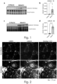
- Example 2 Galectin-3 is upregulated in muscular dystrophy
- the purpose of this study is to develop an analytical method, for the histological detection of lysosomal damage, in transversal sections of skeletal muscles, in animal models and human patients of a range of muscular dystrophies.
- a puncta staining pattern of galectin-3 (Gal-3) was validated recently, in a non-muscular and non-pathological context, as a useful indicator of lysosomal stress response ( Aits et al. 2015. Sensitive detection of lysosomal membrane permeabilization by lysosomal galectin puncta assay. Autophagy 11: 1408-1424 .).
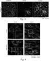
- Example 3 Galectin-3 colocalized with the lysosomal marker LAMP2 (Lysosome Associated Membrane Protein 2)
- Example 4 Gal-3 expressed in the dystrophic muscle by infiltrating monocytes and inside myofibers
- Gal-3 is known to be expressed by infiltrating cells, originating from the immune system. In muscular dystrophy the dystrophic inflammatory muscle is infiltrated by such immune cells. Indeed, the interpretation in previous investigations (cited above) of the upregulation of Gal-3 in the dystrophic muscle, was that galectin-3 is expressed by infiltrating (CD11b positive) immune cells. The co-staining of muscle transversal sections with the monocyte/macrophage marker CD11b revealed indeed double positive cells for CD11b and Galectin-3.
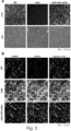
- Example 5 Galectin-3 detects lysosomal damage in the myogenic lineage.
- the lysosomotropic reagent L-leucyl-L-leucine methyl ester is a bioactive molecule that accumulates in the lysosomes, where it induces a lysosomal stress response and membrane damage.
- Example 6 Monitoring lysosomal damage in a gene therapy model for Duchenne muscular dystrophy
- the restoration of dystrophin expression in the mdx mouse model for DMD is a useful experimental model system for gene therapy in muscular dystrophy ( Bourg et al. 2022. Co-Administration of Simvastatin Does Not Potentiate the Benefit of Gene Therapy in the mdx Mouse Model for Duchenne Muscular Dystrophy. Int J Mol Sci 2022, Vol 23, Page 2016 23: 2016 ). However, no evaluation of lysosomal damage has yet been reported in the context of muscle gene therapy.
- Example 7 Monitoring lysosomal damage in a gene therapy model for gamma sarcoglycanopathy
- the LGMD R5 muscular dystrophy is caused by mutations in the SGCG gene, coding for ⁇ -sarcoglycan. Together with ⁇ -, ⁇ -, and ⁇ -sarcoglycan, it participates in the composition of the sarcoglycan subcomplex of the striated myofiber. Mutation or aberrant expression of SGCG resulting in a severe muscular dystrophy. Using a mouse model, the feasibility of a gene replacement strategy in LGMD R5 was recently demonstrated ( Israeli et al. 2019. An AAV-SGCG Dose-Response Study in a ⁇ -Sarcoglycanopathy Mouse Model in the Context of Mechanical Stress. 13: 494-502 .)
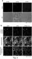
- LGMD R5 mouse model confirm a nearly complete restoration of transgene expression (positive fibers) and tissue architecture (visible in the H&E stain) in the gene therapy treated mice ( figure 6a ).
- the antibody for the detection of the Gammasarcoglycan protein is human-specific (i. e. cannot detect the mouse gammasarcoglycan in the normal mouse and therefore both the healthy (WT) and the null (KO-PBS) mice are detected as gammasarcoglycan negative.
- LGMD R5 mice exhibit elevated expression of both Lamp2 and Gal-3 and their increased puncta colocalization ( figure 6b middle lane).
- Example 8 Detection of Lysosomal damage in muscular dystrophy in human biopsies
- Pompe disease also known as glycogen storage disease type II (GSD2), is an autosomal recessive metabolic disorder.
- the disease is caused by deficiency of the lysosomal acid alpha-glucosidase enzyme, leading to severe cardiac and skeletal muscle myopathy due to progressive accumulation of lysosomal glycogen.
- This accumulation of excess undigested glycogen in the lysosome causes lysosomal dysfunction, which is associated with lysosomal structural deformation and membrane rupture ( Thurberg et al. 2006. Characterization of pre- and post-treatment pathology after enzyme replacement therapy for Pompe disease. Lab Invest 86: 1208-1220 .).
- Example 9 Monitoring lysosomal damage in a gene therapy model for LGMDR9 muscular dystrophy
- LGMDR9 is caused by the lack or the drastically reduced enzymatic activity of the FKRP protein.
- the FKRP protein participates in the glycosylation of ⁇ -dystroglycan, which is important for the matrix anchor of skeletal myofibers.
- LGMDR9 is a recessive autosomal muscular dystrophy, affecting preferentially the muscles of the shoulder and pelvic girdles.
- the severity of the pathology is very heterogeneous.
- the muscular symptoms appear between the first to third decades and vary from the severe, Duchenne-like, to relatively benign courses.
- the heart can also be affected with sometime severe heart failure and death.
- the pathophysiology of LGMDR9 is yet poorly understood, and to our knowledge the participation of lysosomal damage or dysfunction have never been anticipated.
- a mouse model for LGMDR9 (district from PMID: 28334834) was created, characterized, treated by gene therapy, and muscle biopsies were subjected to the Galecin3 puncta assay for the evaluation of lysosomal damage in LGMDR9 and its correction by gene therapy.
- the HSA- Fkrp -Del mouse (with deletion of FKRP specifically in skeletal myofibers) was treated intravenously by the AAV9 hFKRP viral vector (encoding for the human FKRP transgene) for the duration of three months, at a dose of 2.5 10 12 vg/kg.
- mice were sacrificed.
- Vector copy number DNA, and transgene (hFKRP) RNA expressions were detected in the dissected muscle biopsies (including the TA, the psoas, and the diaphragm) (not shown), which confirmed efficient vector transfer and transgene expression respectively.
- Histological correction of the dystrophic phenotype of the TA muscle was further confirmed by H&E analysis (not shown), indicating efficient correction of the dystrophic phenotype.
Landscapes
- Chemical & Material Sciences (AREA)
- Life Sciences & Earth Sciences (AREA)
- Proteomics, Peptides & Aminoacids (AREA)
- Health & Medical Sciences (AREA)
- Organic Chemistry (AREA)
- Wood Science & Technology (AREA)
- Analytical Chemistry (AREA)
- Zoology (AREA)
- Genetics & Genomics (AREA)
- Engineering & Computer Science (AREA)
- Pathology (AREA)
- Immunology (AREA)
- Microbiology (AREA)
- Molecular Biology (AREA)
- Biotechnology (AREA)
- Biophysics (AREA)
- Physics & Mathematics (AREA)
- Biochemistry (AREA)
- Bioinformatics & Cheminformatics (AREA)
- General Engineering & Computer Science (AREA)
- General Health & Medical Sciences (AREA)
- Measuring Or Testing Involving Enzymes Or Micro-Organisms (AREA)
Priority Applications (1)
| Application Number | Priority Date | Filing Date | Title |
|---|---|---|---|
| EP23194414.1A EP4516927A1 (de) | 2023-08-31 | 2023-08-31 | Galectin-3, ein biomarker für lysosomale schädigung bei muskeldystrophie |
Applications Claiming Priority (1)
| Application Number | Priority Date | Filing Date | Title |
|---|---|---|---|
| EP23194414.1A EP4516927A1 (de) | 2023-08-31 | 2023-08-31 | Galectin-3, ein biomarker für lysosomale schädigung bei muskeldystrophie |
Publications (1)
| Publication Number | Publication Date |
|---|---|
| EP4516927A1 true EP4516927A1 (de) | 2025-03-05 |
Family
ID=87889490
Family Applications (1)
| Application Number | Title | Priority Date | Filing Date |
|---|---|---|---|
| EP23194414.1A Withdrawn EP4516927A1 (de) | 2023-08-31 | 2023-08-31 | Galectin-3, ein biomarker für lysosomale schädigung bei muskeldystrophie |
Country Status (1)
| Country | Link |
|---|---|
| EP (1) | EP4516927A1 (de) |
Citations (4)
| Publication number | Priority date | Publication date | Assignee | Title |
|---|---|---|---|---|
| WO2014049282A1 (fr) | 2012-09-26 | 2014-04-03 | Genethon | Procede pour le diagnostic de dystrophies musculaires |
| EP2882447A1 (de) * | 2012-08-10 | 2015-06-17 | The Board Of Regents Of The Nevada System Of Higher Education, On Behalf Of The University Of Nevada, Reno | Verfahren zur diagnose, prognose und behandlung von muskeldystrophie |
| WO2015104403A1 (en) | 2014-01-09 | 2015-07-16 | Genethon | Methods for diagnosing or monitoring muscular dystrophies |
| US20210299169A1 (en) * | 2020-02-18 | 2021-09-30 | Unm Rainforest Innovations | Galectin-3 Governs and Coordinates ESCRT and Autophagic Responses During Endomembrane Damage |
-
2023
- 2023-08-31 EP EP23194414.1A patent/EP4516927A1/de not_active Withdrawn
Patent Citations (4)
| Publication number | Priority date | Publication date | Assignee | Title |
|---|---|---|---|---|
| EP2882447A1 (de) * | 2012-08-10 | 2015-06-17 | The Board Of Regents Of The Nevada System Of Higher Education, On Behalf Of The University Of Nevada, Reno | Verfahren zur diagnose, prognose und behandlung von muskeldystrophie |
| WO2014049282A1 (fr) | 2012-09-26 | 2014-04-03 | Genethon | Procede pour le diagnostic de dystrophies musculaires |
| WO2015104403A1 (en) | 2014-01-09 | 2015-07-16 | Genethon | Methods for diagnosing or monitoring muscular dystrophies |
| US20210299169A1 (en) * | 2020-02-18 | 2021-09-30 | Unm Rainforest Innovations | Galectin-3 Governs and Coordinates ESCRT and Autophagic Responses During Endomembrane Damage |
Non-Patent Citations (14)
| Title |
|---|
| "UniprotKB / Swiss-Prot", Database accession no. P1 793 1 |
| AARTSMA-RUS ASPITALI P.: "Circulating Biomarkers for Duchenne Muscular Dystrophy.", J NEUROMUSCUL DIS., vol. 2, no. 2, 22 July 2015 (2015-07-22), pages S49 - S58 |
| AITS ET AL.: "Sensitive detection of lysosomal membrane permeabilization by lysosomal galectin puncta assay.", AUTOPHAGY, vol. 11, 2015, pages 1408 - 1424 |
| AITS SONJA ET AL: "Sensitive detection of lysosomal membrane permeabilization by lysosomal galectin puncta assay", AUTOPHAGY, vol. 11, no. 8, 3 August 2015 (2015-08-03), US, pages 1408 - 1424, XP093128858, ISSN: 1554-8627, DOI: 10.1080/15548627.2015.1063871 * |
| BOUCHARDTREMBLAY, J., CLIN. MED, vol. 12, 2023, pages 4769 |
| BOURG ET AL., PMID, vol. 35216132, 2022 |
| BOURG ET AL.: "Co-Administration of Simvastatin Does Not Potentiate the Benefit of Gene Therapy in the mdx Mouse Model for Duchenne Muscular Dystrophy.", INT J MOL SCI, vol. 23, 2022, pages 2016 |
| CHAMBERLAIN, JEFFREY S ET AL.: "Microdystrophin Expression as a Surrogate Endpoint for Duchenne Muscular Dystrophy Clinical Trials", HUMAN GENE THERAPY, vol. 34, no. 9-10, 2023, pages 404 - 415 |
| ISRAELI ET AL., AN AAV-SGCG DOSE-RESPONSE STUDY IN A Γ-SARCOGLYCANOPATHY MOUSE MODEL IN THE CONTEXT OF MECHANICAL STRESS., vol. 13, 2019, pages 494 - 502 |
| ISRAELI ET AL., PMID, vol. 31194043, 2019 |
| JULIE M CRUDELE ET AL: "Abstract", HUMAN MOLECULAR GENETICS, vol. 28, no. R1, 25 June 2019 (2019-06-25), GB, pages R102 - R107, XP055720521, ISSN: 0964-6906, DOI: 10.1093/hmg/ddz128 * |
| OZAWA EHAGIWARA YYOSHIDA M.: "Creatine kinase, cell membrane and Duchenne muscular dystrophy.", MOL CELL BIOCHEM., vol. 190, no. 1-2, January 1999 (1999-01-01), pages 143 - 51, XP002245782 |
| THURBERG ET AL.: "Characterization of pre- and post-treatment pathology after enzyme replacement therapy for Pompe disease.", LAB INVEST, vol. 86, 2006, pages 1208 - 1220 |
| VAN PUTTEN ET AL.: "Comparison of skeletal muscle pathology and motor function of dystrophin and utrophin deficient mouse strains.", NEUROMUSCUL DISORD, vol. 22, 2012, pages 406 - 417 |
Similar Documents
| Publication | Publication Date | Title |
|---|---|---|
| Grohmann et al. | Characterization of Ighmbp2 in motor neurons and implications for the pathomechanism in a mouse model of human spinal muscular atrophy with respiratory distress type 1 (SMARD1) | |
| Fugier et al. | Misregulated alternative splicing of BIN1 is associated with T tubule alterations and muscle weakness in myotonic dystrophy | |
| Friedrich et al. | Evidence for FHL1 as a novel disease gene for isolated hypertrophic cardiomyopathy | |
| Vardarajan et al. | Identification of Alzheimer disease-associated variants in genes that regulate retromer function | |
| Xu et al. | Adeno-associated virus 9 mediated FKRP gene therapy restores functional glycosylation of α-dystroglycan and improves muscle functions | |
| Gurvich et al. | DMD exon 1 truncating point mutations: amelioration of phenotype by alternative translation initiation in exon 6 | |
| WO2015089575A1 (en) | Method of diagnosis and treatment | |
| EP4579240A1 (de) | Cf48-nierenverletzungsbiomarker und verwendung davon in einem nierenverletzungsbehandlungsarzneimittel | |
| KR20110081846A (ko) | 미네랄로코르티코이드 수용체 활성화의 바이오마커 | |
| Crawford Parks et al. | Muscle-specific expression of the RNA-binding protein Staufen1 induces progressive skeletal muscle atrophy via regulation of phosphatase tensin homolog | |
| CN112961913B (zh) | lncRNA在复发性流产诊治中的应用 | |
| KR102242639B1 (ko) | 간 섬유화 진단용 바이오마커 miRNA4668-5p | |
| Christiansen et al. | ANO5-related muscle diseases: From clinics and genetics to pathology and research strategies | |
| Chen et al. | CPNE1 regulates myogenesis through the PERK-eIF2α pathway mediated by endoplasmic reticulum stress | |
| ES2369552T3 (es) | Procedimiento in vitro para la diagnosis de enfermedades neurodegenerativas. | |
| US20090280517A1 (en) | Methods of diagnosis and prognosis for a muscular dystrophy | |
| EP4516927A1 (de) | Galectin-3, ein biomarker für lysosomale schädigung bei muskeldystrophie | |
| He et al. | Cardiac CIP protein regulates dystrophic cardiomyopathy | |
| KR102583540B1 (ko) | 퇴행성 뇌질환에서 zbtb16의 용도 | |
| KR101365206B1 (ko) | 심장근육병 진단용 마커 Orai1 | |
| WO2011083637A1 (ja) | 肝疾患治療薬 | |
| Cassidy et al. | Exploring the enigmatic association between PNLIP variants and risk of chronic pancreatitis in a large Chinese cohort | |
| Llansó et al. | Repeat Expansions in PLIN4 Cause Autosomal Dominant Vacuolar Myopathy With Sarcolemmal Features | |
| CN110191956A (zh) | 蛋白质积累的调节及为其应用 | |
| EP2646572B1 (de) | Diagnose und behandlung von chronischem herzversagen |
Legal Events
| Date | Code | Title | Description |
|---|---|---|---|
| PUAI | Public reference made under article 153(3) epc to a published international application that has entered the european phase |
Free format text: ORIGINAL CODE: 0009012 |
|
| STAA | Information on the status of an ep patent application or granted ep patent |
Free format text: STATUS: THE APPLICATION HAS BEEN PUBLISHED |
|
| AK | Designated contracting states |
Kind code of ref document: A1 Designated state(s): AL AT BE BG CH CY CZ DE DK EE ES FI FR GB GR HR HU IE IS IT LI LT LU LV MC ME MK MT NL NO PL PT RO RS SE SI SK SM TR |
|
| STAA | Information on the status of an ep patent application or granted ep patent |
Free format text: STATUS: THE APPLICATION IS DEEMED TO BE WITHDRAWN |
|
| 18D | Application deemed to be withdrawn |
Effective date: 20250906 |