EP4520257A1 - Sensorisierte chirurgische vorrichtung und verfahren zum betrieb solch einer vorrichtung - Google Patents
Sensorisierte chirurgische vorrichtung und verfahren zum betrieb solch einer vorrichtung Download PDFInfo
- Publication number
- EP4520257A1 EP4520257A1 EP24198408.7A EP24198408A EP4520257A1 EP 4520257 A1 EP4520257 A1 EP 4520257A1 EP 24198408 A EP24198408 A EP 24198408A EP 4520257 A1 EP4520257 A1 EP 4520257A1
- Authority
- EP
- European Patent Office
- Prior art keywords
- tissue
- anatomical tissue
- electrodes
- processing
- control means
- Prior art date
- Legal status (The legal status is an assumption and is not a legal conclusion. Google has not performed a legal analysis and makes no representation as to the accuracy of the status listed.)
- Pending
Links
Images

Classifications
-
- A—HUMAN NECESSITIES
- A61—MEDICAL OR VETERINARY SCIENCE; HYGIENE
- A61B—DIAGNOSIS; SURGERY; IDENTIFICATION
- A61B5/00—Measuring for diagnostic purposes; Identification of persons
- A61B5/0059—Measuring for diagnostic purposes; Identification of persons using light, e.g. diagnosis by transillumination, diascopy, fluorescence
- A61B5/0075—Measuring for diagnostic purposes; Identification of persons using light, e.g. diagnosis by transillumination, diascopy, fluorescence by spectroscopy, i.e. measuring spectra, e.g. Raman spectroscopy, infrared absorption spectroscopy
-
- A—HUMAN NECESSITIES
- A61—MEDICAL OR VETERINARY SCIENCE; HYGIENE
- A61B—DIAGNOSIS; SURGERY; IDENTIFICATION
- A61B5/00—Measuring for diagnostic purposes; Identification of persons
- A61B5/0059—Measuring for diagnostic purposes; Identification of persons using light, e.g. diagnosis by transillumination, diascopy, fluorescence
- A61B5/0082—Measuring for diagnostic purposes; Identification of persons using light, e.g. diagnosis by transillumination, diascopy, fluorescence adapted for particular medical purposes
-
- A—HUMAN NECESSITIES
- A61—MEDICAL OR VETERINARY SCIENCE; HYGIENE
- A61B—DIAGNOSIS; SURGERY; IDENTIFICATION
- A61B5/00—Measuring for diagnostic purposes; Identification of persons
- A61B5/05—Detecting, measuring or recording for diagnosis by means of electric currents or magnetic fields; Measuring using microwaves or radio waves
- A61B5/053—Measuring electrical impedance or conductance of a portion of the body
- A61B5/0537—Measuring body composition by impedance, e.g. tissue hydration or fat content
-
- A—HUMAN NECESSITIES
- A61—MEDICAL OR VETERINARY SCIENCE; HYGIENE
- A61B—DIAGNOSIS; SURGERY; IDENTIFICATION
- A61B5/00—Measuring for diagnostic purposes; Identification of persons
- A61B5/05—Detecting, measuring or recording for diagnosis by means of electric currents or magnetic fields; Measuring using microwaves or radio waves
- A61B5/053—Measuring electrical impedance or conductance of a portion of the body
- A61B5/0538—Measuring electrical impedance or conductance of a portion of the body invasively, e.g. using a catheter
-
- A—HUMAN NECESSITIES
- A61—MEDICAL OR VETERINARY SCIENCE; HYGIENE
- A61B—DIAGNOSIS; SURGERY; IDENTIFICATION
- A61B5/00—Measuring for diagnostic purposes; Identification of persons
- A61B5/68—Arrangements of detecting, measuring or recording means, e.g. sensors, in relation to patient
- A61B5/6846—Arrangements of detecting, measuring or recording means, e.g. sensors, in relation to patient specially adapted to be brought in contact with an internal body part, i.e. invasive
- A61B5/6847—Arrangements of detecting, measuring or recording means, e.g. sensors, in relation to patient specially adapted to be brought in contact with an internal body part, i.e. invasive mounted on an invasive device
-
- A—HUMAN NECESSITIES
- A61—MEDICAL OR VETERINARY SCIENCE; HYGIENE
- A61B—DIAGNOSIS; SURGERY; IDENTIFICATION
- A61B5/00—Measuring for diagnostic purposes; Identification of persons
- A61B5/72—Signal processing specially adapted for physiological signals or for diagnostic purposes
- A61B5/7235—Details of waveform analysis
- A61B5/7264—Classification of physiological signals or data, e.g. using neural networks, statistical classifiers, expert systems or fuzzy systems
-
- A—HUMAN NECESSITIES
- A61—MEDICAL OR VETERINARY SCIENCE; HYGIENE
- A61B—DIAGNOSIS; SURGERY; IDENTIFICATION
- A61B2505/00—Evaluating, monitoring or diagnosing in the context of a particular type of medical care
- A61B2505/05—Surgical care
Definitions
- the present invention relates to a sensorized surgical device and method of operation of such a device.
- the invention relates to a device for surgical use, and its operating method, such as a forceps or the like, equipped with integrated sensors for the identification of cancerous tissue operating in real time, but which can be used for any medical circumstance in which there is a need for real-time detection of the condition of a tissue.
- the intraoperative cryostat examination is an anatomic-pathologic diagnosis performed during surgery on a fragment of the suspected neoplastic lesion, according to the result of which, the surgery itself can be conducted conservatively or demolition.
- the application of said intraoperative examination is to clarify the diagnosis of the disease, in case this has not been previously made, or in case new diagnostic questions emerge in the operating room, or, finally, to assess the residual disease in the margins of the excised surgical piece, thus guiding the subsequent therapeutic strategy.
- the extemporaneous examination indicated above involves a biopsy of the surgical piece, with sampling and diagnosis at the same time as the surgery itself, but with often not easily reducible and predictable times, which inevitably burden the total operative time.
- the extemporaneous examination involves numerous complex procedures that need to be carried out in a predetermined sequence: biopsy collection, freezing of the same inside the cryostat and cutting by a laboratory technician, preparation of the section for its adhesion on the object slide, staining and reading by the anatomo-pathologist for diagnosis.
- DEXA dual energy X-ray absorptiometry
- Laser spectroscopy thanks to the Raman effect, can also calculate a given typical value for a given tissue.
- the role of laser spectroscopy in determining neoplastic tissue is described in detail.
- the problem is addressed more from a clinical point of view.
- optical properties absorption and scattering
- patent papers are presented describing devices that can identify the presence of cancer in real time, based on laser spectroscopy and optical property analysis.
- Nitric oxide diffuses into the interstitial space of cancer cells, so the detection of this gas, performed by needle aspiration [8] and subsequent analysis, is an important additional data.
- a further peculiarity of the neoplastic cell is the increase in membrane potential, which is managed by the ATP-ase pumps, i.e., enzymes capable of removing Na+ ions from the cell to favor the entry of K+ ions.
- the value of the resting potential varies, but in excitable cells is between -90 and -70 MV.
- the membrane potential in old cells goes down to -50MV and, in degenerate cells it goes down to - 15MV.
- the purpose of the present invention to propose a sensorized surgical device that can not only recognize tissue, but also determine the possible presence of cancerous tissue.
- Another purpose of the invention is to propose a low cost device that can be easily integrated into known devices.
- a sensorized device for detecting conductive properties of an anatomical tissue comprising a first jaw, a second jaw, mechanically connected to said first jaw and articulated with respect to it, so that said tissue can be caught between said two jaws, at least two application electrodes for applying an alternating current to said anatomical tissue, wherein said application electrodes are arranged on said jaws, at least two measurement electrodes, for detecting the impedance of said anatomical tissue, arranged on one of said jaws, a light source for emitting one or more light radiations on the anatomical tissue, and an optical receiver, configured to receive said light radiation to receive an optical spectrum of said anatomical tissue and generate a signal containing information on the optical spectrum, processing and control means, connected to said application electrodes and to said measurement electrodes, and signaling means, connected to said processing and control means, characterized in that said processing and control means are configured for: measuring the impedance between said measurement electrodes, when an alternating electric current is applied to the anatomical tissue by
- said processing and control means may be configured to measure impedance by detecting both the real part, associated with the resistance, and the imaginary part, associated with the capacity of said anatomical tissue.
- said sensor may comprise a biosensor, connected to said processing and control means, and said processing and control means may be configured for measuring the change in membrane potential, extract a characteristic value or pattern of said membrane potential, and determining diagnostic information relating to the anatomical tissue arranged between said jaws based on said characteristic value or pattern of said sensed membrane potential, to determine the presence of cancer cells.
- said biosensor may consists of a pair of electrodes, each to be positioned in contact with the anatomical tissue to be examined.
- said sensor may comprise further electrodes arranged on said jaws, connected to said processing and control means, and said processing and control means may be configured to measure the ratio between the module of the voltage between said measurement electrodes and the module of the alternating electric current applied to the anatomical tissue, and the difference between the phase of the voltage between said further electrodes and the phase of the alternating electric current applied to said anatomical tissue.
- said neural network algorithm may be based on a neural network of the Convolutional Neural Network (CNN) type.
- CNN Convolutional Neural Network
- said processing and control means may comprise: an analysis unit connected to said measurement electrodes and to said application electrodes; and a control logic unit connected to said analysis unit, wherein said control logic unit processes the impedance between said measurement electrodes, when an alternating electric current is applied to the anatomical tissue by means of said application electrodes, and processes said signal containing information on the optical spectrum.
- said sensor may comprise first focusing means for focusing the light radiation emitted by said light source, wherein said first focusing means are connected to said light source and are arranged on said first jaw, and second focusing means for focusing the light radiation received by said optical receiver, wherein said second focusing means are connected to said optical receiver and are arranged on said first jaw.
- said diagnostic information are based on the fact that cancerous tissues, according to the type of pathology, have different resistance and/or capacity (impedance) values in the case of healthy or cancerous tissue.
- said analysis unit may also comprise a plurality of LEDs, each of which is associated with a specific signal to signal the possible presence of potential cancerous tissue.
- said sensor may comprise a handle, and said analysis unit is arranged in said handle.
- processing by means of said processing and control means the signal from said analysis electrodes and said signal containing information on the optical spectrum using a neural network algorithm resident on a processor trained to: H1. extracting a characteristic impedance value or pattern; H2. determining diagnostic information relating to the anatomical tissue arranged between said jaws on the basis of said extracted characteristic impedance value or pattern and said detected optical spectrum; and I. activating said signaling means by means of processing and control means, so as to transmit a signal regarding the presence of cancerous tissue.
- said processing and control means may discriminate portions of cancerous and non-cancerous tissue on the basis of the different energy state on the basis of the Warburg effect.
- said step F further comprises the sub-step of measuring the variation of the membrane potential by means of said biosensor
- said step H further comprises the following sub-steps: H3. extracting a characteristic value or pattern of said membrane potential; and H4. determining diagnostic information relating to the tissue arranged between said jaws based on said characteristic value or pattern of said detected membrane potential, to determine the presence of cancer cells.
- said signal containing information on the optical spectrum is based on the Raman effect.
- a computer program comprising instructions which, when the program is executed by a computer, cause the computer to execute steps F-H of the method defined above.
- a computer-readable storage medium comprising instructions which, when executed by a computer, cause the computer to execute steps F-H of the method defined above.
- Sensorized surgical device 1 and the proposed methodology allow real-time diagnosis of the presence or absence of cancerous tissue on the organ under examination during surgery, providing an immediate response and providing the ability to perform the measurement on multiple contiguous specimens without the need for multiple biopsies, thereby significantly lowering the costs of using trained personnel and dedicated space.
- the proposed solution extends the capabilities of other electro-optical devices for real-time identification of biological tissues, allowing the surgeon to verify in real time the type (cancerous or noncancerous) of the tissue being analyzed.
- the sensorized surgical device 1 involves, as better described below, several sensors or electrodes for real-time data collection, and an algorithm, implemented on hardware interfaced to the instrument itself, that allows fast and high probability identification of the possible cancerous nature of the tissue under analysis.
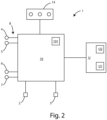
- the sensorized surgical device 1 essentially comprises a first branch 11, a second branch 12, an analysis unit 10, to control the signals of the device 1, and a central control unit U to recognize an anatomical tissue by optical and electrical properties of the same and the possible presence of a cancerous tissue, based on impedance-metric properties.
- the central control unit U is connected by wire or preferably wirelessly with the analysis unit 10.
- the central control unit U will be better described in the following, both structurally and functionally.
- the sensorized surgical device 1 also comprises a light source 2 for emitting one or more light radiations on the anatomical tissue, connected to said analysis unit 10, and an optical receiver 3, connected to said analysis unit 10, configured to receive a light radiation, derive from said light radiation an optical spectrum of said anatomical tissue, and transmit to said analysis unit 10 a signal containing information about the optical spectrum.
- the analysis unit 10 is configured to transmit the signals to the central control unit U for their processing, as further explained below.
- the sensorized surgical device 1 also comprises a pair of application electrodes 4 and 5.
- Each application electrode 4 and 5 has a free portion, to be placed in contact with the anatomical tissue to be examined.
- each of the two electrodes 4 and 5 is connected to the analysis unit 10 with cables 8.
- the analysis unit 10, by means of the application electrodes 4 and 5, is able to apply an alternating electric current to the anatomical tissue.
- the sensorized surgical device 1 also comprises a pair of measurement electrodes 6 and 7, each having a portion to be placed in contact with the anatomical tissue to be examined.
- the two measurement electrodes 6 and 7 are also connected to the analysis unit 10, while the central control unit U can measure the impedance between said measurement electrodes 6 and 7 in terms of modulus and phase when an alternating electric current is applied to said anatomical tissue.
- Light source 2 is activated by said analysis unit 10 so that the anatomical tissue is affected by one or more light radiations emitted by the light source.
- Each light radiation can have a respective predetermined wavelength.
- the optical receiver 3 is electrically connected to said analysis unit 10 and is configured to receive at least the light radiation scattered by said anatomical tissue, having one or more wavelengths different from the predetermined wavelength of the light radiation emitted by the light source 2, as well as, as mentioned above, to extract the optical spectrum and send a signal containing the optical spectrum information to the analysis unit 10.
- the central control unit U is then configured to receive the signal sent by said optical receiver to said analysis unit 10, containing the optical spectrum information of the anatomical tissue.
- Scattered light radiation is the portion of light radiation emitted by said light source 2 that has interacted, generally inelastically, with the molecular structure of the anatomical tissue.
- Optical spectrum means an optical spectrum centered around the wavelength of light radiation emitted by light source 2.
- light source 2 and optical receiver 3 are arranged externally to branches 11 and 12 of device 1.
- the sensorized surgical device 1 may not include the light source 2 and the optical receiver 3.
- Light source 2 and optical receiver 3 can be placed inside surgical forceps 1, preferably inside the handle (not visible in the figure).
- Light source 2 comprises first focusing means 21 to focus the light radiation emitted by light source 2.
- the first focusing means 21 are connected to light source 2.
- the optical receiver 3 comprises second focusing means 31 for focusing the light radiation received by said optical receiver 3.
- the second focusing means 31 are connected to said optical receiver 3.
- Said first focusing means 21 and said second focusing means 31 are arranged on said first jaw 11 and said second jaw 12, respectively.
- the first electrode 4 and the second electrode 5 are placed on the first jaw 11 and the second jaw 12, respectively.
- the two application electrodes 4 and 5 are placed so that at least a portion of anatomical tissue is present between them and said measurement electrodes 6 and 7, so that the alternating electric current flows from one electrode to the other, passing through the portion of anatomical tissue.
- the central control unit U measures the impedance between them when an alternating electric current is applied through said analysis unit 10 to the anatomical tissue.
- the third electrode 6 and the fourth measurement electrode 7 are placed on the first jaw 11 and the second jaw 12, respectively.
- each of said additional electrodes is electrically connected to the analysis unit 10.
- the measurement electrodes 6 and 7 are positioned so that anatomical tissue is present between them, and the impedance between said two further electrodes is measurable by the central control unit U, when an alternating electric current flows between said application electrodes 4 and 5, through the portion of the anatomical tissue under examination.
- the application electrodes 4, 5, and measurement electrodes 6 and 7 can be arranged on a respective jaw 11 or 12 or on same jaws.
- the sensorized surgical device 1 also comprises a biosensor 9 consisting of a pair of electrodes 91 and 92, each one to be placed in contact with the anatomical tissue to be examined.
- the two electrodes 91 and 92 are also connected to the analysis unit 10, which can measure the change of the membrane potential if cancer cells are present.
- the sensorized surgical device 1 may not include the biosensor 9 consisting of the electrodes 91 and 92.
- the analysis unit 10 also comprises a plurality of LEDs 13, each one having a distinct color and in each case associated with a specific signal, e.g., type of tissue to be detected, or presence of potential cancerous tissue.
- Cancerous tissues depending on the type of disease, have different values or patterns of resistance and/or capacitance (impedance) in the case of healthy or cancerous tissue.
- the resistance value increases progressively with the development of the disease compared with the case of healthy tissue.
- capacitance has higher average values in the case of cancerous tissue, while resistance takes on comparable values.
- the central control unit U is able to know the voltage (in terms of modulus and phase) between the two application electrodes 4 and 5, when an alternating electric current is applied to said anatomical tissue by means of said application electrodes 4 and 5. Also, again by means of said application electrodes 6 and 7, the analysis unit is able to detect the impedance between them.
- the recognition unit 10 by means of the central control unit U, is configured to apply an alternating electric current to said anatomical tissue by means of said application electrodes 4 and 5, measure a voltage between said measurement electrodes 6 and 7 (in terms of modulus and phase), when said alternating electric current is applied to said anatomical tissue by means of said application electrodes 4 and 5, to receive signals containing information about said alternating electric current applied to said anatomical tissue by means of said application electrodes 4 and 5, and signals containing information about the corresponding voltage between further electrodes 6 and 7, as well as to calculate an impedance value of said anatomical tissue given by the ratio of said voltage to said alternating electric current.
- the central control unit U is configured to measure the relationship between the voltage modulus between said measuring electrodes and the modulus of the alternating electric current applied to the anatomical tissue, as well as the phase difference between the voltage phase between said further electrodes and the phase of the alternating electric current applied to the anatomical tissue.
- the recognition unit 10 can vary the frequency value of said alternating electric current to identify the anatomical tissue with a high probability.
- the recognition unit 10 is configured to apply, under the control of central control unit U, an additional alternating electric current to the same anatomical tissue via the two application electrodes 4 and 5.
- This additional alternating electric current has a predetermined frequency, different from the above predetermined frequency.
- the central control unit U is thus configured to measure an additional voltage between the additional measurement electrodes 6 and 7 in terms of modulus and phase, when the additional alternating electric current is applied to said anatomical tissue, and calculate an additional impedance value given by the ratio of said additional voltage measured through said additional electrodes 6 and 7, when said additional alternating current is applied to said anatomical tissue, to the additional alternating electric current.
- impedance in fact, is a physical vector quantity expressible as a complex number having a real part and an imaginary part.
- the real part of the impedance is associated with the information about the resistance of the anatomical tissue, and the imaginary part is associated with the information about the reactance of the anatomical tissue.
- the central control unit U is also configured to receive signals sent by said optical receiver 3 containing information about the optical spectrum of the anatomical tissue to be recognized.
- the received optical spectrum takes on different characteristics in the case of healthy and cancerous tissue.
- the Raman shift peak observed at 2850 cm -1 presents much lower intensity in the case of cancerous tissue.
- the signal sent by said optical receiver 3 to said recognition unit 10 is an electrical signal.
- the signals from said electrodes 4 and 5 to said recognition unit 10 and the signals from said further electrodes 6 and 7 to said recognition unit 10 are also electrical signals.
- the recognition unit 10 under the control of central control unit U, is configured to pass a current pulse between electrode pair 91 and 92 of biosensor 9 and read the voltage at the ends.
- the central control unit U is configured to perform in the present embodiment up to four comparisons, one concerning said optical spectrum, one concerning the membrane potential, and the other 2 concerning the real and imaginary part of the impedance, in order to recognize said anatomical tissue with a high probability.
- the central control unit U comprises a database U2 in which data, such as the reference names of a plurality of predetermined anatomical tissues, a plurality of reference optical spectra, each of which is associated with a respective predetermined anatomical tissue, the values of a plurality of reference impedances, each of which is associated with a respective predetermined anatomical tissue, are stored.
- each predetermined anatomical tissue or type of neoplasm or cancer is stored in the U2 database and associated (according to the tissue) with a respective reference optical spectrum and reference impedance value.
- an optical reference spectrum means an optical spectrum centered around a respective wavelength of light radiation emitted by a reference light source.
- said central control unit U is configured to perform a first comparison between the optical spectrum of said anatomical tissue to be recognized, and one or more reference optical spectra stored in said database, and to perform a second comparison between the impedance value obtained by said central control unit U and one or more reference impedance values stored in said database, as well as to identify said anatomical tissue from the combination of said two comparisons as well as the possible presence of cancerous tissue.
- the central control unit U is then capable of recognizing whether an anatomical tissue has cancerous cells from a twofold comparison with data stored in the database.
- the combination of the information on the molecular structure of the anatomical tissue, given by the optical spectrum, and the information on the resistance and reactance of the same anatomical tissue, given by the impedance, allows both the recognition of the anatomical tissue and the determination of the possible presence of cancerous tissue.
- the recognition unit 10 can be configured to perform a third comparison between said additional impedance value, mentioned above, and one or more reference impedance values stored in the database U2, to eliminate possible ambiguities of identification of said anatomical tissue in case of two anatomical tissues have similar electrical properties.
- an alternating electric current is activated, similar to the above, on said anatomical tissue by means of application electrodes 4 and 5.
- This electric current has a predetermined frequency, in which the intensity default value is less than 1 mA so as not to damage the anatomical tissue.
- the alternating electric current has a predetermined frequency between 100Hz and 10MHz.
- a signal is sent via a light source 2 to the optical receiver 3 containing the optical spectrum information of said anatomical tissue.
- the central control unit U calculates or determines an anatomical tissue impedance value given by the ratio between the voltage and alternate electric current.
- a comparison is made between the optical spectrum of said anatomical tissue and reference optical spectra, as well as between the detected or calculated impedance value of the anatomical tissue under consideration and said plurality of reference impedance values, for the purpose of identifying the anatomical tissue based on these comparisons.
- DEXA dual energy X-ray absorptiometry
- the cancer tissue has a different response to bioimpedance, compared with healthy tissue. This analysis is conducted through the analysis electrodes 4 and 5 and measurement electrodes 6 and 7, obtaining useful information for the identification of cancer tissue in real time.
- optical signals as described above is done through the estimation of optical properties (absorption and scattering) and spectral analysis using the Raman effect, so that it is possible to trace the composition of the tissue and some information about its microscopic structure.
- the transmission of the optical signals and the subsequent reception and analysis of the signals received downstream of the interaction with the tissue therefore make it possible to collect data to be used for the identification, as mentioned above. In this way, the sensor device 1 recognizes the type of tissue.
- the sensorized surgical device 1 performs an analysis of the membrane potential and heat release, which is a peculiarity of the neoplastic cell due to the increased membrane potential.
- the value of the resting potential varies and, in degenerated cells, tends to be more depolarized than in healthy cells.
- These data can be retrieved through measurements of electric current and voltage. This processing is carried out by the central control unit U.
- the additional information available makes it possible to obtain additional indicators of the cancerousness of the tissue under analysis.
- the interfacing between the sensorized surgical device 1 and the central control unit U can be, as mentioned, either through wired or, preferably, wireless connections.
- the central control unit U in addition to including the processing algorithms, also controls and synchronizes the overall process through the analysis unit 10, which involves:
- the central control unit U is capable of digitalizing received signals with a sampling rate of at least 100 MS/s and a resolution of at least 8 bits, but at the same time has limited power consumption.
- the central control unit U capable of discriminating a cancerous tissue from a healthy tissue by improving the response time through hardware implementation, i.e., in the processor U1, of a recognition algorithm, which implements neural networks.
- the analysis unit 10 together with the central control unit U, enable effective discrimination of cancerous and non-cancerous portions of tissue. More in detail, the discriminatory ability is related to the Warburg effect, which is peculiar to cancerous tissue compared with healthy tissue.
- the different energy state of cancerous tissue which unlike healthy tissue produces predominantly energy with a high percentage of glycolysis, which, however, is not followed by the usual cycle of mitochondrial respiration, albeit in the presence of adequate oxygen concentration, determines the specificity of bio-impedance characteristics, uptake, diffusion and especially membrane potential variation, which therefore tends to be more depolarized in cancerous tissue than in healthy cells.
- the Warburg effect is based, in fact, on the so-called "aerobic glycolysis", i.e., an essential aspect of the cancer cell metabolism based on the tendency to rely on glycolysis for energy production to a greater extent than in healthy cells.
- the increased glycolysis of cancer cells allows only partial oxidation of glucose molecules and is therefore poorly energy efficient.
- the size of the examination areas it is referred to the ergonomics and standard dimensions of surgical forceps and devices, which, on average, can grasp firmly 1.5 mm wide tissue.
- the data processing is carried out in the analysis unit by means of a previously trained neural network implemented in the central control unit U.
- the neural network is preferably of the CNN type, Convolutional Neural Network, although in other embodiments different types of networks can be used, such as MLP, Multilayer Perceptron networks.
- the training of the neural network is carried out on a hardware platform, i.e., the external dedicated central control unit U, connected, as mentioned, by wire or wireless, based on clinical data collected on both healthy and diseased portions of tissue.
- the data collected through commercial instrumentation on bioimpedance, optical absorption and scattering properties, membrane potential, and heat release enable identification of the structure and weights of neurons in the network optimal for recognition.
- the trained neural network is implemented in the central control unit U, which is a hardware and enables faster response times than a software-based solution. Therefore, the processing is carried out on a dedicated board U2 that houses a state-of-the-art SoC containing both a microprocessor with an embedded Linux system and an FPGA.
- the microprocessor U1 of said central control unit U enables the generation of the required driving signals for the application electrodes 4, 5 and the collection of data produced on the tissue under test; the collected data are then sent to the FPGA of the board U2 for real-time hardware processing.
- the response (binary) signal allows the surgeon to be notified of the outcome of the analysis by turning on one of the LEDs 13, controlled by the analysis unit 10, e.g., a red LED (in case of cancerous tissue) or a green LED (in case of healthy tissue).
- An advantage of the present invention is the integration and the possibility of metabolism of tumor and pathological cells to increase the probability of correctly identifying cancerous tissue.
- the advantage of the solution according to the invention consists of the processing that can provide, as much as possible, an operator-independent result, with objective data derived from impedance analysis, from the analysis of optical signals and not of images interpretable by the more or less experienced eye of a physician, from the analysis of membrane potential and heat release, which is, the latter, a peculiar aspect of the neoplastic cell, endowed with increased membrane potential (see also [14]).
- the integration of bioimpedance and biochemical data are thus a significant advantage for an evaluation of the cancer cell in toto that our inventive proposal brings to increase the objectivity of measurement.
- An additional advantage of the present invention is to obtain real-time diagnosis of the possible presence of cancer cells in an anatomical tissue, avoiding the time-consuming clinical analysis.
Landscapes
- Health & Medical Sciences (AREA)
- Life Sciences & Earth Sciences (AREA)
- Engineering & Computer Science (AREA)
- Physics & Mathematics (AREA)
- General Health & Medical Sciences (AREA)
- Pathology (AREA)
- Biomedical Technology (AREA)
- Heart & Thoracic Surgery (AREA)
- Medical Informatics (AREA)
- Molecular Biology (AREA)
- Surgery (AREA)
- Animal Behavior & Ethology (AREA)
- Biophysics (AREA)
- Public Health (AREA)
- Veterinary Medicine (AREA)
- Nuclear Medicine, Radiotherapy & Molecular Imaging (AREA)
- Radiology & Medical Imaging (AREA)
- Artificial Intelligence (AREA)
- Evolutionary Computation (AREA)
- Fuzzy Systems (AREA)
- Mathematical Physics (AREA)
- Computer Vision & Pattern Recognition (AREA)
- Physiology (AREA)
- Psychiatry (AREA)
- Signal Processing (AREA)
- Spectroscopy & Molecular Physics (AREA)
- Investigating Or Analysing Biological Materials (AREA)
Applications Claiming Priority (1)
| Application Number | Priority Date | Filing Date | Title |
|---|---|---|---|
| IT102023000018600A IT202300018600A1 (it) | 2023-09-11 | 2023-09-11 | Dispositivo chirurgico sensorizzato e metodo di funzionamento di tale dispositivo |
Publications (1)
| Publication Number | Publication Date |
|---|---|
| EP4520257A1 true EP4520257A1 (de) | 2025-03-12 |
Family
ID=88778612
Family Applications (1)
| Application Number | Title | Priority Date | Filing Date |
|---|---|---|---|
| EP24198408.7A Pending EP4520257A1 (de) | 2023-09-11 | 2024-09-04 | Sensorisierte chirurgische vorrichtung und verfahren zum betrieb solch einer vorrichtung |
Country Status (2)
| Country | Link |
|---|---|
| EP (1) | EP4520257A1 (de) |
| IT (1) | IT202300018600A1 (de) |
Citations (11)
| Publication number | Priority date | Publication date | Assignee | Title |
|---|---|---|---|---|
| US2017A (en) | 1841-03-26 | The graphic co | ||
| US6035225A (en) | 1995-01-03 | 2000-03-07 | Omnicorder Technologies | Detection of cancerous lesions by measuring nitric oxide concentrations in tissue |
| US20080306391A1 (en) * | 2001-09-04 | 2008-12-11 | Bioluminate, Inc. | Multisensor probe for tissue identification |
| US20120253202A1 (en) * | 2005-02-21 | 2012-10-04 | Instituto Tecnologico Y De Estudios Superiores De Monterrey, a Mexico corporation | Optoelectronic Device for The Detection of Uterine Cervical Cancer, Comprising A Self-Positioning Attachment |
| US8738124B2 (en) | 2007-12-11 | 2014-05-27 | Epi-Sci, Llc | Electrical bioimpedance analysis as a biomarker of breast density and/or breast cancer risk |
| US9534999B2 (en) | 2012-06-15 | 2017-01-03 | Ellen T. Chen | Nanostructured biomimetic device for detecting a cancer cell or cancer cells |
| ITUB20153157A1 (it) * | 2015-08-18 | 2017-02-18 | Univ Degli Studi Roma La Sapienza | Pinza chirurgica per riconoscere tessuti anatomici. |
| US9723976B2 (en) | 2012-06-27 | 2017-08-08 | Camplex, Inc. | Optics for video camera on a surgical visualization system |
| US9931039B2 (en) | 2012-07-02 | 2018-04-03 | National University Of Singapore | Methods related to real-time cancer diagnostics at endoscopy utilizing fiber-optic Raman spectroscopy |
| WO2019032620A1 (en) * | 2017-08-07 | 2019-02-14 | Utah Valley University | APPARATUS, SYSTEM AND METHOD FOR DIAGNOSTIC IMAGING FORCEPS |
| US20210174957A1 (en) * | 2016-11-21 | 2021-06-10 | Sensome SAS | Characterizing and identifying biological structure |
-
2023
- 2023-09-11 IT IT102023000018600A patent/IT202300018600A1/it unknown
-
2024
- 2024-09-04 EP EP24198408.7A patent/EP4520257A1/de active Pending
Patent Citations (11)
| Publication number | Priority date | Publication date | Assignee | Title |
|---|---|---|---|---|
| US2017A (en) | 1841-03-26 | The graphic co | ||
| US6035225A (en) | 1995-01-03 | 2000-03-07 | Omnicorder Technologies | Detection of cancerous lesions by measuring nitric oxide concentrations in tissue |
| US20080306391A1 (en) * | 2001-09-04 | 2008-12-11 | Bioluminate, Inc. | Multisensor probe for tissue identification |
| US20120253202A1 (en) * | 2005-02-21 | 2012-10-04 | Instituto Tecnologico Y De Estudios Superiores De Monterrey, a Mexico corporation | Optoelectronic Device for The Detection of Uterine Cervical Cancer, Comprising A Self-Positioning Attachment |
| US8738124B2 (en) | 2007-12-11 | 2014-05-27 | Epi-Sci, Llc | Electrical bioimpedance analysis as a biomarker of breast density and/or breast cancer risk |
| US9534999B2 (en) | 2012-06-15 | 2017-01-03 | Ellen T. Chen | Nanostructured biomimetic device for detecting a cancer cell or cancer cells |
| US9723976B2 (en) | 2012-06-27 | 2017-08-08 | Camplex, Inc. | Optics for video camera on a surgical visualization system |
| US9931039B2 (en) | 2012-07-02 | 2018-04-03 | National University Of Singapore | Methods related to real-time cancer diagnostics at endoscopy utilizing fiber-optic Raman spectroscopy |
| ITUB20153157A1 (it) * | 2015-08-18 | 2017-02-18 | Univ Degli Studi Roma La Sapienza | Pinza chirurgica per riconoscere tessuti anatomici. |
| US20210174957A1 (en) * | 2016-11-21 | 2021-06-10 | Sensome SAS | Characterizing and identifying biological structure |
| WO2019032620A1 (en) * | 2017-08-07 | 2019-02-14 | Utah Valley University | APPARATUS, SYSTEM AND METHOD FOR DIAGNOSTIC IMAGING FORCEPS |
Non-Patent Citations (18)
| Title |
|---|
| ANBAR, M., DETECTION OF CANCEROUS LESIONS BY MEASURING NITRIC MCIDE CONCENTRATIONS IN TISSUE. |
| ANDERSON, T. A. ET AL., OPTICAL SENSOR FOR NEEDLE-TIP TISSUE IDENTIFICATION AND DIAGNOSIS |
| BERZINGI, S.NEWMAN, MYU, H. G.: "Altering bioelectricity on inhibition of human breast cancer cells.", CANCER CELL INTEMATIONAL, vol. 16, no. 1, 2016, pages 1 - 9, XP055690344, DOI: 10.1186/s12935-016-0348-8 |
| BRANTLOV SJODAL LHEITMANN BLWARD LC.: "Clinica! value of bioimpedance during long-term cancer therapy.", CLIN CASE REP J., vol. 2, no. 5, 2021, pages 1 - 6 |
| CHEN, E. T., NANOSTRUCTURED BIOMIMETIC DEVICE FOR DETECTING A CANCER CELL OR CANCER CELLS. |
| D'ANDREA VITO ET AL: "Pinza chirurgica per riconoscere tessuti anatomici.", 6 December 2018 (2018-12-06), XP093125468, Retrieved from the Internet <URL:https://www.uniroma1.it/sites/default/files/field_file_allegati/scheda_grafica_tt_266_dandrea_it.pdf> [retrieved on 20240130] * |
| DAVIES, R., ELECTRICAL BIOIMPEDANCE ANALYSIS AS A BIOMARKER OF BREAST DENSITY AND/OR BREAST CANCER RISK |
| DOYLE, TIMOTHY E. ET AL., APPARATUS, SYSTEM AND METHOD FOR DIAGNOSTIC |
| GOMEZ-CORTÉS JCDIAZ-CARMONA JJPADILLA-MEDINA JACALDERON AEGUTIÉRREZ AIBGUTIÉRREZ L6PEZ MPRADO-OLIVAREZ J.: "Electrical Impedance Tomography Technical Contributions for Detection and 3D Geometry Localization of Breast Tumors: A Systematic Review", MICROMACHINES, vol. 13, no. 4, 23 March 2022 (2022-03-23), pages 496 |
| HUANG, Z ET AL., METHODS RELATED TO REAL-TIME CANCER DIAGNOSTICS AT ENDOSCOPY UTILIZING FIBER-OPTIC RAMAN SPECTROSCOPY. |
| KHAN MNWANG QLDREES BSXIANGLI WTENG GCUI XZHAO ZWEI KABRAR M: "A Review on Laser-Induced Breakdown Spectroscopy in Different _Cancers Diagnosis and Classification.", FRONT. PHYS., vol. 10, 2022, pages 821057 |
| MAGAR HEND S. ET AL: "Electrochemical Impedance Spectroscopy (EIS): Principles, Construction, and Biosensing Applications", SENSORS, vol. 21, no. 19, 1 October 2021 (2021-10-01), CH, pages 6578, XP093037100, ISSN: 1424-8220, DOI: 10.3390/s21196578 * |
| NEMETH, D.V.BALDINI, E.SORRENTI, S.D'ANDREA, V.;BELLINI, M.I.: "Cancer Metabolism and Ischemia-Reperfusion Injury: Two Sides of the Same Coin", J CLIN. MED., vol. 11, 2022, pages 5096, Retrieved from the Internet <URL:https://doi.org/10.3390/jcml1175096> |
| SANTOS IP, BARROSO EM, BAKKER SCHUT TC, CASPERS PJ, VAN LANSCHOT CGF, CHOI DH, VAN DER KAMP MF, SMITS RWH, VAN DOOM R, VERDIJK RM,: "Raman spectroscopy for cancer detection and cancer surgery guidance: translation to the clinics.", ANALYST, vol. 142, no. 17, 21 August 2017 (2017-08-21), pages 3025 - 3047 |
| SHELTON IVFREDERICK E.; DECKANDREW C., SURGICAL SYSTEM CONTROL BASED ON MULTIPLE SENSED PARAMETERS |
| SHEN, Y. X.SABOE, P. OSINES, I. T.ERBAKAN, M.KUMAR, M.: "Biomimetic membranes: A review.", JOUMAL OF MEMBRANE SCIENCE, vol. 454, 2014, pages 359 - 381, XP055360785, Retrieved from the Internet <URL:https:/Idoi.org/10.1016/j.memsci.2013.12.019.> DOI: 10.1016/j.memsci.2013.12.019 |
| TESAR, J., OPTICS FOR VIDEO CAMERA ON A SURGICAL VISUALIZATION SYSTEM. |
| ZHANG TAO ET AL: "Advances of deep learning in electrical impedance tomography image reconstruction", FRONTIERS IN BIOENGINEERING AND BIOTECHNOLOGY, vol. 10, 14 December 2022 (2022-12-14), CH, XP093125609, ISSN: 2296-4185, DOI: 10.3389/fbioe.2022.1019531 * |
Also Published As
| Publication number | Publication date |
|---|---|
| IT202300018600A1 (it) | 2025-03-11 |
Similar Documents
| Publication | Publication Date | Title |
|---|---|---|
| CA2134423C (en) | Method and apparatus for tissue type recognition | |
| US9226731B2 (en) | Optically guided needle biopsy system using multi-modal spectroscopy in combination with a transrectal ultrasound probe | |
| Kallaway et al. | Advances in the clinical application of Raman spectroscopy for cancer diagnostics | |
| US20130231573A1 (en) | Apparatus and methods for characterization of lung tissue by raman spectroscopy | |
| Short et al. | Using laser Raman spectroscopy to reduce false positives of autofluorescence bronchoscopies: a pilot study | |
| WO2009043045A1 (en) | Systems and methods for spectral analysis of a tissue mass using an instrument, an optical probe, and a monte carlo or a diffusion algorithm | |
| CN103917155B (zh) | 用于确定组织状态的检测装置 | |
| US10779804B2 (en) | Biopsy needle for biopsy sampling, biopsy device, and methods of manufacturing a biopsy needle or a biopsy device | |
| RU2138192C1 (ru) | Способ распознавания типа ткани и аппарат для осуществления способа | |
| CN114519708B (zh) | 一种cbist成像方法及成像系统 | |
| JP2013514520A (ja) | ラマン分光法によるインビボでの組織の特徴付けのための装置および方法 | |
| WO2008144760A1 (en) | Optical methods to intraoperatively detect positive prostate and kidney cancer margins | |
| WO1996010363A1 (en) | Laser-induced differential normalized fluorescence method for cancer diagnosis | |
| EP2822453A1 (de) | Vorrichtung zur bestimmung einer gewebeeigenschaft | |
| JP2009512500A (ja) | 体内腫瘍のスクリーニングのための超高特定性装置及び方法 | |
| WO2012039679A2 (en) | System for near-infrared autofluorescence measurement of a subject, and method thereof | |
| US10426349B2 (en) | Resonance Raman spectroscopy analyzer instrument for biomolecules in tissues and cells | |
| US8862208B2 (en) | System and method for noninvasive tissue examination | |
| Yu et al. | Deep multi-feature fusion residual network for oral squamous cell carcinoma classification and its intelligent system using Raman spectroscopy | |
| EP4520257A1 (de) | Sensorisierte chirurgische vorrichtung und verfahren zum betrieb solch einer vorrichtung | |
| CN119770001B (zh) | 基于iccd和光纤式门控的拉曼光谱系统 | |
| CN103648424A (zh) | 能量施加规划设备 | |
| Rosenberg et al. | Updated Techniques for Melanoma Diagnosis | |
| Marvdashti et al. | Machine-learning detection of basal cell carcinoma in human skin using polarization sensitive optical coherence tomography | |
| Sharma et al. | A dual-modality optical biopsy approach for in vivo detection of prostate cancer in rat model |
Legal Events
| Date | Code | Title | Description |
|---|---|---|---|
| PUAI | Public reference made under article 153(3) epc to a published international application that has entered the european phase |
Free format text: ORIGINAL CODE: 0009012 |
|
| STAA | Information on the status of an ep patent application or granted ep patent |
Free format text: STATUS: THE APPLICATION HAS BEEN PUBLISHED |
|
| AK | Designated contracting states |
Kind code of ref document: A1 Designated state(s): AL AT BE BG CH CY CZ DE DK EE ES FI FR GB GR HR HU IE IS IT LI LT LU LV MC ME MK MT NL NO PL PT RO RS SE SI SK SM TR |
|
| STAA | Information on the status of an ep patent application or granted ep patent |
Free format text: STATUS: REQUEST FOR EXAMINATION WAS MADE |
|
| 17P | Request for examination filed |
Effective date: 20250912 |
|
| STAA | Information on the status of an ep patent application or granted ep patent |
Free format text: STATUS: EXAMINATION IS IN PROGRESS |
|
| 17Q | First examination report despatched |
Effective date: 20251024 |