EP4520771A2 - Bispezifische antikörper gegen her2 - Google Patents
Bispezifische antikörper gegen her2 Download PDFInfo
- Publication number
- EP4520771A2 EP4520771A2 EP24214055.6A EP24214055A EP4520771A2 EP 4520771 A2 EP4520771 A2 EP 4520771A2 EP 24214055 A EP24214055 A EP 24214055A EP 4520771 A2 EP4520771 A2 EP 4520771A2
- Authority
- EP
- European Patent Office
- Prior art keywords
- region
- seq
- sequence
- antibody
- her2
- Prior art date
- Legal status (The legal status is an assumption and is not a legal conclusion. Google has not performed a legal analysis and makes no representation as to the accuracy of the status listed.)
- Pending
Links
Images















Classifications
-
- C—CHEMISTRY; METALLURGY
- C07—ORGANIC CHEMISTRY
- C07K—PEPTIDES
- C07K16/00—Immunoglobulins [IG], e.g. monoclonal or polyclonal antibodies
- C07K16/08—Immunoglobulins [IG], e.g. monoclonal or polyclonal antibodies against material from viruses
- C07K16/10—RNA viruses
- C07K16/112—Retroviridae (F), e.g. leukemia viruses
- C07K16/114—Lentivirus (G), e.g. human immunodeficiency virus [HIV], feline immunodeficiency virus [FIV] or simian immunodeficiency virus [SIV]
- C07K16/1145—Env proteins, e.g. gp41, gp110/120, gp160, V3, principal neutralising domain [PND] or CD4-binding site
-
- C—CHEMISTRY; METALLURGY
- C07—ORGANIC CHEMISTRY
- C07K—PEPTIDES
- C07K16/00—Immunoglobulins [IG], e.g. monoclonal or polyclonal antibodies
- C07K16/18—Immunoglobulins [IG], e.g. monoclonal or polyclonal antibodies against material from animals or humans
- C07K16/28—Immunoglobulins [IG], e.g. monoclonal or polyclonal antibodies against material from animals or humans against receptors, cell surface antigens or cell surface determinants
- C07K16/2803—Immunoglobulins [IG], e.g. monoclonal or polyclonal antibodies against material from animals or humans against receptors, cell surface antigens or cell surface determinants against the immunoglobulin superfamily
- C07K16/2809—Immunoglobulins [IG], e.g. monoclonal or polyclonal antibodies against material from animals or humans against receptors, cell surface antigens or cell surface determinants against the immunoglobulin superfamily against the T-cell receptor (TcR)-CD3 complex
-
- C—CHEMISTRY; METALLURGY
- C07—ORGANIC CHEMISTRY
- C07K—PEPTIDES
- C07K16/00—Immunoglobulins [IG], e.g. monoclonal or polyclonal antibodies
- C07K16/18—Immunoglobulins [IG], e.g. monoclonal or polyclonal antibodies against material from animals or humans
- C07K16/28—Immunoglobulins [IG], e.g. monoclonal or polyclonal antibodies against material from animals or humans against receptors, cell surface antigens or cell surface determinants
- C07K16/2863—Immunoglobulins [IG], e.g. monoclonal or polyclonal antibodies against material from animals or humans against receptors, cell surface antigens or cell surface determinants against receptors for growth factors, growth regulators
-
- C—CHEMISTRY; METALLURGY
- C07—ORGANIC CHEMISTRY
- C07K—PEPTIDES
- C07K16/00—Immunoglobulins [IG], e.g. monoclonal or polyclonal antibodies
- C07K16/18—Immunoglobulins [IG], e.g. monoclonal or polyclonal antibodies against material from animals or humans
- C07K16/28—Immunoglobulins [IG], e.g. monoclonal or polyclonal antibodies against material from animals or humans against receptors, cell surface antigens or cell surface determinants
- C07K16/2887—Immunoglobulins [IG], e.g. monoclonal or polyclonal antibodies against material from animals or humans against receptors, cell surface antigens or cell surface determinants against CD20
-
- C—CHEMISTRY; METALLURGY
- C07—ORGANIC CHEMISTRY
- C07K—PEPTIDES
- C07K16/00—Immunoglobulins [IG], e.g. monoclonal or polyclonal antibodies
- C07K16/18—Immunoglobulins [IG], e.g. monoclonal or polyclonal antibodies against material from animals or humans
- C07K16/32—Immunoglobulins [IG], e.g. monoclonal or polyclonal antibodies against material from animals or humans against translation products of oncogenes
-
- C—CHEMISTRY; METALLURGY
- C07—ORGANIC CHEMISTRY
- C07K—PEPTIDES
- C07K16/00—Immunoglobulins [IG], e.g. monoclonal or polyclonal antibodies
- C07K16/46—Hybrid immunoglobulins
- C07K16/468—Immunoglobulins having two or more different antigen binding sites, e.g. multifunctional antibodies
-
- C—CHEMISTRY; METALLURGY
- C07—ORGANIC CHEMISTRY
- C07K—PEPTIDES
- C07K2317/00—Immunoglobulins specific features
- C07K2317/20—Immunoglobulins specific features characterized by taxonomic origin
- C07K2317/21—Immunoglobulins specific features characterized by taxonomic origin from primates, e.g. man
-
- C—CHEMISTRY; METALLURGY
- C07—ORGANIC CHEMISTRY
- C07K—PEPTIDES
- C07K2317/00—Immunoglobulins specific features
- C07K2317/20—Immunoglobulins specific features characterized by taxonomic origin
- C07K2317/24—Immunoglobulins specific features characterized by taxonomic origin containing regions, domains or residues from different species, e.g. chimeric, humanized or veneered
-
- C—CHEMISTRY; METALLURGY
- C07—ORGANIC CHEMISTRY
- C07K—PEPTIDES
- C07K2317/00—Immunoglobulins specific features
- C07K2317/30—Immunoglobulins specific features characterized by aspects of specificity or valency
- C07K2317/31—Immunoglobulins specific features characterized by aspects of specificity or valency multispecific
-
- C—CHEMISTRY; METALLURGY
- C07—ORGANIC CHEMISTRY
- C07K—PEPTIDES
- C07K2317/00—Immunoglobulins specific features
- C07K2317/40—Immunoglobulins specific features characterized by post-translational modification
- C07K2317/41—Glycosylation, sialylation, or fucosylation
-
- C—CHEMISTRY; METALLURGY
- C07—ORGANIC CHEMISTRY
- C07K—PEPTIDES
- C07K2317/00—Immunoglobulins specific features
- C07K2317/50—Immunoglobulins specific features characterized by immunoglobulin fragments
- C07K2317/52—Constant or Fc region; Isotype
- C07K2317/526—CH3 domain
-
- C—CHEMISTRY; METALLURGY
- C07—ORGANIC CHEMISTRY
- C07K—PEPTIDES
- C07K2317/00—Immunoglobulins specific features
- C07K2317/50—Immunoglobulins specific features characterized by immunoglobulin fragments
- C07K2317/52—Constant or Fc region; Isotype
- C07K2317/53—Hinge
-
- C—CHEMISTRY; METALLURGY
- C07—ORGANIC CHEMISTRY
- C07K—PEPTIDES
- C07K2317/00—Immunoglobulins specific features
- C07K2317/50—Immunoglobulins specific features characterized by immunoglobulin fragments
- C07K2317/55—Fab or Fab'
-
- C—CHEMISTRY; METALLURGY
- C07—ORGANIC CHEMISTRY
- C07K—PEPTIDES
- C07K2317/00—Immunoglobulins specific features
- C07K2317/70—Immunoglobulins specific features characterized by effect upon binding to a cell or to an antigen
- C07K2317/73—Inducing cell death, e.g. apoptosis, necrosis or inhibition of cell proliferation
-
- C—CHEMISTRY; METALLURGY
- C07—ORGANIC CHEMISTRY
- C07K—PEPTIDES
- C07K2317/00—Immunoglobulins specific features
- C07K2317/70—Immunoglobulins specific features characterized by effect upon binding to a cell or to an antigen
- C07K2317/73—Inducing cell death, e.g. apoptosis, necrosis or inhibition of cell proliferation
- C07K2317/732—Antibody-dependent cellular cytotoxicity [ADCC]
-
- C—CHEMISTRY; METALLURGY
- C07—ORGANIC CHEMISTRY
- C07K—PEPTIDES
- C07K2317/00—Immunoglobulins specific features
- C07K2317/70—Immunoglobulins specific features characterized by effect upon binding to a cell or to an antigen
- C07K2317/73—Inducing cell death, e.g. apoptosis, necrosis or inhibition of cell proliferation
- C07K2317/734—Complement-dependent cytotoxicity [CDC]
-
- C—CHEMISTRY; METALLURGY
- C07—ORGANIC CHEMISTRY
- C07K—PEPTIDES
- C07K2317/00—Immunoglobulins specific features
- C07K2317/70—Immunoglobulins specific features characterized by effect upon binding to a cell or to an antigen
- C07K2317/77—Internalization into the cell
-
- C—CHEMISTRY; METALLURGY
- C07—ORGANIC CHEMISTRY
- C07K—PEPTIDES
- C07K2317/00—Immunoglobulins specific features
- C07K2317/90—Immunoglobulins specific features characterized by (pharmaco)kinetic aspects or by stability of the immunoglobulin
- C07K2317/94—Stability, e.g. half-life, pH, temperature or enzyme-resistance
Definitions
- the present invention relates to bispecific antibodies directed to human epidermal growth factor receptor 2 (HER2) and to uses of such antibodies, in particular their use in the treatment of cancer.
- HER2 human epidermal growth factor receptor 2
- HER2 is a 185-kDa cell surface receptor tyrosine kinase and member of the epidermal growth factor receptor (EGFR) family that comprises four distinct receptors: EGFR/ErbB-1, HER2/ErbB-2, HER3/ErbB-3, and HER4/ErbB-4. Both homo- and heterodimers are formed by the four members of the EGFR family, with HER2 being the preferred and most potent dimerization partner for other ErbB receptors ( Graus-Porta et al., Embo J 1997;16:1647-1655 ; Tao et al., J Cell Sci 2008;121:3207-3217 ).
- EGFR epidermal growth factor receptor
- HER2 can be activated by overexpression or by heterodimerization with other ErbBs that can be activated by ligand binding ( Riese and Stern, Bioessays 1998;20:41-48 ). For HER2, no ligand has been identified. HER2 activation leads to receptor phosphorylation, which triggers a cascade of downstream signals through multiple signaling pathways, such as MAPK, phosphoinositol 3-kinase/AKT, JAK/STAT and PKC, which ultimately results in the regulation of multiple cellular functions, such as growth, survival and differentiation ( Huang et al., Expert Opin Biol Ther 2009;9:97-110 ).
- HER2 overexpression is reported in approximately 20% of the cases and is correlated with poor prognosis ( Reese et al., Stem Cells 1997;15:1-8 ; Andrechek et al., Proc Natl Acad Sci U S A 2000;97:3444-3449 ; and Slamon et al., Science 1987;235:177-182 ).
- HER2 expression has also been associated with other human carcinoma types, including prostate cancer, non-small cell lung cancer, bladder cancer, ovarian cancer, gastric cancer, colon cancer, esophageal cancer and squamous cell carcinoma of the head & neck ( Garcia de Palazzo et al., Int J Biol Markers 1993;8:233-239 ; Ross et al., Oncologist 2003;8:307-325 ; Osman et al., J Urol 2005;174:2174-2177 ; Kapitanovic et al., Gastroenterology 1997;112:1103-1113 ; Turken et al., Neoplasma 2003;50:257-261 ; and Oshima et al., Int J Biol Markers 2001;16:250-254 ).
- Trastuzumab (Herceptin ® ) is a recombinant, humanized monoclonal antibody directed against domain IV of the HER2 protein, thereby blocking ligand-independent HER2 homodimerization, and to a lesser extend heterodimerization of HER2 with other family members in cells with high HER2 overexpression ( Cho et al., Nature 2003;421:756-760 and Wehrman et al., Proc Natl Acad Sci U S A 2006;103:19063-19068 ).
- trastuzumab In cells with modest HER2 expressing levels, trastuzumab was found to inhibit the formation of HER2/EGFR heterodimers (Wehrman et al., (2006), supra; Schmitz et al., Exp Cell Res 2009;315:659-670 ). Trastuzumab mediates antibody-dependent cellular cytotoxicity (ADCC) and prevents ectodomain shedding, which would otherwise result in the formation of a truncated constitutively active protein in HER2 overexpressing cells. Also inhibition of both in vitro and in vivo proliferation of tumor cells expressing high levels of HER2 has been reported for trastuzumab (reviewed in Nahta and Esteva, Oncogene 2007;26:3637-3643 ).
- ADCC antibody-dependent cellular cytotoxicity
- Herceptin ® has been approved both for first-line and adjuvant treatment of HER2 overexpressing metastatic breast cancer, either in combination with chemotherapy, or as a single agent following one or more chemotherapy regimens.
- Trastuzumab has been found to be effective only in 20-50% of HER2 overexpressing breast tumor patients and many of the initial responders show relapse after a few months ( Dinh et al., Clin Adv Hematol Oncol 2007;5:707-717 ).
- Herceptin ® is also approved, in combination with cisplatin and a fluoropyrimidine (either capecitabine or 5-fluorouracil), for the treatment of patients with HER2-overexpressing metastatic gastric or gastroesophageal (GE) junction adenocarcinoma who have not received prior treatment for metastatic disease.
- a fluoropyrimidine either capecitabine or 5-fluorouracil
- Pertuzumab (Omnitarg TM ) is another humanized monoclonal antibody. It is directed against domain II of the HER2 protein, resulting in inhibition of ligand-induced heterodimerization (i.e., HER2 dimerizing with another member of the ErbB family to which a ligand has bound); a mechanism reported to not strictly require high HER2 expression levels ( Franklin et al., Cancer Cell 2004;5:317-328 .). Although pertuzumab also mediates ADCC, the main mechanism of action of pertuzumab relies on its dimerization blockade ( Hughes et al., Mol Cancer Ther 2009;8:1885-1892 ).
- pertuzumab was found to enhance EGFR internalization and downregulation by inhibiting the formation of EGFR/HER2 heterodimers, which otherwise tethers EGFR at the plasma membrane (Hughes et al., 2009, supra ) . This correlates with the observation that EGFR homodimers internalize more efficient than EGFR/HER2 dimers ( Pedersen et al., Mol Cancer Res 2009;7:275-284 ).
- HER2-based therapeutic approach is the combination of HER2 antibodies against different HER2 epitopes, which was reported to be more effective than individual HER2 antibodies in reducing tumor growth in in vitro and in vivo tumor models ( Emde et al., Oncogene 2011;30:1631-1642 ; Spiridon et al., Clin Cancer res 2002;8:1720-1730 ).
- the complementary mechanisms of action of pertuzumab and trastuzumab reportedly results in enhanced anti-tumor effects and efficacy when combined in patients who progressed during prior trastuzumab therapy ( Baselga et al., J Clin Oncol 2010;28:1138-1144 ).
- An alternative approach to improve targeted antibody therapy is by delivering cytotoxic cells or drugs specifically to the antigen-expressing cancer cells.
- T-DM1 consists of trastuzumab conjugated to the fungal toxin maytansine.
- responses in a heavily pretreated patient cohort including prior trastuzumab and/or lapatinib therapy were reported ( Burris et al, 2011, J Clin Oncol 29: 398-405 and Lewis Phillips et al., Cancer Res 2008;68:9280-9290 ).
- HER2-antibody complex efficiently internalizes upon antibody binding.
- Studies on murine HER2 antibodies have shown that certain combinations of antibodies instigate HER2 endocytosis ( Ben-Kasus et al., PNAS 2009;106:3294-9 ).
- Human HER2 antibodies F5 and C1 have been reported to internalize relatively rapidly when bound to HER2 antigen and to bind the same epitope ( WO 99/55367 and WO 2006/116107 ). As compared to EGFR, however, internalization of HER2 is impaired.
- EGFR homodimers internalize much more efficiently than HER2 homodimers ( Dinh et al., Clin Adv Hematol Oncol 2007;5:707-717 ).
- EGFR, and also HER3, can increase endocytosis of HER2 by the formation of EGFR/HER2 and HER3/HER2 heterodimers, respectively ( Baulida et al., J Biol Chem 1996;271:5251-5257 ; Pedersen NM, et al., Mol Cancer Res 2009;7:275-84 ).
- bispecific antibodies can be applied to mediate killing of target cells by combining two different Fab arms in one molecule: one Fab arm that binds the antigen on the tumor cell, and one Fab arm that binds CD3 on cytotoxic T cells (CTL).
- CTL cytotoxic T cells
- the so-called trifunctional antibodies provide bispecific antigen binding by the Fab arms in addition to Fc receptor binding by the Fc region.
- T cells CD3 are recruited to tumor cells (tumor antigen) and, additionally, effector cells bind the Fc domain of the trifunctional antibody. The formed complexes lead to killing of the tumor cells ( Muller and Kontermann, BioDrugs 2010;24:89-98 ).
- Ertumaxomab is one such HER2xCD3 trifunctional antibody, which induces cytotoxicity in cell lines with low HER2 expression ( Jones et al., Lancet Oncol 2009;10:1179-1187 and Kiewe et al., Clin Cancer Res 2006;12:3085-3091 ).
- a complex of T cells and tumor cells can be formed, leading to killing of the tumor cells ( Muller and Kontermann, BioDrugs 2010;24:89-98 , Baeuerle and Reinhardt 2009, Cancer Research 96: 4941 ) by an dual targeting antibody fragment (e.g. dual targeting single chain antibodies).
- Blinatumomab ( Bargou et al, Science 2008, 321:974-976 ) is a single chain antibody construct named BiTE which induces cytotoxicity by targeting CD19 and CD3.
- BiTE a single chain antibody construct named BiTE which induces cytotoxicity by targeting CD19 and CD3.
- Other antibody fragment based T-cell engaging bispecifics have been described ( Moore et al. 2011, Blood 117:4542-4551 , Baeuerle et al., Current opinion in Molecular Therapeutics 2009, 11:22-30 ).
- HER2xEGFR affibody Friedman et al., Biotechnol Appl Biochem. 2009 Aug 21;54(2):121-31
- HER2xHER3 tandem single chain Fv's MM-111 Robinson et al., Br.J.Cancer 2008;99:1415-25 ; WO 2005/117973 ).
- bispecific HER2xHER2 antibodies are characterized by a higher HER2 downmodulation, more efficient inhibition of in vivo tumor growth, improved internalization and/or other advantages over the corresponding monospecific HER2 antibodies.
- at least one of the monospecific HER2 antibodies exhibit HER2 binding characteristics or variable region sequences that differ from antibodies described in the art.
- the bispecific antibodies of the invention are prepared from HER2 antibodies that are fully human or humanized, bind to novel epitopes, and/or have favorable properties for therapeutic use in human patients.
- Each Fab-arm of the bispecific antibodies may further include an Fc-region, optionally comprising modifications promoting the formation of the bispecific antibody, modifications affecting Fc-mediated effector functions, conjugated drugs, or any combination of these and/or other features described herein.
- HER2 also known as ErbB-2, NEU, HER-2, and CD340
- HER2 refers to human epidermal growth factor receptor 2 (SwissProt P04626) and includes any variants, isoforms and species homologs of HER2 which are naturally expressed by cells, including tumor cells, or are expressed on cells transfected with the HER2 gene or cDNA.
- Species homologs include rhesus monkey HER2 (macaca mulatta; Genbank accession No. GI:109114897).
- immunoglobulin refers to a class of structurally related glycoproteins consisting of two pairs of polypeptide chains, one pair of light (L) low molecular weight chains and one pair of heavy (H) chains, all four inter-connected by disulfide bonds.
- L light
- H heavy
- each heavy chain typically is comprised of a heavy chain variable region (abbreviated herein as V H or VH) and a heavy chain constant region.
- the heavy chain constant region typically is comprised of three domains, C H 1, C H 2, and C H 3.
- Each light chain typically is comprised of a light chain variable region (abbreviated herein as V L or VL) and a light chain constant region.
- the light chain constant region typically is comprised of one domain, C L .
- the V H and V L regions may be further subdivided into regions of hypervariability (or hypervariable regions which may be hypervariable in sequence and/or form of structurally defined loops), also termed complementarity determining regions (CDRs), interspersed with regions that are more conserved, termed framework regions (FRs).
- CDRs complementarity determining regions
- Each V H and V L is typically composed of three CDRs and four FRs, arranged from amino-terminus to carboxy-terminus in the following order: FR1, CDR1, FR2, CDR2, FR3, CDR3, FR4 (see also Chothia and Lesk J. Mol. Biol. 196, 901-917 (1987 )).
- CDR sequences herein are identified according to IMGT rules ( Brochet X., Nucl Acids Res.
- the EU index numbering system (Kabat et al, supra)
- the Kabat numbering of residues may be determined for a given antibody as described in Kabat et al., supra.
- antibody in the context of the present invention refers to an immunoglobulin molecule, a fragment of an immunoglobulin molecule, or a derivative of either thereof, which has the ability to specifically bind to an antigen under typical physiological conditions with a half life of significant periods of time, such as at least about 30 minutes, at least about 45 minutes, at least about one hour, at least about two hours, at least about four hours, at least about 8 hours, at least about 12 hours, about 24 hours or more, about 48 hours or more, about 3, 4, 5, 6, 7 or more days, etc., or any other relevant functionally-defined period (such as a time sufficient to induce, promote, enhance, and/or modulate a physiological response associated with antibody binding to the antigen and/or time sufficient for the antibody to recruit an effector activity).
- variable regions of the heavy and light chains of the immunoglobulin molecule contain a binding domain that interacts with an antigen.
- the constant regions of the antibodies (Abs) may mediate the binding of the immunoglobulin to host tissues or factors, including various cells of the immune system (such as effector cells) and components of the complement system such as C1q, the first component in the classical pathway of complement activation.
- a HER2 antibody may also be a multispecific antibody, such as a bispecific antibody, diabody, or similar molecule (see for instance PNAS USA 90(14), 6444-8 (1993 ) for a description of diabodies). Indeed, bispecific antibodies, diabodies, and the like, provided by the present invention may bind any suitable target in addition to a portion of HER2.
- antibody herein, unless otherwise stated or clearly contradicted by context, includes fragments of an antibody that are antigen-binding fragments, i.e., retain the ability to specifically bind to the antigen. It has been shown that the antigen-binding function of an antibody may be performed by fragments of a full-length antibody.
- antigen-binding fragments encompassed within the term "antibody” include (i) a Fab' or Fab fragment, a monovalent fragment consisting of the V L , V H , C L and C H 1 domains, or a monovalent antibody as described in WO2007059782 (Genmab) ; (ii) F(ab') 2 fragments, bivalent fragments comprising two Fab fragments linked by a disulfide bridge at the hinge region; (iii) a Fd fragment consisting essentially of the V H and C H 1 domains; (iv) a Fv fragment consisting essentially of the V L and V H domains of a single arm of an antibody, (v) a dAb fragment ( Ward et al., Nature 341, 544-546 (1989 )), which consists essentially of a V H domain and also called domain antibodies ( Holt et al; Trends Biotechnol.
- the two domains of the Fv fragment, V L and V H are coded for by separate genes, they may be joined, using recombinant methods, by a synthetic linker that enables them to be made as a single protein chain in which the V L and V H regions pair to form monovalent molecules (known as single chain antibodies or single chain Fv (scFv), see for instance Bird et al., Science 242, 423-426 (1988 ) and Huston et al., PNAS USA 85, 5879-5883 (1988 )).
- single chain antibodies single chain antibodies
- scFv single chain Fv
- antibody also includes polyclonal antibodies, monoclonal antibodies (mAbs), antibody-like polypeptides, such as chimeric antibodies and humanized antibodies, and antibody fragments retaining the ability to specifically bind to the antigen (antigen-binding fragments) provided by any known technique, such as enzymatic cleavage, peptide synthesis, and recombinant techniques.
- mAbs monoclonal antibodies
- antibody-like polypeptides such as chimeric antibodies and humanized antibodies
- An antibody as generated can possess any isotype.
- bispecific antibody is in the context of the present invention to be understood as an antibody with two different antigen-binding regions (based on sequence information). This can mean different target binding but includes as well binding to different epitopes in one target.
- Fab-arm or “arm” refers to one heavy chain-light chain pair.
- Fc region refers to an antibody region comprising at least a hinge region, CH2 domain, and a CH3 domain.
- isotype refers to the immunoglobulin class (for instance IgG1, IgG2, IgG3, IgG4, IgD, IgA, IgE, or IgM) that is encoded by heavy chain constant region genes.
- monovalent antibody means in the context of the present invention that an antibody molecule is capable of binding a single molecule of the antigen, and thus is not able of antigen crosslinking.
- an “antibody deficient in effector function” or an “effector-function-deficient antibody” refers to an antibody which has a significantly reduced or no ability to activate one or more effector mechanisms, such as complement activation or Fc receptor binding.
- effector-function deficient antibodies have significantly reduced or no ability to mediate antibody-dependent cell-mediated cytotoxicity (ADCC) and/or complement-dependent cytotoxicity (CDC).
- An example of such an antibody is IgG4.
- Another example is the introduction of mutations in Fc-region which can strongly reduce the interaction with complement proteins and Fc-receptors. See, for example, Bolt S et al., Eur J Immunol 1993, 23:403-411 ; Oganesyan, Acta Crys. 2008, D64, 700-704 ; and Shields et al., JBC 2001, 276: 6591-6604 .
- HER2 antibody or "anti-HER2 antibody” is an antibody as described above, which binds specifically to the antigen HER2.
- HER2xHER2 antibody or "anti-HER2xHER2 antibody” is a multispecific antibody, optionally a bispecific antibody, which comprises two different antigen-binding regions, both of which bind specifically to the antigen HER2, optionally to different HER2 epitopes.
- human antibody is intended to include antibodies having variable and constant regions derived from human germline immunoglobulin sequences.
- the human antibodies of the invention may include amino acid residues not encoded by human germline immunoglobulin sequences ( e.g ., mutations introduced by random or site-specific mutagenesis in vitro or by somatic mutation in vivo ) .
- the term “human antibody”, as used herein is not intended to include antibodies in which CDR sequences derived from the germline of another mammalian species, such as a mouse, have been grafted onto human framework sequences.
- a human antibody is "derived from" a particular germline sequence if the antibody is obtained from a system using human immunoglobulin sequences, for instance by immunizing a transgenic mouse carrying human immunoglobulin genes or by screening a human immunoglobulin gene library, and wherein the selected human antibody is at least 90%, such as at least 95%, for instance at least 96%, such as at least 97%, for instance at least 98%, or such as at least 99% identical in amino acid sequence to the amino acid sequence encoded by the germline immunoglobulin gene.
- a human antibody derived from a particular human germline sequence will display no more than 20 amino acid differences, e.g. no more than 10 amino acid differences, such as no more than 9, 8, 7, 6 or 5, for instance no more than 4, 3, 2, or 1 amino acid difference from the amino acid sequence encoded by the germline immunoglobulin gene.
- heavy chain antibody or “heavy-chain antibody” refers to an antibody which consists only of two heavy chains and lacks the two light chains usually found in antibodies. Heavy chain antibodies, which naturally occur in e.g. camelids, can bind antigens despite their lack of VL domains.
- the antibody of the invention is isolated.
- An "isolated antibody,” as used herein, is intended to refer to an antibody which is substantially free of other antibodies having different antigenic specificities (for instance an isolated antibody that specifically binds to HER2 is substantially free of antibodies that specifically bind antigens other than HER2).
- An isolated antibody that specifically binds to an epitope, isoform or variant of HER2 may, however, have cross-reactivity to other related antigens, for instance from other species (such as HER2 species homologs).
- an isolated antibody may be substantially free of other cellular material and/or chemicals.
- two or more "isolated" monoclonal antibodies having different antigen-binding specificities are combined in a well-defined composition.
- the term “competes with” or “cross-competes with” indicates that the two or more antibodies compete for binding to HER2, e.g. compete for HER2 binding in the assay described in Example 14.
- An antibody “blocks” or “cross-blocks” one or more other antibodies from binding to HER2 if the antibody competes with the one or more other antibodies 25% or more, with 25%-74% representing "partial block” and 75%-100% representing "full block", preferably as determined using the assay of Example 14.
- competition or blocking in the assay of the Examples is only observed when one antibody is coated on the plate and the other is used to compete, and not vice versa.
- the terms “competes with”, “cross-competes with”, “blocks” or “cross-blocks” when used herein is also intended to cover such pairs of antibodies.
- epitope means a protein determinant capable of specific binding to an antibody.
- Epitopes usually consist of surface groupings of molecules such as amino acids or sugar side chains and usually have specific three dimensional structural characteristics, as well as specific charge characteristics. Conformational and nonconformational epitopes are distinguished in that the binding to the former but not the latter is lost in the presence of denaturing solvents.
- the epitope may comprise amino acid residues directly involved in the binding and other amino acid residues, which are not directly involved in the binding, such as amino acid residues which are effectively blocked or covered by the specifically antigen binding peptide (in other words, the amino acid residue is within the footprint of the specifically antigen binding peptide).
- the term "monoclonal antibody” as used herein refers to a preparation of antibody molecules of single molecular composition.
- a monoclonal antibody composition displays a single binding specificity and affinity for a particular epitope.
- the term “human monoclonal antibody” refers to antibodies displaying a single binding specificity which have variable and constant regions derived from human germline immunoglobulin sequences.
- the human monoclonal antibodies may be generated by a hybridoma which includes a B cell obtained from a transgenic or transchromosomal nonhuman animal, such as a transgenic mouse, having a genome comprising a human heavy chain transgene and a light chain transgene, fused to an immortalized cell.
- binding in the context of the binding of an antibody to a predetermined antigen or epitope typically is a binding with an affinity corresponding to a K D of about 10 -7 M or less, such as about 10 -8 M or less, such as about 10 -9 M or less, about 10 -10 M or less, or about 10 -11 M or even less when determined by for instance surface plasmon resonance (SPR) technology in a BIAcore 3000 instrument using the antigen as the ligand and the antibody as the analyte, and binds to the predetermined antigen with an affinity corresponding to a K D that is at least ten-fold lower, such as at least 100 fold lower, for instance at least 1,000 fold lower, such as at least 10,000 fold lower, for instance at least 100,000 fold lower than its affinity for binding to a non-specific antigen (e.g., BSA, casein) other than the predetermined antigen or a closely-related antigen.
- a non-specific antigen e.g., BSA, casein
- the amount with which the affinity is lower is dependent on the K D of the antibody, so that when the K D of the antibody is very low (that is, the antibody is highly specific), then the amount with which the affinity for the antigen is lower than the affinity for a non-specific antigen may be at least 10,000 fold.
- k d (sec -1 ), as used herein, refers to the dissociation rate constant of a particular antibody-antigen interaction. Said value is also referred to as the k off value.
- k a (M -1 x sec -1 ), as used herein, refers to the association rate constant of a particular antibody-antigen interaction.
- K D (M), as used herein, refers to the dissociation equilibrium constant of a particular antibody-antigen interaction.
- K A (M -1 ), as used herein, refers to the association equilibrium constant of a particular antibody-antigen interaction and is obtained by dividing the k a by the k d .
- heterodimeric interaction between the first and second CH3 regions refers to the interaction between the first CH3 region and the second CH3 region in a first-CH3/second-CH3 heterodimeric protein.
- homodimeric interactions of the first and second CH3 regions refers to the interaction between a first CH3 region and another first CH3 region in a first-CH3/first-CH3 homodimeric protein and the interaction between a second CH3 region and another second CH3 region in a second-CH3/second-CH3 homodimeric protein.
- reducing conditions or “reducing environment” refers to a condition or an environment in which a substrate, here a cysteine residue in the hinge region of an antibody, is more likely to become reduced than oxidized.
- the term "inhibits proliferation" (e.g. referring to cells, such as tumor cells) is intended to include any substantial decrease in the cell proliferation when contacted with a HER2 antibody as compared to the proliferation of the same cells not in contact with a HER2 antibody, e.g., the inhibition of proliferation of a cell culture by at least about 10%, at least about 20% or at least about 30%, or at least as much as a reference antibody such as trastuzumab, e.g., as determined by an assay in the Examples, e.g. Example 16.
- the term "promotes proliferation" (e.g. referring to cells, such as tumor cells) is intended to include any substantial increase in the cell proliferation when contacted with a HER2 antibody as compared to the proliferation of the same cells not in contact with a HER2 antibody, e.g. , the promotion of proliferation of a cell culture by at least about 10%, at least about 20% or at least about 30%, or at least as much as a reference antibody as F5, e.g. , as determined by an assay in the Examples.
- the term "internalization”, when used in the context of a HER2 antibody includes any mechanism by which the antibody is internalized into a HER2-expressing cell from the cell-surface and/or from surrounding medium, e.g., via endocytosis.
- the internalization of an antibody can be evaluated using a direct assay measuring the amount of internalized antibody (such as, e.g., the fab-CypHer5E assay described in Example 19), or an indirect assay where the effect of an internalized antibody-toxin conjugate is measured (such as, e.g., the anti-kappa-ETA'assay of Example 18).
- the present invention also provides antibodies comprising functional variants of the V L region, V H region, or one or more CDRs of the antibodies of the examples.
- a functional variant of a V L , V H , or CDR used in the context of a HER2 antibody still allows the antibody to retain at least a substantial proportion (at least about 50%, 60%, 70%, 80%, 90%, 95% or more) of the affinity/avidity and/or the specificity/selectivity of the parent antibody and in some cases such a HER2 antibody may be associated with greater affinity, selectivity and/or specificity than the parent antibody.
- the percent identity between two nucleotide or amino acid sequences may e.g. be determined using the algorithm of E. Meyers and W. Miller, Comput. Appl. Biosci 4, 11-17 (1988 ) which has been incorporated into the ALIGN program (version 2.0), using a PAM120 weight residue table, a gap length penalty of 12 and a gap penalty of 4.
- the percent identity between two amino acid sequences may be determined using the Needleman and Wunsch, J. Mol. Biol. 48, 444-453 (1970 ) algorithm.
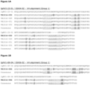
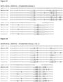
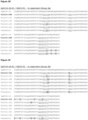
- Exemplary variants include those which differ from a parent antibody VH and/or VL sequence shown in Figures 1 and 2 at one or more "variant" amino acid positions, denoted "X" in the corresponding consensus sequence.
- Preferred variants are those in which the new amino acid is selected from those at the corresponding position in one of the aligned sequences in Figure 1 or 2 (for details on CDR sequence variants, see Table 4).
- the sequence of VH, VL or CDR variants may differ from the sequence of the VH, VL or CDR of the parent antibody sequences mainly by conservative substitutions; for instance at least 10, such as at least 9, 8, 7, 6, 5, 4, 3, 2 or 1 of the substitutions in the variant are conservative amino acid residue replacements.
- conservative substitutions may be defined by substitutions within the classes of amino acids reflected in the following table:
- Acidic Residues Asp (D) and Glu E) Basic Residues Lys (K), Arg (R), and His (H) Hydrophilic Uncharged Residues Ser (S), Thr (T), Asn (N), and Gln (Q) Aliphatic Uncharged Residues Gly (G), Ala (A), Val (V), Leu (L), and Ile (I) Non-polar Uncharged Residues Cys (C), Met (M), and Pro (P) Aromatic Residues Phe (F), Tyr (Y), and Trp (W)
- substitution of an amino acid in a given position is written as e.g. K405R which means a substitution of a lysine in position 405 with an arginine; and ii) for specific variants the specific three or one letter codes are used, including the codes Xaa and X to indicate any amino acid residue.
- substitution of Arginine for Lysine in position 405 is designated as: K405R, or the substitution of any amino acid residue for Lysine in position 405 is designated as K405X.
- deletion of Lysine in position 405 it is indicated by K405*.
- Recombinant host cell (or simply “host cell”), as used herein, is intended to refer to a cell into which an expression vector has been introduced, e.g. an expression vector encoding an antibody of the invention.
- Recombinant host cells include, for example, transfectomas, such as CHO cells, HEK293 cells, NS/0 cells, and lymphocytic cells.
- transgenic non-human animal refers to a non-human animal having a genome comprising one or more human heavy and/or light chain transgenes or transchromosomes (either integrated or non-integrated into the animal's natural genomic DNA) and which is capable of expressing fully human antibodies.
- a transgenic mouse can have a human light chain transgene and either a human heavy chain transgene or human heavy chain transchromosome, such that the mouse produces human HER2 antibodies when immunized with HER2 antigen and/or cells expressing HER2.
- the human heavy chain transgene may be integrated into the chromosomal DNA of the mouse, as is the case for transgenic mice, for instance HuMAb ® mice, such as HCo7, HCo12, or HCo17 mice, or the human heavy chain transgene may be maintained extrachromosomally, as is the case for transchromosomal KM mice as described in WO02/43478 .
- Similar mice, having a larger human Ab gene repertoire, include HCo7 and HCo20 (see e.g. WO2009097006 ).
- transgenic mice are capable of producing multiple isotypes of human monoclonal antibodies to a given antigen (such as IgG, IgA, IgM, IgD and/or IgE) by undergoing V-D-J recombination and isotype switching.
- Transgenic, nonhuman animal can also be used for production of antibodies against a specific antigen by introducing genes encoding such specific antibody, for example by operatively linking the genes to a gene which is expressed in the milk of the animal.
- Treatment refers to the administration of an effective amount of a therapeutically active compound of the present invention with the purpose of easing, ameliorating, arresting or eradicating (curing) symptoms or disease states.
- an “effective amount” or “therapeutically effective amount” refers to an amount effective, at dosages and for periods of time necessary, to achieve a desired therapeutic result.
- a therapeutically effective amount of a HER2 antibody may vary according to factors such as the disease state, age, sex, and weight of the individual, and the ability of the HER2 antibody to elicit a desired response in the individual.
- a therapeutically effective amount is also one in which any toxic or detrimental effects of the antibody or antibody portion are outweighed by the therapeutically beneficial effects.
- an "anti-idiotypic” antibody is an antibody which recognizes unique determinants generally associated with the antigen-binding site of an antibody.
- the invention relates to a bispecific antibody comprising two different antigen-binding regions which bind HER2.
- the invention relates to a bispecific molecule comprising a first antigen binding site from a HER2 antibody described herein and a second antigen binding site from a HER2 antibody described herein with a different binding specificity, such as a binding specificity for a non-overlapping epitope of HER2, i.e. a bispecific antibody wherein the first and second antigen binding regions do not cross-block each other for binding to HER2, e.g. when tested as described in Example 14.
- the bispecific antibody comprises at least one antigen-binding region from an antibody of cross-block group 1, 2, 3 or 4, described below. In one embodiment, the bispecific antibody comprises at least one antigen-binding region from an antibody cross-blocking or binding to the same epitope as a reference antibody selected from cross-block groups 1, 2, 3 and 4, e.g., cross-block group 4. In one embodiment, the bispecific antibody comprises two different antigen-binding regions from the antibodies of cross-block groups 1, 2, 3 and 4, optionally from different cross-block groups. In one embodiment, the bispecific antibody comprises two different antigen-binding regions from antibodies which each cross-block or bind to the same epitope as a reference antibody of cross-block groups 1, 2, 3 and 4, optionally from different cross-block groups. For example, the bispecific antibody may comprise one antigen-binding region from an antibody of cross-block group 1, 2, 3 or 4, and one antigen-binding region from trastuzumab or pertuzumab.
- the bispecific antibody of the present invention may comprise a first antigen-binding region and a second antigen-binding region, which first and second antigen-binding regions bind different epitopes on human epidermal growth factor receptor 2 (HER2).
- HER2 human epidermal growth factor receptor 2
- the first and second antigen-binding region of the bispecific antibody of the present invention may be an antigen-binding region from any of cross-block groups 1, 2, 3, and 4.
- the bispecific antibody of the present invention comprises two different antigen-binding regions which bind HER2. Furthermore, as described below one method of producing a bispecific antibody of the present invention is based on incubating a first and a second HER2 antibody under reducing conditions.
- the antigen-binding regions of a bispecific antibody of the present invention and the antigen-binding region of a first or second HER2 antibody of the present invention may belong to any of cross-block groups 1, 2, 3 and 4 described herein.
- a first or second HER2 antibody of the present invention may comprise an antigen-binding region of any of the HER2 antibodies of cross-block groups 1, 2, 3 and 4, which are described below.
- the bispecific antibody comprises an antigen-binding region of one or more of the human antibodies of cross-blocks 1, 2, 3, or 4, which blocks the binding to HER2.
- the bispecific antibody comprises an antigen-binding region which blocks binding to the same epitope on soluble HER2 as one or more of the human antibodies of cross-blocks 1, 2, 3, or 4.
- the bispecific antibody comprises an antigen-binding region which binds to the same epitope on HER2 as one or more of the human antibodies of cross-blocks 1, 2, 3, or 4.
- the bispecific antibody of the invention comprises one antigen-binding region which blocks the binding to HER2, e.g. soluble HER2, of one or more of the human antibodies of cross-block group 1 described herein, or binds the same epitope on HER2 as one or more of the human antibodies of cross-block group 1 described herein.
- the bispecific antibody then comprises a second antigen-binding region which cross-blocks or binds to the same epitope as an antibody of cross-block groups 2, 3, or 4.
- the antigen-binding region cross-blocks the binding to soluble HER2 of trastuzumab, when determined as described in Example 14.
- the antigen-binding region blocks the binding to HER2, e.g soluble HER2, or binds the same epitope as a reference antibody comprising a VH region comprising the sequence of SEQ ID NO:1 and a VL region comprising the sequence of SEQ ID NO:5 (169).
- the antigen-binding region blocks the binding to HER2, e.g soluble HER2, or binds the same epitope as a reference antibody comprising a VH region comprising the sequence of SEQ ID NO:8 and a VL region comprising the sequence of SEQ ID NO:12 (050).
- the antigen-binding region blocks the binding to HER2, e.g soluble HER2, or binds the same epitope as a reference antibody comprising a VH region comprising the sequence of SEQ ID NO:15 and a VL region comprising the sequence of SEQ ID NO:19 (084).
- the antigen-binding region blocks the binding to HER2, e.g soluble HER2, or binds to the same epitope as a reference antibody comprising VH and VL regions selected from the group consisting of:
- one antigen-binding region binds to HER2 and comprises a VH CDR3, VH region and/or VL region sequence similar or identical to such a sequence of an antibody described herein.
- the antigen-binding region comprises a VH CDR3 region having a sequence selected from the group consisting of
- the antigen-binding region comprises a VH CDR3 region of one of antibodies 123, 161, or 124, as shown in Figure 1 , optionally wherein the VH region is derived from an IgHV1-18-1 germline.
- the bispecific antibody or antigen-binding region comprises a VH region selected from the group consisting of
- the bispecific antibody or antigen-binding region comprises a VH region selected from the preceding embodiments (a) or (b) and a VL region comprising the CDR1, CDR2, and CDR3 sequences of SEQ ID NO:13, XAS (wherein X is A or V), and SEQ ID No: 155, respectively, such as a CDR1 sequence selected from SEQ ID Nos: 13 or 20, a CDR2 which is AAS or VAS, and a CDR3 sequence selected from SEQ ID NOs:14 and 21 (050, 084); respectively, optionally where the VL region is derived from an IgKV1-12-01 germline.
- the bispecific antibody or antigen-binding region comprises a VH region comprising the CDR1, CDR2 and CDR3 sequences of SEQ ID NOs:2, 3 and 4, respectively; and a VL region comprising the CDR1, CDR2 and CDR3 sequences of SEQ ID NOs:6, DAS, and SEQ ID NO:7, respectively (169).
- the bispecific antibody or antigen-binding region comprises a VH region comprising the CDR1, CDR2 and CDR3 sequences of SEQ ID NOs:9, 10 and 11, respectively; and a VL region comprising the CDR1, CDR2 and CDR3 sequences of SEQ ID NOs: 13, AAS, and SEQ ID NO:14, respectively (050).
- the bispecific antibody or antigen-binding region comprises a VH region comprising the CDR1, CDR2 and CDR3 sequences of SEQ ID NOs:16, 17 and 18, respectively; and a VL region comprising the CDR1, CDR2 and CDR3 sequences of SEQ ID NOs:20, VAS, and SEQ ID NO:21, respectively (084).
- the bispecific antibody or antigen-binding region comprises:
- the bispecific antibody comprises an antigen-binding region which blocks the binding to HER2 of one or more of the human antibodies of cross-block group 2 described herein, or binds the same epitope on HER2 as one or more of the human antibodies of cross-block group 2 described herein.
- the bispecific antibody then comprises a second antigen-binding region which cross-blocks, blocks the binding to HER2, e.g soluble HER2, or binds to the same epitope as an antibody of cross-block groups 1, 3, or 4.
- the antigen-binding region cross-blocks the binding to soluble HER2 of pertuzumab, when determined as described in Example 14.
- the antigen-binding region blocks the binding to soluble HER2, e.g soluble HER2, or binds the same epitope as a reference antibody comprising a VH region comprising the sequence of SEQ ID NO:22 and a VL region comprising the sequence of SEQ ID NO:26 (025).
- the antigen-binding region blocks the binding to soluble HER2, e.g soluble HER2, or binds the same epitope as a reference antibody comprising a VH region comprising the sequence of SEQ ID NO:29 and a VL region comprising the sequence of SEQ ID NO:32 (091).
- the antigen-binding region blocks the binding to soluble HER2, e.g soluble HER2, or binds the same epitope as a reference antibody comprising a VH region comprising the sequence of SEQ ID NO:35 and a VL region comprising the sequence of SEQ ID NO:39 (129).
- the antigen-binding region blocks the binding to soluble HER2, e.g soluble HER2, or binds to the same epitope as a reference antibody comprising VH and VL regions selected from the group consisting of:
- the bispecific antibody or antigen-binding region comprises a VH CDR3, VH region and/or VL region sequence similar or identical to a sequence of the novel antibodies described herein.
- the bispecific antibody or antigen-binding region comprises a VH CDR3 region having a sequence selected from the group consisting of
- the bispecific antibody or antigen-binding region comprises a VH CDR3 region of one of antibodies 001, 143, 019, 021, 027, 032, 035, 036, 054 or 094 as shown in Figure 1 , optionally wherein the VH region is derived from an IgHV4-34-1 germline.
- the bispecific antibody or antigen-binding region comprises a VH region selected from the group consisting of
- the bispecific antibody or antigen-binding region comprises a VH region selected from the preceding embodiment (a) and a VL region comprising the CDR1, CDR2, and CDR3 sequences of SEQ ID NO:157, AAS, and SEQ ID No: 164, respectively, such as the CDR1, CDR2, and CDR3 sequences of SEQ ID Nos:27, AAS, and SEQ ID NO:28 (025); respectively, optionally where the VL region is derived from an IgKV1D-16-01 germline.
- the bispecific antibody or antigen-binding region comprises a VH region selected from the preceding embodiment (b) and a VL region comprising the CDR1, CDR2, and CDR3 sequences of SEQ ID NO:33, AX 1 X 2 (wherein X 1 is A or T, preferably A; and X 2 is S or F, preferably S), and SEQ ID No: 158, respectively, such as the CDR1, CDR2 and CDR3 sequences of SEQ ID Nos:33, AAS, and SEQ ID NO:34 (091); respectively, optionally where the VL region is derived from an IgKV1D-16-01 germline.
- the bispecific antibody or antigen-binding region comprises a VH region which is the preceding embodiment (c) and a VL region comprising the CDR1, CDR2, and CDR3 sequences of SEQ ID NO:40, DAS and SEQ ID NO:41 (129), respectively, optionally wherein the VL region is derived from IgKV3-11-01.
- the bispecific antibody or antigen-binding region comprises a VH region comprising the CDR1, CDR2 and CDR3 sequences of SEQ ID NOs:23, 24 and 25, respectively; and a VL region comprising the CDR1, CDR2 and CDR3 sequences of SEQ ID NOs:27, AAS, and SEQ ID NO:28, respectively (025).
- the bispecific antibody or antigen-binding region comprises a VH region comprising the CDR1, CDR2 and CDR3 sequences of SEQ ID NOs:30, 163 and 31, respectively; and a VL region comprising the CDR1, CDR2 and CDR3 sequences of SEQ ID NOs:33, AAS, and SEQ ID NO:34, respectively (091).
- the bispecific antibody or antigen-binding region comprises a VH region comprising the CDR1, CDR2 and CDR3 sequences of SEQ ID NOs:36, 37 and 38, respectively; and a VL region comprising the CDR1, CDR2 and CDR3 sequences of SEQ ID NOs:40, DAS, and SEQ ID NO:41, respectively (129).
- the bispecific antibody or antigen-binding region comprises:
- the bispecific antibody comprises an antigen-binding region which blocks the binding to HER2 of one or more of the human antibodies of cross-block group 3 described herein or binds the same epitope on HER2 as one or more of the human antibodies of cross-block group 3 described herein.
- the bispecific antibody then comprises a second antigen-binding region which cross-blocks, blocks the binding to HER2, e.g soluble HER2, or binds to the same epitope as an antibody of cross-block groups 1, 2, or 4.
- the antigen-binding region cross-blocks the binding to soluble HER2 of F5 and/or C1, when determined as described in Example 14.
- the antigen-binding region blocks the binding to HER2, e.g soluble HER2, or binds the same epitope as a reference antibody comprising a VH region comprising the sequence of SEQ ID NO:46 and a VL region comprising the sequence of SEQ ID NO:49 ( 127 ).
- the antigen-binding region blocks the binding to HER2, e.g soluble HER2, or binds the same epitope as a reference antibody comprising a VH region comprising the sequence of SEQ ID NO:49 and a VL region comprising the sequence of SEQ ID NO:53 (159).
- the antigen-binding region blocks the binding to HER2, e.g soluble HER2, or binds the same epitope as a reference antibody comprising a VH region comprising the sequence of SEQ ID NO:56 and a VL region comprising the sequence of SEQ ID NO:60 (098).
- the antigen-binding region blocks the binding to HER2, e.g soluble HER2, or binds the same epitope as a reference antibody comprising a VH region comprising the sequence of SEQ ID NO:63 and a VL region comprising the sequence of SEQ ID NO:67 (153).
- the antigen-binding region blocks the binding to HER2, e.g soluble HER2, or binds the same epitope as a reference antibody comprising a VH region comprising the sequence of SEQ ID NO:70 and a VL region comprising the sequence of SEQ ID NO:74 (132).
- the antigen-binding region blocks the binding to HER2, e.g soluble HER2, or binds to the same epitope as a reference antibody comprising VH and VL regions selected from the group consisting of:
- the bispecific antibody or antigen-binding region comprises a VH CDR3, VH region and/or VL region sequence similar or identical to a sequence of the novel antibodies described herein.
- the bispecific antibody or antigen-binding region comprises a VH CDR3 region having a sequence selected from the group consisting of
- the bispecific antibody or antigen-binding region comprises a VH CDR3 region of one of antibodies 105, 100, 125 or 162 as shown in Figure 1 , optionally wherein the VH region is derived from an IgHV3-23-1 germline.
- the bispecific antibody or antigen-binding region comprises a VH CDR3 region of one of antibodies 033, 160, 166, 152 or 167 as shown in Figure 1 , optionally wherein the VH region is derived from an IgHV3-30-3-01 germline.
- the bispecific antibody or antigen-binding region comprises a VH region selected from the group consisting of
- the bispecific antibody or antigen-binding region comprises a VH region selected from the preceding embodiment (a) and a VL region comprising the CDR1, CDR2, and CDR3 sequences of SEQ ID NO:47, AAS and SEQ ID NO:48, respectively (127); respectively, optionally where the VL region is derived from an IgKV1D-8-01 germline.
- the bispecific antibody or antigen-binding region comprises a VH region selected from the preceding embodiment (b) and a VL region comprising the CDR1, CDR2, and CDR3 sequences of SEQ ID NO:54, AAS, and SEQ ID No:55 (159); respectively, optionally where the VL region is derived from an IgKV1D-16-01 germline.
- the bispecific antibody or antigen-binding region comprises a VH region which is the preceding embodiment (c) and a VL region comprising the CDR1, CDR2, and CDR3 sequences of SEQ ID NO:159, AAS and SEQ ID NO:160, respectively, such as the VL CDR1, CDR2 and CDR3 sequences of SEQ ID NOS: 61, AAS and SEQ ID NO:62 (098), optionally wherein the VL region is derived from IgKV1D-16-01.
- the bispecific antibody or antigen-binding region comprises a VH region which is the preceding embodiment (e) and a VL region comprising the CDR1, CDR2, and CDR3 sequences of SEQ ID NO:75, DAS and SEQ ID NO:76 (132), respectively, optionally wherein the VL region is derived from IgKV3-11-01.
- the bispecific antibody or antigen-binding region comprises a VH region comprising the CDR1, CDR2 and CDR3 sequences of SEQ ID NOs:43, 44 and 45, respectively; and a VL region comprising the CDR1, CDR2 and CDR3 sequences of SEQ ID NOs:47, AAS, and SEQ ID NO:48, respectively (127).
- the bispecific antibody or antigen-binding region comprises a VH region comprising the CDR1, CDR2 and CDR3 sequences of SEQ ID NOs:57, 58 and 59, respectively; and a VL region comprising the CDR1, CDR2 and CDR3 sequences of SEQ ID NOs:60, AAS, and SEQ ID NO:61, respectively (098).
- the bispecific antibody or antigen-binding region comprises a VH region comprising the CDR1, CDR2 and CDR3 sequences of SEQ ID NOs:64, 65 and 66, respectively; and a VL region comprising the CDR1, CDR2 and CDR3 sequences of SEQ ID NOs:68, DAS, and SEQ ID NO:69, respectively (153).
- the bispecific antibody or antigen-binding region comprises a VH region comprising the CDR1, CDR2 and CDR3 sequences of SEQ ID NOs:71, 72 and 73, respectively; and a VL region comprising the CDR1, CDR2 and CDR3 sequences of SEQ ID NOs:75, DAS, and SEQ ID NO:76, respectively (132).
- the bispecific antibody or antigen-binding region comprises:
- the bispecific antibody comprises an antigen-binding region which binds HER2 but which does not block the binding to soluble HER2 of a second antibody, optionally in immobilized form, comprising the VH and VL sequences of any of trastuzumab, pertuzumab, F5, and C1, when determined as described in Example 14.
- the antigen-binding region blocks or cross-blocks the binding to soluble HER2 of one or more of the human antibodies of cross-block group 4.
- the bispecific antibody then comprises a second antigen-binding region which cross-blocks or binds to the same epitope as an antibody of cross-block groups 1, 2, or 3.
- the antigen-binding region blocks the binding to soluble HER2 of a reference antibody, optionally immobilized, wherein the reference antibody comprises a VH region comprising the sequence of SEQ ID NO:165 and a VL region comprising the sequence of SEQ ID NO:5 (005), preferably wherein the antibody is fully blocking when determined as described in Example 14.
- the antigen-binding region blocks the binding to soluble HER2 of a reference antibody, optionally immobilized, wherein the reference antibody comprises a VH region comprising the sequence of SEQ ID NO:172 and a VL region comprising the sequence of SEQ ID NO:176 (006), preferably wherein the antibody is fully-blocking when determined as described in Example 14.
- the antigen-binding region blocks the binding to soluble HER2 of a reference antibody, optionally immobilized, wherein the reference antibody comprises a VH region comprising the sequence of SEQ ID NO:179 and a VL region comprising the sequence of SEQ ID NO:183 (059), preferably wherein the antibody is fully-blocking when determined as described in Example 14.
- the antigen-binding region blocks the binding to soluble HER2 of a reference antibody, optionally immobilized, wherein the reference antibody comprises a VH region comprising the sequence of SEQ ID NO:186 and a VL region comprising the sequence of SEQ ID NO:190 ( 060 ), preferably wherein the antibody is fully-blocking when determined as described in Example 14.
- the antigen-binding region blocks the binding to soluble HER2 of a reference antibody, optionally immobilized, wherein the reference antibody comprises a VH region comprising the sequence of SEQ ID NO:193 and a VL region comprising the sequence of SEQ ID NO:197 (106), preferably wherein the antibody is fully-blocking when determined as described in Example 14.
- the antigen-binding region blocks the binding to soluble HER2 of a reference antibody, optionally immobilized, wherein the reference antibody comprises a VH region comprising the sequence of SEQ ID NO:200 and a VL region comprising the sequence of SEQ ID NO:204 (111), preferably wherein the antibody is fully-blocking when determined as described in Example 14.
- the antigen-binding region blocks the binding of two, three, four, five, or six reference antibodies of the preceding embodiment, such as, e.g., antibodies 005 and 111, antibodies 005 and 006; antibodies 059 and 106; antibodies 006 and 059; antibodies 059, 106, 005 and 060; antibodies 006, 59, 060, and 111; or antibodies 059, 106, 005, 060, 111 and 006.
- two, three, four, five, or six reference antibodies of the preceding embodiment such as, e.g., antibodies 005 and 111, antibodies 005 and 006; antibodies 059 and 106; antibodies 006 and 059; antibodies 059, 106, 005 and 060; antibodies 006, 59, 060, and 111; or antibodies 059, 106, 005, 060, 111 and 006.
- the antibody when immobilized, competes for binding to soluble HER2 with all antibodies defined in the preceding embodiment for 25% or more, preferably 50% or more, when determined as described in Example 14.
- the antibody binds the same epitope on HER2 as one or more of the novel human antibodies described herein.
- the antigen-binding region binds the same epitope as an antibody comprising a VH region comprising the sequence of SEQ ID NO:165 and a VL region comprising the sequence of SEQ ID NO:169 (005).
- the antigen-binding region binds the same epitope as an antibody comprising a VH region comprising the sequence of SEQ ID NO:172 and a VL region comprising the sequence of SEQ ID NO:176 (006).
- the antigen-binding region binds the same epitope as an antibody comprising a VH region comprising the sequence of SEQ ID NO:186 and a VL region comprising the sequence of SEQ ID NO:190 (060).
- the antigen-binding region binds the same epitope as an antibody comprising a VH region comprising the sequence of SEQ ID NO:193 and a VL region comprising the sequence of SEQ ID NO:197 (106).
- the antigen-binding region binds the same epitope as an antibody comprising a VH region comprising the sequence of SEQ ID NO:200 and a VL region comprising the sequence of SEQ ID NO:204 (111).
- the antigen-binding region binds to the same epitope as at least one antibody selected from the group consisting of:
- the bispecific antibody or antigen-binding region comprises a VH CDR3, VH region and/or VL region sequence similar or identical to a sequence of the HER2 antibodies described herein.
- the bispecific antibody or antigen-binding region comprises a VH CDR3 region having an amino acid sequence selected from the group consisting of
- the bispecific antibody or antigen-binding region comprises a VH CDR3 region of one of antibodies 041, 150, 067, 072, 163, or 093, as shown in Figure 1 , optionally wherein the VH region is derived from an IgHV5-51-1 germline.
- the bispecific antibody or antigen-binding region comprises a VH region selected from the group consisting of
- the bispecific antibody or antigen-binding region comprises a VH region selected from the preceding embodiments (a), (c) or (d) and a VL region comprising the CDR1, CDR2, and CDR3 sequences of SEQ ID NO:232, GAS, and SEQ ID No:233, respectively, such as a CDR1 sequence selected from SEQ ID Nos: 170, 184, 191, 198 and 205, a CDR2 which is GAS, and a CDR3 sequence selected from 171, 85, 192, 199 and 206 (005, 059, 060, 106, 111); respectively, optionally where the VL region is derived from an IgKV3-20-01 germline.
- the bispecific antibody or antigen-binding region comprises a VH region which is the preceding embodiment (b) and a VL region comprising the CDR1, CDR2, and CDR3 sequences of SEQ ID NO:177, DAS, and SEQ ID NO:178 (006), respectively, optionally where the VL region is derived from IgKV3-11-01.
- the bispecific antibody or antigen-binding region comprises a VH region comprising the CDR1, CDR2 and CDR3 sequences of SEQ ID NOs:166, 167 and 168, respectively; and a VL region comprising the CDR1, CDR2 and CDR3 sequences of SEQ ID NOs:170, GAS, and SEQ ID NO:171, respectively (005).
- the bispecific antibody or antigen-binding region comprises a VH region comprising the CDR1, CDR2 and CDR3 sequences of SEQ ID NOs:173, 174 and 175, respectively; and a VL region comprising the CDR1, CDR2 and CDR3 sequences of SEQ ID NOs:177, DAS, and SEQ ID NO:178, respectively (006).
- the bispecific antibody or antigen-binding region comprises a VH region comprising the CDR1, CDR2 and CDR3 sequences of SEQ ID NOs:180, 181 and 182, respectively; and a VL region comprising the CDR1, CDR2 and CDR3 sequences of SEQ ID NOs:184, GAS, and SEQ ID NO:185, respectively (059).
- the bispecific antibody or antigen-binding region comprises a VH region comprising the CDR1, CDR2 and CDR3 sequences of SEQ ID NOs:187, 188 and 189, respectively; and a VL region comprising the CDR1, CDR2 and CDR3 sequences of SEQ ID NOs:191, GAS, and SEQ ID NO:192, respectively (060).
- the bispecific antibody or antigen-binding region comprises a VH region comprising the CDR1, CDR2 and CDR3 sequences of SEQ ID NOs:194, 195 and 196, respectively; and a VL region comprising the CDR1, CDR2 and CDR3 sequences of SEQ ID NOs: 198, GAS, and SEQ ID NO:199, respectively (106).
- the bispecific antibody or antigen-binding region comprises a VH region comprising the CDR1, CDR2 and CDR3 sequences of SEQ ID NOs:201, 202 and 203, respectively; and a VL region comprising the CDR1, CDR2 and CDR3 sequences of SEQ ID NOs:205, GAS, and SEQ ID NO:206, respectively (111).
- the bispecific antibody or antigen-binding region comprises:
- the antigen-binding region e.g. first or second antigen-binding region of a bispecific antibody of the present invention, or a first or second HER2 antibody disclosed herein, blocks binding to HER2 of or binds to the same HER2 epitope as, one or more of the antingen-binding regions or antibodies of cross-block group 1, 2, 3 or 4 described herein, preferably when determined as described in Example 14; and is further characterized by one or more properties described below or determined as described in Examples 12, 13, 15, 16, 17, 18 and 19.
- first and/or second antigen-binding region of the bispecific antibody of the present invention may be same as the antigen-binding region of an antibody or anti-HER2 antibody having one of the following characteristics.
- first and/or second HER2 antibody of the present invention may has one or more of the following characteristics.
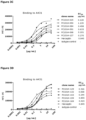
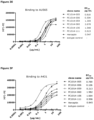
- the anti-HER2 antibody has a lower EC 50 value (half maximal effective concentration) than trastuzumab in binding to A431 cells, preferably an EC 50 value lower than 0.80 ⁇ g/ml, 0.50 ⁇ g/ml, or 0.30 ⁇ g/ml, when determined as described in Example 12, and preferably binds the same epitope as at least one reference antibody comprising the VH and VL regions selected from the group consisting of
- the anti-HER2 antibody specifically binds HER2-positive Rhesus monkey epithelial cells, when determined as described in Example 13, and preferably binds the same epitope as at least one reference antibody comprising the VH and VL regions selected from the group consisting of the VH and VL regions of any of antibodies 169, 050, 084, 025, 091, 129, 127, 159, 098, 153, 132, 005, 006, 059, 060, 106 and 111.
- the anti-HER2 antibody efficiently induces ADCC (antibody-dependent cell-mediated cytotoxicity), preferably achieving a specific 51 Cr-release of at least 30%, more preferably at least 40%, when determined as described in Example 15, and preferably binds the same epitope as at least one reference antibody comprising the VH and VL regions selected from the group consisting of:
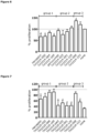
- the anti-HER2 antibody specifically binds HER2-expressing AU565 cells but promotes ligand-independent proliferation of the cells less than any of F5 and C1 when determined as described in Example 16, and preferably binds the same epitope as at least one reference antibody comprising the VH and VL regions selected from the group consisting of
- the anti-HER2 antibody specifically binds HER2-expressing AU565 cells and inhibits ligand-independent proliferation of the cells, preferably inhibiting proliferation by at least 20%, more preferably at least 25%, when determined as described in Example 16, and preferably binds the same epitope as at least one reference antibody comprising the VH and VL regions selected from the group consisting of:
- the anti-HER2 antibody specifically binds HER2-expressing AU565 cells but has no significant effect on, or does not promote, ligand-induced proliferation of the cells, preferably inhibiting proliferation by no more than 25%, more preferably by no more than 15%, when determined as described in Example 17, and binds the same epitope as at least one reference antibody comprising the VH and VL regions selected from the group consisting of:
- the anti-HER2 antibody specifically binds HER2-expressing MCF-7 cells and inhibits ligand-induced proliferation, e.g. it may completely inhibit the ligand-induced effect or inhibit the total proliferation by 50%, e.g. 60% or 70% or 80%, of the cells when determined as described in Example 17, and binds the same epitope as at least one reference antibody comprising the VH and VL regions selected from the group consisting of:
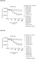
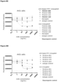
- the anti-HER2 antibody when conjugated directly or indirectly to a therapeutic moiety such as a truncated form of the pseudomonas-exotoxin A, is more effective than trastuzumab in killing AU565 cells, A431 cells, or both AU565 and A431 cells, when determined as described in Example 18.
- the conjugated anti-HER2 antibody has an EC 50 value of less than 70 ng/ml, less than 50 ng/ml, or less than 30 ng/ml in killing AU565 cells and/or A431 cells, when determined as described in Example 18, and binds the same epitope as at least one reference antibody comprising the VH and VL regions of an antibody selected from the group consisting of 169, 091, 050, 084, 098, 05, 153, 129, 132, 127 and 159; preferably selected from antibodies 153, 129, 098, 091 and 025.
- the conjugated anti-HER2 antibody has or results in a higher percentage of killed AU565 cells than trastuzumab and pertuzumab when determined as described in Example 18, preferably killing at least 49%, more preferably at least 60% of the AU565 cells, and binds the same epitope as at least one reference antibody comprising the VH and VL regions of an antibody selected from the group consisting of 169, 091, 050, 084, 098, 025, 153, 129, 132, 127 and 159; preferably selected from antibodies 153, 132, 127, 129, 159 and 025.
- the conjugated anti-HER2 antibody binds to the same epitope as a reference antibody comprising a VH region comprising the sequence of SEQ ID NO:49 and a VL region comprising the sequence of SEQ ID NO:53 (159).
- the conjugated anti-HER2 antibody has a higher percentage of killed AU431 cells than trastuzumab and pertuzumab when determined as described in Example 18, preferably killing at least 50%, more preferably at least 70%, and binds the same epitope as at least one reference antibody comprising the VH and VL regions of an antibody selected from the group consisting of 025, 084, 091, 098, 129 and 153; preferably selected from antibodies 025, 091, 098, 129 and 153.
- the anti-HER2 conjugated antibody binds to the same epitope as a reference antibody comprising a VH region comprising the sequence of SEQ ID NO:56 and a VL region comprising the sequence of SEQ ID NO:60 (098).
- the first or second HER2 antibody or a a anti-HER2 antibody is internalized by tumor cells expressing HER2, such as AU565 cells, to a higher degree than trastuzumab and pertuzumab, preferably more than twice or three times the amount of internalized trastuzumab, preferably when determined according to Example 18, and binds to the same epitope as an antibody comprising VH and VL regions selected from the group consisting of:
- the antibody binds to the same epitope as an antibody comprising VH and VL regions selected from
- the anti-HER2 antibody binds to Domain II or IV of HER2, preferably wherein the antibody does not significantly promote proliferation of HER2 expressing cells, and is more efficiently internalized, or is internalized to a higher degree, than trastuzumab or pertuzumab into HER2-expressing tumor cells, preferably when determined as described in the Examples, e.g. examples 16 and 19, respectively.
- the anti-HER2 antibody enhanced HER2 downmodulation more than trastuzumab e.g. the antibody enhanced HER2 downmodulation by more 30%, such as more than 40% or more than 50% when determined as described in Example 22, preferably wherein the antibody binds to the same epitope as an antibody of cross-block group 3 of the present invention, e.g. an antibody binding to the same epitope as an antibody comprising VH and VL regions selected from the group consisting of:
- the anti-HER2 antibody decreased tumour growth and improved survival in vivo more than trastuzumab, when determined as described in Example 28, preferably wherein the antibody binds to the same epitope as an antibody of cross-block 1 or cross-block 2 of the present invention, e.g. an antibody binding to the same epitope as an antibody comprising VH and VL regions selected from the group consisting of:
- the anti-HER2 antibody decreased tumour growth and improved survival in vivo more than trastuzumab, when determined as described in Example 29, preferably wherein the antibody binds to the same epitope as an antibody of cross-block 2 or cross-block 3 of the present invention, e.g. an antibody binding to the same epitope as an antibody comprising VH and VL regions selected from the group consisting of:
- the anti-HER2 antibody binds to the same epitope as an antibody comprising VH and VL regions selected from the group consisting of:
- the conjugated anti-HER2 antibody kills at least 60%, preferably at least 70% AU565 cells or A431 cells, when determined as described in Example 18, and cross-blocks at least one antibody selected from
- the anti-HER2 antibody of the preceding embodiment fully cross-blocks, preferably bind to the same epitope as, antibody 005, 060, 059, 111, or a combination thereof.
- the anti-HER2 antibody when conjugated directly or indirectly to a therapeutic moiety, is capable of killing tumor cells expressing a lower average amount of HER2 copies per cell than AU565 cells, such as an average of about 500,000 or less, 100,000 or less, or 30,000 or less copies of HER2 per cell (when determined, e.g., as referred to in Example 12), at concentrations where non-conjugated antibody does not induce killing of the cells, preferably when determined as described in Example 17.
- the antibody of the preceding embodiment kills at least 80% of A431 cells when determined as described in Example 18, and cross-blocks at least one antibody selected from
- the antibody of the preceding embodiment fully cross-blocks, preferably bind to the same epitope as, antibody 005, 060, or a combination thereof.
- the anti-HER2 antibody is internalized by tumor cells expressing HER2, such as AU565 cells, more than trastuzumab is, preferably more than twice or three times the amount of internalized trastuzumab, preferably when determined according to Example 19, and cross-blocks at least one antibody selected from the group consisting of:
- the antibody of the preceding embodiment fully cross-blocks, preferably bind to the same epitope as, antibody 005, 006, 059, 060, 106, 111, or a combination thereof.
- the antibody is a bispecific antibody, comprising (i) a first antigen-binding region of a first HER2 antibody as defined herein, and (ii) a second antigen-binding region of a second HER2 antibody as defined herein, wherein the first antigen-binding region binds to a different epitope than the second antigen-binding region.
- the first antigen-binding region comprises a VH region comprising a CDR3 sequence of an antibody of cross-block 1, 2, 3 or 4 as defined herein, such as SEQ ID NO: 4, 25, 66 or 168 ( 169, 025, 153, or 005 ).
- the first antigen-binding region comprises a VH region comprising CDR1, CDR2 and CDR3 sequences of an antibody of cross-block 1, 2, 3 or 4 as defined herein, such as CDR1, CDR2, and CDR3 sequences SEQ ID NOs: 2, 3 and 4 (169), or CDR1, CDR2 and CDR3 sequences of SEQ ID NOs:23, 24 and 25 (025), or CDR1, CDR2 and CDR3 sequences of SEQ ID NOs: 64, 65 and 66 (153), or CDR1, CDR2 CDR3 sequence of SEQ ID NOs: 166, 167 and 168 (005).
- the first antigen-binding region comprises a VH region comprising a CDR3 sequence of an antibody of cross-block 1, 2, 3 or 4 as defined herein, such as CDR3 sequence an antibody of cross-block 1 of SEQ ID NO: 11 (050), or SEQ ID NO: 18 (084); or a CDR3 sequence of an antibody of cross-block 2 of SEQ ID NO: 31 (091), or SEQ ID NO: 38 (129), or a CDR3 sequence of an antibody of cross-block 3 of SEQ ID NO: 45 (127), or SEQ ID NO:52 (159), or SEQ ID NO:59 (098), or SEQ ID NO:73 (132), or a CDR3 sequence of an antibody of cross-block 4 of SEQ ID NO:175 (006), SEQ ID NO: 182 (059), SEQ ID NO:189 (060), SEQ ID NO:196 (106), or SEQ ID NO:203 (111).
- the first antigen-binding region comprises a VH region comprising CDR1, CDR2 and CDR3 sequences of an antibody of cross-block 1, 2 or 3 as defined herein, such as CDR1, CDR2, and CDR3 sequences SEQ ID NOs: 2, 3 and 4 (169), or CDR1, CDR2 and CDR3 sequences of SEQ ID NOs:23, 24 and 25 (025), or CDR1, CDR2 and CDR3 sequences of SEQ ID NOs: 64, 65 and 66 (153), or CDR1, CDR2 CDR3 sequence of SEQ ID NOs: 170, GAS and 171 (005).
- the first antigen-binding region comprises a VH region comprising CDR1, CDR2 and CDR3 sequences of an antibody of cross-block 1, 2, 3 or 4 as defined herein a VL region comprising CDR1, CDR2 and CDR3 sequences of an antibody of cross-block 1, 2, 3 or 4 as defined herein.
- the first antigen-binding region comprises a VH region comprising CDR1, CDR2 and CDR3 sequences of an antibody of cross-block 1, 2, 3 or 4 as defined herein, such as CDR1, CDR2, and CDR3 sequences of an antibody of cross-block 1 of SEQ ID NOs: 9, 10 and 11 (050), or SEQ ID NOs: 16, 17 and 18 (084); or CDR1, CDR2, and CDR3 sequences of an antibody of cross-block 2 of SEQ ID NOs: 30, 163 and 31 (091), or SEQ ID NOs: 36, 37 and 38 (129), or CDR1, CDR2, and CDR3 sequences of an antibody of cross-block 3 SEQ ID NOs: 43, 44 and 45 (127), or SEQ ID NOs:50, 51 and 52 (159), or SEQ ID NOs:57, 58 and 59 (098), or SEQ ID NOs:71, 72 and 73 (132), or CDR1, CDR2 and CDR3 sequences of an antibody of cross-block 1, 2, 3 or 4
- the second antigen-binding region is one of the previous embodiments described for the first antigen-binding region, but wherein the second antigen-binding region binds to a different epitope than the first antigen-binding region.
- the second antigen-binding region is from trastuzumab or pertuzumab, comprising the VH and/or VL CDR1, 2 and 3 sequences or VH and/or VL sequences of trastuzumab or pertuzumab.
- the bispecific antibody comprises a first antigen-binding region and a second antigen-binding region, which first and second antigen-binding regions bind different epitopes on human epidermal growth factor receptor 2 (HER2), and wherein each of the first and second antigen-binding region block the binding to soluble HER2 of a reference antibody independently selected from the group consisting of:
- a further embodiment of the bispecific antibody wherein at least one of said first and second antigen-binding regions block the binding to soluble HER2 of an antibody of (a).
- a further embodiment of the bispecific antibody wherein at least one of said first and second antigen-binding regions block the binding to soluble HER2 of an antibody of (b).
- a further embodiment of the bispecific antibody wherein at least one of said first and second antigen-binding regions block the binding to soluble HER2 of an antibody of (c).
- a further embodiment of the bispecific antibody wherein at least one of said first and second antigen-binding regions block the binding to soluble HER2 of an antibody of (d).
- first and second antigen-binding regions each comprises VH CDR1, CDR2, and CDR3 sequences independently selected from the group consisting of:
- the first and second antigen-binding regions may each comprise a VH region and a VL region independently selected from the group consisting of
- first and the second antigen-binding regions may each comprise a VH region and a VL region independently selected from the group consisting of
- the present invention relates to a bispecific antibody comprising a first antigen-binding region comprising the VH CDR3 sequence of SEQ ID NO:66 ( 153 ) and a second antigen-binding region comprising the VH CDR3 sequence of SEQ ID NO:168 ( 005 ), or vice versa.
- the first antigen-binding region further comprises the VL CDR3 sequence of SEQ ID NO:69 ( 153 ), and the second antigen-binding region further comprises the VL CDR3 sequence of SEQ ID NO:171 ( 005 ).
- the first antigen-binding region further comprises the VH CDR1 sequence of SEQ ID NO:64 and the VH CDR2 sequence of SEQ ID NO:65 ( 153 ), and the second antigen-binding region further comprises the VH CDR1 sequence of SEQ ID NO:166 and a VH CDR2 sequence of SEQ ID NO:167 ( 005 ).
- the first antigen-binding region further comprises the VL CDR1 sequence of SEQ ID NO: 68 and the VL CDR2 sequence of DAS ( 153 ), and the second antigen-binding region further comprises the VL CDR1 sequence of SEQ ID NO:170 and a VL CDR2 sequence of GAS ( 005 ).
- the bispecific antibody comprises a first antigen-binding region comprising a VH region comprising SEQ ID NO:63 and a VL region comprising SEQ ID NO:67 ( 153 ), and a second antigen-binding region comprising a VH region comprising SEQ ID NO:165 and a VL region comprising SEQ ID NO:169 ( 005 ), or vice versa.
- the present invention relates to a bispecific antibody comprising a first antigen-binding region comprising the VH CDR3 sequence of SEQ ID NO:66 ( 153 ) and a second antigen-binding region comprising the VH CDR3 sequence of SEQ ID NO:4 ( 169 ), or vice versa.
- the first antigen-binding region further comprises the VL CDR3 sequence of SEQ ID NO:69 ( 153 ), and the second antigen-binding region further comprises the VL CDR3 sequence of SEQ ID NO:7 ( 169 ).
- the first antigen-binding region further comprises the VH CDR1 sequence of SEQ ID NO:64 and the VH CDR2 sequence of SEQ ID NO:65 ( 153 ), and the second antigen-binding region further comprises the VH CDR1 sequence of SEQ ID NO:2 and a VH CDR2 sequence of SEQ ID NO:3 ( 169 ).
- the first antigen-binding region further comprises the VL CDR1 sequence of SEQ ID NO: 68 and the VL CDR2 sequence of DAS ( 153 ), and the second antigen-binding region further comprises the VL CDR1 sequence of SEQ ID NO: 6 and a VL CDR2 sequence of DAS ( 169 ).
- the bispecific antibody comprises a first antigen-binding region comprising a VH region comprising SEQ ID NO:63 and a VL region comprising SEQ ID NO:67 ( 153 ), and a second antigen-binding region comprising a VH region comprising SEQ ID NO:1 and a VL region comprising SEQ ID NO:5 ( 169 ), or vice versa.
- the present invention relates to a bispecific antibody comprising a first antigen-binding region comprising the VH CDR3 sequence of SEQ ID NO:168 ( 005 ) and a second antigen-binding region comprising the VH CDR3 sequence of SEQ ID NO:4 ( 169 ), or vice versa.
- the first antigen-binding region further comprises the VL CDR3 sequence of SEQ ID NO:171 ( 005 ), and the second antigen-binding region further comprises the VL CDR3 sequence of SEQ ID NO:7 ( 169 ).
- the first antigen-binding region further comprises the VH CDR1 sequence of SEQ ID NO:166 and the VH CDR2 sequence of SEQ ID NO:167 ( 005 ), and the second antigen-binding region further comprises the VH CDR1 sequence of SEQ ID NO:2 and a VH CDR2 sequence of SEQ ID NO:3 ( 169 ).
- the first antigen-binding region further comprises the VL CDR1 sequence of SEQ ID NO: 170 and the VL CDR2 sequence of GAS ( 005 ), and the second antigen-binding region further comprises the VL CDR1 sequence of SEQ ID NO: 6 and a VL CDR2 sequence of DAS ( 169 ).
- the bispecific antibody comprises a first antigen-binding region comprising a VH region comprising SEQ ID NO:165 and a VL region comprising SEQ ID NO:169 ( 005 ), and a second antigen-binding region comprising a VH region comprising SEQ ID NO:1 and a VL region comprising SEQ ID NO:5 ( 169 ), or vice versa.
- the present invention relates to a bispecific antibody comprising a first antigen-binding region comprising the VH CDR3 sequence of SEQ ID NO:25 ( 025 ) and a second antigen-binding region comprising the VH CDR3 sequence of SEQ ID NO:168 ( 005 ), or vice versa.
- the first antigen-binding region further comprises the VL CDR3 sequence of SEQ ID NO:28 ( 025 ), and the second antigen-binding region further comprises the VL CDR3 sequence of SEQ ID NO:171 ( 005 ).
- the first antigen-binding region further comprises the VH CDR1 sequence of SEQ ID NO:23 and the VH CDR2 sequence of SEQ ID NO:24 ( 025 ), and the second antigen-binding region further comprises the VH CDR1 sequence of SEQ ID NO:166 and a VH CDR2 sequence of SEQ ID NO:167 ( 005 ).
- the first antigen-binding region further comprises the VL CDR1 sequence of SEQ ID NO: 27 and the VL CDR2 sequence of AAS ( 025 ), and the second antigen-binding region further comprises the VL CDR1 sequence of SEQ ID NO: 170 and a VL CDR2 sequence of GAS ( 005 ).
- the bispecific antibody comprises a first antigen-binding region comprising a VH region comprising SEQ ID NO:22 and a VL region comprising SEQ ID NO:26 ( 025 ), and a second antigen-binding region comprising a VH region comprising SEQ ID NO:165 and a VL region comprising SEQ ID NO:169 ( 005 ), or vice versa.
- the present invention relates to a bispecific antibody comprising a first antigen-binding region comprising the VH CDR3 sequence of SEQ ID NO:25 ( 025 ) and a second antigen-binding region comprising the VH CDR3 sequence of SEQ ID NO:66 ( 153 ), or vice versa.
- the first antigen-binding region further comprises the VL CDR3 sequence of SEQ ID NO:28 ( 025 ), and the second antigen-binding region further comprises the VL CDR3 sequence of SEQ ID NO:69 ( 153 ).
- the first antigen-binding region further comprises the VH CDR1 sequence of SEQ ID NO:23 and the VH CDR2 sequence of SEQ ID NO:24 ( 025 ), and the second antigen-binding region further comprises the VH CDR1 sequence of SEQ ID NO:64 and a VH CDR2 sequence of SEQ ID NO:65 ( 153 ).
- the first antigen-binding region further comprises the VL CDR1 sequence of SEQ ID NO: 27 and the VL CDR2 sequence of AAS ( 025 ), and the second antigen-binding region further comprises the VL CDR1 sequence of SEQ ID NO: 68 and a VL CDR2 sequence of DAS ( 153 ).
- the bispecific antibody comprising a first antigen-binding region comprising a VH region comprising SEQ ID NO:22 and a VL region comprising SEQ ID NO:26 ( 025 ), and a second antigen-binding region comprising a VH region comprising SEQ ID NO:63 and a VL region comprising SEQ ID NO:67 ( 153 ), or vice versa.
- the present invention relates to a bispecific antibody comprising a first antigen-binding region comprising the VH CDR3 sequence of SEQ ID NO:25 ( 025 ) and a second antigen-binding region comprising the VH CDR3 sequence of SEQ ID NO:4 ( 169 ), or vice versa.
- first antigen-binding region further comprises the VL CDR3 sequence of SEQ ID NO:28 ( 025 ), and the second antigen-binding region further comprises the VL CDR3 sequence of SEQ ID NO:7 ( 169 ).
- the first antigen-binding region further comprises the VH CDR1 sequence of SEQ ID NO:23 and the VH CDR2 sequence of SEQ ID NO:24 ( 025 ), and the second antigen-binding region further comprises the VH CDR1 sequence of SEQ ID NO:2 and a VH CDR2 sequence of SEQ ID NO:3 ( 169 ).
- the first antigen-binding region further comprises the VL CDR1 sequence of SEQ ID NO: 27 and the VL CDR2 sequence of AAS ( 025 ), and the second antigen-binding region further comprises the VL CDR1 sequence of SEQ ID NO: 6 and a VL CDR2 sequence of DAS ( 169 ).
- the present invention relates to a bispecific antibody comprising a first antigen-binding region comprising a VH region comprising SEQ ID NO:22 and a VL region comprising SEQ ID NO:26 ( 025 ), and a second antigen-binding region comprising a VH region comprising SEQ ID NO:1 and a VL region comprising SEQ ID NO:5 ( 169 ), or vice versa.
- the present invention also relates to a bispecific antibody comprising a first antigen-binding region which binds an epitope in HER2 Domain II and a second antigen-binding region which binds an epitope in HER2 Domain III or IV.
- the second antigen-binding region binds an epitope in HER2 Domain III.
- the second antigen-binding region binds an epitope in HER2 Domain IV.
- the first antigen-binding region blocks the binding to soluble HER2 of a reference antibody comprising a VH region comprising the sequence of SEQ ID NO:63 and a VL region comprising the sequence of SEQ ID NO:67 ( 153 ).
- first and/or second antigen-binding region comprises a VH region and, optionally, a VL region, of any of the embodiments described above.
- the present invention also relates to a bispecific antibody, wherein the first and second antigen-binding regions comprise human antibody VH sequences and, optionally, human antibody VL sequences.
- a further embodiment the present invention relates to a bispecific antibody, wherein the first and second antigen-binding regions are from heavy-chain antibodies.
- the present invention relates to a bispecific antibody, wherein the first and second antigen-binding regions comprise a first and second light chain.
- the present invention relates to a bispecific antibody, wherein said first and second light chains are different.
- the bispecific antibody enhances HER2 downmodulation, in particular more than their monospecific counterparts, e.g. the antibody enhanced HER2 downmodulation by more 20%, such as more than 30% or more than 40% when determined as described in example 22, preferably wherein the antibody binds to the same epitopes as bispecific antibody selected from the group consisting of IgG1-005-ITL x IgG1-169-K409R, IgG1-025-ITL x IgG1-005-K409R, IgG1-025-ITL x IgG1-153-K409R, IgG1-025-ITL x IgG1-169-K409R, IgG1-153-ITL x IgG1-005-K409R; and IgG1-153-ITL x IgG1-169-K409R.
- the bispecific antibody specifically binds HER2-expressing AU565 cells and inhibits ligand-induced proliferation of the cells when determined as described in Example 24, and binds the same epitopes as at least one bispecific antibody selected from the group consisting of: IgG1-005-ITL x IgG1-169-K409R, IgG1-025-ITL x IgG1-005-K409R, IgG1-025-ITL x IgG1-153-K409R, IgG1-025-ITL x IgG1-169-K409R, IgG1-153-ITL x IgG1-005-K409R; and IgG1-153-ITL x IgG1-169-K409R.
- the bispecific antibody inhibits proliferation of the AU565 cells more than their monospecific counterparts and is selected from the group consisting of IgG1-005-ITL x IgG1-169-K409R and IgG1-025-ITL x IgG1-005-K409R.
- the bispecific antibody induces PBMC-mediated cytotoxicity when determined as described in Example 31, and binds the same epitopes as at least one bispecific antibody selected from the group consisting of: IgG1-153-ITL x IgG1-169-K409R and IgG1-005-ITL x IgG1-153-K409R.
- the bispecific antibody induces higher levels of PBMC-mediated cytotoxicity than their monospecific counterparts, optionally more than the combination of their monospecific counterparts.
- the bispecific antibody reduces tumor growth and/or results in a better survival of mice in the NCI-N87 human gastric carcinoma xenograft model described in Example 32, and binds the same epitopes as at least one bispecific antibody selected from the group consisting of: IgG1-153-ITL x IgG1-169-K409R and IgG1-005-ITL x IgG1-153-K409R.
- the bispecific antibody reduces tumor growth more than their monospecific counterparts, optionally more than the combination of their monospecific counterparts.
- the bispecific HER2xHER2 antibody of the invention further comprises a first and a second Fc-region, which may be comprised in a first and a second Fab arm which respectively further comprise the first and second antigen-binding regions described above (or vice versa).
- the bispecific antibody of the present invention may in one embodiment comprise a first Fab-arm comprising a first antigen-binding region and a first Fc region, and a second Fab-arm comprising a second antigen-binding region and a second Fc region.
- the bispecific antibody of the present invention may comprise a first Fab-arm comprising a first antigen-binding region and a second Fc region, and a second Fab-arm comprising a second antigen-binding region and a first Fc region.
- the first and second Fc-regions of Fab-arms may be of any isotype, including, but not limited to, IgG1, IgG2, IgG3 and IgG4.
- each of the first and second Fc regions is of the IgG4 isotype or derived therefrom, optionally with one or more mutations or modifications.
- each of the first and second Fc regions is of the IgG1 isotype or derived therefrom, optionally with one or more mutations or modifications.
- one of the Fc regions is of the IgG1 isotype and the other of the IgG4 isotype, or is derived from such respective isotype, optionally with one or more mutations or modifications.
- one or both Fc-regions comprise an IgG1 wildtype sequence (SEQ ID NO:234).
- one or both of the Fc regions comprise a mutation removing the acceptor site for Asn-linked glycosylation or is otherwise manipulated to change the glycosylation properties.
- an N297Q mutation can be used to remove an Asn-linked glycosylation site.
- one or both Fc regions comprise an IgG1 wildtype sequence with an N297Q mutation (SEQ ID NO: 235).
- one or both of the Fc regions are glyco-engineered to reduce fucose and thus enhance ADCC, e.g. by addition of compounds to the culture media during antibody production as described in US2009317869 or as described in van Berkel et al. (2010) Biotechnol. Bioeng. 105:350 or by using FUT8 knockout cells, e.g. as described in Yamane-Ohnuki et al (2004) Biotechnol. Bioeng 87:614 .
- ADCC may alternatively be optimized using the method described by Uma ⁇ a et al. (1999) Nature Biotech 17:176 .
- one or both of the Fc-regions have been engineered to enhance complement activation, e.g. as described in Natsume et al. (2009) Cancer Sci. 100:2411 .
- the first or second antigen-binding regions or a part thereof are of a species in the family Camelidae, see WO2010001251 , or a species of cartilaginous fish, such as the nurse shark.
- the first and second antigen-binding regions or heavy chains are from heavy-chain antibodies.
- the first and/or second Fc-region is conjugated to a drug, a prodrug or a toxin or contains an acceptor group for the same.
- acceptor group may e.g. be an unnatural amino acid.
- the bispecific antibody of the invention comprises a first Fc-region comprising a first CH3 region, and a second Fc-region comprising a second CH3 region, wherein the sequences of the first and second CH3 regions are different and are such that the heterodimeric interaction between said first and second CH3 regions is stronger than each of the homodimeric interactions of said first and second CH3 regions. More details on these interactions and how they can be achieved are provided in PCT/EP2011/056388 , published as WO 11/131746 , which is hereby incorporated by reference in its entirety.
- a stable bispecific HER2xHER2 molecule can be obtained at high yield using a particular method on the basis of two homodimeric starting HER2 antibodies containing only a few, fairly conservative, asymmetrical mutations in the CH3 regions.
- Asymmetrical mutations mean that the sequences of said first and second CH3 regions contain amino acid substitutions at non-identical positions.
- the first Fc-region has an amino acid substitution at a position selected from the group consisting of: 366, 368, 370, 399, 405, 407 and 409
- the second Fc-region has an amino acid substitution at a position selected from the group consisting of: 366, 368, 370, 399, 405, 407 and 409, and wherein the first and second Fc-regions are not substituted in the same positions.
- the first Fc-region has an amino acid substitution at position 366
- said second Fc-region has an amino acid substitution at a position selected from the group consisting of: 368, 370, 399, 405, 407 and 409.
- the amino acid at position 366 is selected from Ala, Asp, Glu, His, Asn, Val, or Gln.
- the first Fc-region has an amino acid substitution at position 368
- said second Fc-region has an amino acid substitution at a position selected from the group consisting of: 366, 370, 399, 405, 407 and 409.
- the first Fc-region has an amino acid substitution at position 370
- said second HER2 antibody has an amino acid substitution at a position selected from the group consisting of: 366, 368, 399, 405, 407 and 409.
- the first Fc-region has an amino acid substitution at position 399
- said second Fc-region has an amino acid substitution at a position selected from the group consisting of: 366, 368, 370, 405, 407 and 409.
- the first Fc-region has an amino acid substitution at position 405, and said second Fc-region has an amino acid substitution at a position selected from the group consisting of: 366, 368, 370, 399, 407 and 409.
- the first Fc-region has an amino acid substitution at position 407
- said second Fc-region has an amino acid substitution at a position selected from the group consisting of: 366, 368, 370, 399, 405, and 409.
- the first Fc-region has an amino acid substitution at position 409
- said second Fc-region has an amino acid substitution at a position selected from the group consisting of: 366, 368, 370, 399, 405, and 407.
- the sequences of said first and second CH3 regions contain asymmetrical mutations, i.e. mutations at different positions in the two CH3 regions, e.g. a mutation at position 405 in one of the CH3 regions and a mutation at position 409 in the other CH3 region.
- the first Fc-region has an amino acid other than Lys, Leu or Met at position 409, e.g. Arg, His, Asp, Glu, Ser, Thr, Asn, Gln, Gly, Pro, Ala, Val, Ile, Phe, Tyr, Trp or Cys
- said second Fc-region has an amino-acid substitution at a position selected from the group consisting of: 366, 368, 370, 399, 405 and 407.
- said first Fc-region has an amino acid other than Lys, Leu or Met at position 409, e.g.
- said second Fc-region has an amino acid other than Phe at position 405, e.g. Lys, Leu, Met, Arg, His, Asp, Glu, Ser, Thr, Asn, Gln, Gly, Pro, Ala, Val, Ile, Tyr, Trp or Cys.
- said first Fc-region has an amino acid other than Lys, Leu or Met, e.g.
- said first Fc-region comprises a Phe at position 405 and an amino acid other than Lys, Leu or Met at position 409, e.g. Arg, His, Asp, Glu, Ser, Thr, Asn, Gln, Gly, Pro, Ala, Val, Ile, Phe, Tyr, Trp or Cys
- said second Fc-region comprises an amino acid other than Phe, e.g. Lys, Leu, Met, Arg, His, Asp, Glu, Ser, Thr, Asn, Gln, Gly, Pro, Ala, Val, Ile, Tyr, Trp or Cys, at position 405 and a Lys at position 409.
- said first Fc-region comprises a Phe at position 405 and an amino acid other than Lys, Leu or Met at position 409, e.g. Arg, His, Asp, Glu, Ser, Thr, Asn, Gln, Gly, Pro, Ala, Val, Ile, Phe, Tyr, Trp or Cys
- said second Fc-region comprises an amino acid other than Phe, Arg or Gly at position 405, e.g. Lys, Leu, Met, His, Asp, Glu, Ser, Thr, Asn, Gln, Pro, Ala, Val, Ile, Tyr, Trp or Cys, and a Lys at position 409.
- said first Fc-region comprises a Phe at position 405 and an amino acid other than Lys, Leu or Met at position 409, e.g. Arg, His, Asp, Glu, Ser, Thr, Asn, Gln, Gly, Pro, Ala, Val, Ile, Phe, Tyr, Trp or Cys
- said second Fc-region comprises a Leu at position 405 and a Lys at position 409.
- said first Fc-region comprises a Phe at position 405 and an Arg at position 409 and said second Fc-region comprises an amino acid other than Phe, Arg or Gly, e.g.
- said first Fc-region comprises Phe at position 405 and an Arg at position 409 and said second Fc-region comprises a Leu at position 405 and a Lys at position 409.
- said first Fc-region comprises an amino acid other than Lys, Leu or Met at position 409, e.g. Arg, His, Asp, Glu, Ser, Thr, Asn, Gln, Gly, Pro, Ala, Val, Ile, Phe, Tyr, Trp or Cys
- said second Fc-region comprises a Lys at position 409, a Thr at position 370 and a Leu at position 405.
- said first Fc-region comprises an Arg at position 409 and said second Fc-region comprises a Lys at position 409, a Thr at position 370 and a Leu at position 405.
- said first Fc-region comprises a Lys at position 370, a Phe at position 405 and an Arg at position 409 and said second Fc-region comprises a Lys at position 409, a Thr at position 370 and a Leu at position 405.
- said first Fc-region comprises an amino acid other than Lys, Leu or Met at position 409, e.g. Arg, His, Asp, Glu, Ser, Thr, Asn, Gln, Gly, Pro, Ala, Val, Ile, Phe, Tyr, Trp or Cys
- said second Fc-region comprises a Lys at position 409 and: a) an Ile at position 350 and a Leu at position 405, or b) a Thr at position 370 and a Leu at position 405.
- said first Fc-region comprises an Arg at position 409 and said second Fc region comprises a Lys at position 409 and: a) an Ile at position 350 and a Leu at position 405, or b) a Thr at position 370 and a Leu at position 405.
- said first Fc-region comprises a Thr at position 350, a Lys at position 370, a Phe at position 405 and an Arg at position 409 and said second HER2 antibody comprises a Lys at position 409 and: a) an Ile at position 350 and a Leu at position 405, or b) a Thr at position 370 and a Leu at position 405.
- said first Fc-region comprises a Thr at position 350, a Lys at position 370, a Phe at position 405 and an Arg at position 409 and said second Fc-region comprises an Ile at position 350, a Thr at position 370, a Leu at position 405 and a Lys at position 409.
- said first Fc-region has an amino acid other than Lys, Leu or Met at position 409, e.g. Arg, His, Asp, Glu, Ser, Thr, Asn, Gln, Gly, Pro, Ala, Val, Ile, Phe, Tyr, Trp or Cys
- said second Fc-region has an amino acid other than Tyr, Asp, Glu, Phe, Lys, Gln, Arg, Ser or Thr at position 407, e.g. His, Asn, Gly, Pro, Ala, Val, Ile, Trp, Leu, Met or Cys.
- said first Fc-region has an amino acid other than Lys, Leu or Met at position 409, e.g.
- said second Fc-region has an Ala, Gly, His, Ile, Leu, Met, Asn, Val or Trp at position 407.
- said first Fc-region has an amino acid other than Lys, Leu or Met at position 409, e.g. Arg, His, Asp, Glu, Ser, Thr, Asn, Gln, Gly, Pro, Ala, Val, Ile, Phe, Tyr, Trp or Cys, and said second Fc-region has a Gly, Leu, Met, Asn or Trp at position 407.
- said first Fc-region has a Tyr at position 407 and an amino acid other than Lys, Leu or Met at position 409, e.g. Arg, His, Asp, Glu, Ser, Thr, Asn, Gln, Gly, Pro, Ala, Val, Ile, Phe, Tyr, Trp or Cys
- said second Fc-region has an amino acid other than Tyr, Asp, Glu, Phe, Lys, Gln, Arg, Ser or Thr at position 407, e.g. His, Asn, Gly, Pro, Ala, Val, Ile, Trp, Leu, Met or Cys, and a Lys at position 409.
- said first Fc-region has a Tyr at position 407 and an amino acid other than Lys, Leu or Met at position 409, e.g. Arg, His, Asp, Glu, Ser, Thr, Asn, Gln, Gly, Pro, Ala, Val, Ile, Phe, Tyr, Trp or Cys
- said second Fc-region has an Ala, Gly, His, Ile, Leu, Met, Asn, Val or Trp at position 407 and a Lys at position 409.
- said first Fc-region has a Tyr at position 407 and an amino acid other than Lys, Leu or Met at position 409, e.g. Arg, His, Asp, Glu, Ser, Thr, Asn, Gln, Gly, Pro, Ala, Val, Ile, Phe, Tyr, Trp or Cys
- said second Fc-region has a Gly, Leu, Met, Asn or Trp at position 407 and a Lys at position 409.
- said first Fc-region has a Tyr at position 407 and an Arg at position 409 and said second Fc-region has an amino acid other than Tyr, Asp, Glu, Phe, Lys, Gln, Arg, Ser or Thr at position 407, e.g. His, Asn, Gly, Pro, Ala, Val, Ile, Trp, Leu, Met or Cys and a Lys at position 409.
- said first Fc-region has a Tyr at position 407 and an Arg at position 409 and said second Fc-region has an Ala, Gly, His, Ile, Leu, Met, Asn, Val or Trp at position 407 and a Lys at position 409.
- said first Fc-region has a Tyr at position 407 and an Arg at position 409 and said second Fc-region has a Gly, Leu, Met, Asn or Trp at position 407 and a Lys at position 409.
- the first Fc-region has an amino acid other than Lys, Leu or Met at position 409, e.g. Arg, His, Asp, Glu, Ser, Thr, Asn, Gln, Gly, Pro, Ala, Val, Ile, Phe, Tyr, Trp or Cys, and the second Fc-region has an amino acid other than Lys, Leu or Met at position 409, e.g. Arg, His, Asp, Glu, Ser, Thr, Asn, Gln, Gly, Pro, Ala, Val, Ile, Phe, Tyr, Trp or Cys, and the second Fc-region has an amino acid other than Lys, Leu or Met at position 409, e.g. Arg, His, Asp, Glu, Ser, Thr, Asn, Gln, Gly, Pro, Ala, Val, Ile, Phe, Tyr, Trp or Cys, and the second Fc-region has an amino acid other than Lys, Leu or Met at position 409
- the first Fc-region has an Arg, Ala, His or Gly at position 409
- the second FC-region has
- the first Fc-region has an Arg at position 409
- the second Fc-region has
- said first and second FC-regions may contain further amino-acid substitutions, deletion or insertions relative to wild-type Fc sequences.
- said first and second Fab-arms (or heavy-chain constant domains) comprising the first and second Fc regions comprise, except for the specified mutations, a sequence independently selected from the following:
- neither said first nor said second Fc-region comprises a Cys-Pro-Ser-Cys sequence in the (core) hinge region.
- both said first and said second Fc-region comprise a Cys-Pro-Pro-Cys sequence in the (core) hinge region.
- one or both Fab-arms comprise a sequence separately selected from the following:
- the antibody is a bispecific antibody, comprising (i) a first Fab-arm comprising an Fc region and VH and VL sequences, which Fab-arm comprises the VH and optionally VL region sequences of ( 005 ), ( 025 ), ( 153 ) or ( 169 ), and which Fab-arm comprises an IgG1 wildtype Fc region, wherein the CH3 region contains a Leu at position 405, and optionally Ile at position 350 and Thr at position 370, and (ii) a second Fab-arm having an Fc region and VH and VL sequences, which Fab-arm comprises the VH and VL region sequences of ( 005 ), ( 025 ), ( 153 ) or ( 169 ), and which Fab-arm comprises a IgG1 wildtype Fc region, wherein the CH3 region contains an Arg at position 409. Specific embodiments are disclosed in the Examples.
- VH and VL region sequences of ( 005 ) may be selected from the group consisting of:
- VH and VL region sequences of ( 025 ) may be selected from the group consisting of:
- VH and VL region sequences of ( 153 ) may be selected from the group consisting of:
- VH and VL region sequences of ( 169 ) may be selected from the group consisting of:
- the F405L mutation appears sufficient to engage human IgG1 in Fab-arm exchange under the indicated. Furthermore, as indicated in the Examples other combinations of mutations may also be suitable.
- the antibody is a bispecific antibody, comprising (i) a first Fab-arm having an Fc region and VH and VL sequences, wherein the VH region comprises the amino acid sequence of SEQ ID NO:165, and the VL region comprises the amino acid sequence of SEQ ID NO:169 ( 005 ), optionally wherein the first Fab-arm comprises an IgG1, ⁇ Fc region, wherein the CH3 region contains a Leu at position 405, and optionally Ile at position 350 and a Thr at position 370; and (ii) a second Fab-arm having an Fc region and VH and VL sequences wherein the VH region comprises the amino acid sequence of SEQ ID NO:1 and the VL region comprises the amino acid sequence of SEQ ID NO:5 ( 169 ), optionally wherein the second Fab-arm comprises an IgG1, ⁇ Fc region having an Arg at position 409.
- the bispecific antibody comprises (i) a first Fab-arm having an Fc region and VH and VL sequences, wherein the VH region comprises the amino acid sequence of SEQ ID NO:22, and the VL region comprises the amino acid sequence of SEQ ID NO:26 ( 025 ), optionally wherein the first Fab-arm comprises an IgG1, ⁇ Fc region, wherein the CH3 region contains a Leu at position 405, and optionally Ile at position 350 and a Thr at position 370; and (ii) a second Fab-arm having an Fc region and VH and VL sequences wherein the VH region comprises the amino acid sequence of SEQ ID NO:165 and the VL region comprises the amino acid sequence of SEQ ID NO:169 ( 005 ), optionally wherein the second Fab-arm comprises an IgG1, ⁇ Fc region having an Arg at position 409.
- the bispecific antibody comprises (i) a first Fab-arm having an Fc region and VH and VL sequences, wherein the VH region comprises the amino acid sequence of SEQ ID NO:22, and the VL region comprises the amino acid sequence of SEQ ID NO:26 ( 025 ), optionally wherein the first Fab-arm comprises an IgG1, ⁇ Fc region, wherein the CH3 region contains a Leu at position 405, and optionally Ile at position 350 and a Thr at position 370; and (ii) a second Fab-arm having an Fc region and VH and VL sequences wherein the VH region comprises the amino acid sequence of SEQ ID NO:63 and the VL region comprises the amino acid sequence of SEQ ID NO:37 ( 153 ), optionally wherein the second Fab-arm comprises an IgG1, ⁇ Fc region having an Arg at position 409.
- the bispecific antibody comprises (i) a first Fab-arm having an Fc region and VH and VL sequences, wherein the VH region comprises the amino acid sequence of SEQ ID NO:22, and the VL region comprises the amino acid sequence of SEQ ID NO:26 ( 025 ), optionally wherein the first Fab-arm comprises an IgG1, ⁇ Fc region, wherein the CH3 region contains a Leu at position 405, and optionally Ile at position 350 and a Thr at position 370; and (ii) a second Fab-arm having an Fc region and VH and VL sequences wherein the VH region comprises the amino acid sequence of SEQ ID NO:1 and the VL region comprises the amino acid sequence of SEQ ID NO:5 ( 169 ), optionally wherein the second Fab-arm comprises an IgG1, ⁇ Fc region having an Arg at position 409.
- the bispecific antibody comprises (i) a first Fab-arm having an Fc region and VH and VL sequences, wherein the VH region comprises the amino acid sequence of SEQ ID NO:63, and the VL region comprises the amino acid sequence of SEQ ID NO:67 ( 153 ), optionally wherein the first Fab-arm comprises an IgG1, ⁇ Fc region, wherein the CH3 region contains a Leu at position 405, and optionally Ile at position 350 and a Thr at position 370; and (ii) a second Fab-arm having an Fc region and VH and VL sequences wherein the VH region comprises the amino acid sequence of SEQ ID NO:165 and the VL region comprises the amino acid sequence of SEQ ID NO:169 ( 005 ), optionally wherein the second Fab-arm comprises an IgG1, ⁇ Fc region having an Arg at position 409.
- the bispecific antibody comprises (i) a first Fab-arm having an Fc region and VH and VL sequences, wherein the VH region comprises the amino acid sequence of SEQ ID NO:63, and the VL region comprises the amino acid sequence of SEQ ID NO:67 ( 153 ), optionally wherein the first Fab-arm comprises an IgG1, ⁇ Fc region, wherein the CH3 region contains a Leu at position 405, and optionally Ile at position 350 and a Thr at position 370; and (ii) a second Fab-arm having an Fc region and VH and VL sequences, wherein the VH region comprises the amino acid sequence of SEQ ID NO:1 and the VL region comprises the amino acid sequence of SEQ ID NO:5 ( 169 ), optionally wherein the second Fab-arm comprises an IgG1, ⁇ Fc region having an Arg at position 409.
- the first and/or second Fab-arm may further comprise CH1 and/or CL sequences.
- a bispecific antibody of the present invention may be selected from the group consisting of: IgG1-005-ITL x IgG1-169-K409R, IgG1-025-ITL x IgG1-005-K409R, IgG1-025-ITL x IgG1-153-K409R, IgG1-025-ITL x IgG1-169-K409R, IgG1-153-ITL x IgG1-005-K409R; and IgG1-153-ITL x IgG1-169-K409R, wherein IgG1-005-ITL means 005 IgG1, ⁇ having Ile at position 350, Thr at position 370, and Leu at position 405, IgG1-005-K409R means 005 IgG1, ⁇ having an Arg at position 409, IgG1-025-ITL means 025 IgG1, ⁇ having Ile at position 350, Thr at position 370, and Le
- the present invention provides bispecific HER2xHER2 antibodies which efficiently bind to and optionally internalize into HER2-expressing tumor cells, typically without significantly promoting ligand-independent proliferation of the cells.
- particular antigen-binding regions can be selected from the set of antibodies or antigen-binding regions provided by the present invention or from those antibodies or antigen-binding regions sharing, e.g ., an epitope or cross-blocking region with the antibodies or antigen-binding regions provided by the present invention.
- Many different formats and uses of bispecific antibodies are known in the art, and were recently been reviewed by Chames and Baty (2009) Curr Opin Drug Disc Dev 12: 276 .
- Exemplary bispecific antibody molecules of the invention comprise (i) a single antibody that has two arms comprising different antigen-binding regions, each one with a specificity to a HER2 epitope, (ii) a single antibody that has one antigen-binding region or arm specific to a first HER2 epitope and a second chain or arm specific to a second HER2 epitope, (iii) a single chain antibody that has specificity to a first HER2 epitope and a second HER2 epitope, e.g ., via two scFvs linked in tandem by an extra peptide linker; (iv) a dual-variable-domain antibody (DVD-Ig), where each light chain and heavy chain contains two variable domains in tandem through a short peptide linkage ( Wu et al., Generation and Characterization of a Dual Variable Domain Immunoglobulin (DVD-IgTM) Molecule, In: Antibody Engineering, Springer Berlin Heidelberg (2010 ));
- the bispecific antibody of the present invention is a diabody, a cross-body, or a bispecific antibody obtained via a controlled Fab arm exchange as those described in the present invention.
- bispecific antibodies examples include but are not limited to
- IgG-like molecules with complementary CH3 domains molecules include but are not limited to the Triomab/Quadroma (Trion Pharma/Fresenius Biotech), the Knobsinto-Holes (Genentech), CrossMAbs (Roche) and the electrostatically-matched (Amgen), the LUZ-Y (Genentech), the Strand Exchange Engineered Domain body (SEEDbody)(EMD Serono), the Biclonic (Merus) and the DuoBody (Genmab A/S).
- IgG-like dual targeting molecules include but are not limited to Dual Targeting (DT)-Ig (GSK/Domantis), Two-in-one Antibody (Genentech), Cross-linked Mabs (Karmanos Cancer Center), mAb 2 (F-Star) and CovX-body (CovX/Pfizer).
- DT Dual Targeting
- Genentech Two-in-one Antibody
- Cross-linked Mabs Karmanos Cancer Center
- mAb 2 F-Star
- CovX-body CovX/Pfizer
- IgG fusion molecules include but are not limited to Dual Variable Domain (DVD)-Ig (Abbott), IgG-like Bispecific (ImClone/Eli Lilly), Ts2Ab (MedImmune/AZ) and BsAb (Zymogenetics), HERCULES (Biogen Idec) and TvAb (Roche).
- DVD Dual Variable Domain
- ImClone/Eli Lilly IgG-like Bispecific
- Ts2Ab MedImmune/AZ
- BsAb Zymogenetics
- HERCULES Biogen Idec
- TvAb Roche.
- Fc fusion molecules include but are not limited to ScFv/Fc Fusions (Academic Institution), SCORPION (Emergent BioSolutions/Trubion, Zymogenetics/BMS), Dual Affinity Retargeting Technology (Fc-DART) (MacroGenics) and Dual(ScFv) 2 -Fab (National Research Center for Antibody Medicine - China).
- Fab fusion bispecific antibodies include but are not limited to F(ab) 2 (Medarex/AMGEN), Dual-Action or Bis-Fab (Genentech), Dock-and-Lock (DNL) (ImmunoMedics), Bivalent Bispecific (Biotecnol) and Fab-Fv (UCB-Celltech).
- ScFv-, diabody-based and domain antibodies include but are not limited to Bispecific T Cell Engager (BiTE) (Micromet, Tandem Diabody (Tandab) (Affimed), Dual Affinity Retargeting Technology (DART) (MacroGenics), Single-chain Diabody (Academic), TCR-like Antibodies (AIT, ReceptorLogics), Human Serum Albumin ScFv Fusion (Merrimack) and COMBODY (Epigen Biotech), dual targeting nanobodies (Ablynx), dual targeting heavy chain only domain antibodies.
- BiTE Bispecific T Cell Engager
- Tandab Tandem Diabody
- DART Dual Affinity Retargeting Technology
- AIT TCR-like Antibodies
- AIT ReceptorLogics
- Human Serum Albumin ScFv Fusion Merrimack
- COMBODY Epigen Biotech
- dual targeting nanobodies Ablynx
- dual targeting heavy chain only domain antibodies dual targeting heavy chain only domain antibodies.
- bispecific antibodies of the present invention include those described in WO 2008119353 (Genmab) , WO 2011131746 (Genmab) and reported by van der Neut-Kolfschoten et al. (Science. 2007 Sep 14;317(5844): 1554-7 ).
- Examples of other platforms useful for preparing bispecific antibodies include but are not limited to BiTE (Micromet), DART (MacroGenics), Fcab and Mab 2 (F-star) , Fc-engineered IgG1 (Xencor) or DuoBody (based on Fab arm exchange, Genmab, this application, described below and in, e.g., Example 20).
- EP1870459 Chougai
- WO 2009089004 Amgen
- EP1870459 Chougai
- WO 2009089004 Amgen
- one or more residues that make up the CH3-CH3 interface in both CH3 domains are replaced with a charged amino acid such that homodimer formation is electrostatically unfavorable and heterodimerization is electrostatically favorable.
- WO2007110205 Merck
- bispecific antibodies Another in vitro method for producing bispecific antibodies has been described in WO 2008119353 (Genmab ) and WO 2011131746 (Genmab ), wherein a bispecific antibody is formed by "Fab-arm” or "half-molecule” exchange (swapping of a heavy chain and attached light chain) between two monospecific IgG4- or IgG4-like antibodies upon incubation under reducing conditions.
- the resulting product is a bispecific antibody having two Fab arms which may comprise different sequences.
- a preferred method for preparing bispecific HER2xHER2 antibodies of the present invention includes the method described in WO 2011131746 (Genmab ) comprising the following steps:
- the first and/or secon Fc region may be of an immunoglobulin.
- step c) the heavy-chain disulfide bonds in the hinge regions of the parent antibodies are reduced and the resulting cysteines are then able to form inter heavy-chain disulfide bond with cysteine residues of another parent parent antibody molecule (originally with a different specificity).
- the reducing conditions in step c) comprise the addition of a reducing agent, e.g.
- step c) comprises restoring the conditions to become non-reducing or less reducing, for example by removal of a reducing agent, e.g . by desalting.
- the first and second antibodies are a first and second HER2 antibody binding to different epitopes of HER2 and/or comprising different antigen-binding sequences.
- said first and/or second homodimeric proteins are full-length antibodies.
- any of the first and second HER2 antibodies described above may used, including first and second HER2 antibodies comprising a first and/or second Fc regions.
- first and second Fc regions comprising a first and/or second Fc regions.
- first and second Fc regions comprising combinations of such first and second Fc regions may include any of those described above.
- the first and second HER2 antibodies may be chosen so as to obtain a bispecific antibody as described herein.
- the first and second antibodies are a first and second HER2 antibody binding to different epitopes of HER2 and/or comprising different antigen-binding sequences.
- said first and/or second homodimeric proteins are full-length antibodies.
- the Fc regions of both said first and said second antibodies are of the IgG1 isotype.
- one of the Fc regions of said antibodies is of the IgG1 isotype and the other of the IgG4 isotype.
- the resulting bispecific antibody comprises an Fc region of an IgG1 and an Fc region of IgG4 and may thus have interesting intermediate properties with respect to activation of effector functions.
- a similar product can be obtained if said first and/or said second antibody comprises a mutation removing the acceptor site for Asn-linked glycosylation or is otherwise manipulated to change the glycosylation properties.
- one or both of the antibodies is glyco-engineered to reduce fucose and thus enhance ADCC, e.g . by addition of compounds to the culture media during antibody production as described in US2009317869 or as described in van Berkel et al. (2010) Biotechnol. Bioeng. 105:350 or by using FUT8 knockout cells, e.g. as described in Yamane-Ohnuki et al (2004) Biotechnol. Bioeng 87:614 .
- ADCC may alternatively be optimized using the method described by Uma ⁇ a et al. (1999) Nature Biotech 17:176 .
- one or both of the antibodies have been engineered to enhance complement activation, e.g. as described in Natsume et al. (2009) Cancer Sci. 100:2411 .
- one or both of the antibodies have been engineered to reduce or increase the binding to the neonatal Fc receptor (FcRn) in order to manipulate the serum half-life of the heterodimeric protein.
- one of the antibody starting proteins has been engineered to not bind Protein A, thus allowing to separate the heterodimeric protein from said homodimeric starting protein by passing the product over a protein A column.
- the antibody or a part thereof e.g. one or more CDRs
- the antibody or a part thereof is of a species in the family Camelidae, see WO2010001251 , or a species of cartilaginous fish, such as the nurse shark, or is a heavy-chain or domain antibody.
- the first and/or second HER2 antibody is conjugated to a drug, a prodrug or a toxin or contains an acceptor group for the same.
- acceptor group may e.g. be an unnatural amino acid.
- the sequences of the first and second CH3 regions of the starting HER2 antibodies are different and are such that the heterodimeric interaction between said first and second CH3 regions is stronger than each of the homodimeric interactions of said first and second CH3 regions WO 2011131746 (Genmab ). More details on these interactions and how they can be achieved are provided in PCT/EP2011/056388 , which is hereby incorporated by reference in its entirety.
- a stable bispecific HER2xHER2 molecule can be obtained at high yield using the above method of the invention on the basis of two homodimeric starting HER2 antibodies containing only a few, fairly conservative, asymmetrical mutations in the CH3 regions.
- Asymmetrical mutations mean that the sequences of said first and second CH3 regions contain amino acid substitutions at non-identical positions.
- the first HER2 antibody has an amino acid substitution at a position selected from the group consisting of: 366, 368, 370, 399, 405, 407 and 409
- the second HER2 antibody has an amino acid substitution at a position selected from the group consisting of: 366, 368, 370, 399, 405, 407 and 409, and wherein the first and second HER2 antibodies are not substituted in the same positions.
- the first HER2 antibody has an amino acid substitution at position 366
- said second HER2 antibody has an amino acid substitution at a position selected from the group consisting of: 368, 370, 399, 405, 407 and 409.
- the amino acid at position 366 is selected from Ala, Asp, Glu, His, Asn, Val, or Gln.
- the first HER2 antibody protein has an amino acid substitution at position 368
- said second HER2 antibody has an amino acid substitution at a position selected from the group consisting of: 366, 370, 399, 405, 407 and 409.
- the first HER2 antibody has an amino acid substitution at position 370
- said second HER2 antibody has an amino acid substitution at a position selected from the group consisting of: 366, 368, 399, 405, 407 and 409.
- the first HER2 antibody has an amino acid substitution at position 399
- said second HER2 antibody has an amino acid substitution at a position selected from the group consisting of: 366, 368, 370, 405, 407 and 409.
- the first HER2 antibody has an amino acid substitution at position 405, and said second HER2 antibody has an amino acid substitution at a position selected from the group consisting of: 366, 368, 370, 399, 407 and 409.
- the first HER2 antibody has an amino acid substitution at position 407
- said second HER2 antibody has an amino acid substitution at a position selected from the group consisting of: 366, 368, 370, 399, 405, and 409.
- the first HER2 antibody has an amino acid substitution at position 409
- said second HER2 antibody has an amino acid substitution at a position selected from the group consisting of: 366, 368, 370, 399, 405, and 407.
- the sequences of said first and second CH3 regions contain asymmetrical mutations, i.e. mutations at different positions in the two CH3 regions, e.g. a mutation at position 405 in one of the CH3 regions and a mutation at position 409 in the other CH3 region.
- the first HER2 antibody has an amino acid other than Lys, Leu or Met at position 409
- said second HER2 antibody has an amino-acid substitution at a position selected from the group consisting of: 366, 368, 370, 399, 405 and 407.
- said first HER2 antibody has an amino acid other than Lys, Leu or Met at position 409
- said second HER2 antibody has an amino acid other than Phe at position 405.
- said first HER2 antibody has an amino acid other than Lys, Leu or Met at position 409
- said second HER2 antibody has an amino acid other than Phe, Arg or Gly at position 405.
- said first HER2 antibody comprises a Phe at position 405 and an amino acid other than Lys, Leu or Met at position 409 and said second HER2 antibody comprises an amino acid other than Phe at position 405 and a Lys at position 409.
- said first HER2 antibody comprises a Phe at position 405 and an amino acid other than Lys, Leu or Met at position 409 and said second HER2 antibody comprises an amino acid other than Phe, Arg or Gly at position 405 and a Lys at position 409.
- said first HER2 antibody comprises a Phe at position 405 and an amino acid other than Lys, Leu or Met at position 409 and said second HER2 antibody comprises a Leu at position 405 and a Lys at position 409.
- said first HER2 antibody comprises a Phe at position 405 and an Arg at position 409 and said second HER2 antibody comprises an amino acid other than Phe, Arg or Gly at position 405 and a Lys at position 409.
- said first HER2 antibody comprises Phe at position 405 and an Arg at position 409 and said second HER2 antibody comprises a Leu at position 405 and a Lys at position 409.
- said first HER2 antibody comprises an amino acid other than Lys, Leu or Met at position 409 and said second homodimeric protein comprises a Lys at position 409, a Thr at position 370 and a Leu at position 405.
- said first homodimeric protein comprises an Arg at position 409 and said second homodimeric protein comprises a Lys at position 409, a Thr at position 370 and a Leu at position 405.
- said first HER2 antibody comprises a Lys at position 370, a Phe at position 405 and an Arg at position 409 and said second HER2 antibody comprises a Lys at position 409, a Thr at position 370 and a Leu at position 405.
- said first HER2 antibody comprises an amino acid other than Lys, Leu or Met at position 409 and said second HER2 antibody comprises a Lys at position 409 and: a) an Ile at position 350 and a Leu at position 405, or b) a Thr at position 370 and a Leu at position 405.
- said first HER2 antibody comprises an Arg at position 409 and said second HER2 antibody comprises a Lys at position 409 and: a) an Ile at position 350 and a Leu at position 405, or b) a Thr at position 370 and a Leu at position 405.
- said first HER2 antibody comprises a Thr at position 350, a Lys at position 370, a Phe at position 405 and an Arg at position 409 and said second HER2 antibody comprises a Lys at position 409 and: a) an Ile at position 350 and a Leu at position 405, or b) a Thr at position 370 and a Leu at position 405.
- said first HER2 antibody comprises a Thr at position 350, a Lys at position 370, a Phe at position 405 and an Arg at position 409 and said second comprises an Ile at position 350, a Thr at position 370, a Leu at position 405 and a Lys at position 409.
- said first HER2 antibody has an amino acid other than Lys, Leu or Met at position 409 and said second HER2 antibody has an amino acid other than Tyr, Asp, Glu, Phe, Lys, Gln, Arg, Ser or Thr at position 407.
- said first HER2 antibody has an amino acid other than Lys, Leu or Met at position 409 and said second HER2 antibody has an Ala, Gly, His, Ile, Leu, Met, Asn, Val or Trp at position 407.
- said first HER2 antibody has an amino acid other than Lys, Leu or Met at position 409 and said second HER2 antibody has a Gly, Leu, Met, Asn or Trp at position 407.
- said first HER2 antibody has a Tyr at position 407 and an amino acid other than Lys, Leu or Met at position 409 and said second HER2 antibody has an amino acid other than Tyr, Asp, Glu, Phe, Lys, Gln, Arg, Ser or Thr at position 407 and a Lys at position 409.
- said first HER2 antibody has a Tyr at position 407 and an amino acid other than Lys, Leu or Met at position 409 and said second HER2 antibody has an Ala, Gly, His, Ile, Leu, Met, Asn, Val or Trp at position 407 and a Lys at position 409.
- said first HER2 antibody has a Tyr at position 407 and an amino acid other than Lys, Leu or Met at position 409 and said second HER2 antibody has a Gly, Leu, Met, Asn or Trp at position 407 and a Lys at position 409.
- said first HER2 antibody has a Tyr at position 407 and an Arg at position 409 and said second HER2 antibody has an amino acid other than Tyr, Asp, Glu, Phe, Lys, Gln, Arg, Ser or Thr at position 407 and a Lys at position 409.
- said first HER2 antibody has a Tyr at position 407 and an Arg at position 409 and said second HER2 antibody has an Ala, Gly, His, Ile, Leu, Met, Asn, Val or Trp at position 407 and a Lys at position 409.
- said first HER2 antibody has a Tyr at position 407 and an Arg at position 409 and said second HER2 antibody has a Gly, Leu, Met, Asn or Trp at position 407 and a Lys at position 409.
- the first HER2 antibody has an amino acid other than Lys, Leu or Met at position 409, and the second HER2 antibody has
- the first HER2 antibody has an Arg, Ala, His or Gly at position 409, and the second homodimeric protein has
- the first HER2 antibody has an Arg at position 409
- the second homodimeric protein has
- said first and second homodimeric protein may contain further amino-acid substitutions, deletion or insertions relative to wild-type Fc sequences.
- said first and second CH3 regions comprise the sequences of IgG1m(a) (SEQ ID NO:236), IgG1m(f) (SEQ ID NO:237), or IgG1m(ax) (SEQ ID NO:238)
- neither said first nor said second HER2 antibody comprises a Cys-Pro-Ser-Cys sequence in the (core) hinge region.
- both said first and said second HER2 antibody comprise a Cys-Pro-Pro-Cys sequence in the (core) hinge region.
- the bispecific antibodies of the invention may also be obtained by co-expression of constructs encoding a first and second polypeptide in a single cell.
- the invention relates to a method for producing a bispecific antibody, said method comprising the following steps:
- the first antigen-binding region may be from a first HER2 antibody of the present invention.
- the second antigen-binding region may be from a second HER2 antibody of the present invention.
- Suitable expression vectors including promoters, enhancers, etc., and suitable host cells for the production of antibodies are well-known in the art.
- suitable host cells include yeast, bacterial and mammalian cells, such as CHO or HEK cells.
- said first CH3 region has an amino acid other than Lys, Leu or Met at position 409 and said second CH3 region has an amino acid other than Phe at position 405.
- said first CH3 region has an amino acid other than Lys, Leu or Met at position 409 and said second CH3 region has an amino acid other than Phe at position 405, such as other than Phe, Arg or Gly at position 405; or said first CH3 region has an amino acid other than Lys, Leu or Met at position 409 and said second CH3 region has an amino acid other than Tyr, Asp, Glu, Phe, Lys, Gln, Arg, Ser or Thr at position 407.
- said first and second polypeptides are full-length heavy chains of two antibodies that bind different epitopes (i.e . said first and second nucleic-acid constructs encode full-length heavy chains of two antibodies that bind different epitopes), and thus the heterodimeric protein is a bispecific antibody.
- This bispecific antibody can be a heavy-chain antibody, or said host cell may further express one or more nucleic-acid constructs encoding a light-chain. If only one light-chain construct is co-expressed with the heavy chain constructs, then a functional bispecific antibody is only formed if the light chain sequence is such that it can form a functional antigen-binding domain with each of the heavy chains. If two or more different light-chain constructs are co-expressed with the heavy chain, multiple products will be formed.
- the co-expression method according to the invention comprises any of the further features described under the in vitro method above.
- the invention relates to an expression vector comprising the first and second nucleic-acid constructs specified herein above.
- the expression vector further comprises a nucleotide sequence encoding the constant region of a light chain, a heavy chain or both light and heavy chains of an antibody, e.g . a human antibody.
- An expression vector in the context of the present invention may be any suitable vector, including chromosomal, non-chromosomal, and synthetic nucleic acid vectors (a nucleic acid sequence comprising a suitable set of expression control elements).
- suitable vectors include derivatives of SV40, bacterial plasmids, phage DNA, baculovirus, yeast plasmids, vectors derived from combinations of plasmids and phage DNA, and viral nucleic acid (RNA or DNA) vectors.
- a HER2 antibody-encoding nucleic acid is comprised in a naked DNA or RNA vector, including, for example, a linear expression element (as described in for instance Sykes and Johnston, Nat Biotech 17, 355-59 (1997 )), a compacted nucleic acid vector (as described in for instance US 6,077, 835 and/or WO 00/70087 ), a plasmid vector such as pBR322, pUC 19/18, or pUC 118/119, a "midge" minimally-sized nucleic acid vector (as described in for instance Schakowski et al., Mol Ther 3, 793-800 (2001 )), or as a precipitated nucleic acid vector construct, such as a CaP04-precipitated construct (as described in for instance WO 00/46147 , Benvenisty and Reshef, PNAS USA 83, 9551-55 (1986 ), Wigler et al., Cell 14, 725 (1978 ),
- the vector is suitable for expression of the HER2 antibody in a bacterial cell.
- expression vectors such as BlueScript (Stratagene), pIN vectors ( Van Heeke & Schuster, J Biol Chem 264, 5503-5509 (1989 ), pET vectors (Novagen, Madison WI) and the like).
- An expression vector may also or alternatively be a vector suitable for expression in a yeast system. Any vector suitable for expression in a yeast system may be employed. Suitable vectors include, for example, vectors comprising constitutive or inducible promoters such as alpha factor, alcohol oxidase and PGH (reviewed in: F. Ausubel et al., ed. Current Protocols in Molecular Biology, Greene Publishing and Wiley InterScience New York (1987 ), and Grant et al., Methods in Enzymol 153, 516-544 (1987 )).
- constitutive or inducible promoters such as alpha factor, alcohol oxidase and PGH
- An expression vector may also or alternatively be a vector suitable for expression in mammalian cells, e.g . a vector comprising glutamine synthetase as a selectable marker, such as the vectors described in Bebbington (1992) Biotechnology (NY) 10:169-175 .
- a nucleic acid and/or vector may also comprises a nucleic acid sequence encoding a secretion/localization sequence, which can target a polypeptide, such as a nascent polypeptide chain, to the periplasmic space or into cell culture media.
- a secretion/localization sequence which can target a polypeptide, such as a nascent polypeptide chain, to the periplasmic space or into cell culture media.
- sequences are known in the art, and include secretion leader or signal peptides.
- the expression vector may comprise or be associated with any suitable promoter, enhancer, and other expression-facilitating elements.
- suitable promoter, enhancer, and other expression-facilitating elements include strong expression promoters (e. g., human CMV IE promoter/enhancer as well as RSV, SV40, SL3-3, MMTV, and HIV LTR promoters), effective poly (A) termination sequences, an origin of replication for plasmid product in E. coli, an antibiotic resistance gene as selectable marker, and/or a convenient cloning site (e.g ., a polylinker).
- Nucleic acids may also comprise an inducible promoter as opposed to a constitutive promoter such as CMV IE.
- the HER2 antibody-encoding expression vector may be positioned in and/or delivered to the host cell or host animal via a viral vector.
- the invention relates to a host cell comprising the first and second nucleic-acid constructs specified herein above.
- the present invention also relates to a recombinant eukaryotic or prokaryotic host cell which produces a bispecific antibody of the present invention, such as a transfectoma.
- host cells include yeast, bacterial, and mammalian cells, such as CHO or HEK cells.
- the host cell may comprise a first and second nucleic acid construct stably integrated into the cellular genome.
- the present invention provides a cell comprising a non-integrated nucleic acid, such as a plasmid, cosmid, phagemid, or linear expression element, which comprises a first and second nucleic acid construct as specified above.
- the invention relates to a transgenic non-human animal or plant comprising nucleic acids encoding one or two sets of a human heavy chain and a human light chain, wherein the animal or plant produces an bispecific antibody of the invention of the invention.
- the present invention also relates to a method for producing a bispecific antibody of the present invention, said method comprising the steps of
- the present invention also relates to a bispecific antibody obtainable by a method of the present invention.
- Monoclonal antibodies such as the first and second HER2 antibodies, for use in the present invention, for example to provide an antigen-binding region sharing an epitope or cross-blocking region with an antibody of cross-block groups 1, 2, 3 or 4 may be produced, e.g ., by the hybridoma method first described by Kohler et al., Nature 256, 495 (1975 ), or may be produced by recombinant DNA methods.
- Monoclonal antibodies may also be isolated from phage antibody libraries using the techniques described in, for example, Clackson et al., Nature 352, 624-628 (1991 ) and Marks et al., J. Mol. Biol. 222, 581-597 (1991 ).
- Monoclonal antibodies may be obtained from any suitable source.
- monoclonal antibodies may be obtained from hybridomas prepared from murine splenic B cells obtained from mice immunized with an antigen of interest, for instance in form of cells expressing the antigen on the surface, or a nucleic acid encoding an antigen of interest.
- Monoclonal antibodies may also be obtained from hybridomas derived from antibody-expressing cells of immunized humans or non-human mammals such as rats, dogs, primates, etc.
- the antibody is a human antibody.
- Human monoclonal antibodies directed against HER2 may be generated using transgenic or transchromosomal mice carrying parts of the human immune system rather than the mouse system.
- transgenic and transchromosomic mice include mice referred to herein as HuMAb ® mice and KM mice, respectively, and are collectively referred to herein as "transgenic mice”.
- the HuMAb ® mouse contains a human immunoglobulin gene miniloci that encodes unrearranged human heavy ( ⁇ and ⁇ ) and ⁇ light chain immunoglobulin sequences, together with targeted mutations that inactivate the endogenous ⁇ and ⁇ chain loci ( Lonberg, N. et al., Nature 368, 856-859 (1994 )). Accordingly, the mice exhibit reduced expression of mouse IgM or ⁇ and in response to immunization, the introduced human heavy and light chain transgenes, undergo class switching and somatic mutation to generate high affinity human IgG, ⁇ monoclonal antibodies (Lonberg, N. et al. (1994), supra; reviewed in Lonberg, N.
- HuMAb ® mice The preparation of HuMAb ® mice is described in detail in Taylor, L. et al., Nucleic Acids Research 20, 6287-6295 (1992 ), Chen, J. et al., International Immunology 5, 647-656 (1993 ), Tuaillon et al., J. Immunol. 152, 2912-2920 (1994 ), Taylor, L.
- mice have a JKD disruption in their endogenous light chain (kappa) genes (as described in Chen et al., EMBO J. 12, 821-830 (1993 )), a CMD disruption in their endogenous heavy chain genes (as described in Example 1 of WO 01/14424 ), and a KCo5 human kappa light chain transgene (as described in Fishwild et al., Nature Biotechnology 14, 845-851 (1996 )).
- kappa endogenous light chain
- CMD disruption in their endogenous heavy chain genes as described in Example 1 of WO 01/14424
- KCo5 human kappa light chain transgene as described in Fishwild et al., Nature Biotechnology 14, 845-851 (1996 )
- the Hco7 mice have a HCo7 human heavy chain transgene (as described in US 5,770,429 ), the HCo12 mice have a HCo12 human heavy chain transgene (as described in Example 2 of WO 01/14424 ), the HCo17 mice have a HCo17 human heavy chain transgene (as described in Example 2 of WO 01/09187 ) and the HCo20 mice have a HCo20 human heavy chain transgene.
- the resulting mice express human immunoglobulin heavy and kappa light chain transgenes in a background homozygous for disruption of the endogenous mouse heavy and kappa light chain loci.
- mice In the KM mouse strain, the endogenous mouse kappa light chain gene has been homozygously disrupted as described in Chen et al., EMBO J. 12, 811-820 (1993 ) and the endogenous mouse heavy chain gene has been homozygously disrupted as described in Example 1 of WO 01/09187 .
- This mouse strain carries a human kappa light chain transgene, KCo5, as described in Fishwild et al., Nature Biotechnology 14, 845-851 (1996 ).
- This mouse strain also carries a human heavy chain transchromosome composed of chromosome 14 fragment hCF (SC20) as described in WO 02/43478 .
- HCo12-Balb/C mice can be generated by crossing HCo12 to KCo5[J/K](Balb) as described in WO/2009/097006 .
- Splenocytes from these transgenic mice may be used to generate hybridomas that secrete human monoclonal antibodies according to well known techniques.
- HER2 antigen-binding regions may be obtained from human antibodies or antibodies from other species identified through display-type technologies, including, without limitation, phage display, retroviral display, ribosomal display, and other techniques, using techniques well known in the art and the resulting molecules may be subjected to additional maturation, such as affinity maturation, as such techniques are well known in the art (see for instance Hoogenboom et al., J. Mol. Biol.
- the bispecific antibody of the invention can be of any isotype.
- the choice of isotype typically will be guided by the desired effector functions, such as ADCC induction.
- Exemplary isotypes are IgG1, IgG2, IgG3, and IgG4. Either of the human light chain constant regions, kappa or lambda, may be used.
- the effector function of the antibodies of the present invention may be changed by isotype switching to, e.g ., an IgG1, IgG2, IgG3, IgG4, IgD, IgA, IgE, or IgM antibody for various therapeutic uses.
- both Fc-regions of an antibody of the present invention are of the IgG1 isotype, for instance an IgG1, ⁇ .
- the two Fc-regions of a bispecific antibody are of the IgG1 and IgG4 isotypes, respectively.
- the Fc-region may be modified in the hinge and/or CH3 region as described elsewhere herein.
- the bispecific antibody of the invention is a full-length antibody, preferably an IgG1 antibody, in particular an IgG1, ⁇ antibody or a variant thereof.
- the bispecific antibody of the invention comprises an antibody fragment or a single-chain antibody.
- Antibody fragments may e.g. be obtained by fragmentation using conventional techniques, and the fragments screened for utility in the same manner as described herein for whole antibodies.
- F(ab') 2 fragments may be generated by treating an antibody with pepsin. The resulting F(ab') 2 fragment may be treated to reduce disulfide bridges with a reducing agent, such as dithiothreitol, to produce Fab' fragments.
- Fab fragments may be obtained by treating an antibody with papain.
- a F(ab') 2 fragment may also be produced by binding Fab' fragments via a thioether bond or a disulfide bond.
- Antibody fragments may also be generated by expression of nucleic acids encoding such fragments in recombinant cells (see for instance Evans et al., J. Immunol. Meth. 184, 123-38 (1995 )).
- a chimeric gene encoding a portion of an F(ab') 2 fragment could include DNA sequences encoding the C H 1 domain and hinge region of the H chain, followed by a translational stop codon to yield such a truncated antibody fragment molecule.
- Bispecific HER2xHER2 antibodies of the invention may also be prepared from single chain antibodies.
- Single chain antibodies are peptides in which the heavy and light chain Fv regions are connected.
- the bispecific antibody of the present invention comprises a single-chain Fv (scFv) wherein the heavy and light chains in the Fv of a HER2 antibody of the present invention are joined with a flexible peptide linker (typically of about 10, 12, 15 or more amino acid residues) in a single peptide chain.
- scFv single-chain Fv
- a flexible peptide linker typically of about 10, 12, 15 or more amino acid residues
- a bispecific antibody can then be formed from two V H and V L from different single-chain HER2 antibodies, or a polyvalent antibody formed from more than two V H and V L chains.
- one or both Fc-regions of the bispecific HER2xHER2 antibody of the invention are effector-function-deficient.
- the effector-function-deficient HER2 antibody is a human stabilized IgG4 antibody, which has been modified to prevent Fab-arm exchange ( van der Neut Kolfschoten et al. (2007) Science 317(5844):1554-7 ).
- Suitable human stabilized IgG4 antibodies are antibodies, wherein arginine at position 409 in a heavy chain constant region of human IgG4, which is indicated in the EU index as in Kabat et al., is substituted with lysine, threonine, methionine, or leucine, preferably lysine (described in WO2006033386 (Kirin )) and/or wherein the hinge region has been modified to comprise a Cys-Pro-Pro-Cys sequence.
- the stabilized IgG4 HER2 antibody is an IgG4 antibody comprising a heavy chain and a light chain, wherein said heavy chain comprises a human IgG4 constant region having a residue selected from the group consisting of: Lys, Ala, Thr, Met and Leu at the position corresponding to 409 and/or a residue selected from the group consisting of: Ala, Val, Gly, Ile and Leu at the position corresponding to 405, and wherein said antibody optionally comprises one or more further substitutions, deletions and/or insertions, but does not comprise a Cys-Pro-Pro-Cys sequence in the hinge region.
- said antibody comprises a Lys or Ala residue at the position corresponding to 409 or the CH3 region of the antibody has been replaced by the CH3 region of human IgG1, of human IgG2 or of human IgG3.
- WO2008145142 (Genmab )
- WO 2011131746 (Genmab ).
- the stabilized IgG4 HER2 antibody is an IgG4 antibody comprising a heavy chain and a light chain, wherein said heavy chain comprises a human IgG4 constant region having a residue selected from the group consisting of: Lys, Ala, Thr, Met and Leu at the position corresponding to 409 and/or a residue selected from the group consisting of: Ala, Val, Gly, Ile and Leu at the position corresponding to 405, and wherein said antibody optionally comprises one or more further substitutions, deletions and/or insertions and wherein said antibody comprises a Cys-Pro-Pro-Cys sequence in the hinge region.
- said antibody comprises a Lys or Ala residue at the position corresponding to 409 or the CH3 region of the antibody has been replaced by the CH3 region of human IgG1, of human IgG2 or of human IgG3.
- the effector-function-deficient HER2 antibody is an antibody of a non-IgG4 type, e.g . IgG1, IgG2 or IgG3 which has been mutated such that the ability to mediate effector functions, such as ADCC, has been reduced or even eliminated.
- a non-IgG4 type e.g . IgG1, IgG2 or IgG3 which has been mutated such that the ability to mediate effector functions, such as ADCC, has been reduced or even eliminated.
- Such mutations have e.g. been described in Dall'Acqua WF et al., J Immunol. 177(2): 1129-1138 (2006 ) and Hezareh M, J Virol. ;75(24): 12161-12168 (2001 ).
- the present invention provides a bispecific HER2xHER2 antibody linked or conjugated to one or more therapeutic moieties, such as a cytotoxin, a chemotherapeutic drug, a cytokine, an immunosuppressant, and/or a radioisotope.
- therapeutic moieties such as a cytotoxin, a chemotherapeutic drug, a cytokine, an immunosuppressant, and/or a radioisotope.
- Such conjugates are referred to herein as “immunoconjugates” or “drug conjugates”.
- Immunoconjugates which include one or more cytotoxins are referred to as "immunotoxins”.
- a cytotoxin or cytotoxic agent includes any agent that is detrimental to (e.g., kills) cells.
- Suitable therapeutic agents for forming immunoconjugates of the present invention include taxol, cytochalasin B, gramicidin D, ethidium bromide, emetine, mitomycin, etoposide, tenoposide, vincristine, vinblastine, colchicin, doxorubicin, daunorubicin, dihydroxy anthracin dione, maytansine or an analog or derivative thereof, enediyene antitumor antibiotics including neocarzinostatin, calicheamycins, esperamicins, dynemicins, lidamycin, kedarcidin or analogs or derivatives thereof, anthracyclins, mitoxantrone, mithramycin, actinomycin D, 1-dehydrotestosterone, glucocorticoids, procaine, t
- rachelmycin or analogs or derivatives of CC-1065
- dolastatin pyrrolo[2,1-c][1,4] benzodiazepins (PDBs) or analogues thereof
- antibiotics such as dactinomycin (formerly actinomycin), bleomycin, daunorubicin (formerly daunomycin), doxorubicin, idarubicin, mithramycin, mitomycin, mitoxantrone, plicamycin, anthramycin (AMC)
- anti-mitotic agents e.g ., tubulin-inhibitors
- monomethyl auristatin E monomethyl auristatin F
- Histone deacetylase inhibitors such as the hydroxamic acids trichostatin A, vorinostat (SAHA), belinostat, LAQ824, and panobinostat as well as the benzamides, entinostat,
- conjugated molecules include antimicrobial/lytic peptides such as CLIP, Magainin 2, mellitin, Cecropin, and P18; ribonuclease (RNase), DNase I, Staphylococcal enterotoxin-A, pokeweed antiviral protein, diphtherin toxin, and Pseudomonas endotoxin.
- RNase ribonuclease
- DNase I DNase I
- Staphylococcal enterotoxin-A Staphylococcal enterotoxin-A
- pokeweed antiviral protein diphtherin toxin
- Pseudomonas endotoxin See, for example, Pastan et al., Cell 47, 641 (1986 ) and Goldenberg, Calif. A Cancer Journal for Clinicians 44, 43 (1994 ).
- Therapeutic agents that may be administered in combination with a HER2 antibody of the present invention as described elsewhere herein, such as, e.g., anti-cancer cytokines or chemokines, are also candidates for therapeutic moieties useful for conjugation to an antibody of the present invention.
- the drug conjugates of the present invention comprise a bispecific antibody as disclosed herein conjugated to auristatins or auristatin peptide analogs and derivates ( US5635483 ; US5780588 ).
- Auristatins have been shown to interfere with microtubule dynamics, GTP hydrolysis and nuclear and cellular division ( Woyke et al (2001) Antimicrob. Agents and Chemother. 45(12): 3580-3584 ) and have anti-cancer ( US5663149 ) and anti-fungal activity ( Pettit et al., (1998) Antimicrob. Agents and Chemother. 42:2961-2965 .
- the auristatin drug moiety may be attached to the antibody via a linker, through the N (amino) terminus or the C (terminus) of the peptidic drug moiety.
- Exemplary auristatin embodiments include the N-terminus-linked monomethyl auristatin drug moieties DE and DF, disclosed in Senter et al., Proceedings of the American Association for Cancer Research. Volume 45, abstract number 623, presented March 28, 2004 and described in US 2005/0238649 ).
- An exemplary auristatin embodiment is MMAE (monomethyl auristatin E).
- Another exemplary auristatin embodiment is MMAF (monomethyl auristatin F).
- a bispecific antibody of the invention comprises a conjugated nucleic acid or nucleic acid-associated molecule.
- the conjugated nucleic acid is a cytotoxic ribonuclease, an antisense nucleic acid, an inhibitory RNA molecule ( e.g ., a siRNA molecule) or an immunostimulatory nucleic acid (e.g ., an immunostimulatory CpG motif-containing DNA molecule).
- a HER2xHER2 antibody of the invention is conjugated to an aptamer or a ribozyme.
- bispecific antibodies comprising one or more radiolabeled amino acids are provided.
- a radiolabeled bispecific antibody may be used for both diagnostic and therapeutic purposes (conjugation to radiolabeled molecules is another possible feature).
- Non-limiting examples of labels for polypeptides include 3H, 14C, 15N, 35S, 90Y, 99Tc, and 125I, 131I, and 186Re.
- Methods for preparing radiolabeled amino acids and related peptide derivatives are known in the art, (see, for instance Junghans et al., in Cancer Chemotherapy and Biotherapy 655-686 (2nd Ed., Chafner and Longo, eds., Lippincott Raven (1996 )) and U.S.
- a radioisotope may be conjugated by the chloramine-T method.
- the bispecific antibody is conjugated to a radioisotope or to a radioisotope-containing chelate.
- the bispecific antibody can be conjugated to a chelator linker, e.g . DOTA, DTPA or tiuxetan, which allows for the bispecific antibody to be complexed with a radioisotope.
- the bispecific antibody may also or alternatively comprise or be conjugated to one or more radiolabeled amino acids or other radiolabeled molecule.
- a radiolabeled HER2xHER2 antibody may be used for both diagnostic and therapeutic purposes.
- the bispecific antibody of the present invention is conjugated to an alpha-emitter.
- Non-limiting examples of radioisotopes include 3 H, 14 C, 15 N, 35 S, 90 Y, 99 Tc, 125 I, 111 In, 131 I, 186 Re, 213 Bs, 225 Ac and 227 Th.
- the bispecific antibody of the present invention may be conjugated to a cytokine selected from the group consisting of IL-2, IL-4, IL-6, IL-7, IL-10, IL-12, IL-13, IL-15, IL-18, IL-23, IL-24, IL-27, IL-28a, IL-28b, IL-29, KGF, IFNo, IFN ⁇ , IFN ⁇ , GM-CSF, CD40L, Flt3 ligand, stem cell factor, ancestim, and TNFo.
- a cytokine selected from the group consisting of IL-2, IL-4, IL-6, IL-7, IL-10, IL-12, IL-13, IL-15, IL-18, IL-23, IL-24, IL-27, IL-28a, IL-28b, IL-29, KGF, IFNo, IFN ⁇ , IFN ⁇ , GM-CSF, CD40L, Flt3 ligand, stem
- Bispecific antibodies may also be chemically modified by covalent conjugation to a polymer to for instance increase their circulating half-life.
- Exemplary polymers, and methods to attach them to peptides are illustrated in for instance US 4,766,106 , US 4,179,337 , US 4,495,285 and US 4,609,546 .
- Additional polymers include polyoxyethylated polyols and polyethylene glycol (PEG) (e.g ., a PEG with a molecular weight of between about 1,000 and about 40,000, such as between about 2,000 and about 20,000).
- PEG polyethylene glycol
- any method known in the art for conjugating the bispecific antibody to the conjugated molecule(s), such as those described above, may be employed, including the methods described by Hunter et al., Nature 144, 945 (1962 ), David et al., Biochemistry 13, 1014 (1974 ), Pain et al., J. Immunol. Meth. 40, 219 (1981 ) and Nygren, J. Histochem. and Cytochem. 30, 407 (1982 ).
- bispecific antibodies may be produced by chemically conjugating the other moiety to the N-terminal side or C-terminal side of the bispecific antibody or fragment thereof (e.g ., a HER2 bispecific antibody H or L chain) (see, e.g., Antibody Engineering Handbook, edited by Osamu Kanemitsu, published by Chijin Shokan (1994 )).
- conjugated bispecific antibody derivatives may also be generated by conjugation at internal residues or sugars, where appropriate.
- the agents may be coupled either directly or indirectly to a bispecific antibody of the present invention.
- One example of indirect coupling of a second agent is coupling via a spacer or linker moiety to cysteine or lysine residues in the bispecific antibody.
- a HER2xHER2 antibody is conjugated to a prodrug molecule that can be activated in vivo to a therapeutic drug via a spacer or linker.
- the linker is cleavable under intracellular conditions, such that the cleavage of the linker releases the drug unit from the bispecific antibody in the intracellular environment.
- the linker is cleavable by a cleavable agent that is present in the intracellular environment (e. g.
- the spacers or linkers may be cleaveable by tumor-cell associated enzymes or other tumor-specific conditions, by which the active drug is formed.
- tumor-cell associated enzymes or other tumor-specific conditions by which the active drug is formed.
- Examples of such prodrug techologies and linkers are described in WO02083180 , WO2004043493 , WO2007018431 , WO2007089149 , WO2009017394 and WO201062171 by Syntarga BV, et al. Suitable antibody-prodrug technology and duocarmycin analogs can also be found in U.S. Patent No. 6,989,452 (Medarex ), incorporated herein by reference.
- the linker can also or alternatively be, e.g.
- a peptidyl linker that is cleaved by an intracellular peptidase or protease enzyme, including but not limited to, a lysosomal or endosomal protease.
- the peptidyl linker is at least two amino acids long or at least three amino acids long.
- Cleaving agents can include cathepsins B and D and plasmin, all of which are known to hydrolyze dipeptide drug derivatives resulting in the release of active drug inside the target cells (see e. g. Dubowchik and Walker, 1999, Pharm. Therapeutics 83:67-123 ).
- the peptidyl linker cleavable by an intracellular protease is a Val-Cit (valine-citrulline) linker or a Phe-Lys (phenylalanine-lysine) linker (see e.g . US6214345 , which describes the synthesis of doxorubicin with the Val-Cit linker and different examples of Phe-Lys linkers).
- Examples of the structures of a Val-Cit and a Phe-Lys linker include but are not limited to MC-vc-PAB described below, MC-vc-GABA, MC-Phe-Lys-PAB or MC-Phe-Lys-GABA, wherein MC is an abbreviation for maleimido caproyl, vc is an abbreviation for Val-Cit, PAB is an abbreviation for p -aminobenzylcarbamate and GABA is an abbreviation for ⁇ -aminobutyric acid.
- An advantage of using intracellular proteolytic release of the therapeutic agent is that the agent is typically attenuated when conjugated and the serum stabilities of the conjugates are typically high.
- the linker unit is not cleavable and the drug is released by antibody degradation (see US 2005/0238649 ).
- a linker is not substantially sensitive to the extracellular environment.
- "not substantially sensitive to the extracellular environment" in the context of a linker means that no more than 20%, typically no more than about 15%, more typically no more than about 10%, and even more typically no more than about 5%, no more than about 3%, or no more than about 1% of the linkers, in a sample of antibody drug conjugate compound, are cleaved when the antibody drug conjugate compound presents in an extracellular environment ( e.g . plasma).
- Whether a linker is not substantially sensitive to the extracellular environment can be determined for example by incubating the antibody drug conjugate compound with plasma for a predetermined time period (e.g . 2, 4, 8, 16 or 24 hours) and then quantitating the amount of free drug present in the plasma.
- exemplary embodiments comprising MMAE or MMAF and various linker components have the following structures (wherein Ab means antibody and p, representing the drug-loading (or average number of cytostatic or cytotoxic drugs per antibody molecule), is 1 to about 8, e.g . p may be from 4-6, such as from 3-5, or p may be 1, 2, 3, 4, 5, 6, 7 or 8).
- Examples where a cleavable linker is combined with an auristatin include MC-vc-PAB-MMAF (also designated as vcMMAF) and MC-vc-PAB-MMAF (also designated as vcMMAE), wherein MC is an abbreviation for maleimido caproyl, vc is an abbreviation for the Val-Cit (valine-citruline) based linker, and PAB is an abbreviation for p -aminobenzylcarbamate.
- auristatins combined with a non-cleavable linker, such as mcMMAF (mc (MC is the same as mc in this context) is an abbreviation of maleimido caproyl).
- the drug linker moiety is vcMMAE.
- the vcMMAE drug linker moiety and conjugation methods are disclosed in WO2004010957 , US7659241 , US7829531 , US7851437 and US 11/833,028 (Seattle Genetics, Inc.) , (which are incorporated herein by reference), and the vcMMAE drug linker moiety is bound to the anti-HER2 bispecific antibodies at the cysteines using a method similar to those disclosed in therein.
- the drug linker moiety is mcMMAF.
- the mcMMAF drug linker moiety and conjugation methods are disclosed in US7498298 , US 11/833,954 , and WO2005081711 (Seattle Genetics, Inc.) , (which are incorporated herein by reference), and the mcMMAF drug linker moiety is bound to the anti-HER2 bispecific antibodies at the cysteines using a method similar to those disclosed in therein.
- the bispecific antibody of the present invention is attached to a chelator linker, e.g . tiuxetan, which allows for the bispecific antibody to be conjugated to a radioisotope.
- a chelator linker e.g . tiuxetan
- each arm (or Fab-arm) of the bispecific antibody is coupled directly or indirectly to the same one or more therapeutic moieties.
- only one arm of the bispecific antibody is coupled directly or indirectly to one or more therapeutic moieties.
- each arm of the bispecific antibody is coupled directly or indirectly to different therapeutic moieties.
- the bispecific antibody is prepared by controlled Fab-arm exchange of two different monospecific HER2 antibodies, e.g. a first and second HER2 antibody, as described herein, such bispecific antibodies can be obtained by using monospecific antibodies which are conjugated or associated with different therapeutic moieties. Accordingly, the present invention provides for a method of preparing bispecific HER2xHER2 antibodies comprising the following steps:
- the first and second therapeutic moieties are the same.
- the first and second therapeutic moieties are different.
- the invention relates to a pharmaceutical composition
- a pharmaceutical composition comprising:
- the pharmaceutical composition of the present invention may contain one bispecific antibody of the present invention or a combination of different bispecific antibodies of the present invention.
- compositions may be formulated in accordance with conventional techniques such as those disclosed in Remington: The Science and Practice of Pharmacy, 19th Edition, Gennaro, Ed., Mack Publishing Co., Easton, PA, 1995 .
- a pharmaceutical composition of the present invention may e.g. include diluents, fillers, salts, buffers, detergents (e. g., a nonionic detergent, such as Tween-20 or Tween-80), stabilizers (e. g., sugars or protein-free amino acids), preservatives, tissue fixatives, solubilizers, and/or other materials suitable for inclusion in a pharmaceutical composition.
- detergents e. g., a nonionic detergent, such as Tween-20 or Tween-80
- stabilizers e. g., sugars or protein-free amino acids
- preservatives e. g., tissue fixatives, solubilizers, and/or other materials suitable for inclusion in a pharmaceutical composition.
- Pharmaceutically acceptable carriers include any and all suitable solvents, dispersion media, coatings, antibacterial and antifungal agents, isotonicity agents, antioxidants and absorption delaying agents, and the like that are physiologically compatible with a bispecific antibody of the present invention.
- suitable aqueous and nonaqueous carriers which may be employed in the pharmaceutical compositions of the present invention include water, saline, phosphate buffered saline, ethanol, dextrose, polyols (such as glycerol, propylene glycol, polyethylene glycol, and the like), and suitable mixtures thereof, vegetable oils, carboxymethyl cellulose colloidal solutions, tragacanth gum and injectable organic esters, such as ethyl oleate, and/or various buffers.
- Pharmaceutically acceptable carriers include sterile aqueous solutions or dispersions and sterile powders for the extemporaneous preparation of sterile injectable solutions or dispersion.
- Proper fluidity may be maintained, for example, by the use of coating materials, such as lecithin, by the maintenance of the required particle size in the case of dispersions, and by the use of surfactants.
- compositions of the present invention may also comprise pharmaceutically acceptable antioxidants for instance (1) water soluble antioxidants, such as ascorbic acid, cysteine hydrochloride, sodium bisulfate, sodium metabisulfite, sodium sulfite and the like; (2) oil-soluble antioxidants, such as ascorbyl palmitate, butylated hydroxyanisole, butylated hydroxytoluene, lecithin, propyl gallate, alpha-tocopherol, and the like; and (3) metal chelating agents, such as citric acid, ethylenediamine tetraacetic acid (EDTA), sorbitol, tartaric acid, phosphoric acid, and the like.
- water soluble antioxidants such as ascorbic acid, cysteine hydrochloride, sodium bisulfate, sodium metabisulfite, sodium sulfite and the like
- oil-soluble antioxidants such as ascorbyl palmitate, butylated hydroxyanisole, butylated hydroxytoluene, lec
- compositions of the present invention may also comprise isotonicity agents, such as sugars, polyalcohols, such as mannitol, sorbitol, glycerol or sodium chloride in the compositions.
- isotonicity agents such as sugars, polyalcohols, such as mannitol, sorbitol, glycerol or sodium chloride in the compositions.
- compositions of the present invention may also contain one or more adjuvants appropriate for the chosen route of administration such as preservatives, wetting agents, emulsifying agents, dispersing agents, preservatives or buffers, which may enhance the shelf life or effectiveness of the pharmaceutical composition.
- adjuvants appropriate for the chosen route of administration such as preservatives, wetting agents, emulsifying agents, dispersing agents, preservatives or buffers, which may enhance the shelf life or effectiveness of the pharmaceutical composition.
- the bispecific antibodies of the present invention may be prepared with carriers that will protect the bispecific antibody against rapid release, such as a controlled release formulation, including implants, transdermal patches, and microencapsulated delivery systems.
- Such carriers may include gelatin, glyceryl monostearate, glyceryl distearate, biodegradable, biocompatible polymers such as ethylene vinyl acetate, polyanhydrides, polyglycolic acid, collagen, polyorthoesters, and polylactic acid alone or with a wax, or other materials well known in the art. Methods for the preparation of such formulations are generally known to those skilled in the art.
- Sterile injectable solutions may be prepared by incorporating the active compound in the required amount in an appropriate solvent with one or a combination of ingredients e.g. as enumerated above, as required, followed by sterilization microfiltration.
- dispersions are prepared by incorporating the active compound into a sterile vehicle that contains a basic dispersion medium and the required other ingredients e.g. from those enumerated above.
- examples of methods of preparation are vacuum drying and freeze-drying (lyophilization) that yield a powder of the active ingredient plus any additional desired ingredient from a previously sterile-filtered solution thereof.
- the actual dosage levels of the active ingredients in the pharmaceutical compositions may be varied so as to obtain an amount of the active ingredient which is effective to achieve the desired therapeutic response for a particular patient, composition, and mode of administration, without being toxic to the patient.
- the selected dosage level will depend upon a variety of pharmacokinetic factors including the activity of the particular compositions of the present invention employed, or the amide thereof, the route of administration, the time of administration, the rate of excretion of the particular compound being employed, the duration of the treatment, other drugs, compounds and/or materials used in combination with the particular compositions employed, the age, sex, weight, condition, general health and prior medical history of the patient being treated, and like factors well known in the medical arts.
- the pharmaceutical composition may be administered by any suitable route and mode.
- a pharmaceutical composition of the present invention is administered parenterally.
- "Administered parenterally" as used herein means modes of administration other than enteral and topical administration, usually by injection, and include epidermal, intravenous, intramuscular, intraarterial, intrathecal, intracapsular, intraorbital, intracardiac, intradermal, intraperitoneal, intratendinous, transtracheal, subcutaneous, subcuticular, intraarticular, subcapsular, subarachnoid, intraspinal, intracranial, intrathoracic, epidural and intrasternal injection and infusion.
- composition is administered by intravenous or subcutaneous injection or infusion.
- the invention relates to a bispecific HER2xHER2 antibody of the invention for use as a medicament.
- the bispecific antibodies of the invention may be used for a number of purposes.
- the antibodies of the invention may be used for the treatment of various forms of cancer, including metastatic cancer and refractory cancer.
- the bispecific antibodies of the invention are used for the treatment of breast cancer, including primary, metastatic, and refractory breast cancer.
- the bispecific antibodies of the invention are used for the treatment of a form of cancer selected from the group consisting of prostate cancer, non-small cell lung cancer, bladder cancer, ovarian cancer, gastric cancer, colorectal cancer, esophageal cancer, squamous cell carcinoma of the head & neck, cervical cancer, pancreatic cancer, testis cancer, malignant melanoma and a soft-tissue cancer (e.g . synovial sarcoma).
- a form of cancer selected from the group consisting of prostate cancer, non-small cell lung cancer, bladder cancer, ovarian cancer, gastric cancer, colorectal cancer, esophageal cancer, squamous cell carcinoma of the head & neck, cervical cancer, pancreatic cancer, testis cancer, malignant melanoma and a soft-tissue cancer (e.g . synovial sarcoma).
- the invention relates to a method for killing a tumor cell expressing HER2, comprising administration, to an individual in need thereof, of an effective amount of an antibody of the invention, such as an antibody drug-conjugate (ADC).
- ADC antibody drug-conjugate
- the present invention also relates to a method for inhibiting growth and/or proliferation of one or more tumor cells expressing HER2, comprising administration, to an individual in need thereof, of a bispecific antibody according to the present invention.
- the present invention also relates to a method for treating cancer, comprising
- said tumor cell is involved in a form of cancer selected from the group consisting of: breast cancer, prostate cancer, non-small cell lung cancer, bladder cancer, ovarian cancer, gastric cancer, colorectal cancer, esophageal cancer and squamous cell carcinoma of the head & neck, cervical cancer, pancreatic cancer, testis cancer, malignant melanoma, and a soft-tissue cancer (e.g ., synovial sarcoma).
- a form of cancer selected from the group consisting of: breast cancer, prostate cancer, non-small cell lung cancer, bladder cancer, ovarian cancer, gastric cancer, colorectal cancer, esophageal cancer and squamous cell carcinoma of the head & neck, cervical cancer, pancreatic cancer, testis cancer, malignant melanoma, and a soft-tissue cancer (e.g ., synovial sarcoma).
- the tumor cell is one that co-expresses HER2 and at least one other member of the EGFR family, preferably EGFR, HER3, or both of EGFR and HER3, and is a tumor cell involved in breast cancer, colorectal cancer, endometrial/cervical cancer, lung cancer, malignant melanoma, ovarian cancer, pancreatic cancer, prostate cancer, testis cancer, a soft-tissue tumor (e.g ., synovial sarcoma), or bladder cancer.
- the invention relates to a method for treating cancer in a subject, comprising selecting a subject suffering from a cancer comprising tumor cells co-expressing HER2 and EGFR and/or HER3, and administering to the subject a bispecific antibody of the invention, optionally in the form of a bispecific antibody conjugated to a cytotoxic agent or drug.
- the subject suffers from a cancer selected from the group consisting of breast cancer, colorectal cancer, endometrial/cervical cancer, lung cancer, malignant melanoma, ovarian cancer, pancreatic cancer, prostate cancer, testis cancer, a soft-tissue tumor (e.g ., synovial sarcoma), or bladder cancer.
- the invention relates to the use of a bispecific antibody that binds to human HER2 for the preparation of a medicament for the treatment of cancer, such as one of the specific cancer indications mentioned above.
- the invention further relates to a bispecific antibody for use in the treatment of cancer, such as one of the cancer indications mentioned above.
- the efficacy of the treatment is being monitored during the therapy, e.g . at predefined points in time, by determining tumor burden or HER2 expression levels on the relevant tumor cells.
- Dosage regimens in the above methods of treatment and uses are adjusted to provide the optimum desired response (e.g ., a therapeutic response). For example, a single bolus may be administered, several divided doses may be administered over time or the dose may be proportionally reduced or increased as indicated by the exigencies of the therapeutic situation. Parenteral compositions may be formulated in dosage unit form for ease of administration and uniformity of dosage.
- An exemplary, non-limiting range for a therapeutically effective amount of a compound of the present invention is about 0.1-100 mg/kg, such as about 0.1-50 mg/kg, for example about 0.1-20 mg/kg, such as about 0.1-10 mg/kg, for instance about 0.5, about such as 0.3, about 1, about 3, about 5, or about 8 mg/kg.
- a physician or veterinarian having ordinary skill in the art may readily determine and prescribe the effective amount of the pharmaceutical composition required.
- the physician or veterinarian could start doses of the bispecific antibody employed in the pharmaceutical composition at levels lower than that required in order to achieve the desired therapeutic effect and gradually increase the dosage until the desired effect is achieved.
- a suitable daily dose of a composition of the present invention will be that amount of the bispecific antibody which is the lowest dose effective to produce a therapeutic effect.
- Administration may e.g. be parenteral, such as intravenous, intramuscular or subcutaneous.
- the bispecific antibodies may be administered by infusion in a weekly dosage of from 10 to 500 mg/m 2 , such as of from 200 to 400 mg/m 2 .
- Such administration may be repeated, e.g ., 1 to 8 times, such as 3 to 5 times.
- the administration may be performed by continuous infusion over a period of from 2 to 24 hours, such as of from 2 to 12 hours.
- the bispecific antibodies may be administered by slow continuous infusion over a long period, such as more than 24 hours, in order to reduce toxic side effects.
- the bispecific antibodies may be administered in a weekly dosage of from 250 mg to 2000 mg, such as for example 300 mg, 500 mg, 700 mg, 1000 mg, 1500 mg or 2000 mg, for up to 8 times, such as from 4 to 6 times when given once a week. Such regimen may be repeated one or more times as necessary, for example, after 6 months or 12 months.
- the dosage may be determined or adjusted by measuring the amount of bispecific antibody of the present invention in the blood upon administration by for instance taking out a biological sample and using anti-idiotypic antibodies which target the antigen binding region of the HER2 bispecific antibodies of the present invention.
- the efficient dosages and the dosage regimens for the bispecific antibodies depend on the disease or condition to be treated and may be determined by the persons skilled in the art.
- An exemplary, non-limiting range for a therapeutically effective amount of a bispecific antibody of the present invention is about 0.1-100 mg/kg, such as about 0.1-50 mg/kg, for example about 0.1-20 mg/kg, such as about 0.1-10 mg/kg, for instance about 0.5, about such as 0.3, about 1, about 3, about 5, or about 8 mg/kg.
- the bispecific antibodies may be administered by maintenance therapy, such as, e.g ., once a week for a period of 6 months or more.
- a bispecific antibody may also be administered prophylactically in order to reduce the risk of developing cancer, delay the onset of the occurrence of an event in cancer progression, and/or reduce the risk of recurrence when a cancer is in remission.
- bispecific antibodies of the invention may also be administered in combination therapy, i.e., combined with other therapeutic agents relevant for the disease or condition to be treated. Accordingly, in one embodiment, the bispecific antibody-containing medicament is for combination with one or more further therapeutic agent, such as a cytotoxic, chemotherapeutic or anti-angiogenic agent.
- a further therapeutic agent such as a cytotoxic, chemotherapeutic or anti-angiogenic agent.
- Such combined administration may be simultaneous, separate or sequential.
- the agents may be administered as one composition or as separate compositions, as appropriate.
- the present invention thus also provides methods for treating a disorder involving cells expressing HER2 as described above, which methods comprise administration of a bispecific antibody of the present invention combined with one or more additional therapeutic agents as described below.
- the present invention provides a method for treating a disorder involving cells expressing HER2 in a subject, which method comprises administration of a therapeutically effective amount of a bispecific antibody of the present invention, and optionally at least one additional therapeutic agent, or an antibody binding to a different epitope than said HER2 antibody, to a subject in need thereof.
- the present invention provides a method for treating or preventing cancer, which method comprises administration of a therapeutically effective amount of a bispecific antibody of the present invention and at least one additional therapeutic agent to a subject in need thereof.
- such an additional therapeutic agent may be selected from an antimetabolite, such as methotrexate, 6-mercaptopurine, 6-thioguanine, cytarabine, fludarabine, 5-fluorouracil, decarbazine, hydroxyurea, asparaginase, gemcitabine or cladribine.
- an antimetabolite such as methotrexate, 6-mercaptopurine, 6-thioguanine, cytarabine, fludarabine, 5-fluorouracil, decarbazine, hydroxyurea, asparaginase, gemcitabine or cladribine.
- such an additional therapeutic agent may be selected from an alkylating agent, such as mechlorethamine, thioepa, chlorambucil, melphalan, carmustine (BSNU), lomustine (CCNU), cyclophosphamide, busulfan, dibromomannitol, streptozotocin, dacarbazine (DTIC), procarbazine, mitomycin C, cisplatin and other platinum derivatives, such as carboplatin.
- an alkylating agent such as mechlorethamine, thioepa, chlorambucil, melphalan, carmustine (BSNU), lomustine (CCNU), cyclophosphamide, busulfan, dibromomannitol, streptozotocin, dacarbazine (DTIC), procarbazine, mitomycin C, cisplatin and other platinum derivatives, such as carboplatin.
- such an additional therapeutic agent may be selected from an anti-mitotic agent, such as taxanes, for instance docetaxel, and paclitaxel, and vinca alkaloids, for instance vindesine, vincristine, vinblastine, and vinorelbine.
- an anti-mitotic agent such as taxanes, for instance docetaxel, and paclitaxel
- vinca alkaloids for instance vindesine, vincristine, vinblastine, and vinorelbine.
- such an additional therapeutic agent may be selected from a topoisomerase inhibitor, such as topotecan or irinotecan, or a cytostatic drug, such as etoposide and teniposide.
- a topoisomerase inhibitor such as topotecan or irinotecan
- a cytostatic drug such as etoposide and teniposide.
- such an additional therapeutic agent may be selected from a growth factor inhibitor, such as an inhibitor of ErbB1 (EGFR) (such as an EGFR antibody, e.g . zalutumumab, cetuximab, panitumumab or nimotuzumab or other EGFR inhibitors, such as gefitinib or erlotinib), another inhibitor of ErbB2 (HER2/neu) (such as a HER2 antibody, e.g . trastuzumab, trastuzumab-DM1 or pertuzumab) or an inhibitor of both EGFR and HER2, such as lapatinib).
- EGFR ErbB1
- HER2/neu another inhibitor of ErbB2
- HER2 antibody e.g . trastuzumab, trastuzumab-DM1 or pertuzumab
- an inhibitor of both EGFR and HER2 such as lapatinib
- such an additional therapeutic agent may be selected from a tyrosine kinase inhibitor, such as imatinib (Glivec, Gleevec STI571) or lapatinib, PTK787/ZK222584.
- a tyrosine kinase inhibitor such as imatinib (Glivec, Gleevec STI571) or lapatinib, PTK787/ZK222584.
- the present invention provides a method for treating a disorder involving cells expressing HER2 in a subject, which method comprises administration of a therapeutically effective amount of a bispecific antibody of the present invention and at least one inhibitor of angiogenesis, neovascularization, and/or other vascularization to a subject in need thereof
- angiogenesis inhibitors examples include urokinase inhibitors, matrix metalloprotease inhibitors (such as marimastat, neovastat, BAY 12-9566, AG 3340, BMS-275291 and similar agents), inhibitors of endothelial cell migration and proliferation (such as TNP-470, squalamine, 2-methoxyestradiol, combretastatins, endostatin, angiostatin, penicillamine, SCH66336 (Schering-Plough Corp, Madison, NJ), R115777 (Janssen Pharmaceutica, Inc, Titusville, NJ) and similar agents), antagonists of angiogenic growth factors (such as such as ZD6474, SU6668, antibodies against angiogenic agents and/or their receptors (such as VEGF ( e.g .
- thalidomide such as CC-5013
- Sugen 5416 such as SU5402
- antiangiogenic ribozyme such as angiozyme
- interferon ⁇ such as interferon a2a
- suramin and similar agents antiangiogenic ribozyme
- VEGF-R kinase inhibitors and other anti-angiogenic tyrosine kinase inhibitors (such as SU011248)
- inhibitors of endothelial-specific integrin/survival signaling such as vitaxin and similar agents
- copper antagonists/chelators such as tetrathiomolybdate, captopril and similar agents
- carboxyamido-triazole CAI
- ABT-627 CM101
- IM862, PNU145156E as well as nucleotide molecules inhibiting angiogenesis (such as antisense-VEGF-
- inhibitors of angiogenesis, neovascularization, and/or other vascularization are anti-angiogenic heparin derivatives (e.g ., heperinase III), temozolomide, NK4, macrophage migration inhibitory factor, cyclooxygenase-2 inhibitors, inhibitors of hypoxia-inducible factor 1, anti-angiogenic soy isoflavones, oltipraz, fumagillin and analogs thereof, somatostatin analogues, pentosan polysulfate, tecogalan sodium, dalteparin, tumstatin, thrombospondin, NM-3, combrestatin, canstatin, avastatin, antibodies against other targets, such as anti-alpha-v/beta-3 integrin and anti-kininostatin antibodies.
- anti-angiogenic heparin derivatives e.g ., heperinase III
- temozolomide e.g
- a therapeutic agent for use in combination with a HER2 bispecific antibody for treating the disorders as described above may be an anti-cancer immunogen, such as a cancer antigen/tumor-associated antigen (e.g ., epithelial cell adhesion molecule (EpCAM/TACSTD1), mucin 1 (MUC1), carcinoembryonic antigen (CEA), tumor-associated glycoprotein 72 (TAG-72), gp100, Melan-A, MART-1, KDR, RCAS1, MDA7, cancer-associated viral vaccines (e.g ., human papillomavirus vaccines) or tumor-derived heat shock proteins,
- a cancer antigen/tumor-associated antigen e.g ., epithelial cell adhesion molecule (EpCAM/TACSTD1), mucin 1 (MUC1), carcinoembryonic antigen (CEA), tumor-associated glycoprotein 72 (TAG-72), gp100, Melan-A, MART-1,
- a therapeutic agent for use in combination with a HER2 bispecific antibody for treating the disorders as described above may be an anti-cancer cytokine, chemokine, or combination thereof.
- suitable cytokines and growth factors include IFN ⁇ , IL-2, IL-4, IL-6, IL-7, IL-10, IL-12, IL-13, IL-15, IL-18, IL-23, IL-24, IL-27, IL-28a, IL-28b, IL-29, KGF, IFNo ( e.g ., INFa2b), IFN ⁇ , GM-CSF, CD40L, Flt3 ligand, stem cell factor, ancestim, and TNFo.
- Suitable chemokines may include Glu-Leu-Arg (ELR)-negative chemokines such as IP-10, MCP-3, MIG, and SDF-1a from the human CXC and C-C chemokine families.
- Suitable cytokines include cytokine derivatives, cytokine variants, cytokine fragments, and cytokine fusion proteins.
- a therapeutic agent for use in combination with a bispecific antibody for treating the disorders as described above may be a cell cycle control/apoptosis regulator (or "regulating agent").
- a cell cycle control/apoptosis regulator may include molecules that target and modulate cell cycle control/apoptosis regulators such as (i) cdc-25 (such as NSC 663284), (ii) cyclin-dependent kinases that overstimulate the cell cycle (such as flavopiridol (L868275, HMR1275), 7-hydroxystaurosporine (UCN-01, KW-2401), and roscovitine (R-roscovitine, CYC202)), and (iii) telomerase modulators (such as BIBR1532, SOT-095, GRN163 and compositions described in for instance US 6,440,735 and US 6,713,055 ).
- Non-limiting examples of molecules that interfere with apoptotic pathways include TNF-related apoptosis-inducing ligand (TRAIL)/apoptosis-2 ligand (Apo-2L), antibodies that activate TRAIL receptors, IFNs, ⁇ and anti-sense Bcl-2.
- TRAIL TNF-related apoptosis-inducing ligand
- Apo-2L apoptosis-2 ligand
- a therapeutic agent for use in combination with a bispecific antibody for treating the disorders as described above may be a hormonal regulating agent, such as agents useful for anti-androgen and anti-estrogen therapy.
- hormonal regulating agents are tamoxifen, idoxifene, fulvestrant, droloxifene, toremifene, raloxifene, diethylstilbestrol, ethinyl estradiol/estinyl, an antiandrogene (such as flutaminde/eulexin), a progestin (such as such as hydroxyprogesterone caproate, medroxy-progesterone/provera, megestrol acepate/megace), an adrenocorticosteroid (such as hydrocortisone, prednisone), luteinizing hormone-releasing hormone (and analogs thereof and other LHRH agonists such as buserelin and goserelin), an aroma
- a therapeutic agent for use in combination with a bispecific antibody for treating the disorders as described above may be an anti-anergic agent, such as molecules that block the activity of CTLA-4, e.g . ipilimumab.
- a therapeutic agent for use in combination with a bispecific antibody for treating the disorders as described above may be an anti-cancer nucleic acid or an anti-cancer inhibitory RNA molecule.
- differentiation inducing agents such as all trans retinoic acid, 13-cis retinoic acid and similar agents
- vitamin D analogues such as seocalcitol and similar agents
- anti-cancer agents which may be relevant as therapeutic agents for use in combination with a bispecific antibody according to the invention for treating the disorders as described above are estramustine and epirubicin.
- anti-cancer agents which may be relevant as therapeutic agents for use in combination with a bispecific antibody according to the invention for treating the disorders as described above are a HSP90 inhibitor like 17-allyl amino geld-anamycin, antibodies directed against a tumor antigen such as PSA, CA125, KSA, integrins, e.g . integrin ⁇ 1, or inhibitors of VCAM.
- anti-cancer agents which may be relevant as therapeutic agents for use in combination with a bispecific antibody for treating the disorders as described above are calcineurin-inhibitors (such as valspodar, PSC 833 and other MDR-1 or p-glycoprotein inhibitors), TOR-inhibitors (such as sirolimus, everolimus and rapamcyin). and inhibitors of "lymphocyte homing" mechanisms (such as FTY720), and agents with effects on cell signaling such as adhesion molecule inhibitors (for instance anti-LFA).
- calcineurin-inhibitors such as valspodar, PSC 833 and other MDR-1 or p-glycoprotein inhibitors
- TOR-inhibitors such as sirolimus, everolimus and rapamcyin
- inhibitors of "lymphocyte homing" mechanisms such as FTY720
- agents with effects on cell signaling such as adhesion molecule inhibitors (for instance anti-LFA).
- the bispecific antibody of the invention is for use in combination with one or more other therapeutic antibodies, such as ofatumumab, zanolimumab, daratumumab, ranibizumab, nimotuzumab, panitumumab, hu806, daclizumab (Zenapax), basiliximab (Simulect), infliximab (Remicade), adalimumab (Humira), natalizumab (Tysabri), omalizumab (Xolair), efalizumab (Raptiva), ipilimumab and/or rituximab.
- other therapeutic antibodies such as ofatumumab, zanolimumab, daratumumab, ranibizumab, nimotuzumab, panitumumab, hu806, daclizumab (Zenapax), basiliximab (Simulect), inf
- two or more different antibodies of the present invention or therapeutic conjugates thereof, as described herein are used in combination for the treatment of disease.
- Particularly interesting combinations include two or more non-blocking antibodies.
- Such combination therapy may lead to binding of an increased number of antibody molecules per cell, which may give increase efficacy, e.g. via activation of complement-mediated lysis.
- combination therapies of the invention include the following: For the treatment of breast cancer, a bispecific antibody of the present invention or a therapeutic conjugate thereof, in combination with methotrexate, paclitaxel, doxorubicin, carboplatin, cyclophosphamide, daunorubicin, epirubicin, 5-fluorouracil, gemcitabine, ixabepilone, mutamycin, mitoxantrone, vinorelbine, docetaxel, thiotepa, vincristine, capecitabine, an EGFR antibody ( e.g .
- HER2 antibody or -conjugate such as, e.g ., trastuzumab, trastuzumab-DM1 or pertuzumab
- an inhibitor of both EGFR and HER2 such as lapatinib
- HER3 inhibitor such as trastuzumab, trastuzumab-DM1 or pertuzumab
- a bispecific antibody of the present invention or a therapeutic conjugate thereof in combination with EGFR inhibitors such as an EGFR antibody, e.g . zalutumumab, cetuximab, panitumumab or nimotuzumab or other EGFR inhibitors (such as gefitinib or erlotinib), or in combination with an another HER2 agent (such as a HER2 antibody, e.g . trastuzumab, trastuzumab-DM1 or pertuzumab) or in combination with an inhibitor of both EGFR and HER2, such as lapatinib, or in combination with a HER3 inhibitor.
- EGFR inhibitors such as an EGFR antibody, e.g . zalutumumab, cetuximab, panitumumab or nimotuzumab or other EGFR inhibitors (such as gefitinib or erlotinib), or in combination
- a bispecific antibody of the present invention or a therapeutic conjugate thereof in combination with one or more compounds selected from: gemcitabine, bevacizumab, FOLFOX, FOLFIRI, XELOX, IFL, oxaliplatin, irinotecan, 5-FU/LV, Capecitabine, UFT, EGFR targeting agents, such as cetuximab, panitumumab, zalutumumab; VEGF inhibitors, or tyrosine kinase inhibitors such as sunitinib.
- a bispecific antibody of the present invention or a therapeutic conjugate thereof in combination with one or more compounds selected from: hormonal/antihormonal therapies; such as antiandrogens, Luteinizing hormone releasing hormone (LHRH) agonists, and chemotherapeutics such as taxanes, mitoxantrone, estramustine, 5FU, vinblastine, and ixabepilone.
- hormonal/antihormonal therapies such as antiandrogens, Luteinizing hormone releasing hormone (LHRH) agonists, and chemotherapeutics such as taxanes, mitoxantrone, estramustine, 5FU, vinblastine, and ixabepilone.
- the present invention provides a method for treating a disorder involving cells expressing HER2 in a subject, which method comprises administration of a therapeutically effective amount of a bispecific antibody, such as a HER2xHER2 antibody of the present invention, and radiotherapy to a subject in need thereof.
- a bispecific antibody such as a HER2xHER2 antibody of the present invention
- the present invention provides a method for treating or preventing cancer, which method comprises administration of a therapeutically effective amount of a bispecific antibody, such as a HER2xHER2 antibody of the present invention, and radiotherapy to a subject in need thereof.
- a bispecific antibody such as a HER2xHER2 antibody of the present invention
- the present invention provides the use of a bispecific antibody of the present invention, for the preparation of a pharmaceutical composition for treating cancer to be administered in combination with radiotherapy.
- Radiotherapy may comprise radiation or associated administration of radiopharmaceuticals to a patient is provided.
- the source of radiation may be either external or internal to the patient being treated (radiation treatment may, for example, be in the form of external beam radiation therapy (EBRT) or brachytherapy (BT)).
- Radioactive elements that may be used in practicing such methods include, e.g ., radium, cesium-137, iridium-192, americium-241, gold-198, cobalt-57, copper-67, technetium-99, iodide-123, iodide-131, and indium-111.
- the present invention provides a method for treating or preventing cancer, which method comprises administration to a subject in need thereof of a therapeutically effective amount of a bispecific antibody of the present invention, in combination with surgery.
- the bispecific antibodies of the invention may also be used for diagnostic purposes.
- the invention relates to a diagnostic composition comprising a bispecific HER2xHER2 antibody as defined herein.
- the bispecific antibodies of the present invention may be used in vivo or in vitro for diagnosing diseases wherein activated cells expressing HER2 play an active role in the pathogenesis, by detecting levels of HER2, or levels of cells which contain HER2 on their membrane surface. This may be achieved, for example, by contacting a sample to be tested, optionally along with a control sample, with the bispecific antibody under conditions that allow for formation of a complex between the bispecific antibody and HER2.
- the invention relates to a method for detecting the presence of HER2 antigen, or a cell expressing HER2, in a sample comprising:
- the method is performed in vitro.
- the present invention provides methods for the identification of, and diagnosis of invasive cells and tissues, and other cells targeted by bispecific antibodies of the present invention, and for the monitoring of the progress of therapeutic treatments, status after treatment, risk of developing cancer, cancer progression, and the like.
- bispecific antibody and/or secondary antibodies used in such techniques are well-known in the art.
- the invention relates to a kit for detecting the presence of HER2 antigen, or a cell expressing HER2, in a sample comprising
- the present invention provides a kit for diagnosis of cancer comprising a container comprising a bispecific HER2xHER2 antibody, and one or more reagents for detecting binding of the bispecific antibody to HER2.
- Reagents may include, for example, fluorescent tags, enzymatic tags, or other detectable tags.
- the reagents may also include secondary or tertiary antibodies or reagents for enzymatic reactions, wherein the enzymatic reactions produce a product that may be visualized.
- HER2-ECDHis aa 1-653 with a C-terminal His6 tag
- HER2-delex16 the naturally occurring HER2 splice variant
- HER2-stumpy aa 648-1256
- the construct contained suitable restriction sites for cloning and an optimal Kozak sequence ( Kozak, M., Gene 1999;234(2):187-208 .).
- constructs were cloned in the mammalian expression vector pEE13.4 (Lonza Biologics; Bebbington, C.R., et al., Biotechnology (N Y) 1992;10(2):169-75 ) and fully sequenced to confirm the correctness of the construct.
- the mammalian expression vectors p33G1f and p33K or p33L containing the fully codon optimized constant region for the human IgG1 heavy chain (allotype f), the human kappa light chain or the human lambda light chain, respectively, were used.
- the mammalian expression vectors pG1f pEE12.4 (Lonza Biologics) and pKappa (pEE6.4 (Lonza Biologics), containing the fully codon-optimized constant region for the human IgG1 heavy chain (allotype f) and the human kappa light chain, respectively, were used.
- Trastuzumab (Herceptin ® ) can be produced in the same manner, using the heavy and light chain sequences described in, e.g., U.S. Patent No. 7,632,924 .
- Freestyle TM 293-F (a HEK-293 subclone adapted to suspension growth and chemically defined Freestyle medium, (HEK-293F)) cells were obtained from Invitrogen and transfected with the appropriate plasmid DNA, using 293fectin (Invitrogen) according to the manufacturer's instructions. In the case of antibody expression, the appropriate heavy chain and light chain expression vectors were co-expressed.
- pEE13.4HER2, pEE13.4HER2-delex16 and pEE13.4HER2-stumpy were transiently transfected in the Freestyle TM CHO-S (Invitrogen) cell line using Freestyle MAX transfection reagent (Invitrogen). Expression of HER2 and HER2-delex16 was tested by means of FACS analysis as described below.
- pEE13.4HER2, pEE13.4HER2-delex16 and pEE13.4HER2-stumpy were stably transfected in NS0 cells by nucleofection (Amaxa).
- a pool of stably transfected cells was established after selection on glutamine dependent growth, based on the integrated glutamine synthetase selection marker ( Barnes, L.M., et al., Cytotechnology 2000;32(2):109-123 ).
- HER2ECDHis was expressed in HEK-293F cells.
- the His-tag in HER2ECDHis enabled purification with immobilized metal affinity chromatography, since the His-tagged protein binds strongly to the resin beads, while other proteins present in the culture supernatant do not bind strongly.
- a chelator fixed onto the chromatographic resin was charged with Co 2+ cations.
- HER2ECDHis containing supernatant was incubated with the resin in batch mode (i.e. solution). After incubation, the beads were retrieved from the supernatant and packed into a column. The column was washed in order to remove weakly bound proteins. The strongly bound HER2ECDHis proteins were then eluted with a buffer containing imidazole, which competes with the binding of His to Co 2+ . The eluent was removed from the protein by buffer exchange on a desalting column.
- Antibodies 001, 019, 021, 025, 027, 032, 033, 035, 036, 049, 050, 051, 054, 055, 084, 091, 094, 098, 100, 105, 123 and 124 were derived from the following immunization: three female HCo12 mice, one male and two female HCo12-Balb/C mice, one male HCo17 mouse and one male HCo20 mouse (Medarex, San Jose, CA, USA) were immunized alternating with 5 ⁇ 10 6 NS0 cells stably transfected with HER2ECD intraperitoneal (IP) and 20 ⁇ g HER2ECDHis protein coupled to the hapten Keyhole Limpet Hemocyanin (KLH) subcutaneous (SC) at the tail base, with an interval of fourteen days.
- IP intraperitoneal
- KLH hapten Keyhole Limpet Hemocyanin
- a maximum of eight immunizations was performed per mouse (four IP and four SC immunizations).
- the first immunization with cells was done in complete Freunds' adjuvant (CFA; Difco Laboratories, Detroit, MI, USA).
- CFA complete Freunds' adjuvant
- IFA incomplete Freunds' adjuvant
- Antibodies 125, 127, 129, 132, 152, 153 and 159 were derived from the following immunization: one male and two female HCo12-Balb/C mice, one female HCo20 mouse, and one female HCo12 mouse (Medarex) were immunized alternating with 5 ⁇ 10 6 NS0 cells stably transfected with HER2delex16 IP and 20 ⁇ g HER2ECDHis protein coupled to the hapten Keyhole Limpet Hemocyanin (KLH) SC at the tail base, with an interval of fourteen days. A maximum of eight immunizations was performed per mouse (four IP and four SC immunizations).
- KLH Keyhole Limpet Hemocyanin
- the first immunization with cells was done in complete Freunds' adjuvant (CFA; Difco Laboratories, Detroit, MI, USA).
- CFA complete Freunds' adjuvant
- IFA incomplete Freunds' adjuvant
- Antibody 143, 160, 161, 162, 166 and 169 were derived from the following immunization: one female and one male Hco12 mouse, one female Hco12-Balb/C mouse, one male HCo17 mouse and one male HCo20 mouse (Medarex) were immunized alternating with 20 ⁇ g HER2ECDHis protein coupled to the hapten Keyhole Limpet Hemocyanin (KLH), alternating IP and SC at the tail base with an interval of fourteen days. A maximum of eight immunizations was performed per mouse (four IP and four SC immunizations). The first immunization was done IP in complete Freunds' adjuvant (CFA; Difco Laboratories, Detroit, MI, USA). The other immunizations were injected using incomplete Freunds' adjuvant (IFA; Difco Laboratories, Detroit, MI, USA).
- CFA complete Freunds' adjuvant
- IFA incomplete Freunds' adjuvant
- Antibodies 005, 006, 041, 044, 059, 060, 067, 072, 093, 106 and 111 were derived from the following immunization procedure: two female HCo12 mice, one female and one male HCo12-Balb/C mouse, one female and one male HCo17 mouse, and two male HCo20 mice (Medarex, San Jose, CA, USA) were immunized every fortnight, alternating between 5 ⁇ 10 6 NS0 cells stably transfected with HER2ECD intraperitoneal (IP) and 20 ⁇ g HER2ECDHis protein coupled to the hapten Keyhole Limpet Hemocyanin (KLH) subcutaneous (SC) at the tail base.
- IP intraperitoneal
- KLH Keyhole Limpet Hemocyanin
- a maximum of eight immunizations was performed per mouse (four IP and four SC immunizations).
- the first immunization with cells was done in complete Freunds' adjuvant (CFA; Difco Laboratories, Detroit, MI, USA).
- CFA complete Freunds' adjuvant
- IFA incomplete Freunds' adjuvant
- Antibody 150 was derived from immunization of one female HCo17 mouse (Medarex) alternating with 5 ⁇ 10 6 NS0 cells stably transfected with HER2delex16 IP and 20 ⁇ g HER2ECDHis protein coupled to the hapten Keyhole Limpet Hemocyanin (KLH) SC at the tail base, with an interval of fourteen days. A maximum of eight immunizations was performed (four IP and four SC immunizations). The first immunization with cells was done in complete Freunds' adjuvant (CFA; Difco Laboratories, Detroit, MI, USA). For all other immunizations, cells were injected IP in PBS and KLH coupled HER2ECD was injected SC using incomplete Freunds' adjuvant (IFA; Difco Laboratories, Detroit, MI, USA).
- CFA complete Freunds' adjuvant
- IFA incomplete Freunds' adjuvant
- Antibody 163 was derived from immunization of one male HCo20 mouse (Medarex) with 20 ⁇ g HER2ECDHis protein coupled to the hapten Keyhole Limpet Hemocyanin (KLH), alternating IP and SC at the tailbase with an interval of fourteen days. A maximum of eight immunizations was performed (four IP and four SC immunizations). The first immunization was done IP in complete Freunds' adjuvant (CFA; Difco Laboratories, Detroit, MI, USA). The other immunizations were injected using incomplete Freunds' adjuvant (IFA; Difco Laboratories, Detroit, MI, USA).
- CFA complete Freunds' adjuvant
- IFA incomplete Freunds' adjuvant
- Example 7 Homogeneous screening assay for the detection of antigen-specific HER2 antibodies
- HER2 antibodies in sera of immunized mice (Example 6) or HuMab (human monoclonal antibody) hybridoma (Example 8) or transfectoma (Example 10) culture supernatant was determined by homogeneous antigen specific screening assays (four quadrant) using Fluorometric Micro volume Assay Technology (FMAT; Applied Biosystems, Foster City, CA, USA). For this, a combination of 4 cell based assays was used.
- FMAT Fluorometric Micro volume Assay Technology
- TC1014-HER2 CHO-S cells transiently expressing the HER2 receptor; produced as described above
- TC1014-HER2delex16 CHO-S cells transiently expressing the extracellular domain of HER2-delex (a 16 amino acid deletion mutant of the HER2 receptor; produced as described above)
- TC1014-HER2stumpy CHO-S cells transiently expressing the extracellular stumpy domain of the HER2 receptor; produced as described above
- CHO-S wild type cells negative control cells which do not express HER2
- HuMab binding of HuMab was detected using a fluorescent conjugate (Goat anti-Human IgG-Cy5; Jackson ImmunoResearch).
- TH1014-Pertuzumab produced in HEK-293F cells
- HuMAb ® -mouse pooled serum and HuMab-KLH were used as negative controls.
- the samples were scanned using an Applied Biosystems 8200 Cellular Detection System (8200 CDS) and 'counts x fluorescence' was used as read-out. Samples were stated positive when counts were higher than 50 and counts x fluorescence were at least three times higher than the negative control.
- HuMAb ® mice with sufficient antigen-specific titer development were sacrificed and the spleen and lymph nodes flanking the abdominal aorta and vena cava were collected. Fusion of splenocytes and lymph node cells to a mouse myeloma cell line was done by electrofusion using a CEEF 50 Electrofusion System (Cyto Pulse Sciences, Glen Burnie, MD, USA), essentially according to the manufacturer's instructions. Next, the primary wells were subcloned using the ClonePix system (Genetix, Hampshire, UK).
- samples were placed in a 384-well plate (Waters, 100 ⁇ l square well plate, part# 186002631). Samples were deglycosylated overnight at 37°C with N-glycosidase F (Roche cat no 11365177001. DTT (15 mg/mL) was added (1 ⁇ L/well) and incubated for 1 h at 37°C. Samples (5 or 6 ⁇ L) were desalted on an Acquity UPLC TM (Waters, Milford, USA) with a BEH300 C18, 1.7 ⁇ m, 2.1x 50 mm column at 60°C.
- RNA of the HER2 HuMabs was prepared from 5 ⁇ 10 6 hybridoma cells and 5'-RACE-Complementary DNA (cDNA) was prepared from 100 ng total RNA, using the SMART RACE cDNA Amplification kit (Clontech), according to the manufacturer's instructions. VH and VL coding regions were amplified by PCR and cloned directly, in frame, in the pG1f and pKappa expression vectors, by ligation independent cloning ( Aslanidis, C. and P.J. de Jong, Nucleic Acids Res 1990;18(20): 6069-74 ).
- the appropriate heavy chain and light chain vectors were transiently co-expressed in Freestyle TM 293-F cells using 293fectin. Clones derived by this process were designated TH1014. For each antibody, 16 VL clones and 8 VH clones were sequenced. Clones with predicted heavy and light chain mass in agreement with the mass of the hybridoma derived material of the same antibody (as determined by mass spectrometry) were selected for further study and expression.
- Table 1A and 1B Heavy chain variable region (VH), light chain variable region (VL) and CDR sequences of HuMabs 169, 050, 084, 025, 091, 129, 127, 159, 098, 153, and 132 (Table 1A) and HuMabs 005, 006, 059, 060, 106, and 111 (Table 1B).
- VH 169 SEQ ID No:2 VH 169, CDR1 GYTFTNYG SEQ ID No:3 VH 169, CDR2 LSAYSGNT SEQ ID No:4 VH 169, CDR3 ARDRIVVRPDYFDY SEQ ID No:5 VL 169 SEQ ID No:6 VL 169, CDR1 QSVSSY VL 169, CDR2 DAS SEQ ID No:7 VL 169, CDR3 QQRSNWPRT SEQ ID No:8 VH 050 SEQ ID No:9 VH 050, CDR1 GFTFSSYA SEQ ID No:10 VH 050, CDR2 ISGRGGTT SEQ ID No:11 VH 050, CDR3 AKARANWDYFDY SEQ ID No:12 VL 050 SEQ ID No:13 VL 050, CDR1 QGISSW VL 050, CDR2 AAS SEQ ID No:14 VL
- HuMab Mouse: Strain: Germline VH: Germline VL: 169 361494 HCo20 IgHV1-18-01 IgKV3-11-01 050 350633 HCo12 IgHV3-23-01 IgKV1-12-01 084 350615 HCo12-BalbC IgHV1-69-04 IgKV1-12-01 025 350631 HCo12 IgHV4-34-01 IgKVlD-16-01 091 350630 HCo12 IgHV4-34-01 IgKVlD-16-01 129 359783 HCo12-BalbC IgHV3-30-3-01 IgKV3-11-01 127 359783 HCo12-BalbC IgHV5-51-01 IgKVID-8-01 159 363503 HCo12 IgHV5-51-01 IgKVlD-16-01 098 350659 HCo17 IgHV3-23-01 IgKV1D-16-01 153
- Table 3A and 3B Heavy chain variable region (VH), light chain variable region (VL) sequences of HuMabs 049, 051, 055, 123, 161, 124, 001, 143, 019, 021, 027, 032, 035, 036, 054, 094 (3A) and HuMabs 041, 150, 067, 072, 163, 093, and 044 (3B).
- the respective CDRs correspond to those underlined in Figures 1 and 2 , for VH and VL sequences, respectively.
- Culture supernatant was filtered over 0.2 ⁇ m dead-end filters, loaded on 5 mL MabSelect SuRe columns (GE Health Care) and eluted with 0.1 M sodium citrate-NaOH, pH 3.
- the eluate was immediately neutralized with 2M Tris-HCl, pH 9 and dialyzed overnight to 12.6 mM NaH2PO4, 140 mM NaCl, pH 7.4 (B.Braun).
- the eluate was loaded on a HiPrep Desalting column and the antibody was exchanged into 12.6 mM NaH2PO4, 140 mM NaCl, pH 7.4 (B.Braun) buffer.
- Example 12 Binding of HER2 antibody clones to tumor cells expressing membrane-bound HER2 measured by means of FACS analysis
- antibody 098 had the best (i.e., lowest) EC 50 value on both types of cells. Also some differences in maximum binding levels were observed between different antibodies, on both AU565 and A431 cells. Of the tested cross-block groups 1-3 antibodies, antibody 098 also had the highest maximum binding level on AU565 cells, whereas antibody 025 had the highest maximum binding level on A431 cells. For antibodies of cross-block group 4, antibodies 005 and 006 demonstrated higher maximum binding levels on A431 as compared to other HER2 antibodies.
- Example 13 Binding of HER2 antibodies to membrane-bound HER2 expressed on Rhesus epithelial cells measured by means of FACS analysis
- HER2 antibodies were cross-reactive with Rhesus monkey HER2 ( Figure 4A and B ). At both tested concentrations (1 ⁇ g/mL and 10 ⁇ g/mL), the HER2 antibodies were able to bind specifically to Rhesus monkey HER2.
- Antibody 127 demonstrated poor binding at 1 ⁇ g/mL concentration, but showed good binding at 10 ⁇ g/mL concentration.
- Antibody 098 had the highest binding level at both antibody concentrations. No binding was observed with the isotype control antibody.
- Example 14 Competition of HER2 antibodies for binding to soluble HER2ECDHis measured in sandwich-ELISA
- the optimal coating concentrations of the tested HER2 antibodies and optimal HER2ECDHis concentration were determined in the following manner: ELISA wells were coated overnight at 4°C with HER2 HuMabs serially diluted in PBS (0.125-8 ⁇ g/mL in 2-fold dilutions). Next, the ELISA wells were washed with PBST (PBS supplemented with 0.05% Tween-20 [Sigma-Aldrich, Zwijndrecht, The Netherlands]) and blocked for one hour at room temperature (RT) with PBSTC (PBST supplemented 2% [v/v] chicken serum [Gibco, Paisley, Scotland]).
- PBST PBS supplemented with 0.05% Tween-20 [Sigma-Aldrich, Zwijndrecht, The Netherlands]
- RT room temperature
- PBSTC PBST supplemented 2% [v/v] chicken serum [Gibco, Paisley, Scotland]
- HER2ECDHis serially diluted in PBSTC (0.25-2 ⁇ g/mL in 2-fold dilutions). Unbound HER2ECDHis was washed away with PBST, and bound HER2ECDHis was incubated for one hour at RT with 0.25 ⁇ g/mL biotinylated rabbit-anti-6xhis-biot (Abcam, Cambridge, UK). The plate was thereafter washed with PBST and incubated for one hour with 0.1 ⁇ g/mL Streptavidin-poly-HRP (Sanquin, Amsterdam, The Netherlands) diluted in PBST.
- Each HER2 antibody was coated to the ELISA wells at the sub-optimal dose that was determined as described above. After blocking of the ELISA wells, the wells were incubated with the predetermined concentration of 1 ⁇ g/mL biotinylated HER2ECDHis in the presence or absence of an excess of a second (competitor) HER2 antibody. The ELISA was then performed as described above. Residual binding of HER2ECDHis to the coated antibody was expressed as a percentage relative to the binding observed in the absence of competitor antibody. Percentage competition was then determined as 100 minus the percentage of inhibition. 75% competition was considered as complete cross-block, whereas 25-74% competition was considered as partial cross-block, and 0-24% competition was considered non-blocking.
- the first group comprised trastuzumab and antibodies 169, 050 and 084, which blocked each other for binding to HER2ECDHis, but did not cross-block antibodies from other groups.
- the second group comprised pertuzumab and antibodies 025, 091 and 129, which blocked each other for binding to HER2ECDHis, except for antibodies 129 and 091 which both cross-blocked pertuzumab and 025, but not each other. None of the antibodies of group 2 blocked antibodies from other groups.
- a third group comprised antibodies C1, F5, 127, 098, 132, 153 and 159, which did not cross-block any antibody from the other groups. Within this group 3, some variation was observed.
- Antibody 127 was the only antibody that was able to cross-block all other antibodies in this group for binding to HER2ECDHis; antibody 159 cross-blocked all other antibodies within this group, except 132; clone 098 cross-blocked all antibodies of group 3, except 132 and 153; antibody 153 cross-blocked 127, 132 and 159 for binding to HER2ECDHis, but not 098, C1 or F5; clone 132 cross-blocked 127, 132 and 153.
- HER2 antibodies of this group competed for binding to HER2ECDHis, at least partially, with themselves.
- Trastuzumab clinical grade Herceptin ®
- pertuzumab TH1014-pert, transiently produced in HEK-293 cells
- C1 and F5 both transiently produced in HEK-293 cells
- Antibodies 005, 006, 059, 060, 106 and 111 all competed with each other for binding to HER2ECDHis, but did not cross-block with trastuzumab, pertuzumab, C1 or F5. Clones 005, 059, 060 and 106 only blocked 006 when 006 was the competitor antibody. In the reverse reaction where 006 was immobilized, no blocking was found with 005, 059, 060 or 106. This was possibly a result of the higher apparent affinity of clone 006 compared to 005, 059, 060, 106 and 111. Values higher than 100% can be explained by avidity effects and the formation of antibody-HER2ECDHis complexes containing two non-blocking antibodies.
- SK-BR-3 cells purchased at ATCC, HTB-30 were harvested (5 ⁇ 10 6 cells), washed (twice in PBS, 1500 rpm, 5 min) and collected in 1 mL RPMI 1640 medium supplemented with 10% cosmic calf serum (CCS) (HyClone, Logan, UT, USA), to which 200 ⁇ Ci 51 Cr (Chromium-51; Amersham Biosciences Europe GmbH, Roosendaal, The Netherlands) was added. The mixture was incubated in a shaking water bath for 1.5 hours at 37°C.
- CCS cosmic calf serum
- the cells were resuspended in RPMI 1640 medium supplemented with 10% CCS, counted by trypan blue exclusion and diluted to a concentration of 1 ⁇ 10 5 cells/mL.
- peripheral blood mononuclear cells were isolated from fresh buffy coats (Sanquin, Amsterdam, The Netherlands) using standard Ficoll density centrifugation according to the manufacturer's instructions (lymphocyte separation medium; Lonza, Verviers, France). After resuspension of cells in RPMI 1640 medium supplemented with 10% CCS, cells were counted by trypan blue exclusion and concentrated to 1 ⁇ 10 7 cells/mL.
- Trastuzumab was produced in CHO cells resulting in an (increased) non-core fucosylation grade of 12.4%, whereas the other HER2 antibodies were produced in HEK cells, resulting on average in 4% non-core fucosylation.
- the amount of spontaneous lysis was determined by incubating 5.000 51 Cr-labeled SK-BR-3 cells in 150 ⁇ L medium, without any antibody or effector cells.
- the level of antibody-independent cell lysis was determined by incubating 5.000 SK-BR-3 cells with 500,000 PBMCs without antibody. Subsequently, the cells were incubated 4 hr at 37°C, 5% CO 2 . To determine the amount of cell lysis, the cells were centrifuged (1.200 rpm, 3 min) and 75 ⁇ L of supernatant was transferred to micronic tubes, after which the released 51 Cr was counted using a gamma counter.
- the measured counts per minute (cpm) were used to calculate the percentage of antibody-mediated lysis as follows: cpm sample - cpm Ab - independent lysis / cpm max . lysis - cpm spontaneous lysis ⁇ 100 % HER2 antibodies from cross-block groups 1 and 2 induced efficient lysis of SK-BR-3 cells through ADCC ( Figure 5A ). From group 3, antibody 153 was the only antibody that induced efficient ADCC, antibody 132 induced about 10% ADCC, and clones 098, 159 and 127 did not induce ADCC. All HER2 antibodies from cross-block group 4 induced efficient lysis of SK-BR-3 cells through ADCC ( Figure 5B ).
- trastuzumab Herceptin ®
- trastuzumab Herceptin ®
- trastuzumab Herceptin ®
- the higher percentage lysis by trastuzumab possibly resulted from an increased non-core fucosylation grade (12.4%) due to its CHO production, compared to ⁇ 4% non-core fucosylation on the other HEK-produced HER2 antibodies, or by recognizing an epitope that induces less internalization of the HER2 receptor-antibody complexes.
- HER2 antibodies were tested for their ability to inhibit proliferation of AU565 cells in vitro. Due to the high HER2 expression levels on AU565 cells ( ⁇ 1,000,000 copies per cell as described in Example 12), HER2 is constitutively active in these cells and thus not dependent on ligand-induced heterodimerization.
- trastuzumab 050 and 169 demonstrated significant inhibition of AU565 cell proliferation (P ⁇ 0.05), whereas 084 had no effect. None of the tested antibodies from group 2 (Pertuzumab, 025, 092 and 129) was able to inhibit AU565 cell proliferation. The tested antibodies from group 3 (098 and 153) did not inhibit AU565 proliferation. In contrast, both antibodies induced enhanced proliferation of AU565 cells compared to untreated cells (098 more than 153). See Figure 6 . For trastuzumab and pertuzumab, this was in accordance with the results described by Juntilla et al. (Cancer Cell 2009;15(5):353-355 ).
- TH1014-F5 significantly enhanced proliferation of AU565 cells indicating that this is an agonistic antibody, whereas none of the other antibodies of cross-block group 4 tested (005, 060 and pertuzumab) had a substantial effect on AU565 proliferation (data not shown). Enhancing proliferation can be an advantage in some therapeutic applications of ADC-conjugates, e . g ., where the cytotoxic action of the drug relies on, or is enhanced by, cell proliferation.
- HER2 is an orphan receptor, its signaling is mainly dependent on activation of other ErbB-family members such as EGFR and Her3. Upon ligand binding, these two receptors can bind to and activate the HER2 receptor, resulting in e.g. proliferation.
- Various publications describe that pertuzumab efficiently inhibits Heregulin- ⁇ 1-induced proliferation ( Franklin MC. Cancer Cell 2004 / Landgraf R. BCR 2007 ).
- Heregulin- ⁇ 1 (PeproTech, Princeton Business Park, US) diluted in 1% CCS containing starvation medium was added to the wells to a final concentration of 1.5 ng/mL. After 4 days incubation, the amount of viable cells was quantified with Alamarblue (BioSource International) according to the manufacturer's instructions. Fluorescence was monitored using the EnVision 2101 Multilabel reader (PerkinElmer) with standard Alamarblue settings. The Alamarblue signal of HER2 antibody-treated ligand-induced cells was plotted as a percentage signal compared to ligand-induced cells incubated without HER2 antibody. Dunnett's test was applied for statistical analysis.
- antibodies 025, 091, 098, 129 and 153 were able to induce effective killing of A431 cells ( ⁇ 75%). The highest percentage of cell-kill, and lowest EC 50 was shown by antibody 098. When conjugated to anti-kappa-ETA', trastuzumab and isotype control antibody did not induce killing of A431 cells. Antibodies 169, 084 and pertuzumab induced percentages of cell kill of no more than about 50%. No cell kill was observed with non-conjugated HER2 antibodies.
- antibodies 005 and 060 were able to induce effective killing of A431 cells ( ⁇ 85%) when conjugated to anti-kappa-ETA'.
- Antibodies 005 and 111 demonstrated killing of A431 cells already at low antibody concentrations (10 ng/mL) with EC 50 values of ⁇ 10 ng/mL. No cell kill was observed with non-conjugated HER2 antibodies of cross-block group 4.
- Table 7 Data shown are EC 50 values and maximal percentage cell kill of A431 cells treated with anti-kappa-ETA'-conjugated HER2 antibodies (A, cross-block groups 1, 2 and 3; B, cross-block group 4), measured in one representative experiment. Cell kill induced by Staurosporin was set as 100% and MFI of untreated cells was set as 0%. "NDet" means not detected.
- CypHer5E is a pH-sensitive dye which is non-fluorescent at basic pH (extracellular: culture medium) and fluorescent at acidic pH (intracellular: lysosomes), with an acid dissociation constant (pKa) of 7.3.
- AU565 cells were seeded in 384-well tissue culture plates (Greiner bio-one), at a density of 3.000 cells/well in normal cell culture medium supplemented with 240 ng/mL fab-CypHer5E (conjugation of Goat-fab-anti-Human IgG [Jackson] with CypHer5E [GE Healthcare, Eindhoven, The Netherlands] was made according to manufacturer's instructions).
- HER2 antibodies were serially diluted in normal cell culture medium, added to the cells and left at room temperature for 9 hours.
- MFI Mean fluorescent intensities of intracellular CypHer5E were measured using the 8200 FMAT (Applied Biosystems, Nieuwerkerk A/D IJssel, The Netherlands) and 'counts x fluorescence' was used as read-out. An isotype control antibody was used as negative control antibody. EC 50 values and maximal MFI were determined by means of non-linear regression (sigmoidal dose-response with variable slope) using GraphPad Prism V4.03 software (GraphPad Software, San Diego, CA, USA).
- Table 8A depicting the EC 50 and maximal MFI values for all tested HER2 antibodies of cross-block groups 1, 2 and 3 in the CypHer5E internalization assay with AU565 cells.
- the maximal MFI values indicate how many HER2 receptors are internalized upon antibody binding. All HER2 antibodies showed higher maximal MFI values (137,904 - 38,801) compared to trastuzumab (35,000) and pertuzumab (TH1014-pert) (32,366), indicating that the tested HER2 antibodies induced enhanced receptor internalization.
- antibodies that did not compete for HER2 binding with trastuzumab or TH1014-pert induced more receptor internalization compared to antibodies that did compete with trastuzumab and TH1014-pert, with the highest MFI achieved by antibodies 098 and 127. Without being limited to theory, this might be inherent to an inability to inhibit HER2 heterodimerization.
- Table 8B depicting the EC 50 values and maximal MFI for all tested HER2 antibodies of cross-block group 4 in the CypHer5E internalization assay with AU565 cells.
- the maximal MFI values reflect how many HER2 antibodies were internalized upon binding.
- All tested human HER2 antibodies of cross-block group 4 showed higher maximal MFI values (130.529-57.428) than trastuzumab (35.000) and TH1014-pert (35.323), indicating that these antibodies induced enhanced receptor internalization.
- the enhanced internalization of TH1014-F5 may be a result from its agonistic activity and the induction of HER2-HER2 dimerization (see Example 16).
- Example 20 Generation of bispecific HER2 x HER2 antibodies by 2-MEA-induced Fab-arm exchange
- Bispecific HER2 ⁇ HER2 antibodies (used in Examples 21-24, 30-33) were derived from IgG1, ⁇ antibodies being modified in their Fc regions (either K409R or T350I/K370T/F405L, which is further referred to as ITL) to allow heterodimerization in the process of bispecific antibody generation as described further in this example.
- the following Fc-modified IgG1, ⁇ antibodies were used: IgG1-HER2-005-ITL, IgG1-HER2-025-ITL, IgG1-HER2-153-ITL, IgG1-HER2-005-K409R, IgG1-HER2-153-K409R, IgG1-HER2-169-K409R.
- IgG1-HER2-153-N297Q-K409R was used.
- the N297Q mutation makes the Fc-domain of the antibodies inert.
- An inert Fc-domain prevents the antibody to interact with Fc-receptors present on e.g. monocytes.
- Monospecific parental antibodies were produced, under serum-free conditions, by performing a transient cotransfection of the relevant heavy and light chain expression vectors in HEK-293F cells (Invitrogen), using 293fectin (Invitrogen), according to the manufacturer's instructions.
- IgG1 antibodies were purified by protein A affinity chromatography. The cell culture supernatants were filtered over a 0.20 ⁇ M dead-end filter, followed by loading on a 5 mL Protein A column (rProtein A FF, GE Healthcare, Uppsala, Sweden) and elution of the IgG with 0.1 M citric acid-NaOH, pH 3.
- the eluate was immediately neutralized with 2 M Tris-HCl, pH 9 and dialyzed overnight to 12.6 mM sodium phosphate, 140 mM NaCl, pH 7.4 (B. Braun, Oss, The Netherlands). After dialysis, samples were sterile filtered over a 0.20 ⁇ M dead-end filter. Concentration of the purified IgGs was determined by nephelometry and absorbance at 280 nm. Purified proteins were analyzed by SDS-PAGE, IEF, mass spectrometry and glycoanalysis.
- Stable bispecific IgG1 antibodies were generated in vitro using a method that is based on the natural process of IgG4 Fab-arm exchange as described in WO 2008119353 (Genmab ) and by van der Neut-Kolfschoten et al. (Science. 2007 Sep 14;317(5844):1554-7 ).
- the basis for this novel method to generate bispecific IgG1 antibodies is the use of complimentary CH3 domains, which promote the formation of heterodimers under specific assay conditions ( WO 2011131746 ).
- Complimentary CH3 domains were obtained by introducing T350I-K370T-F405L or, alternatively F405L, in the first, and K409R in the second of the two "parental" monospecific IgG1 molecules that are combined for the production of bispecific antibodies, according to the following procedure.
- a 1:1 mixture of the two parental antibodies was incubated under mild reducing conditions. Therefore, the antibody mixture was incubated for 90 min at 37°C in 100 ⁇ L 25 mM 2-mercaptoethylamine-HCl (2-MEA) in PBS (0.5 mg/mL final concentration for each parental antibody). Due to the specific design of the homodimers, the reduced products naturally recombine to bispecific heterodimers during this reduction step.
- Example 21 - HER2 ⁇ HER2 bispecific antibodies tested in an in vitro kappa-directed ETA' killing assay
- HER2 ⁇ HER2 bispecific antibodies can deliver a cytotoxic agent into tumor cells after internalization in a generic in vitro cell-based killing assay as described in Example 18 using kappa-directed pseudomonas-exotoxin A (anti-kappa-ETA').
- This assay makes use of a high affinity anti-kappa domain antibody conjugated to a truncated form of the pseudomonas-exotoxin A.
- Similar fusion proteins of antibody binding proteins IgG-binding motif from Streptococcal protein A or protein G
- diphtheria toxin or Pseudomonas exotoxin A have previously been used ( Mazor Y. et al., J. Immunol.
- the bispecific antibodies were produced according to the procedure described in Example 20.
- the HER2 ⁇ HER2 bispecific antibodies were pre-incubated with the anti-kappa-ETA' before incubation with A431 cells.
- A431 cells express ⁇ 15,000 HER2 molecules per cell (determined via Qifi analysis) and are not sensitive to treatment with 'naked' HER2-antibodies.
- the optimal concentration of anti-kappa-ETA' was determined for each cell line, i.e. the maximally tolerated dose that does not lead to induction of non-specific cell death.
- A431 cells (2500 cells/well) were seeded in normal cell culture medium in a 96-wells tissue culture plate (Greiner bio-one) and allowed to adhere for at least 4 hours. These cells were incubated with an anti-kappa-ETA' dilution series, 100, 10, 1, 0.1, 0.01, 0.001 and 0 ⁇ g/mL in normal cell culture medium. After 3 days, the amount of viable cells was quantified with Alamarblue (BioSource International, San Francisco, US) according to the manufacturer's instruction.
- HER2 ⁇ HER2 bispecific antibodies and HER2 monospecific antibodies pre-incubated with anti-kappa-ETA' was tested for their ability to induce cell kill.
- A431 cells were seeded as described above.
- a dilution series of the HER2 specific antibodies (monospecific and bispecific antibodies) was made and pre-incubated for 30 min with the predetermined concentration of anti-kappa-ETA' before adding them to the cells. After 3 days incubation at 37°C, the amount of viable cells was quantified as described above.
- the Alamarblue signal of cells treated with anti-kappa-ETA' pre-incubated with the antibodies was plotted compared to cells treated without antibody treatment.
- EC 50 values and maximal cell death were calculated using GraphPad Prism 5 software. Staurosporin (23.4 ⁇ g/mL) was used as positive control for cell killing. An isotype control antibody (IgG1/kappa; IgG1-3G8-QITL) was used as negative control.
- Figure 9 and Table 9 shows that all anti-kappa-ETA' pre-incubated HER2 bispecific antibodies were able to kill A431 cells in a dose-dependent manner. These results demonstrate that the HER2 bispecific antibodies tested were comparably effective as the most effective one of the parental monospecific antibodies present in the combination in this anti-kappa-ETA' assay.
- the efficacy of bispecific antibody 005X169, 025X169 and 153X169 showed that the efficacy of a monospecific antibody which lacks activity in this in vitro kappa-directed ETA' killing, HER2 specific antibody (169), can be increased through bispecific combination with another HER2 specific antibody.
- Table 9 EC 50 values and maximal percentage cell kill of AU565 cells treated with anti-kappa-ETA'-conjugated HER2 ⁇ HER2 bispecific antibodies. "Ndet" means not detected. antibody percentage kill EC50 [ng/mL] Herceptin 2.79 Ndet IgG1-005-ITL 79.34 2.57 IgG1-005-K409R 79.83 2.87 IgG1-025-ITL 69.81 3.76 IgG1-153-ITL 70.66 12.45 IgG1-153-K409R 72.84 15.47 IgG1-169-K409R 16.45 3.45 IgG1-005-ITL ⁇ IgG1-169-K409R 59.94 4.28 IgG1-025-ITL ⁇ IgG1-005-K409R 63.45 4.27 IgG1-025-ITL ⁇ IgG1-153-K409R 80.82 7.66 IgG1-025-ITL ⁇ IgG
- Example 22 - HER2 receptor downmodulation by incubation with bispecific antibodies targeting different HER2 epitopes
- HER2 ⁇ HER2 bispecific antibodies may bind two different epitopes on two spatially different HER2 receptors. This may allow other HER2 ⁇ HER2 bispecific antibodies to bind to the remaining epitopes on these receptors. This could result in multivalent receptor crosslinking (compared to dimerization induced by monospecific antibodies) and consequently enhance receptor downmodulation.
- HER2 ⁇ HER2 bispecific antibodies induce enhanced downmodulation of HER2
- AU565 cells were incubated with antibodies and bispecific antibodies for three days. Total levels of HER2 and levels of antibody bound HER2 were determined.
- AU565 cells were seeded in a 24-well tissue culture plate (100.000 cells / well) in normal cell culture medium and cultured for three days at 37°C in the presence of 10 ⁇ g/mL HER2 antibody with either the ITL or the K409R mutation or HER2 ⁇ HER2 bispecific antibodies.
- HER2 antibody with either the ITL or the K409R mutation or HER2 ⁇ HER2 bispecific antibodies.
- the combination of two monospecific HER2 antibodies, with unmodified IgG1 backbones was also tested (1:1), at a final concentration of 10 ⁇ g/mL. After washing with PBS, cells were lysed by incubating them for 30 min at room temperature with 25 ⁇ L Surefire Lysis buffer (Perkin Elmer, Turku, Finland).
- HER2 protein levels in the lysates were analyzed using a HER2-specific sandwich ELISA.
- Rabbit-anti-human HER2 intracellular domain antibody (Cell Signaling) was used to capture HER2 and biotinylated goat-anti-human HER2 polyclonal antibody R&D systems, Minneapolis, USA), followed by streptavidin-poly-HRP, were used to detect bound HER2.
- the reaction was visualized using 2,2'-azino-bis 3-ethylbenzothiazoline-6-sulfonic acid (one ABTS tablet diluted in 50 mL ABTS buffer [Roche Diagnostics, Almere, The Netherlands]) and stopped with oxalic acid (Sigma-Aldrich, Zwijndrecht, The Netherlands). Fluorescence at 405 nm was measured on a microtiter plate reader (Biotek Instruments, Winooski, USA) and the amount of HER2 was expressed as a percentage relative to untreated cells.
- Table 10 HER2 ⁇ HER2 bispecific induced downmodulation of HER2 depicted as percentage HER2 compared to untreated cells antibody % HER2 compared to untreated cells
- Herceptin 71 IgG1-005-ITL 54 IgG1-005-K409R 50 IgG1-025-ITL 64 IgG1-153-ITL 43 IgG1-153-K409R 40 IgG1-169-K409R 64 IgG1-005-ITL ⁇ IgG1-169-K409R 29 IgG1-025-ITL ⁇ IgG1-005-K409R 38 IgG1-025-ITL ⁇ IgG1-153-K409R 29 IgG1-025-ITL ⁇ IgG1-169-K409R 34 IgG1-153-ITL ⁇ IgG1-005-K409R 23 IgG1-153-ITL ⁇ IgG1-169-K409R 28 IgG1-005
- HER2 downmodulation as described in Example 22 indicated that HER2 ⁇ HER2 bispecific antibodies were able to increase lysosomal degradation of HER2.
- confocal microscopy technology was applied.
- AU565 cells were grown on glass coverslips (thickness 1.5 micron, Thermo Fisher Scientific, Braunschweig, Germany) in standard tissue culture medium at 37°C for 3 days. Cells were pre-incubated for 1 hour with 50 ⁇ g/mL leupeptin (Sigma) to block lysosomal activity after which 10 ⁇ g/mL HER2 monospecific antibodies or HER2 ⁇ HER2 bispecific antibodies were added.
- the FITC laser intensity was decreased to 10%, smart gain was set at 830 V and smart offset was set at -9.48 %.
- the bispecific antibodies were clearly visualized without pixel saturation, but the monospecific antibodies were sometimes difficult to detect.
- these settings were kept the same for all analyzed confocal slides.
- Goat-anti-mouse IgG-Cy5 was used to stain the lysosomal marker LAMP1 (CD107a).
- the pixel intensity of LAMP1 should not differ between various HER2 antibodies or the HER2 ⁇ HER2 bispecific antibodies tested (one cell had a pixel intensity of Cy5 of roughly 200.000).
- Normalized values for colocalization of FITC and Cy5 [(TPI-FITC ⁇ percentage FITC-Cy5 colocalization)/100] ⁇ [200.000/TPI-Cy5]
- TPI Total Pixel Intensity
- Figure 11 and Table 11 present colocalization, as measured by the FITC pixel intensity overlapping with Cy5 for various monospecific HER2 antibodies and HER2 ⁇ HER2 bispecific antibodies.
- HER2 ⁇ HER2 bispecific antibodies For each antibody or bispecific molecule depicted, three different images were analyzed from one slide containing ⁇ 1, 3 or >5 cells. Significant variation was observed between the different images within each slide. However, it was evident that all HER2 ⁇ HER2 bispecific antibodies demonstrate increased colocalisation with the lysosomal marker LAMP1, when compared with their monospecific counterparts. These results indicate that once internalized, HER2 ⁇ HER2 bispecific antibodies are efficiently sorted towards lysosomal compartments, making them suitable for a bispecific antibody drug conjugate approach.
- Table 11 Mean FITC pixel intensities overlapping with Cy5 depicted as arbitrary units antibody FITC pixel intensity in lysosomes [arbitrary units]
- IgG1-169-K409R 0.220 IgG1-005-ITL ⁇ IgG1-169-K409R 0.531 IgG1-025-ITL ⁇ IgG1-005-K409R 0.347 IgG1-025-ITL ⁇ IgG1-153-K409R 0.582 IgG1-025-ITL ⁇ IgG1-169-K409R 0.439 IgG1-153-ITL ⁇ IgG1-005-K409R 0.494 IgG1-153-ITL ⁇ IgG1-169-K409R 0.604 IgG1-025 + IgG1-169 0.5
- Example 24 Inhibition of proliferation of AU565 cells upon incubation with HER2 monospecific or HER2 ⁇ HER2 bispecific antibodies
- HER2 ⁇ HER2 bispecific antibodies were tested for their ability to inhibit proliferation of AU565 cells in vitro. Due to the high HER2 expression levels on AU565 cells ( ⁇ 1.000.000 copies per cell as determined with Qifi-kit), HER2 is constitutively active in these cells and thus not dependent on ligand-induced heterodimerization. In a 96-wells tissue culture plate (Greiner bio-one, Frickenhausen, Germany), 9.000 AU565 cells were seeded per well in the presence of 10 ⁇ g/mL HER2 antibody or HER2 ⁇ HER2 bispecific antibodies in serum-free cell culture medium. As a control, cells were seeded in serum-free medium without antibody or bispecific antibodies.
- the amount of viable cells was quantified with Alamarblue (BioSource International, San Francisco, US) according to the manufacturer's instructions. Fluorescence was monitored using the EnVision 2101 Multilabel reader (PerkinElmer, Turku, Finland) with standard Alamarblue settings. The Alamarblue signal of antibody-treated cells was plotted as a percentage relative to untreated cells.
- Figure 12 and Table 12 depicts the fluorescent intensity of Alamarblue of AU565 cells after incubation with HER2 antibodies and HER2XHER2 bispecific antibodies.
- Herceptin ® (trastuzumab) was included as positive control and demonstrated inhibition of proliferation as described by Juntilla TT. et al., Cancer Cell 2009; 15: 429-440 . All HER2 ⁇ HER2 bispecific antibodies were able to inhibit proliferation of AU565 cells.
- Table 12 Percentage viable AU565 cells after treatment with HER2 ⁇ HER2 bispecific antibodies. antibody percentage viable cells Herceptin 62 IgG1-005-ITL 91 IgG1-005-K409R 96 IgG1-025-ITL 79 IgG1-153-ITL 98 IgG1-153-K409R 97 IgG1-169-K409R 63 IgG1-005-ITL x IgG1-169-K409R 49 IgG1-025-ITL x IgG1-169-K409R 61 IgG1-025-ITL x IgG1-169-K409R 74 IgG1-025-ITL x IgG1-169-K409R 76 IgG1-153-ITL x IgG1-169-K409R 71 IgG1-153-ITL x IgG1-169-K409R 77 isotype control 95
- AU565 cells were incubated with HER2 antibodies for 3 days, and analyzed for presence of HER2.
- AU565 cells were seeded in a 24-wells tissue culture plate (100.000 cells/well) in normal cell culture medium and cultured for 3 days at 37°C in the presence of 10 ⁇ g/mL HER2 antibody. After washing with PBS, cells were lysed by incubating 30 min at room temperature with 25 ⁇ L Surefire Lysis buffer (Perkin Elmer, Turku, Finland).
- HER2 protein levels in the lysates were analyzed using a HER2-specific sandwich ELISA.
- Rabbit-anti-human HER2 intracellular domain antibody (Cell Signaling) was used to capture HER2 and biotinylated goat-anti-human HER2 polyclonal antibody (R&D), followed by streptavidin-poly-HRP, were used to detect bound HER2.
- ABTS 2,2'-azino-bis 3-ethylbenzothiazoline-6-sulfonic acid
- ABTS buffer 50 mL ABTS buffer [Roche Diagnostics, Almere, The Netherlands]
- oxalic acid Sigma-Aldrich, Zwijndrecht, The Netherlands
- Fluorescence at 405 nm was measured on a microtiter plate reader (Biotek Instruments, Winooski, USA) and the amount of HER2 was expressed as a percentage relative to untreated cells.
- Example 26 Colocalization of HER2 antibodies with lysosomal marker LAMP1 analyzed by confocal microscopy.
- HER2 downmodulation assay as described in Example 25 and the CypHer-5E based internalization assay as described in Example 19 indicated that HER2 antibodies from groups 3 and 4 were more efficiently internalized and targeted towards lysosomes compared to antibodies from Groups 1 and 2.
- AU565 cells were cultured on glass coverslips and treated for 18 hours with the indicated antibodies. Cells were fixed, permeabilized and stained with FITC-conjugated goat anti-human IgG1 to visualize antibody and mouse anti-human CD107a (LAMP1) followed by goat anti-mouse IgG-Cy5 to identify lysosomes.
- Table 14 Mean FITC pixel intensities overlapping with Cy5 depicted as arbitrary units antibody FITC pixel intensity in lysosomes [arbitrary units] TH1014-005 0.619 TH1014-098 0.522 TH1014-153 0.409 TH1014-025 0.248 TH1014-pert 0.214 TH1014-169 0.255 Herceptin 0.236
- HER2 extracellular domain shuffle experiment was performed.
- a small gene-synthesis library with five constructs was generated, swapping the sequences of domain I, II, III or IV of the extracellular domain of human HER2 to the corresponding sequence of chicken HER2 (Gallus gallus isoform B NCBI: NP_001038126.1): 1) fully human HER2 (Uniprot P04626) hereafter named hu-HER2, 2) hu-HER2 with chicken domain I (replacing amino acids (aa) 1-203 of the human HER2 with the corresponding chicken HER2 region) hereafter named hu-HER2-ch(I), 3) hu-HER2 with chicken domain II (replacing amino acids (aa) 204-330 of the human HER2 with the corresponding chicken HER2 region) hereafter named hu-HER2-ch(II), 4) hu
- the human and chicken HER2 orthologs show 67% homology in their extracellular domain with 62% homology in domain I, 72% homology in domain II, 63% homology in domain III and 68% homology in domain IV.
- the constructs were transiently transfected in the Freestyle TM CHO-S (Invitrogen) cell line using Freestyle MAX transfection reagent (Invitrogen) according to the instructions of the manufacturer, and transfected cells were cultured for 20 hours.
- HER2 antibody binding to the transfected cells was analyzed by means of flow cytometry: The transfected CHO-S cells were harvested, washed with FACS buffer and incubated with 10 ⁇ g/mL HER2 antibody (30 minutes on ice).
- Binding of HER2 antibodies was detected using a Phycoerythrin (PE)-conjugated goat-anti-human IgG antibody (Jackson).
- PE Phycoerythrin
- Jackson Phycoerythrin-conjugated goat-anti-human IgG antibody
- BD Cytofix/Cytoperm solution
- DAKO rabbit-anti-human intracellular HER2 antibody
- Jackson secondary PE-conjugated goat-anti-rabbit antibody
- Exemplary binding curves for antibody 153 are shown in Figure 15 . All binding results are shown in Table 15.
- Group 1 HER2 antibodies 050, 084, 169 and Herceptin showed loss of binding to Hu-HER2-ch(IV), but not to the proteins with one of the remaining domains shuffled, demonstrating that the epitopes of Group 1 mAbs reside in HER2 domain IV.
- Group 2 antibodies 025, 091, 129 and pertuzumab showed only loss of binding to Hu-HER2-ch(II), indicating that the epitope resides in HER2 domain II.
- Antibodies 098 and 153 were both defined to Group 3 in cross-competition assays (not shown) but showed some variation in the shuffle experiment.
- Antibody 098 clearly showed loss of binding to Hu-HER2-ch(I) and a minor decrease in binding to Hu-HER2-ch(II), while 153 showed only loss of binding to Hu-HER2-ch(II).
- Group 3 mAbs 098 and 153 can also bind, at least partially, to the HER2 domain II, with epitopes that possibly extend into HER2 domain I, as is the case for 098.
- Antibodies 005, 006, 060 and 111 showed loss of binding upon substitution of HER2 domain III, which demonstrated that the epitope resides in HER2 domain III.
- antibodies 059 and 106 demonstrated loss of binding to both hu-HER2-ch(III) and hu-HER2-ch(I), implying that antibodies 059 and 106 recognize a conformational epitope within these two domains.
- HER2-HuMabs 091 cross-competition Group 2
- 084 and 169 both cross-competition Group 1
- 005 cross-block Group 4
- 10 ⁇ 10 6 NCI-N87 tumor cells in 50% matrigel were injected s.c. in female SCID mice, 10 mice per group.
- BT-474 tumor cells were injected 24 to 72 hours after a whole body irradiation with a ⁇ -source (1.8 Gy, Co60, BioMep, France). 2 ⁇ 10 7 BT-474 cells in 200 ⁇ l of RPMI 1640 containing matrigel (50:50, v:v; BD Biosciences) were injected subcutaneously into the right flank of female Balb/C nude mice. Body weight and tumor volume of the mice was recorded twice a week. Tumor volumes (mm 3 ) were calculated from caliper (PLEXX) measurements as: (width 2 ⁇ length)/2.
- HER2 HuMabs Treatment with HER2 HuMabs was started when the tumors reached a mean volume of 100-200 mm3. Tumor bearing mice were randomized into groups of 8 mice. One group received twice weekly intravenous (i.v.) injections of the control mAb HuMab-HepC. Four other groups received twice weekly i.v. injections of HER2 HuMab 025, 129, 153 and 091, with a first dose of 20 mg/kg and following 9 doses of 5 mg/kg.
- i.v. intravenous
- FIG. 17A and 17B show that BT-474 tumor growth was partially inhibited with HuMab 129 and HuMab 153 treatment (about 30 and 50% of inhibition compared to HuMab-HepC control treatment).
- HuMab-025 and HuMab-091 strongly inhibited the BT-474 tumor growth and the time to reach a tumor volume of 800 mm 3 was significantly delayed by these antibodies. Survival was also improved in the HER2 HuMab receiving mice.
- Multivalent receptor crosslinking by bispecific HER2 ⁇ HER2 antibodies can result in enhanced receptor downmodulation.
- SKOV3 (ATCC) is an ovarian carcinoma cell line that has ⁇ 2 ⁇ 10e5 HER2 copy numbers per cell.
- SKOV3 cells were seeded in 96-wells non-binding plates (100,000 cells/well) in serum-free culture medium and incubated for 30 min at 37°C. Next, cells were incubated for 3 hours at 37°C with 10 ⁇ g/mL antibody, with or without 100 ⁇ M monensin (Dako), which blocks receptor recycling. The combination of two monospecific IgG1 antibodies (1:1) was also tested (10 ⁇ g/mL final total antibody concentration). Quantification of cell surface expressed HER2 molecules was done by indirect immunofluorescence staining and flow cytometry using QIFIKIT ® (Dako) according to the manufacturer's instructions.
- Figure 18 and Table 16 shows that monospecific HER2 antibodies have no significant effect on the HER2 surface expression in SKOV3 cells on their own.
- treatment with a combination of two monospecific HER2 antibodies or HER2 ⁇ HER2 bispecific antibodies clearly reduced the amount of surface-expressed HER2.
- the addition of monensin resulted for all samples in only a minor decrease in surface-expressed HER2 levels compared to those without monensin. This suggests that the majority of internalized HER2 molecules is intracellularly degraded, rather than recycled.
- Example 31 Induction of PBMC-mediated cytotoxicity by bispecific HER2xHER2 antibodies
- HER2xHER2 antibodies were tested in an in vitro cytotoxicity assay using AU565 cells with Peripheral blood mononuclear cells (PBMC) as effector cells, and compared to their parental monospecific HER2 antibodies and the combination thereof.
- AU565 cells were cultured to near confluency. Cells were washed twice with PBS, and trypsinized for 5 minutes at 37°C. 12 mL culture medium was added to inactivate trypsin and cells were spun down for 5 min, 800 rpm. Cells were resuspended in 10 mL culture medium and a single cell suspension was made by passing the cells through a cell strainer.
- PBMC Peripheral blood mononuclear cells
- PBMCs were isolated from a buffy coat from healthy volunteers using Leucosep 30 mL tubes, according to the manufacturer's protocol (Greiner Bio-one). Isolated cells were resuspended in culture medium to a final concentration op 10 ⁇ 10 6 cells/mL. Culture medium was removed from the adhered AU565 cells, and replaced with 50 ⁇ L/well 2x concentrated antibody-dilution and 50 ⁇ L/well of the ⁇ 10 6 /mL PBMC suspension.
- Figure 19 shows that the ability of the monospecific HER2 antibodies to induce PBMC-mediated cytotoxicity was retained in bispecific HER2 ⁇ HER2 antibodies.
- the efficacy of the HER2 ⁇ HER2 bispecific antibodies was comparable (025-ITLx169-K409R; 005-ITLx169-K409R; 025-ITLx005-K409R) or tended to be better (153-ITLx005-K409R and 153-ITLx169-K409R) than for the monospecific parental antibodies or the combination thereof.
- Antibody Fc glycosylation is known to be critical for IgG-Fc ⁇ receptor interactions and thus antibody-dependent cellular cytotoxicity (ADCC).
- ADCC antibody-dependent cellular cytotoxicity
- Example 32 In vivo efficacy of HER2 ⁇ HER2 bispecific antibodies in NCI-N87 human gastric carcinoma xenografts in SCID mice
- Table 17 1st dose 2nd dose 3th dose IqG1-005 800 ⁇ g 400 ⁇ g 200 ⁇ g IgG1-153 1000 ⁇ g 500 ⁇ g 250 ⁇ g IgG1-169 600 ⁇ g 300 ⁇ g 150 ⁇ g IgG1-169 + IgG1-153 300 ⁇ g (169) + 500 ⁇ g (153) 150 ⁇ g (169) + 250 ⁇ g (153) 75 ⁇ g (169) + 125 ⁇ g (153) IgG1-005 + IgG1-153 400 ⁇ g (005) + 500 ⁇ g (153) 200 ⁇ g (005) + 250 ⁇ g (153) 100 ⁇ g (005) + 125 ⁇ g (153) IgG1-153-ITL ⁇ IgG1-169-K409R 800 ⁇ g 400 ⁇ g 200 ⁇ g IgG1-005-ITL ⁇ IgG1-153-K409R 800 ⁇ g 400 ⁇ g 200 ⁇ g IgG1-005-ITL ⁇ IgG1
- both bispecific HER2 ⁇ HER2 antibodies showed on day 41 a lower mean tumor volume than the combination of the monospecific counterparts, which was for IgG1-153-ITL ⁇ IgG1-169-K409R statistically significant.
- Example 33 In vivo efficacy of Her2 ⁇ Her2 bispecific antibodies in NCI-N87 human gastric carcinoma xenografts in SCID mice.
- Table 18 Treatment groups and dosing 1 st dose 2 nd dose IgG1-153-K409R 800 ⁇ g (40 mg/kg) 400 ⁇ g (20 mg/kg) IgG1-169 800 ⁇ g (40 mg/kg)) 400 ⁇ g (20 mg/kg) IgG1-169 + IgG1-153 320 ⁇ g 153 (16 mg/kg) + 400 ⁇ g 169 (20 mg/kg) 130 ⁇ g 153 (8 mg/kg) + 200 ⁇ g 169 (10 mg/kg) IgG1-153-ITL ⁇ IgG1-169-K409R 800 ⁇ g (40 mg/kg) 400 ⁇ g (20 mg/kg) Herceptin 800 ⁇ g (40 mg/kg) 400 ⁇ g (20 mg/kg) IgG1-b12 800 ⁇ g (40 mg/kg) 400 ⁇ g (20 mg/kg)
- FIG. 21A shows that none of the monospecific HER2 antibodies inhibited tumor growth significantly compared to negative control antibody b12. A significant inhibition of tumor growth was found for the bispecific HER2 ⁇ HER2 antibody IgG1-153-ITL ⁇ IgG1-169-K409R compared to the isotype control antibody b12.
- Figure 21B shows a Kaplan-Meier plot displaying the percentage of mice with tumors ⁇ 400 mm 3 .
- the group treated with the HER2xHER2 bispecific IgG1-153-ITL ⁇ IgG1-169-K409R antibody shows signifcant tumor inhibition compared to the control and all other groups.
- IgG1 containing the triple mutation T350I-K370T-F405L (ITL) was compared to the double mutants T350I-K370T (IT), T350I-F405L (IL) and K370T-F405L (TL) were studied using antibodies 2F8 and 7D8, respectively described in WO 02/100348 and WO 04/035607 . Also the single mutant F405L (L) was tested.
- 2-MEA was used as a reductant to induce in vitro Fab-arm exchange (50 ⁇ g of each antibody in 100 ⁇ L PBS/25 mM 2-MEA for 90 min at 37°C).
- unpurified antibody from supernatant of a transient transfection was used after buffer-exchange to PBS using Amicon Ultra centrifugal devices (30k, Millipore, cat. no. UFC803096).
- the reducing agent 2-MEA was removed by desalting the samples using spin columns. The generation of bispecific antibodies was determined by bispecific binding measured in an ELISA.
- IgG1-2F8 The triple (ITL), double mutations (IT, IL and TL) and single mutation (L) were introduced in IgG1-2F8. These mutants were combined with IgG4-7D8, containing a CPSC hinge (wild type) or a stabilized hinge (IgG4-7D8-CPPC), for Fab-arm exchange using 25 mM 2-MEA for 90 min at 37°C.
- Figures 22A-B show that the IgG1-2F8-IL and -TL mutants showed Fab-arm exchange to the same level as the triple mutant ITL, irrespective of the combined IgG4-7D8 (CPSC or CPPC hinge). In contrast, no bispecific binding was found for the combination with the IgG1-2F8-IT mutant.
- Figure 22C shows that also the IgG1-2F8-F405L mutant showed Fab-arm exchange, irrespective of the combined IgG4-7D8 (CPSC or CPPC hinge). These data indicate that the F405L mutation is sufficient to engage human IgG1 for Fab-arm exchange under the conditions mentioned above.
- Example 35 Determinants at the IgG1 409 position for engagement in 2-MEA-induced Fab-arm exchange in combination with IgG1-ITL
- 2-MEA can induce Fab-arm exchange between human IgG1-ITL and IgG4-CPPC.
- the CH3 interface residues of human IgG1 and IgG4 differ at position 409 only: lysine (K) in IgG1 and arginine (R) in IgG4. Therefore, it was tested whether substitution of lysine at position 409 by arginine or any other amino acid (K409X) could enable IgG1 to engage in 2-MEA-induced Fab-arm exchange with IgG1-ITL.
- Fab-arm exchange was found to be intermediate (+) when the 409 position in IgG1-7D8 was F, I, N or Y and high (++) when the 409 position in IgG1-7D8 was A, D, E, G, H, Q, R, S, T, V or W.
- Fab-arm exchange was found to be low (+/-) when the 407 position in IgG1-2F8 was D, F, I, S or T and intermediate (+) when the 407 position in IgG1-2F8 was A, H, N or V, and high (++) when the 407 position in IgG1-2F8 was G, L, M or W.
- Example 38 Determinants at the IgG1 368 position for engagement in 2-MEA-induced Fab-arm exchange in combination with IgG1-K409R
- Examples 34 and 37 show that certain single mutations at position F405 and Y407 are sufficient to enable human IgG1 to engage in Fab-arm exchange when combined with IgG1-K409R.
- further determinants implicated in the Fc:Fc interface positions in the CH3 domain may also mediate the Fab-arm exchange mechanism.
- mutagenesis of the IgG1 368 position was performed and the mutants were tested for engagement in 2-MEA-induced Fab-arm-exchange in combination with human IgG1-K409R.
- All possible IgG1-2F8-L368X mutants (with the exception of C and P) were combined with IgG1-7D8-K409R. The procedure was performed with purified antibodies.
- Fab-arm exchange was found to be intermediate (+) when the 368 position in IgG1-2F8 was K and high (++) when the 368 position in IgG1-2F8 was A, D, E, G, H, I, N, Q, R, S, T, V, or W.
- Example 39 Determinants at the IgG1 370 position for engagement in 2-MEA-induced Fab-arm exchange in combination with IgG1-K409R
- Examples 34, 37 and 38 show that certain single mutations at positions F405, Y407 or L368 are sufficient to enable human IgG1 to engage in Fab-arm exchange when combined with IgG1-K409R.
- further determinants implicated in the Fc:Fc interface positions in the CH3 domain may also mediate the Fab-arm exchange mechanism.
- mutagenesis of the IgG1 370 position was performed and the mutants were tested for engagement in 2-MEA-induced Fab-arm-exchange in combination with human IgG1-K409R. All possible IgG1-2F8-K370X mutants (with the exception of C and P) were combined with IgG1-7D8-K409R. The procedure was performed with purified antibodies.
- Example 40 Determinants at the IgG1 399 position for engagement in 2-MEA-induced Fab-arm exchange in combination with IgG1-K409R
- Example 41 Determinants at the IgG1 366 position for engagement in 2-MEA-induced Fab-arm exchange in combination with IgG1-K409R
- Examples 34 to 40 show that certain single mutations at positions F405, Y407, L368, K370 or D399 are sufficient to enable human IgG1 to engage in Fab-arm exchange when combined with IgG1-K409R.
- further determinants implicated in the Fc:Fc interface positions in the CH3 domain may also mediate the Fab-arm exchange mechanism.
- mutagenesis of the IgG1 366 position was performed and the mutants were tested for engagement in 2-MEA-induced Fab-arm-exchange in combination with human IgG1-K409R.
- All possible IgG1-2F8-T366X mutants (with the exception of C and P) were combined with IgG1-7D8-K409R. The procedure was performed with purified antibodies as described in Example 35.
- Fab-arm exchange was found to be low (+/-) when the 366 position in IgG1-2F8 was F, G, I, L, M or Y, intermediate (+) when the 366 position in IgG1-2F8 was A, D, E, H, N, V or Q.
Landscapes
- Health & Medical Sciences (AREA)
- Chemical & Material Sciences (AREA)
- Immunology (AREA)
- Organic Chemistry (AREA)
- Life Sciences & Earth Sciences (AREA)
- Biochemistry (AREA)
- Biophysics (AREA)
- General Health & Medical Sciences (AREA)
- Genetics & Genomics (AREA)
- Medicinal Chemistry (AREA)
- Molecular Biology (AREA)
- Proteomics, Peptides & Aminoacids (AREA)
- Virology (AREA)
- Oncology (AREA)
- Medicines Containing Antibodies Or Antigens For Use As Internal Diagnostic Agents (AREA)
- Peptides Or Proteins (AREA)
Applications Claiming Priority (8)
| Application Number | Priority Date | Filing Date | Title |
|---|---|---|---|
| DKPA201100312 | 2011-04-20 | ||
| PCT/EP2011/056388 WO2011131746A2 (en) | 2010-04-20 | 2011-04-20 | Heterodimeric antibody fc-containing proteins and methods for production thereof |
| PCT/EP2011/058772 WO2011147982A2 (en) | 2010-05-27 | 2011-05-27 | Monoclonal antibodies against her2 epitope |
| PCT/EP2011/058779 WO2011147986A1 (en) | 2010-05-27 | 2011-05-27 | Monoclonal antibodies against her2 |
| US201161552267P | 2011-10-27 | 2011-10-27 | |
| DKPA201100822 | 2011-10-27 | ||
| PCT/EP2012/057303 WO2012143523A1 (en) | 2011-04-20 | 2012-04-20 | Bispecifc antibodies against her2 |
| EP12715407.8A EP2699260B1 (de) | 2011-04-20 | 2012-04-20 | Bispezifische antikörper gegen her2 |
Related Parent Applications (1)
| Application Number | Title | Priority Date | Filing Date |
|---|---|---|---|
| EP12715407.8A Division EP2699260B1 (de) | 2011-04-20 | 2012-04-20 | Bispezifische antikörper gegen her2 |
Publications (2)
| Publication Number | Publication Date |
|---|---|
| EP4520771A2 true EP4520771A2 (de) | 2025-03-12 |
| EP4520771A3 EP4520771A3 (de) | 2025-07-16 |
Family
ID=45976956
Family Applications (2)
| Application Number | Title | Priority Date | Filing Date |
|---|---|---|---|
| EP24214055.6A Pending EP4520771A3 (de) | 2011-04-20 | 2012-04-20 | Bispezifische antikörper gegen her2 |
| EP12715407.8A Active EP2699260B1 (de) | 2011-04-20 | 2012-04-20 | Bispezifische antikörper gegen her2 |
Family Applications After (1)
| Application Number | Title | Priority Date | Filing Date |
|---|---|---|---|
| EP12715407.8A Active EP2699260B1 (de) | 2011-04-20 | 2012-04-20 | Bispezifische antikörper gegen her2 |
Country Status (1)
| Country | Link |
|---|---|
| EP (2) | EP4520771A3 (de) |
Citations (70)
| Publication number | Priority date | Publication date | Assignee | Title |
|---|---|---|---|---|
| US835A (en) | 1838-07-12 | X i i i x | ||
| US6077A (en) | 1849-01-30 | Improved hinged claw-wrench | ||
| US4179337A (en) | 1973-07-20 | 1979-12-18 | Davis Frank F | Non-immunogenic polypeptides |
| US4495285A (en) | 1981-10-30 | 1985-01-22 | Kimihiro Shimizu | Plasminogen activator derivatives |
| US4609546A (en) | 1982-06-24 | 1986-09-02 | Japan Chemical Research Co., Ltd. | Long-acting composition |
| US4681581A (en) | 1983-12-05 | 1987-07-21 | Coates Fredrica V | Adjustable size diaper and folding method therefor |
| US4735210A (en) | 1985-07-05 | 1988-04-05 | Immunomedics, Inc. | Lymphographic and organ imaging method and kit |
| US4766106A (en) | 1985-06-26 | 1988-08-23 | Cetus Corporation | Solubilization of proteins for pharmaceutical compositions using polymer conjugation |
| US4946778A (en) | 1987-09-21 | 1990-08-07 | Genex Corporation | Single polypeptide chain binding molecules |
| WO1992003918A1 (en) | 1990-08-29 | 1992-03-19 | Genpharm International, Inc. | Transgenic non-human animals capable of producing heterologous antibodies |
| US5102990A (en) | 1989-08-09 | 1992-04-07 | Rhomed Incorporated | Direct radiolabeling of antibodies and other proteins with technetium or rhenium |
| US5101827A (en) | 1985-07-05 | 1992-04-07 | Immunomedics, Inc. | Lymphographic and organ imaging method and kit |
| WO1992022645A1 (en) | 1991-06-14 | 1992-12-23 | Genpharm International, Inc. | Transgenic immunodeficient non-human animals |
| WO1993001227A1 (en) | 1991-07-08 | 1993-01-21 | University Of Massachusetts At Amherst | Thermotropic liquid crystal segmented block copolymer |
| WO1994025585A1 (en) | 1993-04-26 | 1994-11-10 | Genpharm International, Inc. | Transgenic non-human animals capable of producing heterologous antibodies |
| US5545807A (en) | 1988-10-12 | 1996-08-13 | The Babraham Institute | Production of antibodies from transgenic animals |
| US5545806A (en) | 1990-08-29 | 1996-08-13 | Genpharm International, Inc. | Ransgenic non-human animals for producing heterologous antibodies |
| US5589466A (en) | 1989-03-21 | 1996-12-31 | Vical Incorporated | Induction of a protective immune response in a mammal by injecting a DNA sequence |
| US5625126A (en) | 1990-08-29 | 1997-04-29 | Genpharm International, Inc. | Transgenic non-human animals for producing heterologous antibodies |
| US5633425A (en) | 1990-08-29 | 1997-05-27 | Genpharm International, Inc. | Transgenic non-human animals capable of producing heterologous antibodies |
| US5635483A (en) | 1992-12-03 | 1997-06-03 | Arizona Board Of Regents Acting On Behalf Of Arizona State University | Tumor inhibiting tetrapeptide bearing modified phenethyl amides |
| US5648471A (en) | 1987-12-03 | 1997-07-15 | Centocor, Inc. | One vial method for labeling antibodies with Technetium-99m |
| US5661016A (en) | 1990-08-29 | 1997-08-26 | Genpharm International Inc. | Transgenic non-human animals capable of producing heterologous antibodies of various isotypes |
| US5663149A (en) | 1994-12-13 | 1997-09-02 | Arizona Board Of Regents Acting On Behalf Of Arizona State University | Human cancer inhibitory pentapeptide heterocyclic and halophenyl amides |
| US5697902A (en) | 1985-07-05 | 1997-12-16 | Immunomedics, Inc. | Method for imaging and treating organs and tissues |
| US5731168A (en) | 1995-03-01 | 1998-03-24 | Genentech, Inc. | Method for making heteromultimeric polypeptides |
| US5733743A (en) | 1992-03-24 | 1998-03-31 | Cambridge Antibody Technology Limited | Methods for producing members of specific binding pairs |
| WO1998024884A1 (en) | 1996-12-02 | 1998-06-11 | Genpharm International | Transgenic non-human animals capable of producing heterologous antibodies |
| US5770429A (en) | 1990-08-29 | 1998-06-23 | Genpharm International, Inc. | Transgenic non-human animals capable of producing heterologous antibodies |
| US5780588A (en) | 1993-01-26 | 1998-07-14 | Arizona Board Of Regents | Elucidation and synthesis of selected pentapeptides |
| US5789650A (en) | 1990-08-29 | 1998-08-04 | Genpharm International, Inc. | Transgenic non-human animals for producing heterologous antibodies |
| US5814318A (en) | 1990-08-29 | 1998-09-29 | Genpharm International Inc. | Transgenic non-human animals for producing heterologous antibodies |
| US5874299A (en) | 1990-08-29 | 1999-02-23 | Genpharm International, Inc. | Transgenic non-human animals capable of producing heterologous antibodies |
| US5877397A (en) | 1990-08-29 | 1999-03-02 | Genpharm International Inc. | Transgenic non-human animals capable of producing heterologous antibodies of various isotypes |
| US5973972A (en) | 1995-11-03 | 1999-10-26 | Samsung Electronics, Co., Ltd. | Precharge system for a semiconductor memory device |
| WO1999055367A1 (en) | 1998-04-24 | 1999-11-04 | The Regents Of The University Of California | INTERNALIZING ErbB2 ANTIBODIES |
| WO2000046147A2 (en) | 1999-02-03 | 2000-08-10 | Biosante Pharmaceuticals, Inc. | Therapeutic calcium phosphate particles and methods of manufacture and use |
| WO2000070087A1 (en) | 1999-05-14 | 2000-11-23 | Copernicus Therapeutics, Inc. | Automated nucleic acid compaction device |
| WO2001009187A2 (en) | 1999-07-29 | 2001-02-08 | Medarex, Inc. | Human monoclonal antibodies to her2/neu |
| WO2001014424A2 (en) | 1999-08-24 | 2001-03-01 | Medarex, Inc. | Human ctla-4 antibodies and their uses |
| US6214345B1 (en) | 1993-05-14 | 2001-04-10 | Bristol-Myers Squibb Co. | Lysosomal enzyme-cleavable antitumor drug conjugates |
| WO2002043478A2 (en) | 2000-11-30 | 2002-06-06 | Medarex, Inc. | Transgenic transchromosomal rodents for making human antibodies |
| US6440735B1 (en) | 1998-03-31 | 2002-08-27 | Geron Corporation | Dendritic cell vaccine containing telomerase reverse transcriptase for the treament of cancer |
| WO2002083180A1 (en) | 2001-03-23 | 2002-10-24 | Syntarga B.V. | Elongated and multiple spacers in activatible prodrugs |
| WO2002100348A2 (en) | 2001-06-13 | 2002-12-19 | Genmab A/S | Human monoclonal antibodies to epidermal growth factor receptor (egfr) |
| WO2004010957A2 (en) | 2002-07-31 | 2004-02-05 | Seattle Genetics, Inc. | Drug conjugates and their use for treating cancer, an autoimmune disease or an infectious disease |
| US6713055B2 (en) | 2000-11-27 | 2004-03-30 | Geron Corporation | Glycosyltransferase vectors for treating cancer |
| WO2004035607A2 (en) | 2002-10-17 | 2004-04-29 | Genmab A/S | Human monoclonal antibodies against cd20 |
| WO2004043493A1 (en) | 2002-11-14 | 2004-05-27 | Syntarga B.V. | Prodrugs built as multiple self-elimination-release spacers |
| WO2005081711A2 (en) | 2003-11-06 | 2005-09-09 | Seattle Genetics, Inc. | Monomethylvaline compounds capable of conjugation to ligands |
| US6949245B1 (en) | 1999-06-25 | 2005-09-27 | Genentech, Inc. | Humanized anti-ErbB2 antibodies and treatment with anti-ErbB2 antibodies |
| WO2005117973A2 (en) | 2004-05-05 | 2005-12-15 | Merrimack Pharmaceuticals, Inc. | Bispecific binding agents for modulating biological activity |
| US6989452B2 (en) | 2001-05-31 | 2006-01-24 | Medarex, Inc. | Disulfide prodrugs and linkers and stabilizers useful therefor |
| WO2006033386A1 (ja) | 2004-09-22 | 2006-03-30 | Kirin Beer Kabushiki Kaisha | 安定化されたヒトIgG4抗体 |
| WO2006116107A2 (en) | 2005-04-22 | 2006-11-02 | Alza Corporation | Immunoliposome composition for targeting to a her2 cell receptor |
| WO2007018431A2 (en) | 2005-08-05 | 2007-02-15 | Syntarga B.V. | Triazole-containing releasable linkers and conjugates comprising the same |
| WO2007059782A1 (en) | 2005-11-28 | 2007-05-31 | Genmab A/S | Recombinant monovalent antibodies and methods for production thereof |
| WO2007089149A2 (en) | 2006-02-02 | 2007-08-09 | Syntarga B.V. | Water-soluble cc-1065 analogs and their conjugates |
| WO2007110205A2 (en) | 2006-03-24 | 2007-10-04 | Merck Patent Gmbh | Engineered heterodimeric protein domains |
| EP1870459A1 (de) | 2005-03-31 | 2007-12-26 | Chugai Seiyaku Kabushiki Kaisha | Verfahren zur polypeptidherstellung mit regulation des zusammenbaus |
| WO2008119353A1 (en) | 2007-03-29 | 2008-10-09 | Genmab A/S | Bispecific antibodies and methods for production thereof |
| WO2008145142A1 (en) | 2007-05-31 | 2008-12-04 | Genmab A/S | Stable igg4 antibodies |
| WO2009017394A1 (en) | 2007-08-01 | 2009-02-05 | Syntarga B.V. | Substituted cc-1065 analogs and their conjugates |
| WO2009089004A1 (en) | 2008-01-07 | 2009-07-16 | Amgen Inc. | Method for making antibody fc-heterodimeric molecules using electrostatic steering effects |
| WO2009097006A2 (en) | 2007-08-10 | 2009-08-06 | Medarex, Inc. | Hco32 and hco27 and related examples |
| US7632924B2 (en) | 2004-06-18 | 2009-12-15 | Ambrx, Inc. | Antigen-binding polypeptides and their uses |
| US20090317869A1 (en) | 2008-05-02 | 2009-12-24 | Seattle Genetics, Inc. | Methods and compositions for making antibodies and antibody derivatives with reduced core fucosylation |
| WO2010001251A2 (en) | 2008-07-02 | 2010-01-07 | Argen-X B.V. | Antigen binding polypeptides |
| WO2010062171A2 (en) | 2008-11-03 | 2010-06-03 | Syntarga B.V. | Novel cc-1065 analogs and their conjugates |
| WO2011131746A2 (en) | 2010-04-20 | 2011-10-27 | Genmab A/S | Heterodimeric antibody fc-containing proteins and methods for production thereof |
Family Cites Families (5)
| Publication number | Priority date | Publication date | Assignee | Title |
|---|---|---|---|---|
| WO2008031531A1 (en) * | 2006-09-15 | 2008-03-20 | F. Hoffmann-La Roche Ag | Tumor therapy with a combination of anti-her2 antibodies |
| EP2650311A3 (de) * | 2007-11-27 | 2014-06-04 | Ablynx N.V. | Gegen heterodimere Cytokine und/oder deren Rezeptoren gerichtete Aminosäuresequenzen und Polypeptide damit |
| BRPI0812682A2 (pt) * | 2008-06-16 | 2010-06-22 | Genentech Inc | tratamento de cáncer de mama metastático |
| EP2576621B1 (de) * | 2010-05-27 | 2019-04-10 | Genmab A/S | Monoklonale antikörper gegen her2 |
| AU2012245116A1 (en) * | 2011-04-20 | 2013-11-07 | Genmab A/S | Bispecific antibodies against HER2 and CD3 |
-
2012
- 2012-04-20 EP EP24214055.6A patent/EP4520771A3/de active Pending
- 2012-04-20 EP EP12715407.8A patent/EP2699260B1/de active Active
Patent Citations (79)
| Publication number | Priority date | Publication date | Assignee | Title |
|---|---|---|---|---|
| US835A (en) | 1838-07-12 | X i i i x | ||
| US6077A (en) | 1849-01-30 | Improved hinged claw-wrench | ||
| US4179337A (en) | 1973-07-20 | 1979-12-18 | Davis Frank F | Non-immunogenic polypeptides |
| US4495285A (en) | 1981-10-30 | 1985-01-22 | Kimihiro Shimizu | Plasminogen activator derivatives |
| US4495285B1 (de) | 1981-10-30 | 1986-09-23 | Nippon Chemiphar Co | |
| US4609546A (en) | 1982-06-24 | 1986-09-02 | Japan Chemical Research Co., Ltd. | Long-acting composition |
| US4681581A (en) | 1983-12-05 | 1987-07-21 | Coates Fredrica V | Adjustable size diaper and folding method therefor |
| US4766106A (en) | 1985-06-26 | 1988-08-23 | Cetus Corporation | Solubilization of proteins for pharmaceutical compositions using polymer conjugation |
| US4735210A (en) | 1985-07-05 | 1988-04-05 | Immunomedics, Inc. | Lymphographic and organ imaging method and kit |
| US5101827A (en) | 1985-07-05 | 1992-04-07 | Immunomedics, Inc. | Lymphographic and organ imaging method and kit |
| US5697902A (en) | 1985-07-05 | 1997-12-16 | Immunomedics, Inc. | Method for imaging and treating organs and tissues |
| US4946778A (en) | 1987-09-21 | 1990-08-07 | Genex Corporation | Single polypeptide chain binding molecules |
| US5648471A (en) | 1987-12-03 | 1997-07-15 | Centocor, Inc. | One vial method for labeling antibodies with Technetium-99m |
| US5545807A (en) | 1988-10-12 | 1996-08-13 | The Babraham Institute | Production of antibodies from transgenic animals |
| US5589466A (en) | 1989-03-21 | 1996-12-31 | Vical Incorporated | Induction of a protective immune response in a mammal by injecting a DNA sequence |
| US5102990A (en) | 1989-08-09 | 1992-04-07 | Rhomed Incorporated | Direct radiolabeling of antibodies and other proteins with technetium or rhenium |
| USRE35500E (en) | 1989-08-09 | 1997-05-06 | Aberlyn Capital Management Limited Partnership | Direct radiolabeling of antibodies and other proteins with technetium or rhenium |
| US5625126A (en) | 1990-08-29 | 1997-04-29 | Genpharm International, Inc. | Transgenic non-human animals for producing heterologous antibodies |
| US5814318A (en) | 1990-08-29 | 1998-09-29 | Genpharm International Inc. | Transgenic non-human animals for producing heterologous antibodies |
| US5545806A (en) | 1990-08-29 | 1996-08-13 | Genpharm International, Inc. | Ransgenic non-human animals for producing heterologous antibodies |
| WO1992003918A1 (en) | 1990-08-29 | 1992-03-19 | Genpharm International, Inc. | Transgenic non-human animals capable of producing heterologous antibodies |
| US5877397A (en) | 1990-08-29 | 1999-03-02 | Genpharm International Inc. | Transgenic non-human animals capable of producing heterologous antibodies of various isotypes |
| US5633425A (en) | 1990-08-29 | 1997-05-27 | Genpharm International, Inc. | Transgenic non-human animals capable of producing heterologous antibodies |
| US5569825A (en) | 1990-08-29 | 1996-10-29 | Genpharm International | Transgenic non-human animals capable of producing heterologous antibodies of various isotypes |
| US5789650A (en) | 1990-08-29 | 1998-08-04 | Genpharm International, Inc. | Transgenic non-human animals for producing heterologous antibodies |
| US5661016A (en) | 1990-08-29 | 1997-08-26 | Genpharm International Inc. | Transgenic non-human animals capable of producing heterologous antibodies of various isotypes |
| US5874299A (en) | 1990-08-29 | 1999-02-23 | Genpharm International, Inc. | Transgenic non-human animals capable of producing heterologous antibodies |
| US5770429A (en) | 1990-08-29 | 1998-06-23 | Genpharm International, Inc. | Transgenic non-human animals capable of producing heterologous antibodies |
| WO1992022645A1 (en) | 1991-06-14 | 1992-12-23 | Genpharm International, Inc. | Transgenic immunodeficient non-human animals |
| WO1993001227A1 (en) | 1991-07-08 | 1993-01-21 | University Of Massachusetts At Amherst | Thermotropic liquid crystal segmented block copolymer |
| US5733743A (en) | 1992-03-24 | 1998-03-31 | Cambridge Antibody Technology Limited | Methods for producing members of specific binding pairs |
| US5635483A (en) | 1992-12-03 | 1997-06-03 | Arizona Board Of Regents Acting On Behalf Of Arizona State University | Tumor inhibiting tetrapeptide bearing modified phenethyl amides |
| US5780588A (en) | 1993-01-26 | 1998-07-14 | Arizona Board Of Regents | Elucidation and synthesis of selected pentapeptides |
| WO1994025585A1 (en) | 1993-04-26 | 1994-11-10 | Genpharm International, Inc. | Transgenic non-human animals capable of producing heterologous antibodies |
| US6214345B1 (en) | 1993-05-14 | 2001-04-10 | Bristol-Myers Squibb Co. | Lysosomal enzyme-cleavable antitumor drug conjugates |
| US5663149A (en) | 1994-12-13 | 1997-09-02 | Arizona Board Of Regents Acting On Behalf Of Arizona State University | Human cancer inhibitory pentapeptide heterocyclic and halophenyl amides |
| US5731168A (en) | 1995-03-01 | 1998-03-24 | Genentech, Inc. | Method for making heteromultimeric polypeptides |
| US5973972A (en) | 1995-11-03 | 1999-10-26 | Samsung Electronics, Co., Ltd. | Precharge system for a semiconductor memory device |
| WO1998024884A1 (en) | 1996-12-02 | 1998-06-11 | Genpharm International | Transgenic non-human animals capable of producing heterologous antibodies |
| US6440735B1 (en) | 1998-03-31 | 2002-08-27 | Geron Corporation | Dendritic cell vaccine containing telomerase reverse transcriptase for the treament of cancer |
| WO1999055367A1 (en) | 1998-04-24 | 1999-11-04 | The Regents Of The University Of California | INTERNALIZING ErbB2 ANTIBODIES |
| US7244826B1 (en) | 1998-04-24 | 2007-07-17 | The Regents Of The University Of California | Internalizing ERB2 antibodies |
| WO2000046147A2 (en) | 1999-02-03 | 2000-08-10 | Biosante Pharmaceuticals, Inc. | Therapeutic calcium phosphate particles and methods of manufacture and use |
| WO2000070087A1 (en) | 1999-05-14 | 2000-11-23 | Copernicus Therapeutics, Inc. | Automated nucleic acid compaction device |
| US6949245B1 (en) | 1999-06-25 | 2005-09-27 | Genentech, Inc. | Humanized anti-ErbB2 antibodies and treatment with anti-ErbB2 antibodies |
| WO2001009187A2 (en) | 1999-07-29 | 2001-02-08 | Medarex, Inc. | Human monoclonal antibodies to her2/neu |
| WO2001014424A2 (en) | 1999-08-24 | 2001-03-01 | Medarex, Inc. | Human ctla-4 antibodies and their uses |
| US6713055B2 (en) | 2000-11-27 | 2004-03-30 | Geron Corporation | Glycosyltransferase vectors for treating cancer |
| WO2002043478A2 (en) | 2000-11-30 | 2002-06-06 | Medarex, Inc. | Transgenic transchromosomal rodents for making human antibodies |
| WO2002083180A1 (en) | 2001-03-23 | 2002-10-24 | Syntarga B.V. | Elongated and multiple spacers in activatible prodrugs |
| US6989452B2 (en) | 2001-05-31 | 2006-01-24 | Medarex, Inc. | Disulfide prodrugs and linkers and stabilizers useful therefor |
| WO2002100348A2 (en) | 2001-06-13 | 2002-12-19 | Genmab A/S | Human monoclonal antibodies to epidermal growth factor receptor (egfr) |
| US7829531B2 (en) | 2002-07-31 | 2010-11-09 | Seattle Genetics Inc. | Drug conjugates and their use for treating cancer, an autoimmune disease or an infectious disease |
| US7851437B2 (en) | 2002-07-31 | 2010-12-14 | Seattle Genetics Inc. | Drug conjugates and their use for treating cancer, an autoimmune disease or an infectious disease |
| US7659241B2 (en) | 2002-07-31 | 2010-02-09 | Seattle Genetics, Inc. | Drug conjugates and their use for treating cancer, an autoimmune disease or an infectious disease |
| WO2004010957A2 (en) | 2002-07-31 | 2004-02-05 | Seattle Genetics, Inc. | Drug conjugates and their use for treating cancer, an autoimmune disease or an infectious disease |
| WO2004035607A2 (en) | 2002-10-17 | 2004-04-29 | Genmab A/S | Human monoclonal antibodies against cd20 |
| WO2004043493A1 (en) | 2002-11-14 | 2004-05-27 | Syntarga B.V. | Prodrugs built as multiple self-elimination-release spacers |
| US7498298B2 (en) | 2003-11-06 | 2009-03-03 | Seattle Genetics, Inc. | Monomethylvaline compounds capable of conjugation to ligands |
| WO2005081711A2 (en) | 2003-11-06 | 2005-09-09 | Seattle Genetics, Inc. | Monomethylvaline compounds capable of conjugation to ligands |
| US20050238649A1 (en) | 2003-11-06 | 2005-10-27 | Seattle Genetics, Inc. | Monomethylvaline compounds capable of conjugation to ligands |
| WO2005117973A2 (en) | 2004-05-05 | 2005-12-15 | Merrimack Pharmaceuticals, Inc. | Bispecific binding agents for modulating biological activity |
| US7632924B2 (en) | 2004-06-18 | 2009-12-15 | Ambrx, Inc. | Antigen-binding polypeptides and their uses |
| WO2006033386A1 (ja) | 2004-09-22 | 2006-03-30 | Kirin Beer Kabushiki Kaisha | 安定化されたヒトIgG4抗体 |
| EP1870459A1 (de) | 2005-03-31 | 2007-12-26 | Chugai Seiyaku Kabushiki Kaisha | Verfahren zur polypeptidherstellung mit regulation des zusammenbaus |
| WO2006116107A2 (en) | 2005-04-22 | 2006-11-02 | Alza Corporation | Immunoliposome composition for targeting to a her2 cell receptor |
| WO2007018431A2 (en) | 2005-08-05 | 2007-02-15 | Syntarga B.V. | Triazole-containing releasable linkers and conjugates comprising the same |
| WO2007059782A1 (en) | 2005-11-28 | 2007-05-31 | Genmab A/S | Recombinant monovalent antibodies and methods for production thereof |
| WO2007089149A2 (en) | 2006-02-02 | 2007-08-09 | Syntarga B.V. | Water-soluble cc-1065 analogs and their conjugates |
| WO2007110205A2 (en) | 2006-03-24 | 2007-10-04 | Merck Patent Gmbh | Engineered heterodimeric protein domains |
| WO2008119353A1 (en) | 2007-03-29 | 2008-10-09 | Genmab A/S | Bispecific antibodies and methods for production thereof |
| WO2008145142A1 (en) | 2007-05-31 | 2008-12-04 | Genmab A/S | Stable igg4 antibodies |
| WO2009017394A1 (en) | 2007-08-01 | 2009-02-05 | Syntarga B.V. | Substituted cc-1065 analogs and their conjugates |
| WO2009097006A2 (en) | 2007-08-10 | 2009-08-06 | Medarex, Inc. | Hco32 and hco27 and related examples |
| WO2009089004A1 (en) | 2008-01-07 | 2009-07-16 | Amgen Inc. | Method for making antibody fc-heterodimeric molecules using electrostatic steering effects |
| US20090317869A1 (en) | 2008-05-02 | 2009-12-24 | Seattle Genetics, Inc. | Methods and compositions for making antibodies and antibody derivatives with reduced core fucosylation |
| WO2010001251A2 (en) | 2008-07-02 | 2010-01-07 | Argen-X B.V. | Antigen binding polypeptides |
| WO2010062171A2 (en) | 2008-11-03 | 2010-06-03 | Syntarga B.V. | Novel cc-1065 analogs and their conjugates |
| WO2011131746A2 (en) | 2010-04-20 | 2011-10-27 | Genmab A/S | Heterodimeric antibody fc-containing proteins and methods for production thereof |
Non-Patent Citations (120)
| Title |
|---|
| "Current Protocols in Immunology", 2006, JOHN WILEY & SONS |
| "Remington: The Science and Practice of Pharmacy", 1995, MACK PUBLISHING CO. |
| AGUS DB. ET AL., CANCER CELL, vol. 2, 2002, pages 127 - 137 |
| ANDRECHEK ET AL., PROC NATL ACAD SCI U S A, vol. 97, 2000, pages 3444 - 3449 |
| ASLANIDIS, C.P.J. DE JONG, NUCLEIC ACIDS RES, vol. 18, no. 20, 1990, pages 6069 - 74 |
| BAEUERLE ET AL., CURRENT OPINION IN MOLECULAR THERAPEUTICS, vol. 11, 2009, pages 22 - 30 |
| BAEUERLEREINHARDT, CANCER RESEARCH, vol. 96, 2009, pages 4941 |
| BARBAS, CF., J MOL BIOL., vol. 230, no. 3, pages 812 - 23 |
| BARGOU ET AL., SCIENCE, vol. 321, 2008, pages 974 - 976 |
| BARNES, L.M. ET AL., CYTOTECHNOLOGY, vol. 32, no. 2, 2000, pages 109 - 123 |
| BASELGA ET AL., J CLIN ONCOL, vol. 28, 2010, pages 1138 - 1144 |
| BAULIDA ET AL., J BIOL CHEM, vol. 271, 1996, pages 5251 - 5257 |
| BEBBINGTON, BIOTECHNOLOGY (NY, vol. 10, 1992, pages 169 - 175 |
| BEBBINGTON, C.R. ET AL., BIOTECHNOLOGY, vol. 10, no. 2, 1992, pages 169 - 75 |
| BEN-KASUS ET AL., PNAS, vol. 106, 2009, pages 3294 - 9 |
| BENVENISTYRESHEF, PNAS USA, vol. 83, 1986, pages 9551 - 55 |
| BERKEL ET AL., BIOTECHNOL. BIOENG, vol. 105, 2010, pages 350 |
| BIRD ET AL., SCIENCE, vol. 242, 1988, pages 423 - 426 |
| BOLT S ET AL., EUR J IMMUNOL, vol. 23, 1993, pages 403 - 411 |
| BROCHET X., NUCL ACIDS RES., vol. 36, 2008, pages W503 - 508 |
| BROCHET X., NUCL. ACIDS RES., vol. 36, 2008, pages W503 - 508 |
| BURRIS ET AL., J CLIN ONCOL, vol. 29, 2011, pages 398 - 405 |
| CHAMESBATY, CURR OPIN DRUG DISC DEV, vol. 12, 2009, pages 276 |
| CHEN ET AL., EMBO J., vol. 12, 1993, pages 811 - 820 |
| CHEN, J. ET AL., INTERNATIONAL IMMUNOLOGY, vol. 5, 1993, pages 647 - 656 |
| CHISWELLMCCAFFERTY, TIBTECH, vol. 10, 1992, pages 80 - 84 |
| CHO ET AL., NATURE, vol. 421, 2003, pages 756 - 760 |
| CHOTHIALESK, J. MOL. BIOL., vol. 196, 1987, pages 901 - 917 |
| CLACKSON ET AL., NATURE, vol. 352, 1991, pages 624 - 628 |
| CORAROPEARSON, SOMATIC CELL GENETICS, vol. 7, 1981, pages 603 |
| CWIRLA ET AL., PNAS USA, vol. 87, 1990, pages 6378 - 6382 |
| DALL'ACQUA WF ET AL., J IMMUNOL., vol. 177, no. 2, 2006, pages 1129 - 1138 |
| DAVID ET AL., BIOCHEMISTRY, vol. 13, 1974, pages 1014 |
| DINH ET AL., CLIN ADV HEMATOL ONCOL, vol. 5, 2007, pages 707 - 717 |
| DUBOWCHIKWALKER, PHARM. THERAPEUTICS, vol. 83, 1999, pages 67 - 123 |
| E. MEYERSW. MILLER, COMPUT. APPL. BIOSCI, vol. 4, 1988, pages 11 - 17 |
| EMDE ET AL., ONCOGENE, vol. 30, 2011, pages 1631 - 1642 |
| EVANS ET AL., J. IMMUNOL. METH., vol. 184, 1995, pages 123 - 38 |
| FISHWILD, D. ET AL., NATURE BIOTECHNOLOGY, vol. 14, 1996, pages 845 - 851 |
| FRANKLIN ET AL., CANCER CELL, vol. 5, 2004, pages 317 - 328 |
| FRIEDMAN ET AL., BIOTECHNOL APPL BIOCHEM., vol. 54, no. 2, 21 August 2009 (2009-08-21), pages 121 - 31 |
| GARCIA DE PALAZZO ET AL., INT J BIOL MARKERS, vol. 8, 1993, pages 233 - 239 |
| GOLDENBERG, CALIF. A CANCER JOURNAL FOR CLINICIANS, vol. 113, 1994, pages 269 - 315 |
| GRANT ET AL., METHODS IN ENZYMOL, vol. 153, 1987, pages 516 - 544 |
| GRAUS-PORTA ET AL., EMBO J, vol. 16, 1997, pages 1647 - 1655 |
| HANESPLUCTHAU, PNAS USA, vol. 94, 1997, pages 4937 - 4942 |
| HARDING, F.LONBERG, N., ANN. N.Y. ACAD. SCI, vol. 764, 1995, pages 536 - 546 |
| HEZAREH M, J VIROL., vol. 75, no. 24, 2001, pages 12161 - 12168 |
| HOGENBOOM ET AL., IMMUNOL. REVIEWS, vol. 130, 1992, pages 43 - 68 |
| HOLT ET AL., TRENDS BIOTECHNOL., vol. 21, no. 11, pages 484 - 90 |
| HOOGENBOOM ET AL., J. MOL. BIOL., vol. 227, 1991, pages 381 - 597 |
| HUANG ET AL., EXPERT OPIN BIOL THER, vol. 9, 2009, pages 97 - 110 |
| HUGHES ET AL., MOL CANCER THER, vol. 8, 2009, pages 1885 - 1892 |
| HUNTER ET AL., NATURE, vol. 144, 1962, pages 945 |
| HUSTON ET AL., PNAS USA, vol. 85, 1988, pages 5879 - 5883 |
| JONES ET AL., LANCET ONCOL, vol. 10, 2009, pages 1179 - 1187 |
| JUNGHANS ET AL.: "Cancer Chemotherapy and Biotherapy", 1996, LIPPINCOTT RAVEN, pages: 655 - 686 |
| JUNTILLA TT ET AL., CANCER CELL, vol. 15, no. 5, 2009, pages 429 - 440 |
| KAPITANOVIC ET AL., GASTROENTEROLOGY, vol. 112, 1997, pages 1103 - 1113 |
| KIEWE ET AL., CLIN CANCER RES, vol. 12, 2006, pages 3085 - 3091 |
| KOHLER ET AL., NATURE, vol. 256, 1975, pages 495 |
| KOZAK, M., GENE, vol. 234, no. 2, 1999, pages 187 - 208 |
| KREITMAN RJ., BIODRUGS, vol. 23, no. 1, 2009, pages 1 - 13 |
| KUO SR. ET AL., BIOCONJUGATE CHEM., vol. 20, 2009, pages 1975 - 1982 |
| LARSEN SS. ET AL., BREAST CANCER RES TREAT, vol. 58, 2000, pages 41 - 56 |
| LEFRANC MP. ET AL., NUCLEIC ACIDS RESEARCH, vol. 27, 1999, pages 209 - 212 |
| LEWIS PHILLIPS ET AL., CANCER RES, vol. 68, 2008, pages 9280 - 9290 |
| LINDHOFER ET AL., J IMMUNOL, vol. 155, 1995, pages 219 |
| LONBERG, N. ET AL., NATURE, vol. 368, 1994, pages 856 - 859 |
| LONBERG, N.HUSZAR, D., INTERN. REV. IMMUNOL., vol. 13, 1995, pages 65 - 93 |
| MARVINZHU, ACTA PHARMACOL SIN, vol. 26, 2005, pages 649 |
| MAZOR Y. ET AL., J. IMMUNOL. METHODS, vol. 321, 2007, pages 41 - 59 |
| MCCAFFERTY ET AL., NATURE, vol. 348, 1990, pages 552 - 554 |
| MOORE ET AL., BLOOD, vol. 117, 2011, pages 4542 - 4551 |
| MULLERKONTERMANN, BIODRUGS, vol. 24, 2010, pages 89 - 98 |
| NAHTAESTEVA, ONCOGENE, vol. 26, 2007, pages 3637 - 3643 |
| NATSUME ET AL., CANCER SCI., vol. 100, 2009, pages 2411 |
| NEEDLEMANWUNSCH, J. MOL. BIOL., vol. 48, 1970, pages 444 - 453 |
| NYGREN, J., HISTOCHEM. AND CYTOCHEM., vol. 30, 1982, pages 407 |
| OGANESYAN, ACTA CRYS., vol. D64, 2008, pages 700 - 704 |
| OSAMU KANEMITSU: "Antibody Engineering Handbook", 1994, CHIJIN SHOKAN |
| OSHIMA ET AL., INT J BIOL MARKERS, vol. 16, 2001, pages 250 - 254 |
| OSMAN ET AL., J UROL, vol. 174, 2005, pages 2174 - 2177 |
| PAIN ET AL., J. IMMUNOL. METH., vol. 40, 1981, pages 219 |
| PARMLEYSMITH, GENE, vol. 73, 1988, pages 305 - 318 |
| PASTAN ET AL., CELL, vol. 47, 1986, pages 641 |
| PEDERSEN NM ET AL., MOL CANCER RES, vol. 7, 2009, pages 275 - 284 |
| PEREZ ET AL., EUROPEAN SOCIETY FOR MEDICAL ONCOLOGY, 2010 |
| PETTIT ET AL., ANTIMICROB. AGENTS AND CHEMOTHER., vol. 42, 1998, pages 2961 - 2965 |
| PNAS USA, vol. 90, no. 14, 1993, pages 6444 - 8 |
| REESE ET AL., STEM CELLS, vol. 15, 1997, pages 1 - 8 |
| REVETS ET AL., EXPERT OPIN BIOL THER., vol. 5, no. 1, pages 111 - 24 |
| RIESESTERN, BIOESSAYS, vol. 20, 1998, pages 41 - 48 |
| ROBINSON ET AL., BR.J.CANCER, vol. 99, 2008, pages 1415 - 25 |
| ROSS ET AL., ONCOLOGIST, vol. 8, 2003, pages 307 - 325 |
| RUSSEL ET AL., NUCL. ACIDS RESEARCH, vol. 21, 1993, pages 1081 - 1085 |
| SCHAKOWSKI ET AL., MOL THER, vol. 3, 2001, pages 793 - 800 |
| SCHMITZ ET AL., EXP CELL RES, vol. 315, 2009, pages 659 - 670 |
| SCOTT, TIBS, vol. 17, 1992, pages 241 - 245 |
| SENTER ET AL., PROCEEDINGS OF THE AMERICAN ASSOCIATION FOR CANCER RESEARCH, vol. 45, no. 623, 28 March 2004 (2004-03-28) |
| SHIELDS ET AL., JBC, vol. 276, 2001, pages 6591 - 6604 |
| SLAMON ET AL., SCIENCE, vol. 235, 1987, pages 177 - 182 |
| SPIRIDON ET AL., CLIN CANCER RES, vol. 8, 2002, pages 1720 - 1730 |
| SYKESJOHNSTON, NAT BIOTECH, vol. 17, 1997, pages 355 - 59 |
| TAO ET AL., J CELL SCI, vol. 121, 2008, pages 3207 - 3217 |
| TAYLOR, L. ET AL., INTERNATIONAL IMMUNOLOGY, vol. 6, 1994, pages 579 - 591 |
| TAYLOR, L. ET AL., NUCLEIC ACIDS RESEARCH, vol. 20, 1992, pages 6287 - 6295 |
| TUAILLON ET AL., J. IMMUNOL., vol. 152, 1994, pages 2912 - 2920 |
| TURKEN ET AL., NEOPLASMA, vol. 50, 2003, pages 257 - 261 |
| UMANA ET AL., NATURE BIOTECH, vol. 17, 1999, pages 176 |
| VAN BERKEL ET AL., BIOTECHNOL. BIOENG., vol. 105, 2010, pages 350 |
| VAN DER NEUT KOLFSCHOTEN ET AL., SCIENCE, vol. 317, no. 5844, 2007, pages 1554 - 7 |
| VAN DER NEUT-KOLFSCHOTEN ET AL., SCIENCE, vol. 317, no. 5844, 14 September 2007 (2007-09-14), pages 1554 - 7 |
| VAN HEEKESCHUSTER, J BIOL CHEM, vol. 264, 1989, pages 5503 - 5509 |
| VAUGHAN ET AL., NATURE BIOTECH, vol. 14, 1996, pages 309 |
| WARD ET AL., NATURE, vol. 341, 1989, pages 544 - 546 |
| WEHRMAN ET AL., PROC NATL ACAD SCI U S A, vol. 103, 2006, pages 19063 - 19068 |
| WIGLER ET AL., CELL, vol. 14, 1978, pages 725 |
| WOYKE ET AL., ANTIMICROB. AGENTS AND CHEMOTHER., vol. 45, no. 12, 2001, pages 3580 - 3584 |
| YAMANE-OHNUKI ET AL., BIOTECHNOL. BIOENG, vol. 87, 2004, pages 614 |
Also Published As
| Publication number | Publication date |
|---|---|
| EP2699260A1 (de) | 2014-02-26 |
| EP2699260B1 (de) | 2024-11-20 |
| EP4520771A3 (de) | 2025-07-16 |
Similar Documents
| Publication | Publication Date | Title |
|---|---|---|
| US20250066506A1 (en) | Bispecific antibodies against her2 | |
| AU2021261868B2 (en) | Monoclonal antibodies against HER2 | |
| US20210122830A1 (en) | Monoclonal antibodies against her2 epitope | |
| US20140170148A1 (en) | Bispecific antibodies against her2 | |
| HK40123757A (en) | Bispecifc antibodies against her2 | |
| EP2699260B1 (de) | Bispezifische antikörper gegen her2 | |
| AU2016201799B2 (en) | Monoclonal antibodies aganst HER2 epitope | |
| HK40014440A (en) | Monoclonal antibodies against her2 | |
| HK1195248B (en) | Bispecific antibodies against her2 | |
| HK1195248A (en) | Bispecific antibodies against her2 | |
| NZ761608A (en) | Systems and methods for load balancing across media server instances | |
| HK1183882A (en) | Monoclonal antibodies against her2 | |
| HK1183882B (en) | Monoclonal antibodies against her2 | |
| NZ761608B2 (en) | Systems and methods for merging and compressing compact tori | |
| HK1183796B (en) | Monoclonal antibodies against her2 epitope | |
| HK1183796A (en) | Monoclonal antibodies against her2 epitope |
Legal Events
| Date | Code | Title | Description |
|---|---|---|---|
| PUAI | Public reference made under article 153(3) epc to a published international application that has entered the european phase |
Free format text: ORIGINAL CODE: 0009012 |
|
| STAA | Information on the status of an ep patent application or granted ep patent |
Free format text: STATUS: THE APPLICATION HAS BEEN PUBLISHED |
|
| AC | Divisional application: reference to earlier application |
Ref document number: 2699260 Country of ref document: EP Kind code of ref document: P |
|
| AK | Designated contracting states |
Kind code of ref document: A2 Designated state(s): AL AT BE BG CH CY CZ DE DK EE ES FI FR GB GR HR HU IE IS IT LI LT LU LV MC MK MT NL NO PL PT RO RS SE SI SK SM TR |
|
| REG | Reference to a national code |
Ref country code: DE Ref legal event code: R079 Free format text: PREVIOUS MAIN CLASS: C07K0016460000 Ipc: A61K0039395000 |
|
| PUAL | Search report despatched |
Free format text: ORIGINAL CODE: 0009013 |
|
| AK | Designated contracting states |
Kind code of ref document: A3 Designated state(s): AL AT BE BG CH CY CZ DE DK EE ES FI FR GB GR HR HU IE IS IT LI LT LU LV MC MK MT NL NO PL PT RO RS SE SI SK SM TR |
|
| RIC1 | Information provided on ipc code assigned before grant |
Ipc: A61K 39/395 20060101AFI20250606BHEP Ipc: A61P 35/00 20060101ALI20250606BHEP Ipc: C07K 16/32 20060101ALI20250606BHEP |
|
| REG | Reference to a national code |
Ref country code: HK Ref legal event code: DE Ref document number: 40123757 Country of ref document: HK |
|
| STAA | Information on the status of an ep patent application or granted ep patent |
Free format text: STATUS: REQUEST FOR EXAMINATION WAS MADE |
|
| 17P | Request for examination filed |
Effective date: 20260113 |
|
| RAX | Requested extension states of the european patent have changed |
Extension state: BA Payment date: 20260113 Extension state: ME Payment date: 20260113 |
|
| STAA | Information on the status of an ep patent application or granted ep patent |
Free format text: STATUS: THE APPLICATION HAS BEEN WITHDRAWN |