EP4521115A2 - Verfahren zum nachweis von methyliertem cpg - Google Patents
Verfahren zum nachweis von methyliertem cpg Download PDFInfo
- Publication number
- EP4521115A2 EP4521115A2 EP24166517.3A EP24166517A EP4521115A2 EP 4521115 A2 EP4521115 A2 EP 4521115A2 EP 24166517 A EP24166517 A EP 24166517A EP 4521115 A2 EP4521115 A2 EP 4521115A2
- Authority
- EP
- European Patent Office
- Prior art keywords
- dna
- cell
- sample
- tissue
- pathology
- Prior art date
- Legal status (The legal status is an assumption and is not a legal conclusion. Google has not performed a legal analysis and makes no representation as to the accuracy of the status listed.)
- Pending
Links
Images


Classifications
-
- A—HUMAN NECESSITIES
- A61—MEDICAL OR VETERINARY SCIENCE; HYGIENE
- A61P—SPECIFIC THERAPEUTIC ACTIVITY OF CHEMICAL COMPOUNDS OR MEDICINAL PREPARATIONS
- A61P35/00—Antineoplastic agents
-
- C—CHEMISTRY; METALLURGY
- C12—BIOCHEMISTRY; BEER; SPIRITS; WINE; VINEGAR; MICROBIOLOGY; ENZYMOLOGY; MUTATION OR GENETIC ENGINEERING
- C12Q—MEASURING OR TESTING PROCESSES INVOLVING ENZYMES, NUCLEIC ACIDS OR MICROORGANISMS; COMPOSITIONS OR TEST PAPERS THEREFOR; PROCESSES OF PREPARING SUCH COMPOSITIONS; CONDITION-RESPONSIVE CONTROL IN MICROBIOLOGICAL OR ENZYMOLOGICAL PROCESSES
- C12Q1/00—Measuring or testing processes involving enzymes, nucleic acids or microorganisms; Compositions therefor; Processes of preparing such compositions
- C12Q1/68—Measuring or testing processes involving enzymes, nucleic acids or microorganisms; Compositions therefor; Processes of preparing such compositions involving nucleic acids
- C12Q1/6844—Nucleic acid amplification reactions
- C12Q1/6858—Allele-specific amplification
-
- C—CHEMISTRY; METALLURGY
- C12—BIOCHEMISTRY; BEER; SPIRITS; WINE; VINEGAR; MICROBIOLOGY; ENZYMOLOGY; MUTATION OR GENETIC ENGINEERING
- C12Q—MEASURING OR TESTING PROCESSES INVOLVING ENZYMES, NUCLEIC ACIDS OR MICROORGANISMS; COMPOSITIONS OR TEST PAPERS THEREFOR; PROCESSES OF PREPARING SUCH COMPOSITIONS; CONDITION-RESPONSIVE CONTROL IN MICROBIOLOGICAL OR ENZYMOLOGICAL PROCESSES
- C12Q1/00—Measuring or testing processes involving enzymes, nucleic acids or microorganisms; Compositions therefor; Processes of preparing such compositions
- C12Q1/68—Measuring or testing processes involving enzymes, nucleic acids or microorganisms; Compositions therefor; Processes of preparing such compositions involving nucleic acids
- C12Q1/6813—Hybridisation assays
- C12Q1/6834—Enzymatic or biochemical coupling of nucleic acids to a solid phase
- C12Q1/6837—Enzymatic or biochemical coupling of nucleic acids to a solid phase using probe arrays or probe chips
-
- C—CHEMISTRY; METALLURGY
- C12—BIOCHEMISTRY; BEER; SPIRITS; WINE; VINEGAR; MICROBIOLOGY; ENZYMOLOGY; MUTATION OR GENETIC ENGINEERING
- C12Q—MEASURING OR TESTING PROCESSES INVOLVING ENZYMES, NUCLEIC ACIDS OR MICROORGANISMS; COMPOSITIONS OR TEST PAPERS THEREFOR; PROCESSES OF PREPARING SUCH COMPOSITIONS; CONDITION-RESPONSIVE CONTROL IN MICROBIOLOGICAL OR ENZYMOLOGICAL PROCESSES
- C12Q1/00—Measuring or testing processes involving enzymes, nucleic acids or microorganisms; Compositions therefor; Processes of preparing such compositions
- C12Q1/68—Measuring or testing processes involving enzymes, nucleic acids or microorganisms; Compositions therefor; Processes of preparing such compositions involving nucleic acids
- C12Q1/6876—Nucleic acid products used in the analysis of nucleic acids, e.g. primers or probes
- C12Q1/6883—Nucleic acid products used in the analysis of nucleic acids, e.g. primers or probes for diseases caused by alterations of genetic material
- C12Q1/6886—Nucleic acid products used in the analysis of nucleic acids, e.g. primers or probes for diseases caused by alterations of genetic material for cancer
-
- C—CHEMISTRY; METALLURGY
- C12—BIOCHEMISTRY; BEER; SPIRITS; WINE; VINEGAR; MICROBIOLOGY; ENZYMOLOGY; MUTATION OR GENETIC ENGINEERING
- C12Q—MEASURING OR TESTING PROCESSES INVOLVING ENZYMES, NUCLEIC ACIDS OR MICROORGANISMS; COMPOSITIONS OR TEST PAPERS THEREFOR; PROCESSES OF PREPARING SUCH COMPOSITIONS; CONDITION-RESPONSIVE CONTROL IN MICROBIOLOGICAL OR ENZYMOLOGICAL PROCESSES
- C12Q1/00—Measuring or testing processes involving enzymes, nucleic acids or microorganisms; Compositions therefor; Processes of preparing such compositions
- C12Q1/68—Measuring or testing processes involving enzymes, nucleic acids or microorganisms; Compositions therefor; Processes of preparing such compositions involving nucleic acids
- C12Q1/6806—Preparing nucleic acids for analysis, e.g. for polymerase chain reaction [PCR] assay
-
- C—CHEMISTRY; METALLURGY
- C12—BIOCHEMISTRY; BEER; SPIRITS; WINE; VINEGAR; MICROBIOLOGY; ENZYMOLOGY; MUTATION OR GENETIC ENGINEERING
- C12Q—MEASURING OR TESTING PROCESSES INVOLVING ENZYMES, NUCLEIC ACIDS OR MICROORGANISMS; COMPOSITIONS OR TEST PAPERS THEREFOR; PROCESSES OF PREPARING SUCH COMPOSITIONS; CONDITION-RESPONSIVE CONTROL IN MICROBIOLOGICAL OR ENZYMOLOGICAL PROCESSES
- C12Q1/00—Measuring or testing processes involving enzymes, nucleic acids or microorganisms; Compositions therefor; Processes of preparing such compositions
- C12Q1/68—Measuring or testing processes involving enzymes, nucleic acids or microorganisms; Compositions therefor; Processes of preparing such compositions involving nucleic acids
- C12Q1/6844—Nucleic acid amplification reactions
- C12Q1/6853—Nucleic acid amplification reactions using modified primers or templates
- C12Q1/6855—Ligating adaptors
-
- C—CHEMISTRY; METALLURGY
- C12—BIOCHEMISTRY; BEER; SPIRITS; WINE; VINEGAR; MICROBIOLOGY; ENZYMOLOGY; MUTATION OR GENETIC ENGINEERING
- C12Q—MEASURING OR TESTING PROCESSES INVOLVING ENZYMES, NUCLEIC ACIDS OR MICROORGANISMS; COMPOSITIONS OR TEST PAPERS THEREFOR; PROCESSES OF PREPARING SUCH COMPOSITIONS; CONDITION-RESPONSIVE CONTROL IN MICROBIOLOGICAL OR ENZYMOLOGICAL PROCESSES
- C12Q2523/00—Reactions characterised by treatment of reaction samples
- C12Q2523/10—Characterised by chemical treatment
- C12Q2523/125—Bisulfite(s)
-
- C—CHEMISTRY; METALLURGY
- C12—BIOCHEMISTRY; BEER; SPIRITS; WINE; VINEGAR; MICROBIOLOGY; ENZYMOLOGY; MUTATION OR GENETIC ENGINEERING
- C12Q—MEASURING OR TESTING PROCESSES INVOLVING ENZYMES, NUCLEIC ACIDS OR MICROORGANISMS; COMPOSITIONS OR TEST PAPERS THEREFOR; PROCESSES OF PREPARING SUCH COMPOSITIONS; CONDITION-RESPONSIVE CONTROL IN MICROBIOLOGICAL OR ENZYMOLOGICAL PROCESSES
- C12Q2600/00—Oligonucleotides characterized by their use
- C12Q2600/154—Methylation markers
Definitions
- the present invention in some embodiments thereof, relates to methods of detecting methylated CpG.
- DNA methylation serving to repress gene expression
- DNA methylation is a fundamental aspect of tissue identity and state. Methylation has been reported to exhibit tissue-specific patterns, to correlate with gene regulation and expression, and to be suitable as a biomarker for multiple types of cancer and other pathologies.
- cfDNA cell-free circulating DNA
- the methylation pattern of cfDNA may be used to determine its tissue of origin and hence to infer cell death in the source organ.
- a method of determining CpG methylation status in a DNA sample comprising:
- a method of determining CpG methylation status in a DNA sample comprising:
- the DNA sample comprises DNA fragments.
- the method comprising fragmenting the DNA so as to obtain DNA fragments prior to the (d).
- the DNA fragments are about 100-300 nucleotides long.
- a concentration of the DNA prior to amplification in the sample is ⁇ 0.01 pg per ml.
- the DNA is cellular DNA.
- the method comprising lysing the cells of the cellular DNA prior to the (a).
- the DNA is cell-free DNA (cfDNA).
- the amplifying comprises whole DNA amplification.
- the amplifying is effected by PCR.
- the PCR is effected using adaptors ligation.
- the method further comprising ligating the adaptors to the DNA sample prior to the subjecting.
- the adaptors comprise a methylated cytosine nucleotide.
- the adaptor are devoid of a methylated CpG site.
- the adaptors are devoid of an unmethylated cytosine nucleotide.
- one of the adaptors comprises SEQ ID NO: 1.
- one of the adaptors comprises SEQ ID NO: 3.
- the PCR is effected using random primers.
- the amplifying is effected by Multiple Displacement Amplification.
- the method further comprising labeling with the CpG site indicative label.
- the labeling comprises fluorescently labeling.
- the labeling comprises enzymatically labeling.
- the labeling is effected by a methyltransferase (MTase) or a T4-Beta-glucosyl transferase (T4- ⁇ GT).
- MTase methyltransferase
- T4- ⁇ GT T4-Beta-glucosyl transferase
- the MTase is selected from the group consisting of M.TaqI, M.HhaI, M.HpaII, M.MspI, M.SssI and M.MpeI or a mutant or derivative thereof.
- a method of identifying DNA having a methylation pattern distinctive of a cell or tissue type or state comprising determining CpG methylation status in a DNA sample according to the method, wherein the CpG methylation status is indicative of the cell or tissue type or state.
- the cell comprises a pathologic cell.
- the pathologic cell is a cancerous cell, a cell associated with a neurological disease, a cell associated with an autoimmune disease or a grafted cell.
- the pathologic cell is a cancerous cell.
- the cell has been exposed to an agent selected from the group consisting of chemotherapy, chemical treatment, radiation and DNA damaging agent.
- a method of diagnosing a pathology in a subject comprising obtaining a biological sample of the subject and identifying DNA having a methylation pattern distinctive of a cell or tissue type or state according to the method, wherein presence and/or level above a predetermined threshold of the DNA having the methylation pattern distinctive of the cell or tissue type or state is indicative of a pathology associated with the cell or tissue in the subject.
- a pathology in a subject in need thereof comprising:
- a method of monitoring a treatment for a pathology in a subject in need thereof comprising obtaining a biological sample of the subject and identifying DNA having a methylation pattern distinctive of a cell or tissue associated with the pathology according to the method, wherein a decrease above a predetermined threshold of the DNA having the methylation pattern distinctive of the cell or tissue following treatment as compared to same prior to treatment indicates efficacy of treatment of the pathology in the subject.
- the sample is a body fluid sample.
- the fluid is selected from the group consisting of blood, plasma, serum, saliva, tears and urine.
- a method of detecting death of a cell or tissue of interest in a subject comprising determining whether cell-free DNA (cfDNA) comprised in a fluid sample of the subject is derived from the cell or tissue of interest, wherein the determining is effected by the method, wherein presence and/or level above a predetermined threshold of the DNA having a methylation pattern distinctive of the cell or tissue of interest is indicative of death of the cell or tissue of interest.
- cfDNA cell-free DNA
- the method when death of the cell or tissue is associated with a pathology, the method further comprises diagnosing the pathology.
- the pathology is cancer, neurological disease, autoimmune disease or graft injury.
- the pathology is cancer
- the present invention in some embodiments thereof, relates to methods of detecting methylated CpG.
- Methylation has been reported to exhibit tissue-specific patterns, to correlate with gene regulation and expression, and to be suitable as a biomarker for multiple types of cancer and other pathologies.
- the present inventors have developed a novel method for determining methylation status of DNA.
- This method takes advantage of bisulfite conversion which results in conversion of all the unmethylated cytosine nucleotides in a DNA sample into uracil nucleotides, followed by an amplification process which results in conversion of all the uracil nucleotides into thymidine nucleotides allowing further labeling and detection of only CpG sites that were methylated in the original DNA sample.
- a method of determining CpG methylation status in a DNA sample comprising:
- a method of determining CpG methylation status in a DNA sample comprising:
- CpG site refers to a region of DNA where a cytosine nucleotide occurs next to a guanine nucleotide in the linear sequence of bases along its length, the cytosine (C) being separated by only one phosphate (p) from the guanine (G). DNA regions that have a higher concentration of CpG sites are known as "CpG islands”.
- CpG methylation status refers to the pattern of methylation of cytosine nucleotides in the context of CpG sites (or islands) in a DNA sample, and thus refers to the presence or absence of methylated cytosine nucleotides.
- methylation of CpG examples include, without limitation, unmethylated CpG, 5-methylcytosine, 5-hydroxymethylcytosine, 5-carboxycytosine and/or 5-formylcytosine.
- the DNA can be a mammalian DNA (e.g., human) or plant DNA in which CpG modifications typically occur or a synthetic DNA in which CpG modifications may be artificially added.
- the DNA molecule is a complementary polynucleotide sequence (cDNA) to which CpG modifications have been artificially added, a genomic polynucleotide sequence and/or a composite polynucleotide sequences (e.g., a combination of the above).
- cDNA complementary polynucleotide sequence
- genomic polynucleotide sequence e.g., a genomic polynucleotide sequence
- composite polynucleotide sequences e.g., a combination of the above.
- the DNA sample is obtained from a biological sample of a subject.
- a biological sample may comprise a tissue sample or a body fluid sample including, but not limited to, tissue biopsy, tissue section, formalin fixed paraffin embedded (FFPE) specimens, blood, plasma, serum, bone marrow, cerebro-spinal fluid, tears, sweat, lymph fluid, saliva, nasal swab or nasal aspirate, sputum, bronchoalveolar lavage, breast aspirate, pleural effusion, peritoneal fluid, glandular fluid, amniotic fluid, cervical swab or vaginal fluid, ejaculate, semen, prostate fluid, urine, pus, conjunctival fluid, duodenal juice, pancreatic juice, bile, and stool.
- FFPE formalin fixed paraffin embedded
- the DNA sample is obtained from a body fluid sample.
- fluid is selected from the group consisting of blood, plasma, serum, sperm, milk, urine, saliva and cerebral spinal fluid.
- the fluid is selected from the group consisting of blood, plasma, urine, tears and serum.
- the fluid is selected from the group consisting of blood, plasma, serum, saliva, tears and urine.
- the fluid is selected from the group consisting of blood, plasma, serum, saliva and urine.
- the methods disclosed herein are suitable for highly sensitive detection of DNA methylation pattern in any properly prepared sample, and not exclusively in biological samples, or of biological material.
- the sample is an aqueous sample of a nucleic acid.
- the method comprises extracting the DNA e.g. from the biological sample.
- DNA extraction kits are also commercially available, for example the QiaAmp tissue kits.
- Some body fluids should be pre-treated under appropriate condition prior to DNA extraction.
- anti-coagulants contained in whole blood should be able to inhibit DNAse activity.
- a suitable anti-coagulant may be a chelating agent such as EDTA that prevents both DNAse-caused DNA degradation and clotting of the whole blood samples. If other body fluid samples such as sputum are used, cells in these kinds of samples can be collected by the procedures described in prior art.
- collection of cells in a urine sample can simply be achieved by simply centrifugation, while collection of cells in a sputum sample requires DTT treatment of sputum followed by filtering through a nylon gauze mesh filter and then centrifugation.
- a stool stabilizing and homogenizing reagents should be added to stabilize DNA and remove stool particles.
- Human DNA fraction from total stool DNA then can be primarily isolated or purified using commercially available stool DNA isolation kits such as Qiagen DNA Stool Mini Kit (using the protocol for human DNA extraction) or be captured by methyl-binding domain (MBD)-based methylated DNA capture methods after total DNA isolation [ Zhou H et al., Clinical Chemistry, 2007 ].
- the cellular contents are then subjected to denaturation of nucleoproteins and/or inactivation of cellular enzymes, for example, by guanidinium thiocyanate, phenol extraction, proteinase, chelation and/or detergent treatment.
- denaturation/inactivation in some embodiments, the cell lysate is further cleansed of contaminants, for example, by salting out, organic extraction, PEG extraction, chelation and/or adsorption (e.g. diatomaceous earth).
- DNA may be precipitated from the cell lysate for purification.
- Methods for precipitation of DNA include, but are not limited to alcohol (e.g. ethanol, isopropanol) precipitation, sodium acetate + alcohol, and magnetic beads (DNA can be adsorbed onto silica-coated surfaces). DNA can then be processed for detection of profiles of epigenetic modifications according to the methods of the invention.
- the sample comprises cell-free DNA (cfDNA).
- the sample is a serum or plasma sample comprising cfDNA, and the DNA of the sample is cfDNA.
- tissue or cellular components are removed from the samples, leaving cfDNA, or the samples are processed for characterization of the profile of CpG methylation without removal of cells or cellular debris, for example, when the sample is of a bodily fluid.
- the DNA of the sample is in DNA fragments.
- the DNA fragments can be in the range of 20-2000 nucleotides in length.
- the DNA fragments of the sample are 50-1500 nucleotides long, 100-1200 nucleotides long, 150- 1000 nucleotides long, 1000-1500 nucleotides long, 50-300 nucleotides long, about 100, about 200, about 300, about 400, about 500, about 600, about 700, about 800, about 900, about 100, about 1 100, about 1200, about 1300, about 1400, about 1500, about 1600, about 1700, about 1800, about 1900 or about 2000 nucleotides long.
- the DNA fragments are 1000-1500 nucleotides long, 100-300 nucleotides long or about 200 nucleotides long.
- the DNA of the sample is fragmented prior to contacting the sample on the array. Fragmenting the DNA of a sample can be effected by methods known in the art, including but not exclusively enzymatic (e.g. endonuclease) fragmentation, acoustic fragmentation, sonication, centrifugal shearing, point-sink shearing, needle (hypodermic) shearing and the like. In specific embodiments, the DNA is fragmented by shearing. Some methods of DNA fragmentation are detailed in PCT Publication WO 2016/178207 .
- the CpG methylation modifications of interest in the DNA of the samples are present on a plurality of different fragments of the sample DNA.
- the concentration of the DNA in the sample prior to amplification is at least 0.0001 pg / ml, at least 0.001 pg / ml, at least 0.01 pg / ml, at least 0.1 pg / ml, at least 1 pg / ml, at least 10 pg / ml, at least 0.1 ng / ml, at least 1 ng / ml, at least 10 ng / ml or at least 100 ng / ml.
- the concentration of the DNA in the sample prior to amplification is at least 0.01 pg / ml.
- the concentration of the DNA in the sample prior to amplification is 0.1-10 ng/ ⁇ L.
- bisulfite conversion refers to a process of contacting DNA with bisulfite under conditions that allow deamination of unmethylated cytosine nucleotides to uracil nucleotides, while preserving the methylated cytosine nucleotides unchanged.
- reagents for bisulfite conversion include sodium bisulfite, magnesium bisulfite, and trialkylammonium bisulfite.
- the bisulfite conversion conditions which include, but not limited to, reagents, temperature, buffer, salt, ionic strength, pH, and the like may readily be selected and/or designed by one skilled in the art.
- Bisulfite conversion kits are also commercially available from e.g. ZYMO.
- amplifying or “amplification” refers to a process that increases DNA sequences in a sample by producing multiple (i.e., at least 2) copies of the sequences.
- the amplification process results in amplification of the total DNA in the sample in a non-sequence specific manner (e.g. using adaptors or random primers). That is, the amplification process results in the uniform amplification of the entire DNA molecules or fragments in the sample (i.e. whole DNA amplification).
- the amplification process results in the representation of a population of specific DNA sequences in the sample by producing multiple copies of the desired sequences.
- PCR polymerase chain reaction
- LCR ligase chain reaction
- MDA Multiple Displacement Amplification
- a typical amplification reaction is carried out by contacting a forward and reverse primer (a primer pair) to the sample DNA together with any additional amplification reaction reagents under conditions which allow amplification of the target sequence.
- amplification conditions which include, but not limited to, reagents, temperature, buffer, salt, ionic strength, pH, enzymes and the like may readily be selected and/or designed by one skilled in the art.
- amplification conditions generally comprise conditions that promote annealing and/or extension of primer sequences. Such conditions are well-known in the art and depend on the amplification method selected.
- amplification conditions generally comprise thermal cycling, i.e., cycling of the reaction mixture between two or more temperatures. In isothermal amplification reactions, amplification occurs without thermal cycling although an initial temperature increase may be required to initiate the reaction.
- the amplification is effected a PCR.
- the method comprises ligating the adaptors to the DNA sample prior to amplifying.
- the method comprises ligating the adaptors to the DNA sample prior to subjecting the sample to bisulfite conversion.
- Such an adaptor is typically a short, chemically synthesized, single-stranded or double-stranded oligonucleotide that can be ligated to the ends of another DNA molecule.
- the art of adaptors ligation is well known to the skilled in the art.
- Ligation kits and reagents are also available commercially from e.g. New England Biolabs (NEB), Sigma-Aldrich, Thermo Fisher.
- amplification of the DNA is effected using primers complementary to the adaptors.
- the primers or adaptors used for amplification may contain methylated cytosine nucleotides, but not in the context of CpGs, so as to avoid labeling of the primers or adaptors.
- all cytosine nucleotides in the primers or adaptors are methylated.
- the primers or adaptors used for amplification do not comprise unmethylated cytosine nucleotides e.g. in the context of CpGs.
- the primers or adaptors are devoid of CpG sites.
- two adaptors are used (one for the 5' end and on for the 3' end).
- the two adaptors' sequences in use may partially complement one another (between 5-10 bases) for a better ligation process.
- Non-limiting examples of adaptor and their respective primers that can be used with specific embodiments include SEQ ID NO: 1-4 or SEQ ID NO: 5-8.
- Adaptor 3 5-GTCTAGGGAACATAGGATCAGGACT SEQ ID NO: 5 Primer 3 GTC TAG GGA ACA TAG GATGTC SEQ ID NO: 6
- Adaptor 4 5- *P- GGA GAC TAT TGG TGA CTA CAA CTT G SEQ ID NO: 7
- Primer 5 CAAGTTGTAGTCACCAATAGTC SEQ ID NO: 8 * P at the beginning of the sequence represents a phosphate group
- one of the adaptors comprises SEQ ID NO: 1.
- one of the adaptors consists of SEQ ID NO: 1.
- one of the adaptors comprises SEQ ID NO: 3.
- one of the adaptors consists of SEQ ID NO: 3.
- label refers to a detectable moiety which can be attached to a DNA sequence and is indicative of a CpG site.
- labels are known to the skilled in the art and non-limiting examples are further described hereinbelow and in the Examples section that follows. Labels suitable with some embodiments of the invention are commercially available for example from Illumina (e.g. Infinium MethylationEPIC BeadChip Cat No. WG-317- 1002).
- the label is a detectable moiety which can be attached specifically to a CpG site in a DNA sequence. According to specific embodiments, the label recognizes a non-methylated C in the CpG site and does not recognize a methylated C in the CpG site.
- Exemplary labels which are suitable for use with specific embodiments include, but are not limited to, a fluorescent agent, a radioactive agent, a magnetic agent, a chromophore, a bioluminescent agent, a chemiluminescent agent, a phosphorescent agent and a heavy metal cluster, a bulky adduct, an oligonucleotide as well as any other known detectable agents.
- the label is detectable by spectrophotometric measurements, and/or which can be utilized to produce optical imaging.
- labels include, for example, chromophores, fluorescent agents, phosphorescent agents, and heavy metal clusters.
- chromophore refers to a chemical moiety that, when attached to another molecule, renders the latter colored and thus visible when various spectrophotometric measurements are applied.
- fluorescent agent refers to a compound that emits light at a specific wavelength during exposure to radiation from an external source.
- phosphorescent agent refers to a compound emitting light without appreciable heat or external excitation as by slow oxidation of phosphorous.
- a heavy metal cluster can be for example a cluster of gold atoms used, for example, for labeling in electron microscopy techniques (e.g., AFM).
- AFM electron microscopy techniques
- bioluminescent agent describes a substance which emits light by a biochemical process.
- chemiluminescent agent describes a substance which emits light as the result of a chemical reaction.
- the label is a fluorescent labeling agent.
- a fluorescent label can be a protein, quantum dots or small molecules.
- Common dye families include, but are not limited to Xanthene derivatives: fluorescein, rhodamine, Oregon green, eosin, Texas red etc.; Cyanine derivatives: cyanine, indocarbocyanine, oxacarbocyanine, thiacarbocyanine and merocyanine; Naphthalene derivatives (dansyl and prodan derivatives); Coumarin derivatives; oxadiazole derivatives: pyridyloxazole, nitrobenzoxadiazole and benzoxadiazole; Pyrene derivatives: cascade blue etc.; BODIPY (Invitrogen); Oxazine derivatives: Nile red, Nile blue, cresyl violet, oxazine 170 etc.; Acridine derivatives: proflavin, acridine orange, acridine yellow etc.; Arylmethine derivatives: auramine, crystal violet,
- fluorophores include: Hydroxycoumarin; Aminocoumarin; Methoxycoumarin; Cascade Blue; Pacific Blue; Pacific Orange; Lucifer yellow; NBD; R-Phycoerythrin (PE); PE-Cy5 conjugates; PE-Cy7 conjugates; Red 613; PerCP; TruRed; FluorX; Fluorescein; BODIPY-FL; TRITC; X-Rhodamine; Lissamine Rhodamine B; Texas Red; Aliaphycocyanin; APC-Cy7 conjugates.
- Alexa Fluor dyes include: Alexa Fluor 350, Alexa Fluor 405, Alexa Fluor 430, Alexa Fluor 488, Alexa Fluor 500, Alexa Fluor 514, Alexa Fluor 532, Alexa Fluor 546, Alexa Fluor 555, Alexa Fluor 568, Alexa Fluor 594, Alexa Fluor 610, Alexa Fluor 633, Alexa Fluor 647, Alexa Fluor 660, Alexa Fluor 680, Alexa Fluor 700, Alexa Fluor 750, and Alexa Fluor 790.
- Cy Dyes include Cyt, Cy3, Cy3B, Cy3.5, Cy5, Cy5.5 and Cy7.
- Nucleic acid probes include Hoechst 33342, DAPI, Hoechst 33258, SYTOX Blue, ChromomycinA3, Mithramycin, YOYO-1, Ethidium Bromide, Acridine Orange, SYTOX Green, TOTO-1, TO-PRO-1, TO-PRO: Cyanine Monomer, Thiazole Orange, Propidium Iodide (PI), LDS 751, 7-AAD, SYTOX Orange, TOT0-3, TO-PR0-3, and DRAQ5.
- PI Propidium Iodide
- Cell function probes include Indo-1, Fluo-3, DCFH, DHR, SNARF.
- Fluorescent proteins include Y66H, Y66F, EBFP, EBFP2, Azurite, GFPuv, T-Sapphire, Cerulean, mCFP, ECFP, CyPet, Y66W, mKeima-Red, TagCFP, AmCyan1, mTFP1, S65A, Midoriishi Cyan, Wild Type GFP, S65C, TurboGFP, TagGFP, S65L, Emerald, S65T (Invitrogen), EGFP (Ciontech), Azami Green (MBL), ZsGreen1 (Clontech), TagYFP (Evrogen), EYFP (Clontech), Topaz, Venus, mCitrine, YPet, Turbo YFP, ZsYellowl (Clontech), Kusabira Orange (MBL), mOrange, mKO, TurboRFP (Evrogen), tdTomato, TagRFP (Evrogen), DsRed (Cl
- Exemplary fluorescent labels include, but are not limited to, Alexa fluor dyes, Cy dyes, Atto dyes, TAMRA dyes and the like.
- Labeling a DNA molecule with the label may optionally be effected using suitable reagents, such as are known in the art. It is to be noted that, according to specific embodiments, the label is attached to the DNA molecule, for example by means of click chemistry and that the reagents used for the reaction are derivatives of the labeling agent, which include a reactive group.
- the label is attached to the DNA using an enzyme (enzymatic labeling).
- the label is attached to the DNA molecule using a methyltransferase (MTase) or a Beta-glycosyle transferase ( ⁇ GT) and a cofactor to functionalize the DNA, and a label is then covalently attached to the DNA via the functional group.
- MTase methyltransferase
- ⁇ GT Beta-glycosyle transferase
- the label is attached using a click reaction, optionally a copper-free click reaction.
- the label is attached to the DNA molecule using a methyltransferase (MTase).
- MTase methyltransferase
- methyltransferase enzyme refers to an enzyme which transfers the activated methyl group from the natural cofactor S-adenosyl-L-methionine (AdoMet or SAM) to adenine-No, cytosine-N4 or cytosine-C5 within specific double-stranded DNA sequences ranging from two to eight base pairs.
- AdoMet S-adenosyl-L-methionine
- the DNA methyltransferase is an enzyme capable of methylating DNA. More preferably, the methyltransferase is a DNA cytosine-C5 methyltransferase that uses a covalent activation mechanism for the transfer of the methyl group on the C5 position of a target cytosine residue.
- the methyltransferase is a CpG methylation-sensitive MTase, i.e. only recognizes the non-methylated C base in the context of a CpG site (i.e. a region of DNA where a cytosine nucleotide is followed by a guanine nucleotide in the linear sequence of bases along its 5' ⁇ 3' direction).
- a CpG site i.e. a region of DNA where a cytosine nucleotide is followed by a guanine nucleotide in the linear sequence of bases along its 5' ⁇ 3' direction.
- the CpG-methylation sensitive MTase is selected from the group consisting of M.TaqI, M.HhaI, M.HpaII, M.MspI, M.SssI and M.MpeI or a functional mutant or derivative thereof.
- Non-limiting examples of MTase mutants that can be used with specific embodiments of the invention include M.Mpel Q136A/N374A and M.SssIQ142A/N370A [see e.g. Kriukiene et al. Nature Communications volume 4, Article number: 2190 (2013 )].
- the cofactor used for labeling of some embodiments is a small molecular weight AdoMet analogue that contain extended unsaturated side chains instead of a methyl group at the sulfonium center.
- the extended side chain which replaces the methyl group in AdoMet, reduces the reaction rate of the transfer by the MTase due to unfavorable steric effects within the transition state. Therefore, in order to accelerate the reaction rate, a double or triple bond may be placed within the transferred chain, next to the reactive carbon atom, which will lead to stabilization of the transition state and hence to a faster reaction rate.
- the cofactor is AdoYnAzide or a derivative thereof.
- the cofactor of some embodiments described herein may be attached to a detectable moiety such as a fluorescent moiety as well as any other known detectable moieties as further disclosed hereinabove.
- the cofactor is AdoYnTAMRA, AdoYnAtto532 or AdoYnCF640R.
- the label comprises a plurality of labels, for example one for a methylated CpG and the other for an unmethylated CpG.
- the different labels are optionally characterized by different absorption, excitation and/or emission wavelengths.
- methylated and de-methylated cytosine residues are labelled with fluorescent labels of green and red emission spectra, respectively.
- the method comprises labeling with the CpG site indicative label.
- the method comprises labeling CpG sites in the DNA sample following amplification and prior to the contacting on the array.
- the method comprises labeling with a CpG site indicative label following the contacting on the array.
- the method further comprises cleaning the surface of the array (e.g., so as to remove DNA molecules not hybridized to the probes) subsequently to contacting on the array, and prior to determining an amount of the label.
- cleaning the array is effected by rinsing with a liquid, e.g., an aqueous liquid.
- array refers to a plurality of probes attached to a microscopic solid surface in an addressable manner.
- the probes are specific for DNA fragments comprising CpG sites which are distinctive of a cell or tissue type or state.
- the DNA fragments detected by the probes comprise sequences which are differentially methylated with respect to a second non-identical cell or tissue, thereby allowing identifying the methylation signature of the cell or tissue of interest.
- a single microarray can be designed to identify the methylation signature of a single cell or tissue type or state or multiple cells or tissue types or states.
- the solid surface is in a form of a slide (e.g., a glass slide, a plastic slide, a silicon slide), for example, a slide such as used for microscopic observation.
- a slide e.g., a glass slide, a plastic slide, a silicon slide
- the slide is optionally configured to be readable by a commercial optical slide reader.
- a single array slide may contain multiple printed areas, which may allow testing multiple samples in a single slide.
- the array is designed as a grid divided into separated cells (also known as spots, pixels or features) which can be microscopically observed.
- the grid cells are typically round.
- the size of the cells vary between a few nanometers to several hundreds of micrometers.
- the grid cells are separated from each other by a space or a spacer of about 50 - 1000 ⁇ m.
- the grid cells are separated from each other by a space or a spacer of about 500 ⁇ m.
- the grid cells are separated from each other by a space or a spacer of about 1 - 50 ⁇ m.
- the array is a traditional solid-phase array wherein each grid cell comprises identical probes.
- the array is designed such that a plurality of different probes are positioned on a single grid cell.
- plurality of different probes positioned on a single grid cell refers to non-identical probes directed at a plurality DNA target sequences mixed together in a single grid cell.
- the probes should not contain cytosine nucleotides which are not in the context of CpG, and thus all such cytosine residues should be replaced with thymidine residues.
- Arrays that can be used with specific embodiments of the invention are commercially available from e.g. Illumina, Affymetrix, Agilent.
- the sample is contacted with the array under conditions which allow specific hybridization between the probes and the DNA molecules.
- hybridization conditions refer to conditions that promote specific annealing of the probe with its specific DNA target sequence. Such conditions are well-known in the art and include, but not limited to, temperature, buffer, salt, ionic strength, pH, time and the like. Various considerations must be taken into account when selecting the stringency of the hybridization conditions. For example, the more closely the probe reflects the target DNA sequence, the higher the stringency of the assay conditions can be, although the stringency must not be too high so as to prevent hybridization of the probes to the target sequence. Further, the lower the homology of the probes to the target sequence, the lower the stringency of the assay conditions should be, although the stringency must not be too low to allow hybridization to nonspecific DNA sequences. The ability to optimize the reaction conditions is well within the knowledge of one of ordinary skill in the art.
- annealing temperature and timing are determined both by the efficiency with which a probe is expected to anneal to the target and the degree of mismatch that is to be tolerated.
- the temperature generally ranges from about 37 °C to about 50 °C, and usually from about 40 °C to about 45 °C.
- Annealing conditions are generally maintained for a period of time ranging from about 1 minute to about 30 minutes, usually from about 1 minute to about 10 minutes.
- the hybridization conditions comprise a denaturation step in order to dissociate any double-stranded or hybridized nucleic acid present in the reaction mixture prior to the annealing.
- the denaturation step generally comprises heating the reaction mixture to an elevated temperature and maintaining the mixture at the elevated temperature for a sufficient period of time.
- the temperature of the reaction mixture is usually raised to, and maintained at, a temperature ranging from about 85 °C to about 100 °C, usually from about 90 °C to about 98 °C, and more usually from about 93 °C to about 96 °C for a period of time ranging from about 1 to about 30 minutes, usually from about 5 to about 10 minutes.
- the hybridization conditions and steps are as disclosed in the Examples section which follows, which serves as an integral part of the specification of the present invention.
- the cells of the grid or array are typically washed to remove unhybridized DNA, and to allow detection of the CpG methylation status characterizing the cells/tissues/organs/fluids represented by the samples.
- sample DNA is labeled prior to the contacting on the array, and in other embodiments the DNA is labeled subsequent to the contacting on the array, as further detailed hereinabove.
- Detection of labeled DNA following hybridization and washing of the cells (or spots) of the grid or array can be performed using any spectrophotometric, chemical and/or enzymatic methods.
- the label is a fluorescent label
- the labeled DNA is detected using a scanner or a fluorescent microscope, and the number of fluorescent spots and intensity, are analyzed by suitable computer image processing software and hardware.
- methylation patterns are unique to each cell type or tissue and can change during pathologic processes (e.g. cancer)
- the disclosed methods can be used e.g. to identify cell or tissue type or state.
- a method of identifying DNA having a methylation pattern distinctive of a cell or tissue type or state comprising determining CpG methylation status in a DNA sample according to the method, wherein said CpG methylation status is indicative of the cell or tissue type or state.
- the term "distinctive of a cell or tissue type” refers to the differentiation between cells or of multiple cell types-forming a tissue.
- Examples of cells include, but are not limited to a hepatocyte, a cardiomyocyte, a pancreatic beta cell, a pancreatic exocrine cell, a neuronal cell, a pneumocyte, a podocyte, an endothelial cell, a lymphocyte, an adipocyte, an oligodendrocyte, a skeletal muscle cell and an intestinal epithelial cell.
- stem cells are also envisaged for the methods disclosed herein.
- Cells suitable for analysis with the disclosed methods include, but are not limited to fetal cells, embryonic cells, newborn, child, adolescent, adult and geriatric cells.
- tissue refers to part of an organism consisting of cells designed to perform a function or functions.
- tissues include, but are not limited to, liver tissue (comprising e.g. hepatocytes, sinusoidal endothelial cells, phagocytic Kupffer cells and hepatic stellate cells), colon tissue (comprising e.g. simple columnar epithelial cells, enterocytes, Goblet cells, enteroendocrine cells, Paneth cells, microfold cells, cup cells and tuft cells), heart tissue, pancreatic tissue (comprising e.g. exocrine cells, alpha cells, beta cells and delta cells), brain tissue (comprising e.g.
- lung tissue comprising e.g. pneumocyts, squamous epithelial cells, goblet cells and club cells
- renal tissue comprising e.g. glomerulus parietal cells, podocytes, proximal tubule brush border cells, loop of Henle thin segment cells, thick ascending limb cells, kidney distal tubule cells collecting duct principal cells, collecting duct intercalated cells and interstitial kidney cells
- breast tissue comprising e.g. epithelial cells, myoepithelial cells and milk-secreting cuboidal cells
- retina comprising e.g.
- keratinocytes melanocytes, Merkel cells, and Langerhans cells, mechanoreceptors, endothelial cells, adipocytes and fibroblasts), bone (comprising e.g. osteocytes, osteoblasts and osteoclasts), cartilage, connective tissue, blood tissue (comprising e.g. red blood cells, white blood cells and platelets), bladder tissue (comprising e.g. smooth muscle cells and urothelium cells), prostate tissue (comprising e.g. epithelial cells, smooth muscle cells and fibroblasts), thyroid tissue (comprising e.g. follicular cells and parafollicular cells), ovarian tissue, spleen tissue, muscle tissue, vascular tissue, gonadal tissue, hematopoietic tissue.
- blood tissue comprising e.g. red blood cells, white blood cells and platelets
- bladder tissue comprising e.g. smooth muscle cells and urothelium cells
- the tissue is selected from the group consisting of liver tissue, colon tissue, heart tissue, pancreatic tissue, brain tissue, lung tissue, renal tissue, breast tissue, bladder tissue, prostate tissue, blood tissue, thyroid tissue, ovarian tissue and spleen tissue.
- the term "distinctive of a cell or tissue state” refers to the differentiation between a healthy and a pathologic (e.g. cancerous) cell or tissue.
- the cell comprises a pathological cell.
- pathological cells are cells from a tissue affected by disease.
- diseases include different cancers, autoimmune disorders, neurological disorders or a graft injury that are associated with methylation modifications.
- the cell is a cancerous cell.
- Cancerous disease, cells or tissue of which can be detected using the methods of some embodiments of the invention include any solid or non-solid cancer and/or cancer metastasis associated with methylation modifications, including, but is not limited to, tumors of the gastrointestinal tract (colon carcinoma, rectal carcinoma, colorectal carcinoma, colorectal cancer, colorectal adenoma, hereditary nonpolyposis type 1, hereditary nonpolyposis type 2, hereditary nonpolyposis type 3, hereditary nonpolyposis type 6; colorectal cancer, hereditary nonpolyposis type 7, small and/or large bowel carcinoma, esophageal carcinoma, tylosis with esophageal cancer, stomach carcinoma, pancreatic carcinoma, pancreatic endocrine tumors), endometrial carcinoma, dermatofibrosarcoma protuberans, gallbladder carcinoma, Biliary tract tumors, prostate cancer, prostate adenocarcinoma, renal cancer (e.g., Wilms'
- the cancer is breast cancer, gastric cancer, liver cancer, esophageal cancer, acute myeloid leukemia, acute lymphocytic leukemia, chronic myeloid leukemia, chronic lymphoblastic leukemia, colorectal cancer and/or lung cancer.
- Table 2 Cancer type Gene Promoter methylation Breast RARB2, MSH2, ESR1B, AKR1B1, COL6A2, GPX7, HIST1H3C, HOXB4, RASGRF2,TM6SFI, ARHGEF7, TMEFF2, RASSF1, BRCA1, STRATIFIN, RASSF1A Hypermethylation Gastric RUNX3 Hypermethylation Liver CDKN2A Hypermethylation Esophageal APC Hypermethylation Colorectal SEPT9, hMLH1, CDKN2A/p16, HTLF, ALX4, TMEFF2/HPP1, NGFR, SFRP2, NEUROG1, RUNX3,UBE2Q1 Hypermethylation Lung RARB2, RASSF1A, CHFR, STRATI-FIN, SHOX2, RASSF1A APC1 Hypermethylation
- Non-limiting examples of autoimmune diseases, cells or tissue of which can be detected using the methods of some embodiments of the invention include multiple sclerosis, systemic lupus erythematosus, asthma, Sjogren's syndrome, scleroderma, rheumatoid arthritis, primary biliary cirrhosis, Type I diabetes, psoriasis and ulcerative colitis.
- Non-limiting examples of neurodegenerative and psychological disorders, cells or tissue of which can be detected using the methods of some embodiments of the invention include Alzheimer's disease, Huntington's disease, Fragile X syndrome, Autism and psychiatric diseases such as schizophrenia, Rubinstein-Taybi syndrome, bipolar, dementia, alcoholism and addiction, Tatton-Brown, overgrowth syndromes.
- the cell has been exposed to an exogenous agent such as chemotherapy, chemical treatment, radiation and DNA damaging agent.
- an exogenous agent such as chemotherapy, chemical treatment, radiation and DNA damaging agent.
- the methods of some embodiments of the invention can be used to distinguish between cells, tissue, organs and/or states characteristic of certain pathologies, the methods for detection of DNA CpG methylation status can be used to diagnose pathology as well as treating and monitoring treatment.
- a method of diagnosing a pathology in a subject comprising obtaining a biological sample of the subject and identifying DNA having a methylation pattern distinctive of a cell or tissue type or state according to the method disclosed herein, wherein presence and/or level above a predetermined threshold of said DNA having said methylation pattern distinctive of said cell or tissue type or state is indicative of a pathology associated with said cell or tissue in said subject.
- a pathology in a subject in need thereof comprising:
- a method of monitoring a treatment for a pathology in a subject in need thereof comprising obtaining a biological sample of the subject and identifying DNA having a methylation pattern distinctive of a cell or tissue associated with the pathology according to the method disclosed herein, wherein a decrease above a predetermined threshold of said DNA having said methylation pattern distinctive of said cell or tissue following treatment as compared to same prior to treatment indicates efficacy of treatment of the pathology in said subject.
- the method can be used to diagnose, treat, monitor treatment of diseases associated with altered methylation status.
- diseases including for example cancer, neurological disease, autoimmune disease or graft injury, are further provided hereinabove.
- diagnosis refers to determining the presence or absence of a pathology (e.g. a disease, disorder, condition or syndrome), classifying a pathology or a symptom, determining a severity of the pathology, monitoring the pathology's progression, forecasting an outcome of the pathology and/or prospects of recovery and screening of a subject for a specific disease.
- a pathology e.g. a disease, disorder, condition or syndrome
- the CpG methylation status distinctive of the cell and/or tissue associated with the pathology is characterized by presence or absence of methylation, which can be of a specific gene(s), of the pathological cells relative to that of healthy cells.
- the CpG methylation status distinctive of the cell and/or tissue associated with the pathology is characterized by a reduction in the extent of methylation, which can be of a specific gene(s), of the pathological cells relative to that of healthy cells.
- the CpG methylation status distinctive of the cell and/or tissue associated with the pathology is characterized by an increase in the extent of methylation, which can be of a specific gene(s), of the pathological cells relative to that of healthy cells.
- the decrease or increase is statistically significant.
- the predetermined threshold is derived from a control subject, such as a healthy subject or a subject prior to treatment.
- the presence and/or level above the predetermined threshold is statistically significant.
- the predetermined threshold is a change of at least 1.5 fold or at least 2 fold in the tested sample as compared to a control sample (e.g. obtained from a healthy subject or from a subject prior to treatment).
- screening of the subject for a specific disease is followed by substantiation of the screen results using gold standard methods (e.g., biopsy, ultrasound, CT, MRI, TAA expression, cytomorphometry, clinical tissue staining (e.g., Vital iodine stain, Tblue stain).
- gold standard methods e.g., biopsy, ultrasound, CT, MRI, TAA expression, cytomorphometry, clinical tissue staining (e.g., Vital iodine stain, Tblue stain).
- Methylation patterns are highly stable under physiologic or pathologic conditions.
- Monitoring of tissue-specific DNA methylation markers in cfDNA has been shown effective for detection of cell death in specific tissues, including pancreatic ⁇ -cell death in type 1 diabetes, oligodendrocyte death in relapsing multiple sclerosis, brain cell death in patients after traumatic or ischemic brain damage, and exocrine pancreas cell death in pancreatic cancer or pancreatitis.
- a method of detecting death of a cell or tissue of interest in a subject comprising determining whether cell-free DNA (cfDNA) comprised in a fluid sample of the subject is derived from the cell or tissue of interest, wherein said determining is effected by the method disclosed herein, wherein presence and/or level above a predetermined threshold of said DNA having a methylation pattern distinctive of said cell or tissue of interest is indicative of death of the cell or tissue of interest.
- cfDNA cell-free DNA
- the method when death of the cell or tissue is associated with a pathology, the method further comprises diagnosing the pathology.
- cfDNA derives, for the most part, from dead cells, and blood levels of cfDNA are known to increase in many conditions, for example, traumatic brain injury, cardiovascular disease, sepsis and intensive exercise.
- the distinctive methylation status is discerned in the cfDNA of a sample or samples from the subject.
- compositions, method or structure may include additional ingredients, steps and/or parts, but only if the additional ingredients, steps and/or parts do not materially alter the basic and novel characteristics of the claimed composition, method or structure.
- a compound or “at least one compound” may include a plurality of compounds, including mixtures thereof.
- range format is merely for convenience and brevity and should not be construed as an inflexible limitation on the scope of the invention. Accordingly, the description of a range should be considered to have specifically disclosed all the possible subranges as well as individual numerical values within that range. For example, description of a range such as from 1 to 6 should be considered to have specifically disclosed subranges such as from 1 to 3, from 1 to 4, from 1 to 5, from 2 to 4, from 2 to 6, from 3 to 6 etc., as well as individual numbers within that range, for example, 1, 2, 3, 4, 5, and 6. This applies regardless of the breadth of the range.
- method refers to manners, means, techniques and procedures for accomplishing a given task including, but not limited to, those manners, means, techniques and procedures either known to, or readily developed from known manners, means, techniques and procedures by practitioners of the chemical, pharmacological, biological, biochemical and medical arts.
- treating includes abrogating, substantially inhibiting, slowing or reversing the progression of a condition, substantially ameliorating clinical or aesthetical symptoms of a condition or substantially preventing the appearance of clinical or aesthetical symptoms of a condition.
- circulating cell-free DNA - Circulating cell-free DNA was extracted using the "Plasma/Serum Cell-Free Circulating DNA Purification Midi Kit (NORGEN, Cat. 55600), according to the manufacturer's instructions.
- This kit employs a two-column method for the isolation of high-quality, high-purity, and inhibitor-free cfDNA from fresh or frozen plasma/serum samples. Briefly, five mL of serum were used as an input volume on the first column to extract cfDNA, which was concentrated on the second mini column into a final elution volume of 30 ⁇ l. The cfDNA obtained was stored at 4 °C.
- NEBNext Ultra II End Repair / dA-Tailing module - cfDNA was end-repaired using NEBNext Ultra II End-Repair/dA-tailing Module (NEB, Cat. E7546), according to the manufacturer's instructions. Briefly, the cfDNA was converted by the NEBNext End Repair Module to blunt ended DNA having 5'-phosphates and 3'-hydroxyl. DNA repaired by the NEBNext End Repair Module was subsequently converted to DNA having 3' dA-tails with the NEBNext dA-tailing Module.
- Adaptors and primers design -
- the adaptors were custom designed to allow the uniform amplification of the cfDNA.
- the adaptors were designed such that following the bisulfite treatment they will remain unchanged, hence, all cytosines in the adaptors' sequence were methylated.
- the adaptors were designed without CpGs sequences. These sequences of the two adaptors, and their respective primers is shown in Table 1 hereinbelow.
- Table 1 List of adaptors and their respective primers Adaptor 1 5-GTCTAGGGAACATAGGATCAGGACT SEQ ID NO: 1 Primer 1 GTCTAGGGAACATAGGATCAG SEQ ID NO: 2 Adaptor 2 5-*P-GTCCTGATCCTATTAATGACTACAACTTG SEQ ID NO: 3 Primer 2 CAAGTTGTAGTCATTAATAGG SEQ ID NO: 4 * P at the beginning of the sequence represents a phosphate group
- Sodium Bisulfite treatment In order to convert all unmethylated cytosine nucleotides into uracil nucleotides, the ligated cfDNA was treated with sodium bisulfite, using the EZ DNA Methylation-Gold Kit (ZYMO, Cat. D5006), according to the manufacturer's instructions, with minor changes. 20 ⁇ L of ligated and purified cfDNA was mixed with 130 ⁇ L of CT conversion buffer, and was placed in a thermocycler for 10 minutes at 98 °C followed by 105 minutes at 64 °C. DNA was then purified as described in the manufacturer's instructions, and was eluted in 20 ⁇ L.
- PCR products were purified first by QIAquick PCR Purification Kit (QIAGEN, Cat. 28106), according to the manufacturer's instructions, then by the Select-a-Size DNA Clean & Concentrator MagBead Kit (ZYMO, Cat. D4084), as described above under "ligation of adaptors”. Purified DNA was eluted in 25-50 ⁇ L and stored at 4 °C.
- Unmethylated CpGs labeling - The amplified DNA was labeled in a two-step chemoenzymatic reaction. In the first step, the M.mpeI double-mutant enzyme was used [Giesbertz, A., Kiss, A., & Weinhold, E. (2019). Triple helix-targeted DNA methylation with DNA methyltransferase-oligodeoxynucleotide conjugates (No. RWTH-2019-00264). horren Chemie.]. The synthetic cofactor used is named AdoYnAzide, and it contains an active azide moiety (N 3 ), which is a chemically reactive group.
- a fluorophore was covalently conjugated to the AdoYnAzide via a high-efficient click chemistry reaction.
- 1 ⁇ g of amplified DNA was mixed with 0.5 ⁇ L. of AdoYnAzide (80 ⁇ M final concentration), 2 ⁇ L of 50 % glycerol (5 % final concentration), 0.2 ⁇ L BSA (0.1 mg / mL final concentration), 2 ⁇ L of 2-mercaptoethanol (10 mM final concentration), 2 ⁇ L of M.mpeI buffer X10 (50 mM Tris-HCl, 100 mM NaCl, 5 % glycerol) and 2 ⁇ L of M.mpel double mutant (5 ⁇ M final concentration).
- the mixture was incubated for 4 hours at 37 °C. Following, 1 ⁇ L of protein K 20 mg/mL (Sigma) was added, and the reaction was incubated for an additional hour at 37 °C. Finally, 0.5 ⁇ L of Dibenzocyclooctyl (DBCO)-PEG4-5/6-TAMRA (Jena Bioscience, Jena, Germany) was added to a final concentration of 150 ⁇ M, and the reaction was incubated over-night at 37 °C.
- DBCO Dibenzocyclooctyl
- the labeled DNA samples were purified from excess fluorophores using Oligo Clean & Concentrator (Zymo research, Cat. D4060), according to manufacturer's recommendations, with two washing steps for optimal results.
- Slide microarray design The custom-made microarray used was manufactured by Phalanx Biotech. Specific short sequences where bioinformatically chosen by their unique methylation pattern in specific CpG islands. This methylation pattern is unique to each cell type or tissue, and can change during pathologic processes (e.g. cancer).
- Figures 1A-B show an example of such an array, which contains 28 different locations, each with four spots (pixels) of a single type of sequence, and additional two locations, each with four spots, each contains a combination of 3-6 sequences, each type of location is specific to a single organ.
- microarray - Human Genome CGH Microarray (Agilent, Cat No. G4827A), containing 60K oligo sequences, including known genes, promoters, miRNAs, PAR and telomeric regions.
- Hybridization to the slide microarray In order to hybridize double-stranded DNA (dsDNA) to the microarray, a unique procedure of heating-cooling of the dsDNA as well as of the microarray was used. Most known assays for microarray hybridization use either RNA or ssDNA, the procedure used enables using dsDNA, and thus avoiding additional steps to turn dsDNA into ssDNA. First, the microarray slide was incubated in a pre-hybridization buffer (20X SSC, 20 % SDS, 5 % BSA) for 20 minutes in 65 °C.
- a pre-hybridization buffer (20X SSC, 20 % SDS, 5 % BSA
- the dsDNA was placed in hybridization solution (20X SSC, 20 % SDS) and incubated for 5 minutes in 95°C. Finally, the microarray slide was heated in a slide compartment to 42 °C, followed by immediate addition of the 95 °C dsDNA into the specific array location. The slide was then incubated for 4 hours in the slide compartment. Following incubation, the slide was washed with two consecutive wash buffers, "wash buffer A” (1.25 mL, 50 ⁇ L 20X SSC, 20 % SDS, 47.5 mL purified water) and "wash buffer B” (135 ⁇ L 20X SSC, 90 mL purified water). Following, the slide was dried under a nitrogen stream, and immediately imaged.
- wash buffer A (1.25 mL, 50 ⁇ L 20X SSC, 20 % SDS, 47.5 mL purified water
- wash buffer B (135 ⁇ L 20X SSC, 90 mL purified water
- Slide imaging - Slides were imaged using InnoScan1100 slide scanner (Innopsys). A 532 nm green laser was used to image the DBCO-TAMRA fluorophore. Scanning parameters were optimized to fit the entire range of fluorescence intensities on the scanned slide and to avoid technical artifacts such as saturation and photomultiplier nonlinearity.
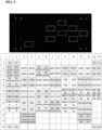
- cfDNA was extracted from the blood of a healthy individual, and was then ligated with costume made adaptors, to allow for the uniform and non-biased amplification of the cfDNA. Following, the DNA went through bisulfite treatment, to convert all unmethylated cytosine nucleotides into uracil nucleotides. Following, all the bisulfite treated DNA was amplified. During the amplification process, all uracil nucleotides (i.e. cytosine nucleotides that were unmethylated in the cfDNA prior to the bisulfite conversion) became thymidine nucleotides.
- CpG methyltransferase was used in-vitro together with a synthetic cofactor to attach a fluorophore to the unmethylated CpG sites.
- the methyltransferase used was a M.MpeI double mutant enzyme that catalyzes the addition of a synthetic cofactor within the CpG sequence, instead of the methyl group.
- the sample was then hybridized to a custom designed microarray, which contains the capture probes of interest. The spots which light up in the array represent the sequences which were labeled and hybridized to the slide ( Figure 2 ).
- methylation pattern is unique to each cell type or tissue and can change during pathologic processes (e.g. cancer)
- a method can further be used to e.g. identify cell or tissue type or state.
- cfDNA was extracted from the blood of four healthy individual, four colon cancer patients, and three blood cancer patients and subjected to the same process described hereinabove. Namely, samples were ligated with custom made adaptors, followed by bisulfite treatment and amplification. Subsequently, fluorescent labeling of unmethylated CpGs was performed by the described two-step chemoenzymatic reaction and the samples were then hybridized to a commercial microarray (Human Genome CGH Microarray, Agilent).
- Adaptors-based amplification (protocol 1): Extraction of circulating cell-free DNA, NEBNext Ultra II End Repair / dA-Tailing module, Adaptors and primers design, Ligation of Adaptors, Sodium Bisulfite treatment, PCR amplification - as described in Examples 1 hereinabove.
- Isotherami nucleic acid amplification (Protocol 2): Isothermal nucleic acid amplification was effected according to Infinium HD Methylation Assay (Illumina kit, Cat No. WG-317- 1002) manufacturer's instructions. Briefly: Sodium Bisulfite treatment - In order to convert all unmethylated cytosine nucleotides into uracil nucleotides, 500 ng of genomic DNA (blood DNA, and U2OS cell line DNA) was treated with sodium bisulfite, using the EZ DNA Methylation-Gold Kit (ZYMO, Cat. D5006), according to manufacturer's protocol.
- EZ DNA Methylation-Gold Kit ZYMO, Cat. D5006
- DNA amplification - Bisulfite-treated DNA was amplified according to the Illumina kit protocol.
- Four ⁇ L of bisulfite treated DNA sample was mixed with four ⁇ L of 0.1M NaOH, vortexed and incubated at room temperature for 10 minutes.
- 68 ⁇ L of RPM and 75 ⁇ L of MSM were added to the sample, which was vortexed and incubated for 24 hours at 37°C.
- Fragment DNA- Amplified DNA was fragmented according to the Illumina kit protocol. 50 ⁇ L of FMS were added to the amplified DNA sample, which was vortexed and incubated for one hour at 37 °C.
- a 2% agarose gel was prepared by dissolving 1.2 gr of agarose (Cat. 889153, TAU storage) in 60 mL of TBE. Six ⁇ L of SYBR safe (Cat. S33102, Invitrogen) were added to the mixture, which was poured into a gel electrophoresis device. The gel was left to stabilize for ⁇ 30 minutes, after which the relevant DNA samples were inserted, as well as a 100 bp ladder (Cat. N3231L, NEB). Gel was ran in 100V for 45 minutes, to receive optimal separation.
- protocol 1 amplified template yielded a six-times stronger band, than that of protocol 2 ( Figure 5 ); indicating that the amplification method effected according to protocol 1 and based on adaptors ligation, is more efficient and specific than the amplification method effected according to protocol 2 that is based on isotheraml nucleic acid amplification.
Landscapes
- Chemical & Material Sciences (AREA)
- Life Sciences & Earth Sciences (AREA)
- Organic Chemistry (AREA)
- Health & Medical Sciences (AREA)
- Proteomics, Peptides & Aminoacids (AREA)
- Zoology (AREA)
- Wood Science & Technology (AREA)
- Engineering & Computer Science (AREA)
- Analytical Chemistry (AREA)
- General Health & Medical Sciences (AREA)
- Genetics & Genomics (AREA)
- Immunology (AREA)
- General Engineering & Computer Science (AREA)
- Physics & Mathematics (AREA)
- Microbiology (AREA)
- Molecular Biology (AREA)
- Biotechnology (AREA)
- Biochemistry (AREA)
- Bioinformatics & Cheminformatics (AREA)
- Biophysics (AREA)
- Chemical Kinetics & Catalysis (AREA)
- Pathology (AREA)
- Animal Behavior & Ethology (AREA)
- General Chemical & Material Sciences (AREA)
- Medicinal Chemistry (AREA)
- Nuclear Medicine, Radiotherapy & Molecular Imaging (AREA)
- Pharmacology & Pharmacy (AREA)
- Public Health (AREA)
- Veterinary Medicine (AREA)
- Hospice & Palliative Care (AREA)
- Oncology (AREA)
- Measuring Or Testing Involving Enzymes Or Micro-Organisms (AREA)
- Steroid Compounds (AREA)
- Investigating Or Analyzing Materials By The Use Of Electric Means (AREA)
- Lubrication Of Internal Combustion Engines (AREA)
Applications Claiming Priority (3)
| Application Number | Priority Date | Filing Date | Title |
|---|---|---|---|
| US202063037020P | 2020-06-10 | 2020-06-10 | |
| EP21821437.7A EP4165213A1 (de) | 2020-06-10 | 2021-06-10 | Verfahren zum nachweis von methyliertem cpg |
| PCT/IL2021/050706 WO2021250677A1 (en) | 2020-06-10 | 2021-06-10 | METHODS OF DETECTING METHYLATED CpG |
Related Parent Applications (2)
| Application Number | Title | Priority Date | Filing Date |
|---|---|---|---|
| EP21821437.7A Division EP4165213A1 (de) | 2020-06-10 | 2021-06-10 | Verfahren zum nachweis von methyliertem cpg |
| PCT/IL2021/050706 Previously-Filed-Application WO2021250677A1 (en) | 2020-06-10 | 2021-06-10 | METHODS OF DETECTING METHYLATED CpG |
Publications (2)
| Publication Number | Publication Date |
|---|---|
| EP4521115A2 true EP4521115A2 (de) | 2025-03-12 |
| EP4521115A3 EP4521115A3 (de) | 2025-06-11 |
Family
ID=78846993
Family Applications (2)
| Application Number | Title | Priority Date | Filing Date |
|---|---|---|---|
| EP24166517.3A Pending EP4521115A3 (de) | 2020-06-10 | 2021-06-10 | Verfahren zum nachweis von methyliertem cpg |
| EP21821437.7A Withdrawn EP4165213A1 (de) | 2020-06-10 | 2021-06-10 | Verfahren zum nachweis von methyliertem cpg |
Family Applications After (1)
| Application Number | Title | Priority Date | Filing Date |
|---|---|---|---|
| EP21821437.7A Withdrawn EP4165213A1 (de) | 2020-06-10 | 2021-06-10 | Verfahren zum nachweis von methyliertem cpg |
Country Status (5)
| Country | Link |
|---|---|
| US (1) | US20230220459A1 (de) |
| EP (2) | EP4521115A3 (de) |
| CA (1) | CA3185430A1 (de) |
| IL (1) | IL298934A (de) |
| WO (1) | WO2021250677A1 (de) |
Families Citing this family (3)
| Publication number | Priority date | Publication date | Assignee | Title |
|---|---|---|---|---|
| JP2025517309A (ja) | 2022-05-17 | 2025-06-05 | ガーダント ヘルス, インコーポレイテッド | 創薬可能な標的を特定するための方法およびがんを処置するための方法 |
| GB2639522A (en) * | 2023-11-14 | 2025-10-01 | Tagomics Ltd | Profiling Method |
| WO2025132539A1 (en) | 2023-12-19 | 2025-06-26 | Qbdc Gmbh | Predicting the quality of a bioprocess and product |
Citations (26)
| Publication number | Priority date | Publication date | Assignee | Title |
|---|---|---|---|---|
| US3791932A (en) | 1971-02-10 | 1974-02-12 | Akzona Inc | Process for the demonstration and determination of reaction components having specific binding affinity for each other |
| US3839153A (en) | 1970-12-28 | 1974-10-01 | Akzona Inc | Process for the detection and determination of specific binding proteins and their corresponding bindable substances |
| US3850578A (en) | 1973-03-12 | 1974-11-26 | H Mcconnell | Process for assaying for biologically active molecules |
| US3850752A (en) | 1970-11-10 | 1974-11-26 | Akzona Inc | Process for the demonstration and determination of low molecular compounds and of proteins capable of binding these compounds specifically |
| US3853987A (en) | 1971-09-01 | 1974-12-10 | W Dreyer | Immunological reagent and radioimmuno assay |
| US3867517A (en) | 1971-12-21 | 1975-02-18 | Abbott Lab | Direct radioimmunoassay for antigens and their antibodies |
| US3879262A (en) | 1972-05-11 | 1975-04-22 | Akzona Inc | Detection and determination of haptens |
| US3901654A (en) | 1971-06-21 | 1975-08-26 | Biological Developments | Receptor assays of biologically active compounds employing biologically specific receptors |
| US3935074A (en) | 1973-12-17 | 1976-01-27 | Syva Company | Antibody steric hindrance immunoassay with two antibodies |
| US3984533A (en) | 1975-11-13 | 1976-10-05 | General Electric Company | Electrophoretic method of detecting antigen-antibody reaction |
| US3996345A (en) | 1974-08-12 | 1976-12-07 | Syva Company | Fluorescence quenching with immunological pairs in immunoassays |
| US4034074A (en) | 1974-09-19 | 1977-07-05 | The Board Of Trustees Of Leland Stanford Junior University | Universal reagent 2-site immunoradiometric assay using labelled anti (IgG) |
| US4098876A (en) | 1976-10-26 | 1978-07-04 | Corning Glass Works | Reverse sandwich immunoassay |
| US4666828A (en) | 1984-08-15 | 1987-05-19 | The General Hospital Corporation | Test for Huntington's disease |
| US4683202A (en) | 1985-03-28 | 1987-07-28 | Cetus Corporation | Process for amplifying nucleic acid sequences |
| US4801531A (en) | 1985-04-17 | 1989-01-31 | Biotechnology Research Partners, Ltd. | Apo AI/CIII genomic polymorphisms predictive of atherosclerosis |
| US4879219A (en) | 1980-09-19 | 1989-11-07 | General Hospital Corporation | Immunoassay utilizing monoclonal high affinity IgM antibodies |
| US5011771A (en) | 1984-04-12 | 1991-04-30 | The General Hospital Corporation | Multiepitopic immunometric assay |
| US5192659A (en) | 1989-08-25 | 1993-03-09 | Genetype Ag | Intron sequence analysis method for detection of adjacent and remote locus alleles as haplotypes |
| US5272057A (en) | 1988-10-14 | 1993-12-21 | Georgetown University | Method of detecting a predisposition to cancer by the use of restriction fragment length polymorphism of the gene for human poly (ADP-ribose) polymerase |
| US5281521A (en) | 1992-07-20 | 1994-01-25 | The Trustees Of The University Of Pennsylvania | Modified avidin-biotin technique |
| WO2014191981A1 (en) | 2013-05-28 | 2014-12-04 | Ramot At Tel-Aviv University Ltd. | Detection of hydroxymethylcytosine bases |
| WO2016178207A1 (en) | 2015-05-04 | 2016-11-10 | Ramot At Tel-Aviv University Ltd. | Methods and kits for fragmenting dna |
| WO2017081689A1 (en) | 2015-11-11 | 2017-05-18 | Ramot At Tel-Aviv University Ltd. | Methods of detecting 5-hydroxymethylcytosine and diagnosing of cancer |
| WO2018029693A1 (en) | 2016-08-10 | 2018-02-15 | Ramot At Tel-Aviv University Ltd. | Analysis of methylation status and copy number |
| WO2019234753A1 (en) | 2018-06-06 | 2019-12-12 | Ramot At Tel-Aviv University Ltd. | Multi-sample array chip for dna modification quantification |
Family Cites Families (4)
| Publication number | Priority date | Publication date | Assignee | Title |
|---|---|---|---|---|
| EP2380993B1 (de) * | 2004-03-08 | 2015-12-23 | Rubicon Genomics, Inc. | Verfahren zur Erstellung und Erweiterung von DNA-Bibliotheken zur sensitiven Erkennung und Analyse von DNA-Methylierung |
| US20060292585A1 (en) * | 2005-06-24 | 2006-12-28 | Affymetrix, Inc. | Analysis of methylation using nucleic acid arrays |
| WO2015159292A2 (en) * | 2014-04-14 | 2015-10-22 | Yissum Research Development Company Of The Hebrew University Of Jerusalem Ltd. | A method and kit for determining the tissue or cell origin of dna |
| BR112019018272A2 (pt) * | 2017-03-02 | 2020-07-28 | Youhealth Oncotech, Limited | marcadores metilação para diagnosticar hepatocelular carcinoma e câncer |
-
2021
- 2021-06-10 EP EP24166517.3A patent/EP4521115A3/de active Pending
- 2021-06-10 IL IL298934A patent/IL298934A/en unknown
- 2021-06-10 CA CA3185430A patent/CA3185430A1/en active Pending
- 2021-06-10 US US18/009,013 patent/US20230220459A1/en active Pending
- 2021-06-10 EP EP21821437.7A patent/EP4165213A1/de not_active Withdrawn
- 2021-06-10 WO PCT/IL2021/050706 patent/WO2021250677A1/en not_active Ceased
Patent Citations (27)
| Publication number | Priority date | Publication date | Assignee | Title |
|---|---|---|---|---|
| US3850752A (en) | 1970-11-10 | 1974-11-26 | Akzona Inc | Process for the demonstration and determination of low molecular compounds and of proteins capable of binding these compounds specifically |
| US3839153A (en) | 1970-12-28 | 1974-10-01 | Akzona Inc | Process for the detection and determination of specific binding proteins and their corresponding bindable substances |
| US3791932A (en) | 1971-02-10 | 1974-02-12 | Akzona Inc | Process for the demonstration and determination of reaction components having specific binding affinity for each other |
| US3901654A (en) | 1971-06-21 | 1975-08-26 | Biological Developments | Receptor assays of biologically active compounds employing biologically specific receptors |
| US3853987A (en) | 1971-09-01 | 1974-12-10 | W Dreyer | Immunological reagent and radioimmuno assay |
| US3867517A (en) | 1971-12-21 | 1975-02-18 | Abbott Lab | Direct radioimmunoassay for antigens and their antibodies |
| US3879262A (en) | 1972-05-11 | 1975-04-22 | Akzona Inc | Detection and determination of haptens |
| US3850578A (en) | 1973-03-12 | 1974-11-26 | H Mcconnell | Process for assaying for biologically active molecules |
| US3935074A (en) | 1973-12-17 | 1976-01-27 | Syva Company | Antibody steric hindrance immunoassay with two antibodies |
| US3996345A (en) | 1974-08-12 | 1976-12-07 | Syva Company | Fluorescence quenching with immunological pairs in immunoassays |
| US4034074A (en) | 1974-09-19 | 1977-07-05 | The Board Of Trustees Of Leland Stanford Junior University | Universal reagent 2-site immunoradiometric assay using labelled anti (IgG) |
| US3984533A (en) | 1975-11-13 | 1976-10-05 | General Electric Company | Electrophoretic method of detecting antigen-antibody reaction |
| US4098876A (en) | 1976-10-26 | 1978-07-04 | Corning Glass Works | Reverse sandwich immunoassay |
| US4879219A (en) | 1980-09-19 | 1989-11-07 | General Hospital Corporation | Immunoassay utilizing monoclonal high affinity IgM antibodies |
| US5011771A (en) | 1984-04-12 | 1991-04-30 | The General Hospital Corporation | Multiepitopic immunometric assay |
| US4666828A (en) | 1984-08-15 | 1987-05-19 | The General Hospital Corporation | Test for Huntington's disease |
| US4683202A (en) | 1985-03-28 | 1987-07-28 | Cetus Corporation | Process for amplifying nucleic acid sequences |
| US4683202B1 (de) | 1985-03-28 | 1990-11-27 | Cetus Corp | |
| US4801531A (en) | 1985-04-17 | 1989-01-31 | Biotechnology Research Partners, Ltd. | Apo AI/CIII genomic polymorphisms predictive of atherosclerosis |
| US5272057A (en) | 1988-10-14 | 1993-12-21 | Georgetown University | Method of detecting a predisposition to cancer by the use of restriction fragment length polymorphism of the gene for human poly (ADP-ribose) polymerase |
| US5192659A (en) | 1989-08-25 | 1993-03-09 | Genetype Ag | Intron sequence analysis method for detection of adjacent and remote locus alleles as haplotypes |
| US5281521A (en) | 1992-07-20 | 1994-01-25 | The Trustees Of The University Of Pennsylvania | Modified avidin-biotin technique |
| WO2014191981A1 (en) | 2013-05-28 | 2014-12-04 | Ramot At Tel-Aviv University Ltd. | Detection of hydroxymethylcytosine bases |
| WO2016178207A1 (en) | 2015-05-04 | 2016-11-10 | Ramot At Tel-Aviv University Ltd. | Methods and kits for fragmenting dna |
| WO2017081689A1 (en) | 2015-11-11 | 2017-05-18 | Ramot At Tel-Aviv University Ltd. | Methods of detecting 5-hydroxymethylcytosine and diagnosing of cancer |
| WO2018029693A1 (en) | 2016-08-10 | 2018-02-15 | Ramot At Tel-Aviv University Ltd. | Analysis of methylation status and copy number |
| WO2019234753A1 (en) | 2018-06-06 | 2019-12-12 | Ramot At Tel-Aviv University Ltd. | Multi-sample array chip for dna modification quantification |
Non-Patent Citations (14)
| Title |
|---|
| "Genome Analysis: A Laboratory Manual Series", vol. 1-4, 1998, COLD SPRING HARBOR LABORATORY PRESS |
| "Immobilized Cells and Enzymes", 1986, IRL PRESS |
| "Nucleic Acid Hybridization", 1985 |
| "Oligonucleotide Synthesis", 1983 |
| "PCR Protocols: A Guide To Methods And Applications", vol. 1-317, 1990, ACADEMIC PRESS |
| "Selected Methods in Cellular Immunology", vol. 1-3, 1980, W. H. FREEMAN AND CO. |
| "Transcription and Translation", 1984 |
| FRESHNEY: "Culture of Animal Cells - A Manual of Basic Technique", vol. 1-3, 1994, APPLETON & LANGE |
| KRIUKIENE ET AL., NATURE COMMUNICATIONS, vol. 4, no. 2190, 2013 |
| MARSHAK ET AL.: "Strategies for Protein Purification and Characterization - A Laboratory Course Manual", 1996, CSHL PRESS |
| PERBAL: "A Practical Guide to Molecular Cloning", 1984, JOHN WILEY & SONS |
| SAMBROOK ET AL.: "Current Protocols in Molecular Biology", 1989, JOHN WILEY AND SONS |
| WATSON ET AL.: "Scientific American Books", article "Recombinant DNA" |
| ZHOU H ET AL., CLINICAL CHEMISTRY, 2007 |
Also Published As
| Publication number | Publication date |
|---|---|
| IL298934A (en) | 2023-02-01 |
| CA3185430A1 (en) | 2021-12-16 |
| US20230220459A1 (en) | 2023-07-13 |
| EP4521115A3 (de) | 2025-06-11 |
| WO2021250677A1 (en) | 2021-12-16 |
| EP4165213A1 (de) | 2023-04-19 |
Similar Documents
| Publication | Publication Date | Title |
|---|---|---|
| JP7035156B2 (ja) | 結腸直腸がんのエピジェネティックマーカー及び該マーカーを使用する診断法 | |
| EP4521115A2 (de) | Verfahren zum nachweis von methyliertem cpg | |
| CN104745575B (zh) | 用于检测细胞增殖性异常或疾病程度分级的基因组合物及其用途 | |
| US20100068720A1 (en) | Method and kit for detection of early cancer or pre-cancer using blood and body fluids | |
| US20200277654A1 (en) | Method for Detecting multiple DNA Mutations and Copy Number Variations | |
| CN106834426A (zh) | 用于检测胰腺癌的组合物及其用途 | |
| KR101255769B1 (ko) | 장암 진단을 위한 장암 특이적 메틸화 마커 gpm6a 유전자의 메틸화 검출방법 | |
| KR101313756B1 (ko) | 간암 특이적 과메틸화 CpG 서열을 이용한 간암의 검출방법 | |
| CN108220428A (zh) | 用于检测胃癌的组合物及其试剂盒和用途 | |
| US20230047930A1 (en) | Methods and arrays for identifying the cell or tissue origin of dna | |
| CN107868822B (zh) | 用于检测食道癌的组合物及其试剂盒和用途 | |
| CN114381498B (zh) | 一种基于协同原位组装G-四链体DNAzyme纳米线的化学发光传感器及其应用 | |
| CN101796185B (zh) | 扩增甲基化核酸或非甲基化核酸的方法 | |
| JP2022533088A (ja) | 変異体核酸を検出するための融解温度法、キットおよびレポータオリゴ | |
| CN113881739B (zh) | 氧化含锯齿末端核酸分子的方法及还原方法与文库构建法 | |
| CN117821585A (zh) | 结直肠癌早期诊断标志物及应用 | |
| HK40116484A (en) | Methods of detecting methylated cpg | |
| JP5151167B2 (ja) | Dnaメチル化測定方法 | |
| KR101145406B1 (ko) | 장암 진단을 위한 장암 특이적 메틸화 마커 유전자의 메틸화 검출방법 | |
| JP4924014B2 (ja) | Dnaメチル化測定方法 | |
| JP4940940B2 (ja) | Dnaメチル化測定方法 | |
| JP4940939B2 (ja) | Dnaメチル化測定方法 | |
| KR101009062B1 (ko) | 위암 진단을 위한 위암 특이적 메틸화 마커 유전자의 메틸화 검출방법 | |
| WO2025147648A1 (en) | Probe blockers for enrichment of target nucleic acid sequences | |
| KR101136505B1 (ko) | 장암 진단을 위한 장암 특이적 메틸화 마커 유전자의 메틸화 검출방법 |
Legal Events
| Date | Code | Title | Description |
|---|---|---|---|
| PUAI | Public reference made under article 153(3) epc to a published international application that has entered the european phase |
Free format text: ORIGINAL CODE: 0009012 |
|
| STAA | Information on the status of an ep patent application or granted ep patent |
Free format text: STATUS: THE APPLICATION HAS BEEN PUBLISHED |
|
| AC | Divisional application: reference to earlier application |
Ref document number: 4165213 Country of ref document: EP Kind code of ref document: P |
|
| AK | Designated contracting states |
Kind code of ref document: A2 Designated state(s): AL AT BE BG CH CY CZ DE DK EE ES FI FR GB GR HR HU IE IS IT LI LT LU LV MC MK MT NL NO PL PT RO RS SE SI SK SM TR |
|
| REG | Reference to a national code |
Ref country code: HK Ref legal event code: DE Ref document number: 40116484 Country of ref document: HK |
|
| REG | Reference to a national code |
Ref country code: DE Ref legal event code: R079 Free format text: PREVIOUS MAIN CLASS: G01N0033530000 Ipc: C12Q0001683700 |
|
| PUAL | Search report despatched |
Free format text: ORIGINAL CODE: 0009013 |
|
| AK | Designated contracting states |
Kind code of ref document: A3 Designated state(s): AL AT BE BG CH CY CZ DE DK EE ES FI FR GB GR HR HU IE IS IT LI LT LU LV MC MK MT NL NO PL PT RO RS SE SI SK SM TR |
|
| RIC1 | Information provided on ipc code assigned before grant |
Ipc: C12Q 1/6837 20180101AFI20250507BHEP |
|
| STAA | Information on the status of an ep patent application or granted ep patent |
Free format text: STATUS: REQUEST FOR EXAMINATION WAS MADE |
|
| 17P | Request for examination filed |
Effective date: 20251014 |
|
| STAA | Information on the status of an ep patent application or granted ep patent |
Free format text: STATUS: EXAMINATION IS IN PROGRESS |
|
| 17Q | First examination report despatched |
Effective date: 20251127 |