EP4527408A2 - Claudin-6-antikörper und arzneimittelkonjugate - Google Patents
Claudin-6-antikörper und arzneimittelkonjugate Download PDFInfo
- Publication number
- EP4527408A2 EP4527408A2 EP24221566.3A EP24221566A EP4527408A2 EP 4527408 A2 EP4527408 A2 EP 4527408A2 EP 24221566 A EP24221566 A EP 24221566A EP 4527408 A2 EP4527408 A2 EP 4527408A2
- Authority
- EP
- European Patent Office
- Prior art keywords
- seq
- antigen
- sequence
- nos
- binding protein
- Prior art date
- Legal status (The legal status is an assumption and is not a legal conclusion. Google has not performed a legal analysis and makes no representation as to the accuracy of the status listed.)
- Pending
Links
Images


Classifications
-
- A—HUMAN NECESSITIES
- A61—MEDICAL OR VETERINARY SCIENCE; HYGIENE
- A61K—PREPARATIONS FOR MEDICAL, DENTAL OR TOILETRY PURPOSES
- A61K47/00—Medicinal preparations characterised by the non-active ingredients used, e.g. carriers or inert additives; Targeting or modifying agents chemically bound to the active ingredient
- A61K47/50—Medicinal preparations characterised by the non-active ingredients used, e.g. carriers or inert additives; Targeting or modifying agents chemically bound to the active ingredient the non-active ingredient being chemically bound to the active ingredient, e.g. polymer-drug conjugates
- A61K47/51—Medicinal preparations characterised by the non-active ingredients used, e.g. carriers or inert additives; Targeting or modifying agents chemically bound to the active ingredient the non-active ingredient being chemically bound to the active ingredient, e.g. polymer-drug conjugates the non-active ingredient being a modifying agent
- A61K47/62—Medicinal preparations characterised by the non-active ingredients used, e.g. carriers or inert additives; Targeting or modifying agents chemically bound to the active ingredient the non-active ingredient being chemically bound to the active ingredient, e.g. polymer-drug conjugates the non-active ingredient being a modifying agent the modifying agent being a protein, peptide or polyamino acid
- A61K47/65—Peptidic linkers, binders or spacers, e.g. peptidic enzyme-labile linkers
-
- A—HUMAN NECESSITIES
- A61—MEDICAL OR VETERINARY SCIENCE; HYGIENE
- A61K—PREPARATIONS FOR MEDICAL, DENTAL OR TOILETRY PURPOSES
- A61K47/00—Medicinal preparations characterised by the non-active ingredients used, e.g. carriers or inert additives; Targeting or modifying agents chemically bound to the active ingredient
- A61K47/50—Medicinal preparations characterised by the non-active ingredients used, e.g. carriers or inert additives; Targeting or modifying agents chemically bound to the active ingredient the non-active ingredient being chemically bound to the active ingredient, e.g. polymer-drug conjugates
- A61K47/51—Medicinal preparations characterised by the non-active ingredients used, e.g. carriers or inert additives; Targeting or modifying agents chemically bound to the active ingredient the non-active ingredient being chemically bound to the active ingredient, e.g. polymer-drug conjugates the non-active ingredient being a modifying agent
- A61K47/68—Medicinal preparations characterised by the non-active ingredients used, e.g. carriers or inert additives; Targeting or modifying agents chemically bound to the active ingredient the non-active ingredient being chemically bound to the active ingredient, e.g. polymer-drug conjugates the non-active ingredient being a modifying agent the modifying agent being an antibody, an immunoglobulin or a fragment thereof, e.g. an Fc-fragment
- A61K47/6801—Drug-antibody or immunoglobulin conjugates defined by the pharmacologically or therapeutically active agent
- A61K47/6803—Drugs conjugated to an antibody or immunoglobulin, e.g. cisplatin-antibody conjugates
- A61K47/68031—Drugs conjugated to an antibody or immunoglobulin, e.g. cisplatin-antibody conjugates the drug being an auristatin
-
- A—HUMAN NECESSITIES
- A61—MEDICAL OR VETERINARY SCIENCE; HYGIENE
- A61K—PREPARATIONS FOR MEDICAL, DENTAL OR TOILETRY PURPOSES
- A61K47/00—Medicinal preparations characterised by the non-active ingredients used, e.g. carriers or inert additives; Targeting or modifying agents chemically bound to the active ingredient
- A61K47/50—Medicinal preparations characterised by the non-active ingredients used, e.g. carriers or inert additives; Targeting or modifying agents chemically bound to the active ingredient the non-active ingredient being chemically bound to the active ingredient, e.g. polymer-drug conjugates
- A61K47/51—Medicinal preparations characterised by the non-active ingredients used, e.g. carriers or inert additives; Targeting or modifying agents chemically bound to the active ingredient the non-active ingredient being chemically bound to the active ingredient, e.g. polymer-drug conjugates the non-active ingredient being a modifying agent
- A61K47/68—Medicinal preparations characterised by the non-active ingredients used, e.g. carriers or inert additives; Targeting or modifying agents chemically bound to the active ingredient the non-active ingredient being chemically bound to the active ingredient, e.g. polymer-drug conjugates the non-active ingredient being a modifying agent the modifying agent being an antibody, an immunoglobulin or a fragment thereof, e.g. an Fc-fragment
- A61K47/6801—Drug-antibody or immunoglobulin conjugates defined by the pharmacologically or therapeutically active agent
- A61K47/6803—Drugs conjugated to an antibody or immunoglobulin, e.g. cisplatin-antibody conjugates
- A61K47/68037—Drugs conjugated to an antibody or immunoglobulin, e.g. cisplatin-antibody conjugates the drug being a camptothecin [CPT] or derivatives
-
- A—HUMAN NECESSITIES
- A61—MEDICAL OR VETERINARY SCIENCE; HYGIENE
- A61K—PREPARATIONS FOR MEDICAL, DENTAL OR TOILETRY PURPOSES
- A61K47/00—Medicinal preparations characterised by the non-active ingredients used, e.g. carriers or inert additives; Targeting or modifying agents chemically bound to the active ingredient
- A61K47/50—Medicinal preparations characterised by the non-active ingredients used, e.g. carriers or inert additives; Targeting or modifying agents chemically bound to the active ingredient the non-active ingredient being chemically bound to the active ingredient, e.g. polymer-drug conjugates
- A61K47/51—Medicinal preparations characterised by the non-active ingredients used, e.g. carriers or inert additives; Targeting or modifying agents chemically bound to the active ingredient the non-active ingredient being chemically bound to the active ingredient, e.g. polymer-drug conjugates the non-active ingredient being a modifying agent
- A61K47/68—Medicinal preparations characterised by the non-active ingredients used, e.g. carriers or inert additives; Targeting or modifying agents chemically bound to the active ingredient the non-active ingredient being chemically bound to the active ingredient, e.g. polymer-drug conjugates the non-active ingredient being a modifying agent the modifying agent being an antibody, an immunoglobulin or a fragment thereof, e.g. an Fc-fragment
- A61K47/6801—Drug-antibody or immunoglobulin conjugates defined by the pharmacologically or therapeutically active agent
- A61K47/6803—Drugs conjugated to an antibody or immunoglobulin, e.g. cisplatin-antibody conjugates
- A61K47/6811—Drugs conjugated to an antibody or immunoglobulin, e.g. cisplatin-antibody conjugates the drug being a protein or peptide, e.g. transferrin or bleomycin
- A61K47/6817—Toxins
-
- A—HUMAN NECESSITIES
- A61—MEDICAL OR VETERINARY SCIENCE; HYGIENE
- A61K—PREPARATIONS FOR MEDICAL, DENTAL OR TOILETRY PURPOSES
- A61K47/00—Medicinal preparations characterised by the non-active ingredients used, e.g. carriers or inert additives; Targeting or modifying agents chemically bound to the active ingredient
- A61K47/50—Medicinal preparations characterised by the non-active ingredients used, e.g. carriers or inert additives; Targeting or modifying agents chemically bound to the active ingredient the non-active ingredient being chemically bound to the active ingredient, e.g. polymer-drug conjugates
- A61K47/51—Medicinal preparations characterised by the non-active ingredients used, e.g. carriers or inert additives; Targeting or modifying agents chemically bound to the active ingredient the non-active ingredient being chemically bound to the active ingredient, e.g. polymer-drug conjugates the non-active ingredient being a modifying agent
- A61K47/68—Medicinal preparations characterised by the non-active ingredients used, e.g. carriers or inert additives; Targeting or modifying agents chemically bound to the active ingredient the non-active ingredient being chemically bound to the active ingredient, e.g. polymer-drug conjugates the non-active ingredient being a modifying agent the modifying agent being an antibody, an immunoglobulin or a fragment thereof, e.g. an Fc-fragment
- A61K47/6835—Medicinal preparations characterised by the non-active ingredients used, e.g. carriers or inert additives; Targeting or modifying agents chemically bound to the active ingredient the non-active ingredient being chemically bound to the active ingredient, e.g. polymer-drug conjugates the non-active ingredient being a modifying agent the modifying agent being an antibody, an immunoglobulin or a fragment thereof, e.g. an Fc-fragment the modifying agent being an antibody or an immunoglobulin bearing at least one antigen-binding site
- A61K47/6849—Medicinal preparations characterised by the non-active ingredients used, e.g. carriers or inert additives; Targeting or modifying agents chemically bound to the active ingredient the non-active ingredient being chemically bound to the active ingredient, e.g. polymer-drug conjugates the non-active ingredient being a modifying agent the modifying agent being an antibody, an immunoglobulin or a fragment thereof, e.g. an Fc-fragment the modifying agent being an antibody or an immunoglobulin bearing at least one antigen-binding site the antibody targeting a receptor, a cell surface antigen or a cell surface determinant
-
- A—HUMAN NECESSITIES
- A61—MEDICAL OR VETERINARY SCIENCE; HYGIENE
- A61K—PREPARATIONS FOR MEDICAL, DENTAL OR TOILETRY PURPOSES
- A61K47/00—Medicinal preparations characterised by the non-active ingredients used, e.g. carriers or inert additives; Targeting or modifying agents chemically bound to the active ingredient
- A61K47/50—Medicinal preparations characterised by the non-active ingredients used, e.g. carriers or inert additives; Targeting or modifying agents chemically bound to the active ingredient the non-active ingredient being chemically bound to the active ingredient, e.g. polymer-drug conjugates
- A61K47/51—Medicinal preparations characterised by the non-active ingredients used, e.g. carriers or inert additives; Targeting or modifying agents chemically bound to the active ingredient the non-active ingredient being chemically bound to the active ingredient, e.g. polymer-drug conjugates the non-active ingredient being a modifying agent
- A61K47/68—Medicinal preparations characterised by the non-active ingredients used, e.g. carriers or inert additives; Targeting or modifying agents chemically bound to the active ingredient the non-active ingredient being chemically bound to the active ingredient, e.g. polymer-drug conjugates the non-active ingredient being a modifying agent the modifying agent being an antibody, an immunoglobulin or a fragment thereof, e.g. an Fc-fragment
- A61K47/6835—Medicinal preparations characterised by the non-active ingredients used, e.g. carriers or inert additives; Targeting or modifying agents chemically bound to the active ingredient the non-active ingredient being chemically bound to the active ingredient, e.g. polymer-drug conjugates the non-active ingredient being a modifying agent the modifying agent being an antibody, an immunoglobulin or a fragment thereof, e.g. an Fc-fragment the modifying agent being an antibody or an immunoglobulin bearing at least one antigen-binding site
- A61K47/6851—Medicinal preparations characterised by the non-active ingredients used, e.g. carriers or inert additives; Targeting or modifying agents chemically bound to the active ingredient the non-active ingredient being chemically bound to the active ingredient, e.g. polymer-drug conjugates the non-active ingredient being a modifying agent the modifying agent being an antibody, an immunoglobulin or a fragment thereof, e.g. an Fc-fragment the modifying agent being an antibody or an immunoglobulin bearing at least one antigen-binding site the antibody targeting a determinant of a tumour cell
-
- A—HUMAN NECESSITIES
- A61—MEDICAL OR VETERINARY SCIENCE; HYGIENE
- A61P—SPECIFIC THERAPEUTIC ACTIVITY OF CHEMICAL COMPOUNDS OR MEDICINAL PREPARATIONS
- A61P35/00—Antineoplastic agents
-
- C—CHEMISTRY; METALLURGY
- C07—ORGANIC CHEMISTRY
- C07K—PEPTIDES
- C07K16/00—Immunoglobulins [IG], e.g. monoclonal or polyclonal antibodies
- C07K16/18—Immunoglobulins [IG], e.g. monoclonal or polyclonal antibodies against material from animals or humans
- C07K16/28—Immunoglobulins [IG], e.g. monoclonal or polyclonal antibodies against material from animals or humans against receptors, cell surface antigens or cell surface determinants
- C07K16/30—Immunoglobulins [IG], e.g. monoclonal or polyclonal antibodies against material from animals or humans against receptors, cell surface antigens or cell surface determinants from tumour cells
-
- G—PHYSICS
- G01—MEASURING; TESTING
- G01N—INVESTIGATING OR ANALYSING MATERIALS BY DETERMINING THEIR CHEMICAL OR PHYSICAL PROPERTIES
- G01N33/00—Investigating or analysing materials by specific methods not covered by groups G01N1/00 - G01N31/00
- G01N33/48—Biological material, e.g. blood, urine; Haemocytometers
- G01N33/50—Chemical analysis of biological material, e.g. blood, urine; Testing involving biospecific ligand binding methods; Immunological testing
- G01N33/53—Immunoassay; Biospecific binding assay; Materials therefor
- G01N33/575—Immunoassay; Biospecific binding assay; Materials therefor for cancer
- G01N33/57575—Immunoassay; Biospecific binding assay; Materials therefor for cancer involving oncogenic proteins
-
- A—HUMAN NECESSITIES
- A61—MEDICAL OR VETERINARY SCIENCE; HYGIENE
- A61K—PREPARATIONS FOR MEDICAL, DENTAL OR TOILETRY PURPOSES
- A61K39/00—Medicinal preparations containing antigens or antibodies
- A61K2039/505—Medicinal preparations containing antigens or antibodies comprising antibodies
-
- C—CHEMISTRY; METALLURGY
- C07—ORGANIC CHEMISTRY
- C07K—PEPTIDES
- C07K2317/00—Immunoglobulins specific features
- C07K2317/20—Immunoglobulins specific features characterized by taxonomic origin
- C07K2317/24—Immunoglobulins specific features characterized by taxonomic origin containing regions, domains or residues from different species, e.g. chimeric, humanized or veneered
-
- C—CHEMISTRY; METALLURGY
- C07—ORGANIC CHEMISTRY
- C07K—PEPTIDES
- C07K2317/00—Immunoglobulins specific features
- C07K2317/30—Immunoglobulins specific features characterized by aspects of specificity or valency
- C07K2317/33—Crossreactivity, e.g. for species or epitope, or lack of said crossreactivity
-
- C—CHEMISTRY; METALLURGY
- C07—ORGANIC CHEMISTRY
- C07K—PEPTIDES
- C07K2317/00—Immunoglobulins specific features
- C07K2317/40—Immunoglobulins specific features characterized by post-translational modification
- C07K2317/41—Glycosylation, sialylation, or fucosylation
-
- C—CHEMISTRY; METALLURGY
- C07—ORGANIC CHEMISTRY
- C07K—PEPTIDES
- C07K2317/00—Immunoglobulins specific features
- C07K2317/50—Immunoglobulins specific features characterized by immunoglobulin fragments
- C07K2317/56—Immunoglobulins specific features characterized by immunoglobulin fragments variable (Fv) region, i.e. VH and/or VL
- C07K2317/565—Complementarity determining region [CDR]
-
- C—CHEMISTRY; METALLURGY
- C07—ORGANIC CHEMISTRY
- C07K—PEPTIDES
- C07K2317/00—Immunoglobulins specific features
- C07K2317/70—Immunoglobulins specific features characterized by effect upon binding to a cell or to an antigen
- C07K2317/73—Inducing cell death, e.g. apoptosis, necrosis or inhibition of cell proliferation
-
- C—CHEMISTRY; METALLURGY
- C07—ORGANIC CHEMISTRY
- C07K—PEPTIDES
- C07K2317/00—Immunoglobulins specific features
- C07K2317/70—Immunoglobulins specific features characterized by effect upon binding to a cell or to an antigen
- C07K2317/77—Internalization into the cell
-
- C—CHEMISTRY; METALLURGY
- C07—ORGANIC CHEMISTRY
- C07K—PEPTIDES
- C07K2317/00—Immunoglobulins specific features
- C07K2317/90—Immunoglobulins specific features characterized by (pharmaco)kinetic aspects or by stability of the immunoglobulin
- C07K2317/92—Affinity (KD), association rate (Ka), dissociation rate (Kd) or EC50 value
-
- C—CHEMISTRY; METALLURGY
- C07—ORGANIC CHEMISTRY
- C07K—PEPTIDES
- C07K2319/00—Fusion polypeptide
Definitions
- Antibodies constitute powerful therapeutic agents characterized by limited side effects due to their ability to specifically target a distinct antigen on a cell, bacteria, virus, or toxin.
- Orthoclone OKT3 the first therapeutic monoclonal antibody, Orthoclone OKT3 was introduced into the market. Since then, this class of biopharmaceutical products has significantly grown.
- forty-seven monoclonal antibody products had received approval in the U.S. or Europe for the treatment of a variety of diseases, including cancer and inflammatory, cardiovascular, respiratory, and infectious diseases.
- antigen-binding proteins which bind to Claudin-6 (CLDN6).
- the antigen-binding protein of the present disclosure binds to a human CLDN6 and optionally binds to a mouse CLDN6.
- the antigen-binding protein binds to the extracellular domain (ECD) of CLDN6.
- the antigen-binding protein binds to Extracellular Loop 2 (EL2) of the ECD of CLDN6.
- the antigen-binding protein binds to EL2 and does not bind to Extracellular Loop 1 (EL1) of the ECD of CLDN6.
- the antigen binding protein binds to additional members of the human Claudin family, including, for example, Claudin-3 (CLDN3), Claudin-4 (CLDN4), and Claudin-9 (CLDN9).
- the antigen binding protein binds to CLDN6 and at least one of CLDN4 and CLDN9.
- the antigen binding protein binds to CLDN6 and does not bind to any other member of the Claudin family.
- the antigen binding protein binds to CLDN6 endogenously expressed by human ovarian cancer cells, e.g., OVCA429 cells, and exhibits an IC50 less than about 1200 nM in a FACS affinity assay with OVCA429 cells.
- the antigen-binding proteins of the present disclosure inhibit tumor growth in a subject, e.g., a human, without any other moiety attached to the antigen-binding protein.
- an antigen-binding protein comprises (a) a heavy chain CDR1 amino acid sequence of SEQ ID NO: 504 or SEQ ID NO: 507, or a variant sequence thereof which differs by only one or two amino acids or which has at least or about 70% sequence identity; (b) a heavy chain CDR2 amino acid sequence of: SEQ ID NOs: 505 or SEQ ID NO: 508, or a variant sequence thereof which differs by only one or two amino acids or which has at least or about 70% sequence identity; (c) a heavy chain CDR3 amino acid sequence of SEQ ID NO: 506 or SEQ ID NO: 509, or a variant sequence thereof which differs by only one or two amino acids or which has at least or about 70% sequence identity; (d) a light chain CDR1 amino acid sequence of: SEQ ID NO: 449 or SEQ ID NO: 476, or a variant sequence thereof which differs by only one or two amino acids or which has at least or about 70% sequence identity; (e) a light chain CDR2 amino acid sequence
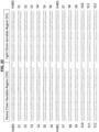
- an antigen-binding protein comprises (a) a heavy chain variable region amino acid sequence of any one of SEQ ID NOs: 490-503, or a heavy chain variable region amino acid sequence labeled as S1-S12 in FIG. 22 , or a variant sequence thereof which differs by only one or two amino acids or which has at least or about 70% sequence identity; or (b) a light chain variable region amino acid sequence of any one of SEQ ID NOs: 380-383, 388-390, 479, and 481, or a light chain variable region amino acid sequence labeled as S1-S12 in FIG. 22 , or a variant sequence thereof which differs by only one or two amino acids or which has at least or about 70% sequence identity; or both (a) and (b).
- an antigen binding protein comprises a pair of amino acid sequences selected from the group consisting of: (a) SEQ ID NOs: 389 and 490; (b) SEQ ID NOs: 389 and 491; (c) SEQ ID NOs: 389 and 492; (d) SEQ ID NOs: 389 and 493; (e) SEQ ID NOs: 389 and 494; (f) SEQ ID NOs: 389 and 495; (g) SEQ ID NOs: 383 and 496; (h) SEQ ID NOs: 383 and 497; (i) SEQ ID NOs: 383 and 498; (j) SEQ ID NOs: 383 and 499; (k) SEQ ID NOs: 383 and 500; (l) SEQ ID NOs: 383 and 501; (m) SEQ ID NOs: 383 and 503; (n) SEQ ID NOs: 389 and 502; (o) the heavy chain variable region sequence labeled as S1 in FIG.
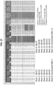
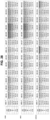
- an antigen-binding protein comprises (a) a heavy chain variable region amino acid sequence set forth as SEQ ID NO: 510 or 513 or in FIG. 23 or FIG. 25 , or a variant sequence thereof which differs by only one or two amino acids or which has at least or about 70% sequence identity; or (b) a light chain variable region amino acid sequence set forth as SEQ ID NO: 511 or 512 or in FIG. 24 or FIG. 26 , or a variant sequence thereof which differs by only one or two amino acids or which has at least or about 70% sequence identity; or (c) both (a) and (b).
- an antigen-binding protein comprises a pair of amino acid sequences wherein the pair comprises (a) a heavy chain variable region amino acid sequence set forth as SEQ ID NO: 510 and a light chain variable region amino acid sequence set forth as SEQ ID NO: 511, or a variant sequence thereof which differs by only 1-5 amino acids or which has at least or about 70% sequence identity; optionally, wherein the 1-5 amino acids which differ are as shown in FIG. 23 for the heavy chain or FIG.
- antigen-binding proteins conjugated to a heterologous moiety inhibit tumor growth in a subject, e.g., a human.
- the conjugated antigen-binding protein is a monoclonal antibody.
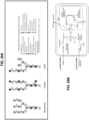
- the antibody is conjugated to an agent that alters the microtubule dynamics, e.g., MMAE.
- the conjugate comprises a cleavable linker, e.g., MC-VC-PAB.
- the conjugate is a homogeneous conjugate or a heterogeneous conjugate.
- the heterologous moiety is conjugated at a specific site of the antigen-binding protein.
- the antigen-binding protein binds to CLDN6 expressed by human cancer cells.
- the antigen-binding protein inhibits a binding interaction between human CLDN6 and a reference anti-CLDN6 antibody.
- the inhibiting action of the antigen-binding proteins provided herein allow such entities to be useful in methods of reducing tumor growth and treating a subject with a tumor or cancer.
- the antigen-binding protein is an antibody, antigen-binding antibody fragment thereof, or antibody protein product.
- the present disclosure also provides antigen-binding proteins comprising at least 3, 4, 5, or all amino acid sequences of a specified group of amino acid sequences.
- the antigen-binding proteins comprise at least 3, 4, 5, or 6 complementary determining region (CDR) amino acid sequences of CLDN6 antibodies disclosed herein.
- the present disclosure further provides antigen-binding proteins comprising amino acid sequences as detailed herein.
- the antigen-binding protein comprises an amino acid sequence of any one of SEQ ID NOs: 490-512, or an amino acid sequence as shown in any one of FIG. 22-FIG. 26 , or a combination thereof, as further described herein.
- Kits and pharmaceutical compositions comprising such entities are moreover contemplated.
- the method comprises culturing a host cell comprising a nucleic acid encoding a antigen-binding protein or a polypeptide as described herein so as to express the antigen-binding protein or polypeptide.
- Methods of treating a subject having cancer are additionally provided herein.
- the method comprises administering to the subject the pharmaceutical composition of the present disclosure in an amount effective for treating the cancer in the subject.
- Also provided are methods of treating a subject with a CLDN6-expressing cancer comprising administering to the subject a pharmaceutical composition described herein. Further contemplated is a method of inhibiting tumor growth in a subject, comprising administering to the subject a pharmaceutical composition described herein.
- a method of reducing tumor size in a subject, or preventing the recurrence of cancer in a subject comprising administering to the subject a pharmaceutical composition described herein.
- Also provided herein is a method of treating cancer in a subject diagnosed to be a low over-expresser of CLDN6, comprising administering to the subject a pharmaceutical composition described herein.
- the administering induces apoptosis in tumor cells, for example in cells expressing CLDN6.
- the administration induces antibody-dependent cell-mediated cytotoxicity (ADCC) or Complement-dependent cytotoxicity (CDC), tumor necrosis and death or depletion of cells, and/or disruption of tumor cell adherence, each of which result tumor regression or slowing of tumor growth.
- ADCC antibody-dependent cell-mediated cytotoxicity
- CDC Complement-dependent cytotoxicity
- Tight junctions also known as occluding junctions or zonulae occludentes, are vertebrate structures located between two adjacent cells that regulate paracellular permeability and maintain cell polarity in epithelial and endothelial cell sheets.
- the claudin (CLDN) family of genes encodes membrane proteins that are important components of tight junctions.
- CLDN proteins comprise four transmembrane (TM) helices (TM1, TM2, TM3, and TM4) and two extracellular loops (EL1 and EL2).
- TM transmembrane
- TM3 transmembrane
- EL1 and EL2 extracellular loops
- the extracellular loops of the CLDN proteins of adjacent cells interact with one another to seal the cellular sheet and regulate paracellular transport between the luminal and basolateral spaces.
- CLDN proteins play a role in various human diseases and pathologies. For example, mutations in the CLCW7 gene have been shown to result in progressive scaling of the skin along with obstruction of bile ducts. Mutants of the CLDN16 gene cause a magnesium wasting disorder. CLDN19 mutations lead to ocular conditions, such as macular colobomata and myopia, while CLDN14 mutations can lead to nonsyndromic recessive deafness.
- CLDN3 and CLDN4 are known to be surface receptors for the Clostridium perfringens enterotoxin in the gut, and CLDN1, CLDN6, and CLDN9 are co-receptors for hepatitis C virus (HCV) entry.
- Several CLDN proteins have been shown to be abnormally expressed in cancers. For instance, CLDN1 is downregulated in breast and colon cancer, whereas CLDN3 and CLDN4 are highly upregulated in multiple cancers.
- Claudin-6 is a member of the CLDN family.
- the gene encoding the human CLDN6 protein is located on the p arm of human chromosome 16 at 16p13.3 and is conserved in chimpanzee, Rhesus monkey, dog, cow, mouse, rat, zebrafish, and frog.
- CLDN6 is generally expressed in humans as a 220-amino acid precursor protein; the first 21 amino acids of which constitute the signal peptide.
- the amino acid sequence of the CLDN6 precursor protein is publically available at the National Center for Biotechnology Information (NCBI) website as NCBI Reference Sequence NP_067018.2 and is provided herein as SEQ ID NO: 1.
- the amino acid at position 143 of SEQ ID NO: 1 is Ile.
- the amino acid at position 143 is a Val.
- SNP single-nucleotide polymorphism
- antigen-binding proteins that bind to Claudin-6 (CLDN6).
- CLDN6 Claudin-6
- the antigen-binding proteins of the present disclosure can take any one of many forms of antigen-binding proteins known in the art.
- the antigen-binding proteins of the present disclosure take the form of an antibody, or antigen-binding antibody fragment, or an antibody protein product.
- the antigen-binding protein comprises, consists essentially of, or consists of an antibody.
- antibody refers to a protein having a conventional immunoglobulin format, comprising heavy and light chains, and comprising variable and constant regions.
- an antibody may be an IgG which is a "Y-shaped" structure of two identical pairs of polypeptide chains, each pair having one "light” (typically having a molecular weight of about 25 kDa) and one "heavy” chain (typically having a molecular weight of about 50-70 kDa).
- An antibody has a variable region and a constant region.
- variable region is generally about 100-110 or more amino acids, comprises three complementarity determining regions (CDRs), is primarily responsible for antigen recognition, and substantially varies among other antibodies that bind to different antigens.
- CDRs complementarity determining regions
- the constant region allows the antibody to recruit cells and molecules of the immune system.
- the variable region is made of the N-terminal regions of each light chain and heavy chain, while the constant region is made of the C-terminal portions of each of the heavy and light chains.
- CDRs of antibodies have been described in the art. Briefly, in an antibody scaffold, the CDRs are embedded within a framework in the heavy and light chain variable region where they constitute the regions largely responsible for antigen binding and recognition.
- a variable region typically comprises at least three heavy or light chain CDRs ( Kabat et al., 1991, Sequences of Proteins of Immunological Interest, Public Health Service N.I.H., Bethesda, Md .; see also Chothia and Lesk, 1987, J. Mol. Biol.
- framework region designated framework regions 1-4, FR1, FR2, FR3, and FR4, by Kabat et al., 1991; see also Chothia and Lesk, 1987, supra).
- Antibodies can comprise any constant region known in the art. Human light chains are classified as kappa and lambda light chains. Heavy chains are classified as mu, delta, gamma, alpha, or epsilon, and define the antibody's isotype as IgM, IgD, IgG, IgA, and IgE, respectively.
- IgG has several subclasses, including, but not limited to IgG1, IgG2, IgG3, and IgG4.
- IgM has subclasses, including, but not limited to, IgM1 and IgM2.
- Embodiments of the present disclosure include all such classes or isotypes of antibodies.
- the light chain constant region can be, for example, a kappa- or lambda-type light chain constant region, e.g., a human kappa- or lambda-type light chain constant region.
- the heavy chain constant region can be, for example, an alpha-, delta-, epsilon-, gamma-, or mu-type heavy chain constant regions, e.g., a human alpha-, delta-, epsilon-, gamma-, or mu-type heavy chain constant region.
- the antibody is an antibody of isotype IgA, IgD, IgE, IgG, or IgM, including any one of IgG1, IgG2, IgG3 or IgG4.
- the antibody comprises a constant region comprising one or more amino acid modifications, relative to the naturally-occurring counterpart, in order to improve half-life/stability or to render the antibody more suitable for expression/manufacturability.
- the antibody comprises a constant region wherein the C-terminal Lys residue that is present in the naturally-occurring counterpart is removed or clipped.
- the antibody can be a monoclonal antibody.
- the antibody comprises a sequence that is substantially similar to a naturally-occurring antibody produced by a mammal, e.g., mouse, rabbit, goat, horse, chicken, hamster, human, and the like.
- the antibody can be considered as a mammalian antibody, e.g., a mouse antibody, rabbit antibody, goat antibody, horse antibody, chicken antibody, hamster antibody, human antibody, and the like.
- the antigen-binding protein is an antibody, such as a human antibody.
- the antigen-binding protein is a chimeric antibody or a humanized antibody.
- chimeric antibody refers to an antibody containing domains from two or more different antibodies.
- a chimeric antibody can, for example, contain the constant domains from one species and the variable domains from a second, or more generally, can contain stretches of amino acid sequence from at least two species.
- a chimeric antibody also can contain domains of two or more different antibodies within the same species.
- the term "humanized” when used in relation to antibodies refers to antibodies having at least CDR regions from a non-human source which are engineered to have a structure and immunological function more similar to true human antibodies than the original source antibodies. For example, humanizing can involve grafting a CDR from a non-human antibody, such as a mouse antibody, into a human antibody.
- Humanizing also can involve select amino acid substitutions to make a non-human sequence more similar to a human sequence.
- Information including sequence information for human antibody heavy and light chain constant regions is publicly available through the Uniprot database as well as other databases well-known to those in the field of antibody engineering and production.
- the IgG2 constant region is available from the Uniprot database as Uniprot number P01859, incorporated herein by reference.
- an antibody can be cleaved into fragments by enzymes, such as, e.g., papain and pepsin.
- Papain cleaves an antibody to produce two Fab fragments and a single Fc fragment.
- Pepsin cleaves an antibody to produce a F(ab') 2 fragment and a pFc' fragment.
- the antigen-binding protein of the present disclosure is an antigen-binding fragment of an antibody (a.k.a., antigen-binding antibody fragment, antigen-binding fragment, antigen-binding portion).
- the antigen-binding antibody fragment is a Fab fragment or a F(ab') 2 fragment.
- Antibody protein products include those based on the full antibody structure and those that mimic antibody fragments which retain full antigen-binding capacity, e.g., scFvs, Fabs and VHH/VH (discussed below).
- the smallest antigen-binding fragment that retains its complete antigen binding site is the Fv fragment, which consists entirely of variable (V) regions.
- a soluble, flexible amino acid peptide linker is used to connect the V regions to a scFv (single chain fragment variable) fragment for stabilization of the molecule, or the constant (C) domains are added to the V regions to generate a Fab fragment [fragment, antigen-binding].
- scFv and Fab fragments can be easily produced in host cells, e.g., prokaryotic host cells.
- ds-scFv disulfide-bond stabilized scFv
- scFab single chain Fab
- minibodies minibodies that comprise different formats consisting of scFvs linked to oligomerization domains.
- the smallest fragments are VHH/VH of camelid heavy chain Abs as well as single domain Abs (sdAb).
- the building block that is most frequently used to create novel antibody formats is the single-chain variable (V)-domain antibody fragment (scFv), which comprises V domains from the heavy and light chain (VH and VL domain) linked by a peptide linker of ⁇ 15 amino acid residues.
- a peptibody or peptide-Fc fusion is yet another antibody protein product.
- the structure of a peptibody consists of a biologically active peptide grafted onto an Fc domain.
- Peptibodies are well-described in the art. See, e.g., Shimamoto et al., mAbs 4(5): 586-591 (2012 ).
- bispecific antibodies can be divided into five major classes: BsIgG, appended IgG, bispecific antibody (BsAb) fragments, bispecific fusion proteins, and BsAb conjugates. See, e.g., Spiess et al., Molecular Immunology 67(2) Part A: 97-106 (2015 ).
- the antigen-binding protein of the present disclosure comprises, consists essentially of, or consists of any one of these antibody protein products.
- the antigen-binding protein of the present disclosure comprises, consists essentially of, or consists of any one of an scFv, Fab VHH/VH, Fv fragment, ds-scFv, scFab, dimeric antibody, multimeric antibody (e.g., a diabody,, triabody, tetrabody), miniAb, peptibody VHH/VH of camelid heavy chain antibody, sdAb, diabody; a triabody; a tetrabody; a bispecific or trispecific antibody, BsIgG, appended IgG, BsAb fragment, bispecific fusion protein, and BsAb conjugate.
- the antigen-binding protein of the present disclosure is an antibody protein product in monomeric form, or polymeric, oligomeric, or multimeric form.
- the antibody comprises two or more distinct antigen binding regions fragments
- the antibody is considered bispecific, trispecific, or multi-specific, or bivalent, trivalent, or multivalent, depending on the number of distinct epitopes that are recognized and bound by the antibody.
- an anti-CLDN6 antibody or antibody variant thereof is selected from the group consisting of a human antibody, a humanized antibody, a chimeric antibody, a monoclonal antibody, a recombinant antibody, an antigen-binding antibody fragment, a single chain antibody, a monomeric antibody, a diabody, a triabody, a tetrabody, a Fab fragment, an IgG1 antibody, an IgG2 antibody, an IgG3 antibody, and an IgG4 antibody.
- the antigen-binding protein of the present disclosure is linked to a therapeutic agent.
- the therapeutic agent may be any known in the art, including, but not limited to, chemotherapeutic agents, cytokines and growth factors, cytotoxic agents, and the like. See “Conjugates” below.
- the CLDN6 is a human CLDN6 having the amino acid sequence of: MASAGMQILGVVLTLLGWVNGLVSCALPMWKVTAFIGNSIVVAQV VWEGLWMSCVVQSTGQMQCKVYDSLLALPQDLQAARALCVIALL VALFGLLVYLAGAKCTTCVEEKDSKARLVLTSGIVFVISGVLTLIPVC WTAHA X IRDFYNPLVAEAQKRELGASLYLGWAASGLLLLGGGLLC CTCPSGGSQGPSHYMARYSTSAPAISRGPSEYPTKNYV, wherein X is Ile or Val (SEQ ID NO: 202).
- the human CLDN6 comprises the amino acid sequence of any one of SEQ ID NOs: 1, 178, and 200-202.
- the antigen-binding proteins of the present disclosure bind to an epitope within an amino acid sequence of CLDN6.
- CLDN6 is a human CLDN6 and the antigen-binding proteins of the present disclosure bind to an epitope within an amino acid sequence of human CLDN6, e.g., SEQ ID NOs: 1, 178, and 200-202.
- epitope is meant the region of or within CLDN6 which is bound by the antigen-binding protein.
- the epitope is a linear epitope.
- Linear epitope refers to the region of or within the CLDN6 which is bound by the antigen-binding protein and which region is composed of contiguous amino acids of the amino acid sequence of the CLDN6.
- the amino acids of a linear epitope are adjacent to each other in the primary structure of the CLDN6. Accordingly, a linear epitope is a fragment or portion of the amino acid sequence of the antigen, i.e., CLDN6.
- the epitope is a conformational or structural epitope.
- conformational epitope or "structural epitope” is meant an epitope which is composed of amino acids which are located in close proximity to one another only when the CLDN6 is in its properly folded state.
- amino acids of a conformational or structural epitope are not adjacent to each other in the primary structure (i.e., amino acid sequence) of the CLDN6.
- a conformational or structural epitope is not made of contiguous amino acids of the amino acid sequence of the antigen (CLDN6).
- the epitope is located within the extracellular domain (ECD) of CLDN6, e.g., human CLDN6.
- the antigen binding protein binds to Extracellular Loop 2 (EL2) of the ECD of CLDN6 having the amino acid sequence of WTAHAIIRDFYNPLVAEAQKREL (SEQ ID NO: 2).
- the epitope to which the antigen-binding protein binds is within SEQ ID NO: 2.
- the antigen-binding protein of the present disclosure binds to an N-terminal portion of SEQ ID NO: 2, e.g., TAHAIIRDFYNPL (SEQ ID NO: 3).
- the antigen-binding protein of the present disclosure binds to a C-terminal portion of SEQ ID NO: 2, e.g., LVAEAQKREL (SEQ ID NO: 4). In various instances, the antigen-binding protein of the present disclosure binds to EL2, but not to Extracellular Loop 1 (EL1) of CLDN6. In various aspects, the epitope(s) to which the antigen binding proteins of the present disclosure bind to is different from the epitope bound by an anti-CLDN6 antibody comprising a light chain variable region comprising the sequence of SEQ ID NO: 185 and a heavy chain variable region comprising the sequence of SEQ ID NO: 186.
- an anti-CLDN6 antibody comprising a light chain variable region comprising the sequence of SEQ ID NO: 185 and a heavy chain variable region comprising the sequence of SEQ ID NO: 186.
- the epitope(s) to which the antigen binding proteins of the present disclosure bind to is different from the epitope bound by an anti-CLDN6 antibody comprising a light chain variable region comprising the sequence of SEQ ID NO: 181 and a heavy chain variable region comprising the sequence of SEQ ID NO: 182.
- the antigen-binding proteins bind to human CLDN6 and a non-human CLDN6.
- the non-human CLDN6 is a CLDN6 of chimpanzee, Rhesus monkey, dog, cow, mouse, rat, zebrafish, or frog.
- the antigen-binding proteins bind to human CLDN6 and mouse CLDN6.
- the antigen-binding proteins provided herein bind to CLDN6 in a non-covalent and reversible manner.
- the binding strength of the antigen-binding protein to CLDN6 may be described in terms of its affinity, a measure of the strength of interaction between the binding site of the antigen-binding protein and the epitope.
- the antigen-binding proteins provided herein have high-affinity for CLDN6 and thus will bind a greater amount of CLDN6 in a shorter period of time than low-affinity antigen-binding proteins.
- the antigen-binding protein has an equilibrium association constant, K A , which is at least 10 5 mol -1 , at least 10 6 mol -1 , at least 10 7 mol -1 , at least 10 8 mol -1 , at least 10 9 mol -1 , or at least 10 10 mol -1 or at least 10 10 mol -1 least 10 10 mol -1 .
- K A can be influenced by factors including pH, temperature and buffer composition.
- the binding strength of the antigen-binding protein to CLDN6 may be described in terms of its sensitivity.
- K D is the equilibrium dissociation constant, a ratio of k off /k on , between the antigen-binding protein and CLDN6.
- K D and K A are inversely related.
- the K D value relates to the concentration of the antigen-binding protein (the amount of antigen-binding protein needed for a particular experiment) and so the lower the K D value (lower concentration) the higher the affinity of the antigen-binding protein.
- the binding strength of the antigen-binding protein to CLDN6 may be described in terms of K D .
- the K D of the antigen-binding proteins provided herein is about 10 -1 , about 10 -2 , about 10 -3 , about 10 -4 , about 10 -5 , about 10 -6 , or less. In various aspects, the K D of the antigen-binding proteins provided herein is micromolar, nanomolar, picomolar or femtomolar. In various aspects, the K D of the antigen-binding proteins provided herein is within a range of about 10 -4 to 10 -6 or 10 -7 to 10 -9 or 10 -10 to 10 -12 or 10 -13 to 10 -15 .
- the K D of the antigen-binding proteins provided herein is within a range of about 1.0 x 10 -12 M to about 1.0 x 10 -8 M. In various aspects, the K D of the antigen-binding proteins is within a range of about 1.0 x 10 -11 M to about 1.0 x 10 -9 M.
- the affinity of the antigen-binding proteins are measured or ranked using a flow cytometry- or Fluorescence-Activated Cell Sorting (FACS)-based assay.
- FACS Fluorescence-Activated Cell Sorting
- Flow cytometry-based binding assays are known in the art. See, e.g., Cedeno-Arias et al., Sci Pharm 79(3): 569-581 (2011 ); Rathanaswami et al., Analytical Biochem 373: 52-60 (2008 ); and Geuijen et al., J Immunol Methods 302(1-2): 68-77 (2005 ).
- the affinity of the antigen-binding proteins are measured or ranked using a competition assay as described in Trikha et al., Int J Cancer 110: 326-335 (2004 ) and Tam et al., Circulation 98(11): 1085-1091 (1998 ), as well as below. See section titled "Competition Assays” below.
- Trikh et al. cells that express the antigen were used in a radioassay.
- the binding of 125 I-labeled antigen-binding protein (e.g., antibody) to the cell surface antigen is measured with the cells in suspension.
- the relative affinity of a CLDN6 antibody is determined via a FACS-based assay in which different concentrations of a CLDN6 antibody conjugated to a fluorophore are incubated with cells expressing CLDN6 and the fluorescence emitted (which is a direct measure of antibody-antigen binding) is determined.
- a curve plotting the fluorescence for each dose or concentration is made.
- the max value is the lowest concentration at which the fluorescence plateaus or reaches a maximum, which is when binding saturation occurs.
- Half of the max value is considered an EC50 or an IC50 and the antibody with the lowest EC50/IC50 is considered as having the highest affinity relative to other antibodies tested in the same manner.
- Such an assay is described herein at Example 5.
- the IC 50 value as determined in a competitive binding inhibition assay, approximates the K D of the antigen-binding protein.
- the competition assay is a FACS-based assay carried out with a reference antibody, fluorophore-conjugated secondary antibody, and cells which express CLDN6.
- the cells are genetically-engineered to overexpress CLDN6.
- the cells are HEK293T cells transduced with a viral vector to express CLDN6.
- the cells endogenously express CLDN6.
- the cells which endogenously express CLDN6 are pre-determined as low CLDN6-expressing cells or high CLDN6-expressing cells.
- the cells are cancer or tumor cells.
- the cells are cells from a cell line, e.g., an ovarian cell line, endometrial cell line, bladder cell line, lung cell line, gastrointestinal (GI) cell line, liver cell line, lung cell line, and the like.
- the cells which endogenously express CLDN6 as selected from the group consisting of OVCA429 ovarian cells, ARK2 endometrial cells, OAW28 ovarian cells, UMUC-4 bladder cells, PEO14 ovarian cells, OV177 ovarian cells, H1693 lung cells, MKN7 upper GI cells, OV-90 ovarian cells, HUH-7 liver cells, JHOS-4 ovarian cells, H1435 lung cells, and NUGC3 upper GI cells.
- the antigen-binding protein inhibits the binding interaction between human CLDN6 expressed by the cells and the reference antibody, which reference antibody is known to bind to CLDN6 but is not an antigen-binding protein of the present disclosure.
- the antigen-binding proteins of the present disclosure compete with the reference antibody for binding to human CLDN6 and thereby reduce the amount of human CLDN6 bound to the reference antibody as determined by an in vitro competitive binding assay.
- the antigen-binding proteins of the present disclosure inhibit the binding interaction between human CLDN6 and the reference antibody and the inhibition is characterized by an IC 50 .
- the antigen-binding proteins exhibit an IC 50 of less than about 2500 nM for inhibiting the binding interaction between human CLDN6 and the reference antibody.
- the antigen-binding proteins exhibit an IC 50 of less than about 2000 nM, less than about 1500 nM, less than about 1000 nM, less than about 900 nm, less than about 800 nm, less than about 700 nm, less than about 600 nm, less than about 500 nm, less than about 400 nm, less than about 300 nm, less than about 200 nm, or less than about 100 nm.
- the antigen-binding proteins exhibit an IC 50 of less than about 90 nM, less than about 80 nM, less than about 70 nM, less than about 60 nM, less than about 50 nM, less than about 40 nM, less than about 30 nM, less than about 20 nM, or less than about 10 nM.
- the antigen binding proteins of the present disclosure compete against a reference antibody known to bind to CLDN6 (which reference antibody is different from any of the antigen-binding proteins of the present disclosure) for binding to CLDN6. See further description under Competition assays.
- Avidity gives a measure of the overall strength of an antibody-antigen complex. It is dependent on three major parameters: affinity of the antigen-binding protein for the epitope, valency of both the antigen-binding protein and CLDN6, and structural arrangement of the parts that interact. The greater an antigen-binding protein's valency (number of antigen binding sites), the greater the amount of antigen (CLDN6) it can bind.
- the antigen-binding proteins have a strong avidity for CLDN6.
- the antigen-binding proteins are multivalent. In various aspects, the antigen-binding proteins are bivalent. In various instances, the antigen antigen-binding proteins are monovalent.
- the antigen-binding proteins of the present disclosure bind to CLDN6 and do not bind to any other member of the CLDN family, e.g., do not cross-react with any other member of the CLDN family.
- the antigen-binding proteins of the present disclosure are CLDN-6 specific.
- the antigen-binding proteins of the present disclosure have a selectivity for CLDN6 which is at least 10-fold, 5-fold, 4-fold, 3-fold, 2-fold greater than the selectivity of the antigen-binding protein for CLDN3, CLDN4, CLDN9, or a combination thereof.
- the antigen-binding proteins of the present disclosure have a selectivity for CLDN6 which is at least 10-fold, 5-fold, 4-fold, 3-fold, 2-fold greater than the selectivity of the antigen-binding protein for each of CLDN3, CLDN4, and CLDN9.
- Selectivity may be based on the K D exhibited by the antigen binding protein for CLDN6, or a CLDN family member, wherein the K D may be determined by techniques known in the art, e.g., surface plasmon resonance, FACS-based affinity assays.
- the antigen-binding proteins of the present disclosure bind to CLDN6 and do not bind to any of Claudin3 (CLDN3), Claudin4 (CLDN4), and Claudin9 (CLDN9).
- the antigen-binding proteins do not bind to any of CLDN3, CLDN4, and CLDN9 and exhibit an IC 50 of less than about 1200 nM (e.g., less than about 1000 nM, less than about 750 nM, less than about 500 nM, less than about 250 nM) in a FACS-based assay with OVCA429 cells endogenously expressing CLDN6.
- the antigen-binding proteins do not bind to any of CLDN3, CLDN4, and CLDN9 and the concentration at which 50% of binding saturation is achieved with OVCA429 cells endogenously expressing CLDN6 is less than about 1200 nM (e.g., less than about 1000 nM, less than about 750 nM, less than about 500 nM, less than about 250 nM).
- the antigen-binding proteins exhibit at least a 5-fold selectivity for CLDN 6 greater than that for CLDN3, CLDN4, and CLDN9 and the concentration at which 50% of binding saturation is achieved with OVCA429 cells endogenously expressing CLDN6 is less than about 1200 nM(e.g., less than about 1000 nM, less than about 750 nM, less than about 500 nM, less than about 250 nM).
- the antigen-binding proteins exhibit an IC50 of less than about 1200 nM (e.g., less than about 1000 nM, less than about 750 nM, less than about 500 nM, less than about 250 nM) for CLDN6 artificial and endogenous models and exhibit a greater than about 5-fold ratio separating CLDN6 IC50s from CLDN3, CLDN4 and/or CLDN9.
- the antigen-binding proteins exhibit an IC50 of less than about 1200 nM (e.g., less than about 1000 nM, less than about 750 nM, less than about 500 nM, less than about 250 nM) for CLDN6 and exhibit an IC50 for any one of CLDN3, CLDN4, and CLDN9 at least 5-fold greater than the IC50.
- the antigen-binding proteins of the present disclosure bind to CLDN6 and cross-react with (e.g., bind to) at least one other member of the CLDN family. In various aspects, the antigen-binding proteins of the present disclosure bind to CLDN6 and one or more of CLDN3, CLDN4, and CLDN9. In various aspects, the antigen-binding proteins of the present disclosure bind to CLDN6 and CLDN4 or CLDN9, but do not bind to CLDN3. In various instances, the antigen-binding proteins of the present disclosure bind to CLDN6 and CLDN4 but binds to neither CLDN3 nor CLDN9. In various instances, the antigen-binding proteins of the present disclosure bind to CLDN6 and CLDN9 but do not bind to either CLDN3 or CLDN4.
- the antigen-binding protein inhibits a binding interaction between human CLDN6 and a reference antibody, which reference antibody is known to bind to CLDN6 but is not an antigen-binding protein of the present disclosure.
- the antigen-binding proteins of the present disclosure compete with the reference antibody for binding to human CLDN6 and thereby reduce the amount of human CLDN6 bound to the reference antibody as determined by an in vitro competitive binding assay.
- the reference antibody binds to an epitope within the amino acid sequence of the extracellular domain of human CLDN6, optionally, within EL2 or EL 1.
- the reference antibody comprises a light chain variable sequence encoded by SEQ ID NO: 179, and a heavy chain variable sequence encoded by SEQ ID NO: 180. In various aspects, the reference antibody comprises a light chain variable sequence of SEQ ID NO: 181, and a heavy chain variable sequence of SEQ ID NO: 182. In various aspects, the antigen-binding proteins of the present disclosure inhibit the binding interaction between human CLDN6 and the reference antibody and the inhibition is characterized by an IC 50 . In various aspects, the antigen-binding proteins exhibit an IC 50 of less than about 2500 nM for inhibiting the binding interaction between human CLDN6 and the reference antibody.
- the antigen-binding proteins exhibit an IC 50 of less than about 2000 nM, less than about 1500 nM, less than about 1000 nM, less than about 900 nm, less than about 800 nm, less than about 700 nm, less than about 600 nm, less than about 500 nm, less than about 400 nm, less than about 300 nm, less than about 200 nm, or less than about 100 nm.
- the antigen-binding proteins exhibit an IC 50 of less than about 90 nM, less than about 80 nM, less than about 70 nM, less than about 60 nM, less than about 50 nM, less than about 40 nM, less than about 30 nM, less than about 20 nM, or less than about 10 nM.
- the antigen-binding proteins of the present disclosure compete with the reference antibody for binding to human CLDN6 and thereby reduce the amount of human CLDN6 bound to the reference antibody as determined by an in vitro competitive binding assay.
- the in vitro competitive binding assay is a FACS-based assay in which the fluorescence of a fluorophore-conjugated secondary antibody which binds to the Fc of the reference antibody is measured in the absence or presence of a particular amount of the antigen-binding protein of the present disclosure.
- a FACS-based assay is described herein in the EXAMPLES.
- the FACS-based assay is carried out with the reference antibody, fluorphore-conjugated secondary antibody and cells which express CLDN6.
- the cells are genetically-engineered to overexpress CLDN6.
- the cells are HEK293T cells transduced with a viral vector to express CLDN6.
- the cells endogenously express CLDN6.
- the cells which endogenously express CLDN6 are pre-determined as low CLDN6-expressing cells or high CLDN6-expressing cells.
- the cells are cancer or tumor cells.
- the cells are cells from a cell line, e.g., an ovarian cell line, endometrial cell line, bladder cell line, lung cell line, gastrointestinal (GI) cell line, liver cell line, lung cell line, and the like.
- a cell line e.g., an ovarian cell line, endometrial cell line, bladder cell line, lung cell line, gastrointestinal (GI) cell line, liver cell line, lung cell line, and the like.
- the cells which endogenously express CLDN6 as selected from the group consisting of OVCA429 ovarian cells, ARK2 endometrial cells, OAW28 ovarian cells, UMUC-4 bladder cells, PEO14 ovarian cells, OV177 ovarian cells, H1693 lung cells, MKN7 upper GI cells, OV-90 ovarian cells, HUH-7 liver cells, JHOS-4 ovarian cells, H1435 lung cells, and NUGC3 upper GI cells.
- the antigen binding proteins of the present disclosure bind to CLDN6 endogenously expressed by one or more of ARK2 cells, OVCA429 cells, LS513 cells, or MCF7 cells with high affinity.
- the antigen binding proteins exhibit an IC 50 of less than about 3000 nM as determined in a FACS-based competitive binding inhibition assay using one or more of ARK2 cells, OVCA429 cells, LS513 cells, or MCF7 cells.
- the antigen binding proteins exhibit an IC 50 of less than about 2500 nM, less than about 2000 nM, less than about 1750 nM, less than about 1500 nM, less than about 1250 nM, less than about 1000 nM, less than about 750 nM, or less than about 500 nM, as determined in a FACS-based competitive binding inhibition assay using one or more of ARK2 cells, OVCA429 cells, LS513 cells, or MCF7 cells.
- the antigen binding proteins exhibit an IC 50 of less than about 400 nM, less than about 300 nM, less than about 200 nM, less than about 100 nM, less than about 75 nM, less than about 50 nM, less than about 25 nM, or less than about 10 nM, as determined in a FACS-based competitive binding inhibition assay using one or more of ARK2 cells, OVCA429 cells, LS513 cells, or MCF7 cells.
- binding assays e.g., competitive binding assays or competition assays, which test the ability of an antibody to compete with a second antibody for binding to an antigen, or to an epitope thereof, are known in the art. See, e.g., Trikha et al., Int J Cancer 110: 326-335 (2004 ); Tam et al., Circulation 98(11): 1085-1091 (1998 ).
- antigen-binding proteins e.g., antibodies, antigen-binding antibody fragments, and antibody protein products
- standard hybridoma methods for producing antibodies are described in, e.g., Harlow and Lane (eds.), Antibodies: A Laboratory Manual, CSH Press (1988 ), and CA. Janeway et al. (eds.), Immunobiology, 5th Ed., Garland Publishing, New York, NY (2001 )).
- An various method of preparing CLDN6 monoclonal antibodies or the present disclosure is provided herein in EXAMPLES.
- adjuvants can be used to increase the immunological response leading to greater antibody production by the host.
- adjuvants include but are not limited to Freund's, mineral gels such as aluminum hydroxide, and surface active substances such as lysolecithin, pluronic polyols, polyanions, peptides, oil emulsions, keyhole limpet hemocyanin, and dinitrophenol.
- BCG Bacilli Calmette-Guerin
- Corynebacterium parvum are potentially useful human adjuvants.
- Methods of testing antibodies for the ability to bind to the epitope of CLDN6 regardless of how the antibodies are produced include any antibody-antigen binding assay, such as, for example, radioimmunoassay (RIA), ELISA, Western blot, immunoprecipitation, SPR, and competitive inhibition assays (see, e.g., Janeway et al., infra, and U.S. Patent Application Publication No. 2002/0197266 , and the above section relating to competition assays).
- RIA radioimmunoassay
- ELISA ELISA
- Western blot Western blot
- immunoprecipitation e.g., SPR
- competitive inhibition assays see, e.g., Janeway et al., infra, and U.S. Patent Application Publication No. 2002/0197266 , and the above section relating to competition assays.
- antigen-binding proteins comprising (a) a heavy chain (HC) complementarity-determining region (CDR) 1 amino acid sequence set forth in Table A or a sequence selected from the group consisting of: SEQ ID NOs: 11, 17, 23, 29, 35, 41, 47, 53, 59, 65, 71, 77, 83, 89, 95, 101, 107, 113, 119, 125, and 131, or a variant sequence thereof which differs by only one or two amino acids or which has at least or about 70% (e.g., at least about 80%, at least about 85%, at least about 90%, at least about 95%) sequence identity; (b) an HC CDR2 amino acid sequence set forth in Table A or a sequence selected from the group consisting of: SEQ ID NOs: 12, 18, 24, 30, 36, 42, 48, 54, 60, 66, 72, 78, 84, 90, 86, 102, 108, 114, 120, 126, and 132, or a variant sequence thereof which differ
- the antigen-binding protein comprises a LC CDR1 amino acid sequence, a LC CDR2 amino acid sequence, and a LC CDR3 amino acid sequence set forth in Table A and at least 1 or 2 of the HC CDR amino acid sequences set forth in Table A.
- the antigen-binding protein comprises a HC CDR1 amino acid sequence, a HC CDR2 amino acid sequence, and a HC CDR3 amino acid sequence set forth in Table A and at least 1 or 2 of the LC CDR amino acid sequences set forth in Table A.
- the antigen-binding protein comprises at least 3, 4, or 5 of the amino acid sequences designated by the SEQ ID NOs: in a single row of Table A. In various embodiments, the antigen-binding protein comprises each of the LC CDR amino acid sequences designated by the SEQ ID NOs: of a single row of Table A and at least 1 or 2 of the HC CDR amino acid sequences designated by the SEQ ID NOs: in of a single row of Table A.
- the antigen-binding protein comprises each of the HC CDR amino acid sequences designated by the SEQ ID NOs: of a single row of Table A and at least 1 or 2 of the LC CDR amino acid sequences designated by the SEQ ID NOs: of a single row of Table A. In various embodiments, the antigen-binding protein comprises all 6 of the CDR amino acid sequences designated by the SEQ ID NOs: of a single row of Table A.
- the antigen-binding protein comprises six CDR amino acid sequences selected from the group consisting of: (a) SEQ ID NOs: 74-79; (b) SEQ ID NOs: 50-55; (c) SEQ ID NOs: 122-127; (d) SEQ ID NOs: 26-31; (e) SEQ ID NOs: 128-133; (f) SEQ ID NOs: 38-43; (g) SEQ ID NOs: 62-67; (h) SEQ ID NOs: 80-85; (i) SEQ ID NOs: 44-49; (j) SEQ ID NOs: 86-91; (k) SEQ ID NOs: 104-109; (l) SEQ ID NOs: 56-61; (m) SEQ ID NOs: 32-37; (n) SEQ ID NOs: 110-115; (o) SEQ ID NOs: 98-103; (p) SEQ ID NOs: 92-97; (q) SEQ ID NOs: 116-
- the amino acid sequences of Table A are separated by at least one or more (e.g., at least 2, 3, 4, 5, 6, 7, 8, 9, 10, or more) intervening amino acid(s).
- the antigen-binding protein comprises (a) a heavy chain variable region amino acid sequence set forth in in Table B or a sequence selected from the group consisting of: SEQ ID NOs: 135, 137, 139, 141, 143, 145, 147, 149, 151, 153, 155, 157, 159, 161, 163, 165, 167, 169, 171, 173, and 175, or a variant sequence thereof which differs by only one or two amino acids or which has at least or about 70% (e.g., at least about 80%, at least about 85%, at least about 90%, at least about 95%) sequence identity; or (b) a light chain variable region amino acid sequence set forth in Table B or a sequence selected from the group consisting of: SEQ ID NOs: 134, 136, 138, 140, 142, 144, 146, 148, 150, 152, 154, 156, 158, 160, 162, 164, 166, 168, 170, 172,
- the antigen-binding protein comprises a pair of amino acid sequences selected from the group consisting of: (a) SEQ ID NOs: 156 and 157; (b) SEQ ID NOs: 148 and 149; (c) SEQ ID NOs: 172 and 173; (d) SEQ ID NOs: 140 and 141; (e) SEQ ID NOs: 174 and 175; (f) SEQ ID NOs: 144 and 145; (g) SEQ ID NOs: 152 and 153; (h) SEQ ID NOs: 158 and 159; (i) SEQ ID NOs: 146 and 147; (j) SEQ ID NOs: 160 and 161; (k) SEQ ID NOs: 166 and 167; (l) SEQ ID NOs: 150 and 151; (m) SEQ ID NOs: 142 and 143; (n) SEQ ID NOs: 168 and 169; (o) SEQ ID NOs: 164 and 165; (p) SEQ ID NOs
- the antigen-binding protein does not comprise a pair of amino acid sequences encoded by the sequences of SEQ ID NOs: 179 and 180. In various aspects, the antigen-binding protein does not comprise a pair of amino acid sequences of SEQ ID NOs: 181 and 182. In various aspects, the antigen-binding protein does not comprise a pair of amino acid sequences encoded by the sequences of SEQ ID NOs: 183 and 184. In various aspects, the antigen-binding protein does not comprise a pair of amino acid sequences of SEQ ID NOs: 185 and 186.
- the antigen-binding protein comprises an amino acid sequence which is similar to an above-referenced amino acid sequence, yet the antigen-binding protein substantially retains its biological function, e.g., its ability to bind to human CLDN6, reduce tumor growth, treat cancer.
- the antigen-binding protein comprises an amino acid sequence which differs by only 1, 2, 3, 4, 5, 6, or more amino acids, relative to the above-referenced amino acid sequence(s).
- the antigen-binding protein comprises a variant sequence of the referenced sequence, which variant sequence differs by only one or two amino acids, relative to the referenced sequence.
- the antigen-binding protein comprising one or more amino acid substitutions that occur outside of the CDRs, e.g, the one or more amino acid substitutions occur within the framework region(s) of the heavy or light chain.
- the antigen-binding protein comprising one or more amino acid substitutions yet the antigen-binding protein retains the amino acid sequences of the six CDRs.
- the antigen-binding protein comprises an amino acid sequence having only 1, 2, 3, 4, 5, 6, or more conservative amino acid substitutions, relative to the above-referenced amino acid sequence(s).
- conservative amino acid substitution refers to the substitution of one amino acid with another amino acid having similar properties, e.g., size, charge, hydrophobicity, hydrophilicity, and/or aromaticity, and includes exchanges within one of the following five groups:
- the conservative amino acid substitution is an exchange within one of the following groups of amino acids:
- the antigen-binding protein comprises an amino acid sequence which has greater than or about 30%, greater than or about 50%, or greater than or about 70% sequence identity to the above-referenced amino acid sequence. In various aspects, the antigen-binding protein comprises an amino acid sequence which has at least 30%, at least 40%, at least 50%, at least 60%, at least 70%, at least 80%, at least 85%, at least 90% or has greater than 90% sequence identity to the above-referenced amino acid sequence. In various aspects, the antigen-binding protein comprises an amino acid sequence that has at least 70%, at least 80%, at least 85%, at least 90% or has greater than 90% sequence identity along the full-length of the above-referenced amino acid sequence. In various aspects, the antigen-binding protein comprises an amino acid sequence having at least 95%, 96%, 97%, 98% or 99% sequence identity along the full-length of the above-referenced amino acid sequence.
- the antigen-binding protein comprises a variant sequence of the referenced sequence, which variant sequence has at least or about 70% sequence identity, relative to the above-referenced sequence. In various aspects, the antigen-binding protein comprises a variant sequence of the referenced sequence, which variant sequence has at least or about 80% sequence identity, relative to the above-referenced sequence. In various aspects, the antigen-binding protein comprises a variant sequence of the referenced sequence, which variant sequence has at least or about 90% sequence identity, relative to the above-referenced sequence. In various aspects, the antigen-binding protein comprises a variant sequence of the referenced sequence, which variant sequence has at least or about 95% sequence identity, relative to the above-referenced sequence.
- the antigen-binding protein comprises one, two, three, four, or five sequences of the SEQ ID NOs. in a single row of Table A and at least one variant sequence having at least or about 70% (e.g., at least about 80%, at least about 85%, at least about 90%, at least about 95%) sequence identity to any of SEQ ID NOs: 8-133.
- the antigen-binding protein comprises one, two, three, four, or five sequences of a set of sequences selected from: (a) SEQ ID NOs: 74-79; (b) SEQ ID NOs: 50-55; (c) SEQ ID NOs: 122-127; (d) SEQ ID NOs: 26-31; (e) SEQ ID NOs: 128-133; (f) SEQ ID NOs: 38-43; (g) SEQ ID NOs: 62-67; (h) SEQ ID NOs: 80-85; (i) SEQ ID NOs: 44-49; (j) SEQ ID NOs: 86-91; (k) SEQ ID NOs: 104-109; (l) SEQ ID NOs: 56-61; (m) SEQ ID NOs: 32-37; (n) SEQ ID NOs: 110-115; (o) SEQ ID NOs: 98-103; (p) SEQ ID NOs: 92-97; (q) SEQ ID NOs:
- the antigen-binding protein comprises four sequences of SEQ ID NOs: 74-79, namely, SEQ ID NOs: 74-77, wherein the antigen-binding protein comprises two variant sequences: one variant sequence having at least or about 70% (e.g., at least about 80%, at least about 85%, at least about 90%, at least about 95%) sequence identity to SEQ ID NO: 78 and another variant sequence having at least or about 70% (e.g., at least about 80%, at least about 85%, at least about 90%, at least about 95%) sequence identity to SEQ ID NO: 79.
- SEQ ID NOs: 74-79 namely, SEQ ID NOs: 74-77
- the antigen-binding protein comprises two variant sequences: one variant sequence having at least or about 70% (e.g., at least about 80%, at least about 85%, at least about 90%, at least about 95%) sequence identity to SEQ ID NO: 78 and another variant sequence having at least or about 70% (e.g., at least about
- the antigen-binding protein comprises a pair of variant sequences having at least or about 70% (e.g., at least about 80%, at least about 85%, at least about 90%, at least about 95%) sequence identity to any of SEQ ID NOs: 134-175.
- the antigen binding protein comprises a pair of variant sequences which have at least or about 70% (e.g., at least about 80%, at least about 85%, at least about 90%, at least about 95%) sequence identity to (a) SEQ ID NOs: 156 and 157; (b) SEQ ID NOs: 148 and 149; (c) SEQ ID NOs: 172 and 173; (d) SEQ ID NOs: 140 and 141; (e) SEQ ID NOs: 174 and 175; (f) SEQ ID NOs: 144 and 145; (g) SEQ ID NOs: 152 and 153; (h) SEQ ID NOs: 158 and 159; (i) SEQ ID NOs: 146 and 147; (j) SEQ ID NOs: 160 and 161; (k) SEQ ID NOs: 166 and 167; (l) SEQ ID NOs: 150 and 151; (m) SEQ ID NOs: 142 and 143; (n) SEQ ID NOs
- the antigen-binding protein comprises a pair of sequences: one sequence of Table B and another sequence which is a variant sequence having at least or about 70% (e.g., at least about 80%, at least about 85%, at least about 90%, at least about 95%) sequence identity to any of SEQ ID NOs: 134-175.
- the antigen-binding protein comprises a pair of sequences: one sequence selected from (a) SEQ ID NOs: 156 and 157; (b) SEQ ID NOs: 148 and 149; (c) SEQ ID NOs: 172 and 173; (d) SEQ ID NOs: 140 and 141; (e) SEQ ID NOs: 174 and 175; (f) SEQ ID NOs: 144 and 145; (g) SEQ ID NOs: 152 and 153; (h) SEQ ID NOs: 158 and 159; (i) SEQ ID NOs: 146 and 147; (j) SEQ ID NOs: 160 and 161; (k) SEQ ID NOs: 166 and 167; (l) SEQ ID NOs: 150 and 151; (m) SEQ ID NOs: 142 and 143; (n) SEQ ID NOs: 168 and 169; (o) SEQ ID NOs: 164 and 165; (p) SEQ ID NOs:
- the antigen-binding protein comprises a sequences of SEQ ID NO: 134 and the antigen-binding protein further comprises a variant sequence having at least or about 70% (e.g., at least about 80%, at least about 85%, at least about 90%, at least about 95%) sequence identity to SEQ ID NO 135.
- the antigen-binding protein comprises an amino acid sequence of an above-referenced amino acid sequence with one or more amino acid substitutions to reduce or eliminate reactive amino acids to decrease or prevent unwanted side chain reactions.
- the antigen-binding protein comprises an amino acid sequence of an above-referenced amino acid sequence with one or more (i) Trp residues substituted with His, Tyr, or Phe; (ii) Asn residues substituted with Gln, Ser, Ala, or Asp; (iii) Asp residues occurring immediately before a Pro residue substituted with Ala, Ser, or Glu, (iv) Asn residues substituted with Gln, Ser, or Ala; and/or (v) Cys residues substituted with Tyr, Ser, or Ala.
- the antigen-binding protein comprises an amino acid sequence of an above-referenced amino acid sequence with an amino acid substitution predicted to have greater binding affinity, greater stability, or other positive attribute, based on SHM events or based on statistical analyses of a multitude of other similar antibody sequences.
- the antigen-binding protein comprises (a) an HC CDR1 amino acid sequence set forth in Table A1 or a sequence selected from the group consisting of: SEQ ID NOs: 452, 455, 461, 465, 71, and 472, or a variant sequence thereof which differs by only one or two amino acids or which has at least or about 70(e.g., at least about 80%, at least about 85%, at least about 90%, at least about 95%) sequence identity; (b) an HC CDR2 amino acid sequence set forth in Table A1 or a sequence selected from the group consisting of: SEQ ID NOs: 475, 456, 462, 466, 468, and 473; or a variant sequence thereof which differs by only one or two amino acids or which has at least or about 70% (e.g., at least about 80%, at least about 85%, at least about 90%, at least about 95%) sequence identity; (c) an HC CDR3 amino acid sequence set forth in Table A1 or a sequence selected from the group consisting of
- the HC CDR1 comprises Gly immediately N-terminal of SEQ ID NO: 452 and, optionally, in some aspects, the HC CDR1 comprises MX immediately C-terminal of SEQ 452, wherein X is H, N, or S.
- the HC CDR3 comprises Ala immediately N-terminal of SEQ ID NO: 453.
- the LC CDR1 further comprises TAS immediately N-terminal of SEQ ID NO: 449, and, optionally, XH immediately C-terminal of SEQ ID NO: 449, wherein X is H, S, Y, or Q.
- the first amino acid of SEQ ID NO: 449 is S or Q.
- the first amino acid of SEQ ID NO: 451 is S or Q.
- the HC CDR1 comprises Gly immediately N-terminal of SEQ ID NO: 455, and optionally, in various aspects, the HC CDR1 comprises MX immediately C-terminal of SEQ ID NO: 455, wherein X is N, S, or H.
- HC CDR2 comprises Gln immediately N-terminal of SEQ ID NO: SEQ ID NO: 456, and optionally H immediately C-terminal of SEQ ID NO: 456.
- the LC CDR1 comprises RIS immediately N-terminal of SEQ ID NO: 476, and optionally, comprises LA immediately C-terminal of SEQ ID NO: 476.
- the LC CDR2 comprises XI,VE immediately C-terminal of SEQ ID NO: 477, wherein X is I or S.
- the HC CDR1 comprises MH immediately C-terminal of SEQ ID NO: 461.
- the HC CDR2 comprises Tyr immediately N-terminal of SEQ ID NO: 462, and optionally, TH immediately C-terminal of SEQ ID NO: 462.
- the HC CDR3 does not include the first two amino acids of SEQ ID NO: 463.
- the LC CDR1 comprises RSS immediately N-terminal of SEQ ID NO: 458, and optionally, LN immediately C-terminal of SEQ ID NO: 458.
- the LC CDR2 comprises XRFS immediately C-terminal of SEQ ID NO: 459, wherein X is Q, S, A, or D.
- the HC CDR1 comprises MH immediately C-terminal of SEQ ID NO: 465.
- the HC CDR2 comprises YI immediately N-terminal of SEQ ID NO: 466, and optionally, Xaa immediately C-terminal of SEQ ID NO: 466, wherein Xaa is N, S, Q, or A.
- the LC CDR1 comprises LAS immediately N-terminal of SEQ ID NO: 464, and optionally, LA immediately C-terminal of SEQ ID NO: 464.
- the LC CDR2 comprises SLAD immediately C-terminal of SEQ ID NO: 57.
- the HC CDR1 comprises MH immediately C-terminal of SEQ ID NO: 71.
- the HC CDR2 comprises Tyr immediately N-terminal of SEQ ID NO: 468 and optionally IY immediately C-terminal of SEQ ID NO: 468.
- the LC CDR1 comprises RAS immediately N-terminal of SEQ ID NO: 68, and optionally SYIH immediately C-terminal to SEQ 68.
- the LC CDR2 comprises XLES immediately C-terminal to SEQ ID NO: 69, wherein X is N, Q, S, A, or D.
- the LC CDR1 comprises KSS immediately N-terminal of SEQ ID NO: 470, and optionally YLA immediately C-terminal to SEQ 470.
- the LC CDR2 comprises TRES immediately C-terminal of SEQ I DNO: 471.
- the HC CDR1 comprises MN immediately C-terminal of SEQ ID NO: 472.
- the HC CDR2 comprises Xaa immediately N-terminal of SEQ 473, wherein Xaa is N, Q, S, or A, and optionally, Thr immediately C-terminal of SEQ 473.
- the antigen-binding protein comprises a LC CDR1 amino acid sequence, a LC CDR2 amino acid sequence, and a LC CDR3 amino acid sequence set forth in Table A1 and at least 1 or 2 of the HC CDR amino acid sequences set forth in Table A1.
- the antigen-binding protein comprises a HC CDR1 amino acid sequence, a HC CDR2 amino acid sequence, and a HC CDR3 amino acid sequence set forth in Table A1 and at least 1 or 2 of the LC CDR amino acid sequences set forth in Table A1.
- the antigen-binding protein comprises at least 3, 4, or 5 of the amino acid sequences designated by the SEQ ID NOs: in a single row of Table A1. In various embodiments, the antigen-binding protein comprises each of the LC CDR amino acid sequences designated by the SEQ ID NOs: of a single row of Table A1 and at least 1 or 2 of the HC CDR amino acid sequences designated by the SEQ ID NOs: in of a single row of Table A1.
- the antigen-binding protein comprises each of the HC CDR amino acid sequences designated by the SEQ ID NOs: of a single row of Table A1 and at least 1 or 2 of the LC CDR amino acid sequences designated by the SEQ ID NOs: of a single row of Table A1. In various embodiments, the antigen-binding protein comprises all 6 of the CDR amino acid sequences designated by the SEQ ID NOs: of a single row of Table A1.
- the antigen-binding protein comprises six CDR amino acid sequences selected from the group consisting of: (a) SEQ ID NOs: 449-453 and 475; (b) SEQ ID NOs: 476-477, 454-457; (c) SEQ ID NOs: 458-463; (d) SEQ ID NOs: 57, 58, 464-467; (e) SEQ ID NOs: 68-71 and 468-469; and (f) SEQ ID NOs: 112, and 470-474.
- the amino acid sequences of Table A1 are separated by at least one or more (e.g., at least 2, 3, 4, 5, 6, 7, 8, 9, 10, or more) intervening amino acid(s).
- the antigen-binding protein comprises (a) a heavy chain variable region amino acid sequence set forth in in Table B1 or a sequence selected from the group consisting of: SEQ ID NO: 478, 480, 482, 484, 486, and 488, or a variant sequence thereof which differs by only one or two amino acids or which has at least or about 70%(e.g., at least about 80%, at least about 85%, at least about 90%, at least about 95%) sequence identity; or (b) a light chain variable region amino acid sequence set forth in Table B1 or a sequence selected from the group consisting of: SEQ ID NO: 479, 481, 483, 485, 487, and 489, or a variant sequence thereof which differs by only one or two amino acids or which has at least or about 70% (e.g., at least about 80%, at least about 85%, at least about 90%, at least about 95%) sequence identity; or (c) both (a) and (b). TABLE B1 HC variable LC variable AB1* 478
- the antigen-binding protein comprises a pair of amino acid sequences selected from the group consisting of: (a) SEQ ID NO: 478 and 479; (b) SEQ ID NO: 480 and 481; (c) SEQ ID NO: 482 and 483; (d) SEQ ID NO: 484 and 485; (e) SEQ ID NO: 486 and 487; and (f) SEQ ID NO: 488 and 489.
- the antigen-binding protein comprises a variant sequence of a sequence having a SEQ ID NO: listed in Table B1 which differs by only one or two amino acids or which has at least or about 70% (e.g., at least about 80%, at least about 85%, at least about 90%, at least about 95%) sequence identity, wherein the different amino acid(s) occur(s) at the positions described below in "Humanized Antibodies”.
- the antigen-binding protein is a humanized version of an antigen binding protein described in Table A, Table A1, Table B, or Table B1.
- the antigen-binding protein is a humanized version of AB1 as set forth in Table B or B1 with one or more (e.g., 2, 3, 4, 5, 6, 7, 8, 9, 10, 11, 12, 13, 14, 15, 16, 17, 18, 19, 20, 21, 22, 23, 24, 25, 26, 27, 28, 29, 30, 31, 32, 33, 34, or 35) amino acid substitutions in the heavy chain variable region at one or more of the following positions: 5, 8, 11, 12, 13, 20, 31, 33, 35, 38, 40, 48, 50, 55, 57, 59, 61, 65, 66, 67, 68, 70, 72, 74, 76, 79, 80, 82, 87, 90, 91, 98, 101, and 116.
- one or more e.g., 2, 3, 4, 5, 6, 7, 8, 9, 10, 11, 12, 13, 14, 15, 16, 17, 18, 19, 20, 21, 22, 23, 24, 25, 26, 27, 28, 29, 30, 31, 32, 33, 34, or 35
- amino acid substitutions in the heavy chain variable region at one or more of the following positions: 5, 8, 11, 12, 13, 20, 31, 33, 35, 38, 40
- the antigen-binding protein comprises an amino acid sequence of SEQ ID NO: 428.
- the antigen-binding protein is a humanized version of AB1 as set forth in Table B or B1 with one or more amino acid substitutions in the heavy chain variable region at one or more (e.g., 1, 2, 3, 4, 5, 6, 7, 8, 9, 10, 11, or 12) of the following positions: 20, 31, 35, 48, 50, 59, 67, 70, 74, 79, 98, 101.
- the antigen-binding protein comprises an amino acid sequence of SEQ ID NO: 429.
- the amino acids at the above-recited positions are selected from the amino acids according to the table below: Position Amino acids Position Amino acids Position Amino acids Position Amino acids Position Amino acids Position Amino acids Position Amino acids Position Amino acids Position Amino acids 5 Q, V 8 A, G 11 L,V 12 A, K 13 R, K 20 M, V 31 S, T, V, D 33 Y, T 35 H, N, S 38 K, R 40 R, A 48 I, M 50 F, V, T, Y, I 55 G, S 57 S, Y 59 D, E, N, S 61 N, A 65 K, Q 66 D, G 67 R, Q, N, K 68 T, V 70 L, M 72 R, A 74 K, T 79 V, D, S, A 82 Q, E 87 T, R 91 S, T 98 N, Q, H, D, R 101 Y 76 ST 116 A, S
- the antigen-binding protein is a humanized version of AB1 as set forth in Table B or B1 with one or more amino acid substitutions in the light chain variable region at one or more e.g., 2, 3, 4, 5, 6, 7, 8, 9, 10, 11, 12, 13, 14, 15, 16, 17, 18, 19, 20, 21, 22, 23, 24, 25, 26, 27, 28, 29, 30, 31, 32, 33, 34, 35, 36, 37, 38, 39, 40, or 41) of the following positions: 1, 3, 4, 9, 10, 11, 15, 17, 21, 24, 27, 29, 32, 34, 35, 43, 44, 48, 51, 52, 53, 54, 55, 56, 61, 67, 71, 72, 73, 79, 80, 81, 84, 90, 92, 93, 94, 95, 96, 101, 107.
- Table B or B1 with one or more amino acid substitutions in the light chain variable region at one or more e.g., 2, 3, 4, 5, 6, 7, 8, 9, 10, 11, 12, 13, 14, 15, 16, 17, 18, 19, 20, 21, 22, 23, 24, 25, 26, 27, 28, 29, 30, 31, 32, 33, 34
- the antigen-binding protein comprises an amino acid sequence of SEQ ID NO: 430.
- the antigen-binding protein is a humanized version of AB1 as set forth in Table B or B1 with one or more amino acid substitutions in the light chain variable region at one or more (e.g., 1, 2, 3, 4, 5, 6, 7, 8, 9, 10, 11, 12, or 13) of the following positions: 4, 21, 32, 34, 48, 51, 53, 61, 67, 79, 84, 91, and 93.
- the antigen-binding protein comprises an amino acid sequence of SEQ ID NO: 431.
- the amino acids at the above-recited positions are selected from the amino acids according to the table below: Position Amino acids Position Amino acids Position Amino acids Position Amino acids Position Amino acids Position Amino acids Position Amino acids 1 Q, D 3 V, Q 4 L, M 9 A, S 10 I, S 11 M, L 15 L, V 17 E, D 21 M, I 24 T, R 27 S, Q 32 T, V, F, D, S 34 F, L 35 H, S, Y, Q, N 43 S, K 44 S, A 48 W, L 51 S, T, Q, A 52 T, A 53 S, T, D, Q 54 N, S 56 A, Q 61 R, Q, S, D, 67 A, S, T, G 71 S, D 72 Y, F 73 S, T 79 M, L 80 E, Q 81 A, P 84 A, F 90 H, Q 91 Q, H, S 93 H, Q, S, Y 94 R, S 97 L, P 101 A, Q
- the antigen-binding protein is a humanized version of AB3 as set forth in Table B or B1 with one or more amino acid substitutions in the heavy chain variable region at one or more (e.g., 1, 2, 3, 4, 5, 6, 7, 8, 9, 10, 11, 12, 13, 14, 15, 16, 17, 18, 19, 20, 21, 22, 23, 24, 25, 26, 27, 28, 29, 30, 31, 32, or 33) of the following positions: 3, 5, 18, 19, 23, 31, 33, 35, 40, 42, 49, 50, 52, 53, 54, 55, 56, 57, 58, 59, 61, 64, 76, 79, 80, 81, 87, 94, 95, 99, 106, 112, 114.
- the antigen-binding protein comprises an amino acid sequence of SEQ ID NO: 432.
- the antigen-binding protein is a humanized version of AB3 as set forth in Table B or B1 with one or more amino acid substitutions in the heavy chain variable region at one or more (e.g., 1, 2, 3, 4, 5, 6, or 7) of the following positions: 31, 35, 50, 55, 79, 99, 106.
- the antigen-binding protein comprises an amino acid sequence of SEQ ID NO: 433.
- the amino acids at the above-recited positions are selected from the amino acids according to the table below: Position Amino acids Position Amino acids Position Amino acids Position Amino acids Position Amino acids Position Amino acids Position Amino acids Position Amino acids Position Amino acids 3 K, Q 5 E, L 18 M, L 19 K, R 23 V, A 31 N, S, R 33 W, A 35 N, S, H 40 S, A 42 E, G 49 A, S 50 Q, S, N, H, A 52 R, S 53 L, G 54 K, S 55 S, N, T, A, G 56 D, G 59 A, S 61 H, Y 64 E, D 76 D, N 79 R, N, Q, D, S 80 S, T 81 V, L 87 N, S 94 G, A 95 T, V, I 99 N, D, T, K, A 106 C, Y, A, S, T 112 T, L 114 I, T
- the antigen-binding protein is a humanized version of AB3 as set forth in Table B or B1 with one or more amino acid substitutions in the light chain variable region at one or more (e.g., 1, 2, 3, 4, 5, 6, 7, 8, 9, 10, 11, 12, 13, 14, 15, 16, 17, 18, 19, 20, 21, 22, 23, 24, 25, 26, 27, 28, or 29) of the following positions: 9, 17, 18, 25, 27, 28, 30, 34, 40, 43, 45, 48, 50, 52, 53, 55, 56, 70, 72, 74, 76, 84, 85, 90, 91, 93, 94, 97, and 100.
- the antigen-binding protein comprises an amino acid sequence of SEQ ID NO: 434.
- the antigen-binding protein is a humanized version of AB3 as set forth in Table B or B1 with one or more amino acid substitutions in the light chain variable region at one or more (e.g., 1, 2, 3, 4, 5, 6, 7, 8, or 9) of the following positions: 25, 34, 48, 53, 55, 84, 85, 90, and 93.
- the antigen-binding protein comprises an amino acid sequence of SEQ ID NO: 435.
- the amino acids at the above-recited positions are selected from the amino acids according to the table below: Position Amino acids Position Amino acids Position Amino acids Position Amino acids Position Amino acids Position Amino acids Position Amino acids 9 A, S 17 E, D 18 T, R 25 I, V, L, T, A 34 A, S, N 40 Q, P 43 S, A 45 Q, K 48 V, I 53 I, V, L, T, S 55 V, T, L, A, Q 70 Q, D 72 S, T 74 K, T 76 N, S 84 G, A 85 N, Q, S, T 90 H, Q, S, T 93 T, S, N, G 100 G, Q 27 E, Q 28 N, S 30 Y, S 50 N, A 52 K, S 56 E, S 91 H, S 94 V, T 97 T, P
- the antigen-binding protein is a humanized version of AB4 as set forth in Table B or B1 with one or more amino acid substitutions in the heavy chain variable region at one or more (e.g., 1, 2, 3, 4, 5, 6, 7, 8, 9, 10, 11, 12, 13, 14, 15, 16, 17, 18, 20, 21, 22, 23, 24, 25, 26, 27, 28, 29, 30, 31, 32, 33, 34, 35, or 36 of the following positions: 5, 11, 12, 13, 20, 29, 31, 33, 37, 38, 40, 45, 48, 50, 55, 56, 57, 59, 61, 62, 65, 66, 67, 68, 70, 72, 74, 76, 79, 82, 84, 87, 91, 97, 101, 117.
- 1, 2, 3, 4, 5, 6, 7, 8, 9, 10, 11, 12, 13, 14, 15, 16, 17, 18, 20, 21, 22, 23, 24, 25, 26, 27, 28, 29, 30, 31, 32, 33, 34, 35, or 36 of the following positions: 5, 11, 12, 13, 20, 29, 31, 33, 37, 38, 40, 45, 48, 50, 55, 56, 57, 59,
- the antigen-binding protein comprises an amino acid sequence of SEQ ID NO: 436.
- the antigen-binding protein is a humanized version of AB4 as set forth in Table B or B1 with one or more amino acid substitutions in the heavy chain variable region at one or more (e.g., 1, 2, 3, 4, 5, 6, 7, 8, 9, 10, 11, 12, 13, 14, 15, 16, 17, 18, or 19) of the following positions: 20, 29, 31, 37, 45, 48, 56, 59, 61, 62, 65, 66, 68, 70, 74, 79, 84, 97, and 101.
- the antigen-binding protein comprises an amino acid sequence of SEQ ID NO: 437.
- the amino acids at the above-recited positions are selected from the amino acids according to the table below: Position Amino acids Position Amino acids Position Amino acids Position Amino acids Position Amino acids Position Amino acids Position Amino acids Position Amino acids Position Amino acids Position Amino acids Position Amino acids Position Amino acids Position Amino acids Position Amino acids Position Amino acids Position Amino acids Position Amino acids Position Amino acids 5 Q, V 11 L, V 12 A, K 13 R, K 20 M, V 33 T, Y 37 I, V, F, Y 38 K, R 40 R, A 45 Q, L, V, T, N 48 I, M 50 Y, I 55 S, G 56 T, G, S, V, D 57 Y, S 59 H, K, S, Q, N 61 I, A, N, F, Y, V 62 K, Q 65 K, Q 66 D, G 67 K, R 68 A, V 70 L, M 72 A, R 74 T,
- the antigen-binding protein is a humanized version of AB4 as set forth in Table B or B1 with one or more amino acid substitutions in the light chain variable region at one or more (e.g., 1, 2, 3, 4, 5, 6, 7, 8, 9, 10, 11, 12, 13, 14, 15, 16, 17, 18, 19, 20, 21, 22, 23, or 24) of the following positions: 7, 14, 17, 18, 31, 33, 39, 41, 42, 44, 50, 51, 55, 57, 60, 81, 88, 92, 94, 95, 96, 99, 100, 105.
- the antigen-binding protein comprises an amino acid sequence of SEQ ID NO: 438.
- the antigen-binding protein is a humanized version of AB4 as set forth in Table B or B1 with one or more amino acid substitutions in the light chain variable region at one or more (e.g., 1, 2, 3, 4, 5, 6, or 7) of the following positions: 33, 39, 55, 57, 81, 95, and 96.
- the antigen-binding protein comprises an amino acid sequence of SEQ ID NO: 439.
- the amino acids at the above-recited positions are selected from the amino acids according to the table below: Position Amino acids Position Amino acids Position Amino acids Position Amino acids Position Amino acids Position Amino acids Position Amino acids Position Amino acids 7 T, S 14 S, T 17 D, Q 18 Q, P 31 Y, H 33 D, N, E, Q 39 H, N, Q, D 41 F, Y 42 L, Q 44 K, R 50 K, R 51 R, L 55 K, R, Q 57 S, T, V 60 D, F 81 R, S, N, D 88 L, V 92 F, Y 94 M, S 95 Q, H , T 96 S, T, G, D 99 W, V 105 G, Q
- the antigen-binding protein is a humanized version of AB18 as set forth in Table B or B1 with one or more amino acid substitutions in the heavy chain variable region at one or more (e.g., 1, 2, 3, 4, 5, 6, 7, 8, 9, 10, 11, 12, 13, 14, 15, 16, 17, 18, 19, 20, 21, 22, 23, 24, or 25) of the following positions: 5, 9, 11, 12, 20, 38, 40, 41, 43, 44, 48, 61, 65, 67, 68, 70, 72, 74, 76, 79, 82, 84, 87, 91, and 116, optionally, one or more (e.g., 1, 2, 3, 4, or 5) of the following positions: 20, 48, 68, 70, 79.
- one or more amino acid substitutions in the heavy chain variable region at one or more (e.g., 1, 2, 3, 4, 5, 6, 7, 8, 9, 10, 11, 12, 13, 14, 15, 16, 17, 18, 19, 20, 21, 22, 23, 24, or 25) of the following positions: 5, 9, 11, 12, 20, 38, 40, 41, 43, 44, 48, 61, 65,
- the antigen-binding protein comprises an amino acid sequence of SEQ ID NO: 440 or 441.
- the amino acids at the above-recited positions are selected from the amino acids according to the table below: Position Amino acids Position Amino acids Position Amino acids Position Amino acids Position Amino acids Position Amino acids 5 K, V 9 P, A 11 L, V 12 E, K 20 I, V 38 K, R 40 S, A 41 N, P 43 K, Q 44 S, G 48 I, V, M 61 N, A 65 T, Q 67 K, R 68 A, V 70 L, M 72 V, R 74 K, T 76 S, T 79 A, V 82 Q, E 84 K, S 87 T, R 91 S, T 116 S, L
- the antigen-binding protein is a humanized version of AB18 as set forth in Table B or B1 with one or more amino acid substitutions in the light chain variable region at one or more (e.g., 1, 2, 3, 4, 5, 6, 7, 8, 9, 10, 11, 12, 13, 14, 15, or 16) of the following positions: 1, 3, 9, 15, 18, 19, 21, 22, 49, 51, 69, 93, 84, 78, 105, and 111, optionally, one or more (e.g., 1, 2, or 3) of the following positions: 19, 21, or 84.
- the antigen-binding protein comprises an amino acid sequence of SEQ ID NO: 442 or 443.
- amino acids at the above-recited positions are selected from the amino acids according to the table below: Position Amino acids Position Amino acids Position Amino acids Position Amino acids Position Amino acids Position Amino acids 1 N, D 3 M, V 9 S, D 15 A, L 18 K, R 19 V, A 21 M, I 22 S, N 49 S, P 51 R, K 69 T, S 83 N, S 84 V, L 89 L, V 105 A, Q 111 L, I
- the antigen-binding protein is a humanized version of AB9 as set forth in Table B or B1 with one or more amino acid substitutions in the heavy chain variable region at one or more (e.g., 1, 2, 3, 4, 5, 6, 7, 8, 9, 10, 11, 12, 13, 14, 15, 16, 17, 18, 19, 20, 21, 22, 23, 24, 25, 26, 27, 28, or 29) of the following positions: 1, 5, 9, 11, 12, 20, 38, 40, 41, 43, 44, 48, 61, 63, 65, 67, 69, 70, 72, 73, 74, 76, 79, 84, 87, 91, 93, 112, and 113.
- the antigen-binding protein comprises an amino acid sequence of SEQ ID NO: 444.
- the amino acids at the above-recited positions are selected from the amino acids according to the table below: Position Amino acids Position Amino acids Position Amino acids Position Amino acids Position Amino acids Position Amino acids Position Amino acids Position Amino acids Position Amino acids 1 E, Q 5 Q, V 9 P, A 11 L, V 12 V, K 20 M, V 38 K R, 40 S, A 41 H, P 43 KQ 44 S, G 48 I, M 61 N, A 63 N, K 65 K, Q 67 K, R 69 A, V 70 L, M 72 V, R 73 N, D 74 K, T 76 S, T 79 A, V 84 R, S 87 T, R 91 S, T 93 A, V 112 T, L 113 L, V
- the antigen-binding protein is a humanized version of AB9 as set forth in Table B or B1 with one or more amino acid substitutions in the light chain variable region at one or more (e.g., 1, 2, 3, 4, 5, 6, 7, 8, 9, 10, 11, 12, 13, 14, or 15) of the following positions: 9, 11, 15, 17, 18, 43, 45, 70, 72, 73, 74, 80, 84, 85, and 100.
- the antigen-binding protein comprises an amino acid sequence of SEQ ID NO: 445.
- amino acids at the above-recited positions are selected from the amino acids according to the table below: Position Amino acids Position Amino acids Position Amino acids Position Amino acids Position Amino acids 9 A, S 11 Q, L 15 L, V 17 E, D 18 S, R 43 S, A 45 Q, K 70 R, D 72 S, T 73 F, L 74 K, R 80 A, P 84 V, A 85 S, T 100 G, Q
- the antigen-binding protein is a humanized version of AB11 as set forth in Table B or B1 with one or more amino acid substitutions in the heavy chain variable region at one or more (e.g., 1, 2, 3, 4, 5, 6, 7, 8, 9, 10, 11, 12, 13, 14, or 15) of the following positions: 1, 15, 18, 19, 42, 49, 63, 75, 76, 78, 80, 84, 88, and 93.
- the antigen-binding protein comprises an amino acid sequence of SEQ ID NO: 446.
- amino acids at the above-recited positions are selected from the amino acids according to the table below: Position Amino acids Position Amino acids Position Amino acids Position Amino acids Position Amino acids 1 D, E 15 R, G 18 R, L 19 K, R 42 E, G 49 A, S 63 T, S 75 P, A 76 T, K 78 T, S 80 F, Y 84 T, N 88 S, A 93 M, V
- the antigen-binding protein is a humanized version of AB11 as set forth in Table B or B1 with one or more amino acid substitutions in the light chain variable region at one or more (e.g., 1, 2, 3, 4, 5, 6, 7, 8, 9, 10, 11, 12, 13, 14, or 15) of the following positions: 4, 9, 17, 22, 64, 78, 80, 81, 82, 83, 84, 87, 89, 104, and 110, optionally, one or more of the following positions: 4, 82, 110.
- the antigen-binding protein comprises an amino acid sequence of SEQ ID NO: 447 or 448.
- the amino acids at the above-recited positions are selected from the amino acids according to the table below: Position Amino acids Position Amino acids Position Amino acids Position Amino acids Position Amino acids Position Amino acids 4 L, M 9 A, D 17 Q, E 22 S, N 64 A, D 78 N, T 80 H, S 81 P, S 82 V, L 83 E, Q 84 E, A 87 A, V 89 T, V 104 A, Q 110 L, I
- the antigen-binding protein comprises (a) a heavy chain variable region amino acid sequence set forth in in Table C or a sequence selected from the group consisting of: 376-379, 384-387, 391-396, 403-408, 412, 413, 416-419, and 422-427 or a variant sequence thereof which differs by only one or two amino acids or which has at least or about 70%, or about 80%, or about 85%, or about 90%, or about 95% sequence identity; or (b) a light chain variable region amino acid sequence set forth in Table C or a sequence selected from the group consisting of: 380-383, 388-390, 397-402, 409-411, 414, 415, 420, and 421 or a variant sequence thereof which differs by only one or two amino acids or which has at least or about 70%, or about 80%, or about 85%, or about 90%, or about 95% sequence identity; or (c) both (a) and (b).
- the humanized antigen-binding protein comprises a pair of amino acid sequences as shown in Table D. TABLE D Humanized AB HC LC 1-1 376 380 1-2 377 380 1-3 377 381 1-4 377 382 1-5 377 383 1-6 378 381 1-7 378 382 1-8 378 383 1-9 379 381 1-10 379 382 1-11 379 383 3-1 384 388 3-2 385 388 3-3 385 389 3-4 386 388 3-5 386 389 3-6 387 388 3-7 387 389 3-9 422 389 4-1 391 397 4-2 392 397 4-3 392 398 4-4 393 398 4-5 394 398 4-6 395 398 4-7 396 398 4-8 423 398 4-9 424 398 4-10 425 398 4-11 426 398 4-12 427 398 9-1 403 409 9-2 404 409 9-3 405 410 9-4 405 411 9-5 406
- the antigen-binding protein comprises a pair of variant sequences, each having at least or about 70% (e.g., at least about 80%, at least about 85%, at least about 90%, at least about 95%) sequence identity to a SEQ ID NO listed in Table C.
- the antigen-binding protein comprises a pair of sequences: one sequence selected from a SEQ ID NO: listed in Table C and another sequence which is a variant sequence having at least or about 70% (e.g., at least about 80%, at least about 85%, at least about 90%, at least about 95%) sequence identity to a sequence having a SEQ ID NO: listed in Table D a sequence having a SEQ ID NO: listed in Table C.
- the antigen-binding protein comprises a pair of sequences: one sequence selected from a SEQ ID NO: listed in Table D, and another sequence which is a variant sequence having at least or about 70% (e.g., at least about 80%, at least about 85%, at least about 90%, at least about 95%) sequence identity to a sequence having a SEQ ID NO: listed in Table D.
- the antigen-binding protein comprises a sequences of SEQ ID NO: 419 and the antigen-binding protein further comprises a variant sequence having at least or about 70% (e.g., at least about 80%, at least about 85%, at least about 90%, at least about 95%) sequence identity to SEQ ID NO 421.
- the antigen-binding protein is a humanized antigen-binding protein as set forth in Table D with one or more (e.g., 2, 3, 4, 5, 6, 7, 8, 9, 10, 11, 12, 13, 14, 15, 16, 17, 18, 19, 20, 21, 22, 23, 24, 25, 26, 27, 28, 29, 30, 31, 32, 33, 34, or 35) amino acid substitutions in the heavy chain (HC) variable region or in the light chain (LC) variable region, or in both.
- the antigen-binding protein is a humanized antigen-binding protein of AB1-11 with one or more amino acid substitutions in the HC variable region, the LC variable region, or both.
- the antigen-binding protein comprises a HC of SEQ ID NO: 379 with 1, 2, 3, 4, or 5 amino acid substitutions.
- the antigen-binding protein comprises a HC CDR1 of SEQ ID NO: 504, a HC CDR2 of SEQ ID NO: 505, a HC CDR3 of SEQ ID NO: 506, or a combination thereof.
- the antigen-binding protein comprises a HC of SEQ ID NO: 503.
- the antigen-binding protein comprises a HC of any one of SEQ ID NOs: 496-501.
- the antigen-binding protein comprises a HC sequence labeled as S7-S12 in FIG. 22 .
- the light chain variable region comprises a LC CDR1 of SEQ ID NO: 449, a LC CDR2 of SEQ ID NO: 450, a LC CDR3 of SEQ ID NO: 451, or a combination thereof.
- the antigen-binding protein comprises a LC of any one of SEQ ID NOs: 380-383, and 479.
- the antigen-binding protein comprises a LC of SEQ ID NO: 383.
- the antigen-binding protein comprises a LC sequence labeled as S7-S12 in FIG. 22 .
- the antigen-binding protein is a humanized antigen-binding protein of AB3-7 with one or more amino acid substitutions in the HC variable region, the LC variable region, or both.
- the antigen-binding protein comprises a HC of SEQ ID NO: 387 with 1, 2, 3, 4, 5, or 6 amino acid substitutions.
- the antigen-binding protein comprises a HC CDR1 of SEQ ID NO: 507, a HC CDR2 of SEQ ID NO: 508, a HC CDR3 of SEQ ID NO: 509, or a combination thereof.
- the antigen-binding protein comprises a HC of SEQ ID NO: 502.
- the antigen-binding protein comprises a HC of any one of SEQ ID NOs: 490-495. In some aspects, the antigen-binding protein comprises a HC sequence labeled as S1-S6 in FIG. 22 . In various instances, the light chain variable region comprises a LC CDR1 of SEQ ID NO: 476, a LC CDR2 of SEQ ID NO: 477, a LC CDR3 of SEQ ID NO: 454, or a combination thereof. In some aspects, the antigen-binding protein comprises a LC of any one of SEQ ID NOs: 388-390, and 481. In exemplary instances, the antigen-binding protein comprises a LC of SEQ ID NO: 389.
- the antigen-binding protein comprises a LC sequence labeled as S1-S6 in FIG. 22 .
- the antigen-binding protein is a humanized antigen-binding protein of AB3 with one or more amino acid substitutions in the HC variable region, the LC variable region, or both.
- the antigen-binding protein comprises a HC of SEQ ID NO: 139 with 1, 2, 3, 4, or 5 (or more) amino acid substitutions.
- the antigen-binding protein comprises a HC of any one of SEQ ID NOs: 510.
- the antigen-binding protein comprises a HC sequence of SEQ ID NO: 510 with 1, 2, 3, 4, or 5 (or more) amino acid substitutions as shown in FIG. 23 .
- the antigen-binding protein comprises a HC of SEQ ID NO: 138 with 1, 2, 3, 4, or 5 (or more) amino acid substitutions.
- the antigen-binding protein comprises a HC of any one of SEQ ID NOs: 511.
- the antigen-binding protein comprises a HC sequence of SEQ ID NO: 511 with 1, 2, 3, 4, or 5 (or more) amino acid substitutions as shown in FIG. 24 .
- the antigen-binding protein is a humanized antigen-binding protein of AB1 with one or more amino acid substitutions in the HC variable region, the LC variable region, or both.
- the antigen-binding protein comprises a HC of SEQ ID NO: 135 with 1, 2, 3, 4, or 5 (or more) amino acid substitutions.
- the antigen-binding protein comprises a HC of any one of SEQ ID NOs: 513.
- the antigen-binding protein comprises a HC sequence of SEQ ID NO: 513 with 1, 2, 3, 4, or 5 (or more) amino acid substitutions as shown in FIG. 25 .
- the antigen-binding protein comprises a HC of SEQ ID NO: 134 with 1, 2, 3, 4, or 5 (or more) amino acid substitutions. In some aspects, the antigen-binding protein comprises a HC of any one of SEQ ID NOs: 512. In some aspects, the antigen-binding protein comprises a HC sequence of SEQ ID NO: 512 with 1, 2, 3, 4, or 5 (or more) amino acid substitutions as shown in FIG. 26 .
- glycosylation a process by which sugar moieties (e.g., glycans, saccharides) are covalently attached to specific amino acids of a protein.
- sugar moieties e.g., glycans, saccharides
- two types of glycosylation reactions occur: (1) N-linked glycosylation, in which glycans are attached to the asparagine of the recognition sequence Asn-X-Thr/Ser, where "X" is any amino acid except proline, and (2) O-linked glycosylation in which glycans are attached to serine or threonine.
- N-linked glycosylation in which glycans are attached to the asparagine of the recognition sequence Asn-X-Thr/Ser, where "X" is any amino acid except proline
- O-linked glycosylation in which glycans are attached to serine or threonine.
- microheterogeneity of protein glycoforms exists due to the large range of gly
- N-glycans have a common core sugar sequence: Man ⁇ 1-6(Man ⁇ 1-3)Man ⁇ 1-4G1cNAc ⁇ 1-4GlcNAc ⁇ 1-Asn-X-Ser/Thr (Man 3 GlcNAc 2 Asn) and are categorized into one of three types: (A) a high mannose (HM) or oligomannose (OM) type, which consists of two N-acetylglucosamine (GalNAc) moieties and a large number (e.g., 5, 6, 7, 8 or 9) of mannose (Man) residues (B) a complex type, which comprises more than two GlcNAc moieties and any number of other sugar types or (C) a hybrid type, which comprises a Man residue on one side of the branch and GlcNAc at the base of a complex branch.
- FIG. 1A (taken from Stanley et al., Chapter 8: N-Glycans, Essentials of Glycobiology, 2nd ed., Cold Spring Harbor
- N-linked glycans typically comprise one or more monosaccharides of galactose (Gal), N-acetylgalactosamine (GalNAc), galactosamine (GalN), glucose (GLc), N-acetylglucoasamine (ClcNAc), glucoasamine (GlcN), mannose (Man), N-Acetylmannosamine (ManNAc), Mannosamine (ManN), xylose (Xyl), N0Acetylneuraminic acid (Neu5Ac), N-Glycolylneuraminic acid (Neu5Gc), 2-keto-3-doxynononic acid (Kdn), fucose (Fuc), Glucuronic acid (GLcA), Iduronic acid (IdoA), Galacturonic acid (Gal A), mannuronic acid (Man A). The commonly used symbols for such saccharides are shown in FIG. 29A .
- N-linked glycosylation begins in the endoplasmic reticulum (ER), where a complex set of reactions result in the attachment of a core glycan structure made essentially of two GlcNAc residues and three Man residues.
- ER endoplasmic reticulum
- the glycan complex formed in the ER is modified by action of enzymes in the Golgi apparatus. If the saccharide is relatively inaccessible to the enzymes, it typically stays in the original HM form. If enzymes can access the saccharide, then many of the Man residues are cleaved off and the saccharide is further modified, resulting in the complex type N-glycans structure.
- mannosidase-1 located in the cis-Golgi can cleave or hydrolyze a HM glycan, while fucosyltransferase FUT-8, located in the medial-Golgi, fucosylates the glycan ( Hanrue Imai- Nishiya (2007), BMC Biotechnology, 7:84 ).
- the sugar composition and the structural configuration of a glycan structure varies, depending on the glycosylation machinery in the ER and the Golgi apparatus, the accessibility of the machinery enzymes to the glycan structure, the order of action of each enzyme and the stage at which the protein is released from the glycosylation machinery, among other factors.
- the antigen-binding proteins comprise an Fc polypeptide.
- the term "Fc polypeptide" as used herein includes native and mutein forms of polypeptides derived from the Fc region of an antibody.
- the Fc polypeptide of the presently disclosed antigen-binding protein comprises a glycan.
- the glycan lacks fucose or is afucosylated.
- the antigen-binding protein comprises an afucosylated glycan.
- afucosylated glycan or "afuco glycan” or “afucosylated glycoform” or “Afuc” refers to glycoforms which lack a core fucose, e.g., an ⁇ 1,6-linked fucose on the GlcNAc residue involved in the amide bond with the Asn of the N-glycosylation site.
- Afucosylated glycoforms include, but are not limited to, A1G0, A2G0, A2G1a, A2G1b, A2G2, and A1G1M5.
- Additional afucosylated glycans include, e.g., A1G1a, G0[H3N4], G0[H4N4], G0[H5N4], FO-N[H3N3]. See, e.g., Reusch and Tejada, Glycobiology 25(12): 1325-1334 (2015 ).
- the present disclosure also provides a composition, e.g., a pharmaceutical composition, comprising an antigen binding protein comprising an Fc polypeptide comprising an afucosylated glycan.
- a composition comprising an antigen binding protein comprising an Fc polypeptide comprising an afucosylated glycan.
- at least or about 25% of the antigen-binding proteins present in the composition are antigen-binding proteins comprising an Fc polypeptide comprising an afucosylated glycan.
- at least or about 25% of the antigen-binding proteins present in the composition are afucosylated.
- at least 30%, 40%, 50%, 60%, 70%, 80%, or 90% or more of the antigen-binding proteins present in the composition are afucosylated.
- compositions comprising antigen-binding proteins of a particular glycoprofile are known in the art.
- the antigen binding proteins are recombinant produced in cells that are genetically modified to alter the activity of an enzyme of the de novo pathway or the salvage pathway. These two pathways of fucose metabolism are shown in FIG. 29B .
- the cells are genetically modified to alter the activity of any one or more of: a fucosyl-transferase (FUT, e.g.,FUT1, FUT2, FUT3, FUT4, FUT5, FUT6, FUT7, FUT8, FUT9), a fucose kinase, a GDP-fucose pyrophosphorylase, GDP-D-mannose-4,6-dehydratase (GMD), and GDP-keto-6-deoxymannose-3,5-epimerase, 4-reductase (FX).
- FUT fucosyl-transferase
- FUT fucosyl-transferase
- FUT fucose kinase
- GDP-fucose pyrophosphorylase GDP-D-mannose-4,6-dehydratase
- GMD GDP-keto-6-deoxymannose-3,5-epimerase
- FX 4-reductase
- the cells are genetically modified to
- WO2017/079165 A1 Kanda et al., J Biotechnol 130, 2007, 300-310 , Yamane-Ohunuki et al., Biotechnol Bioeng 87, 2004, 614-622 , Malphettes et al., Biotechnol Bioeng 106, 2010, 774-783 .
- nucleic acids comprising a nucleotide sequence encoding an antigen-binding protein of the present disclosure.
- nucleic acid includes “polynucleotide,” “oligonucleotide,” and “nucleic acid molecule,” and generally means a polymer of DNA or RNA, or modified forms thereof, which can be single-stranded or double- stranded, synthesized or obtained (e.g., isolated and/or purified) from natural sources, which can contain natural, non-natural or altered nucleotides, and which can contain a natural, non-natural or altered inter-nucleotide linkage, such as a phosphoroamidate linkage or a phosphorothioate linkage, instead of the phosphodiester found between the nucleotides of an unmodified oligonucleotide.
- the nucleic acid can comprise any nucleotide sequence which encodes any of the antigen-binding proteins of the present disclosure.
- the nucleic acid comprises a nucleotide sequence which encodes an antigen-binding protein comprising (a) a heavy chain (HC) complementarity-determining region (CDR) 1 amino acid sequence set forth in Table A or A1 or a sequence selected from the group consisting of: SEQ ID NOs: 11, 17, 23, 29, 35, 41, 47, 53, 59, 65, 71, 77, 83, 89, 95, 101, 107, 113, 119, 125, 131, 452, 455, 461, 465, and 472, or a variant sequence thereof which differs by only one or two amino acids or which has at least or about 70% (e.g., at least or about 80%, at least or about 85%, at least or about 90%, at least or about 95%) sequence identity; (b) an HC CDR2 amino acid sequence set forth in Table A or A1 or a sequence selected from
- the nucleic acid comprises a nucleotide sequence encoding an antigen-binding protein comprising a LC CDR1 amino acid sequence, a LC CDR2 amino acid sequence, and a LC CDR3 amino acid sequence set forth in Table A or A1 and at least 1 or 2 of the HC CDR amino acid sequences set forth in Table A or A1.
- the nucleic acid comprises a nucleotide sequence encoding an antigen-binding protein comprising a HC CDR1 amino acid sequence, a HC CDR2 amino acid sequence, and a HC CDR3 amino acid sequence set forth in Table A or A1 and at least 1 or 2 of the LC CDR amino acid sequences set forth in Table A or A1.
- the nucleic acid comprises a nucleotide sequence encoding an antigen-binding protein comprising (a) at least 3, 4, or 5 of the amino acid sequences designated by the SEQ ID NOs: in a single row of Table A or A1, (b) each of the LC CDR amino acid sequences designated by the SEQ ID NOs: of a single row of Table A or A1 and at least 1 or 2 of the HC CDR amino acid sequences designated by the SEQ ID NOs: in of a single row of Table A or A1, (c) each of the HC CDR amino acid sequences designated by the SEQ ID NOs: of a single row of Table A or A1 and at least 1 or 2 of the LC CDR amino acid sequences designated by the SEQ ID NOs: of a single row of Table A or A1, (d) all 6 of the CDR amino acid sequences designated by the SEQ ID NOs: of a single row of Table A, and/or (e) six CDR amino acid sequences selected from the group
- the nucleic acid comprises a nucleotide sequence encoding an antigen-binding protein comprising (a) a heavy chain variable region amino acid sequence set forth in in Table B or B1 or a sequence selected from the group consisting of: 135, 137, 139, 141, 143, 145, 147, 149, 151, 153, 155, 157, 159, 161, 163, 165, 167, 169, 171, 173, 175, 478, 480, 482, 484, 486 and 488, or a variant sequence thereof which differs by only one or two amino acids or which has at least or about 70% (e.g., at least or about 80%, at least or about 85%, at least or about 90%, at least or about 95%) sequence identity; or (b) a light chain variable region amino acid sequence set forth in Table B or B1 or a sequence selected from the group consisting of: 134, 136, 138, 140, 142, 144, 146, 148, 150,
- the nucleic acid comprises a nucleotide sequence encoding an antigen-binding protein comprising a pair of amino acid sequences selected from the group consisting of: (a) SEQ ID NOs: 156 and 157; (b) SEQ ID NOs: 148 and 149; (c) SEQ ID NOs: 172 and 173; (d) SEQ ID NOs: 140 and 141; (e) SEQ ID NOs: 174 and 175; (f) SEQ ID NOs: 144 and 145; (g) SEQ ID NOs: 152 and 153; (h) SEQ ID NOs: 158 and 159; (i) SEQ ID NOs: 146 and 147; (j) SEQ ID NOs: 160 and 161; (k) SEQ ID NOs: 166 and 167; (l) SEQ ID NOs: 150 and 151; (m) SEQ ID NOs: 142 and 143; (n) SEQ ID NOs: 168 and 169; (
- the nucleic acid comprises a nucleotide sequence encoding an antigen-binding protein comprising a pair of amino acid sequences selected from the group consisting of the pairs listed in Table D.
- the nucleic acid comprises a nucleotide sequence comprising a sequence of any one or more of SEQ ID NOs: 208-375.
- the nucleic acid does not comprise any insertions, deletions, inversions, and/or substitutions.
- the nucleic acid comprises one or more insertions, deletions, inversions, and/or substitutions.
- the nucleic acid comprises a nucleotide sequence which encodes the antigen-binding protein which is a humanized antigen-binding protein as set forth in Table D with one or more (e.g., 2, 3, 4, 5, 6, 7, 8, 9, 10, 11, 12, 13, 14, 15, 16, 17, 18, 19, 20, 21, 22, 23, 24, 25, 26, 27, 28, 29, 30, 31, 32, 33, 34, or 35) amino acid substitutions in the heavy chain (HC) variable region or in the light chain (LC) variable region, or in both.
- Table D one or more amino acid substitutions in the heavy chain (HC) variable region or in the light chain (LC) variable region, or in both.
- the nucleic acid comprises a nucleotide sequence which encodes the antigen-binding protein which is a humanized antigen-binding protein of AB1-11 with one or more amino acid substitutions in the HC variable region, the LC variable region, or both.
- the nucleic acid comprises a nucleotide sequence which encodes the antigen-binding protein comprising a HC of SEQ ID NO: 379 with 1, 2, 3, 4, or 5 amino acid substitutions.
- the nucleic acid comprises a nucleotide sequence which encodes the antigen-binding protein comprising a HC CDR1 of SEQ ID NO: 504, a HC CDR2 of SEQ ID NO: 505, a HC CDR3 of SEQ ID NO: 506, or a combination thereof.
- the nucleic acid comprises a nucleotide sequence which encodes the antigen-binding protein comprising a HC of SEQ ID NO: 503.
- the nucleic acid comprises a nucleotide sequence which encodes the antigen-binding protein comprising a HC of any one of SEQ ID NOs: 496-501.
- the nucleic acid comprises a nucleotide sequence which encodes the antigen-binding protein comprising a HC sequence labeled as S7-S12 in FIG. 22 .
- the nucleic acid comprises a nucleotide sequence which encodes the light chain variable region comprising a LC CDR1 of SEQ ID NO: 449, a LC CDR2 of SEQ ID NO: 450, a LC CDR3 of SEQ ID NO: 451, or a combination thereof.
- the nucleic acid comprises a nucleotide sequence which encodes the antigen-binding protein comprising a LC of any one of SEQ ID NOs: 380-383, and 479.
- the nucleic acid comprises a nucleotide sequence which encodes the antigen-binding protein comprising a LC of SEQ ID NO: 383. In some aspects, the nucleic acid comprises a nucleotide sequence which encodes the antigen-binding protein comprising a LC sequence labeled as S7-S12 in FIG. 22 . In exemplary aspects, the nucleic acid comprises a nucleotide sequence which encodes the antigen-binding protein which is a humanized antigen-binding protein of AB3-7 with one or more amino acid substitutions in the HC variable region, the LC variable region, or both.
- the nucleic acid comprises a nucleotide sequence which encodes the antigen-binding protein comprising a HC of SEQ ID NO: 387 with 1, 2, 3, 4, 5, or 6 amino acid substitutions.
- the nucleic acid comprises a nucleotide sequence which encodes the antigen-binding protein comprising a HC CDR1 of SEQ ID NO: 507, a HC CDR2 of SEQ ID NO: 508, a HC CDR3 of SEQ ID NO: 509, or a combination thereof.
- the nucleic acid comprises a nucleotide sequence which encodes the antigen-binding protein comprising a HC of SEQ ID NO: 502.
- the nucleic acid comprises a nucleotide sequence which encodes the antigen-binding protein comprising a HC of any one of SEQ ID NOs: 490-495. In some aspects, the nucleic acid comprises a nucleotide sequence which encodes the antigen-binding protein comprising a HC sequence labeled as S1-S6 in FIG. 22 . In various instances, the nucleic acid comprises a nucleotide sequence which encodes the light chain variable region comprising a LC CDR1 of SEQ ID NO: 476, a LC CDR2 of SEQ ID NO: 477, a LC CDR3 of SEQ ID NO: 454, or a combination thereof.
- the nucleic acid comprises a nucleotide sequence which encodes the antigen-binding protein comprising a LC of any one of SEQ ID NOs: 388-390, and 481.
- the nucleic acid comprises a nucleotide sequence which encodes the antigen-binding protein comprising a LC of SEQ ID NO: 389.
- the nucleic acid comprises a nucleotide sequence which encodes the antigen-binding protein comprising a LC sequence labeled as S1-S6 in FIG. 22 .
- the nucleic acid comprises a nucleotide sequence which encodes the antigen-binding protein which is a humanized antigen-binding protein of AB3 with one or more amino acid substitutions in the HC variable region, the LC variable region, or both.
- the nucleic acid comprises a nucleotide sequence which encodes the antigen-binding protein comprising a HC of SEQ ID NO: 139 with 1, 2, 3, 4, or 5 (or more) amino acid substitutions.
- the nucleic acid comprises a nucleotide sequence which encodes the antigen-binding protein comprising a HC of any one of SEQ ID NOs: 510.
- the nucleic acid comprises a nucleotide sequence which encodes the antigen-binding protein comprising a HC sequence of SEQ ID NO: 510 with 1, 2, 3, 4, or 5 (or more) amino acid substitutions as shown in FIG. 23 .
- the nucleic acid comprises a nucleotide sequence which encodes the antigen-binding protein comprising a HC of SEQ ID NO: 138 with 1, 2, 3, 4, or 5 (or more) amino acid substitutions.
- the nucleic acid comprises a nucleotide sequence which encodes the antigen-binding protein comprising a HC of any one of SEQ ID NOs: 511.
- the nucleic acid comprises a nucleotide sequence which encodes the antigen-binding protein comprising a HC sequence of SEQ ID NO: 511 with 1, 2, 3, 4, or 5 (or more) amino acid substitutions as shown in FIG. 24 .
- the nucleic acid comprises a nucleotide sequence which encodes the antigen-binding protein which is a humanized antigen-binding protein of AB1 with one or more amino acid substitutions in the HC variable region, the LC variable region, or both.
- the nucleic acid comprises a nucleotide sequence which encodes the antigen-binding protein comprising a HC of SEQ ID NO: 135 with 1, 2, 3, 4, or 5 (or more) amino acid substitutions. In some aspects, the nucleic acid comprises a nucleotide sequence which encodes the antigen-binding protein comprising a HC of any one of SEQ ID NOs: 513. In some aspects, the nucleic acid comprises a nucleotide sequence which encodes the antigen-binding protein comprising a HC sequence of SEQ ID NO: 513 with 1, 2, 3, 4, or 5 (or more) amino acid substitutions as shown in FIG. 25 .
- the nucleic acid comprises a nucleotide sequence which encodes the antigen-binding protein comprising a HC of SEQ ID NO: 134 with 1, 2, 3, 4, or 5 (or more) amino acid substitutions.
- the nucleic acid comprises a nucleotide sequence which encodes the antigen-binding protein comprising a HC of any one of SEQ ID NOs: 512.
- the nucleic acid comprises a nucleotide sequence which encodes the antigen-binding protein comprising a HC sequence of SEQ ID NO: 512 with 1, 2, 3, 4, or 5 (or more) amino acid substitutions as shown in FIG. 26 .
- the nucleic acid does not comprise any insertions, deletions, inversions, and/or substitutions.
- the nucleic acid comprises one or more insertions, deletions, inversions, and/or substitutions.
- the nucleic acids of the present disclosure are recombinant.
- the term “recombinant” refers to (i) molecules that are constructed outside living cells by joining natural or synthetic nucleic acid segments to nucleic acid molecules that can replicate in a living cell, or (ii) molecules that result from the replication of those described in (i) above.
- the replication can be in vitro replication or in vivo replication.
- nucleic acids in some aspects are constructed based on chemical synthesis and/or enzymatic ligation reactions using procedures known in the art. See, for example, Sambrook et al., supra; and Ausubel et al., supra.
- a nucleic acid can be chemically synthesized using naturally occurring nucleotides or variously modified nucleotides designed to increase the biological stability of the molecules or to increase the physical stability of the duplex formed upon hybridization (e.g., phosphorothioate derivatives and acridine substituted nucleotides).
- modified nucleotides that can be used to generate the nucleic acids include, but are not limited to, 5-fluorouracil, 5-bromouracil, 5-chIorouracil, 5-iodouracil, hypoxanthine, xanthine, 4-acetylcytosine, 5-(carboxyhydroxymethyl) uracil, 5- carboxymethylaminomethyl-2-thiouridme, 5-carboxymethylaminomethyluracil, dihydrouracil, beta-D-galactosylqueosine, inosine, N 6 -isopentenyladenine, 1-methylguanine, 1-methylinosine, 2,2-dimethylguanine, 2-methyladenine, 2-methylguanine, 3-methylcytosine, 5-methylcytosine, N -substituted adenine, 7-methylguanine, 5-methylammomethyluracil, 5- methoxyaminomethyl-2-thiouracil, beta-D-mannosyl
- the nucleic acids of the present disclosure in some aspects are incorporated into a vector.
- the present disclosure provides vectors comprising any of the presently disclosed nucleic acids.
- the vector is a recombinant expression vector.
- the term "recombinant expression vector” means a genetically-modified oligonucleotide or polynucleotide construct that permits the expression of an mRNA, protein, polypeptide, or peptide by a host cell, when the construct comprises a nucleotide sequence encoding the mRNA, protein, polypeptide, or peptide, and the vector is contacted with the cell under conditions sufficient to have the mRNA, protein, polypeptide, or peptide expressed within the cell.
- the vectors of the present disclosure are not naturally-occurring as a whole. However, parts of the vectors can be naturally-occurring.
- the presently disclosed vectors can comprise any type of nucleotides, including, but not limited to DNA and RNA, which can be single- stranded or double-stranded, synthesized or obtained in part from natural sources, and which can contain natural, non-natural or altered nucleotides.
- the vectors can comprise naturally-occurring or non-naturally-occurring internucleotide linkages, or both types of linkages. In some aspects, the altered nucleotides or non-naturally occurring internucleotide linkages do not hinder the transcription or replication of the vector.
- the vector of the present disclosure can be any suitable vector, and can be used to transduce, transform or transfect any suitable host. Suitable vectors include those designed for propagation and expansion or for expression or both, such as plasmids and viruses.
- the vector can be a plasmid based expression vector.
- the vector is selected from the group consisting of the pUC series (Fermentas Life Sciences), the pBluescript series (Stratagene, LaJoIIa, CA), the pET series (Novagen, Madison, WI), the pGEX series (Pharmacia Biotech, Uppsala, Sweden), and the pEX series (Clontech, Palo Alto, CA).
- Bacteriophage vectors such as ⁇ GTIO ⁇ GTl1, ⁇ ZapII (Stratagene), ⁇ EMBL4, and ⁇ NMl 149, also can be used.
- plant expression vectors include pBIOl, pBI101.2, pBI101.3, pBI121 and pBIN19 (Clontech).
- animal expression vectors include pEUK-Cl, pMAM and pMAMneo (Clontech).
- the vector is a viral vector, e.g., a retroviral vector.
- heterologous moieties include, but are not limited to, a polymer, a carbohydrate, a lipid, a nucleic acid, an oligonucleotide, a DNA or RNA, an amino acid, peptide, polypeptide, protein, therapeutic agent, (e.g., a cytotoxic agent, cytokine), or a diagnostic agent.
- the heterologous moiety is a polymer.
- the polymer can be branched or unbranched.
- the polymer can be of any molecular weight.
- the polymer in some embodiments has an average molecular weight of between about 2 kDa to about 100 kDa (the term "about” indicating that in preparations of a water soluble polymer, some molecules will weigh more, some less, than the stated molecular weight).
- the average molecular weight of the polymer is in some aspect between about 5 kDa and about 50 kDa, between about 12 kDa to about 40 kDa or between about 20 kDa to about 35 kDa.
- the polymer is selected from the group consisting of: polyamides, polycarbonates, polyalkylenes and derivatives thereof including, polyalkylene glycols, polyalkylene oxides, polyalkylene terepthalates, polymers of acrylic and methacrylic esters, including poly(methyl methacrylate), poly(ethyl methacrylate), poly(butylmethacrylate), poly(isobutyl methacrylate), poly(hexylmethacrylate), poly(isodecyl methacrylate), poly(lauryl methacrylate), poly(phenyl methacrylate), poly(methyl acrylate), poly(isopropyl acrylate), poly(isobutyl acrylate), and poly(octadecyl acrylate), polyvinyl polymers including polyvinyl alcohols, polyvinyl ethers, polyvinyl esters, polyvinyl halides, poly(vinyl acetate), and polyvinylpyr
- a particularly preferred water-soluble polymer for use herein is polyethylene glycol (PEG).
- PEG polyethylene glycol
- polyethylene glycol is meant to encompass any of the forms of PEG that can be used to derivatize other proteins, such as mono-(C1-C10) alkoxy- or aryloxy-polyethylene glycol.
- PEG is a linear or branched neutral polyether, available in a broad range of molecular weights, and is soluble in water and most organic solvents.
- the heterologous moiety is a carbohydrate.
- the carbohydrate is a monosaccharide (e.g., glucose, galactose, fructose), a disaccharide (e.g., sucrose, lactose, maltose), an oligosaccharide (e.g., raffinose, stachyose), a polysaccharide (a starch, amylase, amylopectin, cellulose, chitin, callose, laminarin, xylan, mannan, fucoidan, galactomannan.
- a monosaccharide e.g., glucose, galactose, fructose
- a disaccharide e.g., sucrose, lactose, maltose
- an oligosaccharide e.g., raffinose, stachyose
- a polysaccharide a starch,
- the heterologous moiety is a lipid.
- the lipid in some embodiments, is a fatty acid, eicosanoid, prostaglandin, leukotriene, thromboxane, N-acyl ethanolamine), glycerolipid (e.g., mono-, di-, tri-substituted glycerols), glycerophospholipid (e.g., phosphatidylcholine, phosphatidylinositol, phosphatidylethanolamine, phosphatidylserine), sphingolipid (e.g., sphingosine, ceramide), sterol lipid (e.g., steroid, cholesterol), prenol lipid, saccharolipid, or a polyketide, oil, wax, cholesterol, sterol, fat-soluble vitamin, monoglyceride, diglyceride, triglyceride, a phospholipid.
- glycerolipid
- the heterologous moiety is a therapeutic agent.
- the therapeutic agent can be any of those known in the art.
- therapeutic agents that are contemplated herein include, but are not limited to, natural enzymes, proteins derived from natural sources, recombinant proteins, natural peptides, synthetic peptides, cyclic peptides, antibodies, receptor agonists, cytotoxic agents, immunoglobins, beta-adrenergic blocking agents, calcium channel blockers, coronary vasodilators, cardiac glycosides, antiarrhythmics, cardiac sympathomemetics, angiotensin converting enzyme (ACE) inhibitors, diuretics, inotropes, cholesterol and triglyceride reducers, bile acid sequestrants, fibrates, 3-hydroxy-3-methylgluteryl (HMG)-CoA reductase inhibitors, niacin derivatives, antiadrenergic agents, alpha-adrenergic blocking agents, centrally acting antiadrenergic agents, va
- erythropoieses stimulants erythropoieses stimulants, hematopoietic agents, anemia agents, heparins, antifibrinolytics, hemostatics, blood coagulation factors, adenosine diphosphate inhibitors, glycoprotein receptor inhibitors, fibrinogen-platelet binding inhibitors, thromboxane-A 2 inhibitors, plasminogen activators, antithrombotic agents, glucocorticoids, mineralcorticoids, corticosteroids, selective immunosuppressive agents, antifungals, drugs involved in prophylactic therapy, AIDS-associated infections, cytomegalovirus, non-nucleoside reverse transcriptase inhibitors, nucleoside analog reverse transcriptse inhibitors, protease inhibitors, anemia, Kaposi's sarcoma, aminoglycosides, carbapenems, cephalosporins, glycopoptides, lincosamides, macrolies, oxazolidinones,
- lidocaine articaine hydrochloride, bupivacaine hydrochloride
- antipyretics hynotics and sedatives
- cyclopyrrolones pyrazolopyrimidines
- nonsteroidal anti-inflammatory drugs opioids, para-aminophenol derivatives, alcohol dehydrogenase inhibitor, heparin antagonists, adsorbents, emetics, opoid antagonists, cholinesterase reactivators, nicotine replacement therapy, vitamin A analogs and antagonists, vitamin B analogs and antagonists, vitamin C analogs and antagonists, vitamin D analogs and antagonists, vitamin E analogs and antagonists, vitamin K analogs and antagonists.
- the antigen-binding proteins of the present disclosure can be conjugated to one or more cytokines and growth factors that are effective in inhibiting tumor metastasis, and wherein the cytokine or growth factor has been shown to have an antiproliferative effect on at least one cell population.
- Such cytokines, lymphokines, growth factors, or other hematopoietic factors include, but are not limited to: M-CSF, GM-CSF, TNF, IL-1, IL-2, IL-3, IL-4, IL-5, IL-6, IL-7, IL-8, IL-9, IL-10, IL-11, IL-12, IL-13, IL-14, IL-15, IL-16, IL-17, IL-18, IFN, TNF ⁇ , TNF1, TNF2, G-CSF, Meg-CSF, GM-CSF, thrombopoietin, stem cell factor, and erythropoietin.
- Additional growth factors for use herein include angiogenin, bone morphogenic protein-1, bone morphogenic protein-2, bone morphogenic protein-3, bone morphogenic protein-4, bone morphogenic protein-5, bone morphogenic protein-6, bone morphogenic protein-7, bone morphogenic protein-8, bone morphogenic protein-9, bone morphogenic protein-10, bone morphogenic protein-11, bone morphogenic protein-12, bone morphogenic protein-13, bone morphogenic protein-14, bone morphogenic protein-15, bone morphogenic protein receptor IA, bone morphogenic protein receptor IB, brain derived neurotrophic factor, ciliary neutrophic factor, ciliary neutrophic factor receptor ⁇ , cytokine-induced neutrophil chemotactic factor 1, cytokine-induced neutrophil, chemotactic factor 2 ⁇ , cytokine-induced neutrophil chemotactic factor 2 ⁇ , ⁇ endothelial cell growth factor, endothelin 1, epithelial-derived neutrophil attractant, glial cell line-derived neutrophic factor
- the conjugate comprises a compound as described herein and a cytotoxic agent.
- the cytotoxic agent is any molecule (chemical or biochemical) which is toxic to a cell.
- the results obtained are synergistic. That is to say, the effectiveness of the combination therapy of a compound and the cytotoxic agent is synergistic, i.e., the effectiveness is greater than the effectiveness expected from the additive individual effects of each. Therefore, the dosage of the cytotoxic agent can be reduced and thus, the risk of the toxicity problems and other side effects is concomitantly reduced.
- the cytotoxic agent is a chemotherapeutic agent.
- Chemotherapeutic agents include, but not limited to, platinum coordination compounds, topoisomerase inhibitors, antibiotics, antimitotic alkaloids and difluoronucleosides, as described in U.S. Pat. No. 6,630,124 .
- the chemotherapeutic agent is a platinum coordination compound.
- platinum coordination compound refers to any tumor cell growth inhibiting platinum coordination compound that provides the platinum in the form of an ion.
- the platinum coordination compound is cis-diamminediaquoplatinum (II)-ion; chloro(diethylenetriamine)-platinum(II)chloride; dichloro(ethylenediamine)-platinum(II), diammine(1,1-cyclobutanedicarboxylato) platinum(II) (carboplatin); spiroplatin; iproplatin; diammine(2-ethylmalonato)-platinum(II); ethylenediaminemalonatoplatinum(II); aqua(1,2-diaminodyclohexane)-sulfatoplatinum(II); (1,2-diaminocyclohexane)malonatoplatinum(II); (4-car
- cisplatin is the platinum coordination compound employed in the compositions and methods of the present invention.
- Cisplatin is commercially available under the name PLATINOL TM from Bristol Myers-Squibb Corporation and is available as a powder for constitution with water, sterile saline or other suitable vehicle.
- Other platinum coordination compounds suitable for use in the present invention are known and are available commercially and/or can be prepared by conventional techniques.
- Cisplatin, or cis-dichlorodiammineplatinum II has been used successfully for many years as a chemotherapeutic agent in the treatment of various human solid malignant tumors.
- diamino-platinum complexes have also shown efficacy as chemotherapeutic agents in the treatment of various human solid malignant tumors.
- Such diamino-platinum complexes include, but are not limited to, spiroplatinum and carboplatinum.
- cisplatin and other diamino-platinum complexes have been widely used as chemotherapeutic agents in humans, they have had to be delivered at high dosage levels that can lead to toxicity problems such as kidney damage.
- the chemotherapeutic agent is a topoisomerase inhibitor.
- Topoisomerases are enzymes that are capable of altering DNA topology in eukaryotic cells. They are critical for cellular functions and cell proliferation. Generally, there are two classes of topoisomerases in eukaryotic cells, type I and type II. Topoisomerase I is a monomeric enzyme of approximately 100,000 molecular weight. The enzyme binds to DNA and introduces a transient single-strand break, unwinds the double helix (or allows it to unwind), and subsequently reseals the break before dissociating from the DNA strand.
- Various topoisomerase inhibitors have recently shown clinical efficacy in the treatment of humans afflicted with ovarian, cancer, esophageal cancer or non-small cell lung carcinoma.
- the topoisomerase inhibitor is camptothecin or a camptothecin analog.
- Camptothecin is a water-insoluble, cytotoxic alkaloid produced by Camptotheca accuminata trees indigenous to China and Nothapodytes foetida trees indigenous to India. Camptothecin exhibits tumor cell growth inhibiting activity against a number of tumor cells.
- Compounds of the camptothecin analog class are typically specific inhibitors of DNA topoisomerase I.
- inhibitor of topoisomerase is meant any tumor cell growth inhibiting compound that is structurally related to camptothecin.
- Compounds of the camptothecin analog class include, but are not limited to; topotecan, irinotecan and 9-amino-camptothecin.
- the cytotoxic agent is any tumor cell growth inhibiting camptothecin analog claimed or described in: U.S. Pat. No. 5,004,758, issued on Apr. 2, 1991 and European Patent Application Number 88311366.4, published on Jun. 21, 1989 as 20' Publication Number EP 0 321 122 ; U.S. Pat. No. 4,604,463, issued on Aug. 5, 1986 and European Patent Application Publication Number EP 0 137 145, published on Apr. 17, 1985 ; U.S. Pat. No. 4,473,692, issued on Sep. 25, 1984 and European Patent Application Publication Number EP 0 074 256, published on Mar. 16, 1983 ; U.S. Pat. No. 4,545,880, issued on Oct.
- CPT-11 is a camptothecin analog with a 4-(piperidino)-piperidine side chain joined through a carbamate linkage at C-10 of 10-hydroxy-7-ethyl camptothecin.
- CPT-11 is currently undergoing human clinical trials and is also referred to as irinotecan; Wani et al, J. Med. Chem., 23, 554 (1980 ); Wani et. al., J. Med. Chem., 30, 1774 (1987 ); U.S. Pat. No.
- the topoisomerase inhibitor may be selected from the group consisting of topotecan, irinotecan and 9-aminocamptothecin.
- the camptothecin analog is an active metabolite of irinotecan (CPT-11).
- the camptothecin analog is 7-ethyl-10-hydroxycamptothecin (SN-38).
- SN-38 is formed by hydrolysis of irinotecan by carboxylesterases.
- SN-38 has one of the following structures: SN-38 has been described in U.S. patent application Ser. No. 7,999,083 ; U.S. patent application Ser. No. 8,080,250 ; U.S. patent application Ser. No. 8,759,496 ; U.S. patent application Ser. No. 8,999,344 ; U.S. patent application Ser. No. 10,195,288 ; and U.S. patent application Ser. No. 9,808,537 .
- the camptothecin analog is exatecan methanesulfonate.
- Exatecan methanesulfonate is a water-soluble camptothecin (CPT) that exhibits more potent topoisomerase I inhibitory activity and antitumor activity than other CPT analogs.
- CPT water-soluble camptothecin
- exatecan is effective against p-glycoprotein (P-gp)-mediated multi-drug resistant cells.
- the camptothecin analog is deruxtecan (Dxd), a potent derivative of exatecan, which has 10-fold higher topoisomerase I inhibitory potency than SN-38.
- Dxd has the following structure: Dxd has been described in U.S. patent application Ser. No. 6,407,115 ; U.S. patent application Ser. No. 10,195,288 ; U.S. patent application Ser. No. 9,808,537 ; and U.S. patent application Ser. No. 6,407,115 .
- the chemotherapeutic agent is an antibiotic compound.
- Suitable antibiotic include, but are not limited to, doxorubicin, mitomycin, bleomycin, daunorubicin and streptozocin.
- the chemotherapeutic agent is an antimitotic alkaloid.
- antimitotic alkaloids can be extracted from Cantharanthus roseus, and have been shown to be efficacious as anticancer chemotherapy agents. A great number of semisynthetic derivatives have been studied both chemically and pharmacologically (see, O. Van Tellingen et al, Anticancer Research, 12, 1699-1716 (1992 )).
- the antimitotic alkaloids of the present invention include, but are not limited to, vinblastine, vincristine, vindesine, Taxol and vinorelbine. The latter two antimitotic alkaloids are commercially available from Eli Lilly and Company, and Pierre Fabre Laboratories, respectively (see, U.S. Pat. No. 5,620,985 ).
- the antimitotic alkaloid is vinorelbine.
- the chemotherapeutic agent is a difluoronucleoside.
- 2'-deoxy-2',2'-difluoronucleosides are known in the art as having antiviral activity. Such compounds are disclosed and taught in U.S. Pat. Nos. 4,526,988 and 4,808614 . European Patent Application Publication 184,365 discloses that these same difluoronucleosides have oncolytic activity.
- the 2'-deoxy-2',2'-difluoronucleoside used in the compositions and methods of the present invention is 2'-deoxy-2',2'-difluorocytidine hydrochloride, also known as gemcitabine hydrochloride.
- Gemcitabine is commercially available or can be synthesized in a multi-step process as disclosed and taught in U.S. Pat. Nos. 4,526,988 , 4,808,614 and 5,223,608 , the teachings of which are incorporated herein by reference.
- the chemotherapeutic agent is an anti-mitotic agent which inhibits cell division by blocking tubulin polymerization, destabilizing microtubules, or altering microtubule dynamics, e.g., maytansinoid or a derivative thereof (e.g., DM1 or DM4), auristatin or a derivative thereof.
- the chemotherapeutic agent is an auristatin.
- the auristatin is in some aspects, dolastatin, Monomethyl auristatin E (MMAE), Monomethyl auristatin E (MMAE), or PF-06380101. Auristatins are described in the art.
- the conjugate comprises an antibody of the present disclosure in combination with MMAE.
- the conjugate comprises a linker.
- the linker comprises a cleavable linking moiety.
- the conjugate comprises an antibody of the present disclosure linked to an attachment group which is linked to a cathepsin-cleavable linker, which in turn is linked to a spacer which is linked to MMAE.
- the attachment group is attached to the antibody via a Cys residue of the Fc region of the antibody.
- the attachment group comprises the structure of Formula I:
- the cathepsin cleavable linker comprises the structure of Formula II:
- the spacer comprises the structure of Formula III:
- MMAE has the following structure:
- the present disclosure also provides conjugates comprising an antigen-binding protein of the present disclosure linked to a polypeptide, such that the conjugate is a fusion protein. Therefore, the present disclosure provides fusion proteins comprising an antigen-binding protein of the present disclosure linked to a polypeptide.
- the polypeptide is a diagnostic label, e.g., a fluorescent protein, such as green fluorescent protein, or other tag, e.g., Myc tag.
- the polypeptide is one of the cytokines, lymphokines, growth factors, or other hematopoietic factors listed above.
- the conjugate is directly linked to the heterologous moiety.
- the conjugate comprises a linker that joins the compound of the present disclosure to the heterologous moiety.
- the linker comprises a chain of atoms from 1 to about 60, or 1 to 30 atoms or longer, 2 to 5 atoms, 2 to 10 atoms, 5 to 10 atoms, or 10 to 20 atoms long.
- the chain atoms are all carbon atoms.
- the chain atoms in the backbone of the linker are selected from the group consisting of C, O, N, and S.
- Chain atoms and linkers can be selected according to their expected solubility (hydrophilicity) so as to provide a more soluble conjugate.
- the linker provides a functional group that is subject to cleavage by an enzyme or other catalyst or hydrolytic conditions found in the target tissue or organ or cell.
- the length of the linker is long enough to reduce the potential for steric hindrance.
- the linker is an amino acid or a peptidyl linker. Such peptidyl linkers can be any length.
- Various linkers are from about 1 to 50 amino acids in length, 5 to 50, 3 to 5, 5 to 10, 5 to 15, or 10 to 30 amino acids in length.
- the linker can be cleavable (a cleavable linker), e.g., under physiological conditions, e.g., under intracellular conditions, such that cleavage of the linker releases the drug in the intracellular environment.
- the linker can be cleavable under extracellular conditions, e.g., outside the tumor cells or in the vicinity of the tumor mass, such that cleavage of the linker releases the drug that permeates preferentially inside the tumor cells.
- the linker is not cleavable (a non-cleavable linker), and the drug is released, for example, by antibody degradation.
- the linker can be bonded to a chemically reactive group on the antibody moiety, e.g., to a free amino, imino, hydroxyl, thiol, or carboxyl group (e.g., to the N- or C-terminus, to the epsilon amino group of one or more lysine residues, to the free carboxylic acid group of one or more glutamic acid or aspartic acid residues, to the sulfhydryl group of one or more cysteinyl residues, or to the hydroxyl group of one or more serine or threonine residues).
- a chemically reactive group on the antibody moiety e.g., to a free amino, imino, hydroxyl, thiol, or carboxyl group (e.g., to the N- or C-terminus, to the epsilon amino group of one or more lysine residues, to the free carboxylic acid group of one or more glutamic acid or aspartic
- the site to which the linker is bound can be a natural residue in the amino acid sequence of the antibody moiety, or it can be introduced into the antibody moiety, e.g., by DNA recombinant technology (e.g., by introducing a cysteine or protease cleavage site in the amino acid sequence) or by protein biochemistry (e.g., reduction, pH adjustment, or proteolysis).
- the site to which the linker is bound can also be a non-natural amino acids.
- the site to which the linker is bound can also be a glycan on the antibody.
- the linker is substantially inert under conditions for which the two groups it is connecting are linked.
- the term "bifunctional crosslinking agent,” “bifunctional linker” or “crosslinking agent” refers to a modifying agent that possess two reactive groups at each end of the linker, such that one reactive group can be first reacted with the cytotoxic compound to provide a compound bearing the linker moiety and a second reactive group, which can then react with the antibody.
- one end of the bifunctional crosslinking agent can be first reacted with the antibody to provide an antibody bearing a linker moiety and a second reactive group, which can then react with the cytotoxic compound.
- the linking moiety may contain a chemical bond that allows for the release of the cytotoxic moiety at a particular site.
- Suitable chemical bonds are well known in the art and include disulfide bonds, thioether bonds, acid labile bonds, photolabile bonds, protease/peptidase labile bonds, and esterase labile bonds. See, for example, U.S. Patent Nos. 5,208,020 ; 5,475,092 ; 6,441,163 ; 6,716,821 ; 6,913,748 ; 7,276,497 ; 7,276,499 ; 7,368,565 ; 7,388,026 and 7,414,073 .
- the bonds are disulfide bonds, thioether, and/or protease/peptidase labile bonds.
- the linker is a hydrophilic linker that confers hydrophilicity to the conjugate.
- the hydrophilic linker comprises polyethylene glycol (PEG).
- the hydrophilic linker is CLA2.
- the CLA2 linker has the following structure: CLA2 has been described in U.S. Patent Nos. 8,080,250 ; 8,759,496 ; and 10,195,288 .
- the hydrophilic linker is CL2E.
- the CL2E has the following structure: CL2E has been described in U.S. Patent Nos. 8,080,250 ; 8,759,496 ; and 10,195,288 .
- the linker is cleavable by a cleaving agent that is present in the intracellular environment (e.g., within a lysosome or endosome or caveolea).
- the linker can be, e.g., a peptide linker that is cleaved by an intracellular or extracellular peptidase or protease enzyme, including, but not limited to, a lysosomal or endosomal protease.
- the peptide linker comprises at least two, at least three, at least four, or at least five amino acids long.
- the peptide linker is MC-VC-PAB, comprising valine and citruline residues.
- the MC-VC-PAB linker has the following structure: MC-VC-PAB has been described in U.S. Patent Nos. 7,659,241 ; 7,829,531 ; 6,884,869 ; 6,214,345 ; and 6,214,345 .
- the peptide linker is maleimidocaproyl glycine-glycine-phenylalanine-glycine (MC-GGFG).
- MC-GGFG linker has the following structure: MC-GGFG has been described in U.S. Patent Nos. 9,808,537 and 10,195,288 .
- the cleavable linker is pH-sensitive, i.e., sensitive to hydrolysis at certain pH values.
- the pH-sensitive linker is hydrolyzable under acidic conditions.
- an acid-labile linker that is hydrolyzable in the lysosome e.g., a hydrazone, semicarbazone, thiosemicarbazone, cis-aconitic amide, orthoester, acetal, ketal, or the like
- an acid-labile linker that is hydrolyzable in the lysosome e.g., a hydrazone, semicarbazone, thiosemicarbazone, cis-aconitic amide, orthoester, acetal, ketal, or the like
- can be used see, e.g., US Patent Nos. 5,122,368 ; 5,824,805 ; 5,622,929 ; Dubowchik and Walker, 1999, Pharm.
- the hydrolyzable linker is a thioether linker (such as, e.g., a thioether attached to the therapeutic agent via an acylhydrazone bond (see, e.g., US Patent No. 5,622,929 ).
- the linker is cleavable under reducing conditions (e.g., a disulfide linker).
- Bifunctional crosslinking agents that enable the linkage of an antibody with cytotoxic compounds via disulfide bonds include, but are not limited to, N-succinimidyl-4-(4-nitropyridyl-2-dithio)butanoate, N-succinimidyl-3-(2-pyridyldithio)propionate (SPDP), N-succinimidyl-4-(2-pyridyldithio)pentanoate (SPP), N-succinimidyl-4-(2-pyridyldithio)butanoate (SPDB), N-succinimidyl-4-(2-pyridyldithio)-2-sulfo butanoate (sulfo-SPDB).
- Sulfo-SPDB is described, e.g., in US Patent 8,236,319 , incorporated herein by reference.
- crosslinking agents that introduce thiol groups such as 2-iminothiolane, homocysteine thiolactone, or S-acetylsuccinic anhydride can be used.
- the linker may contain a combination of one or more of the peptide, pH-sensitive, or disulfide linkers described previously.
- Heterobifunctional crosslinking agents are bifunctional crosslinking agents having two different reactive groups. Heterobifunctional crosslinking agents containing both an amine-reactive N-hydroxysuccinimide group (NHS group) and a carbonyl-reactive hydrazine group can also be used to link cytotoxic compounds with an antibody. Examples of such commercially available heterobifunctional crosslinking agents include succinimidyl 6-hydrazinonicotinamide acetone hydrazone (SANH), succinimidyl 4-hydrazidoterephthalate hydrochloride (SHTH) and succinimidyl hydrazinium nicotinate hydrochloride (SHNH).
- SSH succinimidyl 6-hydrazinonicotinamide acetone hydrazone
- SHTH succinimidyl 4-hydrazidoterephthalate hydrochloride
- SHNH succinimidyl hydrazinium nicotinate hydrochloride
- Conjugates bearing an acid-labile linkage can also be prepared using a hydrazine-bearing benzodiazepine derivative of the present invention.
- bifunctional crosslinking agents include succinimidyl-p-formyl benzoate (SFB) and succinimidyl-p-formylphenoxyacetate (SFPA).
- linkers described herein may be used in any combination with the heterologous moiety described herein. All of the above-listed linkers and heterologous moiety described herein are available commercially and/or can be prepared by conventional techniques including those described in the above-listed references.
- the heterologous moiety-to-antigen-binding protein ratio represents the number of a heterologous moiety linked per antigen-binding molecule.
- the HAR ranges from 1 to 15, 1 to 10, 1 to 9, 1 to 8, 1 to 7, 1 to 6, 1 to 5, 1 to 4, 1 to 3, or 1 to 2.
- the HAR ranges from 2 to 10, 2 to 9, 2 to 8, 2 to 7, 2 to 6, 2 to 5, 2 to 4 or 2 to 3.
- the HAR is about 2, about 2.5, about 3, about 4, about 5, or about 6.
- the HAR ranges from about 2 to about 4.
- the HAR may be characterized by conventional means such as mass spectrometry, UV/Vis spectroscopy, ELISA assay, and/or HPLC.
- the conjugates are heterogeneous conjugates (also referred to as "conventional"), wherein the antigen-binding proteins are conjugated to a different number of the heterologous moiety.
- the heterogeneous conjugates follow a Gaussian distribution or quasi-Gaussian distribution of the conjugates, wherein the distribution centers on the average heterologous moiety loading value with some antigen-binding proteins conjugated with higher than average and some antigen-binding proteins conjugated with lower than the average.
- the conjugates are homogeneous conjugates, wherein the substantial percentage of the antigen-binding proteins are conjugated to a defined number of the heterologous moiety.
- the homogeneous conjugates comprise the HAR of 1, 2, 3, 4, 5, 6, 7, 8, 9, or 10.
- the homogeneous conjugates comprise the HAR of 2, 4, 6, or 8.
- the homogeneous conjugates comprise the HAR of 4.
- the homogeneous conjugates comprise the HAR of 2.
- the homogeneous conjugates comprise greater than or equal to 70, 71, 72, 73, 74, 75, 76, 77, 78, 79, 80, 81, 82, 83, 84, 85, 86, 87, 88, 89, 90, 91, 92, 93, 94, 95, 96, 97, 98, 99, or 100 percent conjugates with the defined HAR.
- the homogeneous conjugates comprise about 70, 71, 72, 73, 74, 75, 76, 77, 78, 79, 80, 81, 82, 83, 84, 85, 86, 87, 88, 89, 90, 91, 92, 93, 94, 95, 96, 97, 98, 99, or 100 percent conjugates with the defined HAR.
- the homogeneous conjugates comprise at least 70, 71, 72, 73, 74, 75, 76, 77, 78, 79, 80, 81, 82, 83, 84, 85, 86, 87, 88, 89, 90, 91, 92, 93, 94, 95, 96, 97, 98, 99, or 100 percent conjugates with the defined HAR.
- the homogeneous conjugates comprise the HAR distribution that is not Gaussian or quasi-Gaussian distribution.
- the homogeneity of the homogeneous conjugates is determined by a chromatogram, e.g., HPLC or any suitable chromatography.
- the chromatogram is a HIC chromatogram.
- the homogeneous conjugate may be generated by a site-specific conjugation.
- the heterologous moiety is conjugated to the antigen-binding protein (e.g., antibody) in a site-specific manner.
- antigen-binding protein e.g., antibody
- Various site-specific conjugation methods are known in the art, e.g., thiomab or TDC or conjugation at an unpaired cysteine residue ( Junutula et al. (2008) Nat. Biotechnol. 26:925-932 ; Dimasi et al. (2017) Mol. Pharm. 14:1501-1516 ; Shen et al. (2012) Nat. Biotechnol. 30:184-9 ); thiol bridge linker ( Behrens et al. (2015) Mol. Pharm.
- Hydrophilic metabolites are generally less membrane-permeable, and are thought to be slower in efflux from the lysosomes (the site of conjugate degradation), leading to a delay in the anti-tubulin activity of the released payload. This finding argues for an "ideal" kinetics of payload delivery, but to date, there is no insight into what constitutes such kinetics. Adding to this complexity is the open question of whether ideal kinetics of payload delivery, even if defined for a particular cell type, would apply to all cell types. Thus, it is not possible to predict the most effective in vivo anti-tumor activity merely from the chemical composition of the linker or payload.
- compositions comprising an antigen-binding protein, a nucleic acid, a vector, a host cell, or a conjugate as presently disclosed are provided herein.
- the compositions in some aspects comprise the antigen-binding proteins in isolated and/or purified form.
- the composition comprises a single type (e.g., structure) of an antigen-binding protein of the present disclosure or comprises a combination of two or more antigen-binding proteins of the present disclosure, wherein the combination comprises two or more antigen-binding proteins of different types (e.g., structures).
- the composition comprises agents which enhance the chemico-physico features of the antigen-binding protein, e.g., via stabilizing the antigen-binding protein at certain temperatures, e.g., room temperature, increasing shelf life, reducing degradation, e.g., oxidation protease mediated degradation, increasing half-life of the antigen-binding protein, etc.
- the composition comprises any of the agents disclosed herein as a heterologous moiety or conjugate moiety, optionally in admixture with the antigen-binding proteins of the present disclosure or conjugated to the antigen-binding proteins.
- the composition additionally comprises a pharmaceutically acceptable carrier, diluents, or excipient.
- a pharmaceutically acceptable carrier e.g., a mammal.
- the active agent is present in the pharmaceutical composition at a purity level suitable for administration to a patient.
- the active agent has a purity level of at least about 90%, about 91%, about 92%, about 93%, about 94%, about 95%, about 96%, about 97%, about 98% or about 99%, and a pharmaceutically acceptable diluent, carrier or excipient.
- the compositions contain an active agent at a concentration of about 0.001 to about 30.0 mg/ml.
- the pharmaceutical compositions comprise a pharmaceutically acceptable carrier.
- pharmaceutically acceptable carrier includes any of the standard pharmaceutical carriers, such as a phosphate buffered saline solution, water, emulsions such as an oil/water or water/oil emulsion, and various types of wetting agents. The term also encompasses any of the agents approved by a regulatory agency of the US Federal government or listed in the US Pharmacopeia for use in animals, including humans.
- the pharmaceutical composition can comprise any pharmaceutically acceptable ingredients, including, for example, acidifying agents, additives, adsorbents, aerosol propellants, air displacement agents, alkalizing agents, anticaking agents, anticoagulants, antimicrobial preservatives, antioxidants, antiseptics, bases, binders, buffering agents, chelating agents, coating agents, coloring agents, desiccants, detergents, diluents, disinfectants, disintegrants, dispersing agents, dissolution enhancing agents, dyes, emollients, emulsifying agents, emulsion stabilizers, fillers, film forming agents, flavor enhancers, flavoring agents, flow enhancers, gelling agents, granulating agents, humectants, lubricants, mucoadhesives, ointment bases, ointments, oleaginous vehicles, organic bases, pastille bases, pigments, plasticizers, polishing agents, preservatives, sequestering agents, skin penet
- the pharmaceutical composition comprises formulation materials that are nontoxic to recipients at the dosages and concentrations employed.
- pharmaceutical compositions comprising an active agent and one or more pharmaceutically acceptable salts; polyols; surfactants; osmotic balancing agents; tonicity agents; anti-oxidants; antibiotics; antimycotics; bulking agents; lyoprotectants; anti-foaming agents; chelating agents; preservatives; colorants; analgesics; or additional pharmaceutical agents.
- the pharmaceutical composition can contain formulation materials for modifying, maintaining or preserving, for example, the pH, osmolarity, viscosity, clarity, color, isotonicity, odor, sterility, stability, rate of dissolution or release, adsorption or penetration of the composition.
- formulation materials for modifying, maintaining or preserving for example, the pH, osmolarity, viscosity, clarity, color, isotonicity, odor, sterility, stability, rate of dissolution or release, adsorption or penetration of the composition.
- the pharmaceutical compositions can be formulated to achieve a physiologically compatible pH.
- the pH of the pharmaceutical composition can be for example between about 4 or about 5 and about 8.0 or about 4.5 and about 7.5 or about 5.0 to about 7.5.
- the pH of the pharmaceutical composition is between 5.5 and 7.5.
- the present disclosure provides methods of producing a pharmaceutical composition.
- the method comprises combining the antigen-binding protein, conjugate, fusion protein, nucleic acid, vector, host cell, or a combination thereof, with a pharmaceutically acceptable carrier, diluent, or excipient.
- the active agent can be administered to the subject via any suitable route of administration.
- the active agent can be administered to a subject via parenteral, nasal, oral, pulmonary, topical, vaginal, or rectal administration.
- routes of administration is merely provided to illustrate various embodiments and should not be construed as limiting the scope in any way.
- Formulations suitable for parenteral administration include aqueous and non-aqueous, isotonic sterile injection solutions, which can contain anti-oxidants, buffers, bacteriostats, and solutes that render the formulation isotonic with the blood of the intended recipient, and aqueous and non-aqueous sterile suspensions that can include suspending agents, solubilizers, thickening agents, stabilizers, and preservatives.
- parenteral means not through the alimentary canal but by some other route such as subcutaneous, intramuscular, intraspinal, or intravenous.
- the active agent of the present disclosure can be administered with a physiologically acceptable diluent in a pharmaceutical carrier, such as a sterile liquid or mixture of liquids, including water, saline, aqueous dextrose and related sugar solutions, an alcohol, such as ethanol or hexadecyl alcohol, a glycol, such as propylene glycol or polyethylene glycol, dimethylsulfoxide, glycerol, ketals such as 2,2- dimethyl-l53-dioxolane-4-methanol, ethers, poly(ethyleneglycol) 400, oils, fatty acids, fatty acid esters or glycerides, or acetylated fatty acid glycerides with or without the addition of a pharmaceutically acceptable surfactant, such as a soap or a detergent, suspending agent, such as pectin, carbomers, methylcellulose, hydroxypropylmethylcellulose, or carboxymethylcellulose, or emulsifying agents and other pharmaceutical adjuvants.
- Oils which can be used in parenteral formulations include petroleum, animal, vegetable, or synthetic oils. Specific examples of oils include peanut, soybean, sesame, cottonseed, corn, olive, petrolatum, and mineral. Suitable fatty acids for use in parenteral formulations include oleic acid, stearic acid, and isostearic acid. Ethyl oleate and isopropyl myristate are examples of suitable fatty acid esters.
- Suitable soaps for use in parenteral formulations include fatty alkali metal, ammonium, and triethanolamine salts
- suitable detergents include (a) cationic detergents such as, for example, dimethyl dialkyl ammonium halides, and alkyl pyridinium halides, (b) anionic detergents such as, for example, alkyl, aryl, and olefin sulfonates, alkyl, olefin, ether, and monoglyceride sulfates, and sulfosuccinates, (c) nonionic detergents such as, for example, fatty amine oxides, fatty acid alkanolamides, and polyoxyethylenepolypropylene copolymers, (d) amphoteric detergents such as, for example, alkyl- ⁇ -aminopropionates, and 2-alkyl -imidazoline quaternary ammonium salts, and (e) mixtures thereof.
- the parenteral formulations in some embodiments contain from about 0.5% to about 25% by weight of the active agent of the present disclosure in solution. Preservatives and buffers can be used. In order to minimize or eliminate irritation at the site of inj ection, such compositions can contain one or more nonionic surfactants having a hydrophile-lipophile balance (HLB) of from about 12 to about 17. The quantity of surfactant in such formulations will typically range from about 5% to about 15% by weight. Suitable surfactants include polyethylene glycol sorbitan fatty acid esters, such as sorbitan monooleate and the high molecular weight adducts of ethylene oxide with a hydrophobic base, formed by the condensation of propylene oxide with propylene glycol.
- HLB hydrophile-lipophile balance
- parenteral formulations in some aspects are presented in unit-dose or multi-dose sealed containers, such as ampoules and vials, and can be stored in a freeze-dried (lyophilized) condition requiring only the addition of the sterile liquid excipient, for example, water, for injections, immediately prior to use.
- sterile liquid excipient for example, water
- Extemporaneous injection solutions and suspensions in some aspects are prepared from sterile powders, granules, and tablets of the kind previously described.
- Injectable formulations are in accordance with the present disclosure.
- the requirements for effective pharmaceutical carriers for injectable compositions are well-known to those of ordinary skill in the art (see, e.g., Pharmaceutics and Pharmacy Practice, J. B. Lippincott Company, Philadelphia, PA, Banker and Chalmers, eds., pages 238-250 (1982 ), and ASHP Handbook on Injectable Drugs, Toissel, 4th ed., pages 622-630 (1986 )).
- the active agents of the disclosure are believed to be useful in methods of inhibiting tumor growth, as well as other methods, as further described herein, including methods of treating or preventing cancer.
- the amount or dose of the active agent administered should be sufficient to effect, e.g., a therapeutic or prophylactic response, in the subject or animal over a reasonable time frame.
- the dose of the active agent of the present disclosure should be sufficient to treat cancer as described herein in a period of from about 1 to 4 minutes, 1 to 4 hours or 1 to 4 weeks or longer, e.g., 5 to 20 or more weeks, from the time of administration. In certain embodiments, the time period could be even longer.
- the dose will be determined by the efficacy of the particular active agent and the condition of the animal (e.g., human), as well as the body weight of the animal (e.g., human) to be treated.
- an assay which comprises comparing the extent to which cancer is treated upon administration of a given dose of the active agent of the present disclosure to a mammal among a set of mammals, each set of which is given a different dose of the active agent, could be used to determine a starting dose to be administered to a mammal.
- the extent to which cancer is treated upon administration of a certain dose can be represented by, for example, the extent of tumor regression achieved with the active agent in a mouse xenograft model. Methods of assaying tumor regression are known in the art and described herein in EXAMPLES.
- the dose of the active agent of the present disclosure also will be determined by the existence, nature and extent of any adverse side effects that might accompany the administration of a particular active agent of the present disclosure.
- the attending physician will decide the dosage of the active agent of the present disclosure with which to treat each individual patient, taking into consideration a variety of factors, such as age, body weight, general health, diet, sex, active agent of the present disclosure to be administered, route of administration, and the severity of the condition being treated.
- the dose of the active agent of the present disclosure can be about 0.0001 to about 1 g/kg body weight of the subject being treated/day, from about 0.0001 to about 0.001 g/kg body weight/day, or about 0.01 mg to about 1 g/kg body weight/day.
- the active agents described herein can be modified into a depot form, such that the manner in which the active agent of the present disclosure is released into the body to which it is administered is controlled with respect to time and location within the body (see, for example, U.S. Patent No. 4,450,150 ).
- Depot forms of active agents of the present disclosure can be, for example, an implantable composition comprising the active agents and a porous or non-porous material, such as a polymer, wherein the active agent is encapsulated by or diffused throughout the material and/or degradation of the non-porous material.
- the depot is then implanted into the desired location within the body of the subject and the active agent is released from the implant at a predetermined rate.
- the pharmaceutical composition comprising the active agent in certain aspects is modified to have any type of in vivo release profile.
- the pharmaceutical composition is an immediate release, controlled release, sustained release, extended release, delayed release, or bi-phasic release formulation.
- Methods of formulating peptides for controlled release are known in the art. See, for example, Qian et al., JPharm 374: 46-52 (2009 ) and International Patent Application Publication Nos. WO 2008/130158 , WO2004/033036 ; WO2000/032218 ; and WO 1999/040942 .
- compositions can further comprise, for example, micelles or liposomes, or some other encapsulated form, or can be administered in an extended release form to provide a prolonged storage and/or delivery effect.
- the antigen-binding proteins of the present disclosure are useful for inhibiting tumor growth. Without being bound to a particular theory, the inhibiting action of the antigen-binding proteins provided herein allow such entities to be useful in methods of treating cancer.
- the methods comprise administering to the subject the pharmaceutical composition of the present disclosure in an amount effective for inhibiting tumor growth or reducing tumor size in the subject.
- the growth of an ovarian tumor, melanoma tumor, bladder tumor, or endometrial tumor is inhibited.
- the size of an ovarian tumor, melanoma tumor, bladder tumor, or endometrial tumor is reduced.
- the term “inhibit” or “reduce” and words stemming therefrom may not be a 100% or complete inhibition or reduction. Rather, there are varying degrees of inhibition or reduction of which one of ordinary skill in the art recognizes as having a potential benefit or therapeutic effect.
- the antigen-binding proteins of the present disclosure may inhibit tumor growth or reduce tumor size to any amount or level.
- the inhibition provided by the methods of the present disclosure is at least or about a 10% inhibition (e.g., at least or about a 20% inhibition, at least or about a 30% inhibition, at least or about a 40% inhibition, at least or about a 50% inhibition, at least or about a 60% inhibition, at least or about a 70% inhibition, at least or about a 80% inhibition, at least or about a 90% inhibition, at least or about a 95% inhibition, at least or about a 98% inhibition).
- a 10% inhibition e.g., at least or about a 20% inhibition, at least or about a 30% inhibition, at least or about a 40% inhibition, at least or about a 50% inhibition, at least or about a 60% inhibition, at least or about a 70% inhibition, at least or about a 80% inhibition, at least or about a 90% inhibition, at least or about a 95% inhibition, at least or about a 98% inhibition.
- the reduction provided by the methods of the present disclosure is at least or about a 10% reduction (e.g., at least or about a 20% reduction, at least or about a 30% reduction, at least or about a 40% reduction, at least or about a 50% reduction, at least or about a 60% reduction, at least or about a 70% reduction, at least or about a 80% reduction, at least or about a 90% reduction, at least or about a 95% reduction, at least or about a 98% reduction).
- a 10% reduction e.g., at least or about a 20% reduction, at least or about a 30% reduction, at least or about a 40% reduction, at least or about a 50% reduction, at least or about a 60% reduction, at least or about a 70% reduction, at least or about a 80% reduction, at least or about a 90% reduction, at least or about a 95% reduction, at least or about a 98% reduction.
- the method comprises administering to the subject the pharmaceutical composition of the present disclosure in an amount effective for treating the cancer in the subject.
- the cancer is acute myeloid leukemia, large ⁇ -cell lymphoma, stomach cancer, prostate cancer, melanoma, colon cancer, rectal cancer, bladder cancer, cervical cancer, liver cancer, breast cancer, kidney clear cell carcinoma, head and neck cancer, sarcoma, kidney chromophobe cancer, lower grade glioma, adrenocortical cancer, glioblastoma, kidney papillary cell carcinoma, lung squamous cell carcinoma, thyroid cancer, lung adenocarcinoma, pancreatic cancer, endometroid cancer, uterine carcinsarcoma, or ovarian cancer.
- the cancer is selected from ovarian cancer, endometrioid cancer, uterine cancer, lung cancer, gastric cancer, breast cancer Head and Neck Squamous Cell Carcinoma (HNSCC) cancer, cervical cancer, and bladder.
- HNSCC Head and Neck Squamous Cell Carcinoma
- the term "treat,” as well as words related thereto, do not necessarily imply 100% or complete treatment. Rather, there are varying degrees of treatment of which one of ordinary skill in the art recognizes as having a potential benefit or therapeutic effect.
- the methods of treating cancer of the present disclosure can provide any amount or any level of treatment.
- the treatment provided by the method of the present disclosure can include treatment of one or more conditions or symptoms or signs of the cancer being treated.
- the treatment provided by the methods of the present disclosure can encompass slowing the progression of the cancer.
- the methods can treat cancer by virtue of enhancing the T cell activity or an immune response against the cancer, reducing tumor or cancer growth, reducing metastasis of tumor cells, increasing cell death of tumor or cancer cells, and the like.
- the methods treat by way of delaying the onset or recurrence of the cancer by at least 1 day, 2 days, 4 days, 6 days, 8 days, 10 days, 15 days, 30 days, two months, 3 months, 4 months, 6 months, 1 year, 2 years, 3 years, 4 years, or more. In various aspects, the methods treat by way increasing the survival of the subject.
- the method comprises contacting a biological sample comprising cells or tissue obtained from the subject with an antigen-binding protein, a conjugate, or a fusion protein, as described herein, and assaying for an immunocomplex comprising the antigen-binding protein, conjugate or fusion protein bound to CLDN6.
- the subject is a mammal, including, but not limited to, mammals of the order Rodentia, such as mice and hamsters, and mammals of the order Logomorpha, such as rabbits, mammals from the order Carnivora, including Felines (cats) and Canines (dogs), mammals from the order Artiodactyla, including Bovines (cows) and Swines (pigs) or of the order Perssodactyla, including Equines (horses).
- the mammals are of the order Primates, Ceboids, or Simoids (monkeys) or of the order Anthropoids (humans and apes).
- the mammal is a human.
- the antigen-binding proteins of the present disclosure are provided in a kit.
- the kit comprises the antigen-binding protein(s) as a unit dose.
- unit dose refers to a discrete amount dispersed in a suitable carrier.
- the unit dose is the amount sufficient to provide a subject with a desired effect, e.g., inhibition of tumor growth, reduction of tumor size, treatment of cancer.
- kits comprising an antigen-binding protein of the present disclosure optionally provided in unit doses.
- the kit comprises several unit doses, e.g., a week or month supply of unit doses, optionally, each of which is individually packaged or otherwise separated from other unit doses.
- the components of the kit/unit dose are packaged with instructions for administration to a patient.
- the kit comprises one or more devices for administration to a patient, e.g., a needle and syringe, and the like.
- the antigen-binding protein of the present disclosure, a pharmaceutically acceptable salt thereof, a conjugate comprising the antigen-binding protein, or a multimer or dimer comprising the antigen-binding protein is pre-packaged in a ready to use form, e.g., a syringe, an intravenous bag, etc.
- the kit further comprises other therapeutic or diagnostic agents or pharmaceutically acceptable carriers (e.g., solvents, buffers, diluents, etc.), including any of those described herein.
- the kit comprises an antigen-binding protein of the present disclosure, along with an agent, e.g., a therapeutic agent, used in chemotherapy or radiation therapy.
- the antigen-binding protein of the present disclosure inhibits binding of a reference antibody to CLDN6 endogenously expressed by OVCA429 cells with less than about 1200 nM and the reference antibody comprises a light chain variable sequence of SEQ ID NO: 181 and a heavy chain variable sequence of SEQ ID NO: 182 or a light chain variable sequence of SEQ ID NO: 185 and a heavy chain variable sequence of SEQ ID NO: 186.
- the antigen-binding protein of the present disclosure inhibits binding of a reference antibody to CLDN6 endogenously expressed by OVCA429 cells with less than about 1000 nM or less than 750 nM (e.g., less than about 500 nM, less than about 250 nM, less than aobut 100 nM) and the reference antibody comprises a light chain variable sequence of SEQ ID NO: 181 and a heavy chain variable sequence of SEQ ID NO: 182 or a light chain variable sequence of SEQ ID NO: 185 and a heavy chain variable sequence of SEQ ID NO: 186.
- the antigen-binding protein comprises (a) a heavy chain CDR1 amino acid sequence set forth in Table A or A1 or a sequence selected from the group consisting of: SEQ ID NOs: 11, 17, 23, 29, 35, 41, 47, 53, 59, 65, 71, 77, 83, 89, 95, 101, 107, 113, 119, 125, 131, 452, 455, 461, 465, and 472, or a variant sequence thereof which differs by only one or two amino acids or which has at least or about 70% (e.g., at least or about 85%, at least or about 90%) sequence identity; (b) a heavy chain CDR2 amino acid sequence set forth in Table A or A1 or a sequence selected from the group consisting of: SEQ ID NOs: 12, 18, 24, 30, 36, 42, 48, 54, 60, 66, 72, 78, 84, 90, 86, 102, 108, 114, 120, 126, 132, 475, 456, 462, 466, 4
- the antigen-binding protein comprises a light chain CDR1 amino acid sequence, a light chain CDR2 amino acid sequence, and a light chain CDR3 amino acid sequence set forth in Table A or A1 and 1 or 2 of the heavy chain CDR amino acid sequences set forth in Table A or A1.
- the antigen-binding protein comprises a heavy chain CDR1 amino acid sequence, a heavy chain CDR2 amino acid sequence, and a heavy chain CDR3 amino acid sequence set forth in Table A or A1 and 1 or 2 of the light chain CDR amino acid sequences set forth in Table A or A1.
- the antigen-binding protein comprises six CDR amino acid sequences selected from the group consisting of: (a) SEQ ID NOs: 74-79; (b) SEQ ID NOs: 50-55; (c) SEQ ID NOs: 122-127; (d) SEQ ID NOs: 26-31; (e) SEQ ID NOs: 128-133; (f) SEQ ID NOs: 38-43; (g) SEQ ID NOs: 62-67; (h) SEQ ID NOs: 80-85; (i) SEQ ID NOs: 44-49; (j) SEQ ID NOs: 86-91; (k) SEQ ID NOs: 104-109; (l) SEQ ID NOs: 56-61; (m) SEQ ID NOs: 32-37; (n) SEQ ID NOs: 110-115; (o) SEQ ID NOs: 98-103; (p) SEQ ID NOs: 92-97; (q) SEQ ID NOs: 116-121
- the antigen-binding protein of comprises (a) a heavy chain variable region amino acid sequence set forth in in Table B or a sequence selected from the group consisting of: SEQ ID NOs: 135, 137, 139, 141, 143, 145, 147, 149, 151, 153, 155, 157, 159, 161, 163, 165, 167, 169, 171, 173, and 175, or a variant sequence thereof which differs by only one or two amino acids or which has at least or about 70% (e.g., at least or about 85%, at least or about 90%) sequence identity; or (b) a light chain variable region amino acid sequence set forth in in Table B or a sequence selected from the group consisting of: SEQ ID NOs: 134, 136, 138, 140, 142, 144, 146, 148, 150, 152, 154, 156, 158, 160, 162, 164, 166, 168, 170, 172, 174, and 176, or
- the antigen-binding protein comprises a pair of amino acid sequences selected from the group consisting of: (a) SEQ ID NOs: 156 and 157; (b) SEQ ID NOs: 148 and 149; (c) SEQ ID NOs: 172 and 173; (d) SEQ ID NOs: 140 and 141; (e) SEQ ID NOs: 174 and 175; (f) SEQ ID NOs: 144 and 145; (g) SEQ ID NOs: 152 and 153; (h) SEQ ID NOs: 158 and 159; (i) SEQ ID NOs: 146 and 147; (j) SEQ ID NOs: 160 and 161; (k) SEQ ID NOs: 166 and 167; (l) SEQ ID NOs: 150 and 151; (m) SEQ ID NOs: 142 and 143; (n) SEQ ID NOs: 168 and 169; (o) SEQ ID NOs: 164 and 165; (p) SEQ
- the antigen-binding protein comprises (a) a heavy chain variable region amino acid sequence set forth in in Table B1 or C or a sequence selected from the group consisting of: SEQ ID NOs: 376-379, 384-387, 391-396, 403-408, 412, 413, 416-419, 422-427, 478, 480, 482, 484, 486, and 488 or a variant sequence thereof which differs by only one or two amino acids or which has at least or about 70%, or about 80%, or about 90%, or about 95% sequence identity; or (b) a light chain variable region amino acid sequence set forth in Table B1 or C or a sequence selected from the group consisting of: SEQ ID NOs: 380-383, 388-390, 397-402, 409-411, 414, 415, 420, 421, and 479, 481, 483, 485, 487, and 489 or a variant sequence thereof which differs by only one or two amino acids or which has at least or about 70%, or about 80%, or
- an antigen-binding protein comprising: (A) an HC CDR1 comprising the amino acid sequence of YTFTXYT, wherein X is T, V, D, or S (SEQ ID NO: 452), optionally, comprising the amino acid sequence of YTFTTYT (SEQ ID NO: 11); (B) an HC CDR2 comprising the amino acid sequence of IXPSSGYT, wherein X is Q, S, A, or N (SEQ ID NO: 475), optionally, comprising the amino acid sequence of INPSSGYT (SEQ ID NO: 12); (C) an HC CDR3 comprising the amino acid sequence of AXGDYYVAY, wherein X is N, Q, H, or D (SEQ ID NO: 453), optionally, comprising the amino acid sequence of ANGDYYVAY (SEQ ID NO:13); (D) an LC CDR1 comprising the amino acid sequence of SSVSSXY, wherein X is T, V, F,
- An antigen-binding protein comprising: (A) an HC CDR1 comprising the amino acid sequence of FTFSXYX, wherein X at position 5 is N, S, R, Q, or A and X at position 7 is W, H, Y, F (SEQ ID NO: 455), optionally, comprising the amino acid sequence of FTFSNYW (SEQ ID NO: 23); (B) an HC CDR2 comprising the amino acid sequence of IRLKXDXYAT, wherein X at position 5 is S, N, A, or T and X at position 7 is Q, S, A, N (SEQ ID NO: 456), optionally, comprising the amino acid sequence of IRI,KSDNYAT (SEQ ID NO: 24); (C) an HC CDR3 comprising the amino acid sequence of XDGPPSGX, wherein X at position 1 is N, D, or T and X at position 8 is S, T, A, C, or Y (SEQ ID NO: 457), optionally,
- An antigen-binding protein comprising: (A) an HC CDR1 comprising the amino acid sequence of YTXTXYT, wherein X at position 3 is F, Y, S, or T and X at position 5 is S, T, Y, or D (SEQ ID NO: 461), optionally, comprising the amino acid sequence of YTFTSYT (SEQ ID NO: 29); (B) an HC CDR2 comprising the amino acid sequence of IXPSSXYT, wherein X at position 2 is Q, S, A, or N and X at position 6 is T, S, V, D, or G (SEQ ID NO: 462), optionally, comprising the amino acid sequence of INPSSTYT (SEQ ID NO: 30); (C) an HC CDR3 comprising the amino acid sequence of XRGEXGGFAY, wherein X at position 1 is S, A, T, or V and X at position 5 is L, V, or F (SEQ ID NO: 463), optional
- the antigen-binding protein comprises:
- the variant sequence has at least about 80%, at least or about 85%, at least or about 90% or at least or about 95% sequence identity.
- the antigen-binding protein comprises a light chain CDR1 amino acid sequence of SEQ ID NO: 449, a light chain CDR2 amino acid sequence or SEQ ID NO: 450, and a light chain CDR3 amino acid sequence or SEQ ID NO: 451 and one or two of a heavy chain CDR1 amino acid sequence of SEQ ID NO: 504, a heavy chain CDR2 amino acid sequence or SEQ ID NO: 505, and a heavy chain CDR3 amino acid sequence or SEQ ID NO: 506.
- the antigen-binding protein comprises a light chain CDR1 amino acid sequence of SEQ ID NO: 476, a light chain CDR2 amino acid sequence or SEQ ID NO: 477, and a light chain CDR3 amino acid sequence or SEQ ID NO: 454 and one or two of a heavy chain CDR1 amino acid sequence of SEQ ID NO: 507, a heavy chain CDR2 amino acid sequence or SEQ ID NO: 508, and a heavy chain CDR3 amino acid sequence or SEQ ID NO: 509.
- the antigen-binding protein of comprises six CDR amino acid sequences selected from the group consisting of: SEQ ID NOs: 449-451 and 504-506; and SEQ ID NOs: 476, 477, 454 and 507-509.
- the antigen-binding protein comprises: (a) a heavy chain variable region amino acid sequence of any one of SEQ ID NOs: 490-503, or a heavy chain variable region amino acid sequence labeled as S1-S12 in FIG. 22 , or a variant sequence thereof which differs by only one or two amino acids or which has at least or about 70% sequence identity; or (b) a light chain variable region amino acid sequence of any one of SEQ ID NOs: 380-383, 388-390, 479, and 481, or a light chain variable region amino acid sequence labeled as S1-S12 in FIG.
- the variant sequence has at least about 80% or at least about 85% sequence identity, or the variant sequence has at least about 90% or at least about 95% sequence identity.
- the antigen-binding protein comprises a pair of amino acid sequences:
- the antigen-binding protein is an antibody, e.g., monoclonal antibody. In various aspects, antibody is an IgG.
- the antigen-binding protein inhibits at least about 50% colony growth in a soft agar 3D proliferation assays, inhibits tumor growth in xenograft mice injected with human cancer cells, inhibits tumor growth of in xenograft mice injected with ovarian cancer cells, melanoma cancer cells, bladder cancer cells, or endometrial cancer cells, or inhibits at least 50% tumor growth in xenograft mice injected with ovarian cancer cells, bladder cancer cells, or endometrial cancer cells.
- an antigen-binding protein comprising
- an antigen-binding protein comprising six CDR amino acid sequences selected from the group consisting of: SEQ ID NOs: 449-451 and 504-506; and SEQ ID NOs: 476, 477, 454 and 507-509.
- the present disclosure provides an antigen-binding protein comprising:
- the variant sequence has at least about 85% sequence identity or about 90% or about 95% sequence identity.
- the present disclosure also provides an antigen-binding protein comprising a pair of amino acid sequences selected from the group consisting of:
- an antigen-binding protein comprising:
- the present disclosure also provides an antigen-binding protein comprising a pair of amino acid sequences wherein the pair comprises
- the presently disclosed antigen-binding protein comprises an Fc polypeptide comprising an afucosylated glycan.
- the antigen-binding protein of the present disclosure is an antibody, e.g., a monoclonal antibody. In various instances, the antigen-binding protein is an IgG. In various aspects, the antigen-binding protein inhibits at least about 50% colony growth in a soft agar 3D proliferation assays or inhibits tumor growth in xenograft mice injected with human cancer cells. In various aspects, the antigen-binding protein inhibits tumor growth of in xenograft mice injected with ovarian cancer cells, melanoma cancer cells, bladder cancer cells, or endometrial cancer cells. In various instances, the antigen-binding protein inhibits at least 50% tumor growth in xenograft mice injected with ovarian cancer cells, bladder cancer cells, or endometrial cancer cells.
- the present disclosure provides a conjugate comprising an antigen-binding protein described herein and a heterologous moiety.
- the conjugate comprises a cytotoxic agent or a chemotherapeutic agent, such as, for example, any of those described herein.
- the chemotherapeutic agent in various aspects is an anti-mitotic agent which inhibits cell division by blocking tubulin polymerization.
- the anti-mitotic agent is an auristatin, optionally, MMAE.
- the present disclosure also provides a fusion protein comprising an antigen-binding protein described herein.
- the present disclosure further provides a nucleic acid comprising a nucleotide sequence encoding an antigen binding protein, a conjugate, or a fusion protein, of the present disclosure.
- the present disclosure provides a vector comprising the nucleic acid comprising a nucleotide sequence encoding an antigen binding protein, a conjugate, or a fusion protein, of the present disclosure.
- the present disclosure additionally provides a host cell comprising the nucleic acid or the vector of the present disclosure.
- the present disclosure provides a method of producing an antigen-binding protein that binds to a Claudin6 (CLDN6) protein, comprising (i) culturing the host cell of the present disclosure in a cell culture medium, wherein the host cell comprises a nucleic acid comprising a nucleotide sequence encoding an antigen binding protein of any one of the previous claims, and (ii) harvesting the antigen-binding protein from the cell culture medium.
- CLDN6 Claudin6
- a method of producing a fusion protein comprising an antigen-binding protein that binds to a Claudin6 (CLDN6) protein comprising (i) culturing the host cell of the present disclosure in a cell culture medium, wherein the host cell comprises a nucleic acid comprising a nucleotide sequence encoding a fusion protein of the present disclosure, and (ii) harvesting the fusion protein from the cell culture medium.
- CLDN6 Claudin6
- the present disclosure furthermore provides a method of producing a pharmaceutical composition
- a method of producing a pharmaceutical composition comprising combining an antigen-binding protein, a conjugate, a fusion protein, a nucleic acid, a vector, a host cell, of the present disclosure, or a combination thereof, and a pharmaceutically acceptable carrier, diluent or excipient.
- pharmaceutical compositions comprising antigen-binding protein, a conjugate, a fusion protein, a nucleic acid, a vector, a host cell, of the present disclosure, or a combination thereof, and a pharmaceutically acceptable carrier, diluent or excipient.
- a method of treating a subject with a CLDN6-expressing cancer comprising administering to the subject a pharmaceutical composition described herein in an amount effective to treat the cancer.
- a method of inhibiting tumor growth in a subject comprising administering to the subject a pharmaceutical composition described herein in an amount effective to inhibit tumor growth.
- the present disclosure provides a method of reducing tumor size in a subject, comprising administering to the subject a pharmaceutical composition described herein in an amount effective to reduce tumor size.
- a method of preventing the recurrence of cancer in a subject comprising administering to the subject a pharmaceutical composition described herein in an amount effective to prevent the recurrence of cancer.
- the present disclosure provides a method of detecting Claudin6 (CLDN6) in a sample, comprising contacting the sample with an antigen-binding protein, a conjugate, or a fusion protein, of the present disclosure, and assaying for an immunocomplex comprising the antigen-binding protein, conjugate or fusion protein bound to CLDN6.
- a method of diagnosing a Claudin6 (CLDN6)-positive cancer in a subject comprising contacting a biological sample comprising cells or tissue obtained from the subject with an antigen-binding protein, a conjugate, or a fusion protein, of the present disclosure, and assaying for an immunocomplex comprising the antigen-binding protein, conjugate or fusion protein bound to CLDN6.
- the present disclosure also provides a method of treating cancer in a subject diagnosed to be a low over-expresser of CLDN6.
- the method comprises administering to the subject a presently disclosed pharmaceutical composition in an amount effective to prevent the recurrence of cancer.
- the administering induces apoptosis in tumor cells, optionally, the administering induces apoptosis in cells expressing CLDN6.
- the subject has a tumor and the tumor is semi-quantitatively categorized into one of four groups: high expressers, moderate expressers, low expressers, and non-expressers.
- high expressers are defined as CLDN6 RNA greater than 12 log Fragments Per Kilobase Million (FPKM), wherein the CLDN6 RNA is measured by RNASeq, or CLDN6 protein levels are greater than 3+ as measured by immunohistochemistry (IHC).
- moderate expressers are defined as CLDN6 RNA greater than 10 log FPKM, wherein the CLDN6 RNA is measured by RNASeq, or CLDN6 protein levels are greater than 2+ as measured by IHC.
- low expressers are defined as CLDN6 RNA greater than 6 log FPKM, wherein the CLDN6 RNA is measured by RNASeq, or CLDN6 protein levels are greater than 1+ as measured by IHC.
- non-expressers are defined as CLDN6 RNA less than than 6 log FPKM, wherein the CLDN6 RNA is measured by RNASeq, or CLDN6 protein levels are below IHC detection limits.
- the subject having said tumor is likewise described as a high expresser, moderate expresser, low expresser, or non-expresser of CLDN6.
- This example demonstrates an analysis of CLDN6 RNA levels in different cell and tissue sources.
- CLDN6 RNA levels in patient samples were measured using information contained in The Cancer Genome Atlas (TCGA) database managed by the National Cancer Institute (NCI). CLDN6 levels in normal tissue were measured using information in the Genotype-Tissue Expression (GTEX) database maintained by the Common Fund. The analysis of tissues from the GTEX database showed that CLDN6 is detectable in various sites, including the brain, pituitary, pancreas, kidney, lung, thyroid, and cervix, among other tissues ( FIG. 1 ).
- CLDN6 expression levels were measured in TORL cancer cell lines using Agilent 44K microarrays (4x44K array chip, Agilent Technologies, Santa Clara, CA) and RNA sequencing (RNA-Seq) assays. RNASeq was performed by BGI Americas (Cambridge, MA) using their "RNASeq for quantification" service. As shown in FIG. 2 and FIG. 3 , ovarian, head and neck, lung, and bladder cancer cells expressed the highest levels of CLDN6, though CLDN6 expression levels were detectable in breast, kidney, colon, sarcoma and liver cancer cells.
- This example demonstrates the production of cells engineered to overexpress CLDN6.
- the expression vector was virally transduced into HEK293T cells (for screening purposes) and NIH3T3 cells (for immunizations). Positively transduced cells were selected based on survival in medium containing puromycin (1 ⁇ g/ml). The positive cells were subcloned to obtain a stable, uniform, clonal population of CLDN6 overexpressing cells.
- Subclone CLDN6 expression was confirmed by flow cytometry using a reference CLDN6 monoclonal antibody (mAb) on a BD Biosciences Accuri TM flow cytometer (San Jose, CA). Secondary antibody and conjugate: Alexa Fluor ® 647 Goat anti-mouse IgG (minimal x-reactivity) antibody (Biolegend, San Diego, CA; Cat#405322) was used to detect binding activity between the reference CLDN6 mAb and the CLDN6 expressed by subclone.
- mAb monoclonal antibody
- BD Biosciences Accuri TM flow cytometer San Jose, CA.
- Alexa Fluor ® 647 Goat anti-mouse IgG (minimal x-reactivity) antibody Biolegend, San Diego, CA; Cat#405322
- CLDN6-green fluorescence protein (GFP) fusion protein As shown in FIG. 4 , fluorescence of the GFP was detected in the cell membrane, evidencing that CLDN6 localizes to the cell membrane.
- This example demonstrates the production of reference and control antibodies.
- CLDN6-specific antibodies and control antibodies were made by cloning the antibody heavy and light chain variable regions into the ExpiCHO TM expression system (ThermoFisher Scientific, Waltham, MA) to produce recombinant mouse IgG2A chimeric antibodies. These antibodies were tested alongside newly generated CLDN6 specific antibodies described in Example 5.
- plasmids containing the control and benchmark antibody sequences were transfected using the ExpiCHO TM Expression System (Catalog Number: A29133, ThermoFisher Scientific, USA) according to the manufacturer's protocol.
- the cells were cultured at 37° C and 8% CO 2 at day 1 and then at 32° C and 5% CO 2 post-transfection in media provided in the kit.
- Antibodies were purified by clarifying the ExpiCHO TM culture medium by centrifugation at 1,000g for 10 min followed by 5,000g for 30 min. The supernatant was then filtered using a 0.45 ⁇ m filter followed by a 0.22 ⁇ m filter.
- the supernatant was subjected to affinity purification using protein A/G resins (Life Technologies, Carlsbad, CA; Catalog# 20424) according to the manufacturer's protocol.
- antibody titer in the culture medium was roughly determined to ensure the amount of medium loaded occupied less than 80% of the resin binding capacity.
- the resins were washed with PBS and eluted with Elution Buffer (Life Technologies, Catalog# 21004). The elution fractions were immediately adjusted to physiologic pH by adding Tris Buffer, pH8.0.
- the purified antibodies were subsequently subjected to buffer exchange and protein concentration using Amicon Ultra-15 Centrifugal Filter Unit (Life Technologies, Catalog# UFC900324) in PBS buffer. Antibody concentration was determined by BCA Protein Assay. SDS-PAGE and Coomassie-staining were carried out to test the antibody purity.
- the purified protein was aliquoted and stored at -80°C for long time storage or kept at 4°C for immediate use.
- the integrity of the antibody was validated by SDS-PAGE followed by Coomassie staining under non-reducing vs reducing conditions; under non-reducing condition, one dominating band around 150 kDa, whereas under reducing conditions, two bands were observed, 50 kDa and 25 kDa.
- Antibodies specific for other CLDN family members having sequence similarity ( FIG. 5 ), namely, CLDN3, CLDN4, and CLDN9 were produced in essentially the same manner, except that the antibody sequences contained in the plasmids were antibody sequences specific to CLDN3, CLDN4, or CLDN9.
- This example demonstrates the characterization of cell lines with high endogenous CLDN6 expression.
- a panel of cancer cell lines was analyzed for their endogenous expression of CLDN6 by FACS and Western blot. Briefly, the binding of antibodies to targets were validated by FACS using cells overexpressing CLDN6 (e.g., HEK293T cells overexpressing CLDN6, described in Example 2), and cell lines that endogenously express CLDN6 at high or low levels, as determined in Example 1.
- the CLDN6-expressing cells were incubated with reference or control antibodies (described in Example 3) for 30 min on ice, and, after washing, incubated with Alexa Fluor ® 647 conjugated Goat anti-mouseIgG (minimal x-reactivity) antibody, Biolegend cat#405322 for 30 min on ice. Fluorescence was read by a BD Biosciences Accuri TM flow cytometer (San Jose, CA).
- the overexpressed lines were used to validate the control and reference antibodies, and, once validated, the control and reference antibodies were used to characterize the endogenous cell lines.
- the cells overexpressing CLDN6 were included as positive controls in these assays.
- the FACS assays showed that four ovarian cancer cell lines, in addition to an endometrial cancer cell line, bladder cancer cell line, lung cancer cell line, and upper GI cancer cell line, express CLDN6 on the surface at high levels.
- the high levels of CLDN6 expression was also detected by Western blot.
- An additional two ovarian cancer cell lines, an additional liver cancer cell line, additional lung cancer cell line, and additional upper GI cancer cell line were shown to express CLDN6 to a moderate level on the surface, as detected by Western blot.
- Endometrial tumor cells and bladder tumor cells also expressed high levels of CLDN6 as xenografts in vivo. Endogenous expression levels of CLDN6 by the tested cancer cells lines are summarized in Table 2.
- This example demonstrates the immunization of mice for the production of CLDN6 specific antibodies.
- CLDN6-specific antibodies were produced by immunizing Balb/c and CD1 mice with a mixture of three different peptide immunogens following techniques of the Fred Hutchinson Cancer Research Center.
- the three peptides spanned the second loop in the CLDN6 extracellular domain (i.e., EL2).
- the peptides include the full length of EL2, a peptide spanning the first (N-terminal) half of EL2, and a peptide spanning the second (C-terminal) half of EL2.
- Table 3 provides the sequences of the three peptides.
- mice also were immunized with 3T3 cells overexpressing full length CLDN6 using a plasmid comprising a human CLDN6-myc-DDK expression vector.
- Splenocytes were harvested from the immunized mice and fused with myeloma lines by BTX Electrofusion (BTX, Holliston, MA) to generate hydridomas. 7680 primary hybridoma cultures were generated and cultured in 384-well plates. The ability of the antibodies to bind peptide was assessed by bead array using beads expressing the three different peptides targets. 1920 potential positive antibodies were re-arrayed into 96 well plates further screened by flow cytometry against endogenous and artificial cell line models.
- CLDN6 antibodies were formatted as full-length IgG antibodies using ExpiCHO TM expression.
- the heavy and light chain variable regions of the antibodies were cloned into an antibody expression vector which was engineered in the lab based on a pcDNA TM 3.4-TOPO ® vector (Catalog Number: A14697, ThermoFisher Scientific, USA) and transfected into CHO cells by (According to protocol provided in the kit (ExpiCHO TM Expression System, Catalog Number: A29133, ThermoFisher Scientific, USA)).
- Antibodies were purified and cell surface binding of the antibodies to CLDN6 and the antibody IC50 were determined by FACS in which CLDN6 antibodies were directly conjugated with Alexa Fluor ® 647 NHS Ester (Succinimidyl Ester), Cat# A20106 (ThermoFisher Scientific) following the manufacturer's protocol. CLDN6 antibodies were tested from 0.32nM to 1000 nM (serial dilution 1:5, 6 points) in a 50 ⁇ l volume with 150,000 cells system.
- CLDN6-expressing cells were used in FACS assays to determine the CLDN6 antibody's ability to bind to CLDN6 on the surface of cells and to cross-react with other CLDN family members.
- HEK293 T cells engineered to express human CLDN6 fused to GFP, mouse CLDN6 fused to GFP, CLDN9-GFP, CLDN4-GFP, or CLDN3-GFP, or GFP alone (without CLDN6) were used as artificial models of CLDN6 expression.
- ARK2, OVCA429, LS513 and MCF7 cells were used as endogenous models of CLDN6 expression, as well as models for CLDN3/4 expression.
- the EC50/IC50 of the antibodies (the concentration of the antibody at which half the max value were calculated based on the values of FL4H (gated in viable singlet cells) using the Very Simple IC50 Tool kit available online which allows biological dose-response data to be plotted and fitted to curve types to give the EC50/IC50.
- the max value was the lowest concentration of the antibody at which fluorescence maxes out.
- the antibodies were also screened for their ability to cross-react with other CLDN proteins such as CLDN9, CLDN3, and CLDN4. These values were used to determine each antibody's relative affinity among the set of antibodies tested. Cross-reactivity data were obtained using a similar methodology, but with cells having a different expression profile for CLDN6, CLDN3, CLDN4 and CLDN9.
- This example demonstrates the characterization of chimeric mouse IgG mAbs.
- Soft agar 3D proliferation assays and xenograft binding assays were carried out to further characterize mAbs described in Example 5. Briefly, a 250 ⁇ L top layer mixture containing 10,000 cells in 0.6% SeaPlaque agarose in 1x RPMI medium was plated on top of a solidified 250uL bottom layer of 0.6% SeaPlaque agarose in 1x RPMI medium for each well of a 48-well plate. A 250uL liquid feeder layer containing 1x RPMI medium was placed above the solidified top layer.
- All three layers of the soft agar assay were prepared with or without Trastuzumab, Cldn6 mAbs, or mouse IgG2a control starting at 150ng/mL (1uM) and ending at 1.5ng/mL, diluting 1:10. Each test condition was performed in duplicates. Cells were allowed to form colonies for 3 weeks before staining with 0.05% neutral red, and imaged on the EVOS XL inverted light microscope. Cell lines with a ⁇ 20% decrease in colony number in treated versus control were considered sensitive.
- mice were injected with human cancer cell lines. Briefly, xenograft models of human cancer cell lines were established in six-week-old CD-1 athymic nude mice (Charles River Laboratories). The following conditions were followed for subcutaneous injection of each cell line: ARK2 0.75 ⁇ 10 7 , UMUC4 1.0 ⁇ 10 7 , OV90 1.0 ⁇ 10 7 and M202 0.5 ⁇ 10 7 cells all with 50% matrigel (BD Biosciences). Sufficient numbers of mice were injected to achieve 8 mice per treatment arm. When tumors reached an average size of 150 to 300 mm 3 , mice were randomized into treatment groups.
- each therapeutic antibody (AB3, AB2, Reference Ab1, Reference Ab2, Reference Ab 3 (trastuzumab) and non-targeting IgG2-control were diluted in sterile saline to a working concentration of 1mg/ml for intravenous tail vein (IV) injection.
- trametinib (DMSO-solvate, MedChem Express) was dosed at 1.0mg/kg (10% Cremaphor, 10% PEG400) by PO on a 5 days on, 2 days off schedule for the first weekly cycle, followed by reduction to 0.5mg/kg for the remaining two weeks of dosing.
- Tumor xenografts were measured with calipers thrice a week, and tumor volume in mm 3 was determined by multiplying height ⁇ width ⁇ length. Mice were treated for 2-7 weeks. At the end of study, animals were euthanized and tumor tissue was excised and divided to be stored as snap-frozen or formalin fixed paraffin embedded (FFPE) tissue for biomarker analysis. All animal work was carried out under a protocol approved by IACUC and the University of California at Los Angeles Animal Research Committee. Data was analyzed using StudyLog software from StudyDirector (San Francisco, CA). Results are presented as mean volumes for each group. Error bars represent the standard error (SE) of the mean.
- SE standard error
- FIG. 6-FIG. 10 The results of the xenograft assays are shown in FIG. 6-FIG. 10 .
- each of AB2 and AB3 caused a substantial mean change in tumor volume at Day 14, relative to control IgG2 antibody, in mice bearing endometrial tumors.
- AB3 caused a substantial mean change in tumor volume at Day 35, relative to control IgG2 antibody, in mice bearing bladder tumors.
- FIG. 8A and FIG. 8B show that each of AB2 and AB3 caused a substantial mean change in tumor volume at Day 20, relative to control IgG2 antibody, in mice bearing ovarian tumors.
- FIG. 9B demonstrates that AB3 functions in a CLDN-specific manner, since the model used in FIG. 9A and FIG. 9B did not express any of CLDN6, CLDN3, CLDN4, and CLDN9 and was, therefore, used as a negative control.
- the data of FIG. 9A and FIG. 9B also suggests that AB3 has less off-target activity than Reference Ab1 and Reference Ab2.
- FIG. 10A summarizes the results of FIG. 6-FIG. 9 .
- AB3 significantly reduced tumor growth in mice bearing endometrial, bladder, and ovarian tumors, each of which express CLDN6, but did not reduce tumor growth in mice bearing melanoma tumors, which did not express CLDN6 ( FIG. 10B ).
- FIG. 11 the mean chain in mouse body weight during treatment did not substantially change, suggesting the safety of the treatment.
- mice were injected with one of 10 mAbs described in Example 5 or with a control antibody (mouse IgG2a antibody, reference CLDN6 ab) or with a PBS vehicle control. There were 8 mice per group and each animal was intravenously injected with 10 mg/kg antibody every 4 days.
- FIG. 12A and FIG. 12B several antibodies described in Example 5 reduced tumor volume in mice bearing ovarian tumors. Among the best performers were AB3, AB4, AB7, and AB 10, though all antibodies tested reduced tumor volume, relative to vehicle control.
- the body weight of the animals treated with AB3, AB4, AB7, or AB10 did not significantly change, suggesting their safety.
- This example demonstrates the further characterization of chimeric mouse IgG mAbs.
- TfR and CLDN6 antibodies were labeled with Texas Red TM -X, Succinimidyl Ester, mixed isomers, cat# T6134 (ThermoFisher Scientific).
- Cells were seeded in a ⁇ -Slide 8 Well chamber (Cat# 80826, ibidi Cells In Focus Inc.) one day before the antibody treatment to allow cells to attach and grow.
- Cells were incubated with labeled antibodies for 30 min in dark on ice.
- the chamber containing CLDN6 or TfR labeled cells were read by Echo lab fluorescence microscope to collect images before internalization. The chamber was then incubated at 37°C for 40min to allow internalization process and images were collected again by the Echo lab fluorescence microscope.
- AB3 and AB4 caused a greater degree of CLDN6 internalization, compared to that caused by Reference Ab 1 (Data not shown).
- This example demonstrates the further characterization of chimeric mouse IgG mAbs.
- Two-dimensional (2D) proliferation assays were carried out with select antibodies described in Example 5 as follows: Cells were seeded in duplicate at 5,000 to 20,000 cells per well in a 24-well plate. On the following day, cells were treated with six 1-5 dilutions of mAb (starting at 100 nM of either Trastuzumab, Cldn6 mAbs, or mouse IgG2A control) and a fixed concentration of 1ng/ ⁇ L Monomethyl auristatin E (MMAE) - conjugated anti-mouse secondary (Moradec, LLC) to generate dose response curves. Untreated wells of cells were quantified on Day 1, the day of antibody treatment, and later on Day 6 to determine the range of cell growth. Wells treated with mAbs were quantified on Day 6 and a growth in each treatment condition was determined as a normalized percent ratio against the growth of untreated cells. Quantification was performed on the Z1 Particle Counter (Beckman Coulter, Inc).
- AB2, AB3, AB4, and AB5 demonstrated the greatest efficacy at inhibiting proliferation.
- the IC50 for each of these antibodies was between 0.1 nM and 1 nM.
- Each of AB7, AB10, AB 11, and AB15 also demonstrated the ability to inhibit proliferation in this assay, though to a lesser extent than AB2, AB3, AB4, and AB5.
- VH heavy chain variable
- VL light chain variable
- IMGT the international ImMunoGeneTics information system ® www.imgt.org; founder and director: Marie-Paule Lefranc, adjoin, France
- IMGT human VH genes F+ORF, 273 germline sequences
- IMGT human VLkappa genes F+ORF, 74 germline sequences.
- the acceptor human germline was chosen from those closest in sequence to the parental antibody.
- Table 7 provides, for each VH and VL of each antibody, information on the human germline sequences chosen as the acceptor sequence and the human heavy chain joining region (J gene) chosen.
- the joining region (J gene) was chosen from human joining region sequences compiled at IMGT ® the international ImMunoGeneTics information system ® www.imgt.org (founder and director: Marie-Paule Lefranc, adjoin, France) TABLE 7 Human HC germline Human HC joining region (J gene) Human LC germline Human LC joining region (J gene) AB1 IGHV1-46(allele 1) IGHJ4(allele 1) IGLV1-39(allele 1) IGKJ2(allele 1) AB3 IGHV3-23(allele 1) IGHJ4(allele 1) IGLV1-39(allele 1) IGKJ2(allele 1) AB4 IGHV1-46(allele 1) IGHJ4(allele 1) IGLV2-30(allele 1) IGKJ2(allele 1) AB9
- CDRs were defined according to the AbM definition (see the website of Dr. Andrew C. R. Martin www.bioinf.org.uk/abs/ for a table comparing CDR definitions).
- each of Asn52 (sequential numbering) of CDR2 of the HC and Asn54 of CDR2 in the LC was determined to have a low potential for deamidation based on sequence and conformation.
- each of Asn31 (sequential numbering) of CDR1 of the HC, Asn57 of CDR2 of the HC, Asn 28 of CDR1 of LC, and Asn50 of CDR2 of the LC was determined to have a low potential for deamidation based on sequence and conformation.
- Trp33 of CDR1 of the HC was determined as likely solvent-exposed and to have potential for oxidation, especially under stress conditions.
- CDR3 of the HC it was determined that there is a free Cys106 within the CDR that could be problematic when manufacturing the antibody, as it is likely solvent-exposed. This Cys residue was recommended for alteration to Tyr, Ser or Ala. The maintenance of binding of these altered antibodies are tested.
- Ile53 of CDR2 of the LC was determined to be solvent-exposed and could lead to non-specific binding. It was suggested that this Ile residue be altered to Ser. The maintenance of binding of this altered antibody is tested.
- each of Asn52 (sequential numbering) of the CDR2 of HC and Asn58 of CDR2 of the LC was determined to have a low potential for deamidation based on sequence and conformation.
- the sequence DGNT in the CDR1 of the LC was determined as problematic, as it was determined to have a high potential for isoaspartate formation (at the sequence DG) as well as a potential for deamidation (at the sequence NT). This sequence was recommended for alteration.
- Asn33 (sequential numbering) in CDR1 of HC, and Asn52 and Asn59 in CDR2 of HC were determined to have a low potential for deamidation based on sequence and conformation. Asn54 was determined to have a medium potential for deamidation based on sequence and conformation.
- the NGG sequence in CDR2 of the HC was determined to have a high/ medium potential for deamidation followed by isoaspartate formation. Thus, it was recommended that this amino acid sequence is altered.
- a free Cys 106 in CDR3 of the HC may be problematic when manufacturing the antibody, as it is determined as likely solvent-exposed. This Cys residue is suggested for alteration to Tyr, Ser or Ala.
- Asp54-Ser55 (sequential numbering) in CDR2 of the HC was determined as having a low potential for isoaspartate formation. Asn57 in CDR2 of the LC was determined as having a low potential for deamidation based on sequence and conformation.
- CDR1 of the HC For AB18, Asn33 and CDR-H2 Asn50 (sequential numbering) in CDR1 of the HC was determined as having a low potential for deamidation based on sequence and conformation.
- CDR2 of the HC the Asp-Pro (DP) sequence was determined as having a potential for undergoing fragmentation under acidic conditions.
- Asn34 and Asn37 in the CDR1 of the LC was determined as having a low potential for deamidation based on sequence and conformation.
- Trp56 was determined as likely solvent-exposed and could undergo oxidation, especially under stress conditions.
- Table 8 shows a scheme for combining the humanized VH and VL. If none of the humanized versions is equivalent to the chimeric mAb. Preferred pairs are shown in bold underlined text.
- Parental Humanized Ab # VH (SEQ ID NO:) VL (SEQ ID NO:) AB1 AB1-1 376 380 AB1-2 377 380 AB1-3 377 381 AB1-4 377 382 AB1-5 377 383 AB1-6 378 381 AB3 AB3-1 384 h21G5-L1 (optional) AB3-2 385 388 AB3-3 385 389 AB3-4 386 388 AB3-5 386 389 AB3-6 387 388 AB3-7 (also referred to as AB23) 387 389 AB4 AB4-1 391 397 AB4-2 392 397 AB4-3 392 398 AB4-4 393 398 AB4-5 394 398 AB4-6 395 398 AB4-7 396 398
- Humanized antibodies described in Table 8 were constructed and expressed as essentially described in Example 5. FACS assays were carried out as essentially described in Example 5 to determine relative antigen binding strengths of the humanized antibodies. Two doses (1.5 ⁇ g or 0.3 ⁇ g) of the humanized antibodies were tested for binding to either human CLDN6 or murine CLDN6 which proteins were expressed by engineered 293T clones. The results of the assays are provided in Table 9.
- FACS assays were also carried out to determine relative antigen binding strengths of the humanized antibodies (at either 1.5 ⁇ g or 0.3 ⁇ g) for binding to CLDN6 as expressed by the indicated cancer cell lines.
- the results of the assays are provided in Table 10. 2nd Ab only was used as a negative control. 64A-chim, h64A, and SC27-108-chim were used as reference antibodies Corresponding Parental antibodies (antibodies prior to humanization) were used as controls and designated with "chim".
- the antibodies were derived from AB1, AB3, and AB4.
- Tumor xenografts were measured with calipers thrice a week, and tumor volume in mm 3 was determined by multiplying height ⁇ width ⁇ length. Mice were treated for 2-7 weeks. At the end of study, animals were euthanized and tumor tissue was excised and divided to be stored as snap-frozen or formalin fixed paraffin embedded (FFPE) tissue for biomarker analysis.
- FFPE formalin fixed paraffin embedded
- FIG. 15 shows the xenograft assay results for two versions of humanized AB3 (AB3-2 and AB3-4), wherein treatment involved 10 mg/kg administered Q4D.
- Controls included vehicle control (PBS), human IgG (10 mg/kg Q4D), and the murine version of AB3 (10 mg/kg Q4D).
- humanized AB3-4 demonstrated a decrease in tumor volume over the 35 day treatment period.
- FIG. 16 shows the xenograft assay results for the same treatments as the experiments shown in FIG. 15 , except that two additional controls (64A MSE and a chimeric version thereof (64A-CHIM)) were carried out.
- FIG. 16 shows a marked decrease in tumor volume upon treatment with humanized AB3-4.
- FIG. 17 shows the xenograft assay results for humanized AB1-5.
- Controls included vehicle control (PBS), human IgG (10 mg/kg Q4D), and the murine version of AB1 (10 mg/kg Q4D).
- PBS vehicle control
- human IgG 10 mg/kg Q4D
- murine version of AB1 10 mg/kg Q4D
- FIG. 18 shows the xenograft assay results for humanized AB4-3.
- Controls included vehicle control (PBS), human IgG (10 mg/kg Q4D), and the murine version of AB4 (10 mg/kg Q4D). Mice treated with humanized AB4-3 did not demonstrate a marked decrease in tumor volume.
- FIG. 19-FIG. 21 demonstrate the results of a xenograft assay wherein all 4 humanized antibodies of FIG. 15-FIG. 18 were tested. Mouse and Chimeric versions of reference CLDN6 antibodies were used as controls. As shown in FIG. 19 , mice treated with humanized AB3-4 and AB 1-5 demonstrated decreases in tumor volume. FIG. 20 demonstrates tumor volume over the course of time to Day 55. The body weights of mice in the assay are shown in FIG. 21 .
- This example describes a Next-Generation Sequencing (NGS) analysis of CLDN6 antibodies of the present disclosure.
- the sequences of AB1 and AB3 were subjected to NGS analysis to identify somatic hypermutation (SHM) related variants of the heavy chain and light chain for each antibody.
- the NGS analysis identified points of SHM in both the heavy and light chain sequences for each antibody.
- the analysis revealed 1452 different heavy chain sequences and 326 different light chain sequences for AB3 and 372 different heavy chain sequences and 3081 different light chain sequences for AB1.
- Example results are shown in FIG. 23-FIG. 26 .
- FIG. 23 and FIG. 24 show identified changes to heavy and light chains of a variant AB3, respectively, while FIG. 25 and FIG. 26 show identified changes to heavy and light chains of a variant AB1, respectively. Mutations in Chothia numbering are provided at the bottom of each figure.
- Antibodies with NGS-identified SHM in the heavy chain were selected based on likelihood for binding involvement. Six antibodies were manufactured based on humanized AB3-7 (AB S1-S6) and six antibodies were made based on humanized AB1-11 (AB S7-S12) and subsequently evaluated for phenotype.
- FIG. 22 is a listing of the parental and 12 variant heavy chain sequences paired with light chain sequences. FACS binding studies of the 12 antibodies (AB S1-S12) were carried out as essentially described herein and the results are shown in FIG. 27 .
- FACS binding assays were also carried out with different amounts of AB S1-S12.
- the antibody concentrations tested in this limiting dilution assay were 0.32 nM, 1.6 nM, 8 nM, 40 nM, 200 nM, and 1000 nM and different cell lines expression CLDN6 were used. The results are shown in FIG. 28 .
- CD-1 nude mice (8 mice per group) were subcutaneously injected in the right flank with the UMUC4 or OV-90 cell line. When tumors reached an average size of 150 to 300 mm 3 , mice were randomized into treatment groups.
- each therapeutic antibody humanized AB1-11, humaninzed AB3-7) and a human IgG control antibody were diluted in sterile saline and administered via intravenous tail vein (IV) injection at 10 mg/kg once every 4 days daily (Q4D - FIG. 30 ) or once weekly (QW - FIG. 31 ).
- IV intravenous tail vein
- Tumor xenografts were measured with calipers thrice a week, and tumor volume in mm 3 was determined by multiplying height ⁇ width ⁇ length.
- mice were treated for 21-36 days At the end of study, animals were euthanized and tumor tissue was excised and divided to be stored as snap-frozen or formalin fixed paraffin embedded (FFPE) tissue for biomarker analysis. All animal work was carried out under a protocol approved by IACUC and the University of California at Los Angeles Animal Research Committee. Data was analyzed using StudyLog software from StudyDirector (San Francisco, CA). Results are presented as mean volumes for each group. Error bars represent the standard error (SE) of the mean.
- SE standard error
- FIG. 30 The results of the xenograft assays are shown in FIG. 30 and FIG. 31 .
- Q4D treatment with AB1-11 or AB3-7 led to remarkable decreases in tumor volumes or UMUC4 tumors relative to control treated mice.
- Both humanized antibodies achieved a 60% decrease in tumor volume relative to control mice by the end of the study period.
- the OV-90 tumor volumes of mice treated with either humanized antibody were less than the tumor volume of control treated mice.
- AB1-11 achieved a 33% decrease in tumor volume relative to control mice while AB3-7 achieved a 16% decrease.
- This example demonstrates an antibody drug conjugate tested in vivo.
- mice An conjugate comprising MMAE and humanized AB1-11 was made and tested in vivo using xenograft mice injected with OV-90 ovarian cancer cell lines expression CLDN6 as essentially described in Example 6.
- CD-1 nude mice 8 mice per group) were subcutaneously injected into the right flank with OV-90 cells. When tumors reached an average size of 150 to 300 mm 3 , mice were randomized into treatment groups.
- mice were administered once weekly by tail vein injection (1) 10 mg/kg humanized AB1-11 antibody, (2) 10 mg/kg a human IgG control antibody, (3) 5 mg/kg untargeted conjugate comprising MMAE without AB1-11, and (4) 10 mg/kg targeted antibody drug conjugate (ADC) comprising MMAE and AB1-11.
- the untargeted conjugate comprised MMAE conjugated to a non-targeting human IgG control antibody.
- Tumor xenografts were measured with calipers thrice a week, and tumor volume in mm 3 was determined by multiplying height ⁇ width ⁇ length.
- mice were treated for 20-36 days At the end of study, animals were euthanized and tumor tissue was excised and divided to be stored as snap-frozen or formalin fixed paraffin embedded (FFPE) tissue for biomarker analysis. All animal work was carried out under a protocol approved by IACUC and the University of California at Los Angeles Animal Research Committee. Data was analyzed using StudyLog software from StudyDirector (San Francisco, CA). Results are presented as mean volumes for each group. Error bars represent the standard error (SE) of the mean.
- SE standard error
- the administration of the targeted ADC led to a substantial decrease in tumor volume.
- the tumor volume of mice treated with the targeted ADC exhibited a greater than 70% decrease relative to control treated mice.
- mice subcutaneously injected with UMUC4 bladder cancer cells instead of OV-90 cells were administered once weekly by tail vein injection
- 10 mg/kg humanized AB1-11 antibody (2) 10 mg/kg a human IgG control antibody, (3) 5 mg/kg untargeted conjugate comprising MMAE without AB1-11, and (4) 10 mg/kg targeted antibody drug conjugate (ADC) comprising MMAE and AB1-11.
- ADC targeted antibody drug conjugate
- the untargeted conjugate comprised MMAE conjugated to a non-targeting human IgG control antibody.
- the targeted conjugate was given a total of 3 times, whereas the other treatments were administered once weekly 5 times in total. Tumor volume measurements were made as described above, though measurements for the targeted ADC treated mice were measured for up to 80 days.
- the administration of the targeted ADC demonstrates tumor volumes decreasing from almost 200 mm 3 to almost 0 mm 3 .
- ADCs comprising various linker-drug combinations were made with a humanized AB3-7 antibody (also referred to as AB23, see e.g., Table 8): (a) VC-PAB-MMAE, (b) GGFG-MMAE, (c) CL2A-SN38, (d) GGFG-Dxd, and (e) VC-PAB-Dxd.
- the ADCs were prepared at WuXi Biologies (Shanghai, China).
- FIG. 34A-FIG. 34H show the biochemical characterization of the CLDN6 ADCs comprising AB3-7.
- FIG. 34A is a table summarizing the biochemical properties of the seven CLDN6 ADCs comprising AB3-7.
- the ADCs that are labeled as D4 refers to substantially homogenous conjugates that comprise a significant number of antibodies being conjugated to 4 drugs per antibody (D4 technology, a technology of WuXi Biologies).
- CTRL refers to conventional heterogeneous conjugates (conventional) that comprise the Gaussian distribution of the number of drugs per antibody.
- the ADCs comprised a low percentage of unconjugated antibody, high-molecular-weight (HMWs) species, and unconjugated free drugs.
- the ADCs also comprised a low level of endotoxin, rendering them suitable for in vivo studies.
- FIG. 34B-FIG. 34H represent the HIC-HPLC chromatograms that show the relative abundance of the antibody conjugated to different numbers of drugs for each ADC. For example, the peak area under D0 indicates the relative abundance of the unconjugated AB3-7. Similarly, the peak area under D1, D2, D3...D8 indicates the relative abundance of AB3-7 antibody conjugated to 1, 2, 3... 8 drugs per antibody.
- FIG. 35 shows that the molecular integrity of the CLDN6 ADCs comprising AB3-7 was not altered by conjugation.
- the molecular integrity of the CLDN6 ADCs and unconjugated AB3-7 antibody was assessed by NativePAGE (NativePAGE 4-16% Bis-Tris Protein Gels (Cat.# BN1002BOX, ThermoFisher). Native PAGE separates proteins according to the net charge, size, and shape of their native structure. Under the native condition, all seven CLDN6 ADCs remain structurally intact and did exhibit any indication of degradation compared with the unconjugated AB3-7 antibody. Thus, conjugation does not alter the molecular integrity of the AB3-7 antibody.
- FIG. 36 shows that conjugation does not affect the binding affinity of the AB3-7 antibody.
- Flow cytometry shows that the CLDN6 ADCs comprising AB3-7 retained the specific and high binding affinity for CLDN6.
- the ADC binding activity was analyzed by flow cytometry for three cell lines: (a) UMUC4 bladder cancer cell line (natively expressing CLDN6), (b) HEK293T CLDN6-mGFP A11 (engineered to artificially overexpress (OE) CLDN6), and (c) M202 melanoma cell line (CLDN6-negative cells).
- the ADCs were incubated with -150,000 cells in 50 ⁇ l 2% FBS/PBS at 4°C for 30mins followed by washing with 2% FBS/PBS.
- the ADCs were then incubated with Alexa Fluor ® 647 anti-human IgG Fc Antibody from Biolegend (409320) at 4°C for 30mins.
- the binding was measured by BD Accuri TM C6 Flow Cytometer.
- Seven different CLDN6 ADCs showed similar binding affinity by flow cytometry compared with their unconjugated antibody AB3-7 to CLDN6-positive cell lines (UMUC4 cells and HEK293T CLDN6-mGFP A11), while showing no binding for a CLDN6-negative cell line (M202).
- FIG. 37 further shows that conjugation does not affect the binding affinity of the AB3-7 antibody.
- Measurements of the dissociation constant (KD) demonstrated that the CLDN6 ADCs comprising AB3-7 retained the specific and high binding affinity for CLDN6.
- the cell-based antibody affinity (KD) of the CLDN6 ADCs was measured by KinExA 4000 (Sapidyne Instrument, Boise, Idaho). Briefly, HEK293T CLDN6-mGFP A11 cells were detached using versene and equilibrated with either 500pM or 10nM antibody in 2% FBS/DMEM at 4°C overnight. The cell concentration started at 5 ⁇ 10 6 cells/ml and 2-fold serial dilutions were performed up to 10 points.
- the PMMA beads (Sapidye Instrument, Cat.# 440176) were pre-coated with goat-anti-human IgG (Jackson ImmunoResearch Labs (Cat.# 109-005-003) at 30 ⁇ g/ml.
- Fluorescent secondary antibody Alexa Fluor ® 647 AffiniPure Goat Anti-Human IgG was diluted in 1% BSA/PBS at 0.5 ⁇ g/ml.
- the KD was calculated using two antibody curves analyzed by the n-curve analysis. As demonstrated in FIG. 37 , the binding affinity (KD) is similar across all ADCs and unconjugated AB3-7. Thus, conjugation to various linker-drug combinations did not alter the antibody binding affinity for CLDN6 expressed on the HEK293T CLDN6-mGFP A11 cells.
- FIG. 38 shows that the CLDN6 ADCs comprising AB3-7 are internalized efficiently by cancer cells, an important step in the activation of ADCs.
- ARK2 cells uterine carcinosarcoma expressing CLDN6
- nucleus blue
- lysosome green
- CLDN6 ADCs red
- Images on the top panels were taken at an early stage (within 30min after the ADC/antibody staining), and those on the lower panels were taken at a later stage (between 5-6 hours after the ADC/antibody staining) to show the internalization of CLDN6 complexed with ADC or unconjugated antibody.
- conjugation did not change the antibody internalization rates in comparison with those of its unconjugated antibody AB3-7.
- FIG. 39A-FIG. 39G show the 2D anti-proliferative effect of different CLDN6 ADCs comprising AB3-7 on cancer cells.
- the anti-proliferative activity of the CLDN6 ADCs were tested against four cancer cell lines; CLDN6-positive cell lines (UMUC4, OV90) and CLDN6-negative cell lines (MCF7, M202). Each cell line was seeded uniformly into wells of 48-well plates, ranging from 300-1000 cells per well. After 48hrs, a baseline measurement of the number of cells per well per cell line was measured.
- the remaining wells of cells were treated with a 1:5 serial dilution of 6 drug concentrations, resulting in a final treatment concentration ranging from 200nM (30ng/mL) to 0.064nm (0.0096ng/mL). After 7 days, the anti-proliferative activity was assessed by comparing the counts of cells in the treated versus control untreated.
- the CLDN6 ADCs comprising MC-VC-PAB-MMAE (conventional) and MC-VC-PAB-MMAE (D4 technology) were comparable in exhibiting the greatest anti-proliferative potency and specificity against the CLDN6-positive cells (UMUC4).
- the CLDN6 ADCs comprising CL2A-SN38 (conventional) and CL2A-SN38 (D4 technology) exhibited equally similar anti-proliferative potency but was nonspecific, killing indiscriminately between the CLDN6-positive cells and CLDN6-negative cells.
- the CLDN6 ADCs comprising MC-GGFG-MMAE showed low anti-proliferative potency with specificity against the CLDN6-positive cells (UMUC4).
- the CLDN6 ADCs comprising MC-GGFG-DXD and MC-VC-PAB-DXD show no anti-proliferative effect against any cell line.
- FIG. 39H-FIG. 39N show the 2D anti-proliferative effect of the CLDN6 comprising AB1-11 conjugated to VC-PAB-MMAE (ADC-11) against various cell lines.
- CLDN6-positive cell lines ARK2, OVCA429, H841, OV90, H1693
- CLDN6-negative cell lines MCF7, M202.
- Each cell line was seeded uniformly into wells of 48-well plates, ranging from 300-1000 cells per well. After 48hrs, a baseline measurement of the number of cells per well per cell line was measured.
- the remaining wells of cells were treated with 1:5 serial dilutions of (a) CLDN6-targeting ADC-11, (b) a control ADC (a non-targeting IgG conjugated to VC-PAB-MMAE), (c) an unconjugated non-targeting IgG, and (d) unconjugated AB1-11, resulting in the final treatment concentration ranging from 1000 nM (150 ng/mL) to 0.32 nM (0.0048 ng/mL). After 7 days, the anti-proliferative activity was assessed by comparing the counts of cells in the treated versus untreated control.
- ADC-11 exhibited robust anti-proliferative activity against CLDN6-positive cell lines (ARK2, OVCA429, H841, OV90, H1693) with no activity against the CLDN6-negative cell lines (M202 and MCF7).
- This Example shows the in vivo anti-cancer efficacy of CLDN6 ADCs against cancer cell line xenografts.
- tumor xenografts were measured with calipers 3 times/week, and the tumor volume in mm 3 was determined by multiplying height x width x length. All animal work was carried out under a protocol approved by IACUC and the UCLA Animal Research Committee. Data were analyzed using the StudyLog software from StudyDirector (San Francisco, CA).
- FIG. 40 shows the anti-cancer efficacy of a CLDN6 ADC against CLDN6-positive ovarian cancer cell line (OV90) xenografts.
- the CLDN6 ADC tested herein comprises a humanized AB1-11 conjugated to VC-PAB-MMAE (ADC-11; conventional).
- the OV90 ovarian cancer cell line xenografts were established in six-week-old CD-1 athymic nude mice (Charles River Laboratories) by subcutaneous injection of 1.0 ⁇ 10 7 cells with 50% matrigel (BD Biosciences) into the right rear flank of the animal.
- FIG. 41A-FIG. 41B show no anti-cancer activity of CLDN6 ADC-11 (conventional) against CLDN6-negative melanoma cancer cell line (M202) xenografts.
- ADC-11 did not show any anti-cancer activity in M202 cell line xenografts that do not express CLDN6.
- the M202 melanoma cancer cell line xenografts were established in six-week-old CD-1 athymic nude mice (Charles River Laboratories) by subcutaneous injection of 1.0 ⁇ 10 7 cells with 50% matrigel (BD Biosciences) into the right rear flank of the animal.
- Xenografts were followed post-dosing until tumor progression or 60 days post initiation of the treatment.
- H&E Haemotoxylin and Eosin stain of the xenograft tissues collected at the indicated time points post-treatment with either Hu-IgG1 control antibody or 5mg/kg ADC-23. Histopathological analysis of the xenograft tissue collected during the study showed that epithelial-tumor cell content was lost in ADC-23-treated mice ( FIG. 42B ).
- This Example shows the in vivo anti-cancer activity of CLDN6 ADC-23 against patient-derived xenografts (PDX).
- FIG. 43A-FIG. 43E show the in vivo anti-cancer efficacy of CLDN6 ADC-23 (conventional) against ovarian cancer PDX models.
- a panel of ovarian PDX samples was screened for CLDN6 protein expression by the Western blot analysis ( FIG. 43A ).
- Two CLDN6-positive (DF189 & DF181) and one CLDN6-negative (DF20) samples were selected for the study.
- Ovarian cancer cells from each PDX were transfected with the luciferase enzyme before injection into the intraperitoneal space of immunocompromised mice (NSG) ( FIG. 43B ).
- the mice treated with Hu-IgG1 control or CLDN6 AB3-7 mAb showed a continuous increase in tumor burden in the intraperitoneal space until the mice had to be euthanized at approximately 50 days post-implantation of the cancer cells for each of the PDX models tested.
- mice treated with ADC-23 showed a significant reduction in tumor burden and a significant improvement in overall survival.
- mice with OV90 xenografts were treated with various doses ranging from 0.1 mg/kg to 2.5 mg/kg IV QW for 3 weeks. A reduction in anti-tumor activity was observed as the dosing concentration was decreased.
- Treatment with 2.5 mg/kg of CLDN6 ADC-23 induced uniform xenograft regressions ( FIG. 44A and FIG. 44B ). No impact on mouse body weight was observed in response to the treatment with CLDN6 ADC-23 at any dose given ( FIG. 44C ).
- the selective and dose-dependent activity of the novel CLDN6 ADC-23 was confirmed in the CLDN6-positive ovarian cancer cell line xenografts.
- FIG. 45A-FIG. 45C show that there is no off-target activity of the CLDN6 ADC-23 (D4 technology) in the CLDN6-negative melanoma cancer cell line (M202) xenografts.
- mice bearing the M202 melanoma xenografts were treated with various doses ranging from 0.1 mg/kg to 2.5 mg/kg IV QW for 3 weeks.
- No anti-cancer activity of the CLDN6 ADC against CLDN6-negative M202 xenografts was observed at any dose given ( FIG. 45A-FIG. 45B ).
- No impact on mouse body weight was observed in response to the treatment with CLDN6 ADC at any dose given ( FIG. 45C ).
Landscapes
- Health & Medical Sciences (AREA)
- Life Sciences & Earth Sciences (AREA)
- Engineering & Computer Science (AREA)
- Chemical & Material Sciences (AREA)
- Bioinformatics & Cheminformatics (AREA)
- Immunology (AREA)
- Medicinal Chemistry (AREA)
- General Health & Medical Sciences (AREA)
- Veterinary Medicine (AREA)
- Public Health (AREA)
- Animal Behavior & Ethology (AREA)
- Pharmacology & Pharmacy (AREA)
- Epidemiology (AREA)
- Organic Chemistry (AREA)
- Molecular Biology (AREA)
- Cell Biology (AREA)
- Biochemistry (AREA)
- Proteomics, Peptides & Aminoacids (AREA)
- Genetics & Genomics (AREA)
- Biophysics (AREA)
- Nuclear Medicine, Radiotherapy & Molecular Imaging (AREA)
- General Chemical & Material Sciences (AREA)
- Chemical Kinetics & Catalysis (AREA)
- Biomedical Technology (AREA)
- Urology & Nephrology (AREA)
- Hematology (AREA)
- Biotechnology (AREA)
- Pathology (AREA)
- Food Science & Technology (AREA)
- Analytical Chemistry (AREA)
- Physics & Mathematics (AREA)
- Microbiology (AREA)
- General Physics & Mathematics (AREA)
- Toxicology (AREA)
- Peptides Or Proteins (AREA)
- Oncology (AREA)
- Medicines That Contain Protein Lipid Enzymes And Other Medicines (AREA)
- Medicines Containing Antibodies Or Antigens For Use As Internal Diagnostic Agents (AREA)
- Preparation Of Compounds By Using Micro-Organisms (AREA)
- Micro-Organisms Or Cultivation Processes Thereof (AREA)
Applications Claiming Priority (3)
| Application Number | Priority Date | Filing Date | Title |
|---|---|---|---|
| US201962821391P | 2019-03-20 | 2019-03-20 | |
| PCT/US2020/023981 WO2020191342A1 (en) | 2019-03-20 | 2020-03-20 | Claudin-6 antibodies and drug conjugates |
| EP20772686.0A EP3941946B1 (de) | 2019-03-20 | 2020-03-20 | Claudin-6-antikörper und wirkstoffkonjugate |
Related Parent Applications (1)
| Application Number | Title | Priority Date | Filing Date |
|---|---|---|---|
| EP20772686.0A Division EP3941946B1 (de) | 2019-03-20 | 2020-03-20 | Claudin-6-antikörper und wirkstoffkonjugate |
Publications (2)
| Publication Number | Publication Date |
|---|---|
| EP4527408A2 true EP4527408A2 (de) | 2025-03-26 |
| EP4527408A3 EP4527408A3 (de) | 2025-06-25 |
Family
ID=72521237
Family Applications (2)
| Application Number | Title | Priority Date | Filing Date |
|---|---|---|---|
| EP20772686.0A Active EP3941946B1 (de) | 2019-03-20 | 2020-03-20 | Claudin-6-antikörper und wirkstoffkonjugate |
| EP24221566.3A Pending EP4527408A3 (de) | 2019-03-20 | 2020-03-20 | Claudin-6-antikörper und arzneimittelkonjugate |
Family Applications Before (1)
| Application Number | Title | Priority Date | Filing Date |
|---|---|---|---|
| EP20772686.0A Active EP3941946B1 (de) | 2019-03-20 | 2020-03-20 | Claudin-6-antikörper und wirkstoffkonjugate |
Country Status (19)
| Country | Link |
|---|---|
| US (3) | US20220162299A1 (de) |
| EP (2) | EP3941946B1 (de) |
| JP (2) | JP7682798B2 (de) |
| KR (1) | KR20210154158A (de) |
| CN (1) | CN114174340A (de) |
| AU (1) | AU2020241896A1 (de) |
| BR (1) | BR112021018608A2 (de) |
| CA (1) | CA3134055A1 (de) |
| DK (1) | DK3941946T3 (de) |
| ES (1) | ES3017207T3 (de) |
| FI (1) | FI3941946T3 (de) |
| HU (1) | HUE070544T2 (de) |
| IL (1) | IL286483A (de) |
| MX (1) | MX2021011330A (de) |
| PL (1) | PL3941946T3 (de) |
| PT (1) | PT3941946T (de) |
| SG (1) | SG11202109986TA (de) |
| SI (1) | SI3941946T1 (de) |
| WO (1) | WO2020191342A1 (de) |
Families Citing this family (11)
| Publication number | Priority date | Publication date | Assignee | Title |
|---|---|---|---|---|
| MA54955A (fr) | 2019-02-15 | 2021-12-22 | Integral Molecular Inc | Anticorps anti-claudine 6 et leurs utilisations |
| DK3941946T3 (da) | 2019-03-20 | 2025-03-24 | Univ California | Claudin-6-antistoffer og lægemiddelkonjugater |
| US20250277026A1 (en) * | 2021-03-02 | 2025-09-04 | Novarock Biotherapeutics, Ltd. | Antibodies against claudin-6 and uses thereof |
| KR20230154317A (ko) * | 2021-03-05 | 2023-11-07 | 상하이 젠베이스 바이오테크놀로지 씨오., 엘티디. | 항-cldn6 항체 및 이의 용도 |
| JP2024542823A (ja) * | 2021-12-06 | 2024-11-15 | リジェネロン・ファーマシューティカルズ・インコーポレイテッド | アンタゴニスト抗npr1抗体およびその使用方法 |
| EP4572785A1 (de) * | 2022-08-15 | 2025-06-25 | Dana-Farber Cancer Institute, Inc. | Antikörper gegen cldn4 und verfahren zur verwendung davon |
| TW202430574A (zh) | 2022-11-30 | 2024-08-01 | 美商積分分子股份有限公司 | 針對密連蛋白6之抗體,包括其雙特異性格式 |
| WO2024165057A1 (en) * | 2023-02-07 | 2024-08-15 | LaNova Medicines Limited | Antibodies and antibody-drug conjugates targeting claudin 6 |
| WO2024235175A1 (zh) | 2023-05-12 | 2024-11-21 | 智泽童康(广州)生物科技有限公司 | 抗cldn6抗体及其用途 |
| IL324834A (en) * | 2023-05-25 | 2026-01-01 | Univ California | Dosage of claudin-6 conjugates for cancer treatment |
| WO2025083067A1 (en) * | 2023-10-17 | 2025-04-24 | Adc Therapeutics Sa | Anti-claudin-6 conjugates |
Citations (43)
| Publication number | Priority date | Publication date | Assignee | Title |
|---|---|---|---|---|
| EP0056692A1 (de) | 1981-01-09 | 1982-07-28 | Kabushiki Kaisha Yakult Honsha | 7-Substituierte Camptothecinderivate und Verfahren zu ihrer Herstellung |
| US4342776A (en) | 1979-11-05 | 1982-08-03 | Merck & Co., Inc. | 4-Substituted-3-hydroxy-3-pyrroline-2,5-dione inhibitors of glycolic acid oxidase |
| EP0074256A1 (de) | 1981-09-04 | 1983-03-16 | Kabushiki Kaisha Yakult Honsha | Camptothecine Derivate, Verfahren zu ihrer Herstellung, Zusammensetzungen welche diese Derivate enthalten und ihre Anwendung |
| EP0074770A1 (de) | 1981-09-04 | 1983-03-23 | Kabushiki Kaisha Yakult Honsha | Camptothecin-1-oxide Derivate, Verfahren zu ihrer Herstellung, Zusammensetzungen welche diese Derivate enthalten und Anwendung dieser Derivate zur Erzielung einer Antitumorwirkung |
| EP0088642A2 (de) | 1982-03-10 | 1983-09-14 | Kabushiki Kaisha Yakult Honsha | Camptothecinderivate und Verfahren zu ihrer Herstellung |
| US4450150A (en) | 1973-05-17 | 1984-05-22 | Arthur D. Little, Inc. | Biodegradable, implantable drug delivery depots, and method for preparing and using the same |
| EP0137145A1 (de) | 1983-07-14 | 1985-04-17 | Kabushiki Kaisha Yakult Honsha | Neue Camptothecin-Derivate und Verfahren zu deren Herstellung |
| US4526988A (en) | 1983-03-10 | 1985-07-02 | Eli Lilly And Company | Difluoro antivirals and intermediate therefor |
| EP0184365A2 (de) | 1984-12-04 | 1986-06-11 | Eli Lilly And Company | Tumorbehandlung bei Säugetieren |
| EP0321122A2 (de) | 1987-12-01 | 1989-06-21 | Smithkline Beecham Corporation | Wasserlösliche Camptothecin-Analoge |
| EP0418099A2 (de) | 1989-09-15 | 1991-03-20 | Research Triangle Institute | 10,11-Methylendioxy-20(RS)-Camptothecin und 10,11-Methylendioxy-20(S)-Camptothecin-Analog |
| US5122368A (en) | 1988-02-11 | 1992-06-16 | Bristol-Myers Squibb Company | Anthracycline conjugates having a novel linker and methods for their production |
| US5208020A (en) | 1989-10-25 | 1993-05-04 | Immunogen Inc. | Cytotoxic agents comprising maytansinoids and their therapeutic use |
| US5223608A (en) | 1987-08-28 | 1993-06-29 | Eli Lilly And Company | Process for and intermediates of 2',2'-difluoronucleosides |
| US5475092A (en) | 1992-03-25 | 1995-12-12 | Immunogen Inc. | Cell binding agent conjugates of analogues and derivatives of CC-1065 |
| US5620985A (en) | 1993-07-21 | 1997-04-15 | Pierre Fabre Medicament | Antimitotic binary alkaloid derivatives from catharanthus roseus |
| US5622929A (en) | 1992-01-23 | 1997-04-22 | Bristol-Myers Squibb Company | Thioether conjugates |
| US5824805A (en) | 1995-12-22 | 1998-10-20 | King; Dalton | Branched hydrazone linkers |
| WO1999040942A1 (en) | 1998-02-10 | 1999-08-19 | Yoshitomi Pharmaceutical Industries, Ltd. | Preparations with controlled release |
| WO2000032218A1 (en) | 1998-12-03 | 2000-06-08 | Ferring Bv | Controlled release formulation comprising gnrh-ii |
| US6214345B1 (en) | 1993-05-14 | 2001-04-10 | Bristol-Myers Squibb Co. | Lysosomal enzyme-cleavable antitumor drug conjugates |
| US6407115B1 (en) | 1991-01-16 | 2002-06-18 | Daiichi Pharmaceutical Co., Ltd. | Hexa-cyclic compound |
| US6441163B1 (en) | 2001-05-31 | 2002-08-27 | Immunogen, Inc. | Methods for preparation of cytotoxic conjugates of maytansinoids and cell binding agents |
| US20020197266A1 (en) | 2000-02-08 | 2002-12-26 | Waldemar Debinski | Immunotherapy using interleukin 13 receptor subunit alpha 2 |
| US6630124B1 (en) | 1998-10-16 | 2003-10-07 | Ramot-University Authority For Applied Research And Industrial Development Ltd. | Combination therapy with VIP antagonist |
| US6716821B2 (en) | 2001-12-21 | 2004-04-06 | Immunogen Inc. | Cytotoxic agents bearing a reactive polyethylene glycol moiety, cytotoxic conjugates comprising polyethylene glycol linking groups, and methods of making and using the same |
| WO2004033036A2 (en) | 2002-10-04 | 2004-04-22 | Microchips, Inc. | Medical device for controlled drug delivery and cardiac monitoring and/or stimulation |
| US6884869B2 (en) | 2001-04-30 | 2005-04-26 | Seattle Genetics, Inc. | Pentapeptide compounds and uses related thereto |
| US6913748B2 (en) | 2002-08-16 | 2005-07-05 | Immunogen, Inc. | Cross-linkers with high reactivity and solubility and their use in the preparation of conjugates for targeted delivery of small molecule drugs |
| US20050169933A1 (en) | 2003-10-10 | 2005-08-04 | Immunogen, Inc. | Method of targeting specific cell populations using cell-binding agent maytansinoid conjugates linked via a non-cleavable linker, said conjugates, and methods of making said conjugates |
| US7276499B2 (en) | 1999-11-24 | 2007-10-02 | Immunogen Inc. | Cytotoxic agents comprising taxanes and their therapeutic use |
| US7276497B2 (en) | 2003-05-20 | 2007-10-02 | Immunogen Inc. | Cytotoxic agents comprising new maytansinoids |
| US7388026B2 (en) | 2002-04-05 | 2008-06-17 | Immunogen, Inc. | Prodrugs of CC-1065 analogs |
| US7414073B2 (en) | 2002-08-02 | 2008-08-19 | Immunogen Inc. | Cytotoxic agents containing novel potent taxanes and their therapeutic use |
| WO2008130158A1 (en) | 2007-04-19 | 2008-10-30 | Dong-A Pharmaceutical. Co., Ltd | A biodegradable microsphere composition suitable for the controlled release of glucose controlling peptide and formulation thereof |
| US20090274713A1 (en) | 2008-04-30 | 2009-11-05 | Immunogen Inc. | Cross-linkers and their uses |
| WO2009134976A1 (en) | 2008-04-30 | 2009-11-05 | Immunogen, Inc | Potent conjugates and hydrophilic linkers |
| US7659241B2 (en) | 2002-07-31 | 2010-02-09 | Seattle Genetics, Inc. | Drug conjugates and their use for treating cancer, an autoimmune disease or an infectious disease |
| US7999083B2 (en) | 2002-12-13 | 2011-08-16 | Immunomedics, Inc. | Immunoconjugates with an intracellularly-cleavable linkage |
| US8025098B2 (en) | 2008-06-18 | 2011-09-27 | Xyleco, Inc. | Processing hydrocarbons |
| US20140178905A1 (en) | 2001-12-03 | 2014-06-26 | Amgen Inc. | Antibody categorization based on binding characteristics |
| WO2017079165A1 (en) | 2015-11-02 | 2017-05-11 | Genentech, Inc. | Methods of making fucosylated and afucosylated forms of a protein |
| US9808537B2 (en) | 2012-10-11 | 2017-11-07 | Daiichi Sankyo Company, Limited | Antibody-drug conjugate |
Family Cites Families (16)
| Publication number | Priority date | Publication date | Assignee | Title |
|---|---|---|---|---|
| US581916A (en) | 1897-05-04 | Harvester | ||
| CA2672581A1 (en) | 2006-12-14 | 2008-06-19 | Forerunner Pharma Research Co., Ltd. | Anti-claudin 3 monoclonal antibody and treatment and diagnosis of cancer using the same |
| EP2241578B1 (de) | 2008-01-11 | 2016-04-20 | The University of Tokyo | Anti-cldn6-antikörper |
| DK3305813T3 (da) | 2009-11-11 | 2020-04-20 | Astellas Pharma Inc | Antistoffer, der er specifikke for claudin 6 (cldn6) |
| US8840902B2 (en) | 2010-03-16 | 2014-09-23 | Biontech Ag | Tumor vaccination involving a humoral immune response against self-proteins |
| EP2404936A1 (de) * | 2010-07-06 | 2012-01-11 | Ganymed Pharmaceuticals AG | Krebstherapie mittels CLDN6-zielgerichteter Antikörper im lebenden Organismus |
| ES2989943T3 (es) * | 2011-05-13 | 2024-11-28 | Astellas Pharma Inc | Anticuerpos para el tratamiento de un cáncer que expresa la Claudina 6 |
| WO2014075697A1 (en) * | 2012-11-13 | 2014-05-22 | Biontech Ag | Agents for treatment of claudin expressing cancer diseases |
| JP6499079B2 (ja) * | 2012-11-13 | 2019-04-10 | バイオエヌテック アーゲーBioNTech AG | クローディンを発現するガン疾患を処置するための剤 |
| WO2015014376A1 (en) | 2013-07-31 | 2015-02-05 | Biontech Ag | Diagnosis and therapy of cancer involving cancer stem cells |
| RU2016122041A (ru) | 2013-11-06 | 2017-12-11 | ЭББВИ СТЕМСЕНТРКС ЭлЭлСи | Новые анти-клаудин антитела и способы их применения |
| PT3514172T (pt) | 2014-04-01 | 2020-04-21 | Tron Translationale Onkologie An Der Univ Der Johannes Gutenberg Univ Mainz Gemeinnuetzige Gmbh | Imunorrecetores específicos de claudina-6 e epítopos de células t |
| RU2018124319A (ru) | 2015-12-04 | 2020-01-09 | ЭББВИ СТЕМСЕНТРКС ЭлЭлСи | Новые антитела к клаудину и способы их применения |
| NO20210351A1 (en) | 2017-09-18 | 2021-03-18 | Univ California | Claudin6 antibodies and methods of treating cancer |
| US20220177583A1 (en) | 2019-03-20 | 2022-06-09 | The Regents Of The University Of California | Claudin-6 bispecific antibodies |
| DK3941946T3 (da) | 2019-03-20 | 2025-03-24 | Univ California | Claudin-6-antistoffer og lægemiddelkonjugater |
-
2020
- 2020-03-20 DK DK20772686.0T patent/DK3941946T3/da active
- 2020-03-20 SI SI202030593T patent/SI3941946T1/sl unknown
- 2020-03-20 CN CN202080037007.4A patent/CN114174340A/zh active Pending
- 2020-03-20 US US17/441,157 patent/US20220162299A1/en not_active Abandoned
- 2020-03-20 ES ES20772686T patent/ES3017207T3/es active Active
- 2020-03-20 BR BR112021018608A patent/BR112021018608A2/pt unknown
- 2020-03-20 AU AU2020241896A patent/AU2020241896A1/en active Pending
- 2020-03-20 PL PL20772686.0T patent/PL3941946T3/pl unknown
- 2020-03-20 KR KR1020217033345A patent/KR20210154158A/ko active Pending
- 2020-03-20 PT PT207726860T patent/PT3941946T/pt unknown
- 2020-03-20 WO PCT/US2020/023981 patent/WO2020191342A1/en not_active Ceased
- 2020-03-20 EP EP20772686.0A patent/EP3941946B1/de active Active
- 2020-03-20 HU HUE20772686A patent/HUE070544T2/hu unknown
- 2020-03-20 JP JP2021556712A patent/JP7682798B2/ja active Active
- 2020-03-20 FI FIEP20772686.0T patent/FI3941946T3/fi active
- 2020-03-20 MX MX2021011330A patent/MX2021011330A/es unknown
- 2020-03-20 EP EP24221566.3A patent/EP4527408A3/de active Pending
- 2020-03-20 SG SG11202109986T patent/SG11202109986TA/en unknown
- 2020-03-20 CA CA3134055A patent/CA3134055A1/en active Pending
-
2021
- 2021-09-19 IL IL286483A patent/IL286483A/en unknown
-
2022
- 2022-06-22 US US17/846,900 patent/US12065489B2/en active Active
-
2024
- 2024-08-01 US US18/792,232 patent/US20250066469A1/en active Pending
-
2025
- 2025-05-14 JP JP2025080863A patent/JP2025122026A/ja active Pending
Patent Citations (57)
| Publication number | Priority date | Publication date | Assignee | Title |
|---|---|---|---|---|
| US4450150A (en) | 1973-05-17 | 1984-05-22 | Arthur D. Little, Inc. | Biodegradable, implantable drug delivery depots, and method for preparing and using the same |
| US4342776A (en) | 1979-11-05 | 1982-08-03 | Merck & Co., Inc. | 4-Substituted-3-hydroxy-3-pyrroline-2,5-dione inhibitors of glycolic acid oxidase |
| US4399276A (en) | 1981-01-09 | 1983-08-16 | Kabushiki Kaisha Yakult Honsha | 7-Substituted camptothecin derivatives |
| EP0056692A1 (de) | 1981-01-09 | 1982-07-28 | Kabushiki Kaisha Yakult Honsha | 7-Substituierte Camptothecinderivate und Verfahren zu ihrer Herstellung |
| EP0074770A1 (de) | 1981-09-04 | 1983-03-23 | Kabushiki Kaisha Yakult Honsha | Camptothecin-1-oxide Derivate, Verfahren zu ihrer Herstellung, Zusammensetzungen welche diese Derivate enthalten und Anwendung dieser Derivate zur Erzielung einer Antitumorwirkung |
| US4473692A (en) | 1981-09-04 | 1984-09-25 | Kabushiki Kaisha Yakult Honsha | Camptothecin derivatives and process for preparing same |
| US4513138A (en) | 1981-09-04 | 1985-04-23 | Kabushiki Kaisha Yakult Honsha | Camptothecin-1-oxide derivatives |
| US4545880A (en) | 1981-09-04 | 1985-10-08 | Kabushiki Kaisha Yakult Honsha | Photochemical process for preparing camptothecin derivatives |
| EP0074256A1 (de) | 1981-09-04 | 1983-03-16 | Kabushiki Kaisha Yakult Honsha | Camptothecine Derivate, Verfahren zu ihrer Herstellung, Zusammensetzungen welche diese Derivate enthalten und ihre Anwendung |
| EP0088642A2 (de) | 1982-03-10 | 1983-09-14 | Kabushiki Kaisha Yakult Honsha | Camptothecinderivate und Verfahren zu ihrer Herstellung |
| US4808614A (en) | 1983-03-10 | 1989-02-28 | Eli Lilly And Company | Difluoro antivirals and intermediate therefor |
| US4526988A (en) | 1983-03-10 | 1985-07-02 | Eli Lilly And Company | Difluoro antivirals and intermediate therefor |
| EP0137145A1 (de) | 1983-07-14 | 1985-04-17 | Kabushiki Kaisha Yakult Honsha | Neue Camptothecin-Derivate und Verfahren zu deren Herstellung |
| US4604463A (en) | 1983-07-14 | 1986-08-05 | Kabushiki Kaisha Yakult Honsha | Camptothecin derivatives and process for preparing same |
| EP0184365A2 (de) | 1984-12-04 | 1986-06-11 | Eli Lilly And Company | Tumorbehandlung bei Säugetieren |
| US5223608A (en) | 1987-08-28 | 1993-06-29 | Eli Lilly And Company | Process for and intermediates of 2',2'-difluoronucleosides |
| EP0321122A2 (de) | 1987-12-01 | 1989-06-21 | Smithkline Beecham Corporation | Wasserlösliche Camptothecin-Analoge |
| US5004758A (en) | 1987-12-01 | 1991-04-02 | Smithkline Beecham Corporation | Water soluble camptothecin analogs useful for inhibiting the growth of animal tumor cells |
| US5122368A (en) | 1988-02-11 | 1992-06-16 | Bristol-Myers Squibb Company | Anthracycline conjugates having a novel linker and methods for their production |
| EP0418099A2 (de) | 1989-09-15 | 1991-03-20 | Research Triangle Institute | 10,11-Methylendioxy-20(RS)-Camptothecin und 10,11-Methylendioxy-20(S)-Camptothecin-Analog |
| US5208020A (en) | 1989-10-25 | 1993-05-04 | Immunogen Inc. | Cytotoxic agents comprising maytansinoids and their therapeutic use |
| US6407115B1 (en) | 1991-01-16 | 2002-06-18 | Daiichi Pharmaceutical Co., Ltd. | Hexa-cyclic compound |
| US5622929A (en) | 1992-01-23 | 1997-04-22 | Bristol-Myers Squibb Company | Thioether conjugates |
| US5475092A (en) | 1992-03-25 | 1995-12-12 | Immunogen Inc. | Cell binding agent conjugates of analogues and derivatives of CC-1065 |
| US6214345B1 (en) | 1993-05-14 | 2001-04-10 | Bristol-Myers Squibb Co. | Lysosomal enzyme-cleavable antitumor drug conjugates |
| US5620985A (en) | 1993-07-21 | 1997-04-15 | Pierre Fabre Medicament | Antimitotic binary alkaloid derivatives from catharanthus roseus |
| US5824805A (en) | 1995-12-22 | 1998-10-20 | King; Dalton | Branched hydrazone linkers |
| WO1999040942A1 (en) | 1998-02-10 | 1999-08-19 | Yoshitomi Pharmaceutical Industries, Ltd. | Preparations with controlled release |
| US6630124B1 (en) | 1998-10-16 | 2003-10-07 | Ramot-University Authority For Applied Research And Industrial Development Ltd. | Combination therapy with VIP antagonist |
| WO2000032218A1 (en) | 1998-12-03 | 2000-06-08 | Ferring Bv | Controlled release formulation comprising gnrh-ii |
| US7276499B2 (en) | 1999-11-24 | 2007-10-02 | Immunogen Inc. | Cytotoxic agents comprising taxanes and their therapeutic use |
| US20020197266A1 (en) | 2000-02-08 | 2002-12-26 | Waldemar Debinski | Immunotherapy using interleukin 13 receptor subunit alpha 2 |
| US6884869B2 (en) | 2001-04-30 | 2005-04-26 | Seattle Genetics, Inc. | Pentapeptide compounds and uses related thereto |
| US6441163B1 (en) | 2001-05-31 | 2002-08-27 | Immunogen, Inc. | Methods for preparation of cytotoxic conjugates of maytansinoids and cell binding agents |
| US7368565B2 (en) | 2001-05-31 | 2008-05-06 | Immunogen Inc. | Methods for preparation of cytotoxic conjugates of maytansinoids and cell binding agents |
| US20140178905A1 (en) | 2001-12-03 | 2014-06-26 | Amgen Inc. | Antibody categorization based on binding characteristics |
| US6716821B2 (en) | 2001-12-21 | 2004-04-06 | Immunogen Inc. | Cytotoxic agents bearing a reactive polyethylene glycol moiety, cytotoxic conjugates comprising polyethylene glycol linking groups, and methods of making and using the same |
| US7388026B2 (en) | 2002-04-05 | 2008-06-17 | Immunogen, Inc. | Prodrugs of CC-1065 analogs |
| US7659241B2 (en) | 2002-07-31 | 2010-02-09 | Seattle Genetics, Inc. | Drug conjugates and their use for treating cancer, an autoimmune disease or an infectious disease |
| US7829531B2 (en) | 2002-07-31 | 2010-11-09 | Seattle Genetics Inc. | Drug conjugates and their use for treating cancer, an autoimmune disease or an infectious disease |
| US7414073B2 (en) | 2002-08-02 | 2008-08-19 | Immunogen Inc. | Cytotoxic agents containing novel potent taxanes and their therapeutic use |
| US6913748B2 (en) | 2002-08-16 | 2005-07-05 | Immunogen, Inc. | Cross-linkers with high reactivity and solubility and their use in the preparation of conjugates for targeted delivery of small molecule drugs |
| WO2004033036A2 (en) | 2002-10-04 | 2004-04-22 | Microchips, Inc. | Medical device for controlled drug delivery and cardiac monitoring and/or stimulation |
| US8080250B1 (en) | 2002-12-13 | 2011-12-20 | Immunomedics, Inc. | Immunoconjugates with an intracellularly-cleavable linkage |
| US8759496B2 (en) | 2002-12-13 | 2014-06-24 | Immunomedics, Inc. | Immunoconjugates with an intracellularly-cleavable linkage |
| US7999083B2 (en) | 2002-12-13 | 2011-08-16 | Immunomedics, Inc. | Immunoconjugates with an intracellularly-cleavable linkage |
| US7276497B2 (en) | 2003-05-20 | 2007-10-02 | Immunogen Inc. | Cytotoxic agents comprising new maytansinoids |
| US20050169933A1 (en) | 2003-10-10 | 2005-08-04 | Immunogen, Inc. | Method of targeting specific cell populations using cell-binding agent maytansinoid conjugates linked via a non-cleavable linker, said conjugates, and methods of making said conjugates |
| WO2008130158A1 (en) | 2007-04-19 | 2008-10-30 | Dong-A Pharmaceutical. Co., Ltd | A biodegradable microsphere composition suitable for the controlled release of glucose controlling peptide and formulation thereof |
| US20100129314A1 (en) | 2008-04-30 | 2010-05-27 | Immunogen Inc. | Potent conjugates and hydrophilic linkers |
| US8236319B2 (en) | 2008-04-30 | 2012-08-07 | Immunogen, Inc. | Cross-linkers and their uses |
| US20090274713A1 (en) | 2008-04-30 | 2009-11-05 | Immunogen Inc. | Cross-linkers and their uses |
| WO2009134976A1 (en) | 2008-04-30 | 2009-11-05 | Immunogen, Inc | Potent conjugates and hydrophilic linkers |
| US8025098B2 (en) | 2008-06-18 | 2011-09-27 | Xyleco, Inc. | Processing hydrocarbons |
| US9808537B2 (en) | 2012-10-11 | 2017-11-07 | Daiichi Sankyo Company, Limited | Antibody-drug conjugate |
| US10195288B2 (en) | 2012-10-11 | 2019-02-05 | Daiichi Sankyo Company, Limited | Antibody-drug conjugate |
| WO2017079165A1 (en) | 2015-11-02 | 2017-05-11 | Genentech, Inc. | Methods of making fucosylated and afucosylated forms of a protein |
Non-Patent Citations (63)
| Title |
|---|
| "Antibodies: A Laboratory Manual", 1988, CSH PRESS |
| "ASHP Handbook on Injectable Drugs", 1986, pages: 622 - 630 |
| "Formulation and Process Development Strategies for Manufacturing", 2010, JOHN WILEY & SONS, INC. |
| "Immunobiology", 2001, GARLAND PUBLISHING |
| "NCBI", Database accession no. NP_067018.2 |
| "Pharmaceutics and Pharmacy Practice", 1982, J. B. LIPPINCOTT COMPANY, pages: 238 - 250 |
| "REMINGTON'S PHARMACEUTICAL SCIENCES", 1990, MACK PUBLISHING COMPANY |
| "Uniprot", Database accession no. P01859 |
| A. H. KIBBE: "Handbook of Pharmaceutical Excipients", 2000, PHARMACEUTICAL PRESS |
| ARORA: "Cell Culture Media: A Review", MATER METHODS, vol. 3, 2013, pages 175 |
| ATKINSON ET AL., PESTIC SCI, vol. 28, 1990, pages 215 - 224 |
| AXUP ET AL., PROC NATL ACAD SCI U.S.A, 2012 |
| BEERLI ET AL., PLOS ONE, vol. 10, 2015, pages e0131177 |
| BEHRENS ET AL., MOL. PHARM., vol. 12, 2015, pages 3986 - 98 |
| CEDENO-ARIAS ET AL., SCI PHARM, vol. 79, no. 3, 2011, pages 569 - 581 |
| CHAND ET AL., BIOLOGICALS, vol. 46, 2017, pages 168 - 171 |
| CHOTHIA ET AL., NATURE, vol. 342, 1989, pages 877 - 883 |
| CHOTHIALESK, J. MOL. BIOL., vol. 196, 1987, pages 901 - 917 |
| DENNLER ET AL., METHODS MOL. BIO., vol. 1045, 2013, pages 205 - 15 |
| DENNLER, BIOCONJUG CHEM., vol. 25, 2014, pages 569 - 78 |
| DIMASI ET AL., MOL. PHARM., vol. 14, 2017, pages 1501 - 1516 |
| DUBOWCHIKWALKER, PHARM. THERAPEUTICS, vol. 83, 1999, pages 67 - 123 |
| E. W. MARTIN: "Remington's Pharmaceutical Sciences", 1980, MACK PUBLISHING CO. |
| FRENZEL ET AL., FRONT IMMUNOL, vol. 4, 2013, pages 217 |
| GEUIJEN ET AL., J IMMUNOL METHODS, vol. 302, no. 1-2, 2005, pages 68 - 77 |
| GOOLIA ET AL., J VET DIAGN INVEST, vol. 29, no. 2, 2017, pages 250 - 253 |
| HANRUE IMAI- NISHIYA, BMC BIOTECHNOLOGY, vol. 7, 2007, pages 84 |
| HOWARTH ET AL., CELL BIOL. TOXICOL., vol. 26, no. 1, 2010, pages 1 - 20 |
| JANEWAY ET AL.: "Immunobiology: The Immune System in Health and Disease", 1999, ELSEVIER SCIENCE LTD., article "Structure of the Antibody Molecule and the Immunoglobulin Genes" |
| JUNUTULA ET AL., NAT. BIOTECHNOL., vol. 26, 2008, pages 925 - 932 |
| KABAT ET AL.: "Sequences of Proteins of Immunological Interest", 1991, PUBLIC HEALTH SERVICE N.I.H. |
| KANDA ET AL., J BIOTECHNOL, vol. 130, 2007, pages 300 - 310 |
| KHAN, ADV PHARM BULL, vol. 3, no. 2, 2013, pages 257 - 263 |
| LI ET AL., CELL CHEM BIOL, vol. 24, 2017, pages 433 - 442 |
| LI ET AL.: "Cell culture processes for monoclonal antibody production", MABS, vol. 2, no. 5, 2010, pages 466 - 477, XP055166177, DOI: 10.4161/mabs.2.5.12720 |
| LIU ET AL., ANAL BIOCHEM, vol. 525, 2017, pages 89 - 91 |
| MADERNA, A. ET AL., MOL PHARMACEUTICS, vol. 12, no. 6, 2015, pages 1798 - 1812 |
| MALPHETTES ET AL., BIOTECHNOL BIOENG, vol. 106, 2010, pages 774 - 783 |
| MILLER, BIOESSAYS, vol. 11, no. 4, 1989, pages 91 - 96 |
| NEVILLE ET AL., BIOL. CHEM., vol. 264, 1989, pages 14653 - 14661 |
| NITTA ET AL.: "Proc. 14th International Congr. Chemotherapy", 1985, TOKYO PRESS, pages: 28 - 30 |
| O. AB ET AL., MOL. CANCER THER., vol. 14, 2015, pages 1605 - 1613 |
| O. VAN TELLINGEN ET AL., ANTICANCER RESEARCH, vol. 12, 1992, pages 1699 - 1716 |
| OKELEY, BIOCONJUG CHEM, vol. 24, 2013, pages 1650 - 5 |
| QIAN ET AL., J PHARM, vol. 374, 2009, pages 46 - 52 |
| RAMAKRISHNANQASBA, J BIOL CHEM, vol. 277, 2002, pages 20833 - 9 |
| RATHANASWAMI ET AL., ANALYTICAL BIOCHEM, vol. 373, 2008, pages 52 - 60 |
| REUSCHTEJADA, GLYCOBIOLOGY, vol. 25, no. 12, 2015, pages 1325 - 1334 |
| SHEN ET AL., NAT. BIOTECHNOL., vol. 30, 2012, pages 184 - 9 |
| SHIMAMOTO ET AL., MABS, vol. 4, no. 5, 2012, pages 586 - 591 |
| SPIESS ET AL., MOLECULAR IMMUNOLOGY, vol. 67, no. 2, 2015, pages 97 - 106 |
| STROP ET AL., CHEM BIOL, vol. 20, 2013, pages 161 - 7 |
| TAM ET AL., CIRCULATION, vol. 98, no. 11, 1998, pages 1085 - 1091 |
| TANG ET AL., NAT PROTOC, vol. 12, 2017, pages 1702 - 1721 |
| TIAN ET AL., PROC NATL ACAD SCI U.S.A, vol. 111, 2014, pages 1766 - 71 |
| TRIKHA ET AL., INT J CANCER, vol. 110, 2004, pages 326 - 335 |
| VAN GEEL ET AL., BIOCONJUG CHEM, vol. 26, 2015, pages 2233 - 42 |
| WANI ET AL., J. MED. CHEM., vol. 23, 1980, pages 554 |
| WANI ET AL., J. MED. CHEM., vol. 29, 1986, pages 2358 - 2363 |
| WANI, J. MED. CHEM., vol. 30, 1987, pages 1774 |
| WU ET AL., PROC NATL ACAD SCI U.S.A, vol. 106, 2009, pages 3000 - 5 |
| YAMANE-OHUNUKI ET AL., BIOTECHNOL BIOENG, vol. 87, 2004, pages 614 - 622 |
| ZIMMERMAN ET AL., BIOCONJUG CHEM, vol. 25, 2014, pages 510 - 20 |
Also Published As
| Publication number | Publication date |
|---|---|
| BR112021018608A2 (pt) | 2021-11-23 |
| HUE070544T2 (hu) | 2025-06-28 |
| NZ779636A (en) | 2025-05-02 |
| EP3941946A4 (de) | 2022-11-30 |
| KR20210154158A (ko) | 2021-12-20 |
| MX2021011330A (es) | 2021-12-10 |
| AU2020241896A1 (en) | 2021-09-23 |
| CA3134055A1 (en) | 2020-09-24 |
| JP2025122026A (ja) | 2025-08-20 |
| FI3941946T3 (fi) | 2025-03-28 |
| PL3941946T4 (pl) | 2025-05-26 |
| US20250066469A1 (en) | 2025-02-27 |
| ES3017207T3 (en) | 2025-05-12 |
| EP3941946A1 (de) | 2022-01-26 |
| IL286483A (en) | 2021-10-31 |
| US12065489B2 (en) | 2024-08-20 |
| US20220162299A1 (en) | 2022-05-26 |
| EP4527408A3 (de) | 2025-06-25 |
| US20230049752A1 (en) | 2023-02-16 |
| PT3941946T (pt) | 2025-03-26 |
| SG11202109986TA (en) | 2021-10-28 |
| EP3941946B1 (de) | 2024-12-25 |
| PL3941946T3 (pl) | 2025-05-26 |
| CN114174340A (zh) | 2022-03-11 |
| DK3941946T3 (da) | 2025-03-24 |
| WO2020191342A1 (en) | 2020-09-24 |
| JP2022527151A (ja) | 2022-05-31 |
| SI3941946T1 (sl) | 2025-09-30 |
| JP7682798B2 (ja) | 2025-05-26 |
Similar Documents
| Publication | Publication Date | Title |
|---|---|---|
| US11834499B1 (en) | Claudin18 antibodies and methods of treating cancer | |
| US12065489B2 (en) | Claudin-6 antibodies and drug conjugates | |
| JP7682797B2 (ja) | クローディン6二重特異性抗体 | |
| US20250051437A1 (en) | Cdh17 antibodies and methods of treating cancer | |
| EP3826667B1 (de) | Claudin6-antikörper und verfahren zur behandlung von krebs | |
| JP2025503402A5 (de) | ||
| US20250042992A1 (en) | Dlk1 antibodies and methods of treating cancer | |
| JP2024546106A5 (de) | ||
| HK40125763A (en) | Claudin-6 antibodies and drug conjugates | |
| HK40067893A (en) | Claudin-6 antibodies and drug conjugates | |
| HK40067893B (en) | Claudin-6 antibodies and drug conjugates | |
| HK40116828A (en) | Claudin6 antibodies and methods of treating cancer | |
| HK40053388A (en) | Claudin6 antibodies and methods of treating cancer |
Legal Events
| Date | Code | Title | Description |
|---|---|---|---|
| PUAI | Public reference made under article 153(3) epc to a published international application that has entered the european phase |
Free format text: ORIGINAL CODE: 0009012 |
|
| STAA | Information on the status of an ep patent application or granted ep patent |
Free format text: STATUS: THE APPLICATION HAS BEEN PUBLISHED |
|
| AC | Divisional application: reference to earlier application |
Ref document number: 3941946 Country of ref document: EP Kind code of ref document: P |
|
| AK | Designated contracting states |
Kind code of ref document: A2 Designated state(s): AL AT BE BG CH CY CZ DE DK EE ES FI FR GB GR HR HU IE IS IT LI LT LU LV MC MK MT NL NO PL PT RO RS SE SI SK SM TR |
|
| REG | Reference to a national code |
Ref country code: DE Ref legal event code: R079 Free format text: PREVIOUS MAIN CLASS: A61K0039000000 Ipc: C07K0016280000 |
|
| PUAL | Search report despatched |
Free format text: ORIGINAL CODE: 0009013 |
|
| AK | Designated contracting states |
Kind code of ref document: A3 Designated state(s): AL AT BE BG CH CY CZ DE DK EE ES FI FR GB GR HR HU IE IS IT LI LT LU LV MC MK MT NL NO PL PT RO RS SE SI SK SM TR |
|
| RIC1 | Information provided on ipc code assigned before grant |
Ipc: A61K 39/00 20060101ALI20250516BHEP Ipc: A61K 39/395 20060101ALI20250516BHEP Ipc: C07K 16/30 20060101ALI20250516BHEP Ipc: A61K 47/68 20170101ALI20250516BHEP Ipc: C07K 16/28 20060101AFI20250516BHEP Ipc: A61P 35/00 20060101ALI20250516BHEP Ipc: C07K 16/46 20060101ALI20250516BHEP |
|
| REG | Reference to a national code |
Ref country code: HK Ref legal event code: DE Ref document number: 40125763 Country of ref document: HK |
|
| P01 | Opt-out of the competence of the unified patent court (upc) registered |
Free format text: CASE NUMBER: UPC_APP_0012688_4527408/2025 Effective date: 20251110 |
|
| STAA | Information on the status of an ep patent application or granted ep patent |
Free format text: STATUS: REQUEST FOR EXAMINATION WAS MADE |
|
| 17P | Request for examination filed |
Effective date: 20251223 |