EP4530360A1 - Verfahren zur räumlichen strichcodierung - Google Patents
Verfahren zur räumlichen strichcodierung Download PDFInfo
- Publication number
- EP4530360A1 EP4530360A1 EP23200719.5A EP23200719A EP4530360A1 EP 4530360 A1 EP4530360 A1 EP 4530360A1 EP 23200719 A EP23200719 A EP 23200719A EP 4530360 A1 EP4530360 A1 EP 4530360A1
- Authority
- EP
- European Patent Office
- Prior art keywords
- nucleic acids
- biological sample
- target cells
- nucleotides
- oligonucleotide
- Prior art date
- Legal status (The legal status is an assumption and is not a legal conclusion. Google has not performed a legal analysis and makes no representation as to the accuracy of the status listed.)
- Withdrawn
Links
Images


Classifications
-
- C—CHEMISTRY; METALLURGY
- C12—BIOCHEMISTRY; BEER; SPIRITS; WINE; VINEGAR; MICROBIOLOGY; ENZYMOLOGY; MUTATION OR GENETIC ENGINEERING
- C12Q—MEASURING OR TESTING PROCESSES INVOLVING ENZYMES, NUCLEIC ACIDS OR MICROORGANISMS; COMPOSITIONS OR TEST PAPERS THEREFOR; PROCESSES OF PREPARING SUCH COMPOSITIONS; CONDITION-RESPONSIVE CONTROL IN MICROBIOLOGICAL OR ENZYMOLOGICAL PROCESSES
- C12Q1/00—Measuring or testing processes involving enzymes, nucleic acids or microorganisms; Compositions therefor; Processes of preparing such compositions
- C12Q1/68—Measuring or testing processes involving enzymes, nucleic acids or microorganisms; Compositions therefor; Processes of preparing such compositions involving nucleic acids
- C12Q1/6813—Hybridisation assays
- C12Q1/6841—In situ hybridisation
-
- C—CHEMISTRY; METALLURGY
- C12—BIOCHEMISTRY; BEER; SPIRITS; WINE; VINEGAR; MICROBIOLOGY; ENZYMOLOGY; MUTATION OR GENETIC ENGINEERING
- C12P—FERMENTATION OR ENZYME-USING PROCESSES TO SYNTHESISE A DESIRED CHEMICAL COMPOUND OR COMPOSITION OR TO SEPARATE OPTICAL ISOMERS FROM A RACEMIC MIXTURE
- C12P19/00—Preparation of compounds containing saccharide radicals
- C12P19/26—Preparation of nitrogen-containing carbohydrates
- C12P19/28—N-glycosides
- C12P19/30—Nucleotides
- C12P19/34—Polynucleotides, e.g. nucleic acids, oligoribonucleotides
Definitions
- the present invention relates to the field of nucleic acid analysis, barcoding, spatial transcriptomics and next generation sequencing.
- nucleic acids The analysis of nucleic acids is an important tool in molecular biology. Typical applications are next generation sequencing and spatial transcriptomics. For such applications nucleic acid sequences are analysed and assigned to e.g. specific cell types. Especially in spatial transcriptomics the identification and localization of specific nucleic acids within a tissue is of high interest. Therefore it is essential to assign specific nucleic acids to their cell source and place within the sample. This is usually done by adding specific oligonucleotide sequences (so called barcodes) to nucleic acids from target cells.
- barcodes specific oligonucleotide sequences
- oligonucleotide sequences to target nucleic acids is one of the key steps in sequencing applications. These sequences are needed e.g. to distinguish nucleic acids derived from specific samples or cells, to add specific primer binding sites for targeted amplification and/or sequencing.
- Oligonucleotides may be randomly added to isolated nucleic acids from a sample or a single cell during library preparation e.g. by ligating specific adapters or using specific primers.
- a more specific option may be the use of randomly placed beads with oligonucleotides on the surface with cavities (e.g. like Illumina DNA array). Beads can be decoded before using the array. It guarantees the quality of the codes and provides the knowledge of where each beads is located.
- Isolated nucleic acids can be added to the wells and a barcode is attached. However the method relies on isolated nucleic acids. The techniques cannot anticipate where the nucleic acid was localized in the tissue. In addition to that they have no flexibility on how many codes are used and where.
- the kit attaches a spatially defined code to mRNA by contacting a tissue with a surface ("visium slide” ) covered with an array of DNA oligonucleotides and capture sequences attached to the substrate or gel.
- the mRNAs from the tissue diffuse towards the surface and are captured by hybridization to these oligonucleotides, thereby the position of the mRNA is kept in place and can be correlated to the position in the tissue.
- the mRNAs are then copied into cDNA and a code sequence is attached.
- this method has several limitations such as limited capture sequences on the slide, which leads to the problem that not all sequences may be kept in place.
- the method comprises many different steps which can be a source of potential errors e.g. synthesis of the capture sequences / array and quality control, limited spot size of an oligonucleotide which is limited to the inkjet deposit, diffusion and reaction of the substrate. These steps make the method complex and time consuming.
- oligonucleotide sequences to nucleic acids is by direct synthesis within the tissue as disclosed in US11268091B2 .
- the method is based on the cyclic addition of blocked nucleotides followed by chemical unblocking steps. In such a way oligonucleotides of known sequences are directly added to the biomolecules.
- the method is not spatially controllable. Therefore the addition of a spatially defined barcode is not possible.
- One object of the invention was to provide a method for spatial synthesis of oligonucleotides of known sequence (such as barcode sequences) in a biological sample comprising the steps: (A) Providing a biological sample comprising target cells comprising nucleic acids (especially RNA such as mRNA).
- the biological sample comprises different target cells having different spatial positions.
- step Dii same or different spatial positions of said target cells are illuminated with light.
- nucleic acids especially RNA such as mRNA
- oligonucleotides of known sequence in each target cell are generated (see Figure 8 ).
- the present invention provides a method for spatial synthesis of oligonucleotides of known sequence in a tissue sample comprising several steps:
- the method of the invention is for spatial synthesis of oligonucleotides in a biological sample.
- the biological sample may be a tissue sample.
- the biological sample may originate from any specimen, like whole animals, organs, organ like structures such as organoids, tissues slices, cell aggregates.
- the sample may originate from invertebrates, (e.g., Caenorhabditis elegans, Drosophila melanogaster), vertebrates (e.g., Danio rerio, Xenopus laevis) and mammalians (e.g., Mus musculus, Homo sapiens).
- the biological sample may be or may be derived non-malignant or malignant tissue such as cancerous tissue.
- a biological sample may have the form of a tissues slice, cell aggregate or adherent cells.
- the sample may have the form of a tissue slice, wherein the tissue slice may have a thickness of up to 20 ⁇ m, up to 15 ⁇ m, up to 10 ⁇ m. In a preferred embodiment the thickness of the tissue slice may be the 8 ⁇ m. In another preferred embodiment the thickness of the tissue slice may be the 3-4 ⁇ m.
- a biological sample provided for the method of the current invention may be fixed by a fixative prior to step A.
- fixative Several fixation methods are known in the art. Most of them are based on the use of reagents such as formalin or aldehyde fixation.
- the biological sample may be immobilized by trapping in microcavities or by adherence on top of a transparent substrate e.g. on a glass slide.
- the biological sample comprises target cells.
- Target cells may be a specific cell type such as malignant (such as cancer cells) or non-malignant cells.
- Said target cells comprise nucleic acids.
- the nucleic acids may be deoxyribonucleic acid (DNA) or ribonucleic acid (RNA), preferentially mRNA.
- Step B of the invented method comprises the identification of target cells in the biological sample.
- Each target cell can be assigned to a specific position within the biological sample. Based on that a map of target cells can be created.
- Target cells may be identified by methods known in the art. Identification of target cells may be based on morphological features, marker expression profiles or a combination of different features. Morphological features may be identified by microscopy. In another embodiment target cells may be identified by the expression of cell specific markers or proteins. These markers may be identified by fluorescent staining and fluorescence microscopy. Based on such fluorescent microscopy data target cells can be identified and to each target cell a spatial position can be assigned. Based on that a map can be created. Such a map may be created by using segmentation algorithms.
- step C nucleotides comprising a protecting unit at their 3' positions and a template independent transferase are added to the biological sample ( Figure 10 ).
- the nucleotides comprising a protecting unit at their 3' positions are then incorporated by the transferase to each free 3' hydroxyl group of each nucleic acid (especially RNA such as mRNA) in the biological sample.
- Nucleic acids (especially RNA such as mRNA) that comprise the nucleotides comprising a protecting unit at their 3'end cannot be extended. No nucleotide can be added enzymatically at this stage, because no free 3'OH groups are available for extension or addition of further nucleotides.
- a template-independent polymerase used for this step is an enzyme and used in aqueous and biologically compatible conditions.
- a template-independent polymerase may be a terminal transferase, more preferentially a terminal deoxynucleotidyl transferase (TdT) or a variant thereof.
- a template-independent polymerase may be a polymerase modified to become template free.
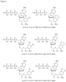
- the nucleotides comprising a protecting unit at their 3' positions may be adenine (A), thymine (T), cytosine (C), or guanine (G) or variants thereof ( Figure 1 ).
- the protection unit comprised at the 3' positions of the nucleotides provided may be directly ( Figure 11 ) or indirectly ( Figure 12 ) cleavable by illumination with light.
- protection units may be indirectly cleavable by illumination with light.
- Exemplary such protection units may be either acid or base labeled molecules such as esters, or cleavable with a phosphine (azido or di-sulfur).
- photo-activatable cleave agent For indirect cleavage with light a reaction partner such as a photo-activatable cleave agent needs to be provided. These photo-activatable cleave agents are capable of removing the protection unit from the incorporated protected nucleotide. Such photo-activatable cleave agents are commonly known in the art. Figure 2-6 exemplary shows such photo-activatable cleave agents and its reactions. Moreover photo-activatable cleave agents are further disclosed in Frechet et al (1992).
- Step D describes the synthesis of an oligonucleotide of known sequence. This is based on cyclic removal of the protection unit and the addition of a nucleotides comprising a protecting unit at their 3' positions by a template independent transferase.
- the first step requires the removal of the protection unit from nucleic acids (especially RNA such as mRNA) comprised in the target cells.
- nucleic acids especially RNA such as mRNA
- the protection unit can be directly ( Figure 13 ) or indirectly ( Figure 14 ) cleaved by illumination with light.
- At least one photo-activatable cleave agent capable of removing the protection unit from the incorporated protected nucleotide needs to be added to the biological sample.
- Light induced cleavage of the protection unit is done by spatially illuminating the target cells with light. Illumination of target cells can be controlled by e.g. IPAM or other systems such as Digital Micromirror Device (DMD)
- a wavelength of 370 nm to 405 nm is used for illumination
- nucleic acids especially RNA such as mRNA
- the protection unit removed only in nucleic acids (especially RNA such as mRNA) of these cells. Therefore, only the nucleic acids (especially RNA such as mRNA) in target cells will have a free 3' hydroxyl group that can be extended by adding a terminal transferase and nucleotides comprising a protecting unit at their 3' positions.
- Nucleic acids in non-illuminated cells will still comprise the protection unit. No nucleotides can be add, because no free 3'hydroxylgroups are available.
- nucleotides comprising a protecting unit at their 3' positions are incorporated to the free 3' hydroxyl groups of the 3'ends of the nucleic acids(especially RNA such as mRNA) in the target cells by a template independent transferase ( Figure 15 and Figure 16 ).
- nucleic acids especially RNA such as mRNA
- No nucleotide can be added enzymatically at this stage, because no free 3'OH groups are available for extension or addition of further nucleotides.
- the template independent transferase used for this step is an enzyme and used in aqueous and biologically compatible conditions.
- a template-independent polymerase is a terminal transferase more preferentially a terminal deoxynucleotidyl transferase (TdT) or a variant thereof.
- a template-independent polymerase may be a polymerase modified to become template free.
- the nucleotides comprising a protecting unit at their 3' positions may be adenine (A), thymine (T), cytosine (C), or guanine (G) or variants thereof ( Figure 1 ).
- the protection unit comprised at the 3' positions of the nucleotides provided may be directly or indirectly cleavable by illumination with light.
- Protection units that are directly cleavable with light are photolabile groups such as nitro phenyl groups. Said protection may be photolabile groups such as groups shown in Figure 1 .
- protection units may be indirectly cleavable by illumination with light.
- Exemplary such protection units may be either acid or base labile molecules such as esters, or cleavable with a phosphine (azido or di-sulfur).
- photo-activatable cleave agent For indirect cleavage with light a reaction partner such as a photo-activatable cleave agent needs to be provided. These photo-activatable cleave agents are capable of removing the protection unit from the incorporated protected nucleotide. Such photo-activatable cleave agents are commonly known in the art. Figure 2-6 exemplary shows such photo-activatable cleave agents and its reactions. Moreover photo-activatable cleave agents are further disclosed in Frechet et al (1992).
- the synthesis steps may be repeated at least at least 3 times, 4 times, 12 times or 20 times.
- nucleotides comprising a protecting unit at their 3'position are provided subsequently and after step each round of synthesis (step D) the unincorporated nucleotides may be removed from the biological sample. This may be done by standard washing steps.
- nucleotides comprising a 3' protection group In each round on synthesis only one kind of nucleotides comprising a 3' protection group is provided. For example in one round of synthesis adenine nucleotides comprising a 3' protection group are provided, only and in another round of synthesis cytosine nucleotides comprising a 3' protection group are provided, only. Based on this it can be controlled which nucleotide is incorporated at which position of the newly synthetized oligonucleotide.
- nucleotides comprising a protecting unit at their 3' positions may be same or different, dependent on the sequence that is intended to be generated.
- the oligonucleotides of known sequence may be or may comprise barcode sequences, primer binding sites or both.
- the protection unit may be removed from the nucleic acids (especially RNA such as mRNA) by illumination with light.
- Nucleic acids (especially RNA such as mRNA) from target cells may be isolated from the biological sample according to methods known in the art
- One aspect of the invention is the synthesis of different oligonucleotide sequences to nucleic acids (especially RNA such as mRNA) of different target cells in the biological sample.
- nucleic acids especially RNA such as mRNA
- the nucleic acids (especially RNA such as mRNA) from each target cell comprise different oligonucleotides of known sequence and the nucleic acid information could be directly assigned to the target cell in the biological sample.
- the biological sample comprises different target cells having different spatial positions.
- step Dii same or different spatial positions of said target cells are illuminated with light.
- nucleic acids especially RNA such as mRNA
- Figure 8 nucleic acids comprising different oligonucleotides of known sequence in each target cell are generated.
- the fidelity of cellular labeling with a specific desired sequence can be affected by various sources of error.
- a longer-than-expected sequence for a given cell could result from: (1) deprotection of oligonucleotides in non-illuminated regions due to diffusion for the indirect labeling approach, (2) deprotection in off-pixel regions due to background light, (3) deprotection in out-of-focus, laterally displaced regions for sufficiently thick samples illuminated with a high-NA objective ( Figure 9 ).
- shorter-than-expected cell sequences can result from: (4) insufficient deprotection in the on-pixel regions and (5) transcriptional elongation error (not all sequences extended with a protected nucleotide).
- the oligonucleotides may be synthesized in a way that the nucleic acids (especially RNA such as mRNA) of each target cell may comprise an oligonucleotide comprising a specific barcode ( Figure 17 )
- said oligonucleotide of known sequence comprises a barcode and an adapter sequence comprising a primer binding site sequence.
- the barcode may be different for nucleic acids (especially RNA such as mRNA) of each target cell, while said adapter sequence is same for nucleic acids (especially RNA such as mRNA) of all target cells.
- nucleic acids such as mRNA may be reverse transcribed and amplified ( Figure 19 & Figure 20 ). Finally nucleic acids may be sequenced. Sequences of nucleic acids can be reassigned to the position of the target cell based on the oligonucleotide of known sequence comprising e.g. a barcode sequence. Based on this method sequencing information and spatial tissue information can be correlated with each other.
- nucleic acids comprising an oligonucleotide of known sequence may be reverse transcribed, amplified and/or sequenced.
- the oligonucleotide of known sequence may comprise an adapter sequence comprising a primer binding site.
- Said primer binding site may be same or different for nucleic acids from different target cells.
- Amplification of the nucleic acids may be done by standard polymerase chain reaction (PCR) ( Figure 20 ).
- An additional adapter sequence may be incorporated to the 5'end of the nucleic acids by PCR.
- Such an amplified nucleic acid would comprise a first adapter sequence at the 3'end and a second adapter sequence at the 5' end ( Figure 21 ).
- these adapter sequences may be used for circularization of the amplified nucleic acid (as shown in Figure 22 ).
- a single oligonucleotide complementary to at least part of the first and second adapter sequence is add to the nucleic acids.
- Said nucleic acids bind to the complementary sequences and form a circle structure.
- the 5' and the 3' end of the nucleic acids are brought in close proximity to each other.
- the ends can be ligated and a circular construct can be formed.
- Circular constructs may be amplified by rolling circle amplification.
- compositions, methods, and respective component(s) thereof are used in reference to compositions, methods, and respective component(s) thereof, that are essential to the method or composition, yet open to the inclusion of unspecified elements, whether essential or not.
- binding and “hybridize” and its grammatical exuviates may be used interchangeably.
- Hybridization of two nucleic acid strands occurs if they are complementary to each other. Hybridization may occur under conditions known in the art.
- the term "complementary” refers to the capacity for precise pairing between two nucleotides via Watson and crick base pairing.
- a nucleotide at a given position of a nucleic acid strand is capable of forming hydrogen bonds with a nucleotide of another nucleic acid strand, then the two nucleic acids are considered to be complementary to one another at that position.
- Complementarity between two single-stranded nucleic acid molecules may be "partial,” in which only some of the nucleotides bind, or it may be complete when total complementarity exists between the single- stranded molecules.
- a “primer” as used herein is a single stranded oligonucleotide made of nucleotides, which is able to bind to complementary nucleic acid sequences. It is understood that all primers described in the current invention may serve as starting point for nucleic acid synthesis / amplification.
- nucleic acid synthesis and “nucleic acid amplification” as used herein, can be used interchangeably.
- the process of nucleic acid synthesis is well known in the art.
- a template nucleic acid is provided, which may be single stranded or double stranded.
- double stranded nucleic acid are used initially a first step is the denaturation into single nucleic acid strands (complement and reverse complement) using techniques known in the art. No denaturation step is needed for single stranded nucleic acids.
- primer are provided that bind to complementary regions of the nucleic acid strands. The 3'end of the primer is then elongated using a polymerase and a complementary strand is generated by filling with complementary nucleotides. As a result a complementary nucleic acid strand is formed.
- reverse transcription is well known in the art.
- a primer is hybridized to a RNA molecule. This primer serves as priming site for the synthesis of cDNA by an enzyme with reverse transcriptase activity.
- the reverse transcriptase can extend the primer and a cDNA is synthetized.
- oligonucleotide refers to biopolymers composed of nucleotide monomers covalently bonded in a chain.
- An amplified nucleic acid may be named as "amplicon”.
- a nucleic acid may be DNA or RNA.
- Oligonucleotides may contain all four nucleotides (A, T, G, C) in each position, or only a subset of the four nucleotides in each position.
- Alternative nucleotides like Uracil, or nucleotide analogues or derivates may also be used.
- Oligonucleotides according to the current invention may comprise a barcode sequence.
- a barcode sequence is a short nucleotide sequence for identification purposes.
Landscapes
- Life Sciences & Earth Sciences (AREA)
- Chemical & Material Sciences (AREA)
- Organic Chemistry (AREA)
- Health & Medical Sciences (AREA)
- Engineering & Computer Science (AREA)
- Zoology (AREA)
- Wood Science & Technology (AREA)
- Molecular Biology (AREA)
- Biotechnology (AREA)
- General Health & Medical Sciences (AREA)
- Proteomics, Peptides & Aminoacids (AREA)
- Genetics & Genomics (AREA)
- Biochemistry (AREA)
- Bioinformatics & Cheminformatics (AREA)
- General Engineering & Computer Science (AREA)
- Microbiology (AREA)
- Chemical Kinetics & Catalysis (AREA)
- Physics & Mathematics (AREA)
- Analytical Chemistry (AREA)
- Biophysics (AREA)
- General Chemical & Material Sciences (AREA)
- Immunology (AREA)
- Measuring Or Testing Involving Enzymes Or Micro-Organisms (AREA)
Priority Applications (1)
| Application Number | Priority Date | Filing Date | Title |
|---|---|---|---|
| EP23200719.5A EP4530360A1 (de) | 2023-09-29 | 2023-09-29 | Verfahren zur räumlichen strichcodierung |
Applications Claiming Priority (1)
| Application Number | Priority Date | Filing Date | Title |
|---|---|---|---|
| EP23200719.5A EP4530360A1 (de) | 2023-09-29 | 2023-09-29 | Verfahren zur räumlichen strichcodierung |
Publications (1)
| Publication Number | Publication Date |
|---|---|
| EP4530360A1 true EP4530360A1 (de) | 2025-04-02 |
Family
ID=88237364
Family Applications (1)
| Application Number | Title | Priority Date | Filing Date |
|---|---|---|---|
| EP23200719.5A Withdrawn EP4530360A1 (de) | 2023-09-29 | 2023-09-29 | Verfahren zur räumlichen strichcodierung |
Country Status (1)
| Country | Link |
|---|---|
| EP (1) | EP4530360A1 (de) |
Citations (8)
| Publication number | Priority date | Publication date | Assignee | Title |
|---|---|---|---|---|
| WO2017007925A1 (en) * | 2015-07-07 | 2017-01-12 | Thermo Fisher Scientific Geneart Gmbh | Enzymatic synthesis of nucleic acid sequences |
| WO2019224544A1 (en) * | 2018-05-23 | 2019-11-28 | Oxford Nanopore Technologies Limited | Polynucleotide synthesis method, system and kit |
| WO2020120442A2 (en) * | 2018-12-13 | 2020-06-18 | Dna Script | Direct oligonucleotide synthesis on cells and biomolecules |
| WO2021221831A1 (en) * | 2020-05-01 | 2021-11-04 | Microsoft Technology Licensing, Llc | Universal template strands for enzymatic polynucleotide synthesis |
| EP4108783A1 (de) * | 2021-06-24 | 2022-12-28 | Miltenyi Biotec B.V. & Co. KG | Räumliche sequenzierung mit mictag |
| WO2023275334A1 (en) * | 2021-07-01 | 2023-01-05 | Miltenyi Biotec B.V. & Co. KG | Unit-dna composition for spatial barcoding and sequencing |
| EP4155416A1 (de) * | 2021-09-23 | 2023-03-29 | Miltenyi Biotec B.V. & Co. KG | Verfahren zur erlangung von raum- und sequenzierungsinformationen von m-rna aus gewebe |
| WO2023152354A1 (en) * | 2022-02-14 | 2023-08-17 | Miltenyi Biotec B.V. & Co. KG | Direct synthesis of oligonucleotides on microtomed tissue slices |
-
2023
- 2023-09-29 EP EP23200719.5A patent/EP4530360A1/de not_active Withdrawn
Patent Citations (9)
| Publication number | Priority date | Publication date | Assignee | Title |
|---|---|---|---|---|
| WO2017007925A1 (en) * | 2015-07-07 | 2017-01-12 | Thermo Fisher Scientific Geneart Gmbh | Enzymatic synthesis of nucleic acid sequences |
| WO2019224544A1 (en) * | 2018-05-23 | 2019-11-28 | Oxford Nanopore Technologies Limited | Polynucleotide synthesis method, system and kit |
| WO2020120442A2 (en) * | 2018-12-13 | 2020-06-18 | Dna Script | Direct oligonucleotide synthesis on cells and biomolecules |
| US11268091B2 (en) | 2018-12-13 | 2022-03-08 | Dna Script Sas | Direct oligonucleotide synthesis on cells and biomolecules |
| WO2021221831A1 (en) * | 2020-05-01 | 2021-11-04 | Microsoft Technology Licensing, Llc | Universal template strands for enzymatic polynucleotide synthesis |
| EP4108783A1 (de) * | 2021-06-24 | 2022-12-28 | Miltenyi Biotec B.V. & Co. KG | Räumliche sequenzierung mit mictag |
| WO2023275334A1 (en) * | 2021-07-01 | 2023-01-05 | Miltenyi Biotec B.V. & Co. KG | Unit-dna composition for spatial barcoding and sequencing |
| EP4155416A1 (de) * | 2021-09-23 | 2023-03-29 | Miltenyi Biotec B.V. & Co. KG | Verfahren zur erlangung von raum- und sequenzierungsinformationen von m-rna aus gewebe |
| WO2023152354A1 (en) * | 2022-02-14 | 2023-08-17 | Miltenyi Biotec B.V. & Co. KG | Direct synthesis of oligonucleotides on microtomed tissue slices |
Non-Patent Citations (3)
| Title |
|---|
| JEAN M.J. FRECHET, PURE & APPL. CHEM., vol. 64, no. 9, 1992, pages 1239 - 1248 |
| JERZY OLEJNIKEDYTA KRZYMANSKA-OLEJNIKKENNETH J. ROTHSCHILD: "Photocleavable Biotin Phosphoramidite for 5' -End-Labeling, Affinity Purification and Phosphorylation of Synthetic Oligonucleotides", NUCLEIC ACIDS RESEARCH, vol. 24, 1 January 1996 (1996-01-01), pages 361 - 366, XP002084583, Retrieved from the Internet <URL:https://doi.org/10.1093/nar/24.2.361> DOI: 10.1093/nar/24.2.361 |
| OLEJNIK JLUDEMANN HCKRZYMANSKA-OLEJNIK EBERKENKAMP SHILLENKAMP FROTHSCHILD KJ: "Photocleavable peptide-DNA conjugates: synthesis and applications to DNA analysis using MALDI-MS", NUCLEIC ACIDS RES, vol. 27, no. 23, 1 December 1999 (1999-12-01), pages 4626 - 31, XP002220753, DOI: 10.1093/nar/27.23.4626 |
Similar Documents
| Publication | Publication Date | Title |
|---|---|---|
| US20250154564A1 (en) | Methods for performing spatial profiling of biological molecules | |
| KR102695749B1 (ko) | 화학적 조성물 및 이것을 사용하는 방법 | |
| US10844426B2 (en) | Methods for detecting and identifying genomic nucleic acids | |
| EP1711631B1 (de) | Nukleinsäurecharakterisierung | |
| JP2023531463A (ja) | 酵素的核酸伸長を用いたin situ単一細胞解析のための組成物及び方法 | |
| JP2022511816A (ja) | 細胞標的の空間指向性量子バーコード化 | |
| KR102893736B1 (ko) | 화학 조성물 및 이의 사용 방법 | |
| KR20190012184A (ko) | 샘플 중 표적 핵산을 검출하는 방법 | |
| US20020072060A1 (en) | Methods for detecting and assaying nucleic acid sequences | |
| WO2022147139A1 (en) | Methods and compositions for light-controlled surface patterning using a polymer | |
| KR20180135476A (ko) | 세포 구성성분을 매트릭스에 부착시키는 방법 | |
| WO2021113330A1 (en) | Sample processing barcoded bead composition, method, manufacturing, and system | |
| EP4530360A1 (de) | Verfahren zur räumlichen strichcodierung | |
| US10036063B2 (en) | Method for sequencing a polynucleotide template | |
| US20250137047A1 (en) | Direct Synthesis of Oligonucleotides on Microtomed Tissue Slices | |
| US10501779B2 (en) | Oligonucleotide trapping | |
| EP4431450A1 (de) | Träger zur sequenzierung von zielnukleinsäuresträngen | |
| WO2024215735A1 (en) | Multiplexed optical barcoding for spatial omics | |
| HK40126998A (en) | Methods for performing spatial profiling of biological molecules |
Legal Events
| Date | Code | Title | Description |
|---|---|---|---|
| PUAI | Public reference made under article 153(3) epc to a published international application that has entered the european phase |
Free format text: ORIGINAL CODE: 0009012 |
|
| STAA | Information on the status of an ep patent application or granted ep patent |
Free format text: STATUS: THE APPLICATION HAS BEEN PUBLISHED |
|
| AK | Designated contracting states |
Kind code of ref document: A1 Designated state(s): AL AT BE BG CH CY CZ DE DK EE ES FI FR GB GR HR HU IE IS IT LI LT LU LV MC ME MK MT NL NO PL PT RO RS SE SI SK SM TR |
|
| STAA | Information on the status of an ep patent application or granted ep patent |
Free format text: STATUS: THE APPLICATION IS DEEMED TO BE WITHDRAWN |
|
| 18D | Application deemed to be withdrawn |
Effective date: 20251003 |