EP4534086A1 - Behandlung von leukodystrophie - Google Patents
Behandlung von leukodystrophie Download PDFInfo
- Publication number
- EP4534086A1 EP4534086A1 EP23386091.5A EP23386091A EP4534086A1 EP 4534086 A1 EP4534086 A1 EP 4534086A1 EP 23386091 A EP23386091 A EP 23386091A EP 4534086 A1 EP4534086 A1 EP 4534086A1
- Authority
- EP
- European Patent Office
- Prior art keywords
- donepezil
- acetylcholinesterase inhibitor
- expression
- myelin
- leukodystrophy
- Prior art date
- Legal status (The legal status is an assumption and is not a legal conclusion. Google has not performed a legal analysis and makes no representation as to the accuracy of the status listed.)
- Withdrawn
Links
Images


Classifications
-
- A—HUMAN NECESSITIES
- A61—MEDICAL OR VETERINARY SCIENCE; HYGIENE
- A61K—PREPARATIONS FOR MEDICAL, DENTAL OR TOILETRY PURPOSES
- A61K31/00—Medicinal preparations containing organic active ingredients
- A61K31/33—Heterocyclic compounds
- A61K31/395—Heterocyclic compounds having nitrogen as a ring hetero atom, e.g. guanethidine or rifamycins
- A61K31/435—Heterocyclic compounds having nitrogen as a ring hetero atom, e.g. guanethidine or rifamycins having six-membered rings with one nitrogen as the only ring hetero atom
- A61K31/44—Non condensed pyridines; Hydrogenated derivatives thereof
- A61K31/445—Non condensed piperidines, e.g. piperocaine
-
- A—HUMAN NECESSITIES
- A61—MEDICAL OR VETERINARY SCIENCE; HYGIENE
- A61P—SPECIFIC THERAPEUTIC ACTIVITY OF CHEMICAL COMPOUNDS OR MEDICINAL PREPARATIONS
- A61P25/00—Drugs for disorders of the nervous system
- A61P25/28—Drugs for disorders of the nervous system for treating neurodegenerative disorders of the central nervous system, e.g. nootropic agents, cognition enhancers, drugs for treating Alzheimer's disease or other forms of dementia
Definitions
- the present invention relates to the treatment of leukodystrophy. Specifically, the present invention relates to the treatment of leukodystrophies such as globoid cell leukodystrophy using acetylcholinesterase inhibitors such as donepezil.
- leukodystrophies such as globoid cell leukodystrophy using acetylcholinesterase inhibitors such as donepezil.
- AD Alzheimer's disease
- a ⁇ proteolytic amyloid-beta
- APP amyloid precursor protein
- AD acetylcholinesterase inhibitors
- AChEis impede the hydrolysis of acetylcholine (ACh), increase the levels of this neurotransmitter in the synaptic cleft and thereby enhance cholinergic transmission.
- Cholinergic activation is considered critical for the maintenance of neuronal function and ensuring successful synaptic transmission in peripheral and central nervous systems.
- AchEi such as donepezil, rivastigmine, and tacrine improve cognitive function and are used for the symptomatic management of AD.
- Marketed AChEi drugs have been reported to have potential disease-modifying targeting mechanisms. For example, these drugs appear to delay the deposition of amyloid plaques and attenuate microglia activation.
- AChE inhibition is also related to reduced lymphocyte proliferation.
- Cholinergic neurotransmission also plays a role in regulating the secretion of proinflammatory cytokines (such as TNF-alpha, IL-6 and IL-1-beta).
- Drugs such as donepezil and rivastigmine are also suggested to play a role in regulating myelin state.
- OPCs oligodendrocyte precursor cells
- OPC-DRG dorsal root ganglion
- the drug managed to increase the length of myelinated axons and promote OPC differentiation to oligodendrocytes (OLs).
- OLs oligodendrocytes
- the number of myelinated axons, as well as the G-ratio of remyelinated axons in the corpus callosum region of mice were found to have increased in further in vivo experiments.
- Donepezil also upregulates the expression of myelin-related genes in OPCs.
- AchEi drugs such as donepezil and rivastigmine are also agonists for sigma-1-receptors (Sig-1R) and bind to these receptors at a low-nanomolar in range similar to AChE.
- Sig-1R sigma-1-receptors
- Sig-1R agonists have been suggested as potential use in demyelination diseases such as multiple sclerosis and vanishing white matter (VWM) disease.
- Globoid cell leukodystrophy is a demyelinating disease associated with oligodendrocyte cell death and aberrant myelin state. Globoid cell leukodystrophy, also known as Krabbe disease (KD), is a rare autosomal recessive disease occurring in approximately 1:100,000. This disease is a lipid storage disorder caused by mutations in the galc gene, which codes for the enzyme galactosylceramidase (GALC). Mutations in galc result in a loss of function and the toxic build-up of metabolites galactosylceramidase and galactosylsphingosine (psychosine).
- KD Globoid cell leukodystrophy
- GLC galactosylceramidase
- oligodendrocytes oligodendrocytes and causes demyelination by various proposed mechanisms, where myelin loss and neuroinflammation are presumably triggered by consequences of absent GALC function not clearing psychosine.
- Globoid cell leukodystrophy is also associated with a cluster of glial and immune cell dysfunctions including astrogliosis, microglia activation, as well as the macrophage recruitment.
- a range of mechanisms that attenuate psychosine-induced cell toxicity and demyelination have been previously described, including (i) sphingosine 1-phosphate receptor functional antagonists, (ii) phospholipase A2 inhibitors, (iii) piezo 1 mechanosensitive ion channel antagonists, (iv) hybrid nanoparticles and (v) D2 receptor antagonists antipsychotics.
- sphingosine 1-phosphate receptor functional antagonists phospholipase A2 inhibitors
- piezo 1 mechanosensitive ion channel antagonists iv
- hybrid nanoparticles iv
- D2 receptor antagonists antipsychotics marketed drugs fingolimod and haloperidol have been described to promote lifespan in the twitcher mouse model of KD.
- an acetylcholinesterase inhibitor for use in the treatment of a leukodystrophy.
- the use comprises administering a therapeutically effective amount of the acetylcholinesterase inhibitor to a subject.
- a method for the treatment of a leukodystrophy in a subject in need thereof comprising administering a therapeutically effective amount of an acetylcholinesterase inhibitor to the subject, thereby treating the leukodystrophy.
- acetylcholinesterase inhibitor in the manufacture of a medicament for the treatment of a leukodystrophy.
- acetylcholinesterase inhibitor in the treatment of a leukodystrophy.
- a sigma-1 receptor (Sig-1R) agonist for use in the treatment of a leukodystrophy, optionally wherein the use comprises administering a therapeutically effective amount of the sigma-1 receptor (Sig-1R) agonist to a subject.
- a method for the treatment of a leukodystrophy in a subject in need thereof comprising administering a therapeutically effective amount of a sigma-1 receptor (Sig-1R) agonist to the subject, thereby treating the leukodystrophy.
- Sig-1R sigma-1 receptor
- a sigma-1 receptor (Sig-1R) agonist in the manufacture of a medicament for use in the treatment of a leukodystrophy.
- a sigma-1 receptor (Sig-1R) agonist in the treatment of a leukodystrophy.
- the leukodystrophy is globoid cell leukodystrophy.
- the leukodystrophy is globoid cell leukodystrophy.
- the acetylcholinesterase inhibitor and/or the sigma-1 receptor (Sig-1R) agonist has acetylcholinesterase inhibitor and/or sigma-1 receptor (Sig-1R) agonist activity.
- the acetylcholinesterase inhibitor and/or the sigma-1 receptor (Sig-1R) agonist has dual acetylcholinesterase inhibitor and sigma-1 receptor (Sig-1R) agonist activity.
- the acetylcholinesterase inhibitor is a reversible acetylcholinesterase inhibitor.
- the sigma-1 receptor (Sig-1R) agonist is a reversible sigma-1 receptor (Sig-1R) agonist.
- the acetylcholinesterase inhibitor and/or the sigma-1 receptor (Sig-1R) agonist is donepezil ((RS)-2-[(1-Benzyl-4-piperidyl)methyl]-5,6-dimethoxy-2,3-dihydroinden-1-one).
- the acetylcholinesterase inhibitor and/or the sigma-1 receptor (Sig-1R) agonist is donepezil hydrochloride.
- the acetylcholinesterase inhibitor and/or the sigma-1 receptor (Sig-1R) agonist is donepezil.
- the acetylcholinesterase inhibitor and/or the sigma-1 receptor (Sig-1R) agonist is administered orally and/or topically.
- the acetylcholinesterase inhibitor and/or the sigma-1 receptor (Sig-1R) agonist is administered orally and/or transdermally.
- the acetylcholinesterase inhibitor and/or the sigma-1 receptor (Sig-1R) agonist is administered orally.
- the acetylcholinesterase inhibitor and/or the sigma-1 receptor (Sig-1R) agonist is administered at a dose of about 1-20, optionally about 5-15, optionally about 10 mg/kg.
- the acetylcholinesterase inhibitor and/or the sigma-1 receptor (Sig-1R) agonist is administered at a dose of about 1-20, optionally about 5-15, optionally about 10mg/kg per day.
- the acetylcholinesterase inhibitor and/or the sigma-1 receptor (Sig-1R) agonist is administered at a dose of about 10mg/kg per day.
- the acetylcholinesterase inhibitor and/or the sigma-1 receptor (Sig-1R) agonist is administered for about 1-80, optionally about 10-70, optionally about 20-60, optionally about 30-50, optionally about 40-50, optionally about 44 days.
- the acetylcholinesterase inhibitor and/or the sigma-1 receptor (Sig-1R) agonist is administered for about 44 days.
- administering increases a level of expression of myelin basic protein (MBP).
- MBP myelin basic protein
- administration of the acetylcholinesterase inhibitor and/or the sigma-1 receptor (Sig-1R) agonist increases a level of expression of myelin basic protein (MBP) relative to a level of expression of myelin basic protein (MBP) prior to administration.
- acetylcholinesterase inhibitor and/or the sigma-1 receptor (Sig-1R) agonist decreases an amount of vimentin in a white matter (WM) of the cerebellar cortex.
- administration of the acetylcholinesterase inhibitor and/or the sigma-1 receptor (Sig-1R) agonist decreases an amount of vimentin in a white matter (WM) of the cerebellar cortex prior to administration.
- administering decreases an amount of vimentin in a white matter (WM) of the cerebellar cortex relative to a granular layer (GL) of the cerebellar cortex.
- administration of the acetylcholinesterase inhibitor and/or the sigma-1 receptor (Sig-1R) agonist decreases an amount of vimentin in a white matter (WM) of the cerebellar cortex relative to a granular layer (GL) of the cerebellar cortex prior to administration.
- WM white matter
- GL granular layer
- acetylcholinesterase inhibitor and/or the sigma-1 receptor (Sig-1R) agonist decreases a level of expression of ionized calcium binding adaptor molecule 1 (lba1).
- administration of the acetylcholinesterase inhibitor and/or the sigma-1 receptor (Sig-1R) agonist decreases a level of expression of ionized calcium binding adaptor molecule 1 (lba1) relative to a level of expression of Iba1 prior to administration.
- Sig-1R sigma-1 receptor
- administration of the acetylcholinesterase inhibitor and/or the sigma-1 receptor (Sig-1R) agonist decreases a level of expression of at least one of vimentin, ionized calcium binding adaptor molecule 1 (lba1), and interleukin-6 (IL-6) relative to the level of expression of at least one of myelin basic protein (MBP), and myelin oligodendrocyte glycoprotein (MOG).
- vimentin ionized calcium binding adaptor molecule 1
- IL-6 interleukin-6
- administration of the acetylcholinesterase inhibitor and/or the sigma-1 receptor (Sig-1R) agonist decreases a level of expression of at least one of vimentin, ionized calcium binding adaptor molecule 1 (Iba1), and interleukin-6 (IL-6) relative to the level of expression of at least one of myelin basic protein (MBP), and myelin oligodendrocyte glycoprotein (MOG) prior to administration.
- vimentin ionized calcium binding adaptor molecule 1
- IL-6 interleukin-6
- Sig-1R acetylcholinesterase inhibitor and/or the sigma-1 receptor (Sig-1R) agonist decreases a level of expression of vimentin, ionized calcium binding adaptor molecule 1 (lba1), and interleukin-6 (IL-6) relative to the level of expression of myelin basic protein (MBP), and myelin oligodendrocyte glycoprotein (MOG).
- vimentin ionized calcium binding adaptor molecule 1
- IL-6 interleukin-6
- administration of the acetylcholinesterase inhibitor and/or the sigma-1 receptor (Sig-1R) agonist decreases a level of expression of vimentin, ionized calcium binding adaptor molecule 1 (lba1), and interleukin-6 (IL-6) relative to the level of expression of myelin basic protein (MBP), and myelin oligodendrocyte glycoprotein (MOG) prior to administration.
- vimentin ionized calcium binding adaptor molecule 1
- IL-6 interleukin-6
- administering decreases a level of expression of vimentin, and ionized calcium binding adaptor molecule 1 (lba1) relative to the level of expression of myelin basic protein (MBP), and myelin oligodendrocyte glycoprotein (MOG).
- vimentin a level of expression of vimentin, and ionized calcium binding adaptor molecule 1 (lba1) relative to the level of expression of myelin basic protein (MBP), and myelin oligodendrocyte glycoprotein (MOG).
- MBP myelin basic protein
- MOG myelin oligodendrocyte glycoprotein
- administration of the acetylcholinesterase inhibitor and/or the sigma-1 receptor (Sig-1R) agonist decreases a level of expression of vimentin, and ionized calcium binding adaptor molecule 1 (Iba1) relative to the level of expression of myelin basic protein (MBP), and myelin oligodendrocyte glycoprotein (MOG) prior to administration.
- Sig-1R sigma-1 receptor
- acetylcholinesterase inhibitor and/or the sigma-1 receptor (Sig-1R) agonist increases body weight.
- administration of the acetylcholinesterase inhibitor and/or the sigma-1 receptor (Sig-1R) agonist increases body weight relative to body weight prior to administration.
- acetylcholinesterase inhibitor and/or the sigma-1 receptor (Sig-1R) agonist increases the lifespan of the subject.
- administration of the acetylcholinesterase inhibitor and/or the sigma-1 receptor (Sig-1R) agonist increases the lifespan of the subject by about 25% relative to the lifespan of a subject suffering from a leukodystrophy.
- a method for diagnosing, detecting or identifying a leukodystrophy in a subject comprising determining a level of expression of myelin basic protein (MBP) in the subject, wherein a level of expression of myelin basic protein (MBP) in the subject less than a level of expression of myelin basic protein (MBP) in a subject not suffering from a leukodystrophy indicates a leukodystrophy in the subject.
- MBP myelin basic protein
- a method for diagnosing, detecting or identifying a leukodystrophy in a subject comprising determining a level of expression of myelin oligodendrocyte glycoprotein (MOG) in the subject, wherein a level of expression of myelin oligodendrocyte glycoprotein (MOG) in the subject less than a level of expression of myelin oligodendrocyte glycoprotein (MOG) in a subject not suffering from a leukodystrophy indicates a leukodystrophy in the subject.
- MOG myelin oligodendrocyte glycoprotein
- a method for diagnosing, detecting or identifying a leukodystrophy in a subject comprising determining a level of expression of vimentin in the subject, wherein a level of expression of vimentin in the subject greater than a level of expression of vimentin in a subject not suffering from a leukodystrophy indicates a leukodystrophy in the subject.
- a method for diagnosing, detecting or identifying a leukodystrophy in a subject comprising determining a level of expression of ionized calcium binding adaptor molecule 1 (Iba1) in the subject, wherein a level of expression of ionized calcium binding adaptor molecule 1 (Iba1) in the subject greater than a level of expression of ionized calcium binding adaptor molecule 1 (lba1) in a subject not suffering from a leukodystrophy indicates a leukodystrophy in the subject.
- a method for diagnosing, detecting or identifying a leukodystrophy in a subject comprising determining a level of expression of interleukin-6 (IL-6) in the subject, wherein a level of expression of interleukin-6 (IL-6) in the subject greater than a level of expression of interleukin-6 (IL-6) in a subject not suffering from a leukodystrophy indicates a leukodystrophy in the subject.
- IL-6 interleukin-6
- the method comprises the step of comparing the level of expression of at least one of myelin basic protein (MBP), myelin oligodendrocyte glycoprotein (MOG), vimentin, ionized calcium binding adaptor molecule 1 (lba1), and interleukin-6 (IL-6) in the subject with the level of expression of at least one of myelin basic protein (MBP), myelin oligodendrocyte glycoprotein (MOG), vimentin, ionized calcium binding adaptor molecule 1 (lba1), and interleukin-6 (IL-6) in the subject.
- MBP myelin basic protein
- MOG myelin oligodendrocyte glycoprotein
- IL-6 interleukin-6
- the method comprises the step of comparing the level of expression of at least one of vimentin, ionized calcium binding adaptor molecule 1 (lba1), and interleukin-6 (IL-6) in the subject with the level of expression of at least one of myelin basic protein (MBP), and myelin oligodendrocyte glycoprotein (MOG) in the subject.
- vimentin ionized calcium binding adaptor molecule 1
- IL-6 interleukin-6
- MBP myelin basic protein
- MOG myelin oligodendrocyte glycoprotein
- the method comprises the step of comparing the level of expression of vimentin, ionized calcium binding adaptor molecule 1 (Iba1), and interleukin-6 (IL-6) in the subject with the level of expression of myelin basic protein (MBP), and myelin oligodendrocyte glycoprotein (MOG) in the subject.
- Iba1 ionized calcium binding adaptor molecule 1
- IL-6 interleukin-6
- MBP myelin basic protein
- MOG myelin oligodendrocyte glycoprotein
- the method comprises the step of comparing the level of expression of at least one of vimentin, and ionized calcium binding adaptor molecule 1 (lba1) in the subject with the level of expression of at least one of myelin basic protein (MBP), and myelin oligodendrocyte glycoprotein (MOG) in the subject.
- vimentin ionized calcium binding adaptor molecule 1
- MBP myelin basic protein
- MOG myelin oligodendrocyte glycoprotein
- the method comprises the step of comparing the level of expression of vimentin, and ionized calcium binding adaptor molecule 1 (that) in the subject with the level of expression of myelin basic protein (MBP), and myelin oligodendrocyte glycoprotein (MOG) in the subject.
- MBP myelin basic protein
- MOG myelin oligodendrocyte glycoprotein
- the level of expression of at least one of vimentin, ionized calcium binding adaptor molecule 1 (lba1), and interleukin-6 (IL-6) in the subject greater than the level of expression of at least one of myelin basic protein (MBP), and myelin oligodendrocyte glycoprotein (MOG) in the subject indicates a leukodystrophy in the subject.
- the level of expression of vimentin, ionized calcium binding adaptor molecule 1 (lba1), and interleukin-6 (IL-6) in the subject greater than the level of expression of myelin basic protein (MBP), and myelin oligodendrocyte glycoprotein (MOG) in the subject indicates a leukodystrophy in the subject.
- the level of expression of vimentin, and ionized calcium binding adaptor molecule 1 (lba1) in the subject greater than the level of expression of myelin basic protein (MBP), and myelin oligodendrocyte glycoprotein (MOG) in the subject indicates a leukodystrophy in the subject.
- mice A breeding colony of heterozygous twitcher mice was established in a pathogen-free environment in the Comparative Medicine Unit at Trinity College Dublin using mice obtained from the Jackson Laboratory and maintained on a C57BL/6J genetic background (C57BL/6J-twi with C57BL/6J-twi). Homozygous and heterozygous animals were identified using genotyping procedure. Heterozygous animals were used only for breeding purposes and were not otherwise used in the study. The mice used in the study were housed in ventilated cages (Techniplast, IT-Varese) under specific pathogen-free conditions and constant environmental conditions (12:12 h light dark cycle, temperature 22 ⁇ 2 deg. °C, relative humidity 45 ⁇ 10 %).
- mice had free access to food and tap water. All mice in the facility were screened regularly according to a health-monitoring program, complying with the Federation of European Laboratory Animal Science Associations' recommendations. The experimental protocol of the study was approved by the Health Products Regulatory Authority (HPRA, project authorization number. AE19136 /P123).
- Donepezil hydrochloride (MW: 379.50 g/mol, Cipla Ltd., India) was administered to both male and female wild-type and homozygous twitcher mice via drinking water (concentration, 20 microgram ( ⁇ g)/mL), which would result in a calculated final dose of 10 mg/kg/d, considering a daily water consumption equal to 3 mL for each mouse ( Sheridan GK, Dev KK. Targeting S1P receptors in experimental autoimmune encephalomyelitis in mice improves early deficits in locomotor activity and increases ultrasonic vocalisations. Sci Rep. 2014;4:5051. doi: 10.1038/srep05051 ). Drinking water is considered as vehicle of the administered drug.
- the rationale for using a low number of vehicle (H 2 O) treated twicher mice primarily based on animal ethics and considered in line with 3R principles. In this case, as a benchmark, two previously published separate studies were used showing the effect of vehicle (H 2 O) administration in twitcher mice.
- Dose selection for oral administration was based on literature pharmacokinetic data ( Shin CY, Kim HS, Cha KH, Won DH, Lee JY, Jang SW, Sohn UD. The Effects of Donepezil, an Acetylcholinesterase Inhibitor, on Impaired Learning and Memory in Rodents. Biomol Ther. 2018;26(3), 274-281. doi: 10.4062/biomolther.2017.189 ).
- Constant and severe trembling and uncontrollable twitching was the highest score, followed immediately by euthanasia.
- the locomotor deficits arising due to demyelination were quantified considering that the minimum score of 1 indicates the absence of the disease, while the maximum score of 4. Fine and easy climbing with long duration was scored with 0, while late fine climbing with 1. Late and short-duration climbing had a score of 2, while the grasping and falling was scored with 3. The maximum climbing score was 4 and it was equivalent to the inability to climb.
- the genotype of the animals was performed on the 15 th postnatal day (P15). The water bottles were filled by the experimenter with 250 mL of water, where the weighted amount of the drug was dissolved, and thus no blinding was included at the level of drug treatment. All data were randomized, and analysis was performed with data blinded to both genotype and drug treatment.
- Donepezil extraction from brain tissue has been described and validated by Papakyriakopoulou et al. ( Papakyriakopoulou P, Rekkas DM, Colombo G, Valsami G. Development and In Vitro-Ex Vivo Evaluation of Novel Polymeric Nasal Donepezil Films for Potential Use in Alzhei'er's Disease Using Experimental Design. Pharmaceutics. 2022; 14(8):1742. doi: 10.3390/pharmaceutics14081742 ), using quercetin as internal standard (ISTD).
- each brain sample was homogenized prior to analysis with a T10 ULTRA-TURRAX ® (IKA Werke, DE-Staufen im Breisgau) in presence of water for injection (WFI) (tissue:WFI ratio 1:1 w/w), and then 25 microlitres ( ⁇ L) of homogenate tissue was vortex-mixed with 50 microlitres ( ⁇ L) of ISTD (quercetin 2 pg/mL in methanol) and 25 microlitres ( ⁇ L) of mobile phase. The mixture was centrifuged and 30 microlitres ( ⁇ L) of the supernatant was received and directly injected onto the HPLC system for donepezil quantification.
- WFI water for injection
- ISTD quercetin 2 pg/mL in methanol
- the HPLC system is composed by a LC-20AD Quaternary Gradient Pump with degasser, with an SIL-HT auto-sampler and a photo-diode array detector SPD-M20A. Analysis was carried out on an analytical reverse phase MZ Analysentechnik Nucleosil 100-5 C18 column (125 ⁇ 4.6 mm, 5 micrometre ( ⁇ m) particle size) connected to a precolumn C-18 (12.5 ⁇ 4.6 mm, 5 micrometre ( ⁇ m) particle size, MZ Analysentechnik) of the same type.
- Mobile phase consisted of phosphate buffer: methanol: acetonitrile (50:40:10) and adjusted to pH 2.8 with orthophosphoric acid (80%), in isocratic mode with flow rate 0.8 mL/min.
- the LC analysis time was 10 min.
- the injection volume was 30 microlitres ( ⁇ L) and the retention time of donepezil and ISTD was 4.5 and 8 min, respectively.
- Detection of donepezil and Que was performed at 268 and 369, respectively, and the calibration curve samples ranged from 0.05 to 3 microgram ( ⁇ g)/mL of donepezil.
- mice were euthanized by placing in a COz chamber and then transcardially perfused with 20 mL of ice-cold PBS, pH 7.4 (flow: 7 mL/min), using a peristaltic pump (Watson-Marlow, Falmouth, Cornwall, UK) to remove the residual blood. Once perfusion was completed, animals were decapitated, and brains were removed and split into two hemispheres. One hemisphere was further separated into cerebellum and cortex, and then stored at 80°C until processed for quantitative reverse transcription PCR (RT-qPCR) and HPLC analysis.
- RT-qPCR quantitative reverse transcription PCR
- the remaining hemisphere was postfixed in 4% paraformaldehyde overnight (4 deg.°C) before being cryoprotected in 30% sucrose solution (4 deg.°C). Brains were then snap frozen on dry ice and embedded in optimal cutting temperature compound (OCT; catalog #361603E, VWR). Parasagittal cerebellar cryosections of 12 micrometre ( ⁇ m) thickness were cut using the Leica CM1850 Cryostat and processed for IHC.
- OCT optimal cutting temperature compound
- Cerebellar cryosections were equilibrated to room temperature for 2h, and then rehydrated with 100 microlitres ( ⁇ L) of PBS on each section. At all the steps of the procedure, the same volume was applied on every section. Sections were permeabilized using 0.2% Triton X-100 (Tx; catalog #T9284, Sigma-Aldrich) in phosphate buffer saline (PBS; catalog #18912-014, gibco) to reduce nonspecific binding and were blocked with 10% bovine serum albumin (BSA; catalog #A3294-100G, Sigma-Aldrich) for 2 h at room temperature. Primary antibody incubations were conducted overnight at 4 deg.°C in PBS containing 2% BSA and 0.1% Tx.
- Tx Triton X-100
- PBS phosphate buffer saline
- BSA bovine serum albumin
- MBP myelin basic protein
- IbA1 Polyclonal Antibody (1:500 dilution; catalog #PA5-27436, Invitrogen)
- anti-vimentin (1:500 dilution; catalog #sc-373717, Santa Cruz Biotechnology).
- Secondary antibody incubations were performed overnight at 4 deg.°C in PBS supplemented with 2% BSA and 0.1% Tx.
- the secondary antibodies used in the study were as follows: Invitrogen goat anti-mouse Alexa Fluor 488 (1:500; A11001, Thermo Fisher Scientific), Invitrogen goat anti-chicken Alexa Fluor 633 (1:500; A21103, Thermo Fisher Scientific), and Invitrogen anti-rabbit Alexa Fluor 488 (1:500; A11008, Thermo Fisher Scientific).
- a counterstain with Hoechst 33342 (1:10,000; catalog #62249, Thermo Fisher Scientific) labeling the nucleus was performed at the end of the immunofluorescence protocol.
- Slices were mounted using the Invitrogen SlowFade TM Gold antifade reagent (S36936, Thermo Fisher Scientific), coverslipped on microscope slides and stored at 4 deg.°C, in the dark, before being imaged.
- RNAs were isolated from cerebellum tissues using the Nucleospin ® total RNA isolation kit (Macherey-Nagel, Germany). The concentrations of isolated RNA from each sample were assessed using a NanoDrop 2000 Spectrophotometer (Thermo Scientific, USA). 10 ⁇ L of the isolated RNA were equalized to 20 microlitres ( ⁇ L) using RNase-free water. Then, the cDNA was synthesized by reverse transcription using the High-Capacity cDNA manufacture kit (Applied Biosystems TM , Thermo Fisher Scientific, USA). Equal volumes of the isolated RNA and cDNA mastermix were mixed, centrifuged, and loaded in the thermal cycler (T100 TM , BIO-RAD, USA).
- Inflammatory gene and protein expression signatures were generated for each animal using the 5 genes (MBP, MOG, Vimentin, lba-1, and IL-6).
- MBP myelin
- MOG mitochondrialin
- Vimentin a gene/protein that is positively associated with myelin
- IL-6 a gene/protein that is negatively associated with myelin
- the inverse data were used.
- the inverted data of Vimentin, lba-1, and IL-6 genes/proteins were averaged and divided by the average value of MBP and MOG genes/proteins original data to produce the inflammation/myelin ratio. This ratio serves as an optimal signature method for the gene and protein expression of each individual animal.
- each arbitrary value in the experimental group was divided by the mean of the control group and multiplied by 100.
- the possible differences among the animal groups during the IHC and RT-qPCR analysis were assessed applying the one-way ANOVA with Bonferroni post hoc test for multiple comparisons, as well as Mann-Whitney nonparametric test between all group pairs.
- Graphical data are represented as the mean ⁇ SEM.
- Donepezil enhances the levels of myelin in cerebellum of twitcher mice
- Donepezil alters Vimentin expression in white matter layer of cerebellum in twitcher mice
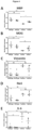
- Astrocytes in the layers of the cerebellum include the Bergmann glial cells of molecular layer (ML) and granular layer (GL) and fibrous astrocytes of white matter (WM) ( Figure 2A ).
- the type III intermediate filament astrocyte marker vimentin was employed to assess the effects of donepezil on astrocytes in twitcher mice.
- Donepezil decreases Iba1 expression in the cerebellum of twitcher mice
- the fluorescence data for ionized calcium binding adaptor molecule 1 (lba1) marker of microglia/macrophage revealed a significant effect of donepezil in wild type and twitcher treated animals ( Figure 3 ).
- KD is associated with alterations in myelin-, glial- and inflammatory-markers.
- MBP myelin markers
- MOG myelin markers
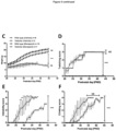
- the RT-qPCR analysis demonstrated the lower gene expression of MBP and MOG in the cerebellum of twitcher mice, while showing higher levels of vimentin, Iba1 and IL6, suggestive of a loss of myelin, enhanced glial cell reactivity and pro-inflammatory phenotypes ( Figure 4A-E ).
- an algorithm was created to generate a gene-expression-signature in which an average of expression values for glia and cytokine inflammatory markers (Vimentin, Iba1 and IL-6) was divided by an average of expression values for myelin markers (MBP and MOG), for each individual animal ( Figure 4F ).
- Donepezil increases the lifespan of twitcher mice
- Donepezil oral administration in twitcher mice via the drinking water from P25 onward showed an increased trend in the body weight from the P27 to P32 compared to vehicle treated twitcher animals, which was not different between these two twitcher groups from P32 onwards to end stage of life ( Figure 5C ). Twitching scoring was progressive with disease in twitcher mice and did not reveal any significant difference between the donepezil treated and vehicle treated animals ( Figure 5D ), where both groups plateaued at P32-P33, until the endpoint of the study.
- Kaplan-Meier survival curves demonstrate that donepezil treatment, at the dose of 10 mg/kg, increased the lifespan of twitcher mice toward 45 days, which was statistically increased compared to vehicle-treated twitcher mice in our current study and previous data ( Figure 5G ).
- Krabbe's disease is associated with genetic mutations and subsequent loss of function of the acid hydrolase galactosylceramidase (GalC) enzyme, which plays a key role in metabolism of galactosylceramide and the toxic galactolipid galactosylsphingosine (psychosine). Strong evidence has been established about the contribution of psychosine accumulation in nervous system and its toxic effects on levels of myelin. Psychosine accumulation has been determined throughout the CNS, in the spinal cord, cerebrum, and cerebellum.
- a natural occurring animal model of KD namely the twitcher mouse, displays myelin damage accompanied by axonal degeneration and the formation of globoid cells in the white matter, and is well recognized as translational model of the disease.
- the present invention provides a direct demonstration that donepezil rescues deficits seen at the pathological level in twitcher's mice, namely that donepezil oral treatment led to significant increase of myelin levels, restoration of astrocyte reactivity, as well as suppression of microglia activation.
- the current data also demonstrates that donepezil enhances mobility of twitcher animals as depicted by immobility and climbing scores, as well as promote life span in these animals.
- Myelin regeneration requires the gradual replacement and renewal of OLs through a two-stages process including the differentiation of OPCs into OLs and the production of myelin sheaths around the neuronal axons. This axonal wrapping is necessary for the transmission of electrical signals along the axon.
- the muscarinic acetylcholine receptors (mAChRs) M1, M3, and M4 have been identified in OPCs, while all the mAChRs subtypes are reported in low levels in mature OLs.
- astrocytes and microglia play as critical role in myelin generation as regulators of migration, proliferation, and maturation stages towards the production of myelinating OLs.
- nicotinic (nAChRs) and muscarinic (mAChRs) receptors are also known to be expressed by astrocytes and microglia and response to changes in levels of ACh neurotransmitter.
- mAChRs muscarinic
- Donepezil is also a well-known agonist of Sig-1R's, where these receptors are involved in the modulation of astrocytes and microglia and regulate permeability of the blood-brain-barrier, perhaps providing another avenue for this drug to elicit its effects in twitcher mice.
- Donepezil is also known to attenuate amyloid-beta-induced gliosis in microglia cells and microglia activation in an AD mouse model.
- Pro-inflammatory signals such as IL-6 have been reported importantly enhanced in the pathological condition of KD and twitcher mice, and in agreement with the present invention, donepezil can lower neuroinflammation after the treatment. The present invention reinforces the argument of donepezil anti-inflammatory properties.
- the body-wide damage caused by the buildup of psychosine thereby limits the efficacy of drugs such as fingolimod, haloperidol and donepezil to show lasting improvement in twitching and mobility or increase the lifespan further. Nevertheless, the overall ameliorated phenotype and achieved increase of lifespan after donepezil treatment may not be considered negligible given the severity of this KD translational model.
- the present invention shows that donepezil has myelin-promoting properties, as well as activity against neuroinflammation and show this drug prolongs lifespan of twitcher mice.
- AD Alzheimer's Disease
- AChEi Acetylcholinesterase inhibitors
- This drug also acts as an agonist for sigma-1 receptors (Sig-1R), which are implicated in demyelination diseases.
- Krabbe disease KD is a demyelinating disorder caused by mutations in the galc gene, resulting in toxic accumulation of psychosine.
- KD sigma-1 receptors
- the present invention provides data showing that donepezil preserves myelin and reduces glial cell reactivity in the brains of twitcher mice.
- donepezil also improves behavioral phenotypes and increases lifespan in twitcher animals.
- the present invention shows that donepezil, with its dual activity as an AChE inhibitor and Sig-1R agonist, may hold promise as a therapeutic candidate for demyelinating diseases, including KD.
- the myelin-promoting properties of donepezil render it promising candidate for the management of demyelinating diseases.
- the present invention shows the effect of an archetypical AchEi drug, namely donepezil, in the twitcher mouse model, a demyelinating model translatable for KD.
- donepezil protects against the loss of myelin, dampens glial cell reactivity, slows behavioral phenotypes and increases lifespan in the twitcher mouse model of KD.
Landscapes
- Health & Medical Sciences (AREA)
- Life Sciences & Earth Sciences (AREA)
- Pharmacology & Pharmacy (AREA)
- Biomedical Technology (AREA)
- Veterinary Medicine (AREA)
- Neurology (AREA)
- Neurosurgery (AREA)
- Public Health (AREA)
- Chemical & Material Sciences (AREA)
- General Health & Medical Sciences (AREA)
- Animal Behavior & Ethology (AREA)
- Bioinformatics & Cheminformatics (AREA)
- Engineering & Computer Science (AREA)
- Medicinal Chemistry (AREA)
- Psychiatry (AREA)
- Nuclear Medicine, Radiotherapy & Molecular Imaging (AREA)
- General Chemical & Material Sciences (AREA)
- Chemical Kinetics & Catalysis (AREA)
- Organic Chemistry (AREA)
- Hospice & Palliative Care (AREA)
- Epidemiology (AREA)
- Medicines That Contain Protein Lipid Enzymes And Other Medicines (AREA)
Priority Applications (3)
| Application Number | Priority Date | Filing Date | Title |
|---|---|---|---|
| EP23386091.5A EP4534086A1 (de) | 2023-10-06 | 2023-10-06 | Behandlung von leukodystrophie |
| PCT/EP2024/077975 WO2025073915A1 (en) | 2023-10-06 | 2024-10-04 | Treatment of leukodystrophy |
| AU2024355466A AU2024355466A1 (en) | 2023-10-06 | 2024-10-04 | Treatment of leukodystrophy |
Applications Claiming Priority (1)
| Application Number | Priority Date | Filing Date | Title |
|---|---|---|---|
| EP23386091.5A EP4534086A1 (de) | 2023-10-06 | 2023-10-06 | Behandlung von leukodystrophie |
Publications (1)
| Publication Number | Publication Date |
|---|---|
| EP4534086A1 true EP4534086A1 (de) | 2025-04-09 |
Family
ID=88647601
Family Applications (1)
| Application Number | Title | Priority Date | Filing Date |
|---|---|---|---|
| EP23386091.5A Withdrawn EP4534086A1 (de) | 2023-10-06 | 2023-10-06 | Behandlung von leukodystrophie |
Country Status (3)
| Country | Link |
|---|---|
| EP (1) | EP4534086A1 (de) |
| AU (1) | AU2024355466A1 (de) |
| WO (1) | WO2025073915A1 (de) |
Citations (2)
| Publication number | Priority date | Publication date | Assignee | Title |
|---|---|---|---|---|
| US20070129402A1 (en) * | 2004-12-27 | 2007-06-07 | Eisai Research Institute | Sustained release formulations |
| WO2018207192A1 (en) * | 2017-05-11 | 2018-11-15 | Ramot At Tel-Aviv University Ltd. | Methods of treating leukodystrophies |
-
2023
- 2023-10-06 EP EP23386091.5A patent/EP4534086A1/de not_active Withdrawn
-
2024
- 2024-10-04 AU AU2024355466A patent/AU2024355466A1/en active Pending
- 2024-10-04 WO PCT/EP2024/077975 patent/WO2025073915A1/en active Pending
Patent Citations (2)
| Publication number | Priority date | Publication date | Assignee | Title |
|---|---|---|---|---|
| US20070129402A1 (en) * | 2004-12-27 | 2007-06-07 | Eisai Research Institute | Sustained release formulations |
| WO2018207192A1 (en) * | 2017-05-11 | 2018-11-15 | Ramot At Tel-Aviv University Ltd. | Methods of treating leukodystrophies |
Non-Patent Citations (6)
| Title |
|---|
| BECHET SO'SULLIVAN SAYSSEL JFAGAN SGDEV KK: "Fingolimod Rescues Demyelination in a Mouse Model of Krabbe's Disease", J NEUROSCI., vol. 40, no. 15, 2020, pages 3104 - 3118 |
| BEETON CGARCIA ACHANDY KG: "Induction and clinical scoring of chronic-relapsing experimental autoimmune encephalomyelitis", J VIS EXP., no. 5, 2007, pages 224 |
| PAPAKYRIAKOPOULOU PREKKAS DMCOLOMBO GVALSAMI G: "Development and In Vitro-Ex Vivo Evaluation of Novel Polymeric Nasal Donepezil Films for Potential Use in Alzhei'er's Disease Using Experimental Design", PHARMACEUTICS, vol. 14, no. 8, 2022, pages 1742 |
| SHARMA KDEV KK: "The Effects of Antipsychotics in Experimental Models of Krabbe Disease", BIOMEDICINES, vol. 11, 2023, pages 1313 |
| SHIN CYKIM HSCHA KHWON DHLEE JYJANG SWSOHN UD: "The Effects of Donepezil, an Acetylcholinesterase Inhibitor, on Impaired Learning and Memory in Rodents", BIOMOL THER., vol. 26, no. 3, 2018, pages 274 - 281 |
| WICKS SELONDOT HZHANG B ET AL.: "Effect of intrastriatal mesenchymal stromal cell injection on progression of a murine model of Krabbe disease", BEHAV BRAIN RES., vol. 225, no. 2, 2011, pages 415 - 425, XP028297525, DOI: 10.1016/j.bbr.2011.07.051 |
Also Published As
| Publication number | Publication date |
|---|---|
| AU2024355466A1 (en) | 2026-03-19 |
| WO2025073915A1 (en) | 2025-04-10 |
Similar Documents
| Publication | Publication Date | Title |
|---|---|---|
| Rojas | The role of glutamate and its receptors in autism and the use of glutamate receptor antagonists in treatment | |
| Hammerschmidt et al. | Selective loss of noradrenaline exacerbates early cognitive dysfunction and synaptic deficits in APP/PS1 mice | |
| Dell'Orco et al. | Neuronal atrophy early in degenerative ataxia is a compensatory mechanism to regulate membrane excitability | |
| Takata et al. | Galantamine-induced amyloid-β clearance mediated via stimulation of microglial nicotinic acetylcholine receptors | |
| Teixeira et al. | Early axonal loss accompanied by impaired endocytosis, abnormal axonal transport, and decreased microtubule stability occur in the model of Krabbe's disease | |
| Zhu et al. | Autophagy modulation for Alzheimer’s disease therapy | |
| Giacoppo et al. | Triggering of inflammasome by impaired autophagy in response to acute experimental Parkinson’s disease: involvement of the PI3K/Akt/mTOR pathway | |
| US10426815B2 (en) | Prevention and treatment of itch with an MRGPR antagonist | |
| Rocha et al. | Microglia‐specific knock‐out of NF‐κB/IKK2 increases the accumulation of misfolded α‐synuclein through the inhibition of p62/sequestosome‐1‐dependent autophagy in the rotenone model of Parkinson's disease | |
| JP2018076332A (ja) | (3aR)−1,3a,8−トリメチル−1,2,3,3a,8,8a−ヘキサヒドロピロロ[2,3−b]インドール−5−イルフェニルカルバメートの有効量およびその使用方法 | |
| CN103260612A (zh) | MeCP2相关性病症的治疗 | |
| Balu et al. | A small-molecule TLR4 antagonist reduced neuroinflammation in female E4FAD mice | |
| Wang et al. | Early activation of Toll-like receptor-3 reduces the pathological progression of Alzheimer’s disease in APP/PS1 mouse | |
| Liu et al. | PAR2-mediated epigenetic upregulation of α-synuclein contributes to the pathogenesis of Parkinson׳ s disease | |
| Karaman et al. | Lack of effect of ceftriaxone, a GLT-1 transporter activator, on spatial memory in mice | |
| Ismael et al. | Verapamil inhibits TXNIP-NLRP3 inflammasome activation and preserves functional recovery after intracerebral hemorrhage in mice | |
| Möser et al. | Inhibition of the protein kinase IKKepsilon attenuates neuropathic pain in mice | |
| Jati et al. | Chromogranin A deficiency attenuates tauopathy by altering epinephrine–alpha-adrenergic receptor signaling in PS19 mice | |
| Kearney et al. | Silencing Parkinson’s risk allele Rit2 sex-specifically compromises motor function and dopamine neuron viability | |
| Zhou et al. | Molecular insights into tau pathology and its therapeutic strategies in Alzheimer's disease | |
| Barnett et al. | Loss of lysosomal acid lipase contributes to Alzheimer's disease pathology and cognitive decline | |
| EP4534086A1 (de) | Behandlung von leukodystrophie | |
| Coyle et al. | Novel drug treatments for schizophrenia | |
| CN114007607A (zh) | 用于治疗神经变性疾病的材料和方法 | |
| Yang et al. | Nicotine Attenuates Pathogenesis of Parkinson’s Disease via α7-nAChR-Mediated Lipid Metabolic Reprogramming and Anti-inflammatory Signaling |
Legal Events
| Date | Code | Title | Description |
|---|---|---|---|
| PUAI | Public reference made under article 153(3) epc to a published international application that has entered the european phase |
Free format text: ORIGINAL CODE: 0009012 |
|
| STAA | Information on the status of an ep patent application or granted ep patent |
Free format text: STATUS: THE APPLICATION HAS BEEN PUBLISHED |
|
| AK | Designated contracting states |
Kind code of ref document: A1 Designated state(s): AL AT BE BG CH CY CZ DE DK EE ES FI FR GB GR HR HU IE IS IT LI LT LU LV MC ME MK MT NL NO PL PT RO RS SE SI SK SM TR |
|
| STAA | Information on the status of an ep patent application or granted ep patent |
Free format text: STATUS: THE APPLICATION IS DEEMED TO BE WITHDRAWN |
|
| 18D | Application deemed to be withdrawn |
Effective date: 20251010 |