EP4535307A1 - Système et procédé d'analyse d'échantillon biologique en vue de la caractérisation de parasites - Google Patents
Système et procédé d'analyse d'échantillon biologique en vue de la caractérisation de parasites Download PDFInfo
- Publication number
- EP4535307A1 EP4535307A1 EP23201942.2A EP23201942A EP4535307A1 EP 4535307 A1 EP4535307 A1 EP 4535307A1 EP 23201942 A EP23201942 A EP 23201942A EP 4535307 A1 EP4535307 A1 EP 4535307A1
- Authority
- EP
- European Patent Office
- Prior art keywords
- images
- parasite
- biological sample
- microscope
- image
- Prior art date
- Legal status (The legal status is an assumption and is not a legal conclusion. Google has not performed a legal analysis and makes no representation as to the accuracy of the status listed.)
- Pending
Links
Images



Classifications
-
- G—PHYSICS
- G06—COMPUTING OR CALCULATING; COUNTING
- G06V—IMAGE OR VIDEO RECOGNITION OR UNDERSTANDING
- G06V20/00—Scenes; Scene-specific elements
- G06V20/60—Type of objects
- G06V20/69—Microscopic objects, e.g. biological cells or cellular parts
- G06V20/698—Matching; Classification
-
- G—PHYSICS
- G06—COMPUTING OR CALCULATING; COUNTING
- G06T—IMAGE DATA PROCESSING OR GENERATION, IN GENERAL
- G06T7/00—Image analysis
- G06T7/0002—Inspection of images, e.g. flaw detection
- G06T7/0012—Biomedical image inspection
Definitions
- the present disclosure is related to a semi-automated method and system for analyzing biological samples in order to detect, identify, classify, and/or count parasites.
- the disclosure may in particular be applied for the identification of poultry, swine and other animal or human parasites, especially gastrointestinal parasites, such as the agents of swine (i.e., Eimeria debliecki, Eimeria spinosa, Cystoisospora ( Isospora )) and poultry (i.e., E. acervulina, E. maxima, E. tenella, E. necatrix, E. brunetti, E. mitis, and E. praecox ) coccidiosis.
- Biological sample means a sample comprising living organisms.
- Flotation method is a classical process in parasitology that selectively separates parasites (i.e., larvae, ova, cysts and oocysts) upon whether they are hydrophilic or hydrophobic. Combined with direct microscopy it represents one of the most applied coprological diagnosis of these pathogens.
- it remains laboring and timeconsuming methods.
- alternative method like serology and molecular biology becomes the easiest solution for rapid and accurate identification and/or quantification of parasitic specimens from biological samples such as feces and droppings.
- these alternative methods Despite the improvement reached by these alternative methods, they remain expensive and require sophisticated laboratory equipment, which constitutes a limitation of their use as rapid and efficient methods.
- Bio shape characterization for automatic image recognition and diagnosis of protozoan parasites of the genus Eimeria This method is based on the elaboration of a database of single layer photomicrographs of the seven Eimeria species of domestic fowl to serve as reference template for the comparative automated identification of parasite images previously captured from biological samples.
- the approach relies on a Bayesian classifier and image similarity based on morphometric features of the parasites (i.e., curvature, geometry, and texture).
- the image database as well as the image processing analysis are provided as an open-source server namely COCCIMORPH.
- the present invention is directed to the improvement of the diagnosis of parasites, especially gastrointestinal parasites with a focus on the agent of coccidiosis.
- the invention aims at proposing a method for rapid and accurate identification, classification and/or numeration of parasitic specimens in animal populations, and/or in vaccine samples to control oocysts maturity (sporulation) and viability within the production process of vaccines.
- an aim of the present disclosure is to provide a more reliable and faster method of analyzing biological samples for detection, identification, classification and/or counting of parasites.
- a method for analyzing a biological sample comprising parasites comprising:
- the region containing the parasite is extracted from at least one image of the set corresponding to a determined layer of the biological sample.
- processing the acquired set of images comprises:
- the number of successive images is predetermined according to the spacing between the different layers of the sample.
- the region containing the parasite is extracted from a high-definition image generated from the acquired set of images.
- the high-definition image comprises selected image portions from the images of different layers of the biological sample.
- the selected image portions have the highest sharpness value of the set of images.
- the processing comprises application, on an input image formed of at least one image of the set of images or of a high-definition image generated from the set of images, of a trained segmentation algorithm.
- the trained segmentation algorithm is a convolutional neural network configured for performing instance segmentation, for instance a CNN having a U-net architecture.
- the trained segmentation algorithm is configured to extract, from the input image, a set of features related to the shape and/or dimensions of the parasite.
- the different focal planes of acquisition of the images at different layers are spaced from one another by a constant interval.
- the biological sample is placed on a microscope slide
- the acquisition comprises scanning, with the microscope, the whole slide at different layers of the biological sample to obtain a set of whole slide images, and dividing each set of whole slide images into a plurality of set of images where each set of images comprises images acquired at different focal planes of the microscope and for a constant position of the biological sample in a plane orthogonal to the microscope's optical axis.
- the method further comprises counting the number of parasites contained within the biological sample.
- counting the number of parasites comprises counting the number of parasites of each species.
- the step of acquiring, with a microscope, a set of images of different layers of the biological sample is performed at a magnification between x40 and x80, preferably at x80.
- the second set of morphologic features represents the microstructure of the parasite, such as the structure of the internal and external walls, micropyle, micropyle cap, oocyst residuum, sporocyst residuum, polar granule, steida body, substeida body, parasteida body, and/or refractile body.
- the method further comprises determining a parasite stage, sporulated or non sporulated, of the at least one parasite.
- the trained classification algorithm is a deep conventional neural network.
- the classification algorithm has been trained by supervised learning on a database comprising, for each of a plurality of parasite species, a plurality of images of parasites of a determined species, where each image has been acquired by a microscope with a magnification of at least x40, an identification of the parasite species.
- the method is applied for detection and identification of gastrointestinal parasites selected among the group consisting of:
- the biological sample is selected from a sample of a vaccine batch, a feces sample, a blood sample or swabs sample.
- a system for analyzing a biological sample comprising:
- a computer-program product comprising code instructions for implementing a method comprising the following steps, when it is implemented by a processor:
- the proposed method enables acquiring a set of images of a biological sample comprising parasites at different layers of the sample, and processing the set of acquired images in order to locate, and then determine a species of the parasite.
- Processing a set of images acquired at different layers of the sample enables obtaining a plurality of views at different layers of the same parasite, on which are visible different morphological features of the parasite. Resultantly, the determination of the species by the trained classification algorithm is more accurate.
- the set of images acquired at different layers enable detecting and analyzing parasites which may be located at overlapping positions within the sample. Accordingly, it is thus possible to count more reliably a number of parasites contained within the sample.
- the images are acquired at a magnification of at least x40, and preferably of x80, so that the internal structures of the parasite are sharply represented on the images, and the trained classification algorithm may perform parasite species determination based on these internal structures.
- the automated slide scanning can be performed with up to x80 magnification, but for future projects, when needed and suitable, the magnification can be greater than 80x, for example x120.
- the biological sample may be a sample of a vaccine batch, a feces sample, a blood sample or swabs sample.
- the method may be applied in view of detecting and identifying gastrointestinal parasites, blood parasites, or any type of parasites analyzed in histopathology. Identification of a parasite may include determination of the species of the parasite. It may further include stage identification of the parasite, i.e. determining a sporulation stage of the parasite. In embodiments, the method may also be applied to count a number of parasites contained within the sample, where said counting may further include the counting of parasites of each determined species among the sample.
- the method is applied to analysis of poultry and swine gastrointestinal parasites, preferably selected among:
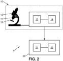
- the method may be implemented using an analysis system 1, represented schematically in figure 2 , comprising a microscope 10.
- the microscope 10 is an electronic microscope, including magnifying optics 11 and an optical detector or camera 12, and is coupled to a control unit 13 which is configured for controlling operation of the microscope, and a memory 14 for saving the acquired images.
- the microscope may exhibit a variable magnification power of at least x20, and preferably exhibits a variable magnification power over a range covering at least x40 to x80 or x120.
- the microscope further comprises a slide support 15, which is configured to receive a slide carrying a biological sample to be analyzed, the slide support 15 being movable relative to the optical axis of the microscope.
- the slide support 15 is movable in depth with respect to the optical axis in order to enable acquisition of images at different focal planes, and is also movable within a plane orthogonal to the microscope's optical axis, in order to enable acquisition of images of a portion of interest of a slide.
- the motion of the slide support 15 may be controlled by the control unit 13.
- the analysis system 1 further comprises a processing device 20, comprising at least a computer 21 and a memory 22, configured for processing the images acquires by the microscope 10.
- the memory 22 may in particular store code instructions executed by the computer 21 for performing the image processing steps of the method disclosed below.
- the memory 22 may also store trained algorithms (i.e. for instance segmentation and classification algorithms) which may be implemented during the image processing.
- the computer 21 and memory 22 may be formed respectively by the control unit 13 and memory 14 of the microscope.
- the microscope may incorporate computational hardware configured for both controlling operation of the microscope for image acquisition and further processing of the acquired images for analysis of the samples.
- the processing device 20 may be distinct from the control unit 13 and memory 14 of the microscope.
- the processing device may be physically collocated with the microscope or may be a remote device that can be accessed by a suitable communication network.
- a tissue sample may be positioned on a slide for acquisition of a plurality of images by the microscope.
- the sample has a thickness which may comprise a plurality of objects of interests, located in different focal planes.
- the method for analyzing the biological sample comprises a first step 100 of acquiring, with the microscope 10, a set of a plurality of images of different layers of the biological sample, where the images of different layers are acquired at different focal planes of the microscope and for a constant position of the biological sample in a plane orthogonal to the microscope's optical axis.
- the various images of the set represent more or less sharply various internal structures of the parasite. In particular, some structures are visible in only some of all the images of the set.
- the correspondence of the internal structures is shown in figure 4b which represents the morphological structures of a sporulated Eimeria sp. oocyst.
- the number of images of the set of acquired images may also be determined according to the type of sample, and in particular to the type of parasite, to be analyzed.
- the set of acquired images comprises at least 3 images, for instance between 3 and 15 images, preferably between 5 and 10 images.
- this interval may be comprised between 0.05pm and 1 ⁇ m, preferably between 0.1 ⁇ m and 0.5 ⁇ m.
- the interval may be constant and pre-determined, or it may be determined dynamically by the control unit 13, for instance according to a number of sharp pixels or sharp structures that appear at the considered focal plane.
- All the images of the set of images are acquired at a constant magnification power, which may be of at least x40, preferably of x80.
- step 100 comprises acquisition of a plurality of sets of images for a plurality of different relative positions of the slide (and sample) relative to the microscope, each set of images corresponding to a respective portion of the slide.
- the different portions of the slide for which sets of images are acquired may cover the whole surface occupied by the sample. This may be in particular interest for counting parasites contained in the sample, or for identifying all parasites species present within the sample.
- the step of acquiring a set of images at different layers of the biological sample may comprise whole slide scanning of the sample at each of the plurality of different layers, resulting in a set of whole slide images (WSI) acquired at the respective layers of the sample.
- the images may be acquired or converted into grayscale, and step 100 may also comprise a step of contrast enhancement.
- the method may then comprise a step 200 of generating, from the acquired set or sets of images; a single, high-definition image.
- high-definition image is meant an image having an average resolution that is at least as good as the image having the best average resolution from the set of images.
- the morphological features of the parasite are, in the high-definition image, at least as sharp as the sharpest image from the set. This step may also apply to whole scan images, as shown in figure 1b .
- the high-definition image may be an image selected among the set of acquired images according to a determined criterion.
- the criterion may be of selecting the image layer having highest resolution among all layers of the set, where resolution is defined as the minimal distance between two objects for which the two objects still appear to be distinct.
- the criterion may be of selecting the image having highest average sharpness value among the images of the set.
- the sharpness value may be computed, for an edge of the image, as the distance between two pixels belonging to a line crossing the edge, the two pixels exhibiting an intensity value of respectively 10 and 90% of the maximum intensity of the edge.
- the high-definition image is thus an image selected among the set of images acquired directly from the microscope.
- the high-definition image may be generated from the set of images.
- the high-definition image may be a so-called Enhanced Depth of Focus image, hereinafter denoted EDF, which is a single, composite image, comprising selected image portions from a plurality of images of the set.
- the image portions may be selected as exhibiting highest sharpness value among the images of the set.
- the high-definition image may be a composite image having the same dimensions as each of the acquired images, where the composite image is formed of a plurality of juxtaposed portions, the juxtaposed portions preferably having no superposed areas, where each portion is taken from one among the set of acquired images, as having the highest sharpness value.
- each pixel of the image may be assigned a sharpness value
- the composite image is formed by selecting, for each position within the image, the pixel corresponding to this position having the highest sharpness value among all the images of the set.
- One may refer for instance to the US 8,199,997 for a method of generating an EDF image from a stack of images acquired at different layers of the sample.
- the high-definition image may be an image formed out of the set of images and in which the 3D information is preserved.
- the high-definition image may be a so-called Z-stack image, which may be obtained from the acquired set of images by application of the method disclosed in US 11,263,735 .
- a Z-stack image is an specific type of EdF image in which the spatial arrangement of the objects visible in the images is preserved, in particular along the depth axis.
- the high-definition image may be a three-dimensional image formed by the stack of two-dimensional images at different Z-positions. The three-dimensional representation provides a more comprehensive view of the sample's structure, enabling to analyze its features in three dimensions.
- figure 1b shows a step 250 of dividing a whole slide image into tiles performed after a possible step 200 of generating a HD whole slide image
- the order of the steps may be the other way around and thus the whole slide images of the set may be divided into tiles and HD images may be generated from the obtained sets of tiles.
- the step of generating a single high-definition image is nevertheless optional and the further steps of the method may also apply using as input the acquired set of images corresponding to different focal planes of the microscope.
- Step 300 When step 300 is implemented on a single high-definition image generated from a set of acquired images, each extracted region is a two-dimensional region of the high-definition image.
- Step 300 may include a substep 310 of segmenting the high-definition image to generate a segmentation mask in which each parasite is segmented, and a substep 320 of extracting, from the high-definition image, a region of interest for each parasite that is centered on the parasite.
- step 300 when step 300 is implemented on a set of images acquired at different layers of the sample, step 300 may comprise:
- the three-dimensional volume corresponds to a bounding box containing the parasite considered in volume.
- the number of successive two-dimensional regions of interest that may be selected to form said three-dimensional volume may be determined according to the spacing between the different layers of the sample, so that the total thickness of the volume corresponds to the typical thickness of the type of parasite that is looked for.
- Step 300 may thus be implemented a plurality of times, and comprising a step of detecting the position of a parasite in a plurality of images of the set that correspond to layers of the parasite that are sufficiently distant from one another to relate to two different parasites.
- the method may also comprise a preliminary step 250 of cutting the whole slide images (of the single, high-definition whole slide image, as the case may be) into a plurality of tiles of equal dimensions, where the plurality of tiles forms a partition of the whole slide image, and the step 300 may then be performed on the obtained tiles.
- the method comprises generating a single whole-slide image
- the step 250 results in a plurality of tiles where each tile corresponds to part of the whole-slide image.
- 250 results in a plurality of stacks of images acquired at different layers of the sample, where each stack correspond to a tile.
- the trained segmentation algorithm may for instance be a convolutional Neural Network having a U-net architecture.
- Such algorithm performs a processing of the image(s) to extract therefrom a set of morphological features related to the shape and/or dimensions of the parasite, and determine from said set of features, whether the input image(s) contain a parasite or not and the contour of the parasite.
- the morphological features which are extracted may for instance relate to one, or more, or any combination of the following features:
- the segmentation algorithm may be trained to locate different types of parasites, which increases its overall ability to detect any of the different types of parasites for which it has been trained.
- step 320 may comprise implementing a script for extracting the region of interest and, as the case may be, the 3D volume of interest from the image(s).
- the trained segmentation algorithm may be configured to also perform substep 320 and extract the region of interest from the image(s) after having located the parasite.
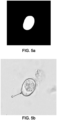
- figure 5a is shown an example of a segmentation mask representing the contour of a parasite present within an input image
- figure 5b is shown a region of interest extracted from the image and the segmentation mask.
- the extracted region of interest is centered on the parasite and is further annotated with the contour of the parasite, represented by a line C.
- the output of step 300 is thus a two-dimensional or three-dimensional region of interest extracted from respectively an image or a set of images, in which the parasite is centered.
- the 2D or 3D region may be associated with metadata comprising the coordinates, within the initial whole slide image(s) of the region, or within a tile obtained from the initial whole slide image as well as the identification of the tile.
- the method then comprises a step 400 of determining at least a species of the parasite by application, on the extracted region of interest, of a trained classification algorithm.
- step 400 may also comprise characterizing other features of the parasite, for instance:
- the trained classification algorithm may for instance be a convolutional neural network, for instance having a VGG16 or YOLO architecture.
- the step 400 may comprise running in parallel a plurality of trained classification algorithms having different structures - the computer 21 in that case may have a multicore processor where each core runs a respective algorithm - and determining the parasite species by a majority vote among all the algorithms that are run.
- the trained classification algorithm may be configured to determine the parasite species by extracting a set of morphologic features representing the microstructures of the parasite.
- This extraction may in particular comprise determining whether or not the outer wall of the parasite is formed by a double wall. Moreover, this extraction may comprise determining presence or not, or any of the following organelles (internal structures) of the parasite:
- the extraction of the set of morphologic features relative to the microstructures of the parasite may further comprise determining features relative to the organelle, such as localization, dimensions, shape, a descriptor of a wall of the organelle, etc.
- extracted features may include dimension, shape, localization within the parasite.
- oocyst residuum and sporocyst residuum polar granules, steida body, substeida body, parasteida body and refractile body, all these structures can be observed only on sporulated oocysts, and enable to distinguish among various species. Their presence can thus be used as a criterion for determining a sporulated state of an oocyst, and then features extracted from these internal structures may enable differentiate between parasite species.
- the extracted features may comprise the size, geometric position within the parasite, dimensions, density.
- the classification algorithm may further be configured to differentiate whether the sample belongs to a vaccine strain or a field strain according to the amount of glucose present in the parasites, which itself derives from the size of glucose bags present in the parasite.
- the parasite wall is determined as a double wall
- further features may be extracted such as: thickness of the internal and external membranes forming the double wall, shape and area covered by each of the internal and external membranes.
- the training may be performed by feeding, during a first training phase 510, the segmentation algorithm with a bunch of images that have been manually processed and annotated.
- the images are all issued from a mono-infested sample, i.e. a sample containing a single species of parasite. Said sample may in particular be a vaccine batch.
- images are acquired in accordance with steps 100 and optionally 200 disclosed above, where the magnification of the microscope is at least x40 and preferably x80, in order to have the internal microstructures of the parasite visible on the images.
- EDF EDF
- Z-stack image Z-stack image for instance
- an operator may manually extract regions of interest where each region is centered on a single parasite, and may annotate the region of interest with at least one of:
- the training of the segmentation algorithm may be performed by providing the algorithm with the input image (as input) and the region of interest, or coordinates of the parasite (depending on the type of output that may provide the algorithm) as output.
- the training of the classification algorithm may then be performed by providing the region of interest and the annotations.
- the classification algorithm may be trained using as input the whole slide image, the same image on which the regions of interest are represented as respective bounding boxes, as well as annotations associated to each bounding box, so that the classification algorithm is trained to annotate a whole slide image with the regions of interest extracted at step 300 and the annotations relative to the parasite.
- the same training may be continued with additional images of the same species of parasite, where the additional images are regions of interest extracted by the segmentation algorithm.
- the first and second training phases 510, 520 may then be repeated for a plurality of additional species that the classification algorithm may be configured to identify.
- this also enables increasing the capacity of the algorithm to differentiate between two species.
- the training may be reinforced with the output of the classification algorithm run on either mono-infested or multi-infested sample.
Landscapes
- Engineering & Computer Science (AREA)
- Health & Medical Sciences (AREA)
- Life Sciences & Earth Sciences (AREA)
- Biomedical Technology (AREA)
- General Health & Medical Sciences (AREA)
- Molecular Biology (AREA)
- Physics & Mathematics (AREA)
- General Physics & Mathematics (AREA)
- Multimedia (AREA)
- Theoretical Computer Science (AREA)
- Investigating Or Analysing Biological Materials (AREA)
Priority Applications (2)
| Application Number | Priority Date | Filing Date | Title |
|---|---|---|---|
| EP23201942.2A EP4535307A1 (fr) | 2023-10-05 | 2023-10-05 | Système et procédé d'analyse d'échantillon biologique en vue de la caractérisation de parasites |
| PCT/EP2024/078060 WO2025073971A1 (fr) | 2023-10-05 | 2024-10-04 | Système et procédé d'analyse d'un échantillon biologique ou environnemental en vue d'une caractérisation de parasites |
Applications Claiming Priority (1)
| Application Number | Priority Date | Filing Date | Title |
|---|---|---|---|
| EP23201942.2A EP4535307A1 (fr) | 2023-10-05 | 2023-10-05 | Système et procédé d'analyse d'échantillon biologique en vue de la caractérisation de parasites |
Publications (1)
| Publication Number | Publication Date |
|---|---|
| EP4535307A1 true EP4535307A1 (fr) | 2025-04-09 |
Family
ID=88295769
Family Applications (1)
| Application Number | Title | Priority Date | Filing Date |
|---|---|---|---|
| EP23201942.2A Pending EP4535307A1 (fr) | 2023-10-05 | 2023-10-05 | Système et procédé d'analyse d'échantillon biologique en vue de la caractérisation de parasites |
Country Status (2)
| Country | Link |
|---|---|
| EP (1) | EP4535307A1 (fr) |
| WO (1) | WO2025073971A1 (fr) |
Citations (5)
| Publication number | Priority date | Publication date | Assignee | Title |
|---|---|---|---|---|
| US8199997B2 (en) | 2007-08-17 | 2012-06-12 | Cdx Laboratories, Inc. | Feature dependent extended depth of focusing on semi-transparent biological specimens |
| US20170161545A1 (en) * | 2015-05-28 | 2017-06-08 | Tokitae Llc | Image analysis systems and related methods |
| US20190121112A1 (en) * | 2017-10-25 | 2019-04-25 | Olympus Corporation | Image processing device and microscope system |
| US11263735B2 (en) | 2018-08-01 | 2022-03-01 | Cdx Medical Ip, Inc. | Enhanced extended depth of focusing on biological samples |
| WO2022167005A1 (fr) * | 2021-02-08 | 2022-08-11 | 中国科学院自动化研究所 | Procédé basé sur un réseau neuronal profond pour détecter une morphologie de cellule vivante, et produit associé |
-
2023
- 2023-10-05 EP EP23201942.2A patent/EP4535307A1/fr active Pending
-
2024
- 2024-10-04 WO PCT/EP2024/078060 patent/WO2025073971A1/fr active Pending
Patent Citations (5)
| Publication number | Priority date | Publication date | Assignee | Title |
|---|---|---|---|---|
| US8199997B2 (en) | 2007-08-17 | 2012-06-12 | Cdx Laboratories, Inc. | Feature dependent extended depth of focusing on semi-transparent biological specimens |
| US20170161545A1 (en) * | 2015-05-28 | 2017-06-08 | Tokitae Llc | Image analysis systems and related methods |
| US20190121112A1 (en) * | 2017-10-25 | 2019-04-25 | Olympus Corporation | Image processing device and microscope system |
| US11263735B2 (en) | 2018-08-01 | 2022-03-01 | Cdx Medical Ip, Inc. | Enhanced extended depth of focusing on biological samples |
| WO2022167005A1 (fr) * | 2021-02-08 | 2022-08-11 | 中国科学院自动化研究所 | Procédé basé sur un réseau neuronal profond pour détecter une morphologie de cellule vivante, et produit associé |
Non-Patent Citations (7)
| Title |
|---|
| "An Analysis Technique for biological shape I, Inform", CONTROL, vol. 25, no. 4, 1974, pages 357 - 370 |
| ABDALLA MOHAMED A E ET AL: "Recognition of protozoan parasites from microscopic images: Eimeria species in chickens and rabbits as a case study", 2017 39TH ANNUAL INTERNATIONAL CONFERENCE OF THE IEEE ENGINEERING IN MEDICINE AND BIOLOGY SOCIETY (EMBC), IEEE, 11 July 2017 (2017-07-11), pages 1517 - 1520, XP033152335, [retrieved on 20170913], DOI: 10.1109/EMBC.2017.8037124 * |
| CASTANON ET AL: "Biological shape characterization for automatic image recognition and diagnosis of protozoan parasites of the genus Eimeria", PATTERN RECOGNITION, ELSEVIER, GB, vol. 40, no. 7, 30 March 2007 (2007-03-30), pages 1899 - 1910, XP022009128, ISSN: 0031-3203, DOI: 10.1016/J.PATCOG.2006.12.006 * |
| CESAR A. B. CASTANON ET AL., BIOLOGICAL SHAPE CHARACTERIZATION FOR AUTOMATIC IMAGE RECOGNITION AND DIAGNOSIS OF PROTOZOAN PARASITES OF THE GENUS EIMERIA |
| G�RRIZ MARC ET AL: "Leishmaniasis Parasite Segmentation and Classification Using Deep Learning", SAT 2015 18TH INTERNATIONAL CONFERENCE, AUSTIN, TX, USA, SEPTEMBER 24-27, 2015; [LECTURE NOTES IN COMPUTER SCIENCE; LECT.NOTES COMPUTER], SPRINGER, BERLIN, HEIDELBERG, vol. 10945 Chap.6, no. 558, 17 June 2018 (2018-06-17), pages 53 - 62, XP047477441, ISBN: 978-3-540-74549-5, [retrieved on 20180617] * |
| LOPEZ-OSORIO SARA ET AL: "Morphometric analysis of aerobic Eimeria bovis sporogony using live cell 3D holotomographic microscopy imaging", PARASITOLOGY RESEARCH, SPRINGER BERLIN HEIDELBERG, BERLIN/HEIDELBERG, vol. 121, no. 4, 11 October 2021 (2021-10-11), pages 1179 - 1189, XP037811188, ISSN: 0932-0113, [retrieved on 20211011], DOI: 10.1007/S00436-021-07338-X * |
| RECOGNITION, vol. 37, no. 1, 2004, pages 1 - 19 |
Also Published As
| Publication number | Publication date |
|---|---|
| WO2025073971A1 (fr) | 2025-04-10 |
Similar Documents
| Publication | Publication Date | Title |
|---|---|---|
| Castañón et al. | Biological shape characterization for automatic image recognition and diagnosis of protozoan parasites of the genus Eimeria | |
| Quinn et al. | Deep convolutional neural networks for microscopy-based point of care diagnostics | |
| US9297995B2 (en) | Automatic stereological analysis of biological tissue including section thickness determination | |
| Suzuki et al. | Automatic segmentation and classification of human intestinal parasites from microscopy images | |
| CN110543892A (zh) | 一种基于多层随机森林的零部件识别方法 | |
| Kudisthalert et al. | Counting and classification of malarial parasite from giemsa-stained thin film images | |
| Jones et al. | Malaria parasite detection on microscopic blood smear images with integrated deep learning algorithms. | |
| JP5777367B2 (ja) | パターン識別装置、パターン識別方法及びプログラム | |
| Suzuki et al. | Automated diagnosis of human intestinal parasites using optical microscopy images | |
| CN109543595B (zh) | 基于深度可分离卷积神经网络的电线的训练方法和检测方法 | |
| CN105718954B (zh) | 一种基于视触觉融合的目标属性和类别的识别方法 | |
| CN116229189B (zh) | 基于荧光内窥镜的图像处理方法、装置、设备及存储介质 | |
| Özbilge et al. | Ensembling object detection models for robust and reliable malaria parasite detection in thin blood smear microscopic images | |
| CN113111797A (zh) | 一种结合自编码器与视角变换模型的跨视角步态识别方法 | |
| Rai Dastidar | Automated focus distance estimation for digital microscopy using deep convolutional neural networks | |
| CN119942533A (zh) | 一种基于改进YOLOv8s的寄生虫卵检测方法 | |
| EP4535307A1 (fr) | Système et procédé d'analyse d'échantillon biologique en vue de la caractérisation de parasites | |
| García et al. | Pollen grains contour analysis on verification approach | |
| CN116645636A (zh) | 一种基于卷积神经网络的视觉抓取检测方法 | |
| CN118314236B (zh) | 一种mds显微数字成像处理方法及系统 | |
| Mishra et al. | Analysis of different Machine Learning and Deep Learning Techniques for Malaria Parasite Detection | |
| Moghadam et al. | Tracking leukocytes in intravital time lapse images using 3D cell association learning network | |
| Nugroho et al. | Identification of plasmodium falciparum and plasmodium vivax on digital image of thin blood films gf | |
| Alvarez-Ramos et al. | Automatic classification of Nosema pathogenic agents through machine vision techniques and kernel-based vector machines | |
| Boufenar et al. | Identification of Chicken Eimeria Species using Deep Learning Approaches |
Legal Events
| Date | Code | Title | Description |
|---|---|---|---|
| PUAI | Public reference made under article 153(3) epc to a published international application that has entered the european phase |
Free format text: ORIGINAL CODE: 0009012 |
|
| STAA | Information on the status of an ep patent application or granted ep patent |
Free format text: STATUS: THE APPLICATION HAS BEEN PUBLISHED |
|
| AK | Designated contracting states |
Kind code of ref document: A1 Designated state(s): AL AT BE BG CH CY CZ DE DK EE ES FI FR GB GR HR HU IE IS IT LI LT LU LV MC ME MK MT NL NO PL PT RO RS SE SI SK SM TR |
|
| STAA | Information on the status of an ep patent application or granted ep patent |
Free format text: STATUS: REQUEST FOR EXAMINATION WAS MADE |
|
| 17P | Request for examination filed |
Effective date: 20251006 |
|
| STAA | Information on the status of an ep patent application or granted ep patent |
Free format text: STATUS: EXAMINATION IS IN PROGRESS |
|
| 17Q | First examination report despatched |
Effective date: 20251212 |