EP4542562A2 - Cadre anatomique de positionnement - Google Patents
Cadre anatomique de positionnement Download PDFInfo
- Publication number
- EP4542562A2 EP4542562A2 EP24202208.5A EP24202208A EP4542562A2 EP 4542562 A2 EP4542562 A2 EP 4542562A2 EP 24202208 A EP24202208 A EP 24202208A EP 4542562 A2 EP4542562 A2 EP 4542562A2
- Authority
- EP
- European Patent Office
- Prior art keywords
- interest
- point
- normalized coordinates
- prompt
- points
- Prior art date
- Legal status (The legal status is an assumption and is not a legal conclusion. Google has not performed a legal analysis and makes no representation as to the accuracy of the status listed.)
- Pending
Links
Images
Classifications
-
- G—PHYSICS
- G16—INFORMATION AND COMMUNICATION TECHNOLOGY [ICT] SPECIALLY ADAPTED FOR SPECIFIC APPLICATION FIELDS
- G16H—HEALTHCARE INFORMATICS, i.e. INFORMATION AND COMMUNICATION TECHNOLOGY [ICT] SPECIALLY ADAPTED FOR THE HANDLING OR PROCESSING OF MEDICAL OR HEALTHCARE DATA
- G16H20/00—ICT specially adapted for therapies or health-improving plans, e.g. for handling prescriptions, for steering therapy or for monitoring patient compliance
- G16H20/30—ICT specially adapted for therapies or health-improving plans, e.g. for handling prescriptions, for steering therapy or for monitoring patient compliance relating to physical therapies or activities, e.g. physiotherapy, acupressure or exercising
-
- G—PHYSICS
- G16—INFORMATION AND COMMUNICATION TECHNOLOGY [ICT] SPECIALLY ADAPTED FOR SPECIFIC APPLICATION FIELDS
- G16H—HEALTHCARE INFORMATICS, i.e. INFORMATION AND COMMUNICATION TECHNOLOGY [ICT] SPECIALLY ADAPTED FOR THE HANDLING OR PROCESSING OF MEDICAL OR HEALTHCARE DATA
- G16H10/00—ICT specially adapted for the handling or processing of patient-related medical or healthcare data
- G16H10/60—ICT specially adapted for the handling or processing of patient-related medical or healthcare data for patient-specific data, e.g. for electronic patient records
-
- G—PHYSICS
- G16—INFORMATION AND COMMUNICATION TECHNOLOGY [ICT] SPECIALLY ADAPTED FOR SPECIFIC APPLICATION FIELDS
- G16H—HEALTHCARE INFORMATICS, i.e. INFORMATION AND COMMUNICATION TECHNOLOGY [ICT] SPECIALLY ADAPTED FOR THE HANDLING OR PROCESSING OF MEDICAL OR HEALTHCARE DATA
- G16H15/00—ICT specially adapted for medical reports, e.g. generation or transmission thereof
-
- G—PHYSICS
- G16—INFORMATION AND COMMUNICATION TECHNOLOGY [ICT] SPECIALLY ADAPTED FOR SPECIFIC APPLICATION FIELDS
- G16H—HEALTHCARE INFORMATICS, i.e. INFORMATION AND COMMUNICATION TECHNOLOGY [ICT] SPECIALLY ADAPTED FOR THE HANDLING OR PROCESSING OF MEDICAL OR HEALTHCARE DATA
- G16H30/00—ICT specially adapted for the handling or processing of medical images
- G16H30/40—ICT specially adapted for the handling or processing of medical images for processing medical images, e.g. editing
-
- G—PHYSICS
- G16—INFORMATION AND COMMUNICATION TECHNOLOGY [ICT] SPECIALLY ADAPTED FOR SPECIFIC APPLICATION FIELDS
- G16H—HEALTHCARE INFORMATICS, i.e. INFORMATION AND COMMUNICATION TECHNOLOGY [ICT] SPECIALLY ADAPTED FOR THE HANDLING OR PROCESSING OF MEDICAL OR HEALTHCARE DATA
- G16H40/00—ICT specially adapted for the management or administration of healthcare resources or facilities; ICT specially adapted for the management or operation of medical equipment or devices
- G16H40/60—ICT specially adapted for the management or administration of healthcare resources or facilities; ICT specially adapted for the management or operation of medical equipment or devices for the operation of medical equipment or devices
- G16H40/67—ICT specially adapted for the management or administration of healthcare resources or facilities; ICT specially adapted for the management or operation of medical equipment or devices for the operation of medical equipment or devices for remote operation
-
- G—PHYSICS
- G16—INFORMATION AND COMMUNICATION TECHNOLOGY [ICT] SPECIALLY ADAPTED FOR SPECIFIC APPLICATION FIELDS
- G16H—HEALTHCARE INFORMATICS, i.e. INFORMATION AND COMMUNICATION TECHNOLOGY [ICT] SPECIALLY ADAPTED FOR THE HANDLING OR PROCESSING OF MEDICAL OR HEALTHCARE DATA
- G16H50/00—ICT specially adapted for medical diagnosis, medical simulation or medical data mining; ICT specially adapted for detecting, monitoring or modelling epidemics or pandemics
- G16H50/20—ICT specially adapted for medical diagnosis, medical simulation or medical data mining; ICT specially adapted for detecting, monitoring or modelling epidemics or pandemics for computer-aided diagnosis, e.g. based on medical expert systems
-
- G—PHYSICS
- G16—INFORMATION AND COMMUNICATION TECHNOLOGY [ICT] SPECIALLY ADAPTED FOR SPECIFIC APPLICATION FIELDS
- G16H—HEALTHCARE INFORMATICS, i.e. INFORMATION AND COMMUNICATION TECHNOLOGY [ICT] SPECIALLY ADAPTED FOR THE HANDLING OR PROCESSING OF MEDICAL OR HEALTHCARE DATA
- G16H50/00—ICT specially adapted for medical diagnosis, medical simulation or medical data mining; ICT specially adapted for detecting, monitoring or modelling epidemics or pandemics
- G16H50/70—ICT specially adapted for medical diagnosis, medical simulation or medical data mining; ICT specially adapted for detecting, monitoring or modelling epidemics or pandemics for mining of medical data, e.g. analysing previous cases of other patients
Definitions
- the present disclosure generally relates to data processing, and more particularly to a framework for anatomical positioning.
- a written radiology report is the primary work product of a diagnostic radiologist and acts as the main method of communicating results of a radiologic study or procedure, including findings in medical images.
- Radiology reports are intended to be reused after image reading is complete. When the reports are re-used, clinicians often have to manually find the corresponding entries in the images. These entries may include the findings in organs and in sub-tissue levels, and are usually described with respect to their locations relative to some known anatomical landmarks.
- This process may be automated by automatically mapping the text entries in the radiology reports to the anatomical landmarks in the images. Navigating to the specific anatomical landmarks is not a trivial problem because (1) the anatomical landmarks may appear in different forms and orders; and (2) storing the information in unstructured text limits the ability to process data on a large scale. For example, identifying patients with liver disease may require taking all type of mentions (e.g., "liver", "hepatic") into consideration.
- Parsing methods may be considered as baselines for associating text input to anatomical coordinates, since the image identifiers (e.g., image and series numbers) mentioned in the text follows a well-structured pattern. However, each keyword must be manually engineered and extracted, thereby restricting its application. Another approach is to use the locations of the anatomical landmarks in the images identified by segmentation or landmarking methods. However, the scope is limited to only landmarks and organs with defined structure.
- Described herein is a framework for anatomical positioning.
- input text is mapped into normalized coordinates using an artificial neural network.
- a location in a target image that corresponds to the normalized coordinates is determined and presented.
- a user selection of a point-of-interest in a medical image is received.
- a context set of points nearest to the point-of-interest is determined.
- a prompt containing the point-of-interest and context set of points is constructed.
- a large language model may then generate text data associated with the point-of-interest in response to the prompt.
- sequences of instructions designed to implement the methods can be compiled for execution on a variety of hardware platforms and for interface to a variety of operating systems.
- implementations of the present framework are not described with reference to any particular programming language. It will be appreciated that a variety of programming languages may be used.
- an image, or a portion thereof corresponding to an object (e.g., a tissue, an organ, a tumor, etc., of a subject (e.g., a patient, etc.)
- an image, or a portion of thereof e.g., an ROI
- an ROI corresponding to the image of a lung or a heart may be described as that the ROI includes a lung or a heart.
- an image of or including a chest may be referred to a chest image, or simply a chest.
- the present framework uses semantic coordinates for anatomical relationships.
- the present framework maps information between anatomical and textual domains.
- the anatomical information may include the position of anatomical landmarks in a normalized coordinate system.
- the textual information may include text entries in the radiology reports corresponding to the anatomical landmarks.
- the present framework may be used in either or both forward and backward directions.
- the framework may estimate the position of a specific anatomical landmark in terms of normalized coordinates using text input entries. Instead of specific organ labels in the output, a continuous normalized coordinate system that maps to a standard human body is used. This kind of mapping may describe the anatomy in various granularities.
- the semantic meaning of the presented normalized coordinates may be identified using a large-language-model (LLM) with knowledge of human anatomy.
- Anatomical landmarks may be identified using only positional information from the image data.
- Keyword level detectors of anatomies may work only a certain degree.
- Machine learning models may provide better generalization.
- the present framework utilizes the existing landmarks and LLMs to generate more precise estimations.
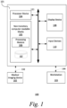
- computer system 101 includes a processor device or central processing unit (CPU) 104 coupled to one or more non-transitory computer-readable media 105 (e.g., computer storage or memory device), display device 108 (e.g., monitor) and various input devices 110 (e.g., mouse, touchpad or keyboard) via an input-output interface 121.
- Computer system 101 may further include support circuits such as a cache, a power supply, clock circuits and a communications bus.
- Various other peripheral devices such as additional data storage devices and printing devices, may also be connected to the computer system 101.
- the present technology may be implemented in various forms of hardware, software, firmware, special purpose processors, or a combination thereof, either as part of the microinstruction code or as part of an application program or software product, or a combination thereof, which is executed via the operating system.
- the techniques described herein are implemented as computer-readable program code tangibly embodied in one or more non-transitory computer-readable media 105.
- the present techniques may be implemented by a processing module 106 and database 107.
- Non-transitory computer-readable media 105 may include random access memory (RAM), read-only memory (ROM), magnetic floppy disk, flash memory, and other types of memories, or a combination thereof.
- the computer-readable program code is executed by processor device 104 to process data provided by, for example, medical imaging device 102.
- the computer system 101 is a general-purpose computer system that becomes a specific-purpose computer system when executing the computer-readable program code.
- the computer-readable program code is not intended to be limited to any particular programming language and implementation thereof. It will be appreciated that a variety of programming languages and coding thereof may be used to implement the teachings of the disclosure contained herein.
- the same or different computer-readable media 105 may be used for storing a database, including, but not limited to, image datasets, a knowledge base, individual subject data, electronic medical records (EMRs), diagnostic reports (or documents) for subjects, or a combination thereof.
- EMRs electronic medical records
- Medical imaging device 102 acquires medical image data 132. Such medical image data 132 may be processed by processing module 106. Medical imaging device 102 may be a radiology scanner and/or appropriate peripherals (e.g., keyboard and display device) for acquiring, collecting and/or storing such medical image data 132. Medical imaging device 102 may acquire medical image data 132 from a subject or patient by using techniques such as high-resolution computed tomography (HRCT), magnetic resonance (MR) imaging, computed tomography (CT), helical CT, X-ray, angiography, positron emission tomography (PET), fluoroscopy, ultrasound, single photon emission computed tomography (SPECT), or a combination thereof. Other types of imaging modalities are also useful. Medical imaging device 102 may be controlled using a medical imaging software.
- HRCT high-resolution computed tomography
- MR magnetic resonance
- CT computed tomography
- CT computed tomography
- PET positron emission tomography
- fluoroscopy
- Medical image data 132 represents a medical image (or in certain examples more than one medical image).
- medical image data 132 may include an array or list of pixel or voxel values. When processed by suitable image viewing software, the medical image data 132 results in a rendering of the medical image (or medical images) that it represents.
- the image file containing the medical image data 132 may further include one or more attributes and attribute values.
- a specific example of an image file is a Digital Imaging and Communications in Medicine (DICOM) file.
- the one or more attribute values are separate to and distinct from the medical image data 132, and instead comprise a text string indicating content of the medical image data 132.
- Such attribute values may, in some examples, be referred to as metadata of the image file.
- the part of the image file that stores the attributes and attribute values may be referred to as a header of the image file, and the attributes and attribute values may be referred to as header data of the image file.
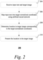
- FIG. 2 is a flow chart of an exemplary method 200 of estimating at image location from text input. It should be understood that the steps of the method 200 may be performed in the order shown or a different order. Additional, different, or fewer steps may also be provided. Further, the method 200 may be implemented with the system 100 of FIG. 1 , a different system, or a combination thereof.
- processing module 106 maps the input text into target normalized coordinates using an artificial neural network.
- the input text may be extracted from a radiology report manually or automatically by the processing module 106.
- the target normalized coordinates may be defined with reference to a selected anatomical atlas of a standard body or part thereof.
- Keywords in the training set are one or more words that describe anatomical features (e.g., landmarks or organs) in an anatomical atlas (e.g., human body atlas). Examples of such keywords include, but are not limited to, upper lobe, pulmonary artery, pulmonary vein, middle lobe, aorta, aortic arch, left ventricle, right atrium, kidney center, and so forth.
- the keywords may describe the respective anatomical feature at different levels of granularities (e.g., body, chest, heart, tricuspid valve). Each keyword maps to corresponding normalized coordinates of a landmark or bounding box of an organ in an anatomical atlas.
- normalization is performed by subtraction of pre-defined landmark coordinates.
- a known landmark is pre-defined as the origin of the coordinate system.
- Such pre-defined landmark becomes a carina with coordinates (0, 0, 0).
- Any other coordinates in the initial frame of the atlas (or target image) may be computed by subtracting the offset of the pre-defined landmark.
- distances are specified in standard units of measurements (e.g., millimeters), which may be easier to use in the real world.
- processing module 106 determines the location in the target image that corresponds to the target normalized coordinates estimated by the trained artificial neural network.
- the corresponding location is determined by performing a search to find the location within the target image with normalized coordinates that match the target normalized coordinates.
- the framework may start from a first image location within the target image, normalize the coordinates of that first image location, and estimate where to find the target normalized coordinates.
- a trained regression neural network may be used to normalize the coordinates.
- the regression neural network may include, for example, an 8-layer residual network. Other types of artificial neural networks are also useful.
- the input of the neural network may be the first image location and associated image intensity data, and the output of the neural network is the normalized coordinates of the first image location.
- landmark detectors are used to determine the location in the target image corresponding to the target normalized coordinates. Landmarks may be assigned to the normalized coordinate space and the corresponding location may be computed from nearby landmarks using, for example, linear interpolation methods.
- processing module 106 presents the location in the target image.
- the determined location may be highlighted in the target image and displayed at, for example, workstation 103.
- the user when the user selects the hyperlink at the input text, the user is directed to that corresponding location in the target image.
- the user may select the hyperlink using, for example, a mouse, cursor or other user interface component.
- the corresponding location in the target image may be highlighted and displayed, for instance, in a window next to the radiology report whenever the hyperlink at the input text is selected.
- Other ways of presenting the location are also possible.
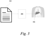
- FIG. 3 is schematic diagram of the exemplary method 200 of estimating an image location from text input.
- the radiology report 302 of a patient is processed by the processing module 106 to extract input text.
- the input text is fed to the trained artificial neural network 304, which maps the input text to normalized coordinates.
- the corresponding location 306 in a target image of the patient is determined based on the normalized coordinates.
- the corresponding location 306 may be highlighted on the target image when, for example, the user selects a hyperlink inserted at the input text on the radiology report 302.
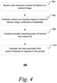
- FIG. 4 is a flow chart of an exemplary method 400 of determining text data associated with a point-of-interest in a medical image. It should be understood that the steps of the method 400 may be performed in the order shown or a different order. Additional, different, or fewer steps may also be provided. Further, the method 400 may be implemented with the system 100 of FIG. 1 , a different system, or a combination thereof.
- processing module 106 receives a user selection of a point-of-interest in a medical image and determines normalized coordinates of the point-of-interest.
- the medical image may be acquired from a current patient by, for example, medical imaging device 102.
- the medical image may be a CT, MR, PET, SPECT or any other type of image.
- the user may select the point-of-interest via, for example, a user interface (e.g., graphical user interface) displayed at workstation 103.
- processing module 106 determines the normalized coordinates of the point-of-interest.
- the normalized coordinates may be determined with the use of the medical image pixel (or voxel) value associated with the point-of-interest.
- a trained regression neural network may be used to estimate the normalized coordinates of the point-of-interest.
- the regression neural network may include, for example, an 8-layer residual network. Other types of artificial neural networks are also useful.
- the input of the trained regression neural network may be the voxel (or pixel) intensity value associated with the point-of-interest, and the output of the neural network is the corresponding normalized coordinates.
- processing module 106 generates a context set of points nearest to the point-of-interest.
- the context set of points may be identified from, for example, a database 107 of landmarks.
- the landmarks in the database 107 may be identified in an anatomical atlas as possible points-of-interest and defined in a normalized coordinate system.
- the normalized coordinate system is a continuous coordinate system that is scaled to a predefined bounding box.
- the context set of points may include a plurality of landmarks and their corresponding normalized coordinates nearest to the point-of-interest.
- These nearest landmarks may be determined by looking up the database of landmarks using the normalized coordinates of the point-of-interest and computing the distances between the landmarks in the database 107 and the point-of-interest to find the nearest landmarks with the shortest distances (e.g., less than a pre-determined threshold distance).
- processing module 106 constructs a prompt containing the point-of-interest and the context set of points.
- the prompt may include the normalized coordinates and names of the context set of points and the point-of-interest.
- the prompt may then be sent to a trained large language model (LLM) to generate text data associated with the point-of-interest.
- LLM is an artificial intelligence model that can be used for understanding and generating human-like responses after training with massive amounts of data.
- the LLM may include one or more artificial neural networks (e.g., transformers) that are trained using self-supervised and/or semi-supervised learning.
- the LLM uses deep learning techniques and has encoded anatomical knowledge to ensure better comprehension of relationships and relative locations between anatomical features.
- the LLM may be trained with medical health record data where anatomical information is frequently present. Such medical health record data may be obtained from an electronic health record dataset of a large number of patients (e.g., over 1 million) and include real medical images and associated radiology reports.
- the prompt requests the LLM to use the context set of points to generate text data in association with an anatomical feature at the point-of-interest.
- the prompt may request for a description (e.g., name, label) of the anatomical feature at the point-of-interest.
- An exemplary prompt is as follows: "what anatomical landmark/organ is likely to be in position [10.30, - 83.40, 9.179259], if the spatial position of the suprasternal notch is in [7.6, -71.22, 49.87], the spatial position of the common carotid artery origin (r) is in [-1.51, -43.21, 36.80], the spatial position of the aorta asc.
- a trained LLM is used to generate text data associated with the point-of-interest in response to the prompt.
- Processing module 106 may receive the generated text data from the trained LLM and display the text data at, for example, workstation 103.
- the generated text data may describe or provide semantic meaning to the point-of-interest in response to the prompt.
- the knowledge encoded in the LLM model is richer than the limited landmarks present in the database 107 of landmarks. This information may be useful for both report generation and information retrieval of findings for any selected point-of-interest. For example, generated text data may be compared to existing medical history of the patient to find mentions in corresponding locations.
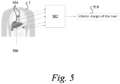
- FIG. 5 is schematic diagram of the exemplary method 400 of determining text data associated with a point-of-interest in a medical image.
- the method employs an LLM 502 with the knowledge of human anatomy to identify anatomical landmarks using only positional information from medical image I .
- the normalization of the coordinates of the point-of-interest 504 creates the relationship between the current medical image and the atlas image with the associated landmark database. Therefore, nearby landmarks may be identified directly with distances in normalized coordinate space. Given these landmarks' positions in a normalized coordinate system, the LLM 502 may identify any randomly selected point 504 in the medical image I .
- a user may submit a request 506 that indicates a selection of a point-of-interest r j (504) on image I .
- Processing module 106 constructs a prompt containing the context set c and point-of-interest r i (504).
- the prompt is sent to LLM 502.
Landscapes
- Health & Medical Sciences (AREA)
- Engineering & Computer Science (AREA)
- Medical Informatics (AREA)
- Public Health (AREA)
- Primary Health Care (AREA)
- Epidemiology (AREA)
- General Health & Medical Sciences (AREA)
- Biomedical Technology (AREA)
- Data Mining & Analysis (AREA)
- Databases & Information Systems (AREA)
- Pathology (AREA)
- Nuclear Medicine, Radiotherapy & Molecular Imaging (AREA)
- General Business, Economics & Management (AREA)
- Business, Economics & Management (AREA)
- Radiology & Medical Imaging (AREA)
- Life Sciences & Earth Sciences (AREA)
- Biophysics (AREA)
- Physical Education & Sports Medicine (AREA)
- Image Analysis (AREA)
- Image Processing (AREA)
- Apparatus For Radiation Diagnosis (AREA)
Applications Claiming Priority (2)
| Application Number | Priority Date | Filing Date | Title |
|---|---|---|---|
| US202363585743P | 2023-09-27 | 2023-09-27 | |
| US18/442,667 US20250104844A1 (en) | 2023-09-27 | 2024-02-15 | Anatomical positioning framework |
Publications (2)
| Publication Number | Publication Date |
|---|---|
| EP4542562A2 true EP4542562A2 (fr) | 2025-04-23 |
| EP4542562A3 EP4542562A3 (fr) | 2025-06-25 |
Family
ID=92899773
Family Applications (1)
| Application Number | Title | Priority Date | Filing Date |
|---|---|---|---|
| EP24202208.5A Pending EP4542562A3 (fr) | 2023-09-27 | 2024-09-24 | Cadre anatomique de positionnement |
Country Status (1)
| Country | Link |
|---|---|
| EP (1) | EP4542562A3 (fr) |
-
2024
- 2024-09-24 EP EP24202208.5A patent/EP4542562A3/fr active Pending
Also Published As
| Publication number | Publication date |
|---|---|
| EP4542562A3 (fr) | 2025-06-25 |
Similar Documents
| Publication | Publication Date | Title |
|---|---|---|
| JP7467348B2 (ja) | 医用画像データの表示 | |
| Linguraru et al. | Automated segmentation and quantification of liver and spleen from CT images using normalized probabilistic atlases and enhancement estimation | |
| US8311303B2 (en) | Method and system for semantics driven image registration | |
| Tan et al. | Automated vessel segmentation in lung CT and CTA images via deep neural networks | |
| US20180196873A1 (en) | Visualization framework based on document representation learning | |
| US10878564B2 (en) | Systems and methods for processing 3D anatomical volumes based on localization of 2D slices thereof | |
| JP7618003B2 (ja) | 文書作成支援装置、方法およびプログラム | |
| JP7258772B2 (ja) | 全人的患者放射線医療ビューワ | |
| CN119487545A (zh) | 分析医学图像 | |
| US10853409B2 (en) | Systems and methods for image search | |
| Misera et al. | Weakly supervised deep learning in radiology | |
| US20170083665A1 (en) | Method and System for Radiology Structured Report Creation Based on Patient-Specific Image-Derived Information | |
| US20170221204A1 (en) | Overlay Of Findings On Image Data | |
| US12387826B2 (en) | Document creation apparatus, document creation method, and document creation program for creating document based on medical image | |
| JP2024054748A (ja) | 言語特徴抽出モデルの生成方法、情報処理装置、情報処理方法及びプログラム | |
| US20240363230A1 (en) | Method for automated processing of volumetric medical images | |
| EP4542562A2 (fr) | Cadre anatomique de positionnement | |
| CN119919434A (zh) | 基于深度学习的肝胆部位分割方法、装置、设备及介质 | |
| US20250104844A1 (en) | Anatomical positioning framework | |
| US20240153072A1 (en) | Medical information processing system and method | |
| CN117457140A (zh) | 基于深度学习的宫颈癌诊断报告生成方法、装置和设备 | |
| Liu et al. | Automated segmentation of fetal ultrasound images using feature attention supervised network | |
| WO2021107142A1 (fr) | Dispositif, procédé et programme d'aide à la création de documents | |
| EP4510142A1 (fr) | Détection assistée par ordinateur guidée par un dossier médical | |
| Prakash et al. | Deep learning-based head and neck segmentation in CT angiography |
Legal Events
| Date | Code | Title | Description |
|---|---|---|---|
| PUAI | Public reference made under article 153(3) epc to a published international application that has entered the european phase |
Free format text: ORIGINAL CODE: 0009012 |
|
| STAA | Information on the status of an ep patent application or granted ep patent |
Free format text: STATUS: REQUEST FOR EXAMINATION WAS MADE |
|
| 17P | Request for examination filed |
Effective date: 20240924 |
|
| AK | Designated contracting states |
Kind code of ref document: A2 Designated state(s): AL AT BE BG CH CY CZ DE DK EE ES FI FR GB GR HR HU IE IS IT LI LT LU LV MC ME MK MT NL NO PL PT RO RS SE SI SK SM TR |
|
| PUAL | Search report despatched |
Free format text: ORIGINAL CODE: 0009013 |
|
| AK | Designated contracting states |
Kind code of ref document: A3 Designated state(s): AL AT BE BG CH CY CZ DE DK EE ES FI FR GB GR HR HU IE IS IT LI LT LU LV MC ME MK MT NL NO PL PT RO RS SE SI SK SM TR |
|
| RIC1 | Information provided on ipc code assigned before grant |
Ipc: G16H 50/70 20180101ALI20250521BHEP Ipc: G16H 30/40 20180101ALI20250521BHEP Ipc: G16H 20/30 20180101AFI20250521BHEP |