EP4545554A1 - Rekombinantes polypeptid mit einem metallbindenden motiv - Google Patents
Rekombinantes polypeptid mit einem metallbindenden motiv Download PDFInfo
- Publication number
- EP4545554A1 EP4545554A1 EP23205682.0A EP23205682A EP4545554A1 EP 4545554 A1 EP4545554 A1 EP 4545554A1 EP 23205682 A EP23205682 A EP 23205682A EP 4545554 A1 EP4545554 A1 EP 4545554A1
- Authority
- EP
- European Patent Office
- Prior art keywords
- binding
- recombinant polypeptide
- metal
- igg
- target
- Prior art date
- Legal status (The legal status is an assumption and is not a legal conclusion. Google has not performed a legal analysis and makes no representation as to the accuracy of the status listed.)
- Withdrawn
Links
Images

Classifications
-
- C—CHEMISTRY; METALLURGY
- C07—ORGANIC CHEMISTRY
- C07K—PEPTIDES
- C07K14/00—Peptides having more than 20 amino acids; Gastrins; Somatostatins; Melanotropins; Derivatives thereof
- C07K14/195—Peptides having more than 20 amino acids; Gastrins; Somatostatins; Melanotropins; Derivatives thereof from bacteria
- C07K14/315—Peptides having more than 20 amino acids; Gastrins; Somatostatins; Melanotropins; Derivatives thereof from bacteria from Streptococcus (G), e.g. Enterococci
-
- B—PERFORMING OPERATIONS; TRANSPORTING
- B01—PHYSICAL OR CHEMICAL PROCESSES OR APPARATUS IN GENERAL
- B01D—SEPARATION
- B01D15/00—Separating processes involving the treatment of liquids with solid sorbents; Apparatus therefor
- B01D15/08—Selective adsorption, e.g. chromatography
- B01D15/26—Selective adsorption, e.g. chromatography characterised by the separation mechanism
- B01D15/38—Selective adsorption, e.g. chromatography characterised by the separation mechanism involving specific interaction not covered by one or more of groups B01D15/265 and B01D15/30 - B01D15/36, e.g. affinity, ligand exchange or chiral chromatography
- B01D15/3804—Affinity chromatography
- B01D15/3809—Affinity chromatography of the antigen-antibody type, e.g. protein A, G or L chromatography
-
- C—CHEMISTRY; METALLURGY
- C07—ORGANIC CHEMISTRY
- C07K—PEPTIDES
- C07K1/00—General methods for the preparation of peptides, i.e. processes for the organic chemical preparation of peptides or proteins of any length
- C07K1/14—Extraction; Separation; Purification
- C07K1/16—Extraction; Separation; Purification by chromatography
- C07K1/22—Affinity chromatography or related techniques based upon selective absorption processes
-
- C—CHEMISTRY; METALLURGY
- C12—BIOCHEMISTRY; BEER; SPIRITS; WINE; VINEGAR; MICROBIOLOGY; ENZYMOLOGY; MUTATION OR GENETIC ENGINEERING
- C12N—MICROORGANISMS OR ENZYMES; COMPOSITIONS THEREOF; PROPAGATING, PRESERVING, OR MAINTAINING MICROORGANISMS; MUTATION OR GENETIC ENGINEERING; CULTURE MEDIA
- C12N15/00—Mutation or genetic engineering; DNA or RNA concerning genetic engineering, vectors, e.g. plasmids, or their isolation, preparation or purification; Use of hosts therefor
- C12N15/09—Recombinant DNA-technology
- C12N15/11—DNA or RNA fragments; Modified forms thereof; Non-coding nucleic acids having a biological activity
- C12N15/62—DNA sequences coding for fusion proteins
Definitions
- the present disclosure generally relates to a recombinant polypeptide comprising an IgG-binding domain derived from Streptococcus Protein G (SpG) and a metal-binding motif.
- the present disclosure further relates to a ligand comprising the recombinant polypeptide, a chromatography matrix comprising the ligand coupled to the matrix, and a method for purification of sample proteins utilizing the chromatography matrix.
- the present disclosure also relates to a nucleic acid encoding the recombinant polypeptide and to an expression system comprising the nucleic acid.
- Antibodies also known as immunoglobulins (Ig), are commonly used in the field of biological sciences, and represent an important class of biotherapeutic drugs. Antibodies may be used to treat various diseases, including allergy, autoimmune diseases, and cancer.
- An antibody is a relatively large, Y-shaped protein, and contains two antigen-binding fragments (Fab), and one crystallizable fragment (Fc) forming the trunk of the Y-shape.
- Fab antigen-binding fragments
- Fc crystallizable fragment
- Immunoglobulin G is the main type of antibody found in blood and extracellular fluid and represents the majority of the antibody-based immunity against invading pathogens.
- Ig-binding proteins are often used in affinity chromatography applications for recovery of immunoglobulins (Ig) from different samples.
- SpA Staphylococcus aureus protein A
- EP 3 523 318 B1 relates to an IgG-binding polypeptide mutant of domain Z derived from SpA domain B, which binds to the Fc-region of an immunoglobulin, and which comprises a metal-binding motif inserted between helix 2 and 3 of the Z domain.
- the IgG-binding polypeptide mutant allows for elution of IgG by removal of calcium at a pH of 5.5 to 7.0.
- Streptococcal protein G is another protein with affinity to immunoglobulins. SPG generally binds to the Fc fragment of IgGs from various mammalian species.
- SPG consists of repetitively arranged domains, wherein the C-terminal domains (C1, C2 and C3) are responsible for IgG-binding, and the N-terminal domains bind to human serum albumin.
- SPG streptococcal protein G
- IgG-binding polypeptides derived from streptococcal protein G will become important for recovery and isolation of immunoglobulins or fragments thereof.
- SPG streptococcal protein G
- a recombinant polypeptide comprising an IgG-binding domain derived from Streptococcus Protein G (SPG) and a metal-binding motif, wherein the recombinant polypeptide has a first, target-binding configuration in the presence of metal ions, and a second, non-target-binding configuration in the absence of metal ions, wherein the recombinant polypeptide is configured to bind to IgG in the target-binding configuration.
- SPG Streptococcus Protein G
- the inventors have found that a metal-binding motif may be successfully introduced in an IgG-binding domain derived from SPG and have thereby provided a recombinant metal-dependent protein G-binding domain.
- the recombinant polypeptide Upon binding of a metal ion, the recombinant polypeptide can switch from an inactive state; i.e. non-target binding configuration, to a functional, i.e. target-binding configuration.
- the inventors have made an engineering effort and combined a semi-rational protein design and selection by Escherichia coli display followed by fluorescence-activated cell sorting and isolated several polypeptides where the inherent target affinity to antibodies and antibody fragments have been rendered metal ion dependent. Once depleted of metal ions, the target interaction of these proteins is lost.
- recombinant polypeptide means a polypeptide formed by using recombinant DNA techniques.
- the recombinant polypeptide is derived from an IgG-binding domain of streptococcal protein G (SPG), into which a metal-binding motif is introduced.
- SPG streptococcal protein G
- the recombinant polypeptide may also be referred to as an engineered polypeptide.
- IgG-binding domain means the C1, C2 or C3 domain of Streptococcus protein G (SPG).
- the IgG-binding domain i.e. the C1, C2 or C3 domain may be intact, but the introduction of the metal-binding motif into the domain provides a recombinant polypeptide. It is also conceivable that the IgG-binding domain comprises one or a plurality of mutations.
- the "metal-binding motif' is typically a metal-binding loop.
- a loop is a peptide fragment generally void of secondary structure and which may impact protein folding.
- the metal-binding loop binds metal ions, and in doing so, the loop (and the recombinant polypeptide) switches configuration from an inactive, non-target binding configuration to a configuration in which the polypeptide binds to an immunoglobulin (IgG).
- IgG immunoglobulin
- the recombinant polypeptide may have a different conformation in the first, target-binding configuration, and the second, non-target-binding configuration.
- the folding of the metal-binding motif; i.e. metal-binding loop may be different.
- the IgG-binding domains of streptococcal protein G include domain C1, C2, and C3. Accordingly, the recombinant polypeptide of the present disclosure may comprise domain C1, C2 or C3.
- the C domains of streptococcal protein G i.e. domain C1, C2, and C3 are small (55 residue) peptides with immunoglobulin-binding activity.
- Domain C1, C2 and C3 of streptococcal protein G each consists of four antiparallel beta-sheets crossed over by an alpha helix.
- the C domains have a similar structure but differ slightly in the amino acid sequences.
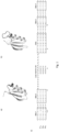
- Figure 1a illustrates the crystal structure of the C2 domain, wherein the insertion site for the metal-binding loop is marked in darker grey.
- figure 1b an exemplary metal-binding loop (C2 CaV3 ) has been inserted.
- the metal-binding motif is typically inserted N-terminally (upstream) of the alpha helix of the IgG-binding domain, i.e. domain C1, C2 or C3.
- the metal-binding motif is inserted between the second beta strand and the alpha helix of the IgG-binding domain, i.e. domain C1, C2 or C3 (see figure 1c ).
- Figure 1c illustrates the sequences of domain C1, C2, and C3, respectively (wild type), and the metal-binding loop insertion site.
- Domain C1 differs from domain C2 in two positions located in the first beta sheet (see arrows in figure 1c ).
- Domain C3 differs from domain C2 in three positions throughout the scaffold, one in the second beta sheet, one in the alpha helix, and one in the third beta sheet (see arrows in figure 1c ).
- the IgG-binding domain is domain C1 or domain C2.
- the recombinant polypeptide is configured to bind to the Fab region of the immunoglobulin (IgG) in the first, target-binding configuration.
- the binding between the recombinant polypeptide and the IgG is highly selective and specific.
- the binding surface for Fab is mainly located to the second beta-sheet and the C-terminal end of the alpha helix.
- the IgG-binding domains of protein G predominantly interact with the Fc region of IgG.
- the inventors have surprisingly found that the recombinant polypeptides of the present disclosure can selectively bind to the Fab-region of IgG with high specificity.
- Fab fragments may be used for co-crystallization experiments to aid in protein structure determination and for visualization of tumors through radio imaging (3).
- the metal-binding motif may be a calcium-binding motif, and wherein the metal ions are calcium ions.
- the metal-binding motif is preferably a metal-binding loop comprising 10-14 amino acids, preferably from 12-13 amino acids.
- Such a metal-binding loop is stable, and the loop structure is maintained intact. If the metal-binding loop is too small or too big, the stability and structure of the loop may be impaired. If the loop is too small, the folding of the loop, and the ability to bind metal-ions may be impaired.
- the metal-binding motif may be defined by the following sequence: X 4 X 5 X 6 X 7 X 8 X 9 X 10 X 11 X 12 X 13 X 14 X 15 X 16 (SEQ ID NO: 1), wherein
- the metal-binding motif comprises a plurality of polar amino acids.
- the plurality of polar amino acids may be selected from arginine (R), asparagine (N), glutamine (Q), glutamic acid (E), histidine (H), lysine (K), serine (S), and threonine (T).
- Polar side chains contain groups that are either charged at pH 7 (e.g. aspartic acid and glutamic acid) or groups that are able to participate in hydrogen bonding.
- the metal-binding motif may comprise a polar amino acid residue in position X 4 , X 6 , X 7 , X 8 , X 10 , X 12 , X 14 , and X 15 .
- polar amino acids are negatively charged.
- the polar and negatively charged amino acids are believed to coordinate calcium by pointing inwards to the loop.
- the metal-binding motif comprises at least three, preferably four amino acids selected from aspartic acid (D) or asparagine (N).
- the metal-binding motif contains aspartic acid (D) in position X 4 , position X 8 , and position X 15 .
- the metal-binding motif may also contain aspartic acid (D) in position X 12 .
- the metal-binding motif contains phenylalanine (F), tyrosine (Y) or arginine (R) in position X 10 .
- X 10 is phenylalanine (F), tyrosine (Y) or arginine (R).
- the recombinant polypeptide may further comprise a linker upstream of the metal-binding motif, wherein the linker comprises 1 to 4 amino acids, preferably 2 to 3 amino acids.
- the linker prevents the protein structure from becoming disrupted.
- the linker is arranged N-terminally of the metal-binding motif, i.e. the metal-binding loop.
- the linker preferably comprises 2 or 3 amino acids selected from serine (S), threonine (T), glycine (G) and/or proline (P).
- Serine and threonine impart polar characteristics, whereas glycine is associated with flexibility.
- Proline imparts rigidity to the metal-binding motif.
- the linker contains at least one proline (P) residue.
- the metal-binding motif comprising the linker may be defined by: X 1 X 2 X 3 X 4 X 5 X 6 X 7 X 8 X 9 X 10 X 11 X 12 X 13 X 14 X 15 X 16 (SEQ ID NO: 2), wherein
- X 1 X 2 X 3 represents the linker.
- the recombinant polypeptide of the present disclosure may be defined by:
- SEQ ID NO:3 represents a recombinant polypeptide comprising domain C1.
- SEQ ID NO:4 represents a recombinant polypeptide comprising domain C2.
- SEQ ID NO:5 represents a recombinant polypeptide comprising domain C3.
- the metal-binding motif by a sequence selected from SEQ ID NO: 6-19 or a corresponding sequence having at least 80%, preferably at least 90 %, more preferably at least 95% sequence homology with the sequence.
- the metal-binding motif may be selected from:
- the IgG-binding domain may comprise at least one mutation, wherein the mutation may be a replacement of glutamic acid (E) with glycine (G) in the first amino acid residue of the alpha helix of the IgG-binding domain.
- E glutamic acid
- G glycine
- the inventors have found that replacing the glutamic acid residue in this position; i.e. the first amino acid residue of the alpha helix of the IgG-binding domain impacts the ability of the metal-binding motif (and the recombinant polypeptide) to retain its calcium dependence.
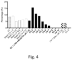
- the recombinant polypeptide of the present disclosure may be defined by a sequence selected from SEQ ID NO:20-38, or a corresponding sequence having at least 80%, preferably at least 90%, more preferably at least 95% sequence homology with the sequence (see also figure 6 ).
- a ligand comprising a recombinant polypeptide as described hereinabove.
- the ligand may be a multimer, e.g. a dimer, trimer, tetramer, pentamer, hexamer, heptamer, or an octamer.
- the ligand may further comprise linking peptides and/or signal peptides between the monomer units.
- the ligand may also comprise a C-terminal cysteine for subsequent coupling to a chromatography resin of a chromatography matrix.
- a chromatography matrix comprising the ligand as described hereinbefore coupled to the matrix.
- the chromatography matrix is not limited to a specific matrix, but any matrix known to the skilled person may be utilized.
- the chromatography matrix may be of natural or synthetic origin.
- the chromatography matrix is a solid matrix, e.g. a polysaccharide matrix.
- the ligand may be coupled to the matrix by any coupling technique known to the skilled person. For example, amino acids and/or carboxy group present in the ligand may be utilized. Alternatively, the ligand may be coupled to the solid support of the matrix by thioether bonds. In this regard, the ligand may be provided with a terminal cysteine residue for subsequent use in the coupling.
- a spacer molecule may be introduced between the matrix support and the ligand to facilitate the coupling of the ligand to the matrix.
- the ligand is coupled to the matrix by non-covalent bonding, such as physical adsorption or biospecific adsorption.
- the sample comprising target proteins may be antibodies or parts thereof, such as IgG or Fab-fusion proteins.
- the antibodies or parts thereof may be present in complex sample mixtures, e.g. culture supernatants.
- the step d) of eluting bound target proteins may be carried out at a pH from 5 to 8, preferably from 5.5 to 7.
- the step d) of eluting the bound target proteins typically comprises adding an agent which disrupts the bond between the metal ions and the metal-binding motif.
- the agent may be a metal-binding molecule, a salt, such as sodium chloride (NaCl) and/or a chelating agent, e.g. EDTA, EGTA, glutamate diacetate or citrate glutamate.
- nucleic acid encoding a recombinant polypeptide as described hereinabove.
- the present disclosure also encompasses a DNA sequence that can be used in the production of the recombinant polypeptides by expression thereof in a recombinant host according to well-established biotechnological methods.
- the DNA sequence may be defined by SEQ ID NO:39 or a corresponding sequence having at least 90 %, preferably at least 95%, more preferably at least 99% sequence homology with this sequence.
- SEQ ID NO:39 encodes the recombinant polypeptide defined by SEQ ID NO:20.
- an expression system comprising a nucleic acid as described hereinabove.
- the present invention provides novel recombinant polypeptides derived from the IgG-binding domains C 1, C2, and C3 of streptococcal protein G (SPG).
- SPG streptococcal protein G
- the target binding of the IgG-binding domains has been rendered metal ion dependent by the introduction of a metal-binding motif. Accordingly, the recombinant polypeptides display a switchable target affinity depending on the presence of metal ions.
- the recombinant polypeptides may be used as ligands for purification of immunoglobulins (Ig) by affinity chromatography. Such ligands may circumvent the currently harsh elution conditions associated with immunoglobulin G purification.
- the invention provides a modular way of tailoring the target affinity of several protein domains with inherent antibody affinity and new tools optimizable for gentle IgG purification, particularly Fab-fragment purification.
- a recombinant polypeptide comprising an IgG-binding domain derived from Streptococcus Protein G (SPG) and a metal-binding motif, wherein the recombinant polypeptide has a first, target-binding configuration in the presence of metal ions, and a second, non-target-binding configuration in the absence of metal ions, wherein the recombinant polypeptide is configured to bind to the Fab-region of IgG in the target-binding configuration.
- SPG Streptococcus Protein G
- a ligand comprising the recombinant polypeptide described hereinbefore.
- a chromatography matrix comprising the ligand coupled to the matrix.
- nucleic acid encoding a recombinant polypeptide as defined hereinabove.
- an expression system comprising the nucleic acid is provided.
- the library was designed to consist of the C2 domain with a randomized stretch of 12 amino acids between the second beta strand and the alpha helix. Upstream of this stretch a linker region of 0-3 amino acids was included and oligonucleotides encoding this library was obtained using trinucleotide synthesis (Twist Bioscience, San Francisco, CA, US). 200 ng of DNA was PCR-amplified during 20 cycles before performing traditional cloning to insert it into the pBad2.2 E. coli display vector (10).
- the final ligation product was electroporated into BL21(DE3) cells (Lucigen, Middleton, WI, US) which were cultivated in 100 ml LB media supplemented with 100 ⁇ g/ml carbenicillin overnight at 37°C, 150 rpm. Cells were harvested through centrifugation the following morning and the practical library size was calculated to be 1.5 ⁇ 10 8 variants through replicate titrations. Stocks containing 15% glycerol was made with 1.5 ⁇ 10 9 cells and stored at - 80°C.
- the ligation products were electroporated into BL21(DE3) cells (Lucigen, Middleton, WI, US) which were cultivated in 100 ml LB media supplemented with 100 ⁇ g/ml carbenicillin overnight at 37°C, 150 rpm. Centrifugation was used to harvest the cells the following morning and the practical library sizes was estimated to range from 2 ⁇ 10 6 - 9 ⁇ 10 7 variants.
- Mouse IgG1 (mIgG1) and human polyclonal Fab fragment (hFab) was ordered from Jackson ImmunoResearch (West Grove, PA, US) and biotinylated using EZ-link Sulfo-NHS-LC-Biotin (Thermo Scientific, Waltham, MA, US) according to the manufacturer's instructions. The biotinylation was stopped by removing non-reacted biotin using a NAP5 desalting column (Cytiva, Uppsala, Sweden).
- 1.5 ⁇ 10 9 cells were cultivated in 100 ml LB containing 100 ⁇ g/ml carbenicillin at 37°C, 150 rpm until the OD reached 0.5-0.8 and the culture was induced with 0.6% w/v L-arabinose and grown overnight at 25°C.
- the induced E. coli cells were washed with TBSC (50 mM Tris, 150 mM NaCl, 1 mM CaCl 2 , pH 7) before being resuspended in the same buffer but containing 150 nM of biotinylated mIgG1 or 500 nM of biotinylated hFab and incubated on a rotamixer at room temperature (RT) for two hours.
- TBSC 50 mM Tris, 150 mM NaCl, 1 mM CaCl 2 , pH 7
- RT room temperature
- the cells were washed with TBSC, resuspended in 150 nM human serum albumin (HSA)-Alexa 647 conjugate and 2 ⁇ g/ml streptavidin conjugated with R-phycoerythrin (SAPE) (Invitrogen, Waltham, MA, US) and incubated dark on ice for 30 minutes.
- the cells were subsequently washed and lastly resuspended in cold TBSC before being sorted using a MoFlo Astrios EQ cell sorter (Beckman Coulter, Brea, CA, US).
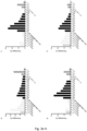
- the sort gate was set to capture cells displaying the library (detected library expression with (HSA)-Alexa-647 conjugate, X-axis) and binding to the target (detected target binding with SAPE, Y-axis).
- Sorted cells were collected in a 15 ml tube containing LB media and was incubated during rotation for one hour at 37°C before being inoculated to 150 ml of LB media supplemented with 100 ⁇ g/ml carbenicillin and cultivated overnight at 37°C, 150 rpm. Six rounds of selection in solution were performed following this procedure, round 2-6 also included a negative selection step using magnetic activated cell sorting (MACS) prior to the positive fluorescent activated cell sorting (FACS) described.
- MCS magnetic activated cell sorting
- FACS positive fluorescent activated cell sorting
- MACS was implemented to remove non-calcium dependent binders by pre-incubating target (150 nM mIgG1 or 500 nM hFab) with streptavidin-coated Dynabeads TM MyOne TM Streptavidin C1 (Thermo Scientific) before allowing the cells to be incubated with the target-coated beads for 2 hours in MBSE (25 mM MES, 150 mM NaCl, 100 mM EDTA, pH 5.5). The magnetic beads were thoroughly washed, captured and the resulting supernatant was used as input for FACS.
- target 150 nM mIgG1 or 500 nM hFab
- streptavidin-coated Dynabeads TM MyOne TM Streptavidin C1 (Thermo Scientific) before allowing the cells to be incubated with the target-coated beads for 2 hours in MBSE (25 mM MES, 150 mM NaCl, 100 mM EDTA, pH
- the buffers used for purification were MBSC (25 mM MES, 150 mM NaCl, 1 mM CaCl 2 , pH 6) for equilibration, 5 mM NH 4 Ac supplemented with 1 mM CaCl 2 , pH 6 for washing and 0.5 M HAc, pH 2.8 for elution.
- MBSC 25 mM MES, 150 mM NaCl, 1 mM CaCl 2 , pH 6) for equilibration, 5 mM NH 4 Ac supplemented with 1 mM CaCl 2 , pH 6 for washing and 0.5 M HAc, pH 2.8 for elution.
- Upon elution the samples were buffer exchanged on PD-10 Desalting columns (Cytiva) to TBSC and the final purity was analyzed using SDS-PAGE.
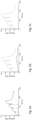
- the affinity to the native targets mIgG1, human polyclonal Fab- and human polyclonal Fc was determined using a Biacore T200 instrument (Cytiva). By amine coupling, each target was diluted in 10 mM NaAc, pH 4.5 and immobilized in separate flow cells on a Series S CM5-chip. Single-cycle kinetics was obtained at 25°C through injection of a two-fold dilution series (1000 nM, 500 nM, 250 nM, 125 nM) at a flow-rate of 30 ⁇ l/min.
- the experiment was performed both with TBSCT (TBSC, 0.05% (v/v) Tween20, pH 7) and TBSET (TBS, 100 mM EDTA, 0.05% (v/v) Tween20, pH 7) as running buffer and after each cycle the surfaces were regenerated with 10 mM HCl.
- 0.5 ml of IgG Sepharose 6 FF (Cytiva) was packed in a column and pulsed with 5 CV 0.5 M HAc (pH 2.8) and 20 CV 1 ⁇ TBS-C, pH 7.5 (50 mM Tris, 150 mM NaCl, 1 mM CaCl2). 0.5 mg of pure protein (each variant) was added followed by a wash with 10 CV TBS-C. 10CV of elution with 100 mM EDTA in TBS, pH 7.5, 0.5 ml fractions. After elution, a control elution with 3 CV of 0.5 M HAc, pH 2.8 was performed. All eluted fractions were measured at 280 nm.
- the protein structure of C2 including its previously characterized antibody binding surfaces was investigated to find a suitable insertion site for a metal-coordinating loop that could render the inherent target affinity calcium-dependent.
- the binding surface for Fab is mainly located to the second beta-sheet and the C-terminal end of the alpha helix while the interaction with Fc is identified to be present at the C-terminal end of the alpha helix, extending into the third beta-sheet.
- the library was designed to include four different linker lengths of 0-3 amino acids chosen to allow for different characteristics such as polarity (Ser/Thr), flexibility (Gly), and rigidity (Pro). Following the linker, the library was intended to cover a large number of possible calcium-binding loops consisting of 13 amino acids with designed randomizations in all but two positions ( Figure 1 ).
- Directed evolution using bacterial display was performed towards full-length mouse IgG1 and polyclonal human Fab-fragments in order to enrich for variants with calcium-dependent target interaction.
- the strategy consisted of alternating negative and positive selection pressures.
- the negative selection was performed in a calcium-free environment and aimed at eliminating variants that was still capable of interacting with the target independent of the presence of calcium.
- the positive selection was carried out with fluorescence-activated cell sorting to isolate variants with retained target affinity when supplemented with calcium. After six selection rounds, a target-binding cell population seemingly calcium-dependent could be seen.
- the selection output from round 5 and 6 was used as starting material for error-prone PCR aiming to insert an average of two to four amino acid substitutions per variant, randomly distributed across the domain.
- Bacterial display was continuedly used as the selection system with alternating negative and positive selection rounds. Although, this time the designed strategy included using magnetic-activated cell sorting (MACS) also to isolate variants which interacted with the inherent target in the presence of calcium but could be eluted from the magnetic beads by removal of calcium.
- MCS magnetic-activated cell sorting
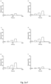
- C2 CaV3 (defined by SEQ ID NO:31) was showing an intact calcium-dependent human polyclonal Fab-binding while interaction with human polyclonal Fc-fragments could not be detected ( Fig. 2g ).
- C2 Ca EP7 revealed one mutation in the scaffold located to the first position succeeding the inserted calcium-binding loop (i.e. the first amino acid residue of the alpha helix) showing a glutamate exchanged for a glycine.
- the importance of this evolved feature was investigated by back-mutating it to a glutamate residue in a version referred to as C2Ca EP7_G35E.
- this version was tested for calcium-dependent elution in the exact same gentle purification set-up as previously, a majority of the sample remained bound to the column until the control elution with low pH. Further, the tolerance to different amino acids in position 25 and 28 was evaluated; i.e. position X 7 and X 10 of the metal-binding loop.
- polypeptides are defined by SEQ ID NO:21 (C2Ca EP7_F28R ), SEQ ID NO:22 (C2Ca EP7_F28Y ), SEQ ID NO:23 (C2Ca EP7_F28H ), and SEQ ID NO:24 (C2Ca EP7_N25K ).
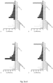
- Candidate C2 CaEP7 was evaluated further by multimerizing it to become a tetramer produced with a C-terminal cysteine alongside the monomeric version. Through thiol coupling, both monomer and tetramer C2 Ca EP7 was immobilized to equimolar amounts on separate chip surfaces. A two-fold dilution series of full-length mIgG1 (125 nM- 1000 nM) was injected and the calcium-dependent binding previously eluded for the monomer could be seen again in this reversed set-up ( Figure 5 ). The tetramer demonstrated higher binding signals, showing that a multimeric version would have improved binding capacity. Moreover, the complete calcium-dependency was still retained for the tetrameric version of C2 CaEP7 .
- Naturally occurring proteins have evolved to incorporate and transmit signals or regulate their level of activity through induced conformational changes in the protein structure.
- the library was designed to be introduced N-terminally of the alpha helix in the C2 domain. Further, this insertion site was believed to be in close enough proximity to both inherent binding surfaces of C2 thereby endowing calcium-dependent target affinity without risking destroying the delicate protein structure completely.
- By implementing cell display it was possible to early on observe that the protein structure of the library members seemed intact as they evidently could be expressed on cells and interact with the native target.
- the sequential utilization of MACS and FACS enabled directed removal of non-calcium dependent variants during the negative selection and focused enrichment of variants ensured to display target interaction in the presence of calcium.
Landscapes
- Chemical & Material Sciences (AREA)
- Health & Medical Sciences (AREA)
- Life Sciences & Earth Sciences (AREA)
- Genetics & Genomics (AREA)
- Organic Chemistry (AREA)
- Molecular Biology (AREA)
- Engineering & Computer Science (AREA)
- Biophysics (AREA)
- General Health & Medical Sciences (AREA)
- Biochemistry (AREA)
- Biomedical Technology (AREA)
- Zoology (AREA)
- Analytical Chemistry (AREA)
- General Engineering & Computer Science (AREA)
- Proteomics, Peptides & Aminoacids (AREA)
- Bioinformatics & Cheminformatics (AREA)
- Medicinal Chemistry (AREA)
- Wood Science & Technology (AREA)
- Biotechnology (AREA)
- Physics & Mathematics (AREA)
- Chemical Kinetics & Catalysis (AREA)
- Immunology (AREA)
- Microbiology (AREA)
- Plant Pathology (AREA)
- Gastroenterology & Hepatology (AREA)
- Peptides Or Proteins (AREA)
Priority Applications (2)
| Application Number | Priority Date | Filing Date | Title |
|---|---|---|---|
| EP23205682.0A EP4545554A1 (de) | 2023-10-25 | 2023-10-25 | Rekombinantes polypeptid mit einem metallbindenden motiv |
| PCT/EP2024/079670 WO2025087842A1 (en) | 2023-10-25 | 2024-10-21 | A recombinant polypeptide comprising a metal-binding motif |
Applications Claiming Priority (1)
| Application Number | Priority Date | Filing Date | Title |
|---|---|---|---|
| EP23205682.0A EP4545554A1 (de) | 2023-10-25 | 2023-10-25 | Rekombinantes polypeptid mit einem metallbindenden motiv |
Publications (1)
| Publication Number | Publication Date |
|---|---|
| EP4545554A1 true EP4545554A1 (de) | 2025-04-30 |
Family
ID=88511516
Family Applications (1)
| Application Number | Title | Priority Date | Filing Date |
|---|---|---|---|
| EP23205682.0A Withdrawn EP4545554A1 (de) | 2023-10-25 | 2023-10-25 | Rekombinantes polypeptid mit einem metallbindenden motiv |
Country Status (2)
| Country | Link |
|---|---|
| EP (1) | EP4545554A1 (de) |
| WO (1) | WO2025087842A1 (de) |
Citations (1)
| Publication number | Priority date | Publication date | Assignee | Title |
|---|---|---|---|---|
| WO2018046475A1 (en) * | 2016-09-08 | 2018-03-15 | Ge Healthcare Bioprocess R&D Ab | Target-binding polypeptide mutant of an igg-binding polypeptide comprising a metal binding motif |
-
2023
- 2023-10-25 EP EP23205682.0A patent/EP4545554A1/de not_active Withdrawn
-
2024
- 2024-10-21 WO PCT/EP2024/079670 patent/WO2025087842A1/en active Pending
Patent Citations (2)
| Publication number | Priority date | Publication date | Assignee | Title |
|---|---|---|---|---|
| WO2018046475A1 (en) * | 2016-09-08 | 2018-03-15 | Ge Healthcare Bioprocess R&D Ab | Target-binding polypeptide mutant of an igg-binding polypeptide comprising a metal binding motif |
| EP3523318A1 (de) | 2016-09-08 | 2019-08-14 | GE Healthcare BioProcess R&D AB | Target-bindender polypeptidmutant eines igg-bindenden polypeptids mit einem metallbindenden motiv |
Non-Patent Citations (5)
| Title |
|---|
| A. E. SAUER-ERIKSSONG. J. KLEYWEGTM. UHLENT. A. JONES, STRUCTURE, vol. 3, 2004, pages 1 - 14 |
| F.L. MOFFATT JR., S.A. GULAC, A.N. SARAFINI, G.N. SFAKANCKIS, R. DOP, D.S. ROBINSON,D. FRANSCESCHI, J. BOGGS, A.S. LIVINGSTONE: "A thousand points of light or just a dim light bulb? Radiolabeled antibodies and colorectal cancer imaging", CANCER INVESTIGATION, vol. 17, 1999, pages 322 - 334 |
| GIFFORD JESSICA L. ET AL: "Structures and metal-ion-binding properties of the Ca2+-binding helix-loop-helix EF-hand motifs", BIOCHEMICAL JOURNAL, vol. 405, no. 2, 15 July 2007 (2007-07-15), GB, pages 199 - 221, XP093141637, ISSN: 0264-6021, Retrieved from the Internet <URL:https://watermark.silverchair.com/bj4050199.pdf?token=AQECAHi208BE49Ooan9kkhW_Ercy7Dm3ZL_9Cf3qfKAc485ysgAABGcwggRjBgkqhkiG9w0BBwagggRUMIIEUAIBADCCBEkGCSqGSIb3DQEHATAeBglghkgBZQMEAS4wEQQMJoVXPQpIJ8KekaFMAgEQgIIEGjygxuFeyLnWeq1GwhUGxzKaZDLyzoqOfewVlltRUVA19aHNuJKXUlrwQuxcCTmeubY8S6qB7LJupDy7dqMCWyL4wQ> DOI: 10.1042/BJ20070255 * |
| J. P. DERRICK, D. B. WIGLEY: "The third IgG-binding domain from streptococcal protein G: An analysis by X-ray crystallography of the structure alone and in a complex with Fab", MOL. BIOL., vol. 243, 1994, pages 906 - 918, XP024008128, DOI: 10.1006/jmbi.1994.1691 |
| STEPHEN F MARINO ET AL: "'Morphs' (MRFs): metal-reversible folding domains for differential IgG binding", CHEMISTRY & BIOLOGY, vol. 8, no. 12, 1 December 2001 (2001-12-01), GB, pages 1221 - 1229, XP055421936, ISSN: 1074-5521, DOI: 10.1016/S1074-5521(01)00088-6 * |
Also Published As
| Publication number | Publication date |
|---|---|
| WO2025087842A1 (en) | 2025-05-01 |
Similar Documents
| Publication | Publication Date | Title |
|---|---|---|
| Feldwisch et al. | Design of an optimized scaffold for affibody molecules | |
| US10065995B2 (en) | Protein for affinity-separation matrix | |
| Nilvebrant et al. | The albumin-binding domain as a scaffold for protein engineering | |
| CN103890174B (zh) | IgG结合性肽及利用其检测和纯化IgG的方法 | |
| ES2325362T3 (es) | Una proteina mutada de union a inmunoglobulina. | |
| EP1179014B1 (de) | Derivate der z-domäne des proteins a von staphylococcus (spa), welche mit mindestens einer domäne des humanen faktors viii interagieren | |
| JP6805831B2 (ja) | 免疫グロブリンに親和性を有するタンパク質、およびそれを用いたアフィニティ分離剤、液体クロマトグラフィー用カラム | |
| JP2016025872A (ja) | 免疫グロブリンに親和性を有するタンパク質および免疫グロブリン結合性アフィニティーリガンド | |
| JP6802349B2 (ja) | アルカリ耐性が向上した変異型免疫グロブリン結合タンパク質 | |
| KR102497083B1 (ko) | 돌연변이된 스캐폴드를 갖는 결합 폴리펩티드 | |
| JP2016079153A (ja) | プロテインg変異体 | |
| KR102446636B1 (ko) | 인간 보체 c5에 결합하는 안정한 폴리펩티드 | |
| JP2022519808A (ja) | アフィニティー精製用免疫グロブリン結合タンパク質 | |
| JP2023533529A (ja) | アフィニティ精製のための免疫グロブリン結合タンパク質 | |
| JP5257997B2 (ja) | タグペプチド及びその利用 | |
| CN106188251B (zh) | 一种免疫球蛋白结合蛋白突变体及其用途 | |
| EP4545554A1 (de) | Rekombinantes polypeptid mit einem metallbindenden motiv | |
| JP7242116B2 (ja) | 金属結合モチーフを含むIgG結合ポリペプチドの標的結合ポリペプチド変異体 | |
| Boström et al. | Purification systems based of bacterial surface proteins | |
| WO2005000883A1 (en) | Polypeptides having binding affinity for insulin | |
| US20180215836A1 (en) | Immunoglobulin-binding modified protein | |
| Jönsson et al. | Calcium as a molecular switch: Gentle antibody-purification through directed evolution of Protein G | |
| Cao et al. | Phage-based molecular directed evolution yields multiple tandem human IgA affibodies with intramolecular binding avidity | |
| HK40050539A (en) | Stable polypeptides binding to human complement c5 | |
| Mersich et al. | Identification of a ligand for IgG‐Fc derived from a soluble peptide library based on fusion proteins secreted by S. cerevisiae |
Legal Events
| Date | Code | Title | Description |
|---|---|---|---|
| PUAI | Public reference made under article 153(3) epc to a published international application that has entered the european phase |
Free format text: ORIGINAL CODE: 0009012 |
|
| STAA | Information on the status of an ep patent application or granted ep patent |
Free format text: STATUS: THE APPLICATION HAS BEEN PUBLISHED |
|
| AK | Designated contracting states |
Kind code of ref document: A1 Designated state(s): AL AT BE BG CH CY CZ DE DK EE ES FI FR GB GR HR HU IE IS IT LI LT LU LV MC ME MK MT NL NO PL PT RO RS SE SI SK SM TR |
|
| STAA | Information on the status of an ep patent application or granted ep patent |
Free format text: STATUS: THE APPLICATION IS DEEMED TO BE WITHDRAWN |
|
| 18D | Application deemed to be withdrawn |
Effective date: 20251031 |