EP4545562A1 - Fusionsprotein mit anti-cd73-antikörper und il-2 sowie verwendung davon - Google Patents
Fusionsprotein mit anti-cd73-antikörper und il-2 sowie verwendung davon Download PDFInfo
- Publication number
- EP4545562A1 EP4545562A1 EP23827535.8A EP23827535A EP4545562A1 EP 4545562 A1 EP4545562 A1 EP 4545562A1 EP 23827535 A EP23827535 A EP 23827535A EP 4545562 A1 EP4545562 A1 EP 4545562A1
- Authority
- EP
- European Patent Office
- Prior art keywords
- seq
- fusion protein
- amino acid
- cells
- acid sequence
- Prior art date
- Legal status (The legal status is an assumption and is not a legal conclusion. Google has not performed a legal analysis and makes no representation as to the accuracy of the status listed.)
- Pending
Links
Images


Classifications
-
- A—HUMAN NECESSITIES
- A61—MEDICAL OR VETERINARY SCIENCE; HYGIENE
- A61K—PREPARATIONS FOR MEDICAL, DENTAL OR TOILETRY PURPOSES
- A61K38/00—Medicinal preparations containing peptides
- A61K38/16—Peptides having more than 20 amino acids; Gastrins; Somatostatins; Melanotropins; Derivatives thereof
- A61K38/17—Peptides having more than 20 amino acids; Gastrins; Somatostatins; Melanotropins; Derivatives thereof from animals; from humans
- A61K38/19—Cytokines; Lymphokines; Interferons
- A61K38/20—Interleukins [IL]
- A61K38/2013—IL-2
-
- A—HUMAN NECESSITIES
- A61—MEDICAL OR VETERINARY SCIENCE; HYGIENE
- A61K—PREPARATIONS FOR MEDICAL, DENTAL OR TOILETRY PURPOSES
- A61K39/00—Medicinal preparations containing antigens or antibodies
-
- A—HUMAN NECESSITIES
- A61—MEDICAL OR VETERINARY SCIENCE; HYGIENE
- A61K—PREPARATIONS FOR MEDICAL, DENTAL OR TOILETRY PURPOSES
- A61K39/00—Medicinal preparations containing antigens or antibodies
- A61K39/395—Antibodies; Immunoglobulins; Immune serum, e.g. antilymphocytic serum
-
- A—HUMAN NECESSITIES
- A61—MEDICAL OR VETERINARY SCIENCE; HYGIENE
- A61K—PREPARATIONS FOR MEDICAL, DENTAL OR TOILETRY PURPOSES
- A61K39/00—Medicinal preparations containing antigens or antibodies
- A61K39/395—Antibodies; Immunoglobulins; Immune serum, e.g. antilymphocytic serum
- A61K39/39533—Antibodies; Immunoglobulins; Immune serum, e.g. antilymphocytic serum against materials from animals
- A61K39/39558—Antibodies; Immunoglobulins; Immune serum, e.g. antilymphocytic serum against materials from animals against tumor tissues, cells, antigens
-
- A—HUMAN NECESSITIES
- A61—MEDICAL OR VETERINARY SCIENCE; HYGIENE
- A61P—SPECIFIC THERAPEUTIC ACTIVITY OF CHEMICAL COMPOUNDS OR MEDICINAL PREPARATIONS
- A61P35/00—Antineoplastic agents
-
- C—CHEMISTRY; METALLURGY
- C07—ORGANIC CHEMISTRY
- C07K—PEPTIDES
- C07K14/00—Peptides having more than 20 amino acids; Gastrins; Somatostatins; Melanotropins; Derivatives thereof
- C07K14/435—Peptides having more than 20 amino acids; Gastrins; Somatostatins; Melanotropins; Derivatives thereof from animals; from humans
- C07K14/52—Cytokines; Lymphokines; Interferons
- C07K14/54—Interleukins [IL]
- C07K14/55—IL-2
-
- C—CHEMISTRY; METALLURGY
- C07—ORGANIC CHEMISTRY
- C07K—PEPTIDES
- C07K16/00—Immunoglobulins [IG], e.g. monoclonal or polyclonal antibodies
- C07K16/18—Immunoglobulins [IG], e.g. monoclonal or polyclonal antibodies against material from animals or humans
- C07K16/28—Immunoglobulins [IG], e.g. monoclonal or polyclonal antibodies against material from animals or humans against receptors, cell surface antigens or cell surface determinants
- C07K16/2896—Immunoglobulins [IG], e.g. monoclonal or polyclonal antibodies against material from animals or humans against receptors, cell surface antigens or cell surface determinants against molecules with a "CD"-designation, not provided for elsewhere
-
- C—CHEMISTRY; METALLURGY
- C07—ORGANIC CHEMISTRY
- C07K—PEPTIDES
- C07K16/00—Immunoglobulins [IG], e.g. monoclonal or polyclonal antibodies
- C07K16/40—Immunoglobulins [IG], e.g. monoclonal or polyclonal antibodies against enzymes
-
- A—HUMAN NECESSITIES
- A61—MEDICAL OR VETERINARY SCIENCE; HYGIENE
- A61K—PREPARATIONS FOR MEDICAL, DENTAL OR TOILETRY PURPOSES
- A61K39/00—Medicinal preparations containing antigens or antibodies
- A61K2039/505—Medicinal preparations containing antigens or antibodies comprising antibodies
-
- A—HUMAN NECESSITIES
- A61—MEDICAL OR VETERINARY SCIENCE; HYGIENE
- A61K—PREPARATIONS FOR MEDICAL, DENTAL OR TOILETRY PURPOSES
- A61K39/00—Medicinal preparations containing antigens or antibodies
- A61K2039/505—Medicinal preparations containing antigens or antibodies comprising antibodies
- A61K2039/507—Comprising a combination of two or more separate antibodies
-
- C—CHEMISTRY; METALLURGY
- C07—ORGANIC CHEMISTRY
- C07K—PEPTIDES
- C07K2317/00—Immunoglobulins specific features
- C07K2317/30—Immunoglobulins specific features characterized by aspects of specificity or valency
- C07K2317/31—Immunoglobulins specific features characterized by aspects of specificity or valency multispecific
-
- C—CHEMISTRY; METALLURGY
- C07—ORGANIC CHEMISTRY
- C07K—PEPTIDES
- C07K2317/00—Immunoglobulins specific features
- C07K2317/50—Immunoglobulins specific features characterized by immunoglobulin fragments
- C07K2317/56—Immunoglobulins specific features characterized by immunoglobulin fragments variable (Fv) region, i.e. VH and/or VL
- C07K2317/569—Single domain, e.g. dAb, sdAb, VHH, VNAR or nanobody®
-
- C—CHEMISTRY; METALLURGY
- C07—ORGANIC CHEMISTRY
- C07K—PEPTIDES
- C07K2317/00—Immunoglobulins specific features
- C07K2317/60—Immunoglobulins specific features characterized by non-natural combinations of immunoglobulin fragments
- C07K2317/62—Immunoglobulins specific features characterized by non-natural combinations of immunoglobulin fragments comprising only variable region components
- C07K2317/622—Single chain antibody (scFv)
-
- C—CHEMISTRY; METALLURGY
- C07—ORGANIC CHEMISTRY
- C07K—PEPTIDES
- C07K2317/00—Immunoglobulins specific features
- C07K2317/70—Immunoglobulins specific features characterized by effect upon binding to a cell or to an antigen
- C07K2317/71—Decreased effector function due to an Fc-modification
-
- C—CHEMISTRY; METALLURGY
- C07—ORGANIC CHEMISTRY
- C07K—PEPTIDES
- C07K2317/00—Immunoglobulins specific features
- C07K2317/70—Immunoglobulins specific features characterized by effect upon binding to a cell or to an antigen
- C07K2317/73—Inducing cell death, e.g. apoptosis, necrosis or inhibition of cell proliferation
-
- C—CHEMISTRY; METALLURGY
- C07—ORGANIC CHEMISTRY
- C07K—PEPTIDES
- C07K2317/00—Immunoglobulins specific features
- C07K2317/70—Immunoglobulins specific features characterized by effect upon binding to a cell or to an antigen
- C07K2317/76—Antagonist effect on antigen, e.g. neutralization or inhibition of binding
-
- C—CHEMISTRY; METALLURGY
- C07—ORGANIC CHEMISTRY
- C07K—PEPTIDES
- C07K2317/00—Immunoglobulins specific features
- C07K2317/90—Immunoglobulins specific features characterized by (pharmaco)kinetic aspects or by stability of the immunoglobulin
- C07K2317/92—Affinity (KD), association rate (Ka), dissociation rate (Kd) or EC50 value
-
- C—CHEMISTRY; METALLURGY
- C07—ORGANIC CHEMISTRY
- C07K—PEPTIDES
- C07K2317/00—Immunoglobulins specific features
- C07K2317/90—Immunoglobulins specific features characterized by (pharmaco)kinetic aspects or by stability of the immunoglobulin
- C07K2317/94—Stability, e.g. half-life, pH, temperature or enzyme-resistance
-
- C—CHEMISTRY; METALLURGY
- C07—ORGANIC CHEMISTRY
- C07K—PEPTIDES
- C07K2319/00—Fusion polypeptide
-
- C—CHEMISTRY; METALLURGY
- C07—ORGANIC CHEMISTRY
- C07K—PEPTIDES
- C07K2319/00—Fusion polypeptide
- C07K2319/30—Non-immunoglobulin-derived peptide or protein having an immunoglobulin constant or Fc region, or a fragment thereof, attached thereto
-
- C—CHEMISTRY; METALLURGY
- C07—ORGANIC CHEMISTRY
- C07K—PEPTIDES
- C07K2319/00—Fusion polypeptide
- C07K2319/33—Fusion polypeptide fusions for targeting to specific cell types, e.g. tissue specific targeting, targeting of a bacterial subspecies
Definitions
- the present invention relates to a fusion protein in which an anti-CD73 antibody or an antigen-binding fragment thereof and IL-2 are linked to each other, and the use thereof.
- Cancer immunotherapy is a method of treating cancer using the body's immune system. It can induce the immune system to attack cancer cells by targeting antigens such as cancer cell surface proteins and can increase the activity of immune cells by controlling the tumor microenvironment that causes immune evasion of cancer cells.
- the tumor microenvironment is an environment in which not only cancer cells, but also fibroblasts, blood vessels, lymphatic vessels, immune cells, extracellular matrix, and adipocytes, which exist in cancer tissues, proliferate and evolve.
- studies have reported that the ATP-AMP-adenosine-A2AR/A2BR signaling pathway can regulate the activity of immune cells in the tumor microenvironment ( S. Vigano et al., Front Immunol(2019) 10:925 ).
- immune suppressor cells such as regulatory T cells or tumor-associated macrophages (TAMs) are activated, and the activity of cytotoxic T cells and NK cells is inhibited.
- TAMs tumor-associated macrophages
- studies have been conducted on a method of activating immunity in the tumor microenvironment by inhibiting this signaling pathway by treatment with a substance that target CD39, an ectonucleotidase that converts ATP to AMP, CD73, an ectonucleotidase that converts AMP to adenosine, or A2AR/A2BR receptors that bind to adenosine.
- interleukin 2 also called T-cell growth factor (TCGF)
- TCGF T-cell growth factor
- IL-2 is synthesized mainly by activated T cells, particularly CD4+ helper T cells.
- IL-2 stimulates the proliferation and differentiation of T cells and induces the production of cytotoxic T lymphocytes (CTLs) and the differentiation of peripheral blood lymphocytes into cytotoxic cells and lymphokine-activated killer cells (LAK cells).
- CTLs cytotoxic T lymphocytes
- LAK cells lymphokine-activated killer cells
- the present inventors have conducted studies to develop a new combination of proteins as a fusion protein that enhances the activity of immune cells by controlling the tumor microenvironment.
- a fusion protein dimer comprising an anti-CD73 antibody or an antigen-binding fragment thereof and IL-2 can control the tumor microenvironment and effectively activates immune cells.
- the present inventors have demonstrated that the fusion protein dimer is effective as an anticancer agent, thereby completing the present invention.
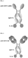
- one aspect of the present invention provides a fusion protein comprising: an antibody or a fragment thereof that specifically binds to CD73; and IL-2.
- Another aspect of the present invention provides a fusion protein dimer in which the two fusion proteins are linked to each other.
- Still another aspect of the present invention provides a polynucleotide encoding the fusion protein, an expression vector containing the polynucleotide, and a transformed cell into which the expression vector has been introduced.
- Yet another aspect of the present invention provides a method for producing a fusion protein dimer comprising: an antibody or an antigen-binding fragment thereof that specifically binds to CD73; and IL-2, the method comprising steps of: i) culturing the transformed cell; and ii) collecting the fusion protein dimer.
- Still yet another aspect of the present invention provides a pharmaceutical composition for preventing or treating cancer comprising the fusion protein or the fusion protein dimer as an active ingredient.
- a further aspect of the present invention provides a method for preventing or treating cancer, comprising a step of administering the fusion protein or the fusion protein dimer to a subject.
- Another further aspect of the present invention provides the use of the fusion protein or the fusion protein dimer for preventing or treating cancer.
- the fusion protein or dimer thereof according to the present invention which comprises an anti-CD73 antibody or an antigen-binding fragment thereof and IL-2, is able to inhibit adenosine production (ATP ⁇ AMP ⁇ adenosine) by binding to CD73 in cancer cells. This inhibition blocks the A2AR and A2BR signaling pathways, thereby controlling the tumor microenvironment. Furthermore, the fusion protein or dimer thereof is able to activate immune cells (CD8+ T cells and CD4+ T cells) along with less activation of immunosuppressive cells such as regulatory T cells (Tregs). In addition, IL-2 or a variant thereof in the fusion protein of the present invention is able to activate immune cells. Therefore, the fusion protein and dimer thereof of the present invention may be useful for the prevention or treatment of cancer.
- Fusion Protein Comprising: Antibody or Antigen-Binding Fragment Thereof That Specifically Binds to CD73; and IL-2
- One aspect of the present invention provides a fusion protein comprising: an antibody or an antigen-binding fragment thereof that specifically binds to CD73; and IL-2.
- CD73 Cluster of Differentiation 73
- CD73 refers to an ectonucleotidase that catalyzes the dephosphorylation of 5' nucleotides, primarily converting adenosine monophosphate (AMP) to adenosine.
- AMP adenosine monophosphate
- CD73 forms a homodimer on the cell membrane via a GPI anchor.
- Each monomer is 65 kDa, with its N-terminal and C-terminal domains connected by a flexible helical linker.
- CD73 is involved in the production of adenosine, is overexpressed in cancer cells, and induces immunosuppression.
- CD73 is expressed in many tumor cells, including leukemia, bladder cancer, glioma, glioblastoma, ovarian cancer, melanoma, prostate cancer, thyroid cancer, esophageal cancer, and breast cancer.
- CD73 is known to be expressed on the surface of immunosuppressive cells (including regulatory T cells (Tregs) and myeloid-derived suppressor cells (MDSCs)).
- Tregs regulatory T cells
- MDSCs myeloid-derived suppressor cells
- CD73 may be mammalian CD73, preferably human CD73, without limitation.
- the CD73 protein includes, but is not limited to, both native and mutant CD73 proteins.
- the native CD73 protein generally refers to a polypeptide comprising the amino acid sequence of the native CD73 protein, and the amino acid sequence of the native CD73 protein generally refers to the amino acid sequence found in naturally occurring CD73.
- Information about CD73 is available from known databases such as GenBank of the National Institutes of Health.
- CD73 may have the amino acid sequence (SEQ ID NO: 41) corresponding to GenBank accession number NP_002517.1, but it is not limited to this sequence.
- the term "antigen" refers to a molecule capable of selectively binding to an antibody.
- the target antigen may be a polypeptide, a carbohydrate, a nucleic acid, a lipid, a hapten, or other naturally occurring or synthetic compound.
- the antigen is a polypeptide and may be a protein present on the cell surface or within the cell.
- the term "specifically binds" means binding that is measurably different from a non-specific interaction. Specific binding can be determined by competition with a control molecule that is similar to the target that has no binding activity.
- the antibody or antigen-binding fragment thereof that specifically binds to CD73 may generally refer to molecules capable of specifically forming an antigen-antibody complex with CD73.
- the antibody or antigen-binding fragment thereof may be used in any form as long as it contains an antigen-binding site capable of specifically binding to CD73.
- the antibody or antigen-binding fragment thereof may include other amino acids that are not directly involved in binding, or amino acids whose effects are blocked by the residues of the antigen-binding site.
- the term "antibody” refers to molecules containing an antigen-binding site, and immunologically active fragments of immunoglobulin molecules containing an antigen-binding site.
- the immunoglobulin molecules may be immunoglobulin molecules of IgG, IgE, IgM, IgD, IgA, IgY, or a subclass thereof.
- the heavy and light chains of immunoglobulins may each include a constant region and a variable region.
- the light and heavy chain variable regions of immunoglobulins contain three variable regions, called complementarity determining regions (CDRs), and four framework regions (FRs).
- CDRs complementarity determining regions
- FRs framework regions
- the antibodies or antigen-binding fragments thereof of the present invention may include monoclonal antibodies, polyclonal antibodies, synthetic antibodies, human antibodies, humanized antibodies, non-human antibodies, and any fragments thereof, as well as immunoconjugates.
- the term "antigen-binding fragment of the antibody” refers to a portion of a polypeptide within the overall structure of an immunoglobulin to which an antigen can bind.
- the antigen-binding fragment include, but are not limited to, Fab fragments, Fab' fragments, F(ab')2 fragments, bispecific Fab dimers (Fab2), trispecific Fab trimers (Fab3), Fv, single-chain Fv proteins ("scFv”), bis-scFv (scFv)2, minibodies, diabodies, triabodies, tetrabodies, disulfide stabilized Fv proteins ("dsFv”), single-domain antibodies (sdAb), and portions of full-length antibodies responsible for antigen binding. Regardless of the structure, the fragment of the antibody is able bind to the same antigen recognized by the intact antibody.
- the single-domain antibody also called a nanobody, refers to an antibody fragment consisting of a single monomeric variable antibody domain.
- the single-domain antibody is able to specifically bind to a specific antigen like a whole antibody.
- the single-domain antibody has a molecular weight of 12 to 15 kDa, which is smaller than that of common antibodies (150-160 kDa), Fab fragments (about 50 kDa), and scFv (about 25 kDa).
- the single-domain antibody may be obtained, for example, by monomerizing the dimeric variable domains of human or mouse conventional immunoglobulin G (IgG), and may be VHH derived from the heavy chain antibody of Camelids, or a VNAR fragment obtained from IgNAR of Cartilaginous fishes.
- IgG immunoglobulin G
- IL-2 refers to any wild-type IL-2 obtained from any vertebrate source, including mammals such as primates (e.g., humans) and rodents (e.g., mice and rats), unless otherwise stated.
- IL-2 has a protein size of 15.5 kDa to 16 kDa and consists of 133 amino acids.
- IL-2 may be obtained from animal cells, but also includes one obtained from recombinant cells capable of producing IL-2.
- IL-2 may be wild-type IL-2 or a variant thereof.
- IL-2 or a variant thereof may be collectively expressed by the term "IL-2 protein" or "IL-2 polypeptide".
- IL-2, an IL-2 protein, an IL-2 polypeptide, and an IL-2 variant specifically bind to, for example, an IL-2 receptor. This specific binding may be identified by methods known to those skilled in the art.
- IL-2 variant refers to a form in which a portion of the amino acids in the full-length IL-2 or the above-described fragment of IL-2 is substituted. That is, an IL-2 variant may have an amino acid sequence different from wild-type IL-2 or a fragment thereof. However, the IL-2 variant may have activity equivalent or similar to the wild-type IL-2.
- IL-2 activity may, for example, refer to specific binding to an IL-2 receptor, in which the specific binding can be measured by methods known to those skilled in the art.
- the IL-2 variant may be obtained by substitution of a portion of the amino acids in the wild-type IL-2.
- An embodiment of the IL-2 variant obtained by amino acid substitution may be obtained by substitution of at least one of the 38 th , 42 nd 45 th , 61 st , and 72 nd amino acids in the amino acid sequence of SEQ ID NO: 40.
- the IL-2 variant may be obtained by substituting at least one of the 38 th , 42 nd , 45 th , 61 st , and 72 nd amino acids in the amino acid sequence of SEQ ID NO: 40 with another amino acid.
- the amino acid at a position complementarily corresponding to that in the amino acid sequence of SEQ ID NO: 40 may be substituted with another amino acid.
- one, two, three, four, five, six, seven, eight, nine, or ten amino acids may be substituted as long as such an IL-2 variant maintains IL-2 activity.
- one to five amino acids may be substituted.
- the IL-2 variant may be in a form in which the amino acids at two positions are substituted. Specifically, the IL-2 variant may be obtained by substitution of the 38 th and 42 nd amino acids in the amino acid sequence of SEQ ID NO: 40. In addition, in one embodiment, the IL-2 variant may be obtained by substitution of the 38 th and 45 th amino acids in the amino acid sequence of SEQ ID NO: 40. In addition, in one embodiment, the IL-2 variant may be obtained by substitution of the 38 th and 61 st amino acids in the amino acid sequence of SEQ ID NO: 40. In addition, in one embodiment, the IL-2 variant may be obtained by substitution of the 38 th and 72 nd amino acids in the amino acid sequence of SEQ ID NO: 40.
- the IL-2 variant may be obtained by substitution of the 42 nd and 45 th amino acids in the amino acid sequence of SEQ ID NO: 40. In addition, in one embodiment, the IL-2 variant may be obtained by substitution of the 42 nd and 61 st amino acids in the amino acid sequence of SEQ ID NO: 40. In addition, in one embodiment, the IL-2 variant may be obtained by substitution of the 42 nd and 72 nd amino acids in the amino acid sequence of SEQ ID NO: 40. In addition, in one embodiment, the IL-2 variant may be obtained by substitution of the 45 th and 61 st amino acids in the amino acid sequence of SEQ ID NO: 40.
- the IL-2 variant may be obtained by substitution of the 45 th and 72 nd amino acids in the amino acid sequence of SEQ ID NO: 40. In addition, in one embodiment, the IL-2 variant may be obtained by substitution of the 61 st and 72 nd amino acids in the amino acid sequence of SEQ ID NO: 40.
- the IL-2 variant may be in a form in which the amino acids at three positions are substituted. Specifically, the IL-2 variant may be obtained by substitution of the 38 th , 42 nd , and 45 th amino acids in the amino acid sequence of SEQ ID NO: 40. In addition, in one embodiment, the IL-2 variant may be obtained by substitution of the 38 th , 42 nd , and 61 st amino acids in the amino acid sequence of SEQ ID NO: 40. In addition, in one embodiment, the IL-2 variant may be obtained by substitution of the 38 th , 42 nd , and 72 nd amino acids in the amino acid sequence of SEQ ID NO: 40.
- the IL-2 variant may be obtained by substitution of the 38 th , 45 th , and 61 st amino acids in the amino acid sequence of SEQ ID NO: 40. In addition, in one embodiment, the IL-2 variant may be obtained by substitution of the 38 th , 45 th , and 72 nd amino acids in the amino acid sequence of SEQ ID NO: 40. In addition, in one embodiment, the IL-2 variant may be obtained by substitution of the 38 th , 61 st , and 72 nd amino acids in the amino acid sequence of SEQ ID NO: 40.
- the IL-2 variant may be obtained by substitution of the 42 nd , 45 th , and 61 st amino acids in the amino acid sequence of SEQ ID NO: 40. In addition, in one embodiment, the IL-2 variant may be obtained by substitution of the 42 nd , 45 th , and 72 nd amino acids in the amino acid sequence of SEQ ID NO: 40. In addition, in one embodiment, the IL-2 variant may be obtained by substitution of the 45 th , 61 st , and 72 nd amino acids in the amino acid sequence of SEQ ID NO: 40.
- the "another amino acid" introduced by substitution may be any one selected from the group consisting of alanine, arginine, asparagine, aspartic acid, cysteine, glutamic acid, glutamine, histidine, isoleucine, leucine, lysine, methionine, phenylalanine, proline, serine, threonine, tryptophan, tyrosine, and valine.
- amino acid substitution for the IL-2 variant in the amino acid sequence of SEQ ID NO: 40, the 38 th amino acid cannot be substituted with arginine, the 42 nd amino acid cannot be substituted with phenylalanine, the 45 th amino acid cannot be substituted with tyrosine, the 61 st amino acid cannot be substituted with glutamic acid, and the 72 nd amino acid cannot be substituted with leucine.
- amino acid substitution for the IL-2 variant in the amino acid sequence of SEQ ID NO: 40, the 38 th amino acid, arginine, may be substituted with an amino acid other than arginine.
- amino acid substitution for an IL-2 variant in the amino acid sequence of SEQ ID NO: 40, the 38 th amino acid, arginine, may be substituted with alanine (R38A).
- amino acid substitution for the IL-2 variant in the amino acid sequence of SEQ ID NO: 40, the 42 nd amino acid, phenylalanine, may be substituted with an amino acid other than phenylalanine.
- amino acid substitution for the IL-2 variant in the amino acid sequence of SEQ ID NO: 40, the 42 nd amino acid, phenylalanine, may be substituted with alanine (F42A).
- the 45 th amino acid, tyrosine in the amino acid sequence of SEQ ID NO: 40, may be substituted with an amino acid other than tyrosine.
- the 45 th amino acid, tyrosine in the amino acid sequence of SEQ ID NO: 40, may be substituted with alanine (Y45A).
- the 61 st amino acid, glutamic acid may be substituted with an amino acid other than glutamic acid.
- the 61 st amino acid, glutamic acid may be substituted with arginine (E61R).
- the 72 nd amino acid, leucine in the amino acid sequence of SEQ ID NO: 40, may be substituted with an amino acid other than leucine.
- the 72 nd amino acid, leucine in the amino acid sequence of SEQ ID NO: 40, may be substituted with glycine (L72G).
- the IL-2 variant may be obtained by at least one substitution selected from the group consisting of R38A, F42A, Y45A, E61R, and L72G, in the amino acid sequence of SEQ ID NO: 40.
- the IL-2 variant may be obtained by two or three amino acid substitutions selected from the group consisting of R38A, F42A, Y45A, E61R, and L72G.
- the IL-2 variant may be in a form in which the amino acids at two positions are substituted. Specifically, the IL-2 variant may be obtained by the substitutions R38A and F42A. In addition, in one embodiment, the IL-2 variant may be obtained by the substitutions R38A and Y45A. In addition, in one embodiment, the IL-2 variant may be obtained by the substitutions R38A and E61R. In addition, in one embodiment, the IL-2 variant may be obtained by the substitutions R38A and L72G. In addition, in one embodiment, the IL-2 variant may be obtained by the substitutions F42A and Y45A. In addition, in one embodiment, the IL-2 variant may be obtained by the substitutions F42A and E61R. In addition, in one embodiment, the IL-2 variant may be obtained by the substitutions F42A and L72G. In addition, in one embodiment, the IL-2 variant may be obtained by the substitutions E61R and L72G. In addition, in one embodiment, the IL-2 variant may be obtained by the
- the IL-2 variant may be in a form in which the amino acids at three positions are substituted. Specifically, the IL-2 variant may be obtained by the substitutions R38A, F42A, and Y45A. In addition, in one embodiment, the IL-2 variant may be obtained by the substitutions R38A, F42A, and E61R. In addition, in one embodiment, the IL-2 variant may be obtained by the substitutions R38A, F42A, and L72G. In addition, in one embodiment, the IL-2 variant may be obtained by the substitutions R38A, Y45A, and E61R. In addition, in one embodiment, the IL-2 variant may be obtained by the substitutions R38A, Y45A, and L72G.
- the IL-2 variant may be obtained by the substitutions F42A, Y45A, and E61R. In addition, in one embodiment, the IL-2 variant may be obtained by the substitutions F42A, Y45A, and L72G. In addition, in one embodiment, the IL-2 variant may be obtained by the substitutions F42A, E61R, and L72G. In addition, in one embodiment, the IL-2 variant may be obtained by the substitutions Y45A, E61R, and L72G.
- the IL-2 variant may be in a form in which the amino acids at four positions are substituted. Specifically, the IL-2 variant may be obtained by the substitutions R38A, F42A, Y45A, and E61R. In addition, in one embodiment, the IL-2 variant may be obtained by the substitutions R38A, F42A, Y45A, and L72G. In addition, in one embodiment, the IL-2 variant may be obtained by the substitutions R38A, F42A, E61R, and L72G. In addition, in one embodiment, the IL-2 variant may be obtained by the substitutions R38A, Y45A, E61R, and L72G. In addition, in one embodiment, the IL-2 variant may be obtained by the substitutions F42A, Y45A, E61R, and L72G.
- the IL-2 variant may be obtained by the substitutions R38A, F42A, Y45A, E61R, and L72G.
- the IL-2 variant of the present invention may be obtained by at least one substitution selected from the group consisting of R38A, F42A, and E61R in the amino acid sequence of SEQ ID NO: 40. More preferably, the IL-2 variant of the present invention may be obtained by the substitutions R38A, F42A, and E61R in the amino acid sequence of SEQ ID NO: 40. Specifically, the IL-2 variant of the present invention may have the amino acid sequence of SEQ ID NO: 8.
- the IL-2 variant may be characterized by having low in vivo toxicity.
- the low in vivo toxicity may be a side effect caused by binding of IL-2 to the IL-2 receptor alpha chain (IL-2R ⁇ ).
- IL-2 variants described in the present application have a low binding affinity for the IL-2 receptor alpha chain (IL-2R ⁇ ) and thus have lower in vivo toxicity than the wild-type IL-2.
- Fusion Protein Comprising: Antibody or Antigen-Binding Fragment Thereof That Specifically Binds to CD73; and IL-2 or Variant Thereof
- the fusion protein of the present invention may comprise: a single-domain antibody that specifically binds to CD73; an Fc region; and IL-2 or a variant thereof.
- the Fc region and the IL-2 or variant thereof may be linked by a linker.
- the fusion protein may comprise the following structural formula (I):
- the antibody or fragment thereof that specifically binds to CD73 is as described above, and IL-2 is as described above.
- the antigen-binding fragment may comprise a single-domain antibody.
- the antibody or antigen-binding fragment thereof, specifically the single-domain antibody may comprise: a CDR1 region comprising an amino acid sequence selected from the group consisting of SEQ ID NOs: 15, 18, and 21; a CDR2 region comprising an amino acid sequence selected from the group consisting of SEQ ID NOs: 16, 19, and 22; and a CDR3 region comprising an amino acid sequence selected from the group consisting of SEQ ID NOs: 17, 20, and 23.
- the antigen-binding fragment may comprise: a CDR1 comprising the amino acid sequence of SEQ ID NO: 15, a CDR2 comprising the amino acid sequence of SEQ ID NO: 16, and a CDR3 comprising the amino acid sequence of SEQ ID NO: 17; or a CDR1 comprising the amino acid sequence of SEQ ID NO: 18, a CDR2 comprising the amino acid sequence of SEQ ID NO: 19, and a CDR3 comprising the amino acid sequence of SEQ ID NO: 20; or a CDR1 comprising the amino acid sequence of SEQ ID NO: 21, a CDR2 comprising the amino acid sequence of SEQ ID NO: 22, and a CDR3 comprising the amino acid sequence of SEQ ID NO: 23.
- the antigen-binding fragment may comprise any one amino acid sequence selected from the group consisting of SEQ ID Nos: 2, 3, and 4.
- the peptide linker (1) may consist of 1 to 50 contiguous amino acids, or 3 to 30 contiguous amino acids, or 5 to 15 amino acids. In one embodiment, the peptide linker (1) may consist of 12 amino acids. In addition, the peptide linker 1 may contain at least one cysteine. Specifically, the peptide linker (1) may contain one, two, or three cysteines. In addition, the peptide linker (1) may be derived from the hinge of an immunoglobulin. For example, the hinge may be selected from the hinge regions of various IgG subclass antibodies.
- the hinge may be a form in which one or more of the amino acids in the hinge region derived from an immunoglobulin are substituted with other amino acid(s), or may be a sequence obtained by adding one or more amino acids to the hinge region.
- the peptide linker (1) may be a peptide linker consisting of the amino acid sequence of SEQ ID NO: 5.
- the peptide linker (2) may consist of 1 to 30 contiguous amino acids, or 2 to 20 contiguous amino acids, or 2 to 10 amino acids.
- the peptide linker (2) may be (G4S)n (where n is an integer ranging from 1 to 10). In (G4S)n, n may be 1, 2, 3, 4, 5, 6, 7, 8, 9, or 10.
- the peptide linker (2) may be a peptide linker consisting of the amino acid sequence of SEQ ID NO: 7.
- the immunoglobulin Fc region refers to a protein that comprises immunoglobulin heavy chain constant region 2 (CH2) and heavy chain constant region 3 (CH3), but does not comprise immunoglobulin heavy and light chain variable regions and light chain constant region (CL).
- the immunoglobulin Fc region may be derived from IgG, IgA, IgE, IgD, or IgM.
- the immunoglobulin Fc region may be derived from IgG1, IgG2, IgG3, or IgG4, which is a subclass of IgG.
- the immunoglobulin Fc region may be derived from IgG4.
- the immunoglobulin Fc region may be an Fc region variant as well as a wild-type Fc region.
- Fc region variant may refer to a form which is different from the wild-type Fc region in terms of glycosylation pattern, or has a high glycosylation compared to the wild-type Fc region, or has a low glycosylation compared to the wild-type Fc region, or is deglycosylated.
- an aglycosylated Fc region is also included.
- the Fc region or a variant thereof may be adapted to have an adjusted number of sialic acids, fucosylations, or glycosylations, through culture conditions or genetic manipulation of a host.
- glycosylation of the immunoglobulin Fc region may be modified by conventional methods such as chemical methods, enzymatic methods, and genetic engineering methods using microorganisms.
- the Fc region variant may be in a mixed form of respective Fc regions of immunoglobulins, IgG, IgA, IgE, IgD, and IgM.
- the Fc region variant may be in a form in which some amino acids of the Fc region are substituted with other amino acids.
- the Fc region may comprise the amino acid sequence of SEQ ID NO: 6.
- the fusion protein of the present invention may be a polypeptide comprising the heavy chain variable region (VH) and heavy chain constant region 1
- (CH1) of an anti-CD73 antibody, an Fc region, and IL-2 or a variant thereof may be linked by a linker.
- the fusion protein may comprise the following structural formulas (II) and (III):
- the antibody or antigen-binding fragment thereof may comprise: a heavy chain variable region comprising an HCDR1 comprising the amino acid sequence of SEQ ID NO: 24, an HCDR2 comprising the amino acid sequence of SEQ ID NO: 25, and an HCDR3 comprising the amino acid sequence of SEQ ID NO: 26; and a light chain variable region comprising an LCDR1 comprising the amino acid sequence of SEQ ID NO: 27, an LCDR2 comprising the amino acid sequence (Asp-Ala-Ser (DAS)) of SEQ ID NO: 28, and an LCDR3 comprising the amino acid sequence of SEQ ID NO: 29.
- a heavy chain variable region comprising an HCDR1 comprising the amino acid sequence of SEQ ID NO: 24, an HCDR2 comprising the amino acid sequence of SEQ ID NO: 25, and an HCDR3 comprising the amino acid sequence of SEQ ID NO: 26
- a light chain variable region comprising an LCDR1 comprising the amino acid sequence of SEQ ID NO: 27, an LCDR2 comprising the amino
- the heavy chain variable region may comprise the amino acid sequence of SEQ ID NO: 9.
- the light chain variable region may comprise the amino acid sequence of SEQ ID NO: 13.
- the peptide linker (3) may consist of 1 to 50 contiguous amino acids, or 3 to 30 contiguous amino acids, or 5 to 15 amino acids. In one embodiment, the peptide linker 3 may consist of 12 amino acids. In addition, the peptide linker (3) may contain at least one cysteine. Specifically, it may contain one, two, or three cysteines. In addition, the peptide linker (3) may be derived from the hinge of an immunoglobulin. For example, the hinge may be selected from the hinge regions of various IgG subclass antibodies.
- the hinge may be a form in which one or more of the amino acids in the hinge region derived from an immunoglobulin are substituted with other amino acid(s), or may be a sequence obtained by adding one or more amino acids to the hinge region.
- the peptide linker (3) may be a peptide linker consisting of the amino acid sequence of SEQ ID NO: 11.
- the peptide linker (4) may consist of 1 to 30 contiguous amino acids, or 2 to 20 contiguous amino acids, or 2 to 10 amino acids.
- the peptide linker (4) may be (G4S)n (where n is an integer ranging from 1 to 10). In (G4S)n, n may be 1, 2, 3, 4, 5, 6, 7, 8, 9, or 10.
- the peptide linker (4) may be a peptide linker consisting of the amino acid sequence of SEQ ID NO: 12.
- the "Fc region fragment or variant thereof" used in this embodiment is as described above.
- Another aspect of the present invention provides a dimer obtained by linking two fusion proteins, each comprising: an antibody or an antigen-binding fragment thereof that specifically binds to CD73; and IL-2 or a variant thereof.
- the antibody or antigen-binding fragment thereof that specifically binds to CD73 is as described above, and IL-2 is also as described above.
- the linkage between the fusion proteins constituting the dimer may be achieved by a disulfide bond formed by cysteines present in the linker, without being limited thereto.
- the fusion proteins constituting the dimer may be the same proteins, resulting in a homodimer.
- the dimer may be a dimer in which two fusion proteins, each comprising a single-domain antibody that specifically binds to CD73, an Fc region, and IL-2 or a variant thereof, are linked to each other by a disulfide bond formed by cysteines.
- the dimer may be a dimer in which two fusion proteins, each comprising a polypeptide comprising the heavy chain variable region (VH) and heavy chain constant region 1 (CH1) of an anti-CD73 antibody, an Fc region, and IL-2 or a variant thereof, and a polypeptide comprising the light chain variable region (VL) and light chain constant region (CL) of the anti-CD73 antibody, are linked to each other by a disulfide bond formed by cysteines.
- VH heavy chain variable region
- CH1 heavy chain constant region 1
- CL light chain constant region
- Another aspect of the present invention provides a polynucleotide encoding a fusion protein comprising: an antibody or an antigen-binding fragment thereof that specifically binds to CD73; and IL-2 or a variant thereof.
- the antibody or antigen-binding fragment thereof that specifically binds to CD73 is as described above, and the IL-2 or variant thereof is also as described above.
- the polynucleotide encoding the fusion protein comprising the single-domain antibody that specifically binds to CD73, the Fc region, and the IL-2 or variant thereof may comprise any one nucleotide sequence selected from the group consisting of SEQ ID NO: 31, SEQ ID NO: 33, and SEQ ID NO: 35.
- polypeptide comprising the heavy chain variable region (VH) and heavy chain constant region 1 (CH1) of the anti-CD73 antibody, the Fc region, and the IL-2 or variant thereof, and the polypeptide comprising the light chain variable region (VL) and light chain constant region (CL) of the anti-CD73 antibody may be encoded by the nucleotide sequence of SEQ ID NO: 37 and the nucleotide sequence of SEQ ID NO: 39, respectively.
- one or more nucleotides may be altered by substitution, deletion, insertion, or a combination thereof.
- synthetic methods well known in the art may be used, such as those described in Engels and Uhlmann, Angew Chem IntEd Eng., 37: 73-127, 1988 . Such methods include, for example, triester, phosphite, phosphoramidite and H-phosphate methods, PCR and other autoprimer methods, oligonucleotide syntheses on solid supports, and the like.
- the polynucleotide may comprise a nucleotide sequence having an identity of at least about 70%, at least about 75%, at least about 80%, at least about 85%, at least about 86%, at least about 87%, at least about 88%, at least about 89%, at least about 90%, at least about 91%, at least about 92%, at least about 93%, at least about 94%, at least about 95%, at least about 96%, at least about 97%, at least about 98%, at least about 99%, or at least about 100% to the nucleotide sequence of each of SEQ ID NO: 31, SEQ ID NO: 33, SEQ ID NO: 35, SEQ ID NO: 37, and SEQ ID NO: 39.
- the polynucleotide may further comprise a signal sequence or a leader sequence.
- signal sequence refers to a nucleic acid encoding a signal peptide that directs secretion of a target protein. The signal peptide is translated and then cleaved in a host cell.
- the signal sequence of the present invention is a polynucleotide encoding an amino acid sequence that initiates migration of a protein across the endoplasmic reticulum (ER) membrane.
- Signal sequences are well known in the art for their characteristics. Such signal sequences typically comprise 16 to 30 amino acid residues, but may comprise more or fewer amino acid residues than such amino acid residues.
- a typical signal peptide is composed of three regions, that is, a basic N-terminal region, a central hydrophobic region, and a more polar C-terminal region.
- the central hydrophobic region comprises 4 to 12 hydrophobic residues that cause the signal sequence to be immobilized during migration of an immature polypeptide through the membrane lipid bilayer.
- signal sequences are cleaved in the lumen of ER by cellular enzymes, commonly known as signal peptidases.
- the signal sequence may be a secretory signal sequence of tPa (tissue plasminogen activator), HSV gDs (signal sequence of Herpes simplex virus glycoprotein D), an IgG signal sequence, or a growth hormone.
- tPa tissue plasminogen activator
- HSV gDs signal sequence of Herpes simplex virus glycoprotein D
- IgG signal sequence an IgG signal sequence
- a secretory signal sequence used in higher eukaryotic cells including mammals and the like may be used.
- Signal sequences useful in the present invention include antibody light chain signal sequences, e.g., antibody 14.18 ( Gillies et al., J. Immunol. Meth.
- the signal sequence may comprise the amino acid sequence of SEQ ID NO: 1.
- Another aspect of the present invention provides an expression vector containing a polynucleotide encoding a fusion protein comprising: an antibody or an antigen-binding fragment thereof that specifically binds to CD73; and IL-2 or a variant thereof.
- a polynucleotide encoding a fusion protein comprising: an antibody or an antigen-binding fragment thereof that specifically binds to CD73; and IL-2 or a variant thereof.
- the antibody or antigen-binding fragment thereof that specifically binds to CD73, the antigen-binding site, and the IL-2 and variant thereof are as described above.
- the polynucleotide encoding the fusion protein comprising the anti-CD73 single-domain antibody and the IL-2 or variant thereof may comprise any one nucleotide sequence selected from the group consisting of SEQ ID NO: 30, SEQ ID NO: 32, and SEQ ID NO: 34.
- the polynucleotide encoding the fusion protein comprising the anti-CD73 antibody and the IL-2 or variant thereof may comprise the nucleotide sequence of SEQ ID NO: 37, which encodes the heavy chain region of the anti-CD73 antibody, and the nucleotide sequence of SEQ ID NO: 39, which encodes the light chain region of the anti-CD73 antibody.
- the sequence encoding the heavy chain region may be the nucleotide sequence of SEQ ID NO: 36
- the sequence encoding the light chain region may be the nucleotide sequence of SEQ ID NO: 38.
- the polynucleotide may be inserted into an expression vector, and as an example, it may be inserted into a bicistronic expression vector.
- the term "vector” refers to a vector that may be introduced into a host cell and recombined and inserted into the genome of the host cell.
- the vector is understood as a nucleic acid vehicle containing a nucleotide sequence that is capable of self-replication as an episome.
- the vector include linear nucleic acids, plasmids, phagemids, cosmids, RNA vectors, viral vectors, mini-chromosomes, and analogs thereof.
- the viral vector include, but are not limited to, retroviruses, adenoviruses, and adeno-associated viruses.
- the vector may include plasmid DNA, phage DNA, or the like, and examples thereof include commercially developed plasmids (pUC18, pBAD, pIDTSAMRT-AMP, and the like), E. coli-derived plasmids (pYG601BR322, pBR325, pUC118, pUC119, and the like), Bacillus subtilis-derived plasmids (pUB110, pTP5, and the like), yeast-derived plasmids (YEp13, YEp24, YCp50, and the like), phage DNA (Charon4A, Charon21A, EMBL3, EMBL4, ⁇ gt10, ⁇ gt11, ⁇ ZAP, and the like), animal viral vectors (retroviruses, adenoviruses, vaccinia viruses, and the like), insect viral vectors (baculoviruses and the like). Since the vector exhibits different expression levels and modification of a protein
- the plasmid may contain a selection marker such as an antibiotic resistance gene, and host cells maintaining the plasmid may be cultured under selective conditions.
- the term "gene expression” or “expression” of a target protein is understood to mean transcription of DNA sequences, translation of mRNA transcripts, and secretion of fusion protein products or fragments thereof.
- a useful expression vector may be RcCMV (Invitrogen, Carlsbad) or a variant thereof.
- the expression vector may contain a human cytomegalovirus (CMV) promoter for promoting continuous transcription of a target gene in mammalian cells, and a bovine growth hormone polyadenylation signal sequence for increasing the stability level of RNA after transcription.
- CMV human cytomegalovirus
- Another aspect of the present invention provides a transformed cell into which an expression vector containing a polynucleotide encoding a fusion protein comprising: an antibody or an antigen-binding fragment thereof that specifically binds to CD73; and IL-2 or a variant thereof, has been introduced.
- the expression vector containing the polynucleotide is as described above.
- transformed cell refers to prokaryotic cells and eukaryotic cells into which a recombinant expression vector has been introduced.
- the transformed cell may be produced by transforming a host cell by introduction of the vector.
- the fusion protein of the present invention may be produced by expressing the polynucleotide contained in the vector.
- the transformation may be performed by various methods.
- the transformation method is not particularly limited as long as it can produce the fusion protein of the present invention.
- examples of the transformation method include CaCl 2 precipitation, Hanahan method whose efficiency has been increased by using a reducing agent such as dimethyl sulfoxide (DMSO) in CaCl 2 precipitation, electroporation, calcium phosphate precipitation, protoplast fusion, agitation using silicon carbide fiber, Agrobacterium-mediated transformation, transformation using PEG, dextran sulfate-, lipofectamine-, and desiccation/inhibition transformation, and the like.
- the target can be delivered into cells using virus particles by means of infection.
- the vector may be introduced into the host cell by gene bombardment or the like.
- the host cell used to produce the transformed cell is also not particularly limited as long as it can produce the antibody of the present invention.
- examples of the host cell include, but are not limited to, prokaryotic cells, eukaryotic cells, and cells of mammalian, plant, insect, fungal, or bacterial origin.
- prokaryotic cells E. coli may be used.
- eukaryotic cells yeast may be used.
- mammalian cells CHO cells, F2N cells, COS cells, BHK cells, Bowes melanoma cells, HeLa cells, 911 cells, AT1080 cells, A549 cells, SP2/0 cells, human lymphoblastoids, NSO cells, HT-1080 cells, PERC.6 cells, HEK 293 cells, HEK293T cells, or the like may be used, without being limited thereto.
- any cells which are known to those skilled in the art to be usable as mammalian host cells may be used.
- glycosylation pattern of the antibody may be adjusted by manipulating, through methods known to those skilled in the art, glycosylation-related genes possessed by host cells.
- Another aspect of the present invention provides a method for producing a fusion protein dimer comprising: an antibody or an antigen-binding fragment thereof that specifically binds to CD73; and IL-2 or a variant thereof.
- a fusion protein dimer comprising: an antibody or an antigen-binding fragment thereof that specifically binds to CD73; and IL-2 or a variant thereof.
- the fusion protein and the fusion protein dimer are as described above.
- the method for producing the fusion protein dimer may comprise steps of: i) culturing the transformed cell; and ii) collecting the fusion protein dimer.
- the term “culturing” refers to a method of growing microorganisms under appropriately artificially controlled environmental conditions.
- Culturing the transformed cell may be carried out using methods well known in the art.
- the culturing method is not particularly limited as long as it can express and produce the antibody of the present invention.
- the culturing may be carried out in a batch process, or carried out continuously in a fed batch or repeated fed batch process.
- the step of collecting the antibody from the culture may be performed by methods known in the art.
- the collecting method is not particularly limited as long as the produced fusion protein of the present invention may be collected.
- the collecting method may be a method such as centrifugation, filtration, extraction, spraying, drying, evaporation, precipitation, crystallization, electrophoresis, fractional dissolution (e.g., ammonium sulfate precipitation), chromatography (e.g., ion exchange, affinity, hydrophobicity, and size exclusion), or the like.
- Another aspect of the present invention provides a pharmaceutical composition for preventing or treating cancer containing, as an active ingredient, a fusion protein or fusion protein dimer comprising: an antibody or an antigen-binding fragment thereof that specifically binds to CD73; and IL-2 or a variant thereof.
- a fusion protein or fusion protein dimer comprising: an antibody or an antigen-binding fragment thereof that specifically binds to CD73; and IL-2 or a variant thereof.
- the fusion protein and the fusion protein dimer are as described above.
- the present invention provides a pharmaceutical composition for preventing or treating cancer containing, as an active ingredient, a fusion protein comprising an anti-CD73 single-domain antibody and IL-2 or a variant thereof, or a dimer of the fusion proteins.
- the present invention also provides a pharmaceutical composition for preventing or treating cancer containing, as an active ingredient, a fusion protein comprising an anti-CD73 antibody and IL-2 or a variant thereof, or a dimer of the fusion proteins.
- cancer is classified as a disease in which normal tissue cells proliferate indefinitely for some reason and continue to grow rapidly regardless of the living phenomenon of the living organism or the surrounding tissue condition.
- the cancer in the present invention may be, but is not limited to, any one cancer selected from the group consisting of various human cancers, for example, gastric cancer, liver cancer, lung cancer, colorectal cancer, breast cancer, prostate cancer, ovarian cancer, pancreatic cancer, cervical cancer, thyroid cancer, laryngeal cancer, acute myeloid leukemia, brain tumor, neuroblastoma, retinoblastoma, head and neck cancer, salivary gland cancer, and lymphoma.
- the fusion protein may be contained in any amount (effective amount) depending on the application, dosage form, and blending purpose thereof, as long as the fusion protein can exhibit anticancer activity or, in particular, a therapeutic effect on cancer.
- a conventional effective amount of the fusion protein will be determined within a range of 0.001 wt% to 20.0 wt% based on the total weight of the composition.
- the "effective amount” refers to an amount of an active ingredient that can induce the effect of alleviating or treating the condition of a disease, in particular the effect of alleviating or treating the condition of cancer. Such an effective amount can be experimentally determined within the scope of common knowledge of those skilled in the art.
- treatment may be used to mean both therapeutic treatment and prophylactic treatment, and covers any application or administration for treating a disease in a mammal, including a human.
- the term includes inhibiting or slowing progression of the disease; partially or fully relieving the disease by restoring or repairing a lost, missing, or defective function; stimulating an inefficient process; or alleviating a serious disease.
- prevention may be used to mean alleviating or reducing the pathological condition or disease of an individual.
- an "enhanced efficacy" e.g., an improvement in efficacy
- an improvement in efficacy may be due to improved pharmacokinetic parameters as well as improved potency, and may be measured by comparing clearance rates in test animals or in human subjects, as well as parameters such as cancer disease treatment or alleviation.
- composition of the present invention is administered in a "therapeutically effective amount".
- the term "administration” means introducing a given substance into a subject by any suitable method.
- the composition may be administered through any general routes as long as it can reach the target tissue.
- the composition may be administered intraperitoneally, intravenously, intramuscularly, subcutaneously, intradermally, orally, topically, intranasally, intrapulmonary, or intrarectally, without being limited thereto.
- the term "therapeutically effective amount” or “pharmaceutically effective amount” refers to an amount of a compound or composition effective to prevent or treat the disease in question, which is sufficient to treat the disease at a reasonable benefit/risk ratio applicable to medical treatment and does not cause adverse effects.
- a level of the effective amount may be determined depending on factors including the patient's health condition, the type and severity of disease, the activity of drug, sensitivity to drug, the mode of administration, the time of administration, the route of administration, excretion rate, the duration of treatment, formulation or drugs used in combination with the composition, and other factors well known in the medical field.
- the therapeutically effective amount means an amount of drug effective to treat cancer.
- the pharmaceutical composition may further contain a pharmaceutically acceptable carrier.
- the pharmaceutically acceptable carrier may be any carrier that is a non-toxic substance suitable for delivery to a patient. Distilled water, alcohol, fat, wax, and inert solid may be contained as the carrier. A pharmaceutically acceptable adjuvant (buffer, dispersant) may also be contained in the pharmaceutical composition.
- the pharmaceutical composition may be prepared into a parenteral formulation depending on its route of administration using conventional methods known in the art.
- pharmaceutically acceptable means that the carrier does not have more toxicity than the subject to whom/which the pharmaceutical composition is to be applied (prescribed) can adapt while not inhibiting activity of the active ingredient.
- the pharmaceutical composition When the pharmaceutical composition is prepared into a parenteral formulation, it may be made into preparations in the form of injections, transdermal patches, nasal inhalants, or suppositories with suitable carriers according to methods known in the art.
- suitable carriers for formulation into injectable formulations, sterile water, ethanol, polyol such as glycerol or propylene glycol, or a mixture thereof may be used as a suitable carrier; and an isotonic solution, such as Ringer's solution, phosphate buffered saline (PBS) containing triethanol amine, sterile water for injection, or 5% dextrose may preferably be used.
- PBS phosphate buffered saline
- Formulation of pharmaceutical compositions is known in the art, and reference may specifically be made to Remington's Pharmaceutical Sciences (19th ed., 1995 ), and the like. This document is considered part of the present specification.
- a preferred dose of the pharmaceutical composition may range from 0.0001 ⁇ g/kg to 100 g/kg per day, depending on the patient's condition, body weight, sex, age, severity of the patient, and route of administration.
- the dose may be administered once or several times a day. Such a dose should not be construed as limiting the scope of the present invention in any way.
- compositions of the present invention may further contain any compound or natural extract known to have a cancer therapeutic effect.
- Another aspect of the present invention provides the use of a fusion protein or fusion protein dimer comprising: an antibody or an antigen-binding fragment thereof that specifically binds to CD73; and IL-2 or a variant thereof, for manufacture of a medicament for preventing or treating cancer.
- Another aspect of the present invention provides a method for preventing or treating cancer, comprising a step of administering to a subject a fusion protein or fusion protein dimer comprising: an antibody or an antigen-binding fragment thereof that specifically binds to CD73; and IL-2 or a variant thereof.
- the subject may be a mammal, preferably a human.
- the subject may be a patient suffering from cancer, or a subject who is highly likely to suffer from cancer.
- the route of administration, dose, and frequency of administration of the fusion protein may vary depending on the patient's condition and the presence or absence of side effects, and the fusion protein may be administered to the subject in various ways and amounts.
- the optimal administration method, dosage, and frequency of administration of the fusion protein may be selected within appropriate ranges by a person skilled in the art.
- a preferred dose of the fusion protein comprising: the antigen binding site that specifically binds to CD73; and IL-2 or a variant thereof, may range from 0.0001 ⁇ g/kg to 100 g/kg per day depending on the patient's condition, weight, sex, age, severity of the patient, and the route of administration.
- the dose may be administered once or several times a day. Such a dose should not be construed as limiting the scope of the present invention in any way.
- a fusion protein comprising: an anti-CD73 single domain antibody (AHF10235, AHF10240, or AHP04167) that specifically binds to CD73; and an IL-2 variant, a polynucleotide (SEQ ID NO: 30, 32 or 34) encoding a signal peptide (SEQ ID NO: 1), an anti-CD73 single domain antibody (SEQ ID NO: 2, 3 or 4), linker (1) (SEQ ID NO: 5), an IgG4 Fc region (SEQ ID NO: 6), linker (2) (SEQ ID NO: 7), and an IL-2 variant (SEQ ID NO: 8) with three amino acid substitutions was cloned into a pCGS3 vector (Sigma-Aldrich ® ) using a Bioxp TM 3250 SYSTEM (Tables 1 to 3).
- mIgG signal MEWSWVFLFFLSVTTGVHS 1 Anti-hCD73 sdAb (AHF10240) 3 Linker (1) GGGGS AESKYGPPCPPCP 5 hIgG4 Fc (F01) 6 Linker (2) GGGGSGGGGSGGGGS 7 hIL2v3 8 [Table 3] Amino acid sequence SEQ ID NO. mIgG signal MEWSWVFLFFLSVTTGVHS 1 Anti-hCD73 sdAb (AHP04167) 4 Linker (1) GGGGS AESKYGPPCPPCP 5 hIgG4 Fc (F01) 6 Linker (2) GGGGSGGGGSGGGGS 7 hIL2v3 8
- each of the vectors was introduced into CHO cells (Expi-CHO TM , Thermo Fisher Scientific) to express the fusion proteins.
- the cells were cultured under the conditions of 37°C, 125 rpm, and 8% CO 2 . Thereafter, the cultures were harvested and the fusion protein dimers were purified therefrom.
- the three purified fusion protein dimers were named "GI-108A1 (AHF10235)", “GI-108A1 (AHF10240)", and "GI-108A1 (AHP04167)", respectively.
- each of the three fusion protein dimers was purified using chromatography with Protein A resin. After filtering the harvested cultures, each of the filtered cultures was loaded onto a column and flowed through the column. Next, the fusion protein dimers were collected using 50 mM glycine (pH 3.4).
- a buffer containing each of the three collected fusion protein dimers (GI-108A1 (AHF10235), GI-108A1 (AHF10240), and GI-108A1 (AHP04167)) was exchanged with PBS (phosphate buffered saline, pH 7.4) through dialysis.
- PBS phosphate buffered saline, pH 7.4
- GI-108A1 Peak Name RT Area %Area Height 1 Peak 1 12.067 24947 0.90 762 2 Peak 2 14.015 124244 4.50 2122 3 Peak 3 14.980 377981 13.68 6230 4 Monomer 17.022 1983970 71.81 48598 GI-108A1 (AHF10240) Peak Name RT Area %Area Height 1 Peak 1 11.962 198392 6.85 7601 2 Peak 2 13.697 138199 4.77 2536 3 Peak 3 14.694 351133 12.12 6847 4 Monomer 16.739 2183777 75.36 60301 GI-108A1 (AHP04167) Peak Name RT Area %Area Height 1 Peak 1 12.111 21365 0.41 331 2 Peak 2 1
- the vector was introduced into CHO cells (Expi-CHO TM , Thermo Fisher Scientific) to express the fusion protein dimer. After introducing the vector, the cells were cultured under the conditions of 37°C, 125 rpm, and 8% CO 2 . Thereafter, the culture was harvested and the fusion protein dimer was purified therefrom. The purified fusion protein dimer was named "GI-108B1". [Table 6] Amino acid sequence SEQ ID NO.
- the GI-108B1 was purified using chromatography with Protein A resin. After filtering the harvested culture, the filtered culture was loaded onto a column and flowed through the column. Next, the fusion protein dimer was collected using 50 mM glycine (pH 3.4).
- a polynucleotide (SEQ ID NO: 42) encoding a signal peptide (SEQ ID NO: 1), an anti-CD73 antibody heavy chain variable region (SEQ ID NO: 9) and constant region (SEQ ID NO: 10), linker (1) (SEQ ID NO: 11), and an IgG4 Fc region (SEQ ID NO: 6), and a polynucleotide (SEQ ID NO: 38) encoding a signal peptide (SEQ ID NO: 1), and an anti-CD73 antibody light chain variable region (SEQ ID NO: 13) and constant region (SEQ ID NO: 14) were cloned into a pCGS3 vector (Sigma-Aldrich ® ) using a BioXP TM 3250 SYSTEM.
- the vector was introduced into CHO cells (Expi-CHO TM , Thermo Fisher Scientific) to express the control antibody anti-CD73 antibody.
- the cells were cultured under conditions of 37°C, 125 rpm, and 8% CO 2 .
- the culture was harvested and the control anti-CD73 antibody was purified therefrom. Purification was performed in the same manner as in Example 1.
- a polynucleotide (SEQ ID NO: 43) encoding a signal peptide (SEQ ID NO: 1), linker (1) (SEQ ID NO: 11), an IgG4 Fc region (SEQ ID NO: 6), linker (2) (SEQ ID NO: 12), and an IL-2 variant (SEQ ID NO: 8) with three amino acid substitutions was cloned into a pCGS3 vector (Sigma-Aldrich ® ) using a BioXP TM 3250 SYSTEM.
- the vector was introduced into CHO cells (Expi-CHO TM , Thermo Fisher Scientific) to express the fusion protein. After introducing the vector, the cells were cultured for 7 days under conditions of 37°C, 125 rpm, 5% CO 2 , and 80% humidity. The culture was then harvested and the IgG4 Fc-IL-2v fusion protein was purified therefrom.
- GI-108A1 AHF04167
- GI-108A1 AHP10240
- GI-108B1 in which the anti-CD73 antibody that specifically binds to CD73 was linked to the IL-2 variant
- Aldesleukin trade name: Proleukin ® , Novartis
- each of GI-108A1 (AHF10240), GI-108A1 (AHP04167), GI-108B1, and Oleclumab was diluted in HBS-EP+ buffer (Cytiva, USA) and reversibly immobilized on Sensor Chip CM5 (Cytiva, USA) using a human antibody capture kit (Cytiva, USA).
- CD73 was diluted in HBS-EP+ buffer and then analyzed for association and dissociation at a flow rate of 30 ⁇ L/min for 5 minutes each.
- hIL-2R ⁇ was irreversibly immobilized on Sensor Chip CM5 using a His capture kit (Cytiva, USA).
- His capture kit (Cytiva, USA).
- Each of GI-108A1 (AHF10240), GI-108A1 (AHP04167), GI-108B1 and Proleukin ® was then applied for 1 minute, followed by analysis of association and dissociation.
- binding affinities to hIL-2R ⁇ and hIL-2R ⁇ were measured by a three-step analysis method including treating Sensor Chip SA (Cytiva, USA) with biotinylated CD73, and then binding each of GI-108A1 (AHF10240), GI-108A1 (AHP04167), GI-108B1 and Proleukin ® to CD73, followed by treatment with each of hIL-2R ⁇ and hIL-2R ⁇ diluted in HBS-P+ buffer (Cytiva, USA).
- association was performed for 1 minute and dissociation for 1 minute, and for hIL-2R ⁇ , association was performed for 5 minutes and dissociation for 2 hours at each concentration, in a single cycle kinetics analysis.
- Each sensorgram was normalized and subtracted compared to a blank cell to calculate affinity.
- Table 9 shows the results of measuring the binding affinities of GI-108A1 (AHF10240), GI-108A1 (AHP04167) and GI-108B1 to CD73 and IL-2 receptors using the Biacore TM T200 instrument (Cytiva).
- This experiment was conducted to evaluate the activity of the IL-2 variant region of GI-108. Specifically, the experiment was conducted using HEK-Blue TM IL-2 reporter cells (InvivoGen Inc.) expressing IL-2R ⁇ . HEK-Blue TM IL-2 reporter cells are induced to produce the reporter protein SEAP (secreted embryonic alkaline phosphatase) protein when the JAK-STAT pathway is activated by IL-2. In this experiment, the JAK-STAT pathway activation by IL-2 was evaluated by detecting the SEAP protein.
- SEAP secreted embryonic alkaline phosphatase
- HEK-Blue TM IL-2 reporter cells were cultured in a DMEM medium (Gibco TM ) containing 10% FBS (Gibco TM ), 100 U/mL penicillin (Welgene Inc.), 100 ⁇ g/mL streptomycin (Welgene Inc.), and 100 ⁇ g/mL Normocin TM (cat. ant-nr-1, InvivoGen Inc.).
- the HEK-Blue TM IL-2 reporter cells were stabilized by subculture, and then harvested using trypsin (Gibco TM ). Next, dead cells were removed by washing with PBS. The separated cells were suspended in a culture medium at about 2.8 ⁇ 10 5 cells/mL.
- GI-108A1 (AHF10240), GI-108A1 (AHP04167), GI-108B1, and Proleukin ® as a control group were each diluted with PBS and dispensed into a 96-well plate (cat. 30096, SPL) at 20 ⁇ L per well. 180 ⁇ L of the prepared cell suspension was added to each well of the treated 96-well plate at a density of about 3 x 10 4 cells/well, and the cells were cultured in an incubator at 37°C under 5% CO 2 for 24 hours.
- the 96-well plate was taken out of the incubator and centrifuged at 300xg for 5 minutes. Then, 20 ⁇ L of the supernatant was transferred to each well of a fresh 96-well plate. 180 ⁇ L of QUANTI-Blue TM solution (cat. Rep-qbs, InvivoGen Inc.) dissolved at room temperature was dispensed into each well containing the supernatant. Each well was then incubated in an incubator for 30 minutes at 37°C under 5% CO 2 . After incubation, the absorbance was measured at a wavelength of 630 nm using a spectrophotometer (VersaMax TM Absorbance Microplate Reader). Data were analyzed using Graphpad prism 8.0 software.
- CTLL-2 cells were cultured in complete RPMI 1640 medium (Thermo Fisher Scientific) supplemented with 0.2 mM L-glutamine, T-STIM TM culture supplement with ConA (concanavalin-A), 0.2 mM sodium pyruvate, and 10% FBS.
- GI-108A1 (AHF10240), GI-108A1 (AHP04167), GI-108B1, and the control group Proleukin ® were each diluted with PBS and dispensed in a 96-well plate (cat. 30096, SPL) at 50 ⁇ L per well.
- the prepared CTLL-2 cells were added to each well of the drug-treated 96-well plate at a density of 1.2 x 10 5 cells per well.
- 10 ⁇ L of WST-1 was added to each well which was then incubated at 37°C for 4 hours, followed by measurement of the absorbance.
- the absorbances were measured at wavelengths of 450 nm and 690 nm (reference) using a spectrophotometer (VersaMax TM Absorbance Microplate Reader).
- CTLL-2 cell proliferation data were analyzed by subtracting the absorbance value at 690 nm from the absorbance value at 450 nm.
- GI-108A1 AHF10240
- GI-108A1 AHP04167
- GI-108B1 Proleukin ®
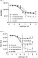
- the EC 50 values of GI-108A1 (AHF10240) and GI-108A1 (AHP04167) were 2.02 nM and 3.93 nM, respectively, and the EC 50 value of GI-108B1 was 1.70 nM.
- the EC 50 value of the positive control Proleukin ® was 0.08 nM.
- MDA-MB-231 cells human breast cancer cells
- GI-108A1 AHF10240
- GI-108A1 AHP04167
- GI-108B1 the control Oleclumab
- the 96-well plates were centrifuged, and the supernatants were transferred to new 96-well plates. Thereafter, the adenosine concentration of each of the supernatants was measured using an Adenosine Assay kit (Cell Biolabs, Inc) according to the manufacturer's protocol. IC 50 values were calculated using GraphPad Prism software.
- GI-108A1 As a result, the degree of luminescence was reduced by each of GI-108A1 (AHF10240), GI-108A1 (AHP04167), GI-108B1, and Oleclumab in a concentration-dependent manner. Thereby, it was confirmed that both types of GI-108A1 and GI-108B1 inhibited membrane-bound CD73 enzyme activity ( FIG. 5 ).
- the IC 50 values of GI-108A1 (AHF10240) and GI-108A1 (AHP04167) were 2.75 nM and 5.58 nM, respectively, and the IC 50 value of GI-108B1 was 3.37 nM.
- the IC 50 value of the control Oleclumab was 1.47 nM. Although the IC 50 value of GI-108B1 was higher than those of GI-108A1 (AHF10240) and Oleclumab, GI-108B1 showed the highest potential in enzyme inhibition efficacy.
- GI-108A1 AHF10240
- GI-108A1 AHP04167
- GI-108B1 the control Oleclumab
- the reaction was terminated by treating each well with 25 ⁇ L of AMP-Glo TM Reagent (Promega), and each well was shaken for 2 minutes and then incubated at room temperature for 1 hour. Each well was treated with AMP detection solution, shaken for 2 minutes, and then incubated at room temperature for 1 hour. The degree of luminescence was measured using the GloMax ® -Multi Detection System (Promega), and IC 50 values were calculated using GraphPad Prism 8.0 software.
- GI-108A1 (AHF10240) and GI-108A1 (AHP04167) were 0.060 nM and 0.102 nM, respectively, and the IC 50 values of GI-108B1 and Oleclumab were 0.126 nM and 0.03 nM, respectively. Although the IC 50 value of GI-108B1 was higher than those of both types of GI-108A1 and Oleclumab, GI-108B1 showed the highest potential in enzyme inhibition efficacy.
- human PBMCs were cultured in TexMACS TM medium (Miltenyi Biotec) containing 5% human serum (Sigma-Aldrich ® ), 1% penicillin/streptomycin and 5 mM ⁇ -mercaptoethanol (Thermo Fisher Scientific) under humidified conditions of 37°C and 5% CO 2 overnight. The next day, the cells were labeled with CTV and seeded in each well of 96-well plates at 1 x 10 6 cells/ml. The cells were stimulated using ⁇ CD3/CD28 dynabeads (1: 1 ratio).
- the cells were treated with vehicle (hIgG4), Proleukin ® , GI-108B1, anti-CD73 antibody (GI- ⁇ CD73), IgG4 Fc-IL-2v and Oleclumab alone, a combination of anti-CD73 antibody (GI- ⁇ CD73) and IgG4 Fc-IL-2v, or a combination of Oleclumab and IgG4 Fc-IL-2v.
- vehicle hIgG4
- Proleukin ® GI-108B1
- anti-CD73 antibody GI- ⁇ CD73
- IgG4 Fc-IL-2v anti-CD73 antibody
- Oleclumab IgG4 Fc-IL-2v
- Each of the drugs was added to each well to a final concentration of 50 nM.
- 500 ⁇ M of AMP was added to each well, and the cells were cultured for 3 days under humidified conditions of 37°C and 5% CO 2 .
- the cells were re-stimulated with ⁇ CD3/CD28 dynabeads (1:1 ratio) and 50 nM or 500 ⁇ M AMP in the presence of Brefeldin A under humidified conditions of 37°C and 5% CO 2 for 4 hours.
- surface markers of the cells were stained using the following antibodies: anti-CD3-PerCP-Cy5.5 (Clone: UCHT1, BD Biosciences), anti-CD8-Alexa Fluor700 (Clone: RPA-T8, Biolegend Inc.), and Fixable Viability Dye eFluor 780 (Invitrogen Inc.).
- the cells were treated with fixation/permeabilization buffer and then stained with anti-IFN ⁇ -APC antibody (Clone: 4S.B3, Biolegend Inc.). Data on the stained cells were obtained using a Symphony A3 instrument and analyzed using FlowJo software.
- GI-108B1 healthy human PBMCs were treated with Proleukin ® , GI-108B1, IgG4 Fc-IL-2v and anti-CD73 antibody (GI- ⁇ CD73) alone, or a combination of anti-CD73 antibody (GI- ⁇ CD73) and IgG4 Fc-IL-2v, and pSTAT5 in T cell populations (CD8+ T cells and Treg cells) in the PBMCs was measured.
- human PBMCs were treated with various concentrations of Proleukin ® , GI-108B1, IgG4 Fc-IL-2v, and anti-CD73 antibody alone, or with a combination of anti-CD73 antibody and IgG4 Fc-IL-2v, and incubated at 37°C for 20 minutes.
- cells were stained with the following antibodies: anti-CD3-BV510 (Clone SP34-2, BD Biosciences), anti-CD4-PE-Cy7 (Clone OKT-4, Biolegend Inc.), anti-CD8-AF700 (Clone RPA-T8, Biolegend Inc.), anti-CD25-BV421 (Clone M-A251, BD Biosciences), anti-CD56-BV605 (Clone NCAM16.2, BD Biosciences), anti-CD127-BV650 (Clone A019DS, Biolegend Inc.), and Fixable Viability Dye eFluor 780 (Invitrogen Inc.).
- the cells were fixed for 12 minutes by treatment with TFP Fix/Perm buffer (BD Biosciences), and incubated with Perm buffer III (BD Biosciences) for 30 minutes. The cells were then stained with anti-pSTAT5-AF647 (Clone 47/Stat5, BD Biosciences). Data on the stained cells were measured using Cytek Aurora (Cytek Biosciences) and analyzed using FlowJo software (BD Biosciences).
- Human PBMCs were suspended in 4Cell ® Nutri-T GMP medium (Sartorius), and the cells were dispensed in a 96-well plate at a density of 5 x 10 5 cells per well. Then, the cells were treated with GI-108B1 or Proleukin ® at a final concentration of 10 nM in the presence of ⁇ CD3/CD28 dynabeads (1: 1 ratio, Thermo Fisher Scientific). Every 2 to 3 days, the medium was replaced with a fresh medium containing each drug. The cells were counted on days 3, 5, 7, 10, and 12 using the ADAM TM -MC2 Cell Counter (Nanoentek).
- the cells were treated with fixation/permeabilization buffer (BD Biosciences) and stained with anti-Foxp3-PE antibody (Clone PCH101, Invitrogen Inc.). Data on the stained cells were measured using the Cytek Aurora instrument and analyzed using FlowJo software.
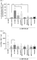
- the number of CD8+ T cells in the group treated with GI-108B1 was higher than in the group treated with Proleukin ® , and the number of Treg cells was about 2-fold higher in the group treated with Proleukin ® than in the group treated with GI-108B1.
- the ratio of the number of CD8+ T cells to the number of Treg cells (CD8+ T/Treg)
- it was confirmed that the CD8+ T/Treg in the group treated with GI-108B1 was about 2.46-fold higher than that in the group treated with Proleukin ® .
- GI-108B1 exhibited a superior effect on CD8+ T cell proliferation over Treg cell proliferation compared to Proleukin ® ( FIG. 15 ).
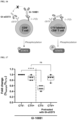
- CD8+ T cells express both CD73 and the IL-2 receptor complex, so signaling induced by the binding of GI-108B1 to CD73 and IL-2 ⁇ on the same CD8+ T cells can exhibit a synergistic effect on the activation of CD8+ T cells. Accordingly, to examine whether GI-108B1 cis-binds to CD73 and IL-2 ⁇ on the same CD8+ T cells, cis-binding analysis was performed using CTV-CD8+ T cells (anti-CD73 antibody-treated/untreated group) and CTV+CD8+ T cells ( FIG. 16 ). Specifically, CD8+ T cells were isolated from human PBMCs using the EasySep TM Human CD8+ T Cell Isolation Kit (STEMCELL technologies).
- CTV Cell Trace TM Violet, Thermo Fisher Scientific
- the CTV-unlabeled cells were divided into two groups, and one group was pre-treated with a saturating concentration (1,000 nM) of anti-CD73 antibody at 4°C for 2 hours to block all CD73 on the cell surface. Then, unbound anti-CD73 antibody was washed out.
- the CTV-unlabeled cells, pre-treated or untreated with anti-CD73 antibody were co-cultured with CTV-labeled cells at a 1:1 ratio.
- the co-cultured cells were treated with 0.5 nM GI-108B1 and incubated at 37°C for 20 minutes. Thereafter, the cells were fixed with Cytofix fixation buffer (BD Biosciences) for 12 minutes, and then treated with Phosflow Perm buffer III (BD Biosciences) and incubated for 30 minutes.
- the cells were then stained with anti-pSTAT5-AF647 (Clone 47/Stat5, BD Biosciences). Data for the stained cells were measured using Cytek Aurora and analyzed using FlowJo software.
- CTV-CD8+ T cells and CTV+CD8+ T cells showed similar levels of STAT5 phosphorylation, but in the case of CTV+CD8+ T cells co-cultured with CTV-CD8+ T cells pretreated with anti-CD73 antibody, the STAT5 phosphorylation level in CTV-CD8+ T cells was reduced by 40 to 60%, and the STAT5 phosphorylation level in CTV+CD8+ T cells remained the same ( FIG. 17 ).
- GI-108B1 To evaluate the immune cell-mediated tumor killing ability of GI-108B1, the anticancer effect of GI-108B1 was evaluated using a co-culture system of MDA-MB-231 cells and healthy human PBMC in the presence of anti-CD3/CD28 dynabeads and/or AMP. Specifically, the cytotoxicity of PBMCs against MDA-MB-231 wild-type cells was evaluated using flow cytometry-based Annexin V/7-AAD assay. PBMCs were purchased from Zen-bio Inc. or STEMCELL Technologies.
- PBMCs were treated with hIgG4 (5 nM) alone, a combination of anti-CD73 antibody (GI- ⁇ CD73, 5 nM) and IgG4 Fc-IL-2v (5 nM), or GI-108B1 (5 nM) alone, and also treated with ⁇ CD3/CD28 dynabeads (1:1 ratio) or 500 ⁇ M of AMP and cultured for 2 days before co-culture with target cells (MDA-MB-231 wild-type cells).
- Target cells were labeled with CTV and dispensed into a 24-well plate at a density of 1 x 10 5 cells per well.
- pre-stimulated PBMCs were added to the target cells at a ratio of effector cells to target cells of 10:1, followed by co-culture for 24 hours at 37°C under 5% CO 2 .
- the cells were stained with Annexin V and 7-AAD (Biolegend Inc.), and the death rate of the CTV-labeled target cells was measured using the Symphony A3 instrument and analyzed using FlowJo software.
- the group in which PBMCs treated with anti-CD3/CD28 dynabeads, AMP and GI-108B1 were co-cultured with target cells showed a significantly higher target-cell death rate than the group in which PBMCs treated with anti-CD3/CD28 dynabeads, AMP and a combination of anti-CD73 antibody (GI- ⁇ CD73) and IgG4 Fc-IL-2v were co-cultured with target cells, indicating the highest cell death rate ( FIG. 19 ).
- GI-108B1 was administered to a tumor model transplanted with the MC38 cell line (MC38-hCD73), a C57BL/6 murine colon cancer cell line in which the mouse CD73 gene was replaced with the human CD73 gene, the tumor growth inhibitory effect was evaluated.
- a suspension of MC38-hCD73 cells (SMOC, China) was administered subcutaneously to the right back of each of C57BL/6J-hCD73 female mice (6-8 weeks old, SMOC, China) at 1 x 10 6 cells/100 ⁇ L.
- mice were randomly selected when the average tumor volume was 90 to 120 mm 3 .
- the animals were selected considering their physiological state, weight change, and tumor growth rate.
- the tumor volume in each animal before administration was set as the value measured at the time of grouping, and the antitumor efficacy was evaluated by comparison with the control group (vehicle (PBS), G1).
- the tumor volume in the GI-108B 1-administered group was reduced compared to the control group (vehicle (PBS)) ( FIG. 20 ).
- the tumor growth inhibition rate only one mouse in the control group (vehicle (PBS)) showed a tumor growth inhibition rate of over 50%, and none showed over 80%.
- the GI-108B1-administered group 5 mice showed a tumor growth inhibition rate of over 30%, and 3 mice over 50% ( FIG. 21 ).
- 2 mice in the control group (vehicle (PBS)) died on day 25, whereas all mice in the GI-108B1-administered group survived on day 25 ( FIG. 22 ).
- a tumor model obtained by transplanting MDA-MB-231 cells (human breast cancer cells) into mice into which the human immune system has been introduced each of test substances GI-108A1 (AHP04167), GI-108B1, and Pembrolizumab (product name: Keytruda, MSD), an anti-PD-1 antibody, was administered, and then the tumor growth inhibitory effect was evaluated.
- a suspension of human peripheral blood cells (Stemexpress, USA) was filled into a disposable syringe (31G, cat.
- NOG-B2m NOD.Cg-B2mtm1UncPrkdescidIl2rgtm1Wjl/SzJ
- mice 7-week-old, The Jackson Laboratory, USA
- NOG-B2m female mice were purchased from The Jackson Laboratory, and NOG-B2m female mice were maintained in individually ventilated cages housed in an animal biosafety level-3 facility at 19 to 25°C and 30 to 70% humidity.
- the MDA-MB-231 human triple-negative breast cancer (TNBC) cell line was purchased from ATCC and cultured in DMEM medium (Thermo Fisher Scientific) containing 10% FBS (Thermo Fisher Scientific) and 1% antibiotic/antimycotic (Gibco TM ).
- the cultured MDA-MB-231 cells were harvested using trypsin (Gibco TM ) and then suspended in PBS.
- mice with no abnormalities in health About 20 days after transplanting the MDA-MB-231 cells, the tumor volume was measured in mice with no abnormalities in health, and 32 mice were selected so that the average tumor volume of each group reached 80 to 100 mm 3 .
- the animals were selected considering their physiological state (respiration, fur, behavior, tail, posture, body fluids, diet, morphological deformation, metabolism, etc.), weight change, FACS analysis results, and tumor growth rate.
- the test groups were formed as shown in Table 11 below and the test substances were administered thereto ( FIG. 23 ).
- mice showed a tumor growth inhibition rate of over 30%, 2 mice over 50%, with none over 80%.
- mice showed a tumor growth inhibition rate of over 30%, 3 mice over 50%, and 1 mouse over 80%.
- mice showed a tumor growth inhibition rate of over 30%, 4 mice over 50%, and 3 mice over 80% ( FIG. 26 ).
- GI-108B1 To analyze the vascular permeability of GI-108B1, 1 ⁇ 10 5 HUVEC cells (Lonza) were placed in a collagen-coated trans-well using a trans-well insert (9321012, cellQART ® ), and cultured for 3 days to form a confluent monolayer. 1 ⁇ 10 5 PBMCs were dispensed into a 96-well plate, and then treated with Proleukin ® (5 nM) or GI-108B1 (5 nM) and cultured for 24 hours at 37°C under 5% CO 2 .
- Proleukin ® Proleukin ®
- GI-108B1 5 nM
- the HUVEC cells cultured in the trans-well were treated with the drug and the culture supernatant of the PBMCs and cultured for 24 hours at 37°C under 5% CO 2 .
- 100 ng/ml of TNF- ⁇ (PeproTech) was used.
- the upper chamber was treated with dextran-FITC (70 kDa) and incubated for 1 hour and 30 minutes.
- the fluorescence of dextran-FITC that moved to the lower chamber was measured at wavelengths of 485 nm/500-550 nm (Ex/Em) using GloMax ® Discover (Promega).
- Statistical calculations were analyzed by one-way ANOVA using Prism 8.0 (Graph Pad Software Inc.), and Dunnett's test was performed based on the negative control.
Landscapes
- Health & Medical Sciences (AREA)
- Chemical & Material Sciences (AREA)
- Life Sciences & Earth Sciences (AREA)
- Medicinal Chemistry (AREA)
- Organic Chemistry (AREA)
- General Health & Medical Sciences (AREA)
- Immunology (AREA)
- Proteomics, Peptides & Aminoacids (AREA)
- Veterinary Medicine (AREA)
- Pharmacology & Pharmacy (AREA)
- Animal Behavior & Ethology (AREA)
- Public Health (AREA)
- Genetics & Genomics (AREA)
- Biophysics (AREA)
- Biochemistry (AREA)
- Molecular Biology (AREA)
- Engineering & Computer Science (AREA)
- Epidemiology (AREA)
- Zoology (AREA)
- Gastroenterology & Hepatology (AREA)
- Bioinformatics & Cheminformatics (AREA)
- Mycology (AREA)
- Microbiology (AREA)
- Chemical Kinetics & Catalysis (AREA)
- Toxicology (AREA)
- General Chemical & Material Sciences (AREA)
- Nuclear Medicine, Radiotherapy & Molecular Imaging (AREA)
- Biomedical Technology (AREA)
- Oncology (AREA)
- Peptides Or Proteins (AREA)
- Medicines That Contain Protein Lipid Enzymes And Other Medicines (AREA)
- Micro-Organisms Or Cultivation Processes Thereof (AREA)
- Medicines Containing Antibodies Or Antigens For Use As Internal Diagnostic Agents (AREA)
Applications Claiming Priority (2)
| Application Number | Priority Date | Filing Date | Title |
|---|---|---|---|
| KR20220076279 | 2022-06-22 | ||
| PCT/KR2023/008664 WO2023249425A1 (ko) | 2022-06-22 | 2023-06-22 | 항-cd73 항체 및 il-2를 포함하는 융합단백질 및 이의 용도 |
Publications (2)
| Publication Number | Publication Date |
|---|---|
| EP4545562A1 true EP4545562A1 (de) | 2025-04-30 |
| EP4545562A4 EP4545562A4 (de) | 2025-10-01 |
Family
ID=89380237
Family Applications (1)
| Application Number | Title | Priority Date | Filing Date |
|---|---|---|---|
| EP23827535.8A Pending EP4545562A4 (de) | 2022-06-22 | 2023-06-22 | Fusionsprotein mit anti-cd73-antikörper und il-2 sowie verwendung davon |
Country Status (10)
| Country | Link |
|---|---|
| US (2) | US12414984B2 (de) |
| EP (1) | EP4545562A4 (de) |
| JP (1) | JP2025521387A (de) |
| KR (1) | KR20240000394A (de) |
| CN (1) | CN118974093A (de) |
| AU (1) | AU2023287432A1 (de) |
| CL (1) | CL2024003716A1 (de) |
| IL (1) | IL317834A (de) |
| MX (1) | MX2024014595A (de) |
| WO (1) | WO2023249425A1 (de) |
Family Cites Families (14)
| Publication number | Priority date | Publication date | Assignee | Title |
|---|---|---|---|---|
| US5229109A (en) * | 1992-04-14 | 1993-07-20 | Board Of Regents, The University Of Texas System | Low toxicity interleukin-2 analogues for use in immunotherapy |
| US7371371B2 (en) * | 2001-08-13 | 2008-05-13 | University Of Southern California | Interleukin-2 mutants with reduced toxicity |
| PT1454138E (pt) * | 2001-12-04 | 2012-03-28 | Merck Patent Gmbh | Imunocitoquinas com seletividade modulada |
| WO2009061853A2 (en) * | 2007-11-05 | 2009-05-14 | Massachusetts Institute Of Technology | Mutant interleukin-2 (il-2) polypeptides |
| US11180554B2 (en) | 2016-12-13 | 2021-11-23 | Astellas Pharma Inc. | Anti-human CD73 antibody |
| CN110240654A (zh) * | 2018-03-07 | 2019-09-17 | 复旦大学 | 结合cd73的抗体-药物偶联物 |
| US20210147570A1 (en) * | 2018-04-12 | 2021-05-20 | Bristol-Myers Squibb Company | Anticancer combination therapy with cd73 antagonist antibody and pd-1/pd-l1 axis antagonist antibody |
| US20230104217A1 (en) | 2018-09-17 | 2023-04-06 | Gi Innovation, Inc. | Fusion protein comprising il-2 protein and cd80 protein, and use thereof |
| US11492384B2 (en) | 2018-09-17 | 2022-11-08 | Gi Innovation, Inc. | Fusion protein comprising IL-2 protein and CD80 protein, and use thereof |
| CN112442132A (zh) * | 2019-09-05 | 2021-03-05 | 复旦大学 | 靶向肿瘤的重组双功能融合蛋白及其应用 |
| ES3040184T3 (en) * | 2020-03-18 | 2025-10-29 | Gi Innovation Inc | Fusion protein preparation comprising il-2 and cd80 proteins |
| KR102313505B1 (ko) | 2020-06-30 | 2021-10-18 | (주)지아이이노베이션 | 항-lag-3 항체 및 il-2를 포함하는 융합단백질 및 이의 용도 |
| TWI815194B (zh) * | 2020-10-22 | 2023-09-11 | 美商基利科學股份有限公司 | 介白素2-Fc融合蛋白及使用方法 |
| IL310292A (en) * | 2021-07-22 | 2024-03-01 | Univ Dundee | Medicinal myotains |
-
2023
- 2023-06-22 WO PCT/KR2023/008664 patent/WO2023249425A1/ko not_active Ceased
- 2023-06-22 JP JP2024551932A patent/JP2025521387A/ja active Pending
- 2023-06-22 EP EP23827535.8A patent/EP4545562A4/de active Pending
- 2023-06-22 IL IL317834A patent/IL317834A/en unknown
- 2023-06-22 CN CN202380032064.7A patent/CN118974093A/zh active Pending
- 2023-06-22 KR KR1020230080204A patent/KR20240000394A/ko not_active Ceased
- 2023-06-22 AU AU2023287432A patent/AU2023287432A1/en active Pending
-
2024
- 2024-08-26 US US18/815,390 patent/US12414984B2/en active Active
- 2024-11-25 MX MX2024014595A patent/MX2024014595A/es unknown
- 2024-12-04 CL CL2024003716A patent/CL2024003716A1/es unknown
-
2025
- 2025-08-21 US US19/306,002 patent/US20260061032A1/en active Pending
Also Published As
| Publication number | Publication date |
|---|---|
| IL317834A (en) | 2025-02-01 |
| US12414984B2 (en) | 2025-09-16 |
| JP2025521387A (ja) | 2025-07-10 |
| KR20240000394A (ko) | 2024-01-02 |
| MX2024014595A (es) | 2025-01-09 |
| AU2023287432A1 (en) | 2025-01-02 |
| CN118974093A (zh) | 2024-11-15 |
| CL2024003716A1 (es) | 2025-03-21 |
| EP4545562A4 (de) | 2025-10-01 |
| US20260061032A1 (en) | 2026-03-05 |
| WO2023249425A1 (ko) | 2023-12-28 |
| US20240408176A1 (en) | 2024-12-12 |
Similar Documents
| Publication | Publication Date | Title |
|---|---|---|
| JP7587656B2 (ja) | Pd-l1及びcd137に結合する抗体分子 | |
| US20220153876A1 (en) | Bispecific antibodies for use in stem cell transplantation | |
| US11214618B2 (en) | LAG-3 binding members | |
| KR102089072B1 (ko) | 항-인간 4-1bb 항체 및 그의 용도 | |
| WO2015070061A1 (en) | Anti-wt1/hla bi-specific antibody | |
| CA3100232A1 (en) | Heavy chain antibodies binding to cd38 | |
| CN111133007A (zh) | 与胞外酶结合的重链抗体 | |
| KR102261582B1 (ko) | 항-pd-l1-항-tim-3 이중특이적 항체 | |
| KR20190117467A (ko) | IFN-γ-유도성 조절 T 세포 전환가능 항암 (IRTCA) 항체 및 그의 용도 | |
| EP4545562A1 (de) | Fusionsprotein mit anti-cd73-antikörper und il-2 sowie verwendung davon | |
| HK40114315A (zh) | 包含抗cd73抗体和il-2的融合蛋白及其用途 | |
| KR20140025836A (ko) | 항 HER2/Neu 단일클론항체와 IL-7가 연결된 융합 단일클론 항체 및 이를 포함하는 암 치료용 조성물 | |
| EP4477228A1 (de) | Pharmazeutische zusammensetzung zur krebsbehandlung mit fusionsprotein mit il-12 und anti-fap-antikörper und antikrebsmittel | |
| RU2777573C2 (ru) | Антитела против 4-1bb человека и их применение | |
| HK40122804A (en) | Antibody molecules that bind pd-l1 and cd137 | |
| BR122022022755A2 (pt) | Anticorpos de cadeia pesada multispecíficos com regiões constantes de cadeia pesada modificadas | |
| HK40050345B (en) | Antibody molecules that bind pd-l1 and cd137 | |
| HK40050345A (en) | Antibody molecules that bind pd-l1 and cd137 |
Legal Events
| Date | Code | Title | Description |
|---|---|---|---|
| STAA | Information on the status of an ep patent application or granted ep patent |
Free format text: STATUS: THE INTERNATIONAL PUBLICATION HAS BEEN MADE |
|
| PUAI | Public reference made under article 153(3) epc to a published international application that has entered the european phase |
Free format text: ORIGINAL CODE: 0009012 |
|
| STAA | Information on the status of an ep patent application or granted ep patent |
Free format text: STATUS: REQUEST FOR EXAMINATION WAS MADE |
|
| 17P | Request for examination filed |
Effective date: 20240822 |
|
| AK | Designated contracting states |
Kind code of ref document: A1 Designated state(s): AL AT BE BG CH CY CZ DE DK EE ES FI FR GB GR HR HU IE IS IT LI LT LU LV MC ME MK MT NL NO PL PT RO RS SE SI SK SM TR |
|
| A4 | Supplementary search report drawn up and despatched |
Effective date: 20250902 |
|
| DAV | Request for validation of the european patent (deleted) | ||
| DAX | Request for extension of the european patent (deleted) | ||
| RIC1 | Information provided on ipc code assigned before grant |
Ipc: C07K 16/28 20060101AFI20250827BHEP Ipc: C07K 14/55 20060101ALI20250827BHEP Ipc: A61P 35/00 20060101ALI20250827BHEP Ipc: A61K 39/00 20060101ALI20250827BHEP |