EP4548841A1 - Verfahren zur verbesserung der genauigkeit und zuverlässigkeit nichtmedizinischer wearable-vorrichtungen zur erzeugung eines alarms im zusammenhang mit akutem myokardinfarkt - Google Patents
Verfahren zur verbesserung der genauigkeit und zuverlässigkeit nichtmedizinischer wearable-vorrichtungen zur erzeugung eines alarms im zusammenhang mit akutem myokardinfarkt Download PDFInfo
- Publication number
- EP4548841A1 EP4548841A1 EP23207896.4A EP23207896A EP4548841A1 EP 4548841 A1 EP4548841 A1 EP 4548841A1 EP 23207896 A EP23207896 A EP 23207896A EP 4548841 A1 EP4548841 A1 EP 4548841A1
- Authority
- EP
- European Patent Office
- Prior art keywords
- ecg
- qrs
- ppg
- ami
- signal
- Prior art date
- Legal status (The legal status is an assumption and is not a legal conclusion. Google has not performed a legal analysis and makes no representation as to the accuracy of the status listed.)
- Pending
Links
Images



Classifications
-
- A—HUMAN NECESSITIES
- A61—MEDICAL OR VETERINARY SCIENCE; HYGIENE
- A61B—DIAGNOSIS; SURGERY; IDENTIFICATION
- A61B5/00—Measuring for diagnostic purposes; Identification of persons
- A61B5/68—Arrangements of detecting, measuring or recording means, e.g. sensors, in relation to patient
- A61B5/6801—Arrangements of detecting, measuring or recording means, e.g. sensors, in relation to patient specially adapted to be attached to or worn on the body surface
- A61B5/6802—Sensor mounted on worn items
- A61B5/681—Wristwatch-type devices
-
- A—HUMAN NECESSITIES
- A61—MEDICAL OR VETERINARY SCIENCE; HYGIENE
- A61B—DIAGNOSIS; SURGERY; IDENTIFICATION
- A61B5/00—Measuring for diagnostic purposes; Identification of persons
- A61B5/02—Detecting, measuring or recording for evaluating the cardiovascular system, e.g. pulse, heart rate, blood pressure or blood flow
- A61B5/024—Measuring pulse rate or heart rate
- A61B5/02416—Measuring pulse rate or heart rate using photoplethysmograph signals, e.g. generated by infrared radiation
-
- A—HUMAN NECESSITIES
- A61—MEDICAL OR VETERINARY SCIENCE; HYGIENE
- A61B—DIAGNOSIS; SURGERY; IDENTIFICATION
- A61B5/00—Measuring for diagnostic purposes; Identification of persons
- A61B5/24—Detecting, measuring or recording bioelectric or biomagnetic signals of the body or parts thereof
- A61B5/316—Modalities, i.e. specific diagnostic methods
- A61B5/318—Heart-related electrical modalities, e.g. electrocardiography [ECG]
- A61B5/346—Analysis of electrocardiograms
- A61B5/349—Detecting specific parameters of the electrocardiograph cycle
-
- A—HUMAN NECESSITIES
- A61—MEDICAL OR VETERINARY SCIENCE; HYGIENE
- A61B—DIAGNOSIS; SURGERY; IDENTIFICATION
- A61B5/00—Measuring for diagnostic purposes; Identification of persons
- A61B5/72—Signal processing specially adapted for physiological signals or for diagnostic purposes
- A61B5/7235—Details of waveform analysis
- A61B5/7264—Classification of physiological signals or data, e.g. using neural networks, statistical classifiers, expert systems or fuzzy systems
- A61B5/7267—Classification of physiological signals or data, e.g. using neural networks, statistical classifiers, expert systems or fuzzy systems involving training the classification device
Definitions
- the present invention relates to the field of generating an alert for suspected acute myocardial infarction using non-medical equipment, and more particularly, to a method for reducing the time required to initiate the alert and improving accuracy and reliability of an alert about a suspected acute myocardial infarction.
- AMI Acute myocardial infarction
- a heart attack is a serious health condition characterized by the sudden blockage of one or more coronary arteries that supply blood to the heart muscle.
- the blockage is typically caused by the formation of a blood clot, which impedes blood flow and causes damage to the heart muscle due to the lack of oxygen and nutrients.
- Symptoms of AMI can vary from person to person and may include chest pain, shortness of breath, nausea, vomiting, lightheadedness, and sweating. Prompt medical attention is crucial, as the condition can lead to severe complications or even death if left untreated.
- Eurostat Eurostat. Avoidable Deaths in 2016 - For People Under 75, Two Deaths Out of Three in the EU could Have Been Avoided. Eurostat News Release 136/2019
- effective health management and timely interventions have the potential to prevent up to two-thirds of premature deaths, with AMI being the leading cause. Therefore, the development of new preventive strategies is a pressing priority.
- AMI often affects the heart muscle and the heart's electrical conduction system, and these effects can be observed in an electrocardiogram (ECG).
- ECG clinical features characteristic of AMI depend on the location and extent of the heart muscle damage caused by the blockage of a coronary artery.
- QRS complex representing ventricular depolarization
- T wave representing ventricular repolarization
- AMI manifests as an elevation of the ST segment, which represents the period between ventricular depolarization and repolarization. ST elevation above the baseline is usually observed in the ECG leads corresponding to the location of the infarcted heart area.
- the primary focus is on analyzing changes in the ST segment of the ECG.
- the ECG may show an inverted T wave in the leads affected by AMI.
- AMI can also lead to changes in the heart's electrical axis, causing shifts in the QRS complex and T-wave direction. These deviations can provide further clues about the extent and location of myocardial damage.
- AMI can disrupt the heart's electrical conduction system, leading to arrhythmias, such as frequent ventricular premature beats (VPBs), ventricular tachycardia, or ventricular fibrillation.
- AMI can lead to other conduction system-related issues, such as left or right bundle branch blocks.
- a reduced-lead ECG such as a 6-lead ECG
- a 6-lead ECG may be used for prolonged (e.g., 1, or 2-day) ECG monitoring outside the clinical setting.
- the 6-lead ECG is obtained from 6 electrodes including four limb and two chest electrodes. These leads capture the heart's electrical signals from various angles, although are less informative compared to the 12-lead ECG.
- clinical reduced-lead ECG recorders may offer some benefits, they suffer from a limitation that the occurrence of an AMI cannot be predicted, making it difficult to prescribe ECG monitoring in advance. Therefore, there is a critical need for convenient and affordable technology that can facilitate the timely identification of AMI, and that is simple enough for people to use on their own.
- This invention overcomes specific limitations of the prior art, particularly in terms of reducing the time needed to acquire a multi-lead ECG and improving the accuracy and reliability of AMI-related alerts.
- the present invention introduces a method for reducing the time for obtaining multi-lead ECG and improving accuracy and reliability of alerts related to suspected AMI when non-medical wearable devices with fewer than 12 ECG leads are employed. Information missing due to reduced number of ECG leads, i.e., less than 12 leads, is compensated for by steps according to the invention.
- the method does not encompass any diagnostic procedures and is not, in itself, a diagnostic method for AMI.
- the method involves analysis of physiological signals such as multi-lead ECG and PPGs of different wavelengths, for generating a prompt, accurate, and reliable alert for AMI.
- the method steps that compensate for the missing information due to the reduced number of ECG leads are carried out on personal computing devices like personal computers, smartphones, or wrist-worn devices such as smartwatches, and a server. These steps comprises processing physiological signals, extracting static and dynamic AMI features, assessing ST segment abnormality and QRS-T angle in the ECG, detecting bundle branch block, calculating ECG signal quality index, estimating the reduction in oxygen saturation, detecting ventricular premature beats, detecting ventricular arrhythmia, calculating PPG signal quality index, training a reservoir computing model, such as an echo state neural network, on individual data.
- Various hyperparameters such as reservoir size, spectral radius, sparsity, input scaling, output scaling, and noise level, are tuned to optimize the performance.
- an alert on suspected AMI can be generated based on the data acquired when implementing method according to the invention if the alert score exceeds a predefined threshold.
- the alert may be conveyed using auditory, visual, or vibratory modalities.
- the generated alert concerning a suspected AMI, complements the user's selfevaluation of own condition with objective information gathered by the non-medical device. In essence, the user receives an alert based on an analysis of a combination of diverse AMI-related features extracted from distinct physiological signals obtained by the non-medical device.
- steps of the method (100) for improving accuracy and reliability of non-medical wearable devices for generating an alert related to AMI are carried out on a personal computing device such as a standard personal computer, a portable computer, a handheld device, such as a smartphone, a wrist-worn device, such as a wristwatch, and a server.
- a personal computing device such as a standard personal computer, a portable computer, a handheld device, such as a smartphone, a wrist-worn device, such as a wristwatch, and a server.
- the method (100) steps according to the invention comprise preprocessing (102) physiological signals obtained by a non-medical device, detecting fiducial points in the physiological signals (104), extracting AMI features (106), training a reservoir computing model using individual data (108).
- An alert (118) on a suspected AMI can be produced (114) if an alert score (110), computed using data obtained while carrying out method steps according to the invention, exceeds a predefined threshold (112). Analysis steps are repeated if the threshold is not exceeded (116).
- An alert (118) may be implemented using audible, vibrating, visual, or a combination of multiple alert-producing modalities to promptly draw the attention of the person who uses the device.
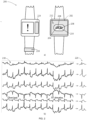
- an example of a non-medical personal computing device is a wearable wrist-worn device (200) comprising integrated bio-optical sensors for capturing PPGs of different wavelengths and biopotential electrodes for acquiring multi-lead ECG signals.
- the bottom (202) of the wearable wrist-worn device (200) comprises a red-light LED (204), an infrared light LED (206), and a greenlight LED (208).
- a photodetector (210) disposed on the bottom (202) of the wearable wrist-worn device (200), captures the reflected lights to obtain PPG signals of corresponding wavelengths.
- green light approximately 525 nm wavelength
- red approximately 660 nm wavelength
- infrared approximately 940 nm wavelength
- red, infrared, and green light PPG signals are preprocessed (102) using infinite impulse response filters.
- a low-pass infinite impulse response filter with a cutoff frequency of 6 Hz is used.
- a high-pass infinite impulse response filter with a cutoff frequency of 0.5 Hz is applied.
- the embedded software within the wrist-worn device processes continuously acquired PPGs in real time.
- a bottom biopotential electrode (212), located at the bottom of the wearable wrist-worn device (200), and a top biopotential electrode (214), located at the top of the wearable wrist-worn device (200), are configured to capture multi-lead ECG signals of which the top biopotential electrode (214) needs to be touched with a finger of the opposite hand.
- a further electrode (216), located at a distance from the top biopotential electrode (214), for example on a strap of the wearable wrist-worn device (200), must be brought into contact with chest of the wearer, specifically in the approximate location of precordial ECG lead V1. This arrangement allows the simultaneous acquisition of a 6-lead ECG of modified limb and chest leads (218).
- the wrist-worn device is configured to acquire a raw multi-lead ECG signal (218), which is then transmitted to the server through an internet connection for preprocessing (102).
- preprocessing a finite impulse response filter configured with a cutoff frequency set at 40 Hz is used to attenuate high-frequency noise while preserving the frequency components inherent to the ECG signal.
- an infinite impulse response filter configured with a cutoff frequency set at 0.05 Hz is used to mitigate ECG disturbances due to baseline wandering and artifacts resulting from suboptimal skin-electrode contact.
- the weighted averaging of ECG beats from multiple leads is used to obtain a representative ECG beat that is used for extracting static AMI features (220). Weighted averaging is accomplished by assigning varying weights to ECG beats derived from specific ECG leads, such as modified limb and chest leads, based on their noise level, expressed as the root mean square of signal values. ECG beats of superior quality with minimal noise are assigned higher weights, whereas those exhibiting a higher noise level are assigned lower weights. Signal to noise ratio is assessed in the TP segment in an ECG which represents the interval between the conclusion of the T wave and the onset of the P wave. Normally, the TP interval aligns with the baseline thus any deviations in the TP interval can be considered as being due to noise or artifacts.
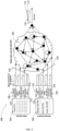
- the AMI alert (300) relies on a reservoir computing model such as an echo state neural network (302), capable of processing temporal data.
- the reservoir computing model utilizes static (304) and dynamic (306) AMI features as inputs.
- Static AMI features (304) may include ST segment abnormality index (308), QRS-T angle (310), the occurrence of bundle branch block (312), and ECG signal quality index (314), all of which are computed from the multi-lead ECG and updated with each intermittent ECG recording.
- Dynamic AMI features (306) such as reduction in oxygen saturation (SpO 2 ) (316), the number of ventricular premature beats per minute (318), the occurrence of ventricular arrhythmias (320), and PPG signal quality index (322), are updated continuously on a beat-to-beat basis.
- the numerical values of the ECG signal quality index (314) and the PPG signal quality index (322) are employed to adjust the importance of AMI features that are input into the reservoir computing model. In other words, when the signal quality index takes a low value, indicating poor signal quality, the importance of AMI features is proportionally reduced (324).
- the reservoir computing model produces an alert score (110), and when this score exceeds a predetermined threshold (112), it triggers the generation of an alert for a suspected AMI at that specific time instance (118).
- ECG and PPG signals are used to extract AMI features.
- the embedded software of the wrist-worn device (200) conducts real-time extraction of PPG-based AMI features, i.e., estimates a reduction in oxygen saturation (SpO 2 ) (316), the number of ventricular premature beats per minute (318), the occurrence of ventricular arrhythmias (320), and the quality of the PPG signal (322).
- the QRS-T complex has to be identified. To achieve this, several steps are taken. Firstly, the R peak within the QRS complex is pinpointed using the algorithm which encompasses bandpass filtering, differentiation, rectification, integration, and thresholding. Following the localization of the R peak, it serves as a reference point for determining both the termination of the T wave and the onset of the Q wave. A search for the T wave is conducted within a 200-millisecond window following the R peak. The method employed for detecting the T-wave endpoint employs a derivativebased approach. This technique involves the computation of the derivative of the ECG signal, with the endpoint of the T wave recognized as the point where the slope gradually recedes, signifying a return to the baseline. Similarly, the onset of the Q wave is identified using the same principle. By calculating the derivative of the ECG signal and identifying a notable shift in slope, the initiation of the Q wave is ascertained, indicating a distinct transition from the baseline.
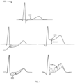
- the ST segment deviation (400) from the baseline of the ECG (402) is an important clinical feature of AMI. This deviation can manifest as either depression (404) or elevation (406), indicating abnormal electrical activity within the heart muscle, often associated with AMI.
- ST depression is a more than 0.1 mV deviation of the ST segment below the baseline (404)
- ST elevation is a deviation of more than 0.3 mV above the baseline (406).
- the method refrains from using predefined thresholds to identify ST segment abnormality since no step of diagnosis is involved. Instead, the numerical value of the ST abnormality will vary depending on the extent of the abnormality observed.
- the resulting abnormality index provides a quantifiable measure of the observed abnormality, reflecting the degree of deviation from the baseline.
- all acceptable-quality ST segments in the intermittent ECG are averaged and serve as a representative ST segment for subsequent calculations.
- QRS-T angle 500
- the direction of the depolarization (QRS complex) and repolarization (T-wave) waves may deviate from their baseline spatial orientation (502), often increasing the angle between the QRS (504) and T (506) vectors, i.e., the spatial QRS-T angle (508).
- Normal QRS-T angles (510) typically range between 30-70o, while angles exceeding 90o may suggest AMI.
- the conventional approach for calculating the spatial QRS-T angle requires a vectorcardiogram to estimate the xyz coordinates of the QRS ( u QRS ) and T ( u T ) vectors.
- the vectorcardiogram composed by combining three orthogonal ECG leads, is a three-dimensional representation of the 12-lead ECG (512). These leads capture the electrical activity of the heart along the right-to-left (x), head-to-feet (y), and front-to-back (z) axes.
- all existing methods for obtaining a vectorcardiogram necessitate the acquisition of two frontal and six precordial ECG leads to reconstruct the heart's electrical activity in three-dimensional space.
- the conventional approach for reconstructing the vectorcardiogram is not applicable when dealing with a modified 6-lead ECG obtained from a non-medical device, such as a wrist-worn device. Therefore, a deep learning model (514) is utilized to estimate u QRS and u T .
- the model is a one-dimensional convolutional neural network with regression output trained with 12-lead ECGs to predict the three coordinates of the vectorcardiogram-derived QRS and T vectors.
- These QRS and T vectors serve as output (516) while predictions are generated from signal-averaged beat (518) of the subset of ECG leads acquired by the wrist-worn device serve as the input.
- the average beat serves as a representative QRS-T complex of all acceptable-quality beats in the intermittent ECG to ensure robustness to noise.
- the deep-learning model is designed to determine the coordinates of u QRS and u T , rather than directly calculating the angles. This approach is chosen to effectively utilize any spatial information present in the input ECG leads during the training process. To achieve this, a composite loss function is implemented to guide the model within the three-dimensional space.
- the hyperparamters w 1 , w 2 and w 3 weigh the penalization factor of L ⁇ , L ⁇ and L r .
- the precise numerical value of the estimated QRS-T angle serves as an input to the reservoir computing model in the preferred embodiment of the invention.
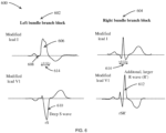
- a bundle branch block is detected in a multi-lead ECG acquired using a non-medical wrist-worn device (200), incorporating analysis of time and waveform features.
- the bundle branch block as observed in the ECG, is characterized by a discernible ECG pattern in the modified leads I and V1 compared to the reference ECG obtained during a non-AMI condition.
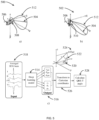
- the left bundle branch block (602) is distinguished by a broad monophasic R-wave in modified lead I (606), the absence of a Q-wave in modified lead I (608), and the presence of deep and broad S-waves in the modified lead V1 (610).
- the right bundle branch block (604) is distinguished by the presence of an rSR' complex, where the normal QRS complex is replaced by two R-waves and a large S-wave in the modified lead V1 (612). The initial R-wave is followed by a small S-wave, succeeded by a second prominent R-wave.
- both left and right bundle branch blocks share a common feature: a prolonged QRS complex duration of equal to or greater than 0.12 seconds (614).
- the outcome of the bundle branch block detector is binary, providing a distinction between two possibilities: a value of "0" indicates the absence of a bundle branch block, while a value of "1" indicates the presence of a bundle branch block.
- the quality assessment of the ECG signal involves calculating the ECG quality index, which compares how accurately different QRS detectors (e.g., Pan-Tompkins, Hamilton-Tompkins) detect QRS complexes. This is accomplished by using two or more QRS detectors on the ECG signal and checking if they detect the same QRS complex within the predefined error range (e.g., 10 ms).
- the analysis window size is set to match the duration of the entire ECG signal.
- ECG quality is assessed based on the specific numerical value of the ECG quality index, rather than using predefined thresholds.
- the ECG quality index is represented by a numerical value ranging from 0 to 1. A value of 0 indicates poor quality, while a value approaching 1 signifies good quality. This numerical value is then used to adjust the significance of other ECG-based AMI features when they are input into the reservoir computing model (324).
- each ECG-based input (such as the ST segment abnormality index, QRS-T angle, or the presence of bundle branch block) is scaled by the ECG quality index value.
- the ECG quality is poor, e.g., the value of the ECG quality index approaches 0, these inputs become significantly less influential and contribute negligibly to the AMI alert score.
- the reduction in SpO 2 is estimated from wrist PPG signals.
- the principle of SpO 2 estimation is rooted in the fact that hemoglobin absorbs red and infrared light differently depending on whether it is bound to oxygen or not. Oxygenated hemoglobin absorbs more infrared wavelength and less red wavelength, while deoxygenated hemoglobin absorbs more red wavelength and less infrared wavelength.
- a green wavelength PPG (704) which is often of better quality, serves as a reference for a normalized least-mean-square adaptive filter (706) of first order with the adaptation step of 0.1 to improve the quality of red and infrared PPG signals (708).
- the morphology of the wrist PPG signal (712) is modified to align with the morphology of the finger PPG signal.
- a system identification method such as the autoregressive moving average (ARMA) model, is employed to establish the relationship between finger and wrist PPG signals.
- the ARMA model estimates the individual transfer function by utilizing wrist PPG signals as the input and finger PPG signals (710) as the output of the system. Subsequently, the estimated transfer function, essentially a frequency response function, is applied to independently process wrist PPG signals, eliminating the need for acquiring finger PPG data through conventional inconvenient means.
- ARMA autoregressive moving average
- red and infrared wavelength PPG signals are processed to extract the alternating component (AC) from the baseline component (DC).
- the baseline component represents the steady-state blood volume level in the tissue, and it is a result of the average blood volume in the microvascular bed.
- the baseline component is relatively constant during the cardiac cycle and exhibits only slow-changing oscillations.
- the first step involves removing high-frequency noise using a low-pass filter with a cutoff frequency of 6 Hz.
- a normalized least-mean-square adaptive filter of a first order with an adaptation step of 0.01 is applied.
- the alternating component represents the pulsatile changes in blood volume associated with each heartbeat.
- a high-pass filter with a cutoff frequency of 0.5 Hz and a normalized least-mean-square adaptive filter of a first-order with an adaptation step of 0.1 are used.
- the ratio of the amplitudes of the alternating and baseline components of the red and infrared PPG signals is proportional to the amount of oxygenated hemoglobin in the blood.
- VPBs ventricular arrhythmias
- ventricular arrhythmias including VPBs
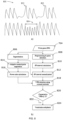
- the algorithm for detecting VPBs utilizes temporal and power-derived features extracted from an individual PPG pulse of green light PPG (704).
- a sliding analysis window of 10 s with 50% overlap is used for PPG segmentation (806).
- Positive peaks of the preprocessed PPG are detected using a threshold-crossing technique (808).
- the intervals between PPG pulses i.e., PP intervals (810), are normalized (812) to make them independent of heart rate, and a normalization principle based on the ratio of the current and mean values of the intervals is employed.
- a second high-pass finite impulse response filter with a variable cutoff frequency is used to extract higher frequency components, and a pre-whitened least squares adaptive filter is employed with an order of 12 and a forgetting factor of 0.999 to suppress normal PPG pulses (814) and in such a way to enhance premature pulses which are likely VPBs.
- Power ratios are computed in segments between adjacent PPG pulses, involving both preprocessed PPG and the signal after adaptive filtering.
- VPBs are detected using a feed-forward artificial neural network with a hidden layer of 50 neurons, trained using a backpropagation learning technique (818).
- Ventricular arrhythmias such as ventricular tachycardia and ventricular flutter, are characterized by an accelerated heart rate (804) resulting from irregular electrical impulses arising from the ventricles of the heart.
- Ventricular tachycardia is typically accompanied by a regular heart rhythm that exceeds 100 beats per minute, while ventricular flutter also exhibits a regular rhythm with a range commonly falling between 250 and 350 beats per minute. If three or more consecutive VPBs in the PPG (820) with a heart rhythm exceeding 100 beats per minute are detected (822), the entire episode of VPBs is assigned to ventricular arrhythmia (824), and it results in a high output (“1") being generated for this AMI feature. Otherwise, a low output (“0") is generated.
- PPG pulse quality is assessed using template matching involving comparing the shape of a reference pulse waveform with the shape of the measured pulse waveform. Obtaining a reference pulse waveform may be done by averaging several high-quality PPG pulse waveforms. The reference waveform should represent the typical shape of a high-quality PPG pulse. Preprocessing of the measured pulse waveform involves filtering to remove noise, baseline correction, and normalization to ensure that the waveform has a consistent amplitude and duration.
- Aligning the reference waveform with the measured waveform can be done using cross-correlation or other alignment methods to ensure that the reference waveform is properly aligned with the corresponding portion of the measured waveform.
- Calculating the similarity score which represents how closely the measured waveform matches the reference waveform, can be done using a similarity metric such as the correlation coefficient.
- This numerical value is subsequently employed to modify the relative importance of various PPG-based AMI features when they are fed into the reservoir computing model (324).
- each PPG-based input e.g., reduction in SpO 2 , the count of ventricular premature beats per minute, and the occurrence of ventricular arrhythmias
- the PPG quality index value When the PPG quality is low, for instance, as the PPG quality index value approaches 0, these inputs exert substantially less influence and make only a negligible contribution to the AMI alert score.
- the echo state neural network is utilized as the decision-making model in this embodiment of the invention based on its distinct advantages over conventional machine learning and deep learning approaches.
- the echo state neural network offers ease of training, as it employs fixed, randomly initialized recurrent weights and only output weights adjusted during training.
- the echo state neural network is computationally efficient, e.g., can be parallelized for efficient implementation on hardware accelerators, making it suitable for applications with limited computational resources, such as wrist-worn devices.
- the echo state neural network exhibits robustness to noise and can effectively work with noisy data, while also demonstrating good generalization to unseen data.
- the performance of an echo state network depends on several hyperparameters that need to be tuned to achieve optimal performance.
- the size of the reservoir which is the recurrent layer of the echo state network, is set to 1000.
- the spectral radius which is a scaling factor that is applied to the weights of the reservoir and affects the stability and dynamic range of the network, is set to 1.
- the sparsity of the reservoir which is the proportion of zero-valued weights in the reservoir, is set to 20 %.
- Input scaling which is a scaling factor applied to the input weights to affect the sensitivity of the network to the input data, is set to 1.
- the output scaling which refers to the transformation applied to the activations of the neurons in the reservoir to produce the final network output, is set to 1.
- the noise level which may improve the robustness of the network and prevent overfitting, is set to 0.001.
- the goal of training (900) the echo state network is to enable the model to recognize changes in AMI features compared to those estimated during a non-AMI condition.
- the training process may involve a sequence of 60 non-pathological cardiac cycles (902) for each person's data which may be obtained before the occurrence of AMI. Then, a deviation in AMI features is added for another 60 cardiac cycles to mimic AMI (904).
- the ST segment abnormality index (308) is increased by 50%
- the QRS-T angle (310) is increased by 50%
- the occurrence of bundle branch block (312) is identified, i.e., the output of this particular feature is set to "1”
- SpO 2 is reduced by 10 % (316)
- the number of VPBs per min (318) is increased by 20% compared to baseline numbers of a particular person
- the occurrence of ventricular arrhythmias (320) is identified, i.e., the output of this particular feature is set to "1”.
- the values of ECG (314) and PPG (322) signal quality indexes from non-pathological cardiac cycles are repeated for artificially constructed pathological cardiac cycles.
- the training data for the echo state network consists of input AMI features and a target output signal that is constructed.
- a low level (“0") is assigned to non-AMI features (906), while a high level (“1") is assigned to artificially constructed AMI features that reflect extreme deviation (908).
- the output weights may be trained using various techniques, such as Ridge regression or pseudoinverse (910).
- a new set of features When a new set of features is provided as input to the echo state network, it undergoes a forward pass through the dynamic reservoir of neurons and output layers.
- the network evaluates the input AMI features and produces output values that indicate the likelihood of an AMI.
- a high output value, approaching 1, signifies a high probability of AMI.
- an alert is triggered when the network output surpasses the predefined threshold value of 0.5.
- the interaction between the server and the wrist-worn device is facilitated through a bidirectional communication channel.
- the wrist-worn device transmits physiological data, including ECG signals and dynamic AMI features extracted from the wrist PPGs to the secure server via an internet connection established either through a smartphone or a GSM module integrated into the wrist-worn device.
- the server autonomously extracts static AMI features from the ECG signals.
- the server provides real-time feedback to the wrist-worn device, promptly issuing a notification alert to the device on a suspected AMI.
Landscapes
- Health & Medical Sciences (AREA)
- Life Sciences & Earth Sciences (AREA)
- Engineering & Computer Science (AREA)
- Cardiology (AREA)
- Physics & Mathematics (AREA)
- Animal Behavior & Ethology (AREA)
- Public Health (AREA)
- Biomedical Technology (AREA)
- Heart & Thoracic Surgery (AREA)
- Medical Informatics (AREA)
- Molecular Biology (AREA)
- Surgery (AREA)
- Biophysics (AREA)
- General Health & Medical Sciences (AREA)
- Pathology (AREA)
- Veterinary Medicine (AREA)
- Artificial Intelligence (AREA)
- Physiology (AREA)
- Evolutionary Computation (AREA)
- Fuzzy Systems (AREA)
- Mathematical Physics (AREA)
- Computer Vision & Pattern Recognition (AREA)
- Psychiatry (AREA)
- Signal Processing (AREA)
- Measurement And Recording Of Electrical Phenomena And Electrical Characteristics Of The Living Body (AREA)
Priority Applications (1)
| Application Number | Priority Date | Filing Date | Title |
|---|---|---|---|
| EP23207896.4A EP4548841A1 (de) | 2023-11-06 | 2023-11-06 | Verfahren zur verbesserung der genauigkeit und zuverlässigkeit nichtmedizinischer wearable-vorrichtungen zur erzeugung eines alarms im zusammenhang mit akutem myokardinfarkt |
Applications Claiming Priority (1)
| Application Number | Priority Date | Filing Date | Title |
|---|---|---|---|
| EP23207896.4A EP4548841A1 (de) | 2023-11-06 | 2023-11-06 | Verfahren zur verbesserung der genauigkeit und zuverlässigkeit nichtmedizinischer wearable-vorrichtungen zur erzeugung eines alarms im zusammenhang mit akutem myokardinfarkt |
Publications (1)
| Publication Number | Publication Date |
|---|---|
| EP4548841A1 true EP4548841A1 (de) | 2025-05-07 |
Family
ID=88697549
Family Applications (1)
| Application Number | Title | Priority Date | Filing Date |
|---|---|---|---|
| EP23207896.4A Pending EP4548841A1 (de) | 2023-11-06 | 2023-11-06 | Verfahren zur verbesserung der genauigkeit und zuverlässigkeit nichtmedizinischer wearable-vorrichtungen zur erzeugung eines alarms im zusammenhang mit akutem myokardinfarkt |
Country Status (1)
| Country | Link |
|---|---|
| EP (1) | EP4548841A1 (de) |
Cited By (2)
| Publication number | Priority date | Publication date | Assignee | Title |
|---|---|---|---|---|
| CN120511078A (zh) * | 2025-07-21 | 2025-08-19 | 辽宁泰阳医药科技开发有限公司 | 一种基于大数据的采供血样本数据处理方法及系统 |
| CN120753668A (zh) * | 2025-09-05 | 2025-10-10 | 杭州光学精密机械研究所 | 多模态生理参数融合的持续监测方法及系统 |
Citations (18)
| Publication number | Priority date | Publication date | Assignee | Title |
|---|---|---|---|---|
| US20080200774A1 (en) | 2007-02-16 | 2008-08-21 | Hongyue Luo | Wearable Mini-size Intelligent Healthcare System |
| US8688189B2 (en) | 2005-05-17 | 2014-04-01 | Adnan Shennib | Programmable ECG sensor patch |
| US9402571B2 (en) | 2011-01-06 | 2016-08-02 | Siemens Medical Solutions Usa, Inc. | Biological tissue function analysis |
| US9706952B2 (en) | 2011-01-06 | 2017-07-18 | Siemens Healthcare Gmbh | System for ventricular arrhythmia detection and characterization |
| AU2016380869A1 (en) | 2015-12-30 | 2018-07-12 | Vectracor, Inc. | Mobile cardiac monitoring device |
| US20200100693A1 (en) * | 2017-10-03 | 2020-04-02 | Salutron, Inc. | Arrhythmia monitoring using photoplethysmography |
| EP3654347A1 (de) | 2018-11-16 | 2020-05-20 | Kaunas University of Technology | System und verfahren zur personalisierten überwachung von lebensbedrohlichen gesundheitszuständen in patienten mit chronischer nierenerkrankung |
| WO2020104986A2 (en) | 2018-11-22 | 2020-05-28 | Kaunas University Of Technology | A long-term non-invasive system for monitoring and characterizing atrial arrhythmias in patients with post-stroke conditions |
| US20210137392A1 (en) * | 2018-06-28 | 2021-05-13 | Wellbeingsoft Inc. | Electrocardiogram measurement method and system using wearable device |
| US11213239B2 (en) | 2019-09-26 | 2022-01-04 | Vie Longue Biotech Inc. | Portable multi-lead electrocardiogram device with inclined left and right hand contacts |
| US20220015679A1 (en) | 2018-12-14 | 2022-01-20 | Heartbeam, Inc. | Hand held device for automatic cardiac risk and diagnostic assessment |
| US11419538B2 (en) | 2015-04-09 | 2022-08-23 | Heartbeam, Inc. | Electrocardiogram patch devices and methods |
| US20220287613A1 (en) * | 2018-09-17 | 2022-09-15 | Alivecor, Inc. | Mobile cardiac monitoring and analysis |
| US11529085B1 (en) | 2022-04-21 | 2022-12-20 | Heartbeam, Inc. | Apparatus for generating an electrocardiogram |
| EP4105941A1 (de) | 2021-06-16 | 2022-12-21 | Kauno Technologijos Universitetas | Verfahren zur erstellung einer kausalitätsbewertung zwischen auslösern von vorhofflimmern und mustern von vorhofflimmern |
| US20230009430A1 (en) * | 2019-08-19 | 2023-01-12 | Verily Life Sciences Llc | Systems and methods to detect cardiac events |
| WO2023018318A1 (ko) * | 2021-08-13 | 2023-02-16 | 헥사첵 주식회사 | 복수의 심전도 리드 신호를 획득하는 웨어러블 디바이스 |
| US20230079489A1 (en) * | 2020-10-22 | 2023-03-16 | Samsung Electronics Co., Ltd. | Wearable device and method for measuring biometric information |
-
2023
- 2023-11-06 EP EP23207896.4A patent/EP4548841A1/de active Pending
Patent Citations (18)
| Publication number | Priority date | Publication date | Assignee | Title |
|---|---|---|---|---|
| US8688189B2 (en) | 2005-05-17 | 2014-04-01 | Adnan Shennib | Programmable ECG sensor patch |
| US20080200774A1 (en) | 2007-02-16 | 2008-08-21 | Hongyue Luo | Wearable Mini-size Intelligent Healthcare System |
| US9402571B2 (en) | 2011-01-06 | 2016-08-02 | Siemens Medical Solutions Usa, Inc. | Biological tissue function analysis |
| US9706952B2 (en) | 2011-01-06 | 2017-07-18 | Siemens Healthcare Gmbh | System for ventricular arrhythmia detection and characterization |
| US11419538B2 (en) | 2015-04-09 | 2022-08-23 | Heartbeam, Inc. | Electrocardiogram patch devices and methods |
| AU2016380869A1 (en) | 2015-12-30 | 2018-07-12 | Vectracor, Inc. | Mobile cardiac monitoring device |
| US20200100693A1 (en) * | 2017-10-03 | 2020-04-02 | Salutron, Inc. | Arrhythmia monitoring using photoplethysmography |
| US20210137392A1 (en) * | 2018-06-28 | 2021-05-13 | Wellbeingsoft Inc. | Electrocardiogram measurement method and system using wearable device |
| US20220287613A1 (en) * | 2018-09-17 | 2022-09-15 | Alivecor, Inc. | Mobile cardiac monitoring and analysis |
| EP3654347A1 (de) | 2018-11-16 | 2020-05-20 | Kaunas University of Technology | System und verfahren zur personalisierten überwachung von lebensbedrohlichen gesundheitszuständen in patienten mit chronischer nierenerkrankung |
| WO2020104986A2 (en) | 2018-11-22 | 2020-05-28 | Kaunas University Of Technology | A long-term non-invasive system for monitoring and characterizing atrial arrhythmias in patients with post-stroke conditions |
| US20220015679A1 (en) | 2018-12-14 | 2022-01-20 | Heartbeam, Inc. | Hand held device for automatic cardiac risk and diagnostic assessment |
| US20230009430A1 (en) * | 2019-08-19 | 2023-01-12 | Verily Life Sciences Llc | Systems and methods to detect cardiac events |
| US11213239B2 (en) | 2019-09-26 | 2022-01-04 | Vie Longue Biotech Inc. | Portable multi-lead electrocardiogram device with inclined left and right hand contacts |
| US20230079489A1 (en) * | 2020-10-22 | 2023-03-16 | Samsung Electronics Co., Ltd. | Wearable device and method for measuring biometric information |
| EP4105941A1 (de) | 2021-06-16 | 2022-12-21 | Kauno Technologijos Universitetas | Verfahren zur erstellung einer kausalitätsbewertung zwischen auslösern von vorhofflimmern und mustern von vorhofflimmern |
| WO2023018318A1 (ko) * | 2021-08-13 | 2023-02-16 | 헥사첵 주식회사 | 복수의 심전도 리드 신호를 획득하는 웨어러블 디바이스 |
| US11529085B1 (en) | 2022-04-21 | 2022-12-20 | Heartbeam, Inc. | Apparatus for generating an electrocardiogram |
Non-Patent Citations (4)
| Title |
|---|
| FRANCO GIUSEPPE ET AL: "Continuous Blood Pressure Estimation Through Optimized Echo State Networks", 9 September 2019, ADVANCES IN DATABASES AND INFORMATION SYSTEMS; [LECTURE NOTES IN COMPUTER SCIENCE; LECT.NOTES COMPUTER], SPRINGER INTERNATIONAL PUBLISHING, CHAM, PAGE(S) 48 - 61, ISBN: 978-3-319-10403-4, XP047520740 * |
| MCCABE, J.M. ET AL.: "Physician Accuracy in Interpreting ST-Elevation Myocardial Infarction ECGs", J AM HEART ASSOC, vol. 2, no. 5, 2013, pages e000268 |
| MUHLESTEIN, J.B. ET AL.: "Feasibility of Combining Serial Smartphone Single-Lead ECGs for ST-Elevation Myocardial Infarction Diagnosis", AM HEART J, vol. 221, 2020, pages 125 - 135, XP086024743, DOI: 10.1016/j.ahj.2019.12.016 |
| SPACCAROTELLA, C.A.M. ET AL.: "Multichannel ECGs by Smartwatch for ST-Segment Change Diagnosis", JAMA CARDIOL, vol. 5, no. 10, 2020, pages 1176 - 1180 |
Cited By (2)
| Publication number | Priority date | Publication date | Assignee | Title |
|---|---|---|---|---|
| CN120511078A (zh) * | 2025-07-21 | 2025-08-19 | 辽宁泰阳医药科技开发有限公司 | 一种基于大数据的采供血样本数据处理方法及系统 |
| CN120753668A (zh) * | 2025-09-05 | 2025-10-10 | 杭州光学精密机械研究所 | 多模态生理参数融合的持续监测方法及系统 |
Similar Documents
| Publication | Publication Date | Title |
|---|---|---|
| Bashar et al. | Noise detection in electrocardiogram signals for intensive care unit patients | |
| Orphanidou | Signal quality assessment in physiological monitoring: state of the art and practical considerations | |
| Satija et al. | A review of signal processing techniques for electrocardiogram signal quality assessment | |
| Fallet et al. | Can one detect atrial fibrillation using a wrist-type photoplethysmographic device? | |
| Oweis et al. | QRS detection and heart rate variability analysis: A survey | |
| WO2019161609A1 (zh) | 多参数监护数据分析方法和多参数监护仪 | |
| WO2019161608A1 (zh) | 多参数监护数据分析方法和多参数监护系统 | |
| CN108577830B (zh) | 一种面向用户的体征信息动态监护方法和动态监护系统 | |
| Bashar et al. | VERB: VFCDM-based electrocardiogram reconstruction and beat detection algorithm | |
| Tadi et al. | Comprehensive analysis of cardiogenic vibrations for automated detection of atrial fibrillation using smartphone mechanocardiograms | |
| EP4548841A1 (de) | Verfahren zur verbesserung der genauigkeit und zuverlässigkeit nichtmedizinischer wearable-vorrichtungen zur erzeugung eines alarms im zusammenhang mit akutem myokardinfarkt | |
| KR20190113552A (ko) | Ppg ibi 및 모폴로지를 기반으로 하는 수동 부정맥 감지 장치 및 방법 | |
| Polanía et al. | Method for classifying cardiac arrhythmias using photoplethysmography | |
| Kumar et al. | STSR: Spectro-temporal super-resolution analysis of a reference signal less photoplethysmogram for heart rate estimation during physical activity | |
| Lu et al. | Uncertainties in the analysis of heart rate variability: A systematic review | |
| Lee et al. | ECG measurement system for vehicle implementation and heart disease classification using machine learning | |
| CN110881967A (zh) | 一种无创多节段外周动脉血管弹性功能检测方法及其仪器 | |
| CN116421156A (zh) | 一种融合光电容积脉搏波与心电信号的心力衰竭风险预测系统及装置 | |
| Wong et al. | Applied machine learning for blood pressure estimation using a small, real-world electrocardiogram and photoplethysmogram dataset | |
| CN112438712A (zh) | 非接触式血压变异性实时测量系统、计算机设备和存储介质 | |
| Bashar et al. | Smartwatch based atrial fibrillation detection from photoplethysmography signals | |
| Chakraborty et al. | A robust PPG onset and systolic peak detection algorithm based on Hilbert transform | |
| Ahmed et al. | Heart rate and inter-beat interval computation to diagnose stress using ECG sensor signal | |
| Lutin et al. | Learning based quality indicator aiding heart rate estimation in wrist-worn PPG | |
| Conn et al. | Nontraditional electrocardiogram and algorithms for inconspicuous in-home monitoring: comparative study |
Legal Events
| Date | Code | Title | Description |
|---|---|---|---|
| PUAI | Public reference made under article 153(3) epc to a published international application that has entered the european phase |
Free format text: ORIGINAL CODE: 0009012 |
|
| STAA | Information on the status of an ep patent application or granted ep patent |
Free format text: STATUS: THE APPLICATION HAS BEEN PUBLISHED |
|
| AK | Designated contracting states |
Kind code of ref document: A1 Designated state(s): AL AT BE BG CH CY CZ DE DK EE ES FI FR GB GR HR HU IE IS IT LI LT LU LV MC ME MK MT NL NO PL PT RO RS SE SI SK SM TR |
|
| STAA | Information on the status of an ep patent application or granted ep patent |
Free format text: STATUS: REQUEST FOR EXAMINATION WAS MADE |
|
| 17P | Request for examination filed |
Effective date: 20250609 |