EP4552664A1 - Procédé et dispositif pour le traitement de l'ostéomyélite dans des os longs par pression négative - Google Patents
Procédé et dispositif pour le traitement de l'ostéomyélite dans des os longs par pression négative Download PDFInfo
- Publication number
- EP4552664A1 EP4552664A1 EP23208606.6A EP23208606A EP4552664A1 EP 4552664 A1 EP4552664 A1 EP 4552664A1 EP 23208606 A EP23208606 A EP 23208606A EP 4552664 A1 EP4552664 A1 EP 4552664A1
- Authority
- EP
- European Patent Office
- Prior art keywords
- wound
- sponge
- bone canal
- bone
- perforated plastic
- Prior art date
- Legal status (The legal status is an assumption and is not a legal conclusion. Google has not performed a legal analysis and makes no representation as to the accuracy of the status listed.)
- Withdrawn
Links
Images

Classifications
-
- A—HUMAN NECESSITIES
- A61—MEDICAL OR VETERINARY SCIENCE; HYGIENE
- A61M—DEVICES FOR INTRODUCING MEDIA INTO, OR ONTO, THE BODY; DEVICES FOR TRANSDUCING BODY MEDIA OR FOR TAKING MEDIA FROM THE BODY; DEVICES FOR PRODUCING OR ENDING SLEEP OR STUPOR
- A61M1/00—Suction or pumping devices for medical purposes; Devices for carrying-off, for treatment of, or for carrying-over, body-liquids; Drainage systems
- A61M1/90—Negative pressure wound therapy devices, i.e. devices for applying suction to a wound to promote healing, e.g. including a vacuum dressing
- A61M1/91—Suction aspects of the dressing
- A61M1/915—Constructional details of the pressure distribution manifold
-
- A—HUMAN NECESSITIES
- A61—MEDICAL OR VETERINARY SCIENCE; HYGIENE
- A61M—DEVICES FOR INTRODUCING MEDIA INTO, OR ONTO, THE BODY; DEVICES FOR TRANSDUCING BODY MEDIA OR FOR TAKING MEDIA FROM THE BODY; DEVICES FOR PRODUCING OR ENDING SLEEP OR STUPOR
- A61M1/00—Suction or pumping devices for medical purposes; Devices for carrying-off, for treatment of, or for carrying-over, body-liquids; Drainage systems
- A61M1/90—Negative pressure wound therapy devices, i.e. devices for applying suction to a wound to promote healing, e.g. including a vacuum dressing
- A61M1/91—Suction aspects of the dressing
- A61M1/916—Suction aspects of the dressing specially adapted for deep wounds
-
- A—HUMAN NECESSITIES
- A61—MEDICAL OR VETERINARY SCIENCE; HYGIENE
- A61M—DEVICES FOR INTRODUCING MEDIA INTO, OR ONTO, THE BODY; DEVICES FOR TRANSDUCING BODY MEDIA OR FOR TAKING MEDIA FROM THE BODY; DEVICES FOR PRODUCING OR ENDING SLEEP OR STUPOR
- A61M2210/00—Anatomical parts of the body
- A61M2210/02—Bones
Definitions
- the present invention refers to a procedure and device for the treatment of osteomyelitis in long bones by negative pressure.
- CN113456924A discloses a method of treating osteomyelitis and a lavage drainage device thereof.
- the lavage drainage device comprises an inner drainage cannula and an outer lavage cannula disposed outside the inner drainage cannula as a sheath.
- a closed lavage cavity is formed between the inner drainage cannula and the outer lavage cannula, and a lavage opening communicates with the lavage cannula.
- a cavity and a liquid inlet are formed in the external lavage cannula wall.
- the front end of the inner drainage cannula extends out of the outer lavage cannula and is provided with a backflow inlet; the liquid inlet is used to introduce lavage liquid into the lavage cavity, the rear end of the internal drainage cannula is connected with a negative pressure equipment, and the lavage liquid sprayed out of the lavage opening sequentially passes through the reflux opening and the internal drainage cannula under the negative pressure effect provided by the negative pressure equipment and is discharged from the rear end of the internal drainage cannula.
- the lavage structure and drainage structure are combined with each other so as to overcome the defects of large trauma to an operation area, non-smooth drainage and the like caused by the separate effect of traditional lavage and drainage. A new lavage treatment during and after clinical osteomyelitis surgery is also provided.
- This device does not differ from conventional treatment for the treatment of osteomyelitis where lavage and suction are performed separately. It involves the union of two cannulas, external and internal, with different diameters. This device comprising unified cannulas does not ensure the removal of all contaminated tissues since the internal cannula is designed to aspirate the fluid, and it does so through the more distal end with a single suction port that can be easily occluded.
- CN206482909U describes a negative pressure lavage and draining device for osteomyelitis, which includes a drainage cannula and a lavage cannula and a T-shaped medical sponge, the wear of the lavage cannula affects the inner cavity of the drainage cannula, leaving the circulation-free space between the outer wall of the lavage cannula and the inner face of the drainage cannula.
- the outer wall of the upstream drainage cannula is provided with the inlet cannula which is used to connect one end of the drainage cannula, the end of the inlet cannula being fixed on the outer wall of the drainage cannula.
- the lavage cannula penetrates the necrotic tissue and at the same time, the necrotic tissue comes out easily through the drainage cannula.
- This device is to wash the contaminated area with a solution, assisted by a valve that allows the liquid to enter, and it is closed when a sensor is activated.
- This liquid remains in the distal zone of the lavage cannula and on the T-shaped sponge, causing a local effect.
- another valve allows the starting of the suction system to extract the fluid that has entered by the action of the negative pressure, taking it to the outside.
- the T-shaped sponge turns out to be a common zone for both the incoming and outgoing fluids.
- the device of the invention is a combination of a multi-perforated cannula with opposed alternating holes, said cannula being covered all along by a sponge, and the total diameter of the device is approximately 10 mm (0.39 in).
- the positioning of the device inside the bone is done with the help of a metal guide which is then removed.
- the cannula holes are placed approximately 20 mm (0.78 in) apart in an alternating and opposite arrangement, as mentioned above.
- This arrangement together with the sponge, allows the surface area to be enlarged to transport all the fluids generated inside the bone more efficiently.
- the sponge lattice by the action of the negative pressure, retains the larger particles preventing the infection from being perpetuated by sequestration within the bone canal.
- the device of the invention has been developed to be used with negative pressure without using irrigation to clean the canal while the surgical procedure of excision and lavage is previous to the placement of the device of the invention.
- the object of the present invention is a procedure for the treatment of osteomyelitis in long bones characterized in that it comprises the following steps:
- Still another object of the invention is a device for the treatment of osteomyelitis in long bones by negative pressure used in the above procedure, characterized in that it comprises:
- Figure 1 illustrates the device of the invention which comprises a multi-perforated plastic cannula 1 with holes 2 which is introduced into the distal or proximal end of the bone canal of long bones with osteomyelitis to aspirate and remove, by applying negative pressure, fluids, and particles (biological substances) to perform a more effective surgical toilet.
- the holes 2 of said a multi-perforated plastic cannula 1 facilitate the entry of the fluids generated inside the bone canal and the transport of the same by means of a negative suction pump to an external container.
- Said holes 2 are alternating and oppositely arranged approximately every 20 mm (0.78 in).
- said a multi-perforated plastic cannula 1 is covered by a sponge 4, preferably made from 600 microns hydrophobic non-collapsible cross-linked polyurethane, which allows enhancement of the suction surface of all the fluids generated inside the infected long bones.
- Said a multi-perforated plastic cannula 1 covered with said sponge 4 has a metal guide 3 arranged inside to facilitate the insertion thereof in said bone canal.
- the bone canal of a long bone is approached through a wound made on the patient, either at its distal or proximal end by means of a wound made on a patient. Subsequently, the bone canal is reamed to a maximum diameter of approximately 12 mm (0.47 in) without affecting the cortical layer, samples are taken for culture, and finally the contents of the bone canal are washed and aspirated.
- said a multi-perforated plastic cannula 1 covered with said sponge 4 is inserted in said bone canal aided with said metal guide 3, the total diameter of the device being approximately 10 mm (0.39 in), i.e., approximately 2 mm (0.07 in) less than the diameter of said bone canal after the above-mentioned reaming.
- This difference in 2 mm (0.07 in) ensures easy placement and removal, preventing the adherence of the material to the internal wall of the bone.
- Said external graduated container is connected to a suction pump, wherein the suction or negative pressure is kept constant at approximately 125 mm Hg or 167 HPa.
- said suction pump is turned on in order to continuously aspirate and transfer the fluid generated inside said bone canal to said external graduated container, and thus, the aspirated fluid may be measured.
- aureus (29213 0.5 Mc Facland) was inoculated through a perforation in the femoral cortex. Once this was done, the sheep completed a waiting period of 30 days for the replication of the bacteria. Once this period was over, the second step was carried out. It consisted of the toilet, excision, and culture sampling, adding the placement of the device of the invention, implemented every 48 hours. The results obtained by laboratory and pathological anatomy testing were recorded.
- the literature describes a wide variety of medium-sized animals to study the development of infections.
- the sheep was selected due to its ease of handling, low-maintenance requirements, and docile nature.
- the size of its femur bone allows the use of the same surgical instruments applicable to humans without major changes that adjust to the anatomy of the animal.
- 6 to 12 weeks is established as an average period for the development of infections. In our case, due to unexpected results at the beginning, changes had to be made because of the rapid deterioration produced by the long wait which caused the subsequent septicemia and death of the animal.
- the objective of this investigation is to verify the effectiveness of the application of the device of the invention for the treatment of recurrent infections in diaphyseal fractures, complications and collateral effects thereof (adherence of the material to the bone, fat embolism, difficulties in the placement and removal of the placed material). It is also to verify the reduction of the titer of microorganisms in the samples obtained from the animals.
- a multi-perforated plastic cannula with Holes enable the easy entry of the fluids generated inside the bone canal and the transport of the same by negative suction into an external graduated container to control the discharge obtained and to measure the aspirated volume, as shown in Figure 2 .
- the plastic cannula has a 600-micron hydrophobic, non-collapsible, cross-linked polyurethane covering that boosts the suction surface which absorbs all the fluids generated inside long bones, as shown in Figure 3 .
- Metal Guide It is inserted along the multi-perforated cannula which has been already coated with the sponge, for the easy placement of the device of the invention inside the bone canal. Immediately after the device is placed inside the bone canal, the metal guide is removed.
- a self-adherent absorption disk which includes an aspiration hose.
- This latter is further connected, by means of a connector, to a collector hose which, in turn, is connected/joined to an external graduated container, which allows measuring the wound discharge achieved by the negative pressure suction generated by a suction pump to which said external graduated container is connected.
- the negative pressure is kept constant at approximately 125 mm Hg or 167 HPa, preventing leakage at the connectors.
- the lateral aspect of the femur was reamed through the cortex using a 3.5 mm (0.13 in) drill bit.
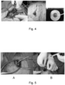
- the content of the bone marrow was aspirated with a catheter and a syringe to increase the space and the subsequent inoculation of bacteria into the bone, as shown in Figure 5 .
- the sheep was taken to a pen where it recovered from anesthesia until completing a 30-day guarding period for bacterial development, under the control of the veterinary staff in charge.
- This experiment sought to develop a degree of deep infection achieved through direct inoculation of bacteria into the bone.
- the designed study started with three sheep, which were inoculated in the first step with St. aureus through the lateral aspect of the femur. They were then taken to a large pen and kept for observation for 1 month. During the first 2 weeks, the sheep behaved normally. In the third week, they exhibited changes in posture and gait as well as in their general mood with loss of body mass, becoming progressively more lethargic with a decrease in their normal motility. The rapid deterioration of their general condition forced us to cull one of the three sheep without treatment. The other two received the treatment through the device of the invention according to the designed protocol. In the operating room, it was found that the sheep had developed a deep infection and had organized tissue showing evidence of the initial host response.
- the pathological anatomy of the fracture site showed hemorrhage, focal necrosis, and acute inflammation.
- the degree of infection was determined by the colony count of the reaming material obtained during surgery, showing variable decreasing changes (see Table 1). In one case where no antibiotic (ATB) was administered, the values showed a moderate increase.
- the pathological anatomy examinations showed the following findings: bone and soft tissue (necrosis-hemorrhage inflammatory tissue (3/8)), lung (bronchopneumonia and bronchial secretion (2/8)), liver (lymphocytic infiltrate (1/8)), skeletal muscle tissue (parasites (2/8)), spleen (parenchymal congestion (1/8)), heart and kidney were normal. Fat embolism was evaluated as a possible complication from reaming, and was not found in any of the cases tested. This shows that repeated reaming every 48 hours does not increase the development of fat embolism.
- the development of the device of the invention, and the application thereof in sheep had the purpose of validating the efficacy of such development at an intramedullary level. It was created as a response to the search for infection control by relapses where historical treatments had been proven unsuccessful.
- the device of the invention proved to be valid as an additional treatment for the management of infections in long bones.
Landscapes
- Health & Medical Sciences (AREA)
- Heart & Thoracic Surgery (AREA)
- Vascular Medicine (AREA)
- Engineering & Computer Science (AREA)
- Anesthesiology (AREA)
- Biomedical Technology (AREA)
- Hematology (AREA)
- Life Sciences & Earth Sciences (AREA)
- Animal Behavior & Ethology (AREA)
- General Health & Medical Sciences (AREA)
- Public Health (AREA)
- Veterinary Medicine (AREA)
- External Artificial Organs (AREA)
Priority Applications (1)
| Application Number | Priority Date | Filing Date | Title |
|---|---|---|---|
| EP23208606.6A EP4552664A1 (fr) | 2023-11-08 | 2023-11-08 | Procédé et dispositif pour le traitement de l'ostéomyélite dans des os longs par pression négative |
Applications Claiming Priority (1)
| Application Number | Priority Date | Filing Date | Title |
|---|---|---|---|
| EP23208606.6A EP4552664A1 (fr) | 2023-11-08 | 2023-11-08 | Procédé et dispositif pour le traitement de l'ostéomyélite dans des os longs par pression négative |
Publications (1)
| Publication Number | Publication Date |
|---|---|
| EP4552664A1 true EP4552664A1 (fr) | 2025-05-14 |
Family
ID=88745835
Family Applications (1)
| Application Number | Title | Priority Date | Filing Date |
|---|---|---|---|
| EP23208606.6A Withdrawn EP4552664A1 (fr) | 2023-11-08 | 2023-11-08 | Procédé et dispositif pour le traitement de l'ostéomyélite dans des os longs par pression négative |
Country Status (1)
| Country | Link |
|---|---|
| EP (1) | EP4552664A1 (fr) |
Citations (4)
| Publication number | Priority date | Publication date | Assignee | Title |
|---|---|---|---|---|
| US20070219585A1 (en) * | 2006-03-14 | 2007-09-20 | Cornet Douglas A | System for administering reduced pressure treatment having a manifold with a primary flow passage and a blockage prevention member |
| CN206482909U (zh) | 2016-12-05 | 2017-09-12 | 罗善超 | 骨髓炎负压引流冲洗装置 |
| CN113456924A (zh) | 2021-07-14 | 2021-10-01 | 中南大学湘雅二医院 | 一种骨髓炎治疗方法及其冲洗引流装置 |
| CN114344604A (zh) * | 2022-01-25 | 2022-04-15 | 核工业总医院 | 用于治疗骨髓炎的超声振动灌洗装置 |
-
2023
- 2023-11-08 EP EP23208606.6A patent/EP4552664A1/fr not_active Withdrawn
Patent Citations (4)
| Publication number | Priority date | Publication date | Assignee | Title |
|---|---|---|---|---|
| US20070219585A1 (en) * | 2006-03-14 | 2007-09-20 | Cornet Douglas A | System for administering reduced pressure treatment having a manifold with a primary flow passage and a blockage prevention member |
| CN206482909U (zh) | 2016-12-05 | 2017-09-12 | 罗善超 | 骨髓炎负压引流冲洗装置 |
| CN113456924A (zh) | 2021-07-14 | 2021-10-01 | 中南大学湘雅二医院 | 一种骨髓炎治疗方法及其冲洗引流装置 |
| CN114344604A (zh) * | 2022-01-25 | 2022-04-15 | 核工业总医院 | 用于治疗骨髓炎的超声振动灌洗装置 |
Similar Documents
| Publication | Publication Date | Title |
|---|---|---|
| Zhang et al. | Comparison of the efficacy of ultra-mini PCNL, flexible ureteroscopy, and shock wave lithotripsy on the treatment of 1–2 cm lower pole renal calculi | |
| CN102264406B (zh) | 用于将减压施加于皮下组织部位的套筒、歧管和系统 | |
| Colyer et al. | Surgical treatment of the infected hip implant: two-stage reimplantation with a one-month interval | |
| Himeno et al. | A novel treatment strategy using continuous local antibiotic perfusion: a case series study of a refractory infection caused by hypervirulent Klebsiella pneumoniae | |
| Durandy et al. | Mediastinal infection after cardiac operation: a simple closed technique | |
| Falahatkar et al. | Tubeless percutaneous nephrolithotomy for staghorn stones | |
| CN110292468A (zh) | 具有冲洗装置的股骨髋关节间隔器 | |
| Clawson et al. | Treatment of chronic osteomyelitis with emphasis on closed suction-irrigation technic. | |
| Thodis et al. | Peritoneal catheters and related infections | |
| EP4552664A1 (fr) | Procédé et dispositif pour le traitement de l'ostéomyélite dans des os longs par pression négative | |
| Sormaz et al. | Minimally-invasive treatment of hepatic hydatid disease with Perforator-Grinder-Aspirator Apparatus and follow-up of 42 patients | |
| CN103845106A (zh) | 采用细针半环外固定架和vsd灌洗固定干骺部骨缺损的方法 | |
| Tillson | Open fracture management | |
| Ladlow | Surgical drains in wound management and reconstructive surgery | |
| RU2697989C1 (ru) | Способ для борьбы с инфекцией глубоких слоёв мягких тканей при эндопротезировании тазобедренного сустава | |
| RU2478345C1 (ru) | Способ хирургического лечения многокамерного эхинококкоза печени | |
| RU2827673C1 (ru) | Способ дренирования глубоких осколочных ран мягких тканей под ультразвуковым контролем | |
| SU1153902A1 (ru) | Способ устранени дефекта диафиза трубчатой кости | |
| RU2785652C1 (ru) | Устройство для дренирования гнойных полостей | |
| JP7026760B2 (ja) | 薬剤流体の局所投与用の装置 | |
| RU2712013C1 (ru) | Способ подготовки инфицированной периартикулярной раны для имплантации постоянного или временного стабилизирующего устройства | |
| Mader et al. | Long-bone osteomyelitis diagnosis and management | |
| RU2738683C1 (ru) | Способ дренирования плевральной полости после пульмонэктомии | |
| Alhazmi | Approaches to Abscess Management | |
| Wright et al. | Lateral rhinostomy for treatment of severe chronic rhinosinusitis in two rabbits |
Legal Events
| Date | Code | Title | Description |
|---|---|---|---|
| PUAI | Public reference made under article 153(3) epc to a published international application that has entered the european phase |
Free format text: ORIGINAL CODE: 0009012 |
|
| STAA | Information on the status of an ep patent application or granted ep patent |
Free format text: STATUS: THE APPLICATION HAS BEEN PUBLISHED |
|
| AK | Designated contracting states |
Kind code of ref document: A1 Designated state(s): AL AT BE BG CH CY CZ DE DK EE ES FI FR GB GR HR HU IE IS IT LI LT LU LV MC ME MK MT NL NO PL PT RO RS SE SI SK SM TR |
|
| STAA | Information on the status of an ep patent application or granted ep patent |
Free format text: STATUS: THE APPLICATION IS DEEMED TO BE WITHDRAWN |
|
| 18D | Application deemed to be withdrawn |
Effective date: 20251115 |