EP4553843A1 - Verfahren zur patientenpositionierung in einem medizinischen verfahren und medizinisches system - Google Patents
Verfahren zur patientenpositionierung in einem medizinischen verfahren und medizinisches system Download PDFInfo
- Publication number
- EP4553843A1 EP4553843A1 EP23208966.4A EP23208966A EP4553843A1 EP 4553843 A1 EP4553843 A1 EP 4553843A1 EP 23208966 A EP23208966 A EP 23208966A EP 4553843 A1 EP4553843 A1 EP 4553843A1
- Authority
- EP
- European Patent Office
- Prior art keywords
- patient
- immobilization
- positioning
- skeleton model
- medical
- Prior art date
- Legal status (The legal status is an assumption and is not a legal conclusion. Google has not performed a legal analysis and makes no representation as to the accuracy of the status listed.)
- Withdrawn
Links
Images

Classifications
-
- G—PHYSICS
- G16—INFORMATION AND COMMUNICATION TECHNOLOGY [ICT] SPECIALLY ADAPTED FOR SPECIFIC APPLICATION FIELDS
- G16H—HEALTHCARE INFORMATICS, i.e. INFORMATION AND COMMUNICATION TECHNOLOGY [ICT] SPECIALLY ADAPTED FOR THE HANDLING OR PROCESSING OF MEDICAL OR HEALTHCARE DATA
- G16H20/00—ICT specially adapted for therapies or health-improving plans, e.g. for handling prescriptions, for steering therapy or for monitoring patient compliance
- G16H20/40—ICT specially adapted for therapies or health-improving plans, e.g. for handling prescriptions, for steering therapy or for monitoring patient compliance relating to mechanical, radiation or invasive therapies, e.g. surgery, laser therapy, dialysis or acupuncture
-
- G—PHYSICS
- G06—COMPUTING OR CALCULATING; COUNTING
- G06T—IMAGE DATA PROCESSING OR GENERATION, IN GENERAL
- G06T7/00—Image analysis
- G06T7/70—Determining position or orientation of objects or cameras
- G06T7/73—Determining position or orientation of objects or cameras using feature-based methods
- G06T7/75—Determining position or orientation of objects or cameras using feature-based methods involving models
-
- G—PHYSICS
- G16—INFORMATION AND COMMUNICATION TECHNOLOGY [ICT] SPECIALLY ADAPTED FOR SPECIFIC APPLICATION FIELDS
- G16H—HEALTHCARE INFORMATICS, i.e. INFORMATION AND COMMUNICATION TECHNOLOGY [ICT] SPECIALLY ADAPTED FOR THE HANDLING OR PROCESSING OF MEDICAL OR HEALTHCARE DATA
- G16H50/00—ICT specially adapted for medical diagnosis, medical simulation or medical data mining; ICT specially adapted for detecting, monitoring or modelling epidemics or pandemics
- G16H50/20—ICT specially adapted for medical diagnosis, medical simulation or medical data mining; ICT specially adapted for detecting, monitoring or modelling epidemics or pandemics for computer-aided diagnosis, e.g. based on medical expert systems
Definitions
- the invention relates to medical procedures, in particular to medical imaging. More particularly, the invention relates to a method for patient positioning in a medical procedure performed by a medical device, to a corresponding computer-readable medium and a medical system.
- Patient positioning is crucial to image quality in many domains of medical imaging.
- State-of-the-art medical imaging systems are often equipped with means to detect inappropriate positioning, propose correction, or an optimum positioning using either the medical image itself (retrospectively) or additional sensors, such as 3D camera (pro-actively).
- a camera image, a 3D camera image, a low dose scout image or the imaging modality itself is evaluated typically with a dedicated artificial intelligence or image processing software, as described, e.g., in United States patent application US 2010/0215271 A1 .
- a computer-implemented method for patient positioning in a medical procedure performed by a medical device is provided.
- the computer may be a computer of the medical device, a separate computer, or a computer in a cloud. Also, the method may be split on several computers.
- immobilization information is received.
- Said immobilization information is information on immobilization of a patient, wherein the patient has to be positioned optimally for the medical procedure.
- immobilization may refer to any condition that impedes the patient's ability to move, such as casts, external fixations, internal fixations, or medical conditions such as bone dislocations.
- immobilization of the patient is detected.
- a skeleton model of the patient is determined. Based on said skeleton model and on the detected immobilization of the patient, a constraint for patient movement is determined. It is understood that it is possible that more than one constraint, in particular a plurality of constraints for patient movement are determined.
- a constrained optimal patient positioning is determined based on the skeleton model and the constraint for patient movement. That is, for said constrained optimal patient positioning, the reduced mobility of the patient is taken into account. In other words, the optimal patient positioning is restricted to the movements that are still achievable by the patient.
- positioning feedback is provided based on the constrained optimal patient positioning. Since said positioning feedback is based on the constraint for patient movement, it can actually be applied to the patient, in particular without any further manual interaction to take into account the immobilization of the patient. Compared to positioning feedback that does not take into account the immobilization of the patient, the positioning feedback provided by this method is actually helpful for the medical operator and the positioning advice is improved for patients with immobilizations.
- the medical procedure is an X-ray imaging procedure, a computed tomography (CT) imaging procedure, a magnetic resonance imaging (MRI) procedure, an ultrasound (US) imaging procedure, an automated breast ultrasound (ABUS) imaging procedure, an image guided therapy (IGT) procedure and/or a radiotherapy procedure.
- CT computed tomography
- MRI magnetic resonance imaging
- US ultrasound
- ABUS automated breast ultrasound
- IGT image guided therapy
- radiotherapy radiotherapy
- the immobilization information comprises a source image. Detecting immobilization of the patient then comprises analyzing the source image.
- Said source image is a modality image taken by the medical device.
- modality image refers to an image taken by the medical device, e.g., an X-ray image, a CT image, or a US image.
- This modality image may be a preview image or a scout image, in particular taken with a lower radiation dose.
- Said preview image or scout image may be taken especially for the purpose of determining the optimal patient positioning. In this case, the positioning feedback is provided prior to taking the actual medical image, i.e., in a prospective way.
- the modality image may be an actual image taken by the medical device.
- the positioning feedback may either be an indication that the patient positioning was already good enough, or that a re-take of the medical image with improved patient positioning is proposed, i.e., in a retrospective way.
- the source image is an image from an additional sensor.
- Said additional sensor may be an optical camera, e.g., an RGB camera or an infrared camera, or a 3D camera, i.e., a depth camera.
- the source image may be taken before the actual medical procedure is performed such that prospective guidance can be provided based on the source image.
- both a source image from an additional sensor, e.g., a 3D depth camera, and a source image taken by the medical device, e.g., an X-ray image may be used and analyzed to detect the immobilization of the patient.
- This may improve the detection of immobilization, in particular since the information provided by the different source images may be complementary: for example, the source image taken by the additional sensor may easily show casts, while the source image from the medical device may easily show internal fixations. In combination, both casts and internal fixations are easily shown, leading to an improved detection of immobilization.
- the analysis of the source image may be performed, e.g., by image processing techniques, followed by feature recognition, and/or using an artificial intelligence, e.g., a trained neural network.
- analyzing the source image comprises fitting a default skeleton model to the source image.
- Said default skeleton model may be an anatomical skeleton model.
- the modality image taken by the medical device and/or the image from the additional sensor may be used to fit said default skeleton model to the source image, more particularly by matching internal and/or external features, respectively, to said default skeleton model.
- Said fitting may be performed, e.g., by image processing techniques, followed by feature recognition, and/or using an artificial intelligence, e.g., a trained neural network.
- deviations between the skeleton model and the actual anatomy of the patient may be detected.
- bone dislocations may be detected, which may then be analyzed to determine the impact of said bone dislocation on the mobility of the patient and therefore determine an immobilization of the patient due to the bone dislocation.
- the overall detection of immobilization is further improved.
- the immobilization information comprises patient information.
- Said patient information may be an electronic patient record, previous reports and/or examination requests.
- said patient information may comprise information on casts, fixations, implants, or medical conditions that interfere with patient mobility.
- said patient information may already include information on a plurality of immobilizations of the patient.
- natural language processing may be used.
- the patient information may comprise mobility parameters which may be directly used to determine immobilization of the patient.
- the patient information may also be combined with the source images. As an example, the credibility of immobilization detected from a source image may be checked with the information obtained from the patient information.
- the patient information may be used to determine whether the source image from the additional sensor is sufficient to determine the immobilization or a source image from the medical device should be obtained to determine the immobilization of the patient: for example, if the patient information comprises information on internal fixations, a source image from the medical device should be obtained, since the source image from the camera does not provide enough information on said internal fixation.
- the patient information comprises information on internal fixations, a source image from the medical device should be obtained, since the source image from the camera does not provide enough information on said internal fixation.
- detecting immobilization of the patient comprises detecting immobilization devices and/or determining immobilized joints, more particularly due to medical conditions.
- immobilization devices are devices that constrain the movements of the patient, such as casts, internal fixations and/or external fixations.
- Immobilized joints are joints that are, at least potentially, immobilized due to medical conditions of the patient, such as fractures, bone dislocations, arthritis, or an aggregation of fluids. When all of said sources of immobilization are taken into account, an accurate assessment of immobilization of the patient is possible.
- Said fitting may be performed, e.g., by image processing techniques, followed by feature recognition, and/or using an artificial intelligence, e.g., a trained neural network.
- an artificial intelligence e.g., a trained neural network.
- determining the constraint for patient movement comprises spatially correlating detected immobilization with respect to the skeleton model.
- the location of the immobilization in relation to the skeleton model is determined.
- determining the constrained optimal patient positioning comprises computing an optimum patient position based on the skeleton model and then constraining the optimum patient position based on the constraint for patient movement to obtain the constrained optimal patient positioning. That is, the optimum patient position is first computed without taking into account the immobilization of the patient. This can be done, e.g., by algorithms, by a decision tree, by a random forest, or using an artificial intelligence.
- the patient position may be described, e.g., by parameters, wherein the parameters may refer to the flection of a joint and/or to an angle between bones adjacent to a joint.
- the movement constraint that has been determined from the detected immobilization of the patient is applied to the optimum patient position. That is, an interference of the immobilization with the proposed optimum patient position is detected.
- the constrained optimal patient positioning is obtained.
- the optimum patient position comprises a value for a parameter (e.g., an angle) that is not within the possible movement range of the patient
- the value for said parameter that is the closest to the optimum parameter value, but still allowed by the immobilization of the patient is chosen.
- routines for determining the optimum patient position for patients without immobilization may be re-used, allowing for an efficient method for both patients without and with immobilization.
- determining the constrained optimal patient positioning comprises determining a constrained set of possible patient positions and determining an optimal choice of the patient position in the constrained set to obtain the constrained optimal patient positioning. That is, the constrained set of possible patient positions is determined first, taking into account the determined immobilization of the patient. Said set of possible patient positions may be given, e.g., as a set of possible patient positioning parameters. Once the set of possible patient positions has been determined, the optimal choice of the patient position is determined in this constrained set. In particular, an algorithm for finding the optimal choice of the patient position may be provided with the constrained set, such that it searches for the optimal patient position only within the allowed range of patient movements. This option yields particularly good results for the case that the parameters of the patient position are correlated with one another.
- providing positioning feedback comprises providing optimum positioning guidance to achieve the constrained optimal patient positioning.
- the patient may be positioned accordingly with respect to the medical device and the best possible medical result that is possible given the immobilization of the patient can be obtained.
- providing optimum positioning guidance comprises proposing patient re-positioning.
- a new, optimal patient position is provided. This may be done, e.g., by providing a graphic representation of said optimal patient position, or by providing instructions on how to achieve said optimal patient position, for example to bend joints in a particular way. Performing patient re-positioning is most practicable for the joints that are still mobile, i.e., without or with only little immobilization.
- providing optimum positioning guidance comprises proposing equipment re-positioning. This may also be done by providing a graphic representation of the optimal equipment position with respect to the patient, or by providing instructions on how to move the equipment in order to achieve said optimal equipment position.
- providing optimum positioning guidance comprises controlling equipment re-positioning. That is, actuators automatically control the equipment to take the optimum equipment position based on the optimal equipment position. Hence, by moving the patient and/or equipment, the constrained optimal patient positioning can be achieved, yielding the best possible medical result.
- providing positioning feedback comprises providing an assessment on the current patient positioning.
- the positioning feedback may comprise a score, e.g., between 0 and 100, for the current patient positioning, wherein a low score refers to a suboptimal positioning and a high score to an optimal positioning, given the immobilization constraint. If said score exceeds a predetermined threshold, no further patient re-positioning will be performed and the medical procedure will be performed. Or, in the case of retrospective evaluation of the immobilization, a decision whether to re-do the medical procedure or not will be based on said score, e.g., based on a comparison with another predetermined threshold.
- a re-do of the medical procedure e.g., a re-take of the medical image, will not be performed, thus saving time, costs, and radiation exposure for the patient.
- a computer-readable medium comprises instructions which, when executed by a computer, cause the computer to carry out the method according to the above description.
- the instructions correspond to software modules that perform the method according to the above description.
- a medical system comprises a medical device.
- the medical device may be an X-ray imaging device, a computed tomography (CT) imaging device, a magnetic resonance imaging (MRI) device, an ultrasound (US) imaging device, an automated breast ultrasound (ABUS) imaging device, an image guided therapy (IGT) device, or a radiotherapy device.
- CT computed tomography
- MRI magnetic resonance imaging
- US ultrasound
- ABUS automated breast ultrasound
- ITT image guided therapy
- a preferred embodiment of the invention can also be any combination of the dependent claims with the respective independent claim.
- embodiments of the invention are described with reference to different subject matters. In particular, some embodiments are described with reference to device type claims whereas other embodiments are described with reference to method type claims.
- a person skilled in the art will gather from the above and the following description that, unless otherwise notified, in addition to any combination of features belonging to one type of subject matter also any combination between features relating to different subject matters is considered to be disclosed with this application. However, all features can be combined providing synergetic effects that are more than the simple summation of the features.
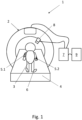
- Fig. 1 shows a schematic view of an embodiment of a medical system 1.
- Said medical system 1 comprises a medical device 2.
- the medical device 2 is shown as a computed tomography (CT) imaging device, however, the invention also applies to other medical devices such as X-ray imaging devices, magnetic resonance imaging (MRI) devices, ultrasound (US) imaging devices, automated breast ultrasound (ABUS) imaging devices, image guided therapy (IGT) devices, or a radiotherapy devices.
- CT computed tomography
- MRI magnetic resonance imaging
- US ultrasound
- ABUS automated breast ultrasound
- ITT image guided therapy
- a patient 3 is shown on a patient table 4.
- the patient 3 is shown with two immobilization devices 5.1 and 5.2, wherein the immobilization device 5.1 is shown as a cast and the immobilization device 5.2 is shown as an internal fixation.
- the patient may have other and/or additional immobilization devices.
- the patient 3 is shown to have an immobilized joint 6, e.g., due to a medical condition.
- the medical system 1 further comprises a computing unit 7 with one or more processors that is connected to the medical device 2. Also, the medical system 1 comprises an additional sensor 8, which is shown as a camera.
- the additional sensor 8 may be, e.g., a depth camera and/or an optical camera, such as an RGB camera.
- the additional sensor 8 is also connected to the computing unit 7 such that the computing unit 7 can receive data from the additional sensor 8.
- the computing unit 7 is connected to an electronic patient record 9, such that the computing unit 7 can receive data from said electronic patient record 9.
- the electronic patient record 9 may be part of the medical imaging system 1, but may also be remote from the medical imaging system 1.
- the computing unit 7 comprises a computer-readable medium that is configured to cause the computing unit 7 to carry out the below described computer-implemented method for patient positioning.
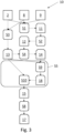
- Fig. 2 shows a flowchart of an embodiment of a computer-implemented method 10 for patient positioning.
- immobilization information 11 is received S1, wherein the immobilization information 11 is information on immobilization of the patient 3.
- the immobilization information 11 comprises a source image, wherein the source image is a modality image taken by the medical device 2 and/or an image from the additional sensor 8.
- the immobilization information 11 comprises patient information, e.g., from the electronic patient record 9, from previous reports and/or examination requests.
- immobilization 12 of the patient 3 is detected S2.
- Detecting S2 immobilization 12 of the patient 3 may comprise detecting immobilization devices such as casts 5.1, internal fixations 5.2 and/or external fixations, and/or determining immobilized joints 6, more particularly due to medical conditions of the patient 3, such as fractures, bone dislocations, arthritis, or an aggregation of fluids.
- the immobilization information 11 comprises source images
- detecting S2 immobilization 12 of the patient 3 comprises analyzing said source images. The analysis of the source image may be performed, e.g., by image processing techniques, followed by feature recognition, and/or using an artificial intelligence, e.g., a trained neural network.
- the analysis of the source image may further comprise fitting a default skeleton model to the source image.
- Said default skeleton model may be an anatomical skeleton model.
- the modality image taken by the medical device 2 and/or the image from the additional sensor 8 may be used to fit said default skeleton model to the source image, more particularly by matching internal and/or external features, respectively, to said default skeleton model.
- Said fitting may be performed, e.g., by image processing techniques, followed by feature recognition, and/or using an artificial intelligence, e.g., a trained neural network.
- bone dislocations may be detected, which may then be analyzed to determine the impact of said bone dislocation on the mobility of the patient and therefore determine an immobilization 12 of the patient 3 due to the bone dislocation.
- the overall detection of immobilization 12 is further improved.
- the method 10 comprises determining S3 a skeleton model 13 of the patient.
- Said skeleton model 13 may be, in particular, determined S3 by fitting a default skeleton model to source images obtained from the medical device 2 and/or the additional camera 8. More particularly, internal and/or external features may be matched to said default skeleton model.
- the fitting may be performed, e.g., by image processing techniques, followed by feature recognition, and/or using an artificial intelligence, e.g., a trained neural network.
- an artificial intelligence e.g., a trained neural network.
- bone dislocations may be detected, which may then be analyzed to determine the impact of said bone dislocation on the mobility of the patient 3 and therefore determine an immobilization 12 of the patient due to the bone dislocation.
- the overall detection of immobilization 12 is further improved.
- a constraint 14 for patient movement is determined S4.
- Determining S4 said constraint 14 for patient movement may comprise spatially correlating detected S2 immobilization 12 with respect to the skeleton model 13. In other words, the location of the immobilization 12 in relation to the skeleton model 13 is determined. Hence, it can be determined which joints of the skeleton model 13 are affected by said immobilization 12, which leads to an accurate description of the immobilization 12 of the patient 3.
- a constrained optimal patient positioning 15 is determined S5 based on the skeleton model 13 and the constraint 14 for patient movement.
- this comprises computing S6 an optimum patient position 16 based on the skeleton model 13 and then constraining S7 the optimum patient position 16 based on the constraint 14 for patient movement. That is, the optimum patient position 16 is first computed S6 without taking into account the immobilization 12 of the patient 3. This can be done, e.g., by algorithms, by a decision tree, by a random forest, or using an artificial intelligence.
- the patient position may be described, e.g., by parameters, wherein the parameters may refer to the flection of a joint and/or to an angle between bones adjacent to a joint.
- the movement constraint 14 that has been determined S4 from the detected immobilization 12 of the patient 3 are applied to the optimum patient position 16. That is, an interference of the immobilization 12 with the proposed optimum patient position 16 is detected.
- the constrained optimal patient positioning 15 is obtained.
- the optimum patient position 16 comprises a value for a parameter (e.g., an angle) that is not within the possible movement range of the patient 3, the value for said parameter that is the closest to the optimum parameter value, but still allowed by the immobilization 12 of the patient 3, is chosen.
- routines for determining the optimum patient position 16 for patients 3 without immobilization may be re-used, allowing for an efficient method for both patients 3 without and with immobilization 12.
- positioning feedback 17 is provided S8 based on the constrained optimal patient positioning 15.
- Providing S8 the positioning feedback 17 may comprise providing optimum positioning guidance to achieve the constrained optimal patient positioning 15, including proposing patient re-positioning, proposing equipment re-positioning, and/or controlling equipment re-positioning. By moving the patient and/or equipment, the constrained optimal patient positioning can be achieved, yielding the best possible medical result.
- providing S8 the positioning feedback 17 may comprise providing an assessment on the current patient positioning. Said assessment may be provided as a score for the current patient positioning. If said score exceeds a predetermined threshold, no further patient re-positioning will be performed and the medical procedure will be performed.
- a decision whether to re-do the medical procedure or not will be based on said score, e.g., based on a comparison with another predetermined threshold.
- a re-do of the medical procedure e.g., a re-take of the medical image, will not be performed, thus saving time, costs, and radiation exposure for the patient.
- Fig. 3 shows another flowchart of an embodiment of a method 10 for patient positioning.
- the step of determining S5 the constrained optimal patient positioning 15 is changed.
- a constrained set 18 of possible patient positions is determined S9 based on the skeleton model 13 and the constraint 14 for patient movement.
- said constrained set 18 is used, together with the skeleton model 13, to determine S10 an optimal choice of the patient position in the constrained set 18. That is, the constrained set 18 of possible patient positions is determined first, taking into account the determined immobilization 12 of the patient 3.
- Said set 18 of possible patient positions may be given, e.g., as a set of possible patient positioning parameters. Once the set 18 of possible patient positions has been determined, the optimal choice of the patient position is determined in this constrained set 18.
- an algorithm for finding the optimal choice of the patient position may be provided with the constrained set, such that it searches for the optimal patient position only within the allowed range of patient movements. This option yields particularly good results for the case that the parameters of the patient position are correlated with one another.
Landscapes
- Engineering & Computer Science (AREA)
- Health & Medical Sciences (AREA)
- Medical Informatics (AREA)
- Public Health (AREA)
- Primary Health Care (AREA)
- General Health & Medical Sciences (AREA)
- Epidemiology (AREA)
- Biomedical Technology (AREA)
- Pathology (AREA)
- Databases & Information Systems (AREA)
- Data Mining & Analysis (AREA)
- Theoretical Computer Science (AREA)
- General Physics & Mathematics (AREA)
- Physics & Mathematics (AREA)
- Computer Vision & Pattern Recognition (AREA)
- Nuclear Medicine, Radiotherapy & Molecular Imaging (AREA)
- Surgery (AREA)
- Urology & Nephrology (AREA)
- Apparatus For Radiation Diagnosis (AREA)
- Magnetic Resonance Imaging Apparatus (AREA)
Priority Applications (2)
| Application Number | Priority Date | Filing Date | Title |
|---|---|---|---|
| EP23208966.4A EP4553843A1 (de) | 2023-11-10 | 2023-11-10 | Verfahren zur patientenpositionierung in einem medizinischen verfahren und medizinisches system |
| PCT/EP2024/080735 WO2025098859A1 (en) | 2023-11-10 | 2024-10-30 | Method for patient positioning in a medical procedure and medical system |
Applications Claiming Priority (1)
| Application Number | Priority Date | Filing Date | Title |
|---|---|---|---|
| EP23208966.4A EP4553843A1 (de) | 2023-11-10 | 2023-11-10 | Verfahren zur patientenpositionierung in einem medizinischen verfahren und medizinisches system |
Publications (1)
| Publication Number | Publication Date |
|---|---|
| EP4553843A1 true EP4553843A1 (de) | 2025-05-14 |
Family
ID=88779431
Family Applications (1)
| Application Number | Title | Priority Date | Filing Date |
|---|---|---|---|
| EP23208966.4A Withdrawn EP4553843A1 (de) | 2023-11-10 | 2023-11-10 | Verfahren zur patientenpositionierung in einem medizinischen verfahren und medizinisches system |
Country Status (2)
| Country | Link |
|---|---|
| EP (1) | EP4553843A1 (de) |
| WO (1) | WO2025098859A1 (de) |
Citations (7)
| Publication number | Priority date | Publication date | Assignee | Title |
|---|---|---|---|---|
| US20100215271A1 (en) | 2009-02-25 | 2010-08-26 | Honda Motor Co., Ltd. | Body feature detection and human pose estimation using inner distance shape contexts |
| US20170354385A1 (en) * | 2016-06-08 | 2017-12-14 | Siemens Healthcare Gmbh | Positioning of an examination object for an imaging method |
| WO2019110105A1 (en) * | 2017-12-07 | 2019-06-13 | Brainlab Ag | Patient positioning using a skeleton model |
| US20210118174A1 (en) | 2019-10-18 | 2021-04-22 | Shanghai United Imaging Intelligence Co., Ltd. | Methods and devices for guiding a patient |
| WO2021141678A1 (en) * | 2020-01-07 | 2021-07-15 | Carestream Health, Inc. | System and method for automated projection radiography |
| US20210244283A1 (en) | 2018-06-11 | 2021-08-12 | Koninklijke Philips N.V. | Position feed back indicator for medical imaging |
| EP4215105A1 (de) * | 2022-01-24 | 2023-07-26 | Koninklijke Philips N.V. | Automatische schmerzerfassung in abhängigkeit von der lage eines patienten |
-
2023
- 2023-11-10 EP EP23208966.4A patent/EP4553843A1/de not_active Withdrawn
-
2024
- 2024-10-30 WO PCT/EP2024/080735 patent/WO2025098859A1/en active Pending
Patent Citations (7)
| Publication number | Priority date | Publication date | Assignee | Title |
|---|---|---|---|---|
| US20100215271A1 (en) | 2009-02-25 | 2010-08-26 | Honda Motor Co., Ltd. | Body feature detection and human pose estimation using inner distance shape contexts |
| US20170354385A1 (en) * | 2016-06-08 | 2017-12-14 | Siemens Healthcare Gmbh | Positioning of an examination object for an imaging method |
| WO2019110105A1 (en) * | 2017-12-07 | 2019-06-13 | Brainlab Ag | Patient positioning using a skeleton model |
| US20210244283A1 (en) | 2018-06-11 | 2021-08-12 | Koninklijke Philips N.V. | Position feed back indicator for medical imaging |
| US20210118174A1 (en) | 2019-10-18 | 2021-04-22 | Shanghai United Imaging Intelligence Co., Ltd. | Methods and devices for guiding a patient |
| WO2021141678A1 (en) * | 2020-01-07 | 2021-07-15 | Carestream Health, Inc. | System and method for automated projection radiography |
| EP4215105A1 (de) * | 2022-01-24 | 2023-07-26 | Koninklijke Philips N.V. | Automatische schmerzerfassung in abhängigkeit von der lage eines patienten |
Also Published As
| Publication number | Publication date |
|---|---|
| WO2025098859A1 (en) | 2025-05-15 |
Similar Documents
| Publication | Publication Date | Title |
|---|---|---|
| EP3378534B1 (de) | Behandlungsassistenzsystem und betriebsverfahren dafür sowie speichermedium zur speicherung eines behandlungshilfsprogramms | |
| US12165314B2 (en) | Method for generating a trained machine learning algorithm | |
| US8099152B2 (en) | Systems and methods for planning medical procedures and designing medical devices based on anatomical scan deformations | |
| EP3993704B1 (de) | Selbstlernendes verteiltes system mit automatisierter erzeugung eines referenzdatensatzes | |
| JP7097877B2 (ja) | 医用画像データセットの品質評価のための装置及び方法 | |
| US20220148180A1 (en) | Medical image processing device, storage medium, medical device, and treatment system | |
| US10445904B2 (en) | Method and device for the automatic generation of synthetic projections | |
| CN113610744A (zh) | X光图像质量控制系统 | |
| US20250127572A1 (en) | Methods and systems for planning a surgical procedure | |
| JP6345471B2 (ja) | X線画像診断装置 | |
| CN120267321A (zh) | 一种基于dr设备的体位辅助检测方法及系统 | |
| Kausch et al. | C-arm positioning for standard projections during spinal implant placement | |
| EP4553843A1 (de) | Verfahren zur patientenpositionierung in einem medizinischen verfahren und medizinisches system | |
| US11903691B2 (en) | Combined steering engine and landmarking engine for elbow auto align | |
| US20250143816A1 (en) | System and method for aligning an imaging device | |
| WO2016113161A1 (en) | Adaptive segmentation for rotational c-arm computed tomography with a reduced angular range | |
| US20250006343A1 (en) | Prospective quality assessment for imaging examination prior to acquisition | |
| EP4530984A1 (de) | Patientenposenbestimmungssystem, medizinisches bildgebungssystem und zugehöriges verfahren | |
| EP4400033A1 (de) | Medizinische bildgebung | |
| CN111358479A (zh) | 一种放射线设备的采集参数确定方法和系统 | |
| EP4636777A1 (de) | Computerimplementiertes verfahren zum betreiben einer medizinischen bildgebungsvorrichtung, medizinische bildgebungsvorrichtung, computerprogramm und elektronisch lesbares speichermedium | |
| US20230417853A1 (en) | Automated detection of critical stations in multi-station magnetic resonance imaging | |
| WO2026021908A1 (en) | Method and processing apparatus for determining medical image acquisition parameters using anatomical information | |
| CN121622078A (zh) | 自适应侦察扫描范围和组合 |
Legal Events
| Date | Code | Title | Description |
|---|---|---|---|
| PUAI | Public reference made under article 153(3) epc to a published international application that has entered the european phase |
Free format text: ORIGINAL CODE: 0009012 |
|
| STAA | Information on the status of an ep patent application or granted ep patent |
Free format text: STATUS: THE APPLICATION HAS BEEN PUBLISHED |
|
| AK | Designated contracting states |
Kind code of ref document: A1 Designated state(s): AL AT BE BG CH CY CZ DE DK EE ES FI FR GB GR HR HU IE IS IT LI LT LU LV MC ME MK MT NL NO PL PT RO RS SE SI SK SM TR |
|
| STAA | Information on the status of an ep patent application or granted ep patent |
Free format text: STATUS: THE APPLICATION IS DEEMED TO BE WITHDRAWN |
|
| 18D | Application deemed to be withdrawn |
Effective date: 20251115 |