EP4555924A1 - Procédé de détermination de fragilité à l'aide de données d'anesthésie - Google Patents
Procédé de détermination de fragilité à l'aide de données d'anesthésie Download PDFInfo
- Publication number
- EP4555924A1 EP4555924A1 EP23306981.4A EP23306981A EP4555924A1 EP 4555924 A1 EP4555924 A1 EP 4555924A1 EP 23306981 A EP23306981 A EP 23306981A EP 4555924 A1 EP4555924 A1 EP 4555924A1
- Authority
- EP
- European Patent Office
- Prior art keywords
- score
- frailty
- subject
- patient
- markers
- Prior art date
- Legal status (The legal status is an assumption and is not a legal conclusion. Google has not performed a legal analysis and makes no representation as to the accuracy of the status listed.)
- Pending
Links
Images




Classifications
-
- A—HUMAN NECESSITIES
- A61—MEDICAL OR VETERINARY SCIENCE; HYGIENE
- A61B—DIAGNOSIS; SURGERY; IDENTIFICATION
- A61B5/00—Measuring for diagnostic purposes; Identification of persons
- A61B5/24—Detecting, measuring or recording bioelectric or biomagnetic signals of the body or parts thereof
- A61B5/316—Modalities, i.e. specific diagnostic methods
- A61B5/369—Electroencephalography [EEG]
- A61B5/372—Analysis of electroencephalograms
- A61B5/374—Detecting the frequency distribution of signals, e.g. detecting delta, theta, alpha, beta or gamma waves
-
- A—HUMAN NECESSITIES
- A61—MEDICAL OR VETERINARY SCIENCE; HYGIENE
- A61B—DIAGNOSIS; SURGERY; IDENTIFICATION
- A61B5/00—Measuring for diagnostic purposes; Identification of persons
- A61B5/02—Detecting, measuring or recording for evaluating the cardiovascular system, e.g. pulse, heart rate, blood pressure or blood flow
- A61B5/021—Measuring pressure in heart or blood vessels
-
- A—HUMAN NECESSITIES
- A61—MEDICAL OR VETERINARY SCIENCE; HYGIENE
- A61B—DIAGNOSIS; SURGERY; IDENTIFICATION
- A61B5/00—Measuring for diagnostic purposes; Identification of persons
- A61B5/02—Detecting, measuring or recording for evaluating the cardiovascular system, e.g. pulse, heart rate, blood pressure or blood flow
- A61B5/024—Measuring pulse rate or heart rate
-
- A—HUMAN NECESSITIES
- A61—MEDICAL OR VETERINARY SCIENCE; HYGIENE
- A61B—DIAGNOSIS; SURGERY; IDENTIFICATION
- A61B5/00—Measuring for diagnostic purposes; Identification of persons
- A61B5/40—Detecting, measuring or recording for evaluating the nervous system
- A61B5/4076—Diagnosing or monitoring particular conditions of the nervous system
- A61B5/4088—Diagnosing of monitoring cognitive diseases, e.g. Alzheimer, prion diseases or dementia
-
- A—HUMAN NECESSITIES
- A61—MEDICAL OR VETERINARY SCIENCE; HYGIENE
- A61B—DIAGNOSIS; SURGERY; IDENTIFICATION
- A61B5/00—Measuring for diagnostic purposes; Identification of persons
- A61B5/48—Other medical applications
- A61B5/4821—Determining level or depth of anaesthesia
-
- A—HUMAN NECESSITIES
- A61—MEDICAL OR VETERINARY SCIENCE; HYGIENE
- A61B—DIAGNOSIS; SURGERY; IDENTIFICATION
- A61B5/00—Measuring for diagnostic purposes; Identification of persons
- A61B5/72—Signal processing specially adapted for physiological signals or for diagnostic purposes
- A61B5/7235—Details of waveform analysis
- A61B5/7264—Classification of physiological signals or data, e.g. using neural networks, statistical classifiers, expert systems or fuzzy systems
- A61B5/7267—Classification of physiological signals or data, e.g. using neural networks, statistical classifiers, expert systems or fuzzy systems involving training the classification device
-
- A—HUMAN NECESSITIES
- A61—MEDICAL OR VETERINARY SCIENCE; HYGIENE
- A61B—DIAGNOSIS; SURGERY; IDENTIFICATION
- A61B5/00—Measuring for diagnostic purposes; Identification of persons
- A61B5/72—Signal processing specially adapted for physiological signals or for diagnostic purposes
- A61B5/7271—Specific aspects of physiological measurement analysis
- A61B5/7275—Determining trends in physiological measurement data; Predicting development of a medical condition based on physiological measurements, e.g. determining a risk factor
-
- G—PHYSICS
- G16—INFORMATION AND COMMUNICATION TECHNOLOGY [ICT] SPECIALLY ADAPTED FOR SPECIFIC APPLICATION FIELDS
- G16H—HEALTHCARE INFORMATICS, i.e. INFORMATION AND COMMUNICATION TECHNOLOGY [ICT] SPECIALLY ADAPTED FOR THE HANDLING OR PROCESSING OF MEDICAL OR HEALTHCARE DATA
- G16H20/00—ICT specially adapted for therapies or health-improving plans, e.g. for handling prescriptions, for steering therapy or for monitoring patient compliance
- G16H20/10—ICT specially adapted for therapies or health-improving plans, e.g. for handling prescriptions, for steering therapy or for monitoring patient compliance relating to drugs or medications, e.g. for ensuring correct administration to patients
-
- G—PHYSICS
- G16—INFORMATION AND COMMUNICATION TECHNOLOGY [ICT] SPECIALLY ADAPTED FOR SPECIFIC APPLICATION FIELDS
- G16H—HEALTHCARE INFORMATICS, i.e. INFORMATION AND COMMUNICATION TECHNOLOGY [ICT] SPECIALLY ADAPTED FOR THE HANDLING OR PROCESSING OF MEDICAL OR HEALTHCARE DATA
- G16H50/00—ICT specially adapted for medical diagnosis, medical simulation or medical data mining; ICT specially adapted for detecting, monitoring or modelling epidemics or pandemics
- G16H50/20—ICT specially adapted for medical diagnosis, medical simulation or medical data mining; ICT specially adapted for detecting, monitoring or modelling epidemics or pandemics for computer-aided diagnosis, e.g. based on medical expert systems
-
- G—PHYSICS
- G16—INFORMATION AND COMMUNICATION TECHNOLOGY [ICT] SPECIALLY ADAPTED FOR SPECIFIC APPLICATION FIELDS
- G16H—HEALTHCARE INFORMATICS, i.e. INFORMATION AND COMMUNICATION TECHNOLOGY [ICT] SPECIALLY ADAPTED FOR THE HANDLING OR PROCESSING OF MEDICAL OR HEALTHCARE DATA
- G16H50/00—ICT specially adapted for medical diagnosis, medical simulation or medical data mining; ICT specially adapted for detecting, monitoring or modelling epidemics or pandemics
- G16H50/30—ICT specially adapted for medical diagnosis, medical simulation or medical data mining; ICT specially adapted for detecting, monitoring or modelling epidemics or pandemics for calculating health indices; for individual health risk assessment
-
- G—PHYSICS
- G16—INFORMATION AND COMMUNICATION TECHNOLOGY [ICT] SPECIALLY ADAPTED FOR SPECIFIC APPLICATION FIELDS
- G16H—HEALTHCARE INFORMATICS, i.e. INFORMATION AND COMMUNICATION TECHNOLOGY [ICT] SPECIALLY ADAPTED FOR THE HANDLING OR PROCESSING OF MEDICAL OR HEALTHCARE DATA
- G16H50/00—ICT specially adapted for medical diagnosis, medical simulation or medical data mining; ICT specially adapted for detecting, monitoring or modelling epidemics or pandemics
- G16H50/70—ICT specially adapted for medical diagnosis, medical simulation or medical data mining; ICT specially adapted for detecting, monitoring or modelling epidemics or pandemics for mining of medical data, e.g. analysing previous cases of other patients
-
- A—HUMAN NECESSITIES
- A61—MEDICAL OR VETERINARY SCIENCE; HYGIENE
- A61B—DIAGNOSIS; SURGERY; IDENTIFICATION
- A61B2503/00—Evaluating a particular growth phase or type of persons or animals
- A61B2503/08—Elderly
-
- A—HUMAN NECESSITIES
- A61—MEDICAL OR VETERINARY SCIENCE; HYGIENE
- A61B—DIAGNOSIS; SURGERY; IDENTIFICATION
- A61B2505/00—Evaluating, monitoring or diagnosing in the context of a particular type of medical care
- A61B2505/05—Surgical care
-
- A—HUMAN NECESSITIES
- A61—MEDICAL OR VETERINARY SCIENCE; HYGIENE
- A61B—DIAGNOSIS; SURGERY; IDENTIFICATION
- A61B5/00—Measuring for diagnostic purposes; Identification of persons
- A61B5/0002—Remote monitoring of patients using telemetry, e.g. transmission of vital signals via a communication network
- A61B5/0015—Remote monitoring of patients using telemetry, e.g. transmission of vital signals via a communication network characterised by features of the telemetry system
- A61B5/0022—Monitoring a patient using a global network, e.g. telephone networks, internet
-
- A—HUMAN NECESSITIES
- A61—MEDICAL OR VETERINARY SCIENCE; HYGIENE
- A61B—DIAGNOSIS; SURGERY; IDENTIFICATION
- A61B5/00—Measuring for diagnostic purposes; Identification of persons
- A61B5/02—Detecting, measuring or recording for evaluating the cardiovascular system, e.g. pulse, heart rate, blood pressure or blood flow
- A61B5/024—Measuring pulse rate or heart rate
- A61B5/02405—Determining heart rate variability
-
- A—HUMAN NECESSITIES
- A61—MEDICAL OR VETERINARY SCIENCE; HYGIENE
- A61B—DIAGNOSIS; SURGERY; IDENTIFICATION
- A61B5/00—Measuring for diagnostic purposes; Identification of persons
- A61B5/20—Measuring for diagnostic purposes; Identification of persons for measuring urological functions restricted to the evaluation of the urinary system
- A61B5/201—Assessing renal or kidney functions
-
- A—HUMAN NECESSITIES
- A61—MEDICAL OR VETERINARY SCIENCE; HYGIENE
- A61B—DIAGNOSIS; SURGERY; IDENTIFICATION
- A61B5/00—Measuring for diagnostic purposes; Identification of persons
- A61B5/42—Detecting, measuring or recording for evaluating the gastrointestinal, the endocrine or the exocrine systems
- A61B5/4222—Evaluating particular parts, e.g. particular organs
- A61B5/4244—Evaluating particular parts, e.g. particular organs liver
-
- G—PHYSICS
- G16—INFORMATION AND COMMUNICATION TECHNOLOGY [ICT] SPECIALLY ADAPTED FOR SPECIFIC APPLICATION FIELDS
- G16H—HEALTHCARE INFORMATICS, i.e. INFORMATION AND COMMUNICATION TECHNOLOGY [ICT] SPECIALLY ADAPTED FOR THE HANDLING OR PROCESSING OF MEDICAL OR HEALTHCARE DATA
- G16H20/00—ICT specially adapted for therapies or health-improving plans, e.g. for handling prescriptions, for steering therapy or for monitoring patient compliance
- G16H20/10—ICT specially adapted for therapies or health-improving plans, e.g. for handling prescriptions, for steering therapy or for monitoring patient compliance relating to drugs or medications, e.g. for ensuring correct administration to patients
- G16H20/17—ICT specially adapted for therapies or health-improving plans, e.g. for handling prescriptions, for steering therapy or for monitoring patient compliance relating to drugs or medications, e.g. for ensuring correct administration to patients delivered via infusion or injection
-
- G—PHYSICS
- G16—INFORMATION AND COMMUNICATION TECHNOLOGY [ICT] SPECIALLY ADAPTED FOR SPECIFIC APPLICATION FIELDS
- G16H—HEALTHCARE INFORMATICS, i.e. INFORMATION AND COMMUNICATION TECHNOLOGY [ICT] SPECIALLY ADAPTED FOR THE HANDLING OR PROCESSING OF MEDICAL OR HEALTHCARE DATA
- G16H20/00—ICT specially adapted for therapies or health-improving plans, e.g. for handling prescriptions, for steering therapy or for monitoring patient compliance
- G16H20/40—ICT specially adapted for therapies or health-improving plans, e.g. for handling prescriptions, for steering therapy or for monitoring patient compliance relating to mechanical, radiation or invasive therapies, e.g. surgery, laser therapy, dialysis or acupuncture
-
- G—PHYSICS
- G16—INFORMATION AND COMMUNICATION TECHNOLOGY [ICT] SPECIALLY ADAPTED FOR SPECIFIC APPLICATION FIELDS
- G16H—HEALTHCARE INFORMATICS, i.e. INFORMATION AND COMMUNICATION TECHNOLOGY [ICT] SPECIALLY ADAPTED FOR THE HANDLING OR PROCESSING OF MEDICAL OR HEALTHCARE DATA
- G16H70/00—ICT specially adapted for the handling or processing of medical references
- G16H70/20—ICT specially adapted for the handling or processing of medical references relating to practices or guidelines
-
- G—PHYSICS
- G16—INFORMATION AND COMMUNICATION TECHNOLOGY [ICT] SPECIALLY ADAPTED FOR SPECIFIC APPLICATION FIELDS
- G16H—HEALTHCARE INFORMATICS, i.e. INFORMATION AND COMMUNICATION TECHNOLOGY [ICT] SPECIALLY ADAPTED FOR THE HANDLING OR PROCESSING OF MEDICAL OR HEALTHCARE DATA
- G16H70/00—ICT specially adapted for the handling or processing of medical references
- G16H70/40—ICT specially adapted for the handling or processing of medical references relating to drugs, e.g. their side effects or intended usage
Definitions
- the invention is in the field of diagnosis and prognosis methods to detect the risk of frailty (risk of occurrence of an adverse health condition in a patient, including death), by combining data and markers developed using data obtained during anesthesia of the patient.
- Sessler et al. conducted pioneering work exploring the impact of 'triple-low' (low mean arterial pressure (MAP) and low Bispectral Index (BIS) during a low minimum alveolar concentration (MAC) fraction; mean arterial pressure less than 75 mmHg, Bispectral Index less than 45, and minimum alveolar fraction less than 0.8) conditions, during GA, on postoperative mortality.
- MAP mean arterial pressure
- BIOS Bispectral Index
- MAC minimum alveolar concentration
- the foundation of the present invention is rooted in a novel hypothesis that shows that anesthesia can be used in methods for frailty detection and occurrence of adverse effects (disease prediction).
- GA is considered as being a physiological stress test that reveals and provides insightful information about the brain and cardiovascular health.
- the inventors were able to predict the development of cardiovascular disease, cognitive decline or death.
- Unique to the invention is the capability to predict long-term (2.5 years) prognosis. This extended outlook makes it possible to scrutinize the systemic health of patients, discerning between frailty and robustness while eliminating confounding influences of subsequent anesthesia or surgical procedures. It is also important to underline that the methods herein disclosed can essentially only be practically conducted within the confines of general anesthesia, as they need use of drugs used that are unsuitable for routine screening tests.
- the methods herein disclosed could also be used to identify and develop other tests for prognosis for longer times.
- the present teachings show use of intraoperative parameters (markers that were obtained during, or associated with anesthesia) in the development of methods to predict frailty of a subject.
- a "marker” is a data or an information obtained from or associated with a subject. It can be actual information about the subject (age, cardiovascular history), measurement of subject's physiological paramaters (EEG, ECG, arterial pressure%) or information about pharmacological elements that are administered to the subject (amount of anesthetic drug, of vasopressor drug). Markers cannot always be used as such in the methods herein disclosed and may be processed to obtain a numerical value therefrom, that will be used in the methods.
- Such processes may include filtering, transformation, integration of a signal (analysis of EEG data), comparison with reference data (for hemodynamic data), normalization of the data (anesthetic data), modification of the data for taking into account the specific circumstances in which it was obtained (subject's weight, duration of surgery for vasopressor data), assigning a numerical value (score) to the data potentially already transformed (such as a binary value 0/1 for cardiovascular history, [0-3] score for other data).
- Frailty is a medical condition of reduced function and health in older individuals. It is most often defined by physiological decline, with marked vulnerability to adverse health outcomes. Frail persons are more likely to be hospitalized, need long term care or die.
- the present invention intends to provide some markers that makes it possible to detect people with frailty, or having an increased frailty risk.
- the invention is based on the combination of readily measurable markers that can be obtained during anesthesia of a subject, as well as on the age. It is to be noted that the adverse heath disorder of patient is not an adverse effect due to anesthesia, but that the markers and data obtained during anesthesia reveal the frailty of the patient
- systemic (or general) frailty indicates that the patient has an increased risk of a cardiovascular adverse event (myocardial infraction or cerebral stroke), of a neurodegenerative adverse event (cognitive decline) and/or of death within 2.5 years (post anesthesia), as compared with patients with no systemic frailty.
- cardiovascular frailty as a subgroup of systemic frailty (increased risk of a cardiovascular adverse event within 2.5 years post anesthesia) or neurologic frailty (increased risk of a neurodegenerative adverse event within 2.5 years post anesthesia) as subgroups of systemic frailty.
- Frailty thus includes systemic, cardiovascular and neurologic frailties.
- the frailty risk that can be detected according to the methods herein disclosed is increased as compared with the general risk of a population of patients not having frailty.
- the risk of the population of patients not having a given frailty is x %
- the risk of patients with the given frailty would be higher than x %.
- a risk of x % within a population indicates that, in the population, x % of the patients present the required phenotype (here the given frailty).
- x % of the patients present the required phenotype (here the given frailty).
- more than x% of the population present the phenotype (here the given frailty). Consequently, it is said that patients have an increased risk of having the phenotype.
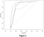
- the quality of a test is generally determined by drawing a Receiving Operating Characteristic (ROC) curve and measuring the Area Under Receiving Operating Characteristic curve (AUROC).
- ROC Receiving Operating Characteristic
- AUROC Area Under Receiving Operating Characteristic curve
- the ROC curve is drawn by plotting the sensitivity versus (1-specificity), after classification of the patients, according to the result obtained for the test, for different thresholds (from 0 to 1).
- ROC curve the area under which has a value superior to 0.7
- the ROC curve has to be understood as a curve allowing prediction of the quality of a test. It is best for the AUROC to be as closed as 1 as possible, this value describing a test which is 100 % specific and sensitive. Consequently, a test with a higher AUROC than another has a "better quality" than the other.
- a diagnosis method comprises
- Some methods shall also include a step i.a), which comprise the steps of modifying the information obtained in i) in order to obtain a new type of information, which is the one that is then compared to the standards in step ii.
- Such modification is the transformation of signal to numerical variables, and combination of the values of variables in a function (such as a logistic regression function or a Cox function), and obtaining an end value.
- the transformation of the values obtained from a patient and the combination thereof in an algorithm or function as herein disclosed is part of a method and provides an intermediate result (an end value or index) that would then to be compared to a reference index (threshold), in order to be able to pose the diagnostic, even though the end value is informative by itself.
- tests herein disclosed are not “gold-standard” tests, in the sense that the output (end value or index calculated by the formulas herein disclosed) is not a definitive answer as to the state of the patient. Indeed, these tests are based on statistics and there may thus be false-positive or false-negative results, which is the reason why the specific experience of the physician in interpreting the index is of importance for making the prognosis and deciding which kind of follow up is to be made to be made for each patient.
- step iii as disclosed above is not direct and immediate from step ii, and the physician must interpret the result from the clinical and general context to be able to reach a conclusion, even though the predictive positive value and the negative predictive value are very high (in view of the high specificity and sensitivity of the tests), indicating that the relative risk is quite increased in the group of patients that respond positively to the tests as compared to the patients that do not respond positively.
- the tests are of great interest in provided a help to the physician when investigating a clinical case.
- the inventors used some markers obtained or generated from data gathered during anesthesia of the subject. These markers are thus used in ex vivo methods, generally after anesthesia of the subject (although it is conceivable that the diagnosis / prognosis test could be performed during anesthesia of the subject). It is to be noted that the tests are not practiced on the human body, in the sense that the diagnosis / prognosis can be reached without the need of the human body of the subject to be present. Indeed, these tests only need data that have been gathered, and are thus no different from the majority of diagnosis tests, that use sample gathered from the patient, without the need of the patient body to be present.
- the methods herein disclosed are computer-implemented, and thus technical method steps are performed by a computer device. As indicated above, some methods herein disclosed also comprise only the steps of generating an end value, which is an intermediate finding of diagnostic relevance, while other methods may also comprise the step of comparing the end value with predetermined values.
- the inventors were able to demonstrate that the acquisition of data from at least one physiological marker associated with the cerebral state of the subject, and at least one physiological marker associated with the cardiovascular state of the subject, measured during anesthesia of the subject can be combined in a function, and that the end result thus obtained can be used to identify a frailty of the subject.
- the age of the patient can also be used in the function that leads to the end result.
- the invention thus relates to the use of physiological and optionally of pharmacological markers and/or of the age of a patient for determining whether a subject has an increased risk (or an absolute) to have an adverse effect (myocardial infraction, stroke, cognitive decline, death) or does not have this risk.
- the invention thus relates to a method for determining whether a subject (especially a human subject) has an increased risk of having frailty, comprising
- the end value is indicative of the presence or of the absence of an increased risk of having a frailty, and can be compared to reference values (cutoffs) to conclude on the actual risk.
- One interest of the method is that it is capable of predicting long-term events, while avoiding positive results in case of post-operative complications, when the function according to which the values are combined has been made as indicated below by excluding patients with post-operative complications (complications within 90 days).
- markers pertaining to the general state of the subject prior to anesthesia include the simplified Lee's score (which is designed to predict the risk of major cardiac complications due to the surgery necessitating anesthesia, such as heart attacks or cardiac death, in patients undergoing non-cardiac surgery), or cardiovascular history of the patient (presence of cardiac events prior to anesthesia (history of any heart-related incidents such as myocardial infarction, heart failure, angina, or cardiac arrest) or of chronic conditions (arrhythmias or chronic heart failure, Arteriopathy of the Lower Limbs or Chronic Kidney Disease)).
- the simplified Lee's score which is designed to predict the risk of major cardiac complications due to the surgery necessitating anesthesia, such as heart attacks or cardiac death, in patients undergoing non-cardiac surgery
- cardiovascular history of the patient presence of cardiac events prior to anesthesia (history of any heart-related incidents such as myocardial infarction, heart failure
- markers used would be the result of the Lee's score, and 0/1 in case of absence/presence of cardiovascular history. It is also possible to provide a score for these markers, as explained below. It is to be noted that these markers provide information as to the instant operative risk (the risk of anesthetizing the patient), and as to the post-operative risk (within 90 days), but not of an increased long time frailty risk (as compared to patients with the same characteristics).
- the method makes it possible to identify an increased risk of systemic frailty. In other embodiments, the method makes it possible to identify an increased risk of cardiovascular frailty. In other embodiments, the method makes it possible to identify an increased risk of neurologic frailty.
- the method both makes it possible to identify the increased risk as indicated above (i.e. a risk that is higher in the population of subjects identified as being at risk by the method as compared to the population of subjects not identified at risk by the method).
- the methods also make it possible to identify patients with an absolute frailty risk (i.e. patients that can be predicted to have an adverse event (as described above) within 2.5 years after anesthesia).
- the methods are performed on data that has been harvested during anesthesia of a subject.
- the methods are however performed independently of the anesthesia. They can thus be performed after the anesthesia (once all needed data is available), but it is not unconceivable to perform the methods when the patient is still under general anesthesia, at a time "t", using data that has been obtained before the time "t". Indeed, the methods do not use instant data obtained during anesthesia.
- BIOS bispectral Index
- an algorithm analyzes interprets the data, and display an end value ranging from 0 to 100, where a value of 0 represents absence of brain activity, and a value of 100 represents the awake state. It is generally sought to maintain BIS values between 40 to 60 (adequate general anesthesia for a surgery), where values below 40 represent a deep hypnotic state.
- the BIS index is known in the art and the main variables incorporated in the algorithm are the frequency and power spectrum of the EEG, the amount of burst suppression, and the degree of synchronization of the EEG. The US Food and Drug Administration approved BIS sensors in 1996.
- PSI Patient State Index
- Such markers are linked to the depth of anesthesia, using EEG acquired signals.
- the inventors propose to use the alpha power, computed from the EEG power spectral density (PSD) extracted from the EEG data.
- PSD EEG power spectral density
- the alpha wave frequency information can be extracted by
- the EEG signal would be processed, in particular by filtering the data to remove frequencies outside the range of interest, removing or interpolating bad channels, and detrending or normalizing the data.
- a window function such as the Hamming, Hanning, or Blackman window, is applied to reduce spectral leakage and improve the spectral resolution of the PSD estimate.
- the Fast Fourier Transform can then be applied to each windowed epoch to estimate the power spectrum.
- the power spectrum is the magnitude squared of the complex FFT coefficients, and represents the distribution of power across different frequencies.
- EEG data could be analyzed using FFT, such as wavelet analysis, Hilbert-Huang transform, or event-related spectral perturbation (ERSP) analysis, which involves calculating a baseline spectral power for a given frequency band (for instance, the baseline could be the values just before induction of anesthesia), and comparing this baseline to the spectral power during anesthesia.
- FFT wavelet analysis
- Hilbert-Huang transform or event-related spectral perturbation
- the PSD calculated for each electrode can then be averaged to obtain the final PSD which is used to compute the alpha power ( ⁇ BP).
- the power is calculated by integration over the 4-13 interval (theta and alpha waves), the 13-30 interval (alpha and beta waves) of the 4-30 interval (theta, alpha and beta waves).
- alpha power is of very interest, or use of a power calculated with alpha and at least one other brain wave information.
- the above markers of cerebral activity of the subject during anesthesia are to be calculated when the subject is under stable anesthesia.
- the anesthesia is considered as stable/stationary when the rate of anesthetic drug administered to the patient is stable (and thus when the blood concentration of anesthetic drugs is stable. It is also preferred that the Mean Arterial Pressure (MAP) is above or equal to 65mm Hg.
- MAP Mean Arterial Pressure
- the moment where the cerebral markers can be measured is generally reached from 10 minutes post induction of anesthesia.
- anesthesia induction would last for 5 minutes and that another 5 minutes would ensure that the stable anesthesia regimen is reached.
- SEF Spectral Edge Frequency
- SEF Spectral Edge Frequency
- alpha power is calculated on an EEC signal of less than 10 minutes, or less than 5 minutes. In order to have enough signal to obtain a representative result, it is preferred when the duration of the EEC signal is at least 30 seconds, preferably at least 1 minute. Using an EEC signal lasting between 3 and 5 minutes is adequate. It is also possible to calculate multiple alpha power values for different times of the EEC signal, and to take the mean of these values. However, since the alpha power is calculated when the subject is in a stable anesthesia state, it is foreseen that the alpha power value will essentially remain constant whenever it is calculated.
- the SF may be used as the marker of cerebral activity, notably when it is below 8 Hz (and that the alpha power thus cannot be calculated).
- the selected value of the SEF is obtained like the indexes as indicated above.
- the physiological marker of the cerebral state is based on an EEG of the subject, that has been obtained during anesthesia, and intends to reflect the depth of anesthesia of the subject, and the cerebral activity of the subject during anesthesia.
- the preferred markers are alpha power (or another marker that would reflect activity of the alpha wave), BIS, PSI, Entropy, or SEF.
- Alpha power is a preferred marker of cerebral state. Any other marker or index that would provide the same type of information can also be used.
- mAP mean arterial pressure
- MAP mean arterial pressure
- One commonly accepted view of the blood pressure/cerebral blood flow relationship is based on the Lassen curve that assumes that there is an optimal range of cerebral perfusion, mAP typically between 60 and 150 mmHg, where the cerebral blood flow is maintained at a relatively constant level.
- the physiological marker used, in the methods herein disclosed, reflects the blood pressure of the patient.
- PP Pulse Pressure
- SBP systolic blood pressure
- DBP diastolic blood pressure
- SBP systolic blood pressure
- HR heart rate
- Such monitoring may be performed using another device than the device used to monitor and/or analyze the EEG.
- the mAP is thus monitored and displayed on another monitor.
- mean arterial pressure is measured with a sphygmomanometer (or electronic electrocardioscope tensiometer) which uses the oscillometric measurement when the sleeve placed on the patient's forearm deflates automatically.
- the maximum value of the oscillation represents the mean arterial pressure.
- This method also allows measurement of the diastolic and systolic blood pressures, and hence of the pulse pressure.
- mAP is measured using a plethysmograph (pulse oximeter located at the tip of a finger).
- a plethysmograph pulse oximeter located at the tip of a finger.
- the mAP is continuously evaluated, based on the value of the height of the dicrotic wave, continuously calculated by plethysmography, using a method comprising:
- PI Perfusion Index
- PVI Pleth Variability Index
- the pressure values that are obtained during the anesthesia are compared to a baseline, which is the mAP or PP that has been measured before anesthesia, when the subject is awake.
- mAP and PP are generally, and according to current good practices, measured every now and then (generally every 5 minutes, or more often every 2-3 minutes). This allows the anesthetist to use a vasopressor to increase mAP in case of decline of such.
- the data obtained is generally a series of discrete values corresponding to the mAP or PP values obtained at different time points during anesthesia (one could also use a series of PI or PVI values).
- RMSE Root Mean Square Error
- the Root Mean Square Error (RMSE) of these values as compared to the baseline value can be calculated and can be used in the functions and methods herein disclosed. Making such calculation (calculating the RMSE with regards to a baseline, rather than with regards to the mean of the series of values) is uncommon in the art.
- RMSE is the square root of the average of squared differences between the measures mAP or PP values (or other values) and the baseline value. RMSE is particularly sensitive to data points that would significantly differ from other observations.
- markers associated with the blood circulation and/or organ perfusion during stable anesthesia are used, notable mAP, PP, PI or PVI.
- mAP sBP, or PP are preferred markers, PP or sBP being the most preferred.
- the values used in the methods herein disclosed should reflect the deviation with regards to the baseline value measured before anesthesia. RMSE of the series of measured data is an adequate value to use in such methods and functions.
- physiological markers are used in all methods and functions herein disclosed, it may be of interest, to detect specific risks of adverse outcomes, to also use markers associated with the pharmacological products and regimen used for the anesthesia of the subject. One will thus use data from at least one marker associated with the products administered to the subject during anesthesia.
- markers can be obtained from the amount of drug provided to the patient during the period of stable anesthesia.
- Other markers associated with the amount of vasopressor and/or of any analgesic drug can also be used in such functions.
- propofol (injected) and sevoflurane (inhaled) are the most common anesthetic agents used for general anesthesia, and the methods herein disclosed are exemplified with these agents, other non-opioid anesthetic drugs that are administered intravenously (such as a barbiturate (in particular amobarbital, methohexital, thiamylal or thiopental), a benzodiazepine (notably diazepam, lorazepam or midazolam), etomidate or ketamine).
- inhaled agents one can also cite desflurane, isoflurane, methoxyflurane, nitrous oxide, or even enflurane, halothane or xenon.
- TCI Target Controlled Infusion
- the TCI is linked to the brain target dose desired by the anesthetist, and after entering the desired parameters in a system, the system controls the infusion pump to inject the proper dosage, taking into account the repartition of the drug within the different body compartments and its elimination.
- TCI is a widely used parameter in anesthesia ( Struys et al, 2016. Anesth. Analg. 122 (1): 56-69 ).
- the Marsh and Schnider models are pharmacokinetic models for determining propofol concentration in the various compartments.
- the Marsh model has been adapted from the Gepts model, in which the rate constants are fixed, whereas compartment volumes and clearances are weight proportional.
- the Schnider model was developed during combined pharmacokinetic-pharmacodynamic modelling studies. It has fixed values for V1, V3, k13, and k31, adjusts V2, k12, and k21 for age, and adjusts k10 (elimination rate constant) according to total weight, lean body mass (LBM), and height.
- the infusion rate remains constant, and guarantees that the target concentration is reached and maintained.
- MAC Minimum Alveolar Concentration, described in Eger et al., 1965. Anesthesiology. 26 (6): 756-63 ).
- MAC is a regular parameter monitored in anesthesia and can be linked to the concentration of anesthetic in the air exhaled by the subject, by the formula proposed by Eiger, which also takes the age into consideration.
- TCI Propofol Target-Controlled Infusion
- MAC volatile anesthetic Minimum Alveolar Concentration
- min(X) is the minimal value of TCI (or MAC) and max(X) is the maximal value of TCI or (MAC) as observed during anesthesia.
- vasopressors that are administered to the subject when hypoperfusion of the brain is detected.
- the vasopressor increases arterial pressure.
- Vasopressors are known in the art and include sympathomimetics (including adrenaline, norepinephrine (also called noradrenalin), dopamine, ephedrine%), glucocorticoids and mineralocorticoids, angiotensinamide...
- sympathomimetics are used instead. It is not always necessary to use such agents during anesthesia, as it will depend on the evolution of the hemodynamic parameters for the subject. Furthermore, since the duration of the anesthesia may vary (depending on the type of surgical or interventional interventions), using the total amount of administered vasopressor molecule mays not be proper.
- mean rate (which can be expressed as a "unit of weight / unit of time” or "unit of mole / unit of time”) may also be pondered (divided) by the subject's weight (as a larger amount of vasopressor is needed for a heavier patient).
- antalgic agent When an antalgic agent is used (morphine or derived thereof), it is likewise preferred to use the total amount used during intervention divided by the duration of intervention (time in the operating room), and preferably pondered by the subject's weight.
- Figure 7 shows clusters of markers. Markers from the same clusters can thus be considered equivalent, and it is consequently possible to substitute one marker for another in the same cluster, or for a close marker. For instance, one could use either beta or alpha power, but also total or theta power. Reference PP or RMSE of PP could be used, or Reference SBP or SBP variability (RMSE).
- the invention also pertains to a method for obtaining a function for identifying frailty or an increased frailty risk in a patient wherein said function combines the values associated with physiological markers of said patient and optionally with pharmacological markers and/or with the age of the patient, comprising:
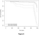
- the analysis in c) is a Cox analysis, providing a Cox function that provides survival (life without adverse effect) overtime. This type of function makes it possible to determine the evolution of the occurrence of the adverse effect and the divergence depending on whether the subject is positive or negative to the test ( Figures 5 and 6 ).
- not all markers identified in b) are used in the analysis in c) (which leads to the regression or Cox function), and it is possible to determine the appropriate number of markers (using at least the markers for neurological and cardiovascular data), and the appropriate markers, according to what is explained in the examples (in particular determining relations between markers to avoid, as much as possible, using correlated markers, removing one marker and calculating the AUROC to determiner whether this marker adds up to the quality of the function).
- the markers that will be identified in b) may vary. Some markers, that would be relevant for some adverse effects may not be relevant for others. Age may also not be relevant in some situations (for some adverse effects).
- the invention also pertains to a method for obtaining a function for identifying frailty or an increased frailty risk in a patient wherein said function combines the values associated with physiological markers of said patient and optionally with pharmacological markers and/or with the age of the patient, comprising:
- the end result is thus obtained aggregating by summation the discrete scores of c) obtained for the markers selected in b) during anesthesia of the subject.
- a discrete score (0, 1, 2...) will be assigned for each marker in c) depending on thresholds that can be identified as explained below.
- "x" thresholds will define "x+1" class and the score (0, 1,...,x+1) is assigned to the marker according to the class to which it is assigned.
- the score "0" is assigned to the marker if its value is below t1
- the score "1” is assigned if the value is between t1 and t2
- the score "2” is assigned if the value is between t2 and t3
- the score "3" is assigned if the value is above t3.
- the same number "x" of classes are selected for each marker, meaning that the score will be comprise between “0” and “n.x” in case “n” markers are used.
- four classes are defined for each marker.
- the score will thus take any integer value between “0” and “3n” when “n” markers are used.
- Decision Tree-based Cutoffs Initially, decision tree algorithms can be employed to ascertain optimal cutoffs for each variable (marker). The decision tree can be constructed by iteratively splitting the data based on the variable (marker) value. The splits can be determined using the Gini impurity criterion. The complexity parameter can be based on the cross validation data to optimize the pruning process and avoid overfitting. The specific cutoffs can be determined by the splits made at each decision node and used to classify the observations into the respective outcome groups (classes).
- Quartile-based Scoring For each marker, or for marker where the decision tree method does not yield distinct or clinically meaningful splits, it is possible to use the quartile distribution method.
- variables were categorized into quartiles, and scores are assigned either in an ascending or descending manner, contingent on the clinical significance of the variable.
- CMEUA One such function is disclosed as CMEUA in the examples and is also part of the invention.
- the number of patients in the cohort should be as large as possible, indicating that it preferably comprises more than 50 patients, preferably more than 100 patients, preferably more than 150 patients. As indicated, there is no upper limit for the number of patients, and the larger, the better.
- the function is a logistic regression that makes it possible to predict occurrence of a cardiovascular adverse event (cardiovascular frailty), and thus identify patients with a higher risk of having such adverse event.
- F1 a1 ⁇ a2 * log alpha1 + a3 * rmse_pp , with
- This function uses, as markers / variables, the alpha power (alpha, that has been transformed to take the log value, and the RMSE of the Pulse Pressure. (rmse_pp).
- This functions thus uses two physiological markers, representing the cerebral activity (alpha power, obtained from an EEG), and the hemodynamic activity (Pulse Power, obtained from a cuff). It provides continuous outputs.
- the cutoff value is 0.053.
- the function makes it possible to predict occurrence of a neurocognitive decline adverse event (neurological frailty), and thus identify patients with a higher risk of having such adverse event.
- F2 b1 + b2 * age ⁇ b3*log alpha + b4 * rmse_pp + b5*log dose_nor_total , with
- F2 18.237 + 0.0686*(age) - 3.184*log(alpha) + 0.0646*(rmse_pp) + 0.286*log(dose_nor_total)
- This function uses the age (in years), the alpha power information transformed to take the log value (log(alpha)), the RMSE of the Pulse Pressure (rmse_pp), and the total dose of noradrenaline provided to the patient (pondered by the time in the block and the subject's weight), as indicated above, transformed to take the log value (log(dose_nor total)).
- the AUROC Absolute Under Receiving Operating Curve
- AUC 0.953
- a PPV 0.826
- a NPV 0.999 (Sensitivity: 0.999, Specificity: 0.966), using a cutoff at -1,0790.
- This function makes it possible to identify patients with no risk, and an decreased risk (as compared to the patients above the cutoff) of having a neurological adverse effect.
- the function is a logistic regression that makes it possible to predict occurrence of an adverse event (cardiovascular and/or cognitive decline and/or death, thus revealing frailty), and thus identify patients with a higher risk of having such adverse event.
- F 3 c 1 ⁇ c 2 * log alpha + c 3 * rmse _ pp + c 4 * age + c 5 * log dose _ nor _ total + c 6 * anesthetic _ concentration , with
- F3 18.0269 - 3.1827*log(alpha) + 0.0655*(rmse_pp) + 0.0679*(age) + 0.3218*log(dose_nor_total) + 0.3543*(anesthetic_concentration)
- This function uses the age (in years), the alpha power information transformed to take the log value (log(alpha)), the RMSE of the Pulse Pressure (rmse_pp), and the total dose of noradrenaline provided to the patient (pondered by the time in the operative room and the weight), as indicated above, transformed to take the log value (log(dose_nor_total)) and the anesthetic concentration (TCI or MAC, standardized as described above).
- the AUROC Absolute Under Receiving Operating Curve
- AUC 0.934
- a PPV 0.872
- a NPV 0.957
- patients above 0.7 can be said to have a higher (and potentially an absolute) frailty risk
- patients below 0.39 can be said to have a lower (and potentially no) frailty risk.
- one can look at other extrinsic evidence (age, chronic diseases) to perform further cardiologic or neurologic investigations.
- the function uses, for each marker, scores from 0-3 assigned as explained above, using cutoffs for each marker value.
- This function makes it possible to predict occurrence of an adverse event (cardiovascular and/or cognitive decline and/or death, thus revealing frailty), and thus identify patients with a higher risk of having such adverse event.
- F4 ⁇ d1 + d2 ⁇ score_alpha + d3 ⁇ score_pp_var + d4 ⁇ score_age + d5 ⁇ score_nor_total + d6 ⁇ score_anesthetic_concentration , with
- F4 -6.933 + 2.702 ⁇ (score_alpha) + 0.617 ⁇ (score_pp_var) + 0.997 ⁇ (score_age) + 0.417 ⁇ (score_nor_total) + 0.0313 ⁇ (score_anesthetic_concentration)
- This function uses the age (in years), the alpha power information, the Pulse Pressure information (RMSE), the total dose of noradrenaline provided to the patient (pondered by the time in the operative room and the weight), and the anesthetic concentration (TCI or MAC, standardized as described above).
- the cutoff value is 0.4158.
- This function has a high NPV, meaning that it allows to exclude from future consideration the patients for which the end value is below the cutoff. Further investigation (by a cardiologist or a neurologist) can be performed for the patients with an end value above the cutoff.
- patients above 0.59 can be said to have a higher (and potentially an absolute) frailty risk
- patients below 0.1 can be said to have a lower (and potentially no) frailty risk.
- one can look at other extrinsic evidence (age, chronic diseases) to perform further cardiologic or neurologic investigations.
- the function is a logistic regression that makes it possible to predict occurrence of an adverse event (cardiovascular and/or cognitive decline and/or death, thus revealing frailty), and thus identify patients with a higher risk of having such adverse event.
- F 5 e 1 ⁇ e 2 * log alpha + e 3 * rmse _ pp + e 4 * log dose _ nor _ total + e 5 * cv _ history _ pre + e6 * age , with
- F5 18.256 - 3.159xlog(alpha) + 0.0547x(rmse_pp) + 0.295xlog(dose_nor_total) + 1.144x(cv_history_pre) + 0.0637x(age)
- This function uses the age (in years), the alpha power information transformed to take the log value (log(alpha)), the RMSE of the Pulse Pressure (rmse_pp), the total dose of noradrenaline provided to the patient (pondered by the time in the operative room and the weight), as indicated above, transformed to take the log value (log(dose_nor_total)) and the past cardiovascular history (cv_history_pre; binary value: 0 if no cardiovascular history, 1 if presence of past cardiovascular event).
- patients above 0.82 can be said to have a higher (and potentially an absolute) frailty risk
- patients below 0.39 can be said to have a lower (and potentially no) frailty risk.
- one can look at other extrinsic evidence (age, chronic diseases) to perform further cardiologic or neurologic investigations.
- markers providing information as to the cerebral and the hemodynamical states of the subject during anesthesia, and optionally other markers associated with the products used for anesthesia (amount of anesthetic and/or of vasopressor used during the stable anesthesia) and the age of the subject, to obtain formulas that can predict increased risk frailty (either systemic or specific to a certain type of adverse effect), or lower risk of frailty.
- cutoffs that are deemed informative enough. Indeed, if a higher PPV is sought, the function will be more stringent, to avoid obtaining false positive. Alternatively, it is possible to choose cutoffs which increase the NPV, to make sure that there are very little false negative patient in the group of patients considered “not at risk”. It is also possible to use two cutoffs (one giving a good PPV, to identify patients at risk (above this cutoff), and another one with a good NPV to identify patients "not at risk (below this cutoff)). When the end result of a patient is between these cutoffs, the patient is in a "grey zone".
- the age of the patient may be taken into consideration, and the physician may address this patient to a cardiologist for a cardiac check-up if the patient is 60 or above, and to a neurologist for a neurologic check-up if the patient is 70 or above or 75 or above.
- the method can be used first to determine general frailty, using one function, then the status of the subject can be refined by using a function providing cardiovascular or neurological frailty. If both functions do not give any risk, other frailty type (liver, renal) may be looked at.
- the physician can determine whether the patient has a latent cardiovascular condition, neurological condition, liver condition, renal condition, or cancer.
- presence of a cardiovascular condition is determined by electrocardiograms, scanner, MRI, echography, or detection of biomarkers associated with cardiovascular diseases (see also below).
- Presence of neurological condition may be determined by performing specific cognitive tests aimed at detecting presence of neurological diseases and/or performing clinical testing such as scanners, electroencephalograms, MRI (see also below).
- Presence of a liver condition may be determined notably by an ex vivo diagnosis method for the presence of liver fibrosis, steatosis, cirrhosis (such as the methods disclosed in WO 02/16949 , WO 2006/082522 , WO 2006/103570 , WO 2013/079711 , WO 2018/050804 , WO 2019/076830 or WO 2020/021058 ), or by liver ultrasound, liver stiffness measurement or MRI.
- Presence of a renal condition may be determined by assessing the level of creatinine or the renal clearance,
- Presence of a cancer may be determined by scanner, MRI or measurement of cancer biomarkers in the blood, urine, cerebrospinal fluid or genome of the patient
- the physician shall perform specific tests aimed at detecting presence of cardiovascular diseases.
- Such tests include physical examination to reveal signs such as high blood pressure, abnormal heart sounds, or peripheral artery disease, performing an electrocardiogram (ECG to detect irregular rhythms, heart attacks, and other heart abnormalities), or an echocardiography (to obtain detailed images of the heart's structure and function, helping to diagnose conditions like valvular heart disease, heart failure, and congenital heart defects), performing cardiac stress testing (stress tests or pharmacological stress tests to evaluate how the heart responds to physical exertion to help diagnose coronary artery disease), have the patient have chest X-ray, which may reveal abnormalities in the heart, lungs, or blood vessels that can be indicative of heart disease, conduct cardiac catheterization (angiography), to identify blockages or narrowing in the blood vessels, or coronary computed tomography angiography (CCTA) to produce detailed images of the coronary arteries, aiding in the diagnosis of coronary artery disease, perform Magnetic Resonance
- Treatment will thus be adapted to the exact condition of the patient (use of statins, of hypotensive medications, stent implantation, angioplasty, cardiac by-pass, or even cardiac graft). Treatments include
- the function and methods allow for specific follow-up, characterization and/or treatment of patients identified as being at risk of cardiovascular disease.
- the physician shall perform specific tests aimed at detecting presence of neurological diseases, such as Alzheimer's disease, attention deficit disorder, dementia with Lewy bodies disease, early onset dementia, epilepsy-related cognitive dysfunction, fronto-temporal dementia, mild cognitive impairment, normal pressure hydrocephalus.
- neurological diseases such as Alzheimer's disease, attention deficit disorder, dementia with Lewy bodies disease, early onset dementia, epilepsy-related cognitive dysfunction, fronto-temporal dementia, mild cognitive impairment, normal pressure hydrocephalus.
- MMSE Mini-Mental State Examination
- MoCA Montreal Cognitive Assessment
- brain imaging Magnetic Resonance Imaging, Positron Emission Tomography (PET) scans, Single Photon Emission Computed Tomography (SPECT) scans
- biomarker analysis Cerebrospinal fluid (CSF) analysis for detecting specific proteins like amyloid-beta and tau, which can be indicative of Alzheimer's disease or other neurodegenerative disorders, looking for blood-based biomarkers such as neurofilament light chain (NfL) and beta-amyloid
- NfL neurofilament light chain
- beta-amyloid neurofilament light chain
- EEG electroencephalography
- Treatments for cognitive decline include
- the function and methods allow for specific follow-up, characterization and/or treatment of patients identified as being at risk of neurological disease.
- the invention also relates to a method for treating a patient having frailty, comprising the steps of performing the method of diagnosis/prognosis as disclosed above and providing the patient with appropriate treatment depending on the type of frailty and the risk identified by potential further investigation.
- the methods herein discussed are performed in vitro, or ex vivo. They can be computer implemented, in particular when the information pertaining to anesthesia and the age of the subject are provided to one or more processor, where the one or more processor perform the transformation of the raw data and the calculation of the end value.
- the first mean is computerized. It may be an electronic spreadsheet with the formula recorded within, that provides the first index as an output when entering the various elements mentioned above. It can also be a computer program that provides the first index as an output after receipt of the various elements mentioned above.
- the first means can present one or more of the following, in either combination:
- a non-transitory computer readable storage medium having stored thereon a computer program comprising program instructions, the computer program being loadable into a data-processing unit and adapted to cause the data-processing unit to carry out a method for calculating a first index by combining the values of a patient, calculated as herein disclosed, through a function as disclosed above, when the computer program is run by the data-processing device.
- the invention in another embodiment, relates to a microprocessor comprising a computer algorithm to perform a method of determining the presence of frailty (or increased risk thereof) in a patient: providing the values of a patient, calculated as herein disclosed, and optionally the age of the patient; and combining the values through a function (as disclosed above, or obtained as disclosed above) to obtain a score (the end-result or first index) useful for presence of frailty (or increased risk thereof) in the patient.
- the invention also relates to the use of the functions, devices and/or microprocessors in order to make a diagnosis of presence of frailty (or increased risk thereof) in a patient by use of the functions and comparing the result to a predetermined threshold to determine the presence of frailty (or increased risk thereof) in the patient.
- the invention also includes a device for diagnosis an increased frailty risk (as defined above) in a patient, comprising a means, wherein the means provides an end result by performing a method as described above.
- the device comprises means for receiving the data used for performing the methods herein disclosed and means for obtaining the numerical values and/or combining such.
- Said end result can be later used to determine presence of an increased frailty risk in said patient and whether it is necessary to initiate treatment or follow-up, or perform further investigations.
- the above method can be computer-implemented.
- a sender is required to identify himself to a server or system within a public or private network. It is preferred when the network is a private network, and when the connection to the server is encrypted. Identification of the sender can be performed using login and password, preferably through strong identification processes (such as one-time password, or using biometric or smart card identification).
- the sender Upon login to the server, the sender is required to fill-in information related to the patient (such as name, sex, birth date, height, weight, social security identification number, social insurance number) and a specific identifier is assigned to the patient in a database. If the database does not contain any entry relating to the patient, one entry is created. If an entry already exists (using the social security identification number allows ensuring the unicity of entry), such entry is activated.
- information related to the patient such as name, sex, birth date, height, weight, social security identification number, social insurance number
- the server shall then receive information pertaining to the cerebral state of the patient during anesthesia. Such information may be a raw EEG, the alpha power if already calculated from the raw EEG, the SEF, the BIS, the Entropy and/or the PSI indexes.
- the server shall also receive information pertaining to the hemodynamic parameters of the patient during anesthesia. This information can be recording of the mAP, of the PP, of the SBP, of the DBP and/or of the HR.
- Information pertaining to the pharmacological parameters of the anesthesia are also received by the server, such as the BPI or the MAC, the amount of vasopressor used, the amount of antalgic drug used and/ the amount of anesthetic drug used, the duration of anesthesia, the duration of stable anesthesia, the duration of time in the operating room.
- the server such as the BPI or the MAC, the amount of vasopressor used, the amount of antalgic drug used and/ the amount of anesthetic drug used, the duration of anesthesia, the duration of stable anesthesia, the duration of time in the operating room.
- Such information is stored in a database, associated with the specific identifier of the patient, so that such data is uniquely associated to the patient.
- the sender may be in a remote place different from where the server is located. It is also envisaged that the information is recovered by the server, using information provided by the sender. For instance, the server may open a connection with the server of the hospital or health establishment where the anesthesia took place, using identifiers provided by the sender, to upload the required information. Alternatively, the information may be filled in by the sender.
- the next step is to transform and optionally standardize the received data in numerical values for each received information.
- This step is performed by a processor in the server or able to use the information from the databases.
- calculating the alpha power the SEF can be performed using the raw EEG data.
- the numerical value associated with BIS or MAC can be standardized according to the methods herein disclosed.
- the value that is associated with the hemodynamic parameters is determined according to the methods herein disclosed.
- the age of the patient may be obtained from the birth date of the patient. Using the duration of in the operating room, the weight of the patient, the amount of vasopressor or antalgic drug can also be obtained in a format as specified above. Such calculation may be performed by a processor in the server.
- Such processor may also be programed to determine the period of stable anesthesia (using the EEG, the hemodynamic parameters, it is indeed possible to identify the induction and the wakening phases), and its duration.
- the processor shall assign the adequate scores.
- the numerical values are to be stored in a database, that is associated with the raw information received, and the unique identifier of the patient (as well as the date of calculus).
- the next step is to combine the numerical values in a function as disclosed herein to obtain an end result, that is optionally normalized (to be between 0 and 1) as disclosed.
- This end result may be compared to one or more set cutoff(s) (reference value).
- the end result and information pertaining to the comparison is then stored in a database associated with the identifier specific of the patient and is preferably date and time-stamped. Thereby, a database is obtained, containing identification related to the patient (patient's specific identifier), associated with the data pertaining to the anesthesia and comparison information, and optionally but preferably the intermediate numerical values and the end result.
- Such information (nature of the risk of the patient) is then transmitted to the sender, optionally with the result of the function and with any other potential information of interest.
- the invention also relates to a computer-implemented method for determining whether a subject has an increased frailty risk, comprising
- the invention also relates to a method for obtaining the information pertaining to the presence of an increased risk of frailty in a subject, comprising
- the invention relates to a microprocessor comprising a computer algorithm to perform a method for calculating the presence or absence of an increased frailty risk from information associated with an anesthesia of a subject, as herein disclosed, through a method as disclosed above.
- the invention also relates to the use of the functions, devices and/or microprocessors in order to assess presence or absence of an increased frailty risk in a subject by use of the methods herein disclosed.
- Anesthetia management General anesthesia was standardized across all procedures. Induction was achieved with sufentanil or remifentanil, followed by atracurium besilate for muscle relaxation. Blood pressure was managed to maintain at 80% of baseline using norepinephrine. Maintenance agents included sevoflurane, desflurane, or propofol, with the choice at the anesthesiologist's discretion. Monitoring protocols aimed to maintain patient state index and spectral edge frequency within specified ranges to prevent burst suppression. Detailed descriptions of the anesthetic drugs, dosages, and monitoring parameters are described below. GA protocols were standardized for both induction and maintenance phases.
- sufentanil at a dosage of 0.2 ⁇ g/kg was used in orthopedic surgery and remifentanil targeted at 3-5 ng/ml, titrated according to Minto's model, for interventional neuroradiology procedures. This was accompanied by the non-depolarizing neuromuscular blocking agent, atracurium besilate. Hemodynamic stability was managed by maintaining blood pressure at 80% of baseline levels, facilitated by the administration of diluted norepinephrine at a concentration of 5 ⁇ g/mL. Maintenance of GA was at the discretion of the attending anaesthesiologist, with the options including sevoflurane, desflurane, or propofol.
- TCI target-controlled infusion
- MAC minimum alveolar concentration
- ketamine during induction was optional, based on the attending anaesthesiologist's judgment.
- intraoperative monitoring aimed to keep the patient state index (PSI) within the range of 25 to 50 and the spectral edge frequency (SEF95) between 8 and 15 Hz, with a specific emphasis on avoiding burst suppression.
- PSI patient state index
- SEF95 spectral edge frequency
- Data collection methodology Perioperative data were systematically gathered from digital patient records and integrated monitoring systems, which included medical history, hemodynamic parameters, cognitive evaluations, and prospective tracking of poor prognosis. To ensure the objectivity of the analysis, all data assessments were conducted with the researchers blinded to patient outcomes. This blinding protocol was essential to maintain impartiality and prevent bias in the evaluation of the data.
- Patient health assessments Preoperative evaluations covered a spectrum of health metrics: frailty via the Clinical Frailty Scale, independence through the Instrumental Activity of Daily Living (IADL) Scale, muscle strength assessment with a dynamometer, pain quantification using the Numerical Rating Scale (NRS), and prognostic indicators.
- IADL Instrumental Activity of Daily Living
- NRS Numerical Rating Scale
- EEG monitoring EEG recordings provided intraoperative insights into cerebral function under anesthesia. Stable periods for EEG analysis were selected as those occurring 15 minutes after induction, with a consistent 5-minute timeframe where the concentration of hypnotic drugs was maintained and MAP exceeded 70 mmHg. A spectrogram, which graphically represents the variation in power across different EEG frequency bands over time, aided in identifying suitable EEG segments for power spectral analysis. The depth of anesthesia was assessed using the Patient State Index (PSI), averaged over these identified stable EEG periods.
- PSI Patient State Index
- the spectrogram was computed after band-pass filtering through the [0.5, 25] Hz range using the Chronux toolbox in Matlab [ Bokil et al. J Neurosci Methods. 2010;192:146-151 ].
- EEG data segments devoid of burst suppression or transitioning features were visually selected on the spectrogram during a period of at least 10 minutes of stable anesthesia.
- a contiguous 5-minute window of EEG data was selected for analysis, ensuring the window was free of artifacts, that the mean arterial pressure was above 70 mmHg, and that the hypnosis concentration was stable for 10 minutes before and after the selected period of analysis.
- the Power Spectral Density was computed using Multitaper methods implemented in the Chronux library [ Bokil et al. J Neurosci Methods. 2010;192:146-151 ].
- Hemodynamic variable analysis continuous monitoring of key indices, including MAP, systolic (SBP) and diastolic (DBP) blood pressures, pulse pressure (PP), and heart rate (HR), was undertaken to evaluate vascular stability.
- Intraoperative hypotension (loH) was defined as any duration during the operation where MAP remained below 65 mmHg or experienced a 20% reduction from the 'reference' value.
- the 'reference' value is established by the initial hemodynamic measurements captured in the operating room before the administration of anesthesia. Conversely, the 'average' value denotes the mean of these measurements throughout the course of the surgery.
- MAP mean arterial pressure
- SBP systolic
- DBP diastolic
- PP pulsed pressure
- HR heart rate
- Intraoperative hypotension (loH) was characterized by a MAP falling below 65 mmHg or a decrease of 20% from its baseline value. Baseline value was defined as the first value of hemodynamics parameters before anesthesia start, the average value as the mean of all values during the procedure.
- RMSE Root Mean Square Error
- Criteria for Favorable Prognosis Conversely, a 'favorable prognosis' was characterized by the absence of mortality, cardiovascular incidents, or cognitive impairment within the 2.5-year follow-up period.
- Cognitive Decline Outcome Assessment Cognitive assessments were performed at entry to the study, typically the day before surgery, at discharge from the hospital or on Day 3 (whichever was earlier), and 2.5 years post-surgery. The use of both MoCA and T-MoCA tools across these three timepoints allowed for a comprehensive assessment of patients' cognitive changes over time and post-surgery.
- the MoCA is a cognitive screening tool that takes approximately 15 minutes to administer. It evaluates multiple cognitive domains: attention and concentration, executive functions, memory, language, visuo-constructional skills, conceptual thinking, calculations, and orientation.
- the T-MoCA is a telephone version of the MoCA and omits the visuospatial/executive and naming component of the original MoCA, resulting in a maximum score of 22.
- Step 1 the initial variable selection was informed by univariate analysis and the identification of established prognostic indicators.
- a correlation matrix was constructed using Pearson's correlation coefficient, which informed the development of a dendrogram through hierarchical clustering. This visual representation of variable clusters, based on their correlation coefficients, facilitated the identification of non-redundant variables, which were then chosen for the predictive models to minimize multicollinearity and maintain the multidimensional integrity of clinical assessments.
- Step 2 the variable selection was refined utilizing a systematic, data-driven stepwise approach guided by the Akaike Information Criterion (AIC), implemented via the 'step' function in R.
- AIC Akaike Information Criterion
- VIF Variance Inflation Factor
- the primary objective of the study below was to assess the discriminative power of the Augmented Model in predicting « poor prognosis » (composite of death, cognitive decline, cardiovascular event).
- the Receiver Operator Curve was employed.
- Secondary objective was to study the ROC-AUC of the Augmented Model for each level of the composite score using multinomial analysis. To evaluate the predictive accuracy of our metrics, the dataset was divided into a training set (70%) and a test set (30%). To gauge the inherent uncertainty and robustness of our model, 2000 bootstrap iterations were undertaken.
- CMEUA Score Computation the Continuous Metrics Evaluation under Anesthesia (CMEUA) score was developed using ten pivotal parameters identified through univariate analysis and known prognostic indicators. These parameters include age, baseline PP values, relative MAP and PP variations during anesthesia, average norepinephrine infusion rate, stable period alpha and theta band power, PSI, and hypnotic drug concentration (either MAC or TCI).
- a decision tree algorithm (implemented via the "rpart" package in R) was initially utilized to determine optimal cutoffs concerning specific outcomes, such as favorable versus poor prognosis.
- a quartile-based distribution scoring system was used. All variables were graded on a 0-3 scale, with higher scores denoting a worse prognosis. Further nuances of the tree design are elucidated below.
- Score Assignation the approach to assigning scores involved two primary strategies:
- the use of both MoCA and T-MoCA tools across these three timepoints allowed for a comprehensive assessment of patients' cognitive changes over time and post-surgery.
- the MoCA is a cognitive screening tool that takes approximately 15 minutes to administer. It evaluates multiple cognitive domains: attention and concentration, executive functions, memory, language, visuo-constructional skills, conceptual thinking, calculations, and orientation.
- the T-MoCA is a telephone version of the MoCA and omits the visuospatial/executive and naming component of the original MoCA, resulting in a maximum score of 22. Consistency across assessments was ensured by converting MoCA scores to a 22-point scale, by excluding the visuospatial/executive and naming component values.
- the secondary endpoint focused on gauging the discriminatory capability of the CMEUA for the composite outcome.
- the Receiver Operator Curve (ROC) and the Area Under the Curve (AUC) metrics were ysed. To evaluate the predictive accuracy of our metrics, the dataset was divided into a training set (70%) and a test set (30%) using a random seed of 123 to ensure reproducibility.
- the ROC-AUC of the CMEUA score was juxtaposed against a clinical model that incorporated age, the ASA score, the existence of hypertension and the Lee Score. Differences between the AUCs were tested for statistical significance using the DeLong test.
- the ROC-AUC (AUROC) of the CMEUA score was studied for each level of the composite score using multinomial analysis.
- a dendrogram-based analysis revealed eight clusters of highly correlated variables. To reduce redundancy, one variable from each cluster was selected for its correlation with the outcome. The chosen variables included age, hypertension status, cardiovascular risk factors, Clinical Frailty Scale, baseline T-MoCA scores, history of cardiovascular disease, Lee Score, and dyslipidemia status. After stepwise regression in constructing the Clinical Model, only age, hypertension status, and history of cardiovascular disease remained as significant predictors.
- TCI Propofol Target-Controlled Infusion
- MAC halogenated gas Minimum Alveolar Concentration
- EEG variable analysis showed that all band powers were significantly reduced in patients with poor prognosis (p ⁇ 0.001 for all parameters, see Table 4). Multivariate regression confirmed the significance of these findings, even after adjustments for anesthetic concentration and patient age (p ⁇ 0.001 for all parameters).
- Dendrogram-based analysis identified six clusters of highly correlated intraoperative variables ( Figure 7 ). To minimize redundancy, one variable from each cluster was chosen for its correlation with patient outcomes. These included Alpha and Theta band powers, Total power, Hypnotic drug concentrations, PP variability, and Norepinephrine dosage. Subsequent stepwise regression analysis retained Alpha Band Power, PP variability, and Norepinephrine dosage as significant prognostic indicators.
- PSI Patient State Index
- the Augmented Model was constructed as described in Example 1. Following stepwise regression, the model included Age, Cardiovascular History, Alpha Band Power, Total Dose of Norepinephrine, and Pulse Pressure Variation. In the test set, the Augmented Model (function F5) achieved an AUC-ROC of 0.948 [95% Cl: 0.890 - 0.998]. In comparison, the Clinical Model, which included only clinical variables, yielded an AUC-ROC of 0.819 [95% Cl: 0.708 - 0.917]. A DeLong's test was employed to compare the performance of the two models, revealing a significant difference with a p-value of 0.012 (refer to Figure 2 ). The 95% confidence interval for the difference in AUC ranged from 0.02 to 0.23.
- the composite CME UA score was constructed as detailed in the methods section. A marked difference was observed between the groups, with a notably higher score in the poor prognosis group: 16.0 (IQR: 14.0-18.0) versus 9.0 (IQR: 7.0-11.0), p-value ⁇ 0.001. These two values can thus be considered as two applicable cutoffs for sorting the patients. When one cutoff is to be used, one can chose 13 as the applicable cutoff (increased risk if CME UA score is higher). Table 7.
- a logistic regression model was built using the training set, considering the CMEUA total score as the sole predictor for the outcome.
- the performance of this model on the test set yielded an AUC-ROC of 0.911 with 95% CI [0.813 - 0.911].
- the enhanced predictive accuracy of the Augmented Model firmly supports the integration of intraoperative variables with clinical data for long-term prognosis.
- the model's considerable improvement in AUC-ROC (AUROC) values compared to the Clinical Model highlights the substantial value added by intraoperative monitoring, particularly hemodynamic and EEG data, to the prognostic process.
- Alpha Band Power provides a real-time insight into brain function and is a distinguishing characteristic of patients with poor prognosis, an observation that remained significant even after adjusting for age and hypnotic drug concentration.
- EEG intraoperative EEG not only as a monitor of immediate brain function but also as a predictor of long-term outcomes, including mortality, which is a novel and valuable extension of its conventional use.
- PP variability and Norepinephrine dosage reflect the patient's cardiovascular response to the stress of anesthesia and surgery, and the results suggest that PP is a relevant indicator of cardiovascular frailty.
- CME UA score meticulously amalgamates patient hemodynamics, brain responses under anaesthesia, and pharmacological dynamics into a single predictive framework. This score not only encapsulates diverse parameters but also discriminates patients who are at an elevated risk of poor prognosis in the long term.
- the development and validation of the CME UA score also mark a paradigm shift in the field, offering clinicians a new, comprehensive tool to prognosticate long term outcomes based on intra-operative data.
- the CME UA score highlights the need for tailored cardiovascular and vasopressor management, emphasizing the role of initial cardiovascular conditions on outcomes. It also underscores the importance of individualized depth of anesthesia, with EEG-derived parameters, like lower Alpha band power, as potential outcome indicators.
Landscapes
- Health & Medical Sciences (AREA)
- Life Sciences & Earth Sciences (AREA)
- Engineering & Computer Science (AREA)
- Medical Informatics (AREA)
- Public Health (AREA)
- General Health & Medical Sciences (AREA)
- Biomedical Technology (AREA)
- Pathology (AREA)
- Physics & Mathematics (AREA)
- Surgery (AREA)
- Heart & Thoracic Surgery (AREA)
- Molecular Biology (AREA)
- Animal Behavior & Ethology (AREA)
- Veterinary Medicine (AREA)
- Biophysics (AREA)
- Physiology (AREA)
- Primary Health Care (AREA)
- Cardiology (AREA)
- Epidemiology (AREA)
- Data Mining & Analysis (AREA)
- Artificial Intelligence (AREA)
- Psychiatry (AREA)
- Databases & Information Systems (AREA)
- Neurology (AREA)
- Signal Processing (AREA)
- Computer Vision & Pattern Recognition (AREA)
- Psychology (AREA)
- Neurosurgery (AREA)
- Chemical & Material Sciences (AREA)
- Bioinformatics & Cheminformatics (AREA)
- Medicinal Chemistry (AREA)
- Hospice & Palliative Care (AREA)
- Evolutionary Computation (AREA)
- Fuzzy Systems (AREA)
- Mathematical Physics (AREA)
- Developmental Disabilities (AREA)
- Child & Adolescent Psychology (AREA)
- Anesthesiology (AREA)
- Vascular Medicine (AREA)
- Investigating Or Analysing Biological Materials (AREA)
Priority Applications (2)
| Application Number | Priority Date | Filing Date | Title |
|---|---|---|---|
| EP23306981.4A EP4555924A1 (fr) | 2023-11-15 | 2023-11-15 | Procédé de détermination de fragilité à l'aide de données d'anesthésie |
| PCT/EP2024/082440 WO2025104213A1 (fr) | 2023-11-15 | 2024-11-14 | Méthode de détermination de fragilité à l'aide de données d'anesthésie |
Applications Claiming Priority (1)
| Application Number | Priority Date | Filing Date | Title |
|---|---|---|---|
| EP23306981.4A EP4555924A1 (fr) | 2023-11-15 | 2023-11-15 | Procédé de détermination de fragilité à l'aide de données d'anesthésie |
Publications (1)
| Publication Number | Publication Date |
|---|---|
| EP4555924A1 true EP4555924A1 (fr) | 2025-05-21 |
Family
ID=89119271
Family Applications (1)
| Application Number | Title | Priority Date | Filing Date |
|---|---|---|---|
| EP23306981.4A Pending EP4555924A1 (fr) | 2023-11-15 | 2023-11-15 | Procédé de détermination de fragilité à l'aide de données d'anesthésie |
Country Status (2)
| Country | Link |
|---|---|
| EP (1) | EP4555924A1 (fr) |
| WO (1) | WO2025104213A1 (fr) |
Citations (11)
| Publication number | Priority date | Publication date | Assignee | Title |
|---|---|---|---|---|
| WO2002016949A1 (fr) | 2000-08-21 | 2002-02-28 | Assistance Publique - Hopitaux De Paris | Diagnostic de maladie fibrogene a l'aide de marqueurs biochimiques |
| WO2006082522A1 (fr) | 2005-02-03 | 2006-08-10 | Assistance Publique - Hopitaux De Paris | Methode diagnostique de la steatose hepatique utilisant des marqueurs biochimiques |
| WO2006103570A2 (fr) | 2005-02-03 | 2006-10-05 | Assistance Publique-Hopitaux De Paris | Methode de diagnostic d'une steato-hepatite alcoolique ou non alcoolique comprenant l'utilisation de marqueurs biochimiques |
| WO2007128518A1 (fr) | 2006-05-04 | 2007-11-15 | Cividjian Andrei | Procede et dispositif non-invasif de mesure en continu de la pression arterielle |
| WO2013079711A1 (fr) | 2011-12-02 | 2013-06-06 | Assistance Publique - Hopitaux De Paris | Procédé de diagnostic de maladies fibrotiques |
| WO2017037369A1 (fr) | 2015-08-28 | 2017-03-09 | Universite Grenoble Alpes | Dispositif et procede de mesure non-invasive du debit aortique sous-diaphragmatique chez un petit mammifere de laboratoire |
| WO2018050804A1 (fr) | 2016-09-16 | 2018-03-22 | Biopredictive | Méthode de diagnostic de stéatoses hépatiques non alcooliques |
| WO2019076830A1 (fr) | 2017-10-16 | 2019-04-25 | Biopredictive | Procédé de pronostic et de suivi de cancer du foie primitif |
| WO2019122406A1 (fr) | 2017-12-22 | 2019-06-27 | Assistance Publique - Hopitaux De Paris | Méthode de mesure de la pression artérielle moyenne |
| WO2020021058A1 (fr) | 2018-07-27 | 2020-01-30 | Biopredictive | Méthode de diagnostic de la stéatose hépatique |
| US20220246310A1 (en) * | 2019-10-11 | 2022-08-04 | The Trustees Of Columbia University In The City Of New York | System, method and computer-accessible medium for anesthesia monitoring using electroencephalographic monitoring |
-
2023
- 2023-11-15 EP EP23306981.4A patent/EP4555924A1/fr active Pending
-
2024
- 2024-11-14 WO PCT/EP2024/082440 patent/WO2025104213A1/fr active Pending
Patent Citations (12)
| Publication number | Priority date | Publication date | Assignee | Title |
|---|---|---|---|---|
| WO2002016949A1 (fr) | 2000-08-21 | 2002-02-28 | Assistance Publique - Hopitaux De Paris | Diagnostic de maladie fibrogene a l'aide de marqueurs biochimiques |
| WO2006082522A1 (fr) | 2005-02-03 | 2006-08-10 | Assistance Publique - Hopitaux De Paris | Methode diagnostique de la steatose hepatique utilisant des marqueurs biochimiques |
| WO2006103570A2 (fr) | 2005-02-03 | 2006-10-05 | Assistance Publique-Hopitaux De Paris | Methode de diagnostic d'une steato-hepatite alcoolique ou non alcoolique comprenant l'utilisation de marqueurs biochimiques |
| WO2007128518A1 (fr) | 2006-05-04 | 2007-11-15 | Cividjian Andrei | Procede et dispositif non-invasif de mesure en continu de la pression arterielle |
| WO2013079711A1 (fr) | 2011-12-02 | 2013-06-06 | Assistance Publique - Hopitaux De Paris | Procédé de diagnostic de maladies fibrotiques |
| WO2017037369A1 (fr) | 2015-08-28 | 2017-03-09 | Universite Grenoble Alpes | Dispositif et procede de mesure non-invasive du debit aortique sous-diaphragmatique chez un petit mammifere de laboratoire |
| WO2018050804A1 (fr) | 2016-09-16 | 2018-03-22 | Biopredictive | Méthode de diagnostic de stéatoses hépatiques non alcooliques |
| WO2019076830A1 (fr) | 2017-10-16 | 2019-04-25 | Biopredictive | Procédé de pronostic et de suivi de cancer du foie primitif |
| WO2019122406A1 (fr) | 2017-12-22 | 2019-06-27 | Assistance Publique - Hopitaux De Paris | Méthode de mesure de la pression artérielle moyenne |
| US20200390347A1 (en) | 2017-12-22 | 2020-12-17 | Assistance Publique - Hopitaux De Paris | System for measuring the mean arterial pressure |
| WO2020021058A1 (fr) | 2018-07-27 | 2020-01-30 | Biopredictive | Méthode de diagnostic de la stéatose hépatique |
| US20220246310A1 (en) * | 2019-10-11 | 2022-08-04 | The Trustees Of Columbia University In The City Of New York | System, method and computer-accessible medium for anesthesia monitoring using electroencephalographic monitoring |
Non-Patent Citations (20)
| Title |
|---|
| ANESTHESIOLOGY, vol. 67, 1987, pages 139 - 142 |
| BOKIL ET AL., J NEUROSCI METHODS., vol. 192, 2010, pages 146 - 151 |
| BOWDLE, ANESTHESIOL CLIN., vol. 24, no. 4, December 2006 (2006-12-01), pages 793 - 822 |
| DROVERORTEGA, BEST PRACT RES CLIN ANAESTHESIOL., vol. 20, no. 1, March 2006 (2006-03-01), pages 121 - 8 |
| DUBOIS ET AL., J ALZHEIMERS ASSOC., vol. 12, 2016, pages 292 - 323 |
| EGER ET AL., ANESTHESIOLOGY, vol. 26, no. 6, 1965, pages 756 - 63 |
| LEE ET AL., CIRCULATION, vol. 100, no. 10, 7 September 1999 (1999-09-07), pages 1043 - 9 |
| PIEPOLI ET AL., EUR HEART J., vol. 37, 2016, pages 2315 - 2381 |
| RAMPIL, ANESTH ANALG, vol. 62, 1983, pages 186 - 192 |
| SCHNIDER ET AL., ANESTHESIOLOGY, vol. 88, 1998, pages 1170 - 82 |
| SCHNIDER ET AL., ANESTHESIOLOGY, vol. 90, 1999, pages 1502 - 16 |
| SESSLER ET AL., ANESTHESIOLOGY, vol. 116, no. 6, June 2012 (2012-06-01), pages 1195 - 203 |
| SESSLER ET AL., ANESTHESIOLOGY, vol. 130, no. 1, January 2019 (2019-01-01), pages 72 - 82 |
| SIGLCHAMOUN, J CLIN MONIT., vol. 10, no. 6, November 1994 (1994-11-01), pages 392 - 404 |
| STOREY J R, STAT SOC SER B STAT METHODOL., vol. 64, 2002, pages 479 - 498 |
| STRUYS ET AL., ANESTH. ANALG., vol. 122, no. 1, 2016, pages 56 - 69 |
| TOUCHARD CYRIL ET AL: "EEG power spectral density under Propofol and its association with burst suppression, a marker of cerebral fragility", CLINICAL NEUROPHYSIOLOGY, vol. 130, no. 8, 28 May 2019 (2019-05-28), pages 1311 - 1319, XP085726961, ISSN: 1388-2457, DOI: 10.1016/J.CLINPH.2019.05.014 * |
| VAKKURI ET AL., ACTA ANAESTHESIOLOGICA SCANDINAVICA, vol. 48, no. 2, 2004, pages 145 - 53 |
| VON ELM ET AL., PLOS MED., vol. 4, 2007, pages e296 |
| WILLINGHAM M. ET AL: "Association between intraoperative electroencephalographic suppression and postoperative mortality", BRITISH JOURNAL OF ANAESTHESIA, vol. 113, no. 6, 22 May 2014 (2014-05-22), pages 1001 - 1008, XP093145419, ISSN: 0007-0912, Retrieved from the Internet <URL:https://www.sciencedirect.com/science/article/pii/S0007091217306578/pdfft?md5=e7ab2dead9f91a3f4b34744d4fb1d45f&pid=1-s2.0-S0007091217306578-main.pdf> DOI: 10.1093/bja/aeu105 * |
Also Published As
| Publication number | Publication date |
|---|---|
| WO2025104213A1 (fr) | 2025-05-22 |
Similar Documents
| Publication | Publication Date | Title |
|---|---|---|
| Hesse et al. | Association of electroencephalogram trajectories during emergence from anaesthesia with delirium in the postanaesthesia care unit: an early sign of postoperative complications | |
| Fava et al. | Clinical characterization of allostatic overload | |
| Zheng et al. | Predicting neurological outcome in comatose patients after cardiac arrest with multiscale deep neural networks | |
| Duclos et al. | Rest-activity cycle disturbances in the acute phase of moderate to severe traumatic brain injury | |
| Di Ieva et al. | Analysis of intracranial pressure: past, present, and future | |
| Valenza et al. | Complexity variability assessment of nonlinear time-varying cardiovascular control | |
| Venkata et al. | Cardiac dysfunction in adult patients with traumatic brain injury: a prospective cohort study | |
| Matusik et al. | Heart rate variability in patients with systemic lupus erythematosus: a systematic review and methodological considerations | |
| US20200375480A1 (en) | Non-Invasive Cardiovascular Risk Assessment Using Heart Rate Variability Fragmentation | |
| Vakulenko et al. | Effectiveness application of the analysis of arterial pulsations recorded during blood pressure measurement for optimization of analgesia during cesarean section in conditions of hyperthyroidism in parturient women In: DV Vakulenko, LO Vakulenko (eds.) Arterial oscillography: New capabilities of the blood pressure monitor with the Oranta-AO information system | |
| Jiang et al. | Depression and increased myocardial ischemic activity in patients with ischemic heart disease | |
| da Silva Castanheira et al. | The neurophysiological brain-fingerprint of Parkinson’s disease | |
| Dzikowicz et al. | Exercise-induced premature ventricular contractions are associated with myocardial ischemia among asymptomatic adult male firefighters: implications for enhanced risk stratification | |
| Compare et al. | Stress-induced cardiomyopathy and psychological wellbeing 1 year after an acute event | |
| Escudero et al. | Early detection and characterization of Alzheimer's disease in clinical scenarios using Bioprofile concepts and K-means | |
| Bhati et al. | Diagnostic performance of resting and post-exercise heart rate variability for detecting cardiac autonomic neuropathy in type 2 diabetes mellitus | |
| Gonzalez-Cava et al. | Machine learning based method for the evaluation of the Analgesia Nociception Index in the assessment of general anesthesia | |
| Christie et al. | Effect of newer generation anticonvulsant prophylaxis on seizure incidence after spontaneous intracerebral hemorrhage | |
| Porcar et al. | Usefulness of pre-anesthetic consultation to reduce the degree of anxiety in patients scheduled for surgical intervention | |
| Nyholm et al. | Specific thresholds of quantitative pupillometry parameters predict unfavorable outcome in comatose survivors early after cardiac arrest | |
| Pichardo-Carmona et al. | Changes in the autonomic cardiorespiratory activity in parturient women with severe and moderate features of preeclampsia | |
| Nelde et al. | Data lake-driven analytics identify nocturnal non-dipping of heart rate as predictor of unfavorable stroke outcome at discharge | |
| Struhal et al. | Heart rate spectra confirm the presence of autonomic dysfunction in dementia patients | |
| Ryu et al. | Machine learning based quantitative pain assessment for the perioperative period | |
| EP4555924A1 (fr) | Procédé de détermination de fragilité à l'aide de données d'anesthésie |
Legal Events
| Date | Code | Title | Description |
|---|---|---|---|
| PUAI | Public reference made under article 153(3) epc to a published international application that has entered the european phase |
Free format text: ORIGINAL CODE: 0009012 |
|
| STAA | Information on the status of an ep patent application or granted ep patent |
Free format text: STATUS: THE APPLICATION HAS BEEN PUBLISHED |
|
| AK | Designated contracting states |
Kind code of ref document: A1 Designated state(s): AL AT BE BG CH CY CZ DE DK EE ES FI FR GB GR HR HU IE IS IT LI LT LU LV MC ME MK MT NL NO PL PT RO RS SE SI SK SM TR |