EP4556013A1 - Composition pharmaceutique pour traiter des maladies oculaires comprenant des exosomes dérivés d'organoïdes rétiniens - Google Patents
Composition pharmaceutique pour traiter des maladies oculaires comprenant des exosomes dérivés d'organoïdes rétiniens Download PDFInfo
- Publication number
- EP4556013A1 EP4556013A1 EP23839885.3A EP23839885A EP4556013A1 EP 4556013 A1 EP4556013 A1 EP 4556013A1 EP 23839885 A EP23839885 A EP 23839885A EP 4556013 A1 EP4556013 A1 EP 4556013A1
- Authority
- EP
- European Patent Office
- Prior art keywords
- retinal
- hsa
- corneal
- mir
- disease
- Prior art date
- Legal status (The legal status is an assumption and is not a legal conclusion. Google has not performed a legal analysis and makes no representation as to the accuracy of the status listed.)
- Pending
Links
Images












Classifications
-
- A—HUMAN NECESSITIES
- A61—MEDICAL OR VETERINARY SCIENCE; HYGIENE
- A61K—PREPARATIONS FOR MEDICAL, DENTAL OR TOILETRY PURPOSES
- A61K9/00—Medicinal preparations characterised by special physical form
- A61K9/0012—Galenical forms characterised by the site of application
- A61K9/0048—Eye, e.g. artificial tears
-
- A—HUMAN NECESSITIES
- A23—FOODS OR FOODSTUFFS; TREATMENT THEREOF, NOT COVERED BY OTHER CLASSES
- A23L—FOODS, FOODSTUFFS OR NON-ALCOHOLIC BEVERAGES, NOT OTHERWISE PROVIDED FOR; PREPARATION OR TREATMENT THEREOF
- A23L33/00—Modifying nutritive qualities of foods; Dietetic products; Preparation or treatment thereof
- A23L33/10—Modifying nutritive qualities of foods; Dietetic products; Preparation or treatment thereof using additives
-
- A—HUMAN NECESSITIES
- A61—MEDICAL OR VETERINARY SCIENCE; HYGIENE
- A61K—PREPARATIONS FOR MEDICAL, DENTAL OR TOILETRY PURPOSES
- A61K31/00—Medicinal preparations containing organic active ingredients
- A61K31/70—Carbohydrates; Sugars; Derivatives thereof
- A61K31/7088—Compounds having three or more nucleosides or nucleotides
- A61K31/7105—Natural ribonucleic acids, i.e. containing only riboses attached to adenine, guanine, cytosine or uracil and having 3'-5' phosphodiester links
-
- A—HUMAN NECESSITIES
- A61—MEDICAL OR VETERINARY SCIENCE; HYGIENE
- A61K—PREPARATIONS FOR MEDICAL, DENTAL OR TOILETRY PURPOSES
- A61K35/00—Medicinal preparations containing materials or reaction products thereof with undetermined constitution
- A61K35/12—Materials from mammals; Compositions comprising non-specified tissues or cells; Compositions comprising non-embryonic stem cells; Genetically modified cells
- A61K35/30—Nerves; Brain; Eyes; Corneal cells; Cerebrospinal fluid; Neuronal stem cells; Neuronal precursor cells; Glial cells; Oligodendrocytes; Schwann cells; Astroglia; Astrocytes; Choroid plexus; Spinal cord tissue
-
- A—HUMAN NECESSITIES
- A61—MEDICAL OR VETERINARY SCIENCE; HYGIENE
- A61P—SPECIFIC THERAPEUTIC ACTIVITY OF CHEMICAL COMPOUNDS OR MEDICINAL PREPARATIONS
- A61P27/00—Drugs for disorders of the senses
- A61P27/02—Ophthalmic agents
-
- A—HUMAN NECESSITIES
- A61—MEDICAL OR VETERINARY SCIENCE; HYGIENE
- A61P—SPECIFIC THERAPEUTIC ACTIVITY OF CHEMICAL COMPOUNDS OR MEDICINAL PREPARATIONS
- A61P9/00—Drugs for disorders of the cardiovascular system
- A61P9/10—Drugs for disorders of the cardiovascular system for treating ischaemic or atherosclerotic diseases, e.g. antianginal drugs, coronary vasodilators, drugs for myocardial infarction, retinopathy, cerebrovascula insufficiency, renal arteriosclerosis
-
- A—HUMAN NECESSITIES
- A23—FOODS OR FOODSTUFFS; TREATMENT THEREOF, NOT COVERED BY OTHER CLASSES
- A23V—INDEXING SCHEME RELATING TO FOODS, FOODSTUFFS OR NON-ALCOHOLIC BEVERAGES AND LACTIC OR PROPIONIC ACID BACTERIA USED IN FOODSTUFFS OR FOOD PREPARATION
- A23V2002/00—Food compositions, function of food ingredients or processes for food or foodstuffs
-
- A—HUMAN NECESSITIES
- A23—FOODS OR FOODSTUFFS; TREATMENT THEREOF, NOT COVERED BY OTHER CLASSES
- A23V—INDEXING SCHEME RELATING TO FOODS, FOODSTUFFS OR NON-ALCOHOLIC BEVERAGES AND LACTIC OR PROPIONIC ACID BACTERIA USED IN FOODSTUFFS OR FOOD PREPARATION
- A23V2200/00—Function of food ingredients
- A23V2200/30—Foods, ingredients or supplements having a functional effect on health
-
- A—HUMAN NECESSITIES
- A23—FOODS OR FOODSTUFFS; TREATMENT THEREOF, NOT COVERED BY OTHER CLASSES
- A23V—INDEXING SCHEME RELATING TO FOODS, FOODSTUFFS OR NON-ALCOHOLIC BEVERAGES AND LACTIC OR PROPIONIC ACID BACTERIA USED IN FOODSTUFFS OR FOOD PREPARATION
- A23V2250/00—Food ingredients
- A23V2250/20—Natural extracts
- A23V2250/204—Animal extracts
-
- A—HUMAN NECESSITIES
- A61—MEDICAL OR VETERINARY SCIENCE; HYGIENE
- A61K—PREPARATIONS FOR MEDICAL, DENTAL OR TOILETRY PURPOSES
- A61K9/00—Medicinal preparations characterised by special physical form
- A61K9/48—Preparations in capsules, e.g. of gelatin, of chocolate
- A61K9/50—Microcapsules having a gas, liquid or semi-solid filling; Solid microparticles or pellets surrounded by a distinct coating layer, e.g. coated microspheres, coated drug crystals
- A61K9/5005—Wall or coating material
- A61K9/5063—Compounds of unknown constitution, e.g. material from plants or animals
- A61K9/5068—Cell membranes or bacterial membranes enclosing drugs
Definitions
- the present invention relates to a pharmaceutical composition, a pharmaceutical preparation and a food composition for the prevention or treatment of eye diseases including degenerative retinal diseases and ocular surface diseases, including exosomes derived from retinal organoids.
- the retina is a transparent nervous tissue that covers the innermost layer of the eye. Light entering the eye passes through the inner layer of the retina and is sensed by the retina's optic cells. Meanwhile, retinal nerve degenerative disease may occur due to congenital or acquired damage to the retina or complications of chronic diseases such as high blood pressure and diabetes. These diseases cause symptoms such as decreased vision, visual disturbances, night blindness, color blindness, color blindness and photopsia (light appears to flash when the eyes are moved).
- Stargardt disease gene therapy targeting retinal pigment epithelial cells has been applied clinically, but it is limited to patients with some genetic mutations, and the current situation is that there is no technology to restore visual function by regenerating the degenerated retina.
- the cornea is the outermost surface of the eye exposed to the external environment and is a transparent, blood-free tissue. Meanwhile, if trauma occurs in the cornea due to an external stimulus, corneal abrasion, keratitis or corneal opacity may occur. Current methods of treating corneal epithelial wounds include lubrication using artificial tears and eye ointments and supportive care such as bandage contact lenses. These treatments are often not effective for permanent wounds.
- exosomes derived from mesenchymal stem cells are effective in treating eye diseases.
- MSCs mesenchymal stem cells
- the cornea and retina are ectoderm
- mesenchymal stem cells are connective tissues derived from mesoderm
- exosomes derived from mesenchymal stem cells will have limited effects in treating eye diseases, and it can be expected that the derived exosomes will have improved therapeutic effects.
- the present invention has demonstrated that the prevention or treatment effect of eye diseases can be improved by injecting retinal organoid-derived exosomes into retina and corneal tissue.
- the inventors of the present invention successfully isolated and extracted exosomes from a culture medium of early retinal organoids and injected the same into the vitreous cavity of rats to see if they had a therapeutic effect on retinal diseases. A significant reduction in cell death and a photoreceptor protection effect were confirmed at 2 and 4 weeks after injection, respectively.
- the inventors of the present invention wanted to see if it had a therapeutic effect on corneal wounds and dry eye syndrome by instilling the same on the ocular surface of rats. Wound healing, epithelial cell proliferation and anti-inflammatory effects were confirmed 24 and 36 hours after injection.
- exosomes derived from early retinal organoids can be used to prevent or treat eye diseases, including degenerative retinal diseases and ocular surface diseases.
- an object of the present invention is to provide a pharmaceutical composition for preventing or treating an eye disease, including exosomes derived from retinal organoids.
- Another object of the present invention is to provide a pharmaceutical preparation for preventing or treating an eye disease, including the pharmaceutical composition.
- Still another object of the present invention is to provide a food composition for preventing or ameliorating an eye disease, including exosomes derived from retinal organoids.
- prevention refers to all actions that suppress or delay the onset of corneal epithelial wounds by administering the pharmaceutical composition according to the present invention.
- treatment refers to all actions in which the symptoms of degenerative retinal disease and corneal disease are improved or beneficially changed by administration of the pharmaceutical composition according to the present invention.
- the term “amelioration” refers to any action in which the symptoms of a disease are ameliorated or completely cured by administration of the composition according to the present invention.
- the term "subject” refers to a subject in need of treatment for a disease, and more specifically refers to a human or non-human primate, a mammal such as a mouse, a rat, a dog, a cat, a horse, cattle and the like, but is not limited thereto.
- the present invention relates to a pharmaceutical composition for preventing or treating an eye disease, including exosomes derived from retinal organoids.
- the retinal organoids may be derived from human induced pluripotent stem cells (hiPSC) and embryonic stem cells (ESC), but are not limited thereto.
- hiPSC human induced pluripotent stem cells
- ESC embryonic stem cells
- the retinal organoid-derived exosomes may include at least one miRNA selected from the group consisting of hsa-miR-122-5p, hsa-miR-3591-3p, hsa-miR-99a-5p, hsa-miR-148a-3p, hsa-let-7a-5p, hsa-let-7b-5p, hsa-let-7c-5p, hsa-let-7f-5p, hsa-miR-125a-5p, hsa-miR-100-5p, hsa-miR-4443, hsa-miR-26a-5p, hsa-miR-128-3p, hsa-miR-27b-3p, hsa-miR-126-3p, hsa-miR-10a-5p, hsa-miR-99b-5p, hsa-let
- the eye disease may be a degenerative retinal disease.
- the degenerative retinal disease may include a hereditary retinal disease, an acquired degenerative retinal disease, retinopathy of prematurity, and optic neuropathy, but is not limited thereto.
- the hereditary retinal disease may include retinitis pigmentosa, X-linked juvenile retinoschisis, Leber congenital amaurosis, Stargardt disease, choroideremia and chorioretinal atrophy (gyrate-atrophy), and the acquired degenerative retinal disease may include wet age-related macular degeneration, dry age-related macular degeneration, diabetic retinopathy, hypertensive retinopathy, retinal detachment, macular hole, macular telangiectasia (MacTel), retinal degeneration, macular degeneration, retinal vein occlusion, retinal artery occlusion and glaucoma, but is not limited thereto.
- the eye disease may be an ocular surface disease of corneal epithelial tissue, stroma and endothelial tissue.
- the ocular surface disease may include hereditary damage, inflammatory damage, immunological damage, traumatic damage, dry eye syndrome, conjunctival lesions, conjunctivitis, keratoconjunctivitis, corneal epithelial tissue damage, corneal stroma damage, corneal endothelial tissue damage, dry eye syndrome and eye strain, but is not limited thereto.
- the corneal epithelial tissue damage may include corneal abrasion, keratitis and corneal dystrophy, and the corneal stromal damage and corneal endothelial tissue damage may include corneal laceration due to trauma, opacity, inflammatory keratitis, non-inflammatory keratitis and corneal dystrophy, but are not limited thereto.
- the pharmaceutical composition for preventing or treating an ocular surface disease of corneal epithelial tissue, stroma and endothelial tissue may have effects of improving proliferation of corneal epithelial cells, corneal edema, opacity, immunosuppression and anti-inflammatory effects, but is not limited thereto.
- the pharmaceutical composition may be for injection or topical administration to the eye.
- the pharmaceutical composition or pharmaceutical preparation of the present invention may be administered orally or parenterally (e.g., intravenously, subcutaneously, intraperitoneally or applied topically, including to the eye) according to the desired method, and while the method of administration is not limited, it can preferably be administered directly to the eye through intraocular injection, including intravitreal injection and subretinal injection, or in the form of eye drops.
- parenterally e.g., intravenously, subcutaneously, intraperitoneally or applied topically, including to the eye
- intraocular injection including intravitreal injection and subretinal injection, or in the form of eye drops.
- the dosage may vary depending on the patient's condition and weight, degree of disease, drug form, administration route and time, but it may be appropriately selected by a person skilled in the art.
- the pharmaceutical composition of the present invention is administered in a pharmaceutically effective amount.
- a pharmaceutically effective amount means an amount sufficient to treat or diagnose a disease with a reasonable benefit/risk ratio that is applicable to medical treatment or diagnosis, and the effective dose level may be determined based on factors including the type and severity of the patient's disease, drug activity, drug sensitivity, time of administration, route of administration and excretion rate, duration of treatment, concurrent medications and other factors well known in the medical field.
- composition of the present invention may be administered alone, but may be administered by being generally mixed with a pharmaceutical carrier selected in consideration of the administration method and standard pharmaceutical practice.
- composition of the present invention may further include pharmaceutically suitable and physiologically acceptable additives such as carriers, excipients and diluents.
- Examples of carriers, excipients and diluents that may be included in the pharmaceutical composition of the present invention include lactose, dextrose, sucrose, sorbitol, mannitol, xylitol, erythritol, maltitol, starch, gum acacia, alginate, gelatin, calcium phosphate, calcium silicate, cellulose, methyl cellulose, microcrystalline cellulose, polyvinyl pyrrolidone, water, methylhydroxybenzoate, propylhydroxybenzoate, talc, magnesium stearate, mineral oil and hyaluronic acid.
- the pharmaceutical composition may be any one formulation selected from the group consisting of topical skin preparations, injections, infusions, sprays, liquids, ointments, suspensions, syrups, emulsions, powders, granules, tablets, sustained-release preparations, eye drops, capsules, intraocular implants, intraocular injections such as intravitreal or subretinal injections, contact lens cleaners and contact lens lubricants, but is not limited thereto.
- Solid preparations for oral administration include tablets, pills, powders, granules, capsules and the like, and these solid preparations may be prepared by mixing the extract with at least one excipient, such as starch, calcium carbonate, sucrose or lactose, gelatin and the like.
- excipients such as starch, calcium carbonate, sucrose or lactose, gelatin and the like.
- lubricants such as magnesium styrate talc are also used.
- Preparations for oral administration include suspensions, oral solutions, emulsions, syrups, ointments and the like, and in addition to the commonly used simple diluents such as water and liquid paraffin, various excipients such as wetting agents, sweeteners, fragrances and preservatives may be included.
- Preparations for parenteral administration include sterilized aqueous solutions, non-aqueous solutions, suspensions, emulsions, freeze-dried preparations, eye drops and the like. Eye drops may be used in liquid, cream, gel or ointment form.
- Non-aqueous solvents and suspensions include propylene glycol, polyethylene glycol, vegetable oil such as olive oil and injectable ester such as ethyl oleate.
- the pharmaceutical composition according to the present invention may be administered to mammals, including humans, through various routes.
- the administration method may be administered in any route commonly used, for example, oral, cutaneous, intravenous, intramuscular, subcutaneous, ocular surface and intraocular (intravitreal, subretinal, etc.) administration, and preferably, it may be administered in the form of an intraocular injection or eye drops.
- the appropriate dosage of the pharmaceutical composition of the present invention varies depending on factors such as preparation method, administration method, patient's age, body weight, gender, pathological condition, food, administration time, administration route, excretion rate and reaction sensitivity, and usually, a skilled physician may easily determine and prescribe an effective dosage for the desired treatment or prevention.
- the pharmaceutical composition of the present invention may be administered as an individual therapeutic agent or in combination with other therapeutic agents, may be administered sequentially or simultaneously with conventional therapeutic agents, and may be administered singly or multiple times. Considering all of the above factors, it is important to administer an amount that can achieve maximum effect with the minimum amount without side effects, and this may be easily determined by a person skilled in the art.
- the administered dose of the pharmaceutical composition of the present invention may vary depending on the patient's age, weight, gender, dosage form, health condition and disease degree, and may be administered by dividing into once or several times a day at regular time intervals according to the judgment of a doctor or pharmacist.
- the effective amount of the pharmaceutical composition of the present invention may vary depending on the patient's age, gender, condition, weight, absorption, inactivation rate and excretion rate of the active ingredient in the body, type of disease and concomitant drug, and when it is used systemically, as a general exosome formulation, 1 ⁇ 10 3 to 1 ⁇ 10 20 may be administered per individual, and in the case of topical use such as intraocular injection or use in the form of eye drops, it may be administered within the range of 1 ⁇ 10 3 to 1 ⁇ 10 20 .
- the dosage may increase or decrease depending on the route of administration, severity of disease, gender, weight, age and the like, the above dosage does not limit the scope of the present invention in any way.
- the present invention also relates a pharmaceutical preparation for preventing or treating an eye disease including a degenerative retinal disease, an ocular surface disease of the epithelial tissue, stroma and endothelial tissue of the cornea, including the pharmaceutical composition.
- the pharmaceutical preparation may be a gene therapy agent, but is not limited thereto.
- the degenerative retinal disease may include hereditary retinal disease, acquired degenerative retinal disease, retinopathy of prematurity and optic neuropathy, but is not limited thereto.
- the hereditary retinal disease includes retinitis pigmentosa, X-linked juvenile retinoschisis, Leber congenital amaurosis, Stargardt disease, choroideremia and chorioretinal atrophy (gyrate-atrophy), and the acquired degenerative retinal disease includes wet age-related macular degeneration, dry age-related macular degeneration, diabetic retinopathy, hypertensive retinopathy, retinal detachment, macular hole, macular telangiectasia (MacTel), retinal degeneration, macular degeneration, retinal vein occlusion, retinal artery occlusion and glaucoma, but is not limited thereto.
- the eye disease may be an ocular surface disease of corneal epithelial tissue, stroma or endothelial tissue.
- the ocular surface disease includes hereditary damage, inflammatory damage, immunological damage, traumatic damage, dry eye syndrome, conjunctival lesions, conjunctivitis, keratoconjunctivitis, corneal epithelial tissue damage, corneal stroma damage, corneal endothelial tissue damage, dry eye syndrome and eye strain, but is not limited thereto.
- the corneal epithelial tissue damage includes corneal abrasion, keratitis and corneal dystrophy, and the corneal stroma damage and corneal endothelial tissue damage include corneal laceration due to trauma, opacity, inflammatory keratitis, non-inflammatory keratitis and corneal dystrophy, but are not limited thereto.
- the present invention also relates to a food composition for preventing or improving an eye disease including a degenerative retinal disease, an ocular surface disease of epithelial tissue, stroma and endothelial tissue of the cornea, including exosomes derived from retinal organoids.
- Examples of food to which the substance can be added include meat, sausages, bread, chocolate, candies, snacks, confectionery, pizza, ramen, other noodles, gum, dairy products including ice cream, various soups, beverages, tea, drink, alcoholic beverages and vitamin complexes, and include all foods in the conventional sense.
- the beverage may contain various flavors or natural carbohydrates as additional ingredients.
- the above-mentioned natural carbohydrates may include monosaccharides such as glucose and fructose, disaccharides such as maltose and sucrose, natural sweeteners such as dextrin and cyclodextrin, and synthetic sweeteners such as saccharin and aspartame.
- the ratio of the natural carbohydrates may be appropriately determined by the selection of a person skilled in the art.
- the food composition of the present invention may contain various nutrients, vitamins, electrolytes, flavors, colorants, pectic acid and salts thereof, alginic acid and salts thereof, organic acids, protective colloidal thickeners, pH adjusters, stabilizers, preservatives, glycerin, alcohol, carbonating agents used in carbonated drinks and the like.
- the food composition of the present invention may contain pulp for the production of natural fruit juice, fruit juice drinks and vegetable drinks. These ingredients may be used independently or in combination. The proportions of these additives may also be appropriately selected by those skilled in the art.
- the present invention relates to a pharmaceutical composition for preventing or treating an eye disease, including exosomes derived from retinal organoids, and the composition of the present invention can be used as a treatment that is capable of suppressing photoreceptor loss in degenerative retinal diseases and suppressing the proliferation and inflammation of corneal epithelial cells.
- hiPSCs were maintained in 6-well vitronectin plates (Thermo Fisher Scientific). E8 medium (Thermo Fisher Scientific) was successfully used to maintain the health and pluripotency of all of hiPSCs.
- EB embryoid body
- EBs were seeded into 6-well plates containing NIM with 10% FBS at a density of approximately 30 to 40 EBs/well.
- the medium was changed from NIM to retinal differentiation medium (RDM) including DMEM/F12 (3:1, Welgene), 2% B27 supplement without vitamin A (Thermo Fisher Scientific), 1 x NEAA and penicillin/streptomycin (Thermo Fisher Scientific).
- RDM retinal differentiation medium
- the loosely attached central portion of the nerve cluster was lifted and separated by pipetting with a P1000 pipettor.
- the collected aggregates were cultured in suspension with RDM, which can form three-dimensional optical cup structures.
- the medium was supplemented with exosome-depleted fetal bovine serum (Thermo Fisher Scientific), 100 mM taurine (Sigma Aldrich, St. Louis, MO) and 2mM GlutaMAX (Thermo Fisher Scientific) on day 35.
- the cell media were changed every 2 to 3 days until the desired stage was reached.
- organoids per plate Five organoids per plate were cultured in 1 mL of culture medium at a ratio of 1 organoid per 200 ⁇ L. On day 60, 500 ⁇ L each was collected from two culture solutions, and 1 mL of culture solution was collected to extract exosomes.
- the collected medium was then centrifuged at 3,000 X g for 10 minutes to remove cell debris and stored frozen at -80°C until further analysis.
- Exosomes were isolated from the culture supernatant of cultured organoids using the miRCURY exosome isolation kit (Qiagen, Hilden, Germany). The culture supernatant was collected from the organoid culture vessel and centrifuged at 3,000 xg for 10 minutes to remove cells and cell debris from the collected culture supernatant. After centrifugation, the supernatant was filtered through a 0.2 micrometer syringe filter, and 400 ⁇ L of buffer A per 1 mL of sample was added to the filtered supernatant. It was stirred to mix thoroughly, reacted at 4°C for 24 hours, and then centrifuged at 10,000 xg for 30 minutes at 20°C. After centrifugation, the supernatant was discarded, and 100 ⁇ L of buffer B was added to the pellet to suspend the same while stirring for 15 seconds. The suspension containing exosomes was stored at 4°C.
- RCS/Kyo rats were used in the present invention and were purchased from Kyoto University. All rats were housed in pathogen-free rooms under standardized conditions and maintained in cages with free access to food and water in rooms with a 12/12 h light/dark cycle, with humidity and temperature maintained at 50% and 23 to 26°C, respectively. Anesthesia was administered by intraperitoneal injection of a 4:1 mixture of zoletyl and lumpun, and a 0.5% mixture of tropicamide and phenylephrine was instilled for mydriasis.
- the particle size distribution of isolated exosomes was analyzed using a NanoSight NS300 system (Malvern Technologies, UK) consisting of a 488 nm laser and a high-sensitivity scientific CMOS camera. Samples were diluted 125 times in particle-free PBS (Gibco, Waltham, MA, USA) according to the manufacturer's recommendations and then analyzed. Data were analyzed using NTA 2.3 software with a detection threshold of 3.
- Exosomes were added to a Formvar-coated copper grid (200 mesh grid formvar/carbon film) for 2 minutes and then fixed (negative stain) with 2% paraformaldehyde and 1% uranyl acetate. Exosomes were visualized using a transmission electron microscope (JEM-1400 FLASH) operating at 100 kV.
- JEM-1400 FLASH transmission electron microscope
- the membrane was then incubated with a 1:5,000 dilution solution of horseradish peroxidase-conjugated secondary antibody (GenDEPOT, Baker, TX, USA) for 1 hour at room temperature. Proteins of interest were detected with enhanced chemiluminescence solution (Amersham Pharmacia Biotech, Buckinghamshire, UK) and visualized using the Azure Biosystems C280 digital system (Azure biosystems, Dublin, CA, USA).
- GenDEPOT horseradish peroxidase-conjugated secondary antibody
- RCS rats were injected with retinal organoid-derived exosomes twice on days 21 and 35. Under surgical microscope observation, a 30G needle was used to make a hole approximately 2 mm behind the limbus. 2 ⁇ L of exosomes were injected using a NanoFil syringe with a 34-G beveled needle.
- RCS rats retinal organoid-derived exosomes or 0.1% PBS were intravitreally injected into one eye of RCS rats at postnatal day 21. The animals were sacrificed immediately after undergoing electroretinogram (ERG) and optomotor response (OMR) experiments 2 weeks later. In Group 2, one eye of RCS rats received sequential intravitreal injections with the same material on postnatal days 21 and 35. Visuomotor response and retinal conductance experiments were performed 2 weeks after the injection schedule was completed, and animals were sacrificed for further analysis.
- ERP electroretinogram
- OMR optomotor response
- Scotopic ERG was used to evaluate functional changes in rat retinal photoreceptors. Before ERG recordings, animals were kept in a dark room for 16 hours. All recording procedures were performed under dim red light ( ⁇ > 600 nm). Hydroxypropyl methylcellulose gel was applied to the cornea and covered with gold-ring contact electrodes, and the reference and ground electrodes were placed subcutaneously in the tail and ear regions, respectively. In order to reduce the signal-to-noise ratio, responses were obtained by averaging 3 to 5 signals at 15-second intervals. The signal was amplified and filtered through a digital band-pass filter from 3 Hz to 500 Hz to generate a- and b-waves.
- White flash stimulation was delivered using a Ganzfeld stimulator (UTAS-3000; LKC Technologies, Gaithersburg, MD, USA).
- UTAS-3000 LKC Technologies, Gaithersburg, MD, USA.
- the eyes of rats adapted to darkness for at least 16 hours were exposed to flash intensities ranging from -2 to 1.5 cd.s/m 2 .
- OMR OMR was measured with the OptoMotry system (Cerebral-Mechanics, Lethbridge, Canada).
- RCS rats were acclimatized to ambient lighting for 30 minutes and then placed on a stimulation platform surrounded by four computer monitors displaying black and white vertical striped patterns. Visual detection was considered successful when the RCS rat stopped moving and reflexively turned its head to begin tracking the movement of the stripes. Afterwards, the detection threshold was obtained from the optomotry software.
- rat RPE cells Primary cultures of rat RPE cells were established in Sprague-Dawley rats. After removing the cornea, lens and retina, the posterior eye cup was immersed in fresh Hank's balanced salt solution (Thermo Fisher Scientific). Next, posterior eyecups were then incubated with dispase (StemCell Technologies) for 30 minutes at 37°C. Then, RPE cells were separated from the basement membrane (Bruch's) and collected through gentle aspiration.
- the harvested RPE cells were plated on 35-mm Matrigel-coated dishes (Corning Life Sciences) including the 'Miller' medium (DMEM supplemented with 20% FBS, N1 medium supplement, 2 mM GlutaMAX, 250 mg/mL taurine, 10 ng/mL hydrocortisone, 13 ng/mL triiodothyronine, and 1% penicillin/streptomycin).
- DMEM fetal bovine serum
- 10 ng/mL hydrocortisone 10 ng/mL hydrocortisone
- 13 ng/mL triiodothyronine 1% penicillin/streptomycin
- penicillin/streptomycin 1% penicillin/streptomycin
- RCS rats were sacrificed under deep anesthesia, the carotid artery was excised with 4% paraformaldehyde in 0.1 M PBS, and the eye cup was removed. After removal of connective tissue, cornea, iris and lens, the posterior eye cup was processed for further morphological studies.
- the posterior eye cup was fixed in 4% paraformaldehyde for 1 hour at 4°C and dehydrated in 30% sucrose solution for 12 to 16 hours at 4°C.
- the posterior eye cup was embedded in OCT compound and stored at -80°C.
- the eye cup was cut into 10 ⁇ m thick cross-sections and mounted on a fine glass slide (Matsunami Glass Inc., Ltd., Osaka, Japan).
- Tissues were stained according to the protocol provided by the manufacturer (12156792910, In Situ Cell Death Detection Kit, TMR red, Roche Diagnostics). Samples were washed three times with PBST and detected with secondary antibody (Alexa Fluor 488, dilution 1:1000; Invitrogen Corp., Carlsbad, CA). Nuclear counterstaining was performed with Hoechst 33,342 (Invitrogen Corp, Carlsbad, CA).
- RNA from RCS rat retina was isolated using TRIzol ® reagent (Invitrogen, Rockville, MD), and primary cDNA was synthesized using the SuperScript III First-Strand Synthesis System (Invitrogen) according to the manufacturer's protocol.
- cDNA was subjected to PCR using the StepOnePlus Real-Time PCR System (Applied Biosystems; Thermo Fisher Scientific, Inc.) using the QuantiSpeed SYBR Hi-ROX kit (Philekorea, Korea). PCR conditions were as follows; Polymerase activation at 95°C for 2 minutes, repeated 40 times for 5 seconds at 95°C (denaturation) and 30 seconds at 60°C (annealing/extension).
- Cells were lysed in whole cell extraction buffer including 10mM HEPES (pH 7.9), 0.1mM EDTA, 0.1mM EGTA, 5% glycerol, 1mM DTT and 400mM NaCl. Protein concentration was measured using the BCA protein assay kit according to the manufacturer's instructions (Thermo Scientific, Rockford, IL). Protein samples from cell lysates were mixed with an equal volume of 5 X SDS sample buffer, boiled for 5 minutes, and separated on a 9% SDS-PAGE gel. After electrophoresis, proteins were blotted with a PVDF membrane.
- the PVDF membrane was blocked for nonspecific binding with 3% BSA for 1 hour and incubated with primary specific antibodies against pERK, ERK, pJNK, JNK, pp38 and p38 in PBS-T (0.1% Tween-20 in PBS) bands were visualized using chemiluminescence reagent (Atto, Tokyo, Japan) and azure imaging system (Azure Biosystems, Dublin, CA). The fluorescence intensity of the bands was analyzed using ImageJ 1.53 software (National Institutes of Health, Bethesda, MD).
- RNA for each sample was bound to the adapter, and then, cDNA was synthesized by reverse transcription using adapter-specific primers.
- PCR was performed to amplify the library, and the library was purified using the QLAquick PCR purification kit (Quaigen, Inc, German) and PAGE gel.
- the yield and size distribution of the small RNA library were evaluated using the Agilent 2100 Bioanalyzer instrument (Agilent Technologies, Inc. USA) for high-sensitivity DNA testing. High-throughput sequences were generated by single-end 75 nucleotide sequencing on the NextSeq500 system (Illumina, SanDiego, CA. USA).
- RNA from RCS rat retina treated with exosomes was extracted using Trizol reagent (Invitrogen, Carlsbad, CA, USA) according to the manufacturer's instructions. The quality of the isolated total RNA was assessed with an Agilent 2100 bioanalyzer using an RNA 6000 Pico Chip (Agilent Technologies, Am stelveen, The Nerlant Technologies), and RNA was quantified using a NanoDrop 2000 spectrophotometer system (Thermo Fisher Scientific, Waltham, MA, USA). The isolated total RNA was used to create a library using the NEBnext Ultra II Directional RNA-Seq Kit (NEW ENGLANG BioLabs, Inc., UK) according to the manufacturer's instructions.
- mRNA was isolated from total RNA using the Poly(A) RNA Selection Kit (LEXGEN, Inc., Austria). The isolated mRNA was synthesized and sheared into cDNA according to the manufacturer's instructions. Indexing was done using Illumina index 1-12, and amplification was performed using PCR. Afterwards, the library was screened using TapeStation HSD1000 screen tape (Agilent Technologies, Amstelveen, The Netherlands) to estimate average fragment size. For quantification, a library quantification kit using the Step One Real-Time PCR system (Life Technologies, Inc., USA) was used. High throughput sequencing was performed using NovaSeq 6000 (Illumina, Inc., USA) with paired-end 100 sequencing.
- the wound healing process was observed at 12-hour intervals from 0 to 36 hours using fluorescein staining dye, and the epithelial wound area was photographed with a camera equipped with a biomicroscope. Using ImageJ software, percent wound healing (%) was measured relative to the initial epithelial nodular area.
- mice rats In order to analyze epithelial cell proliferation, 100 mg/kg of EdU (5-ethyl-2'-deoxyuridine) was injected intraperitoneally to treatment group rats administered with exosomes after creating a wound on the corneal surface and control group non-treated rats administered with a vehicle (PBS). 24 hours after wounding and treatment, mice rats deeply anesthetized and injected intracardiacally with 0.1 M normal saline (PBS) including 150 U/mL of heparin, and 4% phosphate buffer (PB) in 0.1 M. PFA (paraformaldehyde) was injected.
- PBS normal saline
- PB phosphate buffer
- the eye was extracted, and a 360-degree sclerotomy was performed just behind the limbus of the eye to separate the lens and the posterior segment of the eye from the anterior segment of the eye.
- Tissues were fixed in 4% PFA, cultured overnight in a PBS solution including 30% sucrose, and placed in OCT compound (Optimal Cutting Temperature compound). Sections were cut at 10 mm intervals and mounted on adhesive microscope slides (Histobond; Marienfeld-Superior, Lauda-K ⁇ nigshofen, Germany).
- Corneal sections were washed in 3% BSA in PBS for 2 minutes and then washed with saline solution for 2 minutes.
- the proliferative activity of cells was measured by EdU assay using the Click-iT EdU Imaging kit (Invitrogen).
- Nuclei were visualized after staining with Hoechst 33342 dye solution diluted 1:2,000 in PBS for 2 minutes at room temperature. After washing, tissue sections were covered with a coverslip using mounting medium (Dako, Santa Clara, CA) and imaged with a confocal microscope (Leica DMi8, Wetzlar, Germany).
- cDNA was amplified on a StepOnePlus TM Real-Time PCT system (Applied Biosystems; Thermo Fisher Scientific, Inc.) using primers for each target gene and the QuantiSpeed SYBR Hi-Rox kit (PhieKorea, Seoul, Korea).
- Polymerase reaction PCR was performed by repeating a process of DNA denaturation for 5 seconds at 95°C and annealing/extension for 30 seconds at 60°C 40 times. At each step, melting curve analysis was performed at 95°C for 15 seconds, 60°C for 1 minute, and 95°C for 15 seconds to confirm the specificity of the polymerase reaction product.
- GAPDH was used as an internal standard molecule, and mRNA expression levels were quantified using the 2- ⁇ CT method.
- Exosomal miRNA sequence reads were mapped using the bowtie2 software tool to obtain a bam file (alignment file).
- the mature miRNA sequence was used as a reference sequence for mapping.
- Read counts mapped to mature miRNA sequences were extracted from the alignment files using Bioconductor using bedtools (v2.25.0) and the R (version 3.2.2, R development Core Team, 2011) statistical programming language. Read counts were used to determine the expression levels of miRNAs. Standardization between samples was performed using the quantile normalization method of EdgeR, which is an R program package.
- Target genes of miRNAs were derived using miRNET (www.mimet.ca), and genes that were common targets of two or more miRNAs were selected.
- Raw sequencing data quality assessment was performed on sequence reads of RCS rat retinal mRNA using FastQC (https//www.bioinformatics.babraham.ac.uk/projects/fastqc/) ). After removing adapters and low-quality reads ( ⁇ Q20) using FASTX_Trimmer (http://hannonlab.cshl.edu/fastx_toolkit/) and BBMap (https://sourceforge.net/projects/bbmap/)), trimmed reads were mapped to the reference genome using HISAT2 (http://daehwankimlab.github.io/hisat2/main/), and read counts were extracted using HTseq.
- FastQC https//www.bioinformatics.babraham.ac.uk/projects/fastqc/
- FASTX_Trimmer http://hannonlab.cshl.edu/fastx_toolkit/
- BBMap https://sourceforge.net/project
- Data were processed according to the FPKM+Geometric normalization method, and differentially expressed gene (DEG) analysis was performed with the exactTest function of EdgeR, which is an R package (R development Core Team, 2020).
- DEG differentially expressed gene
- the construction of miRNAs was performed using the NEBNext Multiplex Small RNA Library Prep kit (New England BioLabs) according to the manufacturer's instructions.
- GSEA Gene Set Enrichment Analysis
- aggregates derived from human induced pluripotent stem cells formed embryoid bodies and ultimately formed three-dimensional retinal organoids.
- the retinal organoid periphery showed bright, stratified layers, which are unique morphological and phenotypic characteristics.
- Retinal organoids were fluorescently stained with markers such as CHX10, Ki67 and HuC/D expressed in early retinal organoids, and their expression was confirmed to be increased. It was confirmed that the 60-day organoid had the characteristics of early retinal formation consisting of retinal neuroblasts and retinal ganglion cells, which means that retinal organoids appropriate for the time were successfully formed.
- organoid culture fluid was analyzed at 60 days.
- extracellular vesicles (EVs) isolated from the culture medium of retinal organoids were analyzed by electron microscopy (TEM) and showed spherical and cup shapes that were consistent with the size and shape of known extracellular vesicles.
- TEM electron microscopy
- extracellular vesicles were successfully isolated from the 60-day organoid culture.
- NTA nanoparticle tracking analysis
- the diameter of extracellular vesicles ranged from 30 nm to 550 nm, with an average diameter of 149.5 ⁇ 2.9 nm and an average peak diameter of 72 ⁇ 1.8 nm.
- extracellular vesicles produced from retinal organoids included both exosomes and microvesicles, and the majority of extracellular vesicles were exosomes with a diameter of 30 to 150 nm.
- the concentration of extracellular vesicles produced from retinal organoids was also analyzed, and the average concentration was 4.54 ⁇ 10 10 ⁇ 5.86 ⁇ 10 8 particles/mL.
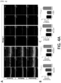
- the inventors of the present invention investigated the effect of retinal organoid-derived exosomes on photoreceptor apoptosis using TUNEL staining. Whereas many TUNEL-positive cells were detected in the outer nuclear layer (ONL) of untreated retinas and vehicle (PBS, phosphate buffer solution)-treated eyes, the number of TUNEL-positive cells in the treated eyes in the retinas of Groups 1 and 2 treated with retinal organoid-derived exosomes was significantly reduced (B and C of FIG. 3a , and E and F of FIG. 3b ).
- the thickness of the outer nuclear layer was significantly preserved in eyes treated with retinal organoid-derived exosomes in Groups 1 and 2 compared to untreated and vehicle-treated eyes (D of FIG. 3a and G of FIG. 3b ). Specifically, in Group 1, the outer nuclear layer thickness was preserved more in the central retina than in the peripheral retina, but in Group 2, the outer nuclear layer thickness was similarly preserved not only in the central retina but also in the peripheral retina.
- OMR also showed that treatment with retinal organoid-derived exosomes significantly preserved the vision of RCS rats compared to vehicle (PBS, phosphate buffer)-treated or untreated controls, which showed similar vision.
- RNA-seq was used to profile total RNA from RCS rat eyes before and after treatment with retinal organoid-derived exosomes.
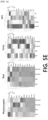
- Total RNA-seq of RCS rat eyes showed that exosome treatment significantly increased the expression of retinal cell markers, including photoreceptors and retinal pigment epithelial cells, compared to the control group ( FIG. 5E ).
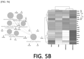
- FIGS. 5A and B Gene ontology analysis confirmed that these genes are involved in endocrine development, response to retinoic acid and visual perception ( FIGS. 5A and B ), among biological processes, and by cell composition, genes acting in the extracellular matrix (38.46%) and photoreceptor outer segments (17.31%) were found to be abundant ( FIG. 5C and D ).
- the inventors of the present invention performed a process of analyzing the miRNA components included in the retinal organoid-derived exosomes used in the treatment.
- the inventors of the present invention analyzed the miRNA content in the retinal organoid-derived exosomes using RNA-seq profiling.
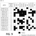
- 128 miRNAs more than 100 per million (cpm) were detected.
- the 20 most abundant miRNAs with more than 1,000 cpm are shown in FIG. 7 .
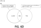
- 4,929 human genes were targets of at least three of the 20 most abundant miRNAs.
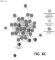
- the biological processes of 293 genes targeted by more than 10 miRNAs were shown to be abundant in ontology terms such as 'regulation of cellular amide metabolism', 'regulation of cell development' and 'response to transforming growth factor beta'.
- genes were enriched in the 'cancer development pathway', 'MAPK signaling pathway' and 'stem cell pluripotency regulation signaling pathway', and among these, the MAPK signaling pathway was selected as the key pathway.
- the inventors of the present invention compared predicted target genes and DEGs in RCS rats treated with exosomes.
- Genes enriched in the MAPK pathway were DUSP16, EGFR, FOS, MAP2K4, MAPK1, MAPK8, MAPT, NF1, RASA2, RASGRP1, RPS6KA5 and TEK in retinal organoid-derived exosomes.
- hsa-let-7b-5p can target 11 out of 12 genes, suggesting that hsa-let-7b-5p may be a key miRNA that regulates the MAPK pathway.
- RNA sequence analysis of RCS rat retina confirmed that retinal organoid-derived exosome treatment significantly reduced MAPK pathway gene expression compared to the control group.
- the inventors of the present invention performed qRT-PCR analysis and found that the expression of MAPK-related genes, including MAP2K4, MAPK1, MAPK8, MAPK10, c-FOS and c-JUN, was significantly lower in retinal organoid-derived exosome-treated retinas than in control retinas ( FIG. 7B ).
- retinal pigment epithelial (RPE) cell model for oxidative stress damage by treating primary rat RPE cells with specific H 2 O 2 concentrations.
- RPE retinal pigment epithelial
- calcein-labeled retinal organoid-derived exosomes were incubated with cultured RPE cells for 30 minutes. Green fluorescein particles were observed throughout the cytoplasm ( FIG. 10A ).
- the expression of p-JNK/JNK and pERK/ERK was significantly suppressed compared to the group treated with H 2 O 2 ( FIGS. 10B and C ).
- the inventors of the present invention investigated the anti-apoptotic effect of retinal organoid-derived exosomes. As expected, the expression level of cleaved caspase-3 was significantly suppressed in the group treated with retinal organoid-derived exosomes compared to the H 2 O 2 treated group.
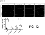
- retinal organoid-derived exosomes In order to measure the wound healing effect of retinal organoid-derived exosomes on the corneal epithelium, a 2 mm-sized damage was made in the rat corneal epithelium, retinal organoid-derived exosomes were injected into the cornea as an experimental group, and a vehicle (PBS, phosphate buffer solution) was injected into the cornea as a control group.
- PBS phosphate buffer solution
- the cornea treated with the vehicle (PBS, phosphate buffer solution) 36 hours after treatment showed blurring, which is commonly found in corneas, but a relatively clear corneal treatment effect could be observed in the cornea treated with retinal organoid-derived exosomes.
- the inventors of the present invention investigated whether treatment with retinal organoid-derived exosomes had an anti-inflammatory effect, which is known to play an important role in the wound healing process.
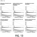
- GSEA analysis was performed on 282 miRNAs detected at least 10 cpm in 21 GO (Gene Ontology) terms including "inflammatory”, “cell adhesion” or “epithelial cell” (FDR q ⁇ 0.05).
- Abundant miRNAs were significantly expressed in "inflammatory cell apoptotic process", “cytokine production involved in inflammatory response”, “neuroinflammatory response”, “inflammatory response to wounding” and “regulation of chronic inflammatory response” (FDR q ⁇ 0.001).
- miRNAs targeting genes in these biological processes were mainly abundant in the top 20.
- exosomal miRNAs targeting TNF, CCL2, CCL5 or IL-6 were present in the top 10. These results indicate that abundant exosomal miRNAs derived from retinal organoids have the effect of preventing inflammatory responses by inducing corneal healing.
- the expression levels of inflammatory cytokines TNF- ⁇ , IL-6, CCL2 and CCL5 were quantitatively compared between the group treated with retinal organoid-derived exosomes to the cornea and the group treated with the vehicle (PBS) (** p ⁇ 0.001).
- the levels of all four types of inflammatory cytokines were reduced in corneas treated with retinal organoid-derived exosomes. This indicates that the anti-inflammatory effect of retinal organoid-derived exosomes plays an important role in wound healing of the corneal epithelium.
- the present invention is an invention that has been performed through the following project.
Landscapes
- Health & Medical Sciences (AREA)
- Life Sciences & Earth Sciences (AREA)
- Chemical & Material Sciences (AREA)
- Engineering & Computer Science (AREA)
- Animal Behavior & Ethology (AREA)
- Medicinal Chemistry (AREA)
- Pharmacology & Pharmacy (AREA)
- General Health & Medical Sciences (AREA)
- Public Health (AREA)
- Veterinary Medicine (AREA)
- Ophthalmology & Optometry (AREA)
- Epidemiology (AREA)
- Developmental Biology & Embryology (AREA)
- Cell Biology (AREA)
- Biomedical Technology (AREA)
- Bioinformatics & Cheminformatics (AREA)
- Organic Chemistry (AREA)
- Nuclear Medicine, Radiotherapy & Molecular Imaging (AREA)
- General Chemical & Material Sciences (AREA)
- Chemical Kinetics & Catalysis (AREA)
- Neurology (AREA)
- Neurosurgery (AREA)
- Biotechnology (AREA)
- Immunology (AREA)
- Virology (AREA)
- Zoology (AREA)
- Nutrition Science (AREA)
- Mycology (AREA)
- Food Science & Technology (AREA)
- Polymers & Plastics (AREA)
- Biochemistry (AREA)
- Molecular Biology (AREA)
- Urology & Nephrology (AREA)
- Vascular Medicine (AREA)
- Cardiology (AREA)
- Heart & Thoracic Surgery (AREA)
- Medicines Containing Material From Animals Or Micro-Organisms (AREA)
Applications Claiming Priority (2)
| Application Number | Priority Date | Filing Date | Title |
|---|---|---|---|
| KR20220087475 | 2022-07-15 | ||
| PCT/KR2023/009680 WO2024014792A1 (fr) | 2022-07-15 | 2023-07-14 | Composition pharmaceutique pour traiter des maladies oculaires comprenant des exosomes dérivés d'organoïdes rétiniens |
Publications (1)
| Publication Number | Publication Date |
|---|---|
| EP4556013A1 true EP4556013A1 (fr) | 2025-05-21 |
Family
ID=89537043
Family Applications (1)
| Application Number | Title | Priority Date | Filing Date |
|---|---|---|---|
| EP23839885.3A Pending EP4556013A1 (fr) | 2022-07-15 | 2023-07-14 | Composition pharmaceutique pour traiter des maladies oculaires comprenant des exosomes dérivés d'organoïdes rétiniens |
Country Status (3)
| Country | Link |
|---|---|
| EP (1) | EP4556013A1 (fr) |
| KR (2) | KR102911969B1 (fr) |
| WO (1) | WO2024014792A1 (fr) |
Families Citing this family (2)
| Publication number | Priority date | Publication date | Assignee | Title |
|---|---|---|---|---|
| EP4713437A1 (fr) * | 2023-05-15 | 2026-03-25 | Australian National University | Procédés de production de vésicules extracellulaires, vésicules extracellulaires et leurs utilisations |
| CN121714599A (zh) * | 2026-02-12 | 2026-03-24 | 武汉大学人民医院(湖北省人民医院) | miR-126-3p的制药新用途 |
Family Cites Families (3)
| Publication number | Priority date | Publication date | Assignee | Title |
|---|---|---|---|---|
| KR101585794B1 (ko) | 2014-10-07 | 2016-01-18 | 가톨릭대학교 산학협력단 | miRNA를 이용한 안과 질환의 예방 또는 치료 |
| KR102340058B1 (ko) * | 2020-05-12 | 2021-12-16 | 경북대학교 산학협력단 | 양막상피세포 유래 엑소좀을 유효성분으로 함유하는 안구질환 예방 또는 치료용 조성물 |
| KR20220069283A (ko) * | 2020-11-20 | 2022-05-27 | 에이템즈 주식회사 | 연골조직 유래 세포외 기질을 유효성분으로 하는 안구질환 예방 또는 치료용 약학조성물 |
-
2023
- 2023-07-07 KR KR1020230088274A patent/KR102911969B1/ko active Active
- 2023-07-14 WO PCT/KR2023/009680 patent/WO2024014792A1/fr not_active Ceased
- 2023-07-14 EP EP23839885.3A patent/EP4556013A1/fr active Pending
-
2026
- 2026-01-08 KR KR1020260003528A patent/KR20260015313A/ko active Pending
Also Published As
| Publication number | Publication date |
|---|---|
| KR20240010413A (ko) | 2024-01-23 |
| KR20260015313A (ko) | 2026-02-02 |
| WO2024014792A1 (fr) | 2024-01-18 |
| KR102911969B1 (ko) | 2026-01-13 |
Similar Documents
| Publication | Publication Date | Title |
|---|---|---|
| Lin et al. | Retina organoid transplants develop photoreceptors and improve visual function in RCS rats with RPE dysfunction | |
| JP7836583B2 (ja) | 視神経疾患治療用組成物、その調製方法およびその使用 | |
| KR20260015313A (ko) | 망막 오가노이드 유래 엑소좀을 포함하는 안구 질환 치료용 약학적 조성물 | |
| EA026193B1 (ru) | Композиции и способы лечения заболеваний сетчатки | |
| Xiang et al. | PEDF protects retinal pigment epithelium from ferroptosis and ameliorates dry AMD-like pathology in a murine model | |
| EP4349347A1 (fr) | Composition pour favoriser l'angiogenèse comprenant des vésicules extracellulaires dérivées d'un agrégat cellulaire de type sphéroïde tridimensionnel | |
| US10517913B2 (en) | Composition for prevention, alleviation, or treatment of peripheral neuropathy comprising Lithospermi Radix extract as an effective component | |
| EP4335445A1 (fr) | Composition pour favoriser la neurorégénération, contenant des vésicules extracellulaires dérivées d'agrégats de cellules sphéroïdes tridimensionnelles | |
| Huang et al. | cRGD-conjugated bilirubin nanoparticles alleviate dry eye disease via activating the PINK1-mediated mitophagy | |
| Sun et al. | Rhodium nanozyme mitigates RPE degeneration and preserves vision in age-related macular degeneration via antioxidant and anti-inflammatory mechanisms | |
| US20240000853A1 (en) | Lipocalin-type prostaglandin d2 synthase production promoting agent | |
| Chen et al. | Vitamin K1 alleviates retinal inflammation following acute ocular hypertension by modulating microglial ferroptosis | |
| CN108135956A (zh) | 视神经保护用组合物 | |
| JP2021138710A (ja) | 眼疾患の処置方法 | |
| Trofimova | Molecular mechanisms of retina pathology and ways of its correction | |
| EP4173630A1 (fr) | Composition pharmaceutique comprenant un milieu conditionné par des cellules souches et un exosome isolé à partir de celle-ci en tant que principe actif pour la prévention ou le traitement d'une maladie oculaire | |
| Jiang et al. | Inhibition of serine racemase prevents retinopathy in diabetic mice | |
| CN120324337B (zh) | 一种含多元微量元素和干细胞提取物的滴眼液及其制备方法和应用 | |
| Jung et al. | Efficacy of Subconjunctivally Applied Everolimus-and Sirolimus-Pretreated MSCs in Preventing Diabetic Retinopathy | |
| Bao et al. | Nicotinamide Mononucleotide Alleviates the Development of Fibrotic Cataract by Modulating the Nrf2/FPN1 Signaling Pathway | |
| CN112569338B (zh) | Tdfa在制备预防和/或治疗眼表炎症疾病的药物中的应用 | |
| CN120796391B (zh) | 一种靶向usp8治疗视网膜退行性疾病的基因治疗方法及其应用 | |
| CN114306306B (zh) | 2-溴棕榈酸在制备治疗生精功能障碍相关疾病的药物中的应用 | |
| CN121774990A (zh) | 七叶皂苷钠在制备防治糖尿病视网膜病变药物中的应用 | |
| US20260097092A1 (en) | Pharmaceutical composition containing mitochondria-targeting compound as active ingredient for treating macular degeneration |
Legal Events
| Date | Code | Title | Description |
|---|---|---|---|
| STAA | Information on the status of an ep patent application or granted ep patent |
Free format text: STATUS: THE INTERNATIONAL PUBLICATION HAS BEEN MADE |
|
| PUAI | Public reference made under article 153(3) epc to a published international application that has entered the european phase |
Free format text: ORIGINAL CODE: 0009012 |
|
| STAA | Information on the status of an ep patent application or granted ep patent |
Free format text: STATUS: REQUEST FOR EXAMINATION WAS MADE |
|
| 17P | Request for examination filed |
Effective date: 20250115 |
|
| AK | Designated contracting states |
Kind code of ref document: A1 Designated state(s): AL AT BE BG CH CY CZ DE DK EE ES FI FR GB GR HR HU IE IS IT LI LT LU LV MC ME MK MT NL NO PL PT RO RS SE SI SK SM TR |
|
| DAV | Request for validation of the european patent (deleted) | ||
| DAX | Request for extension of the european patent (deleted) |