EP4564004A1 - Procédé de détermination de l'hydrophobicité de protéines - Google Patents
Procédé de détermination de l'hydrophobicité de protéines Download PDFInfo
- Publication number
- EP4564004A1 EP4564004A1 EP23212842.1A EP23212842A EP4564004A1 EP 4564004 A1 EP4564004 A1 EP 4564004A1 EP 23212842 A EP23212842 A EP 23212842A EP 4564004 A1 EP4564004 A1 EP 4564004A1
- Authority
- EP
- European Patent Office
- Prior art keywords
- wells
- proteins
- present
- salt concentrations
- salt
- Prior art date
- Legal status (The legal status is an assumption and is not a legal conclusion. Google has not performed a legal analysis and makes no representation as to the accuracy of the status listed.)
- Withdrawn
Links
Images



Classifications
-
- G—PHYSICS
- G01—MEASURING; TESTING
- G01N—INVESTIGATING OR ANALYSING MATERIALS BY DETERMINING THEIR CHEMICAL OR PHYSICAL PROPERTIES
- G01N33/00—Investigating or analysing materials by specific methods not covered by groups G01N1/00 - G01N31/00
- G01N33/48—Biological material, e.g. blood, urine; Haemocytometers
- G01N33/50—Chemical analysis of biological material, e.g. blood, urine; Testing involving biospecific ligand binding methods; Immunological testing
- G01N33/53—Immunoassay; Biospecific binding assay; Materials therefor
- G01N33/543—Immunoassay; Biospecific binding assay; Materials therefor with an insoluble carrier for immobilising immunochemicals
- G01N33/54366—Apparatus specially adapted for solid-phase testing
Definitions
- the application relates to a method for analyzing proteins with different hydrophobicity using microtiter plates whose wells have at least partially different salt concentrations and using functionalized surfaces.
- Biopharmaceuticals including virus particle-based therapeutics, are generally pharmaceutically useful and effective proteins, such as antibodies and virus particles as well as antibody-drug conjugates (ADCs).
- ADCs antibody-drug conjugates
- the by-products are usually incorrectly assembled, e.g. mispaired proteins, in which the various protein domains have not assembled as intended, or fragments of the target protein.
- biopharmaceuticals especially antibodies, Fc fusion proteins, antibody fragments, bi- and multispecific antibodies as well as virus particle-based therapeutics and antibody-drug conjugates (ADCs)
- ADCs virus particle-based therapeutics and antibody-drug conjugates
- Mispairing occurs, for example, when the different amino acid chains produced after expression of bi- or multispecific antibodies are not assembled in the desired manner. Despite various approaches to optimize assembly and shift it toward the desired target protein, this mispairing remains a major problem.
- Proteins that have different hydrophobicity are also antibodies that are differently conjugated with pharmaceutically active substances, so-called antibody drug conjugates (ADCs), if they are differently hydrophobic due to their different conjugation (number and type of conjugated substances, conjugation site in the antibody and type of linker used).
- ADCs antibody drug conjugates
- Viral particles used in gene therapy can also be differently charged and/or have a different degree of hydrophobicity.
- Loading the viral particles with nucleic acids results in the viral particle surface being more negatively charged than with uncharged or only partially charged viral particles.
- the degree of hydrophobicity changes depending on the loading state. Since the loading of the viral particles with nucleic acids is essential for therapeutic It is very important, even in virus particle production processes, to ensure that the virus particles are loaded. Virus particles not loaded with nucleic acids, i.e., empty virus particles, are non-functional byproducts and must be removed.
- nucleic acids influence the charge of the virus capsid, allowing separation based on their charge in ion exchange chromatography.
- the present invention aims to develop methods for the rapid detection of variants of a target protein, namely antibodies, Fc fusion proteins, antibody fragments, bi- and multispecific antibodies as well as virus particle-based therapeutics, antibody-drug conjugates (ADCs) and incorrectly assembled (e.g., mismatched or fragmented) proteins and other by-products.
- a target protein namely antibodies, Fc fusion proteins, antibody fragments, bi- and multispecific antibodies
- viruses particle-based therapeutics e.g., virus particle-based therapeutics, antibody-drug conjugates (ADCs) and incorrectly assembled (e.g., mismatched or fragmented) proteins and other by-products.
- ADCs antibody-drug conjugates
- the detection of the various byproducts should be based on their hydrophobicity. These measurements are particularly suitable for the detection of misassembled proteins and differently conjugated antibody-drug conjugates.
- a method commonly used to characterize and purify proteins and other biological molecules is column chromatography. It can be performed under both atmospheric and elevated pressure. The latter often requires expensive equipment such as HPLC or FPLC.
- Another method is based on the use of biosensors, such as in US 5,804,453 B described.
- a biosensor is a device that combines a biological component with a physical or chemical transducer to generate a measurable signal in response to a target analyte.
- Biosensors can utilize various physical measurement principles to detect the presence of target analytes. Examples include piezoelectric biosensors, optical detection methods based on interferometry or surface plasmon resonance, and calorimetric measurement methods.
- Biosensors that use interferometric methods are, for example, in US 5,804,453 B.
- Biosensors that use piezoelectric processes are also known and, for example, in EP2017613 A1 described.
- Biosensors that use interferometric methods are particularly frequently used in combination with microtiter plates because the biosensors are easy to manufacture and, due to their shape, can be easily immersed into the wells of the microtiter plates.
- biosensors used in so-called biolayer interferometry have two reflective surfaces on which a phase shift is measured when biomolecules attach to the second surface, further away from the light source.
- Biosensor surfaces must be functionalized for the binding of the target analytes. Often, the surfaces are functionalized with amines to realize the further coupling of proteins or peptides via an amide bond, as in WO 2008/033535 described.
- Functionalization is achieved through bifunctional linkers, which, on the one hand, react with the surface of the biosensor and, on the other hand, provide functionality that enables the binding of the actual binding molecule (e.g., an antibody).
- these can be siloxanes, a classic method for functionalizing glass surfaces.
- copolymers of, for example, polystyrene and divinylbenzene can be used, which provide reactive groups for further functionalization of the polymer surfaces.
- the sample is adsorbed onto a so-called stationary phase (the column material, which can consist of various materials).
- stationary phase the column material, which can consist of various materials.
- eluent mobile phase(s)
- the hydrophobic interactions of the analytes with the stationary phase are weakened, and the adsorbed molecules are desorbed and eluted.
- salt gradient refers to the change in salt concentration over a specific spatial and/or temporal range.
- Hydrophobic interaction chromatography uses hydrophobic surfaces, often using salt gradients.
- a high salt concentration is initially used, which disrupts the hydration shell of the proteins. This makes hydrophobic regions of the proteins accessible for hydrophobic interactions. These regions then bind to the hydrophobically functionalized surface, in this case the hydrophobically functionalized column material.
- the hydration shell reconstitutes, i.e., the accessibility of the hydrophobic sites decreases or disappears, so that the proteins are no longer bound and elute depending on the salt concentration used.
- the object of the present invention is to provide devices and methods with which proteins, such as biopharmaceuticals, in particular antibodies, Fc fusion proteins, antibody fragments, bi- and multispecific antibodies as well as virus particle-based therapeutics can be efficiently and quickly qualitatively and can be determined quantitatively.
- proteins such as biopharmaceuticals, in particular antibodies, Fc fusion proteins, antibody fragments, bi- and multispecific antibodies as well as virus particle-based therapeutics can be efficiently and quickly qualitatively and can be determined quantitatively.
- the measurement should also be simple and preferably feasible using tools routinely available in the laboratory.
- the invention thus relates to a method for the analysis of biological molecules, in particular proteins including antibody-drug conjugates, of different hydrophobicity using microtiter plates whose wells have at least partially different salt concentrations and using hydrophobically functionalized surfaces.
- the invention relates to a method according to embodiment A, wherein the proteins are therapeutically active proteins selected from antibodies, Fc fusion proteins, antibody fragments, bi- and multispecific antibodies and virus particle-based therapeutics and antibody-drug conjugates.
- the invention relates to a method according to one of the preceding embodiments, wherein the different final salt concentrations are present in wells lying next to one another or one below the other and thus form a step gradient in which rows or columns with respectively ascending or descending salt contents are present.
- the invention relates to a method according to one of the preceding embodiments, using marker molecules that specifically bind to the proteins to be analyzed, wherein the marker molecules are mono- or polyclonal antibodies or fragments derived therefrom such as nanobodies or single-domain antibodies as well as peptides and RNA- or DNA-based aptamers, and these optionally coupled with fluorescent dyes form fluorescent markers.
- the invention relates to a method according to embodiment D using hydrophobically functionalized particles and the marker molecules to which fluorescent dyes are coupled and which represent fluorescent markers, wherein the fluorescence emission of the unbound proteins labeled with fluorescent markers is measured.
- the invention relates to a method according to embodiment E using a PAIA plate in which some or all of the wells are designed as measuring chambers in which there is a raised area which is shaped such that its base area occupies at least 50% of the bottom area of a measuring chamber or well and preferably has a square base area which tapers at least slightly towards the top and has the shape of a four-sided pyramid, wherein the upper end of the raised area is pointed, flat or convex and advantageously the upper end of the raised area has a diameter of less than 50 ⁇ m so that no test components are deposited there, wherein the height of the raised area is at least 10% and at most 50% of the height of the measuring chamber or well and the bottom of the measuring chamber or well is opaque to light except for the bottom of the raised area, wherein the raised area is transparent to light and forms a measuring window which is used to measure the fluorescence emission of the unbound proteins labelled with fluorescent markers.
- steps b, c, and d can be performed in any order.
- the process can be carried out such that after performing steps a, c, and/or d, the plate is subjected to a drying step. After drying, the process continues with step b, followed by the remaining steps.
- the invention relates to a method according to embodiment G, wherein steps b, c and d can be carried out in any order.
- the invention relates to a method according to embodiment H, wherein in step a a microtiter plate is used in which, in addition to the different amounts of salt, hydrophobically functionalized particles are also present, in which case step c is omitted.
- the invention relates to a method according to embodiment H or I, wherein in step a a microtiter plate is used in which fluorescent markers are present in addition to the amounts of salt, in which case step d is omitted.
- the invention relates to a method according to embodiment I or J, in the wells of which at least partially different amounts of salt are present, which are dimensioned such that during the analysis in the appropriately treated wells lying next to one another or one below the other, at least partially final salt concentrations are present, the distances between which are in the range from 10 to 500 mmol/L, wherein hydrophobically functionalized particles are also present in these wells and optionally fluorescent markers according to embodiment E are present, wherein the aforementioned components salt, particles and fluorescent markers are present in dry form.
- the invention relates to the use of the microtiter plate according to embodiment K for carrying out the method according to embodiment G, wherein steps c and d are omitted.
- the invention relates to a method according to one of the embodiments A to C, using biosensors which have hydrophobically functionalized surfaces and whose shape and dimensions are such that they can be introduced into the wells of the microtiter plates.
- the invention relates to the method according to embodiment M, wherein marker molecules are additionally present, in particular mono- or polyclonal antibodies or fragments derived therefrom such as nanobodies or single-domain antibodies as well as peptides and RNA- or DNA-based aptamers.
- Step c in the above-mentioned procedure can also be carried out as follows: introducing the biosensors with hydrophobic-functionalized surfaces into the wells with the salt gradient.
- Steps d and e of the above procedure can be combined, namely by shaking the microtiter plate, thereby accelerating the reaction, and simultaneous (real-time) measurement of the binding to the surface on the biosensors.
- salts or salt mixtures are used to create the salt gradient.
- the microtiter plate can also have wells without salt gradient loading.
- the final salt concentration is achieved at the latest when the sample is filled. For example, by diluting a specific sample amount with a corresponding amount of liquid or buffer.
- the invention is used primarily for the analysis of proteins.
- therapeutically active proteins such as antibodies and protein formats derived therefrom, such as Fc fusion proteins, antibody fragments, bi- and multispecific antibodies, as well as virus particle-based therapeutics and antibody-drug conjugates.
- hydrophobically functionalized surfaces are preferably hydrophobically functionalized particles or sensors, such as biosensors, whose measuring surface is designed to bind hydrophobic proteins and whose shape and dimensions are such that they can be introduced into the wells of the microtiter plates used.
- the target protein e.g. the biopharmaceutical
- the target protein has binding sites for one or more marker molecules.
- markers especially fluorescent markers
- PAIA plate as described here
- proteins include, for example, host cell proteins, as they are typically present in cell culture supernatants. or misassemblies in the production of bi- or multi-specific antibodies that no longer have a binding site for the marker molecule(s).
- Marker molecules are molecules that can specifically bind biological molecules (e.g., proteins).
- markers molecules include monoclonal or polyclonal antibodies, or fragments derived from them, such as so-called nanobodies or single-domain antibodies, as well as peptides and RNA- or DNA-based aptamers.
- the marker molecules can contain dyes, such as fluorescent dyes. Such markers are referred to below as fluorescent markers.
- the dyes can be covalently bound to the fluorescent markers or, for example, via the well-known streptavidin-biotin system, in which a fluorescently labeled streptavidin or other fluorescently labeled biotin-binding proteins such as avidin or neutravidin is coupled to a biotinylated marker molecule.
- fluorescent markers are particularly suitable for the method and use according to the invention.
- fluorescent markers that can be used according to the invention are Affibody conjugated with Alexa 647 from Affibody AB or Nano-Secondary ® alpaca anti-human IgG/anti-rabbit IgG, recombinant VHH, Alexa Fluor ® 647 (Art. No. CTK0101 - ChromoTek GmbH).
- Other fluorescent markers are commercially available, e.g. from Jackson Immunoresearch or Abcam. These are already coupled with a fluorescent dye and can be used directly.
- monoclonal or polyclonal antibodies can be purchased from these companies and coupled with the various commercially available dyes, e.g. fluorescein, as well as dyes from Biotium, Atto-Tec, and ThermoFisher.
- dyes e.g. fluorescein, as well as dyes from Biotium, Atto-Tec, and ThermoFisher.
- the application of the method according to the invention primarily relates to the analysis, i.e., the identification and, if necessary, quantification of biological molecules, in particular proteins of varying hydrophobicity.
- proteins include, for example, biopharmaceuticals (i.e., therapeutically active proteins), such as antibodies, Fc fusion proteins, antibody fragments, or bi- and multispecific antibodies, as well as virus particle-based therapeutics and antibody-drug conjugates.
- Proteins with different hydrophobicity may be the same protein (i.e., a protein with the same amino acid sequence), but with multiple variants with different hydrophobicity.
- proteins with different hydrophobicity may be the same protein (i.e., a protein with the same amino acid sequence), but with multiple variants with different hydrophobicity.
- Proteins with different hydrophobicity are also those that are incorrectly assembled. These incorrectly assembled proteins have different amino acid sequences or different types and, in some cases, numbers of protein domains, such as the light and heavy chains in antibodies.
- bispecific antibodies these are, for example, the so-called homodimers or halfmers, which each have only one type of heavy and light chain, although such bispecific molecules have different light and heavy chains, which can bind to different binding partners.
- Proteins that exhibit different hydrophobicity are also differently loaded virus particles if they are differently hydrophobic due to their loading with nucleic acids (DNA or RNA), their glycosylation, amidation and/or oxidation of the amino acids of their capsid proteins.
- nucleic acids DNA or RNA
- the method according to the invention with spectrometric analysis uses microtiter plates. It is also conceivable that other specially prepared vessels could be used instead of microtiter plates. If fluorescent markers are used in the method according to the invention, a PAIA plate, as defined herein, is used.
- a particular advantage of the invention is that the samples can be measured directly (if necessary after dilution with water). Purification prior to performing the procedures (the assays) is not required.
- biosensors are used as hydrophobically functionalized surfaces in the method according to the invention. If biosensors are used as hydrophobically functionalized surfaces in the method according to the invention, no PAIA plates are used.
- the advantage of the invention is that a large number of samples can be analyzed for the presence of different proteins in a relatively short time. Due to the simple and, if desired, automated procedure, a measurement result for more than 100 samples simultaneously is usually available in less than 60 minutes. The automation available for microtiter plates (e.g., pipetting robots or automatic readout processes) can be utilized.
- Microtiter plates are well-known and usually rectangular, with a varying number of wells, also called cavities, wells, or recesses. These wells are usually arranged in rows parallel to the long side of the microtiter plate and in columns parallel to the short side.
- Letters are usually used to designate rows and numbers to designate columns.
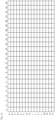
- the wells of a 384-well plate are arranged in 16 rows (from A to P) and 24 columns (from 1 to 24).
- a schematic drawing of such a 384-well plate is shown in Fig. 1 shown.
- the wells of a 96-well plate are arranged in 8 rows (A to H) and 12 columns (1 to 12). This allows each well to be uniquely identified:
- the first row of a 96-well plate contains 12 wells, namely wells A1 to A12.
- the second row contains wells B1 to B12, and so on.
- the special preparation of the microtiter plate involves one or more wells, or in the case of the PAIA plate, as described in more detail below, one or more wells forming a measurement chamber.
- the hydrophobically functionalized surfaces are preferably hydrophobically functionalized particles.
- the wells of the microtiter plate are prepared as follows:
- the desired number of wells is filled with aqueous buffer solutions that have different salt concentrations - in the examples Also called working solution.
- These salt solutions preferably have the same pH value. However, they can also have different pH values.
- Salts suitable for the invention are known and are tailored to the proteins to be analyzed. They can also be easily determined by a person skilled in the art. Examples of such salts are (NH 4 ) 2 SO 4 , NaH 2 PO 4 , Na 2 HPO 4 , Na 2 SO 4 , K 2 SO 4 , NaCl, KCl, MgCl 2 , CaCL 2 , MgSO 4 , and mixtures thereof.
- the different salt concentrations in the working solution are most easily achieved by mixing two buffers with different salt concentrations. If buffer A with no or low salt concentration and buffer B with high salt concentration are available, the desired salt concentration can be adjusted by selecting an appropriate mixing ratio.
- the following table shows an example of an 8-step salt gradient system (final salt concentrations) obtained with (NH 4 ) 2 SO 4 in (30 mM) TRIS buffer (pH 7.5) by diluting a 2.4 molar (NH 4 ) 2 SO 4 solution with TRIS buffer.
- This gradient system shown in Table 1, is particularly suitable for the embodiment of the method in which a drying step is provided.
- Table 1 Example of an 8-step salt gradient system with indication of the salt concentration of the working solution before removal of the solution and the final salt concentration in 60 ⁇ L during the test. No.
- Common buffer systems for handling proteins include PBS systems (phosphate-based saline buffer), buffers from the group of so-called “Good's buffers” such as TRIS (tris(hydroxymethyl)aminomethane), MES (2-(N-morpholino)ethanesulfonic acid), HEPES (4-(2-hydroxyethyl)-1-piperazineethanesulfonic acid), ammonium acetate- or citrate-based buffers, etc.
- PBS systems phosphate-based saline buffer
- Buffers phosphate-based saline buffer
- TRIS tris(hydroxymethyl)aminomethane
- MES 2-(N-morpholino)ethanesulfonic acid
- HEPES 4-(2-hydroxyethyl)-1-piperazineethanesulfonic acid
- ammonium acetate- or citrate-based buffers etc.
- the salt concentration used in the first step of the inventive method can be selected such that the concentration relevant for the test is only reached in the well after the sample solution required for carrying out the method and, if applicable, other test components (such as marker molecules) have been added. This relevant concentration is also referred to below as the final salt concentration.
- the final salt concentration is essentially determined by the salt concentration of the working solution. The amounts of salt that may be introduced into the wells by the test components are small compared to the salt quantity and concentration of the working solution and are normally not added to the final salt concentration.
- the intervals between the final salt concentrations must be chosen as required by experimental accuracy.
- the special feature of the invention is primarily that when carrying out the method, at least partially different final salt concentrations are present in the wells of the microtiter plate arranged in rows or columns.
- the final salt concentration ranges in the individual wells are in the range of 10 to 500 mmol/L, or in the range of 20 to 450 mmol/L, or in the range of 50 to 400 mmol/L.
- the gradient covers a wide range of final salt concentrations.
- the distance between the final salt concentrations is in the range of 50 mmol/L to 100 mmol/L. These concentration distances (so-called concentration steps) can be the same or different. It has been shown that for proteins with large differences in hydrophobicity, eight different final salt concentrations are sufficient to separate the mixtures (a so-called 8-step gradient, as shown, for example, in Table 1).
- the concentrations of the working solution in the wells should be recorded, at least partially. This can be done separately or on the plate.
- the liquid (usually water) can be removed from the wells by drying, preferably at elevated temperature, i.e., a temperature greater than 18 °C, at atmospheric pressure, or under reduced pressure. Drying has the advantage that the prepared microtiter plate can be stored and preserved.
- the fluorescent marker(s) and/or the hydrophobic-functionalized surfaces are dried after filling into the wells.
- the sample containing proteins with different hydrophobicity is usually added in portions to the wells of the microtiter plate, which can also be a PAIA plate, without further treatment (e.g., purification).
- dilution of the sample with water is advantageous to reduce the concentration of buffer substances that could interfere with the experiment.
- the amount and dilution of the sample are selected to achieve the desired final salt concentration.
- Portioning is done manually or automatically with an automated pipetting robot (liquid handler).
- the final salt concentrations present in the wells during the experiment are crucial for the binding properties of the respective proteins with different hydrophobicity to the hydrophobically functionalized surface(s).
- the measurement with the biosensor or - when using the PAIA plate - preferably the fluorescence intensity of the unbound fluorescent markers provides a value from which the hydrophobicity of the biological molecule, in particular protein, can be inferred.
- the invention relates to a method for analyzing proteins of varying hydrophobicity using specially prepared PAIA plates (i.e., whose wells contain at least partially different salt concentrations, as described above) using hydrophobically functionalized surfaces, preferably hydrophobically functionalized particles, and fluorescent markers as markers. Detection is performed via fluorescence measurement.
- the PAIA plate (also called PAIA-plate) is a microtiter plate in SBS format with a certain number of wells, which are designed as measuring chambers in which the elevation (in the WO 2015/135840 A1 referred to as a structural element) is shaped so that its base area occupies at least 50% of the floor area of a measuring chamber or well. They are known and in WO 2015/135840 A1 They are manufactured and distributed by PAIA Biotech GmbH under the trade name PAIAplate 384. These plates have 384 wells.
- PAIA plate to be used can have more or less than 384 wells.
- the elevation has a square base that tapers (at least slightly) towards the top and has the shape of a four-sided pyramid.
- the upper end of the elevation is pointed, flat, or convex, preferably tapering to a point.
- the upper end of the elevation has a diameter of less than 50 ⁇ m, so that no test components are deposited there.
- the height of the elevation is at least 10% and at most 50% of the height of the measuring chamber or well, preferably at least 15% and at most 30%.
- the bottom of the measuring chamber or well is opaque except for the bottom of the elevation.
- the elevation on the other hand, is translucent and forms a measuring window used to measure the fluorescence emission, as in WO 2015/135840 A1 described in detail.
- the PAIA plate is in particular equipped as follows: the desired number of wells (some or all) of the microtiter plate are designed as measuring chambers, therein there is a raised area which is shaped such that its base area occupies at least 50% of the bottom area of a measuring chamber or well and preferably has a square base which tapers at least slightly towards the top and has the shape of a four-sided pyramid, wherein the upper end of the raised area is pointed, flat or convex and advantageously the upper end of the raised area has a diameter of less than 50 ⁇ m so that no test components are deposited there, wherein the height of the raised area is at least 10% and at most 50% of the height of the measuring chamber or well and the bottom of the measuring chamber or well is opaque up to the bottom of the raised area and the raised area is translucent and forms a measuring window which is used to measure the fluorescence emission.
- the measuring chamber or microtiter plate according to the invention is irradiated from below with excitation light, which is then radiated into the sample via the raised area.
- fluorescence readers can be used to read the fluorescence signal.
- the following devices can be used as fluorescence readers according to the invention:
- the invention using the PAIA plate can be used advantageously in particular in the development of cell culture methods for the production of proteins, in particular therapeutically active proteins, such as antibodies, Fc fusion proteins, antibody fragments, bi- and multi-specific antibodies as well as virus particle-based therapeutics and antibody-drug conjugates.
- proteins in particular therapeutically active proteins, such as antibodies, Fc fusion proteins, antibody fragments, bi- and multi-specific antibodies as well as virus particle-based therapeutics and antibody-drug conjugates.
- the invention therefore also relates to the use of PAIA plates for spectrometric analysis, i.e. detection and detection of proteins of different hydrophobicity by means of fluorescence spectroscopy, in particular of therapeutically active proteins, such as antibodies, Fc fusion proteins, antibody fragments, bi- and multispecific antibodies as well as virus particle-based therapeutics and antibody-drug conjugates.
- therapeutically active proteins such as antibodies, Fc fusion proteins, antibody fragments, bi- and multispecific antibodies as well as virus particle-based therapeutics and antibody-drug conjugates.
- the invention also relates to a PAIA plate in which different salt concentrations are present in one or more of the wells and into which hydrophobically functionalized particles and optionally fluorescent markers are incorporated.
- the aforementioned test components are present in dry form in the PAIA plate.
- the invention relates to a PAIA plate in which, in the wells of the plate, at least in one row or column, partially different descending or ascending amounts of salt are present. In dissolved or dry form.
- the method according to the invention uses a microtiter plate in whose wells at least partially different salt concentrations are present, and in which the method according to the invention is carried out with biosensors having hydrophobically functionalized surfaces.
- steps A, B, and C can be carried out in any order. If steps B and/or C are carried out before step A—in any order—a drying step can be provided after step B and/or step C. In the drying step, the liquid introduced with the test components is removed, preferably by drying at elevated temperature, as described here.
- the adjustment in step D can already be carried out by adding the test components mentioned in steps A to C, e.g., taking into account the total volume of the test solution added.
- the process can be interrupted for any length of time after the drying step and continued with the addition of the sample, i.e. step A. This also applies to all other processes in which a drying step is provided.
- the invention relates to a PAIA plate in whose wells at least partially differ in salt concentration, resulting in different final salt concentrations during the experiment, wherein the distances between the final salt concentrations are the same or different and have the same or different distance, which is in the range of 10 to 500 mmol/L.
- the plate can additionally have hydrophobically functionalized surfaces, preferably particles and/or optionally fluorescent markers, wherein the aforementioned test components are present in dissolved or dried form.
- the invention further relates to a so-called kit of parts comprising the PAIA plate with the aforementioned test components in a dried state.
- the invention can also be implemented using biosensors as hydrophobically functionalized surfaces in microtiter plates. In this case, the use of a PAIA plate is not intended.
- the invention also relates to a method for analyzing proteins of different hydrophobicity using hydrophobically functionalized biosensors and using microtiter plates in whose wells at least partially different salt contents are present, from which different final salt concentrations result when the experiment is carried out, wherein the final salt concentrations have the same or different distance, which is in the range of 10 to 500 mmol/L.
- biosensors can be achieved by applying hydrophobically functionalized surfaces to biosensors, which are then inserted into the wells of microtiter plates (not PAIA plates) pretreated as described above, and to which the binding of the target proteins is measured.
- Optical methods such as biolayer interferometry are primarily used for this purpose. This involves a phase shift of the light when molecules bind to the surface of the biosensor. This phase shift is measurable and depends on the number and mass of the molecules bound to the surface.
- Measurements with biosensors can be performed simultaneously, i.e., with multiple biosensors. Alternatively, they can be performed sequentially, i.e., a biosensor is introduced one after the other into the various wells of the microtiter plate containing a different final salt concentration. It must be regenerated and washed before each measurement in a new well.
- Biosensor regeneration is a well-known process. Modern devices are capable of measuring with multiple biosensors simultaneously in different wells, thus achieving significantly higher sample throughput.
- biosensors If only small amounts of a protein are bound during the measurement, or if very small proteins are measured, and therefore only a very weak measurement signal is generated, it is common practice for biosensors to bind a marker that specifically binds to the target molecule, e.g., an antibody with a sufficiently large molecular mass, to the surface after the target molecule has bound to the surface to amplify the signal. If this is not sufficient to amplify the signal, another marker (referred to here as a binder, e.g., a secondary antibody) that specifically binds to the primary antibody can also bind to the biosensor.
- a binder e.g., a secondary antibody
- non-target molecules proteins
- the use of specific marker molecules such as an antibody that specifically binds to the target protein, allows the measurement of a phase shift due to the binding of the marker molecule. This binding depends only on the amount of target molecules bound to the surface. Thus, the measurement signal is specific to the target molecule.
- hydrophobically functionalized surfaces in particular the particles in the case of using a PAIA plate, are selected in the method according to the invention so that they can bind proteins of different hydrophobicity.
- the particles with hydrophobically functionalized surfaces preferably have average diameters in the range of approximately 20 to 200 ⁇ m.
- the particles preferably have average diameters in the range of 5 to 100 ⁇ m or 1 to 70 ⁇ m.
- hydrophobically functionalized surfaces and particles such as those commonly used for processes such as hydrophobic interaction chromatography (HIC), can be used according to the invention.
- Such surfaces, especially particles are known.
- the surfaces and particles often contain nonpolar organic short-chain hydrocarbons, such as (C 3 -C 6 )-alkyl groups, phenyl, propyl groups, and ether or amide groups.
- Suitable surfaces also include those used as stationary phases in reversed-phase chromatography, especially those with long hydrocarbon chains, e.g. (C 8 -C 18 )-alkyl groups.
- Suitable hydrophobically functionalized particles include TOYOPEARL Ether, TOYOPEARL PPG, TOYOPEARL Phenyl, TOYOPEARL Butyl-600M, and TOYOPEARL SuperButyl, as marketed by Tosoh.
- Phenyl Sepharose TM 6 Fast Flow (high sub-base), Butyl Sepharose TM 4 Fast Flow, Octyl Sepharose TM 4 Fast Flow resin, Butyl-S Sepharose TM 6 Fast Flow Phenyl Sepharose TM High Performance, and Capto TM Phenyl (high sub-base) resin, Capto TM Butyl, Capto TM Octyl, and Capto TM Butyl-S, as marketed by Cytiva.
- POROS TM Benzyl Ultra Hydrophobic Interaction Chromatography (HIC) Resin POROS TM Ethyl Hydrophobic Interaction Chromatography (HIC) Resin, as sold by ThermoFisher.
- Fig. 1a Schematic labeling of the wells in a 384-well plate.
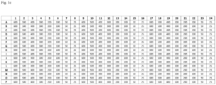
- Fig. 1b Schematic representation of a 384-well plate with a repeating 8-step salt gradient in one column.
- Fig. 1c Schematic representation of a 384-well plate with a repeating 8-step salt gradient in one row.
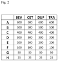
- Fig. 2 Receipt scheme from example 1.
- Fig. 3 Measured fluorescence intensities of the samples from example as a function of the final salt concentration.
- Fig. 4 Measured fluorescence intensities of antibodies and mixtures thereof as a function of the final salt concentration.
- Fig. 5 Relationship between fluorescence intensity and the content of ofatumumab in a well at a salt concentration of 300 mmol/L in different samples.
- PAIAplate 384 The wells of a PAIAplate 384 are used as reaction and measurement chambers. It is available from PAIA Biotech GmbH and is used as described in WO 2015/135840 A1 manufactured. The working volume of one well is 15 ⁇ L-110 ⁇ L.
- the buffer used is a TRIS buffer with pH 7.5 containing: 30 mM TRIS, 1.8 mM NaCl, 0.022% Tween 20, 1 mM CaCl 2 and 1 mM MgCl 2 .
- hydrophobic-functionalized particles carrying butyl groups are used as functionalized surfaces: TOYOPEARL ® Butyl-600M particles (Tosoh, Art. No. 17546802) with an average diameter of approx. 65 ⁇ m.
- the measuring chambers are treated as follows before measurement:
- the prepared PAIA plate After drying, the prepared PAIA plate can be stored or used directly for measurement.
- Nano-Secondary ® alpaca anti-human IgG/anti-rabbit IgG, recombinant VHH, Alexa Fluor ® 647 (Art. No. CTK0101 - ChromoTek GmbH) is added to these wells and everything is mixed.
- the fluorescent marker is used at a concentration of 3 nM in a PBS buffer (phosphate buffered saline) containing 50 ppm BSA (bovine serum albumin).
- PBS buffer phosphate buffered saline
- BSA bovine serum albumin
- microtiter plate is shaken at room temperature at 2200 rpm on an Eppendorf Thermomixer Comfort for 30 minutes and then centrifuged at 500 x g for one minute.
- the fluorescence intensity of the unbound fluorescent markers is measured from below (bottom reading) in each filled measuring chamber in a Tecan Safire fluorescence plate reader at an excitation wavelength of 630 nm and an emission wavelength of 665 nm.
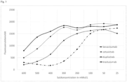
- the measured fluorescence intensities as a function of the final salt concentration are shown in Fig. 3
- the graphs in Fig. 3 thus show the different hydrophobicities of the measured antibodies.
- bevacizumab is almost completely bound to the particles, while dupilumab only partially binds to the hydrophobic particles at this concentration, and cetuximab and trastuzumab do not bind to the hydrophobic particles. This is in line with expectations from the literature, where bevacizumab is described as the most hydrophobic ( Jain et al.: Biophysical properties of the clinical-stage antibody landscape, PNAS (2017) 114 (5) 944-949 ).
- Example 2 Measurement on a mixture of antibodies of different hydrophobicity
- the fluorescent marker used is Nano-Secondary ® alpaca anti-human IgG/anti-rabbit IgG, recombinant VHH, Alexa Fluor ® 647 (Art. No. CTK0101 - ChromoTek GmbH). This is used at a concentration of 3 nM in a PBS buffer (phosphate buffered saline) containing 50 ppm BSA (bovine serum albumin).
- PBS buffer phosphate buffered saline
- BSA bovine serum albumin
- the two monoclonal antibodies bevacizumab (BEV) and ofatumumab (OFA) are used as proteins, individually as well as mixtures of these two molecules in the ratios 75:25, 50:50, and 25:75.
- the total antibody concentration in all samples is 10 ⁇ g/mL.
- microtiter plate is shaken at room temperature at 2200 rpm on an Eppendorf Thermomixer Comfort for 30 minutes and then centrifuged at 500 x g for one minute.
- the fluorescence intensity of the unbound fluorescent markers is then measured from below (bottom reading) in each measuring chamber in a Tecan Safire fluorescence plate reader at an excitation wavelength of 630 nm and an emission wavelength of 665 nm.
- the diagram in Fig. 4 shows the fluorescence intensity curves of the different samples as a function of the salt concentration in the wells.
- the sample containing 100% bevacizumab (BEV) shows no signal increase between the final salt concentrations of 600 mM/L and 300 mM/L, while the other samples in this range show higher fluorescence intensities with increasing ofatumumab content. Therefore, the fluorescence intensity at the final salt concentration of 300 mM/L can be used to determine the ofatumumab content in these mixtures.
- Fig. 5 shows the quasi-linear relationship between the fluorescence intensity in the well with the final salt concentration of 300 mM/L and the percentage of ofatumumab in the samples.
Landscapes
- Health & Medical Sciences (AREA)
- Immunology (AREA)
- Life Sciences & Earth Sciences (AREA)
- Engineering & Computer Science (AREA)
- Molecular Biology (AREA)
- Biomedical Technology (AREA)
- Chemical & Material Sciences (AREA)
- Hematology (AREA)
- Urology & Nephrology (AREA)
- Biotechnology (AREA)
- Microbiology (AREA)
- Cell Biology (AREA)
- Food Science & Technology (AREA)
- Medicinal Chemistry (AREA)
- Physics & Mathematics (AREA)
- Analytical Chemistry (AREA)
- Biochemistry (AREA)
- General Health & Medical Sciences (AREA)
- General Physics & Mathematics (AREA)
- Pathology (AREA)
- Investigating Or Analysing Biological Materials (AREA)
Priority Applications (1)
| Application Number | Priority Date | Filing Date | Title |
|---|---|---|---|
| EP23212842.1A EP4564004A1 (fr) | 2023-11-28 | 2023-11-28 | Procédé de détermination de l'hydrophobicité de protéines |
Applications Claiming Priority (1)
| Application Number | Priority Date | Filing Date | Title |
|---|---|---|---|
| EP23212842.1A EP4564004A1 (fr) | 2023-11-28 | 2023-11-28 | Procédé de détermination de l'hydrophobicité de protéines |
Publications (1)
| Publication Number | Publication Date |
|---|---|
| EP4564004A1 true EP4564004A1 (fr) | 2025-06-04 |
Family
ID=89029939
Family Applications (1)
| Application Number | Title | Priority Date | Filing Date |
|---|---|---|---|
| EP23212842.1A Withdrawn EP4564004A1 (fr) | 2023-11-28 | 2023-11-28 | Procédé de détermination de l'hydrophobicité de protéines |
Country Status (1)
| Country | Link |
|---|---|
| EP (1) | EP4564004A1 (fr) |
Citations (5)
| Publication number | Priority date | Publication date | Assignee | Title |
|---|---|---|---|---|
| US5804453A (en) | 1996-02-09 | 1998-09-08 | Duan-Jun Chen | Fiber optic direct-sensing bioprobe using a phase-tracking approach |
| WO2008033535A2 (fr) | 2006-09-14 | 2008-03-20 | Fortebio, Inc. | Biocapteur sensible aux amines |
| EP2017613A1 (fr) | 2007-07-20 | 2009-01-21 | Genetel Pharmaceuticals Limited | Biocapteur piézoélectrique et réseau de biocapteurs pour détection parallèle de plusieurs biomarqueurs |
| WO2015132840A1 (fr) | 2014-03-03 | 2015-09-11 | 株式会社日立製作所 | Module de capteur de déformation et système de mesure de déformation |
| WO2015135840A1 (fr) | 2014-03-10 | 2015-09-17 | Paia Biotech Gmbh | Procédé et dispositif de détermination d'analytes biologiques |
-
2023
- 2023-11-28 EP EP23212842.1A patent/EP4564004A1/fr not_active Withdrawn
Patent Citations (5)
| Publication number | Priority date | Publication date | Assignee | Title |
|---|---|---|---|---|
| US5804453A (en) | 1996-02-09 | 1998-09-08 | Duan-Jun Chen | Fiber optic direct-sensing bioprobe using a phase-tracking approach |
| WO2008033535A2 (fr) | 2006-09-14 | 2008-03-20 | Fortebio, Inc. | Biocapteur sensible aux amines |
| EP2017613A1 (fr) | 2007-07-20 | 2009-01-21 | Genetel Pharmaceuticals Limited | Biocapteur piézoélectrique et réseau de biocapteurs pour détection parallèle de plusieurs biomarqueurs |
| WO2015132840A1 (fr) | 2014-03-03 | 2015-09-11 | 株式会社日立製作所 | Module de capteur de déformation et système de mesure de déformation |
| WO2015135840A1 (fr) | 2014-03-10 | 2015-09-17 | Paia Biotech Gmbh | Procédé et dispositif de détermination d'analytes biologiques |
Non-Patent Citations (5)
| Title |
|---|
| JAIN ET AL.: "Biophysical properties of the clinical-stage antibody landscape", PNAS, vol. 114, no. 5, 2017, pages 944 - 949 |
| PATRICIA ESTEP ET AL: "An alternative assay to hydrophobic interaction chromatography for high-throughput characterization of monoclonal antibodies", MABS, vol. 7, no. 3, 4 May 2015 (2015-05-04), US, pages 553 - 561, XP055407802, ISSN: 1942-0862, DOI: 10.1080/19420862.2015.1016694 * |
| STEFAN HABERSTOCK: "High throughput Fc-and Fab-specific titer assays using capture beads and fluorescence detection - PLATE BASED TITER QUANTIFICATION WITH PAIA TITER ASSAY AND TECAN SPARK MULTIMODE READER", APPLICATION NOTES, 24 August 2021 (2021-08-24), Switzerland, pages 1 - 4, XP093155032, Retrieved from the Internet <URL:https://www.paiabio.com/_files/ugd/e1c27c_ea0e66f07a7c45b89261ddc974c0973a.pdf> [retrieved on 20240422] * |
| TUSHAR JAIN: "Identifying developability risks for clinical progression of antibodies using high-throughput in vitro and in silico approaches", MABS, vol. 15, no. 1, 18 April 2023 (2023-04-18), US, pages 1 - 21, XP093154303, ISSN: 1942-0862, DOI: 10.1080/19420862.2023.2200540 * |
| YUQI LIU ET AL: "High-throughput screening for developability during early-stage antibody discovery using self-interaction nanoparticle spectroscopy", MABS, vol. 6, no. 2, 6 December 2013 (2013-12-06), US, pages 483 - 492, XP055546137, ISSN: 1942-0870, DOI: 10.4161/mabs.27431 * |
Similar Documents
| Publication | Publication Date | Title |
|---|---|---|
| EP3116650B1 (fr) | Procédé et dispositif de détermination d'analytes biologiques | |
| WO2008141351A1 (fr) | Procédé permettant de déterminer de manière quantitative des analytes au moyen d'un élément test ainsi qu'un système de test et son utilisation | |
| EP1941276A1 (fr) | Procédé pour la détermination d une ou plusieurs substances à analyser dans des échantillons de composition complexe d'origine biologique et son application | |
| DE102005062377A1 (de) | Vorrichtung und Verfahren zum Nachweis von Mykotoxinen | |
| DE112004000250T5 (de) | Probenaufbereitungsplatte für die Massenspektrometrie | |
| EP3167283B1 (fr) | Dispositif, utilisation dudit dispositif, et procédé servant à séparer des substances en exploitant mieux la capacité de milieux chromatographiques | |
| DE3877032T2 (de) | Diagnostischer immunoassay fuer digoxin mit festphasetrennung. | |
| EP0331127B1 (fr) | Procédé pour la production d'une matrice en phase solide | |
| DE102006015001A1 (de) | Verfahren zum Nachweis und/oder zur Anreicherung von Analytproteinen und/oder Analytpeptiden aus einer komplexen Proteinmischung | |
| EP3207372B1 (fr) | Détermination de constantes de liaison par transfert d'équilibre | |
| DE102011109063B3 (de) | Verfahren zum Nachweis von Biomolekülen | |
| EP2411811B1 (fr) | Superposition de phases pour la séparation de particules | |
| DE112015005283B4 (de) | Analysenverfahren und Analysenvorrichtung | |
| WO2024170538A1 (fr) | Procédé de détermination de l'état de charge de protéines | |
| EP4427033A1 (fr) | Procédé de détermination qualitative et quantitative d'agrégats de protéines | |
| WO2022043147A1 (fr) | Réactif de diagnostic pour la détection d'anticorps du sars-cov-2 | |
| WO2003046198A2 (fr) | Procede de selection et d'identification de molecules peptidiques ou proteiques par presentation a la surface de phages | |
| AT524623B1 (de) | Fester Träger umfassend einen Satz Proteinarrays | |
| EP1321771A1 (fr) | Procédé de determination qualitative et/ou quantitative des aggrégats | |
| DE602005005284T2 (de) | Prion-Assays mit hoher Durchlaufleistung | |
| EP1269190B1 (fr) | Procede de detection et/ou de quantification de premieres molecules | |
| EP1880017B1 (fr) | Procédé pour produire des ligands, ligands et kit de test | |
| DE10125258A1 (de) | Verfahren zur Bestimmung des Bindeverhaltens von an Ziel-Molekülen spezifisch bindenden Liganden | |
| WO2011134860A1 (fr) | Détermination d'interactions entre une substance ou un mélange de substances et une cible | |
| DE4339795C2 (de) | Verfahren zum Nachweis von Biomolekülen |
Legal Events
| Date | Code | Title | Description |
|---|---|---|---|
| PUAI | Public reference made under article 153(3) epc to a published international application that has entered the european phase |
Free format text: ORIGINAL CODE: 0009012 |
|
| STAA | Information on the status of an ep patent application or granted ep patent |
Free format text: STATUS: THE APPLICATION HAS BEEN PUBLISHED |
|
| AK | Designated contracting states |
Kind code of ref document: A1 Designated state(s): AL AT BE BG CH CY CZ DE DK EE ES FI FR GB GR HR HU IE IS IT LI LT LU LV MC ME MK MT NL NO PL PT RO RS SE SI SK SM TR |
|
| STAA | Information on the status of an ep patent application or granted ep patent |
Free format text: STATUS: THE APPLICATION IS DEEMED TO BE WITHDRAWN |
|
| 18D | Application deemed to be withdrawn |
Effective date: 20251205 |