EP4567725A1 - Évaluation de qualité d'image médicale - Google Patents
Évaluation de qualité d'image médicale Download PDFInfo
- Publication number
- EP4567725A1 EP4567725A1 EP24150701.1A EP24150701A EP4567725A1 EP 4567725 A1 EP4567725 A1 EP 4567725A1 EP 24150701 A EP24150701 A EP 24150701A EP 4567725 A1 EP4567725 A1 EP 4567725A1
- Authority
- EP
- European Patent Office
- Prior art keywords
- image
- image quality
- quality value
- medical
- medical image
- Prior art date
- Legal status (The legal status is an assumption and is not a legal conclusion. Google has not performed a legal analysis and makes no representation as to the accuracy of the status listed.)
- Pending
Links
Images


Classifications
-
- G—PHYSICS
- G06—COMPUTING OR CALCULATING; COUNTING
- G06T—IMAGE DATA PROCESSING OR GENERATION, IN GENERAL
- G06T7/00—Image analysis
- G06T7/0002—Inspection of images, e.g. flaw detection
-
- G—PHYSICS
- G06—COMPUTING OR CALCULATING; COUNTING
- G06T—IMAGE DATA PROCESSING OR GENERATION, IN GENERAL
- G06T2207/00—Indexing scheme for image analysis or image enhancement
- G06T2207/20—Special algorithmic details
- G06T2207/20081—Training; Learning
-
- G—PHYSICS
- G06—COMPUTING OR CALCULATING; COUNTING
- G06T—IMAGE DATA PROCESSING OR GENERATION, IN GENERAL
- G06T2207/00—Indexing scheme for image analysis or image enhancement
- G06T2207/20—Special algorithmic details
- G06T2207/20084—Artificial neural networks [ANN]
-
- G—PHYSICS
- G06—COMPUTING OR CALCULATING; COUNTING
- G06T—IMAGE DATA PROCESSING OR GENERATION, IN GENERAL
- G06T2207/00—Indexing scheme for image analysis or image enhancement
- G06T2207/30—Subject of image; Context of image processing
- G06T2207/30168—Image quality inspection
Definitions
- the invention relates to the field of imaging, and more specifically to the field of assessing medical image quality.
- image quality is often difficult to assess and can be highly subjective. For technically difficult patients, it is often unclear whether an image taken in a medical exam is of sufficient quality for diagnosis and examination procedures or whether the medical professional carrying out the patient examination should attempt to acquire more images in the hope that an image of better quality can be obtained.
- a method for assessing the quality of a medical image comprises: processing the medical image with a first machine-learning model to determine an absolute image quality value of the medical image; analysing the medical image to select a reference image from a plurality of different possible reference images; processing the medical image and the selected reference image with a second machine-learning model to determine a comparative image quality value describing the quality of the medical image relative to the selected reference image; and determining, based on the absolute image quality value and the comparative image quality value, an overall image quality value of the medical image.
- Proposed concepts thus aim to provide schemes, solutions, concepts, designs, methods, and systems pertaining to assessing the quality of medical images.
- embodiments aim to provide a method for automatically assessing the quality of a medical image by calculating both an absolute image quality value and a comparative image quality value, where the comparative image quality describes the quality of the image relative to a suitably selected reference image.
- both an absolute image quality value and a comparative image quality value may be used to assess the overall image quality of a medical image.
- the second of these values is obtained by providing the medical image to be assessed and a pre-selected reference image as inputs to a machine learning model that then outputs the relative quality of the medical image in relation to the reference image.
- the reference image is selected from a plurality of different possible reference images based on analysis of the medical image to be assessed.
- Automatic image quality assessment is important in the field of medical imaging. During a medical imaging examination process it is important to ensure that a sufficient number of high-quality images have been obtained to be used for measurement and diagnosis. Further, during the diagnosis stage automatic medical image quality assessment may be useful for comparing the suitability of different images of the same patient for diagnosis processes.
- comparative image quality values obtained by pair-wise comparisons between images are often more consistent and reliable than absolute image quality values obtained by machine learning models trained on a dataset of images annotated subjectively with absolute image quality values.
- comparative image quality values may be especially useful in indicating whether a given medical image is of sufficient quality to enable reliable diagnosis from the image, as the quality of an image may be compared to a previously acquired image which resulted in successful diagnosis.
- image quality assessment may be dependent on several characteristics of the image to be assessed and that therefore a suitable reference image should be selected based on analysis of the medical image to be assessed, to ensure the most suitable comparative image quality value is obtained.
- the proposed invention therefore leverages both an absolute image quality value and a comparative image quality value to automatically provide an overall image quality value describing the image quality of a medical image.
- the resulting image quality assessment may be more reliable and accurate than previously known manual or automatic image quality assessments based on subjective annotation of absolute image quality scores.
- analysing the medical image to select a reference image may further comprise: processing the medical image with an image identification model to determine at least one image parameter of the medical image; and selecting, based on the determined at least one image parameter, a reference image from a plurality of different possible reference images. In this way, a suitable reference image can be selected based on various parameters of the medical image to be assessed which may affect its image quality.
- the at least one image parameter may comprise at least one of: a view position; an anatomical feature; an orientation of an anatomical feature; a patient identification value; an examination identification value; a date; and an image sensor identification value. Therefore, the reference image may be selected to show the same anatomical feature as the medical image or be of the same patient for example. Image quality may vary depending on various parameters of the medical image and so selecting a suitable reference image based on at least one of these parameters may be important for ensuring the accuracy of the determined comparative image quality value.
- the first machine learning model may be trained using a deep learning algorithm configured to receive an array of training inputs and respective known outputs.
- the training input of the second machine learning model may be a first training image from a pair of training images and the respective known output may be an absolute image quality value, and wherein the absolute image quality value is calculated from a comparative image quality value associated with the pair of training images. Calculating absolute quality values for a training dataset from comparative quality values in this way may be more accurate than obtaining absolute quality values directly, which is often subjective.
- calculating the absolute quality value of the first training image form the comparative image quality value associated with the pair of training images comprises using an Elo-based transformation system. This provides an accurate, robust, and efficient method of obtaining absolute image quality values from pair-wise image quality comparison values.
- the absolute image quality value may comprise a plurality of values describing the respective image quality of a plurality of different anatomical features present in the medical image.
- the proposed method may therefore provide detailed feedback on the quality of the various features depicted in the image which may provide useful information to a medical professional assessing an image for its usefulness in diagnosis which may require only specific anatomical features to be imaged with high quality.
- the selected reference image may have a substantially equal view direction as the medical image to be assessed. This is a useful feature as image quality can vary substantially with view direction and therefore by ensuring the selected reference image has the same view direction as the medical image to be assessed, image quality may be compared between the two images more fairly.
- the selected reference image depicts the same anatomical features as the medical image to be assessed. This enables the image quality of the depicted anatomical features to be compared directly between the medical image and the reference image, ensuring a more accurate and detailed comparative quality value.
- the comparative image quality value may comprise a plurality of values describing the relative image quality of each anatomical feature present in the medical image to be assessed compared to the image quality of the same anatomical feature depicted in the reference image.
- the proposed method may provide detailed information of the comparative image quality of each anatomical feature in the medical image. This may be useful in medical imaging, since only a certain portion of the whole system being imaged may be important for diagnosis and assessment, and therefore a detailed breakdown of the image quality of each feature in the image enables a medical professional to decide whether the medical image is suitable for its required use.
- the method may further comprise determining, based on comparing the determined overall quality value to a first threshold, a suitability value of the medical image.
- the overall quality value may be used to automatically determine whether the medical image is suitable for use in diagnostic procedures or for further assessment of the state of the patient.
- the overall quality value may comprise at least one of: the absolute image quality value; and the comparative image quality value.
- the overall image quality value may simply comprise one or both of the determined image quality values.
- the method may further include a determination of which image quality value is likely to be more suitable and/or accurate and the overall image quality may be selected from the absolute image quality value and the comparative image quality value based on this determination.
- a computer program comprising code means for implementing the method of any proposed embodiment when said program is run on a processing system.
- a processing arrangement configured to: process a medical image with a first machine-learning model to determine an absolute image quality value of the medical image; analyse the medical image to select a reference image form a plurality of different possible reference images; process the medical image and the selected reference image with a second machine-learning model to determine a comparative image quality value describing the quality of the medical image relative to the selected reference image; and determine, based on the absolute image quality value and the comparative image quality value, an overall image quality value of the medical image.
- a medical imaging system adapted for assessing the quality of a medical image
- the system comprising a processing arrangement configured to: process a medical image with a first machine-learning model to determine an absolute image quality value of the medical image; analyse the medical image to select a reference image form a plurality of different possible reference images; process the medical image and the selected reference image with a second machine-learning model to determine a comparative image quality value describing the quality of the medical image relative to the selected reference image; and determine, based on the absolute image quality value and the comparative image quality value, an overall image quality value of the medical image.
- the medical imaging system may further comprise at least one of an image sensor; and an output interface configured to output an indication of the determined overall image quality of the medical image.
- Embodiments may be employed in combination with conventional/existing medical imaging methods and systems. In this way, embodiments may integrate into legacy systems to improve and/or extend their functionality and capabilities. An improved medical imaging system that automatically assesses image quality may therefore be provided by proposed embodiments.
- Implementations in accordance with the present disclosure relate to various techniques, methods, schemes and/or solutions pertaining to assessing the image quality of a medical image.
- a number of possible solutions may be implemented separately or jointly. That is, although these possible solutions may be described below separately, two or more of these possible solutions may be implemented in one combination or another.
- Embodiments of the invention aim to provide a method for assessing the quality of a medical image. This is achieved by determining an absolute image quality value and a comparative image quality value of the medical image using machine learning models.
- the comparative image quality value describes the relative quality of the image in relation to a suitably selected reference image.
- An overall image quality value is then determined based on the absolute image quality value and the comparative image quality value.
- Proposed concepts thus aim to provide schemes, solutions, concepts, designs, methods, and systems pertaining to automatically assessing the quality of an image using both an absolute quality score and a relative quality score to describe the quality of the image and its potential usefulness in diagnosis.
- an accurate assessment of the quality of the image may be useful. If the image is of low-quality it may be useful for some purposes but not for others, and so automatic image quality assessment at this stage may enable the best images to be selected for certain measurements and/or diagnosis procedures. For example, certain images may have very high image quality for a certain feature of the system being imaged, but low quality for other features. This may make the image only suitable for diagnosis of disorders that affect the feature that is present with high image quality.
- an image quality value may incorporate metrics relating to resolution, signal-to-noise ratio, dynamic range, contrast, aberration, and the presence of artifacts. Other metrics relating to image accuracy and clarity may also be included in the image quality value.
- the image quality value may include more advanced image quality values relating specifically to the visibility of the anatomical features depicted in the image. For example, in the case of an image of a cardiovascular system, an image quality value may describe wall segment visibility or valve visibility. The specific information contained within an image quality value may depend on the type of imaging technique used to acquire the image.
- Image quality values may be qualitative or quantitative. For example, a quantitative image quality value may be high, medium, or low, and a quantitative image quality value may be a quality ranking value between 1 and 100 for example.
- the medical image to be assessed by the present invention may be any medical image for which an image quality assessment is required.
- the medical image may be an image of an anatomical feature of a subject (human or animal) acquired by any available medical imaging technique. This may include x-ray images, ultrasound images, CT (x-ray computed tomography) images, PET (positron emission tomography) scanner images, MRI (magnetic resonance imaging) images, or ultrasound images.
- the method of the present invention relies upon the use of machine learning models.
- the first machine learning model and the second machine learning models employ machine learning algorithms to determine the absolute image quality value and the comparative image quality value respectively.
- a machine-learning algorithm is any self-training algorithm that processes input data in order to produce or predict output data.
- the input data comprises the medical image to be assessed for the first machine learning model and the medical image along with the selected reference image for the second machine learning model.
- the output data comprises an absolute image quality value of the medical image for the first machine learning model and a comparative image quality value describing the quality of the medical image relative to the selected reference image in the case of the second machine learning model.
- Suitable machine-learning algorithms for being employed in the present invention will be apparent to the skilled person.
- suitable machine-learning algorithms include decision tree algorithms and artificial neural networks.
- Other machine-learning algorithms such as logistic regression, support vector machines or Naive Bayesian models are suitable alternatives.
- one or more of the machine learning models may employ a Convolution Neural Network, because CNNs have been proven to be particularly successful at analysing images, and being able to identify objects within images, with a much lower error rate than other types of neural network.
- Neural networks are comprised of layers, each layer comprising a plurality of neurons.
- Each neuron comprises a mathematical operation.
- each neuron may comprise a different weighted combination of a single type of transformation (e.g., the same type of transformation, sigmoid etc. but with different weightings).
- the mathematical operation of each neuron is performed on the input data to produce a numerical output, and the outputs of each layer in the neural network are fed into the next layer sequentially. The final layer provides the output.
- CNNs convolutional neural networks
- RNNs recurrent neural networks
- Some embodiments of the present invention may employ CNN-based learning algorithms, because CNNs have proved to be particularly successful at analysing images, and are able to identify objects within images with a much lower error rate than other types of neural network.
- CNNs typically contain several layers, including a convolutional layer, a pooling layer, and a fully connected layer.
- the convolutional layer consists of a set of learnable filters and extracts features from the input.
- the pooling layer is a form of non-linear down-sampling, reducing the data size by combining the outputs of a plurality of neurons in one layer into a single neuron in the next layer.
- the fully connected layer connects each neuron in one layer to all the neurons in the next layer.
- embodiments may employ a vision transform (ViT).
- Vision transformers are a type of deep learning algorithm that rely on analysing patches of pixels in an image. Vision transformers have proven to be equally successful at image classification tasks as CNNs while requiring substantially fewer computational resources in training.
- Methods of training a machine-learning algorithm are well known.
- such methods comprise obtaining a training dataset, comprising training input data entries and corresponding training output data entries.
- An initialized machine-learning algorithm is applied to each input data entry to generate predicted output data entries.
- An error between the predicted output data entries and corresponding training output data entries is used to modify the machine-learning algorithm. This process can be repeated until the error converges, and the predicted output data entries are sufficiently similar (e.g., ⁇ 1%) to the training output data entries. This is commonly known as a supervised learning technique.
- weightings of the mathematical operation of each neuron may be modified until the error converges.
- Known methods of modifying a neural network include gradient descent, backpropagation algorithms and so on.
- the first machine learning model and the second machine learning model may be trained using a deep learning algorithm configured to receive an array of training inputs and respective known outputs.
- the training inputs of the first machine learning model are medical images and the respective known output is an absolute image quality value of each of the medical images.
- the training inputs for the second machine learning model are pairs of medical images comprising a first training image and a second training image, and the respective known output is a comparative image quality value describing the quality of the first training image relative to the second training image.
- the first machine learning model may be trained to output an absolute image quality value describing the image quality of the medical image to be assessed, and the second machine learning model may be trained to output a comparative image quality value describing the quality of the medical image relative to a reference image.
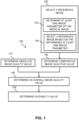
- FIG. 1 there is depicted a simplified flow diagram of a method 100 for assessing the quality of a medical image according to a proposed embodiment.
- the method starts with a step 110 of determining an absolute image quality value of a medical image.
- the determined absolute image quality comprises a plurality of values describing the respective image quality of a plurality of different anatomical features present in the medical image. This may be advantageous as the image quality may vary significantly across the field of view and for features that are orientated differently with respect to the image view direction. For the purposes of diagnosis and measurement, only certain features depicted in the image may need to have high image quality, and so this information may be useful in determining if the medical image is suitable for use.
- the absolute image quality value associated with each feature in the image is determined to be high, medium, or low.
- the method further involves the step 120.
- the medical image is analyzed to select a reference image form a plurality of different possible reference images.
- the step 120 further comprises the sub-steps 122 and 124.
- Step 122 comprises processing the medical image with an image identification model to determine at least one image parameter of the medical image.
- the determined at least one image parameter describes the anatomical features present in the medical image, the view position associated with the medical image, and the imaging technique used to obtain the image.
- it may be determined by the image identification model that the medical image is an ultrasound image of a heart in the posterior-anterior view direction.
- Step 124 comprises selecting a reference image from a plurality of possible reference images based on the determined at least one image parameter.
- the reference image may be selected to also be an ultrasound image of a heart imaged in the posterior-anterior view position.
- the method proceeds to a step 130 comprising processing the medical image and the reference image with a second machine learning model to determine a comparative image quality value describing the quality of the medical image relative to the quality of the selected reference image.
- the comparative image quality value comprises a plurality of values describing the relative image quality of each anatomical feature present in the medical image compared to the image quality of the same anatomical feature depicted in the reference image.
- this comprises a series of determination of higher, lower, or equal image quality in the medical image relative to the reference image for each anatomical feature in the medical image.
- information is provided that directly compares the image quality of common features present in both the reference image and the medical image. This may be useful in application of the method to a medical examination process, as the medical professional carrying out the examination may be provided with information describing whether a particular feature has been imaged with better quality in the past.
- step 140 comprises determining, based on the absolute image quality value and the comparative image quality value, an overall image quality value.
- the overall image quality comprises at least one of: the absolute image quality value and the comparative image quality value.
- the method then proceeds to a step 150 comprising determining a suitability value of the medical image, based on the overall image quality value determined in step 140.
- a suitability value based on the overall image quality value determined in step 140. This describes, based on the image quality of the medical image, how suitable the medical image may be for use in diagnosis and further assessment processes. For example, if the image quality is deemed to be low, the medical image may be deemed unsuitable for use for diagnosis and measurement.
- the suitability value may be a binary determination of "suitable” or "unsuitable” or may alternatively provide further, more detailed determinations of how useful the image is likely to be.
- the absolute image quality value and the comparative image quality value comprise a plurality of values relating to the image quality of various anatomical features depicted in the medical image
- the absolute image quality value and the comparative image quality value may simply be single values describing an image quality of the entirety of the image.
- both the absolute image quality value and the comparative image quality value are qualitative values, in other examples they may be quantitative.
- the absolute image quality value may be an image quality ranking between 1 and 10
- the comparative image quality value may be the different in ranking on this same scale between the quality of the medical image relative to the reference image.
- At least one image parameter determined by the image identification model relates to the anatomical features present in the medical image, the view direction associated with the medical image and the imaging technique used to acquire the medical image
- different image parameters may be determined.
- at least one image parameter may comprise at least one of: a view position; an anatomical feature; an orientation of an anatomical feature; a patient identification value; an examination identification value; a date; and an image sensor identification value. Selecting the reference image based on this image parameter may then involve selecting a reference image that is associated with the same patient as the medical image, and/or acquired by the same examination process or image sensor. The selected reference image could also be an image taken on the same date as the medical image.
- the image identification model may be implemented using any well-known image processing technique.
- the image identification model could be configured to recognise and extract information from metadata tags associated with the medical image.
- the image identification model could include a machine learning model trained to identify imaging parameters of the medical image.
- the sub-steps 122 and 124 may not be present in some embodiments of the present invention, i.e., in some embodiments selecting a suitable reference image may not require processing the medical image with an image identification model to determine an image parameter of the image.
- the medical image may be provided with a tag identifying a suitable reference image (e.g., that has been identified by a medical professional as being suitable) which is used when the image is analysed to select the reference image form a plurality of different possible reference images.
- the key here is simply that information associated with the medical image to be assessed is used to select the reference image from a number of different possible reference images.
- the step 150 may not be carried out in alternative embodiments. Some embodiments may simply result in the production of an overall image quality value.
- the overall image quality value determined in the method 100 comprises at least one of the absolute image quality value and the comparative image quality value.
- the overall image quality value may describe an average of the absolute image quality value and the comparative image quality value.
- other formulas and calculations may be used to derive the overall image quality value from the absolute image quality value and the comparative image quality value depending on the relative reliability of the two values. For example, if the comparative image quality value is deemed to be more accurate than the absolute image quality value, the comparative image quality value of the image may be given a greater weighting than the absolute image quality value in the calculation of the overall image quality value.
- Producing a suitable training dataset for the first machine learning model may be challenging.
- Manual annotations of training images with absolute quality scores are often very subjective and can introduce significant bias into the determination of image quality when a model is trained on an annotated dataset obtained in this way.
- Certain embodiments of the present invention attempt to overcome this issue by calculating image quality annotations for a training dataset using pair-wise image quality comparisons. This may lead to better and more consistent image quality labels. Further details of this annotation process are discussed below with reference to Figs. 2 and 3 .
- Fig. 2 is a simplified block diagram of a user interface used in the process of pair-wise annotation of a training dataset.
- a training dataset of suitable medical images is obtained.
- Annotation of the dataset is then carried out via the following process.
- Pairs of training images including a first training image 210 and a second training image 220 are displayed to a trained individual employed in the role of annotation via a user interface 200.
- the user is then also presented with three annotation outcomes 230, 240, and 250.
- the outcome 230 relates to an annotation designating that the first training image 210 has higher image quality than the second training image 220.
- Outcome 240 relates to an outcome in which the pair of images are annotated to have the same image quality.
- Outcome 250 relates to an annotation in which the second training image 220 is designated to have higher image quality.
- the user interface is a touch sensitive screen and so the annotating individual may then select via the user interface 200 which outcome (230, 240, or 250) they feel is the most suitable for the pair of training images they have been presented with.
- the user interface may comprise an input interface allowing the user to select an annotation outcome, and an output interface for displaying the pair of training images.
- the output interface may be a computer monitor and the input interface maybe a keyboard.
- FIG. 3 A simplified example 300 of such a network is depicted in Fig. 3 .
- Each circle (or node) of the network represents an image of the annotated training dataset.
- the network 300 depicts the images 301, 302, 303, 304, 305, 306, and 307.
- the arrows (or edges) of the network represent pair-wise annotations of comparative image quality. The arrow starts at an image deemed to be of higher quality and points towards an image deemed to be of lower image quality.
- the annotation network 300 it can be seen that the image 301 has been annotated to be of higher quality than both the image 302 and the image 303.
- An annotation network such as that displayed in Figure 3 may then be used to calculate an absolute image quality score for each image of the training dataset. This may be achieved using an Elo-based transformation system. This involves simulating a number of "games" between images of the training database. Each connection (or arrow) in the annotation network determines the outcome of a game between the two associated images, with the image deemed to be of higher quality being said to have "won” the game. For each game played, the rating of the winning image goes up and the rating of the losing image goes down. How much the ratings of the two images are adjusted as a consequence of the game is related to the current rating of the images. Once a sufficient number of games have been simulated the rating values of the set of images stabilise.
- games may be simulated with around 200 iterations until the average rating change as a result of each game is around 0.1%.
- a quality rating for each image is obtained. A higher rating indicates that the image is of higher quality while a lower score indicates that the image is of lower quality.
- An Elo-based rating system naturally follows a Gaussian distribution, with very low and very high ratings being widely scattered.
- a flattening function may be employed to flatten the distribution of quality ratings. This flattening process may result in the images with ratings in the lowest 10% to be mapped to a common absolute image quality score. Similarly, images with the highest 10% of quality ratings may be mapped to a common absolute image quality value.
- the flattened distribution may be more easily learnable by a machine learning model. In this way, an absolute image quality value may be obtained for each image in the training database.
- pair-wise quality annotation in this way to produce absolute quality values for a set of training images, results in a substantially improved interobserver variability across annotators.
- a conventional annotation approach in which annotators are asked to assign quality values to images in a training dataset (for example they may be asked to assign a value of 1-low quality, 2-medium quality, or 3-high quality to each image) results in little to no agreement between experts. It is therefore unsuitable to use this annotation method to annotate a training database for use in training a machine learning model as the resulting model may result in highly subjective image quality scores.
- the pairbased annotation strategy presented above shows substantially improved interobserver variability across annotators.
- the training dataset may contain only images of a particular anatomical feature e.g., a heart.
- the annotator is then presented with two training images of hearts and asked to compare the image quality of each feature of the heart present in the training images.
- the annotator may be asked to compare the image quality of the left ventricle, the mitral valve, the tricuspid valve, the aortic valve, the pulmonary valve, foreshortening, papillary muscles, and the right ventricle.
- the absolute quality value of each image obtained by the above process will comprise a plurality of quality values describing the respective image quality of the plurality of different anatomical features present in the medical image.
- the annotated dataset of medical images produced in this way may be used for training the first machine learning model used in the method of the present invention.
- the first machine learning model may be trained using a deep learning algorithm configured to receive an array of training inputs and respective known outputs, wherein a training input of the first machine learning model is a first training image the respective known output is an absolute image quality value of the first training image.
- the absolute quality value may be calculated from at least one comparative image quality value associated with the first training image and describing the quality of the first training image compared to at least one other training image.
- the absolute image quality value may be calculated from the at least one comparative training image using an Elo-based transformation system as described above.
- FIG. 4 there is depicted simplified block diagram of a medical imaging system 400 for assessing the quality of a medical image according to a proposed embodiment.
- the medical imaging system 400 comprises an image sensor 410, a processing arrangement 420, a memory component 430 and an output interface 440.
- the image sensor 410 is configured to take images of a subject suitably positioned in front of the medical imaging system 400. The image may then be presented to an operator of the medical imaging system via the output interface 440.
- the image sensor 410 may be any suitable image sensor for use in medical imaging.
- the image sensor 410 may comprise at least one of: an x-ray detector, an MRI scanner, an ultrasound detector, a CT scanner, a PET scanner, and an infrared detector.
- the processing arrangement 420 is configured to assess the quality of the image obtained by image sensor 410. It achieves this by: processing the obtained image with a first machine-learning model to determine an absolute image quality value of the image; analysing the image to select a reference image from a plurality of different possible reference images; processing the obtained image and the selected reference image with a second machine learning model to determine a comparative image quality value describing the quality of the obtained image relative to the selected reference image; and determining, based on the absolute image quality value and the comparative image quality value, an overall image quality value of the image obtained by the image sensor.
- the plurality of different possible reference images may be stored in the memory component 430 associated with the processing arrangement 420.
- the obtained image may also be stored in the memory component 430 and may be used as a possible reference image in subsequent examinations using the same medical imaging system.
- the processing arrangement 420 and the memory component 430 may be implemented in different ways, with software and/or hardware, to perform the various functions required.
- the processing arrangement 420 may typically employ one or more microprocessors that may be programmed using software (e.g., microcode) to perform the required functions.
- the processing arrangement 420 may be implemented as a combination of dedicated hardware to perform some functions and one or more programmed microprocessors and associated circuitry to perform other functions. Examples of circuitry that may be employed in various embodiments of the present disclosure include, but are not limited to, conventional microprocessors, application specific integrated circuits (ASICs), and field-programmable gate arrays (FPGAs).
- ASICs application specific integrated circuits
- FPGAs field-programmable gate arrays
- the memory component 430 associated with processing arrangement 420 may comprise one or more storage media such as volatile and non-volatile computer memory such as RAM, PROM, EPROM, and EEPROM.
- the storage media may be encoded with one or more programs that, when executed on one or more processors and/or controllers, perform the required functions.
- Various storage media may be fixed within a processor or controller or may be transportable, such that the one or more programs stored thereon can be loaded into a processor.
- the output interface 440 is further configured to output an indication of the determined overall image quality value to the operator of the medical imaging system.
- An example of such a configuration is shown in Fig. 5 .
- Fig. 5 depicts an example output interface 500 of a system for assessing the quality of a medical image according to a proposed embodiment.
- the output interface 500 displays a medical image 510, and quality scores 1-3 (520, 530, and 540) and comparison rating 550 associated with the medical image 510.
- the quality scores 520, 530, and 540 along with the comparison rating 550 may together comprise the overall image quality value of this embodiment of the proposed invention.
- the quality scores 1-3 may be absolute image quality scores each relate to different anatomical features present in the image 510. For example, using the method of the proposed invention, it may be determined that the medical image 510 has high image quality for a first feature relating to quality score 1, and low image quality for second and third features relating to quality scores 2 and 3. This may be presented to the operator as either a quantitative score (i.e.
- the quality scores 1-3 may be presented in the form of suitability assessments, i.e., they may represent a simple assessment of sufficient image quality for diagnosis or insufficient image quality for diagnosis.
- the quality scores 1-3 may be displayed as numbers, words, or via colour indicators. Although in this example three quality scores are shown, in other embodiments any number of quality scores relating to different features present in the medical image 510 may be shown to the user on the output interface 500.
- the comparison rating may be presented to the user as an indication of the comparative image quality value determined for the medical image 510 via the method of any proposed embodiment.
- the comparison rating may indicate to the operator if images of higher quality have been obtained in the past.
- the comparison rating may indicate in words to the operator that previously acquired images may have better image quality for features 2 and 3 depicted in the medical image 510. In this way, the operator may be presented with information that may prompt them to attempt to obtain more medical images of the same patient with higher image quality for features 2 and 3.
- Figure 6 illustrates an example of a computer 600 within which one or more parts of an embodiment may be employed.
- Various operations discussed above may utilize the capabilities of the computer 600.
- system functional blocks can run on a single computer or may be distributed over several computers and locations (e.g. connected via internet).
- the computer 600 includes, but is not limited to, PCs, workstations, laptops, PDAs, palm devices, servers, storages, and the like.
- the computer 600 may include one or more processors 610, memory 620 and one or more I/O devices 630 that are communicatively coupled via a local interface (not shown).
- the local interface can be, for example but not limited to, one or more buses or other wired or wireless connections, as is known in the art.
- the local interface may have additional elements, such as controllers, buffers (caches), drivers, repeaters, and receivers, to enable communications. Further, the local interface may include address, control, and/or data connections to enable appropriate communications among the aforementioned components.
- the processor 610 is a hardware device for executing software that can be stored in the memory 620.
- the processor 610 can be virtually any custom made or commercially available processor, a central processing unit (CPU), a digital signal processor (DSP), or an auxiliary processor among several processors associated with the computer 600, and the processor 610 may be a semiconductor-based microprocessor (in the form of a microchip) or a microprocessor.
- the memory 620 can include any one or combination of volatile memory elements (e.g., random access memory (RAM), such as dynamic random access memory (DRAM), static random access memory (SRAM), etc.) and non-volatile memory elements (e.g., ROM, erasable programmable read only memory (EPROM), electronically erasable programmable read only memory (EEPROM), programmable read only memory (PROM), tape, compact disc read only memory (CD-ROM), disk, diskette, cartridge, cassette or the like, etc.).
- RAM random access memory
- DRAM dynamic random access memory
- SRAM static random access memory
- non-volatile memory elements e.g., ROM, erasable programmable read only memory (EPROM), electronically erasable programmable read only memory (EEPROM), programmable read only memory (PROM), tape, compact disc read only memory (CD-ROM), disk, diskette, cartridge, cassette or the like, etc.
- the memory 620 may incorporate electronic, magnetic, optical, and/or other types
- the software in the memory 620 may include one or more separate programs, each of which comprises an ordered listing of executable instructions for implementing logical functions.
- the software in the memory 620 includes a suitable operating system (O/S) 650, compiler 660, source code 670, and one or more applications 680 in accordance with exemplary embodiments.
- the application 680 comprises numerous functional components for implementing the features and operations of the exemplary embodiments.
- the application 680 of the computer 600 may represent various applications, computational units, logic, functional units, processes, operations, virtual entities, and/or modules in accordance with exemplary embodiments, but the application 680 is not meant to be a limitation.
- the operating system 650 controls the execution of other computer programs, and provides scheduling, input-output control, file and data management, memory management, and communication control and related services. It is contemplated by the inventors that the application 680 for implementing exemplary embodiments may be applicable on all commercially available operating systems.
- Application 680 may be a source program, executable program (object code), script, or any other entity comprising a set of instructions to be performed.
- a source program then the program is usually translated via a compiler (such as the compiler 660), assembler, interpreter, or the like, which may or may not be included within the memory 620, so as to operate properly in connection with the O/S 650.
- a compiler such as the compiler 660
- interpreter or the like
- the application 680 can be written as an object oriented programming language, which has classes of data and methods, or a procedure programming language, which has routines, subroutines, and/or functions, for example but not limited to, C, C++, C#, Pascal, Python, BASIC, API calls, HTML, XHTML, XML, ASP scripts, JavaScript, FORTRAN, COBOL, Perl, Java, ADA, .NET, and the like.
- the I/O devices 630 may include input devices such as, for example but not limited to, a mouse, keyboard, scanner, microphone, camera, etc. Furthermore, the I/O devices 630 may also include output devices, for example but not limited to a printer, display, etc. Finally, the I/O devices 630 may further include devices that communicate both inputs and outputs, for instance but not limited to, a NIC or modulator/demodulator (for accessing remote devices, other files, devices, systems, or a network), a radio frequency (RF) or other transceiver, a telephonic interface, a bridge, a router, etc. The I/O devices 630 also include components for communicating over various networks, such as the Internet or intranet.
- a NIC or modulator/demodulator for accessing remote devices, other files, devices, systems, or a network
- RF radio frequency
- the I/O devices 630 also include components for communicating over various networks, such as the Internet or intranet.
- the software in the memory 620 may further include a basic input output system (BIOS) (omitted for simplicity).
- BIOS is a set of essential software routines that initialize and test hardware at start-up, start the O/S 650, and support the transfer of data among the hardware devices.
- the BIOS is stored in some type of read-only-memory, such as ROM, PROM, EPROM, EEPROM or the like, so that the BIOS can be executed when the computer 600 is activated.
- the processor 610 When the computer 600 is in operation, the processor 610 is configured to execute software stored within the memory 620, to communicate data to and from the memory 620, and to generally control operations of the computer 600 pursuant to the software.
- the application 680 and the O/S 650 are read, in whole or in part, by the processor 610, perhaps buffered within the processor 610, and then executed.
- a computer readable medium may be an electronic, magnetic, optical, or other physical device or means that can contain or store a computer program for use by or in connection with a computer related system or method.
- the application 680 can be embodied in any computer-readable medium for use by or in connection with an instruction execution system, apparatus, or device, such as a computer-based system, processor-containing system, or other system that can fetch the instructions from the instruction execution system, apparatus, or device and execute the instructions.
- a "computer-readable medium" can be any means that can store, communicate, propagate, or transport the program for use by or in connection with the instruction execution system, apparatus, or device.
- the computer readable medium can be, for example but not limited to, an electronic, magnetic, optical, electromagnetic, infrared, or semiconductor system, apparatus, device, or propagation medium.
- the method of Figure 1 may be implemented in hardware or software, or a mixture of both (for example, as firmware running on a hardware device).
- the functional steps illustrated in the process flowcharts may be performed by suitably programmed physical computing devices, such as one or more central processing units (CPUs) or graphics processing units (GPUs).
- CPUs central processing units
- GPUs graphics processing units
- Each process - and its individual component steps as illustrated in the flowcharts - may be performed by the same or different computing devices.
- a computer-readable storage medium stores a computer program comprising computer program code configured to cause one or more physical computing devices to carry out a method as described above when the program is run on the one or more physical computing devices.
- Storage media may include volatile and non-volatile computer memory such as RAM, PROM, EPROM, and EEPROM, optical discs (like CD, DVD, BD), magnetic storage media (like hard discs and tapes).
- RAM random access memory
- PROM read-only memory
- EPROM erasable programmable read-only memory
- EEPROM electrically erasable programmable read-only memory
- optical discs like CD, DVD, BD
- magnetic storage media like hard discs and tapes.
- Various storage media may be fixed within a computing device or may be transportable, such that the one or more programs stored thereon can be loaded into a processor.
- the blocks shown in the block diagrams of Figure 4 may be separate physical components, or logical subdivisions of single physical components, or may be all implemented in an integrated manner in one physical component.
- the functions of one block shown in the drawings may be divided between multiple components in an implementation, or the functions of multiple blocks shown in the drawings may be combined in single components in an implementation.
- Hardware components suitable for use in embodiments of the present invention include, but are not limited to, conventional microprocessors, application specific integrated circuits (ASICs), and field-programmable gate arrays (FPGAs).
- ASICs application specific integrated circuits
- FPGAs field-programmable gate arrays
- One or more blocks may be implemented as a combination of dedicated hardware to perform some functions and one or more programmed microprocessors and associated circuitry to perform other functions.
- a computer program may be stored/distributed on a suitable medium, such as an optical storage medium or a solid-state medium supplied together with or as part of other hardware, but may also be distributed in other forms, such as via the Internet or other wired or wireless telecommunication systems.
- a suitable medium such as an optical storage medium or a solid-state medium supplied together with or as part of other hardware, but may also be distributed in other forms, such as via the Internet or other wired or wireless telecommunication systems.
- each block in the flowcharts or block diagrams may represent a module, segment, or portion of instructions, which comprises one or more executable instructions for implementing the specified logical function(s).
- the functions noted in the block may occur out of the order noted in the figures.
- two blocks shown in succession may, in fact, be executed substantially concurrently, or the blocks may sometimes be executed in the reverse order, depending upon the functionality involved.
- each block of the block diagrams and/or flowchart illustration, and combinations of blocks in the block diagrams and/or flowchart illustration can be implemented by special purpose hardware-based systems that perform the specified functions or acts or carry out combinations of special purpose hardware and computer instructions, the architecture, functionality, and operation of possible implementations of systems, methods, and computer program products according to various embodiments of the present invention.
- each block in the flowchart or block diagrams may represent a module, segment, or portion of instructions, which comprises one or more executable instructions for implementing the specified logical function(s).
- the functions noted in the block may occur out of the order noted in the figures.
Landscapes
- Engineering & Computer Science (AREA)
- Quality & Reliability (AREA)
- Computer Vision & Pattern Recognition (AREA)
- Physics & Mathematics (AREA)
- General Physics & Mathematics (AREA)
- Theoretical Computer Science (AREA)
- Image Analysis (AREA)
Priority Applications (1)
| Application Number | Priority Date | Filing Date | Title |
|---|---|---|---|
| PCT/EP2024/084098 WO2025119791A1 (fr) | 2023-12-08 | 2024-11-29 | Évaluation de la qualité d'une image médicale |
Applications Claiming Priority (1)
| Application Number | Priority Date | Filing Date | Title |
|---|---|---|---|
| US202363607646P | 2023-12-08 | 2023-12-08 |
Publications (1)
| Publication Number | Publication Date |
|---|---|
| EP4567725A1 true EP4567725A1 (fr) | 2025-06-11 |
Family
ID=89535940
Family Applications (1)
| Application Number | Title | Priority Date | Filing Date |
|---|---|---|---|
| EP24150701.1A Pending EP4567725A1 (fr) | 2023-12-08 | 2024-01-08 | Évaluation de qualité d'image médicale |
Country Status (1)
| Country | Link |
|---|---|
| EP (1) | EP4567725A1 (fr) |
Citations (1)
| Publication number | Priority date | Publication date | Assignee | Title |
|---|---|---|---|---|
| US20180144214A1 (en) * | 2016-11-23 | 2018-05-24 | General Electric Company | Deep learning medical systems and methods for image reconstruction and quality evaluation |
-
2024
- 2024-01-08 EP EP24150701.1A patent/EP4567725A1/fr active Pending
Patent Citations (1)
| Publication number | Priority date | Publication date | Assignee | Title |
|---|---|---|---|---|
| US20180144214A1 (en) * | 2016-11-23 | 2018-05-24 | General Electric Company | Deep learning medical systems and methods for image reconstruction and quality evaluation |
Non-Patent Citations (3)
| Title |
|---|
| BRANKOV JOVAN G ET AL: "Generalization evaluation of numerical observers for image quality assessment", 2006 IEEE NUCLEAR SCIENCE SYMPOSIUM CONFERENCE RECORD, 29 October 2006 (2006-10-29), pages 1696 - 1698, XP093174795, Retrieved from the Internet <URL:https://ieeexplore.ieee.org/stampPDF/getPDF.jsp?tp=&arnumber=4179338&ref=aHR0cHM6Ly9pZWVleHBsb3JlLmllZWUub3JnL2Fic3RyYWN0L2RvY3VtZW50LzQxNzkzMzg=> DOI: 10.1109/NSSMIC.2006.354225 * |
| LEI KE ET AL: "Artifact- and content-specific quality assessment for MRI with image rulers", MEDICAL IMAGE ANALYSIS, OXFORD UNIVERSITY PRESS, OXOFRD, GB, vol. 77, 20 January 2022 (2022-01-20), XP086982022, ISSN: 1361-8415, [retrieved on 20220120], DOI: 10.1016/J.MEDIA.2021.102344 * |
| STEPIEN IGOR ET AL: "A Brief Survey on No-Reference Image Quality Assessment Methods for Magnetic Resonance Images", JOURNAL OF IMAGING, vol. 8, no. 6, 4 June 2022 (2022-06-04), pages 160, XP093174796, ISSN: 2313-433X, Retrieved from the Internet <URL:https://www.mdpi.com/2313-433X/8/6/160/pdf> DOI: 10.3390/jimaging8060160 * |
Similar Documents
| Publication | Publication Date | Title |
|---|---|---|
| JP7730490B2 (ja) | 人工知能モデルを使用機関に特化させる学習方法、これを行う装置 | |
| US11615879B2 (en) | System and method for automated labeling and annotating unstructured medical datasets | |
| US11127138B2 (en) | Automatic detection and quantification of the aorta from medical images | |
| CN113971657B (zh) | 用于医学成像中的异常评估的基于噪声标签的机器学习 | |
| CN112926537B (zh) | 图像处理方法、装置、电子设备以及存储介质 | |
| US11721439B2 (en) | Resolving and steering decision foci in machine learning-based vascular imaging | |
| Schilling et al. | Assessment of deep learning segmentation for real-time free-breathing cardiac magnetic resonance imaging at rest and under exercise stress | |
| KR20220143547A (ko) | 심전도 이미지에 기반한 환자 평가 시스템 및 방법 | |
| CN114730382A (zh) | 使用具有混合质量的标记的医学数据对人工神经网络进行的约束训练 | |
| EP3896702A1 (fr) | Évaluation cardiovasculaire des patients suspectés d'être atteints de covid-19 | |
| US20240335170A1 (en) | System and method for electrocardiogram image-based patient evaluation | |
| US12308119B2 (en) | Mapping brain data to behavior | |
| US20250217979A1 (en) | Method and electronic device for generating representative frame image of medical image | |
| EP4567725A1 (fr) | Évaluation de qualité d'image médicale | |
| Rauseo et al. | A systematic quality scoring analysis to assess automated cardiovascular magnetic resonance segmentation algorithms | |
| WO2025119791A1 (fr) | Évaluation de la qualité d'une image médicale | |
| US20240420461A1 (en) | Contextualizing the analysis of medical images | |
| Ranem et al. | Detecting respiratory motion artefacts for cardiovascular MRIs to ensure high-quality segmentation | |
| EP4253987A1 (fr) | Commande d'imagerie par résonance magnétique | |
| US20230281804A1 (en) | Landmark detection in medical images | |
| Mineo et al. | FeDETR: A Federated Approach for Stenosis Detection in Coronary Angiography | |
| CN114462522A (zh) | 肺癌生存期预测模型训练、预测方法、系统、装置及介质 | |
| KR20240092729A (ko) | 의료 영상 처리 방법 및 디바이스 | |
| JP2024530417A (ja) | 被検者の心臓のモデリング | |
| US20240136064A1 (en) | Medical diagnosis support device, medical diagnosis support method, and storage medium |
Legal Events
| Date | Code | Title | Description |
|---|---|---|---|
| PUAI | Public reference made under article 153(3) epc to a published international application that has entered the european phase |
Free format text: ORIGINAL CODE: 0009012 |
|
| STAA | Information on the status of an ep patent application or granted ep patent |
Free format text: STATUS: THE APPLICATION HAS BEEN PUBLISHED |
|
| AK | Designated contracting states |
Kind code of ref document: A1 Designated state(s): AL AT BE BG CH CY CZ DE DK EE ES FI FR GB GR HR HU IE IS IT LI LT LU LV MC ME MK MT NL NO PL PT RO RS SE SI SK SM TR |
|
| STAA | Information on the status of an ep patent application or granted ep patent |
Free format text: STATUS: THE APPLICATION IS DEEMED TO BE WITHDRAWN |