EP4570257A1 - Utilisation d'une poudre sèche d'exosomes dérivés de plaquettes purifiées pour soulager une inflammation ou une lésion - Google Patents
Utilisation d'une poudre sèche d'exosomes dérivés de plaquettes purifiées pour soulager une inflammation ou une lésion Download PDFInfo
- Publication number
- EP4570257A1 EP4570257A1 EP24218839.9A EP24218839A EP4570257A1 EP 4570257 A1 EP4570257 A1 EP 4570257A1 EP 24218839 A EP24218839 A EP 24218839A EP 4570257 A1 EP4570257 A1 EP 4570257A1
- Authority
- EP
- European Patent Office
- Prior art keywords
- platelet
- solution
- derived
- exosome
- dry powder
- Prior art date
- Legal status (The legal status is an assumption and is not a legal conclusion. Google has not performed a legal analysis and makes no representation as to the accuracy of the status listed.)
- Pending
Links
Images






Classifications
-
- A—HUMAN NECESSITIES
- A61—MEDICAL OR VETERINARY SCIENCE; HYGIENE
- A61K—PREPARATIONS FOR MEDICAL, DENTAL OR TOILETRY PURPOSES
- A61K35/00—Medicinal preparations containing materials or reaction products thereof with undetermined constitution
- A61K35/12—Materials from mammals; Compositions comprising non-specified tissues or cells; Compositions comprising non-embryonic stem cells; Genetically modified cells
- A61K35/14—Blood; Artificial blood
- A61K35/19—Platelets; Megacaryocytes
-
- A—HUMAN NECESSITIES
- A61—MEDICAL OR VETERINARY SCIENCE; HYGIENE
- A61K—PREPARATIONS FOR MEDICAL, DENTAL OR TOILETRY PURPOSES
- A61K38/00—Medicinal preparations containing peptides
- A61K38/16—Peptides having more than 20 amino acids; Gastrins; Somatostatins; Melanotropins; Derivatives thereof
- A61K38/17—Peptides having more than 20 amino acids; Gastrins; Somatostatins; Melanotropins; Derivatives thereof from animals; from humans
- A61K38/18—Growth factors; Growth regulators
- A61K38/1808—Epidermal growth factor [EGF] urogastrone
-
- A—HUMAN NECESSITIES
- A61—MEDICAL OR VETERINARY SCIENCE; HYGIENE
- A61K—PREPARATIONS FOR MEDICAL, DENTAL OR TOILETRY PURPOSES
- A61K38/00—Medicinal preparations containing peptides
- A61K38/16—Peptides having more than 20 amino acids; Gastrins; Somatostatins; Melanotropins; Derivatives thereof
- A61K38/17—Peptides having more than 20 amino acids; Gastrins; Somatostatins; Melanotropins; Derivatives thereof from animals; from humans
- A61K38/18—Growth factors; Growth regulators
- A61K38/1841—Transforming growth factor [TGF]
-
- A—HUMAN NECESSITIES
- A61—MEDICAL OR VETERINARY SCIENCE; HYGIENE
- A61K—PREPARATIONS FOR MEDICAL, DENTAL OR TOILETRY PURPOSES
- A61K38/00—Medicinal preparations containing peptides
- A61K38/16—Peptides having more than 20 amino acids; Gastrins; Somatostatins; Melanotropins; Derivatives thereof
- A61K38/17—Peptides having more than 20 amino acids; Gastrins; Somatostatins; Melanotropins; Derivatives thereof from animals; from humans
- A61K38/18—Growth factors; Growth regulators
- A61K38/1858—Platelet-derived growth factor [PDGF]
-
- A—HUMAN NECESSITIES
- A61—MEDICAL OR VETERINARY SCIENCE; HYGIENE
- A61K—PREPARATIONS FOR MEDICAL, DENTAL OR TOILETRY PURPOSES
- A61K38/00—Medicinal preparations containing peptides
- A61K38/16—Peptides having more than 20 amino acids; Gastrins; Somatostatins; Melanotropins; Derivatives thereof
- A61K38/17—Peptides having more than 20 amino acids; Gastrins; Somatostatins; Melanotropins; Derivatives thereof from animals; from humans
- A61K38/18—Growth factors; Growth regulators
- A61K38/1858—Platelet-derived growth factor [PDGF]
- A61K38/1866—Vascular endothelial growth factor [VEGF]
-
- A—HUMAN NECESSITIES
- A61—MEDICAL OR VETERINARY SCIENCE; HYGIENE
- A61K—PREPARATIONS FOR MEDICAL, DENTAL OR TOILETRY PURPOSES
- A61K38/00—Medicinal preparations containing peptides
- A61K38/16—Peptides having more than 20 amino acids; Gastrins; Somatostatins; Melanotropins; Derivatives thereof
- A61K38/17—Peptides having more than 20 amino acids; Gastrins; Somatostatins; Melanotropins; Derivatives thereof from animals; from humans
- A61K38/22—Hormones
- A61K38/30—Insulin-like growth factors, i.e. somatomedins, e.g. IGF-1, IGF-2
-
- A—HUMAN NECESSITIES
- A61—MEDICAL OR VETERINARY SCIENCE; HYGIENE
- A61K—PREPARATIONS FOR MEDICAL, DENTAL OR TOILETRY PURPOSES
- A61K9/00—Medicinal preparations characterised by special physical form
- A61K9/14—Particulate form, e.g. powders, Processes for size reducing of pure drugs or the resulting products, Pure drug nanoparticles
- A61K9/19—Particulate form, e.g. powders, Processes for size reducing of pure drugs or the resulting products, Pure drug nanoparticles lyophilised, i.e. freeze-dried, solutions or dispersions
-
- A—HUMAN NECESSITIES
- A61—MEDICAL OR VETERINARY SCIENCE; HYGIENE
- A61K—PREPARATIONS FOR MEDICAL, DENTAL OR TOILETRY PURPOSES
- A61K9/00—Medicinal preparations characterised by special physical form
- A61K9/48—Preparations in capsules, e.g. of gelatin, of chocolate
- A61K9/50—Microcapsules having a gas, liquid or semi-solid filling; Solid microparticles or pellets surrounded by a distinct coating layer, e.g. coated microspheres, coated drug crystals
- A61K9/5005—Wall or coating material
- A61K9/5063—Compounds of unknown constitution, e.g. material from plants or animals
- A61K9/5068—Cell membranes or bacterial membranes enclosing drugs
-
- A—HUMAN NECESSITIES
- A61—MEDICAL OR VETERINARY SCIENCE; HYGIENE
- A61M—DEVICES FOR INTRODUCING MEDIA INTO, OR ONTO, THE BODY; DEVICES FOR TRANSDUCING BODY MEDIA OR FOR TAKING MEDIA FROM THE BODY; DEVICES FOR PRODUCING OR ENDING SLEEP OR STUPOR
- A61M1/00—Suction or pumping devices for medical purposes; Devices for carrying-off, for treatment of, or for carrying-over, body-liquids; Drainage systems
- A61M1/02—Blood transfusion apparatus
- A61M1/0272—Apparatus for treatment of blood or blood constituents prior to or for conservation, e.g. freezing, drying or centrifuging
-
- A—HUMAN NECESSITIES
- A61—MEDICAL OR VETERINARY SCIENCE; HYGIENE
- A61M—DEVICES FOR INTRODUCING MEDIA INTO, OR ONTO, THE BODY; DEVICES FOR TRANSDUCING BODY MEDIA OR FOR TAKING MEDIA FROM THE BODY; DEVICES FOR PRODUCING OR ENDING SLEEP OR STUPOR
- A61M1/00—Suction or pumping devices for medical purposes; Devices for carrying-off, for treatment of, or for carrying-over, body-liquids; Drainage systems
- A61M1/36—Other treatment of blood in a by-pass of the natural circulatory system, e.g. temperature adaptation, irradiation ; Extra-corporeal blood circuits
- A61M1/3693—Other treatment of blood in a by-pass of the natural circulatory system, e.g. temperature adaptation, irradiation ; Extra-corporeal blood circuits using separation based on different densities of components, e.g. centrifuging
-
- A—HUMAN NECESSITIES
- A61—MEDICAL OR VETERINARY SCIENCE; HYGIENE
- A61P—SPECIFIC THERAPEUTIC ACTIVITY OF CHEMICAL COMPOUNDS OR MEDICINAL PREPARATIONS
- A61P17/00—Drugs for dermatological disorders
- A61P17/02—Drugs for dermatological disorders for treating wounds, ulcers, burns, scars, keloids, or the like
-
- A—HUMAN NECESSITIES
- A61—MEDICAL OR VETERINARY SCIENCE; HYGIENE
- A61P—SPECIFIC THERAPEUTIC ACTIVITY OF CHEMICAL COMPOUNDS OR MEDICINAL PREPARATIONS
- A61P29/00—Non-central analgesic, antipyretic or antiinflammatory agents, e.g. antirheumatic agents; Non-steroidal antiinflammatory drugs [NSAID]
-
- A—HUMAN NECESSITIES
- A61—MEDICAL OR VETERINARY SCIENCE; HYGIENE
- A61P—SPECIFIC THERAPEUTIC ACTIVITY OF CHEMICAL COMPOUNDS OR MEDICINAL PREPARATIONS
- A61P39/00—General protective or antinoxious agents
-
- C—CHEMISTRY; METALLURGY
- C12—BIOCHEMISTRY; BEER; SPIRITS; WINE; VINEGAR; MICROBIOLOGY; ENZYMOLOGY; MUTATION OR GENETIC ENGINEERING
- C12N—MICROORGANISMS OR ENZYMES; COMPOSITIONS THEREOF; PROPAGATING, PRESERVING, OR MAINTAINING MICROORGANISMS; MUTATION OR GENETIC ENGINEERING; CULTURE MEDIA
- C12N5/00—Undifferentiated human, animal or plant cells, e.g. cell lines; Tissues; Cultivation or maintenance thereof; Culture media therefor
- C12N5/06—Animal cells or tissues; Human cells or tissues
- C12N5/0602—Vertebrate cells
- C12N5/0634—Cells from the blood or the immune system
- C12N5/0644—Platelets; Megakaryocytes
Definitions
- the present invention relates to a purified platelet-derived exosome dry powder, more specifically to the use of a purified platelet-derived exosome dry powder for relieving inflammation or injury.
- Inflammation is a defense mechanism of the human body. Inflammatory reactions are often triggered in various body parts due to infection, trauma, or hypersensitivity to eliminate harmful stimuli, remove pathogens, and facilitate tissue repair. Inflammation can be categorized into acute and chronic inflammation such as dermatitis, allergic rhinitis, pneumonia, hepatitis, and enteritis. Dermatitis can be classified into irritant contact dermatitis (ICD) and allergic contact dermatitis; severe pneumonia can cause diseases such as chronic obstructive pulmonary disease (COPD) and pulmonary fibrosis while severe hepatitis can cause diseases such as liver cirrhosis and cancer.
- ICD irritant contact dermatitis
- COPD chronic obstructive pulmonary disease
- COPD chronic obstructive pulmonary disease
- Fibroblasts play an important role in the repair process of various tissues throughout the body. A series of wound repair responses occur during the repair process after tissue damage (usually accompanied by inflammatory reactions), including cell proliferation, cell migration, fibrogenesis, resolution, and remodeling (Reference 1). Taking skin wounds as an example, fibroblasts rapidly migrate to the provisional matrix and proliferate quickly, leading to fibrogenesis in the injured area. Moreover, fibroblasts are the primary source of extracellular matrix (ECM) proteins, mainly including collagen and fibronectin, which can form granulation tissue and subsequently provide structural integrity to the wound.
- ECM extracellular matrix
- Exosomes are nanoscale extracellular vesicles (EVs) approximately 30-200 nanometers in diameter. They are secreted by most eukaryotic cells and contain bioactive molecules such as proteins, ribonucleic acids, deoxyribonucleic acids, microRNAs, and metabolites. Therefore, exosomes play an important role in intercellular communication, transmitting different information between cells through the bioactive molecules they carry (References 2, 3, 4). Exosomes can be found in a variety of biological fluids such as plasma, urine, semen, saliva, bronchial fluid, cerebral spinal fluid (CSF), breast milk, serum, amniotic fluid, synovial fluid, tears, lymph, bile, and gastric acid (Reference 3). Among these, plasma and serum contain white blood cell-derived exosomes, red blood cell-derived exosomes, and exosomes derived from other cells.
- exosomes derived from certain cells or tissues can achieve tissue regeneration and repair by inhibiting inflammatory responses, promoting proliferation, suppressing apoptosis, and facilitating vascular proliferation.
- clinical studies have found that exosomes are prone to rupture, have unstable quality, and are difficult to preserve during the isolation and purification process, thus affecting their therapeutic efficacy.
- the object of the present invention is to provide a purified platelet-derived exosome dry powder that can effectively relieve or treat the degree of inflammation or injury.
- Another object of the present invention is to provide a method for preparing the purified platelet-derived exosome dry powder.
- a further object of the present invention is to provide a use of the purified platelet-derived exosome dry powder in alleviating or treating the degree of inflammation or injury.
- a further object of the present invention is to provide a use for preparing a pharmaceutical composition or health composition for use in relieving or treating the degree of inflammation or injury.
- the present invention provides a method for preparing purified platelet-derived exosome dry powder, comprising: (a) taking one unit of a solution derived from blood and containing platelets, and performing a process of purifying platelets to obtain a pure platelet solution; wherein step (a) comprises: (a1) taking one unit of a solution derived from blood and containing platelets; (a2) performing a first sedimentation process to separate the solution derived from blood and containing platelets into a plasma layer and a blood cell layer; (a3) removing the blood cell layer to obtain a plasma layer solution; (a4) performing a second sedimentation process to settle platelets into a pellet; (a5) removing a supernatant and mixing the pellet with a solvent to obtain the pure platelet solution; (b) performing an activation procedure to activate platelets in the pure platelet solution and release exosomes to obtain an activated pure platelet solution; wherein the activation procedure is a freezing and then thawing procedure; (c) performing a purification procedure, comprising
- the drying process is a freeze-drying process.
- step (a3) comprises a process of romoving white blood cells so that the number of white blood cells in the pure platelet solution is less than 10 6 per milliliter (mL).
- step (a5) comprises a process of removing plasma so that the albumin concentration in the pure platelet solution is less than 3 g/dL; or step (a5) comprises a process of removing plasma so that the globulin concentration in the pure platelet solution is less than 2 g/dL; or step (a5) comprises a process of removing plasma so that the fibrinogen concentration in the pure platelet solution is less than 100 ⁇ g/mL.
- step (a5) comprises a process of removing plasma so that the albumin concentration in the pure platelet solution is less than 0.25, 0.5, 0.75, 1, 1.25, 1.5, 1.75, 2, 2.25, 2.5, or 2.75 g/dL.
- step (a5) comprises a process of removing plasma so that the globulin concentration in the pure platelet solution is less than 0.25, 0.5, 0.75, 1, 1.25, 1.5, or 1.75 g/dL.
- step (a5) comprises a process of removing plasma so that the fibrinogen concentration in the pure platelet solution is less than 10, 20, 30, 40, 50, 60, 70, 80, or 90 ⁇ g/mL.
- the solution derived from blood and containing platelets is whole blood
- the blood cell layer includes a buffy coat layer and a red blood cells layer.
- the freezing and then thawing procedure comprises: (b1') placing a container holding the pure platelet solution into an environment at -70°C ⁇ -196°C to freeze the pure platelet solution; (b2') raising the temperature of the container to thaw the frozen pure platelet solution, and performing the steps (b1') and (b2') 1, 2, 3, 4, 5, or more than 5 times.
- the freezing and then thawing procedure comprises: (b1) placing a container holding the pure platelet solution into liquid nitrogen (-196°C) to freeze the pure platelet solution; (b2) raising the temperature of the container to thaw the frozen pure platelet solution, and performing the steps (b1) and (b2) 1, 2, 3, 4, 5, or more than 5 times.
- the freezing and then thawing procedure comprises: (b1') placing a container holding the pure platelet solution into an environment at -80°C to freeze the pure platelet solution; (b2') raising the temperature of the container to thaw the frozen pure platelet solution, and performing the steps (b1') and (b2') 1, 2, 3, 4, 5, or more than 5 times.
- the freeze-drying process comprises lowering a temperature to less than or equal to -35°C and lowering a pressure to less than or equal to 80 mTorr.
- the freeze-drying process comprises lowering the temperature to less than or equal to -40°C, -45°C, -50°C, -60°C, -65°C, or -70°C.
- the freeze-drying process comprises lowering the pressure to less than or equal to 70, 60, 50, 40, or 30 mTorr.
- the solution derived from blood and containing platelets is whole blood, apheresis platelets, leukocyte-reduced apheresis platelets, or platelet-rich plasma (PRP), or any combination thereof; alternatively, the solution derived from blood and containing platelets is derived from an autologous blood sample, an allogeneic blood sample or mammalian blood sample, or a combination thereof.
- the solvent in step (a5), is sterile saline, water for injection, 0.45% NaCl, 4.5% hypertonic saline, sterile warm spring water, or isotonic saline; alternatively, in step (a5), a platelet concentration in the pure platelet solution is 1 ⁇ 10 9 -10 ⁇ 10 9 platelets/mL.
- the platelet concentration in the pure platelet solution is 2 ⁇ 10 9 , 3 ⁇ 10 9 , 4 ⁇ 10 9 , 5 ⁇ 10 9 , 6 ⁇ 10 9 , 7 ⁇ 10 9 , 8 ⁇ 10 9 , or 9 ⁇ 10 9 platelets/mL.
- the membrane filtration procedure in step (c2) uses a filter membrane with a pore size less than or equal to 0.45 ⁇ m to obtain a solution enriched in purified platelet-derived exosomes.
- the membrane filtration procedure in step (c2) uses a filter membrane with a pore size less than or equal to 0.22 ⁇ m to obtain a solution enriched in purified platelet-derived exosomes.
- the invention further provides a purified platelet-derived exosome dry powder, which is prepared by a preparation method comprises: (a) taking one unit of a solution derived from blood and containing platelets, and performing a process of purifying platelets to obtain a pure platelet solution, wherein step (a) comprises: (a1) taking one unit of a solution derived from blood and containing platelets; (a2) performing a first sedimentation process to separate the solution into a plasma layer and a blood cell layer; (a3) removing the blood cell layer to obtain a plasma layer solution; (a4) performing a second sedimentation process to settle the platelets into a pellet; (a5) removing a supernatant and mixing the pellet with a solvent to obtain the pure platelet solution; (b) performing an activation procedure to activate platelets in the pure platelet solution and release exosomes to obtain an activated pure platelet solution, wherein the activation procedure is a freezing and then thawing procedure; (c) performing a purification procedure comprising : (c1) performing
- the drying process is a freeze-drying process.
- the freezing and then thawing procedure comprises: (b1') placing a container holding the pure platelet solution into an environment at -80°C to freeze the pure platelet solution; (b2') raising the temperature of the container to thaw the frozen pure platelet solution, and performing the steps (b1') and (b2') 1, 2, 3, 4, 5, or more than 5 times.
- a particle diameter of the platelet-derived exosomes is 20-150 nanometers, and the platelet-derived exosomes contain a exosome marker and a platelet marker; wherein the exosome marker is CD9, CD63, or CD81, or any combination thereof, and the platelet marker is at least one of CD41 and CD42b, or any combination thereof; wherein (1) each gram of purified platelet-derived exosome dry powder comprises at least 1 ⁇ 10 10 platelet-derived exosomes; or (2) each gram of purified platelet-derived exosome dry powder comprises 1 ⁇ 10 11 -1 ⁇ 10 15 platelet-derived exosomes; or (3) each gram of purified platelet-derived exosome dry powder comprises 1 ⁇ 10 12 , 2 ⁇ 10 12 , 3 ⁇ 10 12 , 4 ⁇ 10 12 , 5 ⁇ 10 12 , 6 ⁇ 10 12 , 7 ⁇ 10 12 , 8 ⁇ 10 12 , 9 ⁇ 10 12 , 1 ⁇ 10 13
- each gram of purified platelet-derived exosome dry powder comprises at least 50 ⁇ g of microRNA; or (2) a content of microRNA in each gram of purified platelet-derived exosome dry powder is 100 ⁇ g-2, 500 ⁇ g.
- a content of PDGF-BB (platelet-derived growth factor-BB) in each gram of purified platelet-derived exosome dry powder is 0.01 ng-2,000 ng; or (2) a content of VEGF (vascular endothelial growth factor) in each gram of purified platelet-derived exosome dry powder is 50 pg-100,000 pg; or (3) a content of IGF (insulin-like growth factor) in each gram of purified platelet-derived exosome dry powder is 1 pg-3,000 pg; or (4) a content of TGF- ⁇ 1 (transforming growth factor beta 1) in each gram of purified platelet-derived exosome dry powder is 50 ng-20,000 ng; or (5) a content of EGF (epidermal growth factor) in each gram of purified platelet-derived exosome dry powder is 0.1 ng-200 ng.
- PDGF-BB platelet-derived growth factor-BB
- VEGF vascular endothelial growth factor
- a content of PDGF-BB is 0.01, 0.1, 1, 10, 20, 30, 40, 50, 60, 70, 80, 90, 1 ⁇ 10 2 , 5 ⁇ 10 2 , 1 ⁇ 10 3 , 1.5 ⁇ 10 3 , or 2 ⁇ 10 4 ng.
- a content of VEGF is 50, 100, 150, 200, 250, 300, 350, 400, 450, 500, 550, 600, 650, 700, 750, 800, 850, 900, 950, 1 ⁇ 10 3 , 2 ⁇ 10 3 , 3 ⁇ 10 3 , 4x10 3 , 5 ⁇ 10 3 , 6 ⁇ 10 3 , 7 ⁇ 10 3 , 8 ⁇ 10 3 , 9 ⁇ 10 3 , 1 ⁇ 10 4 , 1.5 ⁇ 10 4 , 2 ⁇ 10 4 , 2.5 ⁇ 10 4 , 3 ⁇ 10 4 , 3.5 ⁇ 10 4 , 4 ⁇ 10 4 , 4.5 ⁇ 10 4 , 5 ⁇ 10 4 , 6 ⁇ 10 4 , 7 ⁇ 10 4 , 8 ⁇ 10 4 , 9 ⁇ 10 4 , or 1 ⁇ 10 5 pg.
- a content of IGF is 1, 10, 20, 30, 40, 50, 60, 70, 80, 90, 1 ⁇ 10 2 , 5 ⁇ 10 2 , 1 ⁇ 10 3 , 1.5 ⁇ 10 3 , 2 ⁇ 10 3 , 2.5 ⁇ 10 3 , or 3 ⁇ 10 3 pg.
- a content of TGF-B1 is 50, 100, 200, 300, 400, 500, 600, 700, 800, 900, 1 ⁇ 10 3 , 5 ⁇ 10 3 , 1 ⁇ 10 4 , 1.5 ⁇ 10 4 , or 2 ⁇ 10 4 ng.
- a content of EGF is 1, 10, 20, 30, 40, 50, 60, 70, 80, 90, 100, 150, or 200 ng.
- a white blood cell-derived exosome is detectable in each gram of purified platelet-derived exosome dry powder, or (2) a number of white blood cell-derived exosomes in each gram of purified platelet-derived exosome dry powder is less than or equal to 1 ⁇ 10 14 ; wherein these white blood cell-derived exosomes contain a white blood cell marker, which is CD45.
- red blood cell-derived exosome is detectable in each gram of purified platelet-derived exosome dry powder, or (2) a number of red blood cell-derived exosomes in each gram of purified platelet-derived exosome dry powder is less than or equal to 1 ⁇ 10 14 ; wherein these red blood cell-derived exosomes contain a red blood cell marker, which is at least one of CD235a or Annexin V, or a combination thereof.
- no exosome other than the platelet-derived exosome is detectable in each gram of purified platelet-derived exosome dry powder; or (2) the number of non-platelet-derived exosomes in each gram of the purified platelet-derived exosome dry powder is less than or equal to 1 ⁇ 10 14 .
- the present invention further provides a use of purified platelet-derived exosome dry powder in the manufacture of a pharmaceutical or health composition for administering an effective dose to a site of an individual to relieve a degree of inflammation or injury at that site of the individual.
- the purified platelet-derived exosome dry power is used in manufacture of the pharmaceutical composition or the health composition to relieve the degree of inflammation or injury in a respiratory tract region, a skin region, a muscle region, a tendon region, a bone region, a joint region, or a ligament region of the individual.
- the present invention further provides a use of purified platelet-derived exosome dry powder in the manufacture of a pharmaceutical or health composition to enhance a migration capability of human dermal fibroblasts into a skin region.
- the skin region is a skin lesion site.
- This invention further provides a use of purified platelet-derived exosome dry powder in the manufacture of a pharmaceutical or health composition to enhance a collagen expression level of human dermal fibroblasts.
- the "purified platelet-derived exosome dry powder” refers to a dry powder enriched in high-purity platelet-derived exosomes.
- each gram of dry powder contains at least 1 ⁇ 10 10 exosomes, in which 80%, 85%, 90%, 95%, 96%, 97%, 98%, 99%, or more than 99.5% of these exosomes are platelet-derived exosomes.
- the "purified platelet-derived exosome” dry powder contains fewer other exosomes compared with the source composition from which the dry powder is derived, in which "other exosomes” refer to exosomes other than platelet-derived exosomes.
- the purified platelet-derived exosome solution refers to a solution enriched in high-purity platelet-derived exosomes (pure platelet-derived exosomes).
- the "purified platelet-derived exosome” solution contains fewer other exosomes compared with the source composition from which the solution is derived, in which "other exosomes” refer to exosomes other than platelet-derived exosomes.
- one unit platelet-containing solution refers to a capacity of a container of any size for a platelet-containing solution.
- a blood bag has a capacity of 250 mL for a platelet-containing solution
- a 50-mL centrifuge tube has a capacity of 50 mL for a platelet-containing solution
- a 1-mL container has a capacity of 1 mL for a platelet-containing solution.
- the "pure platelet solution” refers to a higher-purity platelet solution purified from a platelet-containing solution.
- the "platelet-associated structures” refer to platelets, platelet fragments, or platelet structures formed by platelet aggregation, or any combination thereof.
- the "sedimentation process” refers to a procedure that causes substances to settle and accumulate such as the procedure of using gravitational force, centrifugal force, or electromagnetic force to make substances settle and accumulate.
- health composition or “health product” refers to a nutritional supplement, a composition that can promote the maintenance of normal physiological functions in an individual's tissues when administered to an individual, or a composition that can alleviate an individual's symptoms when administered to an individual.
- the preparation of purified platelet-derived exosome dry powder comprises the following steps:
- the platelet-containing solution is derived from blood.
- the platelet-containing solution is whole blood, apheresis platelets, leukocyte-reduced apheresis platelets, or platelet-rich plasma (PRP), or any combination thereof.
- the platelet-containing solution is derived from an autologous blood sample or an allogeneic blood sample, or a combination thereof.
- step (S11) further comprises the following sub-steps:
- step (S112) further comprises the following sub-steps:
- the solvent is sterile saline, water for injection, 0.45% NaCl, 4.5% hypertonic saline, sterile warm spring water, or isotonic saline, or any combination thereof.
- the platelet concentration in the pure platelet solution is 1 ⁇ 10 9 /mL-1 0 ⁇ 10 9 /mL.
- the activation procedure in step (S12) is a freezing and then thawing procedure (i.e., freeze-thaw procedure), and the step (S12) further comprises the following sub-steps:
- step (S13) further comprises the following sub-steps:
- the drying process in step (S14) is a freeze-drying procedure.
- the freeze-drying procedure in step (S14) comprises lowering the temperature to less than or equal to -30°C and lowering the pressure to less than or equal to 100 mTorr.
- the freeze-drying procedure in step (S14) comprises lowering the temperature to less than or equal to -35°C and lowering the pressure to less than or equal to 80 mTorr.
- Experiment 1 Preparation of purified platelet-derived exosome dry powder and analysis of the composition thereof.
- the process for preparing purified platelet-derived exosome dry powder included the steps for removing white blood cells (WBC) and plasma, activating platelets (e.g., through freezing and thawing), removing platelet-associated structures, and drying (e.g., via a freeze-drying step, a cryogenic drying step, or a vacuum drying step).
- WBC white blood cells
- the purified platelet-derived exosome dry powder contained a low concentration of, or even no, white blood cells (WBCs), plasma, and platelets.
- the purified platelet-derived exosome dry powder comprised a low concentration of or even no albumin.
- the purified platelet-derived exosome dry powder comprised a low concentration of or even no globulin.
- the purified platelet-derived exosome dry powder comprised a low concentration of or even no fibrinogen.
- Experiment 2 Effects of freeze-thawing (activation procedure) on the release levels of platelet-derived exosomes and PDGF-BB.
- Step (S11') of Experiment 1 was performed to prepare a pure platelet solution.
- the platelet concentration in the pure platelet solution was 1 ⁇ 10 9 /mL.
- the pure platelet solution was divided into three groups, i.e., the control group, the -196°C group, and the freeze-thaw 3-cycle group.
- Control group 1 mL of the pure platelet solution was taken, but the activation procedure of step (S12') in Experiment 1 was not performed. In other words, the pure platelet solution was not placed at -80°C for three freezing and then thawing cycles but was allowed to sit for 20-30 minutes. Subsequently, step (S13') of Experiment 1 was performed to prepare the control group solution.
- step (S12') in Experiment 1 1 mL of the pure platelet solution was taken, but the activation procedure of step (S12') in Experiment 1 was not performed. In other words, the pure platelet solution was not placed at -80°C for three freezing and then thawing cycles; instead, it was placed at -196°C to freeze for 20-30 minutes and then thawed at 37°C once. Subsequently, step (S13') of Experiment 1 was performed to prepare the -196°C group solution.
- Freeze-thaw 3-cycle group 1 mL of the pure platelet solution was taken, and steps (S12')-(S13') of Experiment 1 were performed to prepare the freeze-thaw 3-cycle group solution (i.e., the purified platelet-derived exosome solution in this application).
- the experimental group solution was the -196°C or freeze-thaw 3-cycle group solution.
- Figure 2 is the bar chart of the effects of different activation methods on the release level of platelet-derived exosomes and PDGF-BB.
- Table 1 Group Platelet-derived exosome concentration (particles/ml) Platelet-derived exosome release fold (fold) PDGF-BB content (ng/mL) PDGF-BB release fold (fold) Control group 9,610,000,000 1 1.7 1 -196°C group 87,700,000,000 9 4.5 2.65 Freeze-thaw 3-cycle group 282,000,000,000 29 0.8 0.47
- Step (S11') of Experiment 1 was performed to prepare a pure platelet solution.
- the platelet concentration in the pure platelet solution was 1 ⁇ 10 9 /mL.
- the pure platelet solution was divided into a control group and six experimental groups: calcium chloride group, -196°C group, adenosine diphosphate group, collagen group, thrombin group, and freeze-thaw 3-cycle group.
- Control group 1 mL of the pure platelet solution was taken, but the activation procedure of step (S12') in Experiment 1 was not performed. Instead, the pure platelet solution was allowed to sit for 20-30 minutes. Subsequently, the step (S13') of Experiment 1 was performed to prepare the control group solution.
- Step (S12') in Experiment 1 was performed.
- the pure platelet solution was not placed at -80°C for three freezing and then thawing cycles. Instead, it was mixed with calcium chloride to achieve the mixture's calcium chloride concentration of 0.45%.
- the mixture was then placed on a orbital shaker (model: DSR-D-N1, brand: DIGISYSTEM) and allowed to react at 100 rpm at room temperature for 20-30 minutes. Subsequently, step (S13') of Experiment 1 was performed to prepare the calcium chloride group solution.
- step (S12') in Experiment 1 1 mL of the pure platelet solution was taken, but the activation procedure of step (S12') in Experiment 1 was not performed. In other words, the pure platelet solution was not placed at -80°C for three freezing and then thawing cycles; instead, it was placed at -196°C to freeze for 20-30 minutes and then thawed at 37°C once. Subsequently, step (S13') of Experiment 1 was performed to prepare the -196°C group solution.
- Adenosine diphosphate (ADP) group 1 mL of the pure platelet solution was taken, but the activation procedure of step (S12') in Experiment 1 was not performed. In other words, the pure platelet solution was not placed at -80°C for three freezing and then thawing cycles. Instead, it was mixed with adenosine diphosphate (ADP, catalog number: A2754, brand: Sigma-Aldrich) to achieve the mixture's ADP concentration of 60 ⁇ M. The mixture was then placed on a orbital shaker (model: DSR-D-N1, brand: DIGISYSTEM) and allowed to react at 100 rpm at room temperature for 20-30 minutes. Subsequently, step (S13') of Experiment 1 was performed to prepare the ADP group solution.
- ADP adenosine diphosphate
- Collagen group 1 mL of the pure platelet solution was taken, but the activation procedure of step (S12') in Experiment 1 was not performed.
- the pure platelet solution was not placed at -80°C for three freezing and then thawing cycles. Instead, it was mixed with collagen (C7661, brand: Sigma-Aldrich) to achieve the mixture's collagen concentration of 10 ⁇ g/mL.
- the mixture was then placed on a orbital shaker (model: DSR-D-N1, brand: DIGISYSTEM) and allowed to react at 100 rpm at room temperature for 20-30 minutes. Subsequently, step (S13') of Experiment 1 was performed to prepare the collagen group solution.
- Thrombin group 1 mL of the pure platelet solution was taken, but the activation procedure of step (S12') in Experiment 1 was not performed. In other words, the pure platelet solution was not placed at -80°C for three freezing and then thawing cycles. Instead, it was mixed with thrombin (catalog number: T7326, brand: Sigma-Aldrich) to achieve the mixture's thrombin concentration of 1 U/mL. The mixture was then placed on a rotator and allowed to react at 100 rpm at room temperature for 20-30 minutes. Subsequently, step (S13') of Experiment 1 was performed to prepare the thrombin group solution.
- thrombin catalog number: T7326, brand: Sigma-Aldrich
- Freeze-thaw 3-cycle group 1 mL of the pure platelet solution was taken, and steps (S12')-(S13') of Experiment 1 were performed to prepare the freeze-thaw 3-cycle group solution (i.e., the purified platelet-derived exosome solution in this application).
- Nanoparticle quantification analysis was performed using a nanoparticle size analyzer (Tunable Resistive Pulse Sensing (TRPS), model: Exoid, brand: IZON Science) to determine the concentration of platelet-derived exosome in each group's solution. Then the following formulas were used to calculate each group's platelet-derived exosome release fold.
- Experimental group platelet-derived exosome release fold (platelet-derived exosome concentration in the experimental group solution) ⁇ (platelet-derived exosome concentration in the control group solution).
- Table 2 The experimental results are shown in Table 2 and Figure 3 .
- Table 2 Group Platelet-derived exosome concentration (particles/ml) Platelet-derived exosome release fold (fold) Control group 9,610,000,000 1 Calcium chloride (CaCl 2 ) group 7,850,000,000 0.82 -196°C group 87,700,000,000 9.13 Adenosine diphosphate (ADP) group 14,200,000,000 1.48 Collagen group 14,500,000,000 1.51 Thrombin group 10,300,000,000 1.07 Freeze-thaw 3-cycle group 282,000,000,000 29.34
- Figure 3 is the bar chart of the effects of different activation methods on the concentration of platelet-derived exsosomes.
- Step (S11') of Experiment 1 was performed to prepare a pure platelet solution.
- Platelet concentration in the pure platelet solution was 1 ⁇ 10 9 /mL.
- the pure platelet solution was divided into six experimental groups: 1-cycle group, 1-cycle (-196°C) group, 3-cycle group, 3-cycle (-196°C) group, 5-cycle group, and 5-cycle (-196°C) group.
- step (S121')-(S122') of Experiment 1 were performed.
- step (S123') in Experiment 1 was not performed.
- the pure platelet solution was placed at -80°C for freezing and then thawing but only for a single freeze-thaw cycle.
- step (S13') of Experiment 1 was performed to produce the 1-cycle group solution.
- step (S12') in Experiment 1 1 mL of the pure platelet solution was taken, but the activation procedure of step (S12') in Experiment 1 was not performed. In other words, the pure platelet solution was not placed at -80°C for three freezing and then thawing cycles; instead, it was placed at -196°C to freeze for 20-30 minutes and then thawed at 37°C once. Subsequently, step (S13') of Experiment 1 was performed to prepare the 1-cycle (-196°C) group solution.
- 3-cycle (-196°C) group 1 mL of the pure platelet solution was taken, but the activation procedure of step (S12') in Experiment 1 was not performed. In other words, the pure platelet solution was not placed at -80°C for three freezing and then thawing cycles; instead, it was placed at -196°C to freeze for 20-30 minutes and then thawed at 37°C for three cycles. Subsequently, step (S13') of Experiment 1 was performed to prepare the 3-cycle (-196°C) group solution.
- step (S121')-(S122') of Experiment 1 were performed.
- step (S123') in Experiment 1 was not carried out.
- the pure platelet solution was placed at -80°C for freezing and then thawing, it underwent five freeze-thaw cycles instead of three.
- step (S13') of Experiment 1 was performed to prepare the 5-cycle group solution.
- 5-cycle (-196°C) group 1 mL of the pure platelet solution was taken, but the activation procedure of step (S12') in Experiment 1 was not performed. In other words, the pure platelet solution was not placed at -80°C for three freezing and then thawing cycles; instead, it was placed at -196°C to freeze for 20-30 minutes and then thawed at 37°C for five cycles. Subsequently, step (S13') of Experiment 1 was performed to prepare the 5-cycle (-196°C) group solution.
- the concentration of platelet-derived exosomes in each group's solution was analyzed using a nanoparticle size analyzer (Tunable Resistive Pulse Sensing (TRPS), model: Exoid, brand: IZON Science).
- TRPS Resistive Pulse Sensing
- Figure 4 is the bar chart of the effect of freeze-thaw cycles on the concentration of platelet-derived exosomes.
- Experiment 5 The effect of freeze-thaw cycles on the release level of platelet-derived exosomes and total protein.
- Step (S11') of Experiment 1 was performed to prepare a pure platelet solution.
- the platelet concentration in the pure platelet solution was 1 ⁇ 10 9 /mL.
- the pure platelet solution was divided into a control and freeze-thaw 3-cycle group.
- Control group 1 mL of the pure platelet solution was taken, but the activation procedure of step (S12') in Experiment 1 was not performed. Instead, the pure platelet solution was allowed to sit for 20-30 minutes. Then, step (S13') of Experiment 1 was performed to prepare the control group solution.
- Freeze-thaw 3-cycle group 1 mL of the pure platelet solution was taken, and steps (S12')-(S13') of Experiment 1 were performed to prepare the freeze-thaw 3-cycle group solution (i.e., the purified platelet-derived exosome solution in this application).
- the concentration of platelet-derived exosomes in the two group's solution was analyzed using a nanoparticle size analyzer (Tunable Resistive Pulse Sensing (TRPS), model: Exoid, brand: IZON Science). Additionally, total protein quantification in each solution was performed using an automatic biochemical analyzer (TOSHIBA TBA-120FR).
- TRPS Resistive Pulse Sensing
- TOSHIBA TBA-120FR automatic biochemical analyzer
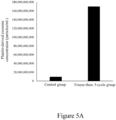
- the experimental results for platelet-derived exosome concentration are shown in Table 4 and Figure 5A .
- the experimental results for total protein content are shown in Table 4 and Figure 5B .
- Data were presented as mean ⁇ SD, and statistical analysis was performed using a t-test. Groups marked with "*" indicated a statistically significant difference compared with the control group (p ⁇ 0.05).
- Table 4 Group Platelet-derived exosome concentration (particles/mL) Average total protein content (g/dL) Control group 9,450,000,000 0.0825 Freeze-thaw 3-cycle group 170,000,000,000 0.1125
- Figure 5A is the bar chart of the effect of freeze-thawing on the concentration of platelet-derived exosomes.
- Figure 5B is the bar chart of the effect of freeze-thawing on total protein amount.
- Experiment 6 Effects of platelet-derived exosome compositions obtained from freeze-thaw activation procedures on alleviating inflammation and tissue repair.
- Experiment 6-1 Effects of platelet-derived exosome compositions obtained from freeze-thaw activation procedures on alleviating inflammation.
- HDFs Human dermal fibroblasts (purchased from ScienCell Research Laboratories, Inc., catalog number: 2320) were cultured in Dulbecco's Modified Eagle Medium (DMEM, Corning 10-013CV) containing 2% fetal bovine serum (FBS), followed by replacing the culture medium with fresh medium for later use.
- DMEM Dulbecco's Modified Eagle Medium
- FBS fetal bovine serum
- Step (S11') of Experiment 1 was performed to prepare a pure platelet solution.
- the platelet concentration in the pure platelet solution was 1 ⁇ 10 9 /mL. Then, the pure platelet solution was divided into control and freeze-thaw 3-cycle group.
- Control group 1 mL of the pure platelet solution was taken, but the activation procedure of step (S12') in Experiment 1 was not performed. Instead, the pure platelet solution was allowed to sit for 20-30 minutes. Then, step (S13') of Experiment 1 was performed to prepare the control group solution.
- the control group solution was mixed with DMEM containing 2% FBS (the volume ratio of the control group solution and DMEM containing 2% FBS was 1:19) to prepare the control group's platelet-derived exosome medium.
- Freeze-thaw 3-cycle group 1 mL of the pure platelet solution was taken, and steps (S12')-(S13') of Experiment 1 were performed to prepare the freeze-thaw 3-cycle group solution (i.e., the purified platelet-derived exosome solution in this application).
- the freeze-thaw 3-cycle group solution was mixed with DMEM containing 2% FBS (the volume ratio of the freeze-thaw 3-cycle group solution and DMEM containing 2% FBS was 1:19) to prepare the three cycles of freezing and thawing group's platelet-derived exosome medium.
- a 96-well plate was divided into untreated wells, LPS control group wells, control group exosome solution wells, and freeze-thaw 3-cycle group exosome solution wells, with two replicates per group.
- HDFs were added to the untreated wells, LPS control group wells, control group exosome solution wells, and freeze-thaw 3-cycle group exosome solution wells, ensuring an equal cell number each well.
- the cells were incubated for 24 hours in a 37°C, 5% carbon dioxide (CO 2 ) incubator.
- the used medium was removed.
- Cells in the LPS control group wells, control group exosome solution wells, and freeze-thaw 3-cycle group exosome solution wells were suspended in DMEM containing 5 ⁇ g/mL LPS and 2% FBS.
- Cells in the untreated wells were suspended in DMEM containing 2% FBS without LPS.
- the cells were incubated for 1 hour in a 37°C, 5% carbon dioxide (CO 2 ) incubator. Subsequently, the following steps were performed separately for each group.
- Untreated wells The used medium was removed. Cells in the untreated wells control wells) were suspended in DMEM containing 2% FBS. The cells were incubated for an additional 24 hours in a 37°C, 5% carbon dioxide (CO 2 ) incubator.
- CO 2 carbon dioxide
- LPS control wells The used medium was removed. Cells in the LPS control wells were suspended in DMEM containing 2% FBS. The cells were incubated for an additional 24 hours in a 37°C, 5% CO 2 incubator.
- Control group exosome solution wells The used medium was removed. Cells in the control group exosome solution wells were suspended in the control group's platelet-derived exosome medium. The cells were incubated for an additional 24 hours in a 37°C, 5% carbon dioxide (CO 2 ) incubator.
- CO 2 carbon dioxide
- Freeze-thaw 3-cycle group exosome solution wells The used medium was removed. Cells in the freeze-thaw 3-cycle group exosome solution wells were suspended in the three cycles of freezing and thawing group's platelet-derived exosome medium. The cells were incubated for an additional 24 hours in a 37°C, 5% carbon dioxide (CO 2 ) incubator.
- CO 2 carbon dioxide
- the cell culture medium from the untreated wells, LPS control wells, control group exosome solution wells, and freeze-thaw 3-cycle group exosome solution wells were transferred into separate microcentrifuge tubes.
- the concentration of human interleukin-6 (human IL-6, an inflammatory factor) in the cell culture medium was measured using an IL-6 ELISA kit (Human IL-6 ELISA Kit, catalog number: ab178013, brand: Abcam).
- the analysis was then performed with a multimode microplate spectrophotometer (model: Varioskan LUX, brand: Thermo Scientific) set to an absorbance wavelength of 450 nm.
- a regression curve of absorbance versus inflammatory factor IL-6 concentration was created based on the instructions of the ELISA kit, and the absorbance values were converted into inflammatory factor IL-6 concentrations.
- freeze-thaw 3-cycle group exosome solution was able to inhibit inflammation in HDFs, thereby alleviating inflammation in skin tissue.
- stimulants such as LPS
- the freeze-thaw 3-cycle group exosome solution can also inhibit the induced inflammatory response, thereby alleviating inflammation in those tissues.
- Experiment 6-2 Effects of platelet-derived exosome compositions obtained from freeze-thaw activation procedures on cell migration.
- HDFs Human dermal fibroblasts identical to those used in Experiment 6-1 were utilized for this experiment. HDFs were cultured in Dulbecco's Modified Eagle Medium (DMEM, Corning 10-013CV) containing 2% fetal bovine serum (FBS), followed by replacing the culture medium with fresh medium for later use.
- DMEM Dulbecco's Modified Eagle Medium
- FBS fetal bovine serum
- a 96-well plate with cell culture inserts was divided into untreated wells, control group exosome solution wells, and freeze-thaw 3-cycle group exosome solution wells, with three replicates per group.
- HDFs were added to the untreated wells, control group exosome solution wells, and freeze-thaw 3-cycle group exosome solution wells, ensuring an equal cell number each well.
- the cells were cultured for 24 hours in a 37°C, 5% carbon dioxide (CO 2 ) incubator.
- the used medium and each group's cell culture inserts were removed, and the plate was documented by taking photographs under a microscope. It was confirmed that the areas originally occupied by the inserts in each group's wells were not covered by cells. Then, the following steps were performed separately for each group.
- Untreated wells Cells were cultured in DMEM containing 2% FBS for an additional 6 hours in a 37°C, 5% carbon dioxide (CO 2 ) incubator.
- Control group exosome solution wells Cells in the control group exosome solution wells were suspended in the control group's platelet-derived exosome medium described in Experiment 6-1. The cells were then incubated for an additional 6 hours in a 37°C, 5% carbon dioxide (CO 2 ) incubator.
- CO 2 carbon dioxide
- Freeze-thaw 3-cycle group exosome solution wells Cells in the freeze-thaw 3-cycle group exosome solution wells were suspended in the three cycles of freezing and thawing group's platelet-derived exosome medium described in Experiment 6-1. The cells were incubated for an additional 6 hours in a 37°C, 5% carbon dioxide (CO 2 ) incubator.
- CO 2 carbon dioxide
- Photographs were taken using a microscope to document the results, and the cell migration coverage area of each group was calculated. Data were presented as mean ⁇ SD, and statistical analysis was performed using a t-test. Groups marked with "***” indicated a statistically significant difference compared with the control group exosome solution (p ⁇ 0.001).
- Figure 6A is the bar chart of the effect of platelet-derived exosome compositions obtained from freeze-thaw activation procedures on the cell migration of human dermal fibroblasts.
- Experiment 6-3 Effects of platelet-derived exosome compositions obtained from freeze-thaw activation procedures on cell proliferation.
- HDFs Human dermal fibroblasts identical to those used in Experiment 6-1 were utilized for this experiment. HDFs were cultured in Dulbecco's Modified Eagle Medium (DMEM, Corning 10-013CV) containing 2% fetal bovine serum (FBS), followed by replacing the culture medium with fresh medium for later use.
- DMEM Dulbecco's Modified Eagle Medium
- FBS fetal bovine serum
- a 96-well plate was divided into untreated wells, control group exosome solution wells, and freeze-thaw 3-cycle group exosome solution wells, with three replicates per group. HDFs were added to the untreated wells, control group exosome solution wells, and freeze-thaw 3-cycle group exosome solution wells, ensuring an equal cell number each well.
- the cells were cultured for 24 hours in a 37°C, 5% carbon dioxide (CO 2 ) incubator. Then, the following steps were performed for each group.
- Untreated wells The used medium was removed. Cells were cultured in DMEM containing 2% FBS for an additional 6 hours in a 37°C, 5% carbon dioxide (CO 2 ) incubator.
- Control group exosome solution wells The used medium was removed. Cells in the control group exosome solution wells were suspended using the control group's platelet-derived exosome medium described in Experiment 6-1. Cells were incubated for an additional 6 hours in a 37°C, 5% carbon dioxide (CO 2 ) incubator.
- CO 2 carbon dioxide
- Freeze-thaw 3-cycle group exosome solution wells Cells in the freeze-thaw 3-cycle group exosome solution wells were suspended using the three cycles freezing and thawing group's platelet-derived exosome medium described in Experiment 6-1. Cells were incubated for an additional 6 hours in a 37°C, 5% carbon dioxide (CO 2 ) incubator.
- CO 2 carbon dioxide
- the used medium was removed.
- 90 ⁇ L of DMEM and 10 ⁇ L of CCK-8 buffer (DOJINDO, catalog number: CK04) were added to each well.
- the plate was incubated for an additional 4 hours in a 37°C, 5% carbon dioxide (CO 2 ) incubator.
- Analysis was performed with a multimode microplate spectrophotometer (model: Varioskan LUX, brand: Thermo Scientific) set to an absorbance wavelength of 450 nm.
- a regression curve of absorbance versus cell numbers was created based on the instructions of the CCK-8 buffer, and the absorbance values were converted into viable cell numbers. Data were presented as mean ⁇ SD, and statistical analysis was performed using a t-test.
- Figure 6B is the bar chart of the effect of platelet-derived exosome compositions obtained from freeze-thaw activation procedures on the cell proliferation of human dermal fibroblasts.
- This experiment was divided into two groups, i.e., the pure platelet and platelet plasma group. Then, the following steps were performed separately for each group.
- Pure platelet group 1 mL of the pure platelet solution described in Experiment 1 was taken, and steps (S12')-(S13') of Experiment 1 were performed to prepare the pure platelet group exosome solution (i.e., the purified platelet-derived exosome solution in this application).
- Platelet plasma group 1 mL of the platelet-rich plasma (PRP) described in Experiment 1 was taken. Then, steps (S12')-(S13') of Experiment 1 were conducted directly to prepare the platelet plasma group exosome solution.
- PRP platelet-rich plasma
- exosome concentration was analyzed using a nanoparticle size analyzer (Tunable Resistive Pulse Sensing (TRPS), model: Exoid, brand: IZON Science). Subsequently, biomarkers of exosomes in the pure platelet group exosome solution and the platelet plasma group exosome solution were individually analyzed using a fully automated exosome fluorescence quantitative analyzer (ExoView, Leprechaun).
- TRPS Resistive Pulse Sensing
- the exosomes in the pure platelet group exosome solution contained exosome markers CD9, CD63, and/or CD81 as well as platelet markers CD41 and CD42b, while they did not possess white blood cell marker CD45 and did not possess at least one of the red blood cell exosome markers CD235a or Annexin V.
- the exosomes in the pure platelet group were 100% platelet-derived.
- the exosomes in the platelet plasma group exosome solution contained at least one of the red blood cell biomarkers CD235a or Annexin V, or the white blood cell biomarker CD45.
- Figure 7 is the bar chart of anallyzing the exosome composition in different biological solutions (platelets and plasma).
- Table 5 Group Platelet-derived exosome concentration (particles/mL) Concentration of exosomes derived from cells other than platelets (particles/mL) Pure platelet group 1,230,000,000,000 0 Platelet plasma group 1,230,000,000,000 1,770,000,000,000
- the concentration of the pure platelet group exosome solution prepared in Experiment 7-1 and the platelet plasma group exosome solution prepared in Experiment 7-1 were adjusted using isotonic sodium chloride solution injection, ensuring that both solutions had the same exosome concentration (the total number of exosomes per unit volume was identical). Then, 1 mL of each solution was taken, and the pure platelet group exosome dry powder and platelet plasma group exosome dry powder were prepared according to step (S14') of Experiment 1.
- HDFs Human dermal fibroblasts identical to those used in Experiment 6-1 were utilized for this experiment. HDFs were cultured in Dulbecco's Modified Eagle Medium (DMEM, Corning 10-013CV) containing 2% fetal bovine serum (FBS), followed by replacing the culture medium with fresh medium for later use.
- DMEM Dulbecco's Modified Eagle Medium
- FBS fetal bovine serum
- a 96-well plate was divided into untreated wells, LPS control group wells, platelet plasma experimental group wells, and purified platelet experimental group wells, with two replicates per group. HDFs were added to the untreated wells, LPS control group wells, platelet plasma experimental group wells, and purified platelet experimental group wells, ensuring an equal cell number each well. The cells were cultured for 24 hours in a 37°C, 5% carbon dioxide (CO 2 ) incubator.
- CO 2 carbon dioxide
- the used medium was removed.
- Cells in the LPS control group wells, platelet plasma experimental group wells, and purified platelet experimental group wells were suspended in DMEM containing 2% FBS and 5 ⁇ g/mL LPS.
- Cells in the untreated wells were suspended in DMEM containing 2% FBS without LPS.
- the cells were incubated for 24 hours in a 37°C, 5% carbon dioxide (CO 2 ) incubator. Then, the following steps were performed separately for each group.
- CO 2 carbon dioxide
- Untreated wells The used medium was removed. Cells were suspended in DMEM containing 2% FBS and incubated for an additional 24 hours in a 37°C, 5% carbon dioxide (CO 2 ) incubator.
- LPS control group wells The used medium was removed. Cells in the LPS control wells were suspended in DMEM containing 2% FBS and incubated for an additional 24 hours in a 37°C, 5% carbon dioxide (CO 2 ) incubator.
- Platelet plasma experimental group wells The platelet plasma group exosome dry powder was reconstituted with 1 mL of DMEM containing 2% FBS to prepare the platelet plasma group's exosome medium. Then, it was mixed with DMEM containing 2% FBS (the volume ratio of the platelet plasma group's exosome medium and DMEM containing 2% FBS was 1:19) to prepare the platelet plasma experimental group's medium. The used medium was removed. Cells in the platelet plasma experimental group wells were suspended using the platelet plasma experimental group's medium and incubated for an additional 24 hours in a 37°C, 5% carbon dioxide (CO 2 ) incubator.
- CO 2 carbon dioxide
- Purified platelet experimental group wells The pure platelet group exosome dry powder was reconstituted with 1 mL of DMEM containing 2% FBS to prepare the purified platelet group's exosome medium. Then, it was mixed with DMEM containing 2% FBS (the volume ratio of the purified platelet group's exosome medium and DMEM containing 2% FBS was 1:19) to prepare the purified platelet experimental group's medium. The used medium was removed. Cells in the purified platelet experimental group wells were suspended using the purified platelet experimental group's medium and incubated for an additional 24 hours in a 37°C, 5% carbon dioxide (CO 2 ) incubator.
- CO 2 carbon dioxide
- Cell culture medium from the untreated wells (control group wells), LPS control group wells, platelet plasma experimental group wells, and purified platelet experimental group wells were transferred into separate microcentrifuge tubes.
- the concentration of human interleukin-6 (human IL-6, an inflammatory factor) in the cell culture medium was measured using an IL-6 ELISA kit (Human IL-6 ELISA Kit, catalog number: ab178013, brand: Abcam). Analysis was then performed with a multimode microplate spectrophotometer (model: Varioskan LUX, brand: Thermo Scientific) set to an absorbance wavelength of 450 nm.
- a regression curve of absorbance versus inflammatory factor IL-6 concentration was created based on the instructions of the ELISA kit, and the absorbance values were converted into inflammatory factor IL-6 concentration.
- Figure 8 is the bar chart of the effects of exosomes in different biological solutions (platelets and plasma) on alleviating inflammation in human dermal fibroblasts.
- Figure 8 shows that the average inflammatory factor release level was 9 pg/mL in the untreated group.
- the average inflammatory factor release level was 396.7 pg/mL in the LPS control group.
- the average inflammatory factor release level was 394.9 pg/mL in the platelet plasma experimental group.
- the average inflammatory factor release level was 261.7 pg/mL in the purified platelet experimental group.
- there was a significant difference in inflammatory factor release level between the purified platelet experimental group and the LPS control group p ⁇ 0.05.
- a significant difference was also observed between the purified platelet experimental group and the platelet plasma experimental group (p ⁇ 0.01). It could be seen that high-purity platelet-derived exosome compositions (i.e., removing exosomes derived from cells other than platelets) could effectively alleviate the inflammation, demonstrating an unexpected effect.
- a purified platelet-derived exosome solution was prepared following steps (S11')-(S13') in Experiment 1.
- HDFs required for this experiment were prepared following the experimental steps of Experiment 6-1.
- a 96-well plate was divided into untreated wells, LPS control group wells, non-freeze-drying group wells, and freeze-drying group wells, with two replicates per group.
- HDFs were added to the untreated wells, LPS control group wells, non-freeze-drying group wells, and freeze-drying group wells, ensuring an equal cell number each well.
- the cells were incubated for 24 hours in a 37°C, 5% carbon dioxide (CO 2 ) incubator.
- CO 2 carbon dioxide
- Untreated wells The used medium was removed. DMEM containing 2% FBS was added to the untreated wells. Cells were incubated for an additional 24 hours in a 37°C, 5% carbon dioxide (CO 2 ) incubator.
- CO 2 carbon dioxide
- LPS control group wells The used medium was removed. DMEM containing 2% FBS was added to the LPS control group wells. Cells were incubated for an additional 24 hours in a 37°C, 5% carbon dioxide (CO 2 ) incubator.
- CO 2 carbon dioxide
- Non-freeze-drying group wells The used medium was removed. 1 mL of purified platelet-derived exosome solution was taken and mixed with DMEM containing 2% FBS (the volume ratio of the purified platelet-derived exosome solution and DMEM containing 2% FBS was 1:19) to obtain a purified platelet-derived exosome medium. The purified platelet-derived exosome medium was then added to the non-freeze-drying group wells. Cells were incubated for an additional 24 hours in a 37°C, 5% carbon dioxide (CO 2 ) incubator.
- CO 2 carbon dioxide
- Freeze-drying group wells The used medium was removed. 1 mL of purified platelet-derived exosome solution was taken, and step (S14') of Experiment 1 was performed to prepare the purified platelet-derived exosome dry powder.
- the purified platelet-derived exosome dry powder was reconstituted with isotonic sodium chloride solution injection to prepare 1 mL of the purified platelet-derived exosome dry powder solution.
- the 1 mL of the purified platelet-derived exosome dry powder solution was mixed with DMEM containing 2% FBS (the volume ratio of the purified platelet-derived exosome dry powder solution and DMEM containing 2% FBS was 1:19) to obtain a purified platelet-derived exosome dry powder medium.
- the purified platelet-derived exosome dry powder medium was added to the freeze-drying group wells. Cells were incubated for an additional 24 hours in a 37°C, 5% carbon dioxide (CO 2 ) incubator.
- Cell culture medium from the untreated wells, LPS control group wells, non-freeze-drying group wells, and freeze-drying group wells were transferred into separate microcentrifuge tubes.
- the concentration of human interleukin-6 (human IL-6, an inflammatory factor) in the cell culture medium was measured using an IL-6 ELISA kit (Human IL-6 ELISA Kit, catalog number: ab178013, brand: Abcam). Analysis was then performed with a multimode microplate spectrophotometer (model: Varioskan LUX, brand: Thermo Scientific) set to an absorbance wavelength of 450 nm.
- a regression curve of absorbance versus inflammatory factor IL-6 concentration was created based on the instructions of the ELISA kit, and the absorbance values were converted into inflammatory factor IL-6 concentration.
- Figure 9 is the bar chart of the effect of the freeze-drying process on the anti-inflammatory efficacy of the product.
- Table 6 Group Average inflammatory factor release level (pg/mL) Untreated group 9 LPS control group 396.7 Non-freeze-drying group 261.7 Freeze-drying group 69.2
- the platelet-derived exosome dry powder obtained through the freeze-drying steps in this application may also suppress these induced inflammatory reactions, thereby alleviating inflammation in these tissues.
- Experiment 9-1 Comparison of platelet-derived exosomes and mesenchymal stromal/stem cell-derived exosomes on cell migration
- MSCs Mesenchymal stromal/stem cells supernatant was purified using a tangential flow filtration (TFF) system to obtain an MSC-derived exosome solution.
- Nanoparticle quantitative analysis was performed using a nanoparticle sizer analyzer. The exosome concentration was then adjusted with isotonic sodium chloride solution injection to achieve an MSC-derived exosome solution comprising 10 7 exosomes per milliliter.
- the purified platelet-derived exosome solution prepared in steps (S11')-(S13') of Experiment 1 was taken. A nanoparticle quantitative analysis of the solution was performed using a nanoparticle analyzer. The exosome concentration was then adjusted with isotonic sodium chloride solution injection to achieve a purified platelet-derived exosome solution comprising 10 7 exosomes per milliliter.
- HDFs Human dermal fibroblasts identical to those used in Experiment 6-1 were utilized for this experiment. HDFs were cultured in Dulbecco's Modified Eagle Medium (DMEM, Corning 10-013CV) containing 2% fetal bovine serum (FBS), followed by replacing the culture medium with fresh medium for later use.
- DMEM Dulbecco's Modified Eagle Medium
- FBS fetal bovine serum
- a 96-well plate with cell culture inserts was divided into untreated wells, freeze-thaw 3-cycle group exosome solution wells, and MSC-derived exosome solution wells, with three replicates per group.
- HDFs were added to the untreated wells, freeze-thaw 3-cycle group exosome solution wells, and MSC-derived exosome solution wells, ensuring an equal cell number each well.
- the cells were cultured for 24 hours in a 37°C, 5% carbon dioxide (CO 2 ) incubator.
- the used medium and each group's cell culture inserts were removed, and the plate was documented by taking photographs under a microscope. Then, the following steps were performed separately for each group.
- Untreated wells DMEM containing 2% FBS was added, and cells were incubated for an additional 6 hours in a 37°C, 5% carbon dioxide (CO 2 ) incubator.
- CO 2 carbon dioxide
- Freeze-thaw 3-cycle group exosome solution wells The purified platelet-derived exosome solution was mixed with DMEM containing 2% FBS (the volume ratio was 1:19) to prepare a purified platelet-derived exosome medium. Then, the purified platelet-derived exosome medium was added to the freeze-thaw 3-cycle group exosome solution wells. Cells were incubated for an additional 6 hours in a 37°C, 5% carbon dioxide (CO 2 ) incubator.
- CO 2 carbon dioxide
- MSC-derived exosome solution wells The MSC-derived exosome solution was mixed with DMEM containing 2% FBS (the volume ratio was 1:19) to prepare a mesenchymal stromal/stem cell (MSC)-derived exosome medium. Then, the MSC-derived exosome medium was added to the MSC-derived exosome solution wells. Cells were incubated for an additional 6 hours in a 37°C, 5% carbon dioxide (CO 2 ) incubator.
- MSC mesenchymal stromal/stem cell
- Photographs were taken using a microscope to document the results, and each group's cell migration coverage area was calculated.
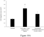
- Figure 10A is the bar chart of the effects of platelet-derived exosomes and mesenchymal stromal/stem cell-derived exosomes on the cell migration of human dermal fibroblasts.
- the average cell migration coverage area in the untreated group was 0.21 mm 2 ; the average cell migration coverage area in the freeze-thaw 3-cycle group exosome solution group was 0.49 mm 2 ; the average cell migration coverage area in the MSC-derived exosome solution group was 0.34 mm 2 .
- There was a significant difference between the freeze-thaw 3-cycle group exosome solution group and the MSC-derived exosome solution group (p ⁇ 0.05). This indicated that the platelet-derived exosome composition in this application exhibited a superior cell migration response compared with the MSC-derived exosomes and was, therefore, more effective in promoting tissue repair.
- a mesenchymal stromal/stem cell (MSC)-derived exosome solution and a purified platelet-derived exosome solution were prepared following the method described in Experiment 9-1.
- the exosome concentration in the mesenchymal stromal/stem cell (MSC)-derived exosome solution was adjusted to 10 6 exosomes/mL
- the exosome concentration in the purified platelet-derived exosome solution was adjusted to 10 6 exosomes/mL
- HDFs Human dermal fibroblasts identical to those used in Experiment 9-1 were utilized for this experiment.
- a 96-well plate was divided into untreated wells, freeze-thaw 3-cycle group exosome solution wells, and MSC-derived exosome solution wells, with three replicates per group.
- HDFs were added to the untreated wells, freeze-thaw 3-cycle group exosome solution wells, and MSC-derived exosome solution wells, ensuring an equal cell number each well.
- the cells were cultured for 24 hours in a 37°C, 5% carbon dioxide (CO 2 ) incubator. Then, the following steps were performed separately for each group.
- CO 2 carbon dioxide
- Untreated wells The used medium was removed. DMEM containing 2% FBS was added to the untreated wells. Cells were incubated for an additional 6 hours in a 37°C, 5% carbon dioxide (CO 2 ) incubator.
- CO 2 carbon dioxide
- Freeze-thaw 3-cycle group exosome solution wells The used medium was removed.
- the purified platelet-derived exosome solution was mixed with DMEM containing 2% FBS (the volume ratio was 1:19) to prepare a purified platelet-derived exosome medium.
- the purified platelet-derived exosome medium was added to the freeze-thaw 3-cycle group exosome solution wells. Cells were incubated for an additional 6 hours in a 37°C, 5% carbon dioxide (CO 2 ) incubator.
- MSC-derived exosome solution wells The used medium was removed. The MSC-derived exosome solution was mixed with DMEM containing 2% FBS (the volume ratio was 1:19) to prepare a mesenchymal stromal/stem cell (MSC)-derived exosome medium. Then, the MSC-derived exosome medium was added to the MSC-derived exosome solution wells. Cells were incubated for an additional 6 hours in a 37°C, 5% carbon dioxide (CO 2 ) incubator.
- MSC mesenchymal stromal/stem cell
- the average cell number in the untreated group was 17,010; the average cell number in the freeze-thaw 3-cycle group exosome solution group was 20,157; the average cell number in the MSC-derived exosome solution group was 17,972.
- the average cell number in the freeze-thaw 3-cycle group exosome solution group was higher than the average cell number in the MSC-derived exosome solution group.
- a mesenchymal stromal/stem cell (MSC)-derived exosome solution and a purified platelet-derived exosome solution were prepared following the method described in Experiment 9-1.
- HDFs Human dermal fibroblasts identical to those used in Experiment 9-1 were utilized for this experiment.
- a 96-well plate was divided into untreated wells, freeze-thaw 3-cycle group exosome solution wells, and MSC-derived exosome solution wells, with three replicates per group.
- HDFs were added to the untreated wells, freeze-thaw 3-cycle group exosome solution wells, and MSC-derived exosome solution wells, ensuring an equal cell number each well.
- the cells were cultured for 24 hours in a 37°C, 5% carbon dioxide (CO 2 ) incubator. Then, the following steps were performed separately for each group.
- CO 2 carbon dioxide
- Untreated wells The used medium was removed. DMEM containing 2% FBS was added to the untreated wells. Cells were incubated for an additional 24 hours in a 37°C, 5% carbon dioxide (CO 2 ) incubator.
- CO 2 carbon dioxide
- Freeze-thaw 3-cycle group exosome solution wells The used medium was removed.
- the purified platelet-derived exosome solution was mixed with DMEM containing 2% FBS (the volume ratio was 1:19) to prepare a purified platelet-derived exosome medium.
- the purified platelet-derived exosome medium was added to the freeze-thaw 3-cycle group exosome solution wells. Cells were incubated for an additional 24 hours in a 37°C, 5% carbon dioxide (CO 2 ) incubator.
- MSC-derived exosome solution wells The used medium was removed. The MSC-derived exosome solution was mixed with DMEM containing 2% FBS (the volume ratio was 1:19) to prepare a mesenchymal stromal/stem cell (MSC)-derived exosome medium. Then, the MSC-derived exosome medium was added to the MSC-derived exosome solution wells. Cells were incubated for an additional 24 hours in a 37°C, 5% carbon dioxide (CO 2 ) incubator.
- MSC mesenchymal stromal/stem cell
- Figure 10C is the bar chart of the effects of platelet-derived exosomes and mesenchymal stromal/stem cell-derived exosomes on the collagen expression in human dermal fibroblasts.
- the average collagen expression level was 22,617.67 pg/mL in the untreated group; the average collagen expression level was 43,311.33 pg/mL in the freeze-thaw 3-cycle group exosome solution group; the average collagen expression level was 38,317.67 pg/mL in the MSC-derived exosome solution group.
- the average collagen expression level in the freeze-thaw 3-cycle group exosome solution group was higher than the average collagen expression level in the MSC-derived exosome solution group, with a significant difference (p ⁇ 0.01). This indicated that the platelet-derived exosome composition in this application had a tissue repair-promoting effect, and the effect was superior to that of MSC-derived exosomes.
Landscapes
- Health & Medical Sciences (AREA)
- Life Sciences & Earth Sciences (AREA)
- Engineering & Computer Science (AREA)
- General Health & Medical Sciences (AREA)
- Veterinary Medicine (AREA)
- Public Health (AREA)
- Animal Behavior & Ethology (AREA)
- Chemical & Material Sciences (AREA)
- Medicinal Chemistry (AREA)
- Pharmacology & Pharmacy (AREA)
- Bioinformatics & Cheminformatics (AREA)
- Zoology (AREA)
- Epidemiology (AREA)
- Immunology (AREA)
- Hematology (AREA)
- Biomedical Technology (AREA)
- Gastroenterology & Hepatology (AREA)
- Proteomics, Peptides & Aminoacids (AREA)
- Organic Chemistry (AREA)
- Cell Biology (AREA)
- Biotechnology (AREA)
- Vascular Medicine (AREA)
- Heart & Thoracic Surgery (AREA)
- General Chemical & Material Sciences (AREA)
- Chemical Kinetics & Catalysis (AREA)
- Virology (AREA)
- Nuclear Medicine, Radiotherapy & Molecular Imaging (AREA)
- Developmental Biology & Embryology (AREA)
- Molecular Biology (AREA)
- Anesthesiology (AREA)
- Wood Science & Technology (AREA)
- Genetics & Genomics (AREA)
- Pain & Pain Management (AREA)
- Rheumatology (AREA)
- Endocrinology (AREA)
- Dermatology (AREA)
- Diabetes (AREA)
- Botany (AREA)
- Biophysics (AREA)
- Cardiology (AREA)
Applications Claiming Priority (1)
| Application Number | Priority Date | Filing Date | Title |
|---|---|---|---|
| US202363608323P | 2023-12-11 | 2023-12-11 |
Publications (1)
| Publication Number | Publication Date |
|---|---|
| EP4570257A1 true EP4570257A1 (fr) | 2025-06-18 |
Family
ID=94382497
Family Applications (2)
| Application Number | Title | Priority Date | Filing Date |
|---|---|---|---|
| EP24218839.9A Pending EP4570257A1 (fr) | 2023-12-11 | 2024-12-10 | Utilisation d'une poudre sèche d'exosomes dérivés de plaquettes purifiées pour soulager une inflammation ou une lésion |
| EP24218844.9A Pending EP4570258A1 (fr) | 2023-12-11 | 2024-12-10 | Utilisation d'un poudre séchée pour le traitment de l'inflammation ou de blessures |
Family Applications After (1)
| Application Number | Title | Priority Date | Filing Date |
|---|---|---|---|
| EP24218844.9A Pending EP4570258A1 (fr) | 2023-12-11 | 2024-12-10 | Utilisation d'un poudre séchée pour le traitment de l'inflammation ou de blessures |
Country Status (4)
| Country | Link |
|---|---|
| US (2) | US20250186499A1 (fr) |
| EP (2) | EP4570257A1 (fr) |
| JP (2) | JP2025106200A (fr) |
| TW (2) | TW202526005A (fr) |
Cited By (1)
| Publication number | Priority date | Publication date | Assignee | Title |
|---|---|---|---|---|
| EP4620478A1 (fr) * | 2024-03-22 | 2025-09-24 | Spirit Scientific Co. LTD. | Composition pharmaceutique comprenant des vésicules extracellulaires dérivées de plaquettes de différentes tailles activées par des cycles de congélation-décongélation, son procédé de préparation et son utilisation pour favoriser la cicatrisation des tissus liés aux voies respiratoires et atténuer l'inflammation |
Families Citing this family (1)
| Publication number | Priority date | Publication date | Assignee | Title |
|---|---|---|---|---|
| WO2025184489A1 (fr) | 2024-02-29 | 2025-09-04 | Consano Bio, Inc. | Produits allogéniques dérivés de plasma et de plaquettes humains et leurs utilisations |
Citations (4)
| Publication number | Priority date | Publication date | Assignee | Title |
|---|---|---|---|---|
| US20220079987A1 (en) * | 2019-01-04 | 2022-03-17 | Direct Biologics Llc | Isolation and use of autogenous platelet derived exosomes |
| US11360097B2 (en) * | 2015-11-12 | 2022-06-14 | Edward J Goetzl | Platelet biomarkers and diagnostic methods for vascular diseases |
| CN115029312B (zh) * | 2022-08-11 | 2022-11-18 | 广东先康达生物科技有限公司 | 一种血小板外泌体的提取方法 |
| EP4292598A1 (fr) * | 2022-06-16 | 2023-12-20 | Spirit Scientific Co. LTD. | Poudre sèche enrichies en facteurs de croissance et son utilisation pour soulager l'inflammation ou des lésions |
Family Cites Families (6)
| Publication number | Priority date | Publication date | Assignee | Title |
|---|---|---|---|---|
| US4897252A (en) * | 1989-01-10 | 1990-01-30 | Arco Chemical Technology, Inc. | Production of hydrogen peroxide |
| NZ509122A (en) * | 1998-06-22 | 2003-09-26 | Cytomedix Inc | Wound healant comprising activated growth from platelets and ascorbic acid |
| EP3254684B1 (fr) * | 2016-06-08 | 2019-10-23 | Lysatpharma GmbH | Lysat de plaquettes humaines ou fraction enrichie de vésicules extracellulaires dérivées de lysat de plaquettes humaines pour utilisation medicale |
| CN108715834B (zh) * | 2018-06-01 | 2021-09-14 | 天晴干细胞股份有限公司 | 一种富含cd41+、cd81+微囊的血小板裂解液制备方法 |
| JPWO2023079655A1 (fr) * | 2021-11-04 | 2023-05-11 | ||
| CN115040539A (zh) * | 2022-07-25 | 2022-09-13 | 重庆市急救医疗中心(重庆市第四人民医院 重庆市急救医学研究所) | 一种优化的血小板外泌体冻干粉在糖尿病足溃疡中的应用及其制备方法 |
-
2024
- 2024-12-10 EP EP24218839.9A patent/EP4570257A1/fr active Pending
- 2024-12-10 US US18/975,685 patent/US20250186499A1/en active Pending
- 2024-12-10 US US18/976,087 patent/US20250186500A1/en active Pending
- 2024-12-10 EP EP24218844.9A patent/EP4570258A1/fr active Pending
- 2024-12-11 TW TW113148047A patent/TW202526005A/zh unknown
- 2024-12-11 JP JP2024216274A patent/JP2025106200A/ja active Pending
- 2024-12-11 JP JP2024216278A patent/JP2025108372A/ja active Pending
- 2024-12-11 TW TW113148048A patent/TW202526006A/zh unknown
Patent Citations (4)
| Publication number | Priority date | Publication date | Assignee | Title |
|---|---|---|---|---|
| US11360097B2 (en) * | 2015-11-12 | 2022-06-14 | Edward J Goetzl | Platelet biomarkers and diagnostic methods for vascular diseases |
| US20220079987A1 (en) * | 2019-01-04 | 2022-03-17 | Direct Biologics Llc | Isolation and use of autogenous platelet derived exosomes |
| EP4292598A1 (fr) * | 2022-06-16 | 2023-12-20 | Spirit Scientific Co. LTD. | Poudre sèche enrichies en facteurs de croissance et son utilisation pour soulager l'inflammation ou des lésions |
| CN115029312B (zh) * | 2022-08-11 | 2022-11-18 | 广东先康达生物科技有限公司 | 一种血小板外泌体的提取方法 |
Non-Patent Citations (3)
| Title |
|---|
| E. J. GOETZL ET AL: "Human plasma platelet-derived exosomes: effects of aspirin", THE FASEB JOURNAL, vol. 30, no. 5, 1 May 2016 (2016-05-01), US, pages 2058 - 2063, XP055384123, ISSN: 0892-6638, DOI: 10.1096/fj.201500150R * |
| LU JINWEI ET AL: "Rejuvenation of tendon stem/progenitor cells for functional tendon regeneration through platelet-derived exosomes loaded with recombinant Yap1", ACTA BIOMATERIALIA, ELSEVIER, AMSTERDAM, NL, vol. 161, 17 February 2023 (2023-02-17), pages 80 - 99, XP087290517, ISSN: 1742-7061, [retrieved on 20230217], DOI: 10.1016/J.ACTBIO.2023.02.018 * |
| SAFFARI SARA ET AL: "Dose-response analysis after administration of a human platelet-derived exosome product on neurite outgrowth in vitro", BIOTECHNOLOGY AND BIOENGINEERING, vol. 120, no. 11, 4 August 2023 (2023-08-04), Hoboken, USA, pages 3191 - 3199, XP093155447, ISSN: 0006-3592, DOI: 10.1002/bit.28520 * |
Cited By (1)
| Publication number | Priority date | Publication date | Assignee | Title |
|---|---|---|---|---|
| EP4620478A1 (fr) * | 2024-03-22 | 2025-09-24 | Spirit Scientific Co. LTD. | Composition pharmaceutique comprenant des vésicules extracellulaires dérivées de plaquettes de différentes tailles activées par des cycles de congélation-décongélation, son procédé de préparation et son utilisation pour favoriser la cicatrisation des tissus liés aux voies respiratoires et atténuer l'inflammation |
Also Published As
| Publication number | Publication date |
|---|---|
| JP2025108372A (ja) | 2025-07-23 |
| EP4570258A1 (fr) | 2025-06-18 |
| TW202526005A (zh) | 2025-07-01 |
| JP2025106200A (ja) | 2025-07-15 |
| US20250186499A1 (en) | 2025-06-12 |
| TW202526006A (zh) | 2025-07-01 |
| US20250186500A1 (en) | 2025-06-12 |
Similar Documents
| Publication | Publication Date | Title |
|---|---|---|
| US20250186500A1 (en) | Use of a purified platelet-derived exosome dry powder for relieving inflammation or injury | |
| US10111906B2 (en) | Serum fraction of platelet-rich fibrin | |
| Kececi et al. | A cost-effective method for obtaining standard platelet-rich plasma | |
| US8501170B2 (en) | Platelet fraction deriving from placental blood | |
| TWI873495B (zh) | 一種富含生長因子的乾粉用於緩解發炎或損傷之用途 | |
| WO2021180237A1 (fr) | Formulation d'inhalation atomisée contenant des vésicules extracellulaires dérivées de cellules humaines, son procédé de préparation et son utilisation | |
| EP4292585A2 (fr) | Méthodes d'isolement de produits de plasma sanguin de cordon ombilical, d'exosomes tissulaires et cellulaires, ainsi que compositions et leurs méthodes d'utilisation | |
| WO2021009660A1 (fr) | Composition et procédés pour améliorer l'épaisseur et la réceptivité de la doublure endométriale | |
| KR20230074475A (ko) | 골수-유래 중간엽 줄기 세포로부터의 미세소포를 사용한 치료의 조성물 및 방법 | |
| EP3881858A1 (fr) | Extrait de facteur de croissance dérivé de plaquettes traité à la chaleur destiné à être utilisé dans un procédé de prévention ou de traitement de défaut d'un tissu | |
| JP7651800B2 (ja) | 成長因子を豊富に含む乾燥粉末が炎症または損傷を緩和する一種の用途 | |
| EP4292598A1 (fr) | Poudre sèche enrichies en facteurs de croissance et son utilisation pour soulager l'inflammation ou des lésions | |
| KR20150061806A (ko) | 활성화된 혈소판 풍부 혈장을 유효성분으로 포함하는 동물의 창상 치료용 수의학적 조성물 | |
| CN114931589A (zh) | 一种富血小板血浆来源外泌体产品的制备方法和应用 | |
| TWI856429B (zh) | 增強富含血小板血漿及其製備方法與用途 | |
| US11965179B1 (en) | Laser treated platelet product | |
| CN115531521B (zh) | 细胞治疗剂及其制法和用途 | |
| EP4622653A1 (fr) | Compositions à base d'un mélange de molécules bioactives et d'exosomes destinées à être utilisées dans le traitement d'états nécessitant une réparation et une régénération de tissu | |
| HK40104400A (en) | Methods for isolating umbilical cord blood plasma products, tissue and cellular exosomes, and compositions and methods of use thereof | |
| WO2025032586A1 (fr) | Plasma acellulaire et ses utilisations pour la régénération tissulaire | |
| WO2022181798A1 (fr) | Procédé de préparation d'un mélange de facteurs de croissance | |
| CN121944041A (zh) | 一种具有神经修复作用的中药复方组合物、细胞培养基和中药-神经干细胞外泌体复合物及应用 | |
| CN120771101A (zh) | 一种细胞外囊泡凝胶制剂以及用于其的温敏性凝胶溶液 |
Legal Events
| Date | Code | Title | Description |
|---|---|---|---|
| PUAI | Public reference made under article 153(3) epc to a published international application that has entered the european phase |
Free format text: ORIGINAL CODE: 0009012 |
|
| STAA | Information on the status of an ep patent application or granted ep patent |
Free format text: STATUS: THE APPLICATION HAS BEEN PUBLISHED |
|
| AK | Designated contracting states |
Kind code of ref document: A1 Designated state(s): AL AT BE BG CH CY CZ DE DK EE ES FI FR GB GR HR HU IE IS IT LI LT LU LV MC ME MK MT NL NO PL PT RO RS SE SI SK SM TR |
|
| STAA | Information on the status of an ep patent application or granted ep patent |
Free format text: STATUS: REQUEST FOR EXAMINATION WAS MADE |
|
| 17P | Request for examination filed |
Effective date: 20251216 |