EP4574015A1 - Test d'impulsion de tête de vision fonctionnelle - Google Patents
Test d'impulsion de tête de vision fonctionnelle Download PDFInfo
- Publication number
- EP4574015A1 EP4574015A1 EP24220971.6A EP24220971A EP4574015A1 EP 4574015 A1 EP4574015 A1 EP 4574015A1 EP 24220971 A EP24220971 A EP 24220971A EP 4574015 A1 EP4574015 A1 EP 4574015A1
- Authority
- EP
- European Patent Office
- Prior art keywords
- test
- image
- property
- test subject
- user input
- Prior art date
- Legal status (The legal status is an assumption and is not a legal conclusion. Google has not performed a legal analysis and makes no representation as to the accuracy of the status listed.)
- Pending
Links
Images




Classifications
-
- A—HUMAN NECESSITIES
- A61—MEDICAL OR VETERINARY SCIENCE; HYGIENE
- A61B—DIAGNOSIS; SURGERY; IDENTIFICATION
- A61B5/00—Measuring for diagnostic purposes; Identification of persons
- A61B5/40—Detecting, measuring or recording for evaluating the nervous system
- A61B5/4005—Detecting, measuring or recording for evaluating the nervous system for evaluating the sensory system
- A61B5/4023—Evaluating sense of balance
-
- A—HUMAN NECESSITIES
- A61—MEDICAL OR VETERINARY SCIENCE; HYGIENE
- A61B—DIAGNOSIS; SURGERY; IDENTIFICATION
- A61B3/00—Apparatus for testing the eyes; Instruments for examining the eyes
- A61B3/0016—Operational features thereof
- A61B3/0041—Operational features thereof characterised by display arrangements
- A61B3/005—Constructional features of the display
-
- A—HUMAN NECESSITIES
- A61—MEDICAL OR VETERINARY SCIENCE; HYGIENE
- A61B—DIAGNOSIS; SURGERY; IDENTIFICATION
- A61B3/00—Apparatus for testing the eyes; Instruments for examining the eyes
- A61B3/0075—Apparatus for testing the eyes; Instruments for examining the eyes provided with adjusting devices, e.g. operated by control lever
-
- A—HUMAN NECESSITIES
- A61—MEDICAL OR VETERINARY SCIENCE; HYGIENE
- A61B—DIAGNOSIS; SURGERY; IDENTIFICATION
- A61B3/00—Apparatus for testing the eyes; Instruments for examining the eyes
- A61B3/02—Subjective types, i.e. testing apparatus requiring the active assistance of the patient
-
- A—HUMAN NECESSITIES
- A61—MEDICAL OR VETERINARY SCIENCE; HYGIENE
- A61B—DIAGNOSIS; SURGERY; IDENTIFICATION
- A61B5/00—Measuring for diagnostic purposes; Identification of persons
- A61B5/103—Measuring devices for testing the shape, pattern, colour, size or movement of the body or parts thereof, for diagnostic purposes
- A61B5/11—Measuring movement of the entire body or parts thereof, e.g. head or hand tremor or mobility of a limb
-
- A—HUMAN NECESSITIES
- A61—MEDICAL OR VETERINARY SCIENCE; HYGIENE
- A61B—DIAGNOSIS; SURGERY; IDENTIFICATION
- A61B5/00—Measuring for diagnostic purposes; Identification of persons
- A61B5/103—Measuring devices for testing the shape, pattern, colour, size or movement of the body or parts thereof, for diagnostic purposes
- A61B5/11—Measuring movement of the entire body or parts thereof, e.g. head or hand tremor or mobility of a limb
- A61B5/1113—Local tracking of patients, e.g. in a hospital or private home
- A61B5/1114—Tracking parts of the body
-
- A—HUMAN NECESSITIES
- A61—MEDICAL OR VETERINARY SCIENCE; HYGIENE
- A61B—DIAGNOSIS; SURGERY; IDENTIFICATION
- A61B5/00—Measuring for diagnostic purposes; Identification of persons
- A61B5/16—Devices for psychotechnics; Testing reaction times ; Devices for evaluating the psychological state
- A61B5/163—Devices for psychotechnics; Testing reaction times ; Devices for evaluating the psychological state by tracking eye movement, gaze, or pupil change
-
- A—HUMAN NECESSITIES
- A61—MEDICAL OR VETERINARY SCIENCE; HYGIENE
- A61B—DIAGNOSIS; SURGERY; IDENTIFICATION
- A61B5/00—Measuring for diagnostic purposes; Identification of persons
- A61B5/48—Other medical applications
- A61B5/4863—Measuring or inducing nystagmus
-
- A—HUMAN NECESSITIES
- A61—MEDICAL OR VETERINARY SCIENCE; HYGIENE
- A61B—DIAGNOSIS; SURGERY; IDENTIFICATION
- A61B5/00—Measuring for diagnostic purposes; Identification of persons
- A61B5/74—Details of notification to user or communication with user or patient; User input means
- A61B5/742—Details of notification to user or communication with user or patient; User input means using visual displays
-
- A—HUMAN NECESSITIES
- A61—MEDICAL OR VETERINARY SCIENCE; HYGIENE
- A61B—DIAGNOSIS; SURGERY; IDENTIFICATION
- A61B5/00—Measuring for diagnostic purposes; Identification of persons
- A61B5/74—Details of notification to user or communication with user or patient; User input means
- A61B5/7475—User input or interface means, e.g. keyboard, pointing device, joystick
-
- A—HUMAN NECESSITIES
- A61—MEDICAL OR VETERINARY SCIENCE; HYGIENE
- A61B—DIAGNOSIS; SURGERY; IDENTIFICATION
- A61B2562/00—Details of sensors; Constructional details of sensor housings or probes; Accessories for sensors
- A61B2562/02—Details of sensors specially adapted for in-vivo measurements
- A61B2562/0219—Inertial sensors, e.g. accelerometers, gyroscopes, tilt switches
Definitions
- the present application relates to a system for evaluating the function of the vestibular system of a test subject.
- the present application further relates to a method of evaluating the function of the vestibular system of a test subject.
- the present application further relates to a data processing system comprising a processing device and program code means for causing the processing device to perform at least some of the steps of the method.
- the present application further relates to a computer program comprising instructions which, when the program is executed by a system, cause the system to carry out the steps of the method.
- the Vestibular Ocular Reflex is an eye movement that allows the gaze of the eyes of a test subject (i.e., of a patient) to stay focused on a target during head movements.
- the VOR can be assessed with different techniques.
- the techniques include video goggles with camera(s) and accelerometer(s) attached, to measure both eye velocity and head velocity. From this a gain can be determined, which is eye velocity divided by head velocity.
- Other techniques use only a head mounted accelerometer to measure head velocity and rely on the test subject's responses to the presence of an Optotype on a screen in front of the test subject to measure visual acuity.
- Visual acuity is a functional measurement whereas gain is an objective mathematical measurement.
- VOR tests/techniques are to evaluate the function of the vestibular system (i.e., comprising vestibular semicircular canals and associated vestibular-ocular reflex (VOR)).
- Peripheral vestibular canal function may be tested with two types of measurements (1) "conventional" video head impulse tests (vHIT) measure angular VOR (aVOR) gain - estimates reduced gain and corrective eye movement, and (2) more recently functional gain is assessed using visual acuity as a measure of how well the aVOR performs in stabilizing gaze during head movements.
- vHIT is performed passively, as the clinician (i.e., the user operating the system) moves the head.
- Functional tests have traditionally been passive also where the clinician moves the test subject's head.
- the time delay may represent the latency of the system, e.g., of the graphics part of the system, such as in the software handling the display of the image and in the display device.
- the step of presenting said image, characterised by a property, for a pre-set time interval by said display device starting from the determined point in time with maximum acceleration value may comprise:
- the processing device may be configured to score the percentage of correct estimates (i.e., from the test subject) of said property at different accelerations of rotation of the head of the test subject in each respective plane.
- the clinician may want to repeat the test protocol. This may be initiated in response to the clinician providing a user input, e.g., instructions/control signal to repeat the test protocol.
- the processing device may be configured to execute one of the plurality of test protocols in response to receiving an instruction to execute a specific one of the plurality of test protocols via the user input device.
- test protocol may be executed when, and if one or more test protocols should be repeated.
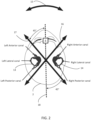
- the at least one plane may comprise a lateral plane.
- the at least one plane may comprise a Right Anterior, Left Posterior (RALP) plane.
- RALP Right Anterior, Left Posterior
- the at least one plane may comprise a Left Anterior, Right Posterior (LARP) plane.
- LRP Left Anterior, Right Posterior
- the at least one plane may comprise a vertical plane.
- the lateral plane may refer to a head rotation in a horizontal plane.
- the test subject's head may be bowed forward by approximately 30°.
- the horizontal canals of each ear (which are a co-planar pair) are stimulated the best.
- the LARP plane may refer to a head rotation in an orientation which best stimulates the Left Anterior, Right Posterior co-planar pair.
- the motion sensing device may be configured to be attached to the eye measurement device.
- the motion sensing device may be releasably attached to the eye measurement device.
- the motion sensing device may be attached before execution of a specific test protocol, and be detached again afterwards if not relevant for the following test protocol.
- the motion sensing device may be arranged at the head of the test subject by use of a headband, and thereby not being directly connected to the eye measurement device.
- the motion sensing device may comprise a single motion sensor, such as an accelerometer, which may be located at a central position of the eye measurement device, such as at the forehead of test subject.
- the motion sensing device may comprise two or more motion sensors, such as two accelerometers, which may be located e.g., near each of the eyes of the test subject.
- the display device may comprise a refresh rate of at least 240 Hz.
- Refresh rate may refer to a scan rate of the display device, such as a scan rate of a monitor.
- the refresh rate may be even higher, as this will only increase the reliability of displaying of the image at the correct moment.
- the user input device may comprise a remote control.
- a remote control may make it easy for the test subject to provide his/her estimate of the property of the image.
- the user input device may comprise a keyboard.
- the user input device may comprise a touch screen.
- the processing device may be configured to execute at least one of a plurality of test protocols.
- the plurality of test protocols may comprise the AHR test protocol.
- the system may comprise the eye measurement device, e.g., a goggle with a camera configured to record at least one of the eyes of the test subject.
- the system may further comprise a motion sensing device, e.g., an accelerometer worn on the head of the test subject.
- the motion sensing device may be attached to the eye measurement device.
- the system may further comprise a processing device configured to execute the AHR test protocol.
- the AHR test protocol may comprise (at a plurality of head rotation velocity values):
- the head may be rotated at multiple different frequencies.
- the frequencies may e.g., be 1-3 Hz or higher.
- the AHR test protocol may comprise that the user/clinician moves the test subject's head left or right with a controlled sinusoidal movement timed with a metronome.
- a fixed image may be displayed by the display device.
- the processing device may measure gain which is eye velocity divided by head velocity (and also phase and asymmetry) at different head rotation frequencies.
- the plurality of test protocols may comprise the vHIT test protocol.
- the system may comprise the eye measurement device, e.g., a goggle with a camera recording at least one of the eyes of the test subject.
- the system may further comprise a motion sensing device, e.g., an accelerometer worn at the head of the test subject.
- the motion sensing device may be attached to the eye measurement device.
- the system may further comprise a processing device configured to execute the vHIT test protocol.
- the vHIT test protocol may comprise:
- the head may be moved in a fast impulse movement.
- the fast impulse movement may be +/- 15 degrees relative to when the head is at a resting position.
- the vHIT test protocol may comprise that the user/clinician quickly moves the test subject's head left or right in the lateral plane with a high-speed impulse movement, or in the RALP plane or LARP plane.
- a fixed image may be displayed by the display device.
- the processing device may measure gain which is eye velocity value divided with head velocity value.
- the plurality of test protocols may comprise the DVA (or DVAT) test protocol.
- the system may comprise a motion sensing device, e.g., an accelerometer worn at the head of the test subject.
- the system may further comprise a processing device configured to execute the DVA test protocol.
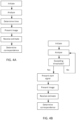
- the DVA test protocol may comprise:
- the DVA test protocol may further comprise:
- the threshold velocity value may be at least 50°/s and/or less than 300°/s.
- the threshold velocity value may be 100°/s.
- the DVA test protocol may comprise that the user/clinician moves the test subject's head left and right (in the horizontal plane) or up and down (in the vertical plane) with a controlled sinusoidal movement timed with a metronome.
- the test subject may view an image, e.g., an optotype, that appears on the display device when a certain head velocity is reached, and the test subject may in response tell the direction the optotype is pointing.
- the processing device may in response determine (e.g., keep score of) the change in optotype size that is needed for the test subject to clearly see the optotype while the head is at maximum velocity and compare this determined optotype size to the optotype size needed to see it clearly when the head is not moving.
- the needed optotype size, when the head is not moved may be determined by reducing the optotype in size until a threshold is found.
- the system may comprise a motion sensing device, e.g., an accelerometer worn at the head of the test subject, and a display device configured to display at least one image.
- the system may further comprise a processing device configured to execute the GST test protocol.
- the GST test protocol may comprise:
- the GST test protocol may further comprise:
- the GST test protocol may comprise that the user/clinician/examiner moves the test subject's head left and right (in the horizontal plane) or up and down (in the vertical plane) with a controlled sinusoidal movement timed with a metronome at 50-300°/s.
- the test subject may view an image, e.g., an optotype, that appears on the display device when a certain velocity value (head speed) is reached, and the test subject may in response tell the direction the image is pointing.
- the processing device e.g., the software
- the plurality of test protocols may comprise a VPT test protocol.
- the visual focus may be determined based on the recorded orientation of at least one of the eyes of the test subject.
- Recorded orientation may refer to the orientation of the eyes of the test subject relative to the display device.
- the system may further comprise a timer.
- the system may be configured to time the duration from presenting an image until receiving a user input.
- the system may be configured to implement a (predetermined) time interval from presenting said image until the processing device presents a predetermined response.
- the predetermined response may indicate that no response will be provided, such as "I don't know", so that the user does not have to select an answer on the computer or through the remote.
- the system may be configured to record a "no response" user input, in case the user does not provide a user input within the (predetermined) time interval.
- the (predetermined) time interval may be 5 seconds, or more.
- the system may be configured to provide (real time) feedback to the user during execution of at least one of said plurality of test protocols.
- said feedback may be provided on a test screen, e.g., which may be visible to the user/operator of the system (e.g., an examiner and/or a clinician).
- a test screen e.g., which may be visible to the user/operator of the system (e.g., an examiner and/or a clinician).
- the feedback may be represented by the acceleration bins at the top of the test screen (e.g., an additional display device).
- the system may comprise a room recording camera (e.g., an external camera attached to a computer (or built into the computer) of the system).
- a room recording camera e.g., an external camera attached to a computer (or built into the computer) of the system.
- the system may be configured to record the patient during the test protocol, via said room recording camera. This may be beneficial to see head movements through video recording and match with test findings/evaluation.
- the system may be configured to record audio during the test protocol, via said room recording camera. Thereby, audio for patient reactions/symptoms noted during the test may be recorded.
- the eye measurement device may comprise a camera for recording said at least one eye of the test subject.
- system further comprising an auxiliary device is moreover provided.
- the system may be adapted to establish a communication link between the elements of the system (e.g., the eye measurement device, the motion sensing device, the display device, the user input device, the processing device, the auxiliary device, etc.) to provide that information (e.g., control and/or status signals, motion signals, start signals, etc.) can be exchanged or forwarded from one to the other.
- the elements of the system e.g., the eye measurement device, the motion sensing device, the display device, the user input device, the processing device, the auxiliary device, etc.
- information e.g., control and/or status signals, motion signals, start signals, etc.
- the auxiliary device may comprise a remote control, a mobile device, a smartphone, or other portable electronic device, such as a tablet or the like, or may comprise a server device, etc.
- the communication link may be a wired or wireless link, e.g., based on Bluetooth or some other standardized scheme.
- a method of evaluating the function of the vestibular system of a test subject is furthermore provided by the present application.
- the method may comprise executing at least one of a plurality of test protocols by a processing device.
- One test protocol (of the plurality of test protocols) may comprise:
- the one test protocol may comprise:
- the one test protocol may comprise presenting an image, characterised by a property, for a pre-set time interval by a display device starting from the determined point in time with maximum acceleration value.
- the one test protocol may comprise:
- the method may further comprise determining a correspondence between said property and said estimate of said property.
- the method of evaluating the function of the vestibular system of the test subject may comprise executing at least one of a plurality of test protocols by the processing device, where one test protocol comprises the AHR test protocol (at a plurality of head rotation velocity values):
- the method may further comprise:
- the head may be rotated at multiple different frequencies.
- the frequencies may e.g., be 1-3 Hz or higher.
- the AHR test protocol may comprise that the user/clinician moves the test subject's head left or right with a controlled sinusoidal movement timed with a metronome.
- a fixed image may be displayed by the display device.
- the processing device may measure gain which is eye velocity divided by head velocity (and also phase and asymmetry) at different head rotation frequencies.
- the method of evaluating the function of the vestibular system of the test subject may comprise executing at least one of a plurality of test protocols by the processing device, where one test protocol comprises the vHIT test protocol:
- the method may further be characterised by head movements of high velocity, low amplitude and being unexpected.
- the head may be moved in a fast impulse movement.
- the fast impulse movement may be +/- 15 degrees relative to when the head is at a resting position.
- the vHIT test protocol may comprise that the user/clinician quickly moves the test subject's head left or right in the lateral plane with a high-speed impulse movement, or in the RALP plane or LARP plane.
- a fixed image may be displayed by the display device.
- the processing device may measure gain which is eye velocity value divided with head velocity value.
- the method of evaluating the function of the vestibular system of the test subject may comprise executing at least one of a plurality of test protocols by the processing device, where one test protocol comprises the DVA test protocol:
- the DVA test protocol may further comprise:
- the method may further comprise:
- the threshold velocity value may be at least 50°/s and/or less than 300°/s.
- the threshold velocity value may be 100°/s.
- the DVA test protocol may comprise that the user/clinician moves the test subject's head left and right (in the horizontal plane) or up and down (in the vertical plane) with a controlled sinusoidal movement timed with a metronome.
- the test subject may view an image, e.g., an optotype, that appears on the display device when a certain head velocity is reached, and the test subject may in response tell the direction the optotype is pointing.
- the processing device may in response determine (e.g., keep score of) the change in optotype size that is needed for the test subject to clearly see the optotype while the head is at maximum velocity and compare this determined optotype size to the optotype size needed to see it clearly when the head is not moving.
- the needed optotype size, when the head is not moving may be determined by reducing the optotype in size until a threshold is found.
- the method of evaluating the function of the vestibular system of the test subject may comprise executing at least one of a plurality of test protocols by the processing device, where one test protocol comprises the GST test protocol:
- the property of said at least one image may relate to an orientation of said image.
- the method may further comprise:
Landscapes
- Health & Medical Sciences (AREA)
- Life Sciences & Earth Sciences (AREA)
- Engineering & Computer Science (AREA)
- Biophysics (AREA)
- Physics & Mathematics (AREA)
- General Health & Medical Sciences (AREA)
- Animal Behavior & Ethology (AREA)
- Public Health (AREA)
- Veterinary Medicine (AREA)
- Biomedical Technology (AREA)
- Heart & Thoracic Surgery (AREA)
- Medical Informatics (AREA)
- Molecular Biology (AREA)
- Surgery (AREA)
- Pathology (AREA)
- Physiology (AREA)
- Oral & Maxillofacial Surgery (AREA)
- Dentistry (AREA)
- Ophthalmology & Optometry (AREA)
- Neurosurgery (AREA)
- Neurology (AREA)
- Educational Technology (AREA)
- Hospice & Palliative Care (AREA)
- Psychiatry (AREA)
- Psychology (AREA)
- Social Psychology (AREA)
- Developmental Disabilities (AREA)
- Child & Adolescent Psychology (AREA)
- Eye Examination Apparatus (AREA)
Applications Claiming Priority (1)
| Application Number | Priority Date | Filing Date | Title |
|---|---|---|---|
| US202363612383P | 2023-12-20 | 2023-12-20 |
Publications (1)
| Publication Number | Publication Date |
|---|---|
| EP4574015A1 true EP4574015A1 (fr) | 2025-06-25 |
Family
ID=93926409
Family Applications (1)
| Application Number | Title | Priority Date | Filing Date |
|---|---|---|---|
| EP24220971.6A Pending EP4574015A1 (fr) | 2023-12-20 | 2024-12-18 | Test d'impulsion de tête de vision fonctionnelle |
Country Status (3)
| Country | Link |
|---|---|
| US (1) | US20250204844A1 (fr) |
| EP (1) | EP4574015A1 (fr) |
| CN (1) | CN120167877A (fr) |
Citations (2)
| Publication number | Priority date | Publication date | Assignee | Title |
|---|---|---|---|---|
| WO2013117727A1 (fr) * | 2012-02-09 | 2013-08-15 | Universität Zürich | Système pour examiner les mouvements oculaires, en particulier le réflexe vestibulo-oculaire et l'acuité visuelle dynamique |
| US11504051B2 (en) * | 2013-01-25 | 2022-11-22 | Wesley W. O. Krueger | Systems and methods for observing eye and head information to measure ocular parameters and determine human health status |
-
2024
- 2024-12-18 US US18/985,107 patent/US20250204844A1/en active Pending
- 2024-12-18 EP EP24220971.6A patent/EP4574015A1/fr active Pending
- 2024-12-20 CN CN202411894622.3A patent/CN120167877A/zh active Pending
Patent Citations (2)
| Publication number | Priority date | Publication date | Assignee | Title |
|---|---|---|---|---|
| WO2013117727A1 (fr) * | 2012-02-09 | 2013-08-15 | Universität Zürich | Système pour examiner les mouvements oculaires, en particulier le réflexe vestibulo-oculaire et l'acuité visuelle dynamique |
| US11504051B2 (en) * | 2013-01-25 | 2022-11-22 | Wesley W. O. Krueger | Systems and methods for observing eye and head information to measure ocular parameters and determine human health status |
Non-Patent Citations (2)
| Title |
|---|
| MICHAEL C SCHUBERT ET AL: "Dynamic Visual Acuity during Passive Head Thrusts in Canal Planes", JOURNAL OF THE ASSOCIATION FOR RESEARCH IN OTOLARYNGOLOGY, SPRINGER-VERLAG, NE, vol. 7, no. 4, 30 June 2006 (2006-06-30), pages 329 - 338, XP019445511, ISSN: 1438-7573, DOI: 10.1007/S10162-006-0047-6 * |
| RAMAT STEFANO, COLNAGHI SILVIA, BOEHLER ANDREAS, ASTORE SERENA, FALCO PAOLA, MANDALÀ MARCO, NUTI DANIELE, COLAGIORGIO PAOLO, VERSI: "A Device for the Functional Evaluation of the VOR in Clinical Settings", FRONTIERS IN NEUROLOGY, vol. 3, pages 1 - 8, XP093306611, ISSN: 1664-2295, DOI: 10.3389/fneur.2012.00039 |
Also Published As
| Publication number | Publication date |
|---|---|
| US20250204844A1 (en) | 2025-06-26 |
| CN120167877A (zh) | 2025-06-20 |
Similar Documents
| Publication | Publication Date | Title |
|---|---|---|
| US9782068B2 (en) | System for diagnosis and therapy of gaze stability | |
| US11389059B2 (en) | Ocular-performance-based head impact measurement using a faceguard | |
| US10945599B1 (en) | System and method for vision testing and/or training | |
| EP3052004B1 (fr) | Systèmes permettant le diagnostic d'une dysfonction de la stabilité de la vision | |
| US10463250B1 (en) | System and method for vision testing | |
| US10736545B1 (en) | Portable system for vestibular testing and/or training of a user | |
| US11179093B2 (en) | Apparatus and method for evaluating cognitive function | |
| US8764193B2 (en) | Method and apparatus for corrective secondary saccades analysis with video oculography system | |
| US20160302713A1 (en) | System and Method for Concussion Detection and Quantification | |
| US11089976B2 (en) | Method and apparatus for assisting spasticity and clonus evaluation using inertial sensor | |
| US11317861B2 (en) | Vestibular-ocular reflex test and training system | |
| US20230172527A1 (en) | Three-dimensional cognitive ability evaluation system | |
| JP2023523371A (ja) | 眼または瞳孔の動きを調査または評価するための方法、システムおよび装置 | |
| WO2018107108A1 (fr) | Procédé pour tester la périphérie de champ visuel | |
| KR102041460B1 (ko) | 두부의 움직임이 필요없는 가상현실을 이용한 어지럼증 진단 장치 | |
| EP2398377A1 (fr) | Dispositif de test d'impulsion sur tête pour évaluation de la fonctionnalité des canaux semi-circulaires et pour la réhabilitation d'une perte vestibulaire | |
| US20170059865A1 (en) | Eyeglasses wearable device, method of controlling the eyeglasses wearable device and data management server | |
| US20250204844A1 (en) | Functional vision head impulse test | |
| CN109464151B (zh) | 注意抑制能力获取方法、装置、设备及存储介质 | |
| US20190350456A1 (en) | Portable Eye Pressure Sensor | |
| Böhler et al. | A software program for the head impulse testing device (HITD) | |
| US20190021589A1 (en) | Device and method for determining eye movements by tactile interface | |
| JP7409732B1 (ja) | 眼振解析装置、プログラム、及び解析システム | |
| US10506966B2 (en) | Methods and systems for determining sensory motor performance based on non-postural information | |
| US20260137333A1 (en) | Gaze stability test systems and methods thereof |
Legal Events
| Date | Code | Title | Description |
|---|---|---|---|
| PUAI | Public reference made under article 153(3) epc to a published international application that has entered the european phase |
Free format text: ORIGINAL CODE: 0009012 |
|
| STAA | Information on the status of an ep patent application or granted ep patent |
Free format text: STATUS: THE APPLICATION HAS BEEN PUBLISHED |
|
| AK | Designated contracting states |
Kind code of ref document: A1 Designated state(s): AL AT BE BG CH CY CZ DE DK EE ES FI FR GB GR HR HU IE IS IT LI LT LU LV MC ME MK MT NL NO PL PT RO RS SE SI SK SM TR |
|
| TPAC | Observations filed by third parties |
Free format text: ORIGINAL CODE: EPIDOSNTIPA |
|
| STAA | Information on the status of an ep patent application or granted ep patent |
Free format text: STATUS: REQUEST FOR EXAMINATION WAS MADE |
|
| 17P | Request for examination filed |
Effective date: 20260102 |