EP4574016A1 - Exophthalmos-evaluierungsvorrichtung, verfahren und system für exophthalmometer - Google Patents
Exophthalmos-evaluierungsvorrichtung, verfahren und system für exophthalmometer Download PDFInfo
- Publication number
- EP4574016A1 EP4574016A1 EP22955633.7A EP22955633A EP4574016A1 EP 4574016 A1 EP4574016 A1 EP 4574016A1 EP 22955633 A EP22955633 A EP 22955633A EP 4574016 A1 EP4574016 A1 EP 4574016A1
- Authority
- EP
- European Patent Office
- Prior art keywords
- mirror
- video
- point
- columns
- visible light
- Prior art date
- Legal status (The legal status is an assumption and is not a legal conclusion. Google has not performed a legal analysis and makes no representation as to the accuracy of the status listed.)
- Pending
Links
Images


Classifications
-
- A—HUMAN NECESSITIES
- A61—MEDICAL OR VETERINARY SCIENCE; HYGIENE
- A61B—DIAGNOSIS; SURGERY; IDENTIFICATION
- A61B3/00—Apparatus for testing the eyes; Instruments for examining the eyes
-
- A—HUMAN NECESSITIES
- A61—MEDICAL OR VETERINARY SCIENCE; HYGIENE
- A61B—DIAGNOSIS; SURGERY; IDENTIFICATION
- A61B3/00—Apparatus for testing the eyes; Instruments for examining the eyes
- A61B3/0008—Apparatus for testing the eyes; Instruments for examining the eyes provided with illuminating means
-
- A—HUMAN NECESSITIES
- A61—MEDICAL OR VETERINARY SCIENCE; HYGIENE
- A61B—DIAGNOSIS; SURGERY; IDENTIFICATION
- A61B3/00—Apparatus for testing the eyes; Instruments for examining the eyes
- A61B3/0016—Operational features thereof
- A61B3/0025—Operational features thereof characterised by electronic signal processing, e.g. eye models
-
- A—HUMAN NECESSITIES
- A61—MEDICAL OR VETERINARY SCIENCE; HYGIENE
- A61B—DIAGNOSIS; SURGERY; IDENTIFICATION
- A61B3/00—Apparatus for testing the eyes; Instruments for examining the eyes
- A61B3/0083—Apparatus for testing the eyes; Instruments for examining the eyes provided with means for patient positioning
-
- A—HUMAN NECESSITIES
- A61—MEDICAL OR VETERINARY SCIENCE; HYGIENE
- A61B—DIAGNOSIS; SURGERY; IDENTIFICATION
- A61B3/00—Apparatus for testing the eyes; Instruments for examining the eyes
- A61B3/10—Objective types, i.e. instruments for examining the eyes independent of the patients' perceptions or reactions
-
- A—HUMAN NECESSITIES
- A61—MEDICAL OR VETERINARY SCIENCE; HYGIENE
- A61B—DIAGNOSIS; SURGERY; IDENTIFICATION
- A61B3/00—Apparatus for testing the eyes; Instruments for examining the eyes
- A61B3/10—Objective types, i.e. instruments for examining the eyes independent of the patients' perceptions or reactions
- A61B3/107—Objective types, i.e. instruments for examining the eyes independent of the patients' perceptions or reactions for determining the shape or measuring the curvature of the cornea
-
- A—HUMAN NECESSITIES
- A61—MEDICAL OR VETERINARY SCIENCE; HYGIENE
- A61B—DIAGNOSIS; SURGERY; IDENTIFICATION
- A61B3/00—Apparatus for testing the eyes; Instruments for examining the eyes
- A61B3/10—Objective types, i.e. instruments for examining the eyes independent of the patients' perceptions or reactions
- A61B3/14—Arrangements specially adapted for eye photography
-
- A—HUMAN NECESSITIES
- A61—MEDICAL OR VETERINARY SCIENCE; HYGIENE
- A61B—DIAGNOSIS; SURGERY; IDENTIFICATION
- A61B3/00—Apparatus for testing the eyes; Instruments for examining the eyes
- A61B3/10—Objective types, i.e. instruments for examining the eyes independent of the patients' perceptions or reactions
- A61B3/14—Arrangements specially adapted for eye photography
- A61B3/145—Arrangements specially adapted for eye photography by video means
Definitions
- the invention relates to the technical field of eye detection, in particular to an exophthalmos-evaluating apparatus and method for an exophthalmometer.
- Ocular prominence refers to the distance from the corneal vertex of the eyeball to the temporal orbital margin.
- the average ocular prominence of normal people is 12-14 mm, with an average of 13mm, and the difference between the two eyes is not more than 2 mm.
- patients with orbital diseases often have increased ocular prominence.
- Hertel exophthalmos meter and CT scanning are the main methods for measuring ocular prominence in clinics.
- the Hertel exophthalmos meter is a contact measurement, which requires manual recording of the degree during the experiment.
- the evaluation results between individuals are closely related to the experience of the diagnostic physician and the measurement method used, which is easy to lead to individual differences.
- the close contact between patients and doctors in the clinical diagnosis and treatment environment increases the risk of infection exposure of both subjects and testers.
- some studies have suggested that when using Hertel exophthalmos meter for detection, because the transverse orbital edge is not completely symmetric, the degree of soft tissue collusion may vary, and there are no unified application standards or operating rules relating to the visual error and experience of the examiner, which will lead to significant differences in the readings of different examiners. Testers with significant experience tend to read 1 mm larger than inexperienced observers.
- the measurement accuracy of CT scans are higher than that in Hertel exophthalmos testing, there are problems such as large radiation doses, high measurement costs, and slow result generation.
- a range sensor is moved on a driver to identify the corneal vertex, and the distance between the light emitter and the range sensor is calculated to obtain the ocular prominence.
- the above methods all have the disadvantages of complex equipment and inaccurate corneal vertex recognition.
- the invention provides an exophthalmos-evaluating apparatus and method for an exophthalmometer, aiming at improving the accuracy of the measurement of ocular prominence.
- the first aspect of the invention provides an exophthalmos-evaluating apparatus for an exophthalmometer, which comprises: A left displacement platform, a left canthal latch point, a left mirror, a camera, a near-infrared light source, a right canthal latch point, a right mirror, a right displacement platform, a visible light column and a housing; the left mirror and the right mirror are respectively arranged on the left canthal latch point and the right canthal latch point, the left canthal latch point and the right canthal latch point are respectively arranged on the left displacement platform and the right displacement platform, the left displacement platform and the right displacement platform are respectively arranged on both sides of the housing, and the left mirror and the right mirror are inclined; the camera is in front of the left mirror and the right mirror; the near-infrared light source is located below the camera; the visible light column is in multiple columns and is arranged in a longitudinal column along the inner side of the housing.
- the apparatus includes two cameras, which are respectively located in front of the left mirror and the right mirror.
- each column of the visible light columns includes one or more discrete point sources of light.
- a second aspect of the present invention provides an exophthalmos-evaluating method, which may be based on the exophthalmos-evaluating apparatus described in the first aspect of the invention, and which comprises the following steps:
- the known position is the position of the optical center of the camera, the distance between the optical center of the camera and the video frame, and the position of the lateral canthus (the outer canthus of the corner of the eye), and the inclination angle of the mirrors is the included angle between the mirror and the imaging surface.
- Step S1 to light up a plurality of columns of visible light columns in sequence, specifically meaning that the visible light columns are illuminated sequentially from the middle to the outside, or from the outside to the middle.
- Step S1 when the visible light column on one side is illuminated, a video of the opposite eye is captured.
- the optical center is O
- the outer canthus of the eye (the lateral canthus) is E
- the distance between the optical center O and the video frame is OB
- the imaging point of the corneal vertex Z on the video frame is C
- the reflection point U of the corneal vertex Z in the mirror is the imaging point A on the video frame
- the orthogonal distance from the imaging point C and the imaging point A to the optical axis are AB and CB respectively
- the inclination angle between the mirror and the eye is ⁇ UED
- the distance from the optical center O to the face plane of the lateral canthus E is OF
- the distance between the lateral canthus E and the optical axis is EF
- the ocular prominence is ZG, which is the orthogonal distance between the corneal vertex Z and a straight line DE.
- the lengths of OB, OF, EF, AB, CB and the angle ⁇ UED are known.
- an evaluation system for implementing a method for evaluating exophthalmos as in the second aspect of the invention comprises:
- a computer-readable storage medium storing at least one program code adapted to be loaded by a processor may perform the method of evaluating exophthalmos as in the second aspect of the invention.
- the technical scheme of the invention has at least the following beneficial effects:
- the pupil center can be accurately identified.
- the corneal vertex can be accurately and easily identified.
- the algorithm of the present invention is used through the principle of optical imaging to accurately calculate the ocular prominence without contacting the cornea, so as to ensure accuracy and safety in the measurement of ocular prominence.
- OB, AB, CB, EF, etc. used in calculations in the present invention all refer to a corresponding line segment or its length.
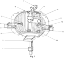
- FIG. 1 provides an exophthalmos-evaluating apparatus and method for an exophthalmometer, including a left displacement platform 3, a left canthal latch point 4, a left mirror 5, cameras 8, a near-infrared light source 9, a right canthal latch point 10, a right mirror 11, a right displacement platform 12, a visible light column 13, and a housing 14.
- the left mirror 5 and the right mirror 11 are respectively configured on the left canthal latch point 4 and the right canthal latch point 10, and the left canthal latch point 4 and the right canthal latch point 10 are respectively on the left displacement platform 3 and the right displacement platform 12; and the left displacement platform 3 and the right displacement platform 12 are respectively on both opposite sides of the housing 14, and the left mirror 5 and the right mirror 11 are inclined; and the cameras 8 are configured in front of the left mirror 5 and the right mirror 11; and the near-infrared light source 9 is configured below the camera 8; and the visible light column 13 is a multiple of columns and is configured longitudinally along an inner side of the housing 14.
- the evaluation apparatus also includes a lower support frame 1, a jaw support 2, a forehead support 6 and an upper support frame 7, wherein the lower support frame 1 and the upper support frame 7 are configured at the lower and upper parts of the housing 14, respectively, for supporting and rotating the entire evaluation apparatus, and the jaw support 2 and the forehead support 6 are configured to fix the user.
- the cameras 8 can be provided in two numbers, which are respectively located in front of the left mirror 5 and the right mirror 11, so as to respectively take videos of the left and right eyes.
- the near-infrared light source 9 can be configured as multiple light sources, evenly distributed under the camera 8.
- Each longitudinal visible light column 13 can be configured as an array of point-like light sources, and is externally wrapped in silicone.
- the left displacement platform 3 and the right displacement platform 12 are first adjusted, and the lateral canthus points of each of the two eyes of the user are fixed using the left canthal latch point 4 and the right canthal latch point 10, to adapt to the facial features of different users.
- the near-infrared light source 9 is turned on, and a series of visible light columns 13 configured orthogonally are turned on from the middle to the outside or from the outside to the middle.
- the visible light columns 13 will successively reflect light of different lengths on the eyeball in the mirror.
- the video frame in which the visible light column 13 forms the longest projection on the cornea through the corneal vertex can be found in the video taken.
- the pupil center can be accurately found by neural network analysis.
- the corneal vertex and pupil center, combined with the known distance and the mirror inclination angle, are used to calculate the ocular prominence.
- this example provides a method for evaluating exophthalmos, which is illustrated below in conjunction with the exophthalmos-evaluating apparatus for an exophthalmometer.
- the method for evaluating exophthalmos involves the following steps:
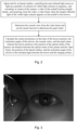
- the corneal vertex can be determined by the video frame having the longest reflected light from the eyeball in the mirror.
- the corneal vertex is the tangent point of the reflected light on the eyeball in the left mirror and the orthogonal line in Fig. 3 .
- the color of different parts of the eye namely the pupil, iris, and sclera, has little effect on imaging, due to the gradual structure of the corneal limbus in the contact part of the iris and sclera, resulting in the inability to accurately identify the spherical center of the eye.
- the invention adopts a near-infrared light field, which can clearly distinguish the sclera, iris and pupil boundaries, and can accurately identify the pupil center in combination with a neural network training model.
- the RITnet neural network model is used in this example.
- the exophthalmos-evaluating apparatus provided in Example 1 for exophthalmos testing also provides the ability to determine the pupillary center by manual operation after determining the corneal vertex from the video frame, which the user can select on demand with the neural network or by manual operation.
- Step S3 Calculating the ocular prominence on the basis of the known positions, the inclination angles of the mirrors, the corneal vertex, and the pupil center.
- the known position is the position of the optical center of the camera, the distance between the optical center of the camera and the video frame, and the position of the lateral canthus
- the inclination angle of the mirror is the included angle between the mirror and the imaging surface.
- the distance, obtained through the known position refers to the distance between the optical center of the camera and the imaging surface (that is, the sensor), which is called the image distance in photography.
- the ocular prominence can be calculated by combining the above-mentioned distance, the inclination angle of the mirror with the imaging point of the pupil center and corneal vertex on the video frame.
- Example 3 further explains how to perform the mathematical calculation of ocular prominence.
- the optical center is O
- the lateral canthus is E
- the orthogonal distance between the optical center O and the video frame is OB
- the imaging point of the corneal vertex Z on the video frame is imaging point C
- the imaging point of the reflection point U of the corneal vertex Z in the mirror on the video frame is the imaging point A
- the orthogonal distance from imaging point C and imaging point A to the optical axis are AB and CB, respectively.
- the angle of inclination between the mirror and the eye is ⁇ UED
- the distance between the optical center O and the face plane where the lateral canthus E is located is OF
- the distance between the lateral canthus E and the optical axis is EF
- the ocular prominence is the orthogonal distance ZG from the corneal vertex Z to the straight line DE.
- the lengths of OB, OF, EF, AB and CB and the angle ⁇ UED are known.
- Step S31 The straight line passing through the reflection point U of the corneal vertex Z in the mirror and parallel to the straight line DE intersects with the optical axis at point W, with ZG at point Y, and with the extension line of the straight line OC at point X.
- both the imaging point C of the corneal vertex Z and the imaging point A of reflection point U of the corneal vertex Z in the mirror are in the video frame.
- the distance from CB and AB of imaging point C and imaging point A to the optical axis can be read in the video frame.
- Step S33 Assume UY ⁇ UX, then ZY ⁇ ⁇ UXtan ⁇ ZUY , where ZY ⁇ is the estimated length of line segment ZY, and YX ⁇ is the estimated length of line segment YX.
- the ocular prominence is calculated according to formula (4) and formula (8).
- the length of line segment YG and line segment ZY in Fig. 4 should be calculated first. Since the length of line segment YG is equal to the length of line segment WF, the ocular prominence can be calculated by the sum of the distances of line segment WF and line segment ZY.
- Step S32 the length of line segment WF and the distance of line segment UX can be obtained by solving the ternary first order equation of formula (3).
- Step S33 it is assumed that UY is equal to UX, that is, YX is equal to zero.
- the estimated value of ZY can be solved by trigonometric functions, that is, ZY ⁇ and the value of YX ⁇ can be calculated by ZY ⁇ , and then the value of ZY ⁇ can be calculated by YX ⁇ , through iterative calculation until ZY ⁇ converges. That is, when the error value calculated for ZY ⁇ is less than the set value twice, then the iteration ends, that is, the length of line segment ZY is considered to be the final calculation result of ZY ⁇ .
- the above calculation process can be automatically calculated by computer programming. By obtaining the video frame having the longest reflected light from the visible light columns in the mirror, input to the computer, one can automatically output the result of ocular prominence.
- Fig. 5 is a structural diagram of an exophthalmos evaluation system provided by an embodiment of the invention, as shown in Fig. 5 .
- the system comprises:
- a computer-readable storage medium including a memory storing at least one piece of program code loaded and executed by a processor to complete an exophthalmos-evaluating method in the above embodiments.
- the computer-readable storage medium may be a Read-Only Memory (ROM), a Random Access Memory (RAM), a compact disc (Compact Disc Read-Only Memory, CDROM), magnetic tape, floppy disk, an optical data storage device and the like.
Landscapes
- Health & Medical Sciences (AREA)
- Life Sciences & Earth Sciences (AREA)
- Engineering & Computer Science (AREA)
- Heart & Thoracic Surgery (AREA)
- Molecular Biology (AREA)
- Biophysics (AREA)
- Ophthalmology & Optometry (AREA)
- Biomedical Technology (AREA)
- Veterinary Medicine (AREA)
- Medical Informatics (AREA)
- Physics & Mathematics (AREA)
- Surgery (AREA)
- Animal Behavior & Ethology (AREA)
- General Health & Medical Sciences (AREA)
- Public Health (AREA)
- Multimedia (AREA)
- Signal Processing (AREA)
- Eye Examination Apparatus (AREA)
Applications Claiming Priority (2)
| Application Number | Priority Date | Filing Date | Title |
|---|---|---|---|
| CN202210989502.6A CN115633935B (zh) | 2022-08-18 | 2022-08-18 | 用于突眼检查仪的眼球突出度的评估装置、方法和系统 |
| PCT/CN2022/142941 WO2024036866A1 (zh) | 2022-08-18 | 2022-12-28 | 用于突眼检查仪的眼球突出度的评估装置、方法和系统 |
Publications (2)
| Publication Number | Publication Date |
|---|---|
| EP4574016A1 true EP4574016A1 (de) | 2025-06-25 |
| EP4574016A4 EP4574016A4 (de) | 2025-11-26 |
Family
ID=84940279
Family Applications (1)
| Application Number | Title | Priority Date | Filing Date |
|---|---|---|---|
| EP22955633.7A Pending EP4574016A4 (de) | 2022-08-18 | 2022-12-28 | Exophthalmos-evaluierungsvorrichtung, verfahren und system für exophthalmometer |
Country Status (6)
| Country | Link |
|---|---|
| US (1) | US20250057415A1 (de) |
| EP (1) | EP4574016A4 (de) |
| JP (1) | JP7802324B2 (de) |
| KR (1) | KR20250040075A (de) |
| CN (1) | CN115633935B (de) |
| WO (1) | WO2024036866A1 (de) |
Families Citing this family (1)
| Publication number | Priority date | Publication date | Assignee | Title |
|---|---|---|---|---|
| TWI831582B (zh) * | 2023-01-18 | 2024-02-01 | 瑞昱半導體股份有限公司 | 偵測系統以及偵測方法 |
Family Cites Families (13)
| Publication number | Priority date | Publication date | Assignee | Title |
|---|---|---|---|---|
| CN102475536B (zh) * | 2010-11-29 | 2013-06-26 | 上海交通大学医学院附属第九人民医院 | 一种测量眼球突出量的方法、仪器及仪器应用 |
| JP5950007B1 (ja) * | 2015-08-18 | 2016-07-13 | 富士ゼロックス株式会社 | 光計測装置 |
| CN104720738B (zh) * | 2015-04-02 | 2017-05-31 | 深圳市斯尔顿科技有限公司 | 利用眼科设备测人眼球突出度的方法及眼科设备 |
| JP2017038681A (ja) * | 2015-08-18 | 2017-02-23 | 富士ゼロックス株式会社 | 光計測装置および光照射受光方法 |
| CN107960985B (zh) * | 2017-12-27 | 2019-08-23 | 蚌埠医学院 | 一种便携式眼球突出度测量仪 |
| JP7265921B2 (ja) * | 2018-09-27 | 2023-04-27 | 株式会社トプコン | 眼科装置 |
| WO2020250903A1 (ja) * | 2019-06-13 | 2020-12-17 | 株式会社ニデック | 眼科装置 |
| CN112315424A (zh) * | 2020-10-30 | 2021-02-05 | 奥佳华瑞(厦门)医疗科技有限公司 | 一种基于双目立体视觉的突眼检测仪 |
| JP7157473B2 (ja) * | 2020-12-22 | 2022-10-20 | 株式会社MITAS Medical | 検眼装置用アダプタ |
| CN215502943U (zh) * | 2021-01-29 | 2022-01-14 | 上海市内分泌代谢病研究所 | 一种面部正面校准装置及眼球突出度测试设备 |
| CN112806956A (zh) * | 2021-02-01 | 2021-05-18 | 上海市内分泌代谢病研究所 | 眼球突出度测量仪及测量方法 |
| CN216823409U (zh) * | 2021-08-31 | 2022-06-28 | 复旦大学附属中山医院 | 一种佩戴式眼球突出度测量装置 |
| CN115067872B (zh) * | 2022-08-18 | 2022-11-29 | 上海佰翊医疗科技有限公司 | 一种眼部参数评估装置 |
-
2022
- 2022-08-18 CN CN202210989502.6A patent/CN115633935B/zh active Active
- 2022-12-28 JP JP2025504150A patent/JP7802324B2/ja active Active
- 2022-12-28 KR KR1020257006170A patent/KR20250040075A/ko active Pending
- 2022-12-28 EP EP22955633.7A patent/EP4574016A4/de active Pending
- 2022-12-28 WO PCT/CN2022/142941 patent/WO2024036866A1/zh not_active Ceased
-
2024
- 2024-10-31 US US18/932,903 patent/US20250057415A1/en active Pending
Also Published As
| Publication number | Publication date |
|---|---|
| JP2025524936A (ja) | 2025-08-01 |
| CN115633935A (zh) | 2023-01-24 |
| US20250057415A1 (en) | 2025-02-20 |
| KR20250040075A (ko) | 2025-03-21 |
| JP7802324B2 (ja) | 2026-01-20 |
| WO2024036866A1 (zh) | 2024-02-22 |
| EP4574016A4 (de) | 2025-11-26 |
| CN115633935B (zh) | 2023-04-14 |
Similar Documents
| Publication | Publication Date | Title |
|---|---|---|
| US7832864B2 (en) | Inverse optical design | |
| RU2601853C2 (ru) | Способ и устройство для определения оптических аберраций глаза | |
| US5632282A (en) | Ocular disease detection apparatus | |
| US5512966A (en) | Ophthalmic pachymeter and method of making ophthalmic determinations | |
| CN109688899B (zh) | 用于确定矫正镜片的组件、计算机程序、系统、以及套件 | |
| CN103767672B (zh) | 眼科装置及眼科装置的控制方法 | |
| ES2706300T3 (es) | Método para la adquisición de datos de imagen de tomografía de coherencia óptica del tejido de la retina de un ojo de un sujeto humano | |
| JP2001314372A (ja) | 眼の収差を決定するための方法及び装置 | |
| CN105310646A (zh) | 基于白睛无影成像的人体健康状况在体分析系统及方法 | |
| EP3888528A1 (de) | Ophthalmische vorrichtung | |
| JP7104516B2 (ja) | 断層画像撮影装置 | |
| EP4574020A1 (de) | Vorrichtung zur auswertung von augenparametern | |
| US20240225441A1 (en) | Method, system and computer-program for estimating refraction of an eye of an individual | |
| US20250057415A1 (en) | Apparatus, method and system for evaluating exophthalmia in exophthalmia testing | |
| US20210235987A1 (en) | Ocular biometry systems and methods | |
| CN120267221A (zh) | 一种眼底oct成像屈光度自适应匹配方法及装置 | |
| CN119446498A (zh) | 一种眼科视力检查数据收集分析系统 | |
| JPH0576496A (ja) | 眼科用測定装置 | |
| Li et al. | Comparative evaluation of 3D exophthalmometry and Hertel exophthalmometer for measuring ocular protrusion | |
| CN119632495B (zh) | 人工智能红外偏心摄影验光仪、验光系统和验光方法 | |
| CN121003414A (zh) | 一种调节静止的屈光度预测系统及设备 | |
| Huang | A Depth Learning-Based Approach for Vision Prevention and Detection Utilized on Mobile Devices | |
| WO2022150448A1 (en) | System and method to measure aberrations by imaging both the crescent and the halo of the crescent | |
| CN120732353A (zh) | 一种双目瞳孔测量方法 | |
| Miller | A handheld open-field infant keratometer (an american ophthalmological society thesis) |
Legal Events
| Date | Code | Title | Description |
|---|---|---|---|
| STAA | Information on the status of an ep patent application or granted ep patent |
Free format text: STATUS: THE INTERNATIONAL PUBLICATION HAS BEEN MADE |
|
| PUAI | Public reference made under article 153(3) epc to a published international application that has entered the european phase |
Free format text: ORIGINAL CODE: 0009012 |
|
| STAA | Information on the status of an ep patent application or granted ep patent |
Free format text: STATUS: REQUEST FOR EXAMINATION WAS MADE |
|
| 17P | Request for examination filed |
Effective date: 20241104 |
|
| AK | Designated contracting states |
Kind code of ref document: A1 Designated state(s): AL AT BE BG CH CY CZ DE DK EE ES FI FR GB GR HR HU IE IS IT LI LT LU LV MC ME MK MT NL NO PL PT RO RS SE SI SK SM TR |
|
| A4 | Supplementary search report drawn up and despatched |
Effective date: 20251028 |
|
| DAV | Request for validation of the european patent (deleted) | ||
| DAX | Request for extension of the european patent (deleted) | ||
| RIC1 | Information provided on ipc code assigned before grant |
Ipc: A61B 3/10 20060101AFI20251022BHEP Ipc: A61B 3/14 20060101ALI20251022BHEP Ipc: A61B 3/00 20060101ALI20251022BHEP |