EP4578382A2 - Appareil et procédé de compensation d'évaluation de tonalité artérielle périphérique - Google Patents
Appareil et procédé de compensation d'évaluation de tonalité artérielle périphérique Download PDFInfo
- Publication number
- EP4578382A2 EP4578382A2 EP25153804.7A EP25153804A EP4578382A2 EP 4578382 A2 EP4578382 A2 EP 4578382A2 EP 25153804 A EP25153804 A EP 25153804A EP 4578382 A2 EP4578382 A2 EP 4578382A2
- Authority
- EP
- European Patent Office
- Prior art keywords
- light
- function
- individual
- volume
- oxygen saturation
- Prior art date
- Legal status (The legal status is an assumption and is not a legal conclusion. Google has not performed a legal analysis and makes no representation as to the accuracy of the status listed.)
- Pending
Links
Images





Classifications
-
- A—HUMAN NECESSITIES
- A61—MEDICAL OR VETERINARY SCIENCE; HYGIENE
- A61B—DIAGNOSIS; SURGERY; IDENTIFICATION
- A61B5/00—Measuring for diagnostic purposes; Identification of persons
- A61B5/02—Detecting, measuring or recording for evaluating the cardiovascular system, e.g. pulse, heart rate, blood pressure or blood flow
- A61B5/02007—Evaluating blood vessel condition, e.g. elasticity, compliance
-
- A—HUMAN NECESSITIES
- A61—MEDICAL OR VETERINARY SCIENCE; HYGIENE
- A61B—DIAGNOSIS; SURGERY; IDENTIFICATION
- A61B5/00—Measuring for diagnostic purposes; Identification of persons
- A61B5/02—Detecting, measuring or recording for evaluating the cardiovascular system, e.g. pulse, heart rate, blood pressure or blood flow
- A61B5/026—Measuring blood flow
- A61B5/0261—Measuring blood flow using optical means, e.g. infrared light
-
- A—HUMAN NECESSITIES
- A61—MEDICAL OR VETERINARY SCIENCE; HYGIENE
- A61B—DIAGNOSIS; SURGERY; IDENTIFICATION
- A61B5/00—Measuring for diagnostic purposes; Identification of persons
- A61B5/02—Detecting, measuring or recording for evaluating the cardiovascular system, e.g. pulse, heart rate, blood pressure or blood flow
- A61B5/026—Measuring blood flow
- A61B5/0295—Measuring blood flow using plethysmography, i.e. measuring the variations in the volume of a body part as modified by the circulation of blood therethrough, e.g. impedance plethysmography
-
- A—HUMAN NECESSITIES
- A61—MEDICAL OR VETERINARY SCIENCE; HYGIENE
- A61B—DIAGNOSIS; SURGERY; IDENTIFICATION
- A61B5/00—Measuring for diagnostic purposes; Identification of persons
- A61B5/145—Measuring characteristics of blood in vivo, e.g. gas concentration or pH-value ; Measuring characteristics of body fluids or tissues, e.g. interstitial fluid or cerebral tissue
- A61B5/1455—Measuring characteristics of blood in vivo, e.g. gas concentration or pH-value ; Measuring characteristics of body fluids or tissues, e.g. interstitial fluid or cerebral tissue using optical sensors, e.g. spectral photometrical oximeters
- A61B5/14551—Measuring characteristics of blood in vivo, e.g. gas concentration or pH-value ; Measuring characteristics of body fluids or tissues, e.g. interstitial fluid or cerebral tissue using optical sensors, e.g. spectral photometrical oximeters for measuring blood gases
-
- A—HUMAN NECESSITIES
- A61—MEDICAL OR VETERINARY SCIENCE; HYGIENE
- A61B—DIAGNOSIS; SURGERY; IDENTIFICATION
- A61B5/00—Measuring for diagnostic purposes; Identification of persons
- A61B5/40—Detecting, measuring or recording for evaluating the nervous system
- A61B5/4029—Detecting, measuring or recording for evaluating the nervous system for evaluating the peripheral nervous systems
- A61B5/4035—Evaluating the autonomic nervous system
-
- A—HUMAN NECESSITIES
- A61—MEDICAL OR VETERINARY SCIENCE; HYGIENE
- A61B—DIAGNOSIS; SURGERY; IDENTIFICATION
- A61B5/00—Measuring for diagnostic purposes; Identification of persons
- A61B5/48—Other medical applications
- A61B5/4806—Sleep evaluation
- A61B5/4818—Sleep apnoea
-
- A—HUMAN NECESSITIES
- A61—MEDICAL OR VETERINARY SCIENCE; HYGIENE
- A61B—DIAGNOSIS; SURGERY; IDENTIFICATION
- A61B5/00—Measuring for diagnostic purposes; Identification of persons
- A61B5/68—Arrangements of detecting, measuring or recording means, e.g. sensors, in relation to patient
- A61B5/6801—Arrangements of detecting, measuring or recording means, e.g. sensors, in relation to patient specially adapted to be attached to or worn on the body surface
- A61B5/6813—Specially adapted to be attached to a specific body part
- A61B5/6825—Hand
- A61B5/6826—Finger
-
- A—HUMAN NECESSITIES
- A61—MEDICAL OR VETERINARY SCIENCE; HYGIENE
- A61B—DIAGNOSIS; SURGERY; IDENTIFICATION
- A61B5/00—Measuring for diagnostic purposes; Identification of persons
- A61B5/72—Signal processing specially adapted for physiological signals or for diagnostic purposes
- A61B5/7203—Signal processing specially adapted for physiological signals or for diagnostic purposes for noise prevention, reduction or removal
-
- A—HUMAN NECESSITIES
- A61—MEDICAL OR VETERINARY SCIENCE; HYGIENE
- A61B—DIAGNOSIS; SURGERY; IDENTIFICATION
- A61B2560/00—Constructional details of operational features of apparatus; Accessories for medical measuring apparatus
- A61B2560/02—Operational features
- A61B2560/0223—Operational features of calibration, e.g. protocols for calibrating sensors
Definitions
- a common method for measuring this fluctuation in blood volume is optical plethysmography, or photoplethysmography, during which an optical plethysmogram, or PhotoPlethysmoGram, or PPG, is used to detect changes in blood volume in the microvascular bed of the tissue.
- a PPG is often obtained by shining light from one or more light sources, such as for example LEDs, onto an investigated volume, and detecting collected light corresponding to the light being reflected or transmitted in the investigated volume on a sensor, wherein the sensor may for example comprise or correspond to a photodetector, such as for example a photodiode.
- the light sources and the sensor can for example be arranged on opposite sides of a finger of an individual, hereby allowing the measurement of a transmission mode PPG, or at the same side of a finger of the individual, hereby allowing the measurement of a reflectance mode PPG.
- the heart pumps arterial blood to the investigated volume.
- the physical events corresponding to a change in arterial blood volume in the investigated volume for example in a cardiac cycle can be captured by optical plethysmography.
- the arterial blood volume in the tissue is also influenced by the diameters of small arteries, or arterioles, in the investigated volume. These arterioles have muscular walls which can contract to reduce the diameter arterioles. In other words, when these arterioles contract and reduce in diameter, the volume of arterial blood comprised in the corresponding arterioles evidently reduces.
- Optical plethysmography is a measurement technique which can be used to monitor changes in the volume of arterial blood comprised in arterioles when these arterioles contract in diameter. Monitoring changes in the arterial blood volume with optical plethysmography therefore empirically provides information on relative changes in muscle tension or 'tone' of the smooth muscle tissue of the arterioles, also referred to as Peripheral Arterial Tone or PAT.
- Hemoglobin is the main molecule which scatters the light emitted onto the investigated volume during a measurement with optical plethysmography. Hemoglobin comprises oxygenated hemoglobin, also known as HbO 2 , and deoxygenated hemoglobin, also known as Hb. The extent to which hemoglobin in the arterial blood is oxygenated is referred to as the oxygen saturation, or SpO 2 . The degree of absorption and scattering of light due to HbO 2 is significantly different from that of Hb and additionally depends on the wavelength of the light used for optical plethysmography.
- the method further comprises the steps of determining an evaluation function which is a function of the light intensities; and wherein the determining a ratio corresponds to determining a ratio of the evaluation function and the compensation function.
- the evaluation function corresponds to the natural logarithm of a function of the light intensities.
- any other evaluation function defined as a function of the light intensities could be used, for example a linear approximation of the natural logarithm of a function of the light intensities, or for example a Taylor series approximation of a function of the light intensities, or for example a linear approximation of other base logarithms of a function of the light intensities.
- the change in arterial blood volume in the investigated volume between the two or more points in time, and hereby peripheral arterial tone, are then assessed by determining a ratio of the evaluation function and of the compensation function.
- the evaluation function corresponds to a logarithm of a function of the light intensities.
- the evaluation function corresponds to the natural logarithm of a function of the light intensities.
- any other evaluation function defined as a function of the light intensities could be used, for example a linear approximation of the logarithm of a function of the light intensities, or for example a Taylor series approximation of a function of the light intensities, or for example a linear approximation of other base logarithms of a function of the light intensities.
- the evaluation function corresponds to a logarithm of a ratio of the light intensities; and wherein the evaluation function depends on one or more of the following:
- Equation (10) therefore corresponds to the evaluation function, i.e. to the logarithm of a ratio of the light intensities measured by the sensor when investigating the investigated volume of the individual with optical plethysmography.
- the method further comprises the steps of:
- the oxygen saturation estimate can be obtained for example from a reference measurement obtained by optical plethysmography.
- the oxygen saturation estimate can be obtained for example by any other suitable apparatus or method which measures oxygen saturation near the investigated volume being assessed.
- the evaluation function can for example be measured by the sensor of the optical plethysmography setup.
- Equation (15) The left-hand side of equation (15), i.e. the calibration ratio, can be computed from the measurements of the sensor of the optical plethysmography setup of both wavelengths ⁇ 1 and ⁇ 2 .
- equation (12) can be rewritten as follows in equation (16): ln I l I h f ⁇ 1 SpO 2 ⁇ ⁇ V blood wherein f ⁇ 1 is corresponds to the compensation function at the first wavelength ⁇ 1 , and which is a non-factorizable function of SpO 2 .
- Optical plethysmography technology uses a simple and noninvasive setup probe or biosensor.
- the optical plethysmography biosensor non-invasively measures pulsatile arterial volume changes in the investigated volume, and thereby assesses PAT, by collecting the optical plethysmography signal.
- the first and/or the second light sources are for example LEDs or any other suitable light sources which can be miniaturized to fit in the optical plethysmography biosensor.
- the first wavelength is for example different from the second wavelength.
- the first wavelength and the second wavelength are comprised in the red spectrum.
- the first wavelength is comprised in the red spectrum
- the second wavelength is comprised in the infra-red spectrum.
- a physical distance between the light sources and the sensor is for example a few millimeters, such as for example less than 3mm.
- the method further comprises the steps of:
- the oxygen saturation estimate depends on a ratio of the concentration of oxygenated hemoglobin on a total concentration of oxygenated and deoxygenated hemoglobin in the arterial blood volume.
- the method further comprises the step of determining the oxygen saturation estimate near the investigated volume of the individual, for example near the distal end of the digit of the individual.
- the compensation function is derived from a regression which maps the oxygen saturation estimate onto the predetermined calibration coefficients.
- Evaluating the compensation function corresponds to determining for example a least-squares regression which maps the oxygen saturation estimate onto the predetermined calibration coefficients.
- a least-squares regression maps the oxygen saturation estimate onto ln I l I h ⁇ 1 ln I l I h ⁇ 2 of equation (15).
- the method further comprises the step of forcing that the regression uses a first-degree rational mapping when evaluating the compensation function.
- the method further comprises the step of forcing that a least-squares regression uses a first-degree rational mapping when estimating the compensation function.
- the least-squares regression maps the oxygen saturation estimate onto ln I l I h ⁇ 1 ln I l I h ⁇ 2 of equation (15), and by forcing that the linear regression uses a first degree rational mapping, i.e. a first degree polynomial in the numerator and a first degree polynomial in the denominator, the least-squares fit yields the best estimate of the predetermined calibration coefficients Q 1 , ⁇ 1 , Q 1 , ⁇ 2 , Q 2 , ⁇ 1 , and Q 2, ⁇ 2 up to a constant scalar.
- the predetermined calibration coefficients up to a constant factor is sufficient.
- compensation function f ⁇ 1 and compensation function f ⁇ 2 can be determined again through a least-squares regression which maps a measured reference oxygen saturation estimate onto a known right-hand side of equation (17).
- the only constraint on the regression is that the mapping makes use of a non-factorizable function of oxygen saturation estimate in the numerator and the denominator.
- an apparatus comprising at least one processor and at least one memory including computer program code, the at least one memory and computer program code configured to, with the at least one processor, cause the apparatus to perform:
- the computer-implemented method reduces the effect on the optical plethysmography signal of changes in the hemoglobin composition of the arterial blood volume, and thereby minimizes or reduces the effect of changes in the hemoglobin composition of the arterial blood volume on the optical measurement of peripheral arterial tone.
- the computer-implemented method indeed determines a compensation function which is a function of the oxygen saturation estimate and divides a function of the light intensities measured by optical plethysmography by the compensation function for this oxygen saturation estimate.
- the resulting evaluation of a change in arterial blood volume in the investigated volume therefore provides a more accurate and robust assessment of peripheral arterial tone of the individual.
- the apparatus does not rely on the use of a light source emitting light at an isosbestic wavelength.
- the apparatus is compatible with any usual setup for optical plethysmography comprising for example two light sources, wherein the two light sources for example emit light of two different wavelengths, such as for example one red wavelength and another infra-red wavelength.
- the apparatus accurately measures changes in arterial blood volume in an investigated volume through measurement setups with just two light sources, which is the minimum number of light sources needed to determine the oxygen saturation estimate.
- Peripheral arterial tone is then mathematically derived by the apparatus from the optical plethysmography measurement with two light sources. This allows miniaturization compared to an optical plethysmography system comprising three light sources and further allows cost optimization by eliminating the need for a third light source.
- a system comprising an apparatus according to a second example aspect of the invention, and further comprises:
- the sensor collects propagated light by optical plethysmography, wherein the propagated light corresponds to the light being transmitted or reflected when propagating in the investigated volume of the individual, such as for example a distal end of a digit of the individual, at the two or more points in time.
- the system further optionally comprises a wireless transmitter comprising a wireless communication interface, wherein the wireless transmitter is configured to transmit the determined peripheral arterial tone wirelessly for further processing by the apparatus.
- the wireless communication interface is preferably a low power communication interface, e.g. a Bluetooth Low Energy, BLE, wireless interface.
- a computer program product comprising computer-executable instructions for causing a system to perform at least the following is provided:
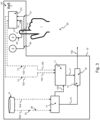
- Fig. 1 illustrates an example embodiment of an apparatus 10 according to the present disclosure.
- the apparatus 10 comprises at least one memory 6, at least one processor, wherein the memory 6 includes computer program code configured to, with the at least one processor, cause the apparatus 10 to perform the following:
- Fig. 2 illustrates an example embodiment of a system 20 according to the present disclosure. Components having identical reference numbers to numbers on Fig. 1 perform the same function.
- the system 20 of Fig. 2 comprises an apparatus 10 according to the present disclosure.
- the system 20 further comprises a light source 2;3 and a sensor 4.
- a light source 2;3 is configured to emit light 40.
- the apparatus 10 is configured to:
- Fig. 3 illustrates an example embodiment of a system 20 according to the present disclosure. Components having identical reference numbers to numbers on Fig. 1 or Fig. 2 perform the same function.
- the system 20 of Fig. 3 comprises an apparatus 10 according to the present disclosure.
- the system 20 further comprises a light source 2;3 and a sensor 4.
- a light source 2;3 is configured to emit light 40.
- the apparatus 10 is configured to:
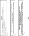
- Figs. 4A and 4B illustrate an example embodiment of the calibration of the apparatus according to the present disclosure.
- Components having identical reference numbers to numbers on Fig. 1 or Fig. 2 or Fig. 3 perform the same function.
- a first light source 2 is provided and emits light 40 at a first wavelength.
- a second light source 3 is provided and emits light 40 at a second wavelength. The second wavelength is preferable different from the first wavelength.
- a sensor 4 is provided. By optical plethysmography, propagated light 41 is collected on the sensor 4 at two points in time 2;3, wherein the propagated light 41 corresponds to the light 40 emitted by the first light source 2 or by the second light source 3 being transmitted or reflected by the investigated volume of an individual when propagating in the investigated volume 11 of the individual 1.
- Fig. 4A for the first wavelength emitted by the light source 2, light intensities 106;107 corresponding to the propagated light 41 collected on the sensor 4 are measured by the sensor 4 at respectively a first point in time 12 and at a second point in time 13.
- Fig. 4B for the second wavelength emitted by the second light source 3, light intensities 108;109 corresponding to the propagated light 41 collected on the sensor 4 are measured by the sensor 4 at respectively a first point in time 12 and at a second point in time 13.
- the sensor 4 determines a first ratio corresponding to a ratio of the first light intensities 106;107 for the first wavelength on Fig. 4A .
- the sensor 4 or, according to an alternative embodiment the apparatus 10 of Fig. 1 or Fig. 2 or Fig. 3 , then determines a second ratio corresponding to a ratio of the second light intensities 108;108 for the second wavelength on Fig. 4B .
- the sensor 4 or, according to an alternative embodiment the apparatus 10 of Fig. 1 or Fig. 2 or Fig. 3 determines a calibration ratio corresponding to the ratio of a function of the first ratio and of a function of the second ratio.
- the determination of the first ratio and/or of the second ratio and/or of the calibration ratio and/or of the predetermined calibration coefficients is performed by any other suitable external device comprising at least one processor and at least one memory including computer program code, the at least one memory and computer program code configured to, with the at least one processor, cause the suitable external device to determine these values.
- Fig. 5 illustrates an example comparison between uncompensated peripheral arterial tone 401 in function of time 60 of an individual, wherein the uncompensated peripheral arterial tone 401 is determined with optical plethysmography without compensation of the influence caused by changes in the hemoglobin composition of the monitored arterial blood volume on the measurement of peripheral arterial tone, and compensated peripheral arterial tone 402 in function of time 60 of the same individual, wherein the compensated peripheral arterial tone 402 is determined by the computer-implemented method according to the present disclosure, or by the apparatus according to the present disclosure, i.e. wherein the compensated peripheral arterial tone 402 is compensated of the influence caused by changes in the hemoglobin composition of the monitored arterial blood volume on the measurement of peripheral arterial tone.
- the peripheral arterial tone estimates are plotted by calculating ln I h I l .
- the peripheral arterial tone estimates are plotted by calculating ln I l I h .
- Fig. 5 also illustrates an example embodiment of the oxygen saturation estimate 104 measured at the same time as the investigated tone 100.
- the level of oxygen saturation estimate 104 is high and the uncompensated peripheral arterial tone 401 and the compensated peripheral arterial tone 402 evolve in a similar manner and overlap as a function of time 60 at a pre-vasoconstriction-event baseline value.
Landscapes
- Health & Medical Sciences (AREA)
- Life Sciences & Earth Sciences (AREA)
- Engineering & Computer Science (AREA)
- Physics & Mathematics (AREA)
- Veterinary Medicine (AREA)
- Animal Behavior & Ethology (AREA)
- Biomedical Technology (AREA)
- Heart & Thoracic Surgery (AREA)
- Medical Informatics (AREA)
- Molecular Biology (AREA)
- Surgery (AREA)
- Pathology (AREA)
- General Health & Medical Sciences (AREA)
- Public Health (AREA)
- Biophysics (AREA)
- Physiology (AREA)
- Cardiology (AREA)
- Neurology (AREA)
- Signal Processing (AREA)
- Hematology (AREA)
- Computer Vision & Pattern Recognition (AREA)
- Neurosurgery (AREA)
- Spectroscopy & Molecular Physics (AREA)
- Optics & Photonics (AREA)
- Psychiatry (AREA)
- Artificial Intelligence (AREA)
- Vascular Medicine (AREA)
- Measurement Of The Respiration, Hearing Ability, Form, And Blood Characteristics Of Living Organisms (AREA)
- Investigating Or Analysing Materials By Optical Means (AREA)
Priority Applications (1)
| Application Number | Priority Date | Filing Date | Title |
|---|---|---|---|
| EP25153804.7A EP4578382A3 (fr) | 2020-06-26 | 2020-06-26 | Appareil et procédé de compensation d'évaluation de tonalité artérielle périphérique |
Applications Claiming Priority (2)
| Application Number | Priority Date | Filing Date | Title |
|---|---|---|---|
| EP20182503.1A EP3928689B1 (fr) | 2020-06-26 | 2020-06-26 | Appareil et procédé de compensation d'évaluation du tonus artériel périphérique |
| EP25153804.7A EP4578382A3 (fr) | 2020-06-26 | 2020-06-26 | Appareil et procédé de compensation d'évaluation de tonalité artérielle périphérique |
Related Parent Applications (1)
| Application Number | Title | Priority Date | Filing Date |
|---|---|---|---|
| EP20182503.1A Division EP3928689B1 (fr) | 2020-06-26 | 2020-06-26 | Appareil et procédé de compensation d'évaluation du tonus artériel périphérique |
Publications (2)
| Publication Number | Publication Date |
|---|---|
| EP4578382A2 true EP4578382A2 (fr) | 2025-07-02 |
| EP4578382A3 EP4578382A3 (fr) | 2025-10-08 |
Family
ID=71170436
Family Applications (2)
| Application Number | Title | Priority Date | Filing Date |
|---|---|---|---|
| EP20182503.1A Active EP3928689B1 (fr) | 2020-06-26 | 2020-06-26 | Appareil et procédé de compensation d'évaluation du tonus artériel périphérique |
| EP25153804.7A Pending EP4578382A3 (fr) | 2020-06-26 | 2020-06-26 | Appareil et procédé de compensation d'évaluation de tonalité artérielle périphérique |
Family Applications Before (1)
| Application Number | Title | Priority Date | Filing Date |
|---|---|---|---|
| EP20182503.1A Active EP3928689B1 (fr) | 2020-06-26 | 2020-06-26 | Appareil et procédé de compensation d'évaluation du tonus artériel périphérique |
Country Status (6)
| Country | Link |
|---|---|
| EP (2) | EP3928689B1 (fr) |
| JP (1) | JP7811186B2 (fr) |
| CN (2) | CN121421527A (fr) |
| AU (1) | AU2021295602A1 (fr) |
| IL (1) | IL299331A (fr) |
| WO (1) | WO2021260190A1 (fr) |
Families Citing this family (10)
| Publication number | Priority date | Publication date | Assignee | Title |
|---|---|---|---|---|
| US20240091476A1 (en) | 2021-01-29 | 2024-03-21 | Resmed Sensor Technologies Limited | Systems and methods for estimating a subjective comfort level |
| WO2022208368A1 (fr) | 2021-03-31 | 2022-10-06 | Resmed Sensor Technologies Limited | Systèmes et procédés de gestion des conditions de pression artérielle d'un utilisateur d'un système de thérapie respiratoire |
| US20240181185A1 (en) | 2021-04-16 | 2024-06-06 | Resmed Sensor Technologies Limited | Systems and methods for characterizing a urser interface or a vent using acoustic data associated with the vent |
| US20240399085A1 (en) | 2021-08-30 | 2024-12-05 | Resmed Digital Health Inc. | Intelligent respiratory entrainment |
| CN120379593A (zh) | 2022-12-20 | 2025-07-25 | 瑞思迈数字健康公司 | 诊断头带 |
| US12579242B2 (en) | 2022-12-21 | 2026-03-17 | Resmed Digital Health Inc. | Systems and methods for digit-based diagnostic chain of custody management |
| EP4652611A1 (fr) | 2023-01-18 | 2025-11-26 | ResMed Sensor Technologies Limited | Systèmes et procédés de caractérisation d'une interface utilisateur à l'aide de données de générateur de flux |
| CN121844363A (zh) | 2023-07-19 | 2026-04-10 | 瑞思迈数字医疗公司 | 使用机器学习和所捕获的图像数据对用户接口进行分类 |
| CN121844390A (zh) | 2023-07-19 | 2026-04-10 | 瑞思迈数字医疗公司 | 使用呼吸治疗数据检测用户接口变更 |
| US12605077B2 (en) | 2023-08-25 | 2026-04-21 | Pranaq Pte. Ltd. | Photoplethysmogram system and method |
Citations (2)
| Publication number | Priority date | Publication date | Assignee | Title |
|---|---|---|---|---|
| WO1998004182A2 (fr) | 1996-07-30 | 1998-02-05 | Itamar Medical (C.M) 1997 Ltd. | Procede et appareil de detection non invasive d'etats pathologiques par controle du tonus arteriel peripherique |
| EP1534115A2 (fr) | 2002-07-15 | 2005-06-01 | Itamar Medical Ltd | Sonde de surface corporelle, dispositif et procede de detection non invasive d'etats medicaux |
Family Cites Families (13)
| Publication number | Priority date | Publication date | Assignee | Title |
|---|---|---|---|---|
| JP4040913B2 (ja) * | 2002-06-07 | 2008-01-30 | 株式会社パルメディカル | 非観血動静脈酸素飽和度測定装置 |
| AU2004203059A1 (en) * | 2004-06-08 | 2005-12-22 | The Government Of The United States Of America As Represented By The Secretary Of The Department Of Health And Human Services, Centers For Disease Control And Prevention | Apparatus and method for assessing peripheral circulation to evaluate a physiological condition |
| US7512431B2 (en) * | 2005-09-13 | 2009-03-31 | Medtronic, Inc. | Normalization method for a chronically implanted optical sensor |
| US8660799B2 (en) * | 2008-06-30 | 2014-02-25 | Nellcor Puritan Bennett Ireland | Processing and detecting baseline changes in signals |
| US10405762B2 (en) * | 2009-04-22 | 2019-09-10 | Vital Metrix, Inc. | System and method for noninvasively measuring ventricular stroke volume and cardiac output |
| US20110082355A1 (en) * | 2009-07-30 | 2011-04-07 | Oxitone Medical Ltd. | Photoplethysmography device and method |
| US20130310669A1 (en) * | 2012-05-20 | 2013-11-21 | Jerusalem College Of Technology | Pulmonary pulse oximetry method for the measurement of oxygen saturation in the mixed venous blood |
| US20150018654A1 (en) * | 2013-07-09 | 2015-01-15 | Xerox Corporation | Method and apparatus for monitoring a subject for fractional blood oxygen saturation |
| WO2017005016A1 (fr) * | 2015-07-03 | 2017-01-12 | Vita-Course Technologies Co., Ltd | Système et procédé pour la surveillance de paramètres physiologiques |
| WO2019026062A1 (fr) * | 2017-07-31 | 2019-02-07 | Jerusalem College Of Technology | Procédé de mesure de la saturation en oxygène du sang artériel et appareil à cet effet |
| CA3086762A1 (fr) * | 2017-12-22 | 2019-06-27 | Assistance Publique - Hopitaux De Paris | Methode de mesure de la pression arterielle moyenne |
| EP4578380A3 (fr) * | 2018-07-11 | 2025-07-30 | Ectosense NV | Appareil, système et procédé de diagnostic du sommeil |
| CN109924960A (zh) * | 2019-01-31 | 2019-06-25 | 深圳市爱都科技有限公司 | 一种血氧饱和度、心率值和压力等级的计算方法和穿戴设备 |
-
2020
- 2020-06-26 EP EP20182503.1A patent/EP3928689B1/fr active Active
- 2020-06-26 EP EP25153804.7A patent/EP4578382A3/fr active Pending
-
2021
- 2021-06-25 IL IL299331A patent/IL299331A/en unknown
- 2021-06-25 CN CN202511682837.3A patent/CN121421527A/zh active Pending
- 2021-06-25 CN CN202180052414.7A patent/CN115988985B/zh active Active
- 2021-06-25 WO PCT/EP2021/067532 patent/WO2021260190A1/fr not_active Ceased
- 2021-06-25 AU AU2021295602A patent/AU2021295602A1/en active Pending
- 2021-06-25 JP JP2022580957A patent/JP7811186B2/ja active Active
Patent Citations (2)
| Publication number | Priority date | Publication date | Assignee | Title |
|---|---|---|---|---|
| WO1998004182A2 (fr) | 1996-07-30 | 1998-02-05 | Itamar Medical (C.M) 1997 Ltd. | Procede et appareil de detection non invasive d'etats pathologiques par controle du tonus arteriel peripherique |
| EP1534115A2 (fr) | 2002-07-15 | 2005-06-01 | Itamar Medical Ltd | Sonde de surface corporelle, dispositif et procede de detection non invasive d'etats medicaux |
Non-Patent Citations (1)
| Title |
|---|
| CHATTERJEE ET AL.: "Monte Carlo Analysis of Optical Interactions in Reflectance and Transmittance Finger Photoplethysmography", SENSORS, vol. 19, no. 4, 15 February 2019 (2019-02-15), pages 789 |
Also Published As
| Publication number | Publication date |
|---|---|
| CN121421527A (zh) | 2026-01-30 |
| IL299331A (en) | 2023-02-01 |
| WO2021260190A1 (fr) | 2021-12-30 |
| JP7811186B2 (ja) | 2026-02-04 |
| AU2021295602A1 (en) | 2023-02-02 |
| EP3928689B1 (fr) | 2025-01-29 |
| EP3928689A1 (fr) | 2021-12-29 |
| CN115988985B (zh) | 2025-12-05 |
| EP4578382A3 (fr) | 2025-10-08 |
| CN115988985A (zh) | 2023-04-18 |
| JP2023532319A (ja) | 2023-07-27 |
Similar Documents
| Publication | Publication Date | Title |
|---|---|---|
| EP3928689B1 (fr) | Appareil et procédé de compensation d'évaluation du tonus artériel périphérique | |
| EP3928690B1 (fr) | Procédé et appareil d'évaluation du tonus artériel périphérique | |
| US8221326B2 (en) | Detection of oximetry sensor sites based on waveform characteristics | |
| US9402573B2 (en) | System and method for detecting fluid responsiveness of a patient | |
| US11445930B2 (en) | System and method for determining stroke volume of a patient | |
| JP2014039875A (ja) | 携帯診断装置 | |
| JP2011521702A (ja) | Co2評価のための方法および装置 | |
| US12262991B2 (en) | System and method for non-invasive monitoring of hematocrit concentration | |
| JP5096310B2 (ja) | 身体の部位における血液の灌流を決定するための方法及び装置 | |
| US20140073890A1 (en) | Systems and methods for determining fluid responsiveness | |
| EP4208085B1 (fr) | Procédé et appareil pour détecter des événements perturbant le sommeil à partir d'un signal indicatif d'un tonus artériel périphérique d'un individu | |
| Campbell | Development of non-invasive, optical methods for central cardiovascular and blood chemistry monitoring | |
| Nguyen et al. | Low-cost point-of-care wireless multimodal wearable to monitor vitals and tissue oxygen saturation | |
| CN118662119A (zh) | 光学呼吸速率预测系统 |
Legal Events
| Date | Code | Title | Description |
|---|---|---|---|
| PUAI | Public reference made under article 153(3) epc to a published international application that has entered the european phase |
Free format text: ORIGINAL CODE: 0009012 |
|
| STAA | Information on the status of an ep patent application or granted ep patent |
Free format text: STATUS: THE APPLICATION HAS BEEN PUBLISHED |
|
| AC | Divisional application: reference to earlier application |
Ref document number: 3928689 Country of ref document: EP Kind code of ref document: P |
|
| AK | Designated contracting states |
Kind code of ref document: A2 Designated state(s): AL AT BE BG CH CY CZ DE DK EE ES FI FR GB GR HR HU IE IS IT LI LT LU LV MC MK MT NL NO PL PT RO RS SE SI SK SM TR |
|
| REG | Reference to a national code |
Ref country code: DE Ref legal event code: R079 Free format text: PREVIOUS MAIN CLASS: A61B0005145500 Ipc: A61B0005020000 |
|
| PUAL | Search report despatched |
Free format text: ORIGINAL CODE: 0009013 |
|
| AK | Designated contracting states |
Kind code of ref document: A3 Designated state(s): AL AT BE BG CH CY CZ DE DK EE ES FI FR GB GR HR HU IE IS IT LI LT LU LV MC MK MT NL NO PL PT RO RS SE SI SK SM TR |
|
| RIC1 | Information provided on ipc code assigned before grant |
Ipc: A61B 5/02 20060101AFI20250903BHEP Ipc: A61B 5/026 20060101ALI20250903BHEP Ipc: A61B 5/0295 20060101ALI20250903BHEP Ipc: A61B 5/00 20060101ALI20250903BHEP Ipc: A61B 5/1455 20060101ALI20250903BHEP |
|
| STAA | Information on the status of an ep patent application or granted ep patent |
Free format text: STATUS: REQUEST FOR EXAMINATION WAS MADE |
|
| 17P | Request for examination filed |
Effective date: 20260408 |