EP4578413A1 - Sonde médicale pour la navigation dans des vaisseaux sanguins de petit diamètre - Google Patents
Sonde médicale pour la navigation dans des vaisseaux sanguins de petit diamètre Download PDFInfo
- Publication number
- EP4578413A1 EP4578413A1 EP24216338.4A EP24216338A EP4578413A1 EP 4578413 A1 EP4578413 A1 EP 4578413A1 EP 24216338 A EP24216338 A EP 24216338A EP 4578413 A1 EP4578413 A1 EP 4578413A1
- Authority
- EP
- European Patent Office
- Prior art keywords
- electrodes
- electrode
- coil
- guidewire
- longitudinal axis
- Prior art date
- Legal status (The legal status is an assumption and is not a legal conclusion. Google has not performed a legal analysis and makes no representation as to the accuracy of the status listed.)
- Pending
Links
Images




Classifications
-
- A—HUMAN NECESSITIES
- A61—MEDICAL OR VETERINARY SCIENCE; HYGIENE
- A61B—DIAGNOSIS; SURGERY; IDENTIFICATION
- A61B18/00—Surgical instruments, devices or methods for transferring non-mechanical forms of energy to or from the body
- A61B18/04—Surgical instruments, devices or methods for transferring non-mechanical forms of energy to or from the body by heating
- A61B18/12—Surgical instruments, devices or methods for transferring non-mechanical forms of energy to or from the body by heating by passing a current through the tissue to be heated, e.g. high-frequency current
- A61B18/14—Probes or electrodes therefor
- A61B18/1482—Probes or electrodes therefor having a long rigid shaft for accessing the inner body transcutaneously in minimal invasive surgery, e.g. laparoscopy
-
- A—HUMAN NECESSITIES
- A61—MEDICAL OR VETERINARY SCIENCE; HYGIENE
- A61B—DIAGNOSIS; SURGERY; IDENTIFICATION
- A61B34/00—Computer-aided surgery; Manipulators or robots specially adapted for use in surgery
- A61B34/20—Surgical navigation systems; Devices for tracking or guiding surgical instruments, e.g. for frameless stereotaxis
-
- A—HUMAN NECESSITIES
- A61—MEDICAL OR VETERINARY SCIENCE; HYGIENE
- A61B—DIAGNOSIS; SURGERY; IDENTIFICATION
- A61B18/00—Surgical instruments, devices or methods for transferring non-mechanical forms of energy to or from the body
- A61B18/04—Surgical instruments, devices or methods for transferring non-mechanical forms of energy to or from the body by heating
- A61B18/12—Surgical instruments, devices or methods for transferring non-mechanical forms of energy to or from the body by heating by passing a current through the tissue to be heated, e.g. high-frequency current
-
- A—HUMAN NECESSITIES
- A61—MEDICAL OR VETERINARY SCIENCE; HYGIENE
- A61B—DIAGNOSIS; SURGERY; IDENTIFICATION
- A61B18/00—Surgical instruments, devices or methods for transferring non-mechanical forms of energy to or from the body
- A61B18/04—Surgical instruments, devices or methods for transferring non-mechanical forms of energy to or from the body by heating
- A61B18/12—Surgical instruments, devices or methods for transferring non-mechanical forms of energy to or from the body by heating by passing a current through the tissue to be heated, e.g. high-frequency current
- A61B18/14—Probes or electrodes therefor
- A61B18/1492—Probes or electrodes therefor having a flexible, catheter-like structure, e.g. for heart ablation
-
- A—HUMAN NECESSITIES
- A61—MEDICAL OR VETERINARY SCIENCE; HYGIENE
- A61B—DIAGNOSIS; SURGERY; IDENTIFICATION
- A61B5/00—Measuring for diagnostic purposes; Identification of persons
- A61B5/06—Devices, other than using radiation, for detecting or locating foreign bodies ; Determining position of diagnostic devices within or on the body of the patient
- A61B5/061—Determining position of a probe within the body employing means separate from the probe, e.g. sensing internal probe position employing impedance electrodes on the surface of the body
- A61B5/062—Determining position of a probe within the body employing means separate from the probe, e.g. sensing internal probe position employing impedance electrodes on the surface of the body using magnetic field
-
- A—HUMAN NECESSITIES
- A61—MEDICAL OR VETERINARY SCIENCE; HYGIENE
- A61B—DIAGNOSIS; SURGERY; IDENTIFICATION
- A61B5/00—Measuring for diagnostic purposes; Identification of persons
- A61B5/24—Detecting, measuring or recording bioelectric or biomagnetic signals of the body or parts thereof
- A61B5/25—Bioelectric electrodes therefor
- A61B5/279—Bioelectric electrodes therefor specially adapted for particular uses
- A61B5/28—Bioelectric electrodes therefor specially adapted for particular uses for electrocardiography [ECG]
- A61B5/283—Invasive
- A61B5/287—Holders for multiple electrodes, e.g. electrode catheters for electrophysiological study [EPS]
-
- A—HUMAN NECESSITIES
- A61—MEDICAL OR VETERINARY SCIENCE; HYGIENE
- A61B—DIAGNOSIS; SURGERY; IDENTIFICATION
- A61B5/00—Measuring for diagnostic purposes; Identification of persons
- A61B5/68—Arrangements of detecting, measuring or recording means, e.g. sensors, in relation to patient
- A61B5/6846—Arrangements of detecting, measuring or recording means, e.g. sensors, in relation to patient specially adapted to be brought in contact with an internal body part, i.e. invasive
- A61B5/6847—Arrangements of detecting, measuring or recording means, e.g. sensors, in relation to patient specially adapted to be brought in contact with an internal body part, i.e. invasive mounted on an invasive device
- A61B5/6852—Catheters
- A61B5/6853—Catheters with a balloon
-
- A—HUMAN NECESSITIES
- A61—MEDICAL OR VETERINARY SCIENCE; HYGIENE
- A61N—ELECTROTHERAPY; MAGNETOTHERAPY; RADIATION THERAPY; ULTRASOUND THERAPY
- A61N1/00—Electrotherapy; Circuits therefor
- A61N1/02—Details
- A61N1/04—Electrodes
- A61N1/05—Electrodes for implantation or insertion into the body, e.g. heart electrode
- A61N1/056—Transvascular endocardial electrode systems
- A61N1/0563—Transvascular endocardial electrode systems specially adapted for defibrillation or cardioversion
-
- A—HUMAN NECESSITIES
- A61—MEDICAL OR VETERINARY SCIENCE; HYGIENE
- A61N—ELECTROTHERAPY; MAGNETOTHERAPY; RADIATION THERAPY; ULTRASOUND THERAPY
- A61N1/00—Electrotherapy; Circuits therefor
- A61N1/18—Applying electric currents by contact electrodes
- A61N1/32—Applying electric currents by contact electrodes alternating or intermittent currents
- A61N1/327—Applying electric currents by contact electrodes alternating or intermittent currents for enhancing the absorption properties of tissue, e.g. by electroporation
-
- A—HUMAN NECESSITIES
- A61—MEDICAL OR VETERINARY SCIENCE; HYGIENE
- A61B—DIAGNOSIS; SURGERY; IDENTIFICATION
- A61B17/00—Surgical instruments, devices or methods
- A61B17/00234—Surgical instruments, devices or methods for minimally invasive surgery
- A61B2017/00292—Surgical instruments, devices or methods for minimally invasive surgery mounted on or guided by flexible, e.g. catheter-like, means
-
- A—HUMAN NECESSITIES
- A61—MEDICAL OR VETERINARY SCIENCE; HYGIENE
- A61B—DIAGNOSIS; SURGERY; IDENTIFICATION
- A61B18/00—Surgical instruments, devices or methods for transferring non-mechanical forms of energy to or from the body
- A61B2018/00053—Mechanical features of the instrument of device
- A61B2018/00273—Anchoring means for temporary attachment of a device to tissue
- A61B2018/00279—Anchoring means for temporary attachment of a device to tissue deployable
- A61B2018/00285—Balloons
-
- A—HUMAN NECESSITIES
- A61—MEDICAL OR VETERINARY SCIENCE; HYGIENE
- A61B—DIAGNOSIS; SURGERY; IDENTIFICATION
- A61B18/00—Surgical instruments, devices or methods for transferring non-mechanical forms of energy to or from the body
- A61B2018/00315—Surgical instruments, devices or methods for transferring non-mechanical forms of energy to or from the body for treatment of particular body parts
- A61B2018/00345—Vascular system
- A61B2018/00351—Heart
-
- A—HUMAN NECESSITIES
- A61—MEDICAL OR VETERINARY SCIENCE; HYGIENE
- A61B—DIAGNOSIS; SURGERY; IDENTIFICATION
- A61B18/00—Surgical instruments, devices or methods for transferring non-mechanical forms of energy to or from the body
- A61B2018/00315—Surgical instruments, devices or methods for transferring non-mechanical forms of energy to or from the body for treatment of particular body parts
- A61B2018/00345—Vascular system
- A61B2018/00351—Heart
- A61B2018/00363—Epicardium
-
- A—HUMAN NECESSITIES
- A61—MEDICAL OR VETERINARY SCIENCE; HYGIENE
- A61B—DIAGNOSIS; SURGERY; IDENTIFICATION
- A61B18/00—Surgical instruments, devices or methods for transferring non-mechanical forms of energy to or from the body
- A61B2018/00315—Surgical instruments, devices or methods for transferring non-mechanical forms of energy to or from the body for treatment of particular body parts
- A61B2018/00345—Vascular system
- A61B2018/00351—Heart
- A61B2018/00386—Coronary vessels
-
- A—HUMAN NECESSITIES
- A61—MEDICAL OR VETERINARY SCIENCE; HYGIENE
- A61B—DIAGNOSIS; SURGERY; IDENTIFICATION
- A61B18/00—Surgical instruments, devices or methods for transferring non-mechanical forms of energy to or from the body
- A61B2018/00571—Surgical instruments, devices or methods for transferring non-mechanical forms of energy to or from the body for achieving a particular surgical effect
- A61B2018/00577—Ablation
-
- A—HUMAN NECESSITIES
- A61—MEDICAL OR VETERINARY SCIENCE; HYGIENE
- A61B—DIAGNOSIS; SURGERY; IDENTIFICATION
- A61B18/00—Surgical instruments, devices or methods for transferring non-mechanical forms of energy to or from the body
- A61B2018/00571—Surgical instruments, devices or methods for transferring non-mechanical forms of energy to or from the body for achieving a particular surgical effect
- A61B2018/00613—Irreversible electroporation
-
- A—HUMAN NECESSITIES
- A61—MEDICAL OR VETERINARY SCIENCE; HYGIENE
- A61B—DIAGNOSIS; SURGERY; IDENTIFICATION
- A61B18/00—Surgical instruments, devices or methods for transferring non-mechanical forms of energy to or from the body
- A61B2018/00636—Sensing and controlling the application of energy
- A61B2018/00773—Sensed parameters
- A61B2018/00839—Bioelectrical parameters, e.g. ECG, EEG
-
- A—HUMAN NECESSITIES
- A61—MEDICAL OR VETERINARY SCIENCE; HYGIENE
- A61B—DIAGNOSIS; SURGERY; IDENTIFICATION
- A61B18/00—Surgical instruments, devices or methods for transferring non-mechanical forms of energy to or from the body
- A61B18/04—Surgical instruments, devices or methods for transferring non-mechanical forms of energy to or from the body by heating
- A61B18/12—Surgical instruments, devices or methods for transferring non-mechanical forms of energy to or from the body by heating by passing a current through the tissue to be heated, e.g. high-frequency current
- A61B18/14—Probes or electrodes therefor
- A61B2018/1405—Electrodes having a specific shape
- A61B2018/1407—Loop
-
- A—HUMAN NECESSITIES
- A61—MEDICAL OR VETERINARY SCIENCE; HYGIENE
- A61B—DIAGNOSIS; SURGERY; IDENTIFICATION
- A61B18/00—Surgical instruments, devices or methods for transferring non-mechanical forms of energy to or from the body
- A61B18/04—Surgical instruments, devices or methods for transferring non-mechanical forms of energy to or from the body by heating
- A61B18/12—Surgical instruments, devices or methods for transferring non-mechanical forms of energy to or from the body by heating by passing a current through the tissue to be heated, e.g. high-frequency current
- A61B18/14—Probes or electrodes therefor
- A61B2018/1467—Probes or electrodes therefor using more than two electrodes on a single probe
-
- A—HUMAN NECESSITIES
- A61—MEDICAL OR VETERINARY SCIENCE; HYGIENE
- A61B—DIAGNOSIS; SURGERY; IDENTIFICATION
- A61B34/00—Computer-aided surgery; Manipulators or robots specially adapted for use in surgery
- A61B34/20—Surgical navigation systems; Devices for tracking or guiding surgical instruments, e.g. for frameless stereotaxis
- A61B2034/2046—Tracking techniques
- A61B2034/2051—Electromagnetic tracking systems
-
- A—HUMAN NECESSITIES
- A61—MEDICAL OR VETERINARY SCIENCE; HYGIENE
- A61B—DIAGNOSIS; SURGERY; IDENTIFICATION
- A61B34/00—Computer-aided surgery; Manipulators or robots specially adapted for use in surgery
- A61B34/20—Surgical navigation systems; Devices for tracking or guiding surgical instruments, e.g. for frameless stereotaxis
- A61B2034/2046—Tracking techniques
- A61B2034/2051—Electromagnetic tracking systems
- A61B2034/2053—Tracking an applied voltage gradient
Definitions
- the present technology relates generally to medical devices, and in particular medical probes with electrodes, and further relates to, but not exclusively, medical probes suitable for use location tracking, anatomical sensing, and/or inducing irreversible electroporation (IRE) of cardiac tissues.
- IRE irreversible electroporation
- the ligament of Marshall which is located on the epicardium between the left atrial appendage and the left pulmonary veins, is often a source of paroxysmal atrial fibrillation (AF).
- Cardiac arrhythmias such as AF, occur when regions of cardiac tissue abnormally conduct electric signals to adjacent tissue. This disrupts the normal cardiac cycle and causes asynchronous rhythm.
- Certain procedures exist for treating arrhythmia including surgically disrupting the origin of the signals causing the arrhythmia and disrupting the conducting pathway for such signals. By selectively ablating cardiac tissue by application of energy via a catheter, it is sometimes possible to cease or modify the propagation of unwanted electrical signals from one portion of the heart to another.
- RF ablation can have certain risks related to thermal heating which can lead to tissue charring, burning, steam pop, phrenic nerve palsy, pulmonary vein stenosis, and esophageal fistula.
- Cryoablation is an alternative approach to RF ablation that generally reduces thermal risks associated with RF ablation. Maneuvering cryoablation devices and selectively applying cryoablation, however, is generally more challenging compared to RF ablation; therefore, cryoablation is not viable in certain anatomical geometries which may be reached by electrical ablation devices.
- the electrodes are connected to the distal tip along the longitudinal axis, with each electrode being configured to (i) sense anatomical signals in the vein of Marshall and provide electrical signals which are indicative of the anatomical signals or (ii) convey ablation energy to a target tissue region proximal the vein of Marshall.
- the processor is configured to estimate a position of the distal tip based on the generated currents and (i) estimate at least one characteristic of the anatomical signals based on the electrical signals or (ii) provide a signal to convey the ablation energy to the electrodes.
- a system including an elongated probe and a guidewire catheter.
- the elongated probe extends along a longitudinal axis and is dimensioned to be inserted into a coronary sinus.
- the elongated catheter shaft includes an opening.
- the guidewire catheter extends through the opening along the longitudinal axis and is dimension to be inserted into a vein of Marshall.
- a first plurality of electrodes is connected to the elongated probe along the longitudinal axis.
- a second plurality of electrodes is connected to the guidewire catheter along the longitudinal axis.
- a thermal technique such as radio frequency (RF) energy and cryoablation
- RF radio frequency
- cryoablation to correct a malfunctioning heart
- cardiac electropotentials need to be measured at various locations of the myocardium.
- temperature measurements during ablation provide data enabling the efficacy of the ablation.
- the electropotentials and the temperatures are measured before, during, and after the actual ablation.
- RF approaches can have risks that can lead to tissue charring, burning, steam pop, phrenic nerve palsy, pulmonary vein stenosis, and esophageal fistula.
- Electroporation can be induced by applying a pulsed electric field across biological cells to cause reversable (temporary) or irreversible (permanent) creation of pores in the cell membrane.
- the cells have a transmembrane electrostatic potential that is increased above a resting potential upon application of the pulsed electric field. While the transmembrane electrostatic potential remains below a threshold potential, the electroporation is reversable, meaning the pores can close when the applied pulse electric field is removed, and the cells can self-repair and survive. If the transmembrane electrostatic potential increases beyond the threshold potential, the electroporation is irreversible, and the cells become permanently permeable.
- the cells die due to a loss of homeostasis and typically die by apoptosis.
- cells of differing types have differing threshold potential. For instance, heart cells have a threshold potential of approximately 500 V/cm, whereas for bone it is 3000 V/cm. These differences in threshold potential allow IRE to selectively target tissue based on threshold potential.
- the technology of this disclosure includes systems and methods for applying electrical signals from catheter electrodes positioned in the vicinity of myocardial tissue to generate a generate ablative energy to ablate the myocardial tissue.
- the systems and methods can be effective to ablate targeted tissue by inducing irreversible electroporation.
- the systems and methods can be effective to induce reversible electroporation as part of a diagnostic procedure. Reversible electroporation occurs when the electricity applied with the electrodes is below the electric field threshold of the target tissue allowing cells to repair. Reversible electroporation does not kill the cells but allows a physician to see the effect of reversible electroporation on electrical activation signals in the vicinity of the target location.
- Example systems and methods for reversible electroporation is disclosed in U.S. Patent Publication 2021/0162210 , the entirety of which is incorporated herein by reference.
- the pulsed electric field, and its effectiveness to induce reversible and/or irreversible electroporation, can be affected by physical parameters of the system and biphasic pulse parameters of the electrical signal.
- Physical parameters can include electrode contact area, electrode spacing, electrode geometry, etc. Examples presented herein generally include physical parameters adapted to effectively induce reversible and/or irreversible electroporation.
- Biphasic pulse parameters of the electrical signal can include voltage amplitude, pulse duration, pulse interphase delay, inter-pulse delay, total application time, delivered energy, etc.
- parameters of the electrical signal can be adjusted to induce both reversible and irreversible electroporation given the same physical parameters. Examples of various systems and methods of ablation including IRE are presented in U.S.
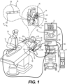
- System 10 includes multiple catheters, which are percutaneously inserted by physician 24 through the patient's 23 vascular system into a chamber or vascular structure of a heart 12.
- probe 14 is used to sense or ablate elements of the heart that are difficult to access, and by way of example, sensing anatomical signals and/or ablation of a portion of a LOM of heart 12 is described herein.
- medical probe 22 can be used, mutatis mutandis, for other therapeutic and/or diagnostic purposes in the heart or in other body organs.
- the plurality of catheters can include catheters dedicated for sensing Intracardiac Electrogram (IEGM) signals, catheters dedicated for ablating and/or catheters dedicated for both sensing and ablating.
- An example medical probe, e.g., a catheter 14, that is configured for sensing IEGM is illustrated herein.
- Physician 24 brings a distal tip of catheter 14 into contact with the heart wall for sensing a target site in heart 12. For ablation, physician 24 would similarly bring a distal end of an ablation catheter to a target site for ablating.
- Catheter 14 is an exemplary catheter that includes one and preferably multiple electrodes 26 optionally distributed over a probe body and configured to sense the IEGM signals.
- Catheter 14 can additionally include one or more position sensors embedded in or near the distal tip 28 for tracking position and orientation of distal tip 28.
- position sensor is a magnetic based position sensor (discussed in greater detail below).
- Magnetic based position sensor may be operated together with a location pad 25 including a plurality of magnetic coils 32 configured to generate magnetic fields in a predefined working volume.
- Real time position of the distal tip 28 of catheter 14 may be tracked based on magnetic fields generated with location pad 25 and sensed by magnetic based position sensor. Details of the magnetic based position sensing technology are described in U.S. Patent Nos. 5,391,199 ; 5,443,489 ; 5,558,091 ; 6,172,499 ; 6,239,724 ; 6,332,089 ; 6,484,118 ; 6,618,612 ; 6,690,963 ; 6,788,967 ; 6,892,091 , each of which are incorporated herein by reference.
- System 10 includes one or more electrode patches 38 positioned for skin contact on patient 23 to establish location reference for location pad 25 as well as impedance-based tracking of electrodes 26.
- impedance-based tracking electrical current is directed toward electrodes 26 and sensed at electrode skin patches 38 so that the location of each electrode can be triangulated via the electrode patches 38. Details of the impedance-based location tracking technology are described in US Patent Nos. 7,536,218 ; 7,756,576 ; 7,848,787 ; 7,869,865 ; and 8,456,182 , each of which are incorporated herein by reference.
- a recorder 11 displays electrograms 21 captured with body surface ECG electrodes 18 and intracardiac electrograms (IEGM) captured with electrodes 26 of catheter 14.
- Recorder 11 may include pacing capability for pacing the heart rhythm and/or may be electrically connected to a standalone pacer.
- System 10 may include an ablation energy generator 50 that is adapted to conduct ablative energy to one or more of electrodes at a distal tip of a catheter configured for ablating.
- Energy produced by ablation energy generator 50 may include, but is not limited to, radiofrequency (RF) energy or pulsed-field ablation (PFA) energy, including unipolar or bipolar high-voltage DC pulses as may be used to effect irreversible electroporation (IRE), or combinations thereof.
- RF radiofrequency
- PFA pulsed-field ablation
- IRE irreversible electroporation
- Patient interface unit (PIU) 30 is an interface configured to establish electrical communication between catheters, electrophysiological equipment, power supply and a workstation 55 for controlling operation of system 10.
- Electrophysiological equipment of system 10 may include for example, multiple catheters, location pad 25, body surface ECG electrodes 18, electrode patches 38, ablation energy generator 50, and recorder 11.
- PIU 30 additionally includes processing capability for implementing real-time computations of location of the catheters and for performing ECG calculations.
- Workstation 55 includes memory, processor unit with memory or storage with appropriate operating software loaded therein, and user interface capability. Workstation 55 may provide multiple functions, optionally including (1) modeling the endocardial anatomy in three-dimensions (3D) and rendering the model or anatomical map 20 for display on a display device 27, (2) displaying on display device 27 activation sequences (or other data) compiled from recorded electrograms 21 in representative visual indicia or imagery superimposed on the rendered anatomical map 20, (3) displaying real-time location and orientation of multiple catheters within the heart chamber, and (5) displaying on display device 27 sites of interest such as places where ablation energy has been applied.
- One commercial product embodying elements of the system 10 is available as the CARTO TM 3 System, available from Biosense Webster, Inc., 31 Technology Drive, Suite 200, Irvine, CA 92618 USA.
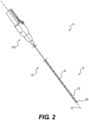
- FIG. 2 is a schematic pictorial illustration showing a perspective view of a medical device 14 with electrodes 26.
- the medical device 14 includes a handle 1000 and a probe body 100 that extends from the handle 1000 along a longitudinal axis 60 and includes a distal tip 28.
- the probe body 100 in the presently described example includes an elongated flexible shaft 102.
- the shaft 102 can be formed from a flexible, biocompatible electrically insulative material such as polyamide-polyether (Pebax) copolymers, polyethylene terephthalate (PET), urethanes, polyimide, parylene, silicone, etc.
- insulative material can include biocompatible polymers including, without limitation, polyetheretherketone (PEEK), polyglycolic acid (PGA), poly(lactic-co-glycolic acid) copolymer (PLGA), polycaprolactive (PCL), poly(3-hydroxybutyrate-co-3-hydroxyvalerate) (PHBV), poly-L-lactide, polydioxanone, polycarbonates, and polyanhydrides with the ratio of certain polymers being selected to control the degree of inflammatory response.
- PEEK polyetheretherketone
- PGA polyglycolic acid
- PLGA poly(lactic-co-glycolic acid) copolymer
- PCL polycaprolactive
- PHBV poly(3-hydroxybutyrate-co-3-hydroxyvalerate)
- poly-L-lactide polydioxanone
- polycarbonates polyanhydrides with the ratio of certain polymers being selected to control the degree of inflammatory response.
- the electrodes 26 can be used to determine the location of the probe body 100 and/or to measure a physiological/anatomical property such as local surface electrical potentials at respective locations on tissue in heart 12. In addition to using electrodes 26 to determine the location and/or measure anatomical signals, electrodes 26 can also be configured to deliver ablation energy (IRE and/or RF) to tissue in heart 12.
- the set of electrodes 26 can include a dome electrode 26A at a distal end of the probe body 100. As the lead electrode 26 at a distal end of the probe body 100, the dome electrode 26A can aid the physician 24 in navigating the catheter 14. The collected signals can be used to help guide the catheter 14, in general and particularly in cases where imaging is otherwise absent.
- Examples of materials ideally suited for forming electrodes 26 include gold, platinum, and palladium (and their respective alloys). These materials also have high thermal conductivity which allows the minimal heat generated on the tissue (i.e., by the ablation energy delivered to the tissue) to be conducted through the electrodes to the back side of the electrodes (i.e., the portions of the electrodes on the inner sides of the spines), and then to the blood pool in heart 12.
- a physician 24 can more accurately determine the position of the distal end of the probe body 100 before using it to sense anatomical signals and/or apply ablative energy to tissue in, e.g., the VOM 42.
- physician 24 inserts the probe body 100 into the subject, and then navigates the probe body 100 into heart 12. Within the heart 12, the probe body is navigated via coronary sinus 40 into LOM 44 until a distal end of the probe body 100 is within proximity to target region 46. The distal end of the probe body 100 can be steered by, for example, one or more pull wires integrated therewith.
- the physician 24 is typically assisted in the navigation by processor 55 using signals from sensors 33, or from electrodes 26, to display an icon representing the location of the distal tip 28 of the probe body 100 on map 20. Additionally, physician 24 can use fluoroscopy to aid in the navigation.
- the location of electrodes 26 relative to target region 46 can be verified by a current tracking module of the PIU 30 using impedances and/or currents between the electrodes 26 and patches 38. Additionally, the location of the electrodes can be verified fluoroscopically.
- medical probe body 100 is maneuvered to bring one or more of the electrodes 26 into contact with the intracardiac tissue, e.g., the inner heart surface.
- the electrodes 26 generate potential gradient signals in response to the sensed electrical potentials, also referred to herein as electrical signals.
- the sensed electrical signals are indicative of a direction and propagation speed of wavefronts caused by anatomical signals, such as electrocardiogram (ECG) signals in the heart 12.
- ECG electrocardiogram
- the physician 24 operates processor 55 and ablation energy generator 50 to supply electrical current to electrodes 26.
- the supplied current can be bipolar, i.e., the current can be passed between the electrodes 26 so as to convey ablation energy to the tissue.
- the supplied ablation energy can be unipolar, i.e., an electrical current may be applied between one of electrodes 26 and a return electrode connected to generator 50.
- the return electrode can be disposed outside the body of patient 23.
- the return electrode may comprise a patch (e.g., electrode patch 38 or the like) coupled to the patient's body.
- RF sinusoidal-like (alternating) electrical current is supplied to the electrodes 26, such that RF ablation of the tissue is performed.
- pulsed current direct current
- IRE irreversible electroporation
- PFA pulsed field ablation
- unipolar ablation energy can be supplied by direct electrical current running between the electrodes 26 on the elongated shaft 202 and the electrode patch(es) 38 or a backpatch.
- insulative material can include biocompatible polymers including, without limitation, polyetheretherketone (PEEK), polyglycolic acid (PGA), poly(lactic-co-glycolic acid) copolymer (PLGA), polycaprolactive (PCL), poly(3-hydroxybutyrate-co-3-hydroxyvalerate) (PHBV), poly-L-lactide, polydioxanone, polycarbonates, and polyanhydrides with the ratio of certain polymers being selected to control the degree of inflammatory response.
- PEEK polyetheretherketone
- PGA polyglycolic acid
- PLGA poly(lactic-co-glycolic acid) copolymer
- PCL polycaprolactive
- PHBV poly(3-hydroxybutyrate-co-3-hydroxyvalerate)
- poly-L-lactide polydioxanone
- polycarbonates polyanhydrides with the ratio of certain polymers being selected to control the degree of inflammatory response.
- the elongated shaft 202 can also include an inflatable balloon 230. As shown in FIG. 8 (the balloon 230 is uninflated in this drawing), the balloon 230 is provided more proximal to the handle 1000 than some or all of the electrodes 26, that is inflatable via a balloon lumen extending through the elongated shaft 202, for purposes that are discussed in greater detail below.
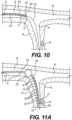
- a distal tip 28 of the elongated shaft 202 is slid along the guidewire 220 into the VOM 42 until it reaches its target region 46. Additionally, or alternatively, the elongated shaft 202 can be further inserted into the VOM 42, as shown in FIG. 11B .
- the balloon 230 can optionally be inflated to occlude the coronary sinus 40 and/or great cardiac vein 48 and/or VOM 42, depending on placement. As shown in FIG. 11A , the balloon 230 occludes the coronary sinus 40. Further inserting the elongated shaft 202 into the VOM 42, as shown in FIG. 11B , positions the balloon 230 such that it can be inflated to occlude the VOM 42.
- RF sinusoidal-like (alternating) electrical current is supplied to the electrodes 26, such that RF ablation of the tissue is performed.
- pulsed current direct current
- IRE irreversible electroporation
- PFA pulsed field ablation
- unipolar ablation energy can be supplied by direct electrical current running between the electrodes 26 on the elongated shaft 202 and the electrode patch(es) 38 or a backpatch.
- bipolar ablation energy can be supplied by direct electrical current running between the electrodes 26 on the elongated shaft 202 themselves and/or another catheter 14A (e.g., a focal catheter or a multielectrode catheter) within the left atrium 12A (see FIG. 1 ).
- another catheter 14A e.g., a focal catheter or a multielectrode catheter
- the lumen 204 can be used to route ethanol therethrough that performs chemical ablation of the LOM 44.
- the balloon 230 that is proximal the electrodes 26 is inflated (as discussed above) to occlude the VOM 42, as shown in FIG. 11B .
- FIGs. 11A and 11B depict exemplary positions in which the balloon 230 can be maneuvered to occlude different vessels of the heart 12.
- saline can also be routed through the lumen 204 for use as a virtual electrode. The inflated balloon 230 prevents the injected fluid from draining before ablation can be performed.
- the physician 24 can withdraw the medical probe body 200 and the guidewire from patient 23.
- the foregoing method describes the mapping/sensing and/or ablation of one target region.
- implementations of the presently described technology are not limited to sensing and/or ablation of a single region, but rather may be used to map/sense and/or ablate two or more separate regions (or an entire vessel) during a single ablation procedure. For example, if there is a second target region, closer to the coronary sinus 40 than target region 46, the electrodes 26 can be moved into proximity with the second region, and the electrodes 26 may be pushed to contact the second region in preparation for sensing anatomical signals and/or ablation of that region.
- FIG. 12 is a schematic pictorial representation of a side view of another medical probe 14 designed in accordance with the presently disclosed technology.
- FIG. 13 is a schematic pictorial illustration showing the heart 12, with an elongated probe 310 (embodied as a shaft) proximal the VOM 42 and a guidewire catheter 302 inserted into the VOM 42.
- an elongated probe 310 embodied as a shaft
- the medical device 14 includes a probe body 300 that extends parallel to or coaxial with a longitudinal axis 60 and includes a distal tip 28.
- the probe body 200 is connected to a handle 1000 (not specifically depicted in this example, but it is understood that it may take the form of previously described handles 1000).
- the handle 1000 can also connect with an energy connector housing, through which energy, such as PFA, can be routed for ablative purposes.
- the probe body 300 in the presently described example includes an elongated coronary sinus catheter embodied as an elongated probe 310 extending along (e.g., coaxial with) a longitudinal axis 60.
- the elongated probe 310 defines a primary lumen, a distal end opening (right side of FIG. 12 ), a side opening 312, and is dimensioned to be inserted into the coronary sinus 40.
- an outer diameter of the coronary sinus catheter 310 can be approximately 6-7 Fr, e.g., .08-.09 inches.
- the elongated probe 310 includes a plurality of electrodes 26 (e.g., ring electrodes substantially flush with or bulging from the probe 310) connected to elongated probe 310 along the longitudinal axis 60.
- the elongated probe 310 can also include a probe balloon 314 that is inflatable via a balloon lumen extending through the elongated probe 310, and is discussed in greater detail below.
- a guidewire catheter 302 that is dimensioned to be inserted into the vein of Marshall 42.
- the guidewire catheter 302 can be extended through the distal end opening ( FIG. 12 ) or through the side opening 312 ( FIG. 13 ) of the elongated probe 310.
- this guidewire catheter 302 has a larger outer diameter.
- the guidewire catheter 302 in the present example has an outer diameter of approximately .03 to .04 inches (2-3 Fr).
- a guidewire lumen (embodied similarly to the lumen 204 previously described) through which a guidewire 302A (which has a diameter of approximately, e.g., .010-.014 inches) passes, which aids the guidewire catheter 302 with intra-cardiac navigation, and is discussed in greater detail below.
- the guidewire catheter 302 includes a plurality of electrodes 26 (e.g., ring electrodes substantially flush with or bulging slightly from the catheter 302) connected thereto along the longitudinal axis 60.
- the guidewire catheter 302 can also include a distalmost electrode 26A as previously described.
- the guidewire catheter 302 can also include a guidewire balloon 304, provided more proximal to the handle 1000 than some or all of the electrodes 26, that is inflatable via a balloon lumen extending through the guidewire catheter 302, and is discussed in greater detail below.
- the probe 310 and guidewire catheter 302 can each be formed from a flexible, biocompatible electrically insulative material such as polyamide-polyether (Pebax) copolymers, polyethylene terephthalate (PET), urethanes, polyimide, parylene, silicone, etc.
- a flexible, biocompatible electrically insulative material such as polyamide-polyether (Pebax) copolymers, polyethylene terephthalate (PET), urethanes, polyimide, parylene, silicone, etc.
- insulative material can include biocompatible polymers including, without limitation, polyetheretherketone (PEEK), polyglycolic acid (PGA), poly(lactic-co-glycolic acid) copolymer (PLGA), polycaprolactive (PCL), poly(3-hydroxybutyrate-co-3-hydroxyvalerate) (PHBV), poly-L-lactide, polydioxanone, polycarbonates, and polyanhydrides with the ratio of certain polymers being selected to control the degree of inflammatory response.
- PEEK polyetheretherketone
- PGA polyglycolic acid
- PLGA poly(lactic-co-glycolic acid) copolymer
- PCL polycaprolactive
- PHBV poly(3-hydroxybutyrate-co-3-hydroxyvalerate)
- poly-L-lactide polydioxanone
- polycarbonates polyanhydrides with the ratio of certain polymers being selected to control the degree of inflammatory response.
- FIG. 13 depicts an exemplary use of the probe body 300.
- physician 24 inserts the elongated probe 310 and guidewire catheter 302 (which is retracted into the coronary sinus catheter 310) into the subject, and then navigates them into heart 12.
- the coronary sinus catheter 310 and guidewire catheter 302 are navigated via the coronary sinus 40 until a distal end of the coronary sinus catheter 310 is within proximity to an entrance of the VOM 42.
- the location of the entrance to the VOM 42 can be determined by the physician 24 using the signals received from the electrodes 26 of the elongated probe 310 and/or the guidewire catheter 302.
- the physician 24 is assisted in the navigation by processor 55 using those signals from electrodes 26, to display an icon representing the location of the distal tip 28 of the probe body 300 on map 20.
- physician 24 can use fluoroscopy to aid in the navigation.
- the probe balloon 314 can optionally be inflated to occlude the coronary sinus 40 and/or great cardiac vein 48, depending on placement. This aids in maintaining the elongated probe 310 in the desired position and also prevents the flow of fluid in the occluded vessel in scenarios where a foreign fluid (e.g., ethanol or saline) is injected into the vessel via the guidewire catheter 302, which is discussed in greater detail below. Because of its purpose (location retention and/or occlusion), no complex designs thereof are required (e.g., no electrodes are positioned on the probe balloon 314 since it is not directly being used for mapping and/or ablation).
- a foreign fluid e.g., ethanol or saline
- the guidewire 302A is first extended distally out of the distal end opening or side opening 312 (in FIG. 13 , the guidewire catheter 302 exits the side opening 312) and routed into the VOM 42, followed by the guidewire catheter 302 being slid over the guidewire 302A.
- the guidewire 302A aids with maneuvering the guidewire catheter 302 out one of the openings of the elongated probe 310 and into the VOM 42, which, as mentioned above, is difficult to access.
- the guidewire balloon 304 can be inflated to occlude the VOM 42 and/or maintain the guidewire catheter 302 in position. Because of its purpose (location retention and/or occlusion), no complex designs thereof are required (e.g., no electrodes are positioned on the balloon 304 since it is not directly being used for mapping and/or ablation).
- the location of electrodes 26 on the guidewire catheter 302 relative to a target region can be verified by a current tracking module of the PIU 30 using impedances and/or currents between the electrodes 26 and patches 38. Additionally, the location of the electrodes can be verified fluoroscopically.
- guidewire catheter 302 is maneuvered to bring one or more of the electrodes 26 into contact with the intracardiac tissue, e.g., the inner heart surface.
- the electrodes 26 generate potential gradient signals in response to the sensed electrical potentials, also referred to herein as electrical signals.
- the sensed electrical signals are indicative of at least one characteristic of the anatomical signals, such as a direction and propagation speed of wavefronts caused by anatomical signals, such as electrocardiogram (ECG) signals in the heart 12.
- ECG electrocardiogram
- the physician 24 operates processor 55 and ablation energy generator 50 to supply electrical current to electrodes 26 of the guidewire catheter 302.
- the supplied current can be bipolar, i.e., the current can be passed between the electrodes 26 so as to convey ablation energy to the tissue.
- the supplied ablation energy can be unipolar, i.e., an electrical current may be applied between one of electrodes 26 and a return electrode connected to generator 50.
- the return electrode can be disposed outside the body of patient 23.
- the return electrode may comprise a patch (e.g., electrode patch 38 or the like) coupled to the patient's body.
- RF sinusoidal-like (alternating) electrical current is supplied to the electrodes 26, such that RF ablation of the tissue is performed.
- pulsed current direct current
- IRE irreversible electroporation
- PFA pulsed field ablation
- unipolar ablation energy can be supplied by direct electrical current running between the electrodes 26 on the guidewire catheter 302 and the electrode patch(es) 38 or a backpatch.
- bipolar ablation energy can be supplied by direct electrical current running between the electrodes 26 on the guidewire catheter 302 themselves and/or electrodes 26 on the elongated probe 26 and/or another catheter 14A (e.g., a focal catheter or a multielectrode catheter) within the left atrium 12A (see FIG. 1 ).
- another catheter 14A e.g., a focal catheter or a multielectrode catheter
- the guidewire catheter 302 the narrow diameter guidewire lumen can be used to route ethanol therethrough that performs chemical ablation of the LOM 44.
- the guidewire balloon 304 is inflated to occlude the VOM 42.
- saline can also be routed through the guidewire lumen for use as a virtual electrode. The inflated balloon 304 prevents the injected fluid from draining before ablation can be performed.
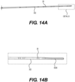
- FIG. 14A is a schematic pictorial representation of a side view of a modified version of the guidewire catheter 302 described with respect to FIGs. 12 and 13 .
- FIG. 14B is a schematic pictorial representation of a detail view of Detail B shown in FIG. 14A .
- the guidewire catheter 302 depicted in these figures is the same as previously described, with the exception that, rather than a guidewire lumen being defined therethrough, the guidewire 302A may be substituted with or further provided with an atraumatic material 302B at a distal end of the guidewire catheter 302, the atraumatic material 302B functioning as a guidewire tip.
Landscapes
- Health & Medical Sciences (AREA)
- Life Sciences & Earth Sciences (AREA)
- Engineering & Computer Science (AREA)
- Surgery (AREA)
- Heart & Thoracic Surgery (AREA)
- General Health & Medical Sciences (AREA)
- Biomedical Technology (AREA)
- Veterinary Medicine (AREA)
- Public Health (AREA)
- Animal Behavior & Ethology (AREA)
- Medical Informatics (AREA)
- Molecular Biology (AREA)
- Nuclear Medicine, Radiotherapy & Molecular Imaging (AREA)
- Physics & Mathematics (AREA)
- Cardiology (AREA)
- Plasma & Fusion (AREA)
- Otolaryngology (AREA)
- Biophysics (AREA)
- Pathology (AREA)
- Robotics (AREA)
- Radiology & Medical Imaging (AREA)
- Human Computer Interaction (AREA)
- Vascular Medicine (AREA)
- Physiology (AREA)
- Surgical Instruments (AREA)
- Measurement And Recording Of Electrical Phenomena And Electrical Characteristics Of The Living Body (AREA)
- Media Introduction/Drainage Providing Device (AREA)
Applications Claiming Priority (1)
| Application Number | Priority Date | Filing Date | Title |
|---|---|---|---|
| US18/525,679 US20250177055A1 (en) | 2023-11-30 | 2023-11-30 | Medical probe for navigating small diameter blood vessels |
Publications (1)
| Publication Number | Publication Date |
|---|---|
| EP4578413A1 true EP4578413A1 (fr) | 2025-07-02 |
Family
ID=93742858
Family Applications (1)
| Application Number | Title | Priority Date | Filing Date |
|---|---|---|---|
| EP24216338.4A Pending EP4578413A1 (fr) | 2023-11-30 | 2024-11-29 | Sonde médicale pour la navigation dans des vaisseaux sanguins de petit diamètre |
Country Status (5)
| Country | Link |
|---|---|
| US (1) | US20250177055A1 (fr) |
| EP (1) | EP4578413A1 (fr) |
| JP (1) | JP2025088771A (fr) |
| CN (1) | CN120053062A (fr) |
| IL (1) | IL317299A (fr) |
Citations (28)
| Publication number | Priority date | Publication date | Assignee | Title |
|---|---|---|---|---|
| US5391199A (en) | 1993-07-20 | 1995-02-21 | Biosense, Inc. | Apparatus and method for treating cardiac arrhythmias |
| US5558091A (en) | 1993-10-06 | 1996-09-24 | Biosense, Inc. | Magnetic determination of position and orientation |
| US6172499B1 (en) | 1999-10-29 | 2001-01-09 | Ascension Technology Corporation | Eddy current error-reduced AC magnetic position measurement system |
| US6239724B1 (en) | 1997-12-30 | 2001-05-29 | Remon Medical Technologies, Ltd. | System and method for telemetrically providing intrabody spatial position |
| US6332089B1 (en) | 1996-02-15 | 2001-12-18 | Biosense, Inc. | Medical procedures and apparatus using intrabody probes |
| US6484118B1 (en) | 2000-07-20 | 2002-11-19 | Biosense, Inc. | Electromagnetic position single axis system |
| US6618612B1 (en) | 1996-02-15 | 2003-09-09 | Biosense, Inc. | Independently positionable transducers for location system |
| US6690963B2 (en) | 1995-01-24 | 2004-02-10 | Biosense, Inc. | System for determining the location and orientation of an invasive medical instrument |
| US20050033135A1 (en) * | 2003-07-29 | 2005-02-10 | Assaf Govari | Lasso for pulmonary vein mapping and ablation |
| US6892091B1 (en) | 2000-02-18 | 2005-05-10 | Biosense, Inc. | Catheter, method and apparatus for generating an electrical map of a chamber of the heart |
| EP1169972B1 (fr) * | 2000-07-07 | 2006-01-18 | Biosense Webster, Inc. | Cathéter pour ablation et mapping |
| US7536218B2 (en) | 2005-07-15 | 2009-05-19 | Biosense Webster, Inc. | Hybrid magnetic-based and impedance-based position sensing |
| US20100079158A1 (en) * | 2008-09-30 | 2010-04-01 | Bar-Tal Meir | Current localization tracker |
| US7756576B2 (en) | 2005-08-26 | 2010-07-13 | Biosense Webster, Inc. | Position sensing and detection of skin impedance |
| US7848787B2 (en) | 2005-07-08 | 2010-12-07 | Biosense Webster, Inc. | Relative impedance measurement |
| US7869865B2 (en) | 2005-01-07 | 2011-01-11 | Biosense Webster, Inc. | Current-based position sensing |
| US20140163550A1 (en) * | 2012-12-11 | 2014-06-12 | William G. Besser | Catheter having reduced force concentration at tissue contact site |
| US20170172456A1 (en) * | 2015-12-22 | 2017-06-22 | Biosense Webster (Israel) Ltd. | Ascertaining a position and orientation for visualizing a tool |
| US20180214215A1 (en) * | 2015-07-30 | 2018-08-02 | St. Jude Medical International Holding S.À R.L. | Roll-sensing sensor assembly |
| US20210162210A1 (en) | 2019-12-03 | 2021-06-03 | Biosense Webster (Israel) Ltd. | Using reversible electroporation on cardiac tissue |
| US20210161592A1 (en) | 2019-12-03 | 2021-06-03 | Biosense Webster (Israel) Ltd. | Pulse Generator for Irreversible Electroporation |
| US20210169568A1 (en) | 2019-12-09 | 2021-06-10 | Biosense Webster (Israel) Ltd. | Oriented irreversible-electroporation (ire) pulses to compensate for cell size and orientation |
| US20210169567A1 (en) | 2019-12-09 | 2021-06-10 | Biosense Webster (Israel) Ltd. | Irreversible-electroporation (ire) balloon catheter with membrane-insulated high-voltage balloon wires |
| US20210169550A1 (en) | 2019-12-05 | 2021-06-10 | Biosense Webster (Israel) Ltd. | Generating and interleaving of irreversible-electroporation and radiofrequnecy ablation (ire/rfa) waveforms |
| US20210177503A1 (en) | 2019-12-11 | 2021-06-17 | Biosense Webster (Israel) Ltd. | Regulating delivery of irreversible electroporation pulses according to transferred energy |
| US20210186604A1 (en) | 2019-12-24 | 2021-06-24 | Biosense Webster (Israel) Ltd. | Irreversible electroporation (ire) based on field, contact force and time |
| US20210196372A1 (en) | 2019-12-31 | 2021-07-01 | Biosense Webster (Israel) Ltd. | Using irrigation on irreversible-electroporation (ire) electrodes to prevent arcing |
| US20210330372A1 (en) * | 2020-04-22 | 2021-10-28 | Biosense Webster (Israel) Ltd. | Ablation of a hard-to-access region |
Family Cites Families (2)
| Publication number | Priority date | Publication date | Assignee | Title |
|---|---|---|---|---|
| US5954665A (en) * | 1995-06-07 | 1999-09-21 | Biosense, Inc. | Cardiac ablation catheter using correlation measure |
| US5727553A (en) * | 1996-03-25 | 1998-03-17 | Saad; Saad A. | Catheter with integral electromagnetic location identification device |
-
2023
- 2023-11-30 US US18/525,679 patent/US20250177055A1/en active Pending
-
2024
- 2024-11-26 IL IL317299A patent/IL317299A/en unknown
- 2024-11-28 CN CN202411724346.6A patent/CN120053062A/zh active Pending
- 2024-11-29 JP JP2024208294A patent/JP2025088771A/ja active Pending
- 2024-11-29 EP EP24216338.4A patent/EP4578413A1/fr active Pending
Patent Citations (32)
| Publication number | Priority date | Publication date | Assignee | Title |
|---|---|---|---|---|
| US5443489A (en) | 1993-07-20 | 1995-08-22 | Biosense, Inc. | Apparatus and method for ablation |
| US5391199A (en) | 1993-07-20 | 1995-02-21 | Biosense, Inc. | Apparatus and method for treating cardiac arrhythmias |
| US5558091A (en) | 1993-10-06 | 1996-09-24 | Biosense, Inc. | Magnetic determination of position and orientation |
| US6690963B2 (en) | 1995-01-24 | 2004-02-10 | Biosense, Inc. | System for determining the location and orientation of an invasive medical instrument |
| US6618612B1 (en) | 1996-02-15 | 2003-09-09 | Biosense, Inc. | Independently positionable transducers for location system |
| US6332089B1 (en) | 1996-02-15 | 2001-12-18 | Biosense, Inc. | Medical procedures and apparatus using intrabody probes |
| US6788967B2 (en) | 1997-05-14 | 2004-09-07 | Biosense, Inc. | Medical diagnosis, treatment and imaging systems |
| US6239724B1 (en) | 1997-12-30 | 2001-05-29 | Remon Medical Technologies, Ltd. | System and method for telemetrically providing intrabody spatial position |
| US6172499B1 (en) | 1999-10-29 | 2001-01-09 | Ascension Technology Corporation | Eddy current error-reduced AC magnetic position measurement system |
| US6892091B1 (en) | 2000-02-18 | 2005-05-10 | Biosense, Inc. | Catheter, method and apparatus for generating an electrical map of a chamber of the heart |
| EP1125549B1 (fr) * | 2000-02-18 | 2006-06-07 | Biosense Webster, Inc. | Catheter pour générer une représentation électrique d'une chambre cardiaque |
| EP1169972B1 (fr) * | 2000-07-07 | 2006-01-18 | Biosense Webster, Inc. | Cathéter pour ablation et mapping |
| US6484118B1 (en) | 2000-07-20 | 2002-11-19 | Biosense, Inc. | Electromagnetic position single axis system |
| US20050033135A1 (en) * | 2003-07-29 | 2005-02-10 | Assaf Govari | Lasso for pulmonary vein mapping and ablation |
| US7869865B2 (en) | 2005-01-07 | 2011-01-11 | Biosense Webster, Inc. | Current-based position sensing |
| US7848787B2 (en) | 2005-07-08 | 2010-12-07 | Biosense Webster, Inc. | Relative impedance measurement |
| US7536218B2 (en) | 2005-07-15 | 2009-05-19 | Biosense Webster, Inc. | Hybrid magnetic-based and impedance-based position sensing |
| US7756576B2 (en) | 2005-08-26 | 2010-07-13 | Biosense Webster, Inc. | Position sensing and detection of skin impedance |
| US20100079158A1 (en) * | 2008-09-30 | 2010-04-01 | Bar-Tal Meir | Current localization tracker |
| US8456182B2 (en) | 2008-09-30 | 2013-06-04 | Biosense Webster, Inc. | Current localization tracker |
| US20140163550A1 (en) * | 2012-12-11 | 2014-06-12 | William G. Besser | Catheter having reduced force concentration at tissue contact site |
| US20180214215A1 (en) * | 2015-07-30 | 2018-08-02 | St. Jude Medical International Holding S.À R.L. | Roll-sensing sensor assembly |
| US20170172456A1 (en) * | 2015-12-22 | 2017-06-22 | Biosense Webster (Israel) Ltd. | Ascertaining a position and orientation for visualizing a tool |
| US20210161592A1 (en) | 2019-12-03 | 2021-06-03 | Biosense Webster (Israel) Ltd. | Pulse Generator for Irreversible Electroporation |
| US20210162210A1 (en) | 2019-12-03 | 2021-06-03 | Biosense Webster (Israel) Ltd. | Using reversible electroporation on cardiac tissue |
| US20210169550A1 (en) | 2019-12-05 | 2021-06-10 | Biosense Webster (Israel) Ltd. | Generating and interleaving of irreversible-electroporation and radiofrequnecy ablation (ire/rfa) waveforms |
| US20210169568A1 (en) | 2019-12-09 | 2021-06-10 | Biosense Webster (Israel) Ltd. | Oriented irreversible-electroporation (ire) pulses to compensate for cell size and orientation |
| US20210169567A1 (en) | 2019-12-09 | 2021-06-10 | Biosense Webster (Israel) Ltd. | Irreversible-electroporation (ire) balloon catheter with membrane-insulated high-voltage balloon wires |
| US20210177503A1 (en) | 2019-12-11 | 2021-06-17 | Biosense Webster (Israel) Ltd. | Regulating delivery of irreversible electroporation pulses according to transferred energy |
| US20210186604A1 (en) | 2019-12-24 | 2021-06-24 | Biosense Webster (Israel) Ltd. | Irreversible electroporation (ire) based on field, contact force and time |
| US20210196372A1 (en) | 2019-12-31 | 2021-07-01 | Biosense Webster (Israel) Ltd. | Using irrigation on irreversible-electroporation (ire) electrodes to prevent arcing |
| US20210330372A1 (en) * | 2020-04-22 | 2021-10-28 | Biosense Webster (Israel) Ltd. | Ablation of a hard-to-access region |
Also Published As
| Publication number | Publication date |
|---|---|
| IL317299A (en) | 2025-06-01 |
| CN120053062A (zh) | 2025-05-30 |
| JP2025088771A (ja) | 2025-06-11 |
| US20250177055A1 (en) | 2025-06-05 |
Similar Documents
| Publication | Publication Date | Title |
|---|---|---|
| EP4268748A2 (fr) | Raccord d'irrigation pour cathéter d'ablation | |
| EP4520284A1 (fr) | Ensemble électrode extensible comprenant une extrémité distale étendue pour un cathéter médical | |
| EP4578413A1 (fr) | Sonde médicale pour la navigation dans des vaisseaux sanguins de petit diamètre | |
| EP4574074A1 (fr) | Cathéter de cartographie et d'ablation | |
| EP4527333A1 (fr) | Dispositif médical avec un collecteur d'irrigation | |
| US20250194983A1 (en) | Medical probe with slitted tube and electrodes | |
| US20250099168A1 (en) | Medical device with a monolithic spine framework | |
| US20250099160A1 (en) | Medical device with an end effector including connecting hubs and an electrode array | |
| US20250195132A1 (en) | Medical probe with spines for pulmonary vein isolation | |
| EP4541303A1 (fr) | Électrodes de circuit flexible pour cathéter lasso | |
| US20240216054A1 (en) | Systems and methods for cylindrical cage mapping and ablation catheters comprising flexible circuits | |
| US20240382251A1 (en) | Electrodes and electrode configurations for a basket catheter | |
| EP4473928A1 (fr) | Ensembles panier extensibles et ensembles panier extensibles avec soulagement de contrainte de fil d'électrode | |
| US20250025624A1 (en) | Flexible irrigation tube for a medical catheter | |
| EP4606334A1 (fr) | Electrodes annulaires modulaires | |
| US20240374304A1 (en) | Electrical connector for a catheter device | |
| IL309727A (en) | Irrigation Hub for an Ablation Catheter | |
| IL318024A (en) | Flow diverter for a catheter |
Legal Events
| Date | Code | Title | Description |
|---|---|---|---|
| PUAI | Public reference made under article 153(3) epc to a published international application that has entered the european phase |
Free format text: ORIGINAL CODE: 0009012 |
|
| STAA | Information on the status of an ep patent application or granted ep patent |
Free format text: STATUS: THE APPLICATION HAS BEEN PUBLISHED |
|
| AK | Designated contracting states |
Kind code of ref document: A1 Designated state(s): AL AT BE BG CH CY CZ DE DK EE ES FI FR GB GR HR HU IE IS IT LI LT LU LV MC ME MK MT NL NO PL PT RO RS SE SI SK SM TR |
|
| STAA | Information on the status of an ep patent application or granted ep patent |
Free format text: STATUS: REQUEST FOR EXAMINATION WAS MADE |
|
| 17P | Request for examination filed |
Effective date: 20251230 |
|
| STAA | Information on the status of an ep patent application or granted ep patent |
Free format text: STATUS: EXAMINATION IS IN PROGRESS |