ES2875762T3 - Therapeutic use of specific ligand in MSRV-associated diseases - Google Patents
Therapeutic use of specific ligand in MSRV-associated diseases Download PDFInfo
- Publication number
- ES2875762T3 ES2875762T3 ES17159699T ES17159699T ES2875762T3 ES 2875762 T3 ES2875762 T3 ES 2875762T3 ES 17159699 T ES17159699 T ES 17159699T ES 17159699 T ES17159699 T ES 17159699T ES 2875762 T3 ES2875762 T3 ES 2875762T3
- Authority
- ES
- Spain
- Prior art keywords
- env
- ligand
- seq
- antibody
- msrv
- Prior art date
- Legal status (The legal status is an assumption and is not a legal conclusion. Google has not performed a legal analysis and makes no representation as to the accuracy of the status listed.)
- Active
Links
Classifications
-
- C—CHEMISTRY; METALLURGY
- C07—ORGANIC CHEMISTRY
- C07K—PEPTIDES
- C07K16/00—Immunoglobulins [IG], e.g. monoclonal or polyclonal antibodies
- C07K16/08—Immunoglobulins [IG], e.g. monoclonal or polyclonal antibodies against material from viruses
- C07K16/10—RNA viruses
- C07K16/112—Retroviridae (F), e.g. leukemia viruses
-
- A—HUMAN NECESSITIES
- A61—MEDICAL OR VETERINARY SCIENCE; HYGIENE
- A61K—PREPARATIONS FOR MEDICAL, DENTAL OR TOILETRY PURPOSES
- A61K39/00—Medicinal preparations containing antigens or antibodies
- A61K39/395—Antibodies; Immunoglobulins; Immune serum, e.g. antilymphocytic serum
-
- A—HUMAN NECESSITIES
- A61—MEDICAL OR VETERINARY SCIENCE; HYGIENE
- A61P—SPECIFIC THERAPEUTIC ACTIVITY OF CHEMICAL COMPOUNDS OR MEDICINAL PREPARATIONS
- A61P1/00—Drugs for disorders of the alimentary tract or the digestive system
- A61P1/18—Drugs for disorders of the alimentary tract or the digestive system for pancreatic disorders, e.g. pancreatic enzymes
-
- A—HUMAN NECESSITIES
- A61—MEDICAL OR VETERINARY SCIENCE; HYGIENE
- A61P—SPECIFIC THERAPEUTIC ACTIVITY OF CHEMICAL COMPOUNDS OR MEDICINAL PREPARATIONS
- A61P17/00—Drugs for dermatological disorders
- A61P17/06—Antipsoriatics
-
- A—HUMAN NECESSITIES
- A61—MEDICAL OR VETERINARY SCIENCE; HYGIENE
- A61P—SPECIFIC THERAPEUTIC ACTIVITY OF CHEMICAL COMPOUNDS OR MEDICINAL PREPARATIONS
- A61P25/00—Drugs for disorders of the nervous system
-
- A—HUMAN NECESSITIES
- A61—MEDICAL OR VETERINARY SCIENCE; HYGIENE
- A61P—SPECIFIC THERAPEUTIC ACTIVITY OF CHEMICAL COMPOUNDS OR MEDICINAL PREPARATIONS
- A61P25/00—Drugs for disorders of the nervous system
- A61P25/08—Antiepileptics; Anticonvulsants
-
- A—HUMAN NECESSITIES
- A61—MEDICAL OR VETERINARY SCIENCE; HYGIENE
- A61P—SPECIFIC THERAPEUTIC ACTIVITY OF CHEMICAL COMPOUNDS OR MEDICINAL PREPARATIONS
- A61P25/00—Drugs for disorders of the nervous system
- A61P25/18—Antipsychotics, i.e. neuroleptics; Drugs for mania or schizophrenia
-
- A—HUMAN NECESSITIES
- A61—MEDICAL OR VETERINARY SCIENCE; HYGIENE
- A61P—SPECIFIC THERAPEUTIC ACTIVITY OF CHEMICAL COMPOUNDS OR MEDICINAL PREPARATIONS
- A61P29/00—Non-central analgesic, antipyretic or antiinflammatory agents, e.g. antirheumatic agents; Non-steroidal antiinflammatory drugs [NSAID]
-
- A—HUMAN NECESSITIES
- A61—MEDICAL OR VETERINARY SCIENCE; HYGIENE
- A61P—SPECIFIC THERAPEUTIC ACTIVITY OF CHEMICAL COMPOUNDS OR MEDICINAL PREPARATIONS
- A61P3/00—Drugs for disorders of the metabolism
- A61P3/08—Drugs for disorders of the metabolism for glucose homeostasis
- A61P3/10—Drugs for disorders of the metabolism for glucose homeostasis for hyperglycaemia, e.g. antidiabetics
-
- A—HUMAN NECESSITIES
- A61—MEDICAL OR VETERINARY SCIENCE; HYGIENE
- A61P—SPECIFIC THERAPEUTIC ACTIVITY OF CHEMICAL COMPOUNDS OR MEDICINAL PREPARATIONS
- A61P31/00—Antiinfectives, i.e. antibiotics, antiseptics, chemotherapeutics
- A61P31/12—Antivirals
-
- A—HUMAN NECESSITIES
- A61—MEDICAL OR VETERINARY SCIENCE; HYGIENE
- A61P—SPECIFIC THERAPEUTIC ACTIVITY OF CHEMICAL COMPOUNDS OR MEDICINAL PREPARATIONS
- A61P31/00—Antiinfectives, i.e. antibiotics, antiseptics, chemotherapeutics
- A61P31/12—Antivirals
- A61P31/14—Antivirals for RNA viruses
-
- A—HUMAN NECESSITIES
- A61—MEDICAL OR VETERINARY SCIENCE; HYGIENE
- A61P—SPECIFIC THERAPEUTIC ACTIVITY OF CHEMICAL COMPOUNDS OR MEDICINAL PREPARATIONS
- A61P35/00—Antineoplastic agents
-
- A—HUMAN NECESSITIES
- A61—MEDICAL OR VETERINARY SCIENCE; HYGIENE
- A61P—SPECIFIC THERAPEUTIC ACTIVITY OF CHEMICAL COMPOUNDS OR MEDICINAL PREPARATIONS
- A61P37/00—Drugs for immunological or allergic disorders
- A61P37/02—Immunomodulators
- A61P37/04—Immunostimulants
-
- A—HUMAN NECESSITIES
- A61—MEDICAL OR VETERINARY SCIENCE; HYGIENE
- A61P—SPECIFIC THERAPEUTIC ACTIVITY OF CHEMICAL COMPOUNDS OR MEDICINAL PREPARATIONS
- A61P43/00—Drugs for specific purposes, not provided for in groups A61P1/00-A61P41/00
-
- A—HUMAN NECESSITIES
- A61—MEDICAL OR VETERINARY SCIENCE; HYGIENE
- A61K—PREPARATIONS FOR MEDICAL, DENTAL OR TOILETRY PURPOSES
- A61K39/00—Medicinal preparations containing antigens or antibodies
- A61K2039/505—Medicinal preparations containing antigens or antibodies comprising antibodies
-
- C—CHEMISTRY; METALLURGY
- C07—ORGANIC CHEMISTRY
- C07K—PEPTIDES
- C07K2317/00—Immunoglobulins specific features
- C07K2317/20—Immunoglobulins specific features characterized by taxonomic origin
- C07K2317/24—Immunoglobulins specific features characterized by taxonomic origin containing regions, domains or residues from different species, e.g. chimeric, humanized or veneered
-
- C—CHEMISTRY; METALLURGY
- C07—ORGANIC CHEMISTRY
- C07K—PEPTIDES
- C07K2317/00—Immunoglobulins specific features
- C07K2317/50—Immunoglobulins specific features characterized by immunoglobulin fragments
- C07K2317/56—Immunoglobulins specific features characterized by immunoglobulin fragments variable (Fv) region, i.e. VH and/or VL
-
- C—CHEMISTRY; METALLURGY
- C07—ORGANIC CHEMISTRY
- C07K—PEPTIDES
- C07K2317/00—Immunoglobulins specific features
- C07K2317/50—Immunoglobulins specific features characterized by immunoglobulin fragments
- C07K2317/56—Immunoglobulins specific features characterized by immunoglobulin fragments variable (Fv) region, i.e. VH and/or VL
- C07K2317/565—Complementarity determining region [CDR]
-
- C—CHEMISTRY; METALLURGY
- C07—ORGANIC CHEMISTRY
- C07K—PEPTIDES
- C07K2317/00—Immunoglobulins specific features
- C07K2317/90—Immunoglobulins specific features characterized by (pharmaco)kinetic aspects or by stability of the immunoglobulin
- C07K2317/92—Affinity (KD), association rate (Ka), dissociation rate (Kd) or EC50 value
Landscapes
- Health & Medical Sciences (AREA)
- Life Sciences & Earth Sciences (AREA)
- Chemical & Material Sciences (AREA)
- Organic Chemistry (AREA)
- Medicinal Chemistry (AREA)
- General Health & Medical Sciences (AREA)
- Animal Behavior & Ethology (AREA)
- Veterinary Medicine (AREA)
- Public Health (AREA)
- Pharmacology & Pharmacy (AREA)
- General Chemical & Material Sciences (AREA)
- Nuclear Medicine, Radiotherapy & Molecular Imaging (AREA)
- Chemical Kinetics & Catalysis (AREA)
- Bioinformatics & Cheminformatics (AREA)
- Engineering & Computer Science (AREA)
- Virology (AREA)
- Immunology (AREA)
- Diabetes (AREA)
- Neurosurgery (AREA)
- Neurology (AREA)
- Biomedical Technology (AREA)
- Pain & Pain Management (AREA)
- Molecular Biology (AREA)
- Genetics & Genomics (AREA)
- Biochemistry (AREA)
- Proteomics, Peptides & Aminoacids (AREA)
- Biophysics (AREA)
- Hematology (AREA)
- Oncology (AREA)
- Communicable Diseases (AREA)
- Dermatology (AREA)
- Psychiatry (AREA)
- Rheumatology (AREA)
- Emergency Medicine (AREA)
- Obesity (AREA)
- Endocrinology (AREA)
- Epidemiology (AREA)
- Mycology (AREA)
- Microbiology (AREA)
- Peptides Or Proteins (AREA)
Abstract
Un ligando que comprende - las tres regiones determinantes de complementariedad (CDR) de la región variable de cadena ligera (VL): CDR1, CDR2 y CDR3 respectivamente expuestas en la SEQ ID NO: 1, SEQ ID NO: 2, SEQ ID NO: 3, - las tres CDR de la región variable de cadena pesada (VH): CDR4, CDR5 y CDR6 respectivamente expuestas en la SEQ ID NO: 4, SEQ ID NO: 5 y SEQ ID NO: 6, en el que dicho ligando se une al antiligando que consiste en la secuencia de aminoácidos definida por la SEQ ID NO: 20 o SEQ ID NO: 32, y dicho ligando se selecciona del grupo que consiste en un anticuerpo, un fragmento scFv y un fragmento Fab, y en el que dicho ligando es para su uso en un método de tratamiento.A ligand comprising - the three complementarity determining regions (CDRs) of the light chain variable region (VL): CDR1, CDR2 and CDR3 respectively set forth in SEQ ID NO: 1, SEQ ID NO: 2, SEQ ID NO: 3, - the three CDRs of the heavy chain variable region (VH): CDR4, CDR5 and CDR6 respectively exposed in SEQ ID NO: 4, SEQ ID NO: 5 and SEQ ID NO: 6, in which said ligand is binds to the anti-ligand consisting of the amino acid sequence defined by SEQ ID NO: 20 or SEQ ID NO: 32, and said ligand is selected from the group consisting of an antibody, a scFv fragment, and a Fab fragment, and wherein said ligand is for use in a treatment method.
Description
DESCRIPCIÓNDESCRIPTION
Uso terapéutico de ligando específico en enfermedades asociadas a MSRVTherapeutic use of specific ligand in MSRV-associated diseases
Sector de la técnicaTechnical sector
El objetivo de la presente solicitud es un ligando que presenta unión significativa a una molécula diana, el antiligando.The target of the present application is a ligand that exhibits significant binding to a target molecule, the antiligand.
Estado de la técnicaState of the art
De acuerdo con la presente invención, el "antiligando" es la proteína ENV (envoltura) de MSRV, MSRV para "retrovirus asociado a esclerosis múltiple" (Perron, et al. (1997). "Molecular identification of a novel retrovirus repeatedly isolated from patients with multiple sclerosis. The Collaborative Research Group on Multiple Sclerosis." Proc Natl Acad Sci USA 94(14): 7583-8.). La "proteína ENV de MSRV" debe entenderse como el producto completo o parcial codificado por genes ENV de MSRV como se define en Komurian-Pradel, F, et al. (1999). Virology 260(1): 1-9, y Rolland A, et al. (2006) J Immunol 176(12): 7636-44 o cualquier molécula que imite las propiedades antigénicas o de unión de ENV de MSRV (mimótopo). "ENV-T" se corresponde a la proteína completa, que se detalla en el ejemplo 2 (restos 1 a 542) y "ENV-SU" también llamada ENV-1 corresponde a la secuencia S30 a K316 como se detalla en el ejemplo 2. ENV-SU también se menciona en Rolland A, et al. (2006) J Immunol 176(12): 7636 44. De forma habitual para retrovirus, MSRV muestra variabilidad en su proteína de envoltura ENV (Perron, H et al. (2000) J Neurovirol 6: S67-75; Voisset, C., O. Bouton, et al. (2000) AIDS Res Hum Retroviruses 16(8): 731-40). Los mimótopos que imitan fragmentos proteicos parciales de ENV de MSRV han demostrado existir y unirse selectivamente por anticuerpos de pacientes con esclerosis múltiple (Jolivet-Reynaud, C., H. Perron, et al. (1999). "Specificities of multiple sclerosis cerebrospinal fluid and serum antibodies against mimotopes." Clin Immunol 93(3): 283-93 PERRON HERVE ET AL: "Monoclonal antibody against envelope protein from Human Endogenous Retrovirus "W' (MSRV-ENV or Syncytin) inhibits TLR4-initated immunotoxicity cascade in human peripheral blood mononuclear cells and displays therapeutic effects in novel preclinical models for Multiple Sclerosis",JOURNAL OF NEUROVIROLOGY, vol. 13, n.° Supl. 1, 2007, páginas 112-113, y 8TH INTERNATIONAL SYMPOSIUM ON NEUROVIROLOGY; SAN DIEGO, CA, EE. UU.; 30 de octubre-2 de noviembre de 2007 ISSN: 1355-0284 desvelan el uso de un anticuerpo anti-MSRV para tartar encefalomielitis alérgica experimental EAE inducida por MRSV/ENV en ratones SCID humanizados. No hay, sin embargo, ninguna divulgación de secuencias de anticuerpos en este resumen.According to the present invention, the "antiligand" is the ENV (envelope) protein of MSRV, MSRV for "multiple sclerosis associated retrovirus" (Perron, et al. (1997). "Molecular identification of a novel retrovirus repeatedly isolated from patients with multiple sclerosis. The Collaborative Research Group on Multiple Sclerosis. "Proc Natl Acad Sci USA 94 (14): 7583-8.). The "MSRV ENV protein" is to be understood as the complete or partial product encoded by MSRV ENV genes as defined in Komurian-Pradel, F, et al. (1999). Virology 260 (1): 1-9, and Rolland A, et al. (2006) J Immunol 176 (12): 7636-44 or any molecule that mimics the antigenic or ENV-binding properties of MSRV (mimotope). "ENV-T" corresponds to the complete protein, which is detailed in example 2 (residues 1 to 542) and "ENV-SU" also called ENV-1 corresponds to the sequence S30 to K316 as detailed in example 2 ENV-SU is also mentioned in Rolland A, et al. (2006) J Immunol 176 (12): 7636 44. Commonly for retroviruses, MSRV shows variability in its envelope protein ENV (Perron, H et al. (2000) J Neurovirol 6: S67-75; Voisset, C. , O. Bouton, et al. (2000) AIDS Res Hum Retroviruses 16 (8): 731-40). Mimotopes that mimic partial protein fragments of MSRV ENV have been shown to exist and selectively bind by antibodies from multiple sclerosis patients (Jolivet-Reynaud, C., H. Perron, et al. (1999). "Specificities of multiple sclerosis cerebrospinal fluid and serum antibodies against mimotopes. "Clin Immunol 93 (3): 283-93 PERRON HERVE ET AL: " Monoclonal antibody against envelope protein from Human Endogenous Retrovirus "W '(MSRV-ENV or Syncytin) inhibits TLR4-initated immunotoxicity cascade in human peripheral blood mononuclear cells and displays therapeutic effects in novel preclinical models for Multiple Sclerosis ", JOURNAL OF NEUROVIROLOGY, vol. 13, no. Suppl. 1, 2007, pages 112-113, and 8TH INTERNATIONAL SYMPOSIUM ON NEUROVIROLOGY; SAN DIEGO, CA , USA; October 30-November 2, 2007 ISSN: 1355-0284 disclose the use of an anti-MSRV antibody to treat experimental allergic encephalomyelitis MRSV / ENV-induced EAE in humanized SCID mice. However, no antibody sequence disclosure in this abstract.
Objeto de la invenciónObject of the invention
La invención se refiere a un ligando como se define en las reivindicaciones.The invention relates to a ligand as defined in the claims.
La presente invención se refiere a un ligando que se une al antiligando que consiste en la secuencia de aminoácidos definida por la SEQ ID NO: 20 o SEQ ID NO: 32 y que comprende cada una de las regiones determinantes de complementariedad (CDR) que tiene las secuencias de aminoácidos SEQ ID NO: 1, SEQ ID NO: 2, SEQ ID NO: 3, SEQ ID NO: 4, SEQ ID NO: 5 y SEQ ID NO: 6 y la presente descripción también desvela cualquier secuencia que tenga:The present invention refers to a ligand that binds to the antiligand that consists of the amino acid sequence defined by SEQ ID NO: 20 or SEQ ID NO: 32 and that comprises each of the complementarity determining regions (CDR) that has the amino acid sequences SEQ ID NO: 1, SEQ ID NO: 2, SEQ ID NO: 3, SEQ ID NO: 4, SEQ ID NO: 5 and SEQ ID NO: 6 and the present description also discloses any sequence having:
varios aminoácidos sustituidos en dichas secuencias como se indica a continuación, y que se sabe que son factibles para obtener secuencias de aminoácidos funcionalmente equivalentes (Huang 1986; Zabin, Horvath et al. 1991; Edgar y Schwartz 1992; Sardana, Emini et al. 1992; Xu, Kapfer et al. 1992; Lamande y Bateman 1993; Verdoliva, Ruvo et al. 1995; Yu, Schurr et al. 1995; Wehrmann, Van Vliet et al. 1996; Ullmann, Hauswald et al. various amino acids substituted in such sequences as indicated below, and known to be feasible to obtain functionally equivalent amino acid sequences (Huang 1986; Zabin, Horvath et al. 1991; Edgar and Schwartz 1992; Sardana, Emini et al. 1992 ; Xu, Kapfer et al. 1992; Lamande and Bateman 1993; Verdoliva, Ruvo et al. 1995; Yu, Schurr et al. 1995; Wehrmann, Van Vliet et al. 1996; Ullmann, Hauswald et al.
1997; Minuth, Kramer et al. 1998; Ullmann, Hauswald et al. 2000; Janke, Martin et al. 2003): de 0 a 3 en CDR1 (SEQ ID NO: 1), de 0 a 2 en CDR2 (SEQ ID NO: 2), de 0 a 2 en CDR3 (SEQ ID NO: 3), de 0 a 1 en CDR4 (SEQ ID NO: 4), de 0 a 4 en CDR5 (SEQ ID NO: 5), de o a 2 en CDR6 (SEQ ID NO: 6), o1997; Minuth, Kramer et al. 1998; Ullmann, Hauswald et al. 2000; Janke, Martin et al. 2003): 0 to 3 in CDR1 (SEQ ID NO: 1), 0 to 2 in CDR2 (SEQ ID NO: 2), 0 to 2 in CDR3 (SEQ ID NO: 3), 0 to 1 in CDR4 (SEQ ID NO: 4), from 0 to 4 in CDR5 (SEQ ID NO: 5), from or to 2 in CDR6 (SEQ ID NO: 6), or
- aminoácidos sustituidos con otros aminoácidos que tienen funciones o propiedades químicas equivalentes, bien conocidos por los expertos en la materia (también llamados "similitud de aminoácidos") como se indica, por ejemplo, en la siguiente lista de aminoácidos similares (código de una letra): G o A, F o Y, D o E, N o Q, K o R o H, S o T, C o M, V o L o I, W o P, y/o sustituidos de acuerdo con la técnica anterior (Huang 1986; Zabin, Horvath et al. 1991; Edgar y Schwartz 1992; Sardana, Emini et al. 1992; Xu, Kapfer et al. 1992; Lamande y Bateman 1993; Verdoliva, Ruvo et al. 1995; Yu, Schurr et al. 1995; Wehrmann, Van Vliet et al. 1996; Ullmann, Hauswald et al. 1997; Minuth, Kramer et al. 1998; Ullmann, Hauswald et al. 2000; Janke, Martin et al. 2003) dentro de dicha secuencias SEQ ID NO: 1 a SEQ ID NO: 6.- amino acids substituted with other amino acids that have equivalent chemical functions or properties, well known to those skilled in the art (also called "amino acid similarity") as indicated, for example, in the following list of similar amino acids (one letter code ): G or A, F or Y, D or E, N or Q, K or R or H, S or T, C or M, V or L or I, W or P, and / or substituted according to the prior art (Huang 1986; Zabin, Horvath et al. 1991; Edgar and Schwartz 1992; Sardana, Emini et al. 1992; Xu, Kapfer et al. 1992; Lamande and Bateman 1993; Verdoliva, Ruvo et al. 1995; Yu, Schurr et al. 1995; Wehrmann, Van Vliet et al. 1996; Ullmann, Hauswald et al. 1997; Minuth, Kramer et al. 1998; Ullmann, Hauswald et al. 2000; Janke, Martin et al. 2003) within said sequences SEQ ID NO: 1 to SEQ ID NO: 6.
Estas variantes son el resultado de deleciones, adiciones o sustituciones de aminoácidos en los péptidos de las SEQ ID NO: 1 a 6 y también están abarcadas por la presente divulgación y pueden obtenerse por métodos conocidos en la técnica tales como por mutagénesis dirigida al sitio o por síntesis química.These variants are the result of amino acid deletions, additions or substitutions in the peptides of SEQ ID NO: 1 to 6 and are also encompassed by the present disclosure and can be obtained by methods known in the art such as site-directed mutagenesis or by chemical synthesis.
Los ligandos de la presente invención tienen la capacidad de unirse al antiligando de la presente invención. The ligands of the present invention have the ability to bind the antiligand of the present invention.
De acuerdo con la presente invención, por la expresión "unir" o "unión" debe entenderse que el ligando reconoce significativamente el antiligando de acuerdo con los criterios dados en el ejemplo 5.In accordance with the present invention, by the term "binding" or "binding" it is to be understood that the ligand significantly recognizes the antiligand according to the criteria given in Example 5.
La presente divulgación también describe un ligando que comprende una región variable de cadena ligera (VL) que comprende las regiones determinantes de complementariedad (CDR) que tienen las secuencias de aminoácidos SEQ ID NO: 1, SEQ ID NO: 2 y SEQ ID NO: 3 o cualquier secuencia que tenga al menos un 80 % de identidad y más preferiblemente un 90 % de identidad con dichas secuencias, y un dominio de región variable de cadena pesada (VH) que comprende las CDR que tienen las secuencias de aminoácidos SEQ ID NO: 4, SEQ ID NO: 5 y SEQ ID NO: 6 o cualquier secuencia que tenga al menos un 80 % de identidad y más preferiblemente un 90 % de identidad con dichas secuencias.The present disclosure also describes a ligand comprising a light chain variable region (VL) comprising the complementarity determining regions (CDR) having the amino acid sequences SEQ ID NO: 1, SEQ ID NO: 2 and SEQ ID NO: 3 or any sequence that has at least 80% identity and more preferably 90% identity with said sequences, and a heavy chain variable region (VH) domain comprising CDRs having the amino acid sequences SEQ ID NO : 4, SEQ ID NO: 5 and SEQ ID NO: 6 or any sequence that has at least 80% identity and more preferably 90% identity with said sequences.
En un aspecto adicional, el ligando comprende una región variable de cadena ligera (VL) que tiene la secuencia de aminoácidos expuesta en la SEQ ID NO: 7 o cualquier secuencia que tenga al menos un 75 % de identidad y más preferiblemente un 80 % e incluso más preferiblemente un 90 % de identidad con dicha secuencia y una región variable de cadena pesada (VH) que tiene la secuencia de aminoácidos expuesta en la SEQ ID NO: 8 o cualquier secuencia que tenga al menos un 75 % de identidad y más preferiblemente un 80 % e incluso más referiblemente un 90 % de identidad con dicha secuencia.In a further aspect, the ligand comprises a light chain variable region (VL) having the amino acid sequence set forth in SEQ ID NO: 7 or any sequence having at least 75% identity and more preferably 80% identity. even more preferably 90% identity with said sequence and a heavy chain variable region (VH) having the amino acid sequence set forth in SEQ ID NO: 8 or any sequence having at least 75% identity and more preferably 80% and even more referable 90% identity with said sequence.
Las variantes de estas secuencias VH y VL según la presente invención se unen significativamente al antiligando. "Identidad de secuencia" significa, por ejemplo, que en una secuencia que tiene un 80 % de identidad de secuencia, están presentes un 80 % de aminoácidos idénticos en la misma posición tras la alineación de las secuencias, pudiendo realizarse dicha alineación por métodos conocidos en la técnica tales como los descritos en Sequence -Evolution - Function Computational Approaches in Comparative genomics. Koonon E. et al., 2003: Kluwer Academic Publishers o de acuerdo con los parámetros por defecto del libro de instrucciones del software "Mac Vector" (Reino Unido).Variants of these VH and VL sequences according to the present invention bind significantly to the antiligand. "Sequence identity" means, for example, that in a sequence that has 80% sequence identity, 80% identical amino acids are present in the same position after alignment of the sequences, such alignment can be performed by known methods in the art such as those described in Sequence-Evolution-Function Computational Approaches in Comparative genomics. Koonon E. et al., 2003: Kluwer Academic Publishers or according to the default parameters of the "Mac Vector" software instruction book (UK).
El ligando de la presente invención también puede definirse como comprendido dentro de una proteína scFv recombinante.The ligand of the present invention can also be defined as comprised within a recombinant scFv protein.
De acuerdo con aspectos adicionales de la invención, el ligando puede estar comprendido en un fragmento Fab, en un anticuerpo, dicho anticuerpo puede ser un anticuerpo policlonal, monoclonal, oligoclonal, quimerizado, modificado por ingeniería o humanizado. En un aspecto particular de la invención, el anticuerpo que comprende ligando es una IgG humana y más particularmente una IgG1 o una IgG4.According to further aspects of the invention, the ligand may be comprised of a Fab fragment, in an antibody, said antibody may be a polyclonal, monoclonal, oligoclonal, chimerized, engineered or humanized antibody. In a particular aspect of the invention, the antibody comprising ligand is a human IgG and more particularly an IgG1 or an IgG4.
Los anticuerpos policlonales, oligoclonales, monoclonales pueden producirse por métodos clásicos tales como los descritos por Kohler y Milstein (1975) o usando los procedimientos descritos en Sambrook et al., A Guide to Molecular Cloning, A Laboratory Manual, 2.a edición (1989) usando los antiligandos descritos anteriormente.Monoclonal, oligoclonal, polyclonal antibodies can be produced by classical methods such as those described by Kohler and Milstein (1975) or using the procedures described in Sambrook et al., A Guide to Molecular Cloning, A Laboratory Manual, 2nd edition (1989 ) using the antiligands described above.
Más particularmente, el antiligando de la presente invención que se usa para obtener los anticuerpos es el antiligando que consiste en la SEQ ID NO: 20 o la SEQ ID NO: 32 o, como se desvela, cualquier secuencia que tenga al menos 75 % de identidad de secuencia con las secuencias expuestas en la SEQ ID NO: 20 o la SEQ ID NO: 32 o cualquier secuencia 100 % complementaria con las mismas.More particularly, the antiligand of the present invention that is used to obtain the antibodies is the antiligand consisting of SEQ ID NO: 20 or SEQ ID NO: 32 or, as disclosed, any sequence having at least 75% of sequence identity with the sequences set forth in SEQ ID NO: 20 or SEQ ID NO: 32 or any sequence 100% complementary thereto.
En particular, habitualmente se usa en inmunización en forma de un péptido unido a una proteína vehículo tal como seroalbúmina o KLH.In particular, it is usually used in immunization in the form of a peptide linked to a carrier protein such as serum albumin or KLH.
La invención presente divulgación también se refiere a una composición farmacéutica que comprende dicho ligando como un principio activo. Este ligando también puede estar presente en la composición farmacéutica en forma de un fragmente scFv, Fab o de un anticuerpo.The present invention disclosure also refers to a pharmaceutical composition comprising said ligand as an active principle. This ligand can also be present in the pharmaceutical composition in the form of an scFv, Fab fragment or of an antibody.
La composición farmacéutica se usa para tratar enfermedades asociadas a MSRV.The pharmaceutical composition is used to treat diseases associated with MSRV.
Dentro del significado de la presente divulgación "tratamiento" abarca tratamientos profilácticos o curativos.Within the meaning of the present disclosure "treatment" encompasses prophylactic or curative treatments.
La composición farmacéutica se administra en cantidades que serán terapéuticamente eficaces e inmunogénicas y conocidas en la técnica, la dosificación que se administra depende del individuo a tratar.The pharmaceutical composition is administered in amounts that will be therapeutically effective and immunogenic and known in the art, the dosage that is administered depends on the individual to be treated.
En un aspecto adicional, la presente solicitud trata de un método de tratamiento que comprende administrar el ligando, el scFv, el fragmento Fab o el anticuerpo o el ligando en cualquier vector molecular o terapéutico adecuado manteniendo sus propiedades de unión como se describe anteriormente o la composición farmacéutica como se describe anteriormente.In a further aspect, the present application deals with a method of treatment that comprises administering the ligand, the scFv, the Fab fragment or the antibody or the ligand in any suitable molecular or therapeutic vector maintaining its binding properties as described above or the pharmaceutical composition as described above.
El método de tratamiento está dirigido al tratamiento de enfermedades asociadas a MSRV.The treatment method is aimed at the treatment of diseases associated with MSRV.
MSRV, es un retrovirus humano aislado por primera vez en pacientes con esclerosis múltiple (Perron, H., B. MSRV, is a human retrovirus isolated for the first time in patients with multiple sclerosis (Perron, H., B.
Lalande, et al. (1991), Lancet 337(8745): 862-3; Perron, H., J. A. Garson, et al. (1997), Proc Natl Acad Sci USA 94(14): 7583-8). Las enfermedades asociadas o síndromes se definen por la presencia en los pacientes correspondientes (i) de ARN o antígenos específicos de MSRV, preferiblemente detectados en fluidos corporales (sangre, líquido cefalorraquídeo, orina), (ii) de un número de copias elevado de ADN o ARN en células o tejidos de órganos con lesiones o disfunciones, (iii) de proteínas o antígenos específicos de MSRV en células o tejidos implicados en el proceso de la enfermedad o del síndrome clínico, o (iv) de proteínas o antígenos de MSRV en fluidos corporales de individuos con la enfermedad o que expresan el síndrome clínico (como se describe en el ejemplo 8, véase a continuación y otros a continuación). Como MSRV tiene homología genética con la familia de retrovirus endógeno humano de tipo W (HERVW) (Blond et al., 1999; Dolei, 2005; Dolei y Perron, 2008) también pueden obtenerse definiciones alternativas o complementarias de enfermedades asociadas a MSRV con ácidos nucleicos de HERV-W, antígenos o proteínas usados para los mismos ensayos de detección (Antony et al., 2004; Arru et al., 2007; Karlsson et al., 2004; Mameli et al., 2007). Además, la expresión de MSRV puede asociarse con la regulación positiva de ciertas copias de HERV-W codetectadas en lesiones patogénicas (Mameli et al., 2009). Por tanto, la definición de enfermedades asociadas a MSRV comprende de forma implícita la asociación con elementos de HERV-W.Lalande, et al. (1991), Lancet 337 (8745): 862-3; Perron, H., JA Garson, et al. (1997), Proc Natl Acad Sci USA 94 (14): 7583-8). Associated diseases or syndromes are defined by the presence in the corresponding patients (i) of MSRV-specific RNA or antigens, preferably detected in body fluids (blood, cerebrospinal fluid, urine), (ii) of a high copy number of DNA o RNA in cells or tissues of organs with lesions or dysfunctions, (iii) of MSRV-specific proteins or antigens in cells or tissues involved in the disease process or clinical syndrome, or (iv) of MSRV proteins or antigens in bodily fluids of individuals with the disease or expressing the clinical syndrome (as described in Example 8, see below and others below). As MSRV has genetic homology with the human endogenous retrovirus type W (HERVW) family (Blond et al., 1999; Dolei, 2005; Dolei and Perron, 2008), alternative or complementary definitions of MSRV-associated diseases with acids can also be obtained. HERV-W nucleic agents, antigens or proteins used for the same detection assays (Antony et al., 2004; Arru et al., 2007; Karlsson et al., 2004; Mameli et al., 2007). Furthermore, MSRV expression may be associated with upregulation of certain codetected copies of HERV-W in pathogenic lesions (Mameli et al., 2009). Therefore, the definition of MSRV-associated diseases implicitly encompasses the association with elements of HERV-W.
La enfermedad asociada a MSRV se selecciona del grupo que comprende esclerosis múltiple, esquizofrenia, síndrome aislado clínicamente (CIS, con síntomas neurológicos), polineuropatía desmielinizante inflamatoria crónica, epilepsia, psoriasis, cáncer, pancreatitis inflamatoria y diabetes tal como diabetes de tipo 1 o tipo 2, cuando se asocian con inflamación o desregulación inmunitaria y con la presencia de productos de expresión de MSRV como se define anteriormente.MSRV-associated disease is selected from the group comprising multiple sclerosis, schizophrenia, clinically isolated syndrome (CIS, with neurological symptoms), chronic inflammatory demyelinating polyneuropathy, epilepsy, psoriasis, cancer, inflammatory pancreatitis, and diabetes such as type 1 or type 1 diabetes. 2, when associated with inflammation or immune dysregulation and with the presence of MSRV expression products as defined above.
En una realización particular, el método de tratamiento de enfermedades asociadas a MSRV comprende la administración del anticuerpo IgG4 o IgG1 como tratamiento crónico con inyecciones repetidas regularmente.In a particular embodiment, the method of treating MSRV-associated diseases comprises the administration of the IgG4 or IgG1 antibody as chronic treatment with regularly repeated injections.
En otro aspecto, la presente divulgación trata de una molécula de ácido nucleico que comprende al menos una secuencia de ácido nucleico de longitud completa expuesta en la SEQ ID NO: 13, SEQ ID nO: 14, SEQ ID NO: 15, SEQ ID NO: 16, SEQ ID NO: 17, SEQ ID NO: 18 o cualquier secuencia que tenga al menos un 70 % y más preferiblemente un 80 % e incluso más preferiblemente un 90 % de identidad con dichas secuencias o cualquier secuencia un 100 % complementaria de la misma.In another aspect, the present disclosure deals with a nucleic acid molecule comprising at least one full-length nucleic acid sequence set forth in SEQ ID NO: 13, SEQ ID NO: 14, SEQ ID NO: 15, SEQ ID NO. : 16, SEQ ID NO: 17, SEQ ID NO: 18 or any sequence that has at least 70% and more preferably 80% and even more preferably 90% identity with said sequences or any sequence 100% complementary to the same.
"Identidad de secuencia" significa, por ejemplo, que en una secuencia que tiene un 80 % de identidad de secuencia, hay un 80 % de nucleótidos idénticos presentes en la misma posición tras la alineación de las secuencias, pudiendo realizarse dicha alineación por métodos conocidos en la técnica (véase anteriormente)."Sequence identity" means, for example, that in a sequence that has 80% sequence identity, there are 80% identical nucleotides present in the same position after alignment of the sequences, such alignment can be performed by known methods in the art (see above).
Preferentemente, la molécula de ácido nucleico comprende cada una de las secuencias expuestas en la SEQ ID NO: 13, SEQ ID NO: 14, SEQ ID NO: 15, SEQ ID NO: 16, SEQ ID NO: 17 y SEQ ID NO: 18 o cualquier secuencia que tenga al menos un 70 % y más preferiblemente un 80 % e incluso más preferiblemente un 90 % de identidad con dichas secuencias o cualquier secuencia un 100 % complementaria de las mismas.Preferably, the nucleic acid molecule comprises each of the sequences set forth in SEQ ID NO: 13, SEQ ID NO: 14, SEQ ID NO: 15, SEQ ID NO: 16, SEQ ID NO: 17 and SEQ ID NO: 18 or any sequence having at least 70% and more preferably 80% and even more preferably 90% identity with said sequences or any sequence 100% complementary thereof.
En un aspecto adicional de la presente solicitud, el ácido nucleico codifica una cadena VH y, más particularmente, está representado por la SEQ ID NO: 10 o 12 o cualquier secuencia que tenga al menos un 70 % y más preferiblemente un 80 % e incluso más preferiblemente un 90 % de identidad con dichas secuencias o cualquier secuencia un 100 % complementaria de la misma, sin limitación a los porcentajes descritos previamente de sustituciones, inserciones y deleciones que mantienen las propiedades antigénicas y de unión de las moléculas de ligando o antiligando originales (Huang 1986; Zabin, Horvath et al. 1991; Edgar y Schwartz 1992; Sardana, Emini et al. 1992; Xu, Kapfer et al. 1992; Lamande y Bateman 1993; Verdoliva, Ruvo et al. 1995; Yu, Schurr et al. 1995; Wehrmann, Van Vliet et al. 1996; Ullmann, Hauswald et al. 1997; Minuth, Kramer et al. 1998; Ullmann, Hauswald et al. 2000; Janke, Martin et al. 2003).In a further aspect of the present application, the nucleic acid encodes a VH chain and, more particularly, is represented by SEQ ID NO: 10 or 12 or any sequence having at least 70% and more preferably 80% and even more preferably 90% identity with said sequences or any sequence 100% complementary thereof, without limitation to the previously described percentages of substitutions, insertions and deletions that maintain the antigenic and binding properties of the original ligand or antiligand molecules (Huang 1986; Zabin, Horvath et al. 1991; Edgar and Schwartz 1992; Sardana, Emini et al. 1992; Xu, Kapfer et al. 1992; Lamande and Bateman 1993; Verdoliva, Ruvo et al. 1995; Yu, Schurr et al. 1995; Wehrmann, Van Vliet et al. 1996; Ullmann, Hauswald et al. 1997; Minuth, Kramer et al. 1998; Ullmann, Hauswald et al. 2000; Janke, Martin et al. 2003).
La secuencia de ácido nucleico también puede codificar una cadena VL que está representada por las secuencia SEQ ID NO: 9 u 11 o cualquier secuencia que tenga al menos un 70 % y más preferiblemente un 80 % e incluso más preferiblemente un 90 % de identidad con dichas secuencias o cualquier secuencia un 100 % complementaria de la misma sin limitación a los porcentajes descritos previamente de sustituciones, inserciones y deleciones que mantienen las propiedades antigénicas y de unión de las moléculas de ligando o antiligando originales (Huang 1986; Zabin, Horvath et al. 1991; Edgar y Schwartz 1992; Sardana, Emini et al. 1992; Xu, Kapfer et al. 1992; Lamande y Bateman 1993; Verdoliva, Ruvo et al. 1995; Yu, Schurr et al. 1995; Wehrmann, Van Vliet et al. 1996; Ullmann, Hauswald et al. 1997; Minuth, Kramer et al. 1998; Ullmann, Hauswald et al. 2000; Janke, Martin et al. 2003).The nucleic acid sequence can also encode a VL chain that is represented by the sequences SEQ ID NO: 9 or 11 or any sequence that has at least 70% and more preferably 80% and even more preferably 90% identity with said sequences or any sequence 100% complementary to the same without limitation to the previously described percentages of substitutions, insertions and deletions that maintain the antigenic and binding properties of the original ligand or antiligand molecules (Huang 1986; Zabin, Horvath et al. . 1991, Edgar and Schwartz 1992; Sardana, Emini et al 1992;. Xu, Kapfer et al 1992;. Lamande and Bateman 1993; Verdoliva, Ruvo et al 1995;. Yu, Schurr et al 1995;. Wehrmann, Van Vliet et al. 1996; Ullmann, Hauswald et al. 1997; Minuth, Kramer et al. 1998; Ullmann, Hauswald et al. 2000; Janke, Martin et al. 2003).
Cualquier ácido nucleico que hibride en condiciones rigurosas con ácidos nucleicos que codifican al menos uno de los péptidos de acuerdo con la presente divulgación también está abarcado por la presente solicitud. Como se usa en este documento, la expresión "condiciones rigurosas " se refiere a condiciones que permiten la hibridación entre las secuencias de sonda y la secuencia de nucleótidos a detectar. Las condiciones rigurosas adecuadas pueden definirse por, por ejemplo, las concentraciones de sal o formamida en las soluciones de prehibridación e hibridación, o por la temperatura de hibridación, y son bien conocidas en la técnica. En particular, la rigurosidad puede aumentarse reduciendo la concentración de sal, aumentando la concentración de formamida o elevando la temperatura de hibridación. El intervalo de temperatura correspondiente a un nivel particular de rigurosidad puede estrecharse adicionalmente calculando la relación de purina a pirimidina del ácido nucleico de interés y ajustando la temperatura en consecuencia. También se conocen en la técnica variaciones sobre los intervalos y condiciones anteriores.Any nucleic acid that hybridizes under stringent conditions to nucleic acids encoding at least one of the peptides according to the present disclosure is also encompassed by the present application. As used herein, the term "stringent conditions" refers to conditions that allow hybridization between the probe sequences and the nucleotide sequence to be detected. Suitable stringent conditions can be defined by, for example, the concentrations of salt or formamide in the prehybridization and hybridization solutions, or by the hybridization temperature, and are well known in the art. In particular, the stringency can be increased by reducing the salt concentration, increasing the formamide concentration, or raising the hybridization temperature. The temperature range corresponding to a particular level of stringency can be further narrowed by calculating the purine to pyrimidine ratio of the nucleic acid of interest and adjusting the temperature accordingly. Variations on the above ranges and conditions are also known in the art.
La presente solicitud también se refiere a un gen quimérico que comprende, unidos de forma funcional entre sí al menos un promotor que es funcional en un organismo hospedador, un ácido nucleico según la presente solicitud y un elemento terminador que es funcional en el mismo organismo hospedador. Los diversos elementos que un gen quimérico puede contener son, en primer lugar, elementos que regulan la transcripción, la traducción y la maduración de proteínas, tales como un promotor, una secuencia que codifica un péptido señal o un péptido de tránsito, o un elemento terminador que constituye una señal de poliadenilación y, en segundo lugar, un polinucleótido que codifica una proteína. La expresión "unidos de forma funcional entre sí" significa que dichos elementos del gen quimérico están unidos entre sí de tal manera que la función de uno de estos elementos se ve afectada por la de otro. A modo de ejemplo, un promotor está unido de forma funcional a una secuencia codificante cuando es capaz de afectar a la expresión de dicha secuencia codificante. La construcción del gen quimérico según la presente solicitud y el ensamblaje de sus diversos elementos puede realizarse usando técnicas bien conocidas para los expertos en la materia, en particular las descritas en Sambrook et al. (1989, Molecular Cloning: A Laboratory Manual, Nolan C. ed., New York: Cold Spring Harbor Laboratory Press). La elección de los elementos reguladores que constituyen el gen quimérico depende esencialmente del organismo hospedador en que deben funcionar, y los expertos en la materia son capaces de seleccionar los elementos reguladores que son funcionales en un organismo hospedador dado. El término "funcional" pretende indicar capaz de funcionar en un organismo hospedador dado.The present application also refers to a chimeric gene comprising, functionally linked to each other, at least one promoter that is functional in a host organism, a nucleic acid according to the present application, and a terminator element that is functional in the same host organism. . The various elements that a chimeric gene can contain are, first of all, elements that regulate the transcription, translation and maturation of proteins, such as a promoter, a sequence encoding a signal peptide or a transit peptide, or an element terminator constituting a polyadenylation signal and, second, a polynucleotide encoding a protein. The expression "functionally linked to each other" means that said elements of the chimeric gene are linked together in such a way that the function of one of these elements is affected by that of another. By way of example, a promoter is operably linked to a coding sequence when it is capable of affecting the expression of said coding sequence. The construction of the chimeric gene according to the present application and the assembly of its various elements can be carried out using techniques well known to those skilled in the art, in particular those described in Sambrook et al. (1989, Molecular Cloning: A Laboratory Manual, Nolan C. ed., New York: Cold Spring Harbor Laboratory Press). The choice of regulatory elements that make up the chimeric gene essentially depends on the host organism in which it is to function, and those skilled in the art are able to select the regulatory elements that are functional in a given host organism. The term "functional" is intended to indicate capable of functioning in a given host organism.
Los promotores que el gen quimérico según la presente solicitud puede contener son constitutivos o inducibles. A modo de ejemplo, un promotor universalmente potente usado para la expresión en células de mamífero es pCMV (promotor de citomegalovirus).The promoters that the chimeric gene according to the present application may contain are constitutive or inducible. By way of example, a universally powerful promoter used for expression in mammalian cells is pCMV (cytomegalovirus promoter).
Según la presente divulgación, el gen quimérico también puede comprender otras secuencias reguladoras, que están localizadas entre el promotor y la secuencia codificante, tales como activadores de la transcripción (potenciadores).According to the present disclosure, the chimeric gene may also comprise other regulatory sequences, which are located between the promoter and the coding sequence, such as transcriptional activators (enhancers).
La presente divulgación también se refiere a un vector de clonación y/o expresión que comprende un gen quimérico según la solicitud. El vector según la presente solicitud es de utilidad para transformar un organismo hospedador y expresar en este organismo un ligando. Este vector puede ser un plásmido, un cósmido, un bacteriófago o un virus. Preferentemente, el vector de transformación es un plásmido. Generalmente, las cualidades principales de este vector deben ser una capacidad de mantenerse así mismo y de autorreplicarse en las células del organismo hospedador, en particular en virtud de la presencia de un origen de replicación, y de expresar un ligando en el mismo. Para el propósito de transformación estable en un organismo hospedador, el vector también puede integrarse en el genoma. La composición del vector puede entonces limitarse a los elementos necesarios para sintetizar el ligando en los hospedadores. La elección de dicho vector, y también las técnicas de inserción del gen quimérico en este vector, se describen minuciosamente en Sambrook et al. (1989, Molecular Cloning: A Laboratory Manual, Nolan C. ed., New York: Cold Spring Harbor Laboratory Press) y son parte del conocimiento general de los expertos en la materia. De forma ventajosa, el vector usado en la presente solicitud también contiene, además del gen quimérico según la presente divulgación, un gen quimérico que codifica un marcador de selección. Este marcador de selección hace posible seleccionar los organismos hospedadores que se transforman de forma eficaz, es decir, aquellos que incorporan el vector. Puede hacerse mención de genes que codifican enzimas fácilmente identificables tales como la enzima GUS, o genes que codifican pigmentos o enzimas que regulan la producción de pigmentos en las células transformadas. Dichos genes marcadores de selección se describen en particular en las solicitudes de patente WO 91/02071, WO 95/06128, WO 96/38567 y WO 97/04103.The present disclosure also relates to a cloning and / or expression vector comprising a chimeric gene according to the application. The vector according to the present application is useful for transforming a host organism and expressing a ligand in this organism. This vector can be a plasmid, a cosmid, a bacteriophage or a virus. Preferably, the transformation vector is a plasmid. Generally, the main qualities of this vector must be an ability to maintain itself and to self-replicate in the cells of the host organism, in particular by virtue of the presence of an origin of replication, and to express a ligand therein. For the purpose of stable transformation in a host organism, the vector can also be integrated into the genome. The composition of the vector can then be limited to the elements necessary to synthesize the ligand in the hosts. The choice of said vector, and also the techniques for inserting the chimeric gene into this vector, are thoroughly described in Sambrook et al. (1989, Molecular Cloning: A Laboratory Manual, Nolan C. ed., New York: Cold Spring Harbor Laboratory Press) and are part of the general knowledge of those skilled in the art. Advantageously, the vector used in the present application also contains, in addition to the chimeric gene according to the present disclosure, a chimeric gene encoding a selection marker. This selection marker makes it possible to select the host organisms that are efficiently transformed, that is, those that incorporate the vector. Mention may be made of genes encoding easily identifiable enzymes such as the GUS enzyme, or genes encoding pigments or enzymes that regulate pigment production in transformed cells. Said selection marker genes are described in particular in patent applications WO 91/02071, WO 95/06128, WO 96/38567 and WO 97/04103.
La presente divulgación también se refiere a organismos hospedadores transformados que contienen al menos un gen quimérico según la presente divulgación, integrado en su genoma o portado en un elemento genético extracromosómico, por ejemplo, un plásmido. La expresión "organismo hospedador" pretende indicar cualquier organismo monocelular o pluricelular inferior o superior en que puede introducirse el gen quimérico según la presente divulgación para producir un ligando de acuerdo con la presente solicitud. Preferiblemente, el organismo hospedador es células CHO (ovario de hámster chino) o HEK (epitelio de riñón humano).The present disclosure also relates to transformed host organisms that contain at least one chimeric gene according to the present disclosure, integrated into their genome or carried in an extrachromosomal genetic element, eg, a plasmid. The term "host organism" is intended to indicate any lower or higher single or multicellular organism into which the chimeric gene according to the present disclosure can be introduced to produce a ligand according to the present application. Preferably, the host organism is CHO (Chinese Hamster Ovary) or HEK (Human Kidney Epithelium) cells.
La expresión "organismo hospedador transformado" pretende indicar un organismo hospedador que ha incorporado en su genoma, o en un elemento genético extracromosómico, por ejemplo, un plásmido, al menos un gen quimérico de acuerdo con la presente divulgación y, por consiguiente, produce un ligando en sus tejidos, o en un medio de cultivo. Para obtener los organismos hospedadores de acuerdo con la presente solicitud, los expertos en la materia pueden usar uno de los muchos métodos de transformación conocidos.The term "transformed host organism" is intended to indicate a host organism that has incorporated into its genome, or into an extrachromosomal genetic element, for example a plasmid, at least one chimeric gene in accordance with the present disclosure and consequently produces a ligand in their tissues, or in a culture medium. To obtain the host organisms according to the present application, those skilled in the art can use one of the many known transformation methods.
Uno de estos métodos consiste en poner las células o los tejidos de los organismos hospedadores a transformar en contacto con polietilenglicol (PEG) y con los vectores de acuerdo con la presente divulgación (Chang y Cohen, 1979, Mol. Gen. Genet. 168(1), 111-115; Mercenier y Chassy, 1988, Biochimie 70(4), 503-517). La electroporación es otro método, que consiste en someter las células o los tejidos a transformar y los vectores de la presente solicitud a un campo eléctrico (Andreason y Evans, 1988, Biotechniques 6(7), 650-660; Shigekawa y Dower, 1989, Aust. J. Biotechnol. 3(1), 56-62). Otro método consiste en inyectar directamente los vectores en las células o en los tejidos por microinyección (Gordon y Ruddle, 1985, Gene 33(2), 121-136). De forma ventajosa, puede usarse el método "biolístico". Consiste en bombardear células o tejidos con partículas en que se han adsorbido los vectores (Bruce et al., 1989, Proc. Natl. Acad. Sci. USA 86(24), 9692-9696; Klein et al., 1992, Biotechnology 10(3), 286-291; patente de Estados Unidos n.° 4.945.050).One of these methods consists in bringing the cells or tissues of the host organisms to be transformed into contact with polyethylene glycol (PEG) and with the vectors according to the present disclosure (Chang and Cohen, 1979, Mol. Gen. Genet. 168 ( 1), 111-115; Mercenier and Chassy, 1988, Biochimie 70 (4), 503-517). Electroporation is another method, which consists of subjecting the cells or tissues to be transformed and the vectors of the present application to an electric field (Andreason and Evans, 1988, Biotechniques 6 (7), 650-660; Shigekawa and Dower, 1989, Aust. J Biotechnol. 3 (1), 56-62). Another method is to directly inject the vectors into cells or tissues by microinjection (Gordon and Ruddle, 1985, Gene 33 (2), 121-136). Advantageously, the "biolistic" method can be used. It consists of bombarding cells or tissues with particles to which the vectors have been adsorbed (Bruce et al., 1989, Proc. Natl. Acad. Sci. USA 86 (24), 9692-9696; Klein et al., 1992, Biotechnology 10 (3), 286-291; US Patent No. 4,945,050).
La presente divulgación también abarca un método para la producción del ligando, el scFv, el fragmento Fab o los anticuerpos descritos anteriormente, que comprende la etapa de cultivar la célula hospedadora descrita anteriormente en condiciones que permiten la síntesis del ligando, el fragmento Fab o el anticuerpo.The present disclosure also encompasses a method for the production of the ligand, the scFv, the Fab fragment or the antibodies described above, which comprises the step of culturing the host cell described above under conditions that allow the synthesis of the ligand, the Fab fragment or the antibody.
El ligando de la presente solicitud se caracteriza por sus propiedades de unión a un antiligando. En una forma específica, el antiligando de la invención se caracteriza porque consiste en la secuencia de aminoácidos definida por la SEQ ID NO: 20, con la selección preferida representada por la SEQ ID NO: 32.The ligand of the present application is characterized by its anti-ligand binding properties. In a specific form, the antiligand of the invention is characterized in that it consists of the amino acid sequence defined by SEQ ID NO: 20, with the preferred selection represented by SEQ ID NO: 32.
Un método de detección en una muestra biológica del antiligando, usando el ligando en la forma de scFv, un fragmento Fab o un anticuerpo de acuerdo con la presente divulgación, también es parte de la presente solicitud. El método comprende las etapas de:A method of detection in a biological sample of the antiligand, using the ligand in the form of scFv, a Fab fragment or an antibody according to the present disclosure, is also part of the present application. The method comprises the stages of:
(a) poner en contacto la muestra con el ligando de acuerdo con la presente divulgación, el scFv, el fragmento Fab o un anticuerpo como se describe anteriormente,(a) contacting the sample with the ligand according to the present disclosure, the scFv, the Fab fragment or an antibody as described above,
(b) detectar la presencia de antiligando en la muestra.(b) detect the presence of antiligand in the sample.
Dicho método de detección puede completarse por una etapa adicional de contacto de la muestra con un ligando que se une específicamente al antígeno GAG de MSRV, codificado por el gen gag de MSRV como se describe en "Komurian-Pradel et al., Virology, 1999; 260(1), páginas 1-9".Said detection method can be completed by an additional step of contacting the sample with a ligand that specifically binds to the MSRV GAG antigen, encoded by the MSRV gag gene as described in "Komurian-Pradel et al., Virology, 1999 ; 260 (1), pages 1-9 ".
De acuerdo con otro aspecto, la presente solicitud también se refiere a un kit de inmunoensayo para la detección del antiligando en una muestra biológica, comprendiendo dicho kit un ligando de acuerdo con la presente divulgación, un scFv, un fragmento Fab o un anticuerpo como se describe anteriormente, y reactivos para la detección de la unión específica del antiligando al ligando anterior, el fragmento Fab o al antígeno, comprendiendo también dicho kit todos los reactivos necesarios para la reacción inmunológica.According to another aspect, the present application also refers to an immunoassay kit for the detection of the antiligand in a biological sample, said kit comprising a ligand according to the present disclosure, a scFv, a Fab fragment or an antibody as described. described above, and reagents for the detection of the specific binding of the antiligand to the previous ligand, the Fab fragment or to the antigen, said kit also comprising all the reagents necessary for the immunological reaction.
Dicho kit puede comprender adicionalmente un ligando que se une específicamente al antígeno GAG, como se define previamente.Such a kit may further comprise a ligand that specifically binds to the GAG antigen, as previously defined.
De acuerdo con otro aspecto, la presente divulgación también trata con el uso de dicho kit de inmunoensayo, como se describe anteriormente, en la detección de una enfermedad asociada a MSRV seleccionada del grupo que comprende esclerosis múltiple, esquizofrenia, síndrome clínicamente aislado, polineuropatía desmielinizante inflamatoria crónica, epilepsia, psoriasis, cáncer, pancreatitis inflamatoria y diabetes, y más particularmente diabetes de tipo 1 o diabetes de tipo 2.According to another aspect, the present disclosure also deals with the use of said immunoassay kit, as described above, in the detection of an MSRV-associated disease selected from the group comprising multiple sclerosis, schizophrenia, clinically isolated syndrome, demyelinating polyneuropathy. inflammatory disease, epilepsy, psoriasis, cancer, inflammatory pancreatitis and diabetes, and more particularly type 1 diabetes or type 2 diabetes.
La muestra biológica puede ser suero, orina, saliva, material de biopsia y similares.The biological sample can be serum, urine, saliva, biopsy material, and the like.
El diseño de inmunoensayos es convencional en la técnica y los protocolos tales como el uso de soportes sólidos o inmunoprecipitación son técnicas bien conocidas. El anticuerpo puede marcarse con fines de detección usando marcadores enzimáticos, fluorescentes, quimioluminiscentes, radiactivos o colorantes. Los ensayos que amplifican las señales del complejo inmunitario tales como ensayos usando biotina y avidina o estreptavidina e inmunoensayos ligados a enzimas tales como ELISA o ensayos de tipo sándwich son parte de la presente solicitud.The design of immunoassays is conventional in the art and protocols such as the use of solid supports or immunoprecipitation are well known techniques. The antibody can be labeled for detection purposes using enzymatic, fluorescent, chemiluminescent, radioactive, or dye labels. Assays that amplify immune complex signals such as assays using biotin and avidin or streptavidin and enzyme-linked immunoassays such as ELISA or sandwich assays are part of the present application.
Descripción de las figurasDescription of the figures
Figura 1: (A) secuencia de aminoácidos de VL, (B) secuencia de aminoácidos de VH, las secuencias CDR están subrayadas. Figure 1: (A) VL amino acid sequence, (B) VH amino acid sequence, CDR sequences are underlined.
Figura 2: estructura de la proteína ENV completa (ENV-T) y el fragmento de escisión superficial (ENV 1 o ENV-SU). Figure 2: Structure of the complete ENV protein (ENV-T) and the surface cleavage fragment (ENV 1 or ENV-SU).
Figura 3: medida de densidad óptica por colorimetría con sustrato de peroxidasa que compara el ligando anticuerpo murino GNb AC1 (IgG1 murino perox) y el fragmento scFv recombinante con ligando (ScFv VH+VL biot) solamente. Figure 3: Measurement of optical density by peroxidase substrate colorimetry comparing the murine antibody ligand GNb AC1 (murine IgG1 perox) and the recombinant scFv fragment with ligand (ScFv VH + VL biot) only.
La concentración del anticuerpo de recubrimiento y de cada ligando de detección fue 5 pg/ml cada uno; la dilución del conjugado de estreptavidina-peroxidasa fue 1/2000.The concentration of the coating antibody and each detection ligand was 5 pg / ml each; the dilution of the streptavidin-peroxidase conjugate was 1/2000.
Figura 4: medida de densidad óptica por colorimetría con sustrato de peroxidasa que compara el ligando anticuerpo murino GNb AC1 y el fragmento de unión Fab con ligando solamente. La concentración de cada ligando de detección o IgG1 fue 10 ng/ml anti-Fab o anti-IgG con peroxidasa de Jackson diluida a 1/250. Se ensayaron diferentes concentraciones de ENV-T. Figure 4: Measurement of optical density by peroxidase substrate colorimetry comparing murine antibody ligand GNb AC1 and Fab-binding fragment with ligand only. The concentration of each detection ligand or IgG1 was 10 ng / ml anti-Fab or anti-IgG with Jackson peroxidase diluted 1/250. Different concentrations of ENV-T were tested.
Figura 5: ensayo de GNbACI y anticuerpos quiméricos IgG1 e IgG4 de acuerdo con la invención sobre dilución en serie del antígeno ENV (ENV-T). La concentración de los anticuerpos fue 1 |jg/ml y el anticuerpo secundario marcado con peroxidasa anti-IgG 1/250. El anticuerpo 2G5E12 es un anticuerpo irrelevante que no se une al antígeno ENV, usado aquí como control negativo.Figure 5: assay of GNbACI and chimeric IgG1 and IgG4 antibodies according to the invention on serial dilution of the ENV antigen (ENV-T). The antibody concentration was 1 µg / ml and the anti-IgG 1/250 peroxidase-labeled secondary antibody. The 2G5E12 antibody is an irrelevant antibody that does not bind to the ENV antigen, used here as a negative control.
Figura 6: ensayo de GNbACI y las construcciones quiméricas de IgG1 e IgG4 sobre la concentración constante de antígeno (ENV-T) sobre dos lotes de ligandos (A) lote 1, (B) lote 2. La concentración de antígeno fue de 1 a 0,0078 de anticuerpo monoclonal murino original (GNb AC1), indicado como mulgG, las construcciones de IgG1 o de IgG4 humana con el ligando (indicado como huIgG1 y huIgG4, jg/ml). La dilución del anticuerpo secundario anti-IgG de ratón o anti-IgG humana de Jackson fue: 1/250.Figure 6: GNbACI assay and IgG1 and IgG4 chimeric constructs on constant antigen concentration (ENV-T) on two lots of ligands (A) lot 1, (B) lot 2. The antigen concentration was 1 to 0.0078 of parent murine monoclonal antibody (GNb AC1), denoted mulgG, the IgG1 or human IgG4 constructs with the ligand (denoted huIgG1 and huIgG4, jg / ml). The dilution of Jackson's anti-mouse IgG or anti-human IgG secondary antibody was: 1/250.
Figura 7: construcciones de anticuerpo humano quimérico GNbAC1, scFv, IgG1 e IgG4 con ligando: inhibición de la activación pro-inflamatoria de p Bm C por antígeno ENV (ENV SU) representada por la reducción de IL-6. La relación entre el anticuerpo o scFv y el antígeno ENV fue 25/1.Figure 7: GNbAC1, scFv, IgG1 and IgG4 chimeric human antibody constructs with ligand: inhibition of pro-inflammatory activation of p Bm C by ENV antigen (ENV SU) represented by reduction of IL-6. The ratio between the antibody or scFv and the ENV antigen was 25/1.
Figura 8: resultados de ApoH-ELlSA. Se ensayaron los sueros del estudio de múltiples centros europeos sobre esclerosis múltiple con enmascaramiento en un laboratorio independiente.Figure 8: ApoH-ELISA results. Sera from the masked multiple sclerosis European multi-center study were tested in an independent laboratory.
Figura 9: antigenemia de ENV y GAG de MSRV en pacientes con esquizofrenia y controles. Los anticuerpos usados son 2A12A5 y 6A2B2 para ENV y 2G5E12 para GAG.Figure 9: antigenemia of ENV and GAG of MSRV in patients with schizophrenia and controls. The antibodies used are 2A12A5 and 6A2B2 for ENV and 2G5E12 for GAG.
Figura 10: densidad óptica (DO) de ApoH-ELISA para la detección del antígeno ENV de MSRV con el anticuerpo monoclonal específico 6A2B2.Figure 10: ApoH-ELISA optical density (OD) for detection of MSRV ENV antigen with specific monoclonal antibody 6A2B2.
Figura 11: seguimiento clínico de ratones SCID humanizados que desarrollan neuroinflamación aguda y desmielinización (encefalomielitis alérgica experimental, un modelo animal de esclerosis múltiple): comparación del resultado clínico de grupos tratados con diferentes anticuerpos en comparación con grupos no tratados. El anticuerpo monoclonal murino original (GNb AC1) se indica como mulgG, las construcciones de IgG1 o de IgG4 humana con el ligando se indican como huIgG1 y huIgG4.Figure 11: Clinical follow-up of humanized SCID mice developing acute neuroinflammation and demyelination (experimental allergic encephalomyelitis, an animal model of multiple sclerosis): comparison of the clinical outcome of groups treated with different antibodies compared to untreated groups. The parent murine monoclonal antibody (GNb AC1) is indicated as mulgG, the IgG1 or human IgG4 constructs with the ligand are indicated as huIgG1 and huIgG4.
Figura 12: curvas de supervivencia de ratones SCID humanizados que desarrollan neuroinflamación aguda y desmielinización (encefalomielitis alérgica experimental, un modelo animal de esclerosis múltiple): comparación del resultado clínico de grupos tratados con diferentes anticuerpos en comparación con grupos no tratados. El anticuerpo monoclonal murino original (GNb AC1) se indica como mulgG, las construcciones de IgG1 o de IgG4 humana con el ligando se indican como huIgG1 y huIgG4.Figure 12: Survival curves of humanized SCID mice developing acute neuroinflammation and demyelination (experimental allergic encephalomyelitis, an animal model of multiple sclerosis): comparison of the clinical outcome of groups treated with different antibodies compared to untreated groups. The parent murine monoclonal antibody (GNb AC1) is indicated as mulgG, the IgG1 or human IgG4 constructs with the ligand are indicated as huIgG1 and huIgG4.
Figura 13: curvas de peso de cada ratón NOD-SCID ensayado en el presente experimento.Figure 13: weight curves of each NOD-SCID mouse tested in the present experiment.
La dosis de la proteína ENV inyectada para cada ratón se indica en paréntesis. La última inyección de la proteína ENV emulsionada en IFA y PTX (P14) se indica por la flecha. Los ratones se llamaron M1 a M6, de acuerdo con la dosis de ENV que han recibido, que se indica al lado del código entre paréntesis (de 0 a 20 microgramos) en la leyenda del gráfico. La interrupción de las curvas corresponde al día de la muerte del animal en la categoría correspondiente.The dose of ENV protein injected for each mouse is indicated in parentheses. The last injection of the ENV protein emulsified in IFA and PTX (P14) is indicated by the arrow. The mice were named M1 to M6, according to the dose of ENV they received, which is indicated next to the code in parentheses (0 to 20 micrograms) in the legend of the graph. The interruption of the curves corresponds to the day of the death of the animal in the corresponding category.
Figura 14: concentraciones de glucosa en sangre (glucemia) en ratones de control (control) y en ratones NOD-SCID a los que se ha inyectado ENV (ENV): comparación entre el día de la primera inyección (P0) y una semana después de la última inyección (P30). La glucemia medida en P30 se expresa como un porcentaje de la medida a P0 (eje Y) en grupos con inyección simulada e inyectados con ENV (eje X: -controles y "ENV").Figure 14: Blood glucose concentrations (glycemia) in control mice (control) and in NOD-SCID mice injected with ENV (ENV): comparison between the day of the first injection (P0) and one week later of the last injection (P30). The glycemia measured at P30 is expressed as a percentage of the measurement at P0 (Y axis) in groups with sham injection and injected with ENV (X axis: -controls and "ENV").
Figura 15: definición de CDR de la cadena pesada del anticuerpo GNb AC1.Figure 15: CDR definition of the GNb AC1 antibody heavy chain.
15A: los aminoácidos identificados de acuerdo con la definición de Chothia se presentan con letra minúscula. Los aminoácidos identificados de acuerdo con la definición de Kabat se presentan con letras subrayadas. 15B: de acuerdo con la definición preferida que combina ambas determinaciones, las regiones c Dr murinas originales a considerar para el injerto del ligando funcional en la cadena pesada variable del anticuerpo IgG4 humano están subrayadas.15A: Amino acids identified according to the Chothia definition are presented with a lower case letter. Amino acids identified according to Kabat's definition are presented with underlined letters. 15B according to the preferred definition combining both determinations, the regions c D r original murine consider for grafting functional ligand in the variable heavy chain of human IgG4 antibody are underlined.
Figura 16: definición de CDR de la cadena ligera del anticuerpo GNb AC1.Figure 16: CDR definition of the light chain of the GNb AC1 antibody.
Las CDR identificadas de acuerdo con la definición de Kabat están subrayadas.The CDRs identified according to the Kabat definition are underlined.
Las CDR identificadas de acuerdo con la definición de contacto (no convencional) tienen letras minúsculas. CDRs identified according to the definition of contact (unconventional) have lowercase letters.
Figura 17: actividad de unión del anticuerpo GNb AC1 quimérico a la proteína ENV inmovilizada. Las condiciones se describen en el texto del ejemplo correspondiente. El eje Y representa la medida de densidad óptica (DO) para cada punto por colorimetría y correlaciona la cantidad de anticuerpo unido a la proteína ENV diana. El eje X representa la concentración de la proteína recombinante ENV usada para recubrir los pocillos correspondientes de la microplaca y, después del lavado, obtener un espectro cuantitativo amplio de la proteína inmovilizada en placa correspondiente.Figure 17: binding activity of the chimeric GNb AC1 antibody to the immobilized ENV protein. The conditions are described in the corresponding example text. The Y-axis represents the optical density (OD) measurement for each point by colorimetry and correlates the amount of antibody bound to the target ENV protein. The X-axis represents the concentration of the ENV recombinant protein used to coat the corresponding wells of the microplate and, after washing, obtain a broad quantitative spectrum of the corresponding plate immobilized protein.
Figura 18: actividad de unión del anticuerpo humanizado H2/VK3 a ENV inmovilizada. Las condiciones se describen en el texto del ejemplo correspondiente. El eje Y representa la mediada de densidad óptica (DO) para cada punto por colorimetría y correlaciona la cantidad de anticuerpo unido a la proteína ENV diana. El eje X representa la concentración de proteína recombinante ENV usada para recubrir los pocillos correspondientes de la microplaca y, después del lavado, obtener un espectro cuantitativo amplio de la proteína inmovilizada en placa correspondiente.Figure 18: binding activity of the humanized antibody H2 / VK3 to immobilized ENV. The conditions are described in the corresponding example text. The Y-axis represents the mean optical density (OD) for each point by colorimetry and correlates the amount of antibody bound to the target ENV protein. The X-axis represents the concentration of ENV recombinant protein used to coat the corresponding wells of the microplate and, after washing, obtain a broad quantitative spectrum of the corresponding plate immobilized protein.
Figura 19: actividad de unión del anticuerpo humanizado H4/VK3 a ENV inmovilizada. Las condiciones se describen en el texto del ejemplo correspondiente. El eje Y representa la mediada de densidad óptica (DO) para cada punto por colorimetría y correlaciona la cantidad de anticuerpo unido a la proteína ENV diana. El eje X representa la concentración de proteína recombinante ENV usada para recubrir los pocilios correspondientes de la microplaca y, después del lavado, obtener un espectro cuantitativo amplio de la proteína inmovilizada en placa correspondiente. Figure 19: binding activity of the humanized antibody H4 / VK3 to immobilized ENV. The conditions are described in the corresponding example text. The Y-axis represents the mean optical density (OD) for each point by colorimetry and correlates the amount of antibody bound to the target ENV protein. X axis represents the concentration of ENV recombinant protein used to coat the corresponding wells of the microplate and, after washing, obtain a broad quantitative spectrum of the corresponding plate immobilized protein.
Figura 20: comparación de la actividad de unión del anticuerpo humanizado H2/VK3 (H2vk3) y el anticuerpo quimérico (GNbAC1 IgG4) a ENV inmovilizada. Las condiciones se describen en el texto del ejemplo correspondiente. El eje Y representa la mediada de densidad óptica (DO) para cada punto por colorimetría y correlaciona la cantidad de anticuerpo unido a la proteína ENV diana. El eje X representa la concentración de proteína recombinante ENV usada para recubrir los pocillos correspondientes de la microplaca y, después del lavado, obtener un espectro cuantitativo amplio de la proteína inmovilizada en placa correspondiente. Figure 20: Comparison of the binding activity of the humanized antibody H2 / VK3 (H2vk3) and the chimeric antibody (GNbAC1 IgG4) to immobilized ENV. The conditions are described in the corresponding example text. The Y-axis represents the mean optical density (OD) for each point by colorimetry and correlates the amount of antibody bound to the target ENV protein. The X-axis represents the concentration of ENV recombinant protein used to coat the corresponding wells of the microplate and, after washing, obtain a broad quantitative spectrum of the corresponding plate immobilized protein.
Figura 21: anticuerpo H2VK3 purificado en condiciones en gel no reductor. Las condiciones se describen en el texto del ejemplo correspondiente. A la izquierda, los números KD indican los niveles (barras) a los que las proteínas convencionales con peso molecular definido (KD) han migrado en el gel, como se muestra en el carril de la izquierda del dibujo. El anticuerpo purificado H2/VK3 se muestra (flecha) como una banda única en el carril central del dibujo, con seroalbúmina bovina (patrón en tampones de anticuerpo, como control mostrado por la flecha a un peso molecular diferente), mostrado en carril derecho del dibujo. Figure 21: H2VK3 antibody purified under non-reducing gel conditions. The conditions are described in the corresponding example text. On the left, the KD numbers indicate the levels (bars) at which conventional proteins with defined molecular weight (KD) have migrated into the gel, as shown in the left lane of the drawing. The purified H2 / VK3 antibody is shown (arrow) as a single band in the middle lane of the drawing, with bovine serum albumin (standard in antibody buffers, as a control shown by the arrow at a different molecular weight), shown in the right lane of the He drew.
Figura 22: comparación de las actividades de unión del anticuerpo purificado H2/VK3 (hH2+hVK3 purificado) y el anticuerpo quimérico purificado (GNbAC1 IgG4) a ENV inmovilizada (0,5 microgramos/ml). Figure 22: Comparison of binding activities of purified H2 / VK3 antibody (hH2 + purified hVK3) and purified chimeric antibody (GNbAC1 IgG4) to immobilized ENV (0.5 micrograms / ml).
El eje X representa la concentración del anticuerpo quimérico IgG4 o el anticuerpo humanizado seleccionado en nanogramos/ml. El eje Y representa la densidad óptica medida por colorimetría, que correlaciona la cantidad de anticuerpo unido a la concentración constante inmovilizada de proteína ENV en el ensayo.The X-axis represents the concentration of the selected IgG4 chimeric antibody or humanized antibody in nanograms / ml. The Y-axis represents the optical density measured by colorimetry, which correlates the amount of antibody bound to the immobilized constant concentration of ENV protein in the assay.
Figura 23: secuencias de aminoácidos de la cadena pesada H2 y de la cadena ligera VK3 de anticuerpo humanizado. Figure 23: amino acid sequences of the H2 heavy chain and the VK3 light chain of humanized antibody.
Los aminoácidos en letras en negrita en superíndice representan aminoácidos murinos mantenidos en la región flanqueante, basándose en su posición consenso con las secuencias de anticuerpo humano analizadas en las bases de datos.Amino acids in superscript bold letters represent murine amino acids maintained in the framework region, based on their consensus position with the human antibody sequences analyzed in the databases.
Figura 24: las citoquinas proinflamatorias están fuertemente reguladas positivamente por estimulación de astrocitos con proteínas relacionadas con ENV de HERV-W. Figure 24: Pro-inflammatory cytokines are strongly up-regulated by stimulation of astrocytes with HERV-W ENV-related proteins.
Los resultados se presentan como la media de valores triplicados. ENV: MSRV-ENV; sincitina: ENV de HERV-W del cromosoma 7q copia; X: anticuerpo irrelevante (isotipo de control); anti-ENV murino: anticuerpo murino GNb AC1; anti-ENV quimérico: IgG4 humana quimérica GNb AC1; pg/ml: picogramos por mililitro.Results are presented as the mean of tripled values. ENV: MSRV-ENV; syncytin: HERV-W ENV of chromosome 7q copy; X: irrelevant antibody (control isotype); murine anti-ENV: murine GNb AC1 antibody; chimeric anti-ENV: GNb AC1 chimeric human IgG4; pg / ml: picograms per milliliter.
Figura 25: cultivos de células mononucleares de sangre periférica. Figure 25: peripheral blood mononuclear cell cultures.
Los resultados se presentan como la media de valores triplicados: ENV: MSRV-ENV; sincitina: ENV de HERV-W del cromosoma 7q copia; X: anticuerpo irrelevante (isotipo de control); anti-ENV murino: anticuerpo murino GNb AC1; anti-ENV quimérico: IgG4 humana quimérica GNb AC1; pg/ml: picogramos por mililitro.Results are presented as the mean of triplicate values: ENV: MSRV-ENV; syncytin: HERV-W ENV of chromosome 7q copy; X: irrelevant antibody (control isotype); murine anti-ENV: murine GNb AC1 antibody; chimeric anti-ENV: GNb AC1 chimeric human IgG4; pg / ml: picograms per milliliter.
Figura 26: detección de proteínas ENV de HERV-W relacionadas glucosiladas humanas con tres formas del ligando GNb AC1 (anticuerpo murino, quimérico y humanizado). A) detección de proteínas ENV de HERV-W relacionadas glucosiladas humanas con el anticuerpo murino GNb AC1. B): detección de proteínas ENV de HERV-W relacionadas glucosiladas humanas con el anticuerpo quimérico GNb AC1. C): detección de proteínas ENV de HERV-W relacionadas glucosiladas humanas con el anticuerpo humanizado. Eje Y: densidad óptica; eje X: proteína ENV de MSRV bacteriana (lote ENV-T 7A); fragmento superficial de ENV de MSRV bacteriano (lote ENV-SU4A); sincitina glucosilada humana; proteína ENV de MSRV glucosilada humana (lote ENV-T P1). Figure 26: Detection of human glycosylated HERV-W related ENV proteins with three forms of the GNb AC1 ligand (murine, chimeric and humanized antibody). A) Detection of human glycosylated related HERV-W ENV proteins with murine antibody GNb AC1. B): detection of human glycosylated related HERV-W ENV proteins with chimeric antibody GNb AC1. C): detection of human glycosylated related HERV-W ENV proteins with the humanized antibody. Y axis: optical density; X-axis: bacterial MSRV ENV protein (lot ENV-T 7A); surface fragment of ENV from bacterial MSRV (lot ENV-SU4A); glycosylated syncytin, human; Human glycosylated MSRV ENV protein (lot ENV-T P1).
Figura 27: valoración del neurocomportamiento de ratas a las que se ha inyectado por vía intracerebral ENV de MSRV de solución simulada (simulación): Figure 27: Neurobehavioral assessment of rats injected intracerebrally with ENV of mock solution MSRV (sham):
La horizontal representada por líneas cruzadas en el eje Y (A) y la vertical representada por erguimientos en el eje Y (B) son la actividad locomotora después de la exposición a la novedad en varios puntos temporales después de la inyección intracerebral (P5, P6, P7, P11 y P12) en ratas simuladas (solución de PBS), ENV-icv (ENV inyectada en los ventrículos intracerebrales) y ENV-hip.The horizontal represented by crossed lines on the Y axis (A) and the vertical represented by uprights on the Y axis (B) are the locomotor activity after exposure to novelty at various time points after intracerebral injection (P5, P6 , P7, P11 and P12) in sham rats (PBS solution), ENV-icv (ENV injected into intracerebral ventricles) and ENV-hip.
Figura 28: valoración del neurocomportamiento de ratas a las que se ha inyectado por vía intracerebral ENV de MSRV de solución simulada (simulación): Figure 28: Neurobehavioral assessment of rats injected intracerebrally with ENV of mock solution MSRV (sham):
La horizontal representada en el eje Y por el número de líneas cruzadas (A) y la vertical representada en el eje Y por el número de erguimientos (B) son la actividad locomotora después de inyección salina en ratas simuladas, ENV-icv y ENV-hip en P11.The horizontal represented on the Y axis by the number of crossed lines (A) and the vertical represented on the Y axis by the number of uprights (B) are the locomotor activity after saline injection in simulated rats, ENV-icv and ENV- hip at P11.
Figura 29: valoración del neurocomportamiento de ratas a las que se ha inyectado por vía intracerebral ENV de MSRV de solución simulada (simulación): Figure 29: Neurobehavioral assessment of rats injected intracerebrally with ENV of mock solution MSRV (sham):
La horizontal representada en el eje Y por el número de líneas cruzadas (A) y la vertical representada en el eje Y por el número de erguimientos (B) son la actividad locomotora después de tensión por confinamiento en ratas simuladas, ENV-icv y ENV-hip en P13.The horizontal represented on the Y axis by the number of crossed lines (A) and the vertical represented on the Y axis by the number of uprights (B) are the locomotor activity after confinement stress in simulated rats, ENV-icv and ENV -hip at P13.
Figura 30: valoración del neurocomportamiento de ratas a las que se ha inyectado por vía intracerebral ENV de MSRV que muestra efecto terapéutico del ligando IgG4 GNbAC1: Figure 30: Neurobehavioral assessment of rats injected intracerebrally with ENV of MSRV showing therapeutic effect of the IgG4 ligand GNbAC1:
(A) actividad locomotora horizontal medida en un campo abierto después de exposición a la novedad en P12 y en P32 - como se indica en el eje X - en ratas simuladas, ratas no tratadas a las que se ha inyectado ENV (ENV+) y ratas ENV+ tratadas con IgG4, como se indica en la leyenda(A) Horizontal locomotor activity measured in an open field after exposure to novelty at P12 and P32 - as indicated on the X-axis - in sham rats, untreated ENV (ENV +) injected rats, and rats VNS + treated with IgG4, as indicated in the legend
(B) confirmación del efecto terapéutico del ligando IgG4 observado en P32 con actividad locomotora horizontal después de tensión por confinamiento; se mide el número de líneas cruzadas en el eje Y en un campo abierto después de una tensión por confinamiento después de la inyección sistémica de recuerdo de proteína ENV en ratas simuladas, ratas ENV+ no tratadas y ratas ENV+ tratadas con IgG4, como se indica en el eje X.(B) confirmation of the therapeutic effect of IgG4 ligand observed in P32 with horizontal locomotor activity after confinement stress; the number of crossed lines on the Y axis is measured in a open field after confinement strain after systemic booster injection of ENV protein into sham rats, untreated ENV + rats, and IgG4-treated ENV + rats, as indicated on the X-axis.
Figura 31: estudio de ratones desnudos a los que se ha injertado linterna "ENV positivo" que muestran efecto terapéutico del ligando IgG1 GNbAC1. Figure 31: Study of nude mice grafted with "ENV positive" flashlight showing therapeutic effect of IgG1 ligand GNbAC1.
Índice esplénico (eje Y) calculado en ratones de control y tratados con IgG1 (eje X) 19 días después de la inyección de las células de linterna. El índice esplénico se calculó del siguiente modo: [(peso del bazo/peso corporal) x 100]. Las barras de error no solapantes indican la significancia estadística.Splenic index (Y-axis) calculated in control and IgG1-treated mice (X-axis) 19 days after lantern cell injection. The splenic index was calculated as follows: [(spleen weight / body weight) x 100]. Non-overlapping error bars indicate statistical significance.
Figura 32: estudio de ratones SCID a los que se ha injertado linterna "ENV positivo" que muestran efecto terapéutico del ligando IgG1 GNbAC1. Figure 32: Study of SCID mice grafted with "ENV positive" lantern showing therapeutic effect of IgG1 ligand GNbAC1.
Relación del peso del bazo/peso corporal (eje Y) calculado en ratones SCID no tratados y tratados con IgG1 7 días después de la inyección de las células de linterna B. La relación del peso del bazo/peso corporal se calculó del siguiente modo: [(peso del bazo/peso corporal) x 100].Spleen weight / body weight ratio (Y-axis) calculated in untreated and IgG1-treated SCID mice 7 days after injection of lantern B cells. The spleen weight / body weight ratio was calculated as follows: [(spleen weight / body weight) x 100].
Figura 33: estudio de ratones SCID a los que se ha injertado linterna "ENV positivo" que muestran el efecto terapéutico del ligando IgG1 GNbAC1. Figure 33: Study of SCID mice grafted with "ENV positive" lantern showing the therapeutic effect of IgG1 ligand GNbAC1.
La cantidad de células linfoblastoides viables y muertas - eje Y -(A) y de otros glóbulos blancos (B) en el fluido peritoneal recogido en ratones de control y tratados con IgG1 - eje X - 7 días después de la inyección de las células de linfoma y 6 días después de la inyección del anticuerpo.The quantity of viable and dead lymphoblastoid cells - Y axis - (A) and of other white blood cells (B) in the peritoneal fluid collected in control mice and treated with IgG1 - X axis - 7 days after the injection of the cells of lymphoma and 6 days after injection of the antibody.
Los siguientes ejemplos sirven para ilustrar la invención sin limitar de ninguna manera la presente invención.The following examples serve to illustrate the invention without limiting the present invention in any way.
Descripción detallada de la invenciónDetailed description of the invention
EjemplosExamples
Ejemplo 1: análisis de anticuerpo específico murino (GNbAC1): identificación de la secuencia y de la estructura de un ligando molecular específico para la proteína ENV de MSRV y sus equivalentes.Example 1: Murine Specific Antibody Analysis (GNbAC1): Identification of the sequence and structure of a molecular ligand specific for the ENV protein of MSRV and its equivalents.
Se obtuvo un hibridoma murino después de la fusión de mieloma de ratón y células esplénicas de un ratón Balb-C inmunizado con una proteína recombinante de MSRV producida en E. coli y purificada de un clon "ENV" de MSRV, como se describe en (Komurian-Pradel, F., G. Paranhos-Baccala, et al., (1999), Virology 260(1): 1-9).A murine hybridoma was obtained after fusion of mouse myeloma and spleen cells from a Balb-C mouse immunized with a recombinant MSRV protein produced in E. coli and purified from an "ENV" clone of MSRV, as described in ( Komurian-Pradel, F., G. Paranhos-Baccala, et al., (1999), Virology 260 (1): 1-9).
La amplificación por PCR de las regiones VH y VL a partir de este hibridoma que produce IgG1/kappa (GNbAC1) se hizo de acuerdo con el siguiente protocolo.The PCR amplification of the VH and VL regions from this hybridoma producing IgG1 / kappa (GNbAC1) was done according to the following protocol.
Se extrajeron los ARN poli(A+) y se purificaron a partir de 5 x 10⁷ células de hibridoma que producen IgG1/kappa usando un kit de purificación de ARNm (Amersham Bioscience) de acuerdo con las instrucciones del fabricante. Se realizó transcripción inversa de 800 ng de ARNm usando un kit de RT-PCR (Amersham Bioscience) de acuerdo con las instrucciones del fabricante. El ADNc que codifica las secuencias génicas de la región variable de la cadena ligera (VL) y pesada (VH) se obtuvo usando el método de amplificación rápida de extremos de ADNc (RACE), como se describe previamente (Ruberti et al., 1994, J. Immunol. Methods 173, 33-39).Poly (A +) RNAs were extracted and purified from 5x10⁷ hybridoma cells producing IgG1 / kappa using an mRNA purification kit (Amersham Bioscience) according to the manufacturer's instructions. Reverse transcription of 800 ng of mRNA was performed using an RT-PCR kit (Amersham Bioscience) according to the manufacturer's instructions. The cDNA encoding the light (VL) and heavy (VH) chain variable region gene sequences was obtained using the rapid cDNA end amplification (RACE) method, as previously described (Ruberti et al., 1994 , J. Immunol. Methods 173, 33-39).
El cebador directo fue la siguiente SEQ ID NO: 21 (RACEforward), los cebadores inversos fueron los siguientes: SEQ ID NO: 22 (CL_Ala130_Fwd) para la amplificación de VL, y SEQ ID NO: 23 (CH1_Pro119_Fwd) para la amplificación de VH, con el código de letras R = A/G, K = G/T, H = A/T/C. Los cebadores inversos CL_Ala130_ y CH1_Pro119_, deducidos a partir de las secuencias consenso publicadas por (Kabat et al., (1991) Sequences of proteins of immunological interest. National Institute of Health Bethesda, MD), son específicos de los extremos N-terminales del dominio kappa/CL e IgG/CH1 de ratón, respectivamente.The forward primer was the following SEQ ID NO: 21 (RACEforward), the reverse primers were the following: SEQ ID NO: 22 (CL_Ala130_Fwd) for VL amplification, and SEQ ID NO: 23 (CH1_Pro119_Fwd) for VH amplification , with the letter code R = A / G, K = G / T, H = A / T / C. The reverse primers CL_Ala130_ and CH1_Pro119_, deduced from the consensus sequences published by (Kabat et al., (1991) Sequences of proteins of immunological interest. National Institute of Health Bethesda, MD), are specific for the N-terminal ends of the mouse kappa / CL and IgG / CH1 domain, respectively.
Los productos de PCR se obtuvieron el ADN polimerasa Taq y se ligaron directamente en el vector pCR®2.1-TOPO® usando un kit de clonación TA (Invitrogen) de acuerdo con las instrucciones del fabricante. Las secuencias de los ADN clonados se determinaron por secuenciación en un secuenciador automático ABI310 usando un kit de reacción listo para secuenciación de ciclo terminador con colorante (Applied Biosystems).The PCR products were obtained from Taq DNA polymerase and ligated directly into the vector pCR®2.1-TOPO® using a TA cloning kit (Invitrogen) according to the manufacturer's instructions. The sequences of the cloned DNAs were determined by sequencing on an ABI310 automated sequencer using a dye-terminator cycle sequencing ready reaction kit (Applied Biosystems).
Estas amplificaciones por PCR, seguidas por las etapas de clonación y secuenciación proporcionaron las secuencias de las cadenas VL y VH representadas en la SEQ ID NO: 7 y 8, respectivamente, por sus secuencias de aminoácidos deducidas a partir de las secuencias originales de nucleótidos. Además, el análisis de estas secuencias de aminoácidos ha permitido la identificación de las regiones determinantes de complementariedad (CDR) implicadas en la especificidad del ligando (de acuerdo con Kabat (Wu y Kabat 1970, An analysis of the sequences of the variable regions of Bence Jones proteins and myeloma light chains and their implications for antibody complementarity. J. Exp. Med. 132:211-250; Kabat et al., 1987, 1991, Sequences of proteins of immunological interest; 4a ed. Impresión del Gobierno de Estados Unidos n.° 165-492) o por la estructura de acuerdo con Chothia (Chothia y Lesk 1987, Canonical structures for the hypervariable regions of immunoglobulins. J. Mol. Biol. 196:901-917; Chothia et al., 1989, Conformations of immunoglobulin hypervariable regions. Nature 342: 877-883). These PCR amplifications, followed by the cloning and sequencing steps, provided the sequences of the VL and VH chains represented in SEQ ID NO: 7 and 8, respectively, by their deduced amino acid sequences from the original nucleotide sequences. Furthermore, the analysis of these amino acid sequences has allowed the identification of the complementarity determining regions (CDR) involved in the specificity of the ligand (according to Kabat (Wu and Kabat 1970, An analysis of the sequences of the variable regions of Bence Jones proteins and myeloma light chains and their implications for antibody complementarity. J. Exp. Med. 132: 211-250; Kabat et al., 1987, 1991, Sequences of proteins of immunological interest; 4th ed. US Government print No. 165-492) or by the structure according to Chothia (Chothia and Lesk 1987, Canonical structures for the hypervariable regions of immunoglobulins. J. Mol. Biol. 196: 901-917; Chothia et al., 1989, Conformations of immunoglobulin hypervariable regions. Nature 342: 877-883).
Las tres secuencias CDR se identifican en la secuencia de aminoácidos de VH (figura 1B) y corresponden a la SEQ ID NO: 4, SEQ ID NO: 5 y SEQ ID NO. 6 y se identifican tres secuencias CDR en la secuencia de aminoácidos de VL (figura 1A) y corresponden a la SEQ ID NO: 1, SEQ ID NO: 2 y SEQ ID NO. 3. Estas secuencias CDR representan las secuencias "mínimas" centrales requeridas para la especificidad de unión del ligando y, por lo tanto, están comprendidas en cualquier composición o construcción molecular que retenga la actividad del ligando específico actualmente identificado. No obstante, se sabe a partir de los expertos en la materia que pueden sustituirse unos pocos aminoácidos con propiedades equivalentes, reteniendo de ese modo la especificidad de las secuencias originales del ligando y haciendo un ligando equivalente. Dichas variaciones son conocidas como posibles en un intervalo máximo de un 10-12 %.The three CDR sequences are identified in the VH amino acid sequence (Figure 1B) and correspond to SEQ ID NO: 4, SEQ ID NO: 5 and SEQ ID NO. 6 and three CDR sequences are identified in the amino acid sequence of VL (Figure 1A) and correspond to SEQ ID NO: 1, SEQ ID NO: 2 and SEQ ID NO. 3. These CDR sequences represent the core "minimal" sequences required for ligand binding specificity and are therefore encompassed in any molecular composition or construct that retains the activity of the currently identified specific ligand. However, it is known to those of skill in the art that a few amino acids with equivalent properties can be substituted, thereby retaining the specificity of the original ligand sequences and making an equivalent ligand. These variations are known to be possible in a maximum range of 10-12%.
Ejemplo 2: ejemplo de un antígeno ENV de MSRV, producción y purificación para inmunización de ratones para obtener esplenocitos reactivos anti-ENV para la generación de hibridomas específicos:Example 2: example of an ENV antigen from MSRV, production and purification for immunization of mice to obtain anti-ENV reactive splenocytes for the generation of specific hybridomas:
Fuente: plásmido pV14 del virión de MSRV (Perron, Jouvin-Marche et al., 2001) que comprende la secuencia proteica correspondiente al número de acceso de la base de datos (NCBI-Entrez/Genbank): AF331500.1,Source: plasmid pV14 of the MSRV virion (Perron, Jouvin-Marche et al., 2001) comprising the protein sequence corresponding to the accession number of the database (NCBI-Entrez / Genbank): AF331500.1,
La figura 2 representa la estructura de la proteína ENV completa (ENV-T, SEQ ID NO: 19) y el fragmento de escisión superficial (ENV 1 o ENV-SU, SEQ ID nO:24). En la figura 2 el péptido señal empieza en el resto n.° 1 (metionina) y finaliza en el resto n.° 29 (treonina).Figure 2 represents the structure of the complete ENV protein (ENV-T, SEQ ID NO: 19) and the surface cleavage fragment (ENV 1 or ENV-SU, SEQ ID NO: 24). In Figure 2 the signal peptide begins at residue # 1 (methionine) and ends at residue # 29 (threonine).
Método de producción:Method of production:
Después del ligamiento de la secuencia codificante de ENV-T proporcionada por Geneart (EE. UU.) en el plásmido de expresión, el vector de expresión pET-15b suministrado por Novagen (EMD Chemicals, Inc., Gibbstown, NJ, Estados Unidos) de acuerdo con las instrucciones del proveedor y las transformación de las bacterias de la cepa de E. coli BL21 por permeabilización clásica con CaCl2 como se describe en "DNA Isolation and Sequencing" (Essential Techniques Series) de Bruce A. Roe, Judy S. Crabtree y Akbar S. Khan publicada por John Wiley & Sons, ISBN 0 471-97324-0 QP625.N89R64 1996 John Wiley & Sons y en "Molecular Biology Techniques: An Intensive Laboratory Course (Paperback) de Katharine G. Field (Autor), Walt Ream (Autor), las bacterias transformadas se cultivan en medio LB en presencia de 30 pg/ml de canamicina a 37 °C hasta la densidad óptica.After ligation of the ENV-T coding sequence provided by Geneart (USA) into the expression plasmid, the expression vector pET-15b supplied by Novagen (EMD Chemicals, Inc., Gibbstown, NJ, USA) according to the supplier's instructions and transformation of E. coli strain BL21 bacteria by classical permeabilization with CaCl2 as described in "DNA Isolation and Sequencing" (Essential Techniques Series) by Bruce A. Roe, Judy S. Crabtree and Akbar S. Khan published by John Wiley & Sons, ISBN 0 471-97324-0 QP625.N89R64 1996 John Wiley & Sons and in "Molecular Biology Techniques: An Intensive Laboratory Course (Paperback) by Katharine G. Field (Author) , Walt Ream (Author), the transformed bacteria are cultured in LB medium in the presence of 30 pg / ml kanamycin at 37 ° C until optical density.
La expresión de la proteína entonces se induce por IPTG 1 mM y el cultivo continúa adicionalmente a 37 °C durante 4 horas.Protein expression is then induced by 1 mM IPTG and culturing continues further at 37 ° C for 4 hours.
Método de extracción:Extraction method:
Después de centrifugación a 5000 g durante 20 minutos a 4 °C, el sedimento bacteriano se resuspende en 20 ml/l de cultivo de tampón de lisis (Tris 20 mM, pH 7,5; NaCl 0,15 M; leupeptina 1 pg/ml, pepstatina 1 pg/ml, PMSF 1 mM, MgCl2 2 mM, lisozima 50 pg/ml). La solución se incuba 30 minutos a 4 °C con agitación y después se sonica en hielo/etanol (4 etapas de 7 minutos al 80 % 0,5). Se añade DNasa 1 mM y la solución se incuba 1 hora a 4 °C con agitación. La suspensión se centrifuga a 40.000 g durante 30 minutos a 4 °C.After centrifugation at 5000 g for 20 minutes at 4 ° C, the bacterial pellet is resuspended in 20 ml / l lysis buffer culture (20 mM Tris, pH 7.5; 0.15 M NaCl; 1 pg / leupeptin ml, 1 pg / ml pepstatin, 1 mM PMSF, 2 mM MgCl2, 50 pg / ml lysozyme). The solution is incubated 30 minutes at 4 ° C with shaking and then sonicated in ice / ethanol (4 steps of 7 minutes at 80% 0.5). 1 mM DNase is added and the solution is incubated for 1 hour at 4 ° C with shaking. The suspension is centrifuged at 40,000 g for 30 minutes at 4 ° C.
El sedimento se resuspende en 7,5 ml/l de cultivo de un tampón de solubilización (Tris 20 mM, pH 7,5, NaCl 150 mM, urea 2 M, SDS al 1,5 %, pmercaptoetanol 50 mM). La solución se incuba 2 horas a 8 °C con agitación.The pellet is resuspended in 7.5 ml / l culture solubilization buffer (20 mM Tris, pH 7.5, 150 mM NaCl, 2 M urea, 1.5% SDS, 50 mM p-mercaptoethanol). The solution is incubated for 2 hours at 8 ° C with shaking.
La suspensión después se centrifuga a 40.000 g durante 30 minutos a 10 °C.The suspension is then centrifuged at 40,000 g for 30 minutes at 10 ° C.
Método de purificación:Purification method:
El sobrenadante de urea se diluye 5 veces en un tampón Tris 20 mM, pH 7,5, NaCl 150 mM, SDS al 1,5 %.The urea supernatant is diluted 5 times in a 20 mM Tris buffer, pH 7.5, 150 mM NaCl, 1.5% SDS.
La purificación se realiza en 1 ml/l de cultivo por cromatografía de afinidad con columna de Ni Sepharose Fast Flow (Amersham BioScience). El sobrenadante se carga a 2 ml/min en la resina después del equilibrado con un tampón Tris 20 mM, pH 7,5, NaCl 150 mM, urea 500 mM, SDS al 1,5 %, pmercaptoetanol 10 mM. La elución de ENV se realiza por etapas a 30 y 50 mM de imidazol.Purification is carried out in 1 ml / l culture by Ni Sepharose Fast Flow column affinity chromatography (Amersham BioScience). The supernatant is loaded at 2 ml / min onto the resin after equilibration with a 20 mM Tris buffer, pH 7.5, 150 mM NaCl, 500 mM urea, 1.5% SDS, 10 mM p mercaptoethanol. The ENV elution is performed stepwise at 30 and 50 mM imidazole.
La purificación se realiza con una columna de desalada (Amersham BioScience, 25 ml de resina). La combinación de la cromatografía de afinidad se carga a 2 ml/minuto en la resina después del equilibrado con un tampón Tris 20 ml, pH 7,5, NaCl 150 mM, SDS al 1,5 %, DTT 10 mM. Las proteínas se eluyen con el mismo tampón.Purification is done with a desalting column (Amersham BioScience, 25 ml resin). The affinity chromatography pool is loaded at 2 ml / minute onto the resin after equilibration with 20 ml Tris buffer, pH 7.5, 150 mM NaCl, 1.5% SDS, 10 mM DTT. Proteins are eluted with the same buffer.
Después de eso, las proteínas se cargan a 1 ml/minuto en gel de filtración Superdex 200 (Amersham Bioscience) equilibrado con tampón Tris 20 mM, pH 7,5, NaCl 150 mM, SDS al 1,5 %, DTT 10 mM. Las proteínas se eluyen con el mismo tampón.After that, the proteins are loaded at 1 ml / minute onto Superdex 200 filtration gel (Amersham Bioscience) equilibrated with 20 mM Tris buffer, pH 7.5, 150 mM NaCl, 1.5% SDS, 10 mM DTT. Proteins are eluted with the same buffer.
Eliminación de endotoxinas: Endotoxin removal:
La purificación se realiza con columna Acticlean (Amersham Bioscience, 8 ml de resina). La combinación se carga a 1 ml/minuto en la resina después de equilibrado con un tampón Tris 20 mM, pH 7,5, NaCl 150 mM, SDS al 1,5 %, DTT 10 mM. Las proteínas se eluyen con el mismo tampón.Purification is done with an Acticlean column (Amersham Bioscience, 8 ml resin). The pool is loaded at 1 ml / minute onto the resin after equilibration with a 20 mM Tris buffer, pH 7.5, 150 mM NaCl, 1.5% SDS, 10 mM DTT. Proteins are eluted with the same buffer.
Controles de calidad del loteBatch quality controls
- Espectrometría de masas MALDI-TOFF: no puede usarse a causa del SDS.- MALDI-TOFF Mass Spectrometry: cannot be used because of SDS.
- Secuenciación N-terminal: ALPYXTFLFT- N-terminal sequencing: ALPYXTFLFT
- Ensayo de endotoxinas: < 5 UE/ml- Endotoxin test: <5 EU / ml
Características del loteLot characteristics
Solubilidad aparente: 100 %Apparent solubility: 100%
Pureza: > 90 %Purity:> 90%
Concentración: 0,05 mg/mlConcentration: 0.05mg / ml
Almacenamiento: -80 °CStorage: -80 ° C
Cantidad: 1,5 mgQuantity: 1.5 mg
Tampón: Tris 20 mM, pH 7,5, NaCl 150 mM, SDS al 1,5 %, DTT 10 mMBuffer: 20 mM Tris, pH 7.5, 150 mM NaCl, 1.5% SDS, 10 mM DTT
Ejemplo 3: evidencias in vitro de actividad de unión del ligando específica para el antígeno ENV de MSRV: I- Objetivo Example 3: In vitro evidence of specific ligand binding activity for the MSRV ENV antigen: I- Objective
Se ha evaluado la afinidad del ligando por el antiligando recombinante en la forma de una proteína recombinante scFv de las secuencias VH VL clonadas, o en la forma de un fragmento Fab escindido del GNbAC1 murino original (que contiene las cadenas VH VL escindidas desprovistas de función y de estructura de anticuerpo murino) por técnicas de inmunoensayo (ELISA). Este ligando se comparó con el GNbAC1 murino original, y con construcciones moleculares que insertan el ligando en las cadenas constantes de IgG1 o IgG4 humanas y las secuencias apropiadas para su uso como vectores farmacológicos.The affinity of the ligand for the recombinant antiligand has been evaluated in the form of a recombinant scFv protein of the cloned VH VL sequences, or in the form of a cleaved Fab fragment of the original murine GNbAC1 (containing the excised VH VL chains devoid of function and murine antibody structure) by immunoassay techniques (ELISA). This ligand was compared with the original murine GNbAC1, and with molecular constructs that insert the ligand into the constant chains of human IgG1 or IgG4 and the appropriate sequences for use as drug vectors.
II- Material y métodosII- Material and methods
a) Clonación de VH y VL:a) Cloning of VH and VL:
Clonación y secuenciación de nucleótidos de la región variable de GNbAC1 de las cadenas ligera (VL) y pesada (VH)Cloning and nucleotide sequencing of the GNbAC1 variable region of the light (VL) and heavy (VH) chains
Se extrajeron ARN poli(A)+ y se purificaron de 5 x 10⁷ células de hibridoma que producen el anticuerpo GNbAC1 usando un kit de purificación de ARNm (Amersham Bioscience) de acuerdo con las instrucciones del fabricante. Se realizó la transcripción inversa de 800 ng de ARNm usando un kit de RT-PCR (Amersham Bioscience) de acuerdo con las instrucciones del fabricante.Poly (A) + RNA was extracted and purified from 5x10⁷ hybridoma cells producing the GNbAC1 antibody using an mRNA purification kit (Amersham Bioscience) according to the manufacturer's instructions. Reverse transcription of 800 ng of mRNA was performed using an RT-PCR kit (Amersham Bioscience) according to the manufacturer's instructions.
El ADNc que codifica las secuencias génicas de la región variable de las cadenas ligera (VL) y pesada (VH) se obtuvo usando el método de amplificación rápida de extremos de ADNc (RACE), como se describe previamente (Ruberti et al., 1994, The use of the RACE method to clone hybridoma cDNA when V region primers fail. J. Immunol. Methods 173, 33-39). El cebador directo fue el siguiente: RACE aller, (SEQ ID NO: 21). Los cebadores inversos fueron los siguientes: CL_Ala130_retour (SEQ ID NO: 25) para la amplificación de VL y CH1_Pro119_retour (SEQ ID NO: 26) para la amplificación de VH, con el código de letras R = A/G, K = G/T, H = A/T/C. Los cebadores inversos CL_Ala130_retour y CH1_Pro119_retour, deducidos de las secuencias consenso publicadas por Kabat et al. (1991, Sequences of proteins of immunological interest. National Institute of Health Bethesda, MD), son específicos de los extremos N-terminales del dominio kappa/CL y el dominio IgG/CH1 de ratón, respectivamente.The cDNA encoding the light (VL) and heavy (VH) chain variable region gene sequences was obtained using the rapid cDNA end amplification (RACE) method, as previously described (Ruberti et al., 1994 , The use of the RACE method to clone hybridoma cDNA when V region primers fail. J. Immunol. Methods 173, 33-39). The forward primer was the following: RACE aller, (SEQ ID NO: 21). The reverse primers were the following: CL_Ala130_retour (SEQ ID NO: 25) for VL amplification and CH1_Pro119_retour (SEQ ID NO: 26) for VH amplification, with the letter code R = A / G, K = G / T, H = A / T / C. The reverse primers CL_Ala130_retour and CH1_Pro119_retour, deduced from the consensus sequences published by Kabat et al. (1991, Sequences of proteins of immunological interest. National Institute of Health Bethesda, MD), are specific for the N-terminal ends of the mouse kappa / CL domain and the mouse IgG / CH1 domain, respectively.
Los productos de PCR se obtuvieron usando la ADN polimerasa Taq y se ligaron directamente en el vector pCR®2.1-TOPO® usando un kit de clonación TA (Invitrogen) de acuerdo con las instrucciones del fabricante. Las secuencias de los ADN clonados se determinaron por secuenciación en un secuenciador automático ABI310 usando un kit de reacción listo para secuenciación de cicloterminador con colorante (Applied Biosystems).The PCR products were obtained using Taq DNA polymerase and ligated directly into the vector pCR®2.1-TOPO® using a TA cloning kit (Invitrogen) according to the manufacturer's instructions. The sequences of the cloned DNAs were determined by sequencing on an ABI310 automated sequencer using a Dye Cycloterminator Sequencing Ready Reaction Kit (Applied Biosystems).
b) Construcción y expresión de scFv:b) Construction and expression of scFv:
El scFv se obtuvo de acuerdo con técnicas descritas en "Mallano A, et al. 2008, Generation and characterization of a human single-chain fragment variable (scFv) antibody against cytosine deaminase from Yeast. M. BMC Biotechnol. Sep 10;8:68".The scFv was obtained according to techniques described in "Mallano A, et al. 2008, Generation and characterization of a human single-chain fragment variable (scFv) antibody against cytosine deaminase from Yeast. M. BMC Biotechnol. Sep 10; 8: 68 ".
c) Fab del anticuerpo GNbAC1c) GNbAC1 antibody Fab
El Fab se obtuvo de acuerdo con las técnicas descritas en "Lefranc G, Lefranc MP. Antibody engineering and perspectives in therapy. Biochimie. Sep 1990; 72(9):639-51".The Fab was obtained according to the techniques described in "Lefranc G, Lefranc MP. Antibody engineering and perspectives in therapy. Biochimie. Sep 1990; 72 (9): 639-51 ".
II-1 MaterialII-1 Material
II-1a Anticuerpos monoclonalesII-1a Monoclonal antibodies
El ligando, las construcciones moleculares de IgG humana o GNbAC1 se produjeron y purificaron en las siguientes concentraciones:The ligand, human IgG or GNbAC1 molecular constructs were produced and purified at the following concentrations:
Tabla 1: Concentración de los diferentes li andos scFv Fab y anticuerposTable 1: Concentration of the different scFv Fab lynds and antibodies

II-1b Proteínas recombinantesII-1b Recombinant proteins
Tanto la proteína ENV completa de MSRV (ENV-T) como las proteínas recombinantes del fragmento de dominio superficial (ENV-SU) se produjeron por Protein Expert en E. coli y se purificaron adicionalmente como se describe en el ejemplo 2.Both the complete ENV protein from MSRV (ENV-T) and the recombinant surface domain fragment proteins (ENV-SU) were produced by Protein Expert in E. coli and further purified as described in Example 2.
T l 2: n nr i n l r ín r m in nT l 2: n nr i n l r ín r m in n

II-1c ELISA de tipo sándwichII-1c sandwich ELISA
- Se recubrieron micropocillos con 100 pl por pocillo de un anticuerpo de captura anti-ENV (3C1D5, proporcionado por bioMérieux) diluido en bicarbonato sódico 50 mM, pH 9,6. Se precintó la placa y se tuvo una noche a 4 °C. - Los micropocillos se lavaron 3 veces con 250 pl por pocillo de PBST al 0,05 % (solución salina tamponada con fosfato con un 0,05 % de Tween 20; Sigma P7949). Después de lo último, las placas se invirtieron y se transfirieron sobre papel absorbente para eliminar cualquier tampón residual.- Microwells were coated with 100 µl per well of an anti-ENV capture antibody (3C1D5, provided by bioMérieux) diluted in 50 mM sodium bicarbonate, pH 9.6. The plate was sealed and kept overnight at 4 ° C. - The microwells were washed 3 times with 250 µl per well of 0.05% PBST (phosphate buffered saline with 0.05% Tween 20; Sigma P7949). After the latter, the plates were inverted and blotted onto absorbent paper to remove any residual buffer.
- Se añadieron 200 pl por pocillo de PBST al 0,05 % leche al 5 %. Las placas se precintaron y se incubaron 1 hora a TA con agitación ligera.- 200 µl per well of 0.05% PBST 5% milk were added. The plates were sealed and incubated 1 hour at RT with light shaking.
- Los micropocillos se lavaron 3 veces con 250 pl por pocillo de PBST al 0,05 %. Después de lo último, las placas se invirtieron y se transfirieron sobre papel absorbente para eliminar cualquier tampón residual.- The microwells were washed 3 times with 250 µl per well of 0.05% PBST. After the latter, the plates were inverted and blotted onto absorbent paper to remove any residual buffer.
- Los micropocillos se incubaron con 100 pl por pocillo de antígeno ENV (ENV-T o ENV-SU) diluido en PBST al 0,05 %. Las placas se precintaron y se incubaron 2 horas a temperatura ambiente en agitación ligera.- The microwells were incubated with 100 µl per well of ENV antigen (ENV-T or ENV-SU) diluted in 0.05% PBST. The plates were sealed and incubated 2 hours at room temperature with light shaking.
- Los micropocillos se lavaron 3 veces con 250 pl por pocillo de PBST al 0,05 %. Después de lo último, las placas se invirtieron y se transfirieron sobre papel absorbente para eliminar cualquier tampón residual.- The microwells were washed 3 times with 250 µl per well of 0.05% PBST. After the latter, the plates were inverted and blotted onto absorbent paper to remove any residual buffer.
- Se añadieron 100 pl por pocillo de anticuerpo de detección (GNbAC1 o fragmento recombinante scFv) diluido en PBST al 0,05 % leche al 5 %. Las placas se precintaron y se incubaron 1 hora a temperatura ambiente en agitación ligera. El GNbAC1 se marcó con peroxidasa y el scFv se marcó con biotina por Squarix, Alemania. - Los micropocillos se lavaron 4 veces con 250 pl por pocillo de PBST al 0,05 %. Después de lo último, las placas se invirtieron y se transfirieron sobre papel absorbente para eliminar cualquier tampón residual.- 100 µl per well of detection antibody (GNbAC1 or recombinant scFv fragment) diluted in PBST 0.05% 5% milk was added. The plates were sealed and incubated for 1 hour at room temperature with light shaking. GNbAC1 was labeled with peroxidase and scFv was labeled with biotin by Squarix, Germany. - The microwells were washed 4 times with 250 µl per well of 0.05% PBST. After the latter, the plates were inverted and blotted onto absorbent paper to remove any residual buffer.
- Se añadieron 100 pl por pocillo de solución de sustrato (1 comprimido de o-fenilendiamina (OPD) diluida en 10 ml de tampón fosfato citrato 0,05 M pH 5 10 pl de H2O2 al 30 % (preparado en el último momento). Las placas se incubaron 30 min a temperatura ambiente en la oscuridad.- 100 µl per well of substrate solution (1 tablet of o-phenylenediamine (OPD) diluted in 10 ml of 0.05 M phosphate citrate buffer pH 5 10 µl of 30% H2O2 (prepared last minute) were added. The plates were incubated 30 min at room temperature in the dark.
- Se añadieron 50 pl por pocillo de solución de parada de H2SO42N.- 50 µl per well of H2SO42N stop solution were added.
- La absorbancia se leyó a 490 nm en 30 minutos de reacción de parada.- The absorbance was read at 490 nm in a 30 minute stop reaction.
II-1d ELISA directo en microplacas recubiertas con antígeno ENV (ENV-T o ENV-SU como se describe anteriormente)II-1d Direct ELISA on ENV antigen coated microplates (ENV-T or ENV-SU as described above)
- Los micropocillos se recubrieron con 100 pl por pocillo de antígeno ENV diluido en bicarbonato sódico 50 mM, pH 9,6. La placa se precintó y se incubó 2 horas a 37 °C en agitación ligera.- The microwells were coated with 100 µl per well of ENV antigen diluted in 50 mM sodium bicarbonate, pH 9.6. The plate was sealed and incubated for 2 hours at 37 ° C with light shaking.
- Los micropocillos se lavaron 4 veces con 250 pl por pocillo de PBS (solución salina tamponada con fosfato).- The microwells were washed 4 times with 250 µl per well of PBS (phosphate buffered saline).
Después de lo último, las placas se invirtieron y se transfirieron sobre papel absorbente para eliminar cualquier tampón residual.After the latter, the plates were inverted and blotted onto absorbent paper to remove any residual buffer.
- Se añadieron 100 pl por pocillo de anticuerpo de detección de acuerdo con la presente invención (GNbAC1 o su fragmento Fab) diluido en PBS BSA al 1 % (PBS con un 1 % de seroalbúmina bovina). La placa se precintó y se incubó 1 hora a temperatura ambiente en agitación ligera. - 100 µl per well of detection antibody according to the present invention (GNbAC1 or its Fab fragment) diluted in PBS 1% BSA (PBS with 1% bovine serum albumin) was added. The plate was sealed and incubated for 1 hour at room temperature with light shaking.
- Los micropocillos se lavaron 4 veces con 250 |jl por pocilio de PBS. Después de lo último, las placas se invirtieron y se transfirieron sobre papel absorbente para eliminar cualquier tampón residual.- The microwells were washed 4 times with 250 µl per well of PBS. After the latter, the plates were inverted and blotted onto absorbent paper to remove any residual buffer.
- Se añadieron 100 j l por pocillo de anticuerpo de detección secundario en PBS BSA al % (anti-IgG de Jacksondiluido 1/250; IgG antihumano con peroxidasa de Jackson 115-035-146 o IgG antirratón con peroxidasa de Jackson 115-035-062, según lo adecuado con el anticuerpo primerio). La placa se precintó y se incubó 1 hora a temperatura ambiente en agitación ligera.- 100 μl per well of secondary detection antibody in PBS% BSA (anti-Jackson IgG diluted 1/250; anti-human IgG with Jackson peroxidase 115-035-146 or anti-mouse IgG with Jackson peroxidase 115-035-062 were added , as appropriate with the primer antibody). The plate was sealed and incubated for 1 hour at room temperature with light shaking.
- Los micropocillos se lavaron 6 veces con 250 j l por pocillo de PBS. Después de lo último, las placas se invirtieron y se transfirieron sobre papel absorbente para eliminar cualquier tampón residual.- The microwells were washed 6 times with 250 µl per well of PBS. After the latter, the plates were inverted and blotted onto absorbent paper to remove any residual buffer.
- Se añadieron 100 j l por pocillo de solución de sustrato (1 comprimido de o-fenilendiamina (OPD) diluida en 10 ml de tampón fosfato citrato 0,05 M pH 5 10 j l de H2O2 al 30 %, preparado en el último momento). Las placas se incubaron 30 min a temperatura ambiente en la oscuridad.- 100 µl per well of substrate solution (1 tablet of o-phenylenediamine (OPD) diluted in 10 ml of 0.05 M phosphate citrate buffer pH 5 10 µl of 30% H2O2, prepared at the last moment) were added. The plates were incubated 30 min at room temperature in the dark.
- Se añadieron 50 j l por pocillo de solución de parada H2SO42 N.- 50 μl were added per well of H2SO42 N stop solution.
- La absorbancia se leyó a 490 nm en 30 minutos de reacción de parada.- The absorbance was read at 490 nm in a 30 minute stop reaction.
III-ResultadosIII-Results
III-1 ELISA de tipo sándwich: IgG1 murina (GNbAC1) y scFvIII-1 Sandwich ELISA: murine IgG1 (GNbAC1) and scFv
Tabla 3: Table 3: Concentraciones de anticuerpo de recubrimiento frente a ligando de detección con peroxidasa o biotina para cada concentración de antígeno o tampón de control. Los resultados corresponden a las densidades ópticasCoating antibody concentrations against detection ligand with peroxidase or biotin for each concentration of antigen or control buffer. The results correspond to the optical densities

Lo resultados con el ELISA realizado en paralelo con el scFv y el GNbAC1 original se presentan con condiciones diferentes en la figura 3.The results with the ELISA performed in parallel with the scFv and the original GNbAC1 are presented with different conditions in Figure 3.
La concentración del anticuerpo de recubrimiento y el ligando de detección fue 5 jg/ml cada uno; la dilución del conjugado de estreptavidina-peroxidasa fue 1/2000.The concentration of the coating antibody and the detection ligand was 5 µg / ml each; the dilution of the streptavidin-peroxidase conjugate was 1/2000.
Se analizó GNbAC1 y el scFv recombinante como ligandos de detección en un ELISA de tipo sándwich frente a las proteínas recombinantes ENV-SU y ENV-T. Como puede observarse en la figura 3, a la misma concentración, el mAb y el scFv son capaces de detectar las proteínas ENV, con un DO sobre la mitad de la de IgG para el scFv, cuando la IgG es divalente y el scFv es monovalente. Por lo tanto, respecto a la cantidad de sitios de unión por molécula, el ligando aislado ha producido mejores resultados que la IgG murina completa. Por tanto, las funciones de anticuerpo no son necesarias y dichos resultados mejorados con el ligando aislado eran inesperados.GNbAC1 and recombinant scFv were tested as detection ligands in a sandwich ELISA against the recombinant ENV-SU and ENV-T proteins. As can be seen in Figure 3, at the same concentration, mAb and scFv are capable of detecting ENV proteins, with an OD over half that of IgG for scFv, when IgG is divalent and scFv is monovalent. . Therefore, with respect to the number of binding sites per molecule, the isolated ligand has produced better results than the whole murine IgG. Therefore, the antibody functions are not necessary and such improved results with the isolated ligand were unexpected.
III-2 ELISA directo: GNbAC1 y FabIII-2 direct ELISA: GNbAC1 and Fab
Tabla 4: Concentraciones de anticuerpo de recubrimiento frente a ligandos de detección marcados con peroxidasa (marcaje directo) para cada concentración de antígeno o tampón de control. Los resultados corresponden a las densidades i m i . NA: M i n li l l liz i n r l mas técnicos) Table 4: Coating antibody concentrations against peroxidase-labeled detection ligands ( direct labeling) for each antigen concentration or control buffer. The results correspond to the imi densities. NA: M in li ll liz inrl more technical)

Se analizó la IgG1 murina y su fragmento Fab como ligandos de detección en un ELISA de tipo sándwich frente a la proteína recombinante e NV-T. Como puede observarse en la figura 4, a la misma concentración, el Fab monovalente detecta la proteína ENV con una densidad óptica superior o igual a la IgG divalente. Nuevamente, se observa que el ligando aislado produce sorprendentemente mejores resultados que la IgG completa. Murine IgG1 and its Fab fragment were analyzed as detection ligands in a sandwich ELISA against the recombinant protein e NV-T. As can be seen in Figure 4, at the same concentration, the monovalent Fab detects the ENV protein with an optical density greater than or equal to the divalent IgG. Again, the isolated ligand is found to produce surprisingly better results than full-length IgG.
Por tanto, se puede concluir que, repetidamente, las funciones de anticuerpo no eran necesarias y que el propio ligando es más eficaz que la IgG murina "natural".Thus, it can be concluded that, repeatedly, the antibody functions were not necessary and that the ligand itself is more efficient than "natural" murine IgG.
Ejemplo 4: Diseño, construcción y análisis in vitro de construcciones moleculares con cadenas constantes de IgG1 e IgG4 humanas y ligando.Example 4: Design, construction and in vitro analysis of molecular constructions with human IgG1 and IgG4 constant chains and ligand.
Después de haber evidenciado el rendimiento mejorado inesperado del ligando aislado que comprende sitios de unión monovalentes con secuencias VH y VL naturales (Fab) o recombinantes (scFv) en detección por inmunoensayo del antígeno diana, se han diseñado y construido secuencias recombinantes para la producción de moléculas IgG1 o IgG4 humanas quiméricas que comprenden las secuencias de ligando (VH VL). Por tanto, se han producido anticuerpos completos como vectores moleculares para el ligando y se evaluaron por inmunoensayos en comparación con la IgG murina original.After having evidenced the unexpected improved performance of the isolated ligand comprising monovalent binding sites with natural (Fab) or recombinant (scFv) VH and VL sequences in detection of the target antigen by immunoassay, recombinant sequences have been designed and constructed for the production of chimeric human IgG1 or IgG4 molecules comprising the ligand sequences (VH VL). Thus, whole antibodies have been produced as molecular vectors for the ligand and were evaluated by immunoassays in comparison to the original murine IgG.
Para producir estos vectores de anticuerpo recombinante con el ligando insertado, se adaptaron clones para su expresión en células CHO y se produjeron anticuerpos y se purificaron por "Polymmun", Viena, Austria. Las condiciones técnicas para las producciones de estos anticuerpos con vectores apropiados se resumen a continuación:To produce these recombinant antibody vectors with the inserted ligand, clones were adapted for expression in CHO cells and antibodies were produced and purified by "Polymmun", Vienna, Austria. The technical conditions for the productions of these antibodies with appropriate vectors are summarized below:
Establecimiento de las líneas celulares CHO recombinantes GNbAC1_IgG1 y GNbAC1_IgG4Establishment of recombinant CHO cell lines GNbAC1_IgG1 and GNbAC1_IgG4
Esta sección describe la fuente de genes del anticuerpo monoclonal recombinante GNbAC1 expresado de forma recombinante como IgG1 e IgG4 quimérica humana/de ratón, en las que las regiones constantes son las humanas y las regiones variables son las secuencias VH y VL originales como se describe en la SEQ ID NO: 8 y en la SEQ ID NO: 7, respectivamente.This section describes the gene source of the recombinant monoclonal antibody GNbAC1 expressed recombinantly as human / mouse IgG1 and chimeric IgG4, where the constant regions are human and the variable regions are the original VH and VL sequences as described in SEQ ID NO: 8 and SEQ ID NO: 7, respectively.
Plásmidos de expresiónExpression plasmids
Cadenas ligeras de IgG1 quiméricas GNbAC1 (LC)GNbAC1 chimeric IgG1 light chains (LC)
Básicamente este plásmido de expresión es el pCIneo disponible en el mercado (Promega), que se usó en una primera etapa para insertar la estructura de cadena ligera kappa humana de codones optimizados (optimización de uso de codones para la expresión en células CHO, diseñado por GENEART, que también incluye optimización de la estructura de ARNm) que incluye una señal kappa de inmunoglobulina secuenciada y la región constante c-kappa humana con sitios de restricción intermedios para insertar cualquier región de cadena ligera variable. De acuerdo con la secuencia primaria de VL (SEQ ID NO: 7), se sintetizó un inserto sintético de nucleótidos en GENEART AG (Regensburg, Alemania) que incluye sitios de restricción en el extremo 3' de la secuencia señal y en el sitio 5' de la región de cadena ligera kappa para insertar la región variable optimizada - codones de VL - (SEQ ID NO: 27) en el vector de expresión abierto en BsiWI y AccIII que porta la estructura de la cadena ligera kappa humana de codones optimizados - codones de IgG1L - bajo el control del promotor de CMV. Adicionalmente, el vector contiene la neomicina fosfotransferasa para la selección de células CHO con G418. La SEQ ID NO: 28 indica la secuencia de nucleótidos de la construcción que asocia los "codones de IgG1L y codones de VL" optimizados usados para la expresión en células CHO.Basically this expression plasmid is the commercially available pCIneo (Promega), which was used in a first step to insert the codon-optimized human kappa light chain structure (codon usage optimization for expression in CHO cells, designed by GENEART, which also includes mRNA structure optimization) that includes a sequenced immunoglobulin kappa signal and the human c-kappa constant region with intermediate restriction sites to insert any variable light chain region. According to the primary sequence of VL (SEQ ID NO: 7), a synthetic nucleotide insert was synthesized in GENEART AG (Regensburg, Germany) that includes restriction sites at the 3 'end of the signal sequence and at site 5 'of the kappa light chain region to insert the optimized variable region - VL codons - (SEQ ID NO: 27) into the open expression vector in BsiWI and AccIII carrying the codon optimized human kappa light chain structure - IgG1L codons - under the control of the CMV promoter. Additionally, the vector contains neomycin phosphotransferase for selection of CHO cells with G418. SEQ ID NO: 28 indicates the nucleotide sequence of the construct associating the optimized "IgG1L codons and VL codons" used for expression in CHO cells.
Cadenas pesadas de IgG1 quiméricas de GNbAC1 (HC)GNbAC1 Chimeric IgG1 Heavy Chains (HC)
El vector de clonación para la generación del vector de expresión eucariota de IgG1 GNbAC1 consiste en dos casetes de expresión eucariotas con regiones reguladores idénticas. Este vector ya contiene una estructura de IgG1 humana de codones optimizados (optimización del uso de codones para la expresión en células CHO, diseñado por GENEART, que también incluye la optimización de la estructura de ARNm) que incluye la región señal de una cadena pesada de inmunoglobulina y la región constante de IgG1 humana con sitios de restricción intermedios para insertar cualquier región de cadena pesada variable.The cloning vector for the generation of the eukaryotic IgG1 expression vector GNbAC1 consists of two eukaryotic expression cassettes with identical regulatory regions. This vector already contains a codon optimized human IgG1 structure (optimization of codon usage for expression in CHO cells, designed by GENEART, which also includes optimization of mRNA structure) that includes the signal region of a heavy chain of immunoglobulin and the human IgG1 constant region with intermediate restriction sites to insert any variable heavy chain region.
La información para la secuencia de aminoácidos de la región variable (VH) del anticuerpo monoclonal de ratón GNbAC1 se presenta en la SEQ ID NO: 8. De acuerdo con esa secuencia primaria, se sintetizó un inserto sintético de nucleótidos en GENEART que incluye sitios de restricción (en el extremo 3' de la secuencia señal y en el sitio 5' de la región CH1 de cadena gamma, para insertar la región pesada variable optimizada (SEQ ID NO: 29) - codones de VH - en el vector de expresión eucariota abierto con AgeI y NheI que porta la estructura de IgG1 humana de codones optimizados bajo el control del promotor de SV40. Adicionalmente, el vector contiene la dihidrofolato reductasa murina como segundo casete de expresión para su uso como marcador de selección/amplificación en cultivos de células animales. La SEQ ID NO: 30 representa la secuencia de nucleótidos de la construcción que asocia los codones optimizados de la estructura de IgG1H humana y los codones de VH optimizados usados para la expresión en células CHO.Information for the amino acid sequence of the variable region (VH) of the mouse monoclonal antibody GNbAC1 is presented in SEQ ID NO: 8. In accordance with that primary sequence, a synthetic nucleotide insert was synthesized in GENEART that includes sites of restriction (at the 3 'end of the signal sequence and at the 5' site of the CH1 gamma chain region, to insert the optimized variable heavy region (SEQ ID NO: 29) - VH codons - into the eukaryotic expression vector Opened with AgeI and NheI carrying the codon-optimized human IgG1 structure under the control of the SV40 promoter. Additionally, the vector contains murine dihydrofolate reductase as a second expression cassette for use as a selection / amplification marker in cell culture Animals SEQ ID NO: 30 represents the nucleotide sequence of the construct that associates the optimized codons of the human IgG1H structure and the optimized VH codons used for the expr esion in CHO cells.
Cadena pesada de IgG4 quimérica GNbAC1 (HC)GNbAC1 chimeric IgG4 heavy chain (HC)
El vector de clonación para la generación del vector de expresión eucariota de IgG4 GNbAC1 consiste en dos casetes de expresión con las regiones reguladoras idénticas y es igual que para la construcción de IgG1.The cloning vector for the generation of the eukaryotic IgG4 expression vector GNbAC1 consists of two expression cassettes with identical regulatory regions and is the same as for the IgG1 construction.
Sin embargo, como no había ninguna región constante de IgG4 humana disponible, se sintetizó la cadena pesada completa incluyendo la secuencia señal, la región pesada variable (VH) de GNbAC1 de ratón y la región constante de IgG4 humana por GENEART.However, as no human IgG4 constant region was available, the entire heavy chain including the signal sequence, mouse GNbAC1 variable heavy region (VH), and human IgG4 constant region was synthesized by GENEART.
La SEQ ID NO: 31 muestra la secuencia de nucleótidos de la región codificante de la cadena pesada optimizada de IgG4 quimérica GNbAC1 (codones de IgG4H). Después de ello, el inserto se introdujo en el vector de expresión eucariota abierto con NotI y SacII que produce la construcción, mostrada en la SEQ ID NO: 32, que asocia los codones IgG4H optimizados y los codones VH optimizados usados para la expresión en células CHO.SEQ ID NO: 31 shows the nucleotide sequence of the optimized heavy chain coding region of chimeric IgG4 GNbAC1 (IgG4H codons). After that, the insert was introduced into the open eukaryotic expression vector with NotI and SacII that produces the construction, shown in SEQ ID NO: 32, which associates the optimized IgG4H codons and the optimized VH codons used for expression in cells. CHO.
Todos los plásmidos se clonaron en GENEART y después de ello se transformaron en la cepa de E. coli TOP10. Las bacterias recombinantes se propagaron en 50 ml de medio LB/Amp y los plásmidos se aislaron con los sistemas Promega PureYield™ Plasmid Midiprep. El rendimiento y la pureza de los plásmidos se controlaron de forma fotométrica con un cociente de la densidad óptica a 260 nm y 280 nm, determinado en al menos 1,5.All plasmids were cloned into GENEART and thereafter transformed into E. coli strain TOP10. Recombinant bacteria were propagated in 50 ml of LB / Amp medium and plasmids were isolated with Promega PureYield ™ Plasmid Midiprep systems. The yield and purity of the plasmids were controlled photometrically with a ratio of the optical density at 260 nm and 280 nm, determined to be at least 1.5.
Establecimiento de las líneas celulares CHO recombinantes GNb AC1_IgG1 y GNb AC1_IgG4Establishment of recombinant CHO cell lines GNb AC1_IgG1 and GNb AC1_IgG4
Procedimiento de transfecciónTransfection procedure
Se eligieron células de ovario de hámster chino deficientes en dihidrofolato reductasa (mencionadas como CHO dhfr-, ATCC n.° CRL 9096) como la línea celular precursora para la generación de la línea de expresión final. Estas células - originarias de la Colección Americana de Cultivos Tipo (ATCC) - se propagaron en "medio de cultivo" que consiste en DMEM suplementado con L-glutamina 4 mM, hipoxantina 0,1 mM, timidina (HT) 0,016 mM, 0,25 g/l de peptona de soja, Pluronic F-68 al 0,1 % y suplemento libre de proteínas (Polymun Scientific) con una relación de división 1:6 dos veces a la semana.Dihydrofolate reductase deficient Chinese hamster ovary cells (referred to as CHO dhfr-, ATCC # CRL 9096) were chosen as the precursor cell line for the generation of the final expression line. These cells - originating from the American Type Culture Collection (ATCC) - were propagated in "culture medium" consisting of DMEM supplemented with 4 mM L-glutamine, 0.1 mM hypoxanthine, 0.016 mM thymidine (HT), 0, 25 g / L soy peptone, 0.1% Pluronic F-68 and protein-free supplement (Polymun Scientific) with a 1: 6 split ratio twice a week.
Se usaron 5 x 10⁶ células lavadas una vez con medio basal y resuspendidas en 10 ml de medio completo para la transfección. Se formaron poliplexes por incubación de 900 pl de polietilenimina (1 mg/ml de PEI lineal, PM: 25.000, Polysciences Inc.) con 12 pg de plásmido HC y 12 pg plásmido LC en un volumen total de 2 ml durante 30 minutos a TA. La interacción de los poliplexes con células CHO en 12 ml duró cuatro horas antes de la centrifugación a 170 g, descartando el sobrenadante y resuspendiendo las células en "medio de cultivo". Después de 24 horas, se remplazó el medio completo por 50 ml de medio de selección compuesto por DMEM suplementado con L-glutamina 4 mM, 0,25 g/l de peptona de soja, Pluronic F-68 al 0,1 %, suplemento libre de proteínas (Polymun Scientific) y 0,5 pg/ml de G418. Se sembraron 100 pl de la suspensión celular por pocillo en cinco placas de 96 pocillos. 4 experimentos de transfección (construcción "codones de IgG1H codones de VH" cotransfectada con "codones de IgG1L codones de VL" para GNbAC1-IgG1 quimérico; construcción "codones de IgG4H codones de VH" cotransfectada con "codones de IgG1L codones de VL" para GNbAC1-IgG4 quimérico) generaron un total de 20 x placas de 6 pocillos (correspondientes a 1920 pocillos) por subtipo de IgG.5 x 10 6 cells washed once with basal medium and resuspended in 10 ml of complete medium were used for transfection. Polyethyleneimine (1 mg / ml linear PEI, MW: 25,000, Polysciences Inc.) with 12 pg of HC plasmid and 12 pg LC plasmid in a total volume of 2 ml for 30 minutes at RT, polyplexes were formed. . The interaction of the polyplexes with CHO cells in 12 ml lasted four hours before centrifugation at 170 g, discarding the supernatant and resuspending the cells in "culture medium". After 24 hours, the complete medium was replaced with 50 ml of selection medium composed of DMEM supplemented with 4 mM L-glutamine, 0.25 g / l soy peptone, 0.1% Pluronic F-68, supplement protein-free (Polymun Scientific) and 0.5 pg / ml G418. 100 µl of the cell suspension were seeded per well in five 96-well plates. 4 transfection experiments (construct "IgG1H codons VH codons" cotransfected with "IgG1L codons VL codons" for chimeric GNbAC1-IgG1; construct "IgG4H codons VH codons" cotransfected with "IgG1L codons VL codons" for GNbAC1-chimeric IgG4) generated a total of 20 x 6-well plates (corresponding to 1920 wells) per IgG subtype.
Después de 10 a 14 días, los clones formados se alimentaron con 100 pl de "medio de amplificación" que consistía en MTX 0,048 pM en medio de selección. Los clones en crecimiento se alimentaron con otros 100 pl de medio de amplificación que contenía de nuevo MTX 0,048 pM y después de ello se analizaron en un ELISA de tipo sándwich doble. El ELISA usó suero policlonal específico gamma humano para el recubrimiento y anticuerpo policlonal acoplado a HRP de cadena kappa humana para la detección.After 10 to 14 days, the formed clones were fed 100 µl of "amplification medium" consisting of 0.048 pM MTX in selection medium. The growing clones were fed a further 100 µl of amplification medium again containing 0.048 pM MTX and thereafter analyzed in a double sandwich ELISA. The ELISA used human gamma-specific polyclonal serum for coating and human kappa chain HRP-coupled polyclonal antibody for detection.
Los clones de alta producción seleccionados se adaptaron a MTX 0,19 pM en medio de selección.Selected high production clones were adapted to 0.19 pM MTX in selection medium.
La tabla 5 describe la selección de los mejores productores de IgG1 GNb AC1 y de IgG4 GNb AC1 a MTX 0,096 y 0,19 pM en matraces Roux T25 y recipientes de centrifugación.Table 5 describes the selection of the best producers of IgG1 GNb AC1 and IgG4 GNb AC1 at 0.096 and 0.19 pM MTX in Roux T25 flasks and centrifuge vessels.
En el caso de GNb AC1_IgG1 se decidió el clon 6B6 y en el caso de GNb AC1_IgG4 se decidió el clon 7C1. En el caso de GNbAC1_IgG1 se hizo la crioconservación de tres clones, GNb AC1_IgG1_6B6, GNb AC1_IgG1_8H2 y GNb AC1_IgG1_18A8 y en el caso de GNb AC1-IgG4 la crioconservación de dos clones, GNb AC1_IgG4_6C1 y GNb AC1_IgG4_7C1. El mejor clon se subclonó por el método de dilución limitante como se describe, por ejemplo, en " Molecular Cloning: A Laboratory Manual (Tercera Edición) Joseph Sambrook, Peter MacCallum Cancer Institute, Melbourne, Australia; David Russell, Cold Spring Harbour Laboratory Books".In the case of GNb AC1_IgG1, clone 6B6 was decided and in the case of GNb AC1_IgG4, clone 7C1 was decided. In the case of GNbAC1_IgG1, three clones were cryopreserved, GNb AC1_IgG1_6B6, GNb AC1_IgG1_8H2 and GNb AC1_IgG1_18A8 and in the case of GNb AC1-IgG4 the cryopreservation of two clones, GNb AC1_Ig1C1G4 and GNb AC1_Ig4C1G4. The best clone was subcloned by the limiting dilution method as described, for example, in "Molecular Cloning: A Laboratory Manual (Third Edition) Joseph Sambrook, Peter MacCallum Cancer Institute, Melbourne, Australia; David Russell, Cold Spring Harbor Laboratory Books ".
Subclonación de los mejores transfectantes productores GNb AC1_IgG1_6B6 y GNb AC1_IgG4_7C1Subcloning of the best producing transfectants GNb AC1_IgG1_6B6 and GNb AC1_IgG4_7C1
La subclonación se realizó en placas de 96 pocillos con 90, 30 y 10 células por pocillo en medio de amplificación con, en el caso de GNb AC1_IgG1_6B6: metotrexato 0,19 pM (Sigma, MTX) y sobrenadante de cultivo de GNb AC1_IgG1_6B6 al 50 % filtrado a 0,2 pm, en el caso de GNb AC1_IgG4_7C1: MTX 0,19 pM y sobrenadante de cultivo de GNb AC1_IgG4_7C1 al 50 % filtrado a 0,2 pm. Los pocillos de crecimiento se adaptaron a MTX 0,38 pM en placas de 96 pocillos, se analizaron por ELISA y se fotografiaron en matraces T25 para exploración adicional y adaptación a MtX 0,77 pM. Subcloning was performed in 96-well plates with 90, 30 and 10 cells per well in amplification medium with, in the case of GNb AC1_IgG1_6B6: 0.19 pM methotrexate (Sigma, MTX) and GNb AC1_IgG1_6B6 culture supernatant at 50 % filtered at 0.2 pm, in the case of GNb AC1_IgG4_7C1: MTX 0.19 pM and 50% GNb AC1_IgG4_7C1 culture supernatant filtered at 0.2 pm. The growth wells were adapted to 0.38 pM MTX in 96 well plates, analyzed by ELISA and photographed in T25 flasks for further screening and adaptation to 0.77 pM MtX.
La tabla 6 describe la selección de los mejores productores de GNb AC1 IgG1_6B6 - y GNb AC1 IgG4_7C1 -subclones a MTX 0,77 pM en matraces Roux T25 y recipientes de centrifugación.Table 6 describes the selection of the best producers of GNb AC1 IgG1_6B6 - and GNb AC1 IgG4_7C1 -subclones at MTX 0.77 pM in Roux T25 flasks and centrifuge vessels.
El mejor productor en el caso de GNb AC1_IgG1_6B6 fue el clon GNb AC1_IgG1_6B6_10E4 y en el caso de GNb AC1_IgG4_7C1 el clon GNb AC1_IgG4_7C1_15B7. Estos dos clones se eligieron para desarrollo adicional de líneas celulares.The best producer in the case of GNb AC1_IgG1_6B6 was the clone GNb AC1_IgG1_6B6_10E4 and in the case of GNb AC1_IgG4_7C1 the clone GNb AC1_IgG4_7C1_15B7. These two clones were chosen for further cell line development.
Se hizo la crioconservación de dos clones de GNb AC1-IgG1: GNb AC1-IgG1_6B6_1D1, GNb AC1_IgG1_6B6_10E4 y de dos clones de GNb AC1-IgG4: GNb AC1-IgG4_7C1_3E11, GNb AC1-IgG4_7C1_15B7.Two GNb AC1-IgG1 clones were cryopreserved: GNb AC1-IgG1_6B6_1D1, GNb AC1_IgG1_6B6_10E4 and of two GNb AC1-IgG4 clones: GNb AC1-IgG4_7C1_3E11, GNb AC1-IgG154_7.
Subclonación de los mejores subclones productores GNb AC1-IgG1_6B6_10E4 y GNb AC1-IgG4_7C1_15B7 Se realizó un procedimiento de subclonación final de nuevo en placas de 96 pocillos con 90, 30 y 10 células por pocillo en medio de amplificación en el caso de GNb AC1_IgGl_6B6_10E4 con MTX 0,77 pM y un 50 % de sobrenadante de cultivo de GNb AC1_IgG1_6B6_10E4 filtrado a 0,2 pm, en el caso de GNb AC1_IgG4_7C1_15B7 con MTX 0,77 pM y un 50 % de sobrenadante de cultivo GNb AC1_IgG4_7C1_15B7 filtrado a 0,2 pm. Los pocillos de crecimiento se analizaron por ELISA, los mejores productores se propagaron a matraces T25 y matraces de centrifugación (Sp125) para exploración adicional.Subcloning of the best producer subclones GNb AC1-IgG1_6B6_10E4 and GNb AC1-IgG4_7C1_15B7 A final subcloning procedure was performed again in 96-well plates with 90, 30 and 10 cells per well in amplification medium in the case of GNb AC1_IgGl_6B6_10 0.77 pM and 50% of GNb AC1_IgG1_6B6_10E4 culture supernatant filtered at 0.2 pm, in the case of GNb AC1_IgG4_7C1_15B7 with MTX 0.77 pM and 50% of GNb AC1_IgG4_7C1_15B7 culture supernatant filtered at 0.2 pm . The growth wells were analyzed by ELISA, the best producers were spread to T25 flasks and spin flasks (Sp125) for further screening.
La tabla 7 describe la selección de los mejores productores de los subclones GNb AC1 IgG1_6B6_10E4 y GNb AC1 IgG4_7C1_15B7 a MTX 0,77 pM en matraces T25 Roux y matraces de centrifugación.Table 7 describes the selection of the best producers of the GNb AC1 IgG1_6B6_10E4 and GNb AC1 IgG4_7C1_15B7 subclones at 0.77 pM MTX in T25 Roux flasks and spin flasks.
Después de los cultivos de centrifugación se crioconservaron, en el caso de GNb AC1_IgG1, dos clones GNb AC1_IgG1_6B6_10E4_18C7 y GNb AC1_IgG1_6B6_10E4_18D12, en el caso de GNb AC1_IgG4, dos clones GNb AC1_IgG4_7C1_15B7_3E4 y GNb AC1_IgG4_7C1_15B7_5G10.After the centrifugation cultures, in the case of GNb AC1_IgG1, two clones GNb AC1_IgG1_6B6_10E4_18C7 and GNb AC1_IgG1_6B6_10E4_18D12 were cryopreserved, in the case of GNb AC1_IgG1 and GNB7_GB7_EgB4, two GNB7_EgB7_GB4_Eg154 clones.
Tabla 5: ocillos seleccionados des ués de transfección de ada tación a diferentes niveles de MTX Table 5: Selected eyes after adaptation transfection at different MTX levels
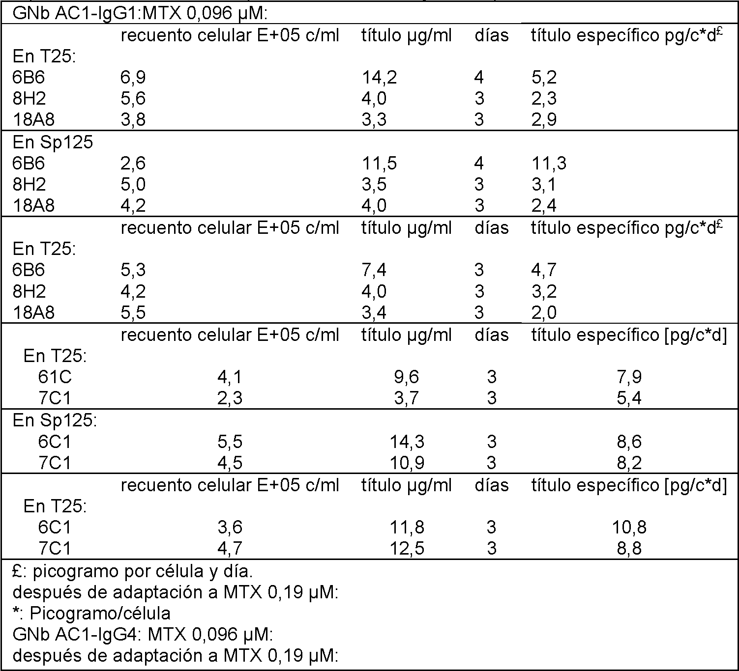
T l : l n l i n l rim r l n i n MTX 77 M T l: lnlinl rim rlnin MTX 77 M


T l 7: l n l i n l n l n i n fin l MTX 77 M: T l 7 : lnlinlnlnin end l MTX 77 M:

Los vectores de anticuerpo de IgG1 y de IgG4 recombinantes se produjeron de esta manera con ligando insertado que comprende las seis secuencias CDR como se describe en el ejemplo 1. Estos vectores que comprenden las seis CDR se analizan en el siguiente ejemplo, para determinar la influencia positiva y negativa de los vectores y, por tanto, proponer una selección y/o un uso adecuado para cada uno de ellos.Recombinant IgG1 and IgG4 antibody vectors were produced in this manner with inserted ligand comprising all six CDR sequences as described in Example 1. These vectors comprising all six CDRs are analyzed in the following example, to determine the influence positive and negative of the vectors and, therefore, propose a selection and / or a suitable use for each of them.
Ejemplo 5: estudio del ligando y sus construcciones quiméricas de IgG1 y de IgG4 humana, frente a la IgG murina original: afinidad in vitro. Example 5: study of the ligand and its chimeric constructions of human IgG1 and IgG4, against the original murine IgG: in vitro affinity.
El material y los métodos fueron los mismos que se describen en los ejemplos 3 y 4.The material and methods were the same as those described in Examples 3 and 4.
5.a. ELISA con diferentes concentraciones de proteína completa ENV de MSRV (ENV-T)5.a. ELISA with different concentrations of whole protein ENV from MSRV (ENV-T)
Tabla 8: densidades ópticas (DO) medidas por ELISA con diferentes ligandos (1 microgramo) o control irrelevante (2G5E12) sobre diferentes concentraciones (columna de la izquierda, en microgramos) y antiligando (ENV-T) recubierto sobre pocillos de placa de microtitulación. La unión se revela con anticuerpo anti-Ig marcado con peroxidasa (1/250; Jackson-EE. UU.) y reacción de sustrato de peroxidasa. El valor promedio de todas las DO del control irrelevante es 0,1004 y su desviación típica (DT) es 0,0205, por lo tanto, puede determinarse un valor de corte, por debajo del cual todos los valores son no específicos con un intervalo de confianza del 99 %: promedio 3*DT = 0,1618. Todos los valores presentados con las construcciones de GNb AC1 son, por lo tanto, significativos de una unión específica a la proteína diana. Table 8: Optical densities ( OD) measured by ELISA with different ligands ( 1 microgram) or irrelevant control ( 2G5E12) on different concentrations ( left column, in micrograms) and antiligand ( ENV-T) coated on microtiter plate wells . Binding is revealed with peroxidase-labeled anti-Ig antibody ( 1/250; Jackson-USA) and peroxidase substrate reaction. The average value of all the OD of the irrelevant control is 0.1004 and its standard deviation ( SD) is 0.0205, therefore, a cut-off value can be determined, below which all values are non-specific with an interval 99% confidence: average 3 * SD = 0.1618. All values reported with the GNb AC1 constructs are therefore significant for specific binding to the target protein.

Se analizó el anticuerpo GNbACI y la versión quimérica de IgG1 y de IgG4 como anticuerpos de detección en un ELISA de tipo sándwich frente a la proteína recombinante ENV. Como puede observarse en la figura 5 y en la tabla 8, a la misma concentración, los mAb murinos y quiméricos son capaces de detectar las presentes concentraciones de proteínas ENV, con cinética similar. La especificidad y afinidad relativa de las nuevas construcciones (ligando en un vector humano) se mantienen, por tanto, y ambas construcciones de IgG1 y de IgG4 humana con el ligando han proporcionado anticuerpos quiméricos humanos capaces de detectar picogramos de la proteína ENV recombinante. Esto es sorprendentemente bueno y confirma la optimización conseguida en todo su diseño completo, las condiciones de construcción y de expresión. También valida la selección de una estructura de ligando estable y robusta, como se describe en el ejemplo 1.The GNbACI antibody and the chimeric version of IgG1 and IgG4 were analyzed as detection antibodies in a sandwich ELISA against the recombinant ENV protein. As can be seen in Figure 5 and Table 8, at the same concentration, murine and chimeric mAbs are capable of detecting the present concentrations of ENV proteins, with similar kinetics. The specificity and relative affinity of the new constructs (ligand in a human vector) are therefore maintained, and both constructs of human IgG1 and human IgG4 with the ligand have provided human chimeric antibodies capable of detecting picograms of the recombinant ENV protein. This is surprisingly good and confirms the optimization achieved across its entire design, build and expression conditions. It also validates the selection for a stable and robust ligand structure, as described in Example 1.
Además, esta experiencia proporciona un medio para identificar moléculas equivalentes al ligando a través de la magnitud de su unión y el antiligando original (ENV), como se evidencia aquí con el ligando (GNb AC1) frente a un ligando irrelevante "no de unión" (2G5E12):Furthermore, this experience provides a means to identify molecules equivalent to the ligand through the magnitude of their binding and the original antiligand (ENV), as evidenced here with the ligand (GNb AC1) versus an irrelevant "non-binding" ligand. (2G5E12):
El antígeno Env-T se recubre sobre pocillos de placa ELISA de microtitulación con diluciones en serie que varían de 1 |jg/ml a aproximadamente 0,01 jg/ml.Env-T antigen is coated on microtiter ELISA plate wells with serial dilutions ranging from 1 µg / ml to about 0.01 µg / ml.
El ligando de referencia (GNbAC1) y un ligando irrelevante (2G5E12) se ensayan a 1 jg/ml y se revelan con un anticuerpo secundario (aquí, anticuerpo secundario anti-IgG marcado con peroxidasa de Jackson Ltd, EE. UU., diluido 1/250, a partir de ahora mencionado como anti-IgG de ratón (H+L) de Jackson o anti-IgG humana; Jackson, EE. UU.).The reference ligand (GNbAC1) and an irrelevant ligand (2G5E12) are assayed at 1 jg / ml and revealed with a secondary antibody (here, peroxidase-labeled anti-IgG secondary antibody from Jackson Ltd, USA, diluted 1 / 250, hereinafter referred to as Jackson anti-mouse IgG (H + L) or anti-human IgG; Jackson, USA).
La curva con el ligando de referencia muestra saturación de la señal (densidad óptica superior o igual a 3) a la concentración máxima de ENV y disminuye progresivamente hasta una densidad óptica de aproximadamente 1,0 0,5, evidenciando de ese modo una curva de respuesta a dosis típica de actividad de unión específica (por encima del valor de corte estadístico calculado de 0,1618; véase la tabla 8). En paralelo, la molécula irrelevante (2G5E12), no muestra curva de respuesta a dosis (curva promedio plana) y oscila entre una densidad óptica de 0,1 y 0,05 a cualquier concentración de ENV, por debajo del valor de corte estadístico calculado de 0,1618 (tabla 8). The curve with the reference ligand shows signal saturation (optical density greater than or equal to 3) at the maximum concentration of ENV and progressively decreases to an optical density of approximately 1.0 0.5, thus evidencing a curve of Typical dose response of specific binding activity (above the calculated statistical cutoff of 0.1618; see Table 8). In parallel, the irrelevant molecule (2G5E12) does not show a dose response curve (flat average curve) and oscillates between an optical density of 0.1 and 0.05 at any concentration of VNS, below the calculated statistical cut-off value. 0.1618 ( Table 8).
Por tanto, cualquier molécula equivalente al ligando, por tanto, puede evidenciarse por,Therefore, any molecule equivalent to the ligand, therefore, can be evidenced by,
1) La existencia de una curva de respuesta a dosis en este ensayo, con las condiciones del presente ejemplo, como se muestra en el párrafo 5z, y1) The existence of a dose response curve in this test, with the conditions of this example, as shown in paragraph 5z, and
2) La ausencia de una curva plana, que oscila por debajo de un corte estadístico calculado (promedio tres desviaciones típicas) de los valores de densidad óptica obtenidos con un anticuerpo irrelevante (véase 2G5E12 en la tabla 8), en comparación con valores por encima del corte obtenido con GNbAC1 de referencia para una concentración de ENV de 0,01 microgramo (véase la tabla 8); o2) The absence of a flat curve, which oscillates below a calculated statistical cutoff (average three standard deviations) of the optical density values obtained with an irrelevant antibody (see 2G5E12 in Table 8), compared to values above from the cutoff obtained with reference GNbAC1 for a VNS concentration of 0.01 microgram (see Table 8); or
3) La existencia de una curva de respuesta a dosis en un ensayo como se describe a continuación, con diluciones en serie del ligando y una concentración fija de antiligando, en el párrafo 5b (tabla 9), y3) The existence of a dose response curve in an assay as described below, with serial dilutions of the ligand and a fixed concentration of antiligand, in paragraph 5b (Table 9), and
4) La ausencia de una curva plana, que oscila por debajo de un corte estadístico calculado (promedio tres desviaciones típicas) de los valores de densidad óptica obtenidos con un anticuerpo irrelevante (véase 2G5E12 en la tabla 9), en comparación con valores por encima del corte obtenido con GNbAC1 de referencia correspondiente a una concentración de 0,01 microgramo/ml, en las mismas condiciones que las descritas en el párrafo 5b (véase la tabla 9).4) The absence of a flat curve, which oscillates below a calculated statistical cutoff (average three standard deviations) of the optical density values obtained with an irrelevant antibody (see 2G5E12 in Table 9), compared to values above of the cut obtained with reference GNbAC1 corresponding to a concentration of 0.01 microgram / ml, under the same conditions as those described in paragraph 5b (see table 9).
5.b ELISA con diferente concentración de mAb5.b ELISA with different mAb concentration
Tabla 9. Densidades ópticas medidas por ELISA con diluciones en serie del ligando y una concentración fija de antiligando. (ENV-T; 0,01 microgramo) recubierto sobre pocillos de placa de microtitulación. La unión se revela con ni r ni-I m r n r xi 12 k n-EE. . r i n r r xi . Table 9. Optical densities measured by ELISA with serial dilutions of the ligand and a fixed concentration of antiligand. ( ENV-T; 0.01 microgram) coated on microtiter plate wells. The junction is revealed with ni r ni-I mrnr xi 12 k n-EE. . rinrr xi.

continuacióncontinuation

Con la detección del anticuerpo secundario anti-ratón, el valor promedio de todas las DO del control irrelevante es 0,0735 y su desviación típica (DT) es 0,0057, por lo tanto, puede determinarse un valor de corte, por debajo del cual todos los valores son no específicos con un intervalo de confianza por 99 %: promedio 3*DT = 0,0907. Todos los valores presentados con GNb AC1 murino correspondiente son, por lo tanto, significativos de una unión específica a la proteína diana. With the detection of the secondary anti-mouse antibody, the average value of all the OD of the irrelevant control is 0.0735 and its standard deviation ( SD) is 0.0057, therefore, a cut-off value can be determined, below the which all values are non-specific with a confidence interval of 99%: average 3 * SD = 0.0907. All values reported with corresponding murine GNb AC1 are therefore significant for specific binding to the target protein.
Con la detección del anticuerpo secundario anti-humano, el valor promedio de todas las DO del control irrelevante es 0,0513 y su desviación típica (DT) es 0,0094, por lo tanto, puede determinarse un valor de corte, por debajo del cual todos los valores son no específicos con un intervalo de confianza del 99 %: promedio 3+DT = 0,0796. Todos los valores presentados con las construcciones quiméricas de GNb AC1 son, por lo tanto, significativos de una unión específica a la proteína diana. With the detection of the secondary anti-human antibody, the average value of all the OD of the irrelevant control is 0.0513 and its standard deviation ( SD) is 0.0094, therefore, a cut-off value can be determined, below the which all values are non-specific with a confidence interval of 99%: average 3 + SD = 0.0796. All values reported with the GNb AC1 chimeric constructs are therefore significant for specific binding to the target protein.
En la figura 6, puede observarse que tanto el anticuerpo IgG murino GNbAC1 como las versiones quiméricas IgG1 e IgG4 son eficaces como anticuerpos de detección en un ELISA de tipo sándwich frente a la proteína recombinante ENV-T, con cantidades tan bajas como unos pocos nanogramos de IgG purificada para detectar menos de unos pocos nanogramos de proteína ENV.In figure 6, it can be seen that both the murine IgG antibody GNbAC1 and the chimeric versions IgG1 and IgG4 are effective as detection antibodies in a sandwich ELISA against the recombinant ENV-T protein, with amounts as low as a few nanograms. of purified IgG to detect less than a few nanograms of ENV protein.
Además, como el anti-IgG humana o de ratón no reacciona de forma cruzada con el anticuerpo murino original o con las construcciones quiméricas con las estructuras de IgG1 o IgG4 humana, el ligando insertado (VH+VL) no ha creado ninguna modificación indeseada y no se detecta en las construcciones humanas, en estas condiciones. 5c: afinidad del GNbAC1 por ENV-TFurthermore, since the anti-human or mouse IgG does not cross-react with the original murine antibody or with the chimeric constructs with the structures of human IgG1 or IgG4, the inserted ligand (VH + VL) has not created any unwanted modifications and it is not detected in human constructions, under these conditions. 5c: affinity of GNbAC1 for ENV-T
Tabla 10: determinación de la afinidad de unión del ligando insertado en los vectores de anticuerpo IgG4 e IgG1 (las ni in i n n l l Table 10: Determination of the binding affinity of the inserted ligand in the IgG4 and IgG1 antibody vectors ( las ni in innll

5.d: punto isoeléctrico de GNbAC15.d: isoelectric point of GNbAC1
Se determinó el punto isoeléctrico de GNbAC1 de acuerdo con técnicas descritas en "Fractionation of complex protein mixtures by liquid-phase isoelectric focusing. Hey J, Posch A, Cohen A, Liu N, Harbers A. Methods Mol Biol.The isoelectric point of GNbAC1 was determined according to techniques described in "Fractionation of complex protein mixtures by liquid-phase isoelectric focusing. Hey J, Posch A, Cohen A, Liu N, Harbers A. Methods Mol Biol.
2008;424:225-39".2008; 424: 225-39 ".
El punto isoeléctrico de las construcciones de IgG1 (pI 8,3) y de IgG4 (7,53) son muy útiles y determinarán la estabilidad y las condiciones de almacenamiento para un uso terapéutico. El pI neutro de la IgG4 por tanto, es mejor para la formulación de un mAb terapéutico que puede ser un tratamiento crónico con inyecciones repetidas de forma regular. Por tanto, la IgG4 es una construcción preferida desde este punto.The isoelectric point of the IgG1 (pI 8.3) and IgG4 (7.53) constructs are very useful and will determine the stability and storage conditions for therapeutic use. The neutral pI of IgG4 is therefore best for the formulation of a therapeutic mAb that can be chronic treatment with repeated injections on a regular basis. Therefore, IgG4 is a preferred construct from this point on.
Ejemplo 6: estudio del ligando y de sus construcciones de IgG1 y de IgG4 humana, frente a GNbACI: actividad inhibidora sobre citoquinas proinflamatorias en cultivos de células mononucleares de sangre periférica (PBMC) humana.Example 6: study of the ligand and its human IgG1 and IgG4 constructs, against GNbACI: inhibitory activity on pro-inflammatory cytokines in human peripheral blood mononuclear cell (PBMC) cultures.
Materiales y métodos:Materials and methods:
Medio de cultivoCulture medium
Las PBMC se cultivaron en RPMI Glutamax (Gibco) FBS al 10 % (Biowest S1810 Sur América) aminoácidos no esenciales al 1 % piruvato al 1 % penicilina al 1 % - estreptomicina, a 37 °C en CO2 al 6,5 %.The PBMC were cultured in RPMI Glutamax (Gibco) 10% FBS (Biowest S1810 South America) 1% non-essential amino acids 1% pyruvate 1% penicillin - streptomycin, at 37 ° C in 6.5% CO2.
Preparación de PBMC a partir de capas leucocitarias Preparation of PBMC from buffy coats
Las capas leucocitarias se proporcionan por HUG.Buffy coats are provided by HUG.
La sangre, diluida con PBS-FBS al 2 % (4 ml 31 ml), se pone suavemente en 15 ml de Ficoll y se centrifuga a 2850 rpm (1659 g) / 20 min / temperatura ambiente / sin rotura.The blood, diluted with PBS-2% FBS (4 ml 31 ml), is gently placed in 15 ml of Ficoll and centrifuged at 2850 rpm (1659 g) / 20 min / room temperature / no break.
Las PBMC después se recogen cuidadosamente y se lavan 3 veces con PBS-SVF al 2 % y se centrifugan a 1500 rpm / 10 min.The PBMCs are then carefully collected and washed 3 times with PBS-2% SVF and centrifuged at 1500 rpm / 10 min.
Las células después se cuentan y se congelan en SVF al 90 % DMSO al 10 %.The cells are then counted and frozen in 90% SVF 10% DMSO.
Preparación de PBMC a partir de células congeladasPreparation of PBMC from frozen cells
- Las PBMC mantenidas a -80 °C se descongelan a 37 °C, se lavan 3 veces con el medio y se centrifugan a 1500 rpm / 10 min.- PBMC kept at -80 ° C are thawed at 37 ° C, washed 3 times with the medium and centrifuged at 1500 rpm / 10 min.
- Las células entonces se cuentan y se diluyen hasta una concentración de habitualmente 1 x 10⁶ células/ml. Ensayo de inhibición- The cells are then counted and diluted to a concentration of usually 1x10⁶ cells / ml. Inhibition test
- La mezcla de ENV mAb se prepara antes de descongelar las PBMC. Los mAb (relación elegida con ENV) ENV (concentración elegida) se mezclan en cada pocillo de placas de 48 pocillos, en 100 ul de medio y se incuban 1 hora a 4 °C.- The ENV mAb mix is prepared before thawing the PBMC. The mAbs (chosen ratio with ENV) ENV (chosen concentration) are mixed in each well of 48-well plates, in 100 ul of medium and incubated for 1 hour at 4 ° C.
- Las PBMC después se añaden en cada pocillo, para una concentración final de 1 x 10⁶ células/ml (0,5 ml o 1 ml final por pocillo).- The PBMC are then added to each well, for a final concentration of 1 x 10⁶ cells / ml (0.5 ml or 1 ml final per well).
- Las células se incuban 24, 48 o 72 horas a 37 °C, CO2 al 5 %.- Cells are incubated 24, 48 or 72 hours at 37 ° C, 5% CO2.
- Los sobrenadantes se recogen por centrifugación a 1400 rpm / 10 min / TA y se mantienen a -20 °C.- The supernatants are collected by centrifugation at 1400 rpm / 10 min / RT and kept at -20 ° C.
Dosificación de citoquinas 11-2d11-2d cytokine dosage
Las citoquinas se dosifican con conjuntos de ELISA BD Pharmingen, para IL-6, IL-12p40, TNF-a e IFN-y. Se siguió el protocolo del proveedor.Cytokines are assayed with BD Pharmingen ELISA sets, for IL-6, IL-12p40, TNF-α, and IFN- y . Provider protocol was followed.
Tabla 11: dosificación de citoquina en diferentes sobrenadantes de cultivo de PBMC (células mononucleares de n r rif ri n in if r n li n . Table 11: Cytokine dosage in different PBMC culture supernatants ( mononuclear cells of nr rif ri n in if rn li n.

Sobre ensayos celulares en PBMC se ensayó el potencial de nuestros anticuerpos o scFv de inhibir la interacción entre la proteína ENV y las células (a través del receptor TLR4) y, por tanto, la producción de citoquinas proinflamatorias tales como IL-6 (inmunidad innata) e IFN-y (inmunidad mediada por linfocitos T). Las diferentes moléculas se ensayaron a la misma relación (mol/mol) con la proteína (25/1) de modo que se pudiera comparar su rendimiento.On cellular assays in PBMC, the potential of our antibodies or scFv to inhibit the interaction between the ENV protein and the cells (through the TLR4 receptor) and, therefore, the production of pro-inflammatory cytokines such as IL-6 (innate immunity) was tested. ) and IFN-y (T cell-mediated immunity). The different molecules were tested at the same ratio (mol / mol) with the protein (25/1) so that their performance could be compared.
Como puede observarse en la figura 7 y en la tabla 11, todas las moléculas de ligando, sean de origen murino o recombinante (scFv o construcciones de IgG y de IgG4 con el ligando), tienen y mantienen sus propiedades inhibidoras, como se representa por la reducción de las citoquinas proinflamatorias (IL-6) producidas por las PBMC. No obstante, puede observarse que GNb AC1 y la IgG4 humana recombinante con el ligando insertado, son eficaces sobre la activación de linfocitos (reduciendo ambos la producción de interferón-gamma), mientras que scFv (muy probablemente porque es monovalente, en esta ocasión) y la construcción de IgG1 humana fueron mucho menos eficaces sobre la inhibición de interferón-gamma. Aquí, el hecho de que la región Fc de IgG1 humana tenga efectos proactivos sobre las células inmunitarias humanas, indica claramente que esta propiedad puede contrarrestar los efectos inhibidores del ligando sobre este tipo de activación de linfocitos por ENV. Con el mismo ligando en el vector de IgG4 humana (que no es inmunológicamente proactiva), presenta el efecto inhibidor del ligando divalente como en la IgG murina original.As can be seen in Figure 7 and Table 11, all ligand molecules, whether of murine or recombinant origin (scFv or IgG and IgG4 constructs with the ligand), have and maintain their inhibitory properties, as represented by the reduction of pro-inflammatory cytokines (IL-6) produced by PBMC. However, it can be seen that GNb AC1 and recombinant human IgG4 with the inserted ligand are effective on lymphocyte activation (both reducing interferon-gamma production), while scFv (most likely because it is monovalent, on this occasion) and the human IgG1 construct were much less effective at inhibiting interferon-gamma. Here, the fact that the Fc region of human IgG1 has proactive effects on human immune cells, clearly indicates that this property can counteract the Inhibitory effects of the ligand on this type of activation of lymphocytes by VNS. With the same ligand in the human IgG4 vector (which is not immunologically proactive), it exhibits the inhibitory effect of the divalent ligand as in the original murine IgG.
Por esta razón, según lo que se expone en la sección del ejemplo 5.d, la IgG4 sería una construcción preferida cuando deben evitarse las funciones inmunitarias de los anticuerpos. Como parece ser aquí el caso, las funciones del anticuerpo no son necesarias para el efecto inhibidor (dado que se produce un ligando divalente, ya que scFv monovalente tiene mala eficacia), sino que también resulta ser perjudicial para el efecto inhibidor del ligando.For this reason, as discussed in Example 5.d section, IgG4 would be a preferred construct when the immune functions of antibodies are to be avoided. As appears to be the case here, the functions of the antibody are not necessary for the inhibitory effect (since a divalent ligand is produced, since monovalent scFv has poor efficacy), but also turns out to be detrimental to the inhibitory effect of the ligand.
Respecto al efecto sobre la producción de IL-6 (de monocitos/macrófagos y, posiblemente también, linfocitos B) IgG1 e IgG4 resultan bastante equivalentes y con buena inhibición, que difiere de lo que se observó con interferóngamma. De forma interesante, el scFv monovalente presenta menos inhibición, pero significativa, de esta citoquina de "inmunidad innata". Por tanto, ciertas activaciones inmunitarias se inhiben bien por ambos vectores de IgG1 y de IgG4 humana con el ligando, pero IgG4 presenta inhibición única tanto de células de inmunidad innata (resultante de IL-6) como de inmunidad adquirida (resultante de IFN-y) de PBMC humanas. De forma interesante, el vector de IgG1, no obstante, proporciona una inhibición más fuerte de las citoquinas proinflamatorias de la inmunidad innata (representadas aquí por el ejemplo de IL-6) y activa algunos clones de células T (como se refleja por las dosificaciones de interferón-gamma), que podría resultar útil en ciertos mecanismos de defensa antivírica.Regarding the effect on IL-6 production (of monocytes / macrophages and possibly also B lymphocytes) IgG1 and IgG4 are quite equivalent and with good inhibition, which differs from what was observed with interferongamma. Interestingly, the monovalent scFv exhibits less, but significant, inhibition of this "innate immunity" cytokine. Thus, certain immune activations are well inhibited by both human IgG1 and IgG4 vectors with the ligand, but IgG4 exhibits unique inhibition of both innate immunity cells (resulting from IL-6) and acquired immunity (resulting from IFN- and ) from human PBMCs. Interestingly, the IgG1 vector, however, provides stronger inhibition of the pro-inflammatory cytokines of innate immunity (represented here by the example of IL-6) and activates some T cell clones (as reflected by the dosages interferon-gamma), which could be useful in certain antiviral defense mechanisms.
Por tanto, se confirma la alta afinidad y actividad biológica del ligando, en una forma de suministro farmacéutico que consiste en vectores de anticuerpo humano con afinidades de unión y especificidades habituales, pero que divergen en los efectos inmunitarios dependiendo de su isotipo.Thus, the high affinity and biological activity of the ligand is confirmed, in a pharmaceutical delivery form consisting of human antibody vectors with common binding affinities and specificities, but diverging in immune effects depending on their isotype.
Ejemplo 7: identificación molecular del sitio de unión del ligando en la proteína ENV diana (secuencia de unión antiligando)Example 7: molecular identification of the ligand binding site on the target ENV protein (antiligand binding sequence)
La ubicación de epítopos de IgG1/kappa murina original (GNb AC1) se ha conseguido por Pepscan BV., Países Bajos.The original murine IgG1 / kappa epitope location (GNb AC1) has been obtained by Pepscan BV., The Netherlands.
A partir de estos resultados, se identifica la secuencia de aminoácidos del sitio de unión del ligando como incluido dentro de la secuencia expuesta en la SEQ ID NO: 20.From these results, the amino acid sequence of the ligand binding site is identified as included within the sequence set forth in SEQ ID NO: 20.
Incluido en la secuencia anterior, el mejor epítopo diana consiste en la parte C-terminal del dominio SU escindido (ENV1) en la proteína ENV completa (ENV-T) y corresponde más particularmente a la siguiente secuencia de aminoácidos seleccionada expuesta en la SEQ ID NO: 32.Included in the above sequence, the best target epitope consists of the C-terminal part of the cleaved SU domain (ENV1) in the complete ENV protein (ENV-T) and corresponds more particularly to the next selected amino acid sequence set out in SEQ ID NO: 32.
Esta secuencia antiligando (y su secuencia seleccionada) no está comprendida exclusivamente, sino también, dentro de la secuencia de aminoácidos primaria de la proteína de la envoltura (ENV) de MSRV como se describe en el ejemplo 2.This antiligand sequence (and its selected sequence) is not comprised exclusively, but also, within the primary amino acid sequence of the MSRV envelope protein (ENV) as described in example 2.
No obstante, los expertos en la materia saben que los aminoácidos pueden sustituirse por su equivalente funcional y, aquí, pueden proporcionar un sitio de unión similar con una secuencia diferente. Además, se han descrito mimótopos "ENV de MSRV", que pueden unirse de forma eficaz a anticuerpos específicos (Jolivet-Reynaud, C., H. Perron, et al., 1999. "Specificities of multiple sclerosis cerebrospinal fluid and serum antibodies against mimotopes." Clin Immunol 93;3: 283-93.).However, those skilled in the art know that amino acids can be substituted for their functional equivalent and, here, can provide a similar binding site with a different sequence. Furthermore, "MSRV ENV" mimotopes have been described, which can efficiently bind specific antibodies (Jolivet-Reynaud, C., H. Perron, et al., 1999. "Specificities of multiple sclerosis cerebrospinal fluid and serum antibodies against mimotopes. "Clin Immunol 93; 3: 283-93.).
Ejemplo 8: evidencias de la presencia de antígeno diana ENV de MSRV en pacientes con enfermedades asociadas a MSRV: ejemplos de enfermedades asociadas o síndromes patológicos en esclerosis múltiple, síndrome aislado clínicamente (neurológico) -CIS-, polineuropatía desmielinizante inflamatoria crónica -CIDP-, esquizofrenia y epilepsia.Example 8: evidence of the presence of MSRV ENV target antigen in patients with MSRV-associated diseases: examples of associated diseases or pathological syndromes in multiple sclerosis, clinically isolated (neurological) syndrome -CIS-, chronic inflammatory demyelinating polyneuropathy -CIDP-, schizophrenia and epilepsy.
8a. Esclerosis múltiple, síndromes aislados clínicamente y polineuropatías: 8a. Multiple sclerosis, clinically isolated syndromes, and polyneuropathies:
Materiales y métodosMaterials and methods
Inmunodosificación de antigenemia de ENVVNS antigenemia immunodosing
Recogida de suero preliminar:Preliminary serum collection:
El estudio se aprobó por los comités éticos de los hospitales universitarios de Créteil y Grenoble, Francia. Se incluyeron pacientes neurológicos de ambos centros. Todos los pacientes han proporcionado su consentimiento informado por escrito antes de su inclusión. Se reclutaron donantes de sangre sanos de los centros de transfusión de Grenoble y de Montpellier. No se obtuvieron controles neurológicos de Grenoble. Los datos clínicos sobre los pacientes se indican en resultados. Las alícuotas de muestra de suero de los pacientes con MS y los controles sanos se codificaron y enviaron a un laboratorio independiente para ensayo enmascarado con ELISA de ApoH usando condiciones de lectura colorimétrica. The study was approved by the ethical committees of the university hospitals of Créteil and Grenoble, France. Neurological patients from both centers were included. All patients have provided their written informed consent prior to inclusion. Healthy blood donors were recruited from the Grenoble and Montpellier blood establishments. Neurological controls were not obtained from Grenoble. Clinical data on patients are indicated in results. Serum sample aliquots from MS patients and healthy controls were coded and sent to an independent laboratory for ApoH ELISA masked assay using colorimetric reading conditions.
Recogida de suero en múltiples centros europeos:Serum collection in multiple European centers:
El estudio se aprobó por los comités éticos de la facultad de medicina, Universidad de Würzburg en Alemania, de la Universidad de Sassari, del Hospital Don Gnocchi's de Milán en Italia, de1Hospital Universitario de Marsella en Francia y de la Universidad de Pamplona en España. Se incluyeron 74 pacientes con MS definida de acuerdo con los criterios de McDonald (McDonald, Compston et al., 2001) y 14 pacientes con síndromes aislados clínicamente (CIS). Los datos clínicos y de tratamiento correspondientes se presentan en la tabla 12 a continuación. (McDonald, Compston et al., 2001). En el caso de recidiva de MS, se extrajeron muestras de sangre antes del inicio del tratamiento con esteroides. Las alícuotas de muestras de suero de los pacientes con MS y los controles sanos se codificaron y enviaron a un laboratorio independiente para ensayo enmascarado con ELISA de ApoH usando condiciones de lectura luminométrica.The study was approved by the ethical committees of the Faculty of Medicine, the University of Würzburg in Germany, the University of Sassari, the Don Gnocchi's Hospital in Milan in Italy, the University Hospital of Marseille in France and the University of Pamplona in Spain. 74 patients with SD defined according to McDonald's criteria (McDonald, Compston et al., 2001) and 14 patients with clinically isolated syndromes (CIS) were included. The corresponding clinical and treatment data are presented in Table 12 below. (McDonald, Compston et al., 2001). In the case of MS relapse, blood samples were drawn before the start of steroid treatment. Aliquots of serum samples from MS patients and healthy controls were coded and sent to an independent laboratory for ApoH ELISA masked assay using luminometric reading conditions.
Recogida de muestras: se recogió un tubo (tubo seco B&D de 7 ml) de sangre. Las muestras se trataron en 2 horas después de su recogida. Después de la coagulación sanguínea se centrifugaron durante 10 minutos a 2800 g a 14 °C. Después se recogió el suero y se dividió en alícuotas en 250 pl en tubos Eppendorf. Las alícuotas se almacenaron congeladas a -20 °C.Sample collection: One tube (7 ml B&D dry tube) of blood was collected. The samples were treated within 2 hours after collection. After blood clotting, they were centrifuged for 10 minutes at 2800 g at 14 ° C. The serum was then collected and aliquoted into 250 µl in Eppendorf tubes. Aliquots were stored frozen at -20 ° C.
Inmunoensayo de ELISA de ApoHApoH ELISA Immunoassay
Método colorimétrico: se cargaron placas de microtitulación recubiertas con ApoH (APOH Technologies, Montpellier, Francia) con muestras de suero diluidas en Tris-HCl 50 mM, pH 7,6; las placas se incubaron durante 1,5 horas a 37 °C; las placas después se lavaron cuatro veces con PBS; se diluyó el mAb anti-ENV de ratón purificado con PBS que contenía BSA al 5 % hasta una concentración de 10 pg/ml y se añadió. Las placas se incubaron durante 1 hora a 37 °C y después se lavaron cuatro veces con PBS. Se añadió anticuerpo de cabra anti-IgG de ratón marcado con peroxidasa (H+L; Sigma) diluido 1:5000 en PBS que contenía BSA al 5 %, las placas se incubaron durante 1 hora a 37 °C y después se lavaron seis veces con PBS. Se añadió la solución de sustrato OPD y las placas se incubaron durante 30 minutos en la oscuridad. La reacción de color se detuvo con H2SO42N. Se leyó la absorbancia a 490 nm con un lector Tecan. El valor de corte estadístico (C.O.) de este ensayo se determinó sobre una serie de sueros negativos de 50 donantes de sangre sanos (BD), con el promedio de triplicados de sueros individuales como el resultado de densidad óptica (DO). El C.O., por tanto, se calculó a partir de la serie validada estadísticamente de controles negativos, como su valor promedio más tres desviaciones típicas (A+3DT; significancia de positividad p<0,01) y se confirmó experimentalmente en un panel de muestras positivas y negativas de referencia. El intervalo de confianza para la determinación de la positividad con el ensayo, por lo tanto, representa el 99,9 %.Colorimetric method: ApoH-coated microtiter plates (APOH Technologies, Montpellier, France) were loaded with serum samples diluted in 50 mM Tris-HCl, pH 7.6; plates were incubated for 1.5 hours at 37 ° C; plates were then washed four times with PBS; Purified mouse anti-ENV mAb was diluted with PBS containing 5% BSA to a concentration of 10 pg / ml and added. The plates were incubated for 1 hour at 37 ° C and then washed four times with PBS. Peroxidase-labeled goat anti-mouse IgG antibody (H + L; Sigma) diluted 1: 5000 in PBS containing 5% BSA was added, the plates were incubated for 1 hour at 37 ° C and then washed six times with PBS. The OPD substrate solution was added and the plates were incubated for 30 minutes in the dark. The color reaction was stopped with H2SO42N. The absorbance at 490 nm was read with a Tecan reader. The statistical cutoff value (C.O.) of this assay was determined on a series of negative sera from 50 healthy blood donors (BD), with the average of triplicates of individual sera as the result of optical density (OD). CO, therefore, was calculated from the statistically validated series of negative controls, as its average value plus three standard deviations (A + 3DT; significance of positivity p <0.01) and was confirmed experimentally in a panel of samples. positive and negative benchmarks. The confidence interval for the determination of positivity with the assay, therefore, represents 99.9%.
Método luminométrico: se cargaron muestras diluidas en Tris-HCl 50 mM, pH 7,6 en microplacas recubiertas con ApoH (APOH Technologies, Montpellier, Francia). Las microplacas se incubaron 1 hora y 30 minutos a 37 °C, se lavaron cuatro veces con PBS. Se añadió mAb de ratón anti-ENV purificado (1 pg/ml en PBS-BSA al 5 %,), las microplacas ser incubaron 1 hora a 37 °C y se lavaron cuatro veces con PBS. Se añadió anticuerpo de cabra anti ratón marcado con peroxidasa (Jackson, dilución 1/2000 en PBS-BSA al 5 %), las microplacas se incubaron 1 hora a 37 °C y se lavaron seis veces con PBS. Se añadió la solución de sustrato SuperSignal femto (Pierce) y se leyó con un lector TECAN.Luminometric method: samples diluted in 50 mM Tris-HCl, pH 7.6 were loaded into ApoH-coated microplates (APOH Technologies, Montpellier, France). The microplates were incubated 1 hour and 30 minutes at 37 ° C, washed four times with PBS. Purified anti-ENV mouse mAb (1 pg / ml in PBS-5% BSA) was added, the microplates were incubated 1 hour at 37 ° C and washed four times with PBS. Peroxidase-labeled goat anti-mouse antibody (Jackson, 1/2000 dilution in PBS-5% BSA) was added, the microplates were incubated 1 hour at 37 ° C and washed six times with PBS. SuperSignal femto substrate solution (Pierce) was added and read with a TECAN reader.
Anticuerpos monoclonalesMonoclonal antibodies
Los anticuerpos monoclonales (mAb) se desarrollaron por bioMerieux (Marcy l'Etoile, Francia) después de inmunizar ratones con la proteína de envoltura (ENV) de MSRV recombinante expresada a partir de regiones de RT-PCR clonadas amplificadas a partir de viriones de MSRV extracelulares purificados. Después del ensayo de los sueros de los ratones por ELISA con ENV, las células esplénicas se fusionaron con la línea celular de mieloma no secretora Sp2/0-Ag14 para obtener hibridomas. Los clones específicos se seleccionaron detectando su producción de anticuerpos en el mismo ensayo ELISA. Por tanto, se aislaron aproximadamente 40 mAb específicos de proteína ENV de MSRV y aproximadamente 28 mAb se seleccionaron adicionalmente y se validó su especificidad de unión. Usando la técnica de ELISA de ApoH sobre sueros humanos, el mAb de mejor unión fue 2A12A5.Monoclonal antibodies (mAbs) were developed by bioMerieux (Marcy l'Etoile, France) after immunizing mice with recombinant MSRV envelope protein (ENV) expressed from cloned RT-PCR regions amplified from MSRV virions. purified extracellular cells. After testing the mouse sera by ENV ELISA, the spleen cells were fused with the non-secretory myeloma cell line Sp2 / 0-Ag14 to obtain hybridomas. Specific clones were selected by detecting their antibody production in the same ELISA assay. Thus, approximately 40 MSRV ENV protein specific mAbs were isolated and approximately 28 mAbs were further screened and their binding specificity validated. Using the ApoH ELISA technique on human sera, the best binding mAb was 2A12A5.
ResultadosResults
Inmunodosificación de proteína ENV de MSRV en suero.Immundosage of MSRV ENV protein in serum.
Se ha desarrollado un inmunoensayo en microplaca original, en que la fase de captura depende de las propiedades particularmente eficaces de la apolipoproteína-H (ApoH) en la unión a proteínas microbianas cuando se asocian con estructuras de la envoltura y/o con los lípidos (Stefas et al., 1997; Stefas et al., 2001). ApoH permite una primera interacción de baja afinidad con las regiones de aminoácidos de la propia proteína, que en segundo lugar activa una reacción alostérica que causa unión de tipo covalente del dominio C-terminal de ApoH con los dominios de unión a lípidos o a membrana. Por lo tanto, las proteínas de la envoltura vírica o las partículas de virión pueden capturarse de forma irreversible y, después de las etapas de lavado que eliminan la muestra original, pueden detectarse antígenos específicos por la adición de un anticuerpo monoclonal dirigido a un epítopo aún expuesto después de la etapa de captura de "ApoH".An original microplate immunoassay has been developed, in which the capture phase depends on the particularly efficient properties of apolipoprotein-H (ApoH) in binding to microbial proteins when associated with envelope structures and / or with lipids ( Stefas et al., 1997; Stefas et al., 2001). ApoH allows a first low affinity interaction with the amino acid regions of the protein itself, which secondly activates an allosteric reaction that causes covalent binding of the C-terminal domain of ApoH with the lipid or membrane binding domains. Thus, viral envelope proteins or virion particles can be irreversibly captured and, after washing steps that remove the original sample, specific antigens can be detected by the addition of a monoclonal antibody targeting a still epitope. exposed after the capture stage of "ApoH".
Para la validación técnica del ensayo, tanto el virión de MSRV, sedimentado y purificado a partir de sobrenadantes de cultivo de células B de MS, de acuerdo con las condiciones descritas previamente (Perron et al., 1997a; Perron et al., 1997b) como la proteína de envoltura (ENV) de MSRV recombinante, se ensayaron con diluciones en serie y diferentes mAb anti-ENV de MSRV. La comparación se hizo con virus bien conocidos, tales como el virus de la hepatitis C (HCV) y el virus de la hepatitis B (HBV), detectados por los mAb específicos correspondientes. Después de ensayos adicionales con muestras reales de suero, el mAb 2A12A5 demostró ser el más eficaz para la inmunodetección de diagnóstico después de la etapa de captura de ApoH y se mantuvo para los siguientes estudios. Primer estudio preliminar enmascarado: esclerosis múltiple -MS- y polineuropatía desmielinizante inflamatoria crónica -CIDP-.For the technical validation of the assay, both the MSRV virion, pelleted and purified from MS B cell culture supernatants, according to the conditions previously described (Perron et al., 1997a; Perron et al., 1997b) as the recombinant MSRV envelope protein (ENV), they were tested with serial dilutions and different anti-MSRV ENV mAbs. The comparison was made with well-known viruses, such as hepatitis C virus (HCV) and hepatitis B virus (HBV), detected by the corresponding specific mAbs. After further testing with actual serum samples, mAb 2A12A5 proved to be the most effective for diagnostic immunodetection after the ApoH capture step and was retained for subsequent studies. First masked preliminary study: multiple sclerosis -MS- and chronic inflammatory demyelinating polyneuropathy -CIDP-.
Para una evaluación preliminar de este inmunoensayo en diferentes grupos de pacientes con diversas enfermedades, primero se analizaron los sueros de 29 pacientes con MS, de 28 pacientes con otras enfermedades neurológicas, de 60 pacientes con enfermedades no neurológicas y de 50 donantes de sangre sanos (un total de 167 muestras de suero). Los resultados se presentan en la tabla 12, para MS y otras enfermedades neurológicas.For a preliminary evaluation of this immunoassay in different groups of patients with various diseases, sera from 29 MS patients, 28 patients with other neurological diseases, 60 patients with non-neurological diseases and 50 healthy blood donors were first analyzed ( a total of 167 serum samples). The results are presented in Table 12, for MS and other neurological diseases.

continuacióncontinuation

Los resultados igual a 1 se consideran como "indeterminados" y las muestras correspondientes, o una nueva muestra de los mismos individuos deben ensayarse de nuevo en un experimento diferente para la determinación. El valor medio (promedio) y la desviación típica - d.t. - de todas las densidades ópticas, se ha determinado en cada grupo y subgrupo de sujetos como se indica a continuación: BD (N = 50): valor medio 0,53, d.t. 0,16.Results equal to 1 are considered "indeterminate" and the corresponding samples, or a new sample from the same individuals, must be retested in a different experiment for determination. The mean (average) value and the standard deviation - d.t. - of all the optical densities, it has been determined in each group and subgroup of subjects as follows: BD (N = 50): mean value 0.53, d.t. 0.16.
MS (N = 29): valor medio de MS positiva (N = 23) 1,27 d.t. 0,22, promedio de MS negativa (N = 6) 0,90 d.t. 0,25, valor medio de todos MS 1,20 d.t. 0,25.MS (N = 29): mean value of positive MS (N = 23) 1.27 d.t. 0.22, mean negative MS (N = 6) 0.90 d.t. 0.25, mean value of all DM 1.20 d.t. 0.25.
OND (N = 20): valor medio de OND negativa 0,88 d.t. 0,10.OND (N = 20): mean value of negative OND 0.88 d.t. 0.10.
CIDP (N = 8): valor medio de CIDP positiva (N = 5) 1,12 d.t. 0,09, promedio de CIDP negativa (N = 3) 0,9 d.t. 0,03, promedio de todos los CIDP 1,04 d.t. 0,13.CIDP (N = 8): mean value of positive CIDP (N = 5) 1.12 d.t. 0.09, mean negative CIDP (N = 3) 0.9 d.t. 0.03, average of all CIDP 1.04 d.t. 0.13.
Se analizó el suero de 29 pacientes con MS de Francia (19 de Créteil, 10 de Grenoble) como evaluación preliminar de este inmunoensayo en diferentes grupos de pacientes con diversas enfermedades. Los resultados en los pacientes de MS se presentan en la tabla 12a. Se usaron diez donantes de sangre sanos como patrones negativos para la determinación del límite de positividad (punto de corte o CO, valor umbral bajo el cual no se detecta señal específica). De acuerdo con el valor de corte estadístico de esta serie, 23 pacientes con MS tienen antigenemia de ENV de MSRV significativamente positiva (DO media = 0,88), mientras que 6 pueden considerarse negativos, aunque bastante cerca del umbral (DO media de "MS negativa" = 0,62). De forma interesante, los casos de MS negativa tuvieron una duración mayor de formas más bien benignas (n.° 24, 25, 28) o estaban experimentando el protocolo de tratamiento con ciclofosfamida.Serums from 29 MS patients from France (19 from Créteil, 10 from Grenoble) were analyzed as a preliminary evaluation of this immunoassay in different groups of patients with various diseases. The results in MS patients are presented in Table 12a. Ten healthy blood donors were used as negative standards for the determination of the positivity limit (cut-off point or CO, threshold value below which no specific signal is detected). According to the statistical cut-off value of this series, 23 MS patients have significantly positive MSRV VNS antigenemia (mean OD = 0.88), while 6 can be considered negative, although quite close to the threshold (mean OD of " Negative MS "= 0.62). Interestingly, the MS negative cases had a longer duration in rather benign forms (# 24, 25, 28) or were undergoing the cyclophosphamide treatment protocol.
En paralelo, se analizó el suero de 28 pacientes con otras enfermedades neurológicas (OND) (12 de Créteil, 16 de Grenoble). Cinco pacientes obtuvieron un resultado positivo, representando por tanto aproximadamente un 18 % de los pacientes con OND ensayados aquí. No obstante, todos los casos de OND positiva tuvieron un diagnostico similar: polineuropatía desmielinizante inflamatoria crónica (CIDP) mientras que los pacientes con OND con otros diagnósticos fueron todos negativos (o "indeterminados" al límite corte según un caso de síndrome de Guillain-Barré agudo). Por tanto, los resultados de los pacientes con OND se presentan en la tabla 12a como OND sin CIDP por separado de los pacientes con CIDP (tabla 12c). Aproximadamente la mitad de los casos de CIDP son positivos pero, dados los presentes números bajos, aquí no se pretendió analizar las enfermedades neurológicas "inflamatorias" frente a "no inflamatorias".In parallel, the serum of 28 patients with other neurological diseases (OND) (12 from Créteil, 16 from Grenoble) was analyzed. Five patients tested positive, thus representing approximately 18% of the OND patients tested here. However, all cases of positive OND had a similar diagnosis: chronic inflammatory demyelinating polyneuropathy (CIDP), while the patients with OND with other diagnoses were all negative (or "indeterminate" at the cutoff according to a case of Guillain-Barré syndrome. acute). Therefore, the results of patients with OND are presented in Table 12a as OND without CIDP separately from patients with CIDP (Table 12c). About half of CIDP cases are positive but, given the present low numbers, it was not intended here to analyze "inflammatory" versus "non-inflammatory" neurological diseases.
Además, se ha ensayado en paralelo el suero de otras enfermedades no neurológicas (ONND) tales como 15 pacientes con infección por el virus de la hepatitis B crónica, así como 15 pacientes con infección por virus de la hepatitis C crónica. Ninguna de estas 30 muestras se encontró positiva. El suero polirreactivo de 30 pacientes con anti-ADN, antinúcleo y antifactor reumatoide, que habitualmente impiden numerosos ensayos serológicos, no produjeron ningún resultado positivo. Cincuenta sueros de donantes de sangre sanos también se ensayaron en paralelo. Ninguno se encontró positivo.Furthermore, serum from other non-neurological diseases (ONND) such as 15 patients with chronic hepatitis B virus infection, as well as 15 patients with chronic hepatitis C virus infection, has been tested in parallel. None of these 30 samples was found positive. Polyreactive serum from 30 patients with anti-DNA, anti-nucleus, and anti-rheumatoid factor, which routinely precludes numerous serological tests, did not produce any positive results. Fifty sera from healthy blood donors were also tested in parallel. None were found positive.
La comparación de los resultados del grupo de MS con cualquier otro grupo, muestra una diferencia significativa, excepto por el subgrupo de CIDP. Los valores de densidad óptica de los grupos completos de MS y OND (incluyendo MS negativo y CIDP), cuando se compara con un ensayo no paramétrico (ensayo de normalidad fallido), son significativamente bastante diferentes (p = <0,001; ensayo de suma de rango de Mann-Whitney T = 517.000). Cuando se compara el grupo completo de MS con todos los casos de CIDP, ya no puede encontrarse una diferencia estadística (p = 0,053; ensayo de suma de rango de Mann-Whitney T = 99.000). No se evidenció ninguna diferencia significativa entre los valores de DO de "MS positiva" y de "CIDP positiva" (p = 0,072; ensayo de suma de rango de Mann-Whitney T = 42.000).Comparison of the results of the MS group with any other group shows a significant difference, except for the CIDP subgroup. The optical density values of the entire MS and OND groups (including negative MS and CIDP), when compared to a non-parametric assay (failed normality assay), are significantly quite different (p = <0.001; sum of Mann-Whitney range T = 517,000). When the entire MS group is compared to all CIDP cases, a statistical difference can no longer be found (p = 0.053; Mann-Whitney rank sum test T = 99,000). There was no significant difference between the OD values of "positive MS" and "positive CIDP" (p = 0.072; Mann-Whitney rank sum test T = 42,000).
Serie de sueros multicéntricos europeos con ensayo enmascarado: esclerosis múltiple - MS - y síndrome aislado clínicamente - CIS.Series of European multicenter sera with masked assay: multiple sclerosis - MS - and clinically isolated syndrome - CIS.
Para confirmar los primeros resultados con un panel mayor de pacientes con MS de diferentes áreas geográficas, se reclutó el suero en una colaboración multicéntrica con departamentos neurológicos de diferentes países europeos. En estas muestras, se usó una lectura luminométrica para mejorar la detección de la señal y la diferenciación con "ruido" de fondo no específico.To confirm the first results with a larger panel of MS patients from different geographic areas, the serum was recruited in a multicenter collaboration with neurological departments from different European countries. In these samples, a luminometric reading was used to improve signal detection and differentiation with non-specific background "noise."
Después de las evaluaciones internas de los sueros con el método colorimétrico, una comparación con la lectura de luminometría confirmó una detección de señal y dinámica potenciadas. Por tanto, se hizo la toma de muestras de suero por ensayo cuadruplicado en pacientes con MS seleccionados aleatoriamente con todas las formas y duraciones de enfermedad, principalmente con tratamiento específico en curso, pero que representan cada origen geográfico de la presente red clínica. Se codificaron y enviaron para ensayo en mascarado a un laboratorio centralizado (APO-H technologies, Montpellier, Francia). Se enviaron sueros no codificados (10 controles negativos y 10 MS positivos) para la validación técnica para la determinación del valor de corte. Además, se enviaron 14 sueros de síndrome aislado clínicamente (CIS, episodio neurológico único e imágenes adicionales y/o anomalías biológicas) codificados y ensayados de forma enmascarada dentro de esta serie. Esta fue la primera evaluación en CIS y se esperaba que comprendiera una mayoría de los primeros episodios de MS.After internal evaluations of the sera with the colorimetric method, a comparison with the luminometry reading confirmed a detection of enhanced signal and dynamics. Therefore, serum samples were taken by quadruplicate assay in randomly selected MS patients with all forms and durations of disease, mainly with specific treatment in progress, but representing each geographic origin of the present clinical network. They were coded and sent for masquerade testing to a centralized laboratory (APO-H technologies, Montpellier, France). Uncoded sera (10 negative controls and 10 positive MS) were sent for technical validation for the determination of the cut-off value. In addition, 14 clinically isolated syndrome sera (CIS, single neurological episode and additional images and / or biological abnormalities) coded and assayed in a masked manner were submitted within this series. This was the first assessment in CIS and was expected to comprise a majority of the early MS episodes.
Los resultados se presentan en la figura 8 (resultados de ELISA de ApoH de sueros del estudio multicéntrico europeo ensayado de forma enmascarada en un laboratorio independiente). Se expresan como la relación de unidades de luminometría (RLU) divididas por el valor de corte determinado dentro de los mismos experimentos, siendo por tanto comparables con la relación de la serie previa (véase, tabla 9). De hecho, el intervalo de relaciones (1 a 6) obtenido aquí dentro de los sueros "MS positivos" con luminometría confirma la optimización técnica de la dinámica de señales en comparación con la colorimetría (1 a 2).The results are presented in Figure 8 (ApoH ELISA results of sera from the European multicenter study assayed in a blinded fashion in an independent laboratory). They are expressed as the ratio of luminometry units (RLU) divided by the cutoff value determined within the same experiments, thus being comparable with the ratio of the previous series (see, table 9). In fact, the range of relationships (1 to 6) obtained here within the "MS positive" sera with luminometry confirms the technical optimization of signal dynamics compared to colorimetry (1 to 2).
Los resultados de ELISA de ApoH de los sueros del estudio multicéntrico europeo se ensayaron de forma enmascarada en un laboratorio independiente.ApoH ELISA results of sera from the European multicenter study were assayed in a blinded fashion in an independent laboratory.
La técnica de lectura usada fue luminometría y los resultados se presentan en el eje Y, como la relación de unidades de luminometría individuales (RLU) divididas por el valor de corte determinado en la serie de sueros negativos de referencia en el mismo experimento. Por tanto, valores >1 son positivos.The reading technique used was luminometry and the results are presented on the Y axis, as the ratio of individual luminometry units (RLU) divided by the cut-off value determined in the series of reference negative sera in the same experiment. Therefore, values> 1 are positive.
Los análisis estadísticos de los resultados de luminometría de ELISA de APO-H entre los grupos dieron los siguientes resultados (valores p del ensayo de Fisher): (i) comparando BD frente a todos MS, BD frente a todos MS CIS, BD frente a r Rm S, BD frente a PPMS, BD frente a SPMS, BD frente a CIS: p < 0,001; (ii) comparando CIS frente a RRMS: p = 0,759, CIS frente a PPMS: p = 0,704, CIS frente a SPMS: p = 0,749, RRMS frente a PPMS, SPMS: p = 1, PPMS frente a SPMS: p = 1.Statistical analyzes of APO-H ELISA luminometry results between groups gave the following results (Fisher's test p-values): (i) comparing BD versus all MS, BD versus all MS CIS, BD versus ar Rm S, BD vs. PPMS, BD vs. SPMS, BD vs. CIS: p <0.001; (ii) comparing CIS vs. RRMS: p = 0.759, CIS vs. PPMS: p = 0.704, CIS vs. SPMS: p = 0.749, RRMS vs. PPMS, SPMS: p = 1, PPMS vs. SPMS: p = 1 .
Aquí, 54 de los 74 casos de MS no seleccionados (73 %), originados en diferentes países y regiones del estudio, tuvieron una antigenemia positiva para la proteína ENV de MSRV, pero ninguno de los 26 BD codificados (según los 10 BD no codificados usados como muestras de referencia para el experimento). La presente diferencia entre MS y BD fue altamente significativa (Ji al cuadrado: p < 0,0001), pero "la serie no enmascarada" diferente con mayores números (por encima de cien) detectó pocos donantes sanos positivos o asintomáticos de sangre (4/103; no mostrado). De forma interesante, 9 de los 14 CIS (aproximadamente un 64 %) fueron positivos, pero con valores inferiores. Here, 54 of the 74 unselected cases of MS (73%), originating in different countries and regions of the study, had a positive antigenemia for the ENV protein of MSRV, but none of the 26 encoded BD (according to the 10 non-encoded BD used as reference samples for the experiment). The present difference between MS and BD was highly significant (Chi-squared: p <0.0001), but different "unmasked series" with higher numbers (above one hundred) detected few positive or asymptomatic healthy blood donors (4 / 103; not shown). Interestingly, 9 of the 14 CIS (approximately 64%) were positive, but with lower values.
Los valores de diferentes grupos (MS, BD, CIS) así como de diferentes subgrupos que representan diferentes formas de MS, primaria progresiva (PPMS), secundaria progresiva (SPMS) y recidivante remitente (RRMS) se compararon con ensayo de Fisher. Los resultados de los donantes de sangre sanos fueron significativamente diferentes de todas las combinaciones de MS y CIS (constantemente p < 0,001), mientras que no se evidenció ninguna diferencia significativa en la detección de antigenemia de ENV de MSRV entre cualquier subgrupo que representa MS posible (CIS) o definitiva, o diferentes formas de evolución de la enfermedad MS (un 71 % fueron positivos en rRmS, un 78 % en PPMS, un 70 % en SPMS). No obstante, se ilustra una ligera tendencia de heterogeneidad en los subgrupos CIS frente a MS por valores p inferiores: p = 0,7 a 0,75, frente a p = 1 entre formas de MS, revelando el último valor una distribución de resultados estadísticamente idéntica.The values of different groups (MS, BD, CIS) as well as of different subgroups representing different forms of MS, primary progressive (PPMS), secondary progressive (SPMS) and relapsing-remitting (RRMS) were compared with Fisher's test. The results of healthy blood donors were significantly different for all combinations of MS and CIS (consistently p <0.001), while no significant difference in detection of VNS antigenemia from MSRV was evidenced between any subgroup representing possible MS. (CIS) or definitive, or different forms of evolution of MS disease (71% were positive in rRmS, 78% in PPMS, 70% in SPMS). However, a slight trend of heterogeneity in the CIS versus MS subgroups is illustrated by lower p values: p = 0.7 to 0.75, compared to p = 1 between forms of MS, the last value revealing a statistically distribution of results. identical.
8b. Serie de enfermedad psiquiátrica: Esquizofrenia-SCZ.8b. Psychiatric illness series: Schizophrenia-SCZ.
Pacientes y métodosPatients and methods
Pacientes y controles sanosHealthy patients and controls
Los pacientes, que cumplen los criterios DSM-IV (American Psychiatric Association: Diagnostic and statistical Manual of Mental Disorders, DSM-IV; 1994) para esquizofrenia se seleccionaron aleatoriamente al final de una hospitalización por un episodio agudo en un departamento de psiquiatría afiliado a la universidad francesa. Fueron criterios de exclusión trastorno neurológico, infección aguda o crónica y serología positiva para virus de la inmunodeficiencia humana (VIH1+2), virus de la hepatitis B y C. La distribución de edad y género en estos controles normales no fue estadísticamente diferente del presente grupo de pacientes. Los síntomas psicóticos se evaluaron con la versión francesa de la escala de signos y síntomas de enfermedad psicótica - SSPI -(Houenou et al., 2007). Se evaluaron los síntomas afectivos con la escala de clasificación de compulsión maniaca de Bech y Rafaelsen y la escala de clasificación de depresión de Montgomery y Asberg - MADRS-(Bech et al., 1978; Montgomery y Asberg, 1979). El protocolo se aprobó por el comité ético local. Se obtuvo el consentimiento informado firmado de todos los sujetos, después de una descripción completa del estudio por el psiquiatra a cargo de las evaluaciones clínicas de los pacientes.Patients, meeting DSM-IV (American Psychiatric Association: Diagnostic and statistical Manual of Mental Disorders, DSM-IV; 1994) criteria for schizophrenia were randomly selected at the end of an acute episode hospitalization in a psychiatric department affiliated with the French university. Exclusion criteria were neurological disorder, acute or chronic infection, and positive serology for human immunodeficiency virus (HIV1 + 2), hepatitis B and C virus. Age and gender distribution in these normal controls was not statistically different from the present one. group of patients. Psychotic symptoms were evaluated with the French version of the scale of signs and symptoms of psychotic illness - SSPI - (Houenou et al., 2007). Affective symptoms were assessed with the Bech and Rafaelsen Manic Compulsion Rating Scale and the Montgomery and Asberg Depression Rating Scale - MADRS- (Bech et al., 1978; Montgomery and Asberg, 1979). The protocol was approved by the local ethics committee. Signed informed consent was obtained from all subjects, after a complete description of the study by the psychiatrist in charge of the clinical evaluations of the patients.
Recogida de sueroSerum collection
Un tubo (tubo seco B y D de 7 ml) de sangre se trató en 2 horas después de la recogida: después de la coagulación se centrifugaron 10 minutos a 2800g y 14 °C. El suero se recogió, se dividió en alícuotas en tubos de baja unión y se almacenó a -80 °C.A tube (dry tube B and D of 7 ml) of blood was treated in 2 hours after collection: after coagulation it was centrifuged for 10 minutes at 2800g and 14 ° C. Serum was collected, aliquoted into low binding tubes, and stored at -80 ° C.
InmunoensayoImmunoassay
Se realizaron ensayos ELISA con la etapa de captura de APO-H (Stefas et al., 1997) con IgG monoclonales (2A12A5, 6A2B2 para ENV de MSRV y 2G5E12 para GAG de MsRV), producidas y seleccionadas por su especificidad por bioMérieux, Marcy L'Etoile, Francia.ELISA assays were performed with the APO-H capture step (Stefas et al., 1997) with monoclonal IgGs (2A12A5, 6A2B2 for ENV from MSRV and 2G5E12 for GAG from MsRV), produced and selected for their specificity by bioMérieux, Marcy L'Etoile, France.
Se cargaron 100 pl por pocillo de las muestras diluidas 1/10 en Tris-HCl 50 mM pH 7,6 en microplacas recubiertas con ApoH (APOH Technologies, Montpellier-Francia). Las microplacas se incubaron 2 horas a 37 °C, se lavaron cuatro veces con 250 pl de PBS por pocillo. Se añadieron 100 pl por pocillo de anticuerpo primario (10 pg/ml en PBS-BSA al 2 %), las microplacas se incubaron 1 hora a 37 °C y se lavaron cuatro veces con PBST al 0,05 % más dos veces con PBS. Se añadieron 100 pl por pocillo de anticuerpo marcado con peroxidasa (antirratón de Jackson, 1/250 en PBS-BSA al 2 %), las microplacas se incubaron 1 hora a 37 °C y se lavaron como previamente. Se añadieron 100 pl de solución de sustrato (OPD) por pocillo, las microplacas se incubaron 30 minutos en la oscuridad y la reacción se detuvo con H2SO42N (50 pl/pocillo). La absorbancia se leyó a 490 nm con un lector Biotek.100 µl per well of samples diluted 1/10 in 50 mM Tris-HCl pH 7.6 were loaded onto ApoH-coated microplates (APOH Technologies, Montpellier-France). The microplates were incubated for 2 hours at 37 ° C, washed four times with 250 µl of PBS per well. 100 µl per well of primary antibody (10 µg / ml in PBS-2% BSA) were added, the microplates were incubated for 1 hour at 37 ° C and washed four times with 0.05% PBST plus two times with PBS . 100 µl per well of peroxidase-labeled antibody (Jackson anti-mouse, 1/250 in PBS-2% BSA) was added, the microplates were incubated for 1 hour at 37 ° C and washed as previously. 100 µl of substrate solution (OPD) was added per well, the microplates were incubated 30 minutes in the dark and the reaction was stopped with H2SO42N (50 µl / well). The absorbance was read at 490 nm with a Biotek reader.
Análisis estadísticos:Statistical analysis:
Se realizaron con el software SigmaStat. Se seleccionó el ensayo de suma de rango de Mann-Whitney de ensayo no paramétrico para la comparación de las series de datos, ya que su distribución nunca se ajustaba a la distribución normal (ensayo de normalidad fallido). Se usó el ensayo de Ji al cuadrado para comparar la prevalencia de antigenemia positiva frente a negativa en cada población, para cada antígeno y/o cada anticuerpo usado. El valor de corte para cada condición, bajo la que están los resultados, se calculó a partir de la serie estadística de controles negativos y su valor promedio más tres desviaciones típicas (M 3DT; significancia de positividad: p < 0,01) y se confirmó sobre las muestras positivas y negativas de preferencia.They were made with SigmaStat software. The nonparametric Mann-Whitney rank sum test was selected for the comparison of the data series, as its distribution never fit the normal distribution (normality test failed). The Chi-square test was used to compare the prevalence of positive versus negative antigenemia in each population, for each antigen and / or each antibody used. The cut-off value for each condition, under which the results are, was calculated from the statistical series of negative controls and its average value plus three standard deviations (M 3DT; significance of positivity: p <0.01) and confirmed on the positive and negative samples preferably.
ResultadosResults
Se han incluido 49 pacientes esquizofrénicos y 49 controles, coincidentes en edad (33+/-6,5 años) y género (73 % hombres, 27 % mujeres). Se incluyeron ocho pacientes en la primera aparición del trastorno esquizofrénico, mientras que la mayoría (N = 41) tenían esquizofrenia crónica grave. Eran eutímicos en el momento de la inclusión tanto para valores depresivos (media de MADRS = 5,6 6,6) y para el valor de compulsión maniaca (media del valor de Bech = 4,8 4,5). Todos los pacientes salvo uno no tratado tomaban fármacos antipsicóticos (27 % antipsicóticos clásicos y 71 % antipsicóticos atípicos). Un tercio de los pacientes (N = 13) eran resistentes al fármaco de acuerdo con los criterios de Kane (Kane 1996).49 schizophrenic patients and 49 controls were included, coinciding in age (33 +/- 6.5 years) and gender (73% men, 27% women). Eight patients were included at the first onset of schizophrenia, while the majority (N = 41) had severe chronic schizophrenia. They were euthymic at the time of inclusion for both depressive values (mean MADRS = 5.6 6.6) and for the manic compulsion value (mean Bech value = 4.8 4.5). All patients except one untreated were taking antipsychotic drugs (27 % classic antipsychotics and 71% atypical antipsychotics). One third of patients (N = 13) were drug resistant according to Kane's criteria (Kane 1996).
Para el antígeno GAG de MSRV, se ensayaron 49 controles y 49 pacientes esquizofrénicos. Para ENV de MSRV se ensayaron 30 controles (debido a limitaciones técnicas) y 49 pacientes esquizofrénicos. Los resultados del inmunoensayo se expresan como la densidad óptica media obtenida en duplicados de suero dividida por el valor de corte para hacer toda la serie comparable con los valores normalizados (figura 22 y tabla 10).For the MSRV GAG antigen, 49 controls and 49 schizophrenic patients were tested. For VNS from MSRV, 30 controls (due to technical limitations) and 49 schizophrenic patients were tested. The results of the immunoassay are expressed as the mean optical density obtained in serum duplicates divided by the cut-off value to make the whole series comparable with the normalized values (Figure 22 and Table 10).
Un 47 % (N = 23) y un 43 % (N = 21) de los sujetos esquizofrénicos tenían antigenemia de ENV de MSRV positiva, respectivamente con los anticuerpos 2A12A5 y 6A2B2. Un 49% (N = 24) de los pacientes esquizofrénicos tenían antigenemia de GAG de MSRV positiva, siendo uno positivo y siendo uno "límite" para GAG solamente. La comparación con los controles sanos reveló una diferencia significativa en la prevalencia de positivos (ensayos de Ji al cuadrado: p < 0,001 para detecciones de ENV; p < 0,0001 para GAG). La comparación de los valores de ELISA en pacientes frente a los controles con el ensayo de suma de rango de Mann-Whitney también confirmó diferencias altamente significativas (p < 0,001; tabla 2). Entre los controles, un sujeto tenía antigenemia significativamente positiva. De forma interesante, hubo una correlación positiva entre los resultados para la proteína ENV y los obtenidos para la proteína GAG. El ensayo de suma de rango de Mann-Whitney que compara los valores de ELISA obtenidos con anti-ENV6A2B2 con los obtenidos con anti-GAG 2G5E12 reveló ausencia de diferencia significativa (p = 0,744), igual que para anti-ENV-2A12A5 en comparación con anti-GAG-2G5E12 (p = 0,290), y para anti-ENV-6A2B2 en comparación con anti-ENV-2A12A5 (p = 0,159). Por lo tanto, estos anticuerpos detectaron una expresión equivalente y/o paralela de los antígenos de MSRV:47% (N = 23) and 43% (N = 21) of the schizophrenic subjects had positive MSRV ENV antigenemia, respectively with the 2A12A5 and 6A2B2 antibodies. Forty-nine percent (N = 24) of schizophrenic patients had positive MSRV GAG antigenemia, one being positive and one being "borderline" for GAG only. Comparison with healthy controls revealed a significant difference in the prevalence of positives (Chi-square tests: p <0.001 for VNS detections; p <0.0001 for GAG). Comparison of ELISA values in patients versus controls with the Mann-Whitney rank sum test also confirmed highly significant differences (p <0.001; Table 2). Among the controls, one subject had significantly positive antigenemia. Interestingly, there was a positive correlation between the results for the ENV protein and those obtained for the GAG protein. The Mann-Whitney rank sum test that compares the ELISA values obtained with anti-ENV6A2B2 with those obtained with anti-GAG 2G5E12 revealed the absence of significant difference (p = 0.744), the same as for anti-ENV-2A12A5 in comparison with anti-GAG-2G5E12 (p = 0.290), and for anti-ENV-6A2B2 compared to anti-ENV-2A12A5 (p = 0.159). Therefore, these antibodies detected an equivalent and / or parallel expression of the MSRV antigens:
Los valores de ELISA de antigenemia de "ENV" variaban entre los positivos, como se muestra en la tabla 10 por las desviaciones típicas aumentadas (0,28 para el anticuerpo 2A12A5, 0,48 para 6A2BA) en comparación con los controles negativos (0,09 y 0,08 respectivamente). Esto está confirmando la detección de una producción dinámica de antígenos de MSRV en ciertos pacientes con esquizofrenia.The ELISA antigenemia values of "ENV" varied between positive, as shown in Table 10 by the increased standard deviations (0.28 for antibody 2A12A5, 0.48 for 6A2BA) compared to negative controls (0 , 09 and 0.08 respectively). This is confirming the detection of dynamic MSRV antigen production in certain patients with schizophrenia.
Tabla 13: Dosificaciones de cápside de MSRV (GAG) y envoltura (ENV) en los sueros de pacientes esquizofrénicos Table 13: MSRV Capsid ( GAG) and Envelope ( ENV) Dosages in Sera from Schizophrenic Patients
Z nr lZ nr l

La relación de ensayo del inmunoensayo (ELISA) (eje Y) es la densidad óptica promedio medida en pocillos duplicados del mismo suero dividida por el valor de corte de la serie correspondiente (véase, Materiales y métodos). El antígeno ENV se dosifica con anticuerpo monoclonal 2A12A5 o anticuerpo monoclonal 6A2B2 y el antígeno GAG se dosifica con el anticuerpo monoclonal 2G5E12 como se indica en las columnas respectivas con los valores trazados.The immunoassay (ELISA) assay ratio (Y-axis) is the average optical density measured in duplicate wells of the same serum divided by the cutoff of the corresponding series (see, Materials and Methods). ENV antigen is dosed with monoclonal antibody 2A12A5 or monoclonal antibody 6A2B2 and GAG antigen is dosed with monoclonal antibody 2G5E12 as indicated in the respective columns with plotted values.
En la figura 9, el valor promedio y los intervalos de confianza (0,01 y 0,001) se representan por barras y recuadros, y la distribución de los valores máximos y mínimos para cada antígeno y anticuerpo (indicados en la parte superior de cada columna) se representan por puntos. La serie de valores de los pacientes con esquizofrenia se indica como "SCZ" en la parte inferior de los diagramas correspondientes, y aquellos de donantes de sangre sanos se marcan como "Controles" (parte inferior/eje X).In Figure 9, the mean value and the confidence intervals (0.01 and 0.001) are represented by bars and boxes, and the distribution of the maximum and minimum values for each antigen and antibody (indicated at the top of each column ) are represented by dots. The series of values from patients with schizophrenia are indicated as "SCZ" at the bottom of the corresponding diagrams, and those from healthy blood donors are marked as "Controls" (bottom / X axis).
8c. Otras enfermedades neurológicas: Epilepsia.8c. Other neurological diseases: Epilepsy.
Los ensayos ELISA se realizaron como se describe anteriormente (8b). Los pacientes con epilepsia y los controles normales se ensayaron en paralelo para la presencia de la proteína ENV de MSRV en su suero. Los resultados se presentan en la figura 10.ELISA assays were performed as described previously (8b). Epilepsy patients and normal controls were tested in parallel for the presence of the MSRV ENV protein in their serum. The results are presented in Figure 10.
Cada barra vertical representa la DO media de resultados duplicados del suero de un único paciente con epilepsia (38 desde la izquierda) o control (24 desde la derecha).Each vertical bar represents the mean OD of duplicate serum results from a single epilepsy patient. (38 from the left) or control (24 from the right).
La barra negra horizontal representa el valor de corte del ensayo, por encima del cual la señal detectada es específica de presencia de antígeno en suero. Se determina por el promedio de los resultados de los donantes de sangre sanos (controles) más tres veces su desviación típica.The horizontal black bar represents the cut-off value of the assay, above which the detected signal is specific for the presence of antigen in serum. It is determined by the average of the results of healthy blood donors (controls) plus three times its standard deviation.
Por tanto, aquí, se ha detectado un subgrupo de 8 pacientes con epilepsia asociada a ENV de MSRV, que puede corresponder simplemente a un subgrupo con esta etiología particular entre otros casos con una causa etiológica diferente. Solamente la epilepsia positiva MSRV es relevante para el tratamiento con el ligando anti-ENV en, por ejemplo, vectores de anticuerpo.Therefore, here, a subgroup of 8 patients with epilepsy associated with VNS of MSRV has been detected, which may simply correspond to a subgroup with this particular etiology among other cases with a different etiological cause. Only MSRV positive epilepsy is relevant for anti-ENV ligand treatment in, for example, antibody vectors.
8d: Pacientes con psoriasis8d: Patients with psoriasis
Se sabe desde hace tiempo que los pacientes con psoriasis expresan retrovirus similar (Iversen, O. J. (1990), "The expression of retrovirus-like particles in psoriasis." J Invest Dermatol 95 (5 Supl.): 41S-43S. / Bjerke, J. R., G. Haukenes, et al. (1983), "Activated T lymphocytes, interferon, and retrovirus-like particles in psoriatic lesions." Arch Dermatol 119(12):955-6). Por lo tanto, su relevancia para los presente vectores terapéuticos que comprenden el ligando es obvia para los expertos en la materia.Psoriasis patients have long been known to express similar retroviruses (Iversen, OJ (1990), "The expression of retrovirus-like particles in psoriasis." J Invest Dermatol 95 (5 Suppl): 41S-43S. / Bjerke, JR, G. Haukenes, et al. (1983), "Activated T lymphocytes, interferon, and retrovirus-like particles in psoriatic lesions." Arch Dermatol 119 (12): 955-6). Therefore, its relevance to the present therapeutic vectors comprising the ligand is obvious to those of skill in the art.
Ejemplo 9: Eficacia in vivo de vectores farmacéuticos que comprenden el ligando y que retienen la afinidad de ligando con características de actividad, frente a GNbAC1. Evidencias in vivo de efectos antiinflamatorios, inmunoprotectores y neuroprotectores del ligando, suministrado en forma de un ligando en vector con un compuesto de vector farmacológico apropiado: Ejemplo de efecto terapéutico en modelos animales con neuroinflamación, desmielinización y/o degeneración neuronal.Example 9: In vivo efficacy of pharmaceutical vectors comprising the ligand and retaining the affinity of ligand with activity characteristics, against GNbAC1. In vivo evidence of anti-inflammatory, immunoprotective and neuroprotective effects of the ligand, supplied as a ligand in vector with an appropriate pharmacological vector compound: Example of therapeutic effect in animal models with neuroinflammation, demyelination and / or neuronal degeneration.
Materiales y métodos:Materials and methods:
EAE inducida por MSRV/ENV en el modelo de ratón SCID humanizado (huSCID).MSRV / ENV-induced EAE in the humanized SCID mouse model (huSCID).
Se adquirieron ratones SCID de 6 a 8 semanas de edad sin patógenos de Charles River, Francia. La humanización de los ratones se consiguió usando PBMC de donantes de sangre sanos (Etablissement Franpais du Sang, Lión, Francia), de acuerdo con el protocolo descrito previamente (Firouzi et al., 2003). En particular, los ratones se irradiaron con radiación gamma y recibieron anticuerpo anti-NK antes de la humanización con 50 x 106 PBMC humanas por inyección intraperitoneal (i.p.). La calidad de la humanización se controló entonces por detección específica de inmunoglobulina humana en suero de ratón. Cuando se necesita más de un donante por serie, todos los subgrupos huSCID se hicieron comparables con la misma proporción de ratones humanizados con cada donación de sangre.Pathogen free 6-8 week old SCID mice were purchased from Charles River, France. Humanization of the mice was achieved using PBMC from healthy blood donors (Etablissement Franpais du Sang, Lyon, France), according to the previously described protocol (Firouzi et al., 2003). In particular, mice were gamma irradiated and received anti-NK antibody prior to humanization with 50 x 10 6 human PBMC by intraperitoneal (ip) injection. The quality of humanization was then monitored by specific detection of human immunoglobulin in mouse serum. When more than one donor per series is needed, all huSCID subgroups were made comparable with the same proportion of humanized mice with each blood donation.
Fue necesario un retardo de 2 semanas antes de la inclusión en el protocolo de EAE (antes de la primera inyección con antígeno de mielina). Después, los grupos de ratones recibieron inyección de proteína básica de mielina (MBP) y adyuvante incompleto de Freund (IFA, que comprende diluyente solamente) para los grupos "simulados de control" o recibieron inyecciones de MBP y proteína ENV de MSRV sin endotoxinas purificada homogeneizada en diluyentes IFA para EAE "inducida por ENV". Cuando se controló la actividad de la enfermedad clínicamente y por MRI para obtener las lesiones causadas y los déficit clínicos en progresión, se estudió el efecto de ligando anti-ENV seleccionados en comparación con el anticuerpo monoclonal murino original (GNb_AC1) con inyección en ratones enfermos (MBP-EAE inducido por ENV en ratones huSCID).A delay of 2 weeks was necessary before inclusion in the EAE protocol (before the first injection with myelin antigen). The groups of mice then received injection of myelin basic protein (MBP) and incomplete Freund's adjuvant (IFA, comprising diluent only) for the "sham control" groups or received injections of MBP and purified endotoxin-free MSRV ENV protein. homogenized in IFA diluents for "VNS-induced" EAE. When the disease activity was monitored clinically and by MRI to obtain the lesions caused and the clinical deficits in progression, the effect of selected anti-ENV ligand was studied in comparison with the original murine monoclonal antibody (GNb_AC1) with injection in diseased mice. (MBP-EAE induced by ENV in huSCID mice).
Para la inducción o "inducción simulada" de EAE, a los animales primero se les inyectó s.c. en el cuello en el día 0, 50 |jg de MBP humana 150 |jg de péptido MBP (péptido MBP 87-99) 20 |jg de proteína ENV recombinante IFA (grupo ENV) o 50 jg de MBP humana 150 jg de péptido MBP (péptido MBP 87-99) IFA solamente (grupo de control). También se inyectaron i.p. 200 ng de toxina pertúsica (PTX) por animal 2 días después en todos los grupos. Se hizo una segunda inyección por vía intraperitoneal (i.p.) de los mismos componentes a la misma dosis (incluyendo el péptido MBP y la proteína completa MBP humana), correspondiente a la descripción previa para cada grupo, en el día indicado (tabla 11). También estuvo acompañada por una inyección similar de 200 ng por animal de PTX. La tercera y última inyección de los mismos inmunógenos se hizo s.c. en el lado opuesto del cuello en el día indicado (tabla 14), acompañada por inyección similar de PTX.For the induction or "sham induction" of EAE, the animals were first injected with s.c. in the neck on day 0, 50 | jg of human MBP 150 | jg of MBP peptide (peptide MBP 87-99) 20 | jg of recombinant ENV protein IFA (ENV group) or 50 jg of human MBP 150 jg of peptide MBP (peptide MBP 87-99) IFA only (control group). They were also injected i.p. 200 ng of pertussis toxin (PTX) per animal 2 days later in all groups. A second injection was made intraperitoneally (i.p.) of the same components at the same dose (including the MBP peptide and the complete human MBP protein), corresponding to the previous description for each group, on the indicated day (Table 11). It was also accompanied by a similar injection of 200 ng per animal of PTX. The third and last injection of the same immunogens was done s.c. on the opposite side of the neck on the indicated day (Table 14), accompanied by a similar injection of PTX.
Tabla 14. Serie preclínica de evaluación del tratamiento con anticuerpo: descripción de los diferentes grupos y Table 14. Preclinical series for the evaluation of antibody treatment: description of the different groups and
" - " ^ -"-" ^ -

continuacióncontinuation

Para el seguimiento, los animales se pesaron 5 días por semana y se valoraron clínicamente. El valor clínico se hizo con los siguientes criterios: 0 = sin signos; 1 = parálisis de la cola o hiperreflexia de una o más extremidades posteriores o debilidad unilateral de la extremidad posterior; 2 = debilidad bilateral de la extremidad posterior o extremidad anterior; 3 = parálisis unilateral adicional o deficiencia mayor; 4 = parálisis completa de la extremidad posterior o extremidad anterior; 5 = parálisis parcial adicional o deficiencia mayor de extremidades opuestas; 6 = moribundo o muerto.For follow-up, the animals were weighed 5 days a week and clinically assessed. The clinical value was made with the following criteria: 0 = no signs; 1 = paralysis of the tail or hyperreflexia of one or more hind limbs or unilateral weakness of the hind limb; 2 = bilateral hindlimb or forelimb weakness; 3 = additional unilateral paralysis or major deficiency; 4 = complete paralysis of the hind limb or forelimb; 5 = additional partial paralysis or major deficiency of opposite extremities; 6 = dying or dead.
La duración total de los experimentos de Hu-SCID no excedió de los dos meses, con la excepción de los estudios de supervivencia que implican los ratones tratados con mAb y de control (cuatro meses).The total duration of the Hu-SCID experiments did not exceed two months, with the exception of survival studies involving mAb-treated and control mice (four months).
MRI y análisis de inmunohistoquímica (imágenes no mostradas en el presente ejemplo):MRI and immunohistochemical analysis (images not shown in this example):
Los animales se controlaron por análisis ponderado T2 de MRI e histología post-mortem, que confirmó ambos tipos de lesiones con inflamación y desmielinización en el sistema nervioso central, así como imágenes (MRI) de la mejora destacada de los patrones inflamatorios en ratones tratados con mejora clínica.Animals were monitored by T2-weighted analysis of MRI and post-mortem histology, which confirmed both types of lesions with inflammation and demyelination in the central nervous system, as well as imaging (MRI) of the marked improvement of inflammatory patterns in mice treated with clinical improvement.
Resultados:Results:
Los ratones SCID con sistema linfoide humano (injertados como se indica anteriormente) proporcionan animales híbridos con un sistema inmunitario humano funcional. Estos animales aquí han recibido tres inyecciones de proteína ENV de MSRV emulsionada con MBP en diluyentes oleosos (IFA), en los días indicados por las flechas azules. Cuando los animales tenían un valor clínico elevado con neuroinflamación en curso visualizada por MRI (EAE), se les inyectó una única dosis (10 |jg por vía intraperitoneal) del anticuerpo monoclonal murino original (GNb AC1, indicado como mulgG en la figura 10) o de una de las construcciones de IgG1 o IgG4 humana con el ligando (indicado como hulgG1 y hulgG4 en la figura 11). Un grupo permaneció sin tratar, para compararlos con los animales tratados y el grupo de "control simulado" al que se le inyectó MBP en IFA sin ENV que permaneció sano, pero recibió una inyección del anticuerpo murino original (GNbAC1) en el mismo día que los animales enfermos tratados. Como puede observarse a partir de los resultados ilustrados en las figuras 11 y 12, todos los ratones no tratados murieron después de 30 días y tuvieron una progresión clínica grave después de la última de las tres inyecciones con proteína ENV de MSRV. Se han observado lesiones graves por histología e inmunohistología, que evidencian desmielinización, infiltración de células linfoides, muerte neuronal, rotura de la barrera hematoencefálica y astrogliosis. De forma interesante, en esclerosis múltiple, la rotura de la barrera hematoencefálica también es una marca distintiva de lesiones activas del SNC.SCID mice with the human lymphoid system (grafted as above) provide hybrid animals with a functional human immune system. These animals here have received three injections of MSRV ENV protein emulsified with MBP in oil diluents (IFA), on the days indicated by the blue arrows. When the animals had a high clinical value with ongoing neuroinflammation visualized by MRI (EAE), they were injected with a single dose (10 µg intraperitoneally) of the original murine monoclonal antibody (GNb AC1, indicated as mulgG in Figure 10). or one of the human IgG1 or IgG4 constructs with the ligand (indicated as hulgG1 and hulgG4 in Figure 11). One group remained untreated, for comparison with treated animals and the "sham control" group injected with MBP in IFA without ENV that remained healthy, but received an injection of the original murine antibody (GNbAC1) on the same day as sick animals treated. As can be seen from the results illustrated in Figures 11 and 12, all untreated mice died after 30 days and had severe clinical progression after the last of the three injections with MSRV ENV protein. Serious histological and immunohistological lesions have been observed, showing demyelination, lymphoid cell infiltration, neuronal death, blood-brain barrier rupture, and astrogliosis. Interestingly, in multiple sclerosis, breakdown of the blood-brain barrier is also a hallmark of active CNS lesions.
Por tanto, el anticuerpo murino o la IgG1 o 4 humana quimérica que comprende el ligando dirigido a ENV, podría difundir desde la inyección intraperitoneal al organismo completo y, en particular, a las lesiones activas del SNC en animales enfermos.Thus, the murine antibody or the chimeric human IgG1 or 4 comprising the ENV-directed ligand could diffuse from intraperitoneal injection to the whole organism and, in particular, to active lesions of the CNS in diseased animals.
De forma destacada, las curvas de supervivencia muestran un 100 % de supervivencia en animales tratados con los anticuerpos quiméricos IgG1 o IgG4 frente a un 0 % en los no tratados en el día 35. Sorprendentemente, la IgG1/kappa murina original (GNbAC1) tiene menos eficacia cuando se inyecta a la misma dosis, ya que un animal murió en el día 28. Además, las curvas clínicas de la figura 11 muestran una mejora muy buena y duradera en animales tratados con las construcciones de IgG1 o 4 humana, pero solamente estabilización o una mejora leve en los animales tratados con el anticuerpo murino original.Notably, the survival curves show 100% survival in animals treated with the chimeric IgG1 or IgG4 antibodies versus 0% in those untreated on day 35. Surprisingly, the original murine IgG1 / kappa (GNbAC1) has less efficacy when injected at the same dose, as one animal died on day 28. Furthermore, the clinical curves in Figure 11 show very good and lasting improvement in animals treated with the human IgG1 or 4 constructs, but only stabilization or slight improvement in animals treated with the original murine antibody.
Dicha mejora destacada de los animales tratados con ligando en los vectores de IgG1 o de IgG4 humana, también se evidenció por control por MRI, en comparación con controles no tratados.Said marked improvement of ligand-treated animals in human IgG1 or IgG4 vectors was also evidenced by MRI control, compared to untreated controls.
Por tanto, la eficacia clínica de la IgG1 o 4 quimérica humana se confirma en modelos animales que muestran neuroinflamación, desmielinización, muerte neuronal, rotura de la barrera hematoencefálica y astrogliosis en el sistema nervioso central (SNC; véase los modelos Hu-SCID y C57/bl6 a los que se ha inyectado la proteína ENV de MSRV). Sorprendentemente, la eficacia se mejora, en comparación con la IgG murina original que contiene el mismo ligando (cadenas VH VL). Thus, the clinical efficacy of human chimeric IgG1 or 4 is confirmed in animal models showing neuroinflammation, demyelination, neuronal death, blood-brain barrier disruption, and central nervous system (CNS) astrogliosis; see Hu-SCID and C57 models. / bl6 injected with the MSRV ENV protein). Surprisingly, the efficacy is improved, compared to the original murine IgG containing the same ligand (VH VL chains).
Por tanto, la eficacia terapéutica en el presente modelo animal de neuroinflamación se pone de manifiesto para otras aplicaciones en enfermedades o síndromes asociados a MSRV, como se define en el texto de la presente invención. Además, el "efecto de ligando" inesperado de una composición mínima o construcción que comprende las seis CDR definidas en el ejemplo 1, hace posible usar el ligando de forma totalmente independiente de las funciones del anticuerpo, pero también variar y elegir los valores añadidos e intereses relativos de diferentes vectores (no exclusivamente relacionados con los isotipos de IgG) para cada posible aplicación terapéutica.Therefore, the therapeutic efficacy in the present neuroinflammation animal model is evident for other applications in diseases or syndromes associated with MSRV, as defined in the text of the present invention. Furthermore, the unexpected "ligand effect" of a minimal composition or construct comprising the six CDRs defined in Example 1, makes it possible to use the ligand completely independently of the functions of the antibody, but also to vary and choose the added values and relative interests of different vectors (not exclusively related to IgG isotypes) for each possible therapeutic application.
Ejemplo 10: la proteína ENV de MSRV se detecta con gran intensidad en ciertas células de biopsias en pacientes con tumor sólido o de biopsias en pacientes con trastornos linfoproliferativos o cánceres linfoides.Example 10: The ENV protein of MSRV is detected with high intensity in certain cells from biopsies in patients with solid tumor or from biopsies in patients with lymphoproliferative disorders or lymphoid cancers.
Materiales y métodos:Materials and methods:
- Anticuerpo de ensayo- Test antibody
GNb AC1, 1.0 ml, concentración: 5,918 mg/mlGNb AC1, 1.0 ml, concentration: 5.918 mg / ml
- Anticuerpo de control negativo- Negative control antibody
Proteína IgG1 kappa de mieloma de ratón (MOPC-21, Sigma), 1,0 ml, concentración: 1,0 mg/mlMouse myeloma IgG1 kappa protein (MOPC-21, Sigma), 1.0 ml, concentration: 1.0 mg / ml
Este anticuerpo se usó simultáneamente con el anticuerpo de ensayo.This antibody was used simultaneously with the test antibody.
- Proteína de inhibición- Inhibition protein
ENV-T (MSRV ENV, GeNeuro SA), 10 ml (10 viales de 1 ml), concentración: 0,05 mg/mlENV-T (MSRV ENV, GeNeuro SA), 10 ml (10 vials of 1 ml), concentration: 0.05 mg / ml
Este antiligando se usó simultáneamente con el anticuerpo de ensayo.This antiligand was used simultaneously with the test antibody.
- Muestras tisulares humanas- Human tissue samples
Se obtuvieron tejidos humanos recogidos de forma ética con el completo consentimiento del paciente de una fuente externa.Ethically collected human tissues were obtained with the full consent of the patient from an external source.
Todos los tejidos se sometieron a ensayo de antigenicidad. Se evaluó una sección teñida con IHC de una proteína habitualmente expresada; S100, CD45, desmina, citoqueratina o vimentina asociada con cada tejido, antes de considerar los tejidos aceptables para su uso en este estudio.All tissues were tested for antigenicity. An IHC stained section of a commonly expressed protein was evaluated; S100, CD45, desmin, cytokeratin or vimentin associated with each tissue, before considering acceptable tissues for use in this study.
- Validación del ensayo- Assay validation
Los parámetros investigados incluyeron:The parameters investigated included:
1. Comparación de la tinción del tejido de control positivo usando formalina tamponada neutra, paraformaldehído y fijación en acetona.1. Comparison of positive control tissue staining using neutral buffered formalin, paraformaldehyde, and acetone fixation.
2. El uso de un método de detección de inmunotinción apropiado.2. The use of an appropriate immunostaining detection method.
3. Determinación del título óptimo del anticuerpo de ensayo, se investigó de 0 a 5,0 pg/ml (el anticuerpo de control de isotipo se procesó a las mismas concentraciones).3. Determination of the optimal titer of the test antibody, 0 to 5.0 pg / ml was investigated (the isotype control antibody was processed at the same concentrations).
4. La especificidad de la tinción se validó por omisión del anticuerpo de ensayo sustituido con tampón. Además, se bloquearon los sitios de unión antigénicos de MSRV usando la proteína ENV-T antes de la incubación del tejido.4. Specificity of staining was validated by omission of the buffer substituted test antibody. In addition, MSRV antigenic binding sites were blocked using ENV-T protein prior to tissue incubation.
5. Se empleó cualquier bloqueo necesario de los materiales endógenos que pudieran interferir con la señal del antígeno diana.5. Any necessary blocking of endogenous materials that could interfere with the target antigen signal was employed.
- Método de tinción inmunohistoquímica (IHC)- Immunohistochemical staining method (IHC)
Como resultado de los hallazgos de la fase de validación, cada una de las muestras tisulares se exploró simultáneamente de la siguiente manera:As a result of the findings of the validation phase, each of the tissue samples was simultaneously explored as follows:
• GNbAC1 omitido del procedimiento de tinción• GNbAC1 omitted from staining procedure
• Anticuerpo MOPC-21 a 1,0 pg/ml• MOPC-21 antibody at 1.0 pg / ml
• GNbAC1 a 0,25, 1,0 y 3,0 pg/ml• GNbAC1 at 0.25, 1.0 and 3.0 pg / ml
Cada muestra tisular se evaluó usando un microscopio óptico. El sistema de valoración identificó el tejido y el tipo celular y reflejó subjetivamente la intensidad de esa tinción.Each tissue sample was evaluated using a light microscope. The titration system identified the tissue and cell type and subjectively reflected the intensity of that staining.
Toda la tinción en los tejidos tratados con el anticuerpo de control negativo o sin anticuerpo, y que no delimitaba específicamente las células individuales, se asumió como no específica. La tinción no específica se registró para tejidos tratados con el anticuerpo de ensayo cuando la tinción inmunohistoquímica en estos tejidos era similar en intensidad y en distribución a los tejidos que no se trataron con el anticuerpo de ensayo y donde las células individuales no estaban delimitadas específicamente.All staining in tissues treated with the negative control or no antibody, and not specifically delineating individual cells, was assumed to be non-specific. Non-specific staining was recorded for tissues treated with the test antibody when the immunohistochemical staining in these tissues was similar in intensity and in distribution to tissues that were not treated with the test antibody and where individual cells were not specifically delineated.
Se registró tinción positiva señalando la estructura tisular o los tipos de células y después indicando la intensidad del siguiente modo:Positive staining was recorded by noting tissue structure or cell types and then indicating intensity as follows:
0 negativo0 negative
levemild
+ moderado+ moderate
++ marcado++ marked
La frecuencia de tinción identificada en cada tipo celular se indicó del siguiente modo:The frequency of staining identified in each cell type was indicated as follows:
<10 % poca<10% little
11-40 % varias11-40% various
41-75 % mucha41-75% a lot
>76 % la mayoría> 76% the majority
Si una sección no se consideraba adecuada para evaluación, no se incluían datos en la tabla de resultados hasta que se repitió, cuando fue posible, la tinción.If a section was deemed unsuitable for evaluation, no data was included in the results table until staining was repeated, where possible.
Hubo menos tinción de membrana a 0,25 jg/ml en comparación con las otras diluciones ensayadas.There was less membrane staining at 0.25 µg / ml compared to the other dilutions tested.
El tejido hepático se incluyó como comparación entre un control positivo conocido y un tejido que no se identificó inicialmente como positivo.Liver tissue was included as a comparison between a known positive control and a tissue that was not initially identified as positive.
No se identificó tinción significativa en el control de tampón negativo o el isotipo. La tinción positiva en comparación con los tejidos teñidos positivamente prácticamente se eliminó cuando se hacía reaccionar ENV-T con el anticuerpo anti-MSRV/ENV GNbAC1.No significant staining was identified in the negative buffer control or the isotype. Positive staining compared to positively stained tissues was virtually eliminated when ENV-T was reacted with anti-MSRV / ENV GNbAC1 antibody.
Para un futuro trabajo de exploración para ensayar mAb, se usó GNb AC1 1,00 jg.mM como concentración óptima, estando 3,00 jg.mM un paso por encima a la misma y siendo 0,25 jg.m l'1 la baja concentración.For future screening work to test mAb, GNb AC1 1.00 jg.mM was used as the optimal concentration, 3.00 jg.mM being one step above it and 0.25 jg.m l'1 being the low concentration.
Como resultado de los hallazgos de la validación, se adoptó el siguiente procedimiento de tinción IHC para la exploración del tejido humano normal congelado.As a result of the validation findings, the following IHC staining procedure was adopted for the examination of frozen normal human tissue.
Procedimiento TiempoProcedure Time
Secar al aire secciones N/A tisulares 10 minutos aprox.Air dry N / A tissue sections for approx. 10 minutes.
Fijar en acetona y secar al aire N/AFix in acetone and air dry N / A
Lavar en agua corriente Aprox. 10 minWash in running water Approx. 10 minutes
Peróxido de hidrógeno al 0,3 % en metanol Aprox. 5 min0.3% hydrogen peroxide in methanol Approx. 5 min
Lavar en agua corriente Aprox. 5 minWash in running water Approx. 5 min
Lavar en PBS Durante una noche a 2-8 °CWash in PBS overnight at 2-8 ° C
Incubar con el anticuerpo monoclonal GNb AC1 a 0,25, 1,0 y 3,0 Aprox. 5 minIncubate with GNb AC1 monoclonal antibody at 0.25, 1.0 and 3.0 Approx. 5 min
jg/ml y con el control de isotipo a 1,0 jg/ml. También omitirjg / ml and with the isotype control at 1.0 jg / ml. Also skip
principalmente del procedimiento de tinción e inhibir con ENV-T a 10mainly from the staining procedure and inhibit with ENV-T at 10
jg/ml con GNb Ac 1 a 1,0 jg/mljg / ml with GNb Ac 1 at 1.0 jg / ml
Lavar en PBS Aprox. 30 minWash in PBS Approx. 30 min
Incubar con EnVision/HRP Aprox. 5 min cada unoIncubate with EnVision / HRP Approx. 5 min each
Lavar en PBS x 2 Aprox. 5 minWash in PBS x 2 Approx. 5 min
Tratar con el sustrato enzimático DAB Aprox. 5 minTreat with DAB enzyme substrate Approx. 5 min
Lavar en agua corriente Aprox. 15 segundosWash in running water Approx. 15 seconds
Teñir con contraste en hematoxilina de Mayers Según lo necesarioContrast Stain in Mayers Hematoxylin As Needed
Lavar en agua corriente Según lo necesarioWash in running water As needed
Deshidratar, limpiar y montarDehydrate, clean and mount
Resultados:Results:
- Cáncer de mama.- Breast cancer.
Tabla 15: Resultados obtenidos con tejidos de carcinoma de mama de diferentes individuos. INTENSIDAD Negativa 0 Leve Moderada + Marcado ++ FRECUENCIA Pocos Varios Muchos Mayoría Table 15: Results obtained with breast carcinoma tissues from different individuals. INTENSITY Negative 0 Slight Moderate + Marked ++ FREQUENCY Few Several Many Most
Hallazgos GNbAC1GNbAC1 findings
microscópicos ENV-T IsotipoConcentr. de anticuerpo ug/ml 10,00 1,00 0,00 0,25 1,00 3,00microscopic ENV-T IsotypeConcentr. antibody ug / ml 10.00 1.00 0.00 0.25 1.00 3.00
Epitelio 0 0 0 Pocos Varios Pocos Ductal/glandular.Epithelium 0 0 0 Few Several Few Ductal / glandular.
Mama (mamario) Citoplasma/membrana + Breast (mammary) Cytoplasm / membrane +
Contenidos luminales. 0 0 0 0 Varios Pocos Luminal contents. 0 0 0 0 Several Few
(carcinoma in Superficie luminal. 0 0 0 0 Varios Muchos situ) (carcinoma in luminal surface. 0 0 0 0 Various Many situ)
+ ++ +
Vasos-endotelio 0 0 0 0 Muchos Pocos Citoplasma/membrana + Vessels-endothelium 0 0 0 0 Many Few Cytoplasm / membrane +
Epitelio 0 0 0 0 Pocos Muchos Ductal/glandular.Epithelium 0 0 0 0 Few Many Ductal / glandular.
Citoplasma/membrana ++ Contenidos luminales. 0 VariosVarios Varios Varios VariosCytoplasm / membrane ++ Luminal contents. 0 Various Various Various Various Various
+ +++ ++
Superficie luminal. 0 0 0 0 Pocos MuchosLuminal surface. 0 0 0 0 Few Many
++ (adenocarcinoma Vasos-endotelio 0 0 0 0 Varios Muchos de mama)++ (Adenocarcinoma Vas-endothelium 0 0 0 0 Various Many breast)
Citoplasma/membrana + +Cytoplasm / membrane + +
Epitelio 0 0 0 0 Pocos Varios Ductal/glandular.Epithelium 0 0 0 0 Few Several Ductal / glandular.
Citoplasma/membrana + Contenidos luminales. 0 0 0 0 0 0Cytoplasm / membrane + Luminal contents. 0 0 0 0 0 0
Superficie luminal. 0 0 0 0 Varios MuchosLuminal surface. 0 0 0 0 Several Many
(adenocarcinoma Vasos-endotelio 0 0 0 0 Varios Varios de mama)(Vessels-endothelium adenocarcinoma 0 0 0 0 Various Various breast)
Citoplasma/membrana +Cytoplasm / membrane +
Por tanto, puede evidenciarse que ENV de MSRV se expresa altamente en células epiteliales ductales/glandulares y en células endoteliales de los vasos sanguíneos adyacentes en 3/3 tumores mamarios de mama ensayados con GnbAC1 específico.Thus, it can be evidenced that ENV of MSRV is highly expressed in ductal / glandular epithelial cells and in endothelial cells of adjacent blood vessels in 3/3 breast mammary tumors tested with specific GnbAC1.
Por tanto, de acuerdo con la anotación de "enfermedades asociadas a MSRV", ahora se evidencia que el carcinoma de mama es una de estas enfermedades humanas.Therefore, according to the annotation of "MSRV-associated diseases", it is now evident that breast carcinoma is one of these human diseases.
Cáncer linfoide y trastornos proliferativos:Lymphoid cancer and proliferative disorders:
Tabla 16: Resultados obtenidos con tejidos linfoides hiperplásicos de diferentes individuos. INTENSIDAD Negativa 0 Leve + Moderada ++ Marcada +++; FRECUENCIA Pocos Varios Muchos Mayoría; NP: no practicable Concentración de anticuerpos ug/ml Table 16: Results obtained with hyperplastic lymphoid tissues from different individuals. INTENSITY Negative 0 Mild + Moderate ++ Marked +++; FREQUENCY Few Several Many Majority ; PN: not practicable Antibody concentration ug / ml
ENV-ENV-
Hallazgos T Isotipo GNb AC1Findings T Isotype GNb AC1
microscópicos 10,00 1,00 0,00 0,25 1,00 3,00 Amígdala Membrana 0 0 Varios Muchos Varios citoplasmática linfoide ++ ++ ++ Membrana citoplasmática 0 0 Pocos Muchos Muchos del endotelio de los vasos + + ++ (hiperplasia Membrana Muchos Mayoría Mayoría folicular)microscopic 10.00 1.00 0.00 0.25 1.00 3.00 Tonsil Membrane 0 0 Several Many Several Lymphoid cytoplasmic ++ ++ ++ Cytoplasmic membrane 0 0 Few Many Many of the endothelium of the vessels + + ++ (Membrane hyperplasia Many Most Most follicular)
citoplasmática linfoide ++ ++ ++ Membrana citoplasmática 0 0 Pocos Muchos Muchos del endotelio de los vasos + + ++ (hiperplasia Glóbulos blancos en membranaMuchos 0 0 NI NI NI folicular) citoplasmática de los vasosCytoplasmic lymphoid ++ ++ ++ Cytoplasmic membrane 0 0 Few Many Many of the endothelium of vessels + + ++ (hyperplasia White blood cells in membrane Many 0 0 NI NI NI follicular) cytoplasmic of the vessels
++
Por tanto, puede evidenciarse que ENV de MSRV se expresa altamente en células linfoides y en células epiteliales de los vasos sanguíneos adyacentes en 2/2 biopsias de amígdala con hiperplasia marcada de pacientes con trastorno linfoproliferativo, cuando se ensayan con GNbAC1. Thus, it can be evidenced that VNS of MSRV is highly expressed in lymphoid cells and in epithelial cells of adjacent blood vessels in 2/2 biopsies of tonsil with marked hyperplasia from patients with lymphoproliferative disorder, when tested with GNbAC1.
Por tanto, de acuerdo con la anotación de "enfermedades asociadas a MSRV", ahora se evidencia que los trastornos linfoproliferativos, incluyendo los cánceres de células linfoides, están entre estas enfermedades humanas.Thus, according to the annotation of "MSRV-associated diseases", it is now evident that lymphoproliferative disorders, including lymphoid cell cancers, are among these human diseases.
- Cáncer renal - Kidney cancer
Tabla 17: Resultados obtenidos con tejidos de carcinoma renal de diferentes individuos. INTENSIDAD Negativa 0 Leve + Moderada ++ Marcada +++; FRECUENCIA Pocos Varios Muchos Mayoría; NP: no practicable Concentración de anticuerpos microg/ml Table 17: Results obtained with renal carcinoma tissues from different individuals. INTENSITY Negative 0 Mild + Moderate ++ Marked +++; FREQUENCY Few Several Many Majority ; PN: not practicable Antibody concentration microg / ml
Hallazgos microscópicos ENV- T Isotipo GNb AC1Microscopic findings ENV- T Isotype GNb AC1
10,00 1,00 0,00 0,25 1,00 3,00 Superficie del lumen de los túbulos 0 0 0 Pocos Pocos Muchos Citoplasma/membrana ++ Glomerular mesangial 0 0 0 0 0 0 (cáncer de riñón) Membrana/citoplasma10.00 1.00 0.00 0.25 1.00 3.00 Surface of the lumen of the tubules 0 0 0 Few Few Many Cytoplasm / membrane ++ Mesangial glomerular 0 0 0 0 0 0 (kidney cancer) Membrane / cytoplasm
Bucles de vasos glomerulares 0 0 0 Pocos Pocos NP Endotelio/citoplasma ++ ++ NP Cilindros urinarios 0 Muchos Mayoría ++ Superficie del lumen de los túbulos Pocos Varios Varios Citoplasma/membrana + + Glomerular mesangial Pocos Pocos Pocos Membrana/citoplasma ++ ++ ++ (carcinoma de células Cápsula de Bowman 0 Varios Varios renales) ++ ++ Bucles de vasos glomerulares 0 Muchos Mayoría Endotelio/citoplasma ++ ++ Cilindros urinarios Pocos Pocos PocosGlomerular vessel loops 0 0 0 Few Few NP Endothelium / cytoplasm ++ ++ NP Urinary casts 0 Many Most ++ Surface of the lumen of the tubules Few Various Various Cytoplasm / membrane + + Mesangial glomerular Few Few Few Membrane / cytoplasm ++ + + ++ (Bowman's capsule cell carcinoma 0 Various Various renal) ++ ++ Glomerular vessel loops 0 Many Most Endothelium / cytoplasm ++ ++ Urinary casts Few Few Few
A pesar de la ligera tinción de fondo en un tipo celular (diferente en cada caso), el análisis fue practicable y, por tanto, se puede evidenciar que ENV de MSRV se expresa altamente en células renales y en células endoteliales de los vasos sanguíneos adyacentes en 2/2 biopsias renales de pacientes con carcinoma renal, cuando se ensayan con anticuerpo anti-ENV de Ms RV específico (GNbAC1).Despite the slight background staining in a cell type (different in each case), the analysis was practicable and, therefore, it can be evidenced that the ENV of MSRV is highly expressed in renal cells and in endothelial cells of adjacent blood vessels. in 2/2 kidney biopsies from patients with renal carcinoma, when tested with specific Ms RV anti-ENV antibody (GNbAC1).
Por tanto, de acuerdo con la anotación de "enfermedades asociadas a MSRV", ahora se evidencia que los cánceres renales son una de estas enfermedades humanas.Therefore, according to the annotation of "MSRV-associated diseases", it is now evident that kidney cancers are one of these human diseases.
Las fotografías correspondientes de visualización en microscopio óptico de secciones tisulares teñidas con anticuerpo anti-ENV específico (GNb AC1) o anticuerpo de control irrelevante (MOPC-21) han confirmado los hallazgos presentados en las tablas 15-17, para cánceres renales, para cánceres linfoides o trastornos linfoproliferativos y para cáncer de mama.The corresponding photographs of visualization in light microscope of tissue sections stained with specific anti-ENV antibody (GNb AC1) or irrelevant control antibody (MOPC-21) have confirmed the findings presented in Tables 15-17, for kidney cancers, for cancers lymphoid or lymphoproliferative disorders and for breast cancer.
Ejemplo 11: A. Las proteínas ENV y GAG de MSRV se detectan con niveles significativos y concordantes en el suero de determinados casos agudos que desarrollan diabetes:Example 11: A. MSRV ENV and GAG proteins are detected at significant and concordant levels in the serum of certain acute cases developing diabetes:
Se recogieron de forma anónima sueros de pacientes con diabetes insulinodependiente aguda de los volúmenes restantes después de ensayo rutinario con fines de diagnóstico y de seguimiento. Se obtuvieron sueros de donantes de sangre sanos de la organización de banco de sangre habilitada.Sera from patients with acute insulin-dependent diabetes were collected anonymously from the remaining volumes after routine testing for diagnostic and follow-up purposes. Sera were obtained from healthy blood donors from the licensed blood bank organization.
Los sueros se ensayaron de acuerdo con la técnica de inmunodetección usando placas de captura de APOH y detección de ligando específico, como se describe previamente en los ejemplos de la presente invención.Sera were assayed according to the immunodetection technique using APOH capture plates and specific ligand detection, as previously described in the examples of the present invention.
La siguiente proporciona la densidad óptica media en ensayos triplicados, medida de sueros de pacientes con diabetes aguda (Diab.) y de donantes representativos de sangre (BD).The following provides the mean optical density in triplicate assays, measured from sera from patients with acute diabetes (Diab.) And representative blood donors (BD).
Se usaron ligandos de anticuerpo monoclonal murino anti-MSRV:Anti-MSRV murine monoclonal antibody ligands were used:
- Un GNbAC1 anti-envoltura (ENV) (Geneuro, Suiza).- A GNbAC1 anti-envelope (ENV) (Geneuro, Switzerland).
- Un anti-poliproteína de la matriz y la cápside (GAG): 2G5E12 (bioMerieux Francia).- A matrix and capsid anti-polyprotein (GAG): 2G5E12 (bioMerieux France).
La unión específica de estos anticuerpos se reveló por un anticuerpo secundario anti-ratón (Jackson, EE. UU., ref.Specific binding of these antibodies was revealed by a secondary anti-mouse antibody (Jackson, USA, ref.
115-035-146). Las diluciones o concentraciones usadas se indican en la siguiente tabla.115-035-146). The dilutions or concentrations used are indicated in the following table.
Los resultados son significativos cuando la relación (P/N) de la densidad óptica media dividida por el valor de corte (determinado a partir del valor medio de controles sanos más dos desviaciones típicas -DT- de la serie correspondiente) es mayor de uno. Dichos valores están en negrita y letras mayúsculas en las filas P/N 2. The results are significant when the ratio (P / N) of the mean optical density divided by the cut-off value (determined from the mean value of healthy controls plus two standard deviations -DT- of the corresponding series) is greater than one. These values are in bold and capital letters in the P / N 2 rows.
Por tanto, puede evidenciarse que diez de los diez y ocho pacientes tienen antigenemia significativa para al menos la proteína ENV, detectada por el anticuerpo monoclonal murino de ligando. Todos los pacientes "MSRV positivos" se detectan por los dos anticuerpos y tienen resultados significativos para ambas proteínas GAG y ENV.Therefore, it can be evidenced that ten of the eighteen patients have significant antigenemia for at least the ENV protein, detected by the murine monoclonal antibody of ligand. All "MSRV positive" patients are detected by both antibodies and have significant results for both GAG and ENV proteins.
Dichos resultados con detección coincidente de dos anticuerpos monoclonales diferentes dirigidos a dos epítopos diferentes que representan dos proteínas diferentes de MSRV (ENV y GAG), son obviamente significativos y valiosos en términos de asociación con MSRV como se describe en otra parte en la presente invención, para la determinación de enfermedades asociadas a MSRV.Such results with coincident detection of two different monoclonal antibodies targeting two different epitopes representing two different MSRV proteins (ENV and GAG), are obviously significant and valuable in terms of association with MSRV as described elsewhere in the present invention, for the determination of diseases associated with MSRV.
Por lo tanto, la diabetes de tipo I u otra diabetes asociada a inflamación comprende un subgrupo de pacientes cuya patogénesis de la enfermedad puede estar causada por los efectos proinflamatorios e inmunopatogénicos de la proteína ENV de MSRV.Thus, type I diabetes or other diabetes associated with inflammation comprises a subset of patients whose disease pathogenesis may be caused by the pro-inflammatory and immunopathogenic effects of the ENV protein of MSRV.
Tabla 18: resultados de antigenemia de ELISA de APOH en pacientes con diabetes (Diab.) en comparación con donantes sanos (BD). Véanse los comentarios detallados en el texto del ejemplo. P/N 2 = relación de densidad óptica calculada como el resultado de la muestra dividido por el valor de corte. El último se determina con donantes sanos como la media del grupo sano dos veces su' desviación típica. _______________Table 18: APOH ELISA antigenemia results in patients with diabetes (Diab.) Compared to healthy donors (BD). See detailed comments in the sample text. P / N 2 = optical density ratio calculated as the sample result divided by the cutoff value. The latter is determined with healthy donors as the mean of the healthy group twice its ' standard deviation. _______________
GNb AC1 2G5E12GNb AC1 2G5E12
1 ug/ml Jackson 080604CP011 ug / ml Jackson 080604CP01
115-035-146 1 ug/ml Jackson115-035-146 1 ug / ml Jackson

- -- -

Ejemplo 12: Descubrimiento inesperado de un modelo animal apropiado para diabetes inducida por ENV. 1. IntroducciónExample 12: Unexpected discovery of a suitable animal model for VNS-induced diabetes. 1. Introduction
El modelo de ratón mutante espontáneo SCID diabético no obeso (NOD-SCID) tiene la mutación SCID transferida a un fondo NOD susceptible a diabetes. Sorprendentemente, los experimentos preliminares realizados por el solicitante revelaron que la inmunización primaria de ratones NOD-SCID humanizados con la proteína (25 |jg) y MBP murina (proteína y péptido) dio lugar sistemáticamente a una muerte rápida de todos los animales ensayados, mientras todos los ratones SCID humanizados que habían recibido los mismos inmunógenos sin la proteína ENV (controles simulados) sobrevivieron. En una primera etapa, se evaluaron los efectos perjudiciales de la proteína ENV en los ratones NOD-SCID de una manera de respuesta a dosis usando control clínico combinado con una estrategia histológica. En una segunda etapa, se evaluaron los efectos beneficiosos de nuestro ligando IgG4 quimérico en la prevención del daño inducido por la proteína ENV.The non-obese diabetic spontaneous mutant SCID mouse model (NOD-SCID) has the SCID mutation transferred to a NOD background susceptible to diabetes. Surprisingly, preliminary experiments carried out by Applicant revealed that primary immunization of humanized NOD-SCID mice with protein (25 µg) and murine MBP (protein and peptide) consistently resulted in rapid death of all tested animals, while all humanized SCID mice that had received the same immunogens without the ENV protein (sham controls) survived. In a first step, the deleterious effects of ENV protein in NOD-SCID mice were evaluated in a dose-response manner using clinical control combined with a histological strategy. In a second stage, the beneficial effects of our chimeric IgG4 ligand in the prevention of damage induced by the ENV protein were evaluated.
2. Evaluación de los efectos perjudiciales de la proteína ENV en ratones NOD-SCID2. Evaluation of the deleterious effects of ENV protein in NOD-SCID mice
a. Materiales y métodosto. Materials and methods
a.1. Animalesa.1. Animals
Se adquirieron seis ratones SCID sin patógenos (de 6 a 8 semanas de edad) de Charles River, Francia. Los animales se mantuvieron 3 por jaula en un ciclo convencional de luz-oscuridad con acceso ad libitum a comida y agua y no se distribuyeron durante un periodo de 8 días de aclimatación. Se tuvo especial cuidado por asegurar condiciones de alojamiento muy limpias. Particularmente, los animales se alojaron en jaulas especiales equipadas con tapa de filtro.Six pathogen-free SCID mice (6-8 weeks old) were purchased from Charles River, France. The animals were kept 3 per cage in a conventional light-dark cycle with ad libitum access to food and water and were not distributed during an 8-day acclimatization period. Special care was taken to ensure very clean accommodation conditions. In particular, the animals were housed in special cages equipped with a filter cover.
a.2. Humanización de ratones NOD-SCIDa.2. Humanization of NOD-SCID mice
Para obtener ratones NOD-SCID, desprovistos del sistema inmunitario linfoide, con un sistema inmunitario humano injertado, los ratones se humanizaron con células mononucleares periféricas humanas ofreciendo de este modo el potencial de estudiar la tarea funcional del sistema inmunitario humano en modelos animales inmunopatogénicos. Esto proporciona resultados mucho más cercanos a la situación humana real en términos de inmunopatología y respuesta a proteínas patogénicas humanas, tales como ENV de MSRV. La humanización de los ratones se consiguió de acuerdo con el protocolo descrito previamente por Firouzi et al., 2003, excepto que los animales no se irradiaron con radiación gamma y no se trataron con ligando anti-NK porque los ratones NOD-SCID están espontáneamente desprovistos de células NK. Se obtuvo sangre (capa leucocitaria, 45-50 ml) de 2 controles sanos del centro de transfusión de sangre (EFS) de Lion. La calidad y la seguridad de la sangre se garantizaron por análisis inmunológico y hematológico. Todos los procedimientos experimentales se realizaron bajo un flujo de aire laminar y usando guantes estériles. Las células mononucleares de sangre periférica (PBMC) humanas se obtuvieron por un método de separación por densidad en gradiente de Ficoll y se administraron a NOD-SCID por inyección i.p. (50.106 células). Como se usó la sangre de dos donantes diferentes para humanizar todos los animales, se humanizó la misma proporción de ratones con cada donante de sangre. Aquí ya puede confirmarse que no se observaron diferencias entre los animales con injerto de cada donante en todos los experimentos futuros, como se describe a continuación.To obtain NOD-SCID mice, devoid of the lymphoid immune system, with an engrafted human immune system, the mice were humanized with human peripheral mononuclear cells thus offering the potential to study the functional task of the human immune system in immunopathogenic animal models. This provides results much closer to the real human situation in terms of immunopathology and response to human pathogenic proteins, such as ENV from MSRV. Humanization of the mice was achieved according to the protocol previously described by Firouzi et al., 2003, except that the animals were not irradiated with gamma radiation and were not treated with anti-NK ligand because NOD-SCID mice are spontaneously depleted. of NK cells. Blood (buffy coat, 45-50 ml) was obtained from 2 healthy controls from the Lion Blood Establishment (EFS). The quality and safety of the blood were guaranteed by immunological and hematological analysis. All experimental procedures were performed under laminar air flow and wearing sterile gloves. Human peripheral blood mononuclear cells (PBMC) were obtained by a Ficoll gradient density separation method and administered to NOD-SCID by ip injection (50,106 cells). Since blood from two different donors was used to humanize all animals, the same proportion of mice were humanized with each blood donor. Here it can already be confirmed that no differences were observed between grafted animals from each donor in all future experiments, as described below.
3. Evaluación de los efectos perjudiciales de la proteína ENV en los ratones NOD-SCID3. Evaluation of the deleterious effects of ENV protein in NOD-SCID mice
Inyecciones de la proteína ENVENV protein injections
En el día 0 (P0), los ratones recibieron una única inyección intraperitoneal (i.p.) (0,5 ml) de proteína ENV (PX'Therapeutics, Francia) con las siguientes dosis: 0,1, 1, 5, 10 y 20 jg. La proteína ENV se diluyó en solución salina tamponada con fosfato (PBS) estéril (Lonza, Francia). Un único ratón recibió inyección con PBS en solitario. On day 0 (P0), the mice received a single intraperitoneal (ip) injection (0.5 ml) of ENV protein (PX'Therapeutics, France) with the following doses: 0.1, 1, 5, 10 and 20 jg. ENV protein was diluted in sterile phosphate buffered saline (PBS) (Lonza, France). A single mouse was injected with PBS alone.
Para evitar cualquier disminución en la concentración en la proteína ENV y en la bioactividad, se consiguió una segunda inyección de proteína ENV emulsionada en 0,5 ml de adyuvante incompleto de Freund (Sigma, Francia) en P7. Cada ratón recibió una única inyección subcutánea en el cuello. Las dosis de proteína ENV usadas para cada ratón fueron las mismas que para la primera inyección en P0.To avoid any decrease in ENV protein concentration and bioactivity, a second injection of ENV protein emulsified in 0.5 ml of incomplete Freund's adjuvant (Sigma, France) in P7 was achieved. Each mouse received a single subcutaneous injection in the neck. The ENV protein doses used for each mouse were the same as for the first injection at P0.
Se realizó una tercera inyección s.c. de proteína ENV emulsionada en diluyente de aceite mineral -"IFA"-(0,5 ml) en el cuello en P14. El mismo día, los ratones recibieron una inyección i.p. de 200 ng (0,5 ml) de toxina pertúsica -"PTX"-(Wako, Alemania) por animal para abrir de forma transitoria la barrera hematoencefálica para el paso de células inmunitarias extra-SNC.A third injection was performed s.c. of ENV protein emulsified in mineral oil diluent - "IFA" - (0.5 ml) on the neck in P14. On the same day, the mice received an i.p. injection. 200 ng (0.5 ml) of pertussis toxin - "PTX" - (Wako, Germany) per animal to temporarily open the blood-brain barrier for the passage of extra-CNS immune cells.
Evaluación clínicaClinical evaluation
Se controló el estado de salud general (aspecto del pelaje, forma de caminar en la jaula, postura) y el peso de los animales 5 días por semana desde P14 hasta P37.The general health status (appearance of the coat, way of walking in the cage, posture) and the weight of the animals were monitored 5 days a week from P14 to P37.
Examen histológicoHistological examination
Se retiraron el cerebro y los órganos internos de todos los animales muertos y se conservaron en formol (10 %) para el posterior examen histológico.The brain and internal organs of all dead animals were removed and preserved in formalin (10%) for subsequent histological examination.
b. Resultadosb. Results
1. Control clínico1. Clinical control
Durante unos pocos primeros días después de la inyección de la proteína ENV emulsionada en IFA y PTX, todos los ratones que habían recibido las dosis mayores de proteína ENV presentaban una disminución del peso corporal (véase la figura 13) asociada con un fuerte empeoramiento de su estado de salud general como se indica por una baja actividad del comportamiento en la jaula y la prosternación. Finalmente, todos estos ratones murieron en los cuatro días siguientes a la última inyección. De forma interesante, los ratones a los que se habían inyectado dosis inferiores de proteína ENV mostraron un ligero aumento del peso corporal asociado con un buen estado de salud general a pesar de la aparición gradual de pelaje erizado leve en los dos ratones restantes a los que se habían inyectado dosis bajas de proteína ENV.During the first few days after the injection of the ENV protein emulsified in IFA and PTX, all the mice that had received the higher doses of ENV protein showed a decrease in body weight (see figure 13) associated with a marked worsening of their body weight. general health status as indicated by low cage behavioral activity and prostration. Ultimately, all of these mice died within four days of the last injection. Interestingly, the mice injected with lower doses of ENV protein showed a slight increase in body weight associated with good general health despite the gradual appearance of mild bristling fur in the remaining two mice. low doses of ENV protein had been injected.
Durante la semana después de la última inyección, el peso corporal de los ratones supervivientes se estabilizó o disminuyó ligeramente (véase la figura 13), pero su estado de salud general estuvo inalterado.During the week after the last injection, the body weight of the surviving mice stabilized or decreased slightly (see Figure 13), but their general health was unchanged.
Durante la segunda semana después de la última inyección, los ratones supervivientes siguieron ganando peso (véase la figura 13) y su estado de salud general se estabilizó.During the second week after the last injection, the surviving mice continued to gain weight (see Figure 13) and their general health status stabilized.
2. Examen histológico2. Histological examination
Objetivos del estudioObjectives of the study
Estudio histológico del páncreas de ratones NOD M4, M5 y M6.Histological study of the pancreas of NOD M4, M5 and M6 mice.
Materiales y métodosMaterials and methods
Se han recibido muestras de tejido pancreático, fijado en formalina. Las muestras tisulares se han incrustado en parafina, cortadas en secciones de 5 pm de grosor. Las secciones tisulares se han teñido con hematoxilina-eosinaazafrán y se han examinado con un microscopio óptico. Se han tomado imágenes numeradas representativas. ResultadosSamples of formalin-fixed pancreatic tissue have been received. Tissue samples have been embedded in paraffin, cut into 5 µm thick sections. Tissue sections have been stained with hematoxylin-eosin-saffron and examined under a light microscope. Representative numbered images have been taken. Results
M4 páncreas: La arquitectura global del páncreas está conservada. Están presentes lesiones inflamatorias focales: forman pequeños infiltrados, de composición polimórfica, con una distribución predominantemente perivascular y periductular. No hay alteración significativa del páncreas exocrino. Se observan lesiones de los islotes endocrinos: hay células inflamatorias presentes a lo largo de su periferia y son visibles células endocrinas apoptóticas.M4 pancreas: The overall architecture of the pancreas is preserved. Focal inflammatory lesions are present: they form small infiltrates, of polymorphic composition, with a predominantly perivascular and periductular distribution. There is no significant alteration of the exocrine pancreas. Endocrine islet lesions are seen: inflammatory cells are present along their periphery and apoptotic endocrine cells are visible.
M5 páncreas: Hay pancreatitis presente. Hay infiltrados inflamatorios grandes presentes dentro del páncreas exocrino y están asociados con necrosis focal de células acinares. A veces hay necrosis fibrinoide presente.M5 pancreas: Pancreatitis is present. Large inflammatory infiltrates are present within the exocrine pancreas and are associated with focal acinar cell necrosis. Fibrinoid necrosis is sometimes present.
M6 páncreas:M6 pancreas:
Hay varias lesiones presentes. Hay áreas confluyentes grandes de necrosis presentes en el páncreas y provocan la necrosis completa de una gran cantidad de células acinares. Además, las lesiones inflamatorias también implican el tejido adiposo peripancreático, con focos de citoesteatonecrosis. El aspecto histológico es típico de pancreatitis necrotizante aguda.There are several injuries present. Large confluent areas of necrosis are present in the pancreas and cause complete necrosis of a large number of acinar cells. In addition, inflammatory lesions also involve peripancreatic adipose tissue, with foci of cytosteatonecrosis. The histological appearance is typical of pancreatitis acute necrotizing.
En resumen:In summary:
- Lesiones inflamatorias focales y leves en páncreas de M4.- Focal and mild inflammatory lesions in the pancreas of M4.
- Pancreatitis aguda de intensidad moderada en páncreas de M5.- Acute pancreatitis of moderate intensity in pancreas of M5.
- Pancreatitis necrotizante aguda grave en páncreas de M6.- Severe acute necrotizing pancreatitis in pancreas of M6.
c. Análisisc. Analysis
Globalmente, nuestros resultados sugieren que la inyección de la proteína ENV es letal en ratones NOD-SCID solamente para dosis mayores de 5 |jg. Como los animales a los que se les ha inyectado dosis altas de proteína ENV presentaban un deterioro en su estado de salud general antes de la inyección de PTX, es poco probable que el daño principal del SNC pudiera explicar los efectos perjudiciales de la proteína ENV observada en ratones NOD-SCID. Es más probable que la proteína ENV haya activado una disfunción principal en uno o varios órganos diferentes dando lugar a la muerte de los animales. Como los ratones NOD-SCID portan un fondo susceptible a diabetes, el estado histológico del páncreas de los animales muertos presentados anteriormente es muy sugerente de inflamación del páncreas inducida por ENV. De forma interesante, la dosis letal más baja induce lesiones en los islotes-beta y el páncreas endocrino solamente, lo que corresponde claramente a lesiones por diabetes y es suficiente para explicar la muerte de los ratones a esta dosis, en ausencia de células secretoras de insulina eliminadas por la inflamación inducida por ENV.Overall, our results suggest that ENV protein injection is lethal in NOD-SCID mice only for doses greater than 5 µg. As animals injected with high doses of ENV protein had a deterioration in their general health before PTX injection, it is unlikely that the main CNS damage could explain the detrimental effects of the ENV protein observed. in NOD-SCID mice. The ENV protein is more likely to have activated a major dysfunction in one or more different organs resulting in the death of the animals. As NOD-SCID mice carry a background susceptible to diabetes, the histological status of the pancreas of the dead animals presented above is highly suggestive of VND-induced inflammation of the pancreas. Interestingly, the lower lethal dose induces lesions in the beta-islets and endocrine pancreas only, which clearly corresponds to lesions due to diabetes and is sufficient to explain the death of the mice at this dose, in the absence of secreting cells of insulin cleared by VNS-induced inflammation.
Con dosis mayores, parece que probablemente estamos más allá de la dosis relativa/peso que podrían encontrarse en diabetes humana y que dichos altos niveles de ENV inducen inflamación mucho mayor obtenida en el páncreas completo, incluyendo la parte exocrina en pancreatitis necrotizante aguda. No obstante, estas observaciones son relevantes para pancreatitis, que aquí se demuestra que aparece cuando se inyectan dosis mayores de proteínas ENV de MSRV.With higher doses, it seems that we are probably beyond the relative dose / weight that could be found in human diabetes and that such high levels of VNS induce much greater inflammation obtained in the entire pancreas, including the exocrine part in acute necrotizing pancreatitis. However, these observations are relevant to pancreatitis, which is here shown to occur when higher doses of MSRV ENV proteins are injected.
4: Control de la glucemia después de inyecciones repetidas de proteína ENV en ratones NOD-SCID4: Glycemic control after repeated injections of ENV protein in NOD-SCID mice
A. IntroducciónA. Introduction
En un experimento previo, se ha demostrado que inyecciones subcutáneas repetidas de 5 jg de proteína ENV en ratones NOD-SCID humanizados pueden dar lugar a muerte del grupo correspondiente de animales que podría estar ligado a una destrucción aguda de las células-beta de los islotes pancreáticos. Para evitar la interpretación ambigua de la evolución de la cinética de glucemia en condiciones que causan muerte repentina de los animales, en el presente estudio se han usado inyecciones repetidas de dosis subletal de proteína ENV para estudiar la glucemia de los ratones NOD-SCID humanizados.In a previous experiment, it has been shown that repeated subcutaneous injections of 5 µg of ENV protein in humanized NOD-SCID mice can result in the death of the corresponding group of animals that could be linked to acute destruction of islet beta cells. pancreatic. To avoid ambiguous interpretation of the evolution of glycemic kinetics under conditions that cause sudden death of animals, repeated injections of sublethal doses of ENV protein have been used in the present study to study the blood glucose of humanized NOD-SCID mice.
B. Materiales y métodosB. Materials and methods
1. Animales1. Animals
Véase 2.a.1 anterior.See 2.a.1 above.
2. Humanización de ratones NOD-SCID2. Humanization of NOD-SCID mice
Véase 2.a.2 anterior.See 2.a.2 above.
3. Inyecciones de proteína ENV3. ENV protein injections
En este estudio, a los ratones se les inyectó una vez a la semana (P0, P9, P16 y P23) durante 4 semanas. Para cada punto temporal dos ratones recibieron una inyección subcutánea en el cuello de 2,5 jg de proteína ENV (PX'Therapeutics, Francia) emulsionada en 0,5 ml de adyuvante incompleto de Freund (Sigma, Francia). Los dos ratones restantes (ratones de control) recibieron una inyección subcutánea en el cuello de 0,5 ml de adyuvante incompleto de Freund.In this study, mice were injected once a week (P0, P9, P16, and P23) for 4 weeks. For each time point two mice received a subcutaneous injection in the neck of 2.5 µg of ENV protein (PX'Therapeutics, France) emulsified in 0.5 ml of incomplete Freund's adjuvant (Sigma, France). The remaining two mice (control mice) received a subcutaneous injection in the neck of 0.5 ml of incomplete Freund's adjuvant.
4. Medición de glucemia4. Blood glucose measurement
En P0 y P30 se recogieron muestras de sangre de la vena de la cola lateral en animales conscientes. Se evaluaron las concentraciones de glucosa en sangre usando un lector de glucemia (Optium Xceed, Abbott, Francia).At P0 and P30 blood samples were collected from the lateral tail vein in conscious animals. Blood glucose concentrations were assessed using a blood glucose reader (Optium Xceed, Abbott, France).
C. ResultadosC. Results
Como se esperaba con la dosis subletal, los ratones que habían recibido cuatro inyecciones de proteína ENV aún estaban vivos después de la cuarta inyección a pesar de la aparición gradual de pelaje erizado leve como se describe previamente.As expected with the sublethal dose, the mice that had received four injections of ENV protein were still alive after the fourth injection despite the gradual appearance of mild bristling fur as seen. described previously.
En P0 (día de la primera inyección de ENV y de la solución simulada en controles), no pudieron detectarse diferencias obvias entre los ratones de control y los ratones a los que se había inyectado ENV. De forma interesante, en P30, la concentración de glucosa en sangre (glucemia) de los ratones a los que se había inyectado ENV se encontró aumentada en comparación con los ratones de control, mientras que la de los ratones de control no era diferente de sus valores previos en P0 (figura 14)At P0 (day of the first injection of ENV and of the mock solution in controls), no obvious differences could be detected between the control mice and the mice that had been injected with ENV. Interestingly, at P30, the blood glucose concentration (glycemia) of the ENV injected mice was found to be increased compared to the control mice, while that of the control mice was not different from their previous values in P0 (figure 14)
Esta variación en la glucemia en los animales tratados con ENV resultó bastante significativa de una evolución hacia hiperglucemia en animales a los que se había inyectado ENV, que es una marca distintiva de diabetes humana y corrobora los hallazgos histopatológicos previos.This variation in blood glucose in animals treated with ENV was quite significant in a progression towards hyperglycemia in animals that had been injected with ENV, which is a hallmark of human diabetes and corroborates previous histopathological findings.
Por lo tanto, estos resultados validan adicionalmente nuestras condiciones experimentales como un modelo preclínico relevante para estudiar fármacos terapéuticos dirigidos a ENV en diabetes.Therefore, these results further validate our experimental conditions as a relevant preclinical model to study therapeutic drugs targeting VNS in diabetes.
Ejemplo 13: Evidencias de un efecto terapéutico del ligando GNbACI, bajo la forma de un ligando IgG4 quimérico, en la prevención de la aparición de enfermedad relacionada con diabetes en un modelo animal apropiado.Example 13: Evidence for a therapeutic effect of the GNbACI ligand, in the form of a chimeric IgG4 ligand, in the prevention of the onset of diabetes-related disease in an appropriate animal model.
1. Materiales y métodos1. Materials and methods
a.1. Animalesa.1. Animals
Este estudio se realizó sobre los dos ratones supervivientes a los que se inyectaron dosis bajas de proteína ENV (0,1 y 1 |jg) usadas en el experimento previo.This study was conducted on the two surviving mice that were injected with low doses of ENV protein (0.1 and 1 µg) used in the previous experiment.
2. Procedimientos experimentales2. Experimental procedures
Como la inyección repetida de 5 jg de proteína ENV ha demostrado dar lugar a una rápida muerte del ratón, se usó esta dosis de exposición para la evaluación de los efectos beneficiosos de nuestro ligando. Como se describe en el experimento previo, los ratones recibieron tres inyecciones s.c. de proteína ENV emulsionada en IFA (0,5 ml) en el cuello en P50, P57 y P64. Para cada punto temporal, los ratones recibieron en el mismo día una única inyección i.p. (0,5 ml) de 1001 jg de ligando quimérico de IgG4 GNbACI. El estado clínico y el peso de cada ratón se controló 5 días por semana desde P50 hasta P81.As the repeated injection of 5 µg of ENV protein has been shown to result in rapid death of the mouse, this challenge dose was used for the evaluation of the beneficial effects of our ligand. As described in the previous experiment, the mice received three injections s.c. of ENV protein emulsified in IFA (0.5 ml) on the neck at P50, P57 and P64. For each time point, mice received a single i.p. injection on the same day. (0.5 ml) of 1001 µg of GNbACI IgG4 chimeric ligand. The clinical status and weight of each mouse was monitored 5 days a week from P50 to P81.
3. Resultados3. Results
La observación de los animales tratados hasta 120 días de seguimiento indica claramente que estos animales tenían supervivencia a largo plazo. Por tanto, la inyección de GNbAC1 los ha protegido y los ha hecho sobrevivir a la dosis mortal de ENV de MSRV inyectada tres veces sucesivas.Observation of the treated animals up to 120 days of follow-up clearly indicates that these animals had long-term survival. Thus, the injection of GNbAC1 has protected them and made them survive the deadly dose of MSRV ENV injected three successive times.
Durante este periodo de seguimiento, su estado de salud general permaneció bueno y siguieron ganando peso, en el intervalo fisiológico (no se observó obesidad).During this follow-up period, their general health remained good and they continued to gain weight, in the physiological range (obesity was not observed).
Puede concluirse, en este modelo animal que corrobora lesiones de diabetes y, además, que puede dar lugar a pancreatitis, que el ligando GNbAC1 IgG4 ha sido eficaz contra las dosis mortales de ENV de MSRV y que tiene efectos terapéuticos de interés en enfermedades tales como diabetes o pancreatitis asociada con inflamación y la expresión de ENV de MSRV o antigenemia.It can be concluded, in this animal model that corroborates diabetes lesions and, in addition, that can lead to pancreatitis, that the GNbAC1 IgG4 ligand has been effective against the lethal doses of MSRV ENV and that it has therapeutic effects of interest in diseases such as diabetes or pancreatitis associated with inflammation and the expression of VNS from MSRV or antigenemia.
Ejemplo 14: Diseño, construcción y análisis in vitro de un anticuerpo humanizado GNbAC1 con cadenas de isotipo IgG4 que comprenden 6 secuencias de aminoácidos CDR del ligando, con (i) optimizaciones en la primera etapa para la inserción en las cadena variables de IgG4 humanizada, (ii) la selección de la mejor combinación de secuencias CDR para la actividad de unión a la diana óptima (proteína ENV) en el vector de IgG4 y (iii) selección de optimizaciones para la expresión estable de IgG4 en células CHO.Example 14: Design, construction and in vitro analysis of a humanized GNbAC1 antibody with IgG4 isotype chains comprising 6 CDR amino acid sequences of the ligand, with (i) optimizations in the first stage for insertion into the variable chains of humanized IgG4, (ii) selection of the best combination of CDR sequences for optimal target binding activity (ENV protein) in the IgG4 vector and (iii) selection of optimizations for stable expression of IgG4 in CHO cells.
A. Diseño de construcciones moleculares, producción y selección de las construcciones, para obtener un vector de anticuerpo IgG4 humanizado y estabilizado con el ligando.A. Design of molecular constructs, production and selection of constructs, to obtain a humanized IgG4 antibody vector stabilized with the ligand.
1. Análisis de regiones Fv de anticuerpo murino anti ENV GNbAC11. Analysis of Fv regions of murine anti ENV GNbAC1 antibody
Para encontrar las regiones flanqueantes humanas adecuadas para la inmunización del anticuerpo murino GNbAC1, se alinearon secuencias de aminoácidos de las regiones Fv de cadena pesada y ligera de GNbAC1 con la base de datos de genes de la línea germinal de anticuerpo humano de Panorama Research Institute (1230 Bordeaux Drive, Sunnyvale, California 94089, EE. UU.). Los mejores aciertos de los genes V de la línea germinal humana se identificaron como candidatos del gen V humano. To find suitable human framework regions for immunization of the murine GNbAC1 antibody, amino acid sequences of the GNbAC1 heavy and light chain Fv regions were aligned with the Panorama Research Institute Human Antibody Germline Gene Database ( 1230 Bordeaux Drive, Sunnyvale, California 94089, USA). The best hits of the human germline V genes were identified as candidates for the human V gene.
A partir de la búsqueda de la base de datos, se identificó que los genes VH1-46 y VH1-69 humanos tienen las secuencias más cercanas a la cadena pesada de GNbAC1 murino. Por lo tanto, se seleccionó un VH1-69 como regiones flanqueantes humanas para la humanización. Usando la misma base de datos, se identificó la secuencia JH4 humana para usarse para la región flanqueante 4 en la cadena pesada humanizada.From the database search, the human VH1-46 and VH1-69 genes were identified as having the sequences closest to the murine GNbAC1 heavy chain. Therefore, a VH1-69 was selected as human framework regions for humanization. Using the same database, the human JH4 sequence was identified to be used for flanking region 4 in the humanized heavy chain.
Para la cadena ligera de anticuerpo, VK1-5, VK3-11, VK1-33, VK1-39 mostraron alta homología con la cadena ligera del anticuerpo murino. Por lo tanto, se eligió VK1-39 para la humanización de la cadena ligera. Usando el mismo método, se identificó JK4 para la construcción de la región flanqueante 4 de la cadena ligera del anticuerpo humano. Para definir las regiones CDR adecuadas para su injerto en la cadena VH humana del anticuerpo humanizado, se volvió a evaluar una delimitación adaptada y optimizada de estas regiones CDR en la cadena pesada variable del anticuerpo murino original. Por tanto, se usó una combinación de definición de Kabat y definición de Chothia (Johnson G, Wu TT. Kabat Database and its applications: future directions. Nucleic Acids Res 2001; 29: 205-6, y Chothia C, Gelfand I, Kister A. Structural determinants in the sequences of immunoglobulin variable domain. J Mol Biol 1998; 278: 457-79), como nuestra definición preferida. En resumen, la definición de Chothia favorece la variabilidad conformacional mientras que la definición de Kabat favorece la variabilidad de secuencia. Las regiones CDR de la cadena pesada del anticuerpo murino GNbAC1 se muestran en la figura 15 (SEQ ID NO: 33, 34 y 35), con selección de las regiones preferidas para la inserción funcional en la cadena pesada variable (VH) de IgG4 humana de acuerdo con la definición preferida descrita previamente.For the antibody light chain, VK1-5, VK3-11, VK1-33, VK1-39 showed high homology with the murine antibody light chain. Therefore, VK1-39 was chosen for light chain humanization. Using the same method, JK4 was identified for the construction of the framework region 4 of the human antibody light chain. To define suitable CDR regions for grafting into the human VH chain of the humanized antibody, an adapted and optimized delineation of these CDR regions in the variable heavy chain of the original murine antibody was re-evaluated. Therefore, a combination of the Kabat definition and the Chothia definition was used (Johnson G, Wu TT. Kabat Database and its applications: future directions. Nucleic Acids Res 2001; 29: 205-6, and Chothia C, Gelfand I, Kister A. Structural determinants in the sequences of immunoglobulin variable domain. J Mol Biol 1998; 278: 457-79), as our preferred definition. In summary, the Chothia definition favors conformational variability while the Kabat definition favors sequence variability. The heavy chain CDR regions of the murine GNbAC1 antibody are shown in Figure 15 (SEQ ID NO: 33, 34 and 35), with selection of the preferred regions for functional insertion into the variable heavy chain (VH) of human IgG4 according to the preferred definition previously described.
Para definir las regiones CDR adecuadas para su injerto en la cadena VL humana del anticuerpo humanizado, también se volvió a evaluar una delimitación adaptada y optimizada de estas regiones CDR dentro de la cadena ligera variable del anticuerpo murino original. Por tanto, se usó una combinación de la definición de Kabat (Johnson G, Wu TT. Kabat Database and its applications: future directions. Nucleic Acids Res 2001; 29: 205-6) y la definición de contacto (Panorama Research Institute, CA, EE. UU.). Después de la evaluación para la cadena ligera del anticuerpo murino, se usaron definiciones de Kabat como nuestra definición preferida para definir las regiones CDR. Las regiones CDR seleccionadas para la cadena ligera del anticuerpo se muestran en la figura 16 (SEQ ID NO: 36 y SEQ ID NO: 38).To define suitable CDR regions for grafting onto the human VL chain of the humanized antibody, an adapted and optimized delineation of these CDR regions within the variable light chain of the original murine antibody was also re-evaluated. Therefore, a combination of the Kabat definition (Johnson G, Wu TT. Kabat Database and its applications: future directions. Nucleic Acids Res 2001; 29: 205-6) and the contact definition (Panorama Research Institute, CA , USA). After evaluation for the murine antibody light chain, Kabat's definitions were used as our preferred definition to define the CDR regions. The selected CDR regions for the antibody light chain are shown in Figure 16 (SEQ ID NO: 36 and SEQ ID NO: 38).
2. Región variable de cadena pesada humanizada2. Humanized heavy chain variable region
Basándose en las CDR del anticuerpo murino y el gen VH1-69 de la línea germinal humana, se diseñaron siete secuencias VH para la humanización de la cadena pesada de GNbAC1. Se denominaron H1, H2, H3, H4, H4A, H4B y H4C. Los fragmentos de ADN de estas regiones V se sintetizaron y fusionaron al extremo 5' de un ADNc de la región constante de IgG4 humana para crear 7 cadenas pesadas de IgG4 de longitud completa. Las cadenas pesadas de IgG4 de longitud completa se insertaron en una estructura de plásmido pCMV en dirección 3' de un promotor de CMV (véase, el ejemplo 4). Los clones que contenían los insertos correctos de cadena pesada se identificaron por digestión con enzimas de restricción y sus secuencias de ADN se confirmaron en consecuencia por análisis de secuenciación y corresponden respectivamente a la SEQ ID NO: 39 hasta la SEQ ID NO: 45.Based on the murine antibody CDRs and the human germline VH1-69 gene, seven VH sequences were designed for the humanization of the GNbAC1 heavy chain. They were designated H1, H2, H3, H4, H4A, H4B and H4C. DNA fragments from these V regions were synthesized and fused to the 5 'end of a human IgG4 constant region cDNA to create 7 full-length IgG4 heavy chains. Full-length IgG4 heavy chains were inserted into a pCMV plasmid backbone downstream of a CMV promoter (see, example 4). Clones containing the correct heavy chain inserts were identified by restriction enzyme digestion and their DNA sequences were accordingly confirmed by sequencing analysis and correspond respectively to SEQ ID NO: 39 to SEQ ID NO: 45.
3. Regiones variables de cadena ligera humanizadas3. Humanized light chain variable regions
Basándose en las CDR de cadena ligera del anticuerpo murino y el gen VK1-39 de la línea germinal humana, se diseñaron y sintetizaron tres secuencias VL humanizadas. Estas secuencias se denominaron VK1, VK2 y VK3. Los tres fragmentos de ADN de VK se fusionaron a la región constante kappa en una estructura de plásmido pTT5 que contenía la secuencia de la cadena kappa humana. Cada fase de lectura abierta de la cadena ligera está dirigida por un promotor de CMV. Para aumentar la expresión génica, la secuencia de cadena ligera kappa también incluye un intrón en la unión de VL y las regiones constantes de cadena ligera. Los clones plasmídicos con insertos correctos se identificaron por digestiones con enzimas de restricción y sus secuencias se confirmaron por análisis de secuenciación de ADN.Based on the murine antibody light chain CDRs and the human germline VK1-39 gene, three humanized VL sequences were designed and synthesized. These sequences were named VK1, VK2, and VK3. The three VK DNA fragments were fused to the kappa constant region in a plasmid framework pTT5 containing the human kappa chain sequence. Each light chain open reading frame is driven by a CMV promoter. To increase gene expression, the kappa light chain sequence also includes an intron at the junction of VL and light chain constant regions. Plasmid clones with correct inserts were identified by restriction enzyme digestions and their sequences were confirmed by DNA sequencing analysis.
4. Expresión de variantes de anticuerpos humanizados4. Expression of humanized antibody variants
Para expresar anticuerpos, se purificaron los plásmidos de siete cadenas pesadas y tres cadenas ligeras usando el kit de purificación de ADN plasmídico Maxi (Qiagen). Además, se purificaron los plásmidos para la cadena pesada de GNbAC1 IgG4 quimérica y la cadena ligera kappa de GNbAC1 quimérico (proporcionado por GeNeuro) usando el mismo kit. Se cultivaron células de ovario de hámster chino (CHO) en medio sin suero (Invitrogen) en placas de 6 pocillos y se cotransfectaron con diversas combinaciones de plásmidos de cadena pesada y ligera a una relación de ADN 1:1. Las transfecciones se realizaron usando el reactivo de transfección Invitrogen Freestyle Max. Se realizó un total de 32 transfecciones, que incluían las combinaciones de 8 cadenas pesadas (7 cadenas pesadas humanizadas más 1 cadena pesada quimérica) y 4 cadenas ligeras (3 cadenas ligeras humanizadas más 1 cadena ligera quimérica). En el día 3 después de la transfección, se recogieron los sobrenadantes de cultivo celular. Las concentraciones de anticuerpos en los sobrenadantes se determinaron por un ensayo ELISA de IgG4 humana en que se usó el anticuerpo quimérico purificado GNbAC1 (suministrado por GeNeuro) para generar una curva patrón. To express antibodies, the seven heavy chain and three light chain plasmids were purified using the Maxi Plasmid DNA Purification Kit (Qiagen). In addition, plasmids for chimeric GNbAC1 IgG4 heavy chain and chimeric GNbAC1 kappa light chain (provided by GeNeuro) were purified using the same kit. Chinese Hamster Ovary (CHO) cells were grown in serum-free medium (Invitrogen) in 6-well plates and co-transfected with various combinations of heavy and light chain plasmids at a 1: 1 DNA ratio. Transfections were performed using the Invitrogen Freestyle Max transfection reagent. A total of 32 transfections were performed, which included the combinations of 8 heavy chains (7 humanized heavy chains plus 1 chimeric heavy chain) and 4 light chains (3 humanized light chains plus 1 chimeric light chain). On day 3 after transfection, cell culture supernatants were collected. Antibody concentrations in the supernatants were determined by a human IgG4 ELISA using the purified chimeric antibody GNbAC1 (supplied by GeNeuro) to generate a standard curve.
5. Exploración preliminar de variantes de anticuerpos humanizados5. Preliminary screening of humanized antibody variants
Para evaluar las variantes de anticuerpos humanizados, se usó el anticuerpo quimérico generado a partir de la cotransfección de las cadenas pesadas quiméricas y ligeras quiméricas en células CHO como un control de referencia/positivo para la actividad de unión a la proteína ENV. Como el anticuerpo quimérico GNbAC1 y todas las variantes de anticuerpos humanizados están en el formato IgG4, se puede usar convenientemente un ensayo de unión basado en ELISA con proteína ENV de MSRV inmovilizada para evaluar las actividades de unión relativas del anticuerpo. En este ensayo de unión, se recubren placas ELISA con proteína ENV recombinante purificada (proporcionada por GeNeuro) a 1 |jg/ml. Las placas se bloquearon con BSA al 1 % y se aplicaron anticuerpos anti-ENV con diversas diluciones a los pocillos. Los anticuerpos unidos se detectaron por un anticuerpo secundario anti-Fc de IgG humana conjugado con HRP, seguido por revelado del color con sustrato de HRP (KPL). Usando este ensayo, se demostró que el anticuerpo quimérico IgG4 anti-ENV tiene muy buena actividad de unión a ENV inmovilizada (figura 17).To evaluate humanized antibody variants, chimeric antibody generated from cotransfection of chimeric heavy and chimeric light chains in CHO cells was used as a reference / positive control for ENV protein binding activity. As the GNbAC1 chimeric antibody and all humanized antibody variants are in the IgG4 format, an ELISA-based binding assay with immobilized MSRV ENV protein can be conveniently used to assess the relative binding activities of the antibody. In this binding assay, ELISA plates are coated with purified recombinant ENV protein (provided by GeNeuro) at 1 µg / ml. The plates were blocked with 1% BSA and anti-ENV antibodies at various dilutions were applied to the wells. Bound antibodies were detected by an HRP-conjugated anti-human IgG Fc secondary antibody, followed by color development with HRP substrate (KPL). Using this assay, the chimeric anti-ENV IgG4 antibody was shown to have very good binding activity to immobilized ENV (Figure 17).
Usando el mismo ensayo de unión, se evaluaron los sobrenadantes recogidos del cultivo CHO que se transfectaron con diversas combinaciones de secuencias pesadas y ligeras humanizadas. Se establecieron dos criterios para determinar el mejor anticuerpo:Using the same binding assay, supernatants collected from the CHO culture that were transfected with various combinations of humanized heavy and light sequences were evaluated. Two criteria were established to determine the best antibody:
- el anticuerpo humanizado debe tener actividades de unión cercanas al anticuerpo quimérico GNbACI;- the humanized antibody must have close binding activities to the GNbACI chimeric antibody;
- el anticuerpo humanizado debe tener un nivel de expresión razonable, es decir, >100 ng/ml, en los sobrenadantes después de la cotransfección de los plásmidos de cadena pesada y ligera.- the humanized antibody should have a reasonable expression level, ie> 100 ng / ml, in the supernatants after cotransfection of the heavy and light chain plasmids.
Basándose en estos criterios de selección, se seleccionó HA/VK3 como el mejor anticuerpo por su buena actividad de unión (figura 18) y expresión (>1 ug/ml en placa de 6 pocillos). H4/VK3 fue el segundo mejor anticuerpo, ya que su actividad de unión a la proteína ENV es inferior a la de H2/VK3 (figura 19). El tercer anticuerpo, H1/VK3 tiene buena actividad de unión, pero tiene mala expresión del anticuerpo (<10 ng/ml en placa de 6 pocillos) (datos no mostrados). Basándose en esta exploración preliminar, se decidió centrarse en H2/VK3 para evaluación adicional.Based on these selection criteria, HA / VK3 was selected as the best antibody for its good binding activity (Figure 18) and expression (> 1 ug / ml in 6-well plate). H4 / VK3 was the second best antibody, since its binding activity to the ENV protein is lower than that of H2 / VK3 (Figure 19). The third antibody, H1 / VK3 has good binding activity, but has poor antibody expression (<10 ng / ml in 6-well plate) (data not shown). Based on this preliminary exploration, it was decided to focus on H2 / VK3 for further evaluation.
6. Evaluación adicional de H2/VK36. Additional evaluation of H2 / VK3
Después de la exploración preliminar, se comparó adicionalmente H2/VK3 con el anticuerpo IgG4 quimérico anti-ENV en el ensayo de unión usando concentraciones normalizadas de anticuerpos. La concentración del anticuerpo H2/VK3 en el sobrenadante se midió cuidadosamente con nuestro ELISA de IgG4 usando el anticuerpo quimérico GNbAC1 purificado como patrón. El anticuerpo H2/VK3 en los sobrenadantes y el anticuerpo IgG4 quimérico GNbAC1 purificado se diluyeron a las mismas concentraciones y se compararon en el ensayo ELISA de unión a ENV. El ensayo demostró que el anticuerpo H2/VK3 humanizado tiene actividad de unión a ENV casi idéntica a la IgG4 quimérica antiproteína ENV (figura 20).After preliminary screening, H2 / VK3 was further compared to the anti-ENV chimeric IgG4 antibody in the binding assay using standard antibody concentrations. The concentration of the H2 / VK3 antibody in the supernatant was carefully measured with our IgG4 ELISA using the purified GNbAC1 chimeric antibody as a standard. The H2 / VK3 antibody in the supernatants and the purified GNbAC1 chimeric IgG4 antibody were diluted to the same concentrations and compared in the ENV binding ELISA. The assay demonstrated that the humanized H2 / VK3 antibody has nearly identical ENV binding activity to the chimeric ENV anti-protein IgG4 (Figure 20).
A continuación, se aumentó en escala la expresión de H2/VK3 de modo que pudiera purificarse una cantidad suficiente del anticuerpo H2/VK3. Se prepararon los plásmidos pCMV H2 y pTT5 VK3 usando el kit de purificación de plásmidos Maxi (Qiagen). Se cultivaron células CHO en medio CHO sin suero y se transfectaron con dos plásmidos usando el reactivo de transfección Invitrogen Freestyle Max. Cinco días después de la transducción, se recogieron los sobrenadantes de cultivo celular y se centrifugaron a 3400 r.p.m. durante 15 minutos. Los sobrenadantes después se pasaron a través de una columna de proteína A (GE), se lavaron con PBS y se eluyeron con tampón de elución a pH 3,5. Las fracciones que contenían proteínas se combinaron y se concentraron a un volumen apropiado por columnas de centrifugación Amicon con un corte de peso molecular de 10 kDa. Las concentraciones de anticuerpos se determinaron por ELISA de IgG4 humana y se verificaron por el ensayo de proteínas de Bradford (Bio-Rad). El anticuerpo H2/VK3 purificado se comprobó en gel de SDS-PAGE no reductor y se observó una única banda al peso molecular de aproximadamente 150 kDa (figura 21).The H2 / VK3 expression was then scaled up so that a sufficient amount of the H2 / VK3 antibody could be purified. Plasmids pCMV H2 and pTT5 VK3 were prepared using the Maxi Plasmid Purification Kit (Qiagen). CHO cells were grown in serum-free CHO medium and transfected with two plasmids using the Invitrogen Freestyle Max transfection reagent. Five days after transduction, cell culture supernatants were collected and centrifuged at 3400 rpm. during 15 minutes. The supernatants were then passed through a protein A (GE) column, washed with PBS, and eluted with pH 3.5 elution buffer. The protein-containing fractions were pooled and concentrated to an appropriate volume by Amicon spin columns with a 10 kDa molecular weight cutoff. Antibody concentrations were determined by human IgG4 ELISA and verified by the Bradford protein assay (Bio-Rad). The purified H2 / VK3 antibody was checked on non-reducing SDS-PAGE gel and a single band was observed at the molecular weight of approximately 150 kDa (Figure 21).
Finalmente, el H2/VK3 purificado se comparó con el anticuerpo GNbAC1 quimérico purificado en el ensayo de unión a ENV. Los resultados demostraron que H2/VK3 purificado tiene una actividad de unión casi idéntica al anticuerpo quimérico purificado (figura 22). Basándose en estos resultados, se concluyó que H2/VK3 cumplía nuestros criterios y se seleccionó como anticuerpo GNbAC1 humanizado. Dados los datos de actividad de unión previos (figura 3-6 y 8) se insertan las 6 secuencias CDR necesarias para mantener la actividad del ligando dentro del anticuerpo humanizado en las secuencias de aminoácidos de las cadenas H2 y VK3 del vector de anticuerpo IgG4 humanizado seleccionado (figura 22) y se exponen en la SEQ ID NO: 49 hasta la SEQ ID NO: 54.Finally, the purified H2 / VK3 was compared to the purified chimeric GNbAC1 antibody in the ENV binding assay. The results demonstrated that purified H2 / VK3 has almost identical binding activity to purified chimeric antibody (Figure 22). Based on these results, it was concluded that H2 / VK3 met our criteria and was selected as the humanized GNbAC1 antibody. Given the previous binding activity data (Figures 3-6 and 8), the 6 CDR sequences necessary to maintain the activity of the ligand within the humanized antibody are inserted into the amino acid sequences of the H2 and VK3 chains of the humanized IgG4 antibody vector. selected (Figure 22) and set forth in SEQ ID NO: 49 to SEQ ID NO: 54.
Se han optimizado por selección y mutación de las secuencias CDR analizadas en las cadenas VH y VL murinas, que se eligieron en primer lugar por su inserción adecuada dentro de las construcciones de VH humana y VL humana principales (SEQ ID NO: 33-38). Las secuencias de aminoácidos para la cadena pesada H2 y la cadena ligera v K3 humanizadas seleccionadas (para la construcción de IgG4 humanizada seleccionada) y sus restos murinos restantes también se muestran en la figura 23.They have been optimized by selection and mutation of the analyzed CDR sequences in the murine VH and VL chains, which were chosen first for their proper insertion within the main human VH and human VL constructs (SEQ ID NO: 33-38) . The amino acid sequences for the selected humanized H2 heavy chain and K3 v light chain (for the selected humanized IgG4 construct) and their remaining murine residues are also shown in Figure 23.
Se produjo 1 mg de construcción de anticuerpo humanizado completo H2/VK3 a partir de células CHO por cotransfección de los plásmidos pCMV H2 y pTT5 VK3 en medio de CHO sin suero y este anticuerpo H2/VK3 se purificó por una columna de proteína A. 1 mg of whole humanized H2 / VK3 antibody construct was produced from CHO cells by cotransfection of plasmids pCMV H2 and pTT5 VK3 in serum-free CHO medium and this H2 / VK3 antibody was purified by a protein A column.
7. Generación de mAb humanizado anti-ENV con la mutación S241P7. Generation of humanized anti-ENV mAb with the S241P mutation
Los anticuerpos IgG4 a veces se encuentra que son funcionalmente monovalentes in vivo. Los estudios recientes han dilucidado que esto se debe al intercambio in vivo de semimoléculas de IgG (una cadena H más una L) entre las moléculas de IgG4. Este proceso produce anticuerpos biespecíficos que en la mayoría de situaciones se comportarán como anticuerpos funcionalmente monovalentes (Aalberse y Schuurman 2002, IgG4 breaking the rules, Immunology. 2002, 105:9-19). Esto está causado en gran medida por la inestabilidad de los puentes disulfuro intercatenarios debido al cambio de P a S en la posición del resto 241 (región de bisagra) y probablemente reducirá la especificidad y potencia del anticuerpo. Para evitar este problema, se ha realizado una evaluación de proteína 3D de la posible sustitución de aminoácidos (usando softwares tales como los disponibles en www.NCBI-ENTREZ con, por ejemplo, Protein Cn3D viewer, o tal como M CLC Main Workbench, CLC Bio company, Aarhus, Dinamarca, o los desarrollados en Panorama Research Institute, CA, EE. UU.). Por tanto, se ha concebido que una modificación original de la secuencia primaria de nucleótidos que consiste en el remplazo del resto de serina (S) existente en la posición 241 por un resto de prolina (P), de la secuencia optimizada de nucleótidos que la codifica dentro de la construcción de nucleótidos, era una solución para una inestabilidad final del vector de anticuerpo IgG4 con el ligando. Por tanto, se realizó mutagénesis dirigida al sitio y el producto de PCR se clonó en el plásmido pCMV, denominado H2 S241P. La secuencia de ADN de la cadena pesada H2 mutada se confirmó por análisis de secuenciación.IgG4 antibodies are sometimes found to be functionally monovalent in vivo. Recent studies have elucidated that this is due to the in vivo exchange of IgG half molecules (one H chain plus one L) between IgG4 molecules. This process produces bispecific antibodies that in most situations will behave as functionally monovalent antibodies (Aalberse and Schuurman 2002, IgG4 breaking the rules, Immunology. 2002, 105: 9-19). This is largely caused by the instability of the interchain disulfide bridges due to the change from P to S at the position of residue 241 (hinge region) and will likely reduce the specificity and potency of the antibody. To avoid this problem, a 3D protein evaluation of possible amino acid substitution has been carried out (using software such as those available at www.NCBI-ENTREZ with, for example, Protein Cn3D viewer, or such as M CLC Main Workbench, CLC Bio company, Aarhus, Denmark, or those developed at Panorama Research Institute, CA, USA). Therefore, it has been conceived that an original modification of the primary nucleotide sequence consisting of the replacement of the serine (S) residue existing at position 241 by a proline (P) residue, of the optimized nucleotide sequence that encoding within the nucleotide construct, it was a solution for a final instability of the IgG4 antibody vector with the ligand. Therefore, site-directed mutagenesis was performed and the PCR product was cloned into the plasmid pCMV, designated H2 S241P. The DNA sequence of the mutated H2 heavy chain was confirmed by sequencing analysis.
Esta optimización ahora se combina con la modificación de la secuencia de cadena ligera kappa con la inclusión de un intrón en la unión de VL y las regiones constantes de cadena ligera, ya mencionada anteriormente, para aumentar la expresión génica.This optimization is now combined with modification of the kappa light chain sequence with the inclusion of an intron at the junction of VL and light chain constant regions, already mentioned above, to increase gene expression.
Esta combinación original de secuencias optimizada a partir de los clones seleccionados previamente por mutaciones de nucleótidos o inserciones con influencia sobre la estructura primaria de aminoácidos o sobre la tasa de producción del producto final se muestra en la figura 23, en que las secuencias correspondientes al producto de anticuerpo IgG4 con la composición de aminoácidos y su estructura inherente proporcionan un vector optimizado, estable y altamente expresado para el ligando de la presente invención, conservando sus propiedades de unión funcional para el antígeno de proteína ENV diana. Muestra las secuencias finales de la cadena pesada del anticuerpo humanizado anti-ENV H2 S241P, con su secuencia de nucleótidos codificante (SEQ ID 55) y su secuencia de aminoácidos (SEQ ID 56). También muestra las secuencias finales de la cadena ligera del anticuerpo humanizado anti-ENV VK3 con su secuencia de nucleótidos codificante (SEQ ID 57), en que el intrón está subrayado: (SEQ ID 58). La secuencia de nucleótidos con corte y empalme de la cadena ligera VK3 sin intrones va a continuación (SEQ ID 59). La última mostrada es la secuencia de aminoácidos codificada correspondiente a la cadena ligera finalmente optimizada VK3 (SEQ ID 60).This original combination of sequences optimized from clones previously selected by nucleotide mutations or insertions with influence on the primary amino acid structure or on the production rate of the final product is shown in Figure 23, in which the sequences corresponding to the product IgG4 antibody with the amino acid composition and its inherent structure provide an optimized, stable and highly expressed vector for the ligand of the present invention, preserving its functional binding properties for the target ENV protein antigen. It shows the final sequences of the heavy chain of the humanized anti-ENV H2 S241P antibody, with its coding nucleotide sequence (SEQ ID 55) and its amino acid sequence (SEQ ID 56). It also shows the end sequences of the light chain of the humanized anti-ENV VK3 antibody with its coding nucleotide sequence (SEQ ID 57), in which the intron is underlined: (SEQ ID 58). The spliced nucleotide sequence of the VK3 light chain without introns follows (SEQ ID 59). The last one shown is the encoded amino acid sequence corresponding to the finally optimized light chain VK3 (SEQ ID 60).
Finalmente, se produjeron 2 mg de la versión final del anticuerpo humanizado con la mutación S241P de células CHO. Se purificaron los plásmidos pCMV H2 S241P y pTT5 por el kit Maxi de ADN plasmídico Qiagen. Las células CHO se cultivaron en medio CHO sin suero y se transfectaron con los plásmidos pCMV H2 S241P y pTT5 usando el reactivo Max de transfección Freestyle (Invitrogen). Después de 5 días, se recogieron los sobrenadantes de cultivo celular y se centrifugaron a 3400 rpm durante 15 minutos. Se purificó la proteína de anticuerpo usando columna de proteína A (GE).Finally, 2 mg of the final version of the humanized antibody with the S241P mutation of CHO cells was produced. Plasmids pCMV H2 S241P and pTT5 were purified by the Qiagen Plasmid DNA Maxi Kit. CHO cells were cultured in serum-free CHO medium and transfected with plasmids pCMV H2 S241P and pTT5 using the Max Freestyle Transfection Reagent (Invitrogen). After 5 days, cell culture supernatants were collected and centrifuged at 3400 rpm for 15 minutes. The antibody protein was purified using protein A (GE) column.
A. Secuencia de aminoácidos y de nucleótidos optimizada para la expresión en células CHO de vectores de anticuerpo IgG4 quimérico y humanizado con el ligando. A. Amino acid and nucleotide sequence optimized for expression in CHO cells of chimeric and humanized IgG4 antibody vectors with the ligand.
B1. Secuencias proteicas y de nucleótidos de codones optimizados (para la expresión en células CHO) de la versión quimérica y humanizada del anticuerpo mAb GNb AC1. B1. Codon optimized nucleotide and protein sequences (for expression in CHO cells) of the chimeric and humanized version of the mAb GNb AC1 antibody.
B1.1 GNb AC1 quimérico B1.1 GNb AC1 chimeric
La secuencia correspondiente a la proteína madura chGNb AC1 IgG4 HC se expone en la SEQ ID 61.The sequence corresponding to the mature chGNb AC1 IgG4 HC protein is set forth in SEQ ID 61.
La secuencia correspondiente a la proteína madura chGNb AC1 LC se expone en la SEQ ID 62.The sequence corresponding to the mature chGNb AC1 LC protein is set forth in SEQ ID 62.
B. 1.2 GNb AC1 humanizadoB. 1.2 GNb AC1 humanized
La secuencia correspondiente a la proteína madura huGNb AC1 IgG4 HC se expone en la SEQ ID 63.The sequence corresponding to the mature huGNb AC1 IgG4 HC protein is set forth in SEQ ID 63.
La secuencia correspondiente a la proteína madura huGNb AC1 LC se expone en la SEQ ID 64.The sequence corresponding to the mature huGNb AC1 LC protein is set forth in SEQ ID 64.
B.2. Secuencias de nucleótidos de la cadena ligera y pesada humanizada de secuencias plasmídicas que incluyen GNbAC1 B.2. Humanized heavy and light chain nucleotide sequences of plasmid sequences including GNbAC1
Las secuencias de nucleótidos de huGNb AC1 LC se exponen en la SEQ ID 65 The nucleotide sequences of huGNb AC1 LC are set forth in SEQ ID 65
Las secuencias de nucleótidos de huGNb AC1IgG4HC se exponen en la SEQ ID 67.The nucleotide sequences of huGNb AC1IgG4HC are set forth in SEQ ID 67.
C. Análisis in vitro complementario de la actividad de unión del ligando IgG4 humanizado seleccionado (vector de anticuerpo IgG4 humanizado estabilizado y de codones optimizados).C. Complementary in vitro analysis of selected humanized IgG4 ligand binding activity (stabilized and codon optimized humanized IgG4 antibody vector).
C1. Análisis bioquímicos de unión de anticuerpo.C1. Biochemical analysis of antibody binding.
Protocolo: incubación 2 h a 37 °C de ENV en tampón bicarbonato 50 mM, pH 9,6 - detección con anticuerpos diluidos en PBS BSA al 1 % durante 1 h a temperatura ambiente - detección con anticuerpo secundario anti-ratón y anti-humano marcado con peroxidasa respectivamente (ref 115-035-146 y ref 109-035-088, Jackson, EE. UU.) diluido 1/1000 en PBS BSA al 1 % e incubado durante 1 h a temperatura ambiente. El revelado se hace añadiendo el sustrato OPD y leyendo la densidad óptica con un espectrofotómetro después de 30 min (se realizan etapas de lavado con PBS entre cada etapa).Protocol: incubation 2 h at 37 ° C of ENV in 50 mM bicarbonate buffer, pH 9.6 - detection with antibodies diluted in PBS 1% BSA for 1 h at room temperature - detection with secondary anti-mouse and anti-human antibody labeled with peroxidase respectively (ref 115-035-146 and ref 109-035-088, Jackson, USA) diluted 1/1000 in PBS 1% BSA and incubated for 1 h at room temperature. Development is done by adding the OPD substrate and reading the optical density with a spectrophotometer after 30 min (washing steps with PBS are performed between each step).
Tabla 19: cinética de unión de respuesta a dosis del anticuerpo GNb AC1 humanizado con una proteína diana, ENV de MSRV.Table 19: Dose response binding kinetics of humanized GNb AC1 antibody with a target protein, MSRV ENV.


Estos resultados muestran claramente que el anticuerpo IgG4 humanizado, en dos experimentos, tiene cinética de respuesta a dosis reproducible con diana de proteína ENV fija y diluciones en serie de anticuerpo (parte superior de la tabla) o concentración de anticuerpo fija y diluciones de proteína diana (parte inferior de la tabla). Son equivalentes a los del mismo isotipo de anticuerpo quimérico, pero mucho mejores que el anticuerpo GNb AC1 original, mientras que no se observa cinética de unión significativa para el anticuerpo de control irrelevante (2G5E12).These results clearly show that humanized IgG4 antibody, in two experiments, has reproducible dose response kinetics with fixed ENV protein target and serial dilutions of antibody (top of table) or fixed antibody concentration and target protein dilutions. (bottom of table). They are equivalent to those of the same chimeric antibody isotype, but much better than the original GNb AC1 antibody, while no significant binding kinetics are observed for the irrelevant control antibody (2G5E12).
C2. Ensayo de reactividad de PBMCC2. PBMC Reactivity Assay
Materiales y métodos: véase el ejemplo 6.Materials and methods: see example 6.
Tabla 20: inhibición de las citoquinas proinflamatorias 11-6 e IFN-y en cultivos de PBMC con proteína ENV de MSRV, por el ligando GNb AC1 insertado tanto en vectores de anticuerpo lgG4 humanizados como quiméricos. NB.Table 20: Inhibition of pro-inflammatory cytokines 11-6 and IFN-y in PBMC cultures with MSRV ENV protein, by GNb AC1 ligand inserted in both humanized and chimeric IgG4 antibody vectors. NB.
La señal de fondo sin ENV se muestra a continuación (sin ENV) y la inducción del control positivo por LPS bacteriano se muestra en la parte inferior Background signal without ENV is shown below ( without ENV) and induction of the positive control by bacterial LPS is shown at the bottom

Estos resultados evidencian una inhibición significativa de:These results show a significant inhibition of:
(i) La inducción de interleuquina 6 (IL-6) por la proteína ENV de MSRV que alcanza el máximo a las 24 h en cultivos de células mononucleares de sangre periférica (con proteína ENV completa, ENV-T, usada a 0,1 microg/ml) se inhibe significativamente en presencia de anticuerpos tanto humanizados como quiméricos.(i) Induction of interleukin 6 (IL-6) by MSRV ENV protein that peaks at 24 h in peripheral blood mononuclear cell cultures (with whole ENV protein, ENV-T, used at 0.1 microg / ml) is significantly inhibited in the presence of both humanized and chimeric antibodies.
(ii) La inducción de interferón gamma (IPN-y) por la proteína ENV de MSRV que alcanza el máximo a las 72 h en cultivos de células mononucleares de sangre periférica (con fragmentos superficial de la proteína ENV, ENV-SU, usada a 0,5 microg/ml) se inhibe fuertemente en presencia del anticuerpo humanizado, pero no en presencia del anticuerpo quimérico. Por tanto, evidencia el efecto mejorado del anticuerpo humanizado sobre la activación de células T, en comparación con el anticuerpo quimérico. En el último vector quimérico, las cadenas VH y VL murinas restantes podrían provocar una activación inmunitaria adversa a través del reconocimiento de células T humanas de xenoinjertos (cadenas proteicas murinas injertadas en la región flanqueante). Esto no sucede con el vector seleccionado de IgG4 humanizada del ligando que se une a proteínas ENV. Esta diferencia no se observa con IL-6 a las 24 h, ya que esto no implica reconocimiento específico de antígeno (inmunidad adquirida con linfocitos T), sino solamente activación de la inmunidad innata, bloqueada por el ligando cuando se une a las proteínas ENV inmunopatogénicas diana.(ii) Induction of interferon gamma (IPN-y) by the ENV protein of MSRV that peaks at 72 h in peripheral blood mononuclear cell cultures (with surface fragments of the ENV protein, ENV-SU, used at 0.5 microg / ml) is strongly inhibited in the presence of the humanized antibody, but not in the presence of the chimeric antibody. Thus, it evidenced the enhanced effect of the humanized antibody on T-cell activation, compared to the chimeric antibody. In the latter chimeric vector, the remaining murine VH and VL chains could elicit adverse immune activation through recognition of human T cells from xenografts (murine protein chains grafted in the flanking region). This is not the case with the humanized IgG4 vector selected from the ligand that binds to ENV proteins. This difference is not observed with IL-6 at 24 h, since this does not imply specific antigen recognition (immunity acquired with T lymphocytes), but only activation of innate immunity, blocked by the ligand when it binds to ENV proteins. immunopathogenic targets.
Ejemplo 15: las proteínas de la envoltura HERV-W del cromosoma 7q (Sincitina) y las partículas de MSRV inducen ambas respuestas proinflamatorias en células inmunitarias y astrocitos que se inhiben por el anticuerpo GNb AC1 anti-ENV de MSRV y su construcción de IgG4 quimérica con el ligando.Example 15: HERV-W envelope proteins from chromosome 7q (Syncytin) and MSRV particles induce both pro-inflammatory responses in immune cells and astrocytes that are inhibited by the MSRV anti-ENV GNb AC1 antibody and its chimeric IgG4 construct with the ligand.
Ha surgido una relación entre las características inmunopatogénicas en seres humanos y el efecto biológico de estas proteínas ENV. Como las secuencias de ENV de MSRV y sincitina comparten más de un 81 % de identidad de secuencia (Mallet, Bouton et al., 2004; Mameli, Astone et al., 2007), se investigó si estas dos proteínas hermanas podrían presentar efectos proinflamatorios similares. Como los estudios previos sobre células mononucleares de sangre periférica (PBMC) y astrocitos cerebrales han demostrado reactividad a ENV de MSRV o sincitina respectivamente, aquí se ha realizado un análisis paralelo sobre ambos tipos celulares.A relationship has emerged between the immunopathogenic characteristics in humans and the biological effect of these ENV proteins. As the ENV sequences of MSRV and syncytin share more than 81% sequence identity (Mallet, Bouton et al., 2004; Mameli, Astone et al., 2007), it was investigated whether these two sister proteins could exhibit pro-inflammatory effects Similar. As previous studies on peripheral blood mononuclear cells (PBMC) and brain astrocytes have shown reactivity to ENV of MSRV or syncytin respectively, here a parallel analysis has been performed on both cell types.
MétodosMethods
Preparación y aislamiento de proteínasProtein Preparation and Isolation
Véase el ejemplo 2See example 2
Aislamiento y preparación de célulasCell isolation and preparation
Se aislaron PBMC humanas de placas leucocitarias de donantes sanos (centro de transfusión - HUG - Ginebra) por centrifugación en gradiente de densidad sobre Ficoll-Paque.Human PBMC were isolated from healthy donor buffy plaques (blood establishment - HUG - Geneva) by density gradient centrifugation on Ficoll-Paque.
Los astrocitos humanos se solicitaron de InVivogen.Human astrocytes were ordered from InVivogen.
Estimulación celularCell stimulation
Se sembraron PBMC en placas de 24 o de 48 pocillos a una concentración de 1 x 10⁶/pocillo en 1 ml de medio que consiste en RPMI Glutamax 1640 (Invitrogen) suplementado con aminoácidos no esenciales al 1 %, penicilina al 1%/estreptomicina, piruvato sódico al 1 % y FCS inactivado por calor al 10 % (BioWest). Las células se incubaron a 37 °C en CO2 al 5 % en atmósfera humidificada durante 24, 48 o 72 h.PBMC were seeded in 24 or 48 well plates at a concentration of 1 x 10⁶ / well in 1 ml of medium consisting of RPMI Glutamax 1640 (Invitrogen) supplemented with 1% non-essential amino acids, 1% penicillin / streptomycin, 1% sodium pyruvate and 10% heat inactivated FCS (BioWest). The cells were incubated at 37 ° C in 5% CO2 in a humidified atmosphere for 24, 48 or 72 h.
Para los experimentos de cultivo celular, se pre-incubaron sincitina, ENV de MSRV y LPS en 100 pl de medio con los anticuerpos dirigidos contra ENV (IgG de ratón monoclonal GNb AC1, GeNeuro) durante 1 h a 4 °C antes de añadirse a las células. For cell culture experiments, syncytin, MSRV ENV, and LPS were pre-incubated in 100 µl of medium with the antibodies directed against ENV (monoclonal mouse IgG GNb AC1, GeNeuro) for 1 h at 4 ° C before being added to the cells. cells.
Ensayos de producción de citoquinasCytokine production assays
Se recogieron los sobrenadantes de cultivo a las 24, 48 o 72 h y se almacenaron a -20 °C antes de la evaluación de la producción de citoquinas por ELISA. Se realizó un kit de ELISA OptEIA de BD Bioscience para la detección de citoquinas humanas, de acuerdo con las instrucciones del fabricante.Culture supernatants were collected at 24, 48 or 72 h and stored at -20 ° C prior to evaluation of cytokine production by ELISA. An ELISA OptEIA kit from BD Bioscience was made for the detection of human cytokines, according to the manufacturer's instructions.
ResultadosResults
ENV de MSRV y sincitina son dos proteínas relacionadas con HERV-WMSRV ENV and syncytin are two HERV-W related proteins
Para comparar las propiedades biológicas asociadas con la proteína de envoltura sincitina de HERV-W (número de acceso NCBI AF072056.2) con las de ENV de MSRV (número de acceso NCBI AF331500.1), se han expresado y producido las dos proteínas en condiciones similares para el presente estudio (condiciones descritas en el ejemplo 2).To compare the biological properties associated with the HERV-W syncytin envelope protein (NCBI accession number AF072056.2) with those of MSRV ENV (NCBI accession number AF331500.1), the two proteins have been expressed and produced in Similar conditions for the present study (conditions described in Example 2).
Para abordar y comparar los efectos biológicos de las dos proteínas relacionadas con HERV-W, se ha estudiado su efecto en la inducción de citoquinas proinflamatorias. Se descubrió que la estimulación de proteínas relacionadas con ENV de HERV-W, regulaba positivamente de forma potente las respuestas proinflamatorias de interleuquina 6 (IL-6) de astrocitos. De forma más importante, esta estimulación no se vio afectada por un anticuerpo irrelevante (que no se une a ninguna ENV), pero se inhibía fuertemente por el anticuerpo murino GNb AC1 y por la IgG4 quimérica humana GNb AC1, lo que demuestra claramente que nuestro ligando en cualquier forma de vector de anticuerpo está inhibiendo los efectos proinflamatorios de las variantes relacionadas con ENV de HERV-W y no de ENV de MSRV solamente.To address and compare the biological effects of the two HERV-W related proteins, their effect on the induction of pro-inflammatory cytokines has been studied. HERV-W stimulation of ENV-related proteins was found to potently up-regulate pro-inflammatory interleukin 6 (IL-6) responses of astrocytes. More importantly, this stimulation was unaffected by an irrelevant antibody (which does not bind to any VNS), but was strongly inhibited by the murine antibody GNb AC1 and the chimeric human IgG4 GNb AC1, clearly demonstrating that our Ligand in any form of antibody vector is inhibiting the pro-inflammatory effects of the ENV-related variants of HERV-W and not of the ENV of MSRV alone.
En la figura 24, se revela un ejemplo de este efecto biológico análogo por la dosificación de liberación de IL-6 en el sobrenadante de cultivo en presencia de cada proteína relacionada con ENV de HERV-W. Este es un efecto específico, ya que también se inhibía por el ligando GNb AC1, en forma de anticuerpos murinos o quiméricos IgG4. De este modo, también se evidencia una eficacia inhibidora similar del ligando sobre diferentes proteínas relacionadas con ENV de HERV-W.In Figure 24, an example of this analogous biological effect is revealed by dosing IL-6 release into the culture supernatant in the presence of each HERV-W ENV-related protein. This is a specific effect, since it was also inhibited by the GNb AC1 ligand, in the form of murine or chimeric IgG4 antibodies. Thus, a similar inhibitory efficacy of the ligand on different HERV-W ENV-related proteins is also evidenced.
Además de los astrocitos, que están implicados en lesiones cerebrales locales e inflación, se han demostrado los efectos inmunitarios sistémicos tanto de ENV de MSRV como de sincitina, que estimulan la producción de citoquinas proinflamatorias en cultivos de células mononucleares de sangre periférica humana (PBMc , es decir, linfocitos y monocitos).In addition to astrocytes, which are implicated in local brain injury and inflation, the systemic immune effects of both MSRV ENV and syncytin have been demonstrated, stimulating the production of pro-inflammatory cytokines in human peripheral blood mononuclear cell (PBMc, i.e. lymphocytes and monocytes).
En la figura 25, se valida un ejemplo de este efecto biológico análogo sobre PBMC por la detección de IL12 P40 (característica de respuesta inmunitaria innata) y se confirmó por la misma dosificación de liberación de IL-6 en el sobrenadante de cultivo para ambas proteínas relacionadas con ENV de HERV-W. Además, como para el experimento con astrocitos, esta liberación de IL-6 también se inhibe específicamente por el ligando GNb AC1, en forma de anticuerpos murinos o quiméricos IgG4.In figure 25, an example of this analogous biological effect on PBMC is validated by the detection of IL12 P40 (characteristic of innate immune response) and confirmed by the same dose of IL-6 release in the culture supernatant for both proteins. related to ENV from HERV-W. Furthermore, as for the astrocyte experiment, this IL-6 release is also specifically inhibited by the GNb AC1 ligand, in the form of murine or chimeric IgG4 antibodies.
Ejemplo 16: el ligando GNb AC1 y el anticuerpo IgG4 GNb AC1 quimérico humano recombinante y el anticuerpo IgG4 humanizado que comprende el ligando se unen tanto a ENV de MSRV como a proteínas ENV 7q/sincitina de HERV-W con glucosilaciones de células humanas.Example 16: GNb AC1 ligand and recombinant human chimeric IgG4 antibody GNb AC1 and humanized IgG4 antibody comprising the ligand bind to both MSRV ENV and HERV-W ENV 7q / syncytin proteins with human cell glycosylations.
1. Materiales y métodos:1. Materials and methods:
a. La proteína recombinante bacteriana ENV se obtuvo como se describe en el ejemplo 2.to. The recombinant bacterial ENV protein was obtained as described in example 2.
b. LA proteína ENV de MSRV glucosilada humana se produjo para Geneuro, por P'X therapeutics, Grenoble, Francia, de acuerdo con el siguiente procedimiento: se produjo proteína análoga ENV de HERV-W, llamada sincitina, con el mismo protocolo.b. The human glycosylated MSRV ENV protein was produced for Geneuro, by P'X therapeutics, Grenoble, France, according to the following procedure: HERV-W analogous ENV protein, called syncytin, was produced with the same protocol.
Proceso de producción de proteína purificada glucosilada ENV de expresión de células humanas.Production process of human cell expression glycosylated purified ENV protein.
Transfección:Transfection:
Se sembraron células HEK-Freestyle a 106 células/ml y se transfectaron con Env-MSRV_pMCMVHE/1 usando el reactivo de transfección 293Fectin.HEK-Freestyle cells were seeded at 106 cells / ml and transfected with Env-MSRV_pMCMVHE / 1 using the 293Fectin transfection reagent.
Recolección y lisis:Collection and lysis:
Tres días después de la transfección, las células se recogieron y se centrifugaron. El sedimento celular se resuspendió en tampón de lisis (PBS suplementado con anti-proteasas) y se realizó alteración celular por sonicación.Three days after transfection, cells were harvested and centrifuged. The cell pellet was resuspended in lysis buffer (PBS supplemented with anti-proteases) and cell disruption was performed by sonication.
Solubilización:Solubilization:
Después de la centrifugación, el sedimento se resuspendió en tampón de solubilización (Tris 50 mM, pH 8, NaCl 100 mM, urea 2 M, FOS-colina10 al 2%) y se solubilizó durante una noche a 4 °C en agitación.After centrifugation, the pellet was resuspended in solubilization buffer (50 mM Tris, pH 8, 100 NaCl mM, 2M urea, 2% FOS-choline10) and solubilized overnight at 4 ° C with stirring.
Dilución antes de la purificación:Dilution before purification:
El siguiente día, la proteína solubilizada se diluyó cuatro veces en tampón de dilución (Tris 50 mM, pH 8, NaCl 100 mM) y se homogeneizó antes de la purificación.The next day, the solubilized protein was diluted four times in dilution buffer (50 mM Tris, pH 8, 100 mM NaCl) and homogenized before purification.
Método de purificación de la combinación 1 de EnvMSRV marca His: cromatografía de afinidad en Ni sepharose Se realiza cromatografía de afinidad en Ni sepharose (GE Heathcare, 4 ml) para purificar Env-MSRV marcada con His.Purification method of His-tag EnvMSRV Pool 1: Ni sepharose affinity chromatography Ni sepharose affinity chromatography (GE Heathcare, 4 mL) is performed to purify His-tagged Env-MSRV.
Tampón de equilibrado: Tris 50 mM, pH 8, NaCl 100 mM, FOS-colina10 al 0,5 %, urea 0,5 M.Equilibration buffer: 50 mM Tris, pH 8, 100 mM NaCl, 0.5% FOS-choline10, 0.5 M urea.
Tampón de elución: Tris 50 mM, pH 8, NaCl 100 mM, FOS-colina10 al 0,5 %, urea 0,5 M, imidazol 1 M.Elution buffer: 50 mM Tris, pH 8, 100 mM NaCl, 0.5% FOS-choline10, 0.5 M urea, 1 M imidazole.
Etapa de elución: gradiente en 30 CV desde un 0 % a un 100 % de tampón de elución.Elution step: gradient in 30 CV from 0% to 100% elution buffer.
Concentración de la combinación: 7 veces usando corte Amicon de 30 kDa.Combination concentration: 7-fold using 30 kDa Amicon cutoff.
Método de purificación de la combinación 2 de EnvMSRV marca His: cromatografía de afinidad en Ni sepharose Esta segunda cromatografía de afinidad se realizó usando el flujo directo de la primera como material de partida = carga, diluida a Tris 50 mM, pH 8, NaCl 100 mM, FOS-colina10 al 0,2 %, urea 0,5 MPurification method of His-tag EnvMSRV pool 2: Ni sepharose affinity chromatography This second affinity chromatography was performed using the direct flow of the first as starting material = load, diluted to 50 mM Tris, pH 8, 100 NaCl mM, 0.2% FOS-choline10, 0.5M urea
Tampón de equilibrado: Tris 50 mM, pH 8, NaCl 100 mM, FOS-colina10 al 0,2 %, urea 0,5 M.Equilibration buffer: 50 mM Tris, pH 8, 100 mM NaCl, 0.2% FOS-choline10, 0.5 M urea.
Tampón de elución: Tris 50 mM, pH 8, NaCl 100 mM, FOS-colina10 al 0,2 %, urea 0,5 M, imidazol 1 M.Elution buffer: 50 mM Tris, pH 8, 100 mM NaCl, 0.2% FOS-choline10, 0.5 M urea, 1 M imidazole.
Etapa de elución: gradiente en 30 CV desde un 0 % a un 100 % de tampón de elución.Elution step: gradient in 30 CV from 0% to 100% elution buffer.
Control de calidad:QA:
El análisis de control de calidad por SDS-PAGE se siguió por tinción con azul de Coomassie y análisis de transferencia de Western usando el anticuerpo murino GNb AC1. La secuencia N-terminal confirmó la identidad de la proteína purificada.Quality control analysis by SDS-PAGE was followed by Coomassie blue staining and Western blot analysis using the murine GNb AC1 antibody. The N-terminal sequence confirmed the identity of the purified protein.
Diálisis de lotes y congelaciónBatch dialysis and freezing
Se dializaron dos lotes de Env-MSRV frente a Tris 50 mM, pH 8, NaCl 100 mM, FOS-colina10 al 0,5 % o 0,2 % antes de la adición de un 10 % de glicerol y congelación en nitrógeno líquido.Two batches of Env-MSRV were dialyzed against 50 mM Tris, pH 8, 100 mM NaCl, 0.5% or 0.2% FOS-choline 10 prior to the addition of 10% glycerol and freezing in liquid nitrogen.
c. Detección de unión específica con ensayo ELISAc. Detection of specific binding with ELISA assay
Todas las preparaciones de ENV se diluyeron en tampón CaCO2 50 mM pH 9,6; se incubaron en pocillos de microplacas de ELISA durante 2 h a 37 °C, para el recubrimiento de la proteína en las microplacas de ELISA. No obstante, debido a la inferior relación de producción, la concentración de sincitina recubierta fue inferior a la de ENV de MSRV.All ENV preparations were diluted in 50 mM CaCO2 buffer pH 9.6; they were incubated in ELISA microplate wells for 2 h at 37 ° C, for protein coating on ELISA microplates. However, due to the lower production ratio, the concentration of coated syncytin was lower than that of ENV from MSRV.
Los anticuerpos de detección se diluyeron en PBS BSA al 1 % y se incubaron 1 h a temperatura ambiente en pocillos triplicados de microplaca. El anticuerpo secundario anti-ratón marcado con peroxidasa para el monoclonal murino (ref 115-035-14) y el anticuerpo secundario anti-humano para recombinantes humanizados y quiméricos humanos (ref 115-035-088) se diluyó 1/1000 en tampón PBS BSA al 1 % y se incubó 1 h a temperatura ambiente. El revelado con OPD para peroxidasa se realizó durante 30 min seguido por lectura de densidad óptica a 490 nm. Se realizaron lavados entre cada etapa de incubación con 4, 4 y 6 ciclos de lavado, respectivamente.Detection antibodies were diluted in PBS 1% BSA and incubated 1 hr at room temperature in triplicate microplate wells. The peroxidase-labeled anti-mouse secondary antibody for murine monoclonal (ref 115-035-14) and the anti-human secondary antibody for humanized and chimeric human recombinants (ref 115-035-088) were diluted 1/1000 in PBS buffer. 1% BSA and incubated 1 h at room temperature. Development with OPD for peroxidase was performed for 30 min followed by optical density reading at 490 nm. Washes were performed between each incubation step with 4, 4, and 6 wash cycles, respectively.
2. Resultados:2. Results:
Como puede observarse a partir de la figura 26 a continuación, ambas preparaciones de proteína purificada glucosilada bacteriana y humana de ENV de MSRV se detectaron fácilmente por todos los tipos de anticuerpo con el ligando (murino, quimérico y humanizado), produciendo una buena señal en medidas de densidad óptica por luminometría. De forma interesante, en las presentes condiciones de ELISA, solamente el anticuerpo humanizado ligando GNb AC1 produjo un buen reconocimiento de sincitina glucosilada humana, mientras que a esta misma concentración de recubrimiento, los anticuerpos murino y quimérico produjeron bajas señales con sincitina.As can be seen from Figure 26 below, both MSRV ENV bacterial and human glycosylated purified protein preparations were easily detected by all types of antibody to the ligand (murine, chimeric, and humanized), producing a good signal in optical density measurements by luminometry. Interestingly, under the present ELISA conditions, only the humanized GNb AC1 ligand antibody produced good recognition of glycosylated human syncytin, while at this same coating concentration, the murine and chimeric antibodies produced low signals with syncytin.
Estos resultados muestran claramente que el ligando de la presente invención, ya sea su vector molecular usado para la unión al epítopo diana (anticuerpo murino, IgG4 quimérico o IgG4 humanizado), se encuentra que se une de forma eficaz a todas las formas de las proteínas diana que comprenden el epítopo, es decir, ambas proteínas ENV glucosiladas bacterianas y humanas y, particularmente con el anticuerpo humanizado, ambas proteínas relacionadas con HERV-W, ENV de m SrV y la proteína 7q ENV de HERV-W, también llamada sincitina.These results clearly show that the ligand of the present invention, whether its molecular vector used for binding to the target epitope (murine antibody, chimeric IgG4 or humanized IgG4), is found to bind effectively to all forms of proteins. target comprising the epitope, ie both proteins ENV glycosylated bacterial and human , and particularly with the humanized antibody, both proteins related HERV-W ENV m s r V and 7q ENV protein of HERV-W, also called syncytin.
Por tanto, el ligando terapéutico se une a las formas glucosiladas humanas de la proteína ENV diana. Thus, the therapeutic ligand binds to the human glycosylated forms of the target ENV protein.
Ejemplo 17: análisis in vivo del anticuerpo quimérico GNb AC1 con el isotipo IgG4 con efecto terapéutico en un modelo animal de anomalías de comportamiento esquizofrénico inducidas por ENV de MSRV.Example 17: In vivo analysis of the chimeric GNb AC1 antibody with the IgG4 isotype with therapeutic effect in an animal model of MSRV ENV-induced schizophrenic behavioral abnormalities.
I. Introducciónintroduction
En el ejemplo 8, se ha demostrado la presencia de la proteína ENV de MSRV en el suero de pacientes con esquizofrenia. Este resultado subraya la necesidad de explorar la relación entre la presencia de la proteína ENV y la aparición de alteraciones cerebrales y deficiencias del comportamiento en modelos animales adecuados. Las teorías de neurodesarrollo de la esquizofrenia postulan que la enfermedad es el resultado de comportamiento de una lesión primaria mucho antes de que la enfermedad se manifieste clínicamente (Weinberger y Lipska, 1995; Lewis y Levitt, 2002). Por tanto, esta característica fisiopatológica tiene que tenerse en cuenta en el diseño de los modelos animales apropiados que imiten los trastornos relacionados con esquizofrenia.In Example 8, the presence of the ENV protein of MSRV has been demonstrated in the serum of patients with schizophrenia. This result underscores the need to explore the relationship between the presence of the ENV protein and the appearance of brain alterations and behavioral deficiencies in suitable animal models. The neurodevelopmental theories of schizophrenia postulate that the disease is the behavioral result of a primary lesion long before the disease manifests itself clinically (Weinberger and Lipska, 1995; Lewis and Levitt, 2002). Therefore, this pathophysiological feature has to be taken into account in the design of appropriate animal models that mimic schizophrenia-related disorders.
II. Experimento 1: Efecto del anticuerpo quimérico IgG4 sobre las alteraciones de comportamiento y anatómicas después de inyección intracerebroventricular unilateral repetida de la proteína ENV en ratas que habían recibido una sensibilización inmunitaria inducida por ENV en la edad adulta prematura.II. Experiment 1: Effect of chimeric IgG4 antibody on behavioral and anatomical alterations after repeated unilateral intracerebroventricular injection of ENV protein in rats that had received ENV-induced immune priming in premature adulthood.
A. Materiales y métodosA. Materials and methods
1. Animales1. Animals
Se adquirieron ratas Sprague-Dawley macho (de 6 a 8 semanas de edad) (n=8) de Charles River, Francia. Los animales se mantuvieron 3 por jaula en un ciclo convencional de luz-oscuridad con acceso ad libitum a comida y agua y no se distribuyeron durante un periodo de 8 días de aclimatación. Todos los procedimientos cumplen la Directiva del Consejo de Comunidades Europeas del 24 de noviembre de 1986 (86/609/EEC) y la Directiva del Consejo Nacional del 19 de octubre de 1987 (87848, "Ministére de l'Agriculture et de la Forét", Francia). Se hicieron todos los esfuerzos por minimizar la cantidad de animales usados y su sufrimiento.Male Sprague-Dawley rats (6 to 8 weeks old) (n = 8) were purchased from Charles River, France. The animals were kept 3 per cage in a conventional light-dark cycle with ad libitum access to food and water and were not distributed during an 8-day acclimatization period. All procedures comply with the Directive of the Council of the European Communities of November 24, 1986 (86/609 / EEC) and the Directive of the National Council of October 19, 1987 (87848, "Ministére de l'Agriculture et de la Forét" , France). Every effort was made to minimize the number of animals used and their suffering.
2. Sensibilización inmunitaria inducida por ENV2. VNS-induced immune sensitization
El primer día, llamado "punto 0" (P0), las ratas se asignaron aleatoriamente a un grupo de inyección simulada (n=2) con inyección de solución salina tamponada con fosfato (PBS) (Lonza, Francia) y un grupo al que se inyecta ENV (n=6) con inyección de 250 ng de proteína ENV recombinante (His-ENVT-081206-1, PX'Therapeutics, Francia) disuelta en PBS.On the first day, called "point 0" (P0), the rats were randomly assigned to a sham injection group (n = 2) with phosphate buffered saline (PBS) injection (Lonza, France) and a group to which ENV (n = 6) is injected with injection of 250 ng of recombinant ENV protein (His-ENVT-081206-1, PX'Therapeutics, France) dissolved in PBS.
Cada rata se anestesió por administración i.p. de una solución mezclada de xilazina 10 mg/kg (Rompun®, Alcyon, Francia) y quetamina 80 mg/kg (Imalgene®, Alcyon, Francia). Se recortó el pelo de un área que se extiende entre las orejas hasta justo antes de los ojos. Se insertan tapones para los oídos y el animal se fija en un instrumento estereotáxico con la barra de incisión superior 5 mm por encima de la línea intraaural. El área del cuero cabelludo recortado se limpió con una solución antiséptica basada en clorhexidina (Alcyon, Francia) y se hizo una incisión entre las orejas hasta un punto entre los ojos. La piel se retrajo con fórceps y se retiró el tejido subyacente, incluyendo el periostio, para exponer un área de cráneo seco y limpio (aproximadamente 15 x 18 mm).Each rat was anesthetized by i.p. of a mixed solution of xylazine 10 mg / kg (Rompun®, Alcyon, France) and ketamine 80 mg / kg (Imalgene®, Alcyon, France). Hair was trimmed from an area that extends between the ears to just before the eyes. Ear plugs are inserted and the animal is fixed in a stereotaxic instrument with the upper incision bar 5 mm above the intraaural line. The trimmed scalp area was cleansed with a chlorhexidine-based antiseptic solution (Alcyon, France) and an incision was made between the ears to a point between the eyes. The skin was retracted with forceps and the underlying tissue, including the periosteum, was removed to expose a clean, dry area of skull (approximately 15 x 18 mm).
Se dirigió la cánula de plástico (d.e. = 0.457 mm y d.i. = 0.267 mm) (Plastic One, EE. UU.) para implantarse en el ventrículo lateral derecho. La cánula se montó en el aparato estereotáxico, la punta "se centró" en el bregma. Se perforó un orificio a través del cráneo sobre el ventrículo lateral (anteroposterior 0,92 mm; medio-lateral, ± 1,7 mm respecto al bregma). La punta de la cánula se colocó sobre el orificio central de modo que la punta estuviera a nivel con la superficie del cráneo. La cánula se bajó 3,5 mm a través de la corteza cerebral en el ventrículo lateral.The plastic cannula (SD = 0.457 mm and ID = 0.267 mm) (Plastic One, USA) was directed to implant in the right lateral ventricle. The cannula was mounted on the stereotaxic apparatus, the tip "focused" on the bregma. A hole was drilled through the skull over the lateral ventricle (anteroposterior 0.92 mm; mediolateral, ± 1.7 mm relative to the bregma). The tip of the cannula was placed over the central hole so that the tip was level with the surface of the skull. The cannula was lowered 3.5 mm through the cerebral cortex into the lateral ventricle.
Para cada rata, se consiguió la inyección de PBS en solitario o con proteína ENV a través de un inyector de acero inoxidable, colocado en y proyectándose 0,5 mm por debajo de la punta de la cánula. El inyector se conectó por tubos de polietileno a una jeringa Hamilton (VWR, Francia) para distribuir de forma manual soluciones sobre un periodo de 3 minutos. El inyector se extrajo 3 minutos después de completarse la eyección para prevenir el flujo de PBS o de proteína ENV junto con la extracción del inyector.For each rat, injection of PBS alone or with ENV protein was achieved through a stainless steel injector, positioned at and projecting 0.5 mm below the tip of the cannula. The injector was connected by polyethylene tubes to a Hamilton syringe (VWR, France) to manually dispense solutions over a period of 3 minutes. The injector was removed 3 minutes after the completion of the ejection to prevent the flow of PBS or ENV protein along with removal of the injector.
La herida se cerró usando sutura quirúrgica con un portaagujas. Se permitió que los animales se recuperaran en una jaula individual durante un periodo de 2 días. Después, los animales se alojaron aleatoriamente tres por jaula y se mantuvieron sin distribuir hasta los experimentos posteriores.The wound was closed using surgical suture with a needle holder. Animals were allowed to recover in a single cage for a period of 2 days. The animals were then randomly housed three per cage and kept undistributed until subsequent experiments.
3. Inyección intracerebroventricular unilateral repetida de proteína ENV después de un periodo de espera Ocho meses después, se implantaron cánulas craneales que permiten inyecciones intracerebroventriculares (icv) repetidas en todas las ratas.3. Repeated unilateral intracerebroventricular injection of ENV protein after a waiting period Eight months later, cranial cannulas were implanted allowing repeated intracerebroventricular (icv) injections in all rats.
El día de la cirugía (P0) se anestesió cada rata por administración i.p. de una solución mezclada de xilazina 10 mg/kg (Rompun®, Alcyon, Francia) y quetamina 80 mg/kg (Imalgene®, Alcyon, Francia). Se recortó el pelo de un área que se extiende entre las orejas hasta justo antes de los ojos. Se insertaron tapones para los oídos y el animal se fija en un instrumento estereotáxico con la barra de incisión superior 5 mm por encima de la línea intraaural. El área del cuero cabelludo recortado se limpió con una solución antiséptica basada en clorhexidina (Alcyon, Francia) y se hizo una incisión entre las orejas hasta un punto entre los ojos. La piel se retrajo con fórceps y se retiró el tejido subyacente, incluyendo el periostio, para exponer un área de cráneo seco y limpio (aproximadamente 15 x 18 mm). Se perforaron tres orificios, dos aproximadamente 7 mm anteriores al bregma sobre el lado derecho e izquierdo del cráneo y el tercero 7 mm posterior al bregma. Se roscaron tornillos de nailon (diámetro = 0,50 mm) (Plastic One, EE. UU.) para ajustarlos firmemente a los orificios y que sirvan como anclajes para el cemento dental. Se tuvo cuidado de conservar la dura en estos procedimientos. Se dirigió la cánula de plástico (d.e. = 0,457 mm y d.i. = 0,267 mm) (Plastic One, EE. UU.) para implantarla en el ventrículo lateral derecho. La cánula se montó en el aparato estereotáxico, la punta "se centró" en el bregma y un punto marcado 0,92 mm posterior y 1,7 mm lateral a la marca cero. Se hizo un orificio con un taladro en este sitio marcado. La punta de la cánula se posicionó sobre el orificio central de modo que la punta estuviera a nivel con la superficie del cráneo. La cánula se bajó 3,5 mm a través de la corteza cerebral en el ventrículo lateral derecho. Con la cánula mantenida en la pinza del instrumento estereotáxico, se creó una pequeña cantidad de masilla dental (Paladur®, Heraeus Kulzer, Francia) alrededor de la cánula y de los tornillos de anclaje. La herida se cerró usando sutura quirúrgica y un portaagujas. La aguja de montaje se extrajo cuando se sedimentó el material dental y se dejó que el animal se retirara del aparato estereotáxico.On the day of surgery (P0) each rat was anesthetized by ip administration of a mixed solution of xylazine 10 mg / kg (Rompun®, Alcyon, France) and ketamine 80 mg / kg (Imalgene®, Alcyon, France). Hair was trimmed from an area that extends between the ears to just before the eyes. Ear plugs were inserted and the animal was fixed in a stereotaxic instrument with the upper incision bar 5 mm above the intraaural line. The trimmed scalp area was cleansed with a chlorhexidine-based antiseptic solution (Alcyon, France) and an incision was made between the ears to a point between the eyes. The skin was retracted with forceps and the underlying tissue, including the periosteum, was removed to expose a clean, dry area of skull (approximately 15 x 18 mm). Three holes were drilled, two approximately 7 mm anterior to the bregma on the right and left side of the skull and the third 7 mm posterior to the bregma. Nylon screws (diameter = 0.50 mm) (Plastic One, USA) were threaded to securely fit the holes and serve as anchors for dental cement. Care was taken to preserve the dura in these procedures. The plastic cannula (OD = 0.457mm and ID = 0.267mm) (Plastic One, USA) was directed for implantation in the right lateral ventricle. The cannula was mounted on the stereotaxic apparatus, the tip "centered" on the bregma and a point marked 0.92 mm posterior and 1.7 mm lateral to the zero mark. A hole was made with a drill in this marked location. The tip of the cannula was positioned over the central hole so that the tip was level with the surface of the skull. The cannula was lowered 3.5 mm through the cerebral cortex into the right lateral ventricle. With the cannula held in the clamp of the stereotaxic instrument, a small amount of dental putty (Paladur®, Heraeus Kulzer, France) was created around the cannula and anchor screws. The wound was closed using surgical suture and a needle holder. The mounting needle was removed when the dental material settled and the animal was allowed to withdraw from the stereotaxic apparatus.
Las inyecciones de PBS o de proteína ENV se consiguieron en el mismo día (P0) y 25 días (P25) después del implante de las cánulas craneales.Injections of PBS or ENV protein were achieved on the same day (P0) and 25 days (P25) after implantation of the cranial cannulas.
Para cada punto temporal, se administró PBS en solitario o 250 ng de proteína ENV recombinante (His-ENVT-081206-1, PX'Therapeutics, Francia) a través de un inyector de acero inoxidable, colocado en y proyectándose 0,5 mm por debajo de la punta de la cánula. El inyector se conectó por tubos de polietileno a una jeringa Hamilton (VWR, Francia) para distribuir de forma manual soluciones (3 pl) sobre un periodo de 3 minutos. El inyector se extrajo 3 minutos después de completarse la eyección para prevenir el flujo de PBS o de proteína ENV junto con la extracción del inyector.For each time point, PBS alone or 250 ng of recombinant ENV protein (His-ENVT-081206-1, PX'Therapeutics, France) was administered through a stainless steel injector, placed at and projecting 0.5 mm down under the tip of the cannula. The injector was connected by polyethylene tubes to a Hamilton syringe (VWR, France) to manually dispense solutions (3 µl) over a period of 3 minutes. The injector was removed 3 minutes after the completion of the ejection to prevent the flow of PBS or ENV protein along with removal of the injector.
4. Administración sistémica del anticuerpo quimérico IgG44. Systemic Administration of Chimeric IgG4 Antibody
El día después del segundo recuerdo de proteína ENV (P26), las ratas a las que se ha inyectado ENV se seleccionaron aleatoriamente para recibir una única inyección intraperitoneal (i.p.) de PBS (n=3) o de 100 pg del anticuerpo quimérico IgG4 diluido en PBS (n=3).On the day after the second ENV protein boost (P26), ENV injected rats were randomly selected to receive a single intraperitoneal (ip) injection of PBS (n = 3) or 100 pg of the diluted IgG4 chimeric antibody. in PBS (n = 3).
5. Imágenes de resonancia magnética (MRI) in vivo 5. In vivo Magnetic Resonance Imaging (MRI)
Se examinó la morfología cerebral de las ratas en P13 y P37 por MRI in vivo. En primer lugar, se anestesiaron las ratas con un sistema aprobado (TEM Sega, Francia) usando inhalación de isoflurano al 3 % con un flujo de 0,6 l/min de aire con un 30 % de oxígeno. Después de la inducción, se mantuvo la anestesia con gas isoflurano al 1,5 hasta el 2 % y 0,6 l/min de flujo. Se controló la temperatura corporal y se mantuvo a 37±1 °C usando una almohadilla térmica de agua en circulación. Se controló la tasa de respiración durante todo el experimento. Las ratas se colocaron en una posición en decúbito prono en un lecho de plástico (Bruker Biospec Anima1Handling Systems, Alemania) equipado con fijación estereotáxica (barra dental y pinzas para las orejas). Después se colocó un catéter intravenoso de calibre 22 en la vena de la cola de las ratas para posteriores inyecciones.The brain morphology of the rats was examined at P13 and P37 by MRI in vivo. First, rats were anesthetized with an approved system (TEM Sega, France) using 3% isoflurane inhalation with a flow of 0.6 L / min of air with 30% oxygen. After induction, anesthesia was maintained with 1.5 to 2% isoflurane gas and 0.6 l / min flow. Body temperature was monitored and maintained at 37 ± 1 ° C using a circulating water heating pad. Respiration rate was monitored throughout the experiment. The rats were placed in a prone position on a plastic bed (Bruker Biospec Anima1Handling Systems, Germany) equipped with stereotaxic fixation (dental bar and ear clips). A 22-gauge intravenous catheter was then placed in the tail vein of the rats for subsequent injections.
Se realizó la exploración en un sistema Bruker 7T Biospec (Bruker, Alemania) equipado con una configuración de gradiente 400 mT/m, usando un serpentín corporal transmisor (d.e. = 112 mm y d.i. = 72 mm) y se usa un serpentín superficial de 25 mm de diámetro para la recepción de señales. Primero se usa un ecolocalizador de gradiente rápido con tres orientaciones ortogonales y un campo de visión de 5 cm para identificar las regiones cerebrales y permite el cálculo de las coordenadas espaciales fijas para las siguientes exploraciones. Se realizan dos adquisiciones rápidas con secuencias de potenciación de la relajación (RARE) en el plano axial. Las primeras imágenes ponderadas T2 se adquieren con secuenciación de impulso de eco de espín usando un tiempo de repetición (TR) de 4200 ms, un único eco con tiempo de eco (TE) de 36 ms, un ancho de banda del receptor de 35714 Hz y un tiempo de exploración de 4 min. Las segundas imágenes ponderadas T2 se adquieren con una secuenciación de impulso eco de espín usando un TR de 3000 ms, dos ecos con TE de 17 ms y 51 ms, un ancho de banda del receptor de 55555 Hz y un tiempo de exploración de 5 min. Para ambas secuencias, se adquirió un total de 30 cortes (800 pm de grosor) con un campo de visión de 2,56 cm y un tamaño de matriz de adquisición de 256 x 256 que produce una resolución en el plano de 100 x 100 pm.The exploration was performed on a Bruker 7T Biospec system (Bruker, Germany) equipped with a 400 mT / m gradient configuration, using a transmitting body coil (de = 112 mm and di = 72 mm) and a 25 surface coil is used. mm in diameter for signal reception. A fast gradient echolocator with three orthogonal orientations and a 5 cm field of view is first used to identify brain regions and allows calculation of fixed spatial coordinates for subsequent scans. Two rapid acquisitions are made with relaxation enhancement sequences (RARE) in the axial plane. The first T2 weighted images are acquired with spin echo pulse sequencing using a repetition time (TR) of 4200 ms, a single echo with echo time (TE) of 36 ms, a receiver bandwidth of 35714 Hz and a scan time of 4 min. Second T2-weighted images are acquired with spin echo pulse sequencing using a TR of 3000 ms, two echoes with TE of 17 ms and 51 ms, a receiver bandwidth of 55555 Hz, and a scan time of 5 min. . For both sequences, a total of 30 slices (800 pm thick) were acquired with a field of view of 2.56 cm and an acquisition matrix size of 256 x 256 that produces an in-plane resolution of 100 x 100 pm .
Para evaluar la dinámica de las barreras cerebromeníngea y cerebroventricular en ratas a las que se ha inyectado proteína ENV, se usó el método de MRI potenciado con gadolinio. Se adquirieron imágenes ponderadas T1 previas a contraste coronal con secuencias de disparo rápido de ángulo bajo (FLASH) y un tiempo de repetición/tiempo de eco = 2,2/1,4 ms. Se adquirió un total de 30 cortes (800 pm de grosor) con un campo de visión de 2,56 cm y un tamaño de matriz de adquisición de 256 x 192 que produce una resolución en el plano de 100 x 133 pm. Después de ello, los animales recibieron una inyección en bolo de gadolinio 0,5 M (Dotarem®, Guerbet, Francia) (1 ml/kg de peso corporal). Se realizó exploración T1 posterior a contaste 10 min después de la administración de gadolinio en las mismas condiciones que las descritas anteriormente.To evaluate the dynamics of the cerebromeningeal and cerebroventricular barriers in rats injected with ENV protein, the gadolinium-enhanced MRI method was used. Coronal contrast pre-contrast T1-weighted images were acquired with low-angle rapid firing sequences (FLASH) and a repetition time / echo time = 2.2 / 1.4 ms. A total of 30 slices (800 pm thick) were acquired with a 2.56 cm field of view and an acquisition matrix size of 256 x 192 producing an in-plane resolution of 100 x 133 pm. Thereafter, the animals received a bolus injection of 0.5 M gadolinium (Dotarem®, Guerbet, France) (1 ml / kg of body weight). A post-count T1 scan was performed 10 min after gadolinium administration under the same conditions as those described above.
6. Análisis del comportamiento6. Behavior analysis
En P15 y P40, las ratas se ensayaron para la actividad locomotora usando un aparato digiscan automatizado vinculado a un ordenador PC (Imetronic, Pessac, Francia). La actividad locomotora se controló en una jaula de ensayo de fotoceldas equipada con una matriz de cuatro haces infrarrojos horizontales paralelos (dos en la parte frontal y dos en la parte posterior) colocados 0,7 cm por encima del suelo para medir la actividad horizontal. Se registró la cantidad de interrupciones del haz de forma automática. La actividad horizontal se expresó en términos de cruces de la jaula (es decir, interrupciones consecutivas en cualquier lado de la jaula). El número de cruces de la jaula se registró de forma continua y se acumuló sobre intervalos de 10 minutos.At P15 and P40, rats were tested for locomotor activity using an automated digiscan apparatus linked to a PC computer (Imetronic, Pessac, France). Locomotor activity was monitored in a photocell test cage equipped with an array of four parallel horizontal infrared beams (two in the front and two in the rear) placed 0.7 cm above the ground to measure horizontal activity. The number of beam interruptions was recorded automatically. Horizontal activity was expressed in terms of cage crossovers (ie, consecutive interruptions on either side of the cage). The number of cage crosses was recorded continuously and accumulated over 10 minute intervals.
Para todas las ratas, se evaluó la actividad locomotora en condiciones de tensión leve (es decir, después de la exposición a un entorno nuevo o después de inyección i.p. de solución salina) y en una exposición a anfetamina. Todos estos ensayos se realizaron durante la fase inactiva (periodo de luz). Para el ensayo de novedad, las ratas se retiraron de su jaula habitual y se colocaron en una jaula de fotoceldas individual y se midió la actividad locomotora durante 1 hora. Después, las ratas recibieron una inyección de solución salina (1 ml/kg, i.p.) y se controló su actividad locomotora durante 1 hora adicional. Finalmente, a los animales se les inyectó D-anfetamina (sulfato 1,5 mg/kg, i.p., Sigma Aldrich, A-5880, lote 90K3354) y se registró su actividad durante dos horas adicionales.For all rats, locomotor activity was evaluated under mild stress conditions (i.e., after exposure to a new environment or after i.p. injection of saline) and in an amphetamine challenge. All of these tests were performed during the inactive phase (light period). For the novelty test, the rats were removed from their usual cage and placed in a single photocell cage and locomotor activity was measured for 1 hour. The rats then received an injection of saline (1 ml / kg, i.p.) and their locomotor activity was monitored for an additional 1 hour. Finally, the animals were injected with D-amphetamine (sulfate 1.5 mg / kg, i.p., Sigma Aldrich, A-5880, batch 90K3354) and their activity was recorded for an additional two hours.
B. ResultadosB. Results
1. Imágenes de resonancia magnética in vivo 1. In vivo magnetic resonance imaging
a) Después de la primera inyección de recuerdo de proteína ENVa) After the first ENV protein booster injection
En P12, el análisis cualitativo de las imágenes ponderadas T2 de la mayoría de los animales reveló hiperseñales grandes correspondientes a los ventrículos laterales. En comparación con las imágenes MRI clásicas de cerebros de rata sin tratar, la ampliación de estas hiperseñales observada en el presente estudio sugiere una inflamación de los ventrículos laterales en los animales correspondientes. Además, se observó una expansión más fuerte del ventrículo lateral derecho (es decir, el de la inyección) en animales tanto de inyección simulada como de ENV. El grado de las hiperseñales correspondientes a los ventrículos laterales observadas en animales a los que se inyectó la proteína ENV fue diferente al de los animales a los que se inyecto PBS, lo que sugiere que la proteína ENV activa los procesos neuroinflamatorios.At P12, qualitative analysis of T2-weighted images from most animals revealed large hypersignals corresponding to the lateral ventricles. Compared with classic MRI images of untreated rat brains, the magnification of these hypersignals observed in the present study suggests an inflammation of the lateral ventricles in the corresponding animals. Furthermore, a stronger expansion of the right lateral ventricle (ie, the injection one) was observed in both sham and VNS animals. The degree of hypersignals corresponding to the lateral ventricles observed in animals injected with the ENV protein was different from that of the animals injected with PBS, suggesting that the ENV protein activates neuroinflammatory processes.
Las comparaciones de las imágenes ponderadas T1 adquiridas antes y después de la inyección con gadolinio revelaron diferencias en los animales a los que se había inyectado PBS o proteína ENV. Estos resultados sugieren que las barreras cerebromeníngea y cerebroventricular pueden alterarse 12 días después de la inyección intracerebroventricular (icv) de la proteína ENV.Comparisons of T1 weighted images acquired before and after gadolinium injection revealed differences in animals injected with PBS or ENV protein. These results suggest that the cerebromeningeal and cerebroventricular barriers may be altered 12 days after intracerebroventricular injection (icv) of the ENV protein.
b) Después de la segunda inyección de recuerdo de proteína ENVb) After the second ENV protein booster injection
Como se describe después de la inyección de recuerdo de proteína ENV, el análisis cualitativo de imágenes ponderadas T2 obtenidas después de la segunda inyección de recuerdo (P37) reveló hiperseñales grandes correspondientes a los ventrículos laterales en la mayoría de los animales. En ratas de simulación, no se detectó ninguna hiperseñal significativa (teniendo en cuenta el aumento de la señal más allá de la señal de fondo normal del líquido cefalorraquídeo que se visualiza normalmente por MRI dentro de los ventrículos cerebrales), que correspondía a los ventrículos laterales entre los dos puntos temporales. De forma destacada, las ratas a las que se había inyectado ENV presentaban una fuerte ampliación de estas hiperseñales después de la segunda inyección. Incluso más significativamente, las hiperseñales podían ampliarse hasta estructuras adyacentes, particularmente el hipocampo. Las imágenes MR obtenidas en ratas a las que se ha inyectado ENV tratadas con el anticuerpo quimérico IgG4 revelaron una fuerte ampliación de las hiperseñales correspondientes a los ventrículos laterales. De forma interesante, la extensión de estas hiperseñales ponderadas T2 en las estructuras adyacentes como el hipocampo estaba limitada en ratas a las que se ha inyectado ENV tratadas con el anticuerpo quimérico IgG4. Como el anticuerpo se inyectó en la periferia del sistema nervioso central - SNC - (por vía intraperitoneal) este último punto pone de relieve los hechos de que:As described after ENV protein booster injection, qualitative analysis of T2-weighted images obtained after the second booster injection (P37) revealed large hypersignals corresponding to the lateral ventricles in most animals. In sham rats, no significant hypersignal was detected (taking into account the increase in signal beyond the normal background signal of cerebrospinal fluid that is normally visualized by MRI within the cerebral ventricles), corresponding to the lateral ventricles between the two time points. Notably, the ENV injected rats exhibited strong amplification of these hypersignals after the second injection. Even more significantly, the hypersignals could extend into adjacent structures, particularly the hippocampus. MR images obtained in ENV-injected rats treated with the chimeric IgG4 antibody revealed a strong magnification of the hypersignals corresponding to the lateral ventricles. Interestingly, the extension of these T2-weighted hypersignals into adjacent structures such as the hippocampus was limited in ENV-injected rats treated with the chimeric IgG4 antibody. As the antibody was injected into the periphery of the central nervous system - CNS - (intraperitoneally) this last point highlights the facts that:
1- los efectos proinflamatorios inmediatos en ventrículos cerebrales que se refieren exclusivamente al protocolo particular usado para el presente modelo animal, que implica una inyección icv directa de la proteína ENV de MSRV, no se inhiben inmediatamente por el anticuerpo quimérico IgG4 ligando inyectado más de 24 horas después de la inyección icv de ENV y, por tanto, después del inicio de la inflamación local del ventrículo. Debe precisarse que la inflamación local del ventrículo no es una característica principal de la patogénesis en psicosis y muy probablemente no implica problemas del neurocomportamiento en el presente modelo. Esta inflamación local inmediata posterior a ivc, por tanto, puede considerarse como un efecto secundario del presente modelo. 1- the immediate pro-inflammatory effects in cerebral ventricles that refer exclusively to the particular protocol used for the present animal model, which involves a direct icv injection of the MSRV ENV protein, are not immediately inhibited by the IgG4 ligand chimeric antibody injected more than 24 hours after the icv injection of VNS and therefore after the onset of local inflammation of the ventricle. It should be pointed out that the local inflammation of the ventricle is not a main feature of the pathogenesis in psychosis and most probably does not involve neurobehavioral problems in the present model. This immediate local inflammation after ivc, therefore, can be considered as a side effect of the present model.
2- las características relevantes que se sabe que están asociadas con la expresión de psicosis, tales como implicación patogénica del hipocampo, aquí se inhiben en ratas a las que se ha inyectado ligando terapéutico de la presente invención, en forma del anticuerpo IgG4 quimérico humano inyectado después de la inyección icv de ENV, a una distancia del SNC y en la periferia del s Nc . Esto se evidencia aquí por la inhibición de la extensión de las hiperseñales, observadas por MRI, desde los ventrículos hasta la región cerebral crítica del hipocampo. Las comparaciones de las imágenes ponderadas T1 adquiridas antes y después de la inyección de gadolinio no revelaron ninguna diferencia aparente en todos los grupos. Estos resultados sugieren que las barreras cerebromeníngea y cerebroventricular no parecen alterarse adicionalmente después de la segunda inyección de recuerdo de la proteína ENV.2- the relevant characteristics that are known to be associated with the expression of psychosis, such as pathogenic involvement of the hippocampus, are here inhibited in rats injected with the therapeutic ligand of the present invention, in the form of the injected human chimeric IgG4 antibody after icv injection of VNS, at a distance from the CNS and in the periphery of the s Nc. This is evidenced here by the inhibition of the spread of hypersignals, observed by MRI, from the ventricles to the critical brain region of the hippocampus. Comparisons of T1-weighted images acquired before and after gadolinium injection did not reveal any apparent difference in all groups. These results suggest that the cerebromeningeal and cerebroventricular barriers do not appear to be further altered after the second booster injection of the ENV protein.
En este primer conjunto de experimentos, se mostró por primera vez que dos inyecciones de recuerdo icv de la proteína ENV pueden dar lugar a procesos neuroinflamatorios en áreas ventriculares y del hipocampo en ratas que habían recibido sensibilización inmunitaria inducida por ENV en la edad adulta prematura. De forma destacada, las alteraciones anatómicas y funcionales del hipocampo son coherentes con los estudios MRI presentados en pacientes esquizofrénicos con disminución cognitiva a largo plazo asociada con pérdida neuronal y agrandamiento ventricular (Bornstein et al., 1992). De forma interesante, el daño en el hipocampo inducido por inyecciones de recuerdo repetidas de proteína ENV se inhibió por la administración del anticuerpo IgG4 quimérico humano, después de la inducción de patogénesis inducida por e Nv , que es coherente con un efecto terapéutico del ligando cuando se administra en la formulación de este anticuerpo quimérico en un modelo preclínico de esquizofrenia.In this first set of experiments, it was shown for the first time that two icv booster injections of the ENV protein can lead to neuroinflammatory processes in ventricular and hippocampal areas in rats that had received ENV-induced immune sensitization in premature adulthood. Notably, the anatomical and functional alterations of the hippocampus are consistent with MRI studies presented in schizophrenic patients with long-term cognitive decline associated with neuronal loss and ventricular enlargement (Bornstein et al., 1992). Interestingly, hippocampal damage induced by repeated booster injections of ENV protein was inhibited by administration of the human chimeric IgG4 antibody, after induction of e Nv-induced pathogenesis, which is consistent with a therapeutic effect of the ligand when is administered in the formulation of this chimeric antibody in a preclinical model of schizophrenia.
III. Experimento 2: Evaluación de las alteraciones del neurocomportamiento después de una única inyección bilateral de proteína ENV en el hipocampo o los ventrículos laterales de ratasIII. Experiment 2: Evaluation of neurobehavioral disturbances after a single bilateral injection of ENV protein in the hippocampus or lateral ventricles of rats
A. Materiales y métodosA. Materials and methods
1. Animales1. Animals
Se adquirieron ratas Sprague-Dawley macho (de 6 a 8 semanas de edad) (n = 6) de Charles River, Francia. Los animales se mantuvieron 3 por jaula en un ciclo convencional de luz-oscuridad con acceso ad libitum a comida y agua y no se distribuyeron durante un periodo de 8 días de aclimatación. Todos los procedimientos cumplen la Directiva del Consejo de Comunidades Europeas del 24 de noviembre de 1986 (86/609/EEC) y la Directiva del Consejo Nacional del 19 de octubre de 1987 (87848, "Ministére de I'Agriculture et de la Forét", Francia). Se hicieron todos los esfuerzos para minimizar la cantidad de animales usados y su sufrimiento.Male Sprague-Dawley rats (6-8 weeks old) (n = 6) were purchased from Charles River, France. The animals were kept 3 per cage in a conventional light-dark cycle with ad libitum access to food and water and were not distributed during an 8-day acclimatization period. All procedures comply with the Directive of the Council of the European Communities of November 24, 1986 (86/609 / EEC) and the Directive of the National Council of October 19, 1987 (87848, "Ministére de I'Agriculture et de la Forét" , France). Every effort was made to minimize the number of animals used and their suffering.
2. Inyección bilateral de proteína ENV en el hipocampo y en los ventrículos laterales2. Bilateral injection of ENV protein into the hippocampus and lateral ventricles
El día de la cirugía (P0), las ratas se asignaron aleatoriamente a un grupo de simulación (n = 2) con inyección de PBS (Lonza, Francia) y dos grupos de ensayo con inyección icv (ratas ENV-icv, n = 2) o intrahipocampo (ratas ENV-hip, n = 2) de 250 ng de proteína ENV recombinante (ENVT, lote 081206-1, PX'Therapeutics, Grenoble, Francia) disuelta en PBS.On the day of surgery (P0), rats were randomly assigned to a sham group (n = 2) with PBS injection (Lonza, France) and two test groups with icv injection (ENV-icv rats, n = 2 ) or intrahippocampus (ENV-hip rats, n = 2) of 250 ng of recombinant ENV protein (ENVT, lot 081206-1, PX'Therapeutics, Grenoble, France) dissolved in PBS.
Cada rata se anestesió por administración i.p. de una solución mezclada de xilacina 10 mg/kg (Rompun®, Alcyon, Francia) y quetamina 80 mg/kg (Imalgéne®, Alcyon, Francia).Each rat was anesthetized by i.p. of a mixed solution of xylazine 10 mg / kg (Rompun®, Alcyon, France) and ketamine 80 mg / kg (Imalgéne®, Alcyon, France).
Se recortó el pelo de un área que se extiende entre las orejas hasta justo anterior a los ojos. Se insertan tapones para los oídos y el animal se fija en un instrumento estereotáxico con la barra de incisión superior 5 mm por encima de la línea intraaural. El área de cuero cabelludo recortado se limpió con una solución antiséptica basada en clorhexidina (Alcyon, Francia) y se hizo una incisión entre las orejas hasta un punto entre los ojos. Se retrajo la piel con fórceps y se retiró el tejido subyacente, incluyendo el periostio, para exponer un área de cráneo seco y limpio (aproximadamente 15 x 18 mm).Hair was trimmed from an area that extends between the ears to just before the eyes. Ear plugs are inserted and the animal is fixed in a stereotaxic instrument with the upper incision bar 5 mm above the intraaural line. The trimmed scalp area was cleansed with a chlorhexidine-based antiseptic solution (Alcyon, France) and an incision was made between the ears to a point between the eyes. The skin was retracted with forceps and the underlying tissue, including the periosteum, was removed to expose a clean, dry area of skull (approximately 15 x 18 mm).
Se dirigió la cánula de plástico (d.e. = 0,457 mm y d.i. = 0,267 mm) (Plastic One, EE. UU.), para implantarse de forma bilateral en el ventrículo lateral o en el hipocampo. La cánula se montó en el aparato estereotáxico, la punta se "centró" en el bregma. Se perforaron orificios de forma bilateral a través del cráneo sobre el ventrículo lateral (anteroposterior, 0,92 mm; medio lateral, ± 1,7 mm respecto al bregma) o el hipocampo (anteroposterior, 4,8 mm; medio lateral, ± 5,0 mm respecto al bregma). La punta de la cánula se colocó sobre los orificios centrales de modo que la punta estuviera a nivel con la superficie del cráneo. La cánula se bajó 3,5 mm a través de la corteza cerebral en el ventrículo lateral o 7,5 mm en el hipocampo.The plastic cannula (SD = 0.457 mm and ID = 0.267 mm) (Plastic One, USA) was directed to be implanted bilaterally in the lateral ventricle or in the hippocampus. The cannula was mounted on the stereotaxic apparatus, the tip "focused" on the bregma. Holes were drilled bilaterally through the skull over the lateral ventricle (anteroposterior, 0.92 mm; medial lateral, ± 1.7 mm relative to the bregma) or the hippocampus (anteroposterior, 4.8 mm; medial lateral, ± 5 , 0 mm with respect to the bregma). The tip of the cannula was placed over the central holes so that the tip was level with the surface of the skull. The cannula was lowered 3.5 mm through the cerebral cortex in the lateral ventricle or 7.5 mm in the hippocampus.
Para cada rata, la inyección de PBS en solitario o proteína ENV se consiguió a través de un inyector de acero inoxidable, se colocó en y proyectándose 0,5 mm por debajo de la punta de la cánula. El inyector se conectó por tubos de polietileno a una jeringa Hamilton (VWR, Francia) para distribuir de forma manual soluciones sobre un periodo de 3 minutos. El inyector se extrajo 3 min después de completarse la eyección para evitar el flujo de PBS o proteína ENV junto con la extracción del inyector. For each rat, injection of PBS alone or ENV protein was achieved through a stainless steel injector, placed in and projecting 0.5 mm below the tip of the cannula. The injector was connected by polyethylene tubes to a Hamilton syringe (VWR, France) to manually dispense solutions over a period of 3 minutes. The injector was removed 3 min after the completion of the ejection to avoid the flow of PBS or ENV protein along with removal of the injector.
La herida se cerró usando sutura quirúrgica en un portaagujas. Se permitió que los animales se recuperaran en una jaula individual durante un periodo de 2 días. Después, los animales se alojaron aleatoriamente tres por jaula y se mantuvieron sin distribuir hasta los experimentos posteriores.The wound was closed using surgical suture on a needle holder. Animals were allowed to recover in a single cage for a period of 2 days. The animals were then randomly housed three per cage and kept undistributed until subsequent experiments.
3. Análisis del comportamiento3. Behavior analysis
Se ensayó la respuesta locomotora de las ratas en condiciones de tensión leve en un campo abierto usando tres paradigmas: novedad, inyección de solución salina y tensión por confinamiento. Se ensayó la respuesta locomotora a novedad en P5, P6, P7, P11 y P12, mientras que la actividad locomotora después de inyección de solución salina y tensión por confinamiento se ensayó una vez en P11 y P13, respectivamente.The locomotor response of rats was tested under mild stress conditions in an open field using three paradigms: novelty, saline injection, and confinement stress. Locomotor response to novelty was tested at P5, P6, P7, P11, and P12, while locomotor activity after saline injection and confinement stress was tested once at P11 and P13, respectively.
El aparato de campo abierto consistía en una caja cuadrática (80 cm x 75 cm x 40 cm) hecha de madera, que estaba débilmente iluminada. El suelo del campo abierto se dividió en nueve zonas cuadradas de tamaño idéntico. Para el ensayo de novedad, las ratas se colocaron individualmente en el centro del campo abierto y se dejó que exploraran el campo durante 5 minutos. Después se permitió que las ratas volvieran a su propia jaula. Se limpió la arena con una solución salina entre los animales. Para el ensayo de inyección de solución salina en P11, las ratas recibieron una inyección de solución salina (1 ml/kg, i.p.) y se colocaron inmediatamente en el centro del campo abierto y se dejó que exploraran el campo durante 5 minutos. Para el ensayo de tensión por confinamiento, los animales se confinaron a través de inmovilización durante 15 minutos en un tubo de plexiglás (5,5 cm x 21 cm) e inmediatamente se colocaron en el centro del campo abierto y se dejó que exploraran el campo durante 5 minutos.The open field apparatus consisted of a quadratic box (80 cm x 75 cm x 40 cm) made of wood, which was dimly lit. The soil of the open field was divided into nine square zones of identical size. For the novelty test, the rats were individually placed in the center of the open field and allowed to explore the field for 5 minutes. The rats were then allowed to return to their own cage. The sand was cleaned with a saline solution between the animals. For the P11 saline injection test, rats received a saline injection (1 ml / kg, i.p.) and were immediately placed in the center of the open field and allowed to explore the field for 5 minutes. For the confinement stress test, animals were confined through immobilization for 15 minutes in a plexiglass tube (5.5 cm x 21 cm) and immediately placed in the center of the open field and allowed to explore the field. for 5 minutes.
Para todos los ensayos, se observó el comportamiento de cada animal por el experimentador. Se cuantificó una actividad motora horizontal global como el número de líneas cruzadas sobre el periodo de 5 min. Además, se valoró el número de erguimientos (levantarse sobre las patas traseras con las patas delanteras en el aire o contra la pared) del siguiente modo: 0 = ausente, 1 = pocas, 2 = moderadas, 3 = altas, 4 = muy altas.For all tests, the behavior of each animal was observed by the experimenter. Global horizontal motor activity was quantified as the number of crossed lines over the 5 min period. In addition, the number of standing (standing on the hind legs with the front legs in the air or against the wall) was assessed as follows: 0 = absent, 1 = few, 2 = moderate, 3 = tall, 4 = very tall .
B. ResultadosB. Results
1. Respuesta locomotora a novedad1. Locomotive response to novelty
Para todos los puntos temporales, después de la exposición a la novedad, todas las ratas presentaban un alto grado de actividad locomotora horizontal y vertical en el campo abierto durante el periodo de 5 min.For all time points, after exposure to novelty, all rats exhibited a high degree of horizontal and vertical locomotor activity in the open field during the 5 min period.
Los resultados globales del ensayo de novedad sobre los diferentes puntos temporales experimentales se presentan en la figura 27. En todos los animales a los que se ha inyectado ENV, el aumento en la actividad locomotora horizontal no se presentaba pronto después de la inyección (P5), sino que surgía gradualmente con el tiempo, particularmente en ratas a las que se había inyectado en el hipocampo (ratas ENV-hip) (figura 27A). De forma interesante, solamente la rata ENV-hip presentaba actividad horizontal exacerbada persistente en P12. Además, se detectó actividad locomotora vertical aumentada tan pronto como el P5 y P6 en ratas ENV-hip, mientras que no se presentaron diferencias con el animal de simulación en las ratas ENV-icv en los mismos puntos temporales.The global results of the novelty test on the different experimental time points are presented in figure 27. In all animals injected with ENV, the increase in horizontal locomotor activity was not present soon after injection (P5) , but arose gradually over time, particularly in rats injected into the hippocampus (ENV-hip rats) (Figure 27A). Interestingly, only the ENV-hip rat exhibited persistent exacerbated horizontal activity at P12. Furthermore, increased vertical locomotor activity was detected as early as P5 and P6 in ENV-hip rats, while no differences were found with the sham animal in ENV-icv rats at the same time points.
2. Actividad locomotora después de la inyección de solución salina2. Locomotor activity after saline injection
Después de la exposición a inyección de solución salina, todas las ratas presentaban un alto grado de actividad locomotora horizontal y vertical en el campo abierto durante el periodo de 5 min.After exposure to saline injection, all rats exhibited a high degree of horizontal and vertical locomotor activity in the open field during the 5 min period.
Solamente las ratas ENV-hip presentaban un aumento claro en la actividad locomotora horizontal en comparación con el animal de simulación (figura 28A), mientras que la actividad locomotora vertical de todos los grupos era similar (figura 28B).Only the ENV-hip rats exhibited a clear increase in horizontal locomotor activity compared to the sham animal (Figure 28A), while the vertical locomotor activity of all groups was similar (Figure 28B).
3. Actividad locomotora después de tensión por confinamiento3. Locomotive activity after confinement stress
Después de la exposición a tensión por confinamiento, todas las ratas presentaban un alto grado de actividad locomotora horizontal y vertical en el campo abierto durante el periodo de 5 min. Para la actividad tanto horizontal como vertical, las ratas ENV-hip presentaban mayor actividad locomotora en comparación con los animales de simulación, mientras que no pudieron detectarse diferencias claras entre las ratas de simulación y ENV-icv (figura 29).After exposure to confinement stress, all rats exhibited a high degree of horizontal and vertical locomotor activity in the open field during the 5 min period. For both horizontal and vertical activity, ENV-hip rats exhibited higher locomotor activity compared to sham animals, while no clear differences could be detected between sham and ENV-icv rats (Figure 29).
En esta segunda serie de experimentos, se demostró que una única inyección bilateral de proteína ENV en los ventrículos laterales o en el hipocampo podría dar lugar a una exacerbación de la sensibilidad a condiciones de tensión leve. Además, las alteraciones en el comportamiento demostraron ser significativamente más graves y persistentes en ratas ENV-hip. Además, solamente se observó una respuesta locomotora aberrante a la exposición a tensión por confinamiento en ratas ENV-hip. Las ratas con inyección bilateral en el hipocampo de proteína ENV proporcionan un modelo relevante, mejor que el modelo con inyección icv, para estudiar tratamientos para esquizofrenia en un modelo preclínico. In this second series of experiments, it was shown that a single bilateral injection of ENV protein into the lateral ventricles or the hippocampus could lead to an exacerbation of sensitivity to mild stress conditions. Furthermore, behavioral disturbances were shown to be significantly more severe and persistent in ENV-hip rats. Furthermore, an aberrant locomotor response to confinement stress exposure was only observed in ENV-hip rats. Rats with bilateral injection into the hippocampus of ENV protein provide a relevant model, better than the icv injection model, for studying treatments for schizophrenia in a preclinical model.
Por lo tanto, se evaluó adicionalmente el efecto terapéutico del ligando IgG4 quimérico humano en este modelo optimizado.Therefore, the therapeutic effect of the human chimeric IgG4 ligand was further evaluated in this optimized model.
IV. Evaluación del efecto terapéutico del ligando IgG4 quimérico sobre los síntomas psicóticos del neurocomportamiento después de una única inyección bilateral de proteína ENV en el hipocampo de ratas.IV. Evaluation of the therapeutic effect of chimeric IgG4 ligand on psychotic neurobehavioral symptoms after a single bilateral injection of ENV protein in the hippocampus of rats.
A. Materiales y métodosA. Materials and methods
1. Animales1. Animals
Iguales que en la parte III del presente ejemplo.Same as in part III of this example.
2. Inyección bilateral de proteína ENV en el hipocampo o en los ventrículos laterales2. Bilateral injection of ENV protein into the hippocampus or lateral ventricles
Igual que en la parte III del presente ejemplo, con inyección adicional de anticuerpo en ciertos animales como se describe a continuación.As in part III of the present example, with additional injection of antibody in certain animals as described below.
Para cada rata, la inyección de cada solución (PBS, ENV y IgG4) se consiguió a través de un inyector de acero inoxidable, colocado en y proyectándose 0,5 mm por debajo de la punta de la cánula. El inyector se conectó por tubos de polietileno a una jeringa Hamilton (VWR, Francia) para distribuir manualmente soluciones sobre un periodo de 3 min. El inyector se extrajo 3 min después de completarse la eyección para evitar el flujo de PBS o proteína ENV junto con la extracción del inyector. Para las ratas ENV tratadas con IgG4, se infundieron 2 |jg del anticuerpo 10 minutos después de la inyección de proteína ENV en ambos hemisferios en las mismas coordenadas. La herida se cerró usando sutura quirúrgica en un portaagujas. Se permitió que los animales se recuperaran en una jaula individual durante un periodo de 2 días. Después, los animales se alojaron aleatoriamente 3 por jaula y se mantuvieron sin distribuir hasta los experimentos posteriores.For each rat, the injection of each solution (PBS, ENV and IgG4) was achieved through a stainless steel injector, placed in and projecting 0.5 mm below the tip of the cannula. The injector was connected through polyethylene tubes to a Hamilton syringe (VWR, France) to manually dispense solutions over a period of 3 min. The injector was removed 3 min after the completion of the ejection to avoid the flow of PBS or ENV protein along with removal of the injector. For ENV rats treated with IgG4, 2 µg of the antibody was infused 10 minutes after injection of ENV protein into both hemispheres at the same coordinates. The wound was closed using surgical suture on a needle holder. Animals were allowed to recover in a single cage for a period of 2 days. The animals were then randomly housed 3 per cage and kept undistributed until subsequent experiments.
3. Resultados3. Results
Estos resultados proporcionan por primera vez una reproducción del experimento previo con inyección estereotáxica en el hipocampo de proteína ENV con una exacerbación de la sensibilidad a condiciones de tensión leve, como en el ejemplo ilustrado a continuación para P12 en la figura 31A.These results provide for the first time a replica of the previous experiment with stereotaxic injection into the hippocampus of ENV protein with an exacerbation of sensitivity to mild stress conditions, as in the example illustrated below for P12 in Figure 31A.
Asombrosamente, y no observado en el experimento previo que detuvo el seguimiento de los animales después de 12 días (P12), las alteraciones del comportamiento de ratas ENV+ (no tratadas) demostraron tener una evolución significativa desde hiperreactividad hasta tensión leve aún observada en P12 hasta hiporreactividad (congelación) observada en este grupo en P32 (representada por la barra central en los histogramas de la figura 30A). A partir de los resultados adicionales sobre el efecto terapéutico del ligando IgG4 en el día 32, esta observación es bastante interesante porque reproduce las características clave de la evolución clínica natural del subtipo de esquizofrenia humana identificado que se correlaciona con la presencia de antigenemia de ENV elevada y c Rp en sangre (como se evidencia en el ejemplo 8): la aparición de una "fase sintomática negativa" (hiporreactividad a tensión para el presente modelo animal) asociada con disminución cognitiva y pérdida neuronal, después de la fase prematura caracterizada por síntomas positivos (hiperreactividad a tensión para el presente modelo animal).Surprisingly, and not observed in the previous experiment that stopped monitoring the animals after 12 days (P12), the behavioral alterations of ENV + rats (untreated) were shown to have a significant evolution from hyperresponsiveness to mild tension still observed in P12 to hyporeactivity (freezing) observed in this group at P32 (represented by the center bar in the histograms of Figure 30A). From the additional results on the therapeutic effect of IgG4 ligand on day 32, this observation is quite interesting because it reproduces the key features of the natural clinical history of the identified human schizophrenia subtype that correlates with the presence of elevated VNS antigenemia. and c R p in blood (as evidenced in example 8): the appearance of a "negative symptomatic phase" (tension hyporesponsiveness for the present animal model) associated with cognitive decline and neuronal loss, after the premature phase characterized by positive symptoms (hyper-reactivity to tension for the present animal model).
De hecho, este es típicamente un efecto posterior asociado con pérdida neuronal que también puede evidenciarse por agrandamiento ventricular cerebral, como en las observaciones de MRI realizadas durante el presente estudio de cerebros de rata tomadas 9 meses después de la inyección icv de ENV primaria. De forma interesante de nuevo, y aunque esto no se había cuantificado objetivamente por ensayos apropiados del comportamiento como en el presente experimento, estas ratas habían evolucionado progresivamente hacia hiposensibilidad marcada durante este largo retardo; esto entonces podría observarse de forma subjetiva pero constantemente.In fact, this is typically a later effect associated with neuronal loss that can also be evidenced by cerebral ventricular enlargement, as in MRI observations made during the present study of rat brains taken 9 months after icv injection of primary ENV. Interestingly again, and although this had not been objectively quantified by appropriate behavioral tests as in the present experiment, these rats had progressively evolved towards marked hyposensitivity during this long delay; this could then be observed subjectively but constantly.
Aquí, con ensayos cuantitativos y apropiados, se confirma el desplazamiento de "síntomas positivos" a "síntomas negativos" por el segundo ensayo con tensión por confinamiento en P32 (figura 30B) de animales a los que se ha inyectado proteína ENV en el hipocampo, frente a los controles de simulación. Por tanto, se enfatiza el significado y la importancia del efecto terapéutico ahora presentado en animales similares tratados con el ligando IgG4, después de una inyección paralela de proteína ENV de MSRV.Here, with appropriate quantitative assays, the shift from "positive symptoms" to "negative symptoms" is confirmed by the second 32 P confinement stress test (Figure 30B) of animals injected with ENV protein in the hippocampus, versus simulation controls. Therefore, the significance and importance of the therapeutic effect now presented in similar animals treated with the IgG4 ligand, after a parallel injection of MSRV ENV protein, is emphasized.
Como puede observarse en la figura 31A, los animales "ENV+" tratados con IgG4 no desarrollaban esta disminución cognitiva con aparición de esta hiporreactividad del comportamiento ("congelación") en P32, y no podían diferenciarse del control de simulación en este punto temporal (como se ilustra por las barras de error solapantes en los histogramas), mientras que esta diferencia fue significativa con los animales no tratados (sin solapamiento de las barras de error en los histogramas). De nuevo, un segundo ensayo del comportamiento después de la tensión por confinamiento ha reproducido esta diferencia entre las ratas "ENV+" tratadas con el ligando IgG4 y los animales no tratados con una diferencia incluso mayor en los resultados entre los dos grupos: mientras que los animales "ENV+" no tratados tenían una actividad significativamente reducida en casi la mitad en comparación con los animales tratados, los últimos obtuvieron resultados indistinguibles de los controles de simulación (figura 31B). As can be seen in Figure 31A, the "ENV +" animals treated with IgG4 did not develop this cognitive decline with the appearance of this behavioral hyporesponsiveness ("freezing") at P32, and could not be differentiated from the sham control at this time point (as is illustrated by the overlapping error bars in the histograms), whereas this difference was significant with the untreated animals (no overlapping of the error bars in the histograms). Again, a second behavioral test after confinement stress has reproduced this difference between the "ENV +" rats treated with the IgG4 ligand and the untreated animals with an even greater difference in the results between the two groups: whereas the Untreated "ENV +" animals had significantly reduced activity by almost half compared to treated animals, the latter obtained indistinguishable results from sham controls (FIG. 31B).
Por tanto, como el subtipo de esquizofrenia con niveles elevados de CRP en suero se identifica como asociado con MSRV en el ejemplo 8 y se caracteriza por esta última fase de "sintomatología negativa" asociada con disminución cognitiva y pérdida neuronal, la evidencia de un efecto terapéutico beneficioso en un modelo preclínico del tratamiento con el ligando IgG4 se evidencia inesperadamente en dicha sintomatología drástica; esto, después de la inyección de ENV de MSRV ha estado iniciando un proceso patogénico relevante en este modelo, que está confirmando un mero efecto terapéutico del ligando de la presente invención en dichas formas de esquizofrenia (Dickerson, F., C. Stallings, et al. 2007).Therefore, as the schizophrenia subtype with elevated serum CRP levels is identified as associated with MSRV in Example 8 and is characterized by this last phase of "negative symptomatology" associated with cognitive decline and neuronal loss, evidence of an effect Beneficial therapeutic in a preclinical model of the treatment with the IgG4 ligand is unexpectedly evidenced in said drastic symptomatology; This, after the injection of ENV of MSRV, has been initiating a relevant pathogenic process in this model, which is confirming a mere therapeutic effect of the ligand of the present invention in said forms of schizophrenia (Dickerson, F., C. Stallings, et al. al. 2007).
EJEMPLO 18: Análisis in vivo del anticuerpo quimérico GNb AC1 con isotipo IgG1 con efecto terapéutico en un modelo animal de cáncer, injertado con células de linfoma humano.EXAMPLE 18: In vivo analysis of the chimeric antibody GNb AC1 with IgG1 isotype with therapeutic effect in an animal model of cancer, grafted with human lymphoma cells.
I. Experimento 1: Efecto del anticuerpo IgG1 quimérico GNb AC1 sobre la migración de células de linfoma B humano inyectadas por vía subcutánea en ratones desnudosI. Experiment 1: Effect of chimeric IgG1 antibody GNb AC1 on the migration of human B lymphoma cells injected subcutaneously in nude mice
A. Materiales y métodosA. Materials and methods
1. Animales1. Animals
Se adquirieron ratones desnudos hembra sin patógenos (de 6 a 8 semanas de edad) (n=4) de Charles River, Francia. Los animales se mantuvieron en la misma jaula en un ciclo convencional de luz-oscuridad con acceso ad libitum a comida y agua y no se distribuyeron durante un periodo de 8 días de aclimatación. Todos los procedimientos cumplen la Directiva del Consejo de Comunidades Europeas del 24 de noviembre de 1986 (86/609/EEC) y la Directiva del Consejo Nacional del 19 de octubre de 1987 (87848, "Ministére de I'Agriculture et de la Forét", Francia). Se hicieron todos los esfuerzos para minimizar la cantidad de animales usados y su sufrimiento.Pathogen-free female nude mice (6 to 8 weeks old) (n = 4) were purchased from Charles River, France. The animals were kept in the same cage in a conventional light-dark cycle with ad libitum access to food and water and were not distributed during an 8-day acclimatization period. All procedures comply with the Directive of the Council of the European Communities of November 24, 1986 (86/609 / EEC) and the Directive of the National Council of October 19, 1987 (87848, "Ministére de I'Agriculture et de la Forét" , France). Every effort was made to minimize the number of animals used and their suffering.
2. Cultivo celular2. Cell culture
La línea celular Akata es una línea celular positiva al virus de Epstein-Barr derivada de un paciente que padecía linfoma de Burkitt. La línea celular se mantuvo en medio RPMI 1640 (Sigma, Francia) suplementado con suero bovino fetal al 10 % (Gibco®, Invitrogen, Francia), 40 U de penicilina por ml and 50 |jg de estreptomicina por ml a 37 °C en una atmósfera humidificada de CO2 al 5 %.The Akata cell line is an Epstein-Barr virus positive cell line derived from a patient suffering from Burkitt's lymphoma. The cell line was maintained in RPMI 1640 medium (Sigma, France) supplemented with 10% fetal bovine serum (Gibco®, Invitrogen, France), 40 U of penicillin per ml and 50 | jg of streptomycin per ml at 37 ° C in a humidified atmosphere of 5% CO2.
3. Inyección de células y administración de anticuerpos3. Injecting cells and administering antibodies
En P0 (primer día), se inyectaron por vía subcutánea 15 x 107 células de linfoma en ratones desnudos. Después de un retardo de 6 h, dos ratones de control recibieron una inyección i.p. de solución salina tamponada con fosfato (PBS) (Lonza, Francia) y un ratón recibió una inyección i.p. del anticuerpo quimérico IgG1 (100 jg/animal). El último ratón se trató de forma simular con el mismo anticuerpo 72 h después de la inyección de células de linfoma.At P0 (first day), 15 x 10 7 lymphoma cells were injected subcutaneously into nude mice. After a 6 h delay, two control mice received an i.p. injection. of phosphate buffered saline (PBS) (Lonza, France) and one mouse received an i.p. injection. of the chimeric IgG1 antibody (100 µg / animal). The last mouse was mock-treated with the same antibody 72 h after injection of lymphoma cells.
4. Examen histológico4. Histological examination
En P19, todos los ratones se sacrificaron por sobredosis de pentobarbital y se les sometió a autopsia para evaluar la presencia de anomalías anatomopatológicas. Se capturaron fotografías con una cámara digital [modelo Coolpix S500 (es decir, 7 millones de píxeles), Nikon, Francia] y se transfirieron de la cámara a un ordenador PC.At P19, all mice were euthanized by pentobarbital overdose and autopsied to assess the presence of pathological abnormalities. Photographs were captured with a digital camera [Coolpix S500 model (ie 7 million pixels), Nikon, France] and transferred from the camera to a PC computer.
B. ResultadosB. Results
Inesperadamente, no pudo detectarse ninguna masa tisular subcutánea visible (tumor) en ratones no tratados, mientras que ambos ratones tratados con IgG1 mostraron una masa localmente delimitada en el sitio de la inyección.Unexpectedly, no visible subcutaneous tissue mass (tumor) could be detected in untreated mice, while both IgG1-treated mice showed a locally delimited mass at the injection site.
En la autopsia, se observó una fuerte esplenomegalia en ambos ratones de control, mientras que ambos ratones tratados con IgG1 no mostraron alteración macroscópica del bazo. Para estimar mejor el agrandamiento del bazo, se calculó un índice esplénico del siguiente modo: [(peso del bazo/peso corporal) x 100]. De forma interesante, los ratones desnudos no tratados presentaron un aumento de factor 2 de la relación de peso del bazo/corporal en comparación con la de los ratones desnudos tratados con IgG1, como se muestra en la figura 32. Además, también pudo ser visible un ligero agrandamiento hepático en los ratones de control en comparación con los ratones tratados con IgG1. El examen macroscópico de otros órganos (corazón, pulmones, riñones, cerebro, intestino y estómago) no reveló ninguna anomalía principal.At autopsy, strong splenomegaly was observed in both control mice, while both IgG1-treated mice showed no macroscopic alteration of the spleen. To better estimate spleen enlargement, a splenic index was calculated as follows: [(spleen weight / body weight) x 100]. Interestingly, untreated nude mice exhibited a factor 2 increase in spleen / body weight ratio compared to IgG1 treated nude mice, as shown in Figure 32. In addition, it could also be visible slight liver enlargement in control mice compared to IgG1 treated mice. Gross examination of other organs (heart, lungs, kidneys, brain, intestine, and stomach) did not reveal any major abnormalities.
C. AnálisisC. Analysis
En este primer experimento, se mostró que la inyección subcutánea de células linfoblastoides en ratones desnudos puede dar lugar a diseminación de las células de linfoma en órganos linfoides, como se evidencia por una fuerte esplenomegalia, mientras que las células inyectadas in situ parecen haber migrado casi todas. En ratones tratados con IgG1 quimérica, se informó de una ausencia de agrandamiento del bazo asociado con la persistencia de la masa de células de linfoma inyectadas de forma local confinadas in situ. In this first experiment, it was shown that subcutaneous injection of lymphoblastoid cells in nude mice can lead to spread of lymphoma cells in lymphoid organs, as evidenced by strong splenomegaly, whereas cells injected in situ appear to have migrated almost all. In treated mice With chimeric IgG1, an absence of spleen enlargement associated with the persistence of the locally confined in situ mass of locally injected lymphoma cells was reported.
De acuerdo con los presentes datos, el anticuerpo IgG1 quimérico GNb AC1 debe haber prevenido la migración de las células de linfoma a través de su efecto de ligando. Estos resultados son, por tanto, un argumento convincente de que el anticuerpo quimérico IgG1 que comprende el ligando de la presente invención puede ser una herramienta útil en el tratamiento de tumores humanos positivos a ENV.According to the present data, the chimeric IgG1 antibody GNb AC1 must have prevented the migration of lymphoma cells through its ligand effect. These results are, therefore, a convincing argument that the chimeric IgG1 antibody comprising the ligand of the present invention may be a useful tool in the treatment of VNS-positive human tumors.
II. Experimento 2: Evidencias in vivo de citotoxicidad celular contra linfoma de células B humano después de inyección del ligando de unión a ENV de MSRV en forma de anticuerpo IgG1 quimérico en un modelo de ratón SCID.II. Experiment 2: In vivo evidences of cellular cytotoxicity against human B-cell lymphoma after injection of the MSRV ENV-binding ligand as chimeric IgG1 antibody in a SCID mouse model.
A. Materiales y métodosA. Materials and methods
1. Animales1. Animals
Se adquirieron ratones SCID hembra (inmunodeficiencia combinada grave; desprovistos de linfocitos T y B funcionales, con población NK reducida) sin patógenos (de 6 a 8 semanas de edad; n = 5) de Charles River, Francia. Los animales se mantuvieron en la misma jaula en un ciclo convencional de luz-oscuridad con acceso ad libitum a comida y agua y no se distribuyeron durante un periodo de 8 días de aclimatación. Todos los procedimientos cumplen la Directiva del Consejo de Comunidades Europeas del 24 de noviembre de 1986 (86/609/EEC) y la Directiva del Consejo Nacional del 19 de octubre de 1987 (87848, "Ministére de I'Agriculture et de la Forét, Francia"). Se hicieron todos los esfuerzos para minimizar la cantidad de animales usados y su sufrimiento.Female SCID mice (severe combined immunodeficiency; devoid of functional T and B lymphocytes, reduced NK population) without pathogens (6 to 8 weeks of age; n = 5) were purchased from Charles River, France. The animals were kept in the same cage in a conventional light-dark cycle with ad libitum access to food and water and were not distributed during an 8-day acclimatization period. All procedures comply with the Directive of the Council of the European Communities of November 24, 1986 (86/609 / EEC) and the Directive of the National Council of October 19, 1987 (87848, "Ministére de I'Agriculture et de la Forét, France"). Every effort was made to minimize the number of animals used and their suffering.
2. Cultivo celular2. Cell culture
La línea celular de linfoma es una línea celular positiva al virus de Epstein-Barr derivada de un paciente que padecía linfoma de Burkitt. La línea celular Akata se mantuvo en medio RPMI 1640 (Sigma, Francia) suplementado con suero bovino fetal al 10 % (Gibco®, Invitrogen, Francia), 40 U de penicilina por ml and 50 |jg de estreptomicina por ml a 37 °C en una atmósfera humidificada de CO2 al 5 %.The lymphoma cell line is an Epstein-Barr virus positive cell line derived from a patient suffering from Burkitt's lymphoma. The Akata cell line was maintained in RPMI 1640 medium (Sigma, France) supplemented with 10% fetal bovine serum (Gibco®, Invitrogen, France), 40 U of penicillin per ml and 50 | jg of streptomycin per ml at 37 ° C in a humidified atmosphere of 5% CO2.
3. Inyección de células y administración de anticuerpos3. Injecting cells and administering antibodies
El primer día P0, se inyectaron por vía intraperitoneal 15 x 107 células linfoblastoides (LC) Akata en todos los ratones SCID. Después de un retardo de 24 h, tres ratones de control recibieron una inyección i.p. de solución salina tamponada con fosfato (PBS) (Lonza, Francia) y dos ratones recibieron una inyección i.p. de 100 jg del anticuerpo quimérico IgG1.On the first day P0, 15 x 10 7 Akata lymphoblastoid cells (LC) were injected intraperitoneally into all SCID mice. After a 24 h delay, three control mice received an i.p. injection. of phosphate buffered saline (PBS) (Lonza, France) and two mice received an i.p. of 100 µg of the chimeric IgG1 antibody.
4. Examen histológico y recuento celular4. Histological examination and cell count
En P7, todos los ratones se sacrificaron por sobredosis de pentobarbital. Después de ello, se recogieron las células del lavado peritoneal en 5 ml de PBS. La determinación de la viabilidad de las LC se basó en tinción con colorante azul de tripano del fluido peritoneal. Se observaron las LC y otros glóbulos blancos y se contaron con microscopia de baja energía. Las células humanas de linfoma de Burkitt se podían identificar fácilmente por su morfología y la confirmación de su especificidad se obtuvo por inmunocitoquímica realizada con anticuerpos monoclonales anti-virus de Epstein-Barr, específicos de proteínas expresadas de latencia, así como con anticuerpos monoclonales específicos de marcadores de células B humanas (no mostrados).At P7, all mice were euthanized by pentobarbital overdose. Thereafter, cells from the peritoneal wash were collected in 5 ml of PBS. Determination of LC viability was based on trypan blue staining of the peritoneal fluid. LC and other white blood cells were observed and counted under low-energy microscopy. Human Burkitt lymphoma cells could be easily identified by their morphology and confirmation of their specificity was obtained by immunocytochemistry performed with monoclonal antibodies against Epstein-Barr virus, specific for expressed latency proteins, as well as with monoclonal antibodies specific for human B cell markers (not shown).
Los ratones también se sometieron a autopsia para evaluar la presencia de anomalías anatomopatológicas. Particularmente, se estimó la alteración esplénica potencial por la relación de peso del bazo/corporal calculada del siguiente modo: [(peso del bazo/peso corporal) x 100].The mice were also autopsied to assess the presence of pathological abnormalities. In particular, the potential splenic alteration was estimated by the spleen / body weight ratio calculated as follows: [(spleen weight / body weight) x 100].
B. ResultadosB. Results
El examen macroscópico de todos los ratones no reveló la presencia de tumor palpable, que fie coherente con la duración muy corta del estudio, justificada por la necesidad de abordar la citotoxicidad dependiente de anticuerpos en un corto retardo después de la inyección de las células de linfoma y, después de ello, la inyección de anticuerpo o simulada. Sin embargo, pudo detectarse fácilmente esplenomegalia en ambos ratones de control, mientras que ambos ratones tratados con IgG1 no mostraron modificación macroscópica del bazo. Como en el experimento previo con inyección subcutánea de las mismas células en ratones desnudos, los ratones SCID no tratados presentaban un aumento de factor 2 de la relación de peso del bazo/corporal en comparación con la de ratones SCID tratados con IgG1 (figura 32). El examen macroscópico de otros órganos (corazón, pulmones, riñones, cerebro, intestino y estómago) no reveló ninguna anomalía principal.Gross examination of all mice did not reveal the presence of palpable tumor, which is consistent with the very short duration of the study, justified by the need to address antibody-dependent cytotoxicity in a short delay after injection of lymphoma cells. and, thereafter, the injection of antibody or sham. However, splenomegaly could be easily detected in both control mice, while both IgG1 treated mice showed no macroscopic modification of the spleen. As in the previous experiment with subcutaneous injection of the same cells in nude mice, the untreated SCID mice exhibited a factor 2 increase in the spleen / body weight ratio compared to that of SCID mice treated with IgG1 (Figure 32). . Gross examination of other organs (heart, lungs, kidneys, brain, intestine, and stomach) did not reveal any major abnormalities.
El análisis del fluido peritoneal de ratones tratados con IgG1 reveló una disminución en la cantidad de células de linfoma tanto vivas como muertas en comparación con la de los ratones de control (figura 33A). De forma muy interesante, aproximadamente un 50 % de las células de linfoma recogidas de la cavidad peritoneal de los ratones tratados con IgG1 quimérica GNb AC1 eran células muertas seis días después de la inyección de anticuerpos. En paralelo, se observó un aumento en la cantidad de otros glóbulos blancos mononucleares (principalmente macrófagos de origen monocítico y unas pocas células NK, en ratones SCID) en ratones tratados con IgG1 en comparación con los ratones de control (figura 33B).Analysis of peritoneal fluid from IgG1-treated mice revealed a decrease in the number of both live and dead lymphoma cells compared to control mice (Figure 33A). In a very Interestingly, approximately 50 % of the lymphoma cells collected from the peritoneal cavity of mice treated with GNb AC1 chimeric IgG1 were dead cells six days after antibody injection. In parallel, an increase in the number of other mononuclear white blood cells (mainly macrophages of monocytic origin and a few NK cells, in SCID mice) was observed in mice treated with IgG1 compared to control mice (Figure 33B).
C. AnálisisC. Analysis
En este segundo experimento, se ha observado que la inyección intraperitoneal de células de linfoma de Burkitt en ratones SCID puede inducir esplenomegalia después de 7 días, de forma coherente con el estudio previo en ratones desnudos que muestra lo mismo después de 19 días. De forma interesante, los ratones tratados con IgG1 no mostraron dicho agrandamiento del bazo.In this second experiment, it has been observed that intraperitoneal injection of Burkitt lymphoma cells into SCID mice can induce splenomegaly after 7 days, consistent with the previous study in nude mice showing the same after 19 days. Interestingly, IgG1-treated mice did not show such an enlarged spleen.
Además, el conjunto de (i) la disminución en la cantidad de células de linfoma vivas recogidas de la cavidad peritoneal, en comparación con animales no tratados, de (ii) la importante proporción de células malignas muertas y de (iii) el aumento en la cantidad de otros glóbulos blancos mononucleares, es altamente indicativo de efectos citotóxico directo dependiente de anticuerpos y/o mediado por células sobre las células tumorales. Este efecto, por tanto, está reflejando (i) la especificidad del ligando que se está uniendo a las células tumorales que expresan el epítopo diana en proteínas ENV de MSRV expuestas en células tumorales humanas como se evidencia en el ejemplo 8 y (ii) los efectos inmunitarios humorales mediados por isotipo IgG1 añadido que implican destrucción de células tumorales. El último efecto puede estar medio a través de, por ejemplo, la activación del complemento por sitios activos de IgG1 y, por ejemplo, actividad tumoricida de los macrófagos inducida a través de la interacción del receptor Fc con GNb Ac 1 IgG1 unido al antígeno tumoral ENV de MSRV.In addition, the combination of (i) the decrease in the amount of live lymphoma cells collected from the peritoneal cavity, compared to untreated animals, of (ii) the significant proportion of dead malignant cells and of (iii) the increase in the amount of other mononuclear white blood cells is highly indicative of direct antibody-dependent and / or cell-mediated cytotoxic effects on tumor cells. This effect, therefore, is reflecting (i) the specificity of the ligand that is binding to tumor cells expressing the target epitope in MSRV ENV proteins exposed in human tumor cells as evidenced in Example 8 and (ii) the Added IgG1 isotype-mediated humoral immune effects involving tumor cell killing. The latter effect may be mediated through, for example, complement activation by IgG1 active sites and, for example, tumoricidal activity of macrophages induced through the interaction of the Fc receptor with GNb Ac 1 IgG1 bound to the tumor antigen. MSRV ENV.
Independientemente de los mecanismos implicados en los efectos de este anticuerpo quimérico IgG1, los resultados demuestran una inhibición de la proliferación de células de linfoma en un modelo animal preclínico con células tumorales humanas, evidenciando por tanto el potencial terapéutico del anticuerpo de ligando anti-ENV en el tratamiento de dichos cánceres positivos a ENV.Regardless of the mechanisms involved in the effects of this chimeric IgG1 antibody, the results demonstrate an inhibition of lymphoma cell proliferation in a preclinical animal model with human tumor cells, thus demonstrating the therapeutic potential of the anti-ENV ligand antibody in the treatment of such VNS-positive cancers.
Referencias:References:
Johnson G, Wu TT. Kabat Database and its applications: future directions. Nucleic Acids Res 2001; 29: 205-6.Johnson G, Wu TT. Kabat Database and its applications: future directions. Nucleic Acids Res 2001; 29: 205-6.
Chothia C, Gelfand I, Kister A. Structural determinants in the sequences of immunoglobulin variable domain. J Mol Biol 1998; 278: 457-79.Chothia C, Gelfand I, Kister A. Structural determinants in the sequences of immunoglobulin variable domain. J Mol Biol 1998; 278: 457-79.
Bech P, Rafaelsen OJ, Kramp P, Bolwig TG (1978) The mania rating scale: scale construction and inter-observer agreement. Neuropharmacology 17:430-431.Bech P, Rafaelsen OJ, Kramp P, Bolwig TG (1978) The mania rating scale: scale construction and inter-observer agreement. Neuropharmacology 17: 430-431.
Edgar PP, Schwartz RD (1992) Functionally relevant gamma-aminobutyric acidA receptors: equivalence between receptor affinity (Kd) and potency (EC50)? Mol Pharmacol 41:1124-1129.Edgar PP, Schwartz RD (1992) Functionally relevant gamma-aminobutyric acidA receptors: equivalence between receptor affinity (Kd) and potency (EC50)? Mol Pharmacol 41: 1124-1129.
Firouzi R, A. R, Michel M, E. J-M, Hauw JJ, Malcus-vocanson C, Lazarini F, Gebhurer L, Seigneurin JM, Touraine J, Sanhadji K, Marche P, Perron H (2003) Multiple Sclerosis Associated Retrovirus Particles Cause T-Lymphocyte Dependent Death with Brain Hemorrhage, in Humanized SCID Mice Model. Journal of Neurovirology 9:79-93.Firouzi R, A. R, Michel M, E. JM, Hauw JJ, Malcus-vocanson C, Lazarini F, Gebhurer L, Seigneurin JM, Touraine J, Sanhadji K, Marche P, Perron H (2003) Multiple Sclerosis Associated Retrovirus Particles Cause T-Lymphocyte Dependent Death with Brain Hemorrhage, in Humanized SCID Mice Model. Journal of Neurovirology 9: 79-93.
Houenou J, Szoke A, Meary A, Loze J-Y, Mathieu M, Leboyer M, Schurhoff F (2007) Psychometric properties of the French version of the signs and symptoms of psychotic illness (SSPI) scale. Encephale 33:744-750.Houenou J, Szoke A, Meary A, Loze J-Y, Mathieu M, Leboyer M, Schurhoff F (2007) Psychometric properties of the French version of the signs and symptoms of psychotic illness (SSPI) scale. Encephale 33: 744-750.
Huang WM (1986) The 52-protein subunit of T4 DNA topoisomerase is homologous to the gyrA-protein of gyrase. Nucleic Acids Res 14:7379-7390.Huang WM (1986) The 52-protein subunit of T4 DNA topoisomerase is homologous to the gyrA-protein of gyrase. Nucleic Acids Res 14: 7379-7390.
Janke C, Martin D, Giraud-Panis MJ, Decoville M, Locker D (2003) Drosophila DSP1 and rat HMGB1 have equivalent DNA binding properties and share a similar secondary fold. J Biochem 133:533-539.Janke C, Martin D, Giraud-Panis MJ, Decoville M, Locker D (2003) Drosophila DSP1 and rat HMGB1 have equivalent DNA binding properties and share a similar secondary fold. J Biochem 133: 533-539.
Kane JM (1996) Treatment-resistant schizophrenic patients. J Clin Psychiatry 57 Supl. 9:35-40.Kane JM (1996) Treatment-resistant schizophrenic patients. J Clin Psychiatry 57 Suppl. 9: 35-40.
Lamande SR, Bateman JF (1993) A type I collagen reporter gene construct for protein engineering studies. Functional equivalence of transfected reporter COL1A1 and endogenous gene products during biosynthesis and in vitro extracellular matrix accumulation. Biochem J 293 (Pt 2):387-394.Lamande SR, Bateman JF (1993) A type I collagen reporter gene construct for protein engineering studies. Functional equivalence of transfected reporter COL1A1 and endogenous gene products during biosynthesis and in vitro extracellular matrix accumulation. Biochem J 293 (Pt 2): 387-394.
McDonald WI, Compston A, Edan G, Goodkin D, Hartung HP, Lublin FD, McFarland HF, Paty DW, Polman CH, Reingold SC, Sandberg-Wollheim M, Sibley W, Thompson A, van den Noort S, Weinshenker BY, Wolinsky JS (2001) Recommended diagnostic criteria for multiple sclerosis: guidelines from the International Panel on the diagnosis of multiple sclerosis. Ann Neurol 50:121-127. McDonald WI, Compston A, Edan G, Goodkin D, Hartung HP, Lublin FD, McFarland HF, Paty DW, Polman CH, Reingold SC, Sandberg-Wollheim M, Sibley W, Thompson A, van den Noort S, Weinshenker BY, Wolinsky JS (2001) Recommended diagnostic criteria for multiple sclerosis: guidelines from the International Panel on the diagnosis of multiple sclerosis. Ann Neurol 50: 121-127.
Minuth T, Kramer B, Lehle K, Jaenicke R, Kohnert U (1998) The spectroscopic analysis, inhibition and binding studies demonstrate the equivalence of Erythrina caffra trypsin inhibitor and the recombinant substitution variant recSerETI. J Biotechnol 62:231-239.Minuth T, Kramer B, Lehle K, Jaenicke R, Kohnert U (1998) The spectroscopic analysis, inhibition and binding studies demonstrate the equivalence of Erythrina caffra trypsin inhibitor and the recombinant substitution variant recSerETI. J Biotechnol 62: 231-239.
Montgomery SA, Asberg M (1979) A new depression scale designed to be sensitive to change. Br J Psychiatry 134:382-389.Montgomery SA, Asberg M (1979) A new depression scale designed to be sensitive to change. Br J Psychiatry 134: 382-389.
Perron H, Firouzi R, Tuke P, Garson JA, Michel M, Beseme F, Bedin F, Mallet F, Marcel E, Seigneurin JM, Mandrand B (1997a) Cell cultures and associated retroviruses in multiple sclerosis. Collaborative Research Group on MS. Acta Neurol Scand Supl. 169:22-31.Perron H, Firouzi R, Tuke P, Garson JA, Michel M, Beseme F, Bedin F, Mallet F, Marcel E, Seigneurin JM, Mandrand B (1997a) Cell cultures and associated retroviruses in multiple sclerosis. Collaborative Research Group on MS. Acta Neurol Scand Supl. 169: 22-31.
Perron H, Garson JA, Bedin F, Beseme F, Paranhos-Baccala G, Komurian-Pradel F, Mallet F, Tuke PW, Voisset C, Blond JL, Lalande B, Seigneurin JM, Mandrand B (1997b) Molecular identification of a novel retrovirus repeatedly isolated from patients with multiple sclerosis. The Collaborative Research Group on Multiple Sclerosis. Proc Natl Acad Sci USA 94:7583-7588.Perron H, Garson JA, Bedin F, Beseme F, Paranhos-Baccala G, Komurian-Pradel F, Mallet F, Tuke PW, Voisset C, Blond JL, Lalande B, Seigneurin JM, Mandrand B (1997b) Molecular identification of a novel retrovirus repeatedly isolated from patients with multiple sclerosis. The Collaborative Research Group on Multiple Sclerosis. Proc Natl Acad Sci USA 94: 7583-7588.
Perron H, Jouvin-Marche E, Michel M, Ounanian-Paraz A, Camelo S, Dumon A, Jolivet-Reynaud C, Marcel F, Souillet Y, Borel E, Gebuhrer L, Santoro L, Marcel S, Seigneurin JM, Marche PN, Lafon M (2001) Multiple sclerosis retrovirus particles and recombinant envelope trigger an abnormal immune response in vitro, by inducing polyclonal Vbeta16 T-lymphocyte activation. Virology 287:321-332.Perron H, Jouvin-Marche E, Michel M, Ounanian-Paraz A, Camelo S, Dumon A, Jolivet-Reynaud C, Marcel F, Souillet Y, Borel E, Gebuhrer L, Santoro L, Marcel S, Seigneurin JM, Marche PN , Lafon M (2001) Multiple sclerosis retrovirus particles and recombinant envelope trigger an abnormal immune response in vitro, by inducing polyclonal Vbeta16 T-lymphocyte activation. Virology 287: 321-332.
Rolland A, Jouvin-Marche E, Viret C, Faure M, Perron H, Marche PN (2006) The envelope protein of a human endogenous retrovirus-W family activates innate immunity through CD14/TLR4 and promotes Th1-like responses. J Immunol 176:7636-7644.**Rolland A, Jouvin-Marche E, Viret C, Faure M, Perron H, Marche PN (2006) The envelope protein of a human endogenous retrovirus-W family activates innate immunity through CD14 / TLR4 and promotes Th1-like responses. J Immunol 176: 7636-7644. **
Sardana W, Emini EA, Gotlib L, Graham DJ, Lineberger DW, Long WJ, Schlabach AJ, Wolfgang JA, Condra JH (1992) Functional analysis of HIV-1 reverse transcriptase amino acids involved in resistance to multiple nonnucleoside inhibitors. J Biol Chem 267:17526-17530.Sardana W, Emini EA, Gotlib L, Graham DJ, Lineberger DW, Long WJ, Schlabach AJ, Wolfgang JA, Condra JH (1992) Functional analysis of HIV-1 reverse transcriptase amino acids involved in resistance to multiple nonnucleoside inhibitors. J Biol Chem 267: 17526-17530.
Stefas E, Rucheton M, Graafland H, Moynier M, Sompeyrac C, Bahraoui EM, Veas F (1997) Human plasmatic apolipoprotein H binds human immunodeficiency virus type 1 and type 2 proteins. AIDS Res Hum Retroviruses 13:97-104.Stefas E, Rucheton M, Graafland H, Moynier M, Sompeyrac C, Bahraoui EM, Veas F (1997) Human plasmatic apolipoprotein H binds human immunodeficiency virus type 1 and type 2 proteins. AIDS Res Hum Retroviruses 13: 97-104.
Stefas I, Rucheton M, D'Angeac AD, Morel-Baccard C, Seigneurin JM, Zarski JP, Martin M, Cerutti M, Bossy JP, Misse D, Graafland H, Veas F (2001) Hepatitis B virus Dane particles bind to human plasma apolipoprotein H. Hepatology 33:207-217.Stefas I, Rucheton M, D'Angeac AD, Morel-Baccard C, Seigneurin JM, Zarski JP, Martin M, Cerutti M, Bossy JP, Misse D, Graafland H, Veas F (2001) Hepatitis B virus Dane particles bind to human plasma apolipoprotein H. Hepatology 33: 207-217.
Ullmann GM, Hauswald M, Jensen A, Knapp EW (2000) Structural alignment of ferredoxin and flavodoxin based on electrostatic potentials: implications for their interactions with photosystem I and ferredoxin-NADP reductase. Proteins 38:301-309.Ullmann GM, Hauswald M, Jensen A, Knapp EW (2000) Structural alignment of ferredoxin and flavodoxin based on electrostatic potentials: implications for their interactions with photosystem I and ferredoxin-NADP reductase. Proteins 38: 301-309.
Ullmann GM, Hauswald M, Jensen A, Kostic NM, Knapp EW (1997) Comparison of the physiologically equivalent proteins cytochrome c6 and plastocyanin on the basis of their electrostatic potentials. Tryptophan 63 in cytochrome c6 may be isofunctional with tyrosine 83 in plastocyanin. Biochemistry 36:16187-16196.Ullmann GM, Hauswald M, Jensen A, Kostic NM, Knapp EW (1997) Comparison of the physiologically equivalent proteins cytochrome c6 and plastocyanin on the basis of their electrostatic potentials. Tryptophan 63 in cytochrome c6 may be isofunctional with tyrosine 83 in plastocyanin. Biochemistry 36: 16187-16196.
Verdoliva A, Ruvo M, Cassani G, Fassina G (1995) Topological mimicry of cross-reacting enantiomeric peptide antigens. J Biol Chem 270:30422-30427.Verdoliva A, Ruvo M, Cassani G, Fassina G (1995) Topological mimicry of cross-reacting enantiomeric peptide antigens. J Biol Chem 270: 30422-30427.
Wehrmann A, Van Vliet A, Opsomer C, Botterman J, Schulz A (1996) The similarities of bar and pat gene products make them equally applicable for plant engineers. Nat Biotechnol 14:1274-1278.Wehrmann A, Van Vliet A, Opsomer C, Botterman J, Schulz A (1996) The similarities of bar and pat gene products make them equally applicable for plant engineers. Nat Biotechnol 14: 1274-1278.
Xu GL, Kapfer W, Walter J, Trautner TA (1992) BsuBl--an isospecific restriction and modification system of Pstl: characterization of the BsuBI genes and enzymes. Nucleic Acids Res 20:6517-6523.Xu GL, Kapfer W, Walter J, Trautner TA (1992) BsuBl - an isospecific restriction and modification system of Pstl: characterization of the BsuBI genes and enzymes. Nucleic Acids Res 20: 6517-6523.
Yu H, Schurr MJ, Deretic V (1995) Functional equivalence of Escherichia coli sigma E and Pseudomonas aeruginosa AlgU: E. coli rpoE restores mucoidy and reduces sensitivity to reactive oxygen intermediates in algU mutants of P. aeruginosa. J Bacteriol 177:3259-3268.Yu H, Schurr MJ, Deretic V (1995) Functional equivalence of Escherichia coli sigma E and Pseudomonas aeruginosa AlgU: E. coli rpoE restores mucoidy and reduces sensitivity to reactive oxygen intermediates in algU mutants of P. aeruginosa. J Bacteriol 177: 3259-3268.
Zabin HB, Horvath MP, Terwilliger TC (1991) Approaches to predicting effects of single amino acid substitutions on the function of a protein. Biochemistry 30:6230-6240.Zabin HB, Horvath MP, Terwilliger TC (1991) Approaches to predicting effects of single amino acid substitutions on the function of a protein. Biochemistry 30: 6230-6240.
Antony JM, Ellestad KK, Hammond R, Imaizumi K, Mallet F, Warren KG, Power C (2007). The human endogenous retrovirus envelope glycoprotein, syncytin-1, regulates neuroinflammation and its receptor expression in multiple sclerosis: a role for endoplasmic reticulum chaperones in astrocytes. J Immunol 179: 1210 24. Antony JM, Ellestad KK, Hammond R, Imaizumi K, Mallet F, Warren KG, Power C (2007). The human endogenous retrovirus envelope glycoprotein, syncytin-1, regulates neuroinflammation and its receptor expression in multiple sclerosis: a role for endoplasmic reticulum chaperones in astrocytes. J Immunol 179: 1210 24.
Antony JM, van Marie G, Opii W, Butterfield DA, Mallet F, Yong VW, Wallace JL, Deacon RM, Warren K, Power C (2004). Human endogenous retrovirus glycoprotein-mediated induction of redox reactants causes oligodendrocyte death and demyelination. Nat Neurosci 7: 1088-95.Antony JM, van Marie G, Opii W, Butterfield DA, Mallet F, Yong VW, Wallace JL, Deacon RM, Warren K, Power C (2004). Human endogenous retrovirus glycoprotein-mediated induction of redox reactants causes oligodendrocyte death and demyelination. Nat Neurosci 7: 1088-95.
Blond JL, Beseme F, Duret L, Bouton O, Bedin F, Perron H, Mandrand B, Mallet F (1999). Molecular characterization and placental expression of HERV-W, a new human endogenous retrovirus family. J Virol 73: 1175-85.Blond JL, Beseme F, Duret L, Bouton O, Bedin F, Perron H, Mandrand B, Mallet F (1999). Molecular characterization and placental expression of HERV-W, a new human endogenous retrovirus family. J Virol 73: 1175-85.
Cheynet V, Oriol G, Mallet F (2006). Identification of the hASCT2-binding domain of the Env ERVWE1/syncytin-1 fusogenic glycoprotein. Retrovirology 3: 41.Cheynet V, Oriol G, Mallet F (2006). Identification of the hASCT2-binding domain of the Env ERVWE1 / syncytin-1 fusogenic glycoprotein. Retrovirology 3:41.
Christensen T, Dissing Sorensen P, Riemann H, Hansen HJ, Moller-Larsen A (1998). Expression of sequence variants of endogenous retrovirus RGH in particle form in multiple sclerosis. Lancet 352: 1033.Christensen T, Dissing Sorensen P, Riemann H, Hansen HJ, Moller-Larsen A (1998). Expression of sequence variants of endogenous retrovirus RGH in particle form in multiple sclerosis. Lancet 352: 1033.
Deb-Rinker P, Klempan TA, O'Reilly RL, Torrey EF, Singh SM (1999). Molecular characterization of a MSRV-like sequence identified by RDA from monozygotic twin pairs discordant for schizophrenia. Genomics 61: 133-44. Dolei A, Serra C, Mameli G, Pugliatti M, Sechi G, Cirotto MC, Rosati G, Sotgiu S (2002). Multiple sclerosisassociated retrovirus (MSRV) in Sardinian MS patients. Neurology 58: 471-3.Deb-Rinker P, Klempan TA, O'Reilly RL, Torrey EF, Singh SM (1999). Molecular characterization of a MSRV-like sequence identified by RDA from monozygotic twin pairs discordant for schizophrenia. Genomics 61: 133-44. Dolei A, Serra C, Mameli G, Pugliatti M, Sechi G, Cirotto MC, Rosati G, Sotgiu S (2002). Multiple sclerosisassociated retrovirus (MSRV) in Sardinian MS patients. Neurology 58: 471-3.
Firouzi R, Rolland A, Michel M, Jouvin-Marche E, Hauw JJ, Malcus-Vocanson C, Lazarini F, Gebuhrer L, Seigneurin JM, Touraine JL, Sanhadji K, Marche PN, Perron H (2003). Multiple sclerosis-associated retrovirus particles cause T lymphocyte-dependent death with brain hemorrhage in humanized SCID mice model. J Neurovirol 9: 79-93.Firouzi R, Rolland A, Michel M, Jouvin-Marche E, Hauw JJ, Malcus-Vocanson C, Lazarini F, Gebuhrer L, Seigneurin JM, Touraine JL, Sanhadji K, Marche PN, Perron H (2003). Multiple sclerosis-associated retrovirus particles cause T lymphocyte-dependent death with brain hemorrhage in humanized SCID mice model. J Neurovirol 9: 79-93.
Flockerzi A, Ruggieri A, Frank O, Sauter M, Maldener E, Kopper B, Wullich B, Seifarth W, Muller-Lantzsch N, Leib-Mosch C, Meese E, Mayer J (2008). Expression patterns of transcribed human endogenous retrovirus HERV-K(HML-2) loci in human tissues and the need for a HERV Transcriptome Project. BMC Genomics 9: 354. Garson JA, Tuke PW, Giraud P, Paranhos-Baccala G, Perron H (1998). Detection of virion-associated MSRV-RNA in serum of patients with multiple sclerosis. Lancet 351: 33.Flockerzi A, Ruggieri A, Frank O, Sauter M, Maldener E, Kopper B, Wullich B, Seifarth W, Muller-Lantzsch N, Leib-Mosch C, Meese E, Mayer J (2008). Expression patterns of transcribed human endogenous retrovirus HERV-K (HML-2) loci in human tissues and the need for a HERV Transcriptome Project. BMC Genomics 9: 354. Garson JA, Tuke PW, Giraud P, Paranhos-Baccala G, Perron H (1998). Detection of virion-associated MSRV-RNA in serum of patients with multiple sclerosis. Lancet 351: 33.
Karlsson H, Bachmann S, Schroder J, McArthur J, Torrey EF, Yolken RH (2001). Retroviral RNA identified in the cerebrospinal fluids and brains of individuals with schizophrenia. Proc Natl Acad Sci U S A 98: 4634-9.Karlsson H, Bachmann S, Schroder J, McArthur J, Torrey EF, Yolken RH (2001). Retroviral RNA identified in the cerebrospinal fluids and brains of individuals with schizophrenia. Proc Natl Acad Sci U S A 98: 4634-9.
Karlsson H, Schroder J, Bachmann S, Bottmer C, Yolken RH (2004). HERV-W-related RNA detected in plasma from individuals with recent-onset schizophrenia or schizoaffective disorder. Mol Psychiatry 9: 12-3.Karlsson H, Schroder J, Bachmann S, Bottmer C, Yolken RH (2004). HERV-W-related RNA detected in plasma from individuals with recent-onset schizophrenia or schizoaffective disorder. Mol Psychiatry 9: 12-3.
Kim HS (2001). Sequence and phylogeny of HERV-W pol fragments. AIDS Res Hum Retroviruses 17: 1665-71. Knerr I, Huppertz B, Weigel C, Dotsch J, Wich C, Schild RL, Beckmann MW, Rascher W (2004). Endogenous retroviral syncytin: compilation of experimental research on syncytin and its possible role in normal and disturbed human placentogenesis. Mol Hum Reprod 10: 581-8.Kim HS (2001). Sequence and phylogeny of HERV-W pol fragments. AIDS Res Hum Retroviruses 17: 1665-71. Knerr I, Huppertz B, Weigel C, Dotsch J, Wich C, Schild RL, Beckmann MW, Rascher W (2004). Endogenous retroviral syncytin: compilation of experimental research on syncytin and its possible role in normal and disturbed human placentogenesis. Mol Hum Reprod 10: 581-8.
Komurian-Pradel F, Paranhos-Baccala G, Bedin F, Ounanian-Paraz A, Sodoyer M, Ott C, Rajoharison A, Garcia E, Mallet F, Mandrand B, Perron H (1999). Molecular cloning and characterization of MSRV-related sequences associated with retrovirus-like particles. Virology 260: 1-9.Komurian-Pradel F, Paranhos-Baccala G, Bedin F, Ounanian-Paraz A, Sodoyer M, Ott C, Rajoharison A, Garcia E, Mallet F, Mandrand B, Perron H (1999). Molecular cloning and characterization of MSRV-related sequences associated with retrovirus-like particles. Virology 260: 1-9.
Langbein M, Strick R, Strissel PL, Vogt N, Parsch H, Beckmann MW, Schild RL (2008). Impaired cytotrophoblast cell-cell fusion is associated with reduced Syncytin and increased apoptosis in patients with placental dysfunction. Mol Reprod Dev 75: 175-83.Langbein M, Strick R, Strissel PL, Vogt N, Parsch H, Beckmann MW, Schild RL (2008). Impaired cytotrophoblast cell-cell fusion is associated with reduced Syncytin and increased apoptosis in patients with placental dysfunction. Mol Reprod Dev 75: 175-83.
Laufer G, Mayer J, Mueller BF, Mueller-Lantzsch N, Ruprecht K (2009). Analysis of transcribed human endogenous retrovirus W env loci clarifies the origin of multiple sclerosis-associated retrovirus env sequences. Retrovirology 6: 37.Laufer G, Mayer J, Mueller BF, Mueller-Lantzsch N, Ruprecht K (2009). Analysis of transcribed human endogenous retrovirus W env loci clarifies the origin of multiple sclerosis-associated retrovirus env sequences. Retrovirology 6:37.
Lavillette D, Marin M, Ruggieri A, Mallet F, Cosset FL, Kabat D (2002). The envelope glycoprotein of human endogenous retrovirus type W uses a divergent family of amino acid transporters/cell surface receptors. J Virol 76: 6442-52.Lavillette D, Marin M, Ruggieri A, Mallet F, Cosset FL, Kabat D (2002). The envelope glycoprotein of human endogenous retrovirus type W uses a divergent family of amino acid transporters / cell surface receptors. J Virol 76: 6442-52.
Linial ML, Miller AD (1990). Retroviral RNA packaging: sequence requirements and implications. En: Retroviruses-strategies of replication. "Swanstrom R, Vogt PK, (eds). Springer-Verlag: Berlin, pág 125-152. Malassine A, Handschuh K, Tsatsaris V, Gerbaud P, Cheynet V, Oriol G, Mallet F, Evain-Brion D (2005). Expression of HERV-W Env glycoprotein (syncytin) in the extravillous trophoblast of first trimester human placenta. Placenta 26: 556-62.Linial ML, Miller AD (1990). Retroviral RNA packaging: sequence requirements and implications. In: Retroviruses-strategies of replication. "Swanstrom R, Vogt PK, (eds). Springer-Verlag: Berlin, pp. 125-152. Malassine A, Handschuh K, Tsatsaris V, Gerbaud P, Cheynet V, Oriol G, Mallet F, Evain-Brion D (2005) . Expression of HERV-W Env glycoprotein (syncytin) in the extravillous trophoblast of first trimester human placenta. Placenta 26: 556-62.
Mallet F, Bouton O, Prudhomme S, Cheynet V, Oriol G, Bonnaud B, Lucotte G, Duret L, Mandrand B (2004). The endogenous retroviral locus ERVWE1 is a bona fide gene involved in hominoid placental physiology. Proc Natl Acad Sci U S A 101: 1731-6.Mallet F, Bouton O, Prudhomme S, Cheynet V, Oriol G, Bonnaud B, Lucotte G, Duret L, Mandrand B (2004). The endogenous retroviral locus ERVWE1 is a bona fide gene involved in hominoid placental physiology. Proc Natl Acad Sci U S A 101: 1731-6.
Mameli G, Astone V, Arru G, Marconi S, Lovato L, Serra C, Sotgiu S, Bonetti B, Dolei A (2007). Brains and peripheral blood mononuclear cells of multiple sclerosis (MS) patients hyperexpress MS-associated retrovirus/HERV-W endogenous retrovirus, but not Human herpesvirus 6. J Gen Virol 88: 264-74.Mameli G, Astone V, Arru G, Marconi S, Lovato L, Serra C, Sotgiu S, Bonetti B, Dolei A (2007). Brains and peripheral blood mononuclear cells of multiple sclerosis (MS) patients hyperexpress MS-associated retrovirus / HERV-W endogenous retrovirus, but not Human herpesvirus 6. J Gen Virol 88: 264-74.
Noorali S, Rotar IC, Lewis C, Pestaner JP, Pace DG, Sison A, Bagasra O (2009). Role of HERV-W Syncytin-1 in Placentation and Maintenance of Human Pregnancy. Appl Immunohistochem Mol Morphol.Noorali S, Rotar IC, Lewis C, Pestaner JP, Pace DG, Sison A, Bagasra O (2009). Role of HERV-W Syncytin-1 in Placentation and Maintenance of Human Pregnancy. Appl Immunohistochem Mol Morphol.
Perron H, Bernard C, Bertrand JB, Lang AB, Popa I, Sanhadji K, Portoukalian J (2009). Endogenous retroviral genes, Herpesviruses and gender in Multiple Sclerosis. J Neurol Sci.Perron H, Bernard C, Bertrand JB, Lang AB, Popa I, Sanhadji K, Portoukalian J (2009). Endogenous retroviral genes, Herpesviruses and gender in Multiple Sclerosis. J Neurol Sci.
Perron H, Firouzi R, Tuke P, Garson JA, Michel M, Beseme F, Bedin F, Mallet F, Marcel E, Seigneurin JM, Mandrand B (1997a). Cell cultures and associated retroviruses in multiple sclerosis. Collaborative Research Group on MS. Acta Neurol Scand Supl. 169: 22-31.Perron H, Firouzi R, Tuke P, Garson JA, Michel M, Beseme F, Bedin F, Mallet F, Marcel E, Seigneurin JM, Mandrand B (1997a). Cell cultures and associated retroviruses in multiple sclerosis. Collaborative Research Group on MS. Acta Neurol Scand Supl. 169: 22-31.
Perron H, Garson JA, Bedin F, Beseme F, Paranhos-Baccala G, Komurian-Pradel F, Mallet F, Tuke PW, Voisset C, Blond JL, Lalande B, Seigneurin JM, Mandrand B (1997b). Molecular identification of a novel retrovirus repeatedly isolated from patients with multiple sclerosis. The Collaborative Research Group on Multiple Sclerosis. Proc Natl Acad Sci U S A 94: 7583-8.Perron H, Garson JA, Bedin F, Beseme F, Paranhos-Baccala G, Komurian-Pradel F, Mallet F, Tuke PW, Voisset C, Blond JL, Lalande B, Seigneurin JM, Mandrand B (1997b). Molecular identification of a novel retrovirus repeatedly isolated from patients with multiple sclerosis. The Collaborative Research Group on Multiple Sclerosis. Proc Natl Acad Sci U S A 94: 7583-8.
Perron H, Jouvin-Marche E, Michel M, Ounanian-Paraz A, Camelo S, Dumon A, Jolivet-Reynaud C, Marcel F, Souillet Y, Borel E, Gebuhrer L, Santoro L, Marcel S, Seigneurin JM, Marche PN, Lafon M (2001). Multiple sclerosis retrovirus particles and recombinant envelope trigger an abnormal immune response in vitro, by inducing polyclonal Vbeta16 T-lymphocyte activation. Virology 287: 321-32.Perron H, Jouvin-Marche E, Michel M, Ounanian-Paraz A, Camelo S, Dumon A, Jolivet-Reynaud C, Marcel F, Souillet Y, Borel E, Gebuhrer L, Santoro L, Marcel S, Seigneurin JM, Marche PN , Lafon M (2001). Multiple sclerosis retrovirus particles and recombinant envelope trigger an abnormal immune response in vitro, by inducing polyclonal Vbeta16 T-lymphocyte activation. Virology 287: 321-32.
Perron H, Lalande B, Gratacap B, Laurent A, Genoulaz O, Geny C, Mallaret M, Schuller E, Stoebner P, Seigneurin JM (1991). Isolation of retrovirus from patients with multiple sclerosis. Lancet 337: 862-3.Perron H, Lalande B, Gratacap B, Laurent A, Genoulaz O, Geny C, Mallaret M, Schuller E, Stoebner P, Seigneurin JM (1991). Isolation of retrovirus from patients with multiple sclerosis. Lancet 337: 862-3.
Perron H, Lazarini F, Ruprecht K, Pechoux-Longin C, Seilhean D, Sazdovitch V, Creange A, Battail-Poirot N, Sibai G, Santoro L, Jolivet M, Darlix JL, Rieckmann P, Arzberger T, Hauw JJ, Lassmann H (2005). Human endogenous retrovirus (HERV)-W ENV and GAG proteins: physiological expression in human brain and pathophysiological modulation in multiple sclerosis lesions. J Neurovirol 11: 23-33.Perron H, Lazarini F, Ruprecht K, Pechoux-Longin C, Seilhean D, Sazdovitch V, Creange A, Battail-Poirot N, Sibai G, Santoro L, Jolivet M, Darlix JL, Rieckmann P, Arzberger T, Hauw JJ, Lassmann H (2005). Human endogenous retrovirus (HERV) -W ENV and GAG proteins: physiological expression in human brain and pathophysiological modulation in multiple sclerosis lesions. J Neurovirol 11: 23-33.
Perron H, Mekaoui L, Bernard C, Veas F, Stefas I, Leboyer M (2008). Endogenous retrovirus type W GAG and envelope protein antigenemia in serum of schizophrenic patients. Biol Psychiatry 64: 1019-23.Perron H, Mekaoui L, Bernard C, Veas F, Stefas I, Leboyer M (2008). Endogenous retrovirus type W GAG and envelope protein antigenemia in serum of schizophrenic patients. Biol Psychiatry 64: 1019-23.
Perron H, Perin JP, Rieger F, Alliel PM (2000). Particle-associated retroviral RNA and tandem RGH/HERV-W copies on human chromosome 7q: possible components of a 'chain-reaction' triggered by infectious agents in multiple sclerosis? J Neurovirol 6 Supl. 2: S67-75.Perron H, Perin JP, Rieger F, Alliel PM (2000). Particle-associated retroviral RNA and tandem RGH / HERV-W copies on human chromosome 7q: possible components of a 'chain-reaction' triggered by infectious agents in multiple sclerosis? J Neurovirol 6 Suppl. 2: S67-75.
Rasmussen HB, Perron H, Clausen J (1993). Do endogenous retroviruses have etiological implications in inflammatory and degenerative nervous system diseases? Acta Neurol Scand 88: 190-8.Rasmussen HB, Perron H, Clausen J (1993). Do endogenous retroviruses have etiological implications in inflammatory and degenerative nervous system diseases? Acta Neurol Scand 88: 190-8.
Rolland A, Jouvin-Marche E, Saresella M, Ferrante P, Cavaretta R, Creange A, Marche P, Perron H (2005). Correlation between disease severity and in vitro cytokine production mediated by MSRV (multiple sclerosis associated retroviral element) envelope protein in patients with multiple sclerosis. J Neuroimmunol 160: 195-203. Rolland A, Jouvin-Marche E, Viret C, Faure M, Perron H, Marche PN (2006). The envelope protein of a human endogenous retrovirus-W family activates innate immunity through CD14/TLR4 and promotes Th1-like responses. J Immunol 176: 7636-44.Rolland A, Jouvin-Marche E, Saresella M, Ferrante P, Cavaretta R, Creange A, Marche P, Perron H (2005). Correlation between disease severity and in vitro cytokine production mediated by MSRV (multiple sclerosis associated retroviral element) envelope protein in patients with multiple sclerosis. J Neuroimmunol 160: 195-203. Rolland A, Jouvin-Marche E, Viret C, Faure M, Perron H, Marche PN (2006). The envelope protein of a human endogenous retrovirus-W family activates innate immunity through CD14 / TLR4 and promotes Th1-like responses. J Immunol 176: 7636-44.
Saresella M, Rolland A, Marventano I, Cavarretta R, Caputo D, Marche P, Perron H, Clerici M (2009). Multiple sclerosis-associated retroviral agent (MSRV)-stimulated cytokine production in patients with relapsing-remitting multiple sclerosis. Mult Scler 15: 443-7.Saresella M, Rolland A, Marventano I, Cavarretta R, Caputo D, Marche P, Perron H, Clerici M (2009). Multiple sclerosis-associated retroviral agent (MSRV) -stimulated cytokine production in patients with relapsing-remitting multiple sclerosis. Mult Scler 15: 443-7.
Serra C, Sotgiu S, Mameli G, Pugliatti M, Rosati G, Dolei A (2001). Multiple sclerosis and multiple sclerosisassociated retrovirus in Sardinia. Neurol Sci 22: 171-3.Serra C, Sotgiu S, Mameli G, Pugliatti M, Rosati G, Dolei A (2001). Multiple sclerosis and multiple sclerosisassociated retrovirus in Sardinia. Neurol Sci 22: 171-3.
Sotgiu S, Arru G, Mameli G, Serra C, Pugliatti M, Rosati G, Dolei A (2006). Multiple sclerosis-associated retrovirus in early multiple sclerosis: a six-year follow-up of a Sardinian cohort. Mult Scler 12: 698-703.Sotgiu S, Arru G, Mameli G, Serra C, Pugliatti M, Rosati G, Dolei A (2006). Multiple sclerosis-associated retrovirus in early multiple sclerosis: a six-year follow-up of a Sardinian cohort. Mult Scler 12: 698-703.
Sotgiu S, Serra C, Mameli G, Pugliatti M, Rosati G, Arru G, Dolei A (2002). Multiple sclerosis-associated Sotgiu S, Serra C, Mameli G, Pugliatti M, Rosati G, Arru G, Dolei A (2002). Multiple sclerosis-associated
Claims (9)
Applications Claiming Priority (3)
| Application Number | Priority Date | Filing Date | Title |
|---|---|---|---|
| US12961308P | 2008-07-08 | 2008-07-08 | |
| US20258109P | 2009-03-13 | 2009-03-13 | |
| US21318909P | 2009-05-15 | 2009-05-15 |
Publications (1)
| Publication Number | Publication Date |
|---|---|
| ES2875762T3 true ES2875762T3 (en) | 2021-11-11 |
Family
ID=41165692
Family Applications (2)
| Application Number | Title | Priority Date | Filing Date |
|---|---|---|---|
| ES17159699T Active ES2875762T3 (en) | 2008-07-08 | 2009-07-08 | Therapeutic use of specific ligand in MSRV-associated diseases |
| ES09780311.8T Active ES2633965T3 (en) | 2008-07-08 | 2009-07-08 | Therapeutic use of specific ligand in diseases associated with MSRV |
Family Applications After (1)
| Application Number | Title | Priority Date | Filing Date |
|---|---|---|---|
| ES09780311.8T Active ES2633965T3 (en) | 2008-07-08 | 2009-07-08 | Therapeutic use of specific ligand in diseases associated with MSRV |
Country Status (24)
| Country | Link |
|---|---|
| US (4) | US8715656B2 (en) |
| EP (2) | EP3211005B1 (en) |
| JP (3) | JP6058264B2 (en) |
| KR (1) | KR101682040B1 (en) |
| CN (1) | CN102143975B (en) |
| AU (1) | AU2009268025B2 (en) |
| BR (1) | BRPI0915667B8 (en) |
| CA (1) | CA2729869C (en) |
| CY (2) | CY1119185T1 (en) |
| DK (1) | DK3211005T3 (en) |
| EA (1) | EA024655B1 (en) |
| ES (2) | ES2875762T3 (en) |
| HR (2) | HRP20171067T1 (en) |
| HU (2) | HUE054712T2 (en) |
| IL (1) | IL210204A (en) |
| LT (2) | LT3211005T (en) |
| MX (1) | MX2010014319A (en) |
| NZ (1) | NZ590515A (en) |
| PL (2) | PL3211005T3 (en) |
| PT (2) | PT3211005T (en) |
| SI (2) | SI2315777T1 (en) |
| SM (1) | SMT201700366T1 (en) |
| WO (1) | WO2010003977A1 (en) |
| ZA (1) | ZA201100446B (en) |
Families Citing this family (10)
| Publication number | Priority date | Publication date | Assignee | Title |
|---|---|---|---|---|
| PE20081216A1 (en) | 2006-09-01 | 2008-09-04 | Therapeutic Human Polyclonals Inc | ENHANCED EXPRESSION OF HUMAN OR HUMANIZED IMMUNOGLOBULIN IN NON-HUMAN TRANSGENIC ANIMALS |
| PL3211005T3 (en) * | 2008-07-08 | 2021-11-29 | Geneuro Sa | Therapeutic use of specific ligand in msrv associated diseases |
| FR2945538B1 (en) * | 2009-05-12 | 2014-12-26 | Sanofi Aventis | HUMANIZED ANTIBODIES SPECIFIC TO THE PROTOFIBRILLARY FORM OF THE BETA-AMYLOID PEPTIDE. |
| UA119032C2 (en) * | 2012-10-02 | 2019-04-25 | Женеро Са | Compounds for treating the remyelination blockade in diseases associated with the expression of herv-w envelope protein |
| EP2949342A1 (en) * | 2014-05-28 | 2015-12-02 | Geneuro SA | Antiretroviral drug targeting human endogenous retrovirus |
| SG11201802698XA (en) * | 2015-10-29 | 2018-05-30 | Hoffmann La Roche | Transgenic rabbit with common light chain |
| JP2018028538A (en) * | 2016-08-12 | 2018-02-22 | 公立大学法人名古屋市立大学 | Diagnostic marker for schizophrenia and related disorders, and use thereof |
| WO2019201908A1 (en) | 2018-04-17 | 2019-10-24 | Geneuro Sa | Method for the detection of the soluble hydrophilic oligomeric form of herv-w envelope protein |
| EP3916015A1 (en) * | 2020-05-28 | 2021-12-01 | Geneuro SA | Anti-herv-w envelope protein antibody for use in the treatment of psychotic diseases |
| CN113940993B (en) * | 2021-12-20 | 2022-02-22 | 深圳万可森生物科技有限公司 | Perch rhabdovirus G2-2M subunit vaccine and preparation method thereof |
Family Cites Families (16)
| Publication number | Priority date | Publication date | Assignee | Title |
|---|---|---|---|---|
| US4945050A (en) | 1984-11-13 | 1990-07-31 | Cornell Research Foundation, Inc. | Method for transporting substances into living cells and tissues and apparatus therefor |
| EP0814166A3 (en) | 1989-08-09 | 1998-05-13 | DeKalb Genetics Corporation | Methods and compositions for the production of stably transformed fertile monocot plants and cells thereof |
| US7705215B1 (en) | 1990-04-17 | 2010-04-27 | Dekalb Genetics Corporation | Methods and compositions for the production of stably transformed, fertile monocot plants and cells thereof |
| US7473423B2 (en) | 1994-04-29 | 2009-01-06 | Mayo Foundation For Medical Education And Research | Human IgM antibodies, and diagnostic and therapeutic uses thereof particularly in the central nervous system |
| FR2734842B1 (en) | 1995-06-02 | 1998-02-27 | Rhone Poulenc Agrochimie | DNA SEQUENCE OF A HYDROXY-PHENYL PYRUVATE DIOXYGENASE GENE AND OBTAINING PLANTS CONTAINING A HYDROXY-PHENYL PYRUVATE DIOXYGENASE GENE, TOLERANT TO CERTAIN HERBICIDES |
| FR2736926B1 (en) | 1995-07-19 | 1997-08-22 | Rhone Poulenc Agrochimie | 5-ENOL PYRUVYLSHIKIMATE-3-PHOSPHATE SYNTHASE MUTEE, CODING GENE FOR THIS PROTEIN AND PROCESSED PLANTS CONTAINING THIS GENE |
| JP2003512844A (en) | 1999-10-28 | 2003-04-08 | ユニヴェルシテ・ドゥ・ジュネーブ | Multiple sclerosis-related superantigen |
| RU2005137325A (en) * | 2003-05-31 | 2006-09-10 | Микромет Аг (De) | PHARMACEUTICAL COMPOSITION CONTAINING A DESIGN SPECIFIC TO ERS |
| FR2865403B1 (en) * | 2004-01-23 | 2009-06-12 | Biomerieux Sa | COMPOSITION FOR THE TREATMENT OF A PATHOLOGY ASSOCIATED WITH MSRV / HERV-W |
| SI1674111T1 (en) | 2004-07-09 | 2011-02-28 | Chugai Pharmaceutical Co Ltd | Anti-glypican 3 antibody |
| WO2006028197A1 (en) | 2004-09-06 | 2006-03-16 | Kirin Beer Kabushiki Kaisha | Anti-a33 antibody |
| JPWO2006067847A1 (en) * | 2004-12-22 | 2008-06-12 | 中外製薬株式会社 | Antibody production method using cells in which fucose transporter function is inhibited |
| WO2006138627A2 (en) | 2005-06-16 | 2006-12-28 | Inhibitex, Inc. | Monoclonal antibodies recognizing a coagulase-negative staphylococcal protein |
| WO2006137354A1 (en) | 2005-06-21 | 2006-12-28 | Medical & Biological Laboratories Co., Ltd. | Antibody having inhibitory effect on amyloid fibril formation |
| FR2912314B1 (en) * | 2007-02-09 | 2012-08-03 | Geneuro Sa | PHARMACEUTICAL COMPOSITION COMPRISING ANTIBODIES AGAINST THE HERV-W ENVELOPE |
| PL3211005T3 (en) * | 2008-07-08 | 2021-11-29 | Geneuro Sa | Therapeutic use of specific ligand in msrv associated diseases |
-
2009
- 2009-07-08 PL PL17159699T patent/PL3211005T3/en unknown
- 2009-07-08 DK DK17159699.2T patent/DK3211005T3/en active
- 2009-07-08 LT LTEP17159699.2T patent/LT3211005T/en unknown
- 2009-07-08 SM SM20170366T patent/SMT201700366T1/en unknown
- 2009-07-08 CN CN200980134828.3A patent/CN102143975B/en active Active
- 2009-07-08 JP JP2011517153A patent/JP6058264B2/en not_active Expired - Fee Related
- 2009-07-08 KR KR1020117002937A patent/KR101682040B1/en not_active Expired - Fee Related
- 2009-07-08 ES ES17159699T patent/ES2875762T3/en active Active
- 2009-07-08 EP EP17159699.2A patent/EP3211005B1/en active Active
- 2009-07-08 EA EA201100160A patent/EA024655B1/en unknown
- 2009-07-08 PL PL09780311T patent/PL2315777T3/en unknown
- 2009-07-08 BR BRPI0915667A patent/BRPI0915667B8/en active IP Right Grant
- 2009-07-08 HU HUE17159699A patent/HUE054712T2/en unknown
- 2009-07-08 WO PCT/EP2009/058663 patent/WO2010003977A1/en not_active Ceased
- 2009-07-08 SI SI200931698T patent/SI2315777T1/en unknown
- 2009-07-08 AU AU2009268025A patent/AU2009268025B2/en not_active Ceased
- 2009-07-08 US US12/997,486 patent/US8715656B2/en active Active
- 2009-07-08 SI SI200932120T patent/SI3211005T1/en unknown
- 2009-07-08 HR HRP20171067TT patent/HRP20171067T1/en unknown
- 2009-07-08 PT PT171596992T patent/PT3211005T/en unknown
- 2009-07-08 EP EP09780311.8A patent/EP2315777B1/en active Active
- 2009-07-08 CA CA2729869A patent/CA2729869C/en active Active
- 2009-07-08 PT PT97803118T patent/PT2315777T/en unknown
- 2009-07-08 LT LTEP09780311.8T patent/LT2315777T/en unknown
- 2009-07-08 NZ NZ590515A patent/NZ590515A/en unknown
- 2009-07-08 HU HUE09780311A patent/HUE033803T2/en unknown
- 2009-07-08 MX MX2010014319A patent/MX2010014319A/en active IP Right Grant
- 2009-07-08 ES ES09780311.8T patent/ES2633965T3/en active Active
-
2010
- 2010-12-23 IL IL210204A patent/IL210204A/en active IP Right Grant
-
2011
- 2011-01-17 ZA ZA2011/00446A patent/ZA201100446B/en unknown
-
2014
- 2014-03-21 US US14/221,963 patent/US9550824B2/en active Active
-
2015
- 2015-03-11 JP JP2015048795A patent/JP6109869B2/en not_active Expired - Fee Related
-
2016
- 2016-12-02 US US15/367,864 patent/US9815888B2/en not_active Expired - Fee Related
-
2017
- 2017-03-08 JP JP2017043877A patent/JP6495361B2/en not_active Expired - Fee Related
- 2017-07-20 CY CY20171100776T patent/CY1119185T1/en unknown
- 2017-10-26 US US15/794,541 patent/US10059758B2/en active Active
-
2021
- 2021-06-02 CY CY20211100481T patent/CY1124281T1/en unknown
- 2021-06-04 HR HRP20210892TT patent/HRP20210892T1/en unknown
Also Published As
Similar Documents
| Publication | Publication Date | Title |
|---|---|---|
| ES2875762T3 (en) | Therapeutic use of specific ligand in MSRV-associated diseases | |
| ES2313780T3 (en) | PROCEDURES FOR IDENTIFYING INDUCERS AND INHIBITORS OF PROTEOLITIC ANTIBODIES, COMPOSITIONS AND THEIR USES. | |
| ES2796560T3 (en) | Specific binding molecules for ASCT2 and their uses | |
| JP6936399B2 (en) | Anti-SEZ6 antibody drug conjugate and usage | |
| BR112019012483A2 (en) | antibody drug conjugates to clear hematopoietic stem cells | |
| ES3041684T3 (en) | Antibody to hepatitis b surface antigen and use thereof | |
| WO2022228827A1 (en) | Human neutralizing monoclonal antibodies against sars-cov-2 and uses thereof | |
| WO2015010125A1 (en) | Antibodies against chikungunya virus and uses thereof | |
| KR101450955B1 (en) | Anti-hcv monoclonal antibody as a medicament for the therapeutic treatment and prevention of hcv infections | |
| HK1243436B (en) | Therapeutic use of specific ligand in msrv associated diseases | |
| HK1158232B (en) | Therapeutic use of specific ligand in msrv associated diseases | |
| CN119256013A (en) | Antigen binding protein targeting CD26 and its drug application | |
| HK40028800A (en) | Anti-sez6 antibody drug conjugates and methods of use |