ES2936072T3 - Diagnostic and prognostic methods for cancer and metastatic disease, and biomarkers for these conditions - Google Patents
Diagnostic and prognostic methods for cancer and metastatic disease, and biomarkers for these conditions Download PDFInfo
- Publication number
- ES2936072T3 ES2936072T3 ES17703216T ES17703216T ES2936072T3 ES 2936072 T3 ES2936072 T3 ES 2936072T3 ES 17703216 T ES17703216 T ES 17703216T ES 17703216 T ES17703216 T ES 17703216T ES 2936072 T3 ES2936072 T3 ES 2936072T3
- Authority
- ES
- Spain
- Prior art keywords
- cancer
- seq
- peptide
- sample
- fluid
- Prior art date
- Legal status (The legal status is an assumption and is not a legal conclusion. Google has not performed a legal analysis and makes no representation as to the accuracy of the status listed.)
- Active
Links
Classifications
-
- A—HUMAN NECESSITIES
- A61—MEDICAL OR VETERINARY SCIENCE; HYGIENE
- A61K—PREPARATIONS FOR MEDICAL, DENTAL OR TOILETRY PURPOSES
- A61K45/00—Medicinal preparations containing active ingredients not provided for in groups A61K31/00 - A61K41/00
- A61K45/06—Mixtures of active ingredients without chemical characterisation, e.g. antiphlogistics and cardiaca
-
- G—PHYSICS
- G01—MEASURING; TESTING
- G01N—INVESTIGATING OR ANALYSING MATERIALS BY DETERMINING THEIR CHEMICAL OR PHYSICAL PROPERTIES
- G01N33/00—Investigating or analysing materials by specific methods not covered by groups G01N1/00 - G01N31/00
- G01N33/48—Biological material, e.g. blood, urine; Haemocytometers
- G01N33/50—Chemical analysis of biological material, e.g. blood, urine; Testing involving biospecific ligand binding methods; Immunological testing
- G01N33/5005—Chemical analysis of biological material, e.g. blood, urine; Testing involving biospecific ligand binding methods; Immunological testing involving human or animal cells
- G01N33/5008—Chemical analysis of biological material, e.g. blood, urine; Testing involving biospecific ligand binding methods; Immunological testing involving human or animal cells for testing or evaluating the effect of chemical or biological compounds, e.g. drugs, cosmetics
- G01N33/5014—Chemical analysis of biological material, e.g. blood, urine; Testing involving biospecific ligand binding methods; Immunological testing involving human or animal cells for testing or evaluating the effect of chemical or biological compounds, e.g. drugs, cosmetics for testing toxicity
- G01N33/5017—Chemical analysis of biological material, e.g. blood, urine; Testing involving biospecific ligand binding methods; Immunological testing involving human or animal cells for testing or evaluating the effect of chemical or biological compounds, e.g. drugs, cosmetics for testing toxicity for testing neoplastic activity
-
- G—PHYSICS
- G01—MEASURING; TESTING
- G01N—INVESTIGATING OR ANALYSING MATERIALS BY DETERMINING THEIR CHEMICAL OR PHYSICAL PROPERTIES
- G01N33/00—Investigating or analysing materials by specific methods not covered by groups G01N1/00 - G01N31/00
- G01N33/48—Biological material, e.g. blood, urine; Haemocytometers
- G01N33/50—Chemical analysis of biological material, e.g. blood, urine; Testing involving biospecific ligand binding methods; Immunological testing
- G01N33/68—Chemical analysis of biological material, e.g. blood, urine; Testing involving biospecific ligand binding methods; Immunological testing involving proteins, peptides or amino acids
- G01N33/6893—Chemical analysis of biological material, e.g. blood, urine; Testing involving biospecific ligand binding methods; Immunological testing involving proteins, peptides or amino acids related to diseases not provided for elsewhere
-
- A—HUMAN NECESSITIES
- A61—MEDICAL OR VETERINARY SCIENCE; HYGIENE
- A61K—PREPARATIONS FOR MEDICAL, DENTAL OR TOILETRY PURPOSES
- A61K31/00—Medicinal preparations containing organic active ingredients
- A61K31/16—Amides, e.g. hydroxamic acids
- A61K31/165—Amides, e.g. hydroxamic acids having aromatic rings, e.g. colchicine, atenolol, progabide
-
- A—HUMAN NECESSITIES
- A61—MEDICAL OR VETERINARY SCIENCE; HYGIENE
- A61K—PREPARATIONS FOR MEDICAL, DENTAL OR TOILETRY PURPOSES
- A61K31/00—Medicinal preparations containing organic active ingredients
- A61K31/33—Heterocyclic compounds
- A61K31/395—Heterocyclic compounds having nitrogen as a ring hetero atom, e.g. guanethidine or rifamycins
- A61K31/40—Heterocyclic compounds having nitrogen as a ring hetero atom, e.g. guanethidine or rifamycins having five-membered rings with one nitrogen as the only ring hetero atom, e.g. sulpiride, succinimide, tolmetin, buflomedil
- A61K31/403—Heterocyclic compounds having nitrogen as a ring hetero atom, e.g. guanethidine or rifamycins having five-membered rings with one nitrogen as the only ring hetero atom, e.g. sulpiride, succinimide, tolmetin, buflomedil condensed with carbocyclic rings, e.g. carbazole
- A61K31/404—Indoles, e.g. pindolol
- A61K31/4045—Indole-alkylamines; Amides thereof, e.g. serotonin, melatonin
-
- A—HUMAN NECESSITIES
- A61—MEDICAL OR VETERINARY SCIENCE; HYGIENE
- A61K—PREPARATIONS FOR MEDICAL, DENTAL OR TOILETRY PURPOSES
- A61K35/00—Medicinal preparations containing materials or reaction products thereof with undetermined constitution
-
- A—HUMAN NECESSITIES
- A61—MEDICAL OR VETERINARY SCIENCE; HYGIENE
- A61K—PREPARATIONS FOR MEDICAL, DENTAL OR TOILETRY PURPOSES
- A61K38/00—Medicinal preparations containing peptides
- A61K38/005—Enzyme inhibitors
-
- A—HUMAN NECESSITIES
- A61—MEDICAL OR VETERINARY SCIENCE; HYGIENE
- A61K—PREPARATIONS FOR MEDICAL, DENTAL OR TOILETRY PURPOSES
- A61K38/00—Medicinal preparations containing peptides
- A61K38/04—Peptides having up to 20 amino acids in a fully defined sequence; Derivatives thereof
- A61K38/08—Peptides having 5 to 11 amino acids
-
- A—HUMAN NECESSITIES
- A61—MEDICAL OR VETERINARY SCIENCE; HYGIENE
- A61K—PREPARATIONS FOR MEDICAL, DENTAL OR TOILETRY PURPOSES
- A61K38/00—Medicinal preparations containing peptides
- A61K38/04—Peptides having up to 20 amino acids in a fully defined sequence; Derivatives thereof
- A61K38/10—Peptides having 12 to 20 amino acids
-
- A—HUMAN NECESSITIES
- A61—MEDICAL OR VETERINARY SCIENCE; HYGIENE
- A61K—PREPARATIONS FOR MEDICAL, DENTAL OR TOILETRY PURPOSES
- A61K39/00—Medicinal preparations containing antigens or antibodies
- A61K39/395—Antibodies; Immunoglobulins; Immune serum, e.g. antilymphocytic serum
- A61K39/39533—Antibodies; Immunoglobulins; Immune serum, e.g. antilymphocytic serum against materials from animals
- A61K39/3955—Antibodies; Immunoglobulins; Immune serum, e.g. antilymphocytic serum against materials from animals against proteinaceous materials, e.g. enzymes, hormones, lymphokines
-
- A—HUMAN NECESSITIES
- A61—MEDICAL OR VETERINARY SCIENCE; HYGIENE
- A61P—SPECIFIC THERAPEUTIC ACTIVITY OF CHEMICAL COMPOUNDS OR MEDICINAL PREPARATIONS
- A61P35/00—Antineoplastic agents
-
- A—HUMAN NECESSITIES
- A61—MEDICAL OR VETERINARY SCIENCE; HYGIENE
- A61P—SPECIFIC THERAPEUTIC ACTIVITY OF CHEMICAL COMPOUNDS OR MEDICINAL PREPARATIONS
- A61P35/00—Antineoplastic agents
- A61P35/04—Antineoplastic agents specific for metastasis
-
- C—CHEMISTRY; METALLURGY
- C07—ORGANIC CHEMISTRY
- C07K—PEPTIDES
- C07K16/00—Immunoglobulins [IG], e.g. monoclonal or polyclonal antibodies
- C07K16/40—Immunoglobulins [IG], e.g. monoclonal or polyclonal antibodies against enzymes
-
- C—CHEMISTRY; METALLURGY
- C12—BIOCHEMISTRY; BEER; SPIRITS; WINE; VINEGAR; MICROBIOLOGY; ENZYMOLOGY; MUTATION OR GENETIC ENGINEERING
- C12N—MICROORGANISMS OR ENZYMES; COMPOSITIONS THEREOF; PROPAGATING, PRESERVING, OR MAINTAINING MICROORGANISMS; MUTATION OR GENETIC ENGINEERING; CULTURE MEDIA
- C12N9/00—Enzymes; Proenzymes; Compositions thereof; Processes for preparing, activating, inhibiting, separating or purifying enzymes
- C12N9/14—Hydrolases (3)
- C12N9/16—Hydrolases (3) acting on ester bonds (3.1)
- C12N9/18—Carboxylic ester hydrolases (3.1.1)
-
- C—CHEMISTRY; METALLURGY
- C12—BIOCHEMISTRY; BEER; SPIRITS; WINE; VINEGAR; MICROBIOLOGY; ENZYMOLOGY; MUTATION OR GENETIC ENGINEERING
- C12Q—MEASURING OR TESTING PROCESSES INVOLVING ENZYMES, NUCLEIC ACIDS OR MICROORGANISMS; COMPOSITIONS OR TEST PAPERS THEREFOR; PROCESSES OF PREPARING SUCH COMPOSITIONS; CONDITION-RESPONSIVE CONTROL IN MICROBIOLOGICAL OR ENZYMOLOGICAL PROCESSES
- C12Q1/00—Measuring or testing processes involving enzymes, nucleic acids or microorganisms; Compositions therefor; Processes of preparing such compositions
- C12Q1/68—Measuring or testing processes involving enzymes, nucleic acids or microorganisms; Compositions therefor; Processes of preparing such compositions involving nucleic acids
- C12Q1/6876—Nucleic acid products used in the analysis of nucleic acids, e.g. primers or probes
- C12Q1/6883—Nucleic acid products used in the analysis of nucleic acids, e.g. primers or probes for diseases caused by alterations of genetic material
- C12Q1/6886—Nucleic acid products used in the analysis of nucleic acids, e.g. primers or probes for diseases caused by alterations of genetic material for cancer
-
- C—CHEMISTRY; METALLURGY
- C12—BIOCHEMISTRY; BEER; SPIRITS; WINE; VINEGAR; MICROBIOLOGY; ENZYMOLOGY; MUTATION OR GENETIC ENGINEERING
- C12Y—ENZYMES
- C12Y301/00—Hydrolases acting on ester bonds (3.1)
- C12Y301/01—Carboxylic ester hydrolases (3.1.1)
- C12Y301/01007—Acetylcholinesterase (3.1.1.7)
-
- G—PHYSICS
- G01—MEASURING; TESTING
- G01N—INVESTIGATING OR ANALYSING MATERIALS BY DETERMINING THEIR CHEMICAL OR PHYSICAL PROPERTIES
- G01N33/00—Investigating or analysing materials by specific methods not covered by groups G01N1/00 - G01N31/00
- G01N33/48—Biological material, e.g. blood, urine; Haemocytometers
- G01N33/50—Chemical analysis of biological material, e.g. blood, urine; Testing involving biospecific ligand binding methods; Immunological testing
- G01N33/53—Immunoassay; Biospecific binding assay; Materials therefor
- G01N33/569—Immunoassay; Biospecific binding assay; Materials therefor for microorganisms, e.g. protozoa, bacteria, viruses
- G01N33/56966—Animal cells
-
- G—PHYSICS
- G01—MEASURING; TESTING
- G01N—INVESTIGATING OR ANALYSING MATERIALS BY DETERMINING THEIR CHEMICAL OR PHYSICAL PROPERTIES
- G01N33/00—Investigating or analysing materials by specific methods not covered by groups G01N1/00 - G01N31/00
- G01N33/48—Biological material, e.g. blood, urine; Haemocytometers
- G01N33/50—Chemical analysis of biological material, e.g. blood, urine; Testing involving biospecific ligand binding methods; Immunological testing
- G01N33/53—Immunoassay; Biospecific binding assay; Materials therefor
- G01N33/575—Immunoassay; Biospecific binding assay; Materials therefor for cancer
-
- G—PHYSICS
- G01—MEASURING; TESTING
- G01N—INVESTIGATING OR ANALYSING MATERIALS BY DETERMINING THEIR CHEMICAL OR PHYSICAL PROPERTIES
- G01N33/00—Investigating or analysing materials by specific methods not covered by groups G01N1/00 - G01N31/00
- G01N33/48—Biological material, e.g. blood, urine; Haemocytometers
- G01N33/50—Chemical analysis of biological material, e.g. blood, urine; Testing involving biospecific ligand binding methods; Immunological testing
- G01N33/53—Immunoassay; Biospecific binding assay; Materials therefor
- G01N33/575—Immunoassay; Biospecific binding assay; Materials therefor for cancer
- G01N33/57515—Immunoassay; Biospecific binding assay; Materials therefor for cancer of the breast
-
- G—PHYSICS
- G01—MEASURING; TESTING
- G01N—INVESTIGATING OR ANALYSING MATERIALS BY DETERMINING THEIR CHEMICAL OR PHYSICAL PROPERTIES
- G01N33/00—Investigating or analysing materials by specific methods not covered by groups G01N1/00 - G01N31/00
- G01N33/48—Biological material, e.g. blood, urine; Haemocytometers
- G01N33/50—Chemical analysis of biological material, e.g. blood, urine; Testing involving biospecific ligand binding methods; Immunological testing
- G01N33/53—Immunoassay; Biospecific binding assay; Materials therefor
- G01N33/575—Immunoassay; Biospecific binding assay; Materials therefor for cancer
- G01N33/5758—Immunoassay; Biospecific binding assay; Materials therefor for cancer involving compounds serving as markers for tumours, cancers or neoplasias, e.g. cellular determinants, receptors, heat shock/stress proteins, A-protein, oligosaccharides or metabolites
-
- G—PHYSICS
- G01—MEASURING; TESTING
- G01N—INVESTIGATING OR ANALYSING MATERIALS BY DETERMINING THEIR CHEMICAL OR PHYSICAL PROPERTIES
- G01N33/00—Investigating or analysing materials by specific methods not covered by groups G01N1/00 - G01N31/00
- G01N33/48—Biological material, e.g. blood, urine; Haemocytometers
- G01N33/50—Chemical analysis of biological material, e.g. blood, urine; Testing involving biospecific ligand binding methods; Immunological testing
- G01N33/74—Chemical analysis of biological material, e.g. blood, urine; Testing involving biospecific ligand binding methods; Immunological testing involving hormones or other non-cytokine intercellular protein regulatory factors such as growth factors, including receptors to hormones and growth factors
- G01N33/78—Thyroid gland hormones, e.g. T3, T4, TBH, TBG or their receptors
-
- A—HUMAN NECESSITIES
- A61—MEDICAL OR VETERINARY SCIENCE; HYGIENE
- A61K—PREPARATIONS FOR MEDICAL, DENTAL OR TOILETRY PURPOSES
- A61K39/00—Medicinal preparations containing antigens or antibodies
- A61K2039/505—Medicinal preparations containing antigens or antibodies comprising antibodies
-
- C—CHEMISTRY; METALLURGY
- C07—ORGANIC CHEMISTRY
- C07K—PEPTIDES
- C07K2317/00—Immunoglobulins specific features
- C07K2317/30—Immunoglobulins specific features characterized by aspects of specificity or valency
- C07K2317/34—Identification of a linear epitope shorter than 20 amino acid residues or of a conformational epitope defined by amino acid residues
-
- C—CHEMISTRY; METALLURGY
- C07—ORGANIC CHEMISTRY
- C07K—PEPTIDES
- C07K2317/00—Immunoglobulins specific features
- C07K2317/70—Immunoglobulins specific features characterized by effect upon binding to a cell or to an antigen
- C07K2317/73—Inducing cell death, e.g. apoptosis, necrosis or inhibition of cell proliferation
-
- C—CHEMISTRY; METALLURGY
- C07—ORGANIC CHEMISTRY
- C07K—PEPTIDES
- C07K2317/00—Immunoglobulins specific features
- C07K2317/70—Immunoglobulins specific features characterized by effect upon binding to a cell or to an antigen
- C07K2317/76—Antagonist effect on antigen, e.g. neutralization or inhibition of binding
-
- C—CHEMISTRY; METALLURGY
- C12—BIOCHEMISTRY; BEER; SPIRITS; WINE; VINEGAR; MICROBIOLOGY; ENZYMOLOGY; MUTATION OR GENETIC ENGINEERING
- C12Q—MEASURING OR TESTING PROCESSES INVOLVING ENZYMES, NUCLEIC ACIDS OR MICROORGANISMS; COMPOSITIONS OR TEST PAPERS THEREFOR; PROCESSES OF PREPARING SUCH COMPOSITIONS; CONDITION-RESPONSIVE CONTROL IN MICROBIOLOGICAL OR ENZYMOLOGICAL PROCESSES
- C12Q2600/00—Oligonucleotides characterized by their use
- C12Q2600/106—Pharmacogenomics, i.e. genetic variability in individual responses to drugs and drug metabolism
-
- G—PHYSICS
- G01—MEASURING; TESTING
- G01N—INVESTIGATING OR ANALYSING MATERIALS BY DETERMINING THEIR CHEMICAL OR PHYSICAL PROPERTIES
- G01N2500/00—Screening for compounds of potential therapeutic value
- G01N2500/04—Screening involving studying the effect of compounds C directly on molecule A (e.g. C are potential ligands for a receptor A, or potential substrates for an enzyme A)
-
- G—PHYSICS
- G01—MEASURING; TESTING
- G01N—INVESTIGATING OR ANALYSING MATERIALS BY DETERMINING THEIR CHEMICAL OR PHYSICAL PROPERTIES
- G01N2800/00—Detection or diagnosis of diseases
- G01N2800/50—Determining the risk of developing a disease
-
- G—PHYSICS
- G01—MEASURING; TESTING
- G01N—INVESTIGATING OR ANALYSING MATERIALS BY DETERMINING THEIR CHEMICAL OR PHYSICAL PROPERTIES
- G01N2800/00—Detection or diagnosis of diseases
- G01N2800/52—Predicting or monitoring the response to treatment, e.g. for selection of therapy based on assay results in personalised medicine; Prognosis
Landscapes
- Health & Medical Sciences (AREA)
- Life Sciences & Earth Sciences (AREA)
- Chemical & Material Sciences (AREA)
- Engineering & Computer Science (AREA)
- General Health & Medical Sciences (AREA)
- Immunology (AREA)
- Medicinal Chemistry (AREA)
- Organic Chemistry (AREA)
- Molecular Biology (AREA)
- Biomedical Technology (AREA)
- Biochemistry (AREA)
- Animal Behavior & Ethology (AREA)
- Veterinary Medicine (AREA)
- Public Health (AREA)
- Bioinformatics & Cheminformatics (AREA)
- Pharmacology & Pharmacy (AREA)
- Genetics & Genomics (AREA)
- Hematology (AREA)
- Urology & Nephrology (AREA)
- Microbiology (AREA)
- Zoology (AREA)
- Biotechnology (AREA)
- Proteomics, Peptides & Aminoacids (AREA)
- Wood Science & Technology (AREA)
- Analytical Chemistry (AREA)
- Pathology (AREA)
- Epidemiology (AREA)
- Physics & Mathematics (AREA)
- Cell Biology (AREA)
- General Physics & Mathematics (AREA)
- General Engineering & Computer Science (AREA)
- Food Science & Technology (AREA)
- General Chemical & Material Sciences (AREA)
- Chemical Kinetics & Catalysis (AREA)
- Nuclear Medicine, Radiotherapy & Molecular Imaging (AREA)
- Oncology (AREA)
- Biophysics (AREA)
- Endocrinology (AREA)
- Toxicology (AREA)
- Hospice & Palliative Care (AREA)
Abstract
La invención se refiere al cáncer y, en particular, a nuevas composiciones farmacéuticas, terapias y métodos para tratar, prevenir o mejorar el cáncer, y especialmente la enfermedad metastásica. La invención también se refiere a métodos de diagnóstico y pronóstico para cáncer y enfermedad metastásica, y biomarcadores para estas condiciones. (Traducción automática con Google Translate, sin valor legal)The invention relates to cancer and, in particular, to novel pharmaceutical compositions, therapies and methods for treating, preventing or ameliorating cancer, and especially metastatic disease. The invention also relates to diagnostic and prognostic methods for cancer and metastatic disease, and biomarkers for these conditions. (Automatic translation with Google Translate, no legal value)
Description
DESCRIPCIÓNDESCRIPTION
Métodos de diagnóstico y pronóstico para el cáncer y enfermedad metastásica, y biomarcadores para estas afecciones Diagnostic and prognostic methods for cancer and metastatic disease, and biomarkers for these conditions
Esta divulgación se refiere al cáncer y, en particular, a nuevas composiciones farmacéuticas, terapias y métodos para tratar, prevenir o mejorar el cáncer y, especialmente, enfermedad metastásica. La invención se relaciona con métodos de diagnóstico y pronóstico para cáncer y enfermedad metastásica, y biomarcadores para estas afecciones.This disclosure relates to cancer and, in particular, to new pharmaceutical compositions, therapies, and methods for treating, preventing, or ameliorating cancer, and especially metastatic disease. The invention relates to diagnostic and prognostic methods for cancer and metastatic disease, and biomarkers for these conditions.
El cáncer y los tumores malignos forman un grupo de enfermedades que implican un crecimiento celular anormal con el potencial de invadir o diseminarse a otras partes del cuerpo, es decir, metástasis. En 2012, se produjeron aproximadamente 14 millones de nuevos casos de cáncer en todo el mundo. Por lo tanto, existe la necesidad de proporcionar un diagnóstico y terapias mejoradas para el tratamiento del cáncer y la metástasis.Cancer and malignant tumors form a group of diseases that involve abnormal cell growth with the potential to invade or spread to other parts of the body, that is, metastasis. In 2012, there were approximately 14 million new cases of cancer worldwide. Therefore, there is a need to provide improved diagnostics and therapies for the treatment of cancer and metastasis.
La acetilcolinesterasa (AChE) se expresa en diferentes etapas de desarrollo en varias formas, todas las cuales tienen una actividad enzimática idéntica, pero que tienen una composición molecular muy diferente. La 'cola' (T-AChE - SEQ ID No: 1) se expresa en las sinapsis y los inventores identificaron previamente dos péptidos que podrían escindirse del extremo terminal C, uno denominado como "T14" (SEQ ID No: 3), dentro de la otra que se conoce como "T30" (SEQ ID No: 2), y que tienen una fuerte homología de secuencia con la región comparable de p-amiloide. El péptido "T14" del extremo terminal C de AChE ha sido identificado como la parte sobresaliente de la molécula de AChE responsable de su gama de acciones no hidrolíticas. El análogo peptídico sintético de 14 aminoácidos (es decir, "T14") y, posteriormente, la secuencia de aminoácidos más grande, más estable y más potente en la que está incrustado (es decir, "T30" - SEQ ID No: 3) muestra acciones comparables a las notificadas para la AChE 'no colinérgica', mientras que el residuo inerte dentro de la secuencia de T30 (es decir, "T15" - SEQ ID No: 4) queda sin efecto.Acetylcholinesterase (AChE) is expressed at different stages of development in various forms, all of which have identical enzymatic activity, but which have a very different molecular composition. The 'tail' (T-AChE - SEQ ID No: 1) is expressed in synapses and the inventors previously identified two peptides that could be cleaved from the C-terminus, one designated as "T14" (SEQ ID No: 3), within the other being known as "T30" (SEQ ID No: 2), and having strong sequence homology to the comparable region of p-amyloid. The "T14" peptide from the C-terminus of AChE has been identified as the protruding part of the AChE molecule responsible for its range of non-hydrolytic actions. The 14 amino acid synthetic peptide analogue (ie "T14") and subsequently the larger, more stable, and stronger amino acid sequence in which it is embedded (ie "T30" - SEQ ID No: 3) shows actions comparable to those reported for 'non-cholinergic' AChE, while the inert residue within the T30 sequence (ie "T15" - SEQ ID No: 4) is ineffective.
El péptido T14 se une a un sitio alostérico en el receptor nicotínico a7, en la que, por sí solo, no tiene efecto. Sin embargo, en presencia de un ligando primario, como la acetilcolina o la colina de la dieta, la T14 aumenta la entrada de calcio inducida por estos agentes primarios. El exceso de calcio puede ser absorbido por las mitocondrias, en la que compromete la fosforilación oxidativa y provoca una fuga de electrones. En consecuencia, se forman radicales libres que luego desestabilizan la membrana y la célula muere.The T14 peptide binds to an allosteric site on the α7 nicotinic receptor, on which, by itself, it has no effect. However, in the presence of a primary ligand, such as dietary acetylcholine or choline, T14 increases the calcium uptake induced by these primary agents. Excess calcium can be absorbed by the mitochondria, where it compromises oxidative phosphorylation and causes electron leakage. Consequently, free radicals are formed which then destabilize the membrane and the cell dies.
Los inventores investigaron los niveles del péptido T14 tóxico, el receptor nicotínico a7 y las proteínas de acetilcolinesterasa utilizando transferencia Western en lisado celular y medios de cultivo celular de siete líneas celulares de cáncer (MEC-1, KG1a, H929, MCF-7, MDA-MB 231, CLL y JJN3). Descubrieron que solo el péptido T14 tóxico se liberaba de las células cancerosas y que las concentraciones de T14 eran más altas fuera de las células que los niveles de T14 dentro de las células cancerosas. A continuación, los inventores continuaron investigando la relación entre el marcador metastásico conocido CD44 con el péptido T14 tóxico, AChE, y el receptor nicotínico a7 en fracciones de membrana y citosol de líneas celulares cancerosas. Se sorprendieron al observar que CD44 está significativa y positivamente correlacionado con la molécula tóxica T14. En conjunto, estos datos sugieren fuertemente que el péptido tóxico T14, incluidas sus rutas metabólicas y de biosíntesis, representa un buen objetivo para tratar el cáncer y la metástasis y, además, que el propio T14 actúa como un biomarcador sólido para la metástasis del cáncer. El documento WO97/35962 divulga un péptido de la SEQ ID No: 3 y un inhibidor del péptido, que puede ser un anticuerpo. El documento WO97/35962 también da a conocer que la AChE completa puede estar asociada con el cáncer. El documento WO01/73446 divulga un inhibidor del péptido de la SEQ ID No: 3 y una detección para encontrar dicho inhibidor. El documento WO02/42778 divulga el péptido de la SEQ ID No: 3 y divulga la detección de compuestos que inhiben el péptido. Cottingham et al., "The intact human acetylcholinesterase C-terminal oligomerization domain is alpha-helical in situ and in isolation, but a shorter fragment forms beta-sheet-rich amyloid fibrils and protofibrillar oligomers", Biochemistry; vol. 42, núm. 36, 2013, páginas 10863-10873 divulga T14 y un anticuerpo producido contra T14. Organer et al., "An acetylcholinesterase-derived peptide inhibits endocytic membrane activity in a human metastatic breast cancer cell line", vol. 1760, núm. 3, 2006, páginas 415-420, divulga que el péptido AChE de 14 mer puede afectar a las células metastásicas de cáncer de mama, cuando se añade exógenamente a las mismas.The inventors investigated the levels of the toxic T14 peptide, α7 nicotinic receptor, and acetylcholinesterase proteins using Western blotting on cell lysate and cell culture media from seven cancer cell lines (MEC-1, KG1a, H929, MCF-7, MDA -MB 231, CLL and JJN3). They found that only the toxic T14 peptide was released from cancer cells and that T14 concentrations were higher outside the cells than T14 levels inside the cancer cells. Next, the inventors continued to investigate the relationship between the known metastatic marker CD44 with the toxic T14 peptide, AChE, and the α7 nicotinic receptor in membrane and cytosol fractions of cancer cell lines. They were surprised to find that CD44 is significantly and positively correlated with the toxic molecule T14. Taken together, these data strongly suggest that the toxic peptide T14, including its metabolic and biosynthetic pathways, represents a good target for treating cancer and metastasis and, furthermore, that T14 itself acts as a robust biomarker for cancer metastasis. . WO97/35962 discloses a peptide of SEQ ID No: 3 and an inhibitor of the peptide, which may be an antibody. WO97/35962 also discloses that full length AChE may be associated with cancer. Document WO01/73446 discloses an inhibitor of the peptide of SEQ ID No: 3 and a screen to find said inhibitor. WO02/42778 discloses the peptide of SEQ ID No: 3 and discloses the detection of compounds that inhibit the peptide. Cottingham et al., "The intact human acetylcholinesterase C-terminal oligomerization domain is alpha-helical in situ and in isolation, but a shorter fragment forms beta-sheet-rich amyloid fibrils and protofibrillar oligomers", Biochemistry; vol. 42, no. 36, 2013, pages 10863-10873 discloses T14 and an antibody raised against T14. Organer et al., "An acetylcholinesterase-derived peptide inhibits endocytic membrane activity in a human metastatic breast cancer cell line," vol. 1760, no. 3, 2006, pages 415-420, discloses that the 14-mer AChE peptide can affect metastatic breast cancer cells, when added exogenously thereto.
La invención es tal como se define en las reivindicaciones.The invention is as defined in the claims.
En un primer aspecto de la invención, se proporciona un método para diagnosticar a un individuo que padece cáncer o enfermedad metastásica, o tiene una predisposición al mismo, o para proporcionar un pronóstico de la afección de un individuo, comprendiendo el método detectar, en una muestra obtenida del individuo, la presencia de un péptido de la SEQ ID No: 3, o un fragmento o variante del mismo que tenga al menos un 85% de identidad de secuencia con la SEQ ID No: 3, en la que la presencia del péptido de la SEQ ID No: 3, o un fragmento o variante del mismo que tenga al menos un 85% de identidad de secuencia con la SEQ ID No: 3, significa que el individuo sufre de cáncer o enfermedad metastásica, o tiene predisposición a padecerla, o la afección del individuo tiene un pronóstico negativo. In a first aspect of the invention, there is provided a method for diagnosing an individual suffering from, or having a predisposition to, cancer or metastatic disease, or for providing a prognosis of an individual's condition, the method comprising detecting, in a sample obtained from the individual, the presence of a peptide of SEQ ID No: 3, or a fragment or variant thereof having at least 85% sequence identity with SEQ ID No: 3, in which the presence of the peptide of SEQ ID No: 3, or a fragment or variant thereof having at least 85% sequence identity to SEQ ID No: 3, signifies that the individual suffers from cancer or metastatic disease, or is predisposed to have it, or the individual's condition has a negative prognosis.
En un segundo aspecto de la invención, se proporciona un kit para usar en el diagnóstico de un sujeto que padece cáncer o enfermedad metastásica, o una predisposición a la misma, o para proporcionar un pronóstico de la afección del sujeto, el kit comprende medios de detección para determinar la concentración de un péptido de la SEQ ID No: 3, o un fragmento o variante del mismo que tenga al menos un 85% de identidad de secuencia con la SEQ ID No: 3, presente en una muestra de un sujeto de prueba, en el que la presencia del péptido de la SEQ ID No: 3, o un fragmento o variante del mismo que tenga al menos un 85% de identidad de secuencia con la SEQ ID No: 3, en la muestra sugiere que el sujeto padece cáncer o enfermedad metastásica, o una predisposición a ello.In a second aspect of the invention, there is provided a kit for use in diagnosing a subject suffering from cancer or metastatic disease, or a predisposition thereto, or for providing a prognosis of the subject's condition, the kit comprises means of screening to determine the concentration of a peptide of SEQ ID No: 3, or a fragment or variant thereof having at least 85% sequence identity to SEQ ID No: 3, present in a sample from a subject of test, wherein the presence of the peptide of SEQ ID No: 3, or a fragment or variant thereof having at least 85% sequence identity to SEQ ID No: 3, in the sample suggests that the subject is suffering from, or a predisposition to, cancer or metastatic disease.
Esta divulgación proporciona un inhibidor de la síntesis y/o actividad de un péptido de la SEQ ID No: 3, para uso en el tratamiento, mejora o prevención del cáncer o enfermedad metastásica.This disclosure provides an inhibitor of the synthesis and/or activity of a peptide of SEQ ID No: 3, for use in the treatment, amelioration or prevention of cancer or metastatic disease.
Esta divulgación proporciona un método para tratar, mejorar o prevenir el cáncer o la enfermedad metastásica en un sujeto, comprendiendo el método, administrar a un sujeto que necesita dicho tratamiento, una cantidad terapéuticamente eficaz de un inhibidor de la síntesis y/o actividad de un péptido de la SEQ ID No: 3.This disclosure provides a method of treating, ameliorating or preventing cancer or metastatic disease in a subject, the method comprising administering to a subject in need of such treatment, a therapeutically effective amount of an inhibitor of the synthesis and/or activity of a peptide of SEQ ID No: 3.
Como se describe en el Ejemplo 1, los inventores detectaron los niveles del péptido T14 tóxico (SEQ ID No: 3), el receptor nicotínico alfa-7 y las proteínas de acetilcolinesterasa (AChE) usando transferencia Western en lisado celular y medios de cultivo celular de siete líneas celulares de cáncer (M e C -1, KG1a, H929, MCF-7, MDA-MB 231, CLL y JjN3), y linfocitos B normales actuando como control. Las células MDA-MB-231, KG1a y MEC-1 son líneas celulares de cáncer altamente migratorias, mientras que H929, JJN3, CLL y MCF-7 son líneas celulares de cáncer menos migratorias, y los linfocitos B son células normales no cancerosas. Se detectaron dentro de las células cancerosas T14, receptores alfa 7 y AChE. Sorprendentemente, las tres proteínas tienen una movilidad similar y sus niveles se correlacionan positivamente entre sí, lo que sugiere que forman complejos entre sí. Sin embargo, fuera de las células cancerosas (es decir, dentro de los medios de cultivo celular), solo se detectó el péptido T14 tóxico. Aún más sorprendente fue que los niveles de T14 eran más altos fuera de las células que dentro de seis de las siete líneas de células cancerosas, lo que sugiere que T14 se produce para ser liberado de la célula. Los inventores también se sorprendieron al ver que los niveles de T14 fuera de las células cancerosas estaban significativamente correlacionados negativamente con los niveles del receptor alfa-7 dentro de las células cancerosas.As described in Example 1, the inventors detected levels of the toxic T14 peptide (SEQ ID No: 3), alpha-7 nicotinic receptor, and acetylcholinesterase (AChE) proteins using Western blotting on cell lysate and cell culture media. of seven cancer cell lines (M e C -1, KG1a, H929, MCF-7, MDA-MB 231, CLL and JjN3), and normal B lymphocytes acting as controls. MDA-MB-231, KG1a and MEC-1 cells are highly migratory cancer cell lines, while H929, JJN3, CLL and MCF-7 are less migratory cancer cell lines, and B lymphocytes are normal non-cancerous cells. Alpha 7 and AChE receptors were detected within T14 cancer cells. Surprisingly, all three proteins have similar mobility and their levels are positively correlated with each other, suggesting that they form complexes with each other. However, outside of cancer cells (ie, within cell culture media), only the toxic T14 peptide was detected. Even more surprising was that T14 levels were higher outside the cells than inside six of the seven cancer cell lines, suggesting that T14 is produced to be released from the cell. The inventors were also surprised to see that T14 levels outside cancer cells were significantly negatively correlated with alpha-7 receptor levels inside cancer cells.
El Ejemplo 2 describe el uso de Transferencia Western para mostrar la relación entre el conocido marcador metastásico CD44 con el péptido T14 tóxico, AChE, y el receptor alfa-7 en fracciones de membrana y citosol de seis líneas celulares de cáncer (Jj N3, MDA-MB231, MCF-7, KG1a, MEC-1 y H929) y también una línea celular derivada del cáncer (PC12). En todas las líneas de células cancerosas, el marcador metastásico (CD44) está significativa y positivamente correlacionado con la molécula tóxica T14, y esta correlación es válida tanto dentro de la membrana de la célula cancerosa como dentro del citosol de la célula cancerosa. Estos hallazgos sugieren fuertemente que T14 es un buen predictor del grado de cáncer y metástasis, es decir, la migración de células tumorales.Example 2 describes the use of Western blotting to show the relationship between the known metastatic marker CD44 with the toxic T14 peptide, AChE, and the alpha-7 receptor in membrane and cytosol fractions of six cancer cell lines (J j N3, MDA-MB231, MCF-7, KG1a, MEC-1 and H929) and also a cancer-derived cell line (PC12). In all cancer cell lines, the metastatic marker (CD44) is significantly and positively correlated with the toxic molecule T14, and this correlation is valid both within the cancer cell membrane and within the cancer cell cytosol. These findings strongly suggest that T14 is a good predictor of cancer grade and metastasis, that is, tumor cell migration.
El cáncer, que se trata, puede ser leucemia. Por ejemplo, el cáncer puede ser leucemia linfocítica o leucemia linfocítica crónica (CLL). El cáncer puede ser leucemia mieloide o leucemia mieloide aguda. El cáncer puede ser mieloma múltiple. El cáncer puede ser cáncer de mama. El cáncer puede ser plasmacitoma.The cancer, which is treated, may be leukemia. For example, the cancer can be lymphocytic leukemia or chronic lymphocytic leukemia (CLL). The cancer can be myeloid leukemia or acute myeloid leukemia. The cancer may be multiple myeloma. The cancer may be breast cancer. The cancer may be plasmacytoma.
Lo más preferiblemente, el inhibidor de la síntesis y/o actividad del péptido T14 (SEQ ID No: 3) de esta divulgación, es para uso en el tratamiento, mejora o prevención de enfermedad metastásica o metástasis.Most preferably, the T14 peptide synthesis and/or activity inhibitor (SEQ ID No: 3) of this disclosure is for use in the treatment, amelioration or prevention of metastatic disease or metastasis.
La acetilcolinesterasa es una serina proteasa que hidroliza la acetilcolina, y será bien conocida por el experto en la materia. La forma principal de acetilcolinesterasa, que se encuentra en el cerebro, se conoce como acetilcolinesterasa de cola (T-AChE). La secuencia de proteína de un ejemplo de acetilcolinesterasa de cola humana (Gen Bank: AAA68151.1) tiene una longitud de 614 aminoácidos y se proporciona en el presente documento como la SEQ ID No: 1, como sigue:Acetylcholinesterase is a serine protease that hydrolyzes acetylcholine, and will be well known to those skilled in the art. The main form of acetylcholinesterase, found in the brain, is known as tail acetylcholinesterase (T-AChE). The protein sequence of an example of human tail acetylcholinesterase (Gen Bank: AAA68151.1) is 614 amino acids in length and is provided herein as SEQ ID No: 1, as follows:
i mrppqcllht pslaspllll llwllgggvg aegredaell vtvrggrlrg irlktpggpv 61 saflgipfae ppmgprrflp pepkqpwsgv vdattfqsvc yqyvdtlypg fegtemwnpn 121 relsedclyl nvwtpyprpt sptpvlvwiy gggfysgass ldvydgrflv qaertvlvsm 131 nyrvgafgf1 alpgsreapg nvglldqrla lqwvqenvaa fggdptsvtl fgesagaasv 241 gmhllsppsr glfhravlqs gapngpwatv gmgearrrat qlahlvgcpp ggtggndtel 301 vaclrtrpaq vlvnhewhvl pqesvfrfsf vpwdgdf ls dtpealinag dfhglqvlvg 361 vvkdegsyf1 vygapgfskd neslisraef lagvrvgvpq vsdlaaeavv lhytdwlhpe 421 dparlreals dvvgdhnvvc pvaqlagrla aqgarvyayv fehrastlsw plwmgvphgy 481 eiefi fgipl dpsrnytaee kifaqrlmry wanfartgdp neprdpkapq wppytagaqq 541 yvsldlrple vrrglraqac afwnrflpkl lsatdtldea erqwkaefhr wssymvhwkn 601 qfdhyskqdr csdli mrppqcllht pslaspllll llwllgggvg aegredaell vtvrggrlrg irlktpggpv 61 saflgipfae ppmgprrflp pepkqpwsgv vdattfqsvc yqyvdtlypg fegtemwnpn 121 relsedclyl nvwtpyprpt sptpvlvwiy gggfysgass ldvydgrflv qaertvlvsm 131 nyrvgafgf1 alpgsreapg nvglldqrla lqwvqenvaa fggdptsvtl fgesagaasv 241 gmhllsppsr glfhravlqs gapngpwatv gmgearrrat qlahlvgcpp ggtggndtel 301 vaclrtrpaq vlvnhewhvl pqesvfrfsf vpwdgdf ls dtpealinag dfhglqvlvg 361 vvkdegsyf1 vygapgfskd neslisraef lagvrvgvpq vsdlaaeavv lhytdwlhpe 421 dparlreals dvvgdhnvvc pvaqlagrla aqgarvyayv fehrastlsw plwmgvphgy 481 eiefi fgipl dpsrnytaee kifaqrlmry wanfartgdp neprdpkapq wppytagaqq 541 yvsldlrple vrrglraqac afwnrflpkl lsatdtldea erqwkaefhr wssymvhwkn 601 qfdhyskqdr csdl
[SEQ ID No:i][SEQ ID No:i]
Se apreciará que los primeros 31 residuos de aminoácidos de la SEQ ID No:1 se eliminan mientras se libera la proteína, dejando así una secuencia de 583 aminoácidos.It will be appreciated that the first 31 amino acid residues of SEQ ID No:1 are removed while the protein is being released, thus leaving a sequence of 583 amino acids.
La secuencia de aminoácidos de T30 (que corresponde a los últimos 30 residuos de aminoácidos de la SEQ ID No: 1) se proporciona en este documento como la SEQ ID No: 2, como sigue: -KAEFHRWSSYMVHWKNQFDHYSKQDRCSDL [SEQ ID No: 2]The amino acid sequence of T30 (corresponding to the last 30 amino acid residues of SEQ ID No: 1) is provided herein as SEQ ID No: 2, as follows: -KAEFHRWSSYMVHWKNQFDHYSKQDRCSDL [SEQ ID No: 2]
La secuencia de aminoácidos del péptido T14 tóxico (que corresponde a los 14 residuos de aminoácidos ubicados hacia el final de la SEQ ID No: 1, y carece de los 15 aminoácidos finales que se encuentran en T30) se proporciona en este documento como la SEQ ID No: 3, como sigue: The amino acid sequence of the toxic T14 peptide (corresponding to the 14 amino acid residues located towards the end of SEQ ID No: 1, and lacking the final 15 amino acids found in T30) is provided herein as SEQ ID No: 3, as follows:
AEFHRWSSYMVHWK [SEQ ID NO: 3]AEFHRWSSYMVHWK [SEQ ID NO: 3]
La secuencia de aminoácidos de T15 (que corresponde a los últimos 15 residuos de aminoácidos de la SEQ ID No: 1) se proporciona en este documento como la SEQ ID No: 4, como sigue: -NQFDHYSKQDRCSDL [SEQ ID NO: 4]The amino acid sequence of T15 (corresponding to the last 15 amino acid residues of SEQ ID No: 1) is provided herein as SEQ ID No: 4, as follows: -NQFDHYSKQDRCSDL [SEQ ID NO: 4]
Los inventores han descubierto que se requiere la inhibición del péptido T14 tóxico para el tratamiento eficaz del cáncer o la metástasis. Los inhibidores pueden usarse para prevenir el desarrollo de cáncer o el desarrollo y propagación de metástasis. Por ejemplo, los inhibidores pueden administrarse a sujetos que están en riesgo (p. ej., una predisposición genética o exposición ambiental adversa) de desarrollar cáncer o metástasis. Los inhibidores también se pueden usar después de cirugía, radioterapia o quimioterapia para evitar que el cáncer se restablezca en un sujeto.The inventors have discovered that inhibition of the toxic T14 peptide is required for effective treatment of cancer or metastasis. The inhibitors can be used to prevent the development of cancer or the development and spread of metastases. For example, the inhibitors can be administered to subjects who are at risk (eg, a genetic predisposition or adverse environmental exposure) of developing cancer or metastasis. Inhibitors may also be used after surgery, radiation therapy, or chemotherapy to prevent cancer from re-establishing in a subject.
Los inventores han demostrado que el péptido T14 tóxico se une a un sitio alostérico en el receptor nicotínico a7, en la que, por sí solo, no tiene efecto sobre la célula. Sin embargo, en presencia de un ligando primario, como la acetilcolina o la colina dietética, T14 aumenta la entrada de calcio en la célula inducida por estos agentes primarios. El exceso de calcio se absorbe en las mitocondrias, en la que compromete la fosforilación oxidativa y provoca una fuga de electrones. En consecuencia, se forman radicales libres que luego desestabilizan la membrana y la célula muere.The inventors have shown that the toxic T14 peptide binds to an allosteric site on the α7 nicotinic receptor, where, by itself, it has no effect on the cell. However, in the presence of a primary ligand, such as acetylcholine or dietary choline, T14 increases calcium entry into the cell induced by these primary agents. Excess calcium is absorbed into the mitochondria, where it compromises oxidative phosphorylation and causes electron leakage. Consequently, free radicals are formed which then destabilize the membrane and the cell dies.
Basándose en el entendimiento de lo anterior, los inventores han diseñado inhibidores que son capaces de disminuir la síntesis o la actividad biológica del péptido T14 tóxico, y que pueden lograr su efecto por varios medios biosintéticos y/o metabólicos. Preferiblemente, el inhibidor puede unirse a T14 y evitar que se una al receptor nicotínico a7. Los inventores han descubierto que cualquier péptido T14 que haya logrado alcanzar el sitio alostérico sigue interactuando con el receptor, y que esta interacción puede provocar sobrerregulación de más receptores en 24 horas, es decir, T14 puede aumentar su propio objetivo. En consecuencia, lo más preferido es la prevención de la interacción de T14 con el sitio alostérico.Based on the understanding of the above, the inventors have designed inhibitors that are capable of decreasing the synthesis or biological activity of the toxic T14 peptide, and that can achieve their effect by various biosynthetic and/or metabolic means. Preferably, the inhibitor can bind to T14 and prevent it from binding to the α7 nicotinic receptor. The inventors have discovered that any T14 peptide that has managed to reach the allosteric site continues to interact with the receptor, and that this interaction can cause upregulation of more receptors in 24 hours, ie T14 can increase its own target. Accordingly, prevention of interaction of T14 with the allosteric site is most preferred.
Por ejemplo, tales inhibidores pueden:For example, such inhibitors can:
(a) reducir la interacción entre el péptido T14 y un sitio alostérico en el receptor alfa-7;(a) reduce the interaction between the T14 peptide and an allosteric site on the alpha-7 receptor;
(b) competir con el péptido T14 endógeno por el sitio alostérico en el receptor alfa-7;(b) compete with the endogenous T14 peptide for the allosteric site on the alpha-7 receptor;
(c) unirse al péptido T14 para reducir su actividad biológica;(c) binding to the T14 peptide to reduce its biological activity;
(d) disminuir la escisión postraduccional del polipéptido acetilcolinesterasa para crear el péptido T14; o(d) decreasing post-translational cleavage of the acetylcholinesterase polypeptide to create the T14 peptide; either
(e) inhibir la translocación de T14 al sitio alostérico en el receptor alfa-7.(e) inhibit the translocation of T14 to the allosteric site on the alpha-7 receptor.
En un primer ejemplo preferido, el inhibidor puede interactuar directamente con el péptido T14 (p. ej. (a) a (c) anteriores) para reducir la actividad bioquímica y/o metabólica de T14 y, por lo tanto, la toxicidad de T14 en las células del sujeto. Preferiblemente, el inhibidor da como resultado una reducción en la interacción de T14 con el sitio alostérico en el receptor alfa-7.In a first preferred example, the inhibitor can directly interact with the T14 peptide (eg (a) to (c) above) to reduce T14 biochemical and/or metabolic activity and thus T14 toxicity. in the subject's cells. Preferably, the inhibitor results in a reduction in the interaction of T14 with the allosteric site on the alpha-7 receptor.
Los inhibidores preferidos comprenden inhibidores de moléculas pequeñas. Dichos inhibidores pueden identificarse como parte de una detección de alto rendimiento de bibliotecas de moléculas pequeñas. Por ejemplo, el método de detección de acuerdo con esta divulgación (véase más adelante) representa un medio adecuado para identificar dichos inhibidores que pueden utilizarse para tratar el cáncer o la metástasis.Preferred inhibitors comprise small molecule inhibitors. Such inhibitors can be identified as part of a high throughput screening of small molecule libraries. For example, the detection method according to this disclosure (see below) represents a suitable means of identifying such inhibitors that can be used to treat cancer or metastasis.
En un ejemplo preferido, el inhibidor comprende un compuesto de Fórmula (I):In a preferred example, the inhibitor comprises a compound of Formula (I):

, en la que:, in which:
Ri es -NR9R10 o -OHRi is -NR9R10 or -OH
R2 esR2 is

R3 es -H o un alquilo o alquenilo C1-5 lineal o ramificado;R3 is -H or a straight or branched C1-5 alkyl or alkenyl;
R4 esR4 is

R5 es -H o un alquilo o alquenilo C1-5 lineal o ramificado;R5 is -H or a straight or branched C1-5 alkyl or alkenyl;

R7 es -H o un alquilo o alquenilo C1-5 lineal o ramificado;R7 is -H or a straight or branched C1-5 alkyl or alkenyl;
R8 es -H; un alquilo o alquenilo C1-5 lineal o ramificado oR8 is -H; a straight or branched C1-5 alkyl or alkenyl or

X i es -NR9R10, -OH oX i is -NR9R10, -OH or

el o cada R9 y R10 son independientemente -H o un alquilo o alquenilo C1-5 lineal o ramificado;the or each R9 and R10 are independently -H or a straight or branched C1-5 alkyl or alkenyl;
R11 es -NH2, -OH o un grupo arilo;R11 is -NH2, -OH or an aryl group;
el o cada m está independientemente entre 0 y 5; ythe or each m is independently between 0 and 5; and
cada n está independientemente entre 0 y 10;each n is independently between 0 and 10;
o una de sus sales, solvatos, formas tautoméricas o formas polimórficas farmacéuticamente aceptables.or a pharmaceutically acceptable salt, solvate, tautomeric form, or polymorphic form thereof.
Puede entenderse que el término "sal" se refiere a cualquier sal de un compuesto proporcionado en el presente documento que conserva sus propiedades biológicas y que no es tóxico ni indeseable de otro modo para uso farmacéutico. Tales sales pueden derivarse de una variedad de contraiones orgánicos e inorgánicos bien conocidos en la técnica. Tales sales incluyen, pero no se limitan a: (1) sales de adición de ácido formadas con ácidos orgánicos o inorgánicos tales como ácido clorhídrico, bromhídrico, sulfúrico, nítrico, fosfórico, sulfámico, acético, trifluoroacético, tricloroacético, propiónico, hexanoico, ciclopentilpropiónico, glicólico, glutárico, pirúvico, láctico, malónico, succínico, sórbico, ascórbico, málico, maleico, fumárico, tartárico, cítrico, benzoico, 3-(4-hidroxibenzoil)benzoico, pícrico, cinámico, mandélico, ftálico, láurico, metanosulfónico, etanosulfónico, 1,2-etano-disulfónico, 2-hidroxietanosulfónico, bencenosulfónico, 4-clorobencenosulfónico, 2-naftalenosulfónico, 4-toluenosulfónico, canfórico, canforsulfónico, 4-metilbiciclo[2.2.2]-oct-2-eno-1-carboxílico, glucoheptónico, 3-fenilpropiónico, trimetilacético, tert-butilacético, laurilsulfúrico, glucónico, benzoico, glutámico, hidroxinaftoico, salicílico, esteárico, ciclohexilsulfámico, quínico, mucónico y similares; o (2) sales de adición de bases formadas cuando un protón ácido presente en el compuesto original (a) es reemplazado por un ion metálico, p. ej., un ion de metal alcalino, un ion alcalinotérreo o un ion de aluminio, o hidróxidos de metal alcalino o metal alcalinotérreo, tales como hidróxido de sodio, potasio, calcio, magnesio, aluminio, litio, zinc y bario, amoníaco o (b) coordinados con una base orgánica, tal como aminas orgánicas alifáticas, alicíclicas o aromáticas, tales como amoníaco, metilamina, dimetilamina, dietilamina, picolina, etanolamina, dietanolamina, trietanolamina, etilendiamina, lisina, arginina, ornitina, colina, N,N'-dibenciletilendiamina, cloroprocaína, dietanolamina, procaína, N-bencilfenetilamina, piperazina N-metilglucamina, tris(hidroximetil)-aminometano, hidróxido de tetrametilamonio y similares.The term "salt" can be understood to refer to any salt of a compound provided herein that retains its biological properties and is not toxic or otherwise undesirable for pharmaceutical use. Such salts can be derived from a variety of organic and inorganic counterions well known in the art. Such salts include, but are not limited to: (1) acid addition salts formed with organic or inorganic acids such as hydrochloric, hydrobromic, sulfuric, nitric, phosphoric, sulfamic, acetic, trifluoroacetic, trichloroacetic, propionic, hexanoic, cyclopentylpropionic acids , glycolic, glutaric, pyruvic, lactic, malonic, succinic, sorbic, ascorbic, malic, maleic, fumaric, tartaric, citric, benzoic, 3-(4-hydroxybenzoyl)benzoic, picric, cinnamic, mandelic, phthalic, lauric, methanesulfonic, ethanesulfonic, 1,2-ethanedisulfonic, 2-hydroxyethanesulfonic, benzenesulfonic, 4-chlorobenzenesulfonic, 2-naphthalenesulfonic, 4-toluenesulfonic, camphoric, camphorsulfonic, 4-methylbicyclo[2.2.2]-oct-2-ene-1-carboxylic , glucoheptonic, 3-phenylpropionic, trimethylacetic, tert-butylacetic, laurylsulfuric, gluconic, benzoic, glutamic, hydroxynaphthoic, salicylic, stearic, cyclohexylsulfamic, quinic, muconic, and the like; or (2) base addition salts formed when an acidic proton present in the parent compound (a) is replaced by a metal ion, e.g. an alkali metal ion, an alkaline earth ion or an aluminum ion, or alkali metal or alkaline earth metal hydroxides, such as sodium, potassium, calcium, magnesium, aluminum, lithium, zinc and barium hydroxide, ammonia or ( b) coordinated with an organic base, such as aliphatic, alicyclic or aromatic organic amines, such as ammonia, methylamine, dimethylamine, diethylamine, picoline, ethanolamine, diethanolamine, triethanolamine, ethylenediamine, lysine, arginine, ornithine, choline, N,N' - dibenzylethylenediamine, chloroprocaine, diethanolamine, procaine, N-benzylphenethylamine, piperazine N-methylglucamine, tris(hydroxymethyl)-aminomethane, tetramethylammonium hydroxide and the like.
Las sales pueden incluir además, solo a modo de ejemplo y sin limitación, sodio, potasio, calcio, magnesio, amonio, tetraalquilamonio y similares, y cuando el compuesto contiene una función básica, sales de ácidos orgánicos o inorgánicos no tóxicos, tales como hidrohaluros, p. ej., clorhidrato y bromhidrato, sulfato, fosfato, sulfamato, nitrato, acetato, trifluoroacetato, tricloroacetato, propionato, hexanoato, ciclopentilpropionato, glicolato, glutarato, piruvato, lactato, malonato, succinato, sorbato, ascorbato, malato, maleato, fumarato, tartrato, citrato, benzoato, 3-(4-hidroxibenzoil)benzoato, picrato, cinamato, mandelato, ftalato, laurato, metanosulfonato (mesilato), etanosulfonato, 1,2-etano-disulfonato, 2-hidroxietanosulfonato, bencenosulfonato (besilato), 4-clorobencenosulfonato, 2-naftaleno sulfonato, 4-toluenosulfonato, canforato, canforsulfonato, 4-metilbiciclo[2.2.2]-oct-2-eno-i-carboxilato, glucoheptonato, 3-fenilpropionato, trimetilacetato, tert-butilacetato, laurilsulfato, gluconato, benzoato, glutamato, hidroxinaftoato, salicilato, estearato, ciclohexilsulfamato, quinato, muconato y similares.Salts may further include, by way of example only and without limitation, sodium, potassium, calcium, magnesium, ammonium, tetraalkylammonium, and the like, and when the compound contains a basic function, salts of non-toxic organic or inorganic acids, such as hydrohalides. , p. g., hydrochloride and hydrobromide, sulfate, phosphate, sulfamate, nitrate, acetate, trifluoroacetate, trichloroacetate, propionate, hexanoate, cyclopentylpropionate, glycolate, glutarate, pyruvate, lactate, malonate, succinate, sorbate, ascorbate, malate, maleate, fumarate, tartrate, citrate, benzoate, 3-(4-hydroxybenzoyl)benzoate, picrate , cinnamate, mandelate, phthalate, laurate, methanesulfonate (mesylate), ethanesulfonate, 1,2-ethanedisulfonate, 2-hydroxyethanesulfonate, benzenesulfonate (besylate), 4-chlorobenzenesulfonate, 2-naphthalenesulfonate, 4-toluenesulfonate, camphorate, camphorsulfonate, 4-methylbicyclo[2.2.2]-oct-2-ene-i-carboxylate, glucoheptonate, 3-phenylpropionate, trimethylacetate, tert-butylacetate, laurylsulfate, gluconate, benzoate, glutamate, hydroxynaphthoate, salicylate, stearate, cyclohexylsulfamate, quinate, muconate and the like.
Puede entenderse que el término "solvato" se refiere a un compuesto proporcionado en el presente documento o una de sus sales, que además incluye una cantidad estequiométrica o no estequiométrica de disolvente unido por fuerzas intermoleculares no covalentes. Donde el solvente es agua, el solvato es un hidrato.The term "solvate" can be understood to refer to a compound provided herein or a salt thereof, further including a stoichiometric or non-stoichiometric amount of solvent bound by non-covalent intermolecular forces. Where the solvent is water, the solvate is a hydrate.
Puede apreciarse que un grupo arilo se refiere a un sustituyente derivado de un anillo aromático. El grupo arilo puede ser un grupo arilo C6-C12. Preferiblemente, el grupo arilo es fenilo, bifenilo o naftilo.It can be appreciated that an aryl group refers to a substituent derived from an aromatic ring. The aryl group may be a C6-C12 aryl group. Preferably the aryl group is phenyl, biphenyl or naphthyl.
Lo más preferiblemente, el compuesto tiene la Fórmula (Ia):Most preferably, the compound has Formula (Ia):
O R3 R4 O R7OR R3 R4 OR R7
« R2 v O R5 R6 *« R2 v OR R5 R6 *
Fórmula (Ia)Formula (Ia)
R1 puede ser -OH. Sin embargo, preferiblemente, R1 es -NR9R10, más preferiblemente R1 es -NR9H, y lo más preferiblemente R1 es -NH2.R1 can be -OH. Preferably, however, R1 is -NR9R10, more preferably R1 is -NR9H, and most preferably R1 is -NH2.
Preferiblemente, R2 esPreferably, R2 is

y n está preferiblemente entre 1 y 5. En consecuencia, n puede ser 1, 2, 3, 4 o 5, y lo más preferiblemente n es 1. Se apreciará que en los ejemplos donde R2 esand n is preferably between 1 and 5. Accordingly, n can be 1, 2, 3, 4 or 5, and most preferably n is 1. It will be appreciated that in examples where R2 is
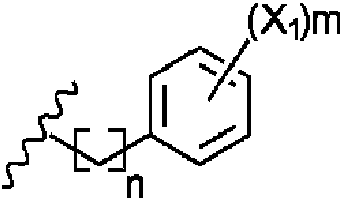
entonces m puede ser0, 1,2, 3, 4 o 5. Preferiblemente m es 1. Preferiblemente, X1 está en la posición para.then m can be 0, 1, 2, 3, 4 or 5. Preferably m is 1. Preferably X1 is in the para position.
En un ejemplo preferido R2 esIn a preferred example R2 is

Preferiblemente, en los ejemplos cuando R2 esPreferably, in the examples when R2 is

entonces al menos uno de Rg o Río es -H, y lo más preferiblemente Rg o Río son ambos -H.then at least one of Rg or Rio is -H, and most preferably Rg or Rio are both -H.
En un ejemplo preferido, R2 esIn a preferred example, R2 is

Se apreciará que R3 puede ser un grupo metilo, etilo, propilo, butilo o pentilo. Preferiblemente, R3 es metilo. Sin embargo, en un ejemplo más preferido, R3 es -H.It will be appreciated that R3 may be a methyl, ethyl, propyl, butyl or pentyl group. Preferably R3 is methyl. However, in a more preferred example, R3 is -H.
En un ejemplo R4 esIn an example R4 is

y n está preferiblemente entre 1 y 5.and n is preferably between 1 and 5.
En consecuencia, n puede ser 1, 2, 3, 4 o 5, y preferiblemente n es 1.Accordingly, n can be 1, 2, 3, 4 or 5, and preferably n is 1.
En ejemplos donde R4 esIn examples where R4 is

entonces n está preferiblemente entre 1 y 7, y más preferiblemente entre 2 y 6. En consecuencia, n puede ser 2, 3, 4, 5 o 6, y es preferible que n sea 3 o 4.then n is preferably between 1 and 7, and more preferably between 2 and 6. Accordingly, n can be 2, 3, 4, 5 or 6, and it is preferable that n is 3 or 4.
En un ejemplo, R4 es preferiblementeIn one example, R4 is preferably

Más preferiblemente, R4 esMore preferably, R4 is

Se apreciará que en los ejemplos donde R4 esIt will be appreciated that in the examples where R4 is

entonces m puede ser 0, 1,2, 3, 4 o 5. Preferiblemente m es 1. Preferiblemente, X1 está en la posición para.then m can be 0, 1,2, 3, 4 or 5. Preferably m is 1. Preferably X1 is in the para position.
Preferiblemente, R4 esPreferably, R4 is

Más preferiblemente, R4 esMore preferably, R4 is

Preferiblemente, en los ejemplos cuando R4 esPreferably, in the examples when R4 is

entonces al menos uno de Rg o R10 es -H, y lo más preferiblemente tanto Rg o R10 son -H.then at least one of Rg or R10 is -H, and most preferably both Rg or R10 are -H.
En consecuencia, R4 es preferiblementeConsequently, R4 is preferably

Más preferiblemente, R4 esMore preferably, R4 is

Más preferiblemente, R4 esMore preferably, R4 is

Se apreciará que R5 puede ser un grupo metilo, etilo, propilo, butilo o pentilo. Preferiblemente, R5 es metilo. Sin embargo, en un ejemplo más preferido, R5 es -H.It will be appreciated that R5 may be a methyl, ethyl, propyl, butyl or pentyl group. Preferably R5 is methyl. However, in a more preferred example, R5 is -H.
En R6 esIn R6 it is

y n está entre 1 y 5. En consecuencia, n puede ser 1, 2, 3, 4 o 5, y preferiblemente n es 1.and n is between 1 and 5. Accordingly, n can be 1, 2, 3, 4 or 5, and preferably n is 1.
Preferiblemente, R6 esPreferably, R6 is

Se apreciará que en los ejemplos donde R6 esIt will be appreciated that in the examples where R6 is

entonces m puede ser 0, 1,2, 3, 4 o 5. Preferiblemente, m es 0. Más preferiblemente, m es 1. Preferiblemente, X1 está en la posición para.then m can be 0, 1.2, 3, 4 or 5. Preferably, m is 0. More preferably, m is 1. Preferably, X1 is in the para position.
En un ejemplo preferido R6 esIn a preferred example R6 is

Preferiblemente, en los ejemplos cuando R6 esPreferably, in the examples when R6 is

entonces al menos uno de Rg o R10 es -H, y lo más preferiblemente tanto Rg o R10 son -H.then at least one of Rg or R10 is -H, and most preferably both Rg or R10 are -H.
Preferiblemente, en los ejemplos cuando R6 esPreferably, in the examples when R6 is

entonces R11 es arilo, y lo más preferiblemente fenilo.then R11 is aryl, and most preferably phenyl.
En un ejemplo preferido, R6 esIn a preferred example, R6 is

g g
Se apreciará que R7 puede ser un grupo metilo, etilo, propilo, butilo o pentilo. Preferiblemente, R7 es metilo. Sin embargo, en un ejemplo más preferido, R7 es -H.It will be appreciated that R7 may be a methyl, ethyl, propyl, butyl or pentyl group. Preferably R7 is methyl. However, in a more preferred example, R7 is -H.
En un ejemplo preferido R8 es -H. Sin embargo, en un ejemplo más preferido R8 esIn a preferred example R8 is -H. However, in a more preferred example R8 is

En un ejemplo preferido, el inhibidor comprende un compuesto de Fórmula (I), en la que:In a preferred example, the inhibitor comprises a compound of Formula (I), wherein:
R2 esR2 is

R3 es H o un alquilo o alquenilo C1-5 lineal o ramificado;R3 is H or a straight or branched C1-5 alkyl or alkenyl;
R4 esR4 is

R5 es H o un alquilo o alquenilo C1-5 lineal o ramificado;R5 is H or a straight or branched C1-5 alkyl or alkenyl;
R6 esR6 is

yand
R7 es H o un alquilo o alquenilo C1-5 lineal o ramificado.R7 is H or a straight or branched C1-5 alkyl or alkenyl.
Preferiblemente, R3 es H, R5 es H y R7 es H.Preferably R3 is H, R5 is H, and R7 is H.
En un ejemplo más preferido, el inhibidor comprende un compuesto de Fórmula (I), en la que:In a more preferred example, the inhibitor comprises a compound of Formula (I), wherein:
R2 esR2 is

R3 es H o un alquilo o alquenilo C1-5 lineal o
R4 esR4 is

R5 es H o un alquilo o alquenilo C1-5 lineal o ramificado; oR5 is H or a straight or branched C1-5 alkyl or alkenyl; either
R6 es R6 is

yand
R7 es H o un alquilo o alquenilo C1-5 lineal o ramificado.R7 is H or a straight or branched C1-5 alkyl or alkenyl.
Preferiblemente, el inhibidor comprende un compuesto de Fórmula (la). Preferiblemente, R3 es H, R5 es H y R7 es H. Preferiblemente, R1 es -OH y R8 es H. Más preferiblemente, R1 es NH2 y R8 esPreferably, the inhibitor comprises a compound of Formula (la). Preferably R3 is H, R5 is H and R7 is H. Preferably R1 is -OH and R8 is H. More preferably R1 is NH2 and R8 is

En un ejemplo alternativo más preferido, el inhibidor comprende un compuesto de Fórmula (I), en la que:In a more preferred alternative example, the inhibitor comprises a compound of Formula (I), wherein:
R2 esR2 is

R3 es H o un alquilo o alquenilo C1-5 lineal o ramificado;R3 is H or a straight or branched C1-5 alkyl or alkenyl;
R4 esR4 is

R5 es H o un alquilo o alquenilo C1-5 lineal o ramificado; oR5 is H or a straight or branched C1-5 alkyl or alkenyl; either
R6 esR6 is

yand
R7 es H o un alquilo o alquenilo C1-5 lineal o ramificado.R7 is H or a straight or branched C1-5 alkyl or alkenyl.
Preferiblemente, el inhibidor comprende un compuesto de Fórmula (Ia). Preferiblemente, R3 es H, R5 es H y R7 es H. Preferiblemente, R1 es -OH y R8 es H. Más preferiblemente, R1 es NH2 y R8 esPreferably, the inhibitor comprises a compound of Formula (Ia). Preferably R3 is H, R5 is H and R7 is H. Preferably R1 is -OH and R8 is H. More preferably R1 is NH2 and R8 is

En un ejemplo incluso más preferido, el inhibidor comprende un compuesto de Fórmula (I), en la que:In an even more preferred example, the inhibitor comprises a compound of Formula (I), wherein:
R2 esR2 is

R3 es H o un alquilo o alquenilo C1-5 lineal o ramificado; R3 is H or a straight or branched C1-5 alkyl or alkenyl;

R5 es H o un alquilo o alquenilo C1-5 lineal o ramificado;R5 is H or a straight or branched C1-5 alkyl or alkenyl;
R6 esR6 is

yand
R7 es H o un alquilo o alquenilo C1-5 lineal o ramificado.R7 is H or a straight or branched C1-5 alkyl or alkenyl.
Preferiblemente, R3 es H, R5 es H y R7 es H.Preferably R3 is H, R5 is H, and R7 is H.
En un ejemplo más preferido, el inhibidor comprende un compuesto de Fórmula (I), en la que:In a more preferred example, the inhibitor comprises a compound of Formula (I), wherein:
R2 esR2 is

R3 es H o un alquilo o alquenilo C1-5 lineal o ramificado;R3 is H or a straight or branched C1-5 alkyl or alkenyl;
R4 esR4 is

R5 es H o un alquilo o alquenilo C1-5 lineal o ramificado; oR5 is H or a straight or branched C1-5 alkyl or alkenyl; either
R6 esR6 is

yand
R7 es H o un alquilo o alquenilo C1-5 lineal o ramificado.R7 is H or a straight or branched C1-5 alkyl or alkenyl.
Preferiblemente, el inhibidor comprende un compuesto de Fórmula (Ia). Preferiblemente, R3 es H, R5 es H y R7 es H. Preferiblemente, R1 es -OH y R8 es H. Más preferiblemente, R1 es NH2 y R8 esPreferably, the inhibitor comprises a compound of Formula (Ia). Preferably R3 is H, R5 is H and R7 is H. Preferably R1 is -OH and R8 is H. More preferably R1 is NH2 and R8 is

En un ejemplo alternativo más preferido, el inhibidor comprende un compuesto de Fórmula (I), en la que:In a more preferred alternative example, the inhibitor comprises a compound of Formula (I), wherein:
R2 es R2 is

R3 es H o un alquilo o alquenilo C1-5 lineal o ramificado;R3 is H or a straight or branched C1-5 alkyl or alkenyl;
R4 esR4 is

R5 es H o un alquilo o alquenilo C1-5 lineal o ramificado; oR5 is H or a straight or branched C1-5 alkyl or alkenyl; either
R6 esR6 is

yand
R7 es H o un alquilo o alquenilo C1-5 lineal o ramificado.R7 is H or a straight or branched C1-5 alkyl or alkenyl.
Preferiblemente, el inhibidor comprende un compuesto de Fórmula (Ia). Preferiblemente, R3 es H, R5 es H y R7 es H. Preferiblemente, R1 es -OH y R8 es H. Más preferiblemente, R1 es NH2 y R8 esPreferably, the inhibitor comprises a compound of Formula (Ia). Preferably R3 is H, R5 is H and R7 is H. Preferably R1 is -OH and R8 is H. More preferably R1 is NH2 and R8 is

Preferiblemente, el inhibidor comprende un compuesto de Fórmula (101) o (103):Preferably, the inhibitor comprises a compound of Formula (101) or (103):

Fórmula (101) Formula (101)

Más preferiblemente, el inhibidor comprende un compuesto de Fórmula (101a) o (103a):More preferably, the inhibitor comprises a compound of Formula (101a) or (103a):

Se apreciará que los compuestos de Fórmula (101a) y (103a) corresponden a los compuestos Trio2 y Trio4, respectivamente, que se analizan en los Ejemplos. Por lo tanto, en otro ejemplo, se proporciona un inhibidor como se define en este documento, para usar en el tratamiento, mejora o prevención del cáncer o enfermedad metastásica, preferiblemente en el que el inhibidor es de Fórmula (101a) o (103a).It will be appreciated that the compounds of Formula (101a) and (103a) correspond to the compounds Trio2 and Trio4, respectively, which are discussed in the Examples. Thus, in another example, an inhibitor is provided as defined herein, for use in the treatment, amelioration or prevention of cancer or metastatic disease, preferably wherein the inhibitor is of Formula (101a) or (103a) .
En un ejemplo preferido, el inhibidor comprende un anticuerpo o fragmento de unión a antígeno del mismo, es decir, un anticuerpo neutralizante de T14. El anticuerpo o fragmento de unión a antígeno del mismo bloquea preferiblemente la interacción de T14 con el sitio alostérico en el receptor alfa-7. Preferiblemente, el anticuerpo se une a T14, formando así un complejo que es incapaz de unirse al receptor para causar toxicidad.In a preferred example, the inhibitor comprises an antibody or antigen-binding fragment thereof, ie, a neutralizing antibody to T14. The antibody or antigen-binding fragment thereof preferably blocks the interaction of T14 with the allosteric site on the alpha-7 receptor. Preferably, the antibody binds to T14, thus forming a complex that is incapable of binding to the receptor to cause toxicity.
El anticuerpo o fragmento de unión a antígeno del mismo puede ser policlonal o monoclonal. El anticuerpo o fragmento de unión a antígeno del mismo puede generarse en un conejo, ratón o rata. The antibody or antigen-binding fragment thereof may be polyclonal or monoclonal. The antibody or antigen-binding fragment thereof can be generated in a rabbit, mouse, or rat.
Preferiblemente, el anticuerpo o fragmento de unión a antígeno del mismo se une específicamente a la SEQ ID No: 3. Preferiblemente, el anticuerpo o fragmento de unión a antígeno del mismo se une específicamente a uno o más aminoácidos en el extremo terminal C de la SEQ ID No: 3. Preferiblemente, el anticuerpo o fragmento de unión a antígeno del mismo se une específicamente a uno o más aminoácidos en la SEQ ID No: 5 (es decir, SYMVHWK, que son los aminoácidos números 7-14 del extremo terminal C de la SEQ ID No: 3). Preferiblemente, el anticuerpo o fragmento de unión a antígeno del mismo se une específicamente a un residuo de lisina (K) del extremo terminal C en el epítopo.Preferably, the antibody or antigen-binding fragment thereof specifically binds to SEQ ID No: 3. Preferably, the antibody or antigen-binding fragment thereof specifically binds to one or more amino acids at the C-terminus of the SEQ ID No: 3. Preferably, the antibody or antigen-binding fragment thereof specifically binds to one or more amino acids in SEQ ID No: 5 (i.e., SYMVHWK, which are amino acid numbers 7-14 from the terminal end of C of SEQ ID No: 3). Preferably, the antibody or antigen-binding fragment thereof specifically binds to a C-terminal lysine (K) residue in the epitope.
Los inventores han observado sorprendentemente que la secuencia de aminoácidos VHWK del extremo terminal C en la SEQ ID No: 3, que se describe en este documento como la SEQ ID No. 6 (es decir, los aminoácidos números 10 14 del extremo terminal C de la SEQ ID No. 3), actúa como un epítopo para el anticuerpo o fragmento de unión a antígeno del mismo. Por consiguiente, más preferiblemente el anticuerpo o fragmento de unión a antígeno del mismo se une específicamente a uno o más aminoácidos en la SEQ ID No. 6. Lo más preferiblemente, el anticuerpo o fragmento de unión a antígeno del mismo se une específicamente a la SEQ ID No. 6. Por lo tanto, se apreciará que el epítopo al que se une el anticuerpo comprende o consiste en la SEQ ID No: 6.The inventors have surprisingly found that the C-terminal amino acid sequence VHWK in SEQ ID No: 3, which is described herein as SEQ ID No. 6 (i.e., C-terminal amino acid numbers 10-14 of SEQ ID No. 3), acts as an epitope for the antibody or antigen-binding fragment thereof. Accordingly, more preferably the antibody or antigen-binding fragment thereof specifically binds to one or more amino acids in SEQ ID No. 6. Most preferably, the antibody or antigen-binding fragment thereof specifically binds to the SEQ ID No. 6. Therefore, it will be appreciated that the epitope to which the antibody binds comprises or consists of SEQ ID No: 6.
Con base en el descubrimiento del epítopo VHWK (SEQ ID No: 6) dentro del péptido T14, los inventores creen que estas secuencias pueden usarse como antígeno para la producción de anticuerpos útiles. Como se describe en los ejemplos, el péptido T14 (SEQ ID No: 3) se entrecruzó con cisteína a la hemocianina de lapa californiana (KLH) que actúa como una proteína transportadora que estimula una respuesta inmunitaria en el huésped. La proteína KLH entrecruzada con T14 se denomina en el presente documento como la SEQ ID No: 7, es decir, CAEFHRWSSYMVHWK (la C se añadió para unir KLH con la A en el extremo terminal N del péptido T14).Based on the discovery of the HWKV epitope (SEQ ID No: 6) within the T14 peptide, the inventors believe that these sequences can be used as an antigen for the production of useful antibodies. As described in the examples, the T14 peptide (SEQ ID No: 3) was cysteine-crosslinked to keyhole limpet hemocyanin (KLH) which acts as a carrier protein stimulating an immune response in the host. The T14 cross-linked KLH protein is referred to herein as SEQ ID No: 7, ie, CAEFHRWSSYMVHWK (the C was added to join KLH with the A at the N-terminus of the T14 peptide).
Preferiblemente, el anticuerpo o fragmento de unión a antígeno del mismo no se une a la SEQ ID NO: 2. Preferiblemente, el anticuerpo o fragmento de unión a antígeno del mismo no se une a la SEQ ID No: 4.Preferably, the antibody or antigen-binding fragment thereof does not bind to SEQ ID NO: 2. Preferably, the antibody or antigen-binding fragment thereof does not bind to SEQ ID No: 4.
Los anticuerpos policlonales se pueden producir como sueros policlonales mediante la inyección de antígeno en animales. Los anticuerpos policlonales preferidos pueden generarse mediante la inoculación de un animal (p. ej., un conejo) con antígeno (p. ej., T14 o fragmentos al mismo, incluido el extremo terminal C) usando técnicas conocidas en el arte. Por ejemplo, el anticuerpo o fragmento de unión a antígeno del mismo puede obtenerse inmunizando un animal huésped con la SEQ ID No:3 y luego recolectando el anticuerpo o fragmento de unión a antígeno del mismo. El animal huésped es lo más preferiblemente un conejo.Polyclonal antibodies can be produced as polyclonal sera by injecting animals with antigen. Preferred polyclonal antibodies can be generated by inoculating an animal (eg, a rabbit) with antigen (eg, T14 or fragments thereof, including the C-terminus) using techniques known in the art. For example, the antibody or antigen-binding fragment thereof can be obtained by immunizing a host animal with SEQ ID No:3 and then harvesting the antibody or antigen-binding fragment thereof. The host animal is most preferably a rabbit.
En otro ejemplo preferido, el inhibidor comprende un anticuerpo monoclonal o un fragmento de unión a antígeno del mismo. Los anticuerpos monoclonales preferidos pueden generarse usando tecnología de hibridoma usando T14 o fragmentos del mismo como antígeno. Preferiblemente, el anticuerpo de la divulgación es un anticuerpo humano. Como se usa en este documento, el término "anticuerpo humano" puede significar un anticuerpo, tal como un anticuerpo monoclonal, que comprende sustancialmente las mismas secuencias de aminoácidos de CDR de cadena ligera y pesada que se encuentran en un anticuerpo humano particular que exhibe inmuno especificidad por la SEQ ID No: 3. Una secuencia de aminoácidos, que es sustancialmente la misma que una CDR de cadena ligera o pesada, exhibe una cantidad considerable de identidad de secuencia cuando se compara con una secuencia de referencia. Tal identidad es definitivamente conocida o reconocible como representativa de la secuencia de aminoácidos del anticuerpo humano particular. Sustancialmente, la misma secuencia de aminoácidos de CDR de cadena ligera y pesada puede tener, p. ej., modificaciones menores o sustituciones conservadoras de aminoácidos. Dicho anticuerpo humano mantiene su función de unión selectiva a la SEQ ID No: 3.In another preferred example, the inhibitor comprises a monoclonal antibody or an antigen-binding fragment thereof. Preferred monoclonal antibodies can be generated using hybridoma technology using T14 or fragments thereof as antigen. Preferably, the antibody of the disclosure is a human antibody. As used herein, the term "human antibody" can mean an antibody, such as a monoclonal antibody, that comprises substantially the same heavy and light chain CDR amino acid sequences as found in a particular human antibody that exhibits immune specificity per SEQ ID No: 3. An amino acid sequence, which is substantially the same as a heavy or light chain CDR, exhibits a considerable amount of sequence identity when compared to a reference sequence. Such identity is definitely known or recognizable as representative of the amino acid sequence of the particular human antibody. Substantially the same heavy and light chain CDR amino acid sequence may have, e.g. eg, minor modifications or conservative amino acid substitutions. Said human antibody maintains its selective binding function to SEQ ID No: 3.
Pueden usarse técnicas de hibridoma convencionales para generar los anticuerpos. El antígeno utilizado para generar anticuerpos monoclonales puede ser la proteína T14 completa o un fragmento de la misma. Los fragmentos preferidos para generar los anticuerpos también pueden ser los péptidos discutidos anteriormente, y particularmente SYMVHWK (SEQ ID No: 5) o VHWK (SEQ ID No: 6). Se prefiere que el anticuerpo sea una Y-inmunoglobulina (IgG).Standard hybridoma techniques can be used to generate the antibodies. The antigen used to generate monoclonal antibodies can be the complete T14 protein or a fragment thereof. Preferred fragments for generating the antibodies may also be the peptides discussed above, and particularly SYMVHWK (SEQ ID No: 5) or VHWK (SEQ ID No: 6). It is preferred that the antibody is a Y-immunoglobulin (IgG).
Otro inhibidor preferido es un fragmento peptídico inactivo del péptido T14 que competirá con el T14 endógeno y por lo tanto reducirá su actividad. Por ejemplo, los mutantes de truncamiento de T14 que no se unen al sitio alostérico en el receptor alfa-7 y que inhiben la capacidad de T14 para interactuar con el sitio alostérico en el receptor alfa-7 también pueden usarse como inhibidores.Another preferred inhibitor is an inactive peptide fragment of the T14 peptide which will compete with endogenous T14 and therefore reduce its activity. For example, T14 truncation mutants that do not bind to the allosteric site on the alpha-7 receptor and that inhibit the ability of T14 to interact with the allosteric site on the alpha-7 receptor can also be used as inhibitors.
En otro ejemplo, el inhibidor puede prevenir o reducir la escisión postraduccional de la enzima acetilcolinesterasa para formar el péptido T14, es decir, (d) anterior. Por ejemplo, el inhibidor de acuerdo con la divulgación puede ser una molécula silenciadora de genes configurada para bloquear o interrumpir la escisión enzimática del polipéptido acetilcolinesterasa en T14.In another example, the inhibitor can prevent or reduce post-translational cleavage of the acetylcholinesterase enzyme to form the T14 peptide, ie, (d) above. For example, the inhibitor according to the disclosure can be a gene silencing molecule configured to block or interrupt enzymatic cleavage of T14 acetylcholinesterase polypeptide.
El término "molécula silenciadora de genes" puede significar cualquier molécula que interfiere con la expresión del péptido T14. Dichas moléculas incluyen, pero no se limitan a, moléculas de ARNi, incluyendo ANpi, ARNpi, miARN, ribozimas y moléculas antisentido.The term "gene silencing molecule" can mean any molecule that interferes with the expression of the T14 peptide. Such molecules include, but are not limited to, RNAi molecules, including siNA, siRNA, miRNA, ribozymes, and antisense molecules.
Como se discutió anteriormente, el gen que codifica la acetilcolinesterasa se expresa y se traduce en la proteína que se muestra como la SEQ ID NO: 1. Las modificaciones postraduccionales de la SEQ ID No: 1 pueden incluir la escisión por una proteasa para crear T14 (SEQ ID No: 3). Por consiguiente, en un ejemplo preferido, el inhibidor comprende una molécula silenciadora de genes que reduce o previene la expresión de la proteasa responsable de escindir T15 (SEQ ID No: 4) de T30 para crear T14.As discussed above, the gene encoding acetylcholinesterase is expressed and translated into the protein shown as SEQ ID NO: 1. Post-translational modifications of SEQ ID No: 1 may include cleavage by a protease to create T14 (SEQ ID No: 3). Accordingly, in a preferred example, the inhibitor comprises a gene silencing molecule that reduces or prevents the expression of the protease responsible for cleaving T15 (SEQ ID No: 4) from T30 to create T14.
En otro ejemplo, el inhibidor puede inhibir la translocación de T14 al receptor alfa-7 (es decir, (e) anterior).In another example, the inhibitor can inhibit the translocation of T14 to the alpha-7 receptor (ie, (e) above).
Se apreciará que los inhibidores de acuerdo con esta divulgación pueden usarse en un medicamento, que puede usarse como monoterapia (es decir, el uso del inhibidor de T14 solo), para tratar, mejorar o prevenir el cáncer o la metástasis. Alternativamente, el inhibidor de T14 se puede usar como complemento o en combinación con terapias conocidas para tratar, mejorar o prevenir el cáncer o la metástasis, tal como radioterapia o quimioterapia, o cualquiera de los fármacos anticancerosos conocidos, tal como Lapatinib.It will be appreciated that the inhibitors in accordance with this disclosure may be used in a medicament, which may be used as monotherapy (ie, use of the T14 inhibitor alone), to treat, ameliorate or prevent cancer or metastasis. Alternatively, the T14 inhibitor can be used as an adjunct to or in combination with therapies known to treat, ameliorate or prevent cancer or metastasis, such as radiotherapy or chemotherapy, or any of the known anti-cancer drugs, such as Lapatinib.
Los inhibidores de acuerdo con esta divulgación pueden combinarse en composiciones que tienen varias formas diferentes dependiendo, en particular, de la manera en que se va a utilizar la composición. Así, p. ej., la composición puede presentarse en forma de polvo, comprimido, cápsula, líquido, pomada, crema, gel, hidrogel, aerosol, atomizador, solución micelar, parche transdérmico, suspensión de liposomas o cualquier otra forma adecuada que pueda ser administrada a una persona o animal que necesita tratamiento. Se apreciará que el vehículo de los medicamentos de acuerdo con la divulgación debe ser bien tolerado por el sujeto al que se le administra, y preferiblemente permite la administración del inhibidor al sitio objetivo, p. ej., a través de la barrera hematoencefálica al tratar tumores cerebrales.The inhibitors according to this disclosure can be combined in compositions that have a number of different forms depending, in particular, on the manner in which the composition is to be used. So e.g. For example, the composition may be in the form of a powder, tablet, capsule, liquid, ointment, cream, gel, hydrogel, aerosol, spray, micellar solution, transdermal patch, liposome suspension, or any other suitable form that can be administered to a person or animal in need of treatment. It will be appreciated that the vehicle for medicaments according to the disclosure should be well tolerated by the subject to which it is administered, and preferably allows delivery of the inhibitor to the target site, e.g. eg, through the blood-brain barrier when treating brain tumors.
Los inhibidores de acuerdo con esta divulgación también pueden incorporarse dentro de un dispositivo de liberación lenta o retardada. Dichos dispositivos pueden, p. ej., insertarse sobre o debajo de la piel, y el medicamento puede liberarse durante semanas o incluso meses. El dispositivo puede estar ubicado al menos adyacente al sitio de tratamiento. Dichos dispositivos pueden ser particularmente ventajosos cuando se requiere un tratamiento a largo plazo con inhibidores usados de acuerdo con la divulgación y que normalmente requeriría una administración frecuente (p. ej., al menos una inyección diaria).Inhibitors according to this disclosure can also be incorporated into a slow or delayed release device. Such devices can e.g. eg, inserted over or under the skin, and the drug can be released for weeks or even months. The device may be located at least adjacent to the treatment site. Such devices may be particularly advantageous where long-term treatment with inhibitors used in accordance with the disclosure is required and would normally require frequent administration (eg, at least one daily injection).
En un ejemplo preferido, los medicamentos de acuerdo con esta divulgación pueden administrarse a un sujeto mediante inyección en el torrente sanguíneo o directamente en un sitio que requiera tratamiento. Por ejemplo, el medicamento puede administrarse mediante inyección en el torrente sanguíneo. Las inyecciones pueden ser intravenosas (bolo o infusión) o subcutáneas (bolo o infusión) o intradérmicas (bolo o infusión).In a preferred example, medicaments in accordance with this disclosure may be administered to a subject by injection into the bloodstream or directly into a site requiring treatment. For example, the drug can be administered by injection into the bloodstream. Injections can be intravenous (bolus or infusion) or subcutaneous (bolus or infusion) or intradermal (bolus or infusion).
Se apreciará que la cantidad de inhibidor que se requiere está determinada por su actividad biológica y biodisponibilidad, que a su vez depende del modo de administración, las propiedades fisicoquímicas del inhibidor y si se está utilizando como monoterapia o en una terapia combinada. La frecuencia de administración también estará influenciada por la vida media del inhibidor dentro del sujeto que se está tratando. Las dosis óptimas por administrar pueden ser determinadas por los expertos en la técnica y variarán con el inhibidor particular en uso, la potencia de la composición farmacéutica, el modo de administración y el avance del cáncer o metástasis. Factores adicionales que dependen del sujeto particular que se esté tratando darán como resultado la necesidad de ajustar las dosis, incluyendo la edad, el peso, el sexo, la dieta y el momento de la administración del sujeto.It will be appreciated that the amount of inhibitor that is required is determined by its biological activity and bioavailability, which in turn depends on the mode of administration, the physicochemical properties of the inhibitor, and whether it is being used as monotherapy or in combination therapy. The frequency of administration will also be influenced by the half-life of the inhibitor within the subject being treated. The optimal doses to be administered can be determined by those skilled in the art and will vary with the particular inhibitor in use, the potency of the pharmaceutical composition, the mode of administration, and the progression of the cancer or metastasis. Additional factors depending on the particular subject being treated will result in the need to adjust dosages, including the subject's age, weight, sex, diet, and timing of administration.
Generalmente, una dosis diaria de entre 0.001 pg/kg de peso corporal y 10 mg/kg de peso corporal, o entre 0.01 pg/kg de peso corporal y 1 mg/kg de peso corporal, del inhibidor de acuerdo con la divulgación puede usarse para tratar, mejorar o prevenir el cáncer o la metástasis, dependiendo del inhibidor que se use.Generally, a daily dose of between 0.001 pg/kg of body weight and 10 mg/kg of body weight, or between 0.01 pg/kg of body weight and 1 mg/kg of body weight, of the inhibitor according to the disclosure may be used. to treat, ameliorate, or prevent cancer or metastasis, depending on which inhibitor is used.
El inhibidor puede administrarse antes, durante o después del inicio del cáncer. Las dosis diarias pueden administrarse en una sola administración (p. ej., una sola inyección diaria o la inhalación de un aerosol nasal). Alternativamente, el inhibidor puede requerir la administración dos o más veces durante un día. Como ejemplo, los inhibidores pueden administrarse como dos (o más dependiendo de la gravedad del cáncer o la metástasis que se esté tratando) dosis diarias de entre 0.07 pg y 700 mg (es decir, suponiendo un peso corporal de 70 kg). Un paciente que recibe tratamiento puede tomar una primera dosis al despertar y luego una segunda dosis por la noche (si está en un régimen de dos dosis) o a intervalos de 3 o 4 horas a partir de entonces. Alternativamente, se puede usar un dispositivo de liberación lenta para proporcionar dosis óptimas de inhibidor de acuerdo con la divulgación a un paciente sin necesidad de administrar dosis repetidas.The inhibitor can be administered before, during, or after the onset of the cancer. Daily doses can be given as a single administration (eg, a single daily injection or nasal spray inhalation). Alternatively, the inhibitor may require administration two or more times during one day. As an example, the inhibitors may be administered as two (or more depending on the severity of the cancer or metastasis being treated) daily doses of between 0.07 pg and 700 mg (ie assuming 70 kg body weight). A patient receiving treatment may take a first dose upon awakening and then a second dose in the evening (if on a two-dose regimen) or at 3-4 hour intervals thereafter. Alternatively, a slow release device can be used to deliver optimal doses of inhibitor according to the disclosure to a patient without the need for repeat dosing.
Procedimientos conocidos, como los empleados convencionalmente por la industria farmacéutica (p. ej., experimentación in vivo, ensayos clínicos, etc.), pueden usarse para formar formulaciones específicas del inhibidor de acuerdo con esta divulgación y regímenes terapéuticos precisos (tales como dosis diarias de los agentes y la frecuencia de administración). Los inventores creen que son los primeros en sugerir una composición de tratamiento contra el cáncer, basada en el uso de inhibidores de esta divulgación.Known procedures, such as those conventionally employed by the pharmaceutical industry (eg, in vivo experimentation, clinical trials, etc.), can be used to form specific inhibitor formulations in accordance with this disclosure and precise therapeutic regimens (such as daily doses of agents and frequency of administration). The inventors believe that they are the first to suggest a cancer treatment composition, based on the use of inhibitors of this disclosure.
Por lo tanto, en un ejemplo, se proporciona una composición farmacéutica anticancerígena o antimetastásica que comprende una cantidad terapéuticamente eficaz del inhibidor de acuerdo con esta divulgación y, opcionalmente, un vehículo farmacéuticamente aceptable.Thus, in one example, an anti-cancer or antimetastatic pharmaceutical composition is provided comprising a therapeutically effective amount of the inhibitor according to this disclosure and, optionally, a pharmaceutically acceptable carrier.
Esta divulgación también proporciona un proceso para preparar la composición farmacéutica anticancerígena o antimetastásica de acuerdo con esta divulgación, comprendiendo el proceso combinar una cantidad terapéuticamente eficaz de inhibidor de acuerdo con el primer aspecto, con un vehículo farmacéuticamente aceptable. This disclosure also provides a process for preparing the anticancer or antimetastatic pharmaceutical composition according to this disclosure, the process comprising combining a therapeutically effective amount of inhibitor according to the first aspect, with a pharmaceutically acceptable carrier.
El inhibidor de esta divulgación inhibe la síntesis y/o actividad de un péptido de la SEQ ID No: 3, como se describió anteriormente.The inhibitor of this disclosure inhibits the synthesis and/or activity of a peptide of SEQ ID No: 3, as described above.
Un "sujeto" puede ser un vertebrado, un mamífero o un animal doméstico. Por lo tanto, los medicamentos de acuerdo con la divulgación se pueden usar para tratar a cualquier mamífero, p. ej., ganado (p. ej., un caballo), mascotas, o se pueden usar en otras aplicaciones veterinarias. Sin embargo, lo más preferiblemente, el sujeto es un ser humano. A "subject" can be a vertebrate, mammal, or domestic animal. Therefore, the medicaments according to the disclosure can be used to treat any mammal, e.g. livestock (eg, a horse), pets, or can be used in other veterinary applications. However, most preferably, the subject is a human.
Una "cantidad terapéuticamente eficaz" del inhibidor es cualquier cantidad que, cuando se administra a un sujeto, es la cantidad de agente activo que se necesita para tratar el cáncer o la metástasis, o producir el efecto deseado. El inhibidor puede usarse como adyuvante para el tratamiento de tumores sólidos o metastásicos, p. ej., con quimioterapia o radioterapia. Esto significa que se requieren dosis y tiempos de exposición más bajos de quimioterapia y/o radioterapia.A "therapeutically effective amount" of the inhibitor is any amount which, when administered to a subject, is the amount of active agent that is needed to treat cancer or metastasis, or produce the desired effect. The inhibitor can be used as an adjuvant for the treatment of solid or metastatic tumors, e.g. For example, with chemotherapy or radiotherapy. This means that lower doses and exposure times of chemotherapy and/or radiotherapy are required.
Por ejemplo, la cantidad terapéuticamente eficaz de inhibidor usada puede ser de aproximadamente 0.001 mg a aproximadamente 800 mg, y preferiblemente de aproximadamente 0.01 mg a aproximadamente 500 mg.For example, the therapeutically effective amount of inhibitor used can be from about 0.001 mg to about 800 mg, and preferably from about 0.01 mg to about 500 mg.
Un "vehículo farmacéuticamente aceptable", tal como se hace referencia en el presente documento, es cualquier compuesto conocido o combinación de compuestos conocidos que los expertos en la técnica saben que es útil para formular composiciones farmacéuticas.A "pharmaceutically acceptable carrier", as referred to herein, is any known compound or combination of known compounds known to those skilled in the art to be useful in formulating pharmaceutical compositions.
En un ejemplo, el vehículo farmacéuticamente aceptable puede ser un sólido y la composición puede estar en forma de polvo o tableta. Sin embargo, el vehículo farmacéutico puede ser un líquido y la composición farmacéutica está en forma de solución. Las composiciones farmacéuticas líquidas, que son soluciones o suspensiones estériles, se pueden utilizar, p. ej., mediante inyección intramuscular, intratecal, epidural, intraperitoneal, intravenosa y particularmente subcutánea.In one example, the pharmaceutically acceptable carrier may be a solid and the composition may be in powder or tablet form. However, the pharmaceutical carrier can be a liquid and the pharmaceutical composition is in the form of a solution. Liquid pharmaceutical compositions, which are sterile solutions or suspensions, can be used, e.g. eg, by intramuscular, intrathecal, epidural, intraperitoneal, intravenous and particularly subcutaneous injection.
El inhibidor y las composiciones de esta divulgación pueden administrarse por vía oral en forma de una solución o suspensión estéril que contiene otros solutos o agentes de suspensión (p. ej., suficiente solución salina o glucosa para hacer que la solución sea isotónica), sales biliares, goma arábiga, gelatina, monooleato de sorbitano, polisorbato 80 (ésteres oleato de sorbitol y sus anhídridos copolimerizados con óxido de etileno) y similares. El inhibidor utilizado de acuerdo con la divulgación también se puede administrar por vía oral en forma de composición líquida o sólida. Las composiciones adecuadas para administración oral incluyen formas sólidas, tales como píldoras, cápsulas, gránulos, tabletas y polvos, y formas líquidas, tales como soluciones, jarabes, elixires y suspensiones. Las formas útiles para la administración parenteral incluyen soluciones, emulsiones y suspensiones estériles.The inhibitor and compositions of this disclosure can be administered orally in the form of a sterile solution or suspension containing other solutes or suspending agents (eg, enough saline or glucose to make the solution isotonic), salts bile, gum acacia, gelatin, sorbitan monooleate, polysorbate 80 (oleate esters of sorbitol and its anhydrides copolymerized with ethylene oxide) and the like. The inhibitor used according to the disclosure can also be administered orally in the form of a liquid or solid composition. Compositions suitable for oral administration include solid forms, such as pills, capsules, granules, tablets, and powders, and liquid forms, such as solutions, syrups, elixirs, and suspensions. Useful forms for parenteral administration include sterile solutions, emulsions, and suspensions.
El conocimiento del papel sorprendente que juega T14 en el cáncer, y especialmente en la enfermedad metastásica, tal como lo demuestran los datos de CD44 en el Ejemplo 2, ha permitido a los inventores desarrollar una detección para identificar si los compuestos de prueba son o no inhibidores probables para tratar, prevenir o mejorar cáncer o metástasis.Knowledge of the surprising role that T14 plays in cancer, and especially metastatic disease, as demonstrated by the CD44 data in Example 2, has enabled the inventors to develop a screen to identify whether or not test compounds are inhibitors likely to treat, prevent or ameliorate cancer or metastasis.
Por lo tanto, de acuerdo con un ejemplo, se proporciona un método para seleccionar un compuesto candidato para probar si el compuesto tiene o no eficacia para tratar, prevenir o mejorar el cáncer o la enfermedad metastásica, comprendiendo el método:Thus, according to one example, a method of selecting a candidate compound for testing whether or not the compound has efficacy in treating, preventing or ameliorating cancer or metastatic disease is provided, the method comprising:
(i) exponer un sistema biológico al compuesto candidato;(i) exposing a biological system to the candidate compound;
(ii) detectar la concentración, expresión o actividad del péptido T14 de la SEQ ID No: 3 en el sistema biológico; y (iii) comparar la concentración, expresión o actividad del péptido T14 en el sistema biológico tratado con el compuesto en relación con la concentración, expresión o actividad encontrada en un sistema biológico de control que no fue tratado con el compuesto,(ii) detecting the concentration, expression or activity of the T14 peptide of SEQ ID No: 3 in the biological system; and (iii) comparing the concentration, expression, or activity of the T14 peptide in the biological system treated with the compound relative to the concentration, expression, or activity found in a control biological system that was not treated with the compound,
en el que un compuesto con eficacia para tratar, prevenir o mejorar el cáncer o la enfermedad metastásica disminuye la concentración, expresión o actividad del péptido T14 en relación con el control.wherein a compound with efficacy to treat, prevent, or ameliorate cancer or metastatic disease decreases T14 peptide concentration, expression, or activity relative to control.
Se apreciará que el método de acuerdo con este ejemplo se puede adaptar de modo que se pueda usar para probar si un compuesto realmente causa o no cáncer o metástasis.It will be appreciated that the method according to this example can be adapted so that it can be used to test whether or not a compound actually causes cancer or metastasis.
Por lo tanto, de acuerdo con otro ejemplo, se proporciona un método para seleccionar un compuesto para probar si el compuesto causa cáncer o no, comprendiendo el método:Therefore, according to another example, a method for selecting a compound to test whether or not the compound causes cancer is provided, the method comprising:
(i) exponer un sistema biológico al compuesto de prueba;(i) exposing a biological system to the test compound;
(ii) detectar la concentración, expresión o actividad del péptido T14 de la SEQ ID No: 3 en el sistema biológico; y (iii) comparar la concentración, expresión o actividad del péptido T14 en el sistema biológico tratado con el compuesto en relación con la concentración, expresión o actividad del péptido T14 encontrado en un sistema biológico de control que no fue tratado con el compuesto,(ii) detecting the concentration, expression or activity of the T14 peptide of SEQ ID No: 3 in the biological system; and (iii) comparing the concentration, expression or activity of the T14 peptide in the biological system treated with the compound relative to the concentration, expression or activity of the T14 peptide found in a control biological system that was not treated with the compound,
en el que un compuesto que es cancerígeno o que aumenta la metástasis aumenta la concentración, expresión o actividad del péptido T14 con respecto al control. wherein a compound that is carcinogenic or increases metastasis increases the concentration, expression, or activity of the T14 peptide relative to control.
Los métodos o ensayos de detección de esta divulgación se basan en la comprensión de los inventores de que el grado de expresión y/o actividad de T14 está estrechamente relacionado con el desarrollo de cáncer y metástasis. El método de detección de esta divulgación es particularmente útil para seleccionar bibliotecas de compuestos para identificar compuestos que pueden usarse como agentes anticancerígenos o antimetástasis usados de acuerdo con la divulgación. Este ejemplo de la divulgación puede usarse para identificar compuestos que son cancerígenos y que deben evitarse. En consecuencia, la detección de esta divulgación puede usarse para el control ambiental (p. ej., para probar efluentes de fábricas) o en pruebas de toxicidad (p. ej., para probar la seguridad de supuestos productos farmacéuticos, cosméticos, alimenticios y similares).The detection methods or assays of this disclosure are based on the inventors' understanding that the degree of expression and/or activity of T14 is closely related to the development of cancer and metastasis. The screening method of this disclosure is particularly useful for screening compound libraries to identify compounds that can be used as anticancer or antimetastasis agents used in accordance with the disclosure. This example of the disclosure can be used to identify compounds that are carcinogenic and should be avoided. Consequently, detection of this disclosure can be used for environmental monitoring (eg, to test factory effluents) or in toxicity tests (eg, to test the safety of alleged pharmaceutical, cosmetic, food, and Similar).
El término "sistema biológico" puede significar cualquier sistema experimental que un experto en la materia entendería para proporcionar información sobre los efectos que un compuesto puede tener sobre la concentración, expresión o actividad del péptido T14 en el entorno fisiológico. El sistema puede comprender: (a) un sujeto de prueba experimental cuando se va a emplear una prueba in vivo; (b) una muestra biológica derivada de un sujeto de prueba (p. ej.: sangre o una fracción de sangre (p. ej., suero o plasma), linfa o una muestra de células/biopsia); (c) un modelo de línea celular (p. ej., una célula que expresa naturalmente el péptido T14 o una célula diseñada para expresar T14); o (d) un sistema in vitro que contiene T14 o su gen y simula el entorno fisiológico de manera que se puede medir la concentración, expresión o actividad del péptido T14.The term "biological system" can mean any experimental system that one of ordinary skill in the art would understand to provide information about the effects that a compound may have on T14 peptide concentration, expression, or activity in the physiological environment. The system may comprise: (a) an experimental test subject when an in vivo test is to be employed; (b) a biological sample derived from a test subject (eg: blood or a fraction of blood (eg, serum or plasma), lymph or a cell/biopsy sample); (c) a cell line model (eg, a cell that naturally expresses the T14 peptide or a cell engineered to express T14); or (d) an in vitro system that contains T14 or its gene and simulates the physiological environment such that concentration, expression, or activity of the T14 peptide can be measured.
La selección preferiblemente ensaya células biológicas o lisados de las mismas. Cuando la detección implica el ensayo de células, pueden estar contenidas dentro de un animal de experimentación (p. ej., un ratón o una rata) cuando el método es una prueba in vivo. Alternativamente, las células pueden estar en una muestra de tejido (por pruebas ex vivo) o las células se pueden hacer crecer en cultivo. Se apreciará que dichas células deben expresar, o pueden ser inducidas a expresar, T14 funcional (es decir, tóxico). También es posible utilizar células que no están naturalmente predispuestas a producir T14 siempre que dichas células se transformen con un vector de expresión. Tales células representan pruebas de prueba preferidas para usar de acuerdo con esta divulgación. Esto se debe a que las células animales o incluso las células procariotas pueden transformarse para expresar el péptido T14 humano y, por lo tanto, representan un buen modelo celular para probar la eficacia de fármacos candidatos para uso en terapia humana. The selection preferably tests biological cells or lysates thereof. When detection involves testing cells, they may be contained within an experimental animal (eg, mouse or rat) when the method is an in vivo test. Alternatively, the cells can be in a tissue sample (by ex vivo testing) or the cells can be grown in culture. It will be appreciated that such cells must express, or can be induced to express, functional (ie, toxic) T14. It is also possible to use cells that are not naturally predisposed to produce T14 as long as such cells are transformed with an expression vector. Such cells represent preferred test specimens for use in accordance with this disclosure. This is because animal cells or even prokaryotic cells can be transformed to express the human T14 peptide and therefore represent a good cell model to test the efficacy of drug candidates for use in human therapy.
Lo más preferido es que las células biológicas usadas de acuerdo con los métodos de detección de esta divulgación se deriven de un sujeto y, en particular, de modelos de xenoinjerto de cáncer (p. ej., xenoinjertos de ratón).Most preferably, the biological cells used in accordance with the detection methods of this disclosure are derived from a subject and, in particular, from xenograft models of cancer (eg, mouse xenografts).
Con respecto a "detectar la concentración, expresión o actividad del péptido T14" de acuerdo con los métodos de detección de la presente divulgación, el término "actividad" puede significar la detección de la interacción de T14 con el sitio alostérico en el receptor alfa-7 o la determinación de un efecto fisiológico de punto final. El término "expresión" puede significar la detección de la proteína T14 en cualquier compartimento de la célula (p. ej., en el citosol, el retículo endoplasmático o el aparato de Golgi).With respect to "detecting the concentration, expression or activity of the T14 peptide" according to the detection methods of the present disclosure, the term "activity" can mean the detection of the interaction of T14 with the allosteric site on the alpha-receptor. 7 or determination of a physiological endpoint effect. The term "expression" can mean detection of the T14 protein in any compartment of the cell (eg, in the cytosol, endoplasmic reticulum, or Golgi apparatus).
La expresión de T14 en el sistema biológico puede detectarse mediante transferencia Western, inmunoprecipitación (IP) o IP conjunta, o inmunohistoquímica.Expression of T14 in the biological system can be detected by Western blotting, immunoprecipitation (IP) or pooled IP, or immunohistochemistry.
Los métodos de detección también pueden basarse en el uso de extractos celulares que comprenden el péptido T14. Dichos extractos se derivan preferiblemente de las células descritas anteriormente.Detection methods can also be based on the use of cell extracts comprising the T14 peptide. Said extracts are preferably derived from the cells described above.
La concentración, expresión o actividad del péptido T14 puede medirse utilizando varias técnicas convencionales. La prueba puede ser una prueba basada en un inmunoensayo. Por ejemplo, los anticuerpos marcados pueden usarse en un inmunoensayo para evaluar la unión de un compuesto a T14 en la muestra. El péptido T14 puede aislarse y detectarse la cantidad de marcador unido a él. Una reducción en el marcador unido (en relación con los controles) sugeriría que el compuesto de prueba compite con el marcador para unirse a T14 y que también era un supuesto agente anticancerígeno o antimetástasis. Alternativamente, puede emplearse una actividad funcional que mida la actividad del péptido T14.The concentration, expression or activity of the T14 peptide can be measured using a number of standard techniques. The test may be an immunoassay-based test. For example, the labeled antibodies can be used in an immunoassay to assess the binding of a compound to T14 in the sample. The T14 peptide can be isolated and the amount of marker bound to it detected. A reduction in the bound marker (relative to controls) would suggest that the test compound competes with the marker for binding to T14 and was also a putative anticancer or antimetastasis agent. Alternatively, a functional activity that measures the activity of the T14 peptide can be used.
Además, pueden usarse técnicas de biología molecular para detectar el péptido T14 en la detección. Por ejemplo, se puede generar ADNc a partir de ARNm extraído de células o sujetos analizados y cebadores diseñados para amplificar secuencias de prueba utilizadas en una reacción en cadena de la polimerasa cuantitativa para amplificar a partir del ADNc. Cuando se utiliza un sujeto (p. ej., un modelo animal o incluso un modelo animal diseñado para expresar T14 humano), el compuesto de prueba debe administrarse al sujeto durante un período de tiempo predeterminado y luego se debe tomar una muestra del sujeto para analizar la concentración, expresión o actividad del péptido T14. La muestra puede ser, p. ej., sangre o tejido de biopsia.In addition, molecular biology techniques can be used to detect the T14 peptide in the detection. For example, cDNA can be generated from mRNA extracted from test cells or subjects and primers designed to amplify test sequences used in a quantitative polymerase chain reaction to amplify from the cDNA. When using a subject (eg, an animal model or even an animal model engineered to express human T14), the test compound should be administered to the subject for a predetermined period of time and then a sample taken from the subject for testing. analyzing the concentration, expression or activity of the T14 peptide. The sample can be e.g. eg, blood or biopsy tissue.
Como se discute en los Ejemplos, los inventores han demostrado claramente que el péptido T14 tóxico es fácilmente detectable fuera de las células cancerosas (es decir, dentro del medio de cultivo celular), mientras que la AChE y el receptor nicotínico alfa-7 no lo eran. Además, los inventores han demostrado una fuerte correlación entre el marcador metastásico (CD44) y la molécula tóxica del péptido T14. Esta correlación es cierta tanto dentro de la membrana de la célula cancerosa como dentro del citosol de la célula cancerosa. En consecuencia, los inventores creen que T14 representa un biomarcador robusto para el cáncer por sí mismo, y en particular, para la metástasis.As discussed in the Examples, the inventors have clearly demonstrated that the toxic T14 peptide is readily detectable outside of cancer cells (i.e., within cell culture medium), whereas AChE and alpha-7 nicotinic receptor are not. were. In addition, the inventors have demonstrated a strong correlation between the metastatic marker (CD44) and the toxic molecule T14 peptide. This correlation is true both within the cancer cell membrane and within the cancer cell cytosol. Consequently, the inventors believe that T14 represents a robust biomarker for cancer itself, and in particular, for metastasis.
Por lo tanto, de acuerdo con un ejemplo, se proporciona el uso de un péptido de la SEQ ID No: 3, o un fragmento o variante del mismo, como biomarcador de diagnóstico o pronóstico para cáncer o enfermedad metastásica. Thus, according to one example, there is provided the use of a peptide of SEQ ID No: 3, or a fragment or variant thereof, as a diagnostic or prognostic biomarker for cancer or metastatic disease.
Lo más preferiblemente, esta divulgación proporciona el uso de un péptido de la SEQ ID No: 3, o un fragmento o variante del mismo, como biomarcador de diagnóstico o pronóstico para la enfermedad metastásica.Most preferably, this disclosure provides for the use of a peptide of SEQ ID No: 3, or a fragment or variant thereof, as a diagnostic or prognostic biomarker for metastatic disease.
De acuerdo con otro aspecto, se proporciona un método para diagnosticar a un individuo que padece cáncer o enfermedad metastásica, o tiene predisposición al mismo, o para proporcionar un pronóstico de la afección de un individuo, comprendiendo el método detectar, en una muestra obtenida del individuo, por la presencia de un péptido de la SEQ ID No: 3, o un fragmento o variante del mismo, en el que la presencia del péptido de la SEQ ID No: 3, o un fragmento o variante del mismo, significa que el individuo padece cáncer o enfermedad metastásica, o tiene predisposición a padecerla, o la afección del individuo tiene un pronóstico negativo.According to another aspect, a method is provided for diagnosing an individual as suffering from, or predisposed to, cancer or metastatic disease, or for providing a prognosis of an individual's condition, the method comprising detecting, in a sample obtained from the individual, by the presence of a peptide of SEQ ID No: 3, or a fragment or variant thereof, in which the presence of the peptide of SEQ ID No: 3, or a fragment or variant thereof, means that the individual has, or is predisposed to, cancer or metastatic disease, or the individual's condition has a negative prognosis.
De acuerdo con otro aspecto de la divulgación, se proporciona un kit para diagnosticar un sujeto que padece cáncer o enfermedad metastásica, o una predisposición al mismo, o para proporcionar un pronóstico de la afección del sujeto, comprendiendo el kit medios de detección para determinar la concentración de un péptido de la SEQ ID No: 3, o un fragmento o variante del mismo, presente en una muestra de un sujeto de prueba, en el que la presencia del péptido de la SEQ ID No: 3, o un fragmento o variante del mismo, en la muestra sugiere que el sujeto padece cáncer o enfermedad metastásica, o una predisposición a ello.According to another aspect of the disclosure, there is provided a kit for diagnosing a subject suffering from, or a predisposition to, cancer or metastatic disease, or for providing a prognosis of the subject's condition, the kit comprising detection means for determining the concentration of a peptide of SEQ ID No: 3, or a fragment or variant thereof, present in a sample from a test subject, wherein the presence of the peptide of SEQ ID No: 3, or a fragment or variant thereof, in the sample suggests that the subject suffers from cancer or metastatic disease, or a predisposition thereto.
Ventajosamente, el uso, el método y el kit de la invención permiten el diagnóstico o pronóstico de un individuo que padece cáncer o enfermedad metastásica, o tiene una predisposición al mismo, más preferiblemente metástasis. Los inventores han encontrado que el péptido T14 tóxico es un biomarcador robusto para identificar individuos que padecen cáncer o enfermedad metastásica. Por lo tanto, los individuos diagnosticados de cáncer o enfermedad metastásica, o que tengan predisposición al mismo, o tengan pronóstico negativo, de acuerdo con el uso, métodos y kit de la invención, pueden beneficiarse del tratamiento terapéutico precoz con el fin de prevenir la aparición del cáncer o enfermedad metastásica.Advantageously, the use, method and kit of the invention allow for the diagnosis or prognosis of an individual suffering from, or having a predisposition to, cancer or metastatic disease, more preferably metastasis. The inventors have found that the toxic T14 peptide is a robust biomarker for identifying individuals suffering from cancer or metastatic disease. Therefore, individuals diagnosed with, or predisposed to, cancer or metastatic disease, or with a negative prognosis, in accordance with the use, methods, and kit of the invention, may benefit from early therapeutic treatment in order to prevent onset of cancer or metastatic disease.
El individuo puede ser un vertebrado, un mamífero o un animal doméstico. Sin embargo, lo más preferiblemente, el individuo es un ser humano. El individuo puede ser un niño o un adulto. Preferiblemente, el método se lleva a cabo in vitro. The individual may be a vertebrate, mammal, or domestic animal. However, most preferably, the individual is a human. The individual may be a child or an adult. Preferably, the method is carried out in vitro.
Preferiblemente, la muestra comprende una muestra biológica. La muestra puede ser cualquier material que se pueda obtener de un individuo del que se pueda obtener proteína. Además, la muestra puede ser sangre, plasma, suero, líquido cefalorraquídeo, orina, sudor, saliva, lágrimas, aspirado de mama, líquido prostático, líquido seminal, líquido vaginal, heces, raspado cervical, citos, líquido amniótico, líquido intraocular, moco, humedad en el aliento, tejido animal, lisados celulares, tejido tumoral, cabello, piel, raspados bucales, linfa, líquido intersticial, uñas, médula ósea, cartílago, priones, polvo óseo, cerumen o combinaciones de los mismos.Preferably, the sample comprises a biological sample. The sample can be any material that can be obtained from an individual from which protein can be obtained. In addition, the sample can be blood, plasma, serum, cerebrospinal fluid, urine, sweat, saliva, tears, breast aspirate, prostatic fluid, seminal fluid, vaginal fluid, feces, cervical scraping, cytoses, amniotic fluid, intraocular fluid, mucus. , breath moisture, animal tissue, cell lysates, tumor tissue, hair, skin, mouth scrapings, lymph, interstitial fluid, nails, bone marrow, cartilage, prions, bone dust, cerumen, or combinations thereof.
La muestra puede comprender sangre, orina, tejido, etc. Lo más preferiblemente, la muestra comprende una muestra de sangre. La sangre puede ser sangre venosa o arterial.The sample may comprise blood, urine, tissue, etc. Most preferably, the sample comprises a blood sample. The blood can be venous or arterial blood.
El kit puede comprender un recipiente de recolección de muestras para recibir la muestra extraída. Las muestras de sangre pueden analizarse inmediatamente para determinar los niveles de T14. Alternativamente, la muestra de sangre puede almacenarse a bajas temperaturas, p. ej., en un refrigerador o incluso congelarse antes de realizar el ensayo de T14. La detección de T14 puede realizarse en sangre completa. Preferiblemente, sin embargo, la muestra de sangre comprende suero sanguíneo. Preferiblemente, la muestra de sangre comprende plasma sanguíneo.The kit may comprise a sample collection container to receive the extracted sample. Blood samples can be immediately tested for T14 levels. Alternatively, the blood sample can be stored at low temperatures, e.g. g., in a refrigerator or even frozen before performing the T14 assay. Detection of T14 can be done in whole blood. Preferably, however, the blood sample comprises blood serum. Preferably, the blood sample comprises blood plasma.
La sangre puede procesarse adicionalmente antes de realizar el ensayo de T14. Por ejemplo, se puede agregar un anticoagulante, tal como citrato (tal como citrato de sodio), hirudina, heparina, PPACK o fluoruro de sodio. Por lo tanto, el recipiente de recolección de muestras puede contener un anticoagulante para evitar que la muestra de sangre se coagule. Alternativamente, la muestra de sangre se puede centrifugar o filtrar para preparar una fracción de plasma o suero, que se puede usar para el análisis. Por lo tanto, se prefiere que el T14 se analice o ensaye en una muestra de plasma sanguíneo o suero sanguíneo. Se prefiere que la concentración de T14 se mida in vitro de una muestra de suero sanguíneo o una muestra de plasma tomada del individuo.The blood may be further processed prior to performing the T14 assay. For example, an anticoagulant may be added, such as citrate (such as sodium citrate), hirudin, heparin, PPACK, or sodium fluoride. Therefore, the sample collection container may contain an anticoagulant to prevent the blood sample from clotting. Alternatively, the blood sample can be centrifuged or filtered to prepare a plasma or serum fraction, which can be used for analysis. Therefore, it is preferred that T14 be analyzed or assayed in a blood plasma or blood serum sample. It is preferred that the T14 concentration is measured in vitro from a blood serum sample or a plasma sample taken from the individual.
Preferiblemente, el kit o método se usa para identificar la presencia o ausencia de células positivas para T14 (es decir, células que comprenden la SEQ ID No: 3) en la muestra, o determinar la concentración de las mismas en la muestra. Los medios de detección pueden comprender un ensayo adaptado para detectar la presencia y/o ausencia de células positivas para T14 en la muestra. El kit o método puede comprender el uso de un control positivo y/o un control negativo contra el cual se puede comparar el ensayo. Por ejemplo, el kit comprende preferiblemente una referencia para la concentración de células positivas para T14 en una muestra de un individuo que (es decir, control positivo) sufre cáncer o enfermedad metastásica y/o una referencia para la concentración de células negativas para T14 en una muestra de alguien que no (es decir, un control negativo) sufre de cáncer o enfermedad metastásica.Preferably, the kit or method is used to identify the presence or absence of T14-positive cells (ie cells comprising SEQ ID No: 3) in the sample, or determine the concentration thereof in the sample. The detection means may comprise an assay adapted to detect the presence and/or absence of T14-positive cells in the sample. The kit or method may comprise the use of a positive control and/or a negative control against which the assay can be compared. For example, the kit preferably comprises a reference for the concentration of T14-positive cells in a sample from an individual (i.e., positive control) suffering from cancer or metastatic disease and/or a reference for the concentration of T14-negative cells in a sample. a sample from someone who does not (ie, a negative control) suffer from cancer or metastatic disease.
El péptido T14 (SEQ ID No: 3) puede ensayarse mediante varias formas conocidas por los expertos en la materia. Por ejemplo, preferiblemente, se pueden emplear inmunoensayos para medir los niveles de péptido T14. Sin embargo, se apreciará que se pueden emplear ensayos no basados en inmuno, p. ej., marcando un compuesto que tiene afinidad con un ligando de la molécula del péptido T14, y luego analizando el marcador. The T14 peptide (SEQ ID No: 3) can be assayed in various ways known to those skilled in the art. For example, preferably, immunoassays can be used to measure T14 peptide levels. However, it will be appreciated that non-immuno based assays may be employed, e.g. eg, by labeling a compound that has affinity for a ligand of the T14 peptide molecule, and then analyzing the label.
El péptido T14 también se puede determinar con análisis de transferencia Western. Por lo tanto, pueden usarse inmunoensayos y análisis de transferencia Western para determinar el nivel de proteína total del péptido T14. La concentración de péptido T14 también puede detectarse mediante ensayo inmunoabsorbente ligado a enzimas (ELISA), ensayo fluorométrico, ensayo quimioluminiscente o análisis de radioinmunoensayo.The T14 peptide can also be determined with Western blot analysis. Therefore, immunoassays and Western blot analysis can be used to determine the total protein level of the T14 peptide. T14 peptide concentration can also be detected by enzyme-linked immunosorbent assay (ELISA), fluorometric assay, chemiluminescent assay, or radioimmunoassay analysis.
El kit puede comprender además un marcador que puede detectarse. El término "marcador" puede significar una fracción que se puede unir al medio de detección, o un fragmento del mismo. Las fracciones se pueden usar, p. ej., para procedimientos terapéuticos o de diagnóstico. Los marcadores terapéuticos incluyen, p. ej., fracciones que se pueden unir a un anticuerpo o fragmento del mismo de la divulgación y usar para monitorear la unión del anticuerpo al péptido T14 (es decir, la SEQ ID No: 3). Como se describe en el presente documento, el anticuerpo o fragmento de unión a antígeno del mismo se une específicamente a la SEQ ID No: 3, o a un fragmento o variante del mismo, y puede usarse como medio de detección, o en el medio de detección. Preferiblemente, el anticuerpo o fragmento de unión a antígeno del mismo no se une a la SEQ ID No: 2 (es decir, T30). Preferiblemente, el anticuerpo o fragmento de unión a antígeno del mismo no se une a la SEQ ID No: 4 (es decir, T15).The kit may further comprise a detectable marker. The term "label" can mean a moiety that can bind to the detection medium, or a fragment thereof. Fractions can be used e.g. g., for therapeutic or diagnostic procedures. Therapeutic markers include, e.g. eg, fractions that can bind to an antibody or fragment thereof of the disclosure and used to monitor binding of the antibody to the T14 peptide (ie, SEQ ID No: 3). As described herein, the antibody or antigen-binding fragment thereof binds specifically to SEQ ID No: 3, or a fragment or variant thereof, and can be used as a means of detection, or in the means of detection. detection. Preferably, the antibody or antigen-binding fragment thereof does not bind to SEQ ID No: 2 (ie T30). Preferably, the antibody or antigen-binding fragment thereof does not bind to SEQ ID No: 4 (ie T15).
Los marcadores de diagnóstico incluyen, p. ej., fracciones que pueden detectarse mediante métodos analíticos. Los métodos analíticos incluyen, p. ej., procedimientos cualitativos y cuantitativos. Los métodos analíticos cualitativos incluyen, p. ej., inmunohistoquímica e inmunofluorescencia indirecta. Los métodos analíticos cuantitativos incluyen, p. ej., procedimientos de inmunoafinidad tales como radioinmunoensayo, ELISA o análisis FACS. Los métodos analíticos también incluyen procedimientos de obtención de imágenes tanto in vitro como in vivo. Los ejemplos específicos de marcadores de diagnóstico que pueden detectarse por medios analíticos incluyen enzimas, radioisótopos, fluorocromos, marcadores quimioluminiscentes y biotina.Diagnostic markers include e.g. eg, fractions that can be detected by analytical methods. Analytical methods include e.g. g., qualitative and quantitative procedures. Qualitative analytical methods include, e.g. eg, immunohistochemistry and indirect immunofluorescence. Quantitative analytical methods include, e.g. g ., immunoaffinity procedures such as radioimmunoassay, ELISA or FACS analysis. Analytical methods also include both in vitro and in vivo imaging procedures. Specific examples of diagnostic markers that can be detected by analytical means include enzymes, radioisotopes, fluorochromes, chemiluminescent markers, and biotin.
Los ejemplos 3 y 4 describen datos de ELISA y transferencia Western para T14 en una variedad de pacientes con cáncer. Se apreciará que la concentración de péptido T14 en pacientes con cáncer depende en gran medida de una serie de factores, p. ej., cuánto ha progresado el cáncer. También se apreciará que la concentración de péptido T14 en individuos que no padecen cáncer puede fluctuar hasta cierto punto, pero que, en promedio durante un período de tiempo dado, la concentración tiende a ser sustancialmente constante. Además, debe apreciarse que la concentración de péptido T14 en un grupo de individuos que no padecen cáncer puede ser diferente a la concentración del péptido T14 en otro grupo de individuos que no padecen cáncer. Sin embargo, el técnico experto sabrá cómo determinar la concentración promedio de péptido T14 en individuos que no padecen cáncer, y esto se denomina concentración "normal" de péptido T14. La concentración normal corresponde a los valores de referencia discutidos anteriormente. Examples 3 and 4 describe ELISA and Western blot data for T14 in a variety of cancer patients. It will be appreciated that the concentration of T14 peptide in cancer patients is highly dependent on a number of factors, e.g. eg, how far the cancer has progressed. It will also be appreciated that the concentration of T14 peptide in non-cancer individuals can fluctuate to some extent, but that, on average over a given period of time, the concentration tends to be substantially constant. Furthermore, it should be appreciated that the concentration of T14 peptide in one group of individuals not suffering from cancer may be different than the concentration of T14 peptide in another group of individuals not suffering from cancer. However, the skilled artisan will know how to determine the average concentration of T14 peptide in individuals not suffering from cancer, and this is called the "normal" concentration of T14 peptide. The normal concentration corresponds to the reference values discussed above.
Las concentraciones en suero de péptido T14 o en plasma de péptido T14 pueden medirse mediante ELISA tipo sándwich de doble anticuerpo, que será conocido por el técnico experto. El ELISA puede comprender el uso de un anticuerpo adecuado para recubrir una placa de microtitulación. Por ejemplo, tal anticuerpo adecuado puede comprender un anticuerpo policlonal anti-péptido T14 humano marcado de conejo purificado por afinidad. Además, el ELISA puede comprender el uso de un anticuerpo adecuado para la detección. Por ejemplo, dicho anticuerpo adecuado puede comprender un anticuerpo monoclonal de ratón anti-péptido T14 humano marcado con peroxidasa. El péptido T14 humano, que puede purificarse a partir de plasma, y que luego puede cuantificarse mediante análisis de aminoácidos, puede usarse para calibrar un patrón de plasma usando técnicas estándar conocidas por el técnico experto.Serum T14 peptide or plasma T14 peptide concentrations can be measured by double antibody sandwich ELISA, which will be known to the skilled artisan. The ELISA may comprise the use of a suitable antibody to coat a microtiter plate. For example, such a suitable antibody may comprise an affinity purified rabbit labeled anti-human T14 peptide polyclonal antibody. Furthermore, the ELISA may comprise the use of a suitable antibody for detection. For example, such a suitable antibody may comprise a peroxidase-labeled mouse anti-human T14 peptide monoclonal antibody. Human T14 peptide, which can be purified from plasma, and then quantitated by amino acid analysis, can be used to calibrate a plasma standard using standard techniques known to the skilled artisan.
Se puede unir un marcador directamente al anticuerpo o al agente de unión secundario que se une específicamente a T14. Tal agente de unión secundario puede ser, p. ej., un anticuerpo secundario. Un anticuerpo secundario puede ser policlonal o monoclonal y de origen humano, de roedor o quimérico.A marker can be attached directly to the antibody or to a secondary binding agent that specifically binds to T14. Such a secondary binding agent can be, e.g. eg, a secondary antibody. A secondary antibody can be polyclonal or monoclonal and of human, rodent or chimeric origin.
Debido a las limitaciones actuales de los ensayos (tanto transferencia Western como ELISA), todos los valores en ambos casos pueden actualmente solo considerarse como grupos relativamente diferentes. Sin embargo, si bien los datos de ELISA pueden no ser totalmente concluyentes, hubo una diferencia sorprendentemente clara y significativa entre los grupos que utilizaron el análisis de transferencia Western donde la supervivencia deficiente con un tiempo más prolongado hasta el primer tratamiento correspondió (P = 0.01) a niveles más bajos de T14. Dado que las transferencias Western medirán agregados del péptido, estos datos sugieren que habrá más monómero libre para actuar como molécula señalizadora en la circulación de aquellos que aún deben ser tratados y, por lo tanto, con peor pronóstico. El hecho de que los datos con ELISA no sean, en la actualidad, concluyentes podría deberse a que el monómero libre que estaría midiendo ha sido absorbido y/o está unido a sus respectivos objetivos, mediando la migración celular.Due to the current limitations of the assays (both Western blotting and ELISA), all values in both cases can currently only be considered as relatively different groups. However, while the ELISA data may not be fully conclusive, there was a surprisingly clear and significant difference between groups using Western blot analysis where poor survival with longer time to first treatment matched (P = 0.01 ) at lower levels than T14. Since Western blots will measure peptide aggregates, these data suggest that there will be more free monomer to act as a signaling molecule in the circulation than those that still need to be treated and therefore have a worse prognosis. The fact that the ELISA data are not currently conclusive could be due to the fact that the free monomer that it would be measuring has been absorbed and/or is bound to its respective targets, mediating cell migration.
Preferiblemente, por lo tanto, se usa transferencia Western para detectar T14 y/o cuantificar los niveles de T14. Preferably, therefore, Western blotting is used to detect T14 and/or quantify T14 levels.
Por lo tanto, de acuerdo con un décimo aspecto de la divulgación, se proporciona un método para tratar a un individuo que tiene susceptibilidad a desarrollar cáncer o enfermedad metastásica, comprendiendo el método:Therefore, according to a tenth aspect of the disclosure, there is provided a method for treating an individual who has a susceptibility to developing cancer or metastatic disease, the method comprising:
(i) determinar la concentración de un péptido de la SEQ ID No: 3, o un fragmento o variante del mismo, presente en una muestra de un sujeto de prueba, en el que la presencia del péptido de la SEQ ID No: 3, o un fragmento o variante del mismo, en la muestra sugiere que el sujeto padece cáncer o enfermedad metastásica, o una predisposición a ello; y (i) determining the concentration of a peptide of SEQ ID No: 3, or a fragment or variant thereof, present in a sample from a test subject, in which the presence of the peptide of SEQ ID No: 3, or a fragment or variant thereof, in the sample suggests that the subject is suffering from, or a predisposition to, cancer or metastatic disease; and
(ii) administrar, al individuo, un agente terapéutico o régimen de tratamiento que prevenga, reduzca o retraso el desarrollo de cáncer o enfermedad metastásica.(ii) administering, to the individual, a therapeutic agent or treatment regimen that prevents, reduces, or delays the development of cancer or metastatic disease.
Los ejemplos de agentes terapéuticos adecuados que pueden administrarse incluyen, entre otros, Herceptina. Los ejemplos de regímenes de tratamiento adecuados incluyen radioterapia o quimioterapia.Examples of suitable therapeutic agents that can be administered include, but are not limited to, Herceptin. Examples of suitable treatment regimens include radiotherapy or chemotherapy.
Se apreciará que la divulgación se extiende a cualquier ácido nucleico o péptido o variante, derivado o análogo del mismo, que comprende sustancialmente las secuencias de aminoácidos o de ácidos nucleicos de cualquiera de las secuencias a las que se hace referencia en el presente documento, incluidas variantes funcionales o fragmentos funcionales de las mismas. Los términos "sustancialmente la secuencia de aminoácidos/nucleótidos/péptidos", "variante funcional" y "fragmento funcional" pueden ser una secuencia que tiene al menos un 40 % de identidad de secuencia con las secuencias de aminoácidos/nucleótidos/péptidos de cualquiera de las secuencias referidas en este documento, p. ej., 40% de identidad con la secuencia identificada como la SEQ ID No: 1-7, y así sucesivamente. It will be appreciated that the disclosure extends to any nucleic acid or peptide or variant, derivative or analog thereof, comprising substantially the amino acid or nucleic acid sequences of any of the sequences referred to herein, including functional variants or functional fragments thereof. The terms "substantially the amino acid/nucleotide/peptide sequence", "functional variant" and "functional fragment" may be a sequence having at least 40% sequence identity to the amino acid/nucleotide/peptide sequences of any of the sequences referred to herein, e.g. eg, 40% identity to the sequence identified as SEQ ID No: 1-7, and so on.
Secuencias de aminoácidos/polinucleótidos/polipéptidos con una identidad de secuencia superior al 65 %, más preferiblemente superior al 70 %, incluso más preferiblemente superior al 75 % y aún más preferiblemente superior al 80 % de identidad de secuencia con cualquiera de las secuencias mencionadas también están previstas. Preferiblemente, la secuencia de aminoácidos/polinucleótidos/polipéptidos tiene al menos un 85 % de identidad con cualquiera de las secuencias mencionadas, más preferiblemente al menos un 90 % de identidad, incluso más preferiblemente al menos un 92 % de identidad, incluso más preferiblemente al menos un 95 % de identidad, incluso más preferiblemente al menos el 97 % de identidad, incluso más preferiblemente al menos el 98 % de identidad y, lo más preferiblemente, al menos el 99 % de identidad con cualquiera de las secuencias a las que se hace referencia en el presente documento.Amino acid/polynucleotide/polypeptide sequences with greater than 65%, more preferably greater than 70%, even more preferably greater than 75%, and still more preferably greater than 80% sequence identity to any of the aforementioned sequences as well are planned. Preferably, the amino acid/polynucleotide/polypeptide sequence has at least 85% identity to any of the aforementioned sequences, more preferably at least 90% identity, even more preferably at least 92% identity, even more preferably to least 95% identity, even more preferably at least 97% identity, even more preferably at least 98% identity, and most preferably at least 99% identity to any of the sequences referred to referenced in this document.
El técnico experto apreciará cómo calcular el porcentaje de identidad entre dos secuencias de aminoácidos/polinucleótidos/polipéptidos. Para calcular el porcentaje de identidad entre dos secuencias de aminoácidos/polinucleótidos/polipéptidos, primero se debe preparar una alineación de las dos secuencias, seguido del cálculo del valor de identidad de secuencia. El porcentaje de identidad para dos secuencias puede tomar diferentes valores dependiendo de:-(i) el método utilizado para alinear las secuencias, p. ej., ClustalW, BLAST, FASTA, Smith-Waterman (implementado en diferentes programas) o alineación estructural a partir de la comparación 3D; y (ii) los parámetros utilizados por el método de alineación, p. ej., alineación local frente a global, la matriz de puntaje de pares utilizada (p. ej., BLOSUM62, PAM250, Gonnet, etc.) y la penalización por hueco, p. ej., forma funcional y constantes. The skilled artisan will appreciate how to calculate percent identity between two amino acid/polynucleotide/polypeptide sequences. To calculate percent identity between two amino acid/polynucleotide/polypeptide sequences, an alignment of the two sequences must first be prepared, followed by calculation of the sequence identity value. The percent identity for two sequences can take different values depending on:-(i) the method used to align the sequences, e.g. eg ClustalW, BLAST, FASTA, Smith-Waterman (implemented in different programs) or structural alignment from 3D comparison; and (ii) the parameters used by the alignment method, e.g. local vs. global alignment, the pairwise scoring matrix used (eg BLOSUM62, PAM250, Gonnet etc.) and gap penalty e.g. eg, functional form and constants.
Una vez realizada la alineación, hay muchas formas diferentes de calcular el porcentaje de identidad entre las dos secuencias. Por ejemplo, uno puede dividir el número de identidades por: (i) la longitud de la secuencia más corta; (ii) la longitud de la alineación; (iii) la longitud media de la secuencia; (iv) el número de posiciones sin huecos; o (iv) el número de posiciones equivalentes excluyendo los voladizos. Además, se apreciará que el porcentaje de identidad también depende fuertemente de la longitud. Por lo tanto, cuanto más corta sea un par de secuencias, mayor será la identidad de secuencia que se puede esperar que ocurra por casualidad.Once the alignment is done, there are many different ways to calculate the percent identity between the two sequences. For example, one can divide the number of identities by: (i) the length of the shortest sequence; (ii) the length of the alignment; (iii) the average length of the sequence; (iv) the number of positions without gaps; or (iv) the number of equivalent positions excluding overhangs. Furthermore, it will be appreciated that percent identity is also strongly dependent on length. Therefore, the shorter a pair of sequences is, the greater the sequence identity that can be expected to occur by chance.
Por lo tanto, se apreciará que la precisión del alineamiento de secuencias de proteína o ADN es un proceso complejo. El popular programa de alineación múltiple ClustalW (Thompson et al., 1994, Nucleic Acids Research, 22,4673-4680; Thompson et al., 1997, Nucleic Acids Research, 24, 4876-4882) es una forma preferida de generar múltiples alineaciones de proteínas o ADN de acuerdo con la divulgación. Los parámetros adecuados para ClustalW pueden ser los siguientes: Para alineaciones de ADN: penalización por apertura de huecos = 15.0, Penalización por extensión de huecos = 6.66 y matriz = identidad. Para alineaciones de proteínas: Penalización por apertura de hueco = 10.0, penalización por extensión de hueco = 0.2 y matriz = Gonnet. Para alineaciones de ADN y proteínas: hueco en el extremo = -1 y distancia entre huecos = 4. Los expertos en la técnica sabrán que puede ser necesario variar estos y otros parámetros para un alineamiento de secuencia óptimo.Therefore, it will be appreciated that the precision of protein or DNA sequence alignment is a complex process. The popular ClustalW multiple alignment program (Thompson et al., 1994, Nucleic Acids Research, 22,4673-4680; Thompson et al., 1997, Nucleic Acids Research, 24, 4876-4882) is a preferred way of generating multiple alignments. of proteins or DNA according to the disclosure. Suitable parameters for ClustalW can be the following: For DNA alignments: gap opening penalty = 15.0, gap extension penalty = 6.66 and matrix = identity. For protein alignments: Gap Opening Penalty = 10.0, Gap Extension Penalty = 0.2, and Matrix = Gonnet. For DNA and protein alignments: end gap = -1 and distance between gaps = 4. Those skilled in the art will know that it may be necessary to vary these and other parameters for optimal sequence alignment.
Preferiblemente, el cálculo de las identidades porcentuales entre dos secuencias de aminoácidos/polinucleótidos/ polipéptidos puede calcularse a partir de dicha alineación como (N/T)*100, donde N es el número de posiciones en las que las secuencias comparten un residuo idéntico y T es el número total de posiciones comparadas, incluidos los huecos, pero excluyendo las salientes. Por lo tanto, el método más preferido para calcular el porcentaje de identidad entre dos secuencias comprende (i) preparar un alineamiento de secuencias usando el programa ClustalW usando un conjunto adecuado de parámetros, p. ej., como se estableció anteriormente; y (ii) insertar los valores de N y T en la siguiente fórmula: - identidad de secuencia = (N/T)*100.Preferably, the calculation of percent identities between two amino acid/polynucleotide/polypeptide sequences can be calculated from said alignment as (N/T)*100, where N is the number of positions where the sequences share an identical residue and T is the total number of positions compared, including gaps, but excluding overhangs. Therefore, the most preferred method of calculating percent identity between two sequences comprises (i) preparing a sequence alignment using the ClustalW program using a suitable set of parameters, e.g. eg, as stated above; and (ii) inserting the values of N and T into the following formula: sequence identity = (N/T)*100.
Los expertos en la técnica conocerán métodos alternativos para identificar secuencias similares. Por ejemplo, una secuencia de nucleótidos sustancialmente similar será codificada por una secuencia que se hibrida con secuencias de ADN o sus complementos en condiciones rigurosas. Por condiciones rigurosas, nos referimos a que el nucleótido se hibrida con el ADN o ARN unido al filtro en cloruro de sodio/citrato de sodio (SSC) 3x a aproximadamente 45 °C seguido de al menos un lavado en SSC/SDS al 0.1%/0.2x a aproximadamente 20 - 65 °C. Alternativamente, un polipéptido sustancialmente similar puede diferir en al menos 1, pero menos de 5, 10, 20, 50 o 100 aminoácidos de las secuencias que se muestran en la SEQ ID No: 1 - 4. Alternative methods for identifying similar sequences will be known to those skilled in the art. For example, a substantially similar nucleotide sequence will be encoded by a sequence that hybridizes to DNA sequences or their complements under stringent conditions. By stringent conditions, we mean that the nucleotide hybridizes to the filter-bound DNA or RNA in 3x sodium chloride/sodium citrate (SSC) at approximately 45°C followed by at least one wash in 0.1% SSC/SDS. /0.2x at about 20 - 65 °C. Alternatively, a substantially similar polypeptide may differ by at least 1, but less than 5, 10, 20, 50, or 100 amino acids from the sequences shown in SEQ ID Nos: 1-4.
Debido a la degeneración del código genético, está claro que cualquier secuencia de ácido nucleico descrita en el presente documento podría variarse o cambiarse sin afectar sustancialmente a la secuencia de la proteína codificada de ese modo, para proporcionar una variante funcional de la misma. Las variantes de nucleótidos adecuadas son aquellas que tienen una secuencia alterada por la sustitución de diferentes codones que codifican el mismo aminoácido dentro de la secuencia, produciendo así un cambio silencioso. Otras variantes adecuadas son aquellas que tienen secuencias de nucleótidos homólogas pero que comprenden la totalidad o partes de la secuencia, que se alteran mediante la sustitución de diferentes codones que codifican un aminoácido con una cadena lateral de propiedades biofísicas similares al aminoácido al que sustituye, para producir un cambio conservador. Por ejemplo, los aminoácidos hidrofóbicos no polares pequeños incluyen glicina, alanina, leucina, isoleucina, valina, prolina y metionina. Los aminoácidos hidrófobos no polares grandes incluyen fenilalanina, triptófano y tirosina. Los aminoácidos polares neutros incluyen serina, treonina, cisteína, asparagina y glutamina. Los aminoácidos cargados positivamente (básicos) incluyen lisina, arginina e histidina. Los aminoácidos cargados negativamente (ácidos) incluyen ácido aspártico y ácido glutámico. Por lo tanto, se apreciará qué aminoácidos se pueden reemplazar con un aminoácido que tenga propiedades biofísicas similares, y el técnico experto conocerá las secuencias de nucleótidos que codifican estos aminoácidos.Due to the degeneracy of the genetic code, it is clear that any nucleic acid sequence described herein could be varied or changed without substantially affecting the sequence of the protein so encoded, to provide a functional variant thereof. Suitable nucleotide variants are those that have a sequence altered by substitution of different codons encoding the same amino acid within the sequence, thus producing a silent change. Other suitable variants are those that have homologous nucleotide sequences but comprise all or parts of the sequence, which are altered by substitution of different codons encoding an amino acid with a side chain of similar biophysical properties to the amino acid it replaces, to make a conservative change. For example, small nonpolar hydrophobic amino acids include glycine, alanine, leucine, isoleucine, valine, proline, and methionine. Large nonpolar hydrophobic amino acids include phenylalanine, tryptophan, and tyrosine. Neutral polar amino acids include serine, threonine, cysteine, asparagine, and glutamine. Positively charged (basic) amino acids include lysine, arginine, and histidine. Negatively charged (acidic) amino acids include aspartic acid and glutamic acid. Thus, it will be appreciated which amino acids can be replaced with an amino acid having similar biophysical properties, and the nucleotide sequences encoding these amino acids will be known to the skilled artisan.
Todas las características descritas en este documento (incluidas las reivindicaciones, el resumen y los dibujos adjuntos), y/o todas las etapas de cualquier método o proceso así divulgado, pueden combinarse con cualquiera de los aspectos anteriores en cualquier combinación, excepto combinaciones en las que al menos algunas de tales características y/o etapas son mutuamente excluyentes.All features described in this document (including the accompanying claims, abstract, and drawings), and/or all steps of any method or process so disclosed, may be combined with any of the foregoing in any combination, except combinations where that at least some of such features and/or steps are mutually exclusive.
Para una mejor comprensión de la invención, y para mostrar cómo se pueden llevar a cabo las realizaciones de la misma, ahora se hará referencia, a modo de ejemplo, a las figuras adjuntas, en las que: -For a better understanding of the invention, and to show how the embodiments thereof can be carried out, reference will now be made, by way of example, to the attached figures, in which:
La Figura 1 son datos de transferencia Western que muestran una movilidad similar entre T14 (A), el receptor alfa 7 (B) y AChE (C) en el lisado celular de siete líneas celulares de cáncer (CL), es decir, MEC-1, KG1a, H929, MCF-7, m D A -M B -231, CLL y JJN3, y linfocitos B normales actuando como control. Las células MDA-MB-231, KG1a y MEC-1 son líneas celulares de cáncer altamente migratorias. H929, JJN3, CLL y MCF-7 son líneas celulares de cáncer menos migratorias. Los linfocitos B son células normales, no cancerosas. Los niveles de péptido/proteína se normalizaron a proteína total (TP) y se representaron juntos para cada línea celular. Los niveles de péptido/proteína fueron similares dentro de cada línea celular con la excepción de las células MCF-7 en las que los niveles de T14 fueron más bajos. Entre líneas celulares, los niveles de péptido/proteína fueron variables. Posteriormente se cuantificaron los niveles de proteína de los anteriores, como se muestra en la Figura 2 y la Figura 5;Figure 1 is Western blot data showing similar motility between T14 (A), alpha 7 receptor (B) and AChE (C) in the cell lysate of seven cancer cell lines (CL), i.e., MEC- 1, KG1a, H929, MCF-7, m DA -MB -231, CLL and JJN3, and normal B lymphocytes acting as controls. MDA-MB-231, KG1a and MEC-1 cells are highly migratory cancer cell lines. H929, JJN3, CLL and MCF-7 are less migratory cancer cell lines. B lymphocytes are normal cells, not cancerous. Peptide/protein levels were normalized to total protein (TP) and plotted together for each cell line. Peptide/protein levels were similar within each cell line with the exception of MCF-7 cells where T14 levels were lower. Between cell lines, peptide/protein levels were variable. Subsequently, the protein levels of the above were quantified, as shown in Figure 2 and Figure 5;
La Figura 2 son gráficos de los datos de transferencia Western que muestran niveles variables de T14, receptor alfa 7 y AChE en lisado celular entre las líneas celulares cancerosas, pero niveles similares dentro de cada línea celular; Figure 2 are graphs of Western blot data showing variable levels of T14, receptor alpha 7, and AChE in cell lysate between cancer cell lines, but similar levels within each cell line;
La Figura 3 son gráficos basados en los datos de transferencia Western que se muestran en la Figura 1. Los niveles de T14, receptor alfa 7 y AChE se normalizaron a proteína total (TP) y el nivel de cada péptido/proteína se correlacionó con el otro en gráficos x-y y se realizaron análisis de regresión lineal, mostrando líneas de mejor ajuste (línea continua) a lo largo de su intervalo de confianza del 95% (líneas punteadas). A): T14 y receptor alfa 7, R2 = 0.6262, P = 0.0193. B): Receptor alfa 7 y AChE, R2 = 0.6705, P = 0.0129. C): T14 y AChE. El círculo en (C) resalta un punto de datos de anomalía que, cuando se excluye, produce una correlación positiva significativa (R2 = 0.7032, P = 0.0184). El hecho de que todas las correlaciones fueran significativamente lineales sugiere que el péptido/proteína existe como un complejo con una proporción de 1:1:1 (D);Figure 3 are graphs based on the Western blot data shown in Figure 1. Levels of T14, alpha 7 receptor, and AChE were normalized to total protein (TP) and the level of each peptide/protein was correlated with the another in x-y plots and linear regression analyzes were performed, showing lines of best fit (solid line) along their 95% confidence interval (dotted lines). A): T14 and receptor alpha 7, R2 = 0.6262, P = 0.0193. B): Alpha 7 receptor and AChE, R2 = 0.6705, P = 0.0129. C): T14 and AChE. The circle in (C) highlights an anomaly data point that, when excluded, produces a significant positive correlation (R2 = 0.7032, P = 0.0184). The fact that all correlations were significantly linear suggests that the peptide/protein exists as a complex with a ratio of 1:1:1 (D);
La Figura 4 son datos de transferencia Western que muestran que se detectó T14 en medios de cultivo de células cancerosas de las siete líneas de células cancerosas, pero no AChE y alfa7. La movilidad de T14 en el medio celular es ligeramente diferente de la del lisado celular. Posteriormente se cuantificaron los niveles de proteína de los anteriores, como se muestra en la Figura 5;Figure 4 is Western blot data showing that T14 was detected in cancer cell culture media from all seven cancer cell lines, but not AChE and alpha7. The mobility of T14 in the cell medium is slightly different from that in the cell lysate. Subsequently, the protein levels of the above were quantified, as shown in Figure 5;
La Figura 5 son gráficos de los datos de Transferencia Western que muestran que las concentraciones de T14 dentro de los medios de cultivo de células cancerosas son significativamente mayores que las concentraciones de los niveles de péptido/proteína T14 celular en líneas celulares MEC-1, KG1a, linfocitos B normales y MDA-MB-231. No se detectaron señales de alfa 7 y AChE en los medios de cultivo de cáncer. Los niveles de péptido/proteína se normalizaron a proteína total (TP) y se graficaron juntos para cada línea celular;Figure 5 are graphs of Western Blot data showing that T14 concentrations within cancer cell culture media are significantly higher than concentrations of cellular T14 peptide/protein levels in MEC-1, KG1a cell lines. , normal B lymphocytes and MDA-MB-231. Alpha 7 and AChE signals were not detected in the cancer culture media. Peptide/protein levels were normalized to total protein (TP) and plotted together for each cell line;
La Figura 6 son gráficos basados en los datos de transferencia Western que se muestran en la Figura 1 y la Figura 4. Los niveles de T14, receptor alfa 7 y AChE se normalizaron a proteína total (TP) y el nivel de T14 dentro de los medios de cultivo del cáncer (T14 extracelular) se correlacionó con cada péptido/proteína intracelular en gráficos x-y y se realizaron análisis de regresión lineal, mostrando líneas de mejor ajuste (línea continua) junto con su intervalo de confianza del 95% (líneas punteadas). A): T14 extracelular y receptor alfa 7 intracelular, R2 = 0.6518, P = 0.0154. B): T14 extracelular y T14 intracelular. Las líneas celulares se dividieron en dos grupos (círculos gris oscuro y gris claro). Gris oscuro por sí solo mostró R2 = 0.9794, P = 0.0105. C): T14 extracelular y AChE intracelular;Figure 6 are graphs based on the Western blot data shown in Figure 1 and Figure 4. Levels of T14, receptor alpha 7, and AChE were normalized to total protein (TP) and the level of T14 within Cancer culture media (extracellular T14) was correlated with each intracellular peptide/protein on x-y plots and linear regression analyzes were performed, showing lines of best fit (solid line) along with their 95% confidence interval (dotted lines). . A): extracellular T14 and intracellular alpha 7 receptor, R2 = 0.6518, P = 0.0154. B): extracellular T14 and intracellular T14. The cell lines were divided into two groups (dark gray and light gray circles). Dark gray on its own showed R2 = 0.9794, P = 0.0105. C): extracellular T14 and intracellular AChE;
La Figura 7 es un gráfico que muestra que los niveles de T14 varían significativamente entre las líneas celulares y entre los medios celulares y el lisado celular detectado por ELISA; Figure 7 is a graph showing that T14 levels vary significantly between cell lines and between cell media and cell lysate detected by ELISA;
La Figura 8 son gráficos que demuestran las correlaciones inversas significativas entre los niveles de T14 en ELISA y las mediciones de transferencia Western para medios celulares y lisado celular en líneas de células cancerosas; Figure 8 are graphs demonstrating significant inverse correlations between T14 levels in ELISA and Western blot measurements for cell media and cell lysate in cancer cell lines;
La Figura 9 muestra datos de transferencia Western que muestran una movilidad similar entre CD44, T14 y AChE en las fracciones de membrana celular y citosol de seis líneas celulares de cáncer y una línea celular derivada de cáncer. El receptor alfa 7 mostró una movilidad diferente a la de CD44, T14 y AChE. El orden de los carriles de izquierda a derecha es: JJN3, MDA-MB231, MCF-7, KG1a, MEC-1, H929 y PC12. Las células MDA-MB-231, KG1a y MEC-1 son líneas celulares de cáncer altamente migratorias, mientras que H929, JJN3, MCF-7 y PC12 son líneas celulares de cáncer menos migratorias;Figure 9 shows Western blot data showing similar motility between CD44, T14 and AChE in the cytosol and cell membrane fractions of six cancer cell lines and one cancer-derived cell line. The alpha 7 receptor showed a different mobility than CD44, T14 and AChE. The lane order from left to right is: JJN3, MDA-MB231, MCF-7, KG1a, MEC-1, H929 and PC12. MDA-MB-231, KG1a, and MEC-1 cells are highly migratory cancer cell lines, while H929, JJN3, MCF-7, and PC12 are less migratory cancer cell lines;
La Figura 10 muestra las expresiones relativas de CD44, T14, AChE y el receptor Alpha 7 con respecto al nivel de proteína total (TP). Los cánceres fuertemente metastásicos tienen más proteínas en la membrana en comparación con el citosol, mientras que esta relación es variable para los cánceres débilmente metastásicos;Figure 10 shows the relative expressions of CD44, T14, AChE and the Alpha 7 receptor with respect to total protein (TP) level. Strongly metastatic cancers have more protein in the membrane compared to the cytosol, whereas this ratio is variable for weakly metastatic cancers;
La Figura 11 muestra correlaciones positivas significativas entre CD44, T14 y AChE, lo que sugiere fuertemente que T14 y AChE son buenos predictores del grado de metástasis del cáncer;Figure 11 shows significant positive correlations between CD44, T14 and AChE, strongly suggesting that T14 and AChE are good predictors of cancer metastasis grade;
La Figura 12 muestra datos de cultivos celulares (es decir, actividad de acetilcolinesterasa) para el compuesto peptidomimético 1 (es decir, Trio2);Figure 12 shows cell culture data (ie, acetylcholinesterase activity) for peptidomimetic compound 1 (ie, Trio2);
La Figura 13 muestra los datos del cultivo celular (es decir, la entrada de iones de calcio) para el compuesto peptidomimético 1 (es decir, Trio2);Figure 13 shows cell culture data (ie calcium ion input) for peptidomimetic compound 1 (ie Trio2);
La Figura 14 muestra los resultados de la formación de imágenes con colorantes sensibles al voltaje (VSDI) en cortes de cerebro para el péptido NBP-14 cíclico de control;Figure 14 shows the results of voltage sensitive dye imaging (VSDI) in brain slices for the control cyclic NBP-14 peptide;
La Figura 15 muestra los resultados de la formación de imágenes con colorantes sensibles al voltaje (VSDI) en cortes de cerebro para el compuesto peptidomimético 1 (es decir, Trio2);Figure 15 shows the results of voltage sensitive dye imaging (VSDI) in brain slices for peptidomimetic compound 1 (ie Trio2);
La Figura 16 muestra el análisis de correlación de los cambios inducidos por la adición de péptidos frente a la amplitud de respuesta de la línea de base respectiva utilizando imágenes de colorantes sensibles al voltaje (VSDI) en cortes de cerebro. Se encontró que los cambios en la amplitud de la respuesta inducidos por T30 estaban negativamente correlacionados con la amplitud de sus respectivas líneas de base (A). Por lo tanto, se realizaron análisis de correlación posteriores para cada experimento en el que se perfundieron péptidos exógenos: B) T15, C) NBP14, D) Trio2, E) T30 en presencia de NBP14 y F) T30 en presencia de Trio2. Unidades en el eje y = AF/Fo; eje x = ISF/Fo;Figure 16 shows the correlation analysis of the changes induced by the addition of peptides against the respective baseline response amplitude using voltage sensitive dye imaging (VSDI) in brain slices. Changes in response amplitude induced by T30 were found to be negatively correlated with the amplitude of their respective baselines (A). Therefore, subsequent correlation analyzes were performed for each experiment in which exogenous peptides were perfused: B) T15, C) NBP14, D) Trio2, E) T30 in the presence of NBP14 and F) T30 in the presence of Trio2. Units on the y axis = AF/Fo; x-axis = ISF/Fo;
La Figura 17 muestra la cuantificación de los efectos mediados por la adición de Trio2 y T30;Figure 17 shows the quantification of the effects mediated by the addition of Trio2 and T30;
La Figura 18 muestra un gráfico que compara la aplicación conjunta de T30 y NBP14 con la de Trio2 y T30 para bloquear los efectos de T30 en la actividad dentro del prosencéfalo basal. La aplicación conjunta de NBP14 pudo bloquear totalmente los efectos inducidos por T30, mientras que T30 con Trio2 provocó una respuesta moduladora similar pero silenciada;Figure 18 shows a graph comparing the co-application of T30 and NBP14 with that of Trio2 and T30 to block the effects of T30 on activity within the basal forebrain. Co-application of NBP14 was able to totally block the effects induced by T30, while T30 with Trio2 elicited a similar but muted modulatory response;
La Figura 19 muestra los datos del cultivo de células PC12 (es decir, la entrada de iones de calcio) para el compuesto peptidomimético 2 (es decir, Trio4);Figure 19 shows PC12 cell culture data (ie calcium ion input) for peptidomimetic compound 2 (ie Trio4);
La Figura 20 muestra (A) mapas de espacio-tiempo de los cambios en la actividad del prosencéfalo basal inducidos por la adición de péptidos (T30 y Trio4) contra la amplitud de la respuesta de línea de base utilizando imágenes de colorantes sensibles al voltaje (VSDI) en cortes de cerebro. En (B) se muestra un gráfico que compara la actividad evocada del prosencéfalo basal para grabaciones con T30 y Trio4 (2 pM) con la de T30 solo en el prosencéfalo basal; Figure 20 shows (A) space-time maps of changes in basal forebrain activity induced by addition of peptides (T30 and Trio4) against baseline response amplitude using voltage-sensitive dye imaging ( VSDI) in brain slices. In (B) a graph is shown comparing basal forebrain evoked activity for recordings with T30 and Trio4 (2 pM) with that of T30 only in the basal forebrain;
La Figura 21 muestra el cambio fraccional de fluorescencia (respuesta serie temporal, n = 29) para registros realizados en presencia de T30 solo o después de la aplicación conjunta de T30 y su bloqueador Trio4 en comparación con la condición de referencia;Figure 21 shows the fractional change in fluorescence (time series response, n=29) for recordings made in the presence of T30 alone or after co-application of T30 and its blocker Trio4 compared to the reference condition;
La Figura 22 muestra un gráfico de barras de la actividad del prosencéfalo basal utilizando el bloqueador Trio4 a una concentración de 4 pM. la aplicación conjunta de Trío4 fue capaz de bloquear totalmente los efectos inducidos por T30 en el prosencéfalo basal de rata;Figure 22 shows a bar graph of basal forebrain activity using Trio4 blocker at a concentration of 4 pM. the co-application of Trio4 was able to totally block the effects induced by T30 in the rat basal forebrain;
La Figura 23 muestra un gráfico de barras del intervalo de niveles de T14 medidos por ELISA en relación con una concentración conocida de T14 exógena. Los pacientes están ordenados concentraciones relativas de T14 en la cohorte de pacientes con CLL examinados;Figure 23 shows a bar graph of the range of T14 levels measured by ELISA relative to a known concentration of exogenous T14. Patients are ordered relative T14 concentrations in the cohort of CLL patients examined;
La Figura 24 muestra una tabla de valores de T14 medidos por ELISA en relación con una concentración conocida de T14 exógena en la cohorte de pacientes con CLL examinada y una agrupación estadística;Figure 24 shows a table of T14 values measured by ELISA relative to a known concentration of exogenous T14 in the examined CLL patient cohort and statistical pooling;
La Figura 25 muestra la estimación de Kaplan Meier realizada en 100 muestras de suero con CLL separadas en el grupo gris oscuro (curva superior) por encima de la mediana y el grupo gris claro (curva inferior) por debajo de la mediana. La relación de riesgos, el valor de P y la mediana de TTFT prevista se indicaron más arriba; Figure 25 shows the Kaplan Meier estimate performed on 100 CLL serum samples separated into the dark gray group (upper curve) above the median and the light gray group (lower curve) below the median. The hazard ratio, P value, and predicted median TTFT are listed above;
La Figura 26 muestra la estimación de Kaplan Meier realizada en 100 muestras de suero con CLL separadas en cuatro grupos no diferentes. La relación de riesgo, el valor de P y la mediana de TTFT prevista se indicaron más arriba; La Figura 27 muestra una tabla de valores de T14 de la cohorte de pacientes con CLL examinada y una agrupación estadística;Figure 26 shows the Kaplan Meier estimate performed on 100 CLL serum samples separated into four non-different groups. The hazard ratio, P value, and predicted median TTFT are listed above; Figure 27 shows a table of T14 values from the examined CLL patient cohort and statistical pooling;
La Figura 28 muestra la estimación de Kaplan Meier realizada en 100 muestras de suero con CLL, separadas en el grupo gris oscuro (curva superior) por encima de la mediana y el grupo gris claro (curva inferior) por debajo de la mediana. La relación de riesgo, el valor de P y la mediana de TTFT prevista se indicaron más arriba; yFigure 28 shows the Kaplan Meier estimate performed on 100 CLL serum samples, separated into the dark gray group (upper curve) above the median and the light gray group (lower curve) below the median. The hazard ratio, P value, and predicted median TTFT are listed above; and
La Figura 29 muestra los resultados de un ensayo de proliferación celular. Tanto T14 como T30 exhiben efectos tóxicos iguales a altas concentraciones (10 j M), el efecto trófico a bajas concentraciones sobre la astroglia es mayor con T30 (120 ± 4 % a 1 pM y 135 ± 9% a 10 pM) que con T14 (96 ± 5 % a 1 pM y 121 ± 8 % a 10 pM).Figure 29 shows the results of a cell proliferation assay. Both T14 and T30 exhibit equal toxic effects at high concentrations (10 µM), the trophic effect at low concentrations on astroglia is greater with T30 (120 ± 4% at 1 pM and 135 ± 9% at 10 pM) than with T14 (96 ± 5% at 1 pM and 121 ± 8% at 10 pM).
Ejemplosexamples
Materiales y MétodosMaterials and methods
Síntesis de anticuerpo policlonalPolyclonal antibody synthesis
El anticuerpo fue sintetizado por encargo de Genosphere Biotechnologies (París, Francia). Se usaron dos conejos de Nueva Zelanda con cuatro inmunizaciones con péptido de KLH ("Pep4": hapteno T14- CAEFHRWSSYMVHWK - SEQ ID No. 7) como inmunógeno durante 70 días. La C se incluyó para unir el péptido T14 a la KLH actuando como inmunógeno. Se sangró a los animales cuatro veces y se agruparon los sangrados. A continuación, el antisuero se pasó a través de una columna de gravedad con soporte de péptido unido covalentemente, después del lavado, los anticuerpos se eluyeron en tampón ácido y la solución se neutralizó. La diálisis adicional contra tampón de PBS y la liofilización completaron el proceso.The antibody was synthesized on behalf of Genosphere Biotechnologies (Paris, France). Two New Zealand rabbits with four immunizations with KLH peptide ("Pep4": hapten T14- CAEFHRWSSYMVHWK - SEQ ID No. 7) were used as immunogen for 70 days. C was included to bind the T14 peptide to KLH acting as an immunogen. Animals were bled four times and the bleeds were pooled. The antiserum was then passed through a gravity column with covalently attached peptide support, after washing, the antibodies were eluted in acidic buffer and the solution was neutralized. Additional dialysis against PBS buffer and lyophilization completed the process.
Optimización de las condiciones del anticuerpo T14Optimization of T14 antibody conditions
Se utilizó el informe del fabricante sobre el anticuerpo para realizar los experimentos de ELISA en las condiciones óptimas. En el informe, el fabricante especifica la densidad óptica relacionada con la concentración de anticuerpos (véase la tabla a continuación) y el protocolo de ELISA utilizado para este procedimiento (véase el protocolo a continuación).The manufacturer's report on the antibody was used to perform the ELISA experiments under optimal conditions. In the report, the manufacturer specifies the optical density related to the antibody concentration (see table below) and the ELISA protocol used for this procedure (see protocol below).
Protocolo del fabricante:Manufacturer protocol:
Los antígenos se revistieron sobre tiras de EIA a razón de 10 jg por pocillo. Los pocillos se lavaron con 200 j l de tampón de PBS.Antigens were coated onto EIA strips at 10 µg per well. The wells were washed with 200 µl of PBS buffer.

Los antisueros se diluyeron en serie, se añadieron en pocillos separados y se incubaron durante dos horas. Los anticuerpos no unidos se lavaron y se añadió el conjugado anti-IgG de conejo-HRP. Las placas se lavaron y se desarrolló el color durante 15 minutos con sustrato de TMB. La absorbancia se leyó a 405 nm (2.00 AUFS). La intensidad del color fue directamente proporcional a la cantidad de anticuerpos. El anticuerpo era positivo si la absorbancia era > 2 veces sobre aquella del suero previamente inmune. La absorbancia de fondo para el suero previamente inmune podría llegar a 0.1 a 0.3.Antisera were serially diluted, added to separate wells, and incubated for two hours. Unbound antibodies were washed away and the anti-rabbit IgG-HRP conjugate was added. Plates were washed and color developed for 15 minutes with TMB substrate. The absorbance was read at 405 nm (2.00 AUFS). The intensity of the color was directly proportional to the amount of antibodies. The antibody was positive if the absorbance was > 2 times that of the previously immune serum. Background absorbance for previously immune serum could be as high as 0.1 to 0.3.
Conclusión:Conclusion:
Para todos los experimentos descritos en el presente documento, la dilución de anticuerpo elegida fue 1:1000. Transferencia WesternFor all experiments described herein, the chosen antibody dilution was 1:1000. Western transfer
Fraccionamiento celularcell fractionation
Se resuspendieron sedimentos de células enteras de las líneas celulares de cáncer (JJN3, MDA-MB231, MCF-7, KG1a, MEC-1, H929, PC12) en 300 jL de tampón de fraccionamiento subcelular (Hepes-NaOH 10 mM, MgCl2 1.5 mM, KCl 10 mM, EDTA 1 mM, DTT imM, cóctel inhibidor de proteinasa 1x, Nonidet P-40 0.05 %) y se dejó en hielo durante 10 min. A continuación, las mezclas se homogeneizaron utilizando un politrón para garantizar la lisis celular. Luego, las mezclas se centrifugaron a 500 g durante 5 min. El sobrenadante resultante contenía la porción de citosol y se retuvo. El sedimento resultante contenía las fracciones de membrana, que luego se resuspendió en otros 300 pL de tampón de fraccionamiento subcelular y se retuvo.Whole cell pellets of the cancer cell lines (JJN3, MDA-MB231, MCF-7, KG1a, MEC-1, H929, PC12) were resuspended in 300 µL of subcellular fractionation buffer (10 mM Hepes-NaOH, 1.5 MgCl2 mM, 10 mM KCl, 1 mM EDTA, imM DTT, 1x proteinase inhibitor cocktail, 0.05% Nonidet P-40) and placed on ice for 10 min. The mixtures were then homogenized using a polytron to ensure cell lysis. The mixtures were then centrifuged at 500 g for 5 min. The resulting supernatant contained the cytosol portion and was retained. The resulting pellet contained the membrane fractions, which was then resuspended in another 300 pL of subcellular fractionation buffer and retained.
Preparación de lisado celularCell lysate preparation
Sedimentos de células enteras de siete líneas celulares de cáncer (MEC-1, CLL, KG1a, JJN3, H929, MCF-7 y MDA-MB) y linfocitos B normales fueron amablemente donados por el profesor Chris Pepper (School of Bioscience, Cardiff University). Los sedimentos de células enteras se solubilizaron en tampón de lisis 1x (base Tris 20 mM, NaCl 137 mM, Tween-20 al 1 %, EDTA 2 mM) que contenían cócteles de inhibidores de proteasa (fosfatasa 1:1, PMSF 1:1, aprotinina 1:1) con una proporción de 17 % v/v. Posteriormente, la mezcla se trituró utilizando un Polytron durante 10 segundos y se agitó a 4 °C durante 2 horas. Luego, las muestras se centrifugaron a 13,000 g durante 30 minutos a 4 °C y los sobrenadantes se tomaron y almacenaron a -80 °C.Whole cell pellets from seven cancer cell lines (MEC-1, CLL, KG1a, JJN3, H929, MCF-7 and MDA-MB) and normal B cells were kindly donated by Professor Chris Pepper (School of Bioscience, Cardiff University ). Whole cell pellets were solubilized in 1x lysis buffer (20 mM Tris base, 137 mM NaCl, 1% Tween-20, 2 mM EDTA) containing protease inhibitor cocktails (1:1 phosphatase, 1:1 PMSF , aprotinin 1:1) with a ratio of 17% v/v. Subsequently, the mixture was ground using a Polytron for 10 seconds and stirred at 4 °C for 2 hours. Then, the samples were centrifuged at 13,000 g for 30 minutes at 4 °C and the supernatants were taken and stored at -80 °C.
Medición de la concentración de muestra de proteínaMeasurement of protein sample concentration
Las concentraciones de proteína de las muestras anteriores se midieron usando el ensayo de proteínas a 660 nm PierceMC (Thermo Scientific). En resumen, se realizó una dilución seriada (0 a 2 mg/mL) a partir de un patrón de 10 mg/mL de albúmina de suero bovino (BSA). Se prepararon tres réplicas de cada concentración de BSA transfiriendo 10 pL de la proteína a una placa transparente de 96 pocillos (Greiner). Luego, las muestras se diluyeron con tres concentraciones (1:1, 1:2, 1:10) y se colocaron tres réplicas de cada concentración en la misma placa de 96 pocillos y cada réplica contenía 10 pL de muestra. Posteriormente, se añadieron 150 pL de reactivo de Pearce a los estándares y todas las muestras y la mezcla se dejó incubar durante 5 min con agitación suave. Finalmente, la placa se leyó en un espectrofotómetro (Molecular Devices) a 660 nm. Las concentraciones de proteína de las muestras se determinaron utilizando la pendiente y la intersección con el eje y de la curva estándar de BSA, ambas calculadas a través de Microsoft Excel.Protein concentrations of the above samples were measured using the Pierce™ 660 nm Protein Assay (Thermo Scientific). Briefly, a serial dilution (0 to 2 mg/mL) was made from a 10 mg/mL bovine serum albumin (BSA) standard. Three replicates of each BSA concentration were prepared by transferring 10 pL of the protein to a clear 96-well plate (Greiner). The samples were then diluted with three concentrations (1:1, 1:2, 1:10) and three replicates of each concentration were placed in the same 96-well plate, each replicate containing 10 pL of sample. Subsequently, 150 pL of Pearce's reagent was added to the standards and all samples and the mixture was allowed to incubate for 5 min with gentle shaking. Finally, the plate was read in a spectrophotometer (Molecular Devices) at 660 nm. Protein concentrations of the samples were determined using the slope and y-intercept of the BSA standard curve, both calculated using Microsoft Excel.
Electroforesis en gel de poliacrilamida de muestras de proteínasPolyacrylamide Gel Electrophoresis of Protein Samples
Los geles de poliacrilamida (geles mini-PROTEAN® sin tinción TGXMC, BIO-RAD) se colocaron en el tanque de electroforesis (BIO-RAD, sistema tetra mini-PROTEAN) y se agregó tampón de procesamiento (base TRIS 25 mM, pH 8.6, glicina 192 mM, SDS al 0.1 %) al gel y a los reservorios del tanque (BioRad). Las muestras de proteína se prepararon mezclándolas con agua destilada y tampón de muestra Laemmli 4x (concentraciones finales: TRIS-HCl 69.5 mM pH 6.8, LDS al 1.1 %, glicerol al 11.1 % (p/v), azul de bromofenol al 0.005 %, BIO-RAD) y mercaptoetanol al 2.5 % (BIO-RAD). Las mezclas se calentaron a 95 °C durante 5 min antes de enfriarlas en hielo. Las muestras y el control positivo se cargaron en los geles y se sometieron a electroforesis junto con un marcador de peso molecular (Precision Plus ProteinMC Dual Xtra Standards, BIO-RAD) a 35 mV durante 90 min. Se colocó un bloque de hielo dentro del tanque en funcionamiento para evitar cualquier sobrecalentamiento.Polyacrylamide gels (TGXMC unstained mini-PROTEAN® gels, BIO-RAD) were placed in the electrophoresis tank (BIO-RAD, tetra mini-PROTEAN system) and running buffer (25 mM TRIS base, pH 8.6) was added. , 192 mM glycine, 0.1% SDS) to the gel and tank reservoirs (BioRad). Protein samples were prepared by mixing with distilled water and 4x Laemmli sample buffer (final concentrations: 69.5 mM TRIS-HCl pH 6.8, 1.1% LDS, 11.1% (w/v) glycerol, 0.005% bromophenol blue, BIO-RAD) and 2.5% mercaptoethanol (BIO-RAD). The mixtures were heated at 95 °C for 5 min before being chilled on ice. Samples and positive control were loaded onto gels and electrophoresed together with a molecular weight marker (Precision Plus Protein™ Dual Xtra Standards, BIO-RAD) at 35 mV for 90 min. A block of ice was placed inside the running tank to prevent any overheating.
Transferencia de muestras de proteínas a la membrana de PVDFTransfer of protein samples to the PVDF membrane
Los geles de apilamiento se recortaron y se transfirieron los geles de separación a una membrana de transferencia de PVDF (Thermo Scientific) en una celda Mini Transblot (BIO-RAD). Brevemente, la membrana de transferencia de PVDF se activó sumergiéndola en metanol durante 1 min, seguida de remojo en agua destilada durante 2 min. Todas las capas se saturaron posteriormente con tampón de transferencia (base TRIS 20 mM, pH 8.6, glicina 154 mM, SDS al 0.8 % p/v y metanol al 20 %). El sándwich de transferencia, en orden de abajo hacia arriba, consiste en una esponja de transferencia, papel secante, el gel, la membrana de transferencia de PVDF, el papel secante y la esponja de transferencia se colocaron en un casete de transferencia, que se insertó en la celda Mini Transblot llena con tampón de transferencia. Finalmente, se realizó la transferencia electroforética durante 90 min a 200 mA. Se colocó un bloque de hielo dentro del tanque de transferencia para evitar cualquier sobrecalentamiento.The stacking gels were cut out and the separating gels were transferred to a PVDF blotting membrane (Thermo Scientific) in a Mini Transblot cell (BIO-RAD). Briefly, the PVDF blotting membrane was activated by immersing it in methanol for 1 min, followed by soaking in distilled water for 2 min. All layers were subsequently saturated with blotting buffer (20 mM TRIS base, pH 8.6, 154 mM glycine, 0.8% w/v SDS, and 20% methanol). The transfer sandwich, in order from bottom to top, consists of a transfer sponge, blotting paper, the gel, the PVDF transfer membrane, the blotting paper, and the transfer sponge were placed in a transfer cassette, which was inserted into Mini Transblot cell filled with transfer buffer. Finally, electrophoretic transfer was performed for 90 min at 200 mA. A block of ice was placed inside the transfer tank to prevent any overheating.
Tinción de la membrana de PVDFPVDF membrane staining
Se usó BLOT-FasttainMC (G-Biosciences, EE. UU.) para teñir la proteína total, actuando como control de carga. Inmediatamente después de la transferencia electroforética, la membrana de transferencia de PVDF se tiñó con la solución fijadora BLOT-FaststainMC diluida (10 veces) durante 2 min con agitación suave. A continuación, la membrana se incubó con solución desarrolladora BLOT-FaststainMC diluida (4 veces) durante 1 min con agitación suave. Posteriormente, la membrana se almacenó a 4 °C en la oscuridad en la solución desarrolladora durante 30 min para permitir que las bandas de proteína alcanzaran la máxima intensidad. Finalmente, la membrana se lavó con agua fría para eliminar la tinción de fondo y se tomaron imágenes usando G Box (Syngene). A continuación, la membrana puede decolorarse con agua desionizada tibia (40 - 45 °C) y quedar lista para la etapa de bloqueo.BLOT-Fasttain™ (G-Biosciences, USA) was used to stain total protein, acting as a loading control. Immediately after electrophoretic blotting, the PVDF blotting membrane was stained with diluted (10-fold) BLOT-Faststain™ fixative solution for 2 min with gentle agitation. The membrane was then incubated with diluted (4-fold) BLOT-Faststain™ developer solution for 1 min with gentle shaking. Subsequently, the membrane was stored at 4 °C in the dark in the developer solution for 30 min to allow the protein bands to reach maximum intensity. Finally, the membrane was washed with cold water to remove background staining and images were taken using G Box (Syngene). The membrane can then be destained with warm DI water (40 - 45°C) and ready for the blocking step.
Detección de bandas de proteínaDetection of protein bands
La membrana de transferencia de PVDF se bloqueó en TBS (solución salina tamponada con TRIS, base TRIS 20 mM, pH 7.5, NaCl 0.5 mM) que contenía leche desnatada en polvo al 5 % durante 1 h, luego se lavó dos veces durante 7 min cada una en TTBS (TBS complementado con Tween-20 al 0.05 % v/v). La membrana se incubó durante la noche a 4 °C con un anticuerpo primario diluido en TTBS que contenía leche desnatada en polvo al 1 %. Al día siguiente, se eliminó el anticuerpo primario. La membrana se lavó tres veces durante 5 min cada vez en TTBS, luego se incubó durante 1 h a temperatura ambiente con el anticuerpo secundario. El anticuerpo secundario de elección depende del tipo de anticuerpo primario utilizado. Puede ser un anticuerpo secundario de cabra anti-ratón conjugado con HRP (a9309, Sigma), diluido en TTBS que contenía leche desnatada en polvo al 1 % (concentración de trabajo: 1:1000) o anticuerpo secundario de cabra anti-conejo conjugado con HRP (ab6721, abeam) diluido en TTBS que contenía leche desnatada en polvo al 1 % (concentración de trabajo: 1:5000). Después de la incubación del anticuerpo secundario, las membranas se lavaron tres veces durante 5 min en TTBS antes de un lavado final de 10 min en TBS. Las bandas de proteínas se detectaron utilizando G Box (Syngene).The PVDF blot membrane was blocked in TBS (TRIS-buffered saline, 20 mM TRIS base, pH 7.5, 0.5 mM NaCl) containing 5% non-fat dry milk for 1 h, then washed twice for 7 min. each in TTBS (TBS supplemented with 0.05% v/v Tween-20). The membrane was incubated overnight. at 4 °C with a primary antibody diluted in TTBS containing 1% skimmed milk powder. The next day, the primary antibody was removed. The membrane was washed three times for 5 min each time in TTBS, then incubated for 1 h at room temperature with the secondary antibody. The secondary antibody of choice depends on the type of primary antibody used. It can be an HRP-conjugated goat anti-mouse secondary antibody (a9309, Sigma), diluted in TTBS containing 1% skimmed milk powder (working concentration: 1:1000) or HRP-conjugated goat anti-rabbit secondary antibody. HRP (ab6721, abeam) diluted in TTBS containing 1% skimmed milk powder (working concentration: 1:5000). After the secondary antibody incubation, membranes were washed three times for 5 min in TTBS before a final 10 min wash in TBS. Protein bands were detected using G Box (Syngene).
Tabla 1 - Anticuerpos primarios utilizados para la detección mediante transferencia Western.Table 1 - Primary antibodies used for detection by Western blotting.

Imágenes de bandas de proteínas y análisis de datosProtein Band Imaging and Data Analysis
La membrana de PVDF se colocó en la caja G (Syngene). Los ajustes de foco y el correspondiente enfoque se ajustaron para garantizar que la membrana fuera más grande en el centro de la pantalla. Las soluciones de luminol y peróxido del sustrato ClarityMC Western ECL (BIO-Rad) se mezclaron en partes iguales y se aplicó a la membrana. Las imágenes se tomaron en la oscuridad a intervalos de 1 min durante 5 min para obtener la señal óptima para las bandas de proteínas. A continuación, la membrana se expuso a luz blanca utilizando un ajuste automático para obtener una imagen a escala molecular. Las imágenes de transferencia analizadas luego usando ImageJ. Box de tamaños iguales se colocaron alrededor de bandas de proteína en cada carril, lo que permitió la medición de las intensidades de las bandas de proteínas. Luego se restó el fondo de las intensidades de las bandas y los resultados se analizaron en Microsoft Excel y el software Graphpad.The PVDF membrane was placed in box G (Syngene). The focus settings and the corresponding focus were adjusted to ensure that the membrane was largest in the center of the screen. The luminol and peroxide solutions of Clarity™ Western ECL substrate (BIO-Rad) were mixed in equal parts and applied to the membrane. Images were taken in the dark at 1 min intervals for 5 min to obtain the optimal signal for the protein bands. The membrane was then exposed to white light using an automatic setting to obtain a molecular-scale image. Blot images then analyzed using ImageJ. Boxes of equal sizes were placed around protein bands in each lane, allowing measurement of protein band intensities. The background of the band intensities was then subtracted and the results analyzed in Microsoft Excel and Graphpad software.
Extracción de anticuerpos para probar nuevamenteExtraction of antibodies to test again
La señal de proteína de la membrana de transferencia de PVDF se puede eliminar y probar nuevamente para una proteína diferente. Brevemente, la membrana se lavó con tampón de extracción suave (glicina 200 mM, SDS 3.5 mM, Tween-20 al 1 % v/v, pH 2.2) dos veces durante 10 min cada vez. Posteriormente, la membrana se lavó dos veces con PBS durante 10 min cada vez y luego se lavó con TTBS dos veces durante 5 min cada vez. Se añadió sustrato ClarityMC Western ECL (BIO-Rad) a la membrana y se tomaron imágenes usando G Box (Syngene) para verificar las señales de proteínas residuales. Si la señal residual era demasiado fuerte, se repetía todo el proceso de extracción. Luego, la membrana estaba lista para la etapa de bloqueo posterior y el sondeo de anticuerpos primarios (véase más arriba).The protein signal from the PVDF blot membrane can be removed and tested again for a different protein. Briefly, the membrane was washed with mild extraction buffer (200 mM glycine, 3.5 mM SDS, 1% v/v Tween-20, pH 2.2) twice for 10 min each. Subsequently, the membrane was washed twice with PBS for 10 min each and then washed with TTBS twice for 5 min each. Clarity™ Western ECL substrate (BIO-Rad) was added to the membrane and images were taken using G Box (Syngene) to verify residual protein signals. If the residual signal was too strong, the entire extraction process was repeated. The membrane was then ready for the subsequent blocking step and primary antibody probing (see above).
ELISA para el anticuerpo peptídico T14ELISA for T14 peptide antibody
Las curvas estándar y las muestras se corrieron por triplicado. Las muestras de medio de cultivo celular y lisado celular se diluyeron 1:10 en tampón de PBS. La curva estándar para la determinación del péptido T14 en muestras de tejido cerebral se diluyó en tampón de PBS. La curva estándar osciló entre 8 y 100 nM de T14 y el blanco fue tampón de PBS solo. Brevemente, se recubrieron inmunoplacas de 96 pocillos (NUNc ) con 100 pL/pocillo de T14 de la muestra o estándar, se recubrieron con parafina y se incubaron durante la noche a 4 °C. Al día siguiente la muestra se retiró sacudiendo la placa sobre un fregadero con agua corriente, y se añadieron 200 pL de la solución de bloqueo que contenía albúmina de suero bovino (BSA) al 2 % en solución salina tamponada con Tris y Tween 20 (TBS-T) y se incubó durante 4 horas a temperatura ambiente. A continuación, se eliminó la solución de bloqueo y se añadieron 100 pL de anticuerpo, diluidos en solución de bloqueo a razón de 1 pg/mL, y se incubó durante la noche a 4 °C. El anticuerpo primario se retiró al día siguiente y los pocillos se lavaron 3 veces con 200 pL de TBS-T. Después se añadieron 100 pL de anticuerpo secundario conjugado con enzima diluido en solución de bloqueo a razón de 0.1 pg/mL y se incubó durante 2 horas a temperatura ambiente; la placa se cubrió con Parafilm durante todas las incubaciones. Después de 2 horas, la placa se lavó 4 veces con TBS-T. La adición de 3,3,5,5-tetrametilbencidina inició la reacción de color. La reacción se detuvo 20 minutos más tarde con una solución de parada que contenía H2SO42 M y se midió la absorbancia a 450 nm en un lector de placas Vmax (Molecular Devices, Wokingham, RU).Standard curves and samples were run in triplicate. Samples of cell culture medium and cell lysate were diluted 1:10 in PBS buffer. The standard curve for the determination of T14 peptide in brain tissue samples was diluted in PBS buffer. The standard curve ranged from 8 to 100 nM T14 and the blank was PBS buffer alone. Briefly, 96-well immunoplates (NUNc) were coated with 100 µL/well of T14 sample or standard, covered with paraffin, and incubated overnight at 4 °C. The next day the sample was removed by shaking the plate over a sink with running water, and 200 pL of the blocking solution containing 2% bovine serum albumin (BSA) in Tween 20 Tris-buffered saline (TBS) was added. -T) and incubated for 4 hours at room temperature. The blocking solution was then removed and 100 pL of antibody, diluted in blocking solution to 1 pg/mL, was added and incubated overnight at 4 °C. The primary antibody was removed the next day and the wells were washed 3 times with 200 µL of TBS-T. Then 100 pL of enzyme-conjugated secondary antibody diluted in blocking solution at 0.1 pg/mL was added and incubated for 2 hours at room temperature; the plate was covered with Parafilm during all incubations. After 2 hours, the plate was washed 4 times with TBS-T. The addition of 3,3,5,5-tetramethylbenzidine initiated the color reaction. The reaction was stopped 20 min later with stop solution containing MH2SO42 and absorbance at 450 nm was measured on a Vmax plate reader (Molecular Devices, Wokingham, UK).
Ejemplo 1Example 1
Los inventores se propusieron detectar los niveles del péptido tóxico T14 (AEFHRWSSYMVHWK - SEQ ID No: 3), el receptor nicotínico alfa-7 y las proteínas de acetilcolinesterasa (AChE) usando transferencia Western en lisado celular y medios de cultivo celular de siete líneas celulares de cáncer (MEC-1, KG1a, H929, MCF-7, MDA-MB 231, CLL, JJN3), y linfocitos B normales actuando como control. Además, se detectaron los niveles del péptido T14 mediante ELISA.The inventors set out to detect levels of the toxic peptide T14 (AEFHRWSSYMVHWK - SEQ ID No: 3), nicotinic receptor alpha-7, and acetylcholinesterase (AChE) proteins using Western blotting on cell lysates and cell culture media from seven cell lines. cancer (MEC-1, KG1a, H929, MCF-7, MDA-MB 231, CLL, JJN3), and normal B lymphocytes acting as control. In addition, T14 peptide levels were detected by ELISA.
Resultados de la Transferencia WesternWestern Transfer Results
Se llevó a cabo la transferencia Western (WB) para detectar los niveles de T14, AChE y receptor de acetilcolina nicotínico alfa 7 en lisado celular (CL) de siete líneas celulares de cáncer diferentes y linfocitos B normales como control. Los datos cualitativos mostraron que se detectaron todos T14, AChE y el receptor alfa 7 en el CL de todas las líneas celulares. Sorprendentemente, las tres proteínas tienen una movilidad prácticamente idéntica (véanse las Figuras 1A, B y C). Los inventores no consideran que esto sea reactividad cruzada de anticuerpos porque las tres proteínas mostraron una movilidad diferente en el homogeneizado de cerebro humano (datos no mostrados). Posteriormente, se cuantificaron los niveles de T14, AChE y receptor alfa 7 y se normalizaron frente a los niveles de proteína totales (Colins et al., 2015).Western blotting (WB) was performed to detect levels of T14, AChE and alpha 7 nicotinic acetylcholine receptor in cell lysate (CL) from seven different cancer cell lines and normal B cells as control. Qualitative data showed that all T14, AChE and receptor alpha 7 were detected in the CL of all cell lines. Surprisingly, all three proteins have virtually identical mobility (see Figures 1A, B and C). The inventors do not consider this to be antibody cross-reactivity because the three proteins showed different motility in the human brain homogenate (data not shown). Subsequently, T14, AChE, and receptor alpha 7 levels were quantified and normalized against total protein levels (Colins et al., 2015).
Los datos cuantitativos mostraron que los niveles de T14, receptor alfa 7 y AChE dentro de cada línea celular eran similares, con la excepción de que la línea MCF-7 tenía niveles bajos de T14 celular (véase la Figura 2). Sin embargo, los niveles de T14, receptor alfa 7 y AChE fueron variables entre líneas celulares (véase la Figura 2).Quantitative data showed that the levels of T14, receptor alpha 7, and AChE within each cell line were similar, with the exception that the MCF-7 line had low levels of cell T14 (see Figure 2). However, the levels of T14, receptor alpha 7 and AChE were variable between cell lines (see Figure 2).
Curiosamente, dentro del lisado celular, se encontraron correlaciones positivas significativas entre los niveles de T14 y el receptor alfa 7 (véase la Figura 3A), el receptor alfa 7 y AChE (véase la Figura 3B), los niveles de T14 y AChE (véase la Figura 3C). Estas correlaciones y la movilidad idéntica de las bandas de proteínas sugieren que los T14, AChE y receptor alfa 7 existen como un complejo proteico dentro de las células cancerosas con una proporción de 1: 1: 1.Interestingly, within the cell lysate, significant positive correlations were found between T14 levels and alpha 7 receptor (see Figure 3A), alpha 7 receptor and AChE (see Figure 3B), T14 and AChE levels (see Figure 3C). These correlations and the identical mobility of the protein bands suggest that T14, AChE, and receptor alpha 7 exist as a protein complex within cancer cells with a 1:1:1 ratio.
Posteriormente, se llevó a cabo Transferencia Western para detectar los niveles de T14 y posiblemente el receptor alfa-7 y AChE liberados en los medios de cultivo de células cancerosas (CCM) de las líneas celulares anteriores. Los datos cualitativos mostraron que solo se liberó T14 en los medios de cultivo celular (véase la Figura 4), pero no AChE (véase la Figura 4). Se liberó una pequeña cantidad de receptor alfa 7 en los medios de cultivo celular (véase la Figura 4), lo que sugiere que podrían provenir de células cancerosas flotantes muertas.Subsequently, Western blotting was carried out to detect the levels of T14 and possibly alpha-7 receptor and AChE released in the cancer cell culture media (CCM) of the above cell lines. Qualitative data showed that only T14 was released into the cell culture media (see Figure 4), but not AChE (see Figure 4). A small amount of receptor alpha 7 was released into the cell culture media (see Figure 4), suggesting that it might have come from dead floating cancer cells.
Los datos cuantitativos mostraron que T14 liberado en los medios de cultivo celular fue mayor que T14, receptor alfa 7 y AChE celulares en las líneas celulares MEC-1, KG1a, linfocitos B normales y MDA-MB-231 (véase la Figura 5). Sin embargo, T14 liberado en los medios de cultivo celular fue similar a los niveles de T14, el receptor alfa 7 y AChE en las líneas celulares primarias CLL, JJN3, H929 y MCF-7 (véase la Figura 5).Quantitative data showed that T14 released into cell culture media was greater than T14, receptor alpha 7, and cellular AChE in MEC-1, KG1a, normal B cells, and MDA-MB-231 cell lines (see Figure 5). However, T14 released into cell culture media was similar to the levels of T14, receptor alpha 7 and AChE in the primary cell lines CLL, JJN3, H929 and MCF-7 (see Figure 5).
Posteriormente, los niveles de T14 dentro del medio de cultivo celular se correlacionaron con los niveles de T14, AChE y receptor alfa 7 dentro del lisado celular. Se encontró una correlación inversa significativa entre los niveles de T14 liberados en los medios de cultivo celular y los niveles del receptor alfa 7 en el lisado celular (véase la Figura 6a). Los inventores creen que esto podría ser un mecanismo patológico general ya que este efecto también ocurre en la corteza cerebral y el locus Coeruleus de los pacientes de Alzheimer (datos no mostrados). No se encontró una correlación evidente entre T14 dentro de los medios de cultivo celular y los niveles de T14 dentro del lisado celular, aunque hubo una correlación inversa significativa cuando solo se incluyeron cuatro líneas celulares en el análisis (véase la Figura 6B, grupo gris oscuro). Además, no se encontró una correlación evidente entre T14 dentro del medio de cultivo celular y los niveles de AChE dentro del lisado celular incluso cuando los grupos de células se analizaron por separado (véase la Figura 6C).Subsequently, the levels of T14 within the cell culture medium were correlated with the levels of T14, AChE, and receptor alpha 7 within the cell lysate. A significant inverse correlation was found between the levels of T14 released in the cell culture media and the levels of the alpha 7 receptor in the cell lysate (see Figure 6a). The inventors believe that this could be a general pathological mechanism as this effect also occurs in the cerebral cortex and locus coeruleus of Alzheimer's patients (data not shown). No apparent correlation was found between T14 within cell culture media and T14 levels within cell lysate, although there was a significant inverse correlation when only four cell lines were included in the analysis (see Figure 6B, dark gray group ). Furthermore, no apparent correlation was found between T14 within the cell culture medium and AChE levels within the cell lysate even when cell groups were analyzed separately (see Figure 6C).
Resultados de ELISAELISA results
Niveles de T14 en lisado celular y medios de cultivo celularT14 levels in cell lysate and cell culture media
El objetivo era determinar los niveles endógenos de T14 en células cancerosas de diversa propensión metastásica, proporcionados amablemente por el profesor Chris Pepper de Cardiff University, tanto en el material intracelular recuperado de las células lisadas como en los niveles de T14 liberados en el medio de crecimiento de las células (CCM) utilizando el método de ELISA. El método utilizó los resultados combinados de una proteína total (ensayo de Pierce) para proporcionar una medida de |jg de T14 por cada mg de proteína en la muestra. Los resultados muestran un alto grado de variación en los niveles de T14 entre las líneas celulares y entre el lisado celular y los medios celulares dentro de las líneas celulares, como se muestra en la Figura 7.The aim was to determine the endogenous levels of T14 in cancer cells of various metastatic propensities, kindly provided by Professor Chris Pepper of Cardiff University, both in intracellular material recovered from cell lysates and in levels of T14 released into the growth medium. of cells (CCM) using the ELISA method. The method used the combined results of a total protein (Pierce assay) to provide a measure of |jg of T14 for each mg of protein in the sample. The results show a high degree of variation in T14 levels between cell lines and between cell lysate and cell media within cell lines, as shown in Figure 7.
T14 fue más alto en el lisado celular en la línea celular MDA-MB, pero la misma línea celular mostró niveles más bajos de T14 en los medios celulares comparativamente. Se demostró que los niveles de T14 eran más altos en la línea celular MCF-7, sin embargo, esta línea celular también tenía una de las concentraciones más bajas de T14 en el lisado celular, lo que sugiere que, aunque T14 se expresa mucho en esta línea celular, hay aproximadamente 5 veces más fuera de las células que dentro de las células. Solo JJN3 y KG1a tenían niveles significativamente diferentes de T14 al comparar los niveles intracelulares y extracelulares. En JJN3, se observa el mismo perfil que en la línea celular MDA-MB con niveles extracelulares de T14 significativamente más altos que los niveles intracelulares. La línea celular KG1a es la única línea celular que demuestra un nivel significativamente mayor de T14 en el lisado celular en comparación con los medios. T14 en los medios de la línea celular CLL estuvo por debajo del límite de detección para este ensayo. T14 was higher in the cell lysate in the MDA-MB cell line, but the same cell line showed lower levels of T14 in the cell media comparatively. T14 levels were shown to be highest in the MCF-7 cell line, however this cell line also had one of the lowest concentrations of T14 in cell lysate, suggesting that although T14 is highly expressed in In this cell line, there are approximately 5 times more outside the cells than inside the cells. Only JJN3 and KG1a had significantly different levels of T14 when comparing intracellular and extracellular levels. In JJN3, the same profile is observed as in the MDA-MB cell line with extracellular levels of T14 significantly higher than intracellular levels. The KG1a cell line is the only cell line that demonstrates a significantly higher level of T14 in cell lysate compared to media. T14 in the CLL cell line media was below the detection limit for this assay.
Comparación de los datos de T14 por ELISA de la línea celular cancerosa con los datos de la línea celular cancerosa WBComparison of T14 ELISA data from cancer cell line with data from WB cancer cell line
Utilizando los resultados obtenidos del ELISA y los recogidos de las transferencias Western, los inventores compararon |jg de T14/mg de proteína y T14/proteína total tanto en el lisado celular como en el medio celular. Se observó una correlación inversa significativa entre los niveles de T14 por WB y T14 por ELISA medidos en los medios celulares, así como en el lisado celular, como se puede observar en la Figura 8.Using the results obtained from the ELISA and those collected from the Western blots, the inventors compared µg T14/mg protein and T14/total protein in both cell lysate and cell medium. A significant inverse correlation was observed between the levels of T14 by WB and T14 by ELISA measured in the cell media, as well as in the cell lysate, as can be seen in Figure 8.
Tanto el medio celular como el lisado celular demuestran correlaciones inversas entre los niveles de T14 medidos por WB y ELISA (P < 0.0001, R2 = 0.4956 y P = 0.0315, R2 = 0.1451 respectivamente) después de realizar un análisis de regresión lineal, mostrando las líneas de mejor ajuste (líneas continuas) junto con el intervalo de confianza del 95 % (líneas punteadas).Both cell medium and cell lysate demonstrate inverse correlations between T14 levels measured by WB and ELISA (P < 0.0001, R2 = 0.4956 and P = 0.0315, R2 = 0.1451 respectively) after performing linear regression analysis, showing the lines of best fit (solid lines) along with the 95% confidence interval (dotted lines).
ResumenSummary
1) Dentro de las células cancerosas, se detectaron T14, receptores alfa 7 y AChE, aunque en cantidades variables. Sorprendentemente, las tres proteínas tienen una movilidad similar y sus niveles se correlacionan positivamente entre sí, lo que sugiere que forman complejos entre sí.1) Within cancer cells, T14, alpha 7 receptors, and AChE were detected, albeit in variable amounts. Surprisingly, all three proteins have similar mobility and their levels are positively correlated with each other, suggesting that they form complexes with each other.
2) Fuera de las células cancerosas (es decir, dentro de los medios de cultivo celular), solo se detectó el péptido T14 tóxico, pero no se liberaron AChE ni alfa-7 en los medios externos. Además, los niveles de T14 fueron más altos fuera de las células que dentro de seis de las siete líneas celulares de cáncer, lo que sugiere que T14 se produce para ser liberado de la célula. Además, la movilidad de T14 fuera de la célula era diferente en comparación con la del interior de las células.2) Outside of cancer cells (ie, within cell culture media), only the toxic T14 peptide was detected, but neither AChE nor alpha-7 were released into the external media. Furthermore, T14 levels were higher outside the cells than inside six of the seven cancer cell lines, suggesting that T14 is produced to be released from the cell. Furthermore, the mobility of T14 outside the cell was different compared to that inside the cells.
3) Curiosamente, los niveles de T14 fuera de las células cancerosas se correlacionaron significativamente negativamente con los niveles del receptor alfa-7 dentro de las células cancerosas. Los inventores postulan que esto podría ser la base de un mecanismo de enfermedad general como también es el caso dentro de la corteza cerebral y el locus coeruleus homogeneizado de pacientes con enfermedad de Alzheimer (pero no de pacientes de control), en la que existe una correlación positiva entre T14 y alfa-7. Los inventores creen que esto puede deberse a las altas concentraciones del péptido T14 que subregulan su receptor objetivo en individuos enfermos.3) Interestingly, T14 levels outside cancer cells were significantly negatively correlated with alpha-7 receptor levels inside cancer cells. The inventors postulate that this could underlie a general disease mechanism as is also the case within the cerebral cortex and locus coeruleus homogenate from Alzheimer's disease patients (but not from control patients), in which there is a positive correlation between T14 and alpha-7. The inventors believe this may be due to high concentrations of the T14 peptide downregulating its target receptor in diseased individuals.
4) Los niveles de T14 dentro y fuera de las células cancerosas también se midieron mediante ELISA. Los niveles de T14 en los medios de células cancerosas y el lisado celular medidos por ELISA se correlacionaron inversamente de manera significativa con los de la transferencia Western. Esto sugeriría que ELISA puede estar midiendo monómeros T14, mientras que la transferencia Western mide niveles agregados de T14, por lo que los niveles son complementarios.4) T14 levels inside and outside cancer cells were also measured by ELISA. T14 levels in cancer cell media and cell lysate as measured by ELISA were significantly inversely correlated with those in Western blotting. This would suggest that ELISA may be measuring T14 monomers, while the Western blot measures aggregated levels of T14, so the levels are complementary.
Ejemplo 2Example 2
El objetivo de este ejemplo fue investigar, mediante transferencia Western, la relación entre el marcador metastásico CD44 con el péptido tóxico T14, AChE, y el receptor alfa-7 en fracciones de membrana y citosol de seis líneas celulares de cáncer (j Jn 3, MDA-MB231, MCF-7, KG1a, MEC-1, H929) y una línea celular derivada de cáncer (PC12).The aim of this example was to investigate, by Western blotting, the relationship between the metastatic marker CD44 with the toxic peptide T14, AChE, and the alpha-7 receptor in membrane and cytosol fractions of six cancer cell lines ( j J n 3 , MDA-MB231, MCF-7, KG1a, MEC-1, H929) and a cancer-derived cell line (PC12).
Resultados de transferencia WesternWestern blot results
Los datos cualitativos mostraron que, en la fracción de membrana de todas las líneas celulares, CD44, AChE y T14 tienen una movilidad similar (como se muestra en la Figura 9A, C, E), pero que alfa-7 mostró una movilidad de proteína diferente (véase la Figura 9G). Se encontró la misma relación entre CD44, AChE y T14 en la porción de citosol de todas las líneas celulares (véanse las Figura 9B, D, F), siendo alfa-7 nuevamente la excepción (véanse la Figura 9H).Qualitative data showed that, in the membrane fraction of all cell lines, CD44, AChE, and T14 have similar motility (as shown in Figure 9A, C, E), but that alpha-7 showed protein motility. different (see Figure 9G). The same relationship between CD44, AChE and T14 was found in the cytosol portion of all cell lines (see Figures 9B, D, F), with alpha-7 again being the exception (see Figure 9H).
Posteriormente, los cuatro niveles de péptido/proteína (CD44, AChE, alfa-7 y T14) se cuantificaron y normalizaron frente a los niveles de proteína total (Colins et al., 2015). Los datos cuantitativos mostraron que las líneas celulares de cáncer fuertemente metastásico (KG1a, MDA-MB231, MEC-1) tienen más péptidos/proteínas en su membrana en comparación con el citosol, mientras que las líneas celulares de cáncer débilmente metastásico/línea celular derivada del cáncer (JJN3, H929, MCF -7 y PC12) mostraron relaciones variables entre la membrana y las proteínas/péptidos citosólicos (véase la Figura 10).Subsequently, all four peptide/protein levels (CD44, AChE, alpha-7, and T14) were quantified and normalized against total protein levels (Colins et al., 2015). Quantitative data showed that strongly metastatic cancer cell lines (KG1a, MDA-MB231, MEC-1) have more peptides/proteins in their membrane compared to the cytosol, while weakly metastatic cancer cell lines/derived cell line cancer (JJN3, H929, MCF-7 and PC12) showed variable relationships between membrane and cytosolic proteins/peptides (see Figure 10).
Lo que es más importante, los niveles del marcador metastásico CD44 se correlacionan significativa y positivamente con AChE tanto en la membrana (véase la Figura 11A) como en las fracciones de citosol (véase la Figura 11B), así como con T14 en las fracciones de membrana (Figura 11C) y citosol (Figura 11D). Además, los niveles de AChE y T14 están significativa y positivamente correlacionados entre sí tanto en la membrana (Figura 11E) como en las fracciones de citosol (Figura 11F). Este hallazgo sugiere fuertemente que AChE y T14 son buenos predictores del grado de metástasis del cáncer y quizás los intermediarios moleculares fundamentales. Por lo tanto, el bloqueo de las acciones de señalización de AChE y T14 se puede utilizar como terapia contra el cáncer.Most importantly, the levels of the metastatic marker CD44 correlate significantly and positively with AChE in both the membrane (see Figure 11A) and cytosol fractions (see Figure 11B), as well as with T14 in the cytosol fractions. membrane (Figure 11C) and cytosol (Figure 11D). Furthermore, AChE and T14 levels are significantly and positively correlated with each other in both membrane (Figure 11E) and cytosol (Figure 11F) fractions. This finding strongly suggests that AChE and T14 are good predictors of the degree of cancer metastasis and perhaps the fundamental molecular intermediates. Therefore, blocking the signaling actions of AChE and T14 can be used as anticancer therapy.
ResumenSummary
1) En seis líneas celulares de cáncer y células PC12, el marcador metastásico (CD44) se correlaciona significativa y positivamente con la molécula T14 tóxica, así como con AChE. 1) In six cancer cell lines and PC12 cells, the metastatic marker (CD44) correlates significantly and positively with the toxic T14 molecule, as well as with AChE.
2) Esta correlación es cierta tanto dentro de la membrana de la célula cancerosa como dentro del citosol de la célula cancerosa.2) This correlation is true both within the cancer cell membrane and within the cancer cell cytosol.
3) Este hallazgo sugiere fuertemente que T14 y AChE son buenos predictores del grado de metástasis del cáncer, y quizás los intermediarios moleculares fundamentales.3) This finding strongly suggests that T14 and AChE are good predictors of the degree of cancer metastasis, and perhaps the fundamental molecular intermediates.
Ejemplo 3 - ELISA de T14 - cohorte de CLLExample 3 - T14 ELISA - CLL cohort
Dada la clara función de T14 en la metástasis del cáncer, los inventores exploraron mediante ELISA el potencial de T14 como biomarcador del cáncer y su relación con la tasa de supervivencia del paciente.Given the clear role of T14 in cancer metastasis, the inventors explored by ELISA the potential of T14 as a cancer biomarker and its relationship to patient survival rate.
Materiales y MétodosMaterials and methods
Anticuerpo T14; El anticuerpo fue sintetizado por Genosphere Biotechnologies (París, Francia). Se usaron dos conejos de Nueva Zelanda con cuatro inmunizaciones de péptido hemocianina de lapa californiana (KLH) (hapteno T14: CAEFHRWSSYMVHWK (SEQ ID No: 7); C se incluyó para enlazar con KLH como inmunógeno) durante 70 días. Se sangró a los animales cuatro veces y se agruparon los sangrados. A continuación, el antisuero se pasó a través de una columna de gravedad con soporte peptídico unido covalentemente y, tras el lavado, los anticuerpos se eluyeron en tampón ácido y la solución se neutralizó. La diálisis adicional contra el tampón de solución salina tamponada con fosfato (PBS) y la liofilización completaron el proceso.T14 antibody; The antibody was synthesized by Genosphere Biotechnologies (Paris, France). Two New Zealand rabbits were used with four immunizations of keyhole limpet hemocyanin (KLH) peptide (hapten T14: CAEFHRWSSYMVHWK (SEQ ID No: 7); C was included to bind to KLH as immunogen) for 70 days. Animals were bled four times and the bleeds were pooled. The antiserum was then passed through a gravity column with covalently attached peptide support and, after washing, the antibodies were eluted in acidic buffer and the solution was neutralized. Additional dialysis against phosphate buffered saline (PBS) buffer and lyophilization completed the process.
ELISA para el anticuerpo peptídico T14: Las curvas estándar y las muestras se corrieron por triplicado. Las muestras de suero humano se diluyeron 1:10,000 y la curva estándar para la determinación del contenido relativo de T14 en las muestras se diluyó en tampón de PBS. La curva estándar osciló entre 3.3 y 40 nM de T14. Brevemente, se recubrieron inmunoplacas de 96 pocillos (NUNC) con 100 pL/pocillo de T14 de muestra o estándar, se cubrieron con Parafilm y se incubaron durante la noche a 4 °C. Al día siguiente, la muestra se retiró sacudiendo la placa sobre un fregadero con agua corriente, y se añadieron 200 mL de la solución de bloqueo que contenía albúmina de suero bovino (BSA) al 2 % en solución salina tamponada con Tris y Tween 20 (TBS-T) y se incubó durante 4 horas a temperatura ambiente. A continuación, se eliminó la solución de bloqueo y se añadieron 100 pL de anticuerpo, diluidos en solución de bloqueo a razón de 1 pg/mL, y se incubó durante la noche a 4 °C. El anticuerpo primario se retiró al día siguiente y los pocillos se lavaron 3 veces con 200 pL de TBS-T. Después se añadieron 100 mL de anticuerpo secundario conjugado con enzima diluido en solución de bloqueo a razón de 0.1 mg/mL y se incubó durante 2 horas a temperatura ambiente; la placa se cubrió con Parafilm durante todas las incubaciones. Después de 2 h, la placa se lavó 4 veces con TBS-T. La adición de 3,3,5,5-tetrametilbencidina inició la reacción de color. La reacción se detuvo 30 minutos más tarde con una solución de parada que contenía H2SO42M y se midió la absorbancia a 450 nm en un lector de placas Versamax (Molecular Devices, Wokingham, RU).ELISA for T14 peptide antibody: Standard curves and samples were run in triplicate. Human serum samples were diluted 1:10,000 and the standard curve for the determination of the relative T14 content in the samples was diluted in PBS buffer. The standard curve ranged from 3.3 to 40 nM T14. Briefly, 96-well immunoplates (NUNC) were coated with 100 pL/well of T14 sample or standard, covered with Parafilm, and incubated overnight at 4 °C. The following day, the sample was removed by shaking the plate over a sink with running water, and 200 mL of the blocking solution containing 2% bovine serum albumin (BSA) in Tris-buffered saline and Tween 20 ( TBS-T) and incubated for 4 hours at room temperature. The blocking solution was then removed and 100 pL of antibody, diluted in blocking solution to 1 pg/mL, was added and incubated overnight at 4 °C. The primary antibody was removed the next day and the wells were washed 3 times with 200 µL of TBS-T. Then 100 mL of enzyme-conjugated secondary antibody diluted in blocking solution at 0.1 mg/mL was added and incubated for 2 hours at room temperature; the plate was covered with Parafilm during all incubations. After 2 h, the plate was washed 4 times with TBS-T. The addition of 3,3,5,5-tetramethylbenzidine initiated the color reaction. The reaction was stopped 30 min later with stop solution containing 2MH2SO4 and absorbance at 450 nm was measured on a Versamax plate reader (Molecular Devices, Wokingham, UK).
Análisis; Los resultados del ensayo se registran en una unidad arbitraria de densidad óptica (DO). La DO del blanco se restó de la señal de cada muestra antes del análisis. La DO de cada muestra en el ensayo se comparó con una línea base de la concentración más baja conocida de T14 en la curva estándar. La densidad óptica podría entonces representarse en relación con una señal conocida y registrarse como una proporción de esta señal. Todas las muestras se compararon en cuanto a su nivel relativo de T14, ya que todavía no es posible obtener una lectura directa de la curva estándar con el método actual en muestras de sangre humana. La agrupación de las muestras en los grupos "alto", "medio alto", "medio bajo" y "bajo" se define por la distancia del valor relativo del valor medio de toda la cohorte. Como los valores se distribuyen normalmente, todos los valores de una desviación estándar de 1 de la media están en el intervalo medio y son "medio alto" si están por encima de la media, y "medio bajo" si están por debajo de la media. Los valores de T14 "bajo" son aquellos que están por debajo y más allá de la desviación estándar de la media. Por el contrario, "alto" son aquellos que están por encima y también más allá de la desviación estándar de la media. Analysis; Assay results are recorded in an arbitrary unit of optical density (OD). The OD of the blank was subtracted from the signal of each sample prior to analysis. The OD of each sample in the assay was compared to a baseline of the lowest known concentration of T14 on the standard curve. The optical density could then be plotted relative to a known signal and recorded as a ratio of this signal. All samples were compared for their relative T14 level, as it is not yet possible to obtain a direct reading of the standard curve with the current method on human blood samples. The grouping of the samples into the groups "high", "medium high", "medium low" and "low" is defined by the distance of the relative value from the mean value of the entire cohort. Because the values are normally distributed, all values within 1 standard deviation of the mean are in the middle range and are "medium high" if they are above the mean, and "medium low" if they are below the mean. . "Low" T14 values are those that are below and beyond the standard deviation of the mean. Conversely, "high" are those that are above and also beyond the standard deviation of the mean.
Resultados y discusiónResults and discussion
Con referencia a la Figura 23, se muestra un gráfico de barras del intervalo de niveles de T14 medidos por ELISA en relación con una concentración conocida de T14 exógeno. Los pacientes están ordenados por rangos según las concentraciones relativas de T14 en la cohorte de pacientes con CLL examinada.Referring to Figure 23, a bar graph of the range of T14 levels measured by ELISA relative to a known concentration of exogenous T14 is shown. Patients are ranked according to the relative concentrations of T14 in the cohort of CLL patients examined.
Con referencia a la Figura 24, se muestra una tabla de valores de T14 medidos por ELISA en relación con una concentración conocida de T14 exógeno en la cohorte de pacientes con CLL examinada y un agrupamiento estadístico.Referring to Figure 24, a table of T14 values measured by ELISA relative to a known concentration of exogenous T14 in the examined CLL patient cohort and statistical pooling is shown.
Con referencia a la Figura 25, se muestran los valores de ELISA en 2 grupos diferentes divididos por la concentración relativa de T14 analizada estadísticamente usando la estimación de Kaplan Meier realizada en 100 muestras de suero de CLL.Referring to Figure 25, the ELISA values in 2 different groups are shown divided by the relative concentration of T14 statistically analyzed using the Kaplan Meier estimate performed on 100 CLL serum samples.
Con referencia a la Figura 26, se muestran los valores de ELISA en 4 grupos diferentes divididos por la concentración relativa de T14 analizada estadísticamente usando la estimación de Kaplan Meier realizada en 100 muestras de suero de CLL. Referring to Figure 26, ELISA values in 4 different groups are shown divided by the relative concentration of T14 statistically analyzed using the Kaplan Meier estimate performed on 100 CLL serum samples.
Ejemplo 4 - Datos de transferencia Western en las muestras de cáncer (cohorte de CLL)Example 4 - Western blot data on cancer samples (CLL cohort)
Dada la función de T14 en la metástasis del cáncer, los inventores exploraron, mediante transferencia Western, el potencial de T14 como biomarcador del cáncer y su relación con la tasa de supervivencia del paciente.Given the role of T14 in cancer metastasis, the inventors explored, by Western blotting, the potential of T14 as a cancer biomarker and its relationship to patient survival rate.
Materiales y MétodosMaterials and methods
Muestras; A un total de 100 pacientes con CLL se les tomó suero antes de su tratamiento y posteriormente fueron tratados y monitoreados durante 16 años.samples; A total of 100 CLL patients had serum taken before their treatment and were subsequently treated and monitored for 16 years.
Transferencia Western; Las concentraciones de proteína se determinaron en las muestras de suero anteriores utilizando el ensayo de proteínas a 660 nm de PierceMC (Thermo Scientific, 22660). Posteriormente, se realizó el análisis de transferencia Western de las muestras utilizando el método previamente establecido [Garcia Rates et al., 2016 - Garcia-Rates, S., Morrill, P., Tu, H., Pottiez, G., Badin, A., Tormo-Garcia, C., Heffner, C., Coen, C. and Greenfield, S. (2016) '(I) pharmacological profiling of a novel modulator of the a7 nicotinic receptor: Blockade of a toxic acetylcholinesterase-derived peptide increased in Alzheimer brains', Neuropharmacology. doi: 10.1016/j. neuropharm.2016.02.006.]. Los anticuerpos primarios utilizados fueron anticuerpos anti-T14 (1:1000) [Garcia Rates et al., 2016]. El anticuerpo secundario utilizado fue anticuerpo de cabra anti-conejo conjugado con peroxidasa de rábano picante (Abcam, ab6721, 1:5000). Las bandas de proteína derivadas de los lisados celulares se cuantificaron utilizando Image J, midiendo la intensidad óptica total y, posteriormente, se normalizaron a los niveles de proteína total utilizando Blot FastStain para controlar el error de carga.Western transfer; Protein concentrations were determined in the above serum samples using the PierceMC 660nm Protein Assay (Thermo Scientific, 22660). Subsequently, Western blot analysis of the samples was performed using the previously established method [Garcia Rates et al., 2016 - Garcia-Rates, S., Morrill, P., Tu, H., Pottiez, G., Badin, A., Tormo-Garcia, C., Heffner, C., Coen, C. and Greenfield, S. (2016) '(I) pharmacological profiling of a novel modulator of the a7 nicotinic receptor: Blockade of a toxic acetylcholinesterase-derived peptide increased in Alzheimer brains', Neuropharmacology. doi: 10.1016/j. neuropharm.2016.02.006.]. The primary antibodies used were anti-T14 antibodies (1:1000) [Garcia Rates et al., 2016]. The secondary antibody used was horseradish peroxidase conjugated goat anti-rabbit antibody (Abcam, ab6721, 1:5000). Protein bands derived from cell lysates were quantified using Image J, measuring total optical intensity, and subsequently normalized to total protein levels using Blot FastStain to control for loading error.
Análisis de los datos; Los valores de T1450 KDa/TP se clasificaron de menor a mayor y se calculó la mediana. Posteriormente, los valores se dividieron en dos grupos: el grupo gris claro contenía valores por debajo de la mediana y el grupo gris oscuro contenía valores por encima de la mediana (Figura 28). Finalmente, se realizó la estimación de Kaplan Meier en los dos grupos anteriores. El análisis puede evaluar el efecto de un tratamiento sobre el número de sujetos que sobrevivieron o se salvaron después de ese tratamiento durante un período de tiempo (TTFT: tiempo hasta el primer tratamiento). La curva de supervivencia se obtiene calculando las probabilidades de ocurrencia de un evento en un momento determinado y multiplicando estas probabilidades sucesivas por cualquier probabilidad calculada anteriormente para obtener la estimación final.Data analysis; The T1450 KDa/TP values were ranked from lowest to highest and the median was calculated. Subsequently, the values were divided into two groups: the light gray group contained values below the median and the dark gray group contained values above the median (Figure 28). Finally, the Kaplan Meier estimate was performed in the two previous groups. The analysis can assess the effect of a treatment on the number of subjects who survived or were saved after that treatment over a period of time (TTFT: time to first treatment). The survival curve is obtained by calculating the probabilities of an event occurring at a given time and multiplying these successive probabilities by any previously calculated probabilities to obtain the final estimate.
ResultadosResults
Con referencia a la Figura 27, se muestra una tabla con valores sin procesar de T14 de 50 KDa normalizados a proteína total de 100 muestras de suero de pacientes con leucemia detectados por WB, grupos separados por debajo y por encima de la mediana.Referring to Figure 27, a table is shown with 50 KDa T14 raw values normalized to total protein from 100 WB-detected leukemia patient serum samples, separate groups below and above the median.
Con referencia a la Figura 28, la estimación de Kaplan Meier realizada en los dos grupos mostró una alta significacia de pronóstico en esta cohorte. Los pacientes con valores de T14 de 50 KDa/TP en suero inferiores a la mediana (grupo gris claro) tenían 2.37 veces más probabilidades de requerir tratamiento en la unidad de tiempo que aquellos con valores de T14/TP en suero superiores a la mediana (grupo gris oscuro), como se ilustra en la relación de riesgo (HR = 2.37, P = 0.01, Figura 42). Además, este análisis de supervivencia mostró que los valores de T14 de 50 KDa/TP son TTFT predicativos. No se alcanzaron los tiempos medios de supervivencia estimados para el grupo de bajo riesgo y 8.05 años para el grupo de alto riesgo.Referring to Figure 28, the Kaplan Meier estimate performed on the two groups showed high prognostic significance in this cohort. Patients with serum T14 values of 50 KDa/TP below the median (light gray group) were 2.37 times more likely to require treatment in unit time than those with serum T14/TP values above the median ( dark gray group), as illustrated by the hazard ratio (HR = 2.37, P = 0.01, Figure 42). Furthermore, this survival analysis showed that T14 values of 50 KDa/TP are predictive of TTFT. The estimated median survival times for the low-risk group and 8.05 years for the high-risk group were not reached.
DiscusiónDiscussion
Los inventores han demostrado previamente que T14 está implicado en el mecanismo del cáncer y es un buen biomarcador del cáncer. Por lo tanto, T14, junto con las herramientas de pronóstico convencionales, puede predecir la supervivencia del paciente y el tiempo hasta el primer tratamiento.The inventors have previously shown that T14 is involved in the mechanism of cancer and is a good biomarker of cancer. Therefore, T14, in conjunction with conventional prognostic tools, can predict patient survival and time to first treatment.
Conclusionesconclusions
En conclusión, la transferencia Western es un método sorprendentemente fiable para detectar el estado de la leucemia. Aunque, en la actualidad, los datos de ELISA pueden no utilizarse de forma fiable como indicación clínica, podrían mostrar el mecanismo subyacente que media en la migración celular.In conclusion, Western blotting is a surprisingly reliable method for detecting leukemia status. Although, at present, ELISA data may not be used reliably as a clinical indication, they could show the underlying mechanism mediating cell migration.
Ejemplo 5 - Diseño y producción de inhibidores peptidomiméticos de T14Example 5 - Design and production of T14 peptidomimetic inhibitors
[Los inventores han diseñado y sintetizado compuestos peptidomiméticos que inhiben la actividad de T14 y, por lo tanto, y compiten con T30 por el sitio activo alostérico del receptor nicotínico. [The inventors have designed and synthesized peptidomimetic compounds that inhibit the activity of T14 and thus compete with T30 for the allosteric active site of the nicotinic receptor.

4-((S)-2((S)-2-acetamido-3-(naftalen-2-il)propanamido)-3-(((S)-1-amino-3-(1H-indol-3-il)-1-oxopropan-2-il)amino)-3-oxopropil)bencenaminio4-((S)-2((S)-2-acetamido-3-(naphthalen-2-yl)propanamido)-3-(((S)-1-amino-3-(1H-indole-3- yl)-1-oxopropan-2-yl)amino)-3-oxopropyl)benzenaminium
Compuesto 2 - Trio 4 (puntuación: -9.4)Composite 2 - Trio 4 (score: -9.4)

4-((S)-2-((S)-2-((S)-2-acetamido-3-(4-benzoilfenil)propanamido)-6-((amino(iminio)metil)amino)hexanamido)-3-amino-3-oxopropil)bencenaminio4-((S)-2-((S)-2-((S)-2-acetamido-3-(4-benzoylphenyl)propanamido)-6-((amino(iminio)methyl)amino)hexanamido)- 3-amino-3-oxopropyl)benzenaminium
Ejemplo 6 - Síntesis de compuestos identificadosExample 6 - Synthesis of identified compounds
Materiales y métodosMaterials and methods
Genosphere Biotechnologies sintetizó los compuestos 1 y 2 inhibidores de T14 (Tri 02 y Tri 04) del ejemplo 5 y analizó su pureza mediante RP-HPLC (>99 % de pureza) y masas por espectroscopia de masas (MS promedio 604.79 para Tri02 y 628.83 para Tri04).T14 inhibitor compounds 1 and 2 (Tri 02 and Tri 04) from Example 5 were synthesized by Genosphere Biotechnologies and analyzed for purity by RP-HPLC (>99% purity) and mass by mass spectroscopy (average MS 604.79 for Tri02 and 628.83 for Tri04).
Breve descripción etapa a etapa de la síntesis de TRI02 - Secuencia: [acetil]-[2Nal][4nh2-F]-Trp-[amida]Brief step-by-step description of the synthesis of TRI02 - Sequence: [acetyl]-[2Nal][4nh2-F]-Trp-[amide]
1) Boc-Trp-OH+ClooEt+NH3.H2O------Boc-Trp-NH2, reacción en THF, extraído con éter acético.1) Boc-Trp-OH+ClooEt+NH3.H2O------Boc-Trp-NH2, reaction in THF, extracted with acetic ether.
2) Boc-Trp-NH2, HCl 4 N, se eliminó Boc-, se obtuvo H-Trp-NH2.HCl, reacción de precipitación con éter dietílico. 3) (2-naftil)-Ala anhídrido acético—Ac-(2-naftil)-Ala-OH, reacción en THF/H2O, extraído con éter acético.2) Boc-Trp-NH2, 4N HCl, removed Boc-, yielded H-Trp-NH2.HCl, precipitation reaction with diethyl ether. 3) (2-naphthyl)-Ala acetic anhydride—Ac-(2-naphthyl)-Ala-OH, reaction in THF/H2O, extracted with acetic ether.
4) Boc-(4-NH2)-Phe-OH H-Trp-NH2.HCl— Boc-(4-NH2)-Phe-Trp-NH2, reacción en DMF, extraído con éter acético.4) Boc-(4-NH2)-Phe-OH H-Trp-NH2.HCl— Boc-(4-NH2)-Phe-Trp-NH2, reaction in DMF, extracted with acetic ether.
5) Boc-(4-NH2)-Phe-Trp-NH2, HCl 4 N, se eliminó Boc-, se obtuvo H-(4-NH2)-Phe-Trp-NH2.HCl, reacción de precipitación con éter dietílico. 5) Boc-(4-NH2)-Phe-Trp-NH2, 4N HCl, removed Boc-, yielded H-(4-NH2)-Phe-Trp-NH2.HCl, precipitation reaction with diethyl ether.
6) Ac-(2-naftil)-Ala-OH H-(4-NH2)-Phe-Trp-NH2.HCl--Ac-(2-naftil)-Ala-(4-NH2)-Phe-Trp-NH2, reacción en DMF, extraído con éter acético.6) Ac-(2-naphthyl)-Ala-OH H-(4-NH2)-Phe-Trp-NH2.HCl--Ac-(2-naphthyl)-Ala-(4-NH2)-Phe-Trp- NH2, reaction in DMF, extracted with acetic ether.
7) Purificación7) Purification
Breve descripción etapa a etapa de la síntesis de TRI04 - Secuencia: [acetil]-[bpa]R[4NH2-F]-[amida]Brief step-by-step description of the synthesis of TRI04 - Sequence: [acetyl]-[bpa]R[4NH2-F]-[amide]
1) Rink Amida MBHA. Se empapa nuevamente la resina en DCM durante 30 minutos, se bombea seco, se lava con DMF 3 veces, se seca con bomba.1) Rink Amide MBHA. Soak the resin in DCM again for 30 minutes, pump dry, wash with DMF 3 times, pump dry.
2) Se añade Fmoc-(4-NH2)Phe-OH, DIEA, HBTU, DMF, N2, reacción durante 30 minutos, se bombea seco, se lava con DMF 6 veces, se bombea en seco.2) Add Fmoc-(4-NH2)Phe-OH, DIEA, HBTU, DMF, N2, react for 30 min, pump dry, wash with DMF 6 times, pump dry.
3) Se añade piperidina/DMF para eliminar Fmoc-, reacción durante 20 minutos, se bombea seco, se lava con DMF 3 veces, se bombea seco.3) Add piperidine/DMF to remove Fmoc-, reaction for 20 min, pump dry, wash with DMF 3x, pump dry.
4) Se añade Fmoc-Arg(Pbf)-OH, DIEA, HBTU, DMF, N2, reacción durante 30 minutos, se bombea seco, se lava con DMF 6 veces, se bombea seco.4) Add Fmoc-Arg(Pbf)-OH, DIEA, HBTU, DMF, N2, react for 30 min, pump dry, wash with DMF 6 times, pump dry.
5) Se repite la etapa 3.5) Repeat step 3.
6) Se añade Fmoc-Bpa-OH, DIEA, HBTU, DMF, N2, reacción durante 30 minutos, se bombea seco, se lava con DMF seis veces, se bombea seco.6) Add Fmoc-Bpa-OH, DIEA, HBTU, DMF, N2, react for 30 minutes, pump dry, wash with DMF six times, pump dry.
7) Se repite la etapa 3.7) Repeat step 3.
8) Se añade anhídrido acético/DMF, N2, reacción durante 30 minutos, se bombea seco, se lava con DMF 3 veces, se bombea seco, lavar con DCM 3 veces, se bombea seco, se lava con MeOH 3 veces, se bombea seco.8) Add Acetic Anhydride/DMF, N2, react 30 min, pump dry, wash with DMF 3x, pump dry, wash with DCM 3x, pump dry, wash with MeOH 3x, pump down dry.
9) El péptido se escinde de la resina, se bombea seco, reacción de precipitación con éter dietílico, obtención del péptido crudo, secado centrífugo.9) Peptide is cleaved from resin, pumped dry, precipitation reaction with diethyl ether, crude peptide obtained, centrifugal drying.
10) Purificación10) Purification
Ejemplo 7 - T30 frente a T14:Example 7 - T30 vs. T14:
Con referencia a la Figura 43, el trabajo inicial de los inventores se realizó con T14 de 14 mer, demostrando ser tróficotóxico en una variedad de preparaciones. Si bien la secuencia activa del péptido AChE se puede atribuir a una secuencia específica de 14 aminoácidos que se origina en la cola del extremo terminal C de AChE (T14) (Greenfield, 2013 -Greenfield, S.A. (2013) 'Discovering and targeting the basic mecanismo of neurodegeneration: The role of peptides from the c-terminus of acetylcholinesterase: Non-hydrolytic effects of ache: The actions of peptides derived from the c-terminal and their relevance to neurodegeneration', Chemico-biological interactions. 203(3), pp. 543-6. doi: 10.1016/j.cbi.2013.03.015), el tratamiento con péptido AChE exógeno en investigaciones ha involucrado más recientemente un péptido de 30 aminoácidos (T30) que incluye el motivo activo T14: el T30 más grande tiene menos probabilidades de formar fibrillas cuando está en solución, por lo que posee una mayor estabilidad y una mayor eficacia que T14 (Bond et al., 2009; Bond, C., Zimmermann, M. and Greenfield, S. (2009) 'Upregulation of alpha7 Nicotinic receptors by Acetylcholinesterase c-terminal peptides', PloS one., 4(3). doi: 0.1371/journal.pone.0004846).Referring to Figure 43, the inventors' initial work was done with 14-mer T14, which has been shown to be trophictoxic in a variety of preparations. While the active sequence of the AChE peptide can be attributed to a specific 14 amino acid sequence originating from the C-terminal tail of AChE (T14) (Greenfield, 2013 -Greenfield, S.A. (2013) 'Discovering and targeting the basic mechanism of neurodegeneration: The role of peptides from the c-terminus of acetylcholinesterase: Non-hydrolytic effects of ache: The actions of peptides derived from the c-terminal and their relevance to neurodegeneration', Chemical-biological interactions. 203(3), pp. 543-6. doi: 10.1016/j.cbi.2013.03.015), exogenous AChE peptide treatment in investigations has more recently involved a 30 amino acid peptide (T30) that includes the active T14 motif: the larger T30 it is less likely to form fibrils when in solution, thus possessing greater stability and higher efficacy than T14 (Bond et al., 2009; Bond, C., Zimmermann, M. and Greenfield, S. (2009)' Upregulation of alpha7 Nicotinic receptors by Acetylcholinesterase c-terminal peptides', PloS one., 4(3). doi: 0.1371/journal.pone.0004846).
Por lo tanto, el péptido T30 se utilizó a lo largo de este estudio (Badin, AS, Morrill, P., Devonshire, I., Greenfield, SA, 2016 Jan 7. (II) Physiological profiling of an endogenous acetylcholinesterase-derived peptide in the basal forebrain: age-related bioactivity and blockade with a novel modulator. Neuropharmacology. 105, 47e60). Los 15 residuos de aminoácidos en el extremo terminal C de T30, 'T15', se ha utilizado como control, ya que resultó ser inerte por sí solo y, por lo tanto, no contribuyó a la bioactividad de T14. Además, la eficacia de T14 en sí se ha demostrado en la necesidad de que el anticuerpo se una a una lisina terminal en el extremo terminal C que no estaría expuesta dentro de la secuencia más larga de T30. Por lo tanto, T30 es útil para explorar los efectos del péptido exógeno o su contraparte endógena.Therefore, the T30 peptide was used throughout this study (Badin, AS, Morrill, P., Devonshire, I., Greenfield, SA, 2016 Jan 7. (II) Physiological profiling of an endogenous acetylcholinesterase-derived peptide in the basal forebrain: age-related bioactivity and blockade with a novel modulator (Neuropharmacology. 105, 47e60). The 15 amino acid residues at the C-terminus of T30, 'T15', has been used as a control, as it was found to be inert on its own and therefore did not contribute to the bioactivity of T14. Furthermore, the efficacy of T14 itself has been demonstrated in the need for the antibody to bind to a terminal lysine at the C-terminus that would not be exposed within the longer T30 sequence. Therefore, T30 is useful to explore the effects of the exogenous peptide or its endogenous counterpart.
En resumen, T30 es una herramienta experimental conveniente para explorar los efectos trófico-tóxicos, proporcionando un control inerte y permitiendo que el anticuerpo detecte T14 endógeno sin contaminación cruzada con la sonda peptídica exógena (T30). Por consiguiente, cualquier dato que muestre que se inhibe la actividad de T30 es una demostración de que también se inhibe la actividad de T14.In summary, T30 is a convenient experimental tool to explore trophic-toxic effects, providing an inert control and allowing the antibody to detect endogenous T14 without cross-contamination with the exogenous peptide probe (T30). Therefore, any data showing that T30 activity is inhibited is a demonstration that T14 activity is also inhibited.
Ejemplo 8 - Evaluación del compuesto 1 (Tri02) y el compuesto 3 (Tri04) en cultivos celularesExample 8 - Evaluation of compound 1 (Tri02) and compound 3 (Tri04) in cell cultures
Los inventores probaron T30, NBP-14 y Tri02 en estudios de cultivos celulares para determinar sus efectos sobre la actividad de la acetilcolinesterasa y la entrada de calcio, y los efectos de Tri04 sobre la entrada de calcio.The inventors tested T30, NBP-14 and Tri02 in cell culture studies to determine their effects on acetylcholinesterase activity and calcium uptake, and the effects of Tri04 on calcium uptake.
Materiales y métodos Materials and methods
1. Ensayo de actividad de AChE1. AChE Activity Assay
La actividad de AChE se midió usando el reactivo de Ellman que mide la presencia de grupos tiol como resultado de la actividad de AChE. En el caso del experimento G4, la actividad de AChE (G4) se probó sola y también junto con NBP14 o Tripéptidos. Las células PC12 se sembraron en placas el día anterior al experimento en cuanto al ensayo de viabilidad celular. Las células se trataron con T30 (1 j M) solo o combinado con NBP14 o Tripéptido (0.5 |jM). Después del tratamiento, se recolectó el sobrenadante (perfundido) de cada tratamiento y se agregaron 25 j L de cada condición a una nueva placa de 96 pocillos de fondo plano, seguido de la adición de 175 j L de reactivo de Ellman (Solución A: KH2PO4 139 mM y K2HpO4 79.66 mM, pH 7.0; solución B (sustrato): yoduro de acetiltiocolina 11.5 mM; Solución C (Reactivo): 5,5'-ditiobis(ácido 2-nitrobenzoico) 8 mM y NaHCO3 15 mM). El reactivo de Ellman se preparó como una mezcla de las 3 soluciones en una proporción de 33(A):3(B):4(C). Se tomaron medidas de absorbancia durante un intervalo de 60 minutos entre experimentos a 405 nm en un lector de placas Vmax (Molecular Devices, Wokingham, RU).AChE activity was measured using Ellman's reagent which measures the presence of thiol groups as a result of AChE activity. In the case of experiment G4, AChE (G4) activity was tested alone and also together with NBP14 or Tripeptides. PC12 cells were plated the day before the experiment for cell viability assay. Cells were treated with T30 (1 µM) alone or combined with NBP14 or Tripeptide (0.5 µM). After treatment, the supernatant (perfused) from each treatment was collected and 25 µL of each condition was added to a new 96-well flat-bottom plate, followed by the addition of 175 µL of Ellman's reagent (Solution A: 139 mM KH2PO4 and 79.66 mM K2HpO4, pH 7.0; Solution B (substrate): 11.5 mM acetylthiocholine iodide; Solution C (Reagent): 8 mM 5,5'-dithiobis(2-nitrobenzoic acid) and 15 mM NaHCO3). Ellman's reagent was prepared as a mixture of the 3 solutions in a ratio of 33(A):3(B):4(C). Absorbance measurements were taken during a 60 minute interval between experiments at 405 nm in a Vmax plate reader (Molecular Devices, Wokingham, UK).
2. Fluorometría de calcio2. Fluorometry of calcium
Las células PC12 se sembraron en placa en 200 j L de medio de Eagle modificado de Dulbecco (DMEM) más 2 mM de medio de L-glutamina el día antes del experimento en placas de 96 pocillos. El día del experimento, la solución Fluo-8 (Abcam) se preparó según lo descrito por el proveedor agregando 20 j L de Fluo-8 en el tampón de ensayo que contiene 9 mL de solución salina equilibrada de Hank (HBSS) y 1 mL de Pluronic F127 Plus. Posteriormente, se retiraron 100 j L de medio de crecimiento y se añadieron 100 j L de solución Fluo-8. Se añadieron tratamientos con T30 junto con NBP14 o Tripéptidos y se incubaron durante 30 minutos en la incubadora y 30 minutos a temperatura ambiente. Después de 1 h, la placa se colocó en el lector de placas de fluorescencia (Fluostar, Optima, BMG Labtech, Ortenberg, Alemania). Antes de leer la fluorescencia, se preparó PNU282987 1 j M, un agonista específico de alfa 7 de los receptores nicotínicos, y se colocó en el inyector Fluostar. Para cada pocillo, la lectura se formó mediante una lectura de fluorescencia basal seguida de una inyección de PNU282987 que indujo un aumento de calcio a través de los receptores nicotínicos.PC12 cells were plated in 200 µL of Dulbecco's modified Eagle's medium (DMEM) plus 2 mM L-glutamine medium the day before the experiment in 96-well plates. On the day of the experiment, the Fluo-8 solution (Abcam) was prepared as described by the supplier by adding 20 µL of Fluo-8 into assay buffer containing 9 mL Hank's balanced salt solution (HBSS) and 1 mL of Pluronic F127 Plus. Subsequently, 100 jL of growth medium were removed and 100 jL of Fluo-8 solution were added. T30 treatments along with NBP14 or Tripeptides were added and incubated for 30 minutes in the incubator and 30 minutes at room temperature. After 1 h, the plate was placed in the fluorescence plate reader (Fluostar, Optima, BMG Labtech, Ortenberg, Germany). Prior to reading the fluorescence, 1 µM PNU282987, a specific alpha 7 nicotinic receptor agonist, was prepared and placed in the Fluostar injector. For each well, the readout was formed by a baseline fluorescence readout followed by an injection of PNU282987 which induced an increase in calcium via nicotinic receptors.
3. Análisis de los datos3. Data analysis
En cada una de las diferentes técnicas celulares, el análisis estadístico se realizó con el promedio de los valores porcentuales de 3 o más experimentos. Las comparaciones entre múltiples grupos de tratamiento y el mismo control se realizaron mediante análisis de varianza unidireccional (ANOVA) y pruebas post-hoc de Tukey utilizando GraphPAD Instat (software GraphPAD, San Diego, CA). Se tomó significación estadística a un valor de p < 0.05.In each of the different cell techniques, the statistical analysis was performed with the average of the percentage values of 3 or more experiments. Comparisons between multiple treatment groups and the same control were made by one-way analysis of variance (ANOVA) and Tukey's post-hoc tests using GraphPAD Instat (GraphPAD software, San Diego, CA). Statistical significance was taken at a value of p < 0.05.
ResultadosResults
Los resultados para Tri02 se muestran en las Figuras 12 y 13, en las que los valores de n que se muestran en los gráficos posteriores se refieren al número de experimentos repetidos. Como puede verse, T30 1 j M aumenta la entrada de calcio y la actividad de AChE y, como se muestra en trabajos previos (véase el documento WO 2005/004430), NBP14 1 j M protege contra estos efectos tóxicos.The results for Tri02 are shown in Figures 12 and 13, where the n values shown in the graphs below refer to the number of replicate experiments. As can be seen, 1 µM T30 increases calcium entry and AChE activity and, as shown in previous work (see WO 2005/004430), 1 µM NBP14 protects against these toxic effects.
Además, como se puede observar en las figuras, Tri02 también protege claramente contra los efectos tóxicos de T30 al reducir tanto la entrada de calcio como la actividad de AChE. Como tal, los inventores están convencidos de que Tri02 actúa como un inhibidor de la actividad de T14 y puede usarse para tratar el cáncer o la metástasis.Furthermore, as can be seen in the figures, Tri02 also clearly protects against the toxic effects of T30 by reducing both calcium entry and AChE activity. As such, the inventors are convinced that Tri02 acts as an inhibitor of T14 activity and can be used to treat cancer or metastasis.
Los resultados para Tri04 en cultivo de células PC12 se muestran en la Figura 19. Las células se derivaron de una línea de células PC12, que provienen de un tumor de la glándula suprarrenal y actúan como neuronas (Bornstein y al., Mol. Psychiatry (2012), 17, 354-358). Como puede verse, Tri04 también protege los efectos tóxicos de T30 al reducir la entrada de calcio en estas células, lo que indica que actúa como un inhibidor de T14 y, por lo tanto, puede usarse para tratar el cáncer o la metástasis.The results for Tri04 in PC12 cell culture are shown in Figure 19. The cells were derived from a line of PC12 cells, which are derived from a tumor of the adrenal gland and act as neurons (Bornstein et al., Mol. Psychiatry ( 2012), 17, 354-358). As can be seen, Tri04 also protects the toxic effects of T30 by reducing calcium entry into these cells, indicating that it acts as an inhibitor of T14 and therefore can be used to treat cancer or metastasis.
Ejemplo 9 - Evaluación del compuesto 1 en cortes de cerebroExample 9 - Evaluation of compound 1 in brain sections
Los inventores probaron NBP-14 y Tri02 en estudios de cortes de cerebro utilizando imágenes de colorantes sensibles al voltaje (VSDI).The inventors tested NBP-14 and Tri02 in brain slice studies using voltage sensitive dye imaging (VSDI).
Materiales y métodosMaterials and methods
1. Preparación de cortes de cerebro1. Preparation of brain slices
Se anestesiaron ratas Wistar macho (14 días de edad) usando isoflurano (~15 mL, 100 % p/p). Se aplicó isoflurano a la cama de algodón en el fondo de una cámara de anestesia (caja de vidrio de 20 * 15 * 15 cm) donde luego se colocaron las ratas durante aproximadamente 45 s hasta que se alcanzó la anestesia completa. Se pellizcó la pata trasera de cada rata anestesiada para comprobar la profundidad adecuada de la anestesia. Tras la confirmación de la anestesia, las ratas se decapitaron rápidamente, y el cerebro se extrajo rápidamente y se sumergió en líquido cefalorraquídeo artificial para "cortes" oxigenado enfriado con hielo (aCSF en mmol: 120 NaCl, 5 KCL, 20 NaHCO3, 2.4 CaCl2, 2 MgSO4, 1.2 KH2PO4, 10 glucosa, 6.7 sal de HEPES y 3.3 HEPES ácido; pH = 7.1). A continuación, se tomaron cortes coronales (400 |jm de espesor) de un bloque de cerebro que contenía el prosencéfalo basal, es decir, el complejo MS-dBB (entre 9.20 y 9.48 mm Interaural y 0.48 y 0.2 mm Bregma, Figura 4A) y la corteza de campo somatosensorial en barril (SiBF, entre 8.08 y 7.20 mm Interaural y -0.92 mm y -1.80 mm Bregma) (Paxinos y Watson, 1998) utilizando un Vibratome (Leica VT1000S). Luego, se transfirieron los cortes a un burbujeador que contenía aCSF oxigenado a temperatura ambiente (registrando aCSF en mmol: 124 de NaCl, 5 de KCL, 20 de NaHCO3, 2.4 de CaCl2, 2 de MgSO4, 1.3 de KH2PO4, 10 de glucosa; pH = 7.4) que era idéntico al utilizado en el registro de VSDI (imágenes de colorantes sensibles al voltaje). A continuación, los cortes se dejaron durante aproximadamente 1 -1.5 horas antes de prepararlos para la tinción con VSD.Male Wistar rats (14 days old) were anesthetized using isoflurane (~15 mL, 100% w/w). Isoflurane was applied to the cotton bed at the bottom of an anesthesia chamber (20*15*15 cm glass box) where the rats were then placed for approximately 45 s until full anesthesia was achieved. The hind paw of each anesthetized rat was pinched to check for the proper depth of anesthesia. Upon confirmation of anesthesia, rats were rapidly decapitated, and the brain was rapidly removed and immersed in ice-cold oxygenated "slice" artificial cerebrospinal fluid (mmol aCSF: 120 NaCl, 5 KCL, 20 NaHCO3, 2.4 CaCl2 , 2 MgSO4, 1.2 KH2PO4, 10 glucose, 6.7 HEPES salt, and 3.3 HEPES acid; pH = 7.1). Then it coronal slices (400 |jm thick) were taken from a brain block containing the basal forebrain, that is, the MS-dBB complex (between 9.20 and 9.48 mm Interaural and 0.48 and 0.2 mm Bregma, Figure 4A) and the cortex of Barrel somatosensory field (SiBF, between 8.08 and 7.20 mm Interaural and -0.92 mm and -1.80 mm Bregma) (Paxinos & Watson, 1998) using a Vibratome (Leica VT1000S). The sections were then transferred to a bubbler containing oxygenated aCSF at room temperature (recording aCSF in mmol: 124 NaCl, 5 KCl, 20 NaHCO3, 2.4 CaCl2, 2 MgSO4, 1.3 KH2PO4, 10 glucose; pH = 7.4) which was identical to that used in VSDI (Voltage Sensitive Dye Imaging) recording. The sections were then left for approximately 1-1.5 hours before being prepared for VSD staining.
2. Configuración de VSD2. VSD Configuration
Los cortes se colocaron en una cámara oscura de alta humedad llena de aCSF burbujeado con 95 % de O2, 5 % de CO2. Una vez allí, se aplicó la solución de colorante (colorante estirilo 4-[2-[6-(dibutilamino)-2-naftalenil]-etenil]-1-(3-sulfopropil hidróxido de piridinio 0.2 mM) (Di-4-NEPPS) al 4 %, Invitrogen, Paisley, RU en 48 % de aCSF, 48 % de suero bovino fetal, 3.5 % de DMSO y 0.4 % de cremóforo EL) (Tominaga et al., 2000) a los cortes durante 20-25 minutos antes de volver a transferirlas a un recipiente de burbujeo que contenía aCSF oxigenado mantenido a temperatura ambiente durante 30 minutos.Sections were placed in a high humidity dark chamber filled with aCSF bubbled with 95% O2, 5% CO2. Once there, the dye solution (dye styryl 4-[2-[6-(dibutylamino)-2-naphthalenyl]-ethenyl]-1-(3-sulfopropyl pyridinium hydroxide 0.2 mM) (Di-4- 4% NEPPS), Invitrogen, Paisley, UK in 48% aCSF, 48% fetal bovine serum, 3.5% DMSO and 0.4% EL cremophore) (Tominaga et al., 2000) to sections for 20-25 min before transferring them back to a bubbling vessel containing oxygenated aCSF held at room temperature for 30 min.
Al iniciar los registros VSDI, los cortes se colocaron en el baño de registro sobre un pequeño trozo de papel de filtro para permitir el flujo de aCSF oxigenado en la parte inferior del corte y para mantenerlo vivo. Luego, el corte se pesó con una rejilla de plástico hecha en casa que se colocó encima del corte. La solución del baño de perfusión se calentó a 30 ± 1 °C mediante un calentador de etapas. Se colocó un electrodo de estimulación bipolar concéntrico (FHC, Maine, EE. UU.) en la banda diagonal ventral del prosencéfalo basal con estimulación a 30 V. Para la adquisición de datos de VSD, se registraron imágenes bidimensionales, equivalentes a 88x60 píxeles, utilizando la cámara de alta resolución MiCam02 (Brain Vision, Japón) con el software de imágenes BV_Analyze. La adquisición de imágenes se acopló al software Spike2 V4.23 (CED Ltd, Cambridge, RU) para alinear la captura de imágenes con el protocolo de estimulación (cada 28 s con 30 repeticiones) a través del Micro 1401 MkII. (CED Ltd, Cambridge, RU). La luz se generó utilizando una lámpara de visualización/óptica Osram halógena xenophot 64634 HLX EFR y se filtró para emitir luz verde (530 ± 10 nm) utilizando un MHF-G150LR (Moritex Corporation) acoplado al sistema de imágenes de alta resolución MiCam02 y filtró la fluorescencia emitida a través de un filtro de paso alto de >590 nm.When starting VSDI recordings, sections were placed in the recording bath on a small piece of filter paper to allow flow of oxygenated aCSF into the bottom of the section and to keep it alive. The cutting was then weighed using a homemade plastic rack that was placed on top of the cutting. The solution in the perfusion bath was heated to 30 ± 1 °C by means of a stage heater. A concentric bipolar stimulation electrode (FHC, Maine, USA) was placed in the ventral diagonal band of the basal forebrain with 30 V stimulation. For VSD data acquisition, two-dimensional images, equivalent to 88x60 pixels, were recorded. using the high resolution camera MiCam02 (Brain Vision, Japan) with the imaging software BV_Analyze. Image acquisition was coupled to Spike2 V4.23 software (CED Ltd, Cambridge, UK) to align image capture with the stimulation protocol (every 28 s with 30 replicates) through the Micro 1401 MkII. (CED Ltd, Cambridge, UK). Light was generated using an Osram halogen xenophot 64634 HLX EFR optical/viewing lamp and filtered to emit green light (530 ± 10 nm) using an MHF-G150LR (Moritex Corporation) coupled to the MiCam02 high-resolution imaging system and filtered. fluorescence emitted through a >590 nm high-pass filter.
3. Preparación y aplicación de fármacos.3. Preparation and application of drugs.
Acetilcolinesterasa (AChE) péptido de 30 aminoácidos del extremo terminal C (T30; secuencia: 'N'-KAEFHRWSSYMVHWKNQFDHYSKQDRCSDL - SEQ ID No: 2), la versión cíclica de la región activa de 14 aminoácidos de T30 (NBP14; secuencia: c[AEFHRWSSYMVHWK] - SEQ ID No: 3; c[] = cíclico, del extremo terminal N al extremo terminal C) y el péptido de 15 aminoácidos inerte contenido dentro de la secuencia de T30 (T15; secuencia: 'N' - NQFDHYSKQDRCSDL - SEQ ID No: 4) se sintetizaron a medida y se adquirieron a través de Genosphere Biotechnologies (París, Francia) con una pureza >99 %. El peptidomimético lineal, Tri02 fue diseñado in silico por Iproteos (Barcelona, España) y sintetizado y adquirido a través de Genosphere Biotechnologies con una pureza >99 %. Todos los patrones de fármacos y péptidos se prepararon en alícuotas congeladas antes de los experimentos. Para la producción de soluciones de perfusión, las soluciones patrón se descongelaron y se agregaron al registro aCSF según corresponda y se aplicó un baño a una velocidad constante de 1.5 mL/min de perfusión utilizando la bomba peristáltica Minipulse 3 (Gilson Scientific Ltd., Bedfordshire, RU). Cada ensayo experimental duró 52 minutos, con 20 minutos para establecer un registro de referencia (perfusión con registro de aCSF solamente), 12 minutos para permitir que la solución del fármaco se perfundiera en el baño y para permitir que las moléculas de colorante se asentaran en las membranas celulares y finalmente, un período de registro de 20 minutos que mide la respuesta en presencia de la solución del fármaco.Acetylcholinesterase (AChE) C-terminal 30 amino acid peptide (T30; sequence: 'N'-KAEFHRWSSYMVHWKNQFDHYSKQDRCSDL - SEQ ID No: 2), the cyclic version of the 14 amino acid active region of T30 (NBP14; sequence: c[AEFHRWSSYMVHWK ] - SEQ ID No: 3; c[] = cyclic, N-terminus to C-terminus) and the inert 15 amino acid peptide contained within the sequence of T30 (T15; sequence: 'N' - NQFDHYSKQDRCSDL - SEQ ID No: 4) were custom synthesized and purchased from Genosphere Biotechnologies (Paris, France) at >99% purity. The linear peptidomimetic, Tri02 was designed in silico by Iproteos (Barcelona, Spain) and synthesized and purchased from Genosphere Biotechnologies at >99% purity. All drug and peptide standards were prepared in frozen aliquots prior to experiments. For production of perfusion solutions, stock solutions were thawed and added to the aCSF log as appropriate and a bath applied at a constant rate of 1.5 mL/min perfusion using the Minipulse 3 peristaltic pump (Gilson Scientific Ltd., Bedfordshire , UK). Each experimental run lasted 52 minutes, with 20 minutes to establish a baseline log (perfusion with aCSF log only), 12 minutes to allow the drug solution to perfuse into the bath, and to allow the dye molecules to settle in the bath. cell membranes and finally, a 20 minute recording period that measures the response in the presence of the drug solution.
4. Análisis de datos y estadística4. Data analysis and statistics
A partir de las imágenes bidimensionales generadas con cada condición del fármaco, se extrajeron los datos críticos, tales como el transcurso del tiempo de activación, la intensidad y la propagación de la señal fluorescente general. Estos datos se procesaron con un script personalizado para convertirlos en archivos utilizables de MatLab (Mathworks Inc. Massachusetts, EE. UU.) y luego se analizaron con una caja de herramientas de Matlab creada específicamente para el análisis de datos de VSDI (Bourgeois et al., 2014). Esta caja de herramientas permite la selección de una geometría de región de interés (ROI) fija que se puede aplicar a cada corte, para extraer y cotejar los datos de una ROI idéntica en todos los sectores utilizados en cada experimento. Para los cortes del prosencéfalo basal, la ROI que se utilizará es el complejo MSdBB, elegido porque abarca el MS (núcleos septales mediales), VDB (banda diagonal ventral) y HDB (banda diagonal horizontal). Más importante aún, esta ROI se eligió para incluir la totalidad de la respuesta evocada. Esta respuesta se puede graficar como una única serie de tiempo promediada o sobre el espacio y el tiempo en un 'mapa de espacio-tiempo' para proporcionar una descripción cualitativa de los datos. Sin embargo, para producir valores cuantificables, se calculó el área bajo la curva de la serie temporal (cambio fraccional de fluorescencia sumado) entre el momento de la estimulación (t = 0) y 156 ms después. Debido a la variabilidad de las respuestas observadas entre cada corte individual, los datos sin procesar generados de cada experimento se normalizaron con respecto a su propia línea de referencia para dar valores de fluorescencia normalizados. Este método de cuantificación se eligió para tener en cuenta los múltiples componentes de la señal generada por VSDI (ChemLa y Chavane, 2010), a saber, el pico inmediato y la respuesta de latencia prolongada (Badin et al., 2016). Las estadísticas se realizaron en Prism Graphpad 6.From the two-dimensional images generated with each drug condition, critical data such as activation time course, intensity, and spread of the overall fluorescent signal were extracted. These data were processed with a custom script to convert them into usable MatLab files (Mathworks Inc. Massachusetts, USA) and then analyzed with a Matlab toolbox built specifically for VSDI data analysis (Bourgeois et al. ., 2014). This toolbox allows for the selection of a fixed region of interest (ROI) geometry that can be applied to each slice, in order to extract and collate data from an identical ROI across all slices used in each experiment. For basal forebrain slices, the ROI to be used is the MSdBB complex, chosen because it encompasses the MS (medial septal nuclei), VDB (ventral diagonal band), and HDB (horizontal diagonal band). More importantly, this ROI was chosen to include the entirety of the evoked response. This response can be plotted as a single time series averaged or over space and time on a 'space-time map' to provide a qualitative description of the data. However, to produce quantifiable values, the area under the time series curve (summed fractional fluorescence change) between the time of stimulation (t = 0) and 156 ms later was calculated. Due to the variability of responses observed between each individual slice, the raw data generated from each experiment was normalized to its own baseline to give normalized fluorescence values. This quantification method was chosen to account for the multiple signal components generated by VSDI (ChemLa and Chavane, 2010), namely the immediate peak and the long latency response (Badin et al., 2016). Statistics were done on Prism Graphpad 6.
5. Análisis de péptidos moduladores5. Analysis of Modulating Peptides
A lo largo de los experimentos en los que se utilizó T30, se observó un aumento o una disminución de la señal. Por lo tanto, al promediar estos resultados juntos, no se detectó ningún cambio. Sin embargo, dados los efectos moduladores observados en el pasado de este péptido en varias preparaciones (Bon and Greenfield, 2003, Day and Greenfield, 2004, Greenfield et al., 2004, Badin et al., 2013) y el hecho de que los cambios inducidos por la aplicación de T30 en este tipo de preparación están moderadamente correlacionados negativamente (r = -0.4286, p = 0.0257, correlación de rangos de Spearman, n = 27, Figura 13A) con la amplitud de respuesta de línea de referencia, se decidió que estos resultados deberían dicotomizarse según se observara un aumento o una disminución.Throughout the experiments in which T30 was used, either an increase or a decrease in signal was observed. Therefore, by averaging these results together, no change was detected. However, given the past observed modulatory effects of this peptide in various preparations (Bon and Greenfield, 2003, Day and Greenfield, 2004, Greenfield et al., 2004, Badin et al., 2013) and the fact that the Changes induced by the application of T30 in this type of preparation are moderately negatively correlated (r = -0.4286, p = 0.0257, Spearman rank correlation, n = 27, Figure 13A) with the baseline response amplitude. decided that these outcomes should be dichotomized according to whether an increase or decrease was observed.
Posteriormente, se realizó un análisis de correlación similar para cada experimento en el que se añadió un compuesto exógeno (Figura 16). Tras la determinación de una correlación significativa, los datos se clasificaron en función de si se observó un aumento o una disminución.Subsequently, a similar correlation analysis was performed for each experiment in which an exogenous compound was added (Figure 16). Following determination of a significant correlation, the data was ranked based on whether an increase or decrease was observed.
ResultadosResults
Con referencia a las Figuras 14 y 15, la adición de TriO24 pM recapitula los resultados observados con la aplicación de NBP144 pM.Referring to Figures 14 and 15, the addition of TriO24 pM recapitulates the results observed with the application of NBP144 pM.
Con referencia a la Figura 14, la adición de NBP14 (4 pM) al perfundido indujo pequeñas alteraciones no significativas en la magnitud (fluorescencia sumada) de las respuestas evocadas. Aunque insignificantes, se encontró que estos pequeños cambios inducidos estaban inversamente correlacionados con la magnitud de la respuesta de la línea de referencia; como resultado, los datos se dividieron en ensayos que causaron disminuciones leves (histogramas de la izquierda) y aquellos que causaron un aumento (histogramas de la derecha), tanto en formato de datos reales (arriba) como normalizados (abajo). Si se consideraran juntos, el conjunto de datos no mostraría ningún cambio con respecto a la línea de referencia (ya que los aumentos y las disminuciones se cancelarían entre sí), sin embargo, era crucial verificar que NBP14 no indujera efectos significativos, incluso cuando los cambios de fluorescencia se consideraron por separado.Referring to Figure 14, the addition of NBP14 (4 pM) to the perfusate induced small, non-significant alterations in the magnitude (summed fluorescence) of the evoked responses. Although insignificant, these small induced changes were found to be inversely correlated with the magnitude of the baseline response; as a result, the data were divided into trials that caused slight decreases (histograms on the left) and those that caused an increase (histograms on the right), both in actual (top) and normalized (bottom) data formats. If considered together, the data set would show no change from baseline (as increases and decreases would cancel each other out), however it was crucial to check that NBP14 did not induce significant effects, even when the fluorescence changes were considered separately.
Como se muestra en la Figura 15, la adición de TriO2 (4 pM) al perfundido indujo pequeñas alteraciones en la magnitud (fluorescencia sumada) de las respuestas provocadas, con disminuciones inducidas (n = 8 de 11 en total) que muestran una desviación significativa del nivel de referencia normalizado (histograma inferior izquierdo, p < 0.05). También se encontró que estos cambios estaban inversamente correlacionados con la magnitud de la respuesta de línea base; como resultado, los datos se dividieron en ensayos que causaron disminuciones (histogramas de la izquierda) y aquellos que causaron aumentos (histogramas de la derecha), tanto en formato de datos reales (arriba) como normalizados (abajo). Si se consideraran juntos, el conjunto de datos no mostraría ningún cambio con respecto a la línea base (ya que los aumentos y las disminuciones se cancelarían entre sí), sin embargo, era crucial verificar que NBP14 no indujera efectos significativos, incluso cuando los cambios de fluorescencia se consideraron por separado.As shown in Figure 15, the addition of TriO2 (4 pM) to the perfusate induced small alterations in the magnitude (summed fluorescence) of the elicited responses, with induced decreases (n = 8 of 11 total) showing significant deviation. from the normalized reference level (lower left histogram, p < 0.05). These changes were also found to be inversely correlated with the magnitude of the baseline response; as a result, the data was divided into trials that caused decreases (histograms on the left) and those that caused increases (histograms on the right), both in actual (top) and normalized (bottom) data formats. If considered together, the dataset would show no change from baseline (as increases and decreases would cancel each other out), however it was crucial to check that NBP14 did not induce significant effects, even when changes fluorescence were considered separately.
Análisis de peptidomiméticos moduladoresAnalysis of modulating peptidomimetics
Con referencia a la Figura 16, se muestra un análisis de correlación para los datos de Tri02 (4 pM) y T30 (2 pM) (n = 15) que muestra que su perfusión conjunta induce algunos cambios en la magnitud de las respuestas evocadas, con algunos cortes presentando ligeros aumentos en actividad (n=6) mientras que la mayoría mostró ligeras disminuciones (n=9). Se encontró que esta correlación era significativa (p = 0.0405; r2= -0.534), proporcionando justificación para dividir los datos en aquellos que mostraron aumentos y disminuciones en la actividad evocada como resultado de la aplicación de Tri02 y T30, tal como se hizo para la adición de NBP14 y Tri02 (Figura 14 y 15, respectivamente). Referring to Figure 16, a correlation analysis is shown for the Tri02 (4 pM) and T30 (2 pM) data (n=15) showing that their co-infusion induces some changes in the magnitude of the evoked responses, with some cuts presenting slight increases in activity (n=6) while the majority showed slight decreases (n=9). This correlation was found to be significant (p = 0.0405; r2 = -0.534), providing justification for dividing the data into those showing increases and decreases in evoked activity as a result of Tri02 and T30 application, as was done for the addition of NBP14 and Tri02 (Figure 14 and 15, respectively).
Con referencia a la Figura 17, se muestra la cuantificación de los efectos mediados por la adición de Tri02 y T30: tanto en el caso de aumentos como de disminuciones inducidas, no se encontró que Tri02 protegiera contra las desviaciones inducidas por T30 con respecto al valor de línea base, con disminuciones significativas (panel izquierdo, p <0.01, n = 9) y aumentos (panel derecho, p <0.05, n = 6) informados en los efectos generales.Referring to Figure 17, quantification of effects mediated by the addition of Tri02 and T30 is shown: for both induced increases and decreases, Tri02 was not found to protect against T30-induced deviations from value from baseline, with significant decreases (left panel, p < 0.01, n = 9) and increases (right panel, p < 0.05, n = 6) reported in overall effects.
Como se muestra en la Figura 18, todo el gráfico líneal de efectos normalizados con respecto a la línea de base para experimentos que prueban los efectos de aCSF normal (línea negra), T302 pM (líneas rojas), T30 (2 pM) y NBP144 pM (líneas azules), T30 (2 pM) y TriO2 4 pM, experimentos de control con NBP14 (4 pM) (Figura 11, líneas anaranjadas), experimentos de control con TriO2 (4 pM) (Figura 15, líneas moradas). Este gráfico muestra las disminuciones normalizadas en relación con el valor de línea base y entre sí, con T30 solo induciendo la mayor desviación, y Tri02 mostrando cierta eficacia en el bloqueo de las desviaciones inducidas por T30, aunque todavía se producen cambios significativos en su perfusión conjunta (líneas verdes).As shown in Figure 18, all line plot of effects normalized against baseline for experiments testing the effects of normal aCSF (black line), T302 pM (red lines), T30 (2 pM) and NBP144. pM (blue lines), T30 (2 pM) and 4 pM TriO2, control experiments with NBP14 (4 pM) (Figure 11, orange lines), control experiments with TriO2 (4 pM) (Figure 15, purple lines). This graph shows the normalized decreases relative to baseline and to each other, with T30 alone inducing the largest shunt, and Tri02 showing some efficacy in blocking T30-induced shunts, although significant changes still occur in its perfusion. joint (green lines).
Ejemplo 10 - Evaluación del compuesto 2 en cortes de cerebro Example 10 - Evaluation of compound 2 in brain sections
Los inventores probaron Tri04 en estudios de cortes de cerebro utilizando imágenes de colorantes sensibles al voltaje (VSDI).The inventors tested Tri04 in brain slice studies using voltage sensitive dye imaging (VSDI).
Materiales y métodosMaterials and methods
1. Preparación de cortes de cerebro1. Preparation of brain slices
Se prepararon cortes de cerebro como en el Ejemplo 8.Brain sections were prepared as in Example 8.
2. Configuración de VSD2. VSD Configuration
Los cortes se colocaron en una cámara oscura de alta humedad llena de aCSF burbujeando con 95 % de O2 y 5% de CO2. Una vez allí, la solución de colorante (colorante estidilo 4-[2-[6-(dibutilamino)-2-naftalenil]-etenil]-1-(3-sulfopropil)hidróxido de piridinio (Di-4-ANEPPS, Invitrogen, Paisley, RU) 0.2 mM al 4 % (Tominaga et al., 2000) en 48 % de aCSF, 48 % de suero bovino fetal, 3,5 % de DMSO y 0,4 % de cremóforo EL) se aplicó a los cortes durante 20 a 25 minutos antes de transferirlas a un recipiente burbujeador de aCSF (temperatura ambiente, 22 C+/ -1.5 C) durante 1 h para lavar el exceso de colorante y recuperarlo.Sections were placed in a high humidity dark chamber filled with aCSF bubbling with 95% O2 and 5% CO2. Once there, the dye solution (dye 4-[2-[6-(dibutylamino)-2-naphthalenyl]-ethenyl]-1-(3-sulfopropyl)pyridinium hydroxide estidyl (Di-4-ANEPPS, Invitrogen, Paisley, UK) 0.2 mM 4% (Tominaga et al., 2000) in 48% aCSF, 48% fetal bovine serum, 3.5% DMSO and 0.4% EL cremophore) was applied to sections. for 20 to 25 min before transferring to an aCSF bubbler vessel (room temperature, 22 C+/-1.5 C) for 1 h to wash off excess dye and recover.
Al comenzar los registros de VSD, los cortes se colocaron en el baño de registro en un pequeño trozo de papel de filtro para mantener viva el corte y se pesaron adecuadamente utilizando una rejilla de plástico hecha en casa colocada encima del corte. La solución del baño de perfusión se calentó a 30 /- 1 C mediante un calentador de etapas. Se colocó un electrodo de estimulación bipolar concéntrico (FHC, Maine, EE. UU.) en la banda diagonal ventral del prosencéfalo basal con una estimulación de 30 V. Para la adquisición de datos de VSD, se registraron imágenes de 16 bits con una resolución de 1 ms con una cámara digital (Brain Vision MiCAM Ultima R3-V20 Master) con el software de imágenes Ultima 2004/08 (Brain Vision) acoplado a Spike 2 V6.0 (CED Ltd, Cambridge, RU) que se usó para activar estimulaciones con respecto al ISI apropiado. La luz se generó usando una lámpara de visualización/óptica Osram halógena xenophot 64634 HLX EFR y se filtró para emitir luz verde (530 /- 10 nm) usando un MHF-G150LR (Moritex Corporation) acoplado al sistema de imágenes ultrarrápidas MiCAM Ultima y se filtró la fluorescencia emitida a través de un filtro de paso alto > 590 nm.Upon commencing VSD recordings, sections were placed in the recording bath on a small piece of filter paper to keep the section alive and weighed appropriately using a homemade plastic rack placed on top of the section. The solution in the perfusion bath was heated to 30 /- 1 C by means of a stage heater. A concentric bipolar stimulation electrode (FHC, Maine, USA) was placed in the ventral diagonal band of the basal forebrain with 30 V stimulation. For VSD data acquisition, 16-bit images with a resolution of 16-bit were recorded. of 1 ms with a digital camera (Brain Vision MiCAM Ultima R3-V20 Master) with Ultima Imaging Software 2004/08 (Brain Vision) coupled to Spike 2 V6.0 (CED Ltd, Cambridge, UK) which was used to activate stimulations with respect to the appropriate ISI. Light was generated using an Osram halogen xenophot 64634 HLX EFR optical/viewing lamp and filtered to emit green light (530/-10 nm) using an MHF-G150LR (Moritex Corporation) coupled to the MiCAM Ultima ultra-fast imaging system and was imaged. filtered the emitted fluorescence through a high-pass filter > 590 nm.
3. Preparación y aplicación de fármacos.3. Preparation and application of drugs.
El peptidomimético lineal, Tri04, fue diseñado in silico por Iproteos (Barcelona, España) y sintetizado y adquirido a través de Genosphere Biotechnologies con una pureza >99 %. Todos los patrones de fármacos y péptidos se prepararon en alícuotas congeladas antes de los experimentos. Para la producción de soluciones de perfusión, las soluciones patrón se descongelaron y se agregaron al registro de aCSF según corresponda y se aplicó un baño a una velocidad constante de 1.5 mL/min de perfusión utilizando la bomba peristáltica Minipulse 3 (Gilson Scientific Ltd., Bedfordshire, RU). Cada ensayo experimental duró 52 minutos, con 20 minutos para establecer un registro de referencia (perfusión con registro de aCSF solamente), 12 minutos para permitir que la solución del fármaco se perfundiera en el baño y para permitir que las moléculas de colorante se asentaran en las membranas celulares y finalmente, un período de registro de 15 minutos que mide la respuesta en presencia de la solución del fármaco. The linear peptidomimetic, Tri04, was designed in silico by Iproteos (Barcelona, Spain) and synthesized and purchased from Genosphere Biotechnologies at >99% purity. All drug and peptide standards were prepared in frozen aliquots prior to experiments. For production of perfusion solutions, stock solutions were thawed and added to the aCSF log as appropriate and bath applied at a constant rate of 1.5 mL/min perfusion using the Minipulse 3 peristaltic pump (Gilson Scientific Ltd., Bedfordshire, UK). Each experimental run lasted 52 minutes, with 20 minutes to establish a baseline log (perfusion with aCSF log only), 12 minutes to allow the drug solution to perfuse into the bath, and to allow the dye molecules to settle in the bath. cell membranes and finally, a 15 minute recording period that measures the response in the presence of the drug solution.
5. Análisis de péptidos moduladores5. Analysis of Modulating Peptides
A lo largo de la mayoría de los experimentos en los que se utilizó T30, se observó una disminución de la señal. T30 indujo una inhibición neta (n = 21) en la señal de VSDI registrada en el prosencéfalo basal de ratas p14; este valor en realidad incluye una minoría de casos en los que se observaron efectos insignificantes o marginalmente positivos durante la perfusión de T30 (Badin et al., 2016).Throughout most of the experiments using T30, a decrease in signal was observed. T30 induced a net inhibition (n=21) in the VSDI signal recorded in the basal forebrain of p14 rats; this value actually includes a minority of cases where negligible or marginally positive effects were observed during T30 infusion (Badin et al., 2016).
Resultados y discusiónResults and discussion
Con referencia a las Figuras 20, 21 y 22, la adición de TriO44 pM recapitula los resultados observados anteriormente con la aplicación de NBP144 pM, mientras que TriO42 pM en la solución de perfusión determina un efecto significativo en la actividad de la población del prosencéfalo basal.Referring to Figures 20, 21 and 22, the addition of TriO44 pM recapitulates the results previously observed with the application of NBP144 pM, while TriO42 pM in the perfusion solution determines a significant effect on the activity of the basal forebrain population. .
Análisis de peptidomiméticos moduladoresAnalysis of modulating peptidomimetics
Con referencia a la Figura 20A, se muestra que los mapas de espacio-tiempo exhiben una recuperación en la actividad del prosencéfalo basal debido a la presencia de TriO42 pM en el perfundido que contiene 2 pM de T30 (n = 29). Más específicamente, Tri04 2 pM determina una inversión del efecto inhibitorio de T30 sobre la actividad medida por estimulación directa del prosencéfalo basal de rata.Referring to Figure 20A, space-time maps are shown to exhibit a recovery in basal forebrain activity due to the presence of pM TriO42 in the perfusate containing 2 pM T30 (n=29). More specifically, 2 pM Tri04 determines a reversal of the inhibitory effect of T30 on activity measured by direct stimulation of the rat basal forebrain.
Con referencia a la Figura 20B, los gráficos de barras relativos a las 3 épocas de registro muestran cambios en la respuesta evocada después de la aplicación de Tri04, lo que confirma que la perfusión conjunta de Tri042 pM induce un aumento en la actividad de la red (n = 29, p = 0.06, prueba t pareada de dos colas) causado por una inhibición de los efectos inducidos por T30.Referring to Figure 20B, the bar graphs relating to the 3 recording epochs show changes in the evoked response after Tri04 application, confirming that co-perfusion of Tri042 pM induces an increase in network activity. (n = 29, p = 0.06, two-tailed paired t-test) caused by an inhibition of T30-induced effects.
Con referencia a la Figura 21, se muestra que las series temporales de respuesta en las tres condiciones de registro (línea base, aplicación de T30 al líquido cefalorraquídeo artificial (aCSF) y aplicación conjunta de T30 y Tri04 al aCSF Referring to Figure 21, it is shown that the time series of response in the three recording conditions (baseline, application of T30 to artificial cerebrospinal fluid (aCSF) and joint application of T30 and Tri04 to aCSF
Claims (15)
Applications Claiming Priority (2)
| Application Number | Priority Date | Filing Date | Title |
|---|---|---|---|
| GB1601585.1A GB2546773B (en) | 2016-01-28 | 2016-01-28 | Cancer |
| PCT/GB2017/050227 WO2017130003A2 (en) | 2016-01-28 | 2017-01-30 | Cancer |
Publications (1)
| Publication Number | Publication Date |
|---|---|
| ES2936072T3 true ES2936072T3 (en) | 2023-03-14 |
Family
ID=55590342
Family Applications (1)
| Application Number | Title | Priority Date | Filing Date |
|---|---|---|---|
| ES17703216T Active ES2936072T3 (en) | 2016-01-28 | 2017-01-30 | Diagnostic and prognostic methods for cancer and metastatic disease, and biomarkers for these conditions |
Country Status (12)
| Country | Link |
|---|---|
| US (1) | US11287416B2 (en) |
| EP (1) | EP3407977B1 (en) |
| JP (1) | JP6852907B2 (en) |
| KR (1) | KR20180103938A (en) |
| CN (1) | CN108699541B (en) |
| AU (1) | AU2017211526B2 (en) |
| BR (1) | BR112018015487A2 (en) |
| CA (1) | CA3012233A1 (en) |
| ES (1) | ES2936072T3 (en) |
| GB (1) | GB2546773B (en) |
| MX (1) | MX2018009274A (en) |
| WO (1) | WO2017130003A2 (en) |
Families Citing this family (3)
| Publication number | Priority date | Publication date | Assignee | Title |
|---|---|---|---|---|
| GB201705044D0 (en) * | 2017-03-29 | 2017-05-10 | Neuro-Bio Ltd | Biomarker |
| US11143650B1 (en) * | 2020-04-15 | 2021-10-12 | ProteinSimple | Method for measurement of total protein content and detection of protein via immunoassay in a microfluidic device |
| GB202308779D0 (en) * | 2023-06-13 | 2023-07-26 | Neuro Bio Ltd | Aptamer |
Family Cites Families (10)
| Publication number | Priority date | Publication date | Assignee | Title |
|---|---|---|---|---|
| GB9606040D0 (en) * | 1996-03-22 | 1996-05-22 | Isis Innovation | Active peptide |
| GB9930825D0 (en) * | 1999-12-30 | 2000-02-16 | Synaptica Limited | Animal models for neurodegenerative disease |
| US20040038875A1 (en) * | 2000-03-29 | 2004-02-26 | Greenfield Susan Adele | Alpha 7nicotinic receptor screening assays |
| GB0028578D0 (en) * | 2000-11-23 | 2001-01-10 | Synaptica Ltd | Screening assays |
| CN1244700C (en) * | 2001-03-23 | 2006-03-08 | 中国科学院上海细胞生物学研究所 | A human acetylcholinesterase isoform protein (AR-ACHE) and gene coding sequence |
| GB0708646D0 (en) * | 2007-05-04 | 2007-06-13 | Enkephala Ltd | Biologically active C-terminal fragment of acetylcholinesterase |
| CN102921000B (en) * | 2011-08-08 | 2015-02-18 | 中国科学院上海生命科学研究院 | Application of acetylcholinesterase as nuclease |
| GB2539161A (en) * | 2014-11-26 | 2016-12-14 | Neuro-Bio Ltd | Neurodegenerative disorders |
| GB2538947A (en) * | 2014-12-19 | 2016-12-07 | Neuro-Bio Ltd | Cancer |
| GB201505239D0 (en) * | 2015-03-27 | 2015-05-13 | Neuro Bio Ltd | Antibody |
-
2016
- 2016-01-28 GB GB1601585.1A patent/GB2546773B/en active Active
-
2017
- 2017-01-30 CN CN201780009756.4A patent/CN108699541B/en active Active
- 2017-01-30 US US16/073,121 patent/US11287416B2/en active Active
- 2017-01-30 EP EP17703216.6A patent/EP3407977B1/en active Active
- 2017-01-30 CA CA3012233A patent/CA3012233A1/en active Pending
- 2017-01-30 ES ES17703216T patent/ES2936072T3/en active Active
- 2017-01-30 JP JP2018539306A patent/JP6852907B2/en not_active Expired - Fee Related
- 2017-01-30 KR KR1020187022062A patent/KR20180103938A/en not_active Ceased
- 2017-01-30 MX MX2018009274A patent/MX2018009274A/en unknown
- 2017-01-30 AU AU2017211526A patent/AU2017211526B2/en not_active Ceased
- 2017-01-30 WO PCT/GB2017/050227 patent/WO2017130003A2/en not_active Ceased
- 2017-01-30 BR BR112018015487-3A patent/BR112018015487A2/en not_active Application Discontinuation
Also Published As
| Publication number | Publication date |
|---|---|
| JP6852907B2 (en) | 2021-03-31 |
| WO2017130003A2 (en) | 2017-08-03 |
| KR20180103938A (en) | 2018-09-19 |
| US11287416B2 (en) | 2022-03-29 |
| GB2546773A (en) | 2017-08-02 |
| EP3407977A2 (en) | 2018-12-05 |
| MX2018009274A (en) | 2019-07-04 |
| BR112018015487A2 (en) | 2018-12-18 |
| US20190025289A1 (en) | 2019-01-24 |
| GB2546773B (en) | 2020-06-17 |
| CN108699541B (en) | 2025-02-11 |
| EP3407977B1 (en) | 2022-10-19 |
| RU2018127702A (en) | 2020-03-02 |
| JP2019511701A (en) | 2019-04-25 |
| AU2017211526B2 (en) | 2022-07-21 |
| CA3012233A1 (en) | 2017-08-03 |
| RU2018127702A3 (en) | 2020-05-13 |
| GB201601585D0 (en) | 2016-03-16 |
| CN108699541A (en) | 2018-10-23 |
| WO2017130003A3 (en) | 2017-08-31 |
| AU2017211526A1 (en) | 2018-08-09 |
Similar Documents
| Publication | Publication Date | Title |
|---|---|---|
| ES2706066T3 (en) | Procedure directed to mutant diseases with PTEN and compositions for them | |
| Baggio et al. | Aryl-fluorosulfate-based lysine covalent pan-inhibitors of apoptosis protein (IAP) antagonists with cellular efficacy | |
| JP5898495B2 (en) | Methods and systems for predicting misfolded protein epitopes | |
| ES2307911T3 (en) | USE OF SC6 FOR DIAGNOSIS AND TREATMENT OF CARCINOMA DE MAMA. | |
| ES2657245T3 (en) | Procedures to treat breast cancer | |
| Chen et al. | Hepatoma-derived growth factor/nucleolin axis as a novel oncogenic pathway in liver carcinogenesis | |
| ES2909961T3 (en) | Senescent cell biomarkers | |
| ES2718348T3 (en) | Method to detect cancer | |
| BR112012007318B1 (en) | Antibody or antigen binding fragment thereof, composition and use of an antibody or antigen binding fragment | |
| CN106999541A (en) | Peptidomimetic macrocycles and uses thereof | |
| ES2675558T3 (en) | Gene products of differential expression in tumors and their use | |
| JP2020503252A5 (en) | ||
| ES2639227T3 (en) | Anti S100A7 antibodies for the treatment and diagnosis of cancer | |
| WO2013142965A1 (en) | Sh2 domain variants | |
| AU2017211526B2 (en) | Cancer | |
| US10145849B2 (en) | Monoclonal antibody against necrosis marker PRDX4 and use thereof | |
| CA2555319A1 (en) | Inhibiting cav3 isoforms and the .delta.25b splice variants for the diagnosis and treatment of cancer | |
| RU2783416C2 (en) | Cancer | |
| EP4304660A1 (en) | Methods for increasing efficacy of immunoconjugates targeting adam9 for the treatment of cancer | |
| JP7224628B2 (en) | Anti-MC16 antibody | |
| Cortés-Sarabia | María Lilia Nicolás-Morales1, Víctor Manuel Luna-Pineda2, Carlos Alberto Serrano-Bello2, Miguel David Guerrero-Macedonio1, Cynthia Rodríguez-Nava1, Isela Parra-Rojas1, Mónica Espinoza-Rojo1, Eugenia Flores-Alfaro1, Luz del Carmen Alarcón-Romero1, Amalia Vences-Velázquez1✉ & | |
| Alharbi et al. | Deciphering the Role of Ca2+ Signalling in Cancer Metastasis: From the Bench to the Bedside. Cancers 2021, 13, 179 | |
| Barrott | Targeting Ectopic Hsp90 in Breast Cancer | |
| WO2012133994A1 (en) | Method for screening for a cancer treatment agent using the interaction between pauf and a binding partner thereof | |
| Chen | Characterization of Caspase-8 and p85alpha in Ovarian Cancer |