ES2953614T3 - Alteration of the linc complex to treat laminopathy - Google Patents
Alteration of the linc complex to treat laminopathy Download PDFInfo
- Publication number
- ES2953614T3 ES2953614T3 ES19741662T ES19741662T ES2953614T3 ES 2953614 T3 ES2953614 T3 ES 2953614T3 ES 19741662 T ES19741662 T ES 19741662T ES 19741662 T ES19741662 T ES 19741662T ES 2953614 T3 ES2953614 T3 ES 2953614T3
- Authority
- ES
- Spain
- Prior art keywords
- sun1
- mice
- domain
- lmna
- sun
- Prior art date
- Legal status (The legal status is an assumption and is not a legal conclusion. Google has not performed a legal analysis and makes no representation as to the accuracy of the status listed.)
- Active
Links
- 208000026585 laminopathy Diseases 0.000 title claims abstract description 23
- 230000004075 alteration Effects 0.000 title claims description 11
- 108090000623 proteins and genes Proteins 0.000 claims abstract description 140
- 102000004169 proteins and genes Human genes 0.000 claims abstract description 122
- 210000004413 cardiac myocyte Anatomy 0.000 claims abstract description 100
- 230000035772 mutation Effects 0.000 claims abstract description 73
- 230000014509 gene expression Effects 0.000 claims abstract description 47
- 208000031229 Cardiomyopathies Diseases 0.000 claims abstract description 40
- 201000010099 disease Diseases 0.000 claims abstract description 38
- 208000037265 diseases, disorders, signs and symptoms Diseases 0.000 claims abstract description 38
- 238000000034 method Methods 0.000 claims abstract description 35
- 239000013604 expression vector Substances 0.000 claims abstract description 15
- 210000004292 cytoskeleton Anatomy 0.000 claims abstract description 11
- 210000004027 cell Anatomy 0.000 claims description 72
- 150000007523 nucleic acids Chemical class 0.000 claims description 72
- 101000706557 Homo sapiens SUN domain-containing protein 1 Proteins 0.000 claims description 45
- 102100031130 SUN domain-containing protein 1 Human genes 0.000 claims description 44
- 108700019146 Transgenes Proteins 0.000 claims description 44
- 206010056370 Congestive cardiomyopathy Diseases 0.000 claims description 43
- 201000010046 Dilated cardiomyopathy Diseases 0.000 claims description 43
- 230000000747 cardiac effect Effects 0.000 claims description 42
- 108090000765 processed proteins & peptides Proteins 0.000 claims description 41
- 102100026531 Prelamin-A/C Human genes 0.000 claims description 39
- 101001003584 Homo sapiens Prelamin-A/C Proteins 0.000 claims description 33
- 108020004707 nucleic acids Proteins 0.000 claims description 30
- 102000039446 nucleic acids Human genes 0.000 claims description 30
- 108010076504 Protein Sorting Signals Proteins 0.000 claims description 22
- 230000008685 targeting Effects 0.000 claims description 20
- 210000004899 c-terminal region Anatomy 0.000 claims description 16
- 102100034239 Emerin Human genes 0.000 claims description 12
- 238000003776 cleavage reaction Methods 0.000 claims description 12
- 230000007017 scission Effects 0.000 claims description 12
- 108010087967 type I signal peptidase Proteins 0.000 claims description 12
- 208000024172 Cardiovascular disease Diseases 0.000 claims description 10
- 210000003205 muscle Anatomy 0.000 claims description 10
- JBFQOLHAGBKPTP-NZATWWQASA-N (2s)-2-[[(2s)-4-carboxy-2-[[3-carboxy-2-[[(2s)-2,6-diaminohexanoyl]amino]propanoyl]amino]butanoyl]amino]-4-methylpentanoic acid Chemical compound CC(C)C[C@@H](C(O)=O)NC(=O)[C@H](CCC(O)=O)NC(=O)C(CC(O)=O)NC(=O)[C@@H](N)CCCCN JBFQOLHAGBKPTP-NZATWWQASA-N 0.000 claims description 9
- 239000013607 AAV vector Substances 0.000 claims description 9
- 241000702421 Dependoparvovirus Species 0.000 claims description 9
- 108010089256 lysyl-aspartyl-glutamyl-leucine Proteins 0.000 claims description 9
- 201000009344 Emery-Dreifuss muscular dystrophy Diseases 0.000 claims description 8
- 101000706551 Homo sapiens SUN domain-containing protein 2 Proteins 0.000 claims description 8
- 102100031131 SUN domain-containing protein 2 Human genes 0.000 claims description 8
- 102000004987 Troponin T Human genes 0.000 claims description 8
- 108090001108 Troponin T Proteins 0.000 claims description 8
- 230000007547 defect Effects 0.000 claims description 6
- 206010003671 Atrioventricular Block Diseases 0.000 claims description 5
- 208000010693 Charcot-Marie-Tooth Disease Diseases 0.000 claims description 5
- 208000025500 Hutchinson-Gilford progeria syndrome Diseases 0.000 claims description 5
- 208000007932 Progeria Diseases 0.000 claims description 5
- 201000006938 muscular dystrophy Diseases 0.000 claims description 5
- 230000004850 protein–protein interaction Effects 0.000 claims description 5
- 206010003658 Atrial Fibrillation Diseases 0.000 claims description 4
- 208000026434 Atypical Werner syndrome Diseases 0.000 claims description 4
- 201000008987 Charcot-Marie-Tooth disease type 2B1 Diseases 0.000 claims description 4
- 208000003929 Familial Partial Lipodystrophy Diseases 0.000 claims description 4
- 201000009342 Limb-girdle muscular dystrophy Diseases 0.000 claims description 4
- 208000001145 Metabolic Syndrome Diseases 0.000 claims description 4
- 108010051583 Ventricular Myosins Proteins 0.000 claims description 4
- 201000000690 abdominal obesity-metabolic syndrome Diseases 0.000 claims description 4
- 206010003119 arrhythmia Diseases 0.000 claims description 4
- 230000006793 arrhythmia Effects 0.000 claims description 4
- 208000023463 mandibuloacral dysplasia Diseases 0.000 claims description 4
- 208000030087 premature aging syndrome Diseases 0.000 claims description 4
- 208000034979 restrictive dermopathy Diseases 0.000 claims description 4
- 230000028327 secretion Effects 0.000 claims description 4
- 208000035758 Charcot-Marie-Tooth disease type 2S Diseases 0.000 claims description 3
- 206010010356 Congenital anomaly Diseases 0.000 claims description 3
- 201000011304 dilated cardiomyopathy 1A Diseases 0.000 claims description 3
- 241001655883 Adeno-associated virus - 1 Species 0.000 claims description 2
- 241000972680 Adeno-associated virus - 6 Species 0.000 claims description 2
- 241001164825 Adeno-associated virus - 8 Species 0.000 claims description 2
- 102000016349 Myosin Light Chains Human genes 0.000 claims 1
- 108010067385 Myosin Light Chains Proteins 0.000 claims 1
- 101150077556 LMNA gene Proteins 0.000 abstract description 119
- 150000001875 compounds Chemical class 0.000 abstract description 13
- 206010061818 Disease progression Diseases 0.000 abstract description 2
- 230000005750 disease progression Effects 0.000 abstract description 2
- 230000006872 improvement Effects 0.000 abstract description 2
- 241000699670 Mus sp. Species 0.000 description 222
- 101100258233 Caenorhabditis elegans sun-1 gene Proteins 0.000 description 140
- 235000018102 proteins Nutrition 0.000 description 116
- 102100023306 Nesprin-1 Human genes 0.000 description 54
- 101710202335 Nesprin-1 Proteins 0.000 description 49
- 241000699666 Mus <mouse, genus> Species 0.000 description 46
- 239000005090 green fluorescent protein Substances 0.000 description 41
- 210000004940 nucleus Anatomy 0.000 description 39
- 108020005004 Guide RNA Proteins 0.000 description 36
- 239000007924 injection Substances 0.000 description 36
- 238000002347 injection Methods 0.000 description 36
- 150000001413 amino acids Chemical class 0.000 description 33
- 108091028043 Nucleic acid sequence Proteins 0.000 description 29
- 108700028369 Alleles Proteins 0.000 description 28
- 102100023305 Nesprin-2 Human genes 0.000 description 28
- NKANXQFJJICGDU-QPLCGJKRSA-N Tamoxifen Chemical compound C=1C=CC=CC=1C(/CC)=C(C=1C=CC(OCCN(C)C)=CC=1)/C1=CC=CC=C1 NKANXQFJJICGDU-QPLCGJKRSA-N 0.000 description 28
- 101710202339 Nesprin-2 Proteins 0.000 description 27
- 230000003993 interaction Effects 0.000 description 27
- 238000012217 deletion Methods 0.000 description 23
- 230000037430 deletion Effects 0.000 description 23
- 108010043121 Green Fluorescent Proteins Proteins 0.000 description 22
- 102000004144 Green Fluorescent Proteins Human genes 0.000 description 22
- 230000007170 pathology Effects 0.000 description 22
- 230000002829 reductive effect Effects 0.000 description 22
- 108091033409 CRISPR Proteins 0.000 description 21
- 230000006698 induction Effects 0.000 description 21
- 210000000633 nuclear envelope Anatomy 0.000 description 21
- 235000001014 amino acid Nutrition 0.000 description 19
- 210000001519 tissue Anatomy 0.000 description 19
- 229920001184 polypeptide Polymers 0.000 description 17
- 102000004196 processed proteins & peptides Human genes 0.000 description 17
- 238000004458 analytical method Methods 0.000 description 16
- 239000003814 drug Substances 0.000 description 15
- 210000002472 endoplasmic reticulum Anatomy 0.000 description 15
- 239000012528 membrane Substances 0.000 description 15
- 229960001603 tamoxifen Drugs 0.000 description 14
- 206010016654 Fibrosis Diseases 0.000 description 13
- 210000002950 fibroblast Anatomy 0.000 description 13
- 230000004761 fibrosis Effects 0.000 description 13
- 230000001404 mediated effect Effects 0.000 description 13
- 239000013612 plasmid Substances 0.000 description 13
- 238000012360 testing method Methods 0.000 description 13
- 239000013598 vector Substances 0.000 description 13
- 241001465754 Metazoa Species 0.000 description 12
- 230000000694 effects Effects 0.000 description 12
- 230000006870 function Effects 0.000 description 12
- 102200131515 rs28933091 Human genes 0.000 description 12
- 230000004083 survival effect Effects 0.000 description 12
- 238000011282 treatment Methods 0.000 description 12
- 102000004190 Enzymes Human genes 0.000 description 11
- 108090000790 Enzymes Proteins 0.000 description 11
- 229940079593 drug Drugs 0.000 description 11
- 229940088598 enzyme Drugs 0.000 description 11
- 230000003612 virological effect Effects 0.000 description 11
- 208000002267 Anti-neutrophil cytoplasmic antibody-associated vasculitis Diseases 0.000 description 10
- 108020004414 DNA Proteins 0.000 description 10
- 108010047294 Lamins Proteins 0.000 description 10
- 210000005053 lamin Anatomy 0.000 description 10
- 230000000087 stabilizing effect Effects 0.000 description 10
- 101000624947 Homo sapiens Nesprin-1 Proteins 0.000 description 9
- 239000000872 buffer Substances 0.000 description 9
- 230000034994 death Effects 0.000 description 9
- 230000009467 reduction Effects 0.000 description 9
- 238000012216 screening Methods 0.000 description 9
- 239000000243 solution Substances 0.000 description 9
- 101710082439 Hemagglutinin A Proteins 0.000 description 8
- 108091006905 Human Serum Albumin Proteins 0.000 description 8
- 102000008100 Human Serum Albumin Human genes 0.000 description 8
- 108010029485 Protein Isoforms Proteins 0.000 description 8
- 102000001708 Protein Isoforms Human genes 0.000 description 8
- 241000700605 Viruses Species 0.000 description 8
- 210000000805 cytoplasm Anatomy 0.000 description 8
- 238000000338 in vitro Methods 0.000 description 8
- 239000000047 product Substances 0.000 description 8
- 206010019280 Heart failures Diseases 0.000 description 7
- 108020004485 Nonsense Codon Proteins 0.000 description 7
- 238000000198 fluorescence anisotropy Methods 0.000 description 7
- 230000004217 heart function Effects 0.000 description 7
- 238000003125 immunofluorescent labeling Methods 0.000 description 7
- 238000001727 in vivo Methods 0.000 description 7
- 239000003550 marker Substances 0.000 description 7
- 230000037361 pathway Effects 0.000 description 7
- 230000002861 ventricular Effects 0.000 description 7
- 238000001262 western blot Methods 0.000 description 7
- 108091026890 Coding region Proteins 0.000 description 6
- 241000282326 Felis catus Species 0.000 description 6
- 238000010166 immunofluorescence Methods 0.000 description 6
- 108020004999 messenger RNA Proteins 0.000 description 6
- 210000000107 myocyte Anatomy 0.000 description 6
- 239000002245 particle Substances 0.000 description 6
- 238000011160 research Methods 0.000 description 6
- 238000010186 staining Methods 0.000 description 6
- 230000035882 stress Effects 0.000 description 6
- 230000001629 suppression Effects 0.000 description 6
- 230000001225 therapeutic effect Effects 0.000 description 6
- 238000013518 transcription Methods 0.000 description 6
- 230000035897 transcription Effects 0.000 description 6
- 108020004705 Codon Proteins 0.000 description 5
- LFQSCWFLJHTTHZ-UHFFFAOYSA-N Ethanol Chemical compound CCO LFQSCWFLJHTTHZ-UHFFFAOYSA-N 0.000 description 5
- 101000614814 Homo sapiens Protein KASH5 Proteins 0.000 description 5
- 102100023307 Nesprin-3 Human genes 0.000 description 5
- 101710202348 Nesprin-3 Proteins 0.000 description 5
- 102100021207 Protein KASH5 Human genes 0.000 description 5
- 108010022394 Threonine synthase Proteins 0.000 description 5
- 230000005856 abnormality Effects 0.000 description 5
- 238000007792 addition Methods 0.000 description 5
- 230000037396 body weight Effects 0.000 description 5
- 230000001413 cellular effect Effects 0.000 description 5
- 230000008602 contraction Effects 0.000 description 5
- 238000011161 development Methods 0.000 description 5
- 230000018109 developmental process Effects 0.000 description 5
- 102000004419 dihydrofolate reductase Human genes 0.000 description 5
- 230000002068 genetic effect Effects 0.000 description 5
- 230000002401 inhibitory effect Effects 0.000 description 5
- 210000005240 left ventricle Anatomy 0.000 description 5
- 230000004807 localization Effects 0.000 description 5
- 238000001325 log-rank test Methods 0.000 description 5
- 239000000203 mixture Substances 0.000 description 5
- 231100000350 mutagenesis Toxicity 0.000 description 5
- 210000003540 papillary muscle Anatomy 0.000 description 5
- 239000008194 pharmaceutical composition Substances 0.000 description 5
- 150000003384 small molecules Chemical class 0.000 description 5
- 238000011144 upstream manufacturing Methods 0.000 description 5
- 238000005406 washing Methods 0.000 description 5
- HCHFRAXBELVCGG-JYFOCSDGSA-N (2z,3z)-2,3-bis[(4-methoxyphenyl)methylidene]butanedinitrile Chemical compound C1=CC(OC)=CC=C1\C=C(/C#N)\C(\C#N)=C\C1=CC=C(OC)C=C1 HCHFRAXBELVCGG-JYFOCSDGSA-N 0.000 description 4
- 238000010356 CRISPR-Cas9 genome editing Methods 0.000 description 4
- 101100339737 Candida albicans (strain SC5314 / ATCC MYA-2876) CTA8 gene Proteins 0.000 description 4
- 108010035563 Chloramphenicol O-acetyltransferase Proteins 0.000 description 4
- 108010051219 Cre recombinase Proteins 0.000 description 4
- KCXVZYZYPLLWCC-UHFFFAOYSA-N EDTA Chemical compound OC(=O)CN(CC(O)=O)CCN(CC(O)=O)CC(O)=O KCXVZYZYPLLWCC-UHFFFAOYSA-N 0.000 description 4
- 101150064205 ESR1 gene Proteins 0.000 description 4
- HCHFRAXBELVCGG-UHFFFAOYSA-N Emerin Natural products C1=CC(OC)=CC=C1C=C(C#N)C(C#N)=CC1=CC=C(OC)C=C1 HCHFRAXBELVCGG-UHFFFAOYSA-N 0.000 description 4
- 241000287828 Gallus gallus Species 0.000 description 4
- 102000005720 Glutathione transferase Human genes 0.000 description 4
- 108010070675 Glutathione transferase Proteins 0.000 description 4
- 108010001336 Horseradish Peroxidase Proteins 0.000 description 4
- PIWKPBJCKXDKJR-UHFFFAOYSA-N Isoflurane Chemical compound FC(F)OC(Cl)C(F)(F)F PIWKPBJCKXDKJR-UHFFFAOYSA-N 0.000 description 4
- -1 KASH2 Proteins 0.000 description 4
- 101710175625 Maltose/maltodextrin-binding periplasmic protein Proteins 0.000 description 4
- 102100034256 Mucin-1 Human genes 0.000 description 4
- 229930040373 Paraformaldehyde Natural products 0.000 description 4
- 230000008859 change Effects 0.000 description 4
- 230000002950 deficient Effects 0.000 description 4
- 230000004069 differentiation Effects 0.000 description 4
- 108010056197 emerin Proteins 0.000 description 4
- 238000005516 engineering process Methods 0.000 description 4
- 238000010362 genome editing Methods 0.000 description 4
- 238000003205 genotyping method Methods 0.000 description 4
- 210000005003 heart tissue Anatomy 0.000 description 4
- 238000003119 immunoblot Methods 0.000 description 4
- 238000010185 immunofluorescence analysis Methods 0.000 description 4
- 230000001976 improved effect Effects 0.000 description 4
- 238000003780 insertion Methods 0.000 description 4
- 230000037431 insertion Effects 0.000 description 4
- 229960002725 isoflurane Drugs 0.000 description 4
- 231100000225 lethality Toxicity 0.000 description 4
- 239000007788 liquid Substances 0.000 description 4
- 238000004519 manufacturing process Methods 0.000 description 4
- 238000010172 mouse model Methods 0.000 description 4
- 210000003098 myoblast Anatomy 0.000 description 4
- 229920002866 paraformaldehyde Polymers 0.000 description 4
- 230000000750 progressive effect Effects 0.000 description 4
- 230000002035 prolonged effect Effects 0.000 description 4
- 239000007787 solid Substances 0.000 description 4
- FWBHETKCLVMNFS-UHFFFAOYSA-N 4',6-Diamino-2-phenylindol Chemical compound C1=CC(C(=N)N)=CC=C1C1=CC2=CC=C(C(N)=N)C=C2N1 FWBHETKCLVMNFS-UHFFFAOYSA-N 0.000 description 3
- 229920000936 Agarose Polymers 0.000 description 3
- 125000001433 C-terminal amino-acid group Chemical group 0.000 description 3
- 238000010453 CRISPR/Cas method Methods 0.000 description 3
- 101710096681 Centlein Proteins 0.000 description 3
- 102100031608 Centlein Human genes 0.000 description 3
- 108010077544 Chromatin Proteins 0.000 description 3
- 229920002307 Dextran Polymers 0.000 description 3
- 102100038132 Endogenous retrovirus group K member 6 Pro protein Human genes 0.000 description 3
- 102100031780 Endonuclease Human genes 0.000 description 3
- 108010042407 Endonucleases Proteins 0.000 description 3
- PEDCQBHIVMGVHV-UHFFFAOYSA-N Glycerine Chemical compound OCC(O)CO PEDCQBHIVMGVHV-UHFFFAOYSA-N 0.000 description 3
- 241000761557 Lamina Species 0.000 description 3
- 102100022745 Laminin subunit alpha-2 Human genes 0.000 description 3
- 102000006835 Lamins Human genes 0.000 description 3
- 241000124008 Mammalia Species 0.000 description 3
- OKKJLVBELUTLKV-UHFFFAOYSA-N Methanol Chemical compound OC OKKJLVBELUTLKV-UHFFFAOYSA-N 0.000 description 3
- 102100033921 Nesprin-4 Human genes 0.000 description 3
- 101710202349 Nesprin-4 Proteins 0.000 description 3
- 101710163270 Nuclease Proteins 0.000 description 3
- 241000283973 Oryctolagus cuniculus Species 0.000 description 3
- 238000010222 PCR analysis Methods 0.000 description 3
- 239000002033 PVDF binder Substances 0.000 description 3
- 108091005804 Peptidases Proteins 0.000 description 3
- 239000004365 Protease Substances 0.000 description 3
- HEMHJVSKTPXQMS-UHFFFAOYSA-M Sodium hydroxide Chemical compound [OH-].[Na+] HEMHJVSKTPXQMS-UHFFFAOYSA-M 0.000 description 3
- 102100026260 Titin Human genes 0.000 description 3
- 108060008724 Tyrosinase Proteins 0.000 description 3
- 230000001640 apoptogenic effect Effects 0.000 description 3
- 238000003556 assay Methods 0.000 description 3
- 210000004369 blood Anatomy 0.000 description 3
- 239000008280 blood Substances 0.000 description 3
- 210000003483 chromatin Anatomy 0.000 description 3
- 201000006815 congenital muscular dystrophy Diseases 0.000 description 3
- 230000006378 damage Effects 0.000 description 3
- 238000013461 design Methods 0.000 description 3
- 229940000406 drug candidate Drugs 0.000 description 3
- 230000013020 embryo development Effects 0.000 description 3
- 210000002257 embryonic structure Anatomy 0.000 description 3
- 238000002474 experimental method Methods 0.000 description 3
- 239000012634 fragment Substances 0.000 description 3
- 230000037433 frameshift Effects 0.000 description 3
- 239000000499 gel Substances 0.000 description 3
- 239000011521 glass Substances 0.000 description 3
- 238000003384 imaging method Methods 0.000 description 3
- 238000010820 immunofluorescence microscopy Methods 0.000 description 3
- 230000001939 inductive effect Effects 0.000 description 3
- 230000008595 infiltration Effects 0.000 description 3
- 238000001764 infiltration Methods 0.000 description 3
- 239000003112 inhibitor Substances 0.000 description 3
- 230000003834 intracellular effect Effects 0.000 description 3
- 238000002955 isolation Methods 0.000 description 3
- 239000011159 matrix material Substances 0.000 description 3
- 238000005259 measurement Methods 0.000 description 3
- 239000000178 monomer Substances 0.000 description 3
- 210000004165 myocardium Anatomy 0.000 description 3
- 210000002353 nuclear lamina Anatomy 0.000 description 3
- 239000002773 nucleotide Substances 0.000 description 3
- 125000003729 nucleotide group Chemical group 0.000 description 3
- 230000009984 peri-natal effect Effects 0.000 description 3
- 229920002981 polyvinylidene fluoride Polymers 0.000 description 3
- 238000002360 preparation method Methods 0.000 description 3
- 230000006916 protein interaction Effects 0.000 description 3
- 238000011002 quantification Methods 0.000 description 3
- 230000002441 reversible effect Effects 0.000 description 3
- 210000002966 serum Anatomy 0.000 description 3
- 210000002027 skeletal muscle Anatomy 0.000 description 3
- 239000000758 substrate Substances 0.000 description 3
- 210000000115 thoracic cavity Anatomy 0.000 description 3
- 238000002054 transplantation Methods 0.000 description 3
- 230000002792 vascular Effects 0.000 description 3
- 230000035899 viability Effects 0.000 description 3
- XLYOFNOQVPJJNP-UHFFFAOYSA-N water Substances O XLYOFNOQVPJJNP-UHFFFAOYSA-N 0.000 description 3
- FWMNVWWHGCHHJJ-SKKKGAJSSA-N 4-amino-1-[(2r)-6-amino-2-[[(2r)-2-[[(2r)-2-[[(2r)-2-amino-3-phenylpropanoyl]amino]-3-phenylpropanoyl]amino]-4-methylpentanoyl]amino]hexanoyl]piperidine-4-carboxylic acid Chemical compound C([C@H](C(=O)N[C@H](CC(C)C)C(=O)N[C@H](CCCCN)C(=O)N1CCC(N)(CC1)C(O)=O)NC(=O)[C@H](N)CC=1C=CC=CC=1)C1=CC=CC=C1 FWMNVWWHGCHHJJ-SKKKGAJSSA-N 0.000 description 2
- 108010085238 Actins Proteins 0.000 description 2
- 102000007469 Actins Human genes 0.000 description 2
- 108010065511 Amylases Proteins 0.000 description 2
- 102000013142 Amylases Human genes 0.000 description 2
- IJGRMHOSHXDMSA-UHFFFAOYSA-N Atomic nitrogen Chemical compound N#N IJGRMHOSHXDMSA-UHFFFAOYSA-N 0.000 description 2
- 101710117545 C protein Proteins 0.000 description 2
- 108091079001 CRISPR RNA Proteins 0.000 description 2
- 102000029816 Collagenase Human genes 0.000 description 2
- 108060005980 Collagenase Proteins 0.000 description 2
- 206010013801 Duchenne Muscular Dystrophy Diseases 0.000 description 2
- 102000001301 EGF receptor Human genes 0.000 description 2
- 108060006698 EGF receptor Proteins 0.000 description 2
- 238000002965 ELISA Methods 0.000 description 2
- 102100039328 Endoplasmin Human genes 0.000 description 2
- 101710091045 Envelope protein Proteins 0.000 description 2
- WZUVPPKBWHMQCE-UHFFFAOYSA-N Haematoxylin Chemical compound C12=CC(O)=C(O)C=C2CC2(O)C1C1=CC=C(O)C(O)=C1OC2 WZUVPPKBWHMQCE-UHFFFAOYSA-N 0.000 description 2
- 101710154606 Hemagglutinin Proteins 0.000 description 2
- 241000581650 Ivesia Species 0.000 description 2
- 206010023509 Kyphosis Diseases 0.000 description 2
- 108010021099 Lamin Type A Proteins 0.000 description 2
- 102000008201 Lamin Type A Human genes 0.000 description 2
- 102100026517 Lamin-B1 Human genes 0.000 description 2
- 108090001060 Lipase Proteins 0.000 description 2
- 102000004882 Lipase Human genes 0.000 description 2
- 239000004367 Lipase Substances 0.000 description 2
- 108060001084 Luciferase Proteins 0.000 description 2
- 239000005089 Luciferase Substances 0.000 description 2
- 101150107698 MYH6 gene Proteins 0.000 description 2
- 101100205326 Mus musculus Syne1 gene Proteins 0.000 description 2
- 108060008487 Myosin Proteins 0.000 description 2
- 102000003505 Myosin Human genes 0.000 description 2
- 102000005604 Myosin Heavy Chains Human genes 0.000 description 2
- 229930193140 Neomycin Natural products 0.000 description 2
- 101710093908 Outer capsid protein VP4 Proteins 0.000 description 2
- 101710135467 Outer capsid protein sigma-1 Proteins 0.000 description 2
- 102000035195 Peptidases Human genes 0.000 description 2
- 101710095344 Pericentriolar material 1 protein Proteins 0.000 description 2
- 102100033422 Pericentriolar material 1 protein Human genes 0.000 description 2
- 108010069820 Pro-Opiomelanocortin Proteins 0.000 description 2
- 239000000683 Pro-Opiomelanocortin Substances 0.000 description 2
- 108010076181 Proinsulin Proteins 0.000 description 2
- 101710176177 Protein A56 Proteins 0.000 description 2
- 102000006010 Protein Disulfide-Isomerase Human genes 0.000 description 2
- 101710188315 Protein X Proteins 0.000 description 2
- 108091058545 Secretory proteins Proteins 0.000 description 2
- 102000040739 Secretory proteins Human genes 0.000 description 2
- 241000700584 Simplexvirus Species 0.000 description 2
- FAPWRFPIFSIZLT-UHFFFAOYSA-M Sodium chloride Chemical compound [Na+].[Cl-] FAPWRFPIFSIZLT-UHFFFAOYSA-M 0.000 description 2
- 108010090804 Streptavidin Proteins 0.000 description 2
- 241000193996 Streptococcus pyogenes Species 0.000 description 2
- 102100036407 Thioredoxin Human genes 0.000 description 2
- 101710120037 Toxin CcdB Proteins 0.000 description 2
- 108010033576 Transferrin Receptors Proteins 0.000 description 2
- 102100026144 Transferrin receptor protein 1 Human genes 0.000 description 2
- 108091005906 Type I transmembrane proteins Proteins 0.000 description 2
- 102000003425 Tyrosinase Human genes 0.000 description 2
- 230000004913 activation Effects 0.000 description 2
- 238000000246 agarose gel electrophoresis Methods 0.000 description 2
- XAGFODPZIPBFFR-UHFFFAOYSA-N aluminium Chemical compound [Al] XAGFODPZIPBFFR-UHFFFAOYSA-N 0.000 description 2
- 229910052782 aluminium Inorganic materials 0.000 description 2
- 235000019418 amylase Nutrition 0.000 description 2
- 229940025131 amylases Drugs 0.000 description 2
- 238000010171 animal model Methods 0.000 description 2
- 238000013459 approach Methods 0.000 description 2
- AFYNADDZULBEJA-UHFFFAOYSA-N bicinchoninic acid Chemical compound C1=CC=CC2=NC(C=3C=C(C4=CC=CC=C4N=3)C(=O)O)=CC(C(O)=O)=C21 AFYNADDZULBEJA-UHFFFAOYSA-N 0.000 description 2
- 230000005540 biological transmission Effects 0.000 description 2
- 230000015572 biosynthetic process Effects 0.000 description 2
- 230000000903 blocking effect Effects 0.000 description 2
- 210000000234 capsid Anatomy 0.000 description 2
- 230000001426 cardiotropic effect Effects 0.000 description 2
- 230000030833 cell death Effects 0.000 description 2
- 210000000170 cell membrane Anatomy 0.000 description 2
- 239000001913 cellulose Substances 0.000 description 2
- 229920002678 cellulose Polymers 0.000 description 2
- 238000010367 cloning Methods 0.000 description 2
- 229960002424 collagenase Drugs 0.000 description 2
- 238000010276 construction Methods 0.000 description 2
- 230000008828 contractile function Effects 0.000 description 2
- 238000007796 conventional method Methods 0.000 description 2
- 210000004748 cultured cell Anatomy 0.000 description 2
- 230000003436 cytoskeletal effect Effects 0.000 description 2
- 231100000433 cytotoxic Toxicity 0.000 description 2
- 230000001472 cytotoxic effect Effects 0.000 description 2
- 238000001514 detection method Methods 0.000 description 2
- 230000001627 detrimental effect Effects 0.000 description 2
- 238000010586 diagram Methods 0.000 description 2
- 239000006185 dispersion Substances 0.000 description 2
- 230000005584 early death Effects 0.000 description 2
- 238000006911 enzymatic reaction Methods 0.000 description 2
- 210000001723 extracellular space Anatomy 0.000 description 2
- GNBHRKFJIUUOQI-UHFFFAOYSA-N fluorescein Chemical group O1C(=O)C2=CC=CC=C2C21C1=CC=C(O)C=C1OC1=CC(O)=CC=C21 GNBHRKFJIUUOQI-UHFFFAOYSA-N 0.000 description 2
- 231100000221 frame shift mutation induction Toxicity 0.000 description 2
- 239000007789 gas Substances 0.000 description 2
- 238000001415 gene therapy Methods 0.000 description 2
- 108010017007 glucose-regulated proteins Proteins 0.000 description 2
- RWSXRVCMGQZWBV-WDSKDSINSA-N glutathione Chemical compound OC(=O)[C@@H](N)CCC(=O)N[C@@H](CS)C(=O)NCC(O)=O RWSXRVCMGQZWBV-WDSKDSINSA-N 0.000 description 2
- 230000012010 growth Effects 0.000 description 2
- 230000036541 health Effects 0.000 description 2
- 208000019622 heart disease Diseases 0.000 description 2
- 239000000185 hemagglutinin Substances 0.000 description 2
- 238000011534 incubation Methods 0.000 description 2
- 208000015181 infectious disease Diseases 0.000 description 2
- 208000021267 infertility disease Diseases 0.000 description 2
- 230000005764 inhibitory process Effects 0.000 description 2
- 210000003963 intermediate filament Anatomy 0.000 description 2
- 230000016507 interphase Effects 0.000 description 2
- QWTDNUCVQCZILF-UHFFFAOYSA-N isopentane Chemical compound CCC(C)C QWTDNUCVQCZILF-UHFFFAOYSA-N 0.000 description 2
- 235000019421 lipase Nutrition 0.000 description 2
- 230000033001 locomotion Effects 0.000 description 2
- 239000006166 lysate Substances 0.000 description 2
- 239000012139 lysis buffer Substances 0.000 description 2
- 239000000463 material Substances 0.000 description 2
- 238000000520 microinjection Methods 0.000 description 2
- 210000004688 microtubule Anatomy 0.000 description 2
- 235000013336 milk Nutrition 0.000 description 2
- 239000008267 milk Substances 0.000 description 2
- 210000004080 milk Anatomy 0.000 description 2
- 230000004048 modification Effects 0.000 description 2
- 238000012986 modification Methods 0.000 description 2
- 210000000663 muscle cell Anatomy 0.000 description 2
- 229960004927 neomycin Drugs 0.000 description 2
- 229910052757 nitrogen Inorganic materials 0.000 description 2
- 230000006780 non-homologous end joining Effects 0.000 description 2
- 238000001543 one-way ANOVA Methods 0.000 description 2
- 230000008520 organization Effects 0.000 description 2
- 238000004806 packaging method and process Methods 0.000 description 2
- 239000012188 paraffin wax Substances 0.000 description 2
- 230000002093 peripheral effect Effects 0.000 description 2
- 230000002028 premature Effects 0.000 description 2
- 238000012545 processing Methods 0.000 description 2
- 108020003519 protein disulfide isomerase Proteins 0.000 description 2
- 238000000751 protein extraction Methods 0.000 description 2
- 230000006798 recombination Effects 0.000 description 2
- 238000005215 recombination Methods 0.000 description 2
- 238000005070 sampling Methods 0.000 description 2
- 238000007480 sanger sequencing Methods 0.000 description 2
- 210000002235 sarcomere Anatomy 0.000 description 2
- 210000000130 stem cell Anatomy 0.000 description 2
- 239000006228 supernatant Substances 0.000 description 2
- 239000000725 suspension Substances 0.000 description 2
- 230000002459 sustained effect Effects 0.000 description 2
- 238000002560 therapeutic procedure Methods 0.000 description 2
- 108060008226 thioredoxin Proteins 0.000 description 2
- 229940094937 thioredoxin Drugs 0.000 description 2
- 231100000331 toxic Toxicity 0.000 description 2
- 230000002588 toxic effect Effects 0.000 description 2
- 238000010361 transduction Methods 0.000 description 2
- 230000026683 transduction Effects 0.000 description 2
- 125000001493 tyrosinyl group Chemical group [H]OC1=C([H])C([H])=C(C([H])=C1[H])C([H])([H])C([H])(N([H])[H])C(*)=O 0.000 description 2
- 241000701161 unidentified adenovirus Species 0.000 description 2
- 210000003934 vacuole Anatomy 0.000 description 2
- RGPUSZZTRKTMNA-UHFFFAOYSA-N 1-benzofuran-7-carbaldehyde Chemical compound O=CC1=CC=CC2=C1OC=C2 RGPUSZZTRKTMNA-UHFFFAOYSA-N 0.000 description 1
- QKNYBSVHEMOAJP-UHFFFAOYSA-N 2-amino-2-(hydroxymethyl)propane-1,3-diol;hydron;chloride Chemical compound Cl.OCC(N)(CO)CO QKNYBSVHEMOAJP-UHFFFAOYSA-N 0.000 description 1
- 101150090724 3 gene Proteins 0.000 description 1
- DEXFNLNNUZKHNO-UHFFFAOYSA-N 6-[3-[4-[2-(2,3-dihydro-1H-inden-2-ylamino)pyrimidin-5-yl]piperidin-1-yl]-3-oxopropyl]-3H-1,3-benzoxazol-2-one Chemical compound C1C(CC2=CC=CC=C12)NC1=NC=C(C=N1)C1CCN(CC1)C(CCC1=CC2=C(NC(O2)=O)C=C1)=O DEXFNLNNUZKHNO-UHFFFAOYSA-N 0.000 description 1
- 108010063503 Actinin Proteins 0.000 description 1
- 102000010825 Actinin Human genes 0.000 description 1
- 241000702423 Adeno-associated virus - 2 Species 0.000 description 1
- 239000012103 Alexa Fluor 488 Substances 0.000 description 1
- 239000012109 Alexa Fluor 568 Substances 0.000 description 1
- 239000012114 Alexa Fluor 647 Substances 0.000 description 1
- 241000416162 Astragalus gummifer Species 0.000 description 1
- 108091003079 Bovine Serum Albumin Proteins 0.000 description 1
- 238000011814 C57BL/6N mouse Methods 0.000 description 1
- 101150018759 CG10 gene Proteins 0.000 description 1
- 101100539484 Caenorhabditis elegans unc-84 gene Proteins 0.000 description 1
- 241000283707 Capra Species 0.000 description 1
- 208000020597 Cardiac conduction disease Diseases 0.000 description 1
- 206010049993 Cardiac death Diseases 0.000 description 1
- 102000011022 Chorionic Gonadotropin Human genes 0.000 description 1
- 108010062540 Chorionic Gonadotropin Proteins 0.000 description 1
- 102000008186 Collagen Human genes 0.000 description 1
- 108010035532 Collagen Proteins 0.000 description 1
- 108010002947 Connectin Proteins 0.000 description 1
- 206010011703 Cyanosis Diseases 0.000 description 1
- 238000007702 DNA assembly Methods 0.000 description 1
- 230000005778 DNA damage Effects 0.000 description 1
- 231100000277 DNA damage Toxicity 0.000 description 1
- 238000007400 DNA extraction Methods 0.000 description 1
- 206010011878 Deafness Diseases 0.000 description 1
- 206010011906 Death Diseases 0.000 description 1
- 102100034690 Delta(14)-sterol reductase LBR Human genes 0.000 description 1
- 206010013012 Dilatation ventricular Diseases 0.000 description 1
- 102100033996 Double-strand break repair protein MRE11 Human genes 0.000 description 1
- 101100300807 Drosophila melanogaster spn-A gene Proteins 0.000 description 1
- 102100031480 Dual specificity mitogen-activated protein kinase kinase 1 Human genes 0.000 description 1
- 101710146526 Dual specificity mitogen-activated protein kinase kinase 1 Proteins 0.000 description 1
- 206010013975 Dyspnoeas Diseases 0.000 description 1
- 108010069091 Dystrophin Proteins 0.000 description 1
- 102000001039 Dystrophin Human genes 0.000 description 1
- 241000283074 Equus asinus Species 0.000 description 1
- 108091029865 Exogenous DNA Proteins 0.000 description 1
- 108700024394 Exon Proteins 0.000 description 1
- 108010037362 Extracellular Matrix Proteins Proteins 0.000 description 1
- 102000010834 Extracellular Matrix Proteins Human genes 0.000 description 1
- 102100024785 Fibroblast growth factor 2 Human genes 0.000 description 1
- 108090000379 Fibroblast growth factor 2 Proteins 0.000 description 1
- 108010010803 Gelatin Proteins 0.000 description 1
- 108010024636 Glutathione Proteins 0.000 description 1
- 102000003886 Glycoproteins Human genes 0.000 description 1
- 108090000288 Glycoproteins Proteins 0.000 description 1
- 102000006771 Gonadotropins Human genes 0.000 description 1
- 108010086677 Gonadotropins Proteins 0.000 description 1
- 208000012766 Growth delay Diseases 0.000 description 1
- 206010053759 Growth retardation Diseases 0.000 description 1
- HVLSXIKZNLPZJJ-TXZCQADKSA-N HA peptide Chemical compound C([C@@H](C(=O)N[C@@H](CC(O)=O)C(=O)N[C@@H](C(C)C)C(=O)N1[C@@H](CCC1)C(=O)N[C@@H](CC(O)=O)C(=O)N[C@@H](CC=1C=CC(O)=CC=1)C(=O)N[C@@H](C)C(O)=O)NC(=O)[C@H]1N(CCC1)C(=O)[C@@H](N)CC=1C=CC(O)=CC=1)C1=CC=C(O)C=C1 HVLSXIKZNLPZJJ-TXZCQADKSA-N 0.000 description 1
- 101710088172 HTH-type transcriptional regulator RipA Proteins 0.000 description 1
- 208000010496 Heart Arrest Diseases 0.000 description 1
- 101000945982 Homo sapiens Delta(14)-sterol reductase LBR Proteins 0.000 description 1
- 101000591400 Homo sapiens Double-strand break repair protein MRE11 Proteins 0.000 description 1
- 101100235725 Homo sapiens LMNB1 gene Proteins 0.000 description 1
- 101001003581 Homo sapiens Lamin-B1 Proteins 0.000 description 1
- 101000581507 Homo sapiens Methyl-CpG-binding domain protein 1 Proteins 0.000 description 1
- 101000624956 Homo sapiens Nesprin-2 Proteins 0.000 description 1
- 101000640295 Homo sapiens Nesprin-4 Proteins 0.000 description 1
- 101001134861 Homo sapiens Pericentriolar material 1 protein Proteins 0.000 description 1
- 101100205325 Homo sapiens SYNE1 gene Proteins 0.000 description 1
- 101100205327 Homo sapiens SYNE2 gene Proteins 0.000 description 1
- 101100205329 Homo sapiens SYNE3 gene Proteins 0.000 description 1
- 102000012411 Intermediate Filament Proteins Human genes 0.000 description 1
- 108010061998 Intermediate Filament Proteins Proteins 0.000 description 1
- 101150028190 Kash5 gene Proteins 0.000 description 1
- COLNVLDHVKWLRT-QMMMGPOBSA-N L-phenylalanine Chemical compound OC(=O)[C@@H](N)CC1=CC=CC=C1 COLNVLDHVKWLRT-QMMMGPOBSA-N 0.000 description 1
- 102100026519 Lamin-B2 Human genes 0.000 description 1
- 241000713666 Lentivirus Species 0.000 description 1
- 206010024264 Lethargy Diseases 0.000 description 1
- 102000019149 MAP kinase activity proteins Human genes 0.000 description 1
- 108040008097 MAP kinase activity proteins Proteins 0.000 description 1
- 102000043136 MAP kinase family Human genes 0.000 description 1
- 108091054455 MAP kinase family Proteins 0.000 description 1
- 229940124647 MEK inhibitor Drugs 0.000 description 1
- 102100027383 Methyl-CpG-binding domain protein 1 Human genes 0.000 description 1
- 108091022875 Microtubule Proteins 0.000 description 1
- 102000029749 Microtubule Human genes 0.000 description 1
- 241001529936 Murinae Species 0.000 description 1
- 101000624958 Mus musculus Nesprin-1 Proteins 0.000 description 1
- 101100205328 Mus musculus Syne2 gene Proteins 0.000 description 1
- 208000021642 Muscular disease Diseases 0.000 description 1
- 108010021466 Mutant Proteins Proteins 0.000 description 1
- 102000008300 Mutant Proteins Human genes 0.000 description 1
- 102100038895 Myc proto-oncogene protein Human genes 0.000 description 1
- 101710135898 Myc proto-oncogene protein Proteins 0.000 description 1
- 201000009623 Myopathy Diseases 0.000 description 1
- 108010084498 Myosin Heavy Chains Proteins 0.000 description 1
- 206010028980 Neoplasm Diseases 0.000 description 1
- 108091034117 Oligonucleotide Proteins 0.000 description 1
- 108010038807 Oligopeptides Proteins 0.000 description 1
- 102000015636 Oligopeptides Human genes 0.000 description 1
- 108700026244 Open Reading Frames Proteins 0.000 description 1
- 101150090128 PCM1 gene Proteins 0.000 description 1
- 102100024315 Pericentrin Human genes 0.000 description 1
- 101710179262 Pericentrin Proteins 0.000 description 1
- 102000003992 Peroxidases Human genes 0.000 description 1
- 108091000080 Phosphotransferase Proteins 0.000 description 1
- 229920002873 Polyethylenimine Polymers 0.000 description 1
- 229920001213 Polysorbate 20 Polymers 0.000 description 1
- 229940124158 Protease/peptidase inhibitor Drugs 0.000 description 1
- 238000002123 RNA extraction Methods 0.000 description 1
- 235000011449 Rosa Nutrition 0.000 description 1
- 101150052703 SYNE1 gene Proteins 0.000 description 1
- 101150117170 SYNE2 gene Proteins 0.000 description 1
- 101150062336 SYNE3 gene Proteins 0.000 description 1
- 229920001800 Shellac Polymers 0.000 description 1
- 108020004459 Small interfering RNA Proteins 0.000 description 1
- 241000191967 Staphylococcus aureus Species 0.000 description 1
- 206010049418 Sudden Cardiac Death Diseases 0.000 description 1
- 101150081494 TMPO gene Proteins 0.000 description 1
- 102000013530 TOR Serine-Threonine Kinases Human genes 0.000 description 1
- 108010065917 TOR Serine-Threonine Kinases Proteins 0.000 description 1
- 238000012288 TUNEL assay Methods 0.000 description 1
- 241000577395 Thenus Species 0.000 description 1
- 229920001615 Tragacanth Polymers 0.000 description 1
- 101710150448 Transcriptional regulator Myc Proteins 0.000 description 1
- 239000007983 Tris buffer Substances 0.000 description 1
- 229920004890 Triton X-100 Polymers 0.000 description 1
- 239000013504 Triton X-100 Substances 0.000 description 1
- 102000004243 Tubulin Human genes 0.000 description 1
- 108090000704 Tubulin Proteins 0.000 description 1
- 102100033254 Tumor suppressor ARF Human genes 0.000 description 1
- 101710102803 Tumor suppressor ARF Proteins 0.000 description 1
- 206010047281 Ventricular arrhythmia Diseases 0.000 description 1
- 241000251539 Vertebrata <Metazoa> Species 0.000 description 1
- 241000711975 Vesicular stomatitis virus Species 0.000 description 1
- 102000013814 Wnt Human genes 0.000 description 1
- 108050003627 Wnt Proteins 0.000 description 1
- JLCPHMBAVCMARE-UHFFFAOYSA-N [3-[[3-[[3-[[3-[[3-[[3-[[3-[[3-[[3-[[3-[[3-[[5-(2-amino-6-oxo-1H-purin-9-yl)-3-[[3-[[3-[[3-[[3-[[3-[[5-(2-amino-6-oxo-1H-purin-9-yl)-3-[[5-(2-amino-6-oxo-1H-purin-9-yl)-3-hydroxyoxolan-2-yl]methoxy-hydroxyphosphoryl]oxyoxolan-2-yl]methoxy-hydroxyphosphoryl]oxy-5-(5-methyl-2,4-dioxopyrimidin-1-yl)oxolan-2-yl]methoxy-hydroxyphosphoryl]oxy-5-(6-aminopurin-9-yl)oxolan-2-yl]methoxy-hydroxyphosphoryl]oxy-5-(6-aminopurin-9-yl)oxolan-2-yl]methoxy-hydroxyphosphoryl]oxy-5-(6-aminopurin-9-yl)oxolan-2-yl]methoxy-hydroxyphosphoryl]oxy-5-(6-aminopurin-9-yl)oxolan-2-yl]methoxy-hydroxyphosphoryl]oxyoxolan-2-yl]methoxy-hydroxyphosphoryl]oxy-5-(5-methyl-2,4-dioxopyrimidin-1-yl)oxolan-2-yl]methoxy-hydroxyphosphoryl]oxy-5-(4-amino-2-oxopyrimidin-1-yl)oxolan-2-yl]methoxy-hydroxyphosphoryl]oxy-5-(5-methyl-2,4-dioxopyrimidin-1-yl)oxolan-2-yl]methoxy-hydroxyphosphoryl]oxy-5-(5-methyl-2,4-dioxopyrimidin-1-yl)oxolan-2-yl]methoxy-hydroxyphosphoryl]oxy-5-(6-aminopurin-9-yl)oxolan-2-yl]methoxy-hydroxyphosphoryl]oxy-5-(6-aminopurin-9-yl)oxolan-2-yl]methoxy-hydroxyphosphoryl]oxy-5-(4-amino-2-oxopyrimidin-1-yl)oxolan-2-yl]methoxy-hydroxyphosphoryl]oxy-5-(4-amino-2-oxopyrimidin-1-yl)oxolan-2-yl]methoxy-hydroxyphosphoryl]oxy-5-(4-amino-2-oxopyrimidin-1-yl)oxolan-2-yl]methoxy-hydroxyphosphoryl]oxy-5-(6-aminopurin-9-yl)oxolan-2-yl]methoxy-hydroxyphosphoryl]oxy-5-(4-amino-2-oxopyrimidin-1-yl)oxolan-2-yl]methyl [5-(6-aminopurin-9-yl)-2-(hydroxymethyl)oxolan-3-yl] hydrogen phosphate Polymers Cc1cn(C2CC(OP(O)(=O)OCC3OC(CC3OP(O)(=O)OCC3OC(CC3O)n3cnc4c3nc(N)[nH]c4=O)n3cnc4c3nc(N)[nH]c4=O)C(COP(O)(=O)OC3CC(OC3COP(O)(=O)OC3CC(OC3COP(O)(=O)OC3CC(OC3COP(O)(=O)OC3CC(OC3COP(O)(=O)OC3CC(OC3COP(O)(=O)OC3CC(OC3COP(O)(=O)OC3CC(OC3COP(O)(=O)OC3CC(OC3COP(O)(=O)OC3CC(OC3COP(O)(=O)OC3CC(OC3COP(O)(=O)OC3CC(OC3COP(O)(=O)OC3CC(OC3COP(O)(=O)OC3CC(OC3COP(O)(=O)OC3CC(OC3COP(O)(=O)OC3CC(OC3COP(O)(=O)OC3CC(OC3COP(O)(=O)OC3CC(OC3CO)n3cnc4c(N)ncnc34)n3ccc(N)nc3=O)n3cnc4c(N)ncnc34)n3ccc(N)nc3=O)n3ccc(N)nc3=O)n3ccc(N)nc3=O)n3cnc4c(N)ncnc34)n3cnc4c(N)ncnc34)n3cc(C)c(=O)[nH]c3=O)n3cc(C)c(=O)[nH]c3=O)n3ccc(N)nc3=O)n3cc(C)c(=O)[nH]c3=O)n3cnc4c3nc(N)[nH]c4=O)n3cnc4c(N)ncnc34)n3cnc4c(N)ncnc34)n3cnc4c(N)ncnc34)n3cnc4c(N)ncnc34)O2)c(=O)[nH]c1=O JLCPHMBAVCMARE-UHFFFAOYSA-N 0.000 description 1
- 238000009825 accumulation Methods 0.000 description 1
- 210000005006 adaptive immune system Anatomy 0.000 description 1
- 101150063416 add gene Proteins 0.000 description 1
- 230000032683 aging Effects 0.000 description 1
- 235000004279 alanine Nutrition 0.000 description 1
- 125000001931 aliphatic group Chemical group 0.000 description 1
- 230000001668 ameliorated effect Effects 0.000 description 1
- 230000003466 anti-cipated effect Effects 0.000 description 1
- 210000000709 aorta Anatomy 0.000 description 1
- 210000002376 aorta thoracic Anatomy 0.000 description 1
- 230000006907 apoptotic process Effects 0.000 description 1
- 230000001363 autoimmune Effects 0.000 description 1
- 230000001580 bacterial effect Effects 0.000 description 1
- 230000008901 benefit Effects 0.000 description 1
- 208000036815 beta tubulin Diseases 0.000 description 1
- 230000033228 biological regulation Effects 0.000 description 1
- 210000000988 bone and bone Anatomy 0.000 description 1
- 210000004556 brain Anatomy 0.000 description 1
- 201000011510 cancer Diseases 0.000 description 1
- 230000005189 cardiac health Effects 0.000 description 1
- 239000000969 carrier Substances 0.000 description 1
- 230000015556 catabolic process Effects 0.000 description 1
- 230000021164 cell adhesion Effects 0.000 description 1
- 238000004113 cell culture Methods 0.000 description 1
- 239000013592 cell lysate Substances 0.000 description 1
- 230000012292 cell migration Effects 0.000 description 1
- 230000004663 cell proliferation Effects 0.000 description 1
- 239000006285 cell suspension Substances 0.000 description 1
- 230000005754 cellular signaling Effects 0.000 description 1
- 238000012512 characterization method Methods 0.000 description 1
- 238000006243 chemical reaction Methods 0.000 description 1
- 239000003153 chemical reaction reagent Substances 0.000 description 1
- 239000003795 chemical substances by application Substances 0.000 description 1
- 229920001436 collagen Polymers 0.000 description 1
- 238000004891 communication Methods 0.000 description 1
- 230000001447 compensatory effect Effects 0.000 description 1
- 230000000295 complement effect Effects 0.000 description 1
- 239000002299 complementary DNA Substances 0.000 description 1
- 239000000470 constituent Substances 0.000 description 1
- 238000001816 cooling Methods 0.000 description 1
- 239000002285 corn oil Substances 0.000 description 1
- 235000005687 corn oil Nutrition 0.000 description 1
- 239000013078 crystal Substances 0.000 description 1
- 238000012866 crystallographic experiment Methods 0.000 description 1
- 238000009109 curative therapy Methods 0.000 description 1
- 230000009089 cytolysis Effects 0.000 description 1
- 230000001086 cytosolic effect Effects 0.000 description 1
- 238000013480 data collection Methods 0.000 description 1
- 230000007812 deficiency Effects 0.000 description 1
- 230000007850 degeneration Effects 0.000 description 1
- 230000002939 deleterious effect Effects 0.000 description 1
- 230000001419 dependent effect Effects 0.000 description 1
- 238000009795 derivation Methods 0.000 description 1
- 230000006866 deterioration Effects 0.000 description 1
- 210000000188 diaphragm Anatomy 0.000 description 1
- 230000003205 diastolic effect Effects 0.000 description 1
- 239000012973 diazabicyclooctane Substances 0.000 description 1
- 230000029087 digestion Effects 0.000 description 1
- 230000001079 digestive effect Effects 0.000 description 1
- 102000038379 digestive enzymes Human genes 0.000 description 1
- 108091007734 digestive enzymes Proteins 0.000 description 1
- 239000000539 dimer Substances 0.000 description 1
- 238000006471 dimerization reaction Methods 0.000 description 1
- AFABGHUZZDYHJO-UHFFFAOYSA-N dimethyl butane Natural products CCCC(C)C AFABGHUZZDYHJO-UHFFFAOYSA-N 0.000 description 1
- 239000004205 dimethyl polysiloxane Substances 0.000 description 1
- 235000013870 dimethyl polysiloxane Nutrition 0.000 description 1
- RNTXMYSPASRLFT-UHFFFAOYSA-L disodium;2-[[n'-[hydroxy(oxido)phosphoryl]carbamimidoyl]-methylamino]acetate Chemical compound [Na+].[Na+].OC(=O)CN(C)C(N)=NP([O-])([O-])=O RNTXMYSPASRLFT-UHFFFAOYSA-L 0.000 description 1
- 108010007093 dispase Proteins 0.000 description 1
- 238000006073 displacement reaction Methods 0.000 description 1
- 239000012153 distilled water Substances 0.000 description 1
- 238000009826 distribution Methods 0.000 description 1
- VHJLVAABSRFDPM-QWWZWVQMSA-N dithiothreitol Chemical compound SC[C@@H](O)[C@H](O)CS VHJLVAABSRFDPM-QWWZWVQMSA-N 0.000 description 1
- 230000005782 double-strand break Effects 0.000 description 1
- 238000007877 drug screening Methods 0.000 description 1
- 239000000975 dye Substances 0.000 description 1
- 230000002526 effect on cardiovascular system Effects 0.000 description 1
- 230000000459 effect on growth Effects 0.000 description 1
- 230000008030 elimination Effects 0.000 description 1
- 238000003379 elimination reaction Methods 0.000 description 1
- 230000002255 enzymatic effect Effects 0.000 description 1
- YQGOJNYOYNNSMM-UHFFFAOYSA-N eosin Chemical compound [Na+].OC(=O)C1=CC=CC=C1C1=C2C=C(Br)C(=O)C(Br)=C2OC2=C(Br)C(O)=C(Br)C=C21 YQGOJNYOYNNSMM-UHFFFAOYSA-N 0.000 description 1
- 230000034964 establishment of cell polarity Effects 0.000 description 1
- 210000002744 extracellular matrix Anatomy 0.000 description 1
- 239000000284 extract Substances 0.000 description 1
- 239000011536 extraction buffer Substances 0.000 description 1
- 239000012091 fetal bovine serum Substances 0.000 description 1
- 239000000835 fiber Substances 0.000 description 1
- 230000002600 fibrillogenic effect Effects 0.000 description 1
- 238000007667 floating Methods 0.000 description 1
- 238000002875 fluorescence polarization Methods 0.000 description 1
- 238000007710 freezing Methods 0.000 description 1
- 230000008014 freezing Effects 0.000 description 1
- 230000004927 fusion Effects 0.000 description 1
- 238000001502 gel electrophoresis Methods 0.000 description 1
- 239000008273 gelatin Substances 0.000 description 1
- 229920000159 gelatin Polymers 0.000 description 1
- 235000019322 gelatine Nutrition 0.000 description 1
- 235000011852 gelatine desserts Nutrition 0.000 description 1
- 238000010363 gene targeting Methods 0.000 description 1
- 231100000118 genetic alteration Toxicity 0.000 description 1
- 230000004077 genetic alteration Effects 0.000 description 1
- 229960003180 glutathione Drugs 0.000 description 1
- 239000010931 gold Substances 0.000 description 1
- 229910052737 gold Inorganic materials 0.000 description 1
- 210000002288 golgi apparatus Anatomy 0.000 description 1
- 239000002622 gonadotropin Substances 0.000 description 1
- 230000005484 gravity Effects 0.000 description 1
- 230000003370 grooming effect Effects 0.000 description 1
- 231100000001 growth retardation Toxicity 0.000 description 1
- 210000002064 heart cell Anatomy 0.000 description 1
- 208000012727 heart conduction disease Diseases 0.000 description 1
- 238000010438 heat treatment Methods 0.000 description 1
- 229920000669 heparin Polymers 0.000 description 1
- ZFGMDIBRIDKWMY-PASTXAENSA-N heparin Chemical compound CC(O)=N[C@@H]1[C@@H](O)[C@H](O)[C@@H](COS(O)(=O)=O)O[C@@H]1O[C@@H]1[C@@H](C(O)=O)O[C@@H](O[C@H]2[C@@H]([C@@H](OS(O)(=O)=O)[C@@H](O[C@@H]3[C@@H](OC(O)[C@H](OS(O)(=O)=O)[C@H]3O)C(O)=O)O[C@@H]2O)CS(O)(=O)=O)[C@H](O)[C@H]1O ZFGMDIBRIDKWMY-PASTXAENSA-N 0.000 description 1
- HNDVDQJCIGZPNO-UHFFFAOYSA-N histidine Natural products OC(=O)C(N)CC1=CN=CN1 HNDVDQJCIGZPNO-UHFFFAOYSA-N 0.000 description 1
- 102000057244 human SUN1 Human genes 0.000 description 1
- 102000049787 human SYNE1 Human genes 0.000 description 1
- 229940084986 human chorionic gonadotropin Drugs 0.000 description 1
- 208000022368 idiopathic cardiomyopathy Diseases 0.000 description 1
- 238000012744 immunostaining Methods 0.000 description 1
- 230000001771 impaired effect Effects 0.000 description 1
- 238000002513 implantation Methods 0.000 description 1
- 230000002779 inactivation Effects 0.000 description 1
- 208000000509 infertility Diseases 0.000 description 1
- 230000036512 infertility Effects 0.000 description 1
- 231100000535 infertility Toxicity 0.000 description 1
- 238000003331 infrared imaging Methods 0.000 description 1
- 239000007928 intraperitoneal injection Substances 0.000 description 1
- 230000000366 juvenile effect Effects 0.000 description 1
- 108010052219 lamin B2 Proteins 0.000 description 1
- 108010025821 lamin C Proteins 0.000 description 1
- 231100000518 lethal Toxicity 0.000 description 1
- 230000001665 lethal effect Effects 0.000 description 1
- 210000004185 liver Anatomy 0.000 description 1
- 238000011068 loading method Methods 0.000 description 1
- 230000007774 longterm Effects 0.000 description 1
- 210000004072 lung Anatomy 0.000 description 1
- 230000002934 lysing effect Effects 0.000 description 1
- 108010026228 mRNA guanylyltransferase Proteins 0.000 description 1
- 230000014759 maintenance of location Effects 0.000 description 1
- 230000007246 mechanism Effects 0.000 description 1
- 230000000813 microbial effect Effects 0.000 description 1
- 210000003632 microfilament Anatomy 0.000 description 1
- 238000000386 microscopy Methods 0.000 description 1
- 238000013508 migration Methods 0.000 description 1
- 238000002156 mixing Methods 0.000 description 1
- 230000009456 molecular mechanism Effects 0.000 description 1
- 230000003990 molecular pathway Effects 0.000 description 1
- 125000000896 monocarboxylic acid group Chemical group 0.000 description 1
- 230000004660 morphological change Effects 0.000 description 1
- 230000004118 muscle contraction Effects 0.000 description 1
- 238000002703 mutagenesis Methods 0.000 description 1
- 230000002107 myocardial effect Effects 0.000 description 1
- 210000000440 neutrophil Anatomy 0.000 description 1
- 210000004492 nuclear pore Anatomy 0.000 description 1
- 238000010899 nucleation Methods 0.000 description 1
- CXQXSVUQTKDNFP-UHFFFAOYSA-N octamethyltrisiloxane Chemical compound C[Si](C)(C)O[Si](C)(C)O[Si](C)(C)C CXQXSVUQTKDNFP-UHFFFAOYSA-N 0.000 description 1
- 229920002113 octoxynol Polymers 0.000 description 1
- 238000005457 optimization Methods 0.000 description 1
- 230000002018 overexpression Effects 0.000 description 1
- 210000003101 oviduct Anatomy 0.000 description 1
- 231100000915 pathological change Toxicity 0.000 description 1
- 230000036285 pathological change Effects 0.000 description 1
- 239000008188 pellet Substances 0.000 description 1
- 239000000137 peptide hydrolase inhibitor Substances 0.000 description 1
- 230000010412 perfusion Effects 0.000 description 1
- 108040007629 peroxidase activity proteins Proteins 0.000 description 1
- COLNVLDHVKWLRT-UHFFFAOYSA-N phenylalanine Natural products OC(=O)C(N)CC1=CC=CC=C1 COLNVLDHVKWLRT-UHFFFAOYSA-N 0.000 description 1
- 102000020233 phosphotransferase Human genes 0.000 description 1
- 238000004987 plasma desorption mass spectroscopy Methods 0.000 description 1
- 230000010287 polarization Effects 0.000 description 1
- 229920000435 poly(dimethylsiloxane) Polymers 0.000 description 1
- 229920002401 polyacrylamide Polymers 0.000 description 1
- 239000000256 polyoxyethylene sorbitan monolaurate Substances 0.000 description 1
- 235000010486 polyoxyethylene sorbitan monolaurate Nutrition 0.000 description 1
- 230000035755 proliferation Effects 0.000 description 1
- 230000009684 proliferation defect Effects 0.000 description 1
- 125000001500 prolyl group Chemical group [H]N1C([H])(C(=O)[*])C([H])([H])C([H])([H])C1([H])[H] 0.000 description 1
- 238000011321 prophylaxis Methods 0.000 description 1
- 235000019419 proteases Nutrition 0.000 description 1
- 239000013636 protein dimer Substances 0.000 description 1
- 238000003908 quality control method Methods 0.000 description 1
- 238000004445 quantitative analysis Methods 0.000 description 1
- 238000010814 radioimmunoprecipitation assay Methods 0.000 description 1
- ZAHRKKWIAAJSAO-UHFFFAOYSA-N rapamycin Natural products COCC(O)C(=C/C(C)C(=O)CC(OC(=O)C1CCCCN1C(=O)C(=O)C2(O)OC(CC(OC)C(=CC=CC=CC(C)CC(C)C(=O)C)C)CCC2C)C(C)CC3CCC(O)C(C3)OC)C ZAHRKKWIAAJSAO-UHFFFAOYSA-N 0.000 description 1
- 230000001105 regulatory effect Effects 0.000 description 1
- 238000009877 rendering Methods 0.000 description 1
- 230000008439 repair process Effects 0.000 description 1
- 230000004044 response Effects 0.000 description 1
- 108091008146 restriction endonucleases Proteins 0.000 description 1
- 230000000717 retained effect Effects 0.000 description 1
- 210000005241 right ventricle Anatomy 0.000 description 1
- 238000004062 sedimentation Methods 0.000 description 1
- 230000011218 segmentation Effects 0.000 description 1
- 230000009528 severe injury Effects 0.000 description 1
- 239000004208 shellac Substances 0.000 description 1
- ZLGIYFNHBLSMPS-ATJNOEHPSA-N shellac Chemical compound OCCCCCC(O)C(O)CCCCCCCC(O)=O.C1C23[C@H](C(O)=O)CCC2[C@](C)(CO)[C@@H]1C(C(O)=O)=C[C@@H]3O ZLGIYFNHBLSMPS-ATJNOEHPSA-N 0.000 description 1
- 229940113147 shellac Drugs 0.000 description 1
- 235000013874 shellac Nutrition 0.000 description 1
- 238000004904 shortening Methods 0.000 description 1
- 230000019491 signal transduction Effects 0.000 description 1
- 229960002930 sirolimus Drugs 0.000 description 1
- QFJCIRLUMZQUOT-HPLJOQBZSA-N sirolimus Chemical compound C1C[C@@H](O)[C@H](OC)C[C@@H]1C[C@@H](C)[C@H]1OC(=O)[C@@H]2CCCCN2C(=O)C(=O)[C@](O)(O2)[C@H](C)CC[C@H]2C[C@H](OC)/C(C)=C/C=C/C=C/[C@@H](C)C[C@@H](C)C(=O)[C@H](OC)[C@H](O)/C(C)=C/[C@@H](C)C(=O)C1 QFJCIRLUMZQUOT-HPLJOQBZSA-N 0.000 description 1
- 229940126586 small molecule drug Drugs 0.000 description 1
- 239000011780 sodium chloride Substances 0.000 description 1
- 238000002415 sodium dodecyl sulfate polyacrylamide gel electrophoresis Methods 0.000 description 1
- 210000003699 striated muscle Anatomy 0.000 description 1
- 239000000126 substance Substances 0.000 description 1
- 238000006467 substitution reaction Methods 0.000 description 1
- 230000008093 supporting effect Effects 0.000 description 1
- 208000011580 syndromic disease Diseases 0.000 description 1
- 231100000419 toxicity Toxicity 0.000 description 1
- 230000001988 toxicity Effects 0.000 description 1
- 230000005758 transcription activity Effects 0.000 description 1
- 239000012096 transfection reagent Substances 0.000 description 1
- 230000009261 transgenic effect Effects 0.000 description 1
- 230000001052 transient effect Effects 0.000 description 1
- IMNIMPAHZVJRPE-UHFFFAOYSA-N triethylenediamine Chemical compound C1CN2CCN1CC2 IMNIMPAHZVJRPE-UHFFFAOYSA-N 0.000 description 1
- 238000005829 trimerization reaction Methods 0.000 description 1
- LENZDBCJOHFCAS-UHFFFAOYSA-N tris Chemical compound OCC(N)(CO)CO LENZDBCJOHFCAS-UHFFFAOYSA-N 0.000 description 1
- GPRLSGONYQIRFK-MNYXATJNSA-N triton Chemical compound [3H+] GPRLSGONYQIRFK-MNYXATJNSA-N 0.000 description 1
- 230000010415 tropism Effects 0.000 description 1
- 238000013042 tunel staining Methods 0.000 description 1
- 230000007306 turnover Effects 0.000 description 1
- 238000007492 two-way ANOVA Methods 0.000 description 1
- 238000005199 ultracentrifugation Methods 0.000 description 1
- 208000003663 ventricular fibrillation Diseases 0.000 description 1
- 230000000007 visual effect Effects 0.000 description 1
- 238000012800 visualization Methods 0.000 description 1
- 230000003313 weakening effect Effects 0.000 description 1
- 239000012130 whole-cell lysate Substances 0.000 description 1
- 210000004340 zona pellucida Anatomy 0.000 description 1
Classifications
-
- A—HUMAN NECESSITIES
- A61—MEDICAL OR VETERINARY SCIENCE; HYGIENE
- A61K—PREPARATIONS FOR MEDICAL, DENTAL OR TOILETRY PURPOSES
- A61K48/00—Medicinal preparations containing genetic material which is inserted into cells of the living body to treat genetic diseases; Gene therapy
- A61K48/005—Medicinal preparations containing genetic material which is inserted into cells of the living body to treat genetic diseases; Gene therapy characterised by an aspect of the 'active' part of the composition delivered, i.e. the nucleic acid delivered
-
- C—CHEMISTRY; METALLURGY
- C07—ORGANIC CHEMISTRY
- C07K—PEPTIDES
- C07K14/00—Peptides having more than 20 amino acids; Gastrins; Somatostatins; Melanotropins; Derivatives thereof
- C07K14/435—Peptides having more than 20 amino acids; Gastrins; Somatostatins; Melanotropins; Derivatives thereof from animals; from humans
- C07K14/46—Peptides having more than 20 amino acids; Gastrins; Somatostatins; Melanotropins; Derivatives thereof from animals; from humans from vertebrates
- C07K14/47—Peptides having more than 20 amino acids; Gastrins; Somatostatins; Melanotropins; Derivatives thereof from animals; from humans from vertebrates from mammals
-
- C—CHEMISTRY; METALLURGY
- C12—BIOCHEMISTRY; BEER; SPIRITS; WINE; VINEGAR; MICROBIOLOGY; ENZYMOLOGY; MUTATION OR GENETIC ENGINEERING
- C12N—MICROORGANISMS OR ENZYMES; COMPOSITIONS THEREOF; PROPAGATING, PRESERVING, OR MAINTAINING MICROORGANISMS; MUTATION OR GENETIC ENGINEERING; CULTURE MEDIA
- C12N15/00—Mutation or genetic engineering; DNA or RNA concerning genetic engineering, vectors, e.g. plasmids, or their isolation, preparation or purification; Use of hosts therefor
- C12N15/09—Recombinant DNA-technology
- C12N15/63—Introduction of foreign genetic material using vectors; Vectors; Use of hosts therefor; Regulation of expression
- C12N15/79—Vectors or expression systems specially adapted for eukaryotic hosts
- C12N15/85—Vectors or expression systems specially adapted for eukaryotic hosts for animal cells
- C12N15/86—Viral vectors
-
- A—HUMAN NECESSITIES
- A61—MEDICAL OR VETERINARY SCIENCE; HYGIENE
- A61K—PREPARATIONS FOR MEDICAL, DENTAL OR TOILETRY PURPOSES
- A61K48/00—Medicinal preparations containing genetic material which is inserted into cells of the living body to treat genetic diseases; Gene therapy
- A61K48/0008—Medicinal preparations containing genetic material which is inserted into cells of the living body to treat genetic diseases; Gene therapy characterised by an aspect of the 'non-active' part of the composition delivered, e.g. wherein such 'non-active' part is not delivered simultaneously with the 'active' part of the composition
-
- A—HUMAN NECESSITIES
- A61—MEDICAL OR VETERINARY SCIENCE; HYGIENE
- A61P—SPECIFIC THERAPEUTIC ACTIVITY OF CHEMICAL COMPOUNDS OR MEDICINAL PREPARATIONS
- A61P17/00—Drugs for dermatological disorders
-
- A—HUMAN NECESSITIES
- A61—MEDICAL OR VETERINARY SCIENCE; HYGIENE
- A61P—SPECIFIC THERAPEUTIC ACTIVITY OF CHEMICAL COMPOUNDS OR MEDICINAL PREPARATIONS
- A61P21/00—Drugs for disorders of the muscular or neuromuscular system
-
- A—HUMAN NECESSITIES
- A61—MEDICAL OR VETERINARY SCIENCE; HYGIENE
- A61P—SPECIFIC THERAPEUTIC ACTIVITY OF CHEMICAL COMPOUNDS OR MEDICINAL PREPARATIONS
- A61P3/00—Drugs for disorders of the metabolism
-
- A—HUMAN NECESSITIES
- A61—MEDICAL OR VETERINARY SCIENCE; HYGIENE
- A61P—SPECIFIC THERAPEUTIC ACTIVITY OF CHEMICAL COMPOUNDS OR MEDICINAL PREPARATIONS
- A61P3/00—Drugs for disorders of the metabolism
- A61P3/02—Nutrients, e.g. vitamins, minerals
-
- A—HUMAN NECESSITIES
- A61—MEDICAL OR VETERINARY SCIENCE; HYGIENE
- A61P—SPECIFIC THERAPEUTIC ACTIVITY OF CHEMICAL COMPOUNDS OR MEDICINAL PREPARATIONS
- A61P43/00—Drugs for specific purposes, not provided for in groups A61P1/00-A61P41/00
-
- A—HUMAN NECESSITIES
- A61—MEDICAL OR VETERINARY SCIENCE; HYGIENE
- A61P—SPECIFIC THERAPEUTIC ACTIVITY OF CHEMICAL COMPOUNDS OR MEDICINAL PREPARATIONS
- A61P9/00—Drugs for disorders of the cardiovascular system
-
- C—CHEMISTRY; METALLURGY
- C12—BIOCHEMISTRY; BEER; SPIRITS; WINE; VINEGAR; MICROBIOLOGY; ENZYMOLOGY; MUTATION OR GENETIC ENGINEERING
- C12N—MICROORGANISMS OR ENZYMES; COMPOSITIONS THEREOF; PROPAGATING, PRESERVING, OR MAINTAINING MICROORGANISMS; MUTATION OR GENETIC ENGINEERING; CULTURE MEDIA
- C12N7/00—Viruses; Bacteriophages; Compositions thereof; Preparation or purification thereof
-
- G—PHYSICS
- G01—MEASURING; TESTING
- G01N—INVESTIGATING OR ANALYSING MATERIALS BY DETERMINING THEIR CHEMICAL OR PHYSICAL PROPERTIES
- G01N33/00—Investigating or analysing materials by specific methods not covered by groups G01N1/00 - G01N31/00
- G01N33/48—Biological material, e.g. blood, urine; Haemocytometers
- G01N33/50—Chemical analysis of biological material, e.g. blood, urine; Testing involving biospecific ligand binding methods; Immunological testing
- G01N33/5005—Chemical analysis of biological material, e.g. blood, urine; Testing involving biospecific ligand binding methods; Immunological testing involving human or animal cells
- G01N33/5008—Chemical analysis of biological material, e.g. blood, urine; Testing involving biospecific ligand binding methods; Immunological testing involving human or animal cells for testing or evaluating the effect of chemical or biological compounds, e.g. drugs, cosmetics
- G01N33/502—Chemical analysis of biological material, e.g. blood, urine; Testing involving biospecific ligand binding methods; Immunological testing involving human or animal cells for testing or evaluating the effect of chemical or biological compounds, e.g. drugs, cosmetics for testing non-proliferative effects
-
- A—HUMAN NECESSITIES
- A01—AGRICULTURE; FORESTRY; ANIMAL HUSBANDRY; HUNTING; TRAPPING; FISHING
- A01K—ANIMAL HUSBANDRY; AVICULTURE; APICULTURE; PISCICULTURE; FISHING; REARING OR BREEDING ANIMALS, NOT OTHERWISE PROVIDED FOR; NEW BREEDS OF ANIMALS
- A01K2217/00—Genetically modified animals
- A01K2217/07—Animals genetically altered by homologous recombination
- A01K2217/075—Animals genetically altered by homologous recombination inducing loss of function, i.e. knock out
-
- A—HUMAN NECESSITIES
- A01—AGRICULTURE; FORESTRY; ANIMAL HUSBANDRY; HUNTING; TRAPPING; FISHING
- A01K—ANIMAL HUSBANDRY; AVICULTURE; APICULTURE; PISCICULTURE; FISHING; REARING OR BREEDING ANIMALS, NOT OTHERWISE PROVIDED FOR; NEW BREEDS OF ANIMALS
- A01K2227/00—Animals characterised by species
- A01K2227/10—Mammal
- A01K2227/105—Murine
-
- A—HUMAN NECESSITIES
- A01—AGRICULTURE; FORESTRY; ANIMAL HUSBANDRY; HUNTING; TRAPPING; FISHING
- A01K—ANIMAL HUSBANDRY; AVICULTURE; APICULTURE; PISCICULTURE; FISHING; REARING OR BREEDING ANIMALS, NOT OTHERWISE PROVIDED FOR; NEW BREEDS OF ANIMALS
- A01K2267/00—Animals characterised by purpose
- A01K2267/03—Animal model, e.g. for test or diseases
- A01K2267/035—Animal model for multifactorial diseases
- A01K2267/0375—Animal model for cardiovascular diseases
-
- C—CHEMISTRY; METALLURGY
- C12—BIOCHEMISTRY; BEER; SPIRITS; WINE; VINEGAR; MICROBIOLOGY; ENZYMOLOGY; MUTATION OR GENETIC ENGINEERING
- C12N—MICROORGANISMS OR ENZYMES; COMPOSITIONS THEREOF; PROPAGATING, PRESERVING, OR MAINTAINING MICROORGANISMS; MUTATION OR GENETIC ENGINEERING; CULTURE MEDIA
- C12N2750/00—MICROORGANISMS OR ENZYMES; COMPOSITIONS THEREOF; PROPAGATING, PRESERVING, OR MAINTAINING MICROORGANISMS; MUTATION OR GENETIC ENGINEERING; CULTURE MEDIA ssDNA viruses
- C12N2750/00011—Details
- C12N2750/14011—Parvoviridae
- C12N2750/14111—Dependovirus, e.g. adenoassociated viruses
- C12N2750/14141—Use of virus, viral particle or viral elements as a vector
- C12N2750/14143—Use of virus, viral particle or viral elements as a vector viral genome or elements thereof as genetic vector
Landscapes
- Health & Medical Sciences (AREA)
- Life Sciences & Earth Sciences (AREA)
- Engineering & Computer Science (AREA)
- Chemical & Material Sciences (AREA)
- Organic Chemistry (AREA)
- Bioinformatics & Cheminformatics (AREA)
- General Health & Medical Sciences (AREA)
- Genetics & Genomics (AREA)
- Medicinal Chemistry (AREA)
- Public Health (AREA)
- Animal Behavior & Ethology (AREA)
- Veterinary Medicine (AREA)
- Pharmacology & Pharmacy (AREA)
- Biomedical Technology (AREA)
- Biotechnology (AREA)
- Nuclear Medicine, Radiotherapy & Molecular Imaging (AREA)
- General Chemical & Material Sciences (AREA)
- Chemical Kinetics & Catalysis (AREA)
- Zoology (AREA)
- Molecular Biology (AREA)
- Wood Science & Technology (AREA)
- Biochemistry (AREA)
- General Engineering & Computer Science (AREA)
- Immunology (AREA)
- Microbiology (AREA)
- Hematology (AREA)
- Virology (AREA)
- Physics & Mathematics (AREA)
- Cardiology (AREA)
- Heart & Thoracic Surgery (AREA)
- Biophysics (AREA)
- Urology & Nephrology (AREA)
- Toxicology (AREA)
- Plant Pathology (AREA)
- Diabetes (AREA)
- Epidemiology (AREA)
- Obesity (AREA)
- Analytical Chemistry (AREA)
- Food Science & Technology (AREA)
- Tropical Medicine & Parasitology (AREA)
Abstract
La presente invención se refiere al uso de vectores de expresión y otros compuestos en métodos para alterar el complejo enlazador de nucleoesqueleto y citoesqueleto (LINC), desacoplando el núcleo de su enlace con el citoesqueleto, lo que resulta en la mejora de enfermedades causadas por una o más mutaciones de Lmna. las llamadas laminopatías. Más particularmente, la invención se refiere a la expresión de la proteína del dominio SUN negativo dominante y/o de la proteína del dominio KASH negativo dominante para alterar, por ejemplo, el complejo LINC en cardiomiocitos para suprimir la progresión de la enfermedad en la miocardiopatía dilatada (DCM). (Traducción automática con Google Translate, sin valor legal)The present invention relates to the use of expression vectors and other compounds in methods to disrupt the linker nucleoskeleton and cytoskeleton complex (LINC), uncoupling the nucleus from its linkage with the cytoskeleton, resulting in the improvement of diseases caused by a or more Lmna mutations. the so-called laminopathies. More particularly, the invention relates to the expression of dominant negative SUN domain protein and/or dominant negative KASH domain protein to alter, for example, the LINC complex in cardiomyocytes to suppress disease progression in cardiomyopathy. dilated (DCM). (Automatic translation with Google Translate, without legal value)
Description
DESCRIPCIÓNDESCRIPTION
Alteración del complejo linc para tratar la laminopatíaAlteration of the linc complex to treat laminopathy
Campo de la invenciónfield of invention
La presente divulgación se refiere al uso de vectores de expresión y otros compuestos en métodos para alterar el complejo Enlazador del Nucleoesqueleto y el Citoesqueleto (LINC), desacoplar el núcleo de su unión al citoesqueleto, dando como resultado la mejoría de enfermedades provocadas por una o más mutaciones de Lmna, denominadas laminopatías. Más particularmente, la presente divulgación se refiere a la expresión de proteína de dominio SUN dominante negativa o mutada y/o proteína de dominio KASH dominante negativa o mutada para alterar el complejo LINC en, por ejemplo, cardiomiocitos para suprimir la progresión de la enfermedad en la miocardiopatía dilatada (MCD, por sus siglas en inglés).The present disclosure relates to the use of expression vectors and other compounds in methods to disrupt the Linker of the Nucleoskeleton and Cytoskeleton (LINC) complex, uncoupling the nucleus from its attachment to the cytoskeleton, resulting in the improvement of diseases caused by one or more Lmna mutations, called laminopathies. More particularly, the present disclosure relates to the expression of dominant negative or mutated SUN domain protein and/or dominant negative or mutated KASH domain protein to disrupt the LINC complex in, for example, cardiomyocytes to suppress disease progression in dilated cardiomyopathy (DCM).
AntecedentesBackground
La miocardiopatía dilatada (MCD) es la enfermedad más frecuente que afecta al músculo cardíaco, representa aproximadamente el 60 % de todas las miocardiopatías. Se caracteriza por una función sistólica (contráctil) reducida debido al agrandamiento y adelgazamiento de la pared del ventrículo izquierdo o, en algunos casos, de ambos ventrículos. La MCD se asocia a la insuficiencia cardíaca súbita y la muerte cardíaca, dando como resultado altas tasas de hospitalización, la necesidad de un trasplante de corazón y, en consecuencia, una carga de alto coste [J. L. Jefferies y J. A. Towbin, Lancet 375: 752-762 (2010); R. E. Hershberger, et al., Nat Rev Cardiol 10: 531-547 (2013)]. Las causas de la MCD son variadas, pero incluyen una diversidad de factores extrínsecos, (víricos, infiltración autoinmunitaria, alcohol y fármacos). Sin embargo, el 30-40 % de todos los casos tienen una base monogénica, con mutaciones en unos 40 genes vinculados a la MCD. El gen mutado con más frecuencia en la MCD es TTN, que codifica la proteína sarcomérica gigante titina, representando las variantes truncadas en TTN casi el 15-25 % de todas las formas congénitas de MCD [D. S. Herman et al., N Engl J Med 366: 619-628 (2012); U. Tayal, S. et al., Genome Med 9: 20 (2017)]. El segundo gen mutado con más frecuencia es Lamina A (LMNA) que representa hasta el 6-8 % de los pacientes con MCD congénita [U. Tayal, S. et al., Genome Med 9: 20 (2017)].Dilated cardiomyopathy (DCM) is the most common disease affecting the heart muscle, accounting for approximately 60% of all cardiomyopathies. It is characterized by reduced systolic (contractile) function due to enlargement and thinning of the wall of the left ventricle or, in some cases, both ventricles. DCM is associated with sudden heart failure and cardiac death, resulting in high rates of hospitalization, the need for heart transplantation and, consequently, a high cost burden [JL Jefferies and JA Towbin, Lancet 375: 752 -762 (2010); RE Hershberger, et al., Nat Rev Cardiol 10: 531-547 (2013)]. The causes of DCM are varied, but include a variety of extrinsic factors (virals, autoimmune infiltration, alcohol and drugs). However, 30-40% of all cases have a monogenic basis, with mutations in about 40 genes linked to DCM. The most frequently mutated gene in DCM is TTN, which encodes the giant sarcomeric protein titin, with truncating variants in TTN accounting for almost 15-25% of all congenital forms of DCM [DS Herman et al., N Engl J Med 366: 619-628 (2012); U. Tayal, S. et al., Genome Med 9: 20 (2017)]. The second most frequently mutated gene is Lamin A ( LMNA) which accounts for up to 6-8% of patients with congenital DCM [U. Tayal, S. et al., Genome Med 9: 20 (2017)].
La MCD inducida por LMNA se caracteriza por una enfermedad de la conducción cardíaca que se manifiesta por anomalías electrofisiológicas (ECG), que incluye bloqueo auriculoventricular, arritmias ventriculares y fibrilación. El riesgo de muerte cardíaca súbita es mayor en pacientes con miocardiopatía-LMNA que en pacientes con otras formas de MCD [J. H. Van Berlo et al., Hum Mol Genet 14: 2839-2849 (2005)]. Se han identificado unas 450 mutaciones diferentes en el gen LMNA, siendo la mayoría un sentido erróneo, dando como resultado que la mayoría de los casos de MCD se heredan como autosómicos dominantes, complicando esta diversidad de mutaciones los enfoques genéticos para tratar la MCD inducida por LMNA. En un grado limitado la MCD inducida por LMNA puede tratarse colocando un marcapasos. En última instancia, sin embargo, el tratamiento eficaz en la actualidad se logra mediante el trasplante de corazón (R. E. Hershberger y A. Morales, en GeneReviews((R)), M. P. Adam et al., Eds. (Seattle (WA), 1993); G. Captur et al., Heart 104: 468-479 (2018)]. LMNA- induced DCM is characterized by cardiac conduction disease manifested by electrophysiological (ECG) abnormalities, including atrioventricular block, ventricular arrhythmias, and fibrillation. The risk of sudden cardiac death is higher in patients with LMNA-cardiomyopathy than in patients with other forms of DCM [JH Van Berlo et al., Hum Mol Genet 14: 2839-2849 (2005)]. About 450 different mutations have been identified in the LMNA gene, the majority being missense, resulting in the majority of cases of DCM being inherited as autosomal dominant, this diversity of mutations complicating genetic approaches to treating MCD-induced DCM. LMNA. To a limited extent LMNA- induced DCM can be treated by placing a pacemaker. Ultimately, however, effective treatment currently is achieved through heart transplantation (RE Hershberger and A. Morales, in GeneReviews (( R)), MP Adam et al., Eds. (Seattle (WA), 1993); G. Captur et al., Heart 104: 468-479 (2018)].
La estirpes de ratón que portan mutaciones de Lmna por lo general mueren unas pocas semanas después del nacimiento [T. Sullivan et al., J Cell Biol 147: 913-920 (1999); A. T. Bertrand et al., Hum Mol Genet 21: 1037-1048 (2012) ; V. Nikolova et al., J Clin Invest 113: 357-369 (2004); A. S. Wang, et al., Differentiation; research in biological diversity, (2015)]. La causa de muerte prematura en ratones que carecen de Lmna es incierta debido a que múltiples tejidos están afectados. Se cree que la miopatía cardíaca es una de las principales causas contribuyentes, ya que los ratones con Lmna mutante desarrollan MCD con anomalías de conducción y degeneración focal de miocitos [V. Nikolova et al., J Clin Invest 113: 357-369 (2004); L. C. Mounkes, et al., Hum Mol Genet 14: 2167-2180 (2005)], aunque los efectos sobre otros músculos esqueléticos, aún por definir, pueden contribuir a la muerte precoz después del nacimiento.Mouse strains carrying Lmna mutations usually die a few weeks after birth [T. Sullivan et al., J Cell Biol 147: 913-920 (1999); AT Bertrand et al., Hum Mol Genet 21: 1037-1048 (2012) ; V. Nikolova et al., J Clin Invest 113: 357-369 (2004); AS Wang, et al., Differentiation; research in biological diversity, (2015)]. The cause of premature death in mice lacking Lmna is uncertain because multiple tissues are affected. Cardiac myopathy is thought to be a major contributing cause, as mice with mutant Lmna develop DCM with conduction abnormalities and focal myocyte degeneration [V. Nikolova et al., J Clin Invest 113: 357-369 (2004); LC Mounkes, et al., Hum Mol Genet 14: 2167-2180 (2005)], although effects on other skeletal muscles, yet to be defined, may contribute to early death after birth.
Las laminas son proteínas de filamentos intermedios nucleares y son los principales constituyentes de la lamina nuclear, la matriz proteica subyacente a la membrana nuclear interna (MNI). La lamina consiste en las laminas de tipo A, que consiste en 2 formas predominantes, laminas A y lamina C, derivado por corte y empalme alterno de LMNA, mientras que las dos laminas de tipo B (LMNB 1 y 2) están codificadas cada una por dos genes: LMNB1 y LMNB2 [B. Burke y C. L. Stewart, Nat Rev Mol Cell Biol 14: 13-24 (2013)]. La lamina proporciona integridad estructural y mecánica al núcleo, mantiene la forma y la posición nucleares dentro de la célula, además de ser determinantes de la organización de la cromatina [T. Sullivan et al., J Cell Biol 147: 913-920 (1999); I. Solovei et al., Cell 152: 584-598 (2013) ]. Las laminas interactúan con numerosas proteínas de MNI, incluyendo Emerina, los Polipéptidos asociados a lamina (PAL) y las proteínas de dominio SUN [B. van Steensel y A. S. Belmont, Cell 169: 780-791 (2017)], muchos de los cuales, cuando mutan o se presentan en forma de una variante, están relacionados con enfermedades del corazón [H. J. Worman, et al., Cold Spring Harborperspectives in biology 2: a000760 (2010); C. L. Stewart, et al., Exp Cell Res 313: 2144-2156 (2007)]. Juntas, estas proteínas comprenden una red de proteínas integrada, centrada en la lamina, donde la pérdida o mutación de las laminas puede dar como resultado una localización errónea o un cambio en los niveles de expresión de muchas proteínas asociadas a lamina, (emerina, SUN1, LBR y Lap2a) [T. Sullivan et al., J Cell Biol 147: 913-920 (1999); I. Solovei et al., Cell 152: 584-598 (2013); C. Y. Chen et al., Cell 149: 565-577 (2012); T. V. Cohen et al., Hum Mol Genet 22: 2852-2869 (2013); F. Haque et al., J Biol Chem 285: 3487-3498 (2010)]. Entre estas proteínas, donde la expresión se ve afectada por la pérdida o la mutación de Lmna, están SUN1 y Lap2, cuyos niveles aumentan. En el caso de SUN1, el aumento del nivel se debe al recambio reducido, en lugar de la expresión aumentada, dando como resultado niveles altos acumulados en el aparato de Golgi que parecían ser citotóxicos al menos en los modelos de enfermedades en ratones Lmna-1- y LmnaA9 [C. Y. Chen et al., Cell 149: 565-577 (2012); C. Stewart y B. Burke, documento WO/2013/158046]. Sin embargo, cuando los niveles de SUN1 se eliminan genéticamente en ratones con mutaciones de Lmna, esto aumenta la longevidad 3 veces y mejora gran parte de la patología [C. Y. Chen et al., Cell 149: 565-577 (2012); C. Stewart y B. Burke, documento WO/2013/158046]. La mediana de supervivencia de tipo silvestre o Sun1-/- es > 210 días en un seguimiento de 7 meses; los ratones Lmna-/-tenían una mediana de supervivencia de 41 días; los ratones Lmna-/- Sun1+I- tenían una mediana de supervivencia de 54 días; los ratones Lmna-/- Sun1-/- tenían una mediana de supervivencia de 104 días (p ≤ 0,01 comparando y Lmna-/-S u n tl-). Análogamente, mientras que todos los ratones LmnaA.9 expiraron 30 días después del nacimiento, sus compañeros de camada LmnaA9Sun1-/- prosperaron más allá de esta fecha, y la mayoría logró esperanzas de vida de más del doble de esta duración [C. Y. Chen et al., Cell 149: 565-577 (2012)]. A nivel celular, los fibroblastos humanos que albergan una mutación de LMNA que da como resultado el síndrome de progeria de Hutchison-Gilford también presentaron niveles elevados de Sun1. El agotamiento de Sun1 en estas células alivió los defectos de morfología nuclear, sugiriendo nuevamente que el exceso de Sun1 resultante de la mutación en LMNA es citotóxico [C. Y. Chen et al., Cell 149: 565-577 (2012); C. Stewart y B. Burke, documento WO/2013/158046]Lamins are nuclear intermediate filament proteins and are the main constituents of the nuclear lamin, the protein matrix underlying the inner nuclear membrane (INM). Lamina consists of laminas type A, which consists of 2 predominant forms, laminas A and lamina C, derived by alternating splicing from LMNA, while the two laminas type B ( LMNB 1 and 2) are each encoded by two genes: LMNB1 and LMNB2 [B. Burke and CL Stewart, Nat Rev Mol Cell Biol 14: 13-24 (2013)]. Lamina provides structural and mechanical integrity to the nucleus, maintains nuclear shape and position within the cell, and is a determinant of chromatin organization [T. Sullivan et al., J Cell Biol 147: 913-920 (1999); I. Solovei et al., Cell 152: 584-598 (2013). Lamins interact with numerous MNI proteins, including Emerin, lamin-associated polypeptides (LAPs), and SUN domain proteins [B. van Steensel and AS Belmont, Cell 169: 780-791 (2017)], many of which, when mutated or present as a variant, are linked to heart disease [HJ Worman, et al., Cold Spring Harborperspectives in biology 2: a000760 (2010); CL Stewart, et al., Exp Cell Res 313: 2144-2156 (2007)]. Together, these proteins comprise an integrated protein network, centered on lamin, where loss or mutation of lamins can result in mislocalization or a change in the expression levels of many lamin-associated proteins, (emerin, SUN1 , LBR and Lap2a) [T. Sullivan et al., J Cell Biol 147: 913-920 (1999); I. Solovei et al., Cell 152: 584-598 (2013); CY Chen et al., Cell 149: 565-577 (2012); TV Cohen et al., Hum Mol Genet 22: 2852-2869 (2013); F. Haque et al., J Biol Chem 285: 3487-3498 (2010)]. Among these proteins, where expression is affected by loss or mutation of Lmna, are SUN1 and Lap2, whose levels are increased. In the case of SUN1, the increased level is due to reduced turnover, rather than increased expression, resulting in high levels accumulated in the Golgi apparatus that appeared to be cytotoxic at least in Lmna-1 mouse disease models. - and LmnaA9 [CY Chen et al., Cell 149: 565-577 (2012); C. Stewart and B. Burke, document WO/2013/158046]. However, when SUN1 levels are genetically deleted in mice with Lmna mutations, this increases longevity 3-fold and ameliorates much of the pathology [CY Chen et al., Cell 149: 565-577 (2012); C. Stewart and B. Burke, document WO/2013/158046]. Median survival of wild type or Sun1 −/− is >210 days at 7-month follow-up; Lmna −/− mice had a median survival of 41 days; Lmna-/- Sun1+I- mice had a median survival of 54 days; Lmna-/- Sun1-/- mice had a median survival of 104 days (p ≤ 0.01 comparing and Lmna-/-S un tl-). Similarly, while all LmnaA.9 mice expired 30 days after birth, their LmnaA9Sun1 −/− littermates thrived beyond this date, with most achieving lifespans more than twice this length [CY Chen et al . al., Cell 149: 565-577 (2012)]. At the cellular level, human fibroblasts harboring an LMNA mutation that results in Hutchison-Gilford progeria syndrome also exhibited elevated levels of Sun1. Depletion of Sun1 in these cells alleviated nuclear morphology defects, again suggesting that excess Sun1 resulting from mutation in LMNA is cytotoxic [CY Chen et al., Cell 149: 565-577 (2012); C. Stewart and B. Burke, document WO/2013/158046]
Las proteínas de dominio SUN (Sad1p, UNC-84) comparten un dominio SUN C-terminal conservado y se localizan en la MNI [C. J. Malone, et al., Development 126: 3171-3181 (1999)]. En mamíferos, SUN1 y SUN2 son las 2 proteínas SUN principales que se expresan ampliamente en prácticamente todos los tejidos. En el espacio perinuclear, entre la MNI y la membrana nuclear externa (MNE), los extremos C de SUN1 y/o 2 se unen a los extremos C (dominios KASH) de las diferentes proteínas Nesprinas/SYNE/KASH que atraviesan la MNE. Juntas, estas 2 familias de proteínas comprenden los complejos LINC que acoplan físicamente los núcleos en interfase al citoesqueleto [M. Crisp et al., J Cell Biol 172: 41-53 (2006); E. C. Tapley y D. A. Starr, Curr Opin Cell Biol 25: 57-62 (2013)]. Los extremos N de las proteínas de dominio SUN sobresalen en el nucleoplasma y con SUN1, esta región interactúa con pre-laminaA y complejos de poros nucleares. No está claro si el extremo N de SUN2 interactúa con alguna proteína nucleoplásmica/de la EN. Por el contrario, la mayor parte de las proteínas Nesprinas/de dominio KASH se prolongan hacia el citoplasma adyacente a la MNE. Allí, dependiendo de la proteína Nesprina/KASH particular, interactúan directa o indirectamente con las 3 redes de proteínas del citoesqueleto (microtúbulos, microfilamentos de actina y filamentos intermedios) [H. F. Horn, Current topics in developmental biology 109: 287-321 (2014)]. En conjunto, las proteínas SUN y KASH/Nesprina del complejo LINC establecen una conexión física directa entre las redes del citoesqueleto citoplasmático (y sus conexiones, por ejemplo, los complejos de adhesión celular en la membrana celular) y el interior nuclear de la interfase o nucleoplasma. Se cree que el complejo LINC media la transmisión de fuerza entre el núcleo y el citoesqueleto y, en consecuencia, regula los cambios en la expresión génica/organización de la cromatina en respuesta a estímulos mecánicos/físicos [S. G. Alam et al., Scientific reports 6: 38063 (2016)]. Aunque la pérdida de SUN1 o SUN2 por sí sola no tiene un efecto manifiesto sobre el crecimiento y la viabilidad después del nacimiento, los ratones nulos en SUN1 son infértiles y sordos. La pérdida simultánea de Sun1 y Sun2 da como resultado letalidad perinatal, indicando un grado de redundancia durante la embriogénesis [K. Lei et al., Proc Natl Acad Sci USA 106: 10207-10212 (2009)].SUN domain proteins (Sad1p, UNC-84) share a conserved C-terminal SUN domain and localize to the MNI [CJ Malone, et al., Development 126: 3171-3181 (1999)]. In mammals, SUN1 and SUN2 are the 2 major SUN proteins that are widely expressed in virtually all tissues. In the perinuclear space, between the ILM and the outer nuclear membrane (EMN), the C termini of SUN1 and/or 2 bind to the C termini (KASH domains) of the different Nesprins/SYNE/KASH proteins that traverse the MNE. Together, these 2 protein families comprise the LINC complexes that physically couple interphase nuclei to the cytoskeleton [M. Crisp et al., J Cell Biol 172: 41-53 (2006); EC Tapley and DA Starr, Curr Opin Cell Biol 25: 57-62 (2013)]. The N termini of SUN domain proteins protrude into the nucleoplasm and with SUN1, this region interacts with pre-laminA and nuclear pore complexes. It is unclear whether the N terminus of SUN2 interacts with any nucleoplasmic/NE proteins. In contrast, most Nesprins/KASH domain proteins extend into the cytoplasm adjacent to the MNE. There, depending on the particular Nesprin/KASH protein, they interact directly or indirectly with the 3 protein networks of the cytoskeleton (microtubules, actin microfilaments and intermediate filaments) [HF Horn, Current topics in developmental biology 109: 287-321 (2014) ]. Together, the SUN and KASH/Nesprin proteins of the LINC complex establish a direct physical connection between the networks of the cytoplasmic cytoskeleton (and their connections, for example, the cell adhesion complexes in the cell membrane) and the nuclear interior of the interphase or nucleoplasm. The LINC complex is believed to mediate force transmission between the nucleus and cytoskeleton and consequently regulate changes in gene expression/chromatin organization in response to mechanical/physical stimuli [SG Alam et al., Scientific reports 6: 38063 (2016)]. Although loss of SUN1 or SUN2 alone has no overt effect on growth and viability after birth, SUN1 null mice are infertile and deaf. Simultaneous loss of Sun1 and Sun2 results in perinatal lethality, indicating a degree of redundancy during embryogenesis [K. Lei et al., Proc Natl Acad Sci USA 106: 10207-10212 (2009)].
Lombardi et al., J Biol Chem (2011) 286(30): 26743-26753, Crisp et al. J Cell Biol (2006) 172(1): 41-53, Zhou et al. Human Molecular Genetics (2017) 26 (12): 2258-2276 y Razafsky and Hodzic, Genesis (2014) 52(4): 359-365 desvelan versiones dominantes negativas de las proteínas Nesprina y SUN. Chen et al., Cell (2012) 149(3): 565-577 publica que modelos en ratones Lmna-/- y LmnaA9 de enfermedad asociada a mutación de LMNA se caracterizan por la acumulación de proteína Sun1, y que la inactivación de Sun1 mejora la patología en dichos modelos.Lombardi et al., J Biol Chem (2011) 286(30): 26743-26753, Crisp et al. J Cell Biol (2006) 172(1): 41-53, Zhou et al. Human Molecular Genetics (2017) 26 (12): 2258-2276 and Razafsky and Hodzic, Genesis (2014) 52(4): 359-365 reveal dominant negative versions of the Nesprin and SUN proteins. Chen et al., Cell (2012) 149(3): 565-577 publishes that Lmna-/- and LmnaA9 mouse models of LMNA mutation-associated disease are characterized by the accumulation of Sun1 protein, and that inactivation of Sun1 improves pathology in these models.
Existe la necesidad de desarrollar métodos alternativos para mejorar los efectos negativos que tiene la sobreacumulación de Sun1 sobre las células que portan mutaciones de Lmna. La presente divulgación tiene como objetivo proporcionar un método de este tipo.There is a need to develop alternative methods to ameliorate the negative effects that Sun1 overaccumulation has on cells carrying Lmna mutations. The present disclosure is intended to provide such a method.
Sumario de la invenciónSummary of the invention
Sorprendentemente, los inventores han descubierto que la alteración del complejo LINC en lugar de la eliminación de la proteína Sun1 acumulada puede mejorar las enfermedades provocadas por una o más mutaciones de Lmna. Una forma de lograr la alteración es a través de un vector/construcción de expresión que comprende un transgén unido operativamente, cuya expresión genera proteína de dominio SUN dominante negativo o proteína de dominio SUN endógeno mutado y/o proteína de dominio KASH dominante negativo o proteína de dominio KASH endógeno mutado. El dominio SUN dominante negativo exógeno y las proteínas del dominio KASH actúan como competidores de unión al complejo LINC, desacoplando de este modo el núcleo de su unión con el citoesqueleto. Las proteínas de dominio SUN y de dominio KASH mutadas son proteínas Sun y Nesprina endógenas que se han mutado en el dominio SUN o KASH, respectivamente, y no pueden formar un complejo LINC porque no pueden unirse a su compañero de complejo LINC afín. Estas estrategias pueden usarse para alterar el complejo LINC para tratar, por ejemplo, laminopatías. El resultado se logró sin reducir activamente los niveles de proteína SUN1 endógena. Los resultados que se muestran en el presente documento respaldan estas reivindicaciones. Surprisingly, the inventors have discovered that disruption of the LINC complex rather than removal of the accumulated Sun1 protein can ameliorate diseases caused by one or more Lmna mutations. One way to achieve the alteration is through an expression vector/construct comprising an operatively linked transgene, the expression of which generates dominant negative SUN domain protein or mutated endogenous SUN domain protein and/or dominant negative KASH domain protein or protein of mutated endogenous KASH domain. The exogenous dominant-negative SUN domain and KASH domain proteins act as binding competitors to the LINC complex, thereby uncoupling the nucleus from its attachment to the cytoskeleton. Mutated SUN domain and KASH domain proteins are endogenous Sun and Nesprin proteins that have been mutated in the SUN or KASH domain, respectively, and cannot form a LINC complex because they cannot bind to their cognate LINC complex partner. These strategies can be used to alter the LINC complex to treat, for example, laminopathies. The result was achieved without actively reducing endogenous SUN1 protein levels. The results shown herein support these claims.
La presente invención se define en las reivindicaciones.The present invention is defined in the claims.
De acuerdo con un primer aspecto de la presente divulgación, se proporciona una molécula de ácido nucleico aislada, en donde la molécula de ácido nucleico comprende un vector de expresión y un transgén, por lo que el transgén se une operativamente al vector de expresión, en donde la expresión del transgén en una célula transfectada da como resultado la alteración de un complejo LINC en la célula transfectada.According to a first aspect of the present disclosure, an isolated nucleic acid molecule is provided, wherein the nucleic acid molecule comprises an expression vector and a transgene, whereby the transgene is operably linked to the expression vector, in where expression of the transgene in a transfected cell results in disruption of a LINC complex in the transfected cell.
En algunas realizaciones, el vector de expresión es un vector de expresión cardíaco o específico de cardiomiocitos. En algunas realizaciones, el vector de expresión comprende un promotor cardíaco o específico de cardiomiocitos seleccionado del grupo que comprende un promotor de troponina T cardíaca (cTnT), un promotor de cadena pesada de a-miosina (a-MHC) y un promotor de cadena ligera de miosina (MLC2v). Preferentemente, el promotor es el promotor de troponina T cardíaca (cTnT).In some embodiments, the expression vector is a cardiac or cardiomyocyte-specific expression vector. In some embodiments, the expression vector comprises a cardiac or cardiomyocyte-specific promoter selected from the group comprising a cardiac troponin T (cTnT) promoter, an α-myosin heavy chain (α-MHC) promoter, and a chain promoter. myosin light (MLC 2 v). Preferably, the promoter is the cardiac troponin T (cTnT) promoter.
En algunas realizaciones, el promotor específico de cardiomiocitos es el promotor de troponina T cardíaca de pollo (cTnT).In some embodiments, the cardiomyocyte-specific promoter is the chicken cardiac troponin T (cTnT) promoter.
En algunas realizaciones, el vector de expresión tiene tropismo cardíaco/es cardiotrópico.In some embodiments, the expression vector has cardiac tropism/is cardiotropic.
En algunas realizaciones, el vector de expresión es un vector de expresión vírico.In some embodiments, the expression vector is a viral expression vector.
En algunas realizaciones, el vector de expresión vírico se selecciona del grupo que comprende Lentivirus, Adenovirus y virus adenoasociados (AAV, por sus siglas en inglés). Preferentemente, el vector de expresión vírico es un virus adenoasociado (AAV).In some embodiments, the viral expression vector is selected from the group comprising Lentiviruses, Adenoviruses and adeno-associated viruses (AAV). Preferably, the viral expression vector is an adeno-associated virus (AAV).
En algunas realizaciones, el vector de AAV se selecciona del grupo que consiste en AAV9 (serotipo 9), AAV1 (serotipo 1), AAV6 (serotipo 6), AAV8 (serotipo 8), AAV2i8 y AAV9.45.In some embodiments, the AAV vector is selected from the group consisting of AAV9 (serotype 9), AAV1 (serotype 1), AAV6 (serotype 6), AAV8 (serotype 8), AAV2i8 and AAV9.45.
En algunas realizaciones, el vector AAV es AAV9 (serotipo 9).In some embodiments, the AAV vector is AAV9 (serotype 9).
En algunas realizaciones, el transgén comprende secuencias de ácido nucleico para expresar un dominio luminal de una proteína que contiene el dominio SUN, una secuencia señal N-terminal, un sitio de escisión de peptidasa señal y una secuencia peptídica de direccionamiento C-terminal.In some embodiments, the transgene comprises nucleic acid sequences for expressing a luminal domain of a SUN domain-containing protein, an N-terminal signal sequence, a signal peptidase cleavage site, and a C-terminal targeting peptide sequence.
En algunas realizaciones, el dominio luminal de la proteína que contiene el dominio SUN comprende un dominio de superenrollamiento y un dominio SUN.In some embodiments, the luminal domain of the SUN domain-containing protein comprises a supercoiling domain and a SUN domain.
En una realización preferida, el dominio de superenrollamiento está corriente arriba del dominio SUN.In a preferred embodiment, the supercoiling domain is upstream of the SUN domain.
En algunas realizaciones, el transgén comprende además secuencias de ácido nucleico para expresar una secuencia señal N-terminal, un sitio de escisión de peptidasa señal y una secuencia peptídica de direccionamiento C-terminal. En algunas realizaciones, el transgén comprende secuencias de ácido nucleico para expresar una secuencia señal N-terminal, un sitio de escisión de peptidasa señal y una secuencia peptídica de direccionamiento C-terminal y el dominio luminal de la proteína que contiene el dominio SUN o el dominio s Un .In some embodiments, the transgene further comprises nucleic acid sequences for expressing an N-terminal signal sequence, a signal peptidase cleavage site, and a C-terminal targeting peptide sequence. In some embodiments, the transgene comprises nucleic acid sequences for expressing an N-terminal signal sequence, a signal peptidase cleavage site and a C-terminal targeting peptide sequence and the luminal domain of the protein containing the SUN domain or the domain s A n .
Preferentemente, la proteína de dominio SUN es SUN1 o SUN2.Preferably, the SUN domain protein is SUN1 or SUN 2 .
En algunas realizaciones, el dominio luminal de Sun1 comprende los aminoácidos 458-913 de Sun1 de ratón de longitud completa (Uniprot: Q9D666) o su equivalente humano que comprende el dominio de superenrollamiento y el dominio SUN y que carece del dominio transmembrana. En la Figura 7 se muestra un esquema de la estructura de una forma dominante negativa de Sun1.In some embodiments, the luminal domain of Sun1 comprises amino acids 458-913 of full-length mouse Sun1 (Uniprot: Q9D666) or its human equivalent comprising the supercoiling domain and the SUN domain and lacking the transmembrane domain. A schematic of the structure of a dominant negative form of Sun1 is shown in Figure 7.
Para las construcciones de dominio SUN, se espera que el dominio SUN solo (estructura cristalina resuelta por los laboratorios de Kutay y Schwartz [Sosa et al., Cell l49(5):1035-47 (2012)], en lugar del dominio luminal completo (dominio de superenrollamiento y dominio SUN) es suficiente para alterar la interacción SUN-KASH, ya que es capaz de unirse al dominio KASH. La secuencia de ácido nucleico del dominio de SUN Sun1 humano se expone en SEQ ID NO: 80. Sin embargo, la presencia de la secuencia señal y la secuencia KDEL son importantes para dirigir la construcción al espacio perinuclear.For SUN domain constructs, it is expected that the SUN domain alone (crystal structure solved by the laboratories of Kutay and Schwartz [Sosa et al., Cell l49(5):1035-47 (2012)], rather than the luminal domain complete (supercoil domain and SUN domain) is sufficient to alter the SUN-KASH interaction, since it is capable of binding to the KASH domain. The nucleic acid sequence of the human Sun1 SUN domain is set forth in SEQ ID NO: 80. Without However, the presence of the signal sequence and the KDEL sequence are important to direct the construction to the perinuclear space.
En algunas realizaciones, la secuencia señal N-terminal deriva de una proteína secretora o una proteína transmembrana de Tipo I.In some embodiments, the N-terminal signal sequence is derived from a secretory protein or a Type I transmembrane protein.
Preferentemente, la proteína secretora o proteína transmembrana de Tipo I se selecciona del grupo que consiste en albúmina sérica humana, proinsulina, receptor de transferrina, receptor de EGF, pre-pro-opiomelanocortina, enzimas digestivas pancreáticas (por ejemplo, proteasas, amilasas y lipasas), proteínas luminales del retículo endoplásmico, por ejemplo, isomerasas de disulfuro de proteína, GRP94 y combinaciones de los mismos. Más preferentemente, la secuencia señal N-terminal deriva de la albúmina sérica humana.Preferably, the secretory protein or Type I transmembrane protein is selected from the group consisting of human serum albumin, proinsulin, transferrin receptor, EGF receptor, pre-pro-opiomelanocortin, enzymes pancreatic digestive proteins (for example, proteases, amylases and lipases), luminal proteins of the endoplasmic reticulum, for example, protein disulfide isomerases, GRP94 and combinations thereof. More preferably, the N-terminal signal sequence is derived from human serum albumin.
En algunas realizaciones, la secuencia señal N-terminal no está precedida en su extremo N por ningún otro marcador. In some embodiments, the N-terminal signal sequence is not preceded at its N terminus by any other marker.
En algunas realizaciones, el sitio de escisión de peptidasa señal es un sitio de escisión de peptidasa señal derivado de o es uno del grupo que consiste en albúmina sérica humana, proinsulina, receptor de transferrina, receptor de EGF, pre-pro-opiomelanocortina, enzimas digestivas pancreáticas (por ejemplo, proteasas, amilasas y lipasas), proteínas de la luz del retículo endoplásmico, tales como proteínas disulfuro isomerasas, GRP94 y combinaciones de los mismos. Preferentemente, el sitio de escisión de peptidasa señal es un sitio de escisión de peptidasa señal derivado de la albúmina sérica humana.In some embodiments, the signal peptidase cleavage site is a signal peptidase cleavage site derived from or is one of the group consisting of human serum albumin, proinsulin, transferrin receptor, EGF receptor, pre-pro-opiomelanocortin, enzymes pancreatic digestive enzymes (for example, proteases, amylases and lipases), endoplasmic reticulum lumen proteins, such as protein disulfide isomerases, GRP94 and combinations thereof. Preferably, the signal peptidase cleavage site is a signal peptidase cleavage site derived from human serum albumin.
En algunas realizaciones, la secuencia peptídica de direccionamiento C-terminal evita la secreción de un péptido expresado a partir del transgén de acuerdo con cualquier aspecto de la presente divulgación.In some embodiments, the C-terminal targeting peptide sequence prevents secretion of a peptide expressed from the transgene according to any aspect of the present disclosure.
En algunas realizaciones, la secuencia peptídica de direccionamiento C-terminal es una secuencia de recuperación de Golgi del tetrapéptido KDEL. Los ejemplos de dichas estructuras se muestran en las Figuras 11 y 12.In some embodiments, the C-terminal targeting peptide sequence is a Golgi salvage sequence of the KDEL tetrapeptide. Examples of such structures are shown in Figures 11 and 12.
En algunas realizaciones, el transgén comprende una secuencia de ácido nucleico de Sun1DN humanizado o una secuencia de ácido nucleico de Sun2DN humanizado. En una realización preferida, el transgén comprende una secuencia señal, una secuencia de ácido nucleico de Sun1DN humanizado y una secuencia de KDEL como se expone en SEQ ID NO: 4; o el transgén comprende una secuencia señal, una secuencia de ácido nucleico de Sun2DN humanizado y una secuencia de KDEL como se expone en SEQ ID NO: 5.In some embodiments, the transgene comprises a humanized Sun1DN nucleic acid sequence or a humanized Sun2DN nucleic acid sequence. In a preferred embodiment, the transgene comprises a signal sequence, a humanized Sun1DN nucleic acid sequence and a KDEL sequence as set forth in SEQ ID NO: 4; or the transgene comprises a signal sequence, a humanized Sun2DN nucleic acid sequence and a KDEL sequence as set forth in SEQ ID NO: 5.
En algunas realizaciones, el transgén comprende además un marcador de epítopo. Preferentemente, el marcador de epítopo es N-terminal, o está ubicado en cualquier lugar de la molécula de ácido nucleico, excepto en dirección 3' (después) de la secuencia peptídica de direccionamiento C-terminal [por ejemplo, KDEL], o está ubicada en cualquier lugar de la molécula de ácido nucleico, excepto en dirección 5' (antes) de la secuencia señal N-terminal.In some embodiments, the transgene further comprises an epitope tag. Preferably, the epitope tag is N-terminal, or is located anywhere on the nucleic acid molecule except upstream (after) the C-terminal targeting peptide sequence [e.g., KDEL], or is located anywhere on the nucleic acid molecule except upstream of the N-terminal signal sequence.
En algunas realizaciones, el marcador de epítopo se selecciona del grupo que consiste en dominio de unión a celulosa (CBD), cloranfenicol acetil transferasa (CAT), dihidrofolato reductasa (DHFR), uno o más marcadores FLAG, glutatión S-transferasa (GST), proteína verde fluorescente (GFP), hemaglutinina A (HA), histidina (His), virus del herpes simple (VHS), luciferasa, proteína de unión a maltosa (MBP), c-Myc, Proteína A, Proteína G, estreptavidina, T7, tiorredoxina, V5, glucoproteína del virus de la estomatitis vesicular (VSV-G), y combinaciones de los mismos. Preferentemente, el marcador de epítopo es la hemaglutinina A (HA).In some embodiments, the epitope tag is selected from the group consisting of cellulose binding domain (CBD), chloramphenicol acetyl transferase (CAT), dihydrofolate reductase (DHFR), one or more FLAG tags, glutathione S-transferase (GST). , green fluorescent protein (GFP), hemagglutinin A (HA), histidine (His), herpes simplex virus (HSV), luciferase, maltose binding protein (MBP), c-Myc, Protein A, Protein G, streptavidin, T7, thioredoxin, V5, vesicular stomatitis virus glycoprotein (VSV-G), and combinations thereof. Preferably, the epitope marker is hemagglutinin A (HA).
En algunas realizaciones, la molécula de ácido nucleico de la presente divulgación comprende un vector de virus adenoasociado (AAV) que comprende un promotor de troponina T cardíaca de pollo (cTnT), un transgén de acuerdo con cualquier aspecto de la presente divulgación que comprende el dominio luminal de la proteína que contiene el dominio SUN derivada de SUN1, una secuencia señal N-terminal y un sitio de escisión de peptidasa señal, cada uno de los cuales deriva de albúmina sérica humana, una secuencia peptídica de direccionamiento C-terminal que es una secuencia de KDEL, y en donde el transgén opcionalmente comprende además hemaglutinina (HA) como marcador de epítopo N-terminal.In some embodiments, the nucleic acid molecule of the present disclosure comprises an adeno-associated virus (AAV) vector comprising a chicken cardiac troponin T (cTnT) promoter, a transgene according to any aspect of the present disclosure comprising the luminal domain of the SUN domain-containing protein derived from SUN1, an N-terminal signal sequence, and a signal peptidase cleavage site, each of which is derived from human serum albumin, a C-terminal targeting peptide sequence that is a KDEL sequence, and wherein the transgene optionally further comprises hemagglutinin (HA) as an N-terminal epitope marker.
De acuerdo con una realización, un ejemplo de un vector de este tipo se muestra en la Figura 10 y comprende la secuencia de ácido nucleico expuesta en SEQ ID NO: 3.According to one embodiment, an example of such a vector is shown in Figure 10 and comprises the nucleic acid sequence set forth in SEQ ID NO: 3.
En algunas realizaciones, la molécula de ácido nucleico de la presente divulgación comprende un vector de virus adenoasociado (AAV) que comprende un promotor de troponina T cardíaca de pollo (cTnT), un transgén de acuerdo con cualquier aspecto de la presente divulgación que comprende el dominio luminal de la proteína que contiene el dominio SUN derivada de SUN2, una secuencia señal N-terminal y un sitio de escisión de peptidasa señal, cada uno de los cuales deriva de albúmina sérica humana, una secuencia peptídica de direccionamiento C-terminal que es una secuencia de KDEL, y en donde el transgén opcionalmente comprende además hemaglutinina (HA) como el marcador de epítopo N-terminal.In some embodiments, the nucleic acid molecule of the present disclosure comprises an adeno-associated virus (AAV) vector comprising a chicken cardiac troponin T (cTnT) promoter, a transgene according to any aspect of the present disclosure comprising the luminal domain of the SUN domain-containing protein derived from SUN2, an N-terminal signal sequence, and a signal peptidase cleavage site, each of which is derived from human serum albumin, a C-terminal targeting peptide sequence that is a KDEL sequence, and wherein the transgene optionally further comprises hemagglutinin (HA) as the N-terminal epitope tag.
De acuerdo con una realización, una molécula de ácido nucleico de ejemplo comprendería la estructura de vector que se muestra en la Figura 10 y la secuencia de ácido nucleico del transgén expuesta en SEQ ID NO: 5.According to one embodiment, an example nucleic acid molecule would comprise the vector structure shown in Figure 10 and the transgene nucleic acid sequence set forth in SEQ ID NO: 5.
En lugar de expresar componentes de un dominio luminal de una proteína que contiene dominio SUN, puede expresarse un dominio KASH para alterar un complejo LING compitiendo con Nesprinas endógenas (que comprenden un dominio KASH) para unirse a los dominios SUN1 y SUN2.Instead of expressing components of a luminal domain of a SUN domain-containing protein, a KASH domain can be expressed to disrupt a LING complex by competing with endogenous Nesprins (which comprise a KASH domain) for binding to the SUN1 and SUN2 domains.
En consecuencia, en algunas realizaciones de la molécula de ácido nucleico de la presente divulgación, el transgén comprende secuencias de ácido nucleico para expresar un dominio KASH y una secuencia polipeptídica estabilizadora N-terminal. Accordingly, in some embodiments of the nucleic acid molecule of the present disclosure, the transgene comprises nucleic acid sequences for expressing a KASH domain and an N-terminal stabilizing polypeptide sequence.
Preferentemente, el dominio KASH comprende un dominio transmembrana y un péptido que interactúa con SUN. Preferentemente, el transgén comprende secuencias de ácido nucleico para expresar un dominio KASH que atraviesa la membrana nuclear externa, un péptido KASH que interactúa con SUN que se prolonga hacia el espacio perinuclear en el extremo C, y una secuencia polipeptídica estabilizadora N-terminal en el citoplasma.Preferably, the KASH domain comprises a transmembrane domain and a SUN-interacting peptide. Preferably, the transgene comprises nucleic acid sequences for expressing a KASH domain spanning the outer nuclear membrane, a SUN-interacting KASH peptide extending into the perinuclear space at the C terminus, and an N-terminal stabilizing polypeptide sequence at the C-terminus. cytoplasm.
Se entendería que no se espere que funcionen las construcciones del dominio KASH con extensiones después del último aminoácido C-terminal del dominio KASH de origen natural, es decir, marcadores C-terminales, o incluso un único aminoácido carboxiterminal adicional, alterará la interacción de KASH con SUN. Además, una secuencia señal en el extremo N de las construcciones de dominio SUN no puede estar precedida por ningún marcador.It would be understood that KASH domain constructs with extensions after the last C-terminal amino acid of the naturally occurring KASH domain, i.e., C-terminal markers, or even a single additional carboxy-terminal amino acid, would not be expected to function will alter the KASH interaction. with SUN. Furthermore, a signal sequence at the N terminus of SUN domain constructs cannot be preceded by any marker.
En algunas realizaciones, el dominio KASH se selecciona del grupo que consiste en KASH1 (derivado de Nesprina-1 (gen SYNE1)), KASH2 (derivado de Nesprina-2 (gen SYNE2)), KASH3 (derivado de Nesprina-3 (gen SYNE3)), KASH4 (derivado de Nesprina-4 (gen SYNE4)) y KASH5 (derivado de KASH5/CCDC155 (gen KASH5)).In some embodiments, the KASH domain is selected from the group consisting of KASH1 (derived from Nesprina-1 (SYNE1 gene)), KASH2 (derived from Nesprina-2 (SYNE2 gene)), KASH3 (derived from Nesprina-3 (SYNE3 gene) )), KASH4 (derived from Nesprina-4 (SYNE4 gene)) and KASH5 (derived from KASH5/CCDC155 (KASH5 gene)).
En realizaciones preferidas, el dominio KASH 1 comprende la secuencia de aminoácidos humana expuesta en SEQ ID NO: 7; el dominio KASH 2 comprende la secuencia de aminoácidos humana expuesta en SEQ ID NO: 9; el dominio KASH 3 comprende la secuencia de aminoácidos humana expuesta en SEQ ID NO: 11; el dominio KASH 4 comprende la secuencia de aminoácidos humana expuesta en SEQ ID NO: 13; y el dominio KASH 5 comprende la secuencia de aminoácidos humana expuesta en SEQ ID NO: 15. En la Figura 14 se muestra una alineación de las cinco secuencias de aminoácidos de KASH.In preferred embodiments, the KASH 1 domain comprises the human amino acid sequence set forth in SEQ ID NO: 7; the KASH 2 domain comprises the human amino acid sequence set forth in SEQ ID NO: 9; the KASH 3 domain comprises the human amino acid sequence set forth in SEQ ID NO: 11; the KASH 4 domain comprises the human amino acid sequence set forth in SEQ ID NO: 13; and the KASH 5 domain comprises the human amino acid sequence set forth in SEQ ID NO: 15. An alignment of the five KASH amino acid sequences is shown in Figure 14.
En algunas realizaciones, la secuencia de ácido nucleico del dominio KASH tiene al menos el 80 %, al menos el 85 %, al menos el 90 %, al menos el 95 % de identidad de secuencia o el 100 % de identidad de secuencia con respecto a la secuencia de ácido nucleico del dominio KASH1 expuesta en SEQ ID NO: 6; la secuencia de ácido nucleico del dominio KASH2 expuesta en SEQ ID NO: 8; la secuencia de ácido nucleico del dominio KASH3 expuesta en SEQ ID NO: 10; la secuencia de ácido nucleico del dominio KASH4 expuesta en SEQ ID NO: 12; o la secuencia de ácido nucleico del dominio KASH5 expuesta en SEQ ID NO: 14.In some embodiments, the nucleic acid sequence of the KASH domain has at least 80%, at least 85%, at least 90%, at least 95% sequence identity, or 100% sequence identity with respect to to the nucleic acid sequence of the KASH1 domain set forth in SEQ ID NO: 6; the nucleic acid sequence of the KASH2 domain set forth in SEQ ID NO: 8; the nucleic acid sequence of the KASH3 domain set forth in SEQ ID NO: 10; the nucleic acid sequence of the KASH4 domain set forth in SEQ ID NO: 12; or the nucleic acid sequence of the KASH5 domain set forth in SEQ ID NO: 14.
Más preferentemente, con fines de uso clínico, el dominio KASH es el dominio KASH1 humano de SYNE1 que tiene al menos el 80 %, al menos el 85 %, al menos el 90 %, al menos el 95 % de identidad de secuencia o el 100 % de identidad de secuencia con respecto a la secuencia de ácido nucleico del dominio KASH1 humano expuesta en SEQ ID NO: 6.More preferably, for purposes of clinical use, the KASH domain is the human KASH1 domain of SYNE1 that has at least 80%, at least 85%, at least 90%, at least 95% sequence identity or the 100% sequence identity with respect to the human KASH1 domain nucleic acid sequence set forth in SEQ ID NO: 6.
Se entendería que debido a la redundancia en el código genético, una secuencia de ácido nucleico puede tener menos del 100 % de identidad y aun así codificar la misma secuencia de aminoácidos.It would be understood that due to redundancy in the genetic code, a nucleic acid sequence can have less than 100% identity and still encode the same amino acid sequence.
En algunas realizaciones, el dominio KASH no comprende ninguna prolongación después del último aminoácido C-terminal en comparación con un dominio KASH de origen natural.In some embodiments, the KASH domain does not comprise any extension after the last C-terminal amino acid compared to a naturally occurring KASH domain.
En algunas realizaciones, la secuencia polipeptídica estabilizadora N-terminal se selecciona del grupo que consiste en proteína verde fluorescente (GFP), dominio de unión a celulosa (CBD), cloranfenicol acetil transferasa (CAT), dihidrofolato reductasa (DHFR), glutatión S-transferasa (GST), luciferasa, proteína de unión a maltosa (MBP), Proteína A, Proteína G, estreptavidina, tiorredoxina, DHFR, incluyendo múltiples y combinaciones de los mismos.In some embodiments, the N-terminal stabilizing polypeptide sequence is selected from the group consisting of green fluorescent protein (GFP), cellulose binding domain (CBD), chloramphenicol acetyl transferase (CAT), dihydrofolate reductase (DHFR), glutathione S- transferase (GST), luciferase, maltose binding protein (MBP), Protein A, Protein G, streptavidin, thioredoxin, DHFR, including multiples and combinations thereof.
En algunas realizaciones, la secuencia polipeptídica estabilizadora N-terminal forma un dominio discretamente plegado.In some embodiments, the N-terminal stabilizing polypeptide sequence forms a discretely folded domain.
En algunas realizaciones, el vector es el vector de virus adenoasociado (AAV) que comprende un promotor de troponina T cardíaca (cTnT), y el transgén comprende secuencias de ácido nucleico para expresar un dominio KASH y una secuencia polipeptídica estabilizadora N-terminal, en donde el dominio KASH se selecciona del grupo que comprende KASH1, KASH2, KASH3, KASH4 y KASH5.In some embodiments, the vector is the adeno-associated virus (AAV) vector comprising a cardiac troponin T (cTnT) promoter, and the transgene comprises nucleic acid sequences for expressing a KASH domain and an N-terminal stabilizing polypeptide sequence, in where the KASH domain is selected from the group comprising KASH1, KASH2, KASH3, KASH4 and KASH5.
En algunas realizaciones, la secuencia polipeptídica estabilizadora N-terminal es proteína verde fluorescente (GFP). En lugar de expresar componentes de un dominio luminal de una proteína que contiene dominio SUN o un dominio KASH para alterar un complejo LING compitiendo por la unión con Nesprinas endógenas (que comprenden un dominio KASH) o Sun1 y Sun2 (que comprenden un dominio SUN), otro enfoque para alterar el complejo LING es modificar el dominio SUN endógeno o el dominio KASH de manera que no se una o tenga una capacidad de unión reducida para su compañero de unión a complejo LING afín.In some embodiments, the N-terminal stabilizing polypeptide sequence is green fluorescent protein (GFP). Instead of expressing components of a luminal domain of a SUN domain-containing protein or a KASH domain to disrupt a LING complex by competing for binding with endogenous Nesprins (which comprise a KASH domain) or Sun1 and Sun2 (which comprise a SUN domain) , another approach to alter the LING complex is to modify the endogenous SUN domain or KASH domain such that it does not bind or has reduced binding capacity for its cognate LING complex binding partner.
Como tanto el dominio SUN como el dominio KASH están ubicados en los extremos C de sus respectivas proteínas, una forma de producir un dominio SUN o KASH modificado es usar un sistema CRISPR/Cas para modificar los genes que codifican las proteínas de dominio SUN o KASH para generar un codón de parada prematuro en el extremo 3' de las secuencias proteínicas respectivas después de la unión de extremos no homólogos inducida por CRISPR. Esto daría como resultado una proteína truncada con su dominio C-terminal SUN o KASH mutado. La proteína truncada se expresaría y se localizaría en la membrana, pero sería incapaz de interactuar con sus compañeros de complejo LING afines.As both the SUN domain and the KASH domain are located at the C termini of their respective proteins, one way to produce a modified SUN or KASH domain is to use a CRISPR/Cas system to modify the genes that encode the SUN or KASH domain proteins. to generate a premature stop codon at the 3' end of the respective protein sequences after CRISPR-induced non-homologous end joining. This would result in a truncated protein with its C-terminal SUN or KASH domain mutated. The truncated protein would be expressed and localized to the membrane, but would be unable to interact with its cognate LING complex partners.
En consecuencia, en algunas realizaciones de la molécula de ácido nucleico de la presente divulgación, el transgén comprende secuencias de ácido nucleico para expresar un CRISPR-Cas u otro sistema de nucleasa sintética para modificar el ácido nucleico que codifica el dominio SUN o el dominio KASH de la proteína Sun o Nesprin endógena, respectivamente.Accordingly, in some embodiments of the nucleic acid molecule of the present disclosure, the transgene comprises nucleic acid sequences for expressing a CRISPR-Cas or other synthetic nuclease system for modifying the nucleic acid encoding the SUN domain or the KASH domain. of the endogenous Sun or Nesprin protein, respectively.
Los datos que se muestran en el presente documento (Ejemplo 6) sugieren que la modificación del dominio SUN2 o el dominio KASH2 no mejora la patología de Lmna.The data shown herein (Example 6) suggest that modification of the SUN 2 domain or the KASH2 domain does not improve Lmna pathology.
En algunas realizaciones, CRISPR-Cas modifica el dominio SUN endógeno o el dominio KASH de la proteína Sun1 o Nesprina-1, respectivamente, para alterar un complejo LINC. Los ácidos nucleicos respectivos son Sun1 y Syne1. In some embodiments, CRISPR-Cas modifies the endogenous SUN domain or the KASH domain of the Sun1 or Nesprin-1 protein, respectively, to disrupt a LINC complex. The respective nucleic acids are Sun1 and Syne1.
En algunas realizaciones, el transgén comprende secuencias de ácido nucleico para expresar un CRISPR-Cas con una secuencia de ácido nucleico de ARNg que comprende 5'-GCACAATAGCCTCGGATGTCG-3' (SEQ ID NO: 66) para modificar el dominio SUN del ratón Sun1.In some embodiments, the transgene comprises nucleic acid sequences to express a CRISPR-Cas with a gRNA nucleic acid sequence comprising 5'-GCACAATAGCCTCGGATGTCG-3' (SEQ ID NO: 66) to modify the SUN domain of the mouse Sun1.
En algunas realizaciones, el transgén comprende secuencias de ácido nucleico para expresar un CRISPR-Cas con una secuencia de ácido nucleico de ARNg dirigida al dominio SUN1 humano expuesta en SEQ ID NO: 80. Preferentemente, la secuencia de ácido nucleico de ARNg se dirige al extremo del exón 20 que comprende una secuencia de ácido nucleico expuesta en SEQ ID NO: 81. Más preferentemente, la secuencia de ácido nucleico de ARNg se dirige a una secuencia de ácido nucleico de SUN1 seleccionada del grupo que comprende SEQ ID NO: 55; SEQ ID NO: 56; SEQ ID NO: 57; SEQ ID NO: 58; SEQ ID NO: 59; SEQ ID NO: 60; SEQ ID NO: 61; SEQ ID NO: 62; SEQ ID NO: 63; SEQ ID NO: 64 y SEQ ID NO: 65 expuesta en la Tabla 3.In some embodiments, the transgene comprises nucleic acid sequences for expressing a CRISPR-Cas with a gRNA nucleic acid sequence targeting the human SUN1 domain set forth in SEQ ID NO: 80. Preferably, the gRNA nucleic acid sequence targets the end of exon 20 comprising a nucleic acid sequence set forth in SEQ ID NO: 81. More preferably, the gRNA nucleic acid sequence targets a SUN1 nucleic acid sequence selected from the group comprising SEQ ID NO: 55; SEQ ID NO: 56; SEQ ID NO: 57; SEQ ID NO: 58; SEQ ID NO: 59; SEQ ID NO: 60; SEQ ID NO: 61; SEQ ID NO: 62; SEQ ID NO: 63; SEQ ID NO: 64 and SEQ ID NO: 65 set forth in Table 3.
En algunas realizaciones, el transgén comprende secuencias de ácido nucleico para expresar un CRISPR-Cas con una secuencia de ácido nucleico de ARNg que comprende 5'-CCGTTGGTATATCTGAGCAT-3' (SEQ ID NO: 34) para modificar el dominio KASH del ratón Syne-1.In some embodiments, the transgene comprises nucleic acid sequences to express a CRISPR-Cas with a gRNA nucleic acid sequence comprising 5'-CCGTTGGTATATCTGAGCAT-3' (SEQ ID NO: 34) to modify the KASH domain of the mouse Syne- 1.
En algunas realizaciones, el transgén comprende secuencias de ácido nucleico para expresar un CRISPR-Cas con una secuencia de ácido nucleico de ARNg dirigida al dominio KASH humano expuesta en SEQ ID NO: 6. Preferentemente, la secuencia de ácido nucleico de ARNg comprende una secuencia de ácido nucleico seleccionada del grupo que comprende SEQ ID NO: 44; SEQ ID NO: 45; SEQ ID NO: 46; SEQ ID NO: 47; SEQ ID NO: 48; SEQ ID NO: 49; SEQ ID NO: 50; SEQ ID NO: 51; SEQ ID NO: 52; SEQ ID NO: 53 y SEQ ID NO: 54 expuesta en la Tabla 3. In some embodiments, the transgene comprises nucleic acid sequences for expressing a CRISPR-Cas with a gRNA nucleic acid sequence targeting the human KASH domain set forth in SEQ ID NO: 6. Preferably, the gRNA nucleic acid sequence comprises a sequence nucleic acid selected from the group comprising SEQ ID NO: 44; SEQ ID NO: 45; SEQ ID NO: 46; SEQ ID NO: 47; SEQ ID NO: 48; SEQ ID NO: 49; SEQ ID NO: 50; SEQ ID NO: 51; SEQ ID NO: 52; SEQ ID NO: 53 and SEQ ID NO: 54 set forth in Table 3.
En algunas realizaciones, el transgén comprende secuencias de ácido nucleico para expresar un CRISPR-Cas9 o una variante del mismo.In some embodiments, the transgene comprises nucleic acid sequences for expressing a CRISPR-Cas9 or a variant thereof.
En realizaciones preferidas, el transgén es una construcción dominante negativa.In preferred embodiments, the transgene is a dominant negative construct.
En algunas realizaciones, el transgén es un transgén humanizado.In some embodiments, the transgene is a humanized transgene.
En algunas realizaciones, la expresión del transgén da como resultado la alteración de la interacción proteína-proteína entre los dominios SUN y KASH del complejo LINC. Preferentemente, la alteración de la interacción proteína-proteína entre SUN y KASH del complejo LINC se produce entre las interacciones de proteínas seleccionadas del grupo que consiste en Sun1+Nesprina-1, Sun2+Nesprina-1, Sun1+Nesprina-2, Sun1+Nesprina-3, Sun2+Nesprina-2 y Sun2+Nesprina-3. Más preferentemente, la alteración de la interacción proteína-proteína entre SUN y KASH del complejo LINC se produce entre las interacciones de proteínas de Sun1 y Nesprina-1.In some embodiments, expression of the transgene results in disruption of the protein-protein interaction between the SUN and KASH domains of the LINC complex. Preferably, the alteration of the protein-protein interaction between SUN and KASH of the LINC complex occurs between protein interactions selected from the group consisting of Sun1+Nesprin-1, Sun2+Nesprin-1, Sun1+Nesprin-2, Sun1+ Nesprina-3, Sun2+Nesprina-2 and Sun2+Nesprina-3. More preferably, the alteration of the protein-protein interaction between SUN and KASH of the LINC complex occurs between the protein interactions of Sun1 and Nesprin-1.
En algunas realizaciones, el vector AAV se formula para su suministro en el miocardio de un sujeto.In some embodiments, the AAV vector is formulated for delivery into the myocardium of a subject.
De acuerdo con un segundo aspecto de la presente divulgación, se proporciona una molécula de ácido nucleico de cualquier realización de la presente divulgación para su uso en el tratamiento de una enfermedad provocada por una o más mutaciones de Lmna en un sujeto.According to a second aspect of the present disclosure, a nucleic acid molecule of any embodiment of the present disclosure is provided for use in the treatment of a disease caused by one or more Lmna mutations in a subject.
En algunas realizaciones del segundo aspecto, la enfermedad se selecciona del grupo que consiste en dermopatía restrictiva, lipodistrofia parcial familiar (por ejemplo, de tipo Dunnigan), displasia mandibuloacral con lipodistrofia de tipo A, síndrome metabólico, enfermedad de Charcot-Marie-Tooth de tipo 2, enfermedad de Charcot-Marie-Tooth de tipo 2B1 y enfermedades presentadas en letra normal en la Tabla 1. In some embodiments of the second aspect, the disease is selected from the group consisting of restrictive dermopathy, familial partial lipodystrophy (e.g., Dunnigan type), mandibuloacral dysplasia with lipodystrophy type A, metabolic syndrome, Charcot-Marie-Tooth disease of type 2, Charcot-Marie-Tooth disease type 2B1 and diseases presented in normal font in Table 1.


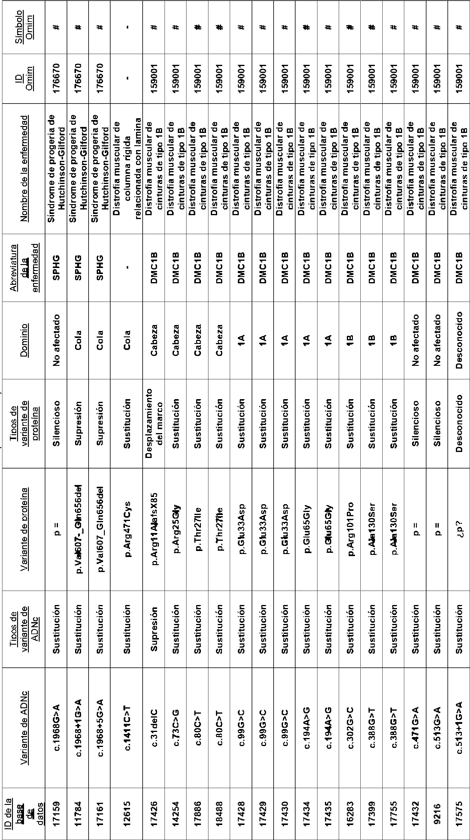


En algunas realizaciones, la molécula de ácido nucleico de acuerdo con cualquier aspecto de la presente divulgación es para su uso en el tratamiento de enfermedades cardiovasculares en un sujeto.In some embodiments, the nucleic acid molecule according to any aspect of the present disclosure is for use in the treatment of cardiovascular diseases in a subject.
En algunas realizaciones, la enfermedad o la enfermedad cardiovascular se caracteriza por la presencia de al menos una mutación de Lmna. In some embodiments, the cardiovascular disease or disease is characterized by the presence of at least one Lmna mutation.
Preferentemente, la enfermedad cardiovascular se selecciona del grupo que consiste en laminopatía, miocardiopatía, tal como miocardiopatía dilatada (MCD), miocardiopatía dilatada 1A, miocardiopatía dilatada con defectos del sistema de conducción, miocardiopatía con bloqueo AV avanzado y arritmia, fibrilación auricular solitaria; distrofia muscular (con frecuencia asociada a miocardiopatía), tal como miocardiopatía asociada a la distrofia muscular de Emery-Dreifuss (autosómica dominante), miocardiopatía asociada a la distrofia muscular de Emery-Dreifuss (autosómica recesiva), miocardiopatía asociada a la distrofia muscular de cinturas de tipo 1B, miocardiopatía asociada a la distrofia muscular congénita; síndromes de envejecimiento prematuro (que se cree que son principalmente vasculares, pero pueden tener compromiso cardíaco) tales como miocardiopatía asociada al síndrome de Werner atípico, miocardiopatía asociada al síndrome de progeria de Hutchinson-Gilford y similares, así como enfermedades presentadas en negrita en la Tabla 1.Preferably, the cardiovascular disease is selected from the group consisting of laminopathy, cardiomyopathy, such as dilated cardiomyopathy (DCM), 1A dilated cardiomyopathy, dilated cardiomyopathy with conduction system defects, cardiomyopathy with advanced AV block and arrhythmia, solitary atrial fibrillation; muscular dystrophy (often associated with cardiomyopathy), such as cardiomyopathy associated with Emery-Dreifuss muscular dystrophy (autosomal dominant), cardiomyopathy associated with Emery-Dreifuss muscular dystrophy (autosomal recessive), cardiomyopathy associated with limb-girdle muscular dystrophy type 1B, cardiomyopathy associated with congenital muscular dystrophy; premature aging syndromes (which are believed to be primarily vascular, but may have cardiac involvement) such as atypical Werner syndrome-associated cardiomyopathy, Hutchinson-Gilford progeria syndrome-associated cardiomyopathy, and the like, as well as diseases presented in bold in the Table 1.
De acuerdo con un tercer aspecto de la presente divulgación, se proporciona un vector de virus adenoasociado (AAV) que comprende un promotor de troponina T cardíaca (cTnT) y el transgén de acuerdo con cualquier aspecto de la presente divulgación.According to a third aspect of the present disclosure, there is provided an adeno-associated virus (AAV) vector comprising a cardiac troponin T (cTnT) promoter and the transgene according to any aspect of the present disclosure.
De acuerdo con un cuarto aspecto de la presente divulgación, se proporciona una composición farmacéutica que comprende la molécula de ácido nucleico de acuerdo con cualquier realización de la presente divulgación para tratar una enfermedad.According to a fourth aspect of the present disclosure, there is provided a pharmaceutical composition comprising the nucleic acid molecule according to any embodiment of the present disclosure for treating a disease.
En algunas realizaciones, la enfermedad es una laminopatía.In some embodiments, the disease is a laminopathy.
En algunas realizaciones, la composición farmacéutica que comprende la molécula de ácido nucleico de acuerdo con la presente divulgación es para su uso en el tratamiento de una enfermedad cardiovascular en un sujeto.In some embodiments, the pharmaceutical composition comprising the nucleic acid molecule according to the present disclosure is for use in the treatment of a cardiovascular disease in a subject.
De acuerdo con un quinto aspecto de la presente divulgación, se proporciona un método de tratamiento de una enfermedad en un sujeto, comprendiendo el método la administración de una cantidad farmacéuticamente eficaz de la molécula de ácido nucleico de acuerdo con cualquier realización de la presente divulgación, o la composición farmacéutica de la presente divulgación.According to a fifth aspect of the present disclosure, there is provided a method of treating a disease in a subject, the method comprising administering a pharmaceutically effective amount of the nucleic acid molecule according to any embodiment of the present disclosure, or the pharmaceutical composition of the present disclosure.
En algunas realizaciones del método de tratamiento de una enfermedad en un sujeto, la enfermedad se caracteriza por la presencia de al menos una mutación de Lmna. In some embodiments of the method of treating a disease in a subject, the disease is characterized by the presence of at least one Lmna mutation.
En algunas realizaciones del método de tratamiento de una enfermedad en un sujeto, el método comprende:In some embodiments of the method of treating a disease in a subject, the method comprises:
(i) someter a ensayo una muestra obtenida de un sujeto que se sospecha que tiene una enfermedad para detectar la presencia o ausencia de al menos una mutación de Lmna; (i) testing a sample obtained from a subject suspected of having a disease for the presence or absence of at least one Lmna mutation;
en donde la presencia de al menos una mutación de Lmna indica que al sujeto se le ha de administrar la composición farmacéutica de la presente divulgación o la molécula de ácido nucleico de la presente divulgación. wherein the presence of at least one Lmna mutation indicates that the subject is to be administered the pharmaceutical composition of the present disclosure or the nucleic acid molecule of the present disclosure.
En algunas realizaciones del método de tratamiento de una enfermedad en un sujeto, la mutación o mutaciones de Lmna afectan a la isoforma A de lamina, o la isoforma C de lamina del gen Lmna, o ambas isoformas A/C de lámina. In some embodiments of the method of treating a disease in a subject, the Lmna mutation(s) affect the lamin A isoform, or the lamin C isoform of the Lmna gene, or both lamin A/C isoforms.
En algunas realizaciones del método de tratamiento de una enfermedad en un sujeto, la enfermedad se selecciona del grupo que consiste en dermopatía restrictiva, lipodistrofia parcial familiar (por ejemplo, de tipo Dunnigan), displasia mandibuloacral con lipodistrofia de tipo A, síndrome metabólico, enfermedad de Charcot-Marie-Tooth de tipo 2, enfermedad de Charcot-Marie-Tooth de tipo 2B1 y enfermedades presentadas en letra normal en la Tabla 1.In some embodiments of the method of treating a disease in a subject, the disease is selected from the group consisting of restrictive dermopathy, familial partial lipodystrophy (e.g., Dunnigan's type), mandibuloacral dysplasia with lipodystrophy type A, metabolic syndrome, disease Charcot-Marie-Tooth disease type 2, Charcot-Marie-Tooth disease type 2B1 and diseases presented in normal font in Table 1.
En algunas realizaciones del método de tratamiento de una enfermedad en un sujeto, la enfermedad es una enfermedad cardiovascular, en donde la enfermedad cardiovascular se selecciona del grupo que consiste en laminopatía, miocardiopatía, tal como miocardiopatía dilatada (MCD), miocardiopatía dilatada 1A, miocardiopatía dilatada con defectos del sistema de conducción, miocardiopatía con bloqueo AV avanzado y arritmia, fibrilación auricular solitaria; distrofia muscular (con frecuencia asociada a miocardiopatía), tal como miocardiopatía asociada a la distrofia muscular de Emery-Dreifuss (autosómica dominante), miocardiopatía asociada a la distrofia muscular de Emery-Dreifuss (autosómica recesiva), miocardiopatía asociada a la distrofia muscular de cinturas de tipo 1B, miocardiopatía asociada a la distrofia muscular congénita; síndromes de envejecimiento prematuro (que se cree que son principalmente vasculares, pero pueden tener compromiso cardíaco) tales como miocardiopatía asociada al síndrome de Werner atípico, miocardiopatía asociada al síndrome de progeria de Hutchinson-Gilford; y enfermedades presentadas en negrita en la Tabla 1.In some embodiments of the method of treating a disease in a subject, the disease is a cardiovascular disease, wherein the cardiovascular disease is selected from the group consisting of laminopathy, cardiomyopathy, such as dilated cardiomyopathy (DCM), dilated cardiomyopathy 1A, cardiomyopathy dilated with conduction system defects, cardiomyopathy with advanced AV block and arrhythmia, solitary atrial fibrillation; muscular dystrophy (often associated with cardiomyopathy), such as cardiomyopathy associated with Emery-Dreifuss muscular dystrophy (autosomal dominant), cardiomyopathy associated with Emery-Dreifuss muscular dystrophy (autosomal recessive), cardiomyopathy associated with limb-girdle muscular dystrophy type 1B, cardiomyopathy associated with congenital muscular dystrophy; premature aging syndromes (thought to be primarily vascular, but may have cardiac involvement) such as atypical Werner syndrome-associated cardiomyopathy, Hutchinson-Gilford progeria syndrome-associated cardiomyopathy; and diseases presented in bold in Table 1.
En algunas realizaciones del método de tratamiento de una enfermedad en un sujeto, el sujeto es un mamífero no humano, tal como un ratón o un ser humano.In some embodiments of the method of treating a disease in a subject, the subject is a mammal not human, such as a mouse or a human being.
En algunas realizaciones del método, el ratón es un ratón N195K (Lmna N195K/ N195K) o uno nuligénico condicional de Lmna (Lmnaflox/flox).In some embodiments of the method, the mouse is an N195K mouse (Lmna N195K/N195K) or a conditional Lmna nulligenic mouse (Lmnaflox/flox).
De acuerdo con un sexto aspecto de la presente divulgación, se proporciona el uso de la composición farmacéutica de acuerdo con la presente divulgación o la molécula de ácido nucleico de acuerdo con la presente divulgación en la fabricación de un medicamento para tratar una enfermedad provocada por una o más mutaciones de Lmna. According to a sixth aspect of the present disclosure, there is provided the use of the pharmaceutical composition according to the present disclosure or the nucleic acid molecule according to the present disclosure in the manufacture of a medicament for treating a disease caused by a or more Lmna mutations.
En algunas realizaciones, la enfermedad es una enfermedad cardiovascular relacionadas con mutaciones de Lmna. En algunas realizaciones, la enfermedad se selecciona del grupo que consiste en dermopatía restrictiva, lipodistrofia parcial familiar (por ejemplo, de tipo Dunnigan), displasia mandibuloacral con lipodistrofia de tipo A, síndrome metabólico, enfermedad de Charcot-Marie-Tooth de tipo 2, enfermedad de Charcot-Marie-Tooth de tipo 2B1 y enfermedades presentadas en letra normal en la Tabla 1.In some embodiments, the disease is a cardiovascular disease related to Lmna mutations. In some embodiments, the disease is selected from the group consisting of restrictive dermopathy, familial partial lipodystrophy (e.g., Dunnigan type), mandibuloacral dysplasia with lipodystrophy type A, metabolic syndrome, Charcot-Marie-Tooth disease type 2, Charcot-Marie-Tooth disease type 2B1 and diseases presented in normal font in Table 1.
En algunas realizaciones, la enfermedad cardiovascular se selecciona del grupo que consiste en laminopatía, miocardiopatía, tal como miocardiopatía dilatada (MCD), miocardiopatía dilatada 1A, miocardiopatía dilatada con defectos del sistema de conducción, miocardiopatía con bloqueo AV avanzado y arritmia, fibrilación auricular solitaria; distrofia muscular (con frecuencia asociada a miocardiopatía), tal como miocardiopatía asociada a la distrofia muscular de Emery-Dreifuss (autosómica dominante), miocardiopatía asociada a la distrofia muscular de Emery-Dreifuss (autosómica recesiva), miocardiopatía asociada a la distrofia muscular de cinturas de tipo 1B, miocardiopatía asociada a la distrofia muscular congénita; síndromes de envejecimiento prematuro (que se cree que son principalmente vasculares, pero pueden tener compromiso cardíaco) tales como miocardiopatía asociada al síndrome de Werner atípico, miocardiopatía asociada al síndrome de progeria de Hutchinson-Gilford y similares, así como enfermedades presentadas en negrita en la Tabla 1.In some embodiments, the cardiovascular disease is selected from the group consisting of laminopathy, cardiomyopathy, such as dilated cardiomyopathy (DCM), dilated cardiomyopathy 1A, dilated cardiomyopathy with conduction system defects, cardiomyopathy with advanced AV block and arrhythmia, solitary atrial fibrillation ; muscular dystrophy (often associated with cardiomyopathy), such as cardiomyopathy associated with Emery-Dreifuss muscular dystrophy (autosomal dominant), cardiomyopathy associated with Emery-Dreifuss muscular dystrophy (autosomal recessive), cardiomyopathy associated with limb-girdle muscular dystrophy type 1B, cardiomyopathy associated with congenital muscular dystrophy; premature aging syndromes (which are believed to be primarily vascular, but may have cardiac involvement) such as atypical Werner syndrome-associated cardiomyopathy, Hutchinson-Gilford progeria syndrome-associated cardiomyopathy, and the like, as well as diseases presented in bold in the Table 1.
De acuerdo con un séptimo aspecto de la presente divulgación, se proporciona un método para cribar candidatos a fármacos capaces de inhibir o alterar el complejo LINC en una célula.According to a seventh aspect of the present disclosure, a method is provided for screening drug candidates capable of inhibiting or altering the LINC complex in a cell.
En consecuencia, en algunas realizaciones, se proporciona un método para cribar candidatos a fármacos capaces de inhibir la interacción de las proteínas de un complejo LINC en una célula, que comprende:Accordingly, in some embodiments, a method is provided for screening drug candidates capable of inhibiting the interaction of proteins of a LINC complex in a cell, comprising:
(a) combinar las proteínas de dicho complejo LINC en presencia de un fármaco para formar un primer complejo; (b) combinar las proteínas en ausencia de dicho fármaco para formar un segundo complejo;(a) combining the proteins of said LINC complex in the presence of a drug to form a first complex; (b) combining the proteins in the absence of said drug to form a second complex;
(c) medir la cantidad de dicho primer complejo y dicho segundo complejo; y(c) measuring the amount of said first complex and said second complex; and
(d) comparar la cantidad de dicho primer complejo con la cantidad de dicho segundo complejo,(d) comparing the amount of said first complex with the amount of said second complex,
en donde si la cantidad de dicho primer complejo es menor que la cantidad de dicho segundo complejo, entonces el fármaco es un fármaco candidato para inhibir la interacción de las proteínas de dicho complejo de LINC en una célula. En algunas realizaciones, el fármaco candidato interrumpe la interacción proteína-proteína entre SUN y KASH del complejo LINC. Preferentemente, el fármaco candidato interrumpe la interacción entre las proteínas Sun1 y Nesprina-1.wherein if the amount of said first complex is less than the amount of said second complex, then the drug is a candidate drug for inhibiting the interaction of proteins of said LINC complex in a cell. In some embodiments, the drug candidate disrupts the protein-protein interaction between SUN and KASH of the LINC complex. Preferably, the candidate drug disrupts the interaction between the Sun1 and Nesprina-1 proteins.
En algunas realizaciones, dicho cribado es un cribado in vitro. In some embodiments, said screening is an in vitro screening.
En algunas realizaciones, dicho complejo se mide mediante un método de ELISA.In some embodiments, said complex is measured by an ELISA method.
En algunas realizaciones, el dominio SUN recombinante se inmoviliza en una superficie sólida y el dominio KASH recombinante se marca con una enzima que puede generar una lectura colorimétrica o quimioluminiscente. Los compuestos que no logran inhibir la interacción SUN-KASH darán como resultado un pocillo en la placa donde el SUN recombinante se uniría al dominio KASH unido a enzima. Después de las etapas de lavado y la incubación con sustratos enzimáticos colorimétricos o quimioluminiscentes, la presencia de la interacción SUN-KASH puede detectarse en lectores de placas convencionales. Si el compuesto puede inhibir la interacción SUN-KASH, entonces siguiendo la etapa de lavado, el dominio KASH se eliminaría y se reduciría o no se produciría ninguna reacción enzimática en el pocillo.In some embodiments, the recombinant SUN domain is immobilized on a solid surface and the recombinant KASH domain is labeled with an enzyme that can generate a colorimetric or chemiluminescent readout. Compounds that fail to inhibit the SUN-KASH interaction will result in a well on the plate where the recombinant SUN would bind to the enzyme-bound KASH domain. After washing steps and incubation with colorimetric or chemiluminescent enzyme substrates, the presence of the SUN-KASH interaction can be detected in conventional plate readers. If the compound can inhibit the SUN-KASH interaction, then following the washing step, the KASH domain would be removed and reduced or no enzymatic reaction would occur in the well.
En algunas realizaciones, si la cantidad de dicho primer complejo es menor que la cantidad de dicho segundo complejo, entonces dicho fármaco es un fármaco candidato para inhibir la interacción de dichas proteínas.In some embodiments, if the amount of said first complex is less than the amount of said second complex, then said drug is a candidate drug for inhibiting the interaction of said proteins.
En algunas realizaciones, dicho complejo se mide mediante un método de anisotropía de fluorescencia. In some embodiments, said complex is measured by a fluorescence anisotropy method.
En algunas realizaciones, el método de anisotropía de fluorescencia emplea dominios SUN y KASH recombinantes. En algunas realizaciones, el dominio KASH se marca con fluorescencia con un resto de fluoresceína y la anisotropía de fluorescencia del dominio KASH que interactúa con el dominio SUN puede medirse usando un equipo convencional, tal como un lector de placas que incorpora una función de espectrómetro de fluorescencia.In some embodiments, the fluorescence anisotropy method employs recombinant SUN and KASH domains. In some embodiments, the KASH domain is fluorescently labeled with a fluorescein moiety and the fluorescence anisotropy of the KASH domain interacting with the SUN domain can be measured using conventional equipment, such as a plate reader incorporating a spectrometer function. fluorescence.
En algunas realizaciones, si la cantidad de dicho primer complejo es menor que la cantidad de dicho segundo complejo, habrá una diferencia en la anisotropía de fluorescencia del KASH fluorescente y dicho fármaco es un fármaco candidato para inhibir la interacción de dichas proteínas.In some embodiments, if the amount of said first complex is less than the amount of said second complex, there will be a difference in the fluorescence anisotropy of the fluorescent KASH and said drug is a candidate drug for inhibiting the interaction of said proteins.
Breve descripción de las figurasBrief description of the figures
La Figura 1 muestra un esquema de las mutaciones en el gen de lamina A/C LMNA y las laminopatías resultantes de las mutaciones. Figure 1 shows a schematic of the mutations in the lamin A/C LMNA gene and the laminopathies resulting from the mutations.
La Figura 2 muestra un esquema del posicionamiento de los componentes de la membrana de la envoltura nuclear y lámina. Figure 2 shows a schematic of the positioning of the membrane components of the nuclear envelope and lamina.
La Figura 3 muestra un esquema de las conexiones entre el núcleo y la matriz extracelular a través del complejo LINC y cómo las mutaciones en lamin A/C pueden dar como resultado una MCD. La membrana plasmática, el citoesqueleto y el núcleo forman una entidad unida mecánica y físicamente. En mutantes de Lmna, el núcleo es estructuralmente débil. Es mucho más susceptible al estrés mecánico de las fuerzas del citoesqueleto. Esto conduce a un daño grave en los núcleos de los miocitos que, a su vez, conduce a una cascada de eventos tales como la apoptosis y la fibrosis que en última instancia da como resultado la MCD. Figure 3 shows a schematic of the connections between the nucleus and the extracellular matrix through the LINC complex and how mutations in lamin A/C can result in DCM. The plasma membrane, cytoskeleton, and nucleus form a mechanically and physically linked entity. In Lmna mutants, the nucleus is structurally weak. It is much more susceptible to mechanical stress from cytoskeletal forces. This leads to severe damage to the nuclei of myocytes which, in turn, leads to a cascade of events such as apoptosis and fibrosis that ultimately results in DCM.
La Figura 4 muestra el efecto de la microinyección de dextrano en el núcleo de ratones Lmna+/+ y Lmna'1' a baja presión. En las células de tipo silvestre, el dextrano permanece en el núcleo, mientras que en las células de mutantes de Lmna, el dextrano se escapa del núcleo hacia el citoplasma. Figure 4 shows the effect of dextran microinjection into the nucleus of Lmna +/+ and Lmna'1' mice at low pressure. In wild-type cells, dextran remains in the nucleus, whereas in Lmna mutant cells, dextran escapes from the nucleus into the cytoplasm.
Las Figuras 5A - 5B muestran esquemas de un complejo LINC (Fig. 5A) y la interacción entre KASH y SUN (Fig. Figures 5A-5B show schematics of a LINC complex ( Fig. 5A ) and the interaction between KASH and SUN ( Fig.
5B). 5B ).
La Figura 6 muestra defectos en el peso corporal y la longevidad en ratones Lmna-' y LmnaA9 mejoran en animales Lmna'l'Sun1'1' y Lmnah9Sun1-' con inactivación homocigótica de Sun1. (A ) Los pesos corporales son promedios de ratones con los genotipos indicados. Se indica el número (n) de animales utilizados. (B) Gráfico de Kaplan-Meier que muestra una esperanza de vida aumentada de ratones Lmna'l'Sun1'/' en comparación con Lmna'1'. La mediana de supervivencia de tipo silvestre o Sun1-' es > 210 días en un seguimiento de 7 meses; los ratones Lmna'1' tienen una mediana de supervivencia de 41 días; los ratones Lmna-'Sun1+/' tienen una mediana de supervivencia de 54 días; los ratones Lmna'l'Sun1'/' tienen una mediana de supervivencia de 104 días (p ≤ 0,01 comparando Lmna'1' y Lmna'l'Sun1'/'). (C) Pesos corporales de ratones LmnaA9 que son de tipo silvestre, heterocigotos u homocigotos para la deficiencia de Sun1. Las cohortes de tipo silvestre y Sun1'/' se representan para su comparación. Los valores son promedios ± ETM de animales en cada cohorte. Se indica el número (n) de animales (p ≤ 0,0001 comparando LmnaA9Sun1+l+ y LmnaA9Sun1-'). (D) Gráfico de Kaplan-Meier que muestra una esperanza de vida aumentada de ratones Lmnah9Sun1-' en comparación con LmnaA9Sun1+l+. Los ratones LmnaA9Sun1+l' también se representan. (p ≤ 0,0001 comparando LmnaA9Sun1+l+ y Lmnañ9Sun1-'). (E) Proliferación celular de los MEF indicados. Las curvas son promedios ± DT, representante de > 3 aislados independientes de embriones de los genotipos indicados. (F) Curvas de proliferación de MAF (fibroblastos adultos de ratón, por sus siglas en inglés) de ratones WT (de tipo silvestre, del inglés wildtype), Sun1-', LmnaA9Sun1+/+ y Lmnah9Sun1-'. Los MAF se sembraron a una densidad de 1000 células por pocillo. Se midió el crecimiento y se presentan los índices celulares normalizados (promedios ± DT). Figure 6 shows defects in body weight and longevity in Lmna-' and LmnaA9 mice improve in Lmna'l'Sun1'1' and Lmnah9Sun1-' animals with homozygous Sun1 knockout. ( A ) Body weights are averages of mice with the indicated genotypes. The number (n) of animals used is indicated. ( B ) Kaplan-Meier plot showing increased lifespan of Lmna'l'Sun1'/' mice compared to Lmna'1'. Median survival of wild type or Sun1-' is >210 days over a 7-month follow-up; Lmna'1' mice have a median survival of 41 days; Lmna-'Sun1+/' mice have a median survival of 54 days; Lmna'l'Sun1'/' mice have a median survival of 104 days (p ≤ 0.01 comparing Lmna'1' and Lmna'l'Sun1'/'). ( C ) Body weights of LmnaA9 mice that are wild type, heterozygous, or homozygous for Sun1 deficiency. The wild-type and Sun1'/' cohorts are depicted for comparison. Values are means ± SEM of animals in each cohort. The number (n) of animals is indicated (p ≤ 0.0001 comparing LmnaA9Sun1+l+ and LmnaA9Sun1-'). ( D ) Kaplan-Meier plot showing increased lifespan of Lmnah9Sun1-' mice compared to LmnaA9Sun1+l+. LmnaA9Sun1+l' mice are also depicted. (p ≤ 0.0001 comparing LmnaA9Sun1+l+ and Lmnañ9Sun1-'). ( E ) Cell proliferation of the indicated MEFs. Curves are means ± SD, representative of >3 independent isolates from embryos of the indicated genotypes. ( F ) Proliferation curves of MAF (mouse adult fibroblasts) from WT , Sun1-', LmnaA9Sun1+/+, and Lmnah9Sun1-' mice. MAFs were seeded at a density of 1000 cells per well. Growth was measured and normalized cellular indices (means ± SD) are presented.
La Figura 7 muestra un esquema de las características de la proteína Sun1 y los componentes utilizados para generar una proteína Sun1 dominante negativa, incluyendo una secuencia señal, una secuencia de superenrollamiento, una secuencia de dominio SUN y una secuencia de KDEL. Figure 7 shows a schematic of the characteristics of the Sun1 protein and the components used to generate a dominant negative Sun1 protein, including a signal sequence, a supercoiling sequence, a SUN domain sequence, and a KDEL sequence.
La Figura 8 muestra un esquema de un plásmido (SEQ ID NO: 1) utilizado para la producción de AAV. Figure 8 shows a schematic of a plasmid (SEQ ID NO: 1) used for AAV production.
La Figura 9 muestra un esquema de un plásmido (SEQ ID NO: 2) que comprende secuencias de AAV2 y AAV9 para la producción de AAV. Figure 9 shows a schematic of a plasmid (SEQ ID NO: 2) comprising AAV2 and AAV9 sequences for AAV production.
La Figura 10 muestra un esquema de una construcción de expresión de AAV (SEQ ID NO: 3) que comprende un promotor específico cardíaco y una secuencia dominante negativa de Sun1. Figure 10 shows a schematic of an AAV expression construct (SEQ ID NO: 3) comprising a cardiac-specific promoter and a Sun1 dominant-negative sequence.
La Figura 11 muestra un esquema de las características de la proteína Sun1 dominante negativa, incluyendo una secuencia señal, una secuencia de superenrollamiento, una secuencia de dominio SUN y una secuencia de KDEL (SEQ ID NO: 4). Figure 11 shows a schematic of the features of the dominant negative Sun1 protein, including a signal sequence, a supercoiling sequence, a SUN domain sequence and a KDEL sequence (SEQ ID NO: 4).
La Figura 12 muestra un esquema de las características de una proteína Sun2 dominante negativa, incluyendo una secuencia señal, una secuencia de dominio luminal y una secuencia de KDEL (SEQ ID NO: 5). Figure 12 shows a schematic of the characteristics of a dominant negative Sun2 protein, including a signal sequence, a luminal domain sequence and a KDEL sequence (SEQ ID NO: 5).
La Figura 13 muestra un esquema de la región de la proteína Sun1 utilizada en construcciones dominantes negativas. Figure 13 shows a schematic of the region of the Sun1 protein used in dominant negative constructs.
La Figura 14 muestra una alineación de las secuencias de aminoácidos del dominio KASH1-KASH5 con restos conservados (SEQ ID NO: 7, 9, 11, 13 y 15, respectivamente). Figure 14 shows an alignment of the amino acid sequences of the KASH1-KASH5 domain with conserved residues (SEQ ID NO: 7, 9, 11, 13 and 15, respectively).
La Figura 15 muestra un esquema del complejo LINC en ratones de tipo silvestre, ratones Sun1 KO, ratones SUN negativos dominantes AAV y ratones con dominio KASH alterado. El esquema para ratones de tipo silvestre se obtiene de Brian Burke, 2012. El esquema para ratones Sun1 KO representa los resultados de Chen et al., 2012. El SUN dominante negativo AAV y los esquemas de dominio KASH alterados representan propuestas de inventores en la fecha de prioridad sobre métodos para la alteración del complejo LINC para mejorar las laminopatías, basándose en los datos obtenidos en ese momento. Figure 15 shows a schematic of the LINC complex in wild-type mice, Sun1 KO mice, AAV dominant negative SUN mice, and KASH domain disrupted mice. The scheme for wild-type mice is obtained from Brian Burke, 2012. The scheme for Sun1 KO mice represents the results of Chen et al., 2012. The SUN dominant negative AAV and altered KASH domain schemes represent inventors' proposals in the priority date on methods for altering the LINC complex to improve laminopathies, based on data obtained at that time.
La Figura 16 muestra una curva de Kaplan Meier de ratones Lmna KO que sobreviven un promedio de 28 días, ratones Sun1 KO que viven más de 300 días y ratones Lmna cardíaco KO/Sun1 KO que viven más de 300 días. Figure 16 shows a Kaplan Meier curve of Lmna KO mice surviving an average of 28 days, Sun1 KO mice living over 300 days, and Lmna cardiac KO/Sun1 KO mice living over 300 days.
La Figura 17 muestra secciones teñidas con HyE de los corazones de ratones Sun1 KO, ratones Lmna cardíaco KO y ratones Lmna cardíacos KO/Sun1 KO, con corazones de LmnaKO/Sun1WT que muestran agrandamiento del ventrículo izquierdo (MCD) en comparación con los corazones WT y LmnaKo/Sun1KO. Figure 17 shows HyE-stained sections from the hearts of Sun1 KO mice, Lmna cardiac KO mice, and Lmna cardiac KO/Sun1 KO mice, with LmnaKO/Sun1WT hearts showing left ventricular enlargement (MCD) compared to WT hearts. and LmnaKo/Sun1KO.
La Figura 18 muestra un esquema de la alteración de un complejo LINC en un ratón Nesprina-1 AKASH. Los ratones LmnaKO Nesprina-1WT tienen una esperanza de vida de aproximadamente 20 días. LmnaKO Nesprina-1-AKASH sobreviven aproximadamente 40 días, que es similar a ratones LmnaKOSunl KO. Figure 18 shows a schematic of the alteration of a LINC complex in a Nesprina-1 AKASH mouse. LmnaKO Nesprina-1WT mice have a lifespan of approximately 20 days. LmnaKO Nesprina-1-AKASH survive approximately 40 days, which is similar to LmnaKOSunl KO mice.
Las Figuras 19A-19B muestran un esquema de la expresión anticipada de AAV-cTNT-ON-SUN y la competencia entre ON-SUN exógeno y Sun1 nativo para unirse al dominio KASh (Figura 19A) con el ON-SUN mostrado en 19B (panel superior) y el efecto del ON-SUN transfectado en el posicionamiento nativo de Nesprin2G en células donde los 2 núcleos en el panel central expresan el ON-SUN y en el panel de fusión ambos muestran pérdida de Nesprina2 de las membranas nucleares (Fig. 19B). Figures 19A-19B show a schematic of the anticipated expression of AAV-cTNT-ON-SUN and the competition between exogenous ON-SUN and native Sun1 to bind to the KAS h domain (Figure 19A) with ON-SUN shown in 19B ( top panel) and the effect of transfected ON-SUN on native positioning of Nesprin2G in cells where the 2 nuclei in the middle panel express ON-SUN and in the fusion panel both show loss of Nesprin2 from nuclear membranes ( Fig. 19B ).
La Figura 20 es una curva de Kaplan Meier que muestra que la alteración de la interacción SUN-KASH in vivo, usando AAV9-cTNT-Sun1 dominante negativo (DNSun1), prolonga la longevidad del Lmna KO específico de corazón en ratones machos y hembras. Figure 20 is a Kaplan Meier curve showing that disruption of the SUN-KASH interaction in vivo, using AAV9-cTNT-Sun1 dominant negative (DNSun1), prolongs the longevity of heart-specific Lmna KO in male and female mice.
La Figura 21 muestra los aminoácidos C-terminales del dominio KASH de Nesprina-2 (KASH2). La secuencia de 14 o 18 aminoácidos del extremo C de KASH2 puede interactuar físicamente con el dominio SUN de SUN2. La pérdida de los últimos 4 aminoácidos de KASH2 o la adición de un único aminoácido alanina en el extremo C de KASH2 es suficiente para alterar la interacción del dominio KASH2 con el dominio SUN. Figure 21 shows the C-terminal amino acids of the KASH domain of Nesprin-2 (KASH2). The 14 or 18 amino acid sequence at the C terminus of KASH2 can physically interact with the SUN domain of SUN2. The loss of the last 4 amino acids of KASH2 or the addition of a single alanine amino acid at the C terminus of KASH2 is sufficient to alter the interaction of the KASH2 domain with the SUN domain.
La Figura 22 muestra un esquema de un método de cribado para detectar agentes que alteran el complejo LINC. Figure 22 shows a schematic of a screening method to detect agents that disrupt the LINC complex.
La Figura 23 muestra un diagrama de flujo que muestra un método de cribado más detallado para identificar una molécula pequeña para alterar el complejo LINC. Figure 23 shows a flow chart showing a more detailed screening method to identify a small molecule to disrupt the LINC complex.
La Figura 24 es una curva de Kaplan Meier que muestra que los ratones de tipo silvestre (C57/BI6) con o sin una mutación de alteración de Nesprina-1 KASH (C'TA8) tienen una esperanza de vida normal. Los ratones con una mutación Lmna nulo/KO (LA-ZP3creNA) y de tipo silvestre (Nesp1+/+) o heterocigoto (Nesp1+/C'TA8) para Nesp1-C'TA8 tienen una mediana de esperanza de vida de 15 o 18 días, que aumenta a 38 días en ratones Lmna KO/mutantes de Nesp1 homocigotos (LA-ZP3creNA; Nesp1 C'TA8/C'TA8). Figure 24 is a Kaplan Meier curve showing that wild-type mice (C57/BI6) with or without a Nesprin-1 KASH disruption mutation (C'TA8) have a normal lifespan. Mice with a Lmna null/KO mutation (LA-ZP3cre N A) and wild type (Nesp1+/+) or heterozygous (Nesp1+/C'TA8) for Nesp1-C'TA8 have a median lifespan of 15 or 18 days, which increases to 38 days in homozygous Lmna KO/Nesp1 mutant mice (LA-ZP3cre N A; Nesp1 C'TA8/C'TA8).
La Figura 25 es una curva de Kaplan Meier que muestra que los ratones con Lmna de tipo silvestre (N1CTA8/ CTA8LA+/+Mcre+/'), o alelos floxados de Lmna pero que carecen de un impulsor de Cre específico para el corazón (N1cta8/ CTA8LAf/fMcre+/+ y N1WT/WTLAf/fMcre+/+), viven durante la duración del experimento (~ 80 días en la presentación de prioridad, que se extendió a 120 días sin cambios). Los ratones con una supresión específica de cardiomiocitos de Lmna (N1WT/WTLAf/fMcre+/_) tienen una esperanza de vida de 22-24 días después de la inducción de la supresión mediada por Cre/loxP mediante el suministro de tamoxifeno (TMX), que aumenta a lo largo del experimento en ratones con una supresión específica de cardiomiocitos de Lmna inducida por TMX y también mutante homocigoto para Nesprina-1 (N1CTA8/CTA8LAf/fMCre+/-). Figure 25 is a Kaplan Meier curve showing that mice with wild-type Lmna (N1CTA8/CTA8LA+/+Mcre+/'), or floxed alleles of Lmna but lacking a heart-specific Cre driver (N1cta8/ CTA8LAf/fMcre+/+ and N1WT/WTLAf/fMcre+/+), live for the duration of the experiment (~80 days in the priority presentation, which was extended to 120 days without changes). Mice with a cardiomyocyte-specific deletion of Lmna (N1WT/WTLAf/fMcre+/_) have a lifespan of 22-24 days after induction of Cre/loxP-mediated deletion by feeding tamoxifen (TMX), which increases throughout the experiment in mice with a TMX-induced cardiomyocyte-specific deletion of Lmna and also a homozygous mutant for Nesprina-1 (N1CTA8/CTA8LAf/fMCre+/-).
Las Figuras 26A-26D muestran curvas de Kaplan Meier de pérdida de Sun1 que prolonga la longevidad de ratones mutantes de Lmna. (Fig.26A) Los ratones de tipo silvestre (C57/BI6) con o sin Sun1 tienen una esperanza de vida normal, mientras que la esperanza de vida promedio posnatal de los ratones LmnaFlx/Flx:Zp3 en los que se suprime LaminaA en todos los tejidos fue de 17,5 días (***P = ≤0,0001; ensayo del rango logarítmico). En un entorno S u n t /- la longevidad aumenta a 32,5 días. (Fig. 26B) Cuando se suprimió LmnaFIxIFIx específica y constitutivamente en corazones cruzando los ratones con la estirpe CreaMyHC, los ratones LmnaFIx/FIx:aMyHcvivieron en promedio 26,5 días. En un entorno de Sun1~F estos ratones vivieron durante más de 6 meses. (Fig. 26C) Se cruzaron LmnaFIx/FIxde 3 5 meses de edad con el Cre específico de cardiomiocitos inducible por Tmx Tg(Myh6-cre/Esr1), (abreviado a mcm), después de una única inyección de Tmx, los ratones mueren en 3-4 semanas. En un entorno de Sun1~F estos ratones vivieron durante más de 1 año. (Fig. 26D) Los ratones LmnaN195K/N195K vivieron un promedio de 78 días en comparación con los ratones LmnaN195K/N195KSun1-/- que tuvieron una esperanza de vida promedio de 111 días. (***P = ≤0,0001, **P = 0,0073 ensayo del rango logarítmico). Figures 26A-26D show Kaplan Meier curves of loss of Sun1 prolonging the longevity of Lmna mutant mice. ( Fig.26A ) Wild-type mice (C57/BI6) with or without Sun1 have a normal lifespan, while the average postnatal lifespan of LmnaFlx/Flx:Zp3 mice in which LaminA is deleted in all tissues was 17.5 days (***P = ≤0.0001; log-rank test). In a S unt environment /- longevity increases to 32.5 days. ( Fig. 26B ) When LmnaFIxIFIx was specifically and constitutively deleted in hearts by crossing the mice with the CreaMyHC strain, the LmnaFIx/FIx:aMyHc mice lived on average 26.5 days. In a Sun1~F environment these mice lived for more than 6 months. ( Fig. 26C ) 3-5-month-old LmnaFIx/FIx were crossed with the Tmx-inducible cardiomyocyte-specific Cre Tg ( Myh6-cre/Esr1), (abbreviated to mcm), after a single injection of Tmx, the mice They die in 3-4 weeks. In a Sun1~F environment these mice lived for more than 1 year. ( Fig. 26D ) LmnaN195K/N195K mice lived an average of 78 days compared to LmnaN195K/N195KSun1 −/− mice that had an average lifespan of 111 days. (***P = ≤0.0001, **P = 0.0073 log-rank test).
Las Figuras 27A-27E muestran la esperanza de vida y el fenotipo de los ratones LmnaFIx/FIx:mcm + Tmx. (Fig. 27A) La esperanza de vida promedio de los ratones LmnaFIx/FIx:mcm fue de 27 días después de una única inyección de Tmx (***P = ≤0,0001; ensayo del rango logarítmico). (Fig. 27B) La PCR detectó el gen Lmna (punta de flecha) floxed (suprimido) solamente en el tejido cardíaco después de la inyección de Tmx y no en otros tejidos o cuando no se inyectó Tmx (Fig. 27C) Los ratones LmnaFIx/FIx:mcm+Tmx desarrollaron cifosis (punta de flecha) 21 días después de la inyección. (Fig. 27D) Las proteínas LaminaA/C, detectada por inmunofluorescencia, estuvieron presentes en el control (i, iii), pero reducidas/ausentes en núcleos de cardiomiocitos (CM) tanto en CM aislados (ii segundo panel) como en secciones de corazón (iv) (puntas de flecha de color blanco) con núcleos de CM detectados por tinción PCM-1, 21 días después de Tmx. (Fig. 27E) Los niveles de LaminaA/C se cuantificaron mediante análisis por Western de lisados de corazón completo 21 días después de la inyección. Se detectó una reducción significativa (***P = ≤0,0001; ensayo T) en la proteína Lamina de tipo A, aunque los niveles de Lamina C no se redujeron tanto en los ratones LmnaF/x/FIx:mcm+Tmx en comparación con LmnaFIx/FIx:mcm+CTL. (Fig. 27F) El análisis cuantitativo se realizó 21 días después de Tmx. La presencia de los sitios LoxP en el gen WT-Lmna (LmnaF/x/FIx) da como resultado una reducción de los niveles de transcrito de Lmna en comparación con los niveles de LmnaWt/Wt, aunque esto no tuvo un efecto manifiesto sobre la longevidad o el crecimiento/viabilidad. Figures 27A-27E show the lifespan and phenotype of LmnaFIx/FIx:mcm + Tmx mice. ( Fig. 27A ) The average lifespan of LmnaFIx/FIx:mcm mice was 27 days after a single Tmx injection (***P = ≤0.0001; log-rank test). ( Fig. 27B ) PCR detected the floxed (arrowhead) Lmna gene (arrowhead) only in cardiac tissue after Tmx injection and not in other tissues or when Tmx was not injected ( Fig. 27C ) LmnaFIx mice /FIx:mcm+Tmx developed kyphosis (arrowhead) 21 days after injection. ( Fig. 27D ) LaminaA/C proteins, detected by immunofluorescence, were present in the control (i, iii), but reduced/absent in cardiomyocyte (CM) nuclei in both isolated CM (second panel ii) and in sections of heart (iv) (white arrowheads) with CM nuclei detected by PCM-1 staining, 21 days after Tmx. ( Fig. 27E ) LaminaA/C levels were quantified by Western analysis of whole heart lysates 21 days after injection. A significant reduction (***P = ≤0.0001; T test) was detected in Lamin type A protein, although Lamin C levels were not reduced as much in LmnaF/x/FIx:mcm+Tmx mice in comparison with LmnaFIx/FIx:mcm+CTL. ( Fig. 27F ) Quantitative analysis was performed 21 days after Tmx. The presence of the LoxP sites in the WT-Lmna gene ( LmnaF/x/FIx) results in a reduction of Lmna transcript levels compared to LmnaWt/Wt levels, although this had no overt effect on the longevity or growth/viability.
Las Figuras 28A-28D muestran ecocardiogramas, la función cardíaca y la histología de ratones LmnaF/x/FIxmcm + Tmx. (Fig. 28A) Los ratones LmnaFIx/FIx:mcm + Tmx muestran una función contráctil cardíaca reducida. (Fig. 28B) Los corazones LmnaFIx/FIx:mcm muestran un % de FE y un % de AF reducidos, y un DIVI aumentado (***P = ≤0,0001, **P = 0,0010; ANOVA de dos vías). (Fig.28C) El análisis histológico de los corazones reveló una mayor infiltración de células nucleadas y espacios intercelulares en corazones LmnaFIx/FIxmcm (i y ii). Se aislaron significativamente menos CM viables (similares a un ladrillo) de los corazones LmnaFIx/FIx:mcm en comparación con controles de LmnaFIx/FIx:mcm (iii). Con mayor aumento, los cardiomiocitos aislados de los corazones LmnaFIx/FIx:mcm contenían grandes vacuolas intracelulares (punta de flecha, iv). (Fig. 28D) La luz del ventrículo izquierdo en los corazones Lmna FIx/FIx:mcm se agrandó (i) junto con un aumento de la fibrosis (ii) (** P = 0,0007 visible como áreas grises más claras en el 28D ii, los paneles centrales y el panel izquierdo de iv) y núcleos apoptóticos revelados por tinción TUNEL (* P = 0,0220; ANOVA de una vía) (panel derecho iii y iv). Todas las muestras y análisis se realizaron en corazones de 21 días después de la inyección de Tmx. Figures 28A-28D show echocardiograms, cardiac function and histology of LmnaF/x/FIxmcm + Tmx mice. ( Fig. 28A ) LmnaFIx/FIx:mcm + Tmx mice show reduced cardiac contractile function. (Fig. 28B) LmnaFIx/FIx:mcm hearts show reduced EF% and AF%, and increased DIVI (***P = ≤0.0001, **P = 0.0010; two-way ANOVA roads). ( Fig.28C ) Histological analysis of the hearts revealed greater infiltration of nucleated cells and intercellular spaces in LmnaFIx/FIxmcm hearts (i and ii). Significantly fewer viable (brick-like) CMs were isolated from LmnaFIx/FIx:mcm hearts compared to LmnaFIx/FIx:mcm controls (iii). At higher magnification, cardiomyocytes isolated from LmnaFIx/FIx:mcm hearts contained large intracellular vacuoles (arrowhead, iv). ( Fig. 28D ) The left ventricular lumen in Lmna FIx/FIx:mcm hearts was enlarged (i) along with increased fibrosis (ii) (** P = 0.0007 visible as lighter gray areas in the 28D ii, middle panels and left panel of iv) and apoptotic nuclei revealed by TUNEL staining (* P = 0.0220; one-way ANOVA) (right panel iii and iv). All sampling and analyzes were performed on hearts 21 days after Tmx injection.
Las Figuras29A-29D muestran cambios en las morfologías nucleares y la estructura del corazón con y sin Sun1 en los ratones LmnaFIx/FIx:mcm después de la inyección de Tmx. (Fig. 29a ) Los núcleos de CM con expresión de Lamina A/C reducida o ausente se indican mediante puntas de flecha de color blanco (1, 3). Los núcleos (2, 4) de CM con niveles normales de Lamina A/C se indican mediante puntas de flecha de color gris. Los niveles de proteína LMNA, medidos tanto por la intensidad de la fluorescencia (5) como por la transferencia Western (6), se redujeron significativamente en LmnaFIx/FIx:mcm Sun1+/++Tmx (***P = 0,0009; ensayo T) y LmnaFIx/FIx:mcm Sun1-/-+Tmx (*P = 0,0359; ensayo T) en comparación con controles LmnaFIx/FIxmcm Sun1+/+ (gráfico inferior, 6) (Fig. 29B). El agrandamiento del ventrículo izquierdo (VI) fue evidente en los corazones LmnaF/x/FIx:mcm Sun1+/++Tmx (panel 1) pero no en el VI de los corazones LmnaFIx/FIx:mcm Sun1-/-+Tmx (panel 2). Los ratones LmnaFIx/FIx:mcm Sun1+/++Tmx tenían una fibrosis significativamente mayor (panel 3, fibrosis en gris) en comparación con los controles, pero no hubo un aumento significativo de la fibrosis en los corazones LmnaFIx/FIxmcm Sun1-/-+Tmx (panel 3) en comparación con los controles (panel 4, cuantificado en el panel 5, ***P = 0,0001; ANOVA de una vía). Las mediciones de la fuerza activa del músculo papilar cardíaco se redujeron significativamente a partir de los ratones LmnaFIx/FIx:mcm Sun1+/++Tmx en comparación con los controles LmnaFIx/FIx:mcm Sun1+I+ (**P = 0,0047; ensayo T) y LmnaFIx/FIx:mcm Sun1-/-+Tmx (*P = 0,0113; ensayo T) (panel 6). (Fig. 29C) Las morfologías nucleares de CM se alteraron significativamente en los ratones L_mnaFIx/FIx:mcm Sun1+/++Tmx (Panel 1, puntas de flecha continuas). En ausencia de TMX, las secciones del corazón de control (CTL, panel 2) muestran pocas anomalías nucleares. En ausencia de Sun1, los cardiomiocitos LmnaFIx/FIx:mcm Sun1-/-+Tmx no mostraron anomalías nucleares (Paneles 3 y 4). En resumen, el panel 5 de la Figura 29C revela que, el 70 % de los CM en los ratones LmnaFIx/FIx:mcm Sun1+/++Tmx tenían rupturas/distorsiones de EN o núcleos deformados en comparación con menos del 1 % de los núcleos de CM en los ratones LmnaFIx/FIx:mcm Sun1-/-+Tmx. (Fig. 29D) Se realizaron análisis de eco en ratones tratados con TMX y de control después de la inducción con Tmx. Los ecocardiogramas (ECG) se realizaron 28 días después de la inyección de Tmx en ratones de 3 a 5 meses de edad (panel 1). Los ECG realizados antes y después de la inducción por Cre revelaron un empeoramiento progresivo de la contractilidad cardíaca en los ratones LmnaFIx/FIxmcm Sun1+/+ +Tmx (línea de color negro continua) en comparación con los ratones LmnaFIx/FIx:mcm Sun1-/-+ Tmx (paneles 2-4). La pérdida de SUN1 conservó la FE (panel 2), el AF (panel 3) y la Deformación longitudinal global (DLG, panel 4) en los ratones LmnaFIx/FIxmcm Sun1-/- + Tmx en comparación con los ratones LmnaFIx/FIx:mcm Sun1+/+ +Tmx. Figures 29A-29D show changes in nuclear morphologies and heart structure with and without Sun1 in LmnaFIx/FIx:mcm mice after Tmx injection. ( Fig. 29a ) CM nuclei with reduced or absent Lamin A/C expression are indicated by white arrowheads (1, 3). CM nuclei (2, 4) with normal levels of Lamin A/C are indicated by gray arrowheads. LMNA protein levels, measured by both fluorescence intensity (5) and Western blotting (6), were significantly reduced in LmnaFIx/FIx:mcm Sun1+/++Tmx (***P = 0.0009; T test) and LmnaFIx/FIx:mcm Sun1-/-+Tmx (*P = 0.0359; T test) compared to LmnaFIx/FIxmcm Sun1+/+ controls (bottom graph, 6) ( Fig. 29B ). Left ventricular (LV) enlargement was evident in LmnaF/x/FIx:mcm Sun1+/++Tmx hearts (panel 1) but not in the LV of LmnaFIx/FIx:mcm Sun1-/-+Tmx hearts (panel 2). LmnaFIx/FIx:mcm Sun1+/++Tmx mice had significantly increased fibrosis (panel 3, fibrosis in gray) compared to controls, but there was no significant increase in fibrosis in LmnaFIx/FIxmcm Sun1-/- hearts. +Tmx (panel 3) compared to controls (panel 4, quantified in panel 5, ***P = 0.0001; one-way ANOVA). Cardiac papillary muscle active force measurements were significantly reduced from LmnaFIx/FIx:mcm Sun1+/++Tmx mice compared to LmnaFIx/FIx:mcm Sun1+I+ mice (**P = 0.0047; T test) and LmnaFIx/FIx:mcm Sun1-/-+Tmx (*P = 0.0113; T test) (panel 6). ( Fig. 29C ) CM nuclear morphologies were significantly altered in L_mnaFIx/FIx:mcm Sun1+/++Tmx mice (Panel 1, solid arrowheads). In the absence of TMX, control heart sections (CTL, panel 2) show few nuclear abnormalities. In the absence of Sun1, LmnaFIx/FIx:mcm Sun1-/-+Tmx cardiomyocytes did not show nuclear abnormalities (Panels 3 and 4). In summary, panel 5 of Figure 29C reveals that 70% of CMs in LmnaFIx/FIx:mcm Sun1+/++Tmx mice had EN ruptures/distortions or deformed nuclei compared to less than 1% of CM nuclei in LmnaFIx/FIx:mcm Sun1-/-+Tmx mice. (Fig. 29D) Echo analyzes were performed on TMX-treated and control mice after Tmx induction. Echocardiograms (ECGs) were performed 28 days after Tmx injection in 3- to 5-month-old mice (panel 1). ECGs performed before and after Cre induction revealed a progressive worsening of cardiac contractility in LmnaFIx/FIxmcm Sun1+/+ +Tmx mice (solid black line) compared to LmnaFIx/FIx:mcm Sun1-/ mice. - + Tmx (panels 2-4). Loss of SUN1 preserved EF (panel 2), FA (panel 3), and Global Longitudinal Strain (GLD, panel 4) in LmnaFIx/FIxmcm Sun1-/- + Tmx mice compared to LmnaFIx/FIx mice: mcm Sun1+/+ +Tmx.
Las Figuras 30A-30B muestran el gráfico de Kaplan Meier y los efectos de la función cardíaca de la supresión de SUN1 en la patología cardíaca inducida por una mutación de sentido erróneo en el gen Lmna (N195K). (Fig. 30A) La ausencia de Sun1 aumenta significativamente la esperanza de vida de los ratones LmaN195K7Flx:mmSun1- /-+ Tmx en comparación con los ratones LmnaN195K/FLX:mcmSun1+/++ Tmx (*P = 0.0101; ensayo del rango logarítmico). Los ratones con una sola copia de la mutación N195K (LmnaN195K/-:mcmSun1+/+ + Tmx) tuvo una esperanza de vida promedio de 47 días, aproximadamente la mitad de la esperanza de vida de los ratones homocigotos, es decir, con dos copias del alelo N195K. (Fig. 30B) Los ecocardiogramas (ECG) realizados antes y después de la inducción por Cre revelaron un empeoramiento progresivo de la contractilidad cardíaca en los ratones LmnaN195K/-:mcmSun1+/+ + Tmx en comparación con los ratones LmnaN195K/-:mcmSun1-/- + Tmx a lo largo del tiempo. Las imágenes de ECG se registraron 28 días después de la inyección de Tmx (paneles del lado izquierdo). La pérdida de SUN1 preservó la FE, el AF y la DLG en los ratones LmnaN195K/Flx:mcm Sun1-/- + Tmx en comparación con los ratones LmnaN195K/Flx:mcm Sun1+/+ + Tmx (3 paneles inferiores del lado derecho). Figures 30A-30B show the Kaplan Meier plot and the cardiac function effects of SUN1 deletion on cardiac pathology induced by a missense mutation in the Lmna gene (N195K). (Fig. 30A) Absence of Sun1 significantly increases the lifespan of LmaN195K7Flx:mmSun1 -/-+ Tmx mice compared to LmnaN195K/FLX:mcmSun1+/++ Tmx mice (*P = 0.0101; log-rank test ). Mice with a single copy of the N195K mutation ( LmnaN195K/-:mcmSun1+/+ + Tmx) had an average lifespan of 47 days, approximately half the lifespan of homozygous mice, that is, with two copies of the N195K allele. (Fig. 30B) Echocardiograms (ECG) performed before and after Cre induction revealed a progressive worsening of cardiac contractility in LmnaN195K/-:mcmSun1+/+ + Tmx mice compared to LmnaN195K/-:mcmSun1- mice. /- + Tmx over time. ECG images were recorded 28 days after Tmx injection (left side panels). Loss of SUN1 preserved EF, PA, and DLG in LmnaN195K/Flx:mcm Sun1-/- + Tmx mice compared to LmnaN195K/Flx:mcm Sun1+/+ + Tmx mice (bottom 3 right panels). .
Las Figuras 31A-31G muestran que los ratones LmnaFlxx/Flx:mcm+Tmx que expresan un DNSun1 transducido por AAV presentan una función cardíaca mejorada y una longevidad aumentada. (Fig. 31A) Protocolo para la transducción mediada por AAV de la miniproteína DN-Sun1 en ratones LmnaFlxx/Flx:mcm +Tmx. Se administra una única inyección de Tmx (IP) en D14 después del nacimiento para inducir la supresión de Lmna. Después, las partículas víricas AAV9-DNSun1 o AAV9-GFP se inyectan en la cavidad torácica el D15 después del nacimiento. El punto final experimental se fijó en 100 días después de Tmx. (Fig. 31B) La miniproteína DNSun1 compite con Sun1 endógena por unirse al dominio KASH de las Nesprinas (en CM, esto es Nesprina1). La miniproteína compite con la SUN1 endógena por la unión al dominio KASH de las Nesprinas. Como la miniproteína DNSun1 no está anclada en la MNI, esto desconecta eficazmente las proteínas SUN endógenas para que no se unan a los dominios KASH, rompiendo así el LINC. (Fig. 31C) La presencia del gen Lmna recombinado después de la inyección de Tmx se confirmó mediante PCR de los tejidos cardíacos (panel superior). Se detectó la expresión robusta de la proteína AAV9-DNSun1 y AAV9-GFP (dosis: 5x10A10 vg/g de ratón) en extractos de corazones enteros 99 días después de la inyección de AAV (panel inferior). (Fig. 31D) Los CM derivados de células madre iPS humanas se transdujeron con DNSun1 usando AVV-OJ como vector. En los CM que expresan altos niveles de DNSun1, indicado por flechas de color gris, la localización de Nesprin1 en la EN está reducida o ausente. La localización de Nesprina1 en la EN se mantiene en CM que no expresan AVV-OJ-ONSun1 o cuando se expresa en niveles más bajos (puntas de flecha de color blanco). (Fig. 3 lE ) Los ratones LmnaFlx/Flx:mcm+Tmx+AAV9-GFP vivieron un promedio de 34,5 d después de la inducción con Tmx, mientras que los ratones LmnaFlx/Flx:mcm +Tmx inyectados con AA9-DNSun1 (5*10A10 vg/g/ratón) vivieron significativamente más tiempo (**P = 0,0038; ensayo del rango logarítmico) a al menos 100D después de Tmx, después de lo cual los ratones se sacrificaron para su análisis. Este conjunto de datos derivó del que se muestra en la Fig. 20, se ajustó retirando los ratones que eran hembras y los que tenían una dosis diferente de virus. La Fig. E(i) representa ratones macho y la Fig. E(ii) representa ratones hembra. (Fig. 31F) 35d después de Tmx, se detectó fibrosis extensa (color azul en la imagen original, color gris en el presente documento) y agrandamiento ventricular en los corazones LmnaFlx/Flx:mcm+Tmx+AAV9-GFP en comparación con los corazones LmnaFlx/Flx:mcm +Tmx AAV9- DNSunl. (Fig. 31G) El análisis por ECG confirmadó que los corazones LmnaFlx/Flx:mcm +Tmx+AAV9-DNSun1 tenían una mejor función cardíaca en comparación con los corazones LmnaFlx/Flx:mcm +Tmx AAV9-GFP 35 días después de la inyección de Tmx. Figures 31A-31G show that LmnaFlxx/Flx:mcm+Tmx mice expressing AAV-transduced DNSun1 exhibit improved cardiac function and increased longevity. (Fig. 31A) Protocol for AAV-mediated transduction of the DN-Sun1 miniprotein in LmnaFlxx/Flx:mcm +Tmx mice. A single injection of Tmx (IP) is given on D14 after birth to induce Lmna suppression. The AAV9-DNSun1 or AAV9-GFP viral particles are then injected into the thoracic cavity on D15 after birth. The experimental endpoint was set at 100 days after Tmx. (Fig. 31B) The DNSun1 miniprotein competes with endogenous Sun1 for binding to the KASH domain of Nesprins (in CM, this is Nesprina1). The miniprotein competes with endogenous SUN1 for binding to the KASH domain of Nesprins. As the DNSun1 miniprotein is not anchored to the MNI, this effectively disengages the endogenous SUN proteins from binding to the KASH domains, thereby cleaving the LINC. (Fig. 31C) The presence of the recombined Lmna gene after Tmx injection was confirmed by PCR of cardiac tissues (upper panel). Robust expression of AAV9-DNSun1 and AAV9-GFP protein (dose: 5x10A10 vg/g mouse) was detected in extracts from whole hearts 99 days after AAV injection (lower panel). (Fig. 31D) CMs derived from human iPS stem cells were transduced with DNSun1 using AVV-OJ as a vector. In CMs expressing high levels of DNSun1, indicated by gray arrows, Nesprin1 localization to the NE is reduced or absent. Nesprina1 localization to the NE is maintained in CMs that do not express AVV-OJ-ONSun1 or when it is expressed at lower levels (white arrowheads). ( Fig. 3 lE ) LmnaFlx/Flx:mcm+Tmx+AAV9-GFP mice lived an average of 34.5 d after induction with Tmx, while LmnaFlx / Flx:mcm +Tmx mice injected with AA9-DNSun1 (5*10 A 10 vg/g/mouse) lived significantly longer (**P = 0.0038; log-rank test) at least 100D after Tmx, after which mice were sacrificed for analysis. This data set derived from that shown in Fig. 20 was adjusted by removing mice that were female and those that had a different dose of virus. Fig. E(i) represents male mice and Fig. E(ii) represents female mice. (Fig. 31F) 35d after Tmx, extensive fibrosis (blue color in original image, gray color herein) and ventricular enlargement were detected in LmnaFlx/Flx:mcm+Tmx+AAV9-GFP hearts compared to hearts LmnaFlx/Flx:mcm +Tmx AAV9- DNSunl. (Fig. 31G) ECG analysis confirmed that LmnaFlx/Flx:mcm +Tmx+AAV9-DNSun1 hearts had better cardiac function compared to LmnaFlx/Flx:mcm +Tmx AAV9-GFP hearts 35 days after injection of Tmx.
Las Figuras 32A-32D muestran modelos de cómo romper el LINC alterando Sun1 protege a los cardiomiocitos del estrés inducido por la contracción (Fig. 32A) Los núcleos de cardiomiocitos que expresan LmnaA/C, son capaces de soportar el estrés mecánico y las fuerzas de tensión transmitidas a través del complejo LINC desde el citoplasma hasta la EN. (Fig. 32B) La pérdida o introducción de una mutación dentro del gen Lmna da como resultado la pérdida o el ensamblaje incorrecto de la lámina nuclear, lo que debilita la Lámina/EN. Los núcleos debilitados se dañan debido a las fuerzas de tensión/esfuerzo ejercidas a través del complejo LINC desde los sarcómeros contráctiles de los cardiomiocitos. (Fig. 32C, D). En ausencia de SUN1 o interrumpiendo su unión a los dominios KASH por expresión de ONSunl, los complejos LINC no conectados ejercen menos fuerza de tensión sobre los núcleos de los cardiomiocitos, permitiendo la supervivencia de los cardiomiocitos mutantes de Lmna. Figures 32A-32D show models of how disrupting LINC disrupting Sun1 protects cardiomyocytes from contraction-induced stress (Fig. 32A). Cardiomyocyte nuclei expressing LmnaA/C are able to withstand mechanical stress and contraction forces. tension transmitted through the LINC complex from the cytoplasm to the EN. (Fig. 32B) Loss or introduction of a mutation within the Lmna gene results in loss or improper assembly of the nuclear lamina, weakening Lamina/EN. Weakened nuclei are damaged due to tension/strain forces exerted through the LINC complex from the contractile sarcomeres of cardiomyocytes. (Fig. 32C , D ). In the absence of SUN1 or disrupting its binding to KASH domains by expression of ONSunl, unconnected LINC complexes exert less tensile force on cardiomyocyte nuclei, allowing survival of Lmna mutant cardiomyocytes.
Figura 33: muestra la estructura del alelo condicional LmnaFlx/Flx. Las ubicaciones de cebadores para genotipificar el gen Lmna tanto antes como después de la recombinación Cre están indicadas para el alelo LmnaFlx alelo (Flox), el alelo Lmna suprimido (A) y el alelo de tipo silvestre [A. S. Wang, et al., Differentiation 89: 11-21 (2015)]. Figure 33: shows the structure of the conditional allele LmnaFlx/Flx. Primer locations for genotyping the Lmna gene both before and after Cre recombination are indicated for the LmnaFlx allele (Flox), the deleted Lmna allele (A), and the wild-type allele [AS Wang, et al., Differentiation 89: 11-21 (2015)].
La Figura 34 muestra un diagrama de las miniproteínas recombinantes AAV9-DNSun1 y AAV9-GFP. La DN-Sun1 incluye el dominio Sun, un marcador HA, una Secuencia señal (SS, para dirigir la proteína al ER) y el KDEL (señal de retención del RE) [M. Crisp et al., J Cell Biol. 172: 41-53 (2006)]. La AAV9-GFP incluye las secuencias SS y KDEL. Se usó GFP como control en lugar de Sun1L-KDEL. Figure 34 shows a diagram of the recombinant miniproteins AAV9-DNSun1 and AAV9-GFP. DN-Sun1 includes the Sun domain, an HA marker, a Signal Sequence (SS, to direct the protein to the ER) and the KDEL (ER retention signal) [M. Crisp et al., J Cell Biol. 172: 41-53 (2006)]. AAV9-GFP includes the SS and KDEL sequences. GFP was used as a control instead of Sun1L-KDEL.
La Figura 35 muestra fotomicrografías de expresión específica de cardiomiocitos de Cre recombinasa después de la inyección de Tmx. Los ratones LmnaFlx/Flx:mcm se cruzaron con los ratones indicadores mT/mG (JAX: Gt(ROSA)26Sortm4(ACTB-tdTomato,-EGFP)Luo/J). En ausencia de Cre, se expresa RFP. Cuando se induce Cre, se expresa GFP. Solo los CM de los ratones LmnaFlx/Flx:mcm expresan GFP tras la inyección de TMX. Los tejidos cardíacos se analizaron 7 días después de la inyección de Tmx. Figure 35 shows photomicrographs of cardiomyocyte-specific expression of Cre recombinase after Tmx injection. LmnaFlx/Flx:mcm mice were crossed with mT/mG reporter mice (JAX: Gt(ROSA)26Sortm4(ACTB-tdTomato,-EGFP)Luo/J). In the absence of Cre, RFP is expressed. When Cre is induced, GFP is expressed. Only CMs from LmnaFlx/Flx:mcm mice express GFP after TMX injection. Cardiac tissues were analyzed 7 days after Tmx injection.
La Figuras 36 muestra que la pérdida de Sun2 no rescata la pérdida de Lmna. La pérdida de Sun2 no prolonga la esperanza de vida de los ratones Lmnaülüdom2-1-. Figure 36 shows that the loss of Sun2 does not rescue the loss of Lmna. Loss of Sun2 does not prolong the lifespan of Lmnaülüdom2-1- mice.
Las Figuras 37A-37E muestran los fenotipos de los corazones LmnaFlxIFlxmcm Sun1+I+ y LmnaFlxIFlxmcm Sun1-1-12-14 meses después de la inyección de Tmx. (Fig. 37A) El análisis histológico de los corazones envejecidos LmnaFlXIFlxI:mcmSun1+l+, 12-14 meses después de la inyección de Tmx, no reveló cambios morfológicos significativos, por ejemplo, agrandamiento del VI o (Fig. 37B) en la fibrosis en comparación con los controles. (Fig. Figures 37A-37E show the phenotypes of LmnaFlxIFlxmcm Sun1+I+ and LmnaFlxIFlxmcm Sun1-1- hearts 12-14 months after Tmx injection. (Fig. 37A) Histological analysis of aged LmnaFlXIFlxI:mcmSun1+l+ hearts, 12-14 months after Tmx injection, revealed no significant morphological changes, e.g., LV enlargement or (Fig. 37B) fibrosis. compared to controls. ( Fig.
37C) El análisis por PCR confirmó la supresión sostenida del gen Lmna. (Fig. 37D) La cuantificación de proteínas reveló una reducción significativa de los niveles de LMNA en los corazones LmnaFlxIFlx:mcmSun1-I-+Tmx 14 meses después de TMX. (Fig. 37E) Los ecocardiogramas (panel del lado izquierdo) de los ratones envejecidos mostraron FE y AF reducidos (paneles del lado derecho) en ambos ratones envejecidos LmnaFlxIFlx:mcmSun1+I+ + CTL y LmnaFlxIFlx:mcmSun1-1- + Tmx. 37C ) PCR analysis confirmed sustained deletion of the Lmna gene. (Fig. 37D) Protein quantification revealed a significant reduction of LMNA levels in LmnaFlxIFlx:mcmSun1-I-+Tmx hearts 14 months after TMX. (Fig. 37E) Echocardiograms (left side panel) of the aged mice showed reduced EF and AF (right side panels) in both LmnaFlxIFlx:mcmSun1+I+ + CTL and LmnaFlxIFlx:mcmSun1-1- + Tmx aged mice.
La Figura 38 muestra que el rescate por AAV9-DNSun1 depende de la dosis de partículas víricas inyectadas. La esperanza de los ratones LmnaFlxIFlxmcm +TMX depende de la dosis de AAV9-DNSun1, dando como resultado las concentraciones más bajas una esperanza de vida más corta. Cada punto representa un ratón, las líneas horizontales indican la media. Figure 38 shows that rescue by AAV9-DNSun1 depends on the dose of viral particles injected. The lifespan of LmnaFlxIFlxmcm +TMX mice depends on the dose of AAV9-DNSun1, with lower concentrations resulting in a shorter lifespan. Each dot represents one mouse, horizontal lines indicate the mean.
Las Figuras 39A-39C muestran los niveles de LaminaAIC después de la inducción por Tmx y la expresión de proteínas expresadas por AAV. (Fig. 39A) Los niveles de LaminaAIC se redujeron significativamente después de la inducción por Tmx, y la presencia de la proteína AAV9-DNSun1 o AAV9-GFP no alteró los niveles de proteína LMNA (Cuantificación de la intensidad de inmunofluorescencia de LaminaAIC). Las cantidades de proteína LaminaAIC, ONSun1 y GFP en corazones completos también se cuantificaron mediante análisis por Western (3 gráficos inferiores). (Análisis realizado 35 días después de Tmx). (Fig. 39B) La expresión de las proteínas DNSun1 y GFP era dependiente de la concentración de partículas víricas inyectadas. (Fig. 39C) La inmunofluorescencia reveló que la mayoría de los CM se infectaron con éxito y expresaron GFP con 5^10A10vgIg de AAV9-GFP (imagen de la izquierda) en comparación con la infección con una concentración 10 veces inferior (5*10A9 AAV9-GFP, imagen derecha) de partículas víricas. Figures 39A-39C show the levels of LaminaAIC after induction by Tmx and the expression of proteins expressed by AAV. (Fig. 39A) LaminaAIC levels were significantly reduced after induction by Tmx, and the presence of AAV9-DNSun1 or AAV9-GFP protein did not alter LMNA protein levels (Quantification of LaminaAIC immunofluorescence intensity). The amounts of LaminaAIC, ONSun1, and GFP protein in whole hearts were also quantified by Western analysis (bottom 3 graphs). (Analysis performed 35 days after Tmx). (Fig. 39B) The expression of DNSun1 and GFP proteins was dependent on the concentration of viral particles injected. (Fig. 39C) Immunofluorescence revealed that the majority of CMs were successfully infected and expressed GFP with 5^10A10vgIg of AAV9-GFP (left image) compared to infection with a 10-fold lower concentration (5*10A9 AAV9-GFP, right image) of viral particles.
Las Figuras 40A-40C muestran que el direccionamiento de CRISPR del dominio Sun1 SUN da como resultado la pérdida de la proteína Sun1. (A, B) Alineación clustal de ADN de Sun1 (Fig. 40A) y las secuencias de aminoácidos (Fig. 40B) (SEQ ID NO: 69 y 72, respectivamente) adyacentes a la mutación inducida por CRISPR en Sun1 de tipo silvestre, Sun1 con inserción de 4 pb (Sun1_plus4; SEQ ID NO: 70 y 73, respectivamente) y Sun1 con supresión de 7 pb (Sun1_del7; SEQ ID NO: 71 y 74, respectivamente). La numeración es de la secuencia codificante de Sun1 (A) y la secuencia de proteína (B). Las letras en negrita en (B) indican el dominio SUN. (Fig. Figures 40A-40C show that CRISPR targeting of the Sun1 SUN domain results in loss of the Sun1 protein. (A, B) Clustal alignment of Sun1 DNA (Fig. 40A) and amino acid sequences (Fig. 40B) (SEQ ID NO: 69 and 72, respectively) adjacent to the CRISPR-induced mutation in wild-type Sun1, Sun1 with 4 bp insertion (Sun1_plus4; SEQ ID NO: 70 and 73, respectively) and Sun1 with 7 bp deletion (Sun1_del7; SEQ ID NO: 71 and 74, respectively). Numbering is of the Sun1 coding sequence (A) and the protein sequence (B). The bold letters in (B) indicate the SUN domain. (Fig.
40C) Tinción de inmunofluorescencia de fibroblastos adultos de ratón derivados de ratones de tipo silvestre y mutantes de Sun1. La expresión de Sun1 se pierde en ratones mutantes, pero la expresión de Sun2 y Nesprina-1 es similar en los 3 genotipos. Barra de escala = 10 μm. 40C) Immunofluorescence staining of adult mouse fibroblasts derived from wild-type and Sun1 mutant mice. The expression of Sun1 is lost in mutant mice, but the expression of Sun2 and Nesprina-1 is similar in the 3 genotypes. Scale bar = 10 μm.
La Figura 41A-D muestra que el direccionamiento de CRISPR del extremo C de Syne1 da como resultado la expresión de una proteína Nesprina-1 mutante. (A, B) Alineación clustal del ADN de Nesprina-1 de tipo silvestre (SEQ ID NO; 75) y Nesprina-1C'TA8 (Nesprin1_CTdel8) (SEQ ID NO: 76) (A) y secuencia de aminoácidos adyacente a la mutación inducida por CRISPR en Nesprina-1 de tipo silvestre (SEQ ID NO: 77) y Nesprina-1C'TA8 (Nesprin1_CTdel8) (SEQ ID NO: 78) (B). TGA en negrita indica el codón de parada del gen Syne1INesprina-1. (C, D) Inmunotransferencias de Nesprina-1 de Syne1INesprina-1 de tipo silvestre y Syne1INesprina-1C'TA8 mutante de tejido cardíaco y muscular. Figure 41A-D shows that CRISPR targeting of the C terminus of Syne1 results in the expression of a mutant Nesprin-1 protein. (A, B) Clustal alignment of wild-type Nesprina-1 (SEQ ID NO: 75) and Nesprina-1C'TA8 (Nesprin1_CTdel8) (SEQ ID NO: 76) DNA (A) and amino acid sequence adjacent to the mutation induced by CRISPR in wild-type Nesprina-1 (SEQ ID NO: 77) and Nesprina-1C'TA8 (Nesprin1_CTdel8) (SEQ ID NO: 78) (B). TGA in bold indicates the stop codon of the Syne1INesprin-1 gene. (C, D) Nesprina-1 immunoblots from wild-type Syne1INesprina-1 and mutant Syne1INesprina-1C'TA8 from heart and muscle tissue.
La Figura 42A-B son fotomicrografías que muestran que la mutación de Syne1 inducida por CRISPR produce resultados en la proteína Nesprina-1 mal localizada y "sin KASH". Tinción de inmunofluorescencia de fibroblastos adultos de ratón (A) y miotubos primarios (B) derivados de ratones de tipo silvestre (WT) y mutantes de Syne1C'TA8. La Nesprina-1 está mal localizada con respecto a la envoltura nuclear en las muestras de mutantes. Las imágenes fusionadas muestran la tinción de Nesprina-1 y ADN. Barra de escala = 10 μm. Figure 42A-B are photomicrographs showing that CRISPR-induced Syne1 mutation results in mislocalized and “KASH-less” Nesprin-1 protein. Immunofluorescence staining of adult mouse fibroblasts (A) and primary myotubes (B) derived from wild-type (WT) and Syne1C'TA8 mutant mice. Nesprin-1 is poorly localized with respect to the nuclear envelope in the mutant samples. Merged images show Nesprin-1 and DNA staining. Scale bar = 10 μm.
La Figura 43A-C son fotomicrografías que muestran que la mutación de Syne1 no altera la localización de determinadas proteínas de la envoltura nuclear. (A-C) Tinción de inmunofluorescencia de miotubos primarios de ratón derivados de ratones de tipo silvestre (WT) y mutantes de Syne1C'TA8. Sun1 (A), Sun2 y emerina (B) y lamina AIC (C) se localizan normalmente en la envoltura nuclear. Las imágenes fusionadas muestran la tinción de proteínas y ADN. Las flechas indican ejemplos de proteínas de envoltura nuclear localizadas normalmente. Barra de escala = 10 μm. Figure 43A-C are photomicrographs showing that the Syne1 mutation does not alter the localization of certain nuclear envelope proteins. (AC) Immunofluorescence staining of primary mouse myotubes derived from wild-type (WT) and Syne1C'TA8 mutant mice. Sun1 (A), Sun2 and emerin (B) and lamin AIC (C) are normally localized to the nuclear envelope. Merged images show protein and DNA staining. Arrows indicate examples of normally localized nuclear envelope proteins. Scale bar = 10 μm.
La Figura 44A-C son fotomicrografías que muestran que la mutación de Syne1 altera la localización de proteínas centrosómicas localizadas en la envoltura nuclear. (A-C) Tinción de inmunofluorescencia de miotubos primarios de ratón derivados de ratones de tipo silvestre (WT) y mutantes de Syne1C'TA8. Pcm1, Pericentrina (Pent) y Akap450, que normalmente se localizan en la envoltura nuclear de los miotubos, se desplazan de la envoltura nuclear en los miotubos mutantes de Syne1C'TA8. MF20 es un anticuerpo para la cadena pesada de miosina, un marcador de miotubos. Las imágenes fusionadas muestran la tinción de proteínas y ADN. Las flechas indican la tinción típica de la envoltura nuclear para estas proteínas centrosómicas. Barra de escala = 10 |jm. Figure 44A-C are photomicrographs showing that the Syne1 mutation alters the localization of centrosomal proteins located in the nuclear envelope. (AC) Immunofluorescence staining of primary mouse myotubes derived from wild-type (WT) and Syne1C'TA8 mutant mice. Pcm1, Pericentrin (Pent), and Akap450, which normally localize to the nuclear envelope of myotubes, are displaced from the nuclear envelope in Syne1C'TA8 mutant myotubes. MF20 is an antibody to myosin heavy chain, a myotube marker. Merged images show protein and DNA staining. Arrows indicate typical nuclear envelope staining for these centrosomal proteins. Scale bar = 10 |jm.
La Figura 45A-C muestra que la mutación de Synel no afecta el fenotipo del ratón. (A-B) Imágenes representativas de ratones macho (A) y hembra (B) de 12 semanas de edad. (C) Peso corporal de ratones machos y hembras, de tipo silvestre (WT) y mutantes de Syne1C'TA8, durante 6 semanas. Figure 45A-C shows that the Synel mutation does not affect the mouse phenotype. (AB) Representative images of 12-week-old male (A) and female (B) mice. (C) Body weight of male and female mice, wild type (WT) and Syne1C'TA8 mutants, over 6 weeks.
Las Figuras 46A-46C muestran construcciones de Syne2 y los ratones mutantes dobles de Syne1/Syne2 experimentan letalidad perinatal. (Fig. 46A) Diseño de construcción de direccionamiento de IRES-Pgal PGK-Neo para generar la mutación de Syne2. (Fig. 46B) Tinción de inmunofluorescencia de fibroblastos adultos de ratón derivados de ratones de tipo silvestre (WT) y mutantes de Syne2 que muestran pérdida de Nesprina-2. (Fig. 46C) Imágenes de cachorros recién nacidos. La fila superior son ratones con al menos 1 alelo Synel o Syne2 de tipo silvestre que aparecen de un rosa saludable. La fila inferior muestra los cachorros doble mutantes cianóticos Synel cTá8/cTá8:syne2'/" que aparecen azules y mueren al nacer. Figures 46A-46C show Syne2 constructs and Syne1/Syne2 double mutant mice experience perinatal lethality. (Fig. 46A) IRES- P gal PGK-Neo targeting construct design to generate the Syne2 mutation. (Fig. 46B) Immunofluorescence staining of adult mouse fibroblasts derived from wild-type (WT) and Syne2 mutant mice showing loss of Nesprin-2. (Fig. 46C) Images of newborn puppies. The top row are mice with at least 1 wild-type Synel or Syne2 allele that appear healthy pink. The bottom row shows cyanotic Synel cTá8/cTá8:syne2'/" double mutant pups that appear blue and die at birth.
La Figura 47 es un gráfico de Kaplan Meier que muestra que una mutación Syne2 no mejora la patología de Lmna. Curva de supervivencia de Kaplan-Meier que muestra que, independientemente de su estado de mutación de Syne2 (tipo silvestre, heterocigoto o mutante), los ratones Lmnañ/ñ mueren a las 3 semanas posteriores al nacimiento. Figure 47 is a Kaplan Meier plot showing that a Syne2 mutation does not improve Lmna pathology. Kaplan-Meier survival curve showing that, regardless of their Syne2 mutation status (wild type, heterozygous, or mutant), Lmnañ/ñ mice die within 3 weeks after birth.
Descripción detallada de realizaciones preferidasDetailed description of preferred embodiments
DefinicionesDefinitions
Determinados términos empleados en la memoria descriptiva, los ejemplos y las reivindicaciones adjuntas se recopilan en el presente documento por comodidad.Certain terms used in the specification, examples and accompanying claims are compiled herein for convenience.
Las expresiones "aminoácido" o "secuencia de aminoácidos", como se usan en el presente documento, se refieren a un oligopéptido, péptido, polipéptido o secuencia de proteína, o a un fragmento de cualquiera de estos, y a moléculas de origen natural o sintéticas. Cuando se cita "secuencia de aminoácidos" en el presente documento para referirse a una secuencia de aminoácidos de una molécula de proteína de origen natural, "secuencia de aminoácidos" y términos similares no pretenden limitar la secuencia de aminoácidos a la secuencia de aminoácidos nativa completa asociada a la molécula de proteína citada.The terms "amino acid" or "amino acid sequence", as used herein, refer to an oligopeptide, peptide, polypeptide or protein sequence, or a fragment of any of these, and to naturally occurring or synthetic molecules. When "amino acid sequence" is cited herein to refer to an amino acid sequence of a naturally occurring protein molecule, "amino acid sequence" and similar terms are not intended to limit the amino acid sequence to the complete native amino acid sequence. associated with the aforementioned protein molecule.
Como se usa en el presente documento, la expresión "que comprende" o "que incluye" ha de interpretarse como que especifica la presencia de las características señaladas, números enteros, etapas o componentes referidos, pero no excluye la presencia o la adición de una o más características, números enteros, etapas o componentes, o grupos de los mismos. Sin embargo, en el contexto con la presente divulgación, la expresión "que comprende" o "que incluye" también incluye "que consiste en". Las variaciones de la expresión "que comprende", tal como "comprender" y "comprende", y de "que incluye", tal como "incluir" e "incluye", tienen correspondientemente significados variados. As used herein, the expression "comprising" or "including" is to be interpreted as specifying the presence of the stated characteristics, integers, steps or components referred to, but does not exclude the presence or addition of a or more characteristics, integers, stages or components, or groups thereof. However, in the context of the present disclosure, the term "comprising" or "including" also includes "consisting of." Variations of the expression "comprising", such as "understand" and "comprises", and of "including", such as "include" and "include", have correspondingly varied meanings.
Como se usa en el presente documento, los términos "CRISPR-Cas" y sistema "CRISPR" se usan algo indistintamente para referirse a un sistema inmunitario adaptativo microbiano que usa nucleasas guiadas por ARN para escindir elementos genéticos extraños. Comprende repeticiones palindrómicas cortas agrupadas regularmente interespaciadas (CRISPR, por sus siglas en inglés), una endonucleasa asociada a CRISPR (Cas) y un ARN guía sintético que puede programarse para identificar e introducir una rotura de doble cadena en un sitio específico dentro de una secuencia de genes diana. Las repeticiones palindrómicas están intercaladas por secuencias variables cortas derivadas de dianas de ADN exógeno conocidas como protoespaciadores, y juntas constituyen la matriz de ARN de CRISPR (ARNcr). Dentro de la diana de ADN, cada protoespaciador siempre está asociado a un motivo adyacente protoespaciador (PAM, por sus siglas en inglés), que puede variar dependiendo del sistema CRISPR específico. CRISPR-Cas9 es una versión específica del sistema que se refiere al uso de la nucleasa Cas9 guiada por ARN, originalmente derivada de Streptococcus pyogenes, por lo que el ADN diana debe preceder inmediatamente a un 5'-NGG PAM. Se conocen variaciones del sistema CRISPR-Cas9 [Ran FA, et al., Nat. Protoc 8, 2281-2308 (2013); Ran FA, et al., Cell 154, 1380-1389 (2013)], incluyendo CRISPR-Cpf1, y aunque CRISPR-Cas9 se ha utilizado en el presente documento en los Ejemplos, no se pretende que la presente divulgación se limite a un sistema CRISPR-Cas particular.As used herein, the terms "CRISPR-Cas" and "CRISPR" system are used somewhat interchangeably to refer to a microbial adaptive immune system that uses RNA-guided nucleases to cleave foreign genetic elements. It comprises clustered regularly interspaced short palindromic repeats (CRISPR), a CRISPR-associated endonuclease (Cas), and a synthetic guide RNA that can be programmed to identify and introduce a double-strand break at a specific site within a sequence. of target genes. The palindromic repeats are interspersed by short variable sequences derived from exogenous DNA targets known as protospacers, and together they constitute the CRISPR RNA (crRNA) matrix. Within the DNA target, each protospacer is always associated with a protospacer adjacent motif (PAM), which can vary depending on the specific CRISPR system. CRISPR-Cas9 is a specific version of the system that refers to the use of the RNA-guided Cas9 nuclease, originally derived from Streptococcus pyogenes, whereby the target DNA must immediately precede a 5'-NGG PAM. Variations of the CRISPR-Cas9 system are known [Ran FA, et al., Nat. Protoc 8, 2281-2308 (2013); Ran FA, et al., Cell 154, 1380-1389 (2013)], including CRISPR-Cpf1, and although CRISPR-Cas9 has been used herein in the Examples, the present disclosure is not intended to be limited to a particular CRISPR-Cas system.
Como se usa en el presente documento, la expresión "dominante negativo" se refiere a una mutación cuyo producto génico afecta negativamente al producto génico de tipo silvestre normal dentro de la misma célula. Esto por lo general ocurre si el producto todavía puede interactuar con los mismos elementos que el producto de tipo silvestre, pero bloquean algún aspecto de su función. En un ejemplo, el transgén se expresa como una proteína y dicha proteína es funcional como un dímero. Una mutación que elimina el dominio funcional, pero conserva el dominio de dimerización provocaría un fenotipo dominante negativo, porque a alguna fracción de dímeros de proteína le faltaría uno de los dominios funcionales.As used herein, the term "dominant negative" refers to a mutation whose gene product negatively affects the normal wild-type gene product within the same cell. This usually occurs if the product can still interact with the same elements as the wild-type product, but they block some aspect of its function. In one example, the transgene is expressed as a protein and said protein is functional as a dimer. A mutation that eliminates the functional domain but retains the dimerization domain would cause a dominant-negative phenotype, because some fraction of protein dimers would be missing one of the functional domains.
Como se usa en el presente documento, la expresión "fuente normal" en referencia a las enfermedades enumeradas en la Tabla 1 se refiere a aquellas enfermedades que están en texto sin formato y no en negrita. La expresión "texto en negrita" tiene su significado ordinario.As used herein, the term "normal source" in reference to the listed diseases in Table 1 refers to those diseases that are in plain text and not in bold. The expression "bold text" has its ordinary meaning.
Como se usa en el presente documento, la expresión "polipéptido estabilizador" o "proteína estabilizadora" se refiere a un polipéptido inerte que se pliega en un dominio discreto, garantizando de este modo que el resto del péptido mantenga, por ejemplo, la topología adecuada. En un ejemplo, la proteína estabilizadora garantiza que la proteína KASH mantenga la topología adecuada en la membrana del retículo endoplásmico y la membrana nuclear externa. En otro ejemplo, el polipéptido estabilizador evita que un polipéptido unido se trasloque, por ejemplo, el espacio perinuclear.As used herein, the term "stabilizing polypeptide" or "stabilizing protein" refers to an inert polypeptide that folds into a discrete domain, thereby ensuring that the rest of the peptide maintains, for example, the proper topology. . In one example, the stabilizing protein ensures that the KASH protein maintains the proper topology in the endoplasmic reticulum membrane and the outer nuclear membrane. In another example, the stabilizing polypeptide prevents a bound polypeptide from translocating to, for example, the perinuclear space.
Como se usa en el presente documento, la expresión "unido operativamente" significa que los componentes a los que se aplica la expresión se encuentran en una relación que les permite realizar sus funciones inherentes en condiciones adecuadas. Por ejemplo, una secuencia de control que está "unida operativamente" a una secuencia codificante de proteína está ligada a la misma, de manera que la expresión de la secuencia codificante de la proteína se consigue en condiciones compatibles con la actividad de transcripción de las secuencias de control. A modo de ejemplo, una primera secuencia de ácido nucleico está unida operativamente con una segunda secuencia de ácido nucleico cuando la primera secuencia de ácido nucleico se coloca en una relación funcional con la segunda secuencia de ácido nucleico. Por ejemplo, un promotor está unido operativamente a una secuencia codificante si el promotor afecta a la transcripción o expresión de la secuencia codificante. Generalmente, las secuencias de ADN unidas operativamente son contiguas y, cuando es necesario unir dos regiones codificantes de proteínas, están en el mismo marco de lectura. As used herein, the term "operationally linked" means that the components to which the term applies are in a relationship that allows them to perform their inherent functions under suitable conditions. For example, a control sequence that is "operably linked" to a protein coding sequence is linked thereto, such that expression of the protein coding sequence is achieved under conditions compatible with the transcription activity of the sequences. of control. By way of example, a first nucleic acid sequence is operatively linked to a second nucleic acid sequence when the first nucleic acid sequence is placed in a functional relationship with the second nucleic acid sequence. For example, a promoter is operably linked to a coding sequence if the promoter affects transcription or expression of the coding sequence. Generally, operatively joined DNA sequences are contiguous and, when two protein-coding regions need to be joined, they are in the same reading frame.
Como se usa en el presente documento, el término "prolongación" se refiere a uno o más aminoácidos que pueden encontrarse unidos al extremo N o C de un péptido deseado.As used herein, the term "extension" refers to one or more amino acids that may be found attached to the N or C terminus of a desired peptide.
Como se usan en el presente documento, los términos "polipéptido", "péptido" o "proteína" se refieren a una o más cadenas de aminoácidos, en donde cada cadena comprende aminoácidos unidos de manera covalente por enlaces peptídicos, y en donde dicho polipéptido o péptido puede comprender una pluralidad de cadenas unidas entre sí de manera no covalente y/o de manera covalente mediante enlaces peptídicos, que tienen la secuencia de las proteínas nativas, es decir, de las proteínas producidas por células de origen natural y específicamente no recombinantes o células modificadas por ingeniería genética o recombinantes, y comprenden moléculas que tienen la secuencia de aminoácidos de la proteína nativa, o moléculas que tienen supresiones, adiciones y/o sustituciones de uno o más aminoácidos de la secuencia nativa. Un "polipéptido", "péptido" o "proteína" puede comprender una cadena de aminoácidos (denominada "un monómero") o una pluralidad de las mismas (denominadas "un multímero").As used herein, the terms "polypeptide", "peptide" or "protein" refer to one or more chains of amino acids, where each chain comprises amino acids covalently linked by peptide bonds, and where said polypeptide or peptide may comprise a plurality of chains linked together non-covalently and/or covalently by peptide bonds, which have the sequence of native proteins, that is, proteins produced by cells of natural origin and specifically non-recombinant. or genetically engineered or recombinant cells, and comprise molecules that have the amino acid sequence of the native protein, or molecules that have deletions, additions and/or substitutions of one or more amino acids of the native sequence. A "polypeptide", "peptide" or "protein" may comprise a chain of amino acids (referred to as a "monomer") or a plurality thereof (referred to as a "multimer").
El término "sujeto" se define en el presente documento como vertebrado, particularmente mamífero, más particularmente, ser humano. Con fines de investigación, el sujeto puede ser particularmente al menos un modelo animal, por ejemplo, un ratón, rata y similares. En particular, para el tratamiento o la profilaxis de una laminopatía, tal como MCD, el sujeto puede ser un ser humano.The term "subject" is defined herein as vertebrate, particularly mammal, more particularly, human. For research purposes, the subject may particularly be at least one animal model, for example, a mouse, rat and the like. In particular, for the treatment or prophylaxis of a laminopathy, such as MCD, the subject may be a human being.
El término "tratamiento", como se usa en el contexto de la presente divulgación se refiere a un tratamiento de mejoría, terapéutico o curativo.The term "treatment" as used in the context of the present disclosure refers to an ameliorative, therapeutic or curative treatment.
Sin quedar ligados a teoría alguna, los inventores afirman que toda la base de la terapia es que la alteración de la función del complejo LINC suprime la mutación de Lmna. Se observa además que el objetivo de los métodos reivindicados es la interacción SUN-KASH en el complejo LINC. Los niveles de proteína endógena no deberían verse afectados.Without being bound by any theory, the inventors state that the entire basis of the therapy is that altering the function of the LINC complex suppresses the Lmna mutation. It is further noted that the objective of the claimed methods is the SUN-KASH interaction in the LINC complex. Endogenous protein levels should not be affected.
Se observa además que el transgén (por ejemplo, el transgén dominante negativo) no funcionará si la proteína de dominio SUN de longitud completa se inserta entre la secuencia señal y el KDEL, ya que invertirá la topología de la membrana de la proteína de modo que el dominio SUN ya no esté en el espacio perinuclear/luz del RE. Solo pueden usarse las regiones que siguen al dominio transmembrana, es decir, el dominio luminal.It is further noted that the transgene (e.g., dominant negative transgene) will not function if the full-length SUN domain protein is inserted between the signal sequence and the KDEL, as it will invert the membrane topology of the protein so that the SUN domain is no longer in the perinuclear space/ER lumen. Only the regions following the transmembrane domain, i.e. the luminal domain, can be used.
Un experto en la materia apreciará que la presente divulgación puede ponerse en práctica sin experimentación indebida de acuerdo con los métodos proporcionados en el presente documento. Los métodos, las técnicas y los productos químicos son como se describen en las referencias proporcionadas o en los protocolos de los libros de texto convencionales de biotecnología y biología molecular.One skilled in the art will appreciate that the present disclosure can be practiced without undue experimentation in accordance with the methods provided herein. The methods, techniques, and chemicals are as described in the references provided or in the protocols of conventional biotechnology and molecular biology textbooks.
EjemplosExamples
Ejemplo 1Example 1
Materiales y métodosMaterials and methods
Los ratones se mantuvieron en las instalaciones del Centro de Recursos Biológicos A*STAR y en las Instalaciones para Animales de la NUS de acuerdo con las pautas del Comité Institucional de Cuidado y Uso de Animales para cada instalación. Los ratones LmnaFMFlx se generaron y caracterizaron como se describió anteriormente [A. S. Wang, et al., Differentiation; research in biological diversity, (2015); I. Solovei et al., Cell 152: 584-598 (2013)] (Figura 33). Para derivar ratones con una supresión global Lmna (Lmnañ/ñ), los presentes inventores cruzaron el alelo floxado (LmnaFlx/Flx) a ratones en los que la recombinasa Cre es impulsada por las secuencias reguladoras del gen de la zona pelúcida 3 del ratón (Zp3;Tg(Zp3-cre)93Knw, reserva JAX 003651) [W. N. de Vries et al., Genesis 26: 110-112 (2000)]. Para obtener la supresión específica de cardiomiocitos de Lmna (LmnaFix/Flx/NIMhc), los presentes inventores cruzaron en primer lugar los ratones LmnaFlx/Flx a ratones en los que la expresión de Cre fue impulsada por el promotor (MyHC;Tg(Myhca-cre)2182Mds, reserva JAX 011038) de la cadena pesada de miosina alfa murina específica cardíaca (Myh6, miosina, polipéptido pesado 6, músculo cardíaco, alfa). Para obtener una supresión específica de cardiomiocitos inducible por tamoxifeno de Lmna (LmnaFlx/Flx.mcm), los presentes inventores cruzaron los ratones LmnaFlx/Flx con ratones en los que la expresión de Cre fue impulsada por el promotor de la cadena pesada de la alfamiosina específica del corazón de ratón (aMHC o alfa-MHC;Myh6) que expresó una recombinasa Cre inducible por tamoxifeno (MerCreMer) específicamente en miocitos cardíacos juveniles y adultos (mcm;Tg(Myh6-cre/Esr1*)1Jmk, reserva JAX 005657). La especificidad de la expresión de mcm Cre para los cardiomiocitos se confirmó cruzando estirpes de Cre con los ratones indicadores mT/mG [M. D. Muzumdar, et al., Genesis 45: 593-605 (2007)] (Figura 35). La generación de los ratones Sun1~F se describió anteriormente [Y. H. Chi et al., Development 136: 965-973 (2009)] como lo fueron los ratones LmnaN195K/N195K [L. C. Mounkes, et al., Hum Mol Genet 14: 2167-2180 (2005)]. Los ratones LmnaA/A:Sun1-F y LmnaFlx/Flxmcm:Sun1-/- se obtuvieron cruzando las respectivas cepas de ratones Lamina-Cre con ratones Sun1+/- ya que los ratones Sun1-/- son infértiles.Mice were maintained at the A*STAR Biological Resource Center facilities and the NUS Animal Facilities in accordance with the Institutional Animal Care and Use Committee guidelines for each facility. LmnaFMFlx mice were generated and characterized as previously described [AS Wang, et al., Differentiation; research in biological diversity, (2015); I. Solovei et al., Cell 152: 584-598 (2013)] (Figure 33). To derive mice with a global Lmna deletion (Lmnañ/ñ), the present inventors crossed the floxed allele ( LmnaFlx/Flx) to mice in which Cre recombinase is driven by the regulatory sequences of the mouse zona pellucida 3 gene ( Zp3;Tg(Zp3-cre)93Knw, JAX reservation 003651) [WN de Vries et al., Genesis 26: 110-112 (2000)]. To obtain cardiomyocyte-specific suppression of Lmna ( LmnaFix/Flx/NIMhc), we first crossed LmnaFlx/Flx mice to mice in which Cre expression was driven by the promoter (MyHC;Tg(Myhca- cre) 2 182Mds, JAX pool 011038) of the cardiac-specific murine alpha myosin heavy chain (Myh6, myosin, heavy polypeptide 6, cardiac muscle, alpha). To obtain tamoxifen-inducible cardiomyocyte-specific suppression of Lmna ( LmnaFlx/Flx.mcm), we crossed LmnaFlx/Flx mice with mice in which Cre expression was driven by the alphamyosin heavy chain promoter. mouse heart-specific ( aMHC or alpha-MHC;Myh6) that expressed a tamoxifen-inducible Cre recombinase (MerCreMer) specifically in juvenile and adult cardiac myocytes (mcm;Tg(Myh6-cre/Esr1*)1Jmk, JAX pool 005657 ). The specificity of mcm Cre expression for cardiomyocytes was confirmed by crossing Cre strains with the mT/mG reporter mice [MD Muzumdar, et al., Genesis 45: 593-605 (2007)] (Figure 35). The generation of Sun1~F mice was described previously [YH Chi et al., Development 136: 965-973 (2009)] as were LmnaN195K/N195K mice [LC Mounkes, et al., Hum Mol Genet 14: 2167 -2180 (2005)]. LmnaA/A:Sun1-F and LmnaFlx/Flxmcm:Sun1-/- mice were obtained by crossing the respective strains of Lamina-Cre mice with Sun1+/- mice since Sun1-/- mice are infertile.
Para someter a ensayo la inserción de sitios loxP y alelo suprimido condicional, el genotipado se realizó con un protocolo de PCR dúplex con los siguientes cebadores:To test the insertion of loxP sites and conditionally deleted allele, genotyping was performed with a duplex PCR protocol with the following primers:

Para someter a ensayo la supresión de Sun1, se usaron los siguientes cebadores:To test for deletion of Sun1, the following primers were used:

Para someter a ensayo el transgén MyHC, se usaron los siguientes cebadores:To test the MyHC transgene, the following primers were used:

Para someter a ensayo la presencia del transgén mcm, se usaron los siguientes cebadores:To test for the presence of the mcm transgene, the following primers were used:

Inyección de tamoxifeno y recolección de tejidoTamoxifen injection and tissue collection
A ratones jóvenes (14 días de edad) y ratones adultos (3-5 meses de edad) se les inyectaron una vez 40 mg/kg de tamoxifeno (Sigma) disueltos en aceite de maíz (Sigma). Los ratones se sacrificaron mediante eutanasia por CO2 o se anestesiaron con una mezcla gaseosa de Isoflurano al 1,5% (BioMac) y 1,5LO2 en diversos puntos temporales después de la inyección de tamoxifeno. Se indujo un paro cardíaco mediante la inyección de KCI al 15 %, seguido de lavado con PBS para retirar la sangre. Los corazones para la inclusión en parafina se aclararon adicionalmente con paraformaldehído (PFA) al 4 %, se dejaron en paraformaldehído (PFA) al 4 % durante la noche, se deshidrataron en etanol al 70 % durante al menos 24 h y se incluyeron en parafina. Los corazones para la criosección se incluyeron en goma de tragacanto (Sigma), se congelaron en isopentano (BDH-AnalaR) enfriado en N2 líquido, se cortaron en secciones de 9 μm con criostato (Leica CM3050), se recogieron en portaobjetos cargados y se almacenaron a -20 °C para la tinción histológica y de inmunofluorescencia. Los corazones para la extracción de proteínas y ARN se congelaron instantáneamente en N2 líquido y se almacenaron para su posterior procesamiento.Young mice (14 days old) and adult mice (3-5 months old) were injected once with 40 mg/kg tamoxifen (Sigma) dissolved in corn oil (Sigma). Mice were euthanized by CO 2 or anesthetized with a gas mixture of 1.5% Isoflurane (BioMac) and 1.5LO 2 at various time points after tamoxifen injection. Cardiac arrest was induced by injection of 15% KCl, followed by washing with PBS to remove blood. Hearts for paraffin embedding were further cleared with 4% paraformaldehyde (PFA), left in 4% paraformaldehyde (PFA) overnight, dehydrated in 70% ethanol for at least 24 h, and embedded in paraffin. Hearts for cryosection were embedded in gum tragacanth (Sigma), frozen in isopentane (BDH-AnalaR) cooled in liquid N 2 , cut into 9 μm sections with cryostat (Leica CM3050), collected on loaded slides, and were stored at −20°C for histological and immunofluorescence staining. Hearts for protein and RNA extraction were snap frozen in liquid N 2 and stored for further processing.
Aislamiento de cardiomiocitosCardiomyocyte isolation
El aislamiento de cardiomiocitos se realizó de acuerdo con el protocolo convencional [M. Ackers Johnson et al., Circulation Research 119: 909 (2016)]. Brevemente, los ratones se anestesiaron con isoflurano (O2 al 100% a 0,5 l/min, dial del atomizador de isoflurano al 4 %). Los corazones de los ratones se detuvieron con KCI al 15 %, la aorta descendente se cortó y los corazones se lavaron con 7 ml de tampón EDTA en el ventrículo derecho. Se pinzó la aorta ascendente usando pinzas de Reynolds, se extrajo todo el corazón y se colocó en un plato de 60 mm que contenía tampón EDTA fresco. Los corazones se digirieron mediante inyecciones secuenciales de 10 ml de tampón EDTA, 3 ml de tampón de perfusión y 30-50 ml de tampón de colagenasa en el ventrículo izquierdo. Se usaron pinzas para tirar suavemente del corazón digerido en trozos más pequeños ~ 1 mm y trituración suave. La actividad enzimática se inhibió mediante la adición de 5 ml de tampón de Parada. La suspensión celular se hizo pasar a través de un filtro de 100 um y cuatro rondas secuenciales de sedimentación por gravedad para enriquecer los miocitos, obteniendo en última instancia una fracción de miocitos altamente pura. El sedimento de miocitos se congeló instantáneamente en N2 líquido y se almacenó a -80 °C hasta su posterior procesamiento.Cardiomyocyte isolation was performed according to the conventional protocol [M. Ackers Johnson et al., Circulation Research 119: 909 (2016)]. Briefly, mice were anesthetized with isoflurane (100% O 2 at 0.5 L/min, 4% isoflurane atomizer dial). The hearts of the mice were stopped with 15% KCl. Descending aorta was cut and hearts were flushed with 7 ml of EDTA buffer into the right ventricle. The ascending aorta was clamped using Reynolds forceps, and the entire heart was removed and placed in a 60-mm dish containing fresh EDTA buffer. Hearts were digested by sequential injections of 10 ml EDTA buffer, 3 ml perfusion buffer, and 30-50 ml collagenase buffer into the left ventricle. Forceps were used to gently pull the digested heart into smaller pieces ~1 mm and gently crush. Enzymatic activity was inhibited by the addition of 5 ml of Stop buffer. The cell suspension was passed through a 100 um filter and four sequential rounds of gravity sedimentation to enrich for myocytes, ultimately obtaining a highly pure myocyte fraction. The myocyte pellet was snap frozen in liquid N 2 and stored at −80°C until further processing.
Microscopía histológica y de inmunofluorescenciaHistological and immunofluorescence microscopy
Para los estudios histológicos, se tiñeron secciones (9 mm) con Hematoxilina y Eosina convencionales para la morfología celular, tinción tricrómica de Masson para detectar colágeno y ensayo TUNEL para detectar núcleos apoptóticos. Las imágenes se obtuvieron en un microscopio Zeiss Axio Imager. Para la inmunofluorescencia en cortes de corazón congelados, las secciones se calentaron a temperatura ambiente, se rehidrataron con PBS, se bloquearon con bloque M.O.M (Vector Shields) y suero de burro (Sigma-Aldrich), y se incubaron con anticuerpos primarios durante la noche a 4 °C. Después, los portaobjetos se lavaron en PBS y se incubaron con anticuerpos secundarios y colorante Hoechst (Sigma-Aldrich) durante 60 minutos, se lavaron con PBS y se montaron en reactivo Prolong-Gold Anti-fade (Invitrogen). Anticuerpos primarios: LMNA/C N-18 (cabra, 1:50, Santa Cruz), Sun1 monoclonal (ratón, puro, de B. Burke), PCM-1 (conejo, 1:200, Sigma) y sarcómero-a-actinina (ratón, 1:100, abcam); Los anticuerpos secundarios fueron: Alexa Fluor 488, 568 y 647 (1:250, Invitrogen). Para la inmunofluorescencia de cardiomiocitos aislados, los miocitos se tiñeron en suspensión y se centrifugaron suavemente para cada cambio de solución, después se sembraron en portaobjetos de vidrio para obtener imágenes con un microscopio confocal invertido Zeiss LSM510. For histological studies, sections (9 mm) were stained with conventional Hematoxylin and Eosin for cellular morphology, Masson's trichrome stain to detect collagen, and TUNEL assay to detect apoptotic nuclei. Images were obtained on a Zeiss Axio Imager microscope. For immunofluorescence on frozen heart sections, sections were warmed to room temperature, rehydrated with PBS, blocked with M.O.M block (Vector Shields) and donkey serum (Sigma-Aldrich), and incubated with primary antibodies overnight. at 4 °C. The slides were then washed in PBS and incubated with secondary antibodies and Hoechst dye (Sigma-Aldrich) for 60 minutes, washed with PBS, and mounted in Prolong-Gold Anti-fade reagent (Invitrogen). Primary antibodies: LMNA/C N-18 (goat, 1:50, Santa Cruz), monoclonal Sun1 (mouse, pure, from B. Burke), PCM-1 (rabbit, 1:200, Sigma) and sarcomere-a- actinin (mouse, 1:100, abcam); The secondary antibodies were: Alexa Fluor 488, 568 and 647 (1:250, Invitrogen). For immunofluorescence of isolated cardiomyocytes, myocytes were stained in suspension and gently centrifuged for each solution change, then plated on glass slides for imaging with a Zeiss LSM510 inverted confocal microscope.
Análisis Western para LMNA, SUN1, marcador Ha y GFP.Western analysis for LMNA, SUN1, Ha marker and GFP.
Se homogeneizaron músculos enteros de los corazones y cuádriceps en tampón de lisis RIPA y se centrifugaron a 13.200 g, 10 min, 4 °C. Los lisados celulares totales se sometieron a electroforesis y se transfirieron a una membrana de PVDF y se bloquearon con Tampón Odyssey Blocking (Li-Cor Biosciences). La membrana se incubó con anticuerpos primarios durante 2 h a temperatura ambiente. Después de esto, la membrana se lavó en solución de lavado de TBST y se incubó en anticuerpos secundarios Odyssey IRDye durante 1 h antes de la visualización en el sistema de imágenes por infrarrojos Odyssey (Li-Cor Biosciences). Los anticuerpos primarios utilizados para la detección de LMNA/C (conejo, Cell Signalling) que es específico de un epítopo en los primeros 50 aminoácidos en LMNA, fueron Sun1 monoclonal (ratón, 1:500, Burke) y beta-tubulina de control (conejo, 1:1000, Abcam).Whole muscles from the hearts and quadriceps were homogenized in RIPA lysis buffer and centrifuged at 13,200 g, 10 min, 4°C. Total cell lysates were electrophoresed and transferred to a PVDF membrane and blocked with Odyssey Blocking Buffer (Li-Cor Biosciences). The membrane was incubated with primary antibodies for 2 h at room temperature. After this, the membrane was washed in TBST wash solution and incubated in Odyssey IRDye secondary antibodies for 1 h before visualization on the Odyssey infrared imaging system (Li-Cor Biosciences). The primary antibodies used for detection of LMNA/C (rabbit, Cell Signaling), which is specific for an epitope in the first 50 amino acids in LMNA, were monoclonal Sun1 (mouse, 1:500, Burke) and control beta-tubulin ( rabbit, 1:1000, Abcam).
Medición de la fuerza activa del músculo papilar cardíaco.Measurement of the active force of the cardiac papillary muscle.
Se preparó músculo papilar de ratón del ventrículo izquierdo de ratón de acuerdo con los métodos descritos antes [C. N. Toepfer, et al., J Physiol 594: 5237-5254 (2016)]. Brevemente, el corazón de ratón explantado se aclaró inmediatamente con solución oxigenada de Krebs-Henseleit enfriada con hielo con 12 unidades/ml de heparina de sodio (EDQM) y monoxima de 2,3-butanodiona 30 mM (BDM, Sigma) y se retiró el exceso de sangre. Después de eso, el corazón se transfirió a una solución de Krebs-Henseleit enfriada con hielo en una placa de Petri de vidrio bajo un microscopio de disección con una fase de enfriamiento. Se extirparon papilas cilíndricas (200-300 mm de diámetro y 1,5-2 mm de longitud) del ventrículo izquierdo. Se engarzaron clips de aluminio en forma de T con un orificio en los extremos de una preparación papilar y los trozos papilares preparados se fijaron con alfileres en una placa de Petri de vidrio con una capa de PDMS sylgard 184 (Dow Corning). Las preparaciones papilares se sumergieron en una solución Triton X-100 al 2 % a 4 °C durante la noche.Mouse papillary muscle was prepared from the mouse left ventricle according to the methods described above [CN Toepfer, et al., J Physiol 594: 5237-5254 (2016)]. Briefly, the explanted mouse heart was immediately rinsed with ice-cold oxygenated Krebs-Henseleit solution with 12 units/ml sodium heparin (EDQM) and 30 mM 2,3-butanedione monoxime (BDM, Sigma) and removed. excess blood. After that, the heart was transferred to ice-cold Krebs-Henseleit solution in a glass Petri dish under a dissecting microscope with a cooling phase. Cylindrical papillae (200-300 mm in diameter and 1.5-2 mm in length) were removed from the left ventricle. T-shaped aluminum clips with a hole were crimped to the ends of a papillary preparation, and the prepared papillary pieces were pinned to a glass Petri dish with a layer of PDMS sylgard 184 (Dow Corning). Papillary preparations were immersed in 2% Triton X-100 solution at 4°C overnight.
La medición de la fuerza se realizó como se describió anteriormente [C. Toepfer et al., J Biol Chem 288: 13446-13454 (2013)]. Los clips de aluminio en forma de T en los extremos de las preparaciones papilares se unieron a los ganchos de un transductor de fuerza (AE801, HJK Sensoren+Systeme) y un servomotor en el equipo experimental y se pegaron con goma laca en etanol (Sigma) para minimizar el movimiento durante el experimento. La fuerza de contracción papilar se midió a 20 °C. La fuerza de contracción máxima se midió en solución activa (TES 100 mM, MgCl26,5 Mm, Ca-EGTA 25 mM, Na2ATP 5,7 mM, Glutatión 20 mM, fosfato de creatina de sodio 21,5 mM, pH = 7,1, la fuerza iónica es de 150 mmol/l) con 32 mmol/l de Ca2+ libre. Los datos se recopilaron y procesaron desde el transductor de fuerza y el dispositivo de obtención de datos DAQ (National Instrument) usando un software personalizado programado por LabVIEW 2013 (National instrument). Se sometieron a ensayo al menos 5 fibras en cada ratón y al menos 3 ratones para cada grupo experimental.Force measurement was performed as previously described [C. Toepfer et al., J Biol Chem 288: 13446-13454 (2013)]. T-shaped aluminum clips at the ends of the papillary preparations were attached to the hooks of a force transducer (AE801, HJK Sensoren+Systeme) and a servomotor in the experimental equipment and glued with shellac in ethanol (Sigma ) to minimize movement during the experiment. Papillary contraction force was measured at 20°C. The maximum contraction force was measured in active solution (100 mM TES, 26.5 Mm MgCl, 25 mM Ca-EGTA, 5.7 mM Na2ATP, 20 mM Glutathione, 21.5 mM sodium creatine phosphate, pH = 7, 1, the ionic strength is 150 mmol/l) with 32 mmol/l of free Ca2+. Data were collected and processed from the force transducer and DAQ data collection device (National Instrument) using custom software programmed by LabVIEW 2013 (National instrument). At least 5 fibers in each mouse and at least 3 mice for each experimental group were tested.
Virus AAV9-DN-Sun1 y AAV9-GFPAAV9-DN-Sun1 and AAV9-GFP viruses
Los vectores DN-Sun1 (SS-HA-Sun1L-KDEL) y GFP (SS-GFP-KDEL) fueron como se describe [M. Crisp et al., J Cell Biol. 172: 41-53 (2006)]. Brevemente, casi todo el dominio luminal de Sun1 se marcó en su extremo NH2 con HA (HA-Sun1L). Para introducir el HA-Sun1L como una forma soluble en el lumen del RE y EPN, la secuencia señal y el sitio de escisión de peptidasa señal de la albúmina sérica humana se fusionaron con el extremo NH2 de HA-Sun1L para producir SS-HA-SunlL. Para evitar su secreción, un tetrapéptido KDEL se fusionó con el extremo COOH de SS-HA-SunlL para formar el SS-HA-Sun1L-KDEL final. La región HA-Sun1L se reemplazó con la secuencia de GFP para generar SS-GFP-KDEL.DN-Sun1 (SS-HA-Sun1L-KDEL) and GFP (SS-GFP-KDEL) vectors were as described [M. Crisp et al., J Cell Biol. 172: 41-53 (2006)]. Briefly, almost the entire luminal domain of Sun1 was labeled at its NH 2 terminus with HA (HA-Sun1L). To introduce HA-Sun1L as a soluble form into the lumen of the ER and EPN, the signal sequence and signal peptidase cleavage site of human serum albumin were fused to the NH 2 terminus of HA-Sun1L to produce SS-HA-SunlL. To prevent its secretion, a KDEL tetrapeptide was fused to the COOH terminus of SS-HA-SunlL to form the final SS-HA-Sun1L-KDEL. The HA-Sun1L region was replaced with the GFP sequence to generate SS-GFP-KDEL.
Los fragmentos de DN-Sun1 y GFP se amplificaron con los cebadores que se enumeran a continuación (se usó el mismo cebador directo para ambos fragmentos) y se ligaron en el plásmido pENN-AAV-cTnT-PI-eGFP (amable regalo del Dr. Jian), se digirieron con Ncol y Kpnl, para producir Penn-AAV-cTnT-Sun1DN (Figura 10; SEQ ID NO: 3).The DN-Sun1 and GFP fragments were amplified with the primers listed below (the same forward primer was used for both fragments) and ligated into the plasmid pENN-AAV-cTnT-PI-eGFP (kind gift from Dr. Jian), were digested with Ncol and Kpnl, to produce Penn-AAV-cTnT-Sun1DN (Figure 10; SEQ ID NO: 3).
Sun1 F de aav 5'-CgagaattcacgcgggccgccATGAAGTGGGTAACCTTTATTTC-3' SEQ ID NO:Sun1 F de aav 5'-CgagaattcacgcgggccgccATGAAGTGGGTAACCTTTATTTC-3' SEQ ID NO:
3030
Sun1 R de aav 5'-CgggtcgactctagaggtaccttaCTACAACTCATCTTTCTGGATG-3' SEQ ID NO:Sun1 R of aav 5'-CgggtcgactctagaggtaccttaCTACAACTCATCTTTCTGGATG-3' SEQ ID NO:
3131
aav GFP Sun R 5'-CgggtcgactctagaggtacttaCTACAACTCATCTTTGGATCC-3' SEQ ID NO:aav GFP Sun R 5'-CgggtcgactctagaggtacttaCTACAACTCATCTTTGGATCC-3' SEQ ID NO:
3232
Todas las enzimas de restricción se adquirieron en NEB. Las reacciones de PCR se realizaron usando la mezcla maestra Q5® Hot Start High-Fidelity 2X Master Mix (NEB, M0494L). Las ligaduras se realizaron usando ensamblaje isotérmico con mezcla maestra NEBuilder® HiFi DNA Assembly Master Mix (NEB, E2621L). Los cebadores utilizados para construir los plásmidos se solicitaron a IDT.All restriction enzymes were purchased from NEB. PCR reactions were performed using Q5® Hot Start High-Fidelity 2X Master Mix (NEB, M0494L). Ligations were performed using isothermal assembly with NEBuilder® HiFi DNA Assembly Master Mix (NEB, E2621L). The primers used to construct the plasmids were ordered from IDT.
Los virus AAV se produjeron de acuerdo con el protocolo convencional [H. wakimoto, et al., en Current Protocols in MolecularBiology. (John Wiley & Sons, Inc., 2001)]. Materiales suministrados por R. Foo: pAAV2/9- el trans-plásmido que codifica el gen de la cápside y la replicasa de AAV (SEQ ID NO: 2; disponible en la Universidad de Pensilvania Penn Vector Core); pAdDeltaF6 - el plásmido auxiliar adenovírico (SEQ ID NO: 1) (disponible en University of Pennsylvania Penn Vector Core); QIAGEN Plasmid Maxi Kit; Células HEK293T (At CC); Reactivo de transfección (polietilenimina, por ejemplo, Polysciences). La cápside de AAV-DJ se obtuvo de Cell Biolabs, Inc. Los plásmidos pAAV2/9, AAV-DJ, pAdDeltaF6, DN-Sun1 y GFP se purificaron usando un kit QIAGEN Plasmid Maxi Kit. Las células HEK293T se transfectaron con la combinación vírica de pAAV2/9, pAdDeltaF6 y plásmidos DN-Sun1 o GFP. Las células se recogieron y el virus se purificó mediante ultracentrifugación en gradiente de lodixanol.AAV viruses were produced according to the conventional protocol [H. wakimoto, et al., in Current Protocols in MolecularBiology. (John Wiley & Sons, Inc., 2001)]. Materials provided by R. Foo: pAAV2/9- the trans-plasmid encoding the AAV capsid and replicase gene (SEQ ID NO: 2; available from the University of Pennsylvania Penn Vector Core); pAdDeltaF6 - the adenoviral helper plasmid (SEQ ID NO: 1) (available from the University of Pennsylvania Penn Vector Core); QIAGEN Plasmid Maxi Kit; HEK293T cells (A t CC); Transfection reagent (polyethyleneimine, e.g. Polysciences). The AAV-DJ capsid was obtained from Cell Biolabs, Inc. Plasmids pAAV2/9, AAV-DJ, pAdDeltaF6, DN-Sun1, and GFP were purified using a QIAGEN Plasmid Maxi Kit. HEK 2 93T cells were transfected with the viral combination of pAAV2/9, pAdDeltaF6 and DN-Sun1 or GFP plasmids. The cells were harvested and the virus was purified by lodixanol gradient ultracentrifugation.
Se usó la siguiente línea temporal para la infección de los corazones de los ratones. Los ratones se genotiparon a los 10 días después del nacimiento. Después se sometieron a 1 inyección IP de Tmx (40 mg/kg de peso del ratón) 14 días después del nacimiento, seguido de una concentración de 5*10A10vg/g de virus AAV9-DN-Sun1 o AAV9-GFP inyectado en la cavidad torácica 15 días después del nacimiento. A ratones adultos (3-5 meses de edad) se les inyectó IP una dosis única de Tmx (40 mg/kg de peso del ratón), seguido de inyección de AAV a una concentración de 5*10A10 vg/g del virus AAV9-DN-Sun1 o AAV9-GFP en la cavidad torácica. Se anestesiaron ratones jóvenes y adultos con una mezcla gaseosa de Isoflurano al 1,5 % (BioMac) y 1,5 l de O2 antes de las inyecciones de virus.The following timeline was used for infection of mouse hearts. Mice were genotyped at 10 days after birth. They were then subjected to 1 IP injection of Tmx (40 mg/kg mouse weight) 14 days after birth, followed by a concentration of 5*10A10vg/g of AAV9-DN-Sun1 or AAV9-GFP virus injected into the cavity. thoracic 15 days after birth. Adult mice (3-5 months old) were injected IP with a single dose of Tmx (40 mg/kg mouse weight), followed by injection of AAV at a concentration of 5*10A10 vg/g of the AAV9-virus. DN-Sun1 or AAV9-GFP in the thoracic cavity. Young and adult mice were anesthetized with a gas mixture of 1.5% Isoflurane (BioMac) and 1.5 L O 2 before virus injections.
Construcción de plásmidos y generación de ARNm y ARNgu de Cas9Plasmid construction and generation of Cas9 mRNA and guRNA
Se obtuvo pX330 de Addgene (n.° 42230, Cambridge, MA, EE.UU.). Las secuencias de ARN de guía única (ARNgu) de Sun1 y Syne1 de 20 nt se diseñaron con la ayuda de la herramienta de diseño CRISPR (crispr.genomeengineering.org). Se envió una región del gen de interés a la herramienta para identificar sitios diana adecuados. Puesto que las mutaciones inespecíficas son posibles en la mutagénesis dirigida mediada por CRISPR/Cas9 en el ratón, la herramienta de diseño CRISPR es capaz de evaluar experimentalmente modificaciones genómicas inespecíficas para cada sitio diana de ARNg y proporcionar sitios inespecíficos predichos por ordenador para cada diana prevista, clasificar la secuencia diana de acuerdo con el análisis de especificidad cuantitativa sobre los efectos de la identidad, posición y distribución del emparejamiento erróneo de pares de bases. Los oligonucleótidos complementarios que contenían las secuencias diana de ARNg se hibridaron y clonaron en el sitio Bbsl de pX330. Las secuencias de ARN guía fueron las siguientes:pX330 was obtained from Addgene (#42230, Cambridge, MA, USA). The 20-nt Sun1 and Syne1 single guide RNA (guRNA) sequences were designed with the help of the CRISPR design tool (crispr.genomeengineering.org). A region of the gene of interest was submitted to the tool to identify suitable target sites. Since nonspecific mutations are possible in CRISPR/Cas9-mediated targeted mutagenesis in the mouse, the CRISPR design tool is capable of experimentally evaluating nonspecific genomic modifications for each gRNA target site and providing computer-predicted nonspecific sites for each predicted target. , classify the target sequence according to quantitative specificity analysis on the effects of identity, position and distribution of base pair mismatches. Complementary oligonucleotides containing the gRNA target sequences were hybridized and cloned into the Bbsl site of pX330. The guide RNA sequences were as follows:
5'-GCACAATAGCCTCGGATGTCG-3' para (SEQ ID NO: 33) Sun1ASUN,5'-GCACAATAGCCTCGGATGTCG-3' for (SEQ ID NO: 33) Sun1ASUN,
5'-CCGTTGGTATATCTGAGCAT-3' para Syne1-parada, (SEQ ID NO: 34) 5'-GGTTATGGCCGATAGGTGCAT-3' para5'-CCGTTGGTATATCTGAGCAT-3' for Syne1-stop, (SEQ ID NO: 34) 5'-GGTTATGGCCGATAGGTGCAT-3' for
Tirosinasa4a (SEQ ID NO: 35) Tyrosinase4a (SEQ ID NO: 35)
Después, estos plásmidos (pSun1ASUN, pSyne1-parada y pTyrosinase4a) se secuenciaron para verificar la inserción correcta de las secuencias diana. Para la transcripción in vitro, se realizó una PCR para generar las plantillas de transcripción apropiadas usando un cebador inverso común (AAAAGCACCGACTCg Gt GCC-3'; SEQ iD NO: 36) y cebadores directos específicos de ARNg que codificaban la secuencia del promotor T7 de la siguiente manera:These plasmids (pSun1ASUN, pSyne1-stop and pTyrosinase4a) were then sequenced to verify correct insertion of the target sequences. For in vitro transcription, PCR was performed to generate the appropriate transcription templates using a common reverse primer (AAAAGCACCGACTCg Gt GCC-3'; SEQ iD NO: 36) and gRNA-specific forward primers encoding the T7 promoter sequence of the following way:
Sun1ASUN: 5'-TTAATACGACTCACTATAGCACAATAGCCTCGGATGTCG-3' (SEQ ID NO: 37);Sun1ASUN: 5'-TTAATACGACTCACTATAGCACAATAGCCTCGGATGTCG-3' (SEQ ID NO: 37);
Syne1-parada: 5'-TTAATACGACTCACTATAGCCGTTGGTATATCTGAGCAT-3' (SEQ ID NO: 38); Tyrosinase4a: 5'-TTAATACGACTCACTATAGGTTATGGCCGATAGGTGCAT-3' (SEQ ID NO: 39)Syne1-stop: 5'-TTAATACGACTCACTATAGCCGTTGGTATATCTGAGCAT-3' (SEQ ID NO: 38); Tyrosinase4a: 5'-TTAATACGACTCACTATAGGTTATGGCCGATAGGTGCAT-3' (SEQ ID NO: 39)
Los productos de la PCR de ARNg se sometieron después a electroforesis en gel de agarosa (agarosa al 1,5 %) para confirmar la PCR satisfactoria, el gel se purificó y se usó como molde para la transcripción in vitro usando el kit MEGAshortscript T7 (Life Technologies). Los ARNg se purificaron usando el kit MEGAclear (Life Technologies) y se eluyeron en agua libre de ARNasa. Después, una muestra de ARNg purificado se sometió a electroforesis en gel de agarosa para realizar controles de calidad antes de inyectarlos en los cigotos.The gRNA PCR products were then subjected to agarose gel electrophoresis (1.5% agarose) to confirm successful PCR, the gel was purified and used as a template for in vitro transcription using the MEGAshortscript T7 kit ( Life Technologies). gRNAs were purified using the MEGAclear kit (Life Technologies) and eluted in RNase-free water. A sample of purified gRNA was then subjected to agarose gel electrophoresis for quality control before being injected into the zygotes.
Generación de ratones mutantes usando CRISPR/Cas9Generation of mutant mice using CRISPR/Cas9
Se superovularon hembras C57BL/6N de 3 a 4 semanas de edad con gonadotropina de suero de yegua embarazada (Calbiochem, 36722, 5 UI/ml). 48 horas más tarde, a las hembras se les inyectó gonadotropina coriónica humana (Sigma, CG10, 5 UI/ml) y se aparearon con machos C57BL6. Al día siguiente, se recogieron embriones fertilizados de 0,5 dpc de los oviductos. Se coinyectaron ARNm de Cas9 (Sigma, CAS9MRNA, 100 ng/ul), ARNg de tirosinasa 4a (50 ng/ul) y ARNg específico de gen (50 ng/ul) en el citoplasma de los embriones en medio M2 (EmbryoMax® Sigma) usando un sistema de microinyección (Nikon). Se usó ARNgu de Syne1-parada para derivar ratones mutantes de Synel C'T y se usó ARNgu de SunlASUN para derivar ratones mutantes de SunlASUN. Los cigotos inyectados se cultivaron en KSOM con aminoácidos (EmbryoMax® Sigma) en una incubadora mantenida a 37 °C con CO2 al 5 % y O2 al 5 % durante 2 horas antes de implantarlo en hembras C3H-ICR pseudopreñadas de 0,5 dpc.3- to 4-week-old C57BL/6N females were superovulated with pregnant mare serum gonadotropin (Calbiochem, 36722, 5 IU/ml). 48 hours later, females were injected with human chorionic gonadotropin (Sigma, CG10, 5 IU/ml) and mated with C57BL6 males. The next day, 0.5 dpc fertilized embryos were collected from the oviducts. Cas9 mRNA (Sigma, CAS9MRNA, 100 ng/ul), tyrosinase 4a gRNA (50 ng/ul), and gene-specific gRNA (50 ng/ul) were coinjected into the cytoplasm of embryos in M2 medium (EmbryoMax® Sigma ) using a microinjection system (Nikon). Syne1-stop guRNA was used to derive Synel C'T mutant mice and SunlASUN guRNA was used to derive SunlASUN mutant mice. Injected zygotes were cultured in KSOM with amino acids (EmbryoMax® Sigma) in an incubator maintained at 37°C with 5% CO 2 and 5% O 2 for 2 hours before implanting into 0 pseudopregnant C 3 H-ICR females. .5 dpc.
Extracción de ADN para genotipado de ratones CRISPR/Cas9DNA extraction for genotyping of CRISPR/Cas9 mice
Se cortaron las colas de los ratones y se colocaron cada una en un tubo Eppendorf de 1,5 ml. Se dispensaron 80 ml de tampón de lisis (NaOH 25 mM, Ed TA 0,2 mM, pH 12) en el tubo y se calentaron a 95 °C durante 60 minutos. Después del calentamiento, el tampón se neutralizó con un volumen igual de Tris-HCl 40 mM, pH 5. Para determinadas aplicaciones, el ADN se extrajo y se purificó a partir de colas de ratón usando el kit DNeasy Blood and Tissue (QIAGEN).The tails were cut from the mice and each was placed in a 1.5 ml Eppendorf tube. 80 mL of lysis buffer (25 mM NaOH, 0.2 mM E d TA, pH 12) was dispensed into the tube and heated at 95 °C for 60 min. After heating, the buffer was neutralized with an equal volume of 40 mM Tris-HCl, pH 5. For certain applications, DNA was extracted and purified from mouse tails using the DNeasy Blood and Tissue kit (QIAGEN).
Genotipado de ratones CRISPR/Cas9Genotyping of CRISPR/Cas9 mice
Los ratones mutantes modificados por CRISPR se genotipificaron mediante PCR seguida de electroforesis en gel usando una agarosa de alta resolución (agarosa MetaPhor al 2 %, Lonza).CRISPR-modified mutant mice were genotyped by PCR followed by gel electrophoresis using a high-resolution agarose (2% MetaPhor agarose, Lonza).
Los cebadores para los ratones Syne1CT'A8 fueron:Primers for Syne1CT'A8 mice were:
Directo: 5'-TGCTCCTGCTGCTGCTTATT-3' SEQ ID NO: 40 yDirect: 5'-TGCTCCTGCTGCTGCTTATT-3' SEQ ID NO: 40 and
Inverso: 5'- ACATGGTGGAGCATTTGTCTCC -3' SEQ ID NO: 41Reverse: 5'- ACATGGTGGAGCATTTGTCTCC -3' SEQ ID NO: 41
Los cebadores para ratones de CRISPR de Sun1 fueron:The Sun1 CRISPR mouse primers were:
Directo: 5'-TGACCTTGAGCTGAAACTGC-3' SEQ ID NO: 42 yDirect: 5'-TGACCTTGAGCTGAAACTGC-3' SEQ ID NO: 42 and
Inverso: 5-TCAGAACACTGGCACACACA-3' SEQ ID NO: 43Reverse: 5-TCAGAACACTGGCACACACA-3' SEQ ID NO: 43
Los ratones mutantes de Lmna se genotipificaron como se describe en el Ejemplo 1. Para determinar la secuencia de mutaciones inducidas por CRISPR, los productos de PCR de ADN de cola de ratón se sometieron a clonación TOPO (Zero BluntTM TOPo Tm PCR Cloning Kit, 450245, Thermo Fisher Scientific). Se aisló ADN plasmídico de al menos 10 colonias bacterianas usando un kit mini-prep (QIAGEN, QlAprepSpin, Miniprep Kit) y se envió para secuenciación Sanger.Lmna mutant mice were genotyped as described in Example 1. To determine the sequence of CRISPR-induced mutations, mouse tail DNA PCR products were subjected to TOPO cloning (Zero BluntTM TOPo Tm PCR Cloning Kit, 450245 , Thermo Fisher Scientific). Plasmid DNA was isolated from at least 10 bacterial colonies using a mini-prep kit (QIAGEN, QlAprepSpin, Miniprep Kit) and sent for Sanger sequencing.
Derivación de mioblastos, fibroblastos y cultivo celular para el estudio de CRISPR/Cas9Derivation of myoblasts, fibroblasts and cell culture for the study of CRISPR/Cas9
Para aislar mioblastos, se obtuvieron extremidades de ratones sacrificados y los músculos se diseccionaron del hueso. La digestión del tejido se realizó incubando los tejidos musculares en una solución enzimática que consistía en volúmenes iguales de dispasa II (Roche, cat. 04942078001) a una concentración de 2,4 U/ml y colagenasa II al 1 % (GIBCO® invitrogen, cat 17101-015) en un baño de agua a 37 °C durante 30 minutos, con mezcla ocasional a intervalos de 10 minutos. Después de 30 minutos, la solución enzimática se neutralizó en medio D10 (medio Eagle modificado de Dulbeco (DMEM) con suero bovino fetal al 10 %). Después, la mezcla se filtró a través de un filtro estéril de 70 μm (BD FalconTM, cat 352350) y un filtro estéril de 40 μm (BD FalconTM, cat 352340). Después, la suspensión se centrifugó, el sobrenadante se retiró y se resuspendió posteriormente en medio F10 (GIBCO® Invitrogen, cat.To isolate myoblasts, limbs were obtained from euthanized mice and the muscles were dissected from the bone. Tissue digestion was performed by incubating muscle tissues in an enzyme solution consisting of equal volumes of dispase II (Roche, cat. 04942078001) at a concentration of 2.4 U/ml and 1% collagenase II (GIBCO® invitrogen, cat 17101-015) in a 37°C water bath for 30 minutes, with occasional mixing at 10 minute intervals. After 30 minutes, the enzyme solution was neutralized in D10 medium (Dulbeco's modified Eagle's medium (DMEM) with 10% fetal bovine serum). The mixture was then filtered through a 70 μm sterile filter (BD FalconTM, cat 352350) and a 40 μm sterile filter (BD FalconTM, cat 352340). The suspension was then centrifuged, the supernatant was removed and subsequently resuspended in F10 medium (GIBCO® Invitrogen, cat.
11550043) suplementado con bFGF 10 μg/ml (GIb Co ®, cat PHG0264) y se sembró en placas de 100 mm. Se permitió que los fibroblastos adultos de ratón se asentaran durante 2 a 3 horas antes de recoger el sobrenadante (con mioblastos flotantes) y se volvieron a sembrar en placas de 60 mm recubiertas con gelatina al 0,15 % (Sigma, cat G1393). Se añadió medio D10 a las placas de 100 mm con MAF. Para diferenciar terminalmente mioblastos a miotubos, el medio se cambió a DMEM suplementado con suero de caballo al 2 % (Thermo Fisher Scientific GIBCO®, cat 16050122). 11550043) supplemented with 10 μg/ml bFGF (GIb Co ®, cat PHG0264) and plated in 100 mm plates. Adult mouse fibroblasts were allowed to settle for 2 to 3 hours before the supernatant was collected (with floating myoblasts) and replated on 60 mm dishes coated with 0.15% gelatin (Sigma, cat G1393). D10 medium was added to the 100 mm plates with MAF. To terminally differentiate myoblasts to myotubes, the medium was changed to DMEM supplemented with 2% horse serum (Thermo Fisher Scientific GIBCO®, cat 16050122).
Inmunotransferencia para el estudio de CRISPR/Cas9Immunoblotting for the study of CRISPR/Cas9
Se generaron lisados de células completas usando la solución del kit Lysis-M (cOmplete; Roche). Las células se lavaron en PBS enfriado con hielo y se lisaron con tampón Roche Lysis M y se centrifugaron a 14.000 g durante 10 minutos para retirar los restos celulares. Para extraer proteínas de una muestra de tejido, se colocaron rápidamente pequeños cortes de tejido en tubos Lysing Matrix D (MP Biomedicals) y se congelaron instantáneamente en nitrógeno líquido. Después de la congelación rápida, los tubos se almacenaron a -80 °C o se usaron directamente para el análisis de proteínas. Se añadió a los tejidos tampón de extracción de proteínas (Tris 50 mM (pH 7,4), NaCl 500 mM, SDS al 0,4 %, EDTA 5 mM (pH 7,4), 1x inhibidor de proteasa (Cóctel de inhibidores de proteasa sin EDTA cOmpleteTM, N.° de Cat. 04693159001, Roche), Tritón al 2 %, Ditiotreitol 1 mM, en agua destilada), que después se homogeneizaron usando el instrumento FastPrepTM-24 (MP Biomedicals). Después, las muestras se centrifugaron a 14.000 g durante 10 minutos para retirar los residuos celulares. La concentración de proteína se cuantificó usando el kit de proteína de ácido bicinconínico (BCA) (Bio-Rad) antes de cargar las muestras de proteína en un gel de poliacrilamida para garantizar que se analizaran cantidades iguales. Todas las muestras de proteínas se resolvieron mediante análisis de gel de SDS-PAGE y se transfirieron a una membrana de fluoruro de polivinilideno (PVDF) (Millipore) mediante transferencia húmeda durante 48 horas a 20V a 4 °C. Las membranas se bloquearon en TBS que contenía Tween 20 al 0,1 % (TBST) suplementado con leche en polvo al 5 % (Anlene) durante 1 hora a temperatura ambiente. El análisis por transferencia Western se realizó usando anticuerpos primarios diluidos en leche en polvo al 5 % (diluida en TBST). Las membranas se incubaron durante 2 horas a temperatura ambiente o durante la noche a 4 °C. Para anticuerpos secundarios, se usaron anticuerpos conjugados con peroxidasa de rábano picante (HRP) (Invitrogen) para la formación de imágenes quimioluminiscentes. Las membranas se incubaron durante 1 hora a temperatura ambiente con los anticuerpos secundarios. Para inmunotransferencias visualizadas por quimioluminiscencia, las membranas se incubaron en sustrato ECL (Pierce) durante 1 minuto antes de exponerlas a una película sensible a la quimioluminiscencia (Thermo Scientific) y posteriormente se procesaron.Whole-cell lysates were generated using Lysis-M kit solution (cOmplete; Roche). Cells were washed in ice-cold PBS and lysed with Roche Lysis M buffer and centrifuged at 14,000 g for 10 minutes to remove cell debris. To extract proteins from a tissue sample, small tissue slices were rapidly placed into Lysing Matrix D tubes (MP Biomedicals) and flash frozen in liquid nitrogen. After rapid freezing, the tubes were stored at −80°C or used directly for protein analysis. Protein extraction buffer (50 mM Tris (pH 7.4), 500 mM NaCl, 0.4% SDS, 5 mM EDTA (pH 7.4), 1x protease inhibitor (Inhibitor Cocktail) was added to the tissues. of EDTA-free protease cOmpleteTM, Cat. No. 04693159001, Roche), 2% Triton, 1 mM Dithiothreitol, in distilled water), which were then homogenized using the FastPrepTM-24 instrument (MP Biomedicals). The samples were then centrifuged at 14,000 g for 10 minutes to remove cellular debris. Protein concentration was quantified using the bicinchoninic acid (BCA) protein kit (Bio-Rad) before loading the protein samples onto a polyacrylamide gel to ensure equal amounts were analyzed. All protein samples were resolved by SDS-PAGE gel analysis and transferred to a polyvinylidene fluoride (PVDF) membrane (Millipore) by wet blotting for 48 h at 20V at 4 °C. Membranes were blocked in TBS containing 0.1% Tween 20 (TBST) supplemented with 5% dry milk (Anlene) for 1 h at room temperature. Western blot analysis was performed using primary antibodies diluted in 5% dry milk (diluted in TBST). Membranes were incubated for 2 h at room temperature or overnight at 4 °C. For secondary antibodies, horseradish peroxidase (HRP)-conjugated antibodies (Invitrogen) were used for chemiluminescent imaging. The membranes were incubated for 1 hour at room temperature with the secondary antibodies. For immunoblots visualized by chemiluminescence, membranes were incubated in ECL substrate (Pierce) for 1 min before being exposed to chemiluminescence-sensitive film (Thermo Scientific) and subsequently processed.
Inmunofluorescencia para el estudio de CRISPR/Cas9Immunofluorescence for the study of CRISPR/Cas9
Las células se cultivaron en portaobjetos de 8 pocillos (Ibidi) y se fijaron en metanol helado durante 15 minutos a -20 °C. Después se aclararon dos veces en PBS y se permeabilizaron y bloquearon con Triton X al 0,1 %, BSA al 3 % en PBS durante 15 minutos a temperatura ambiente. Después, las células fijadas y permeabilizadas se aclararon en PBS tres veces. Después, las muestras se incubaron con anticuerpos primarios (Tabla 2) durante 2 horas a temperatura ambiente o durante la noche a 4 °C. Después, las muestras se lavaron con PBS tres veces y posteriormente se incubaron con anticuerpos secundarios (Life Technologies) y DAPI (Life Technologies) durante 1 hora a temperatura ambiente. Después de tres lavados en PBS, las células se montaron en Anti-fade (DABCO al 1 %, Glicerol al 90 %, PBS al 10 %) y se inspeccionaron usando un microscopio Zeiss 510 Meta Confocal o un microscopio de epifluorescencia invertida Axiovert 200 (Zeiss). Las imágenes se registraron y analizaron usando el software Zeiss ZEN, Software Metamorph o Image J (NIH).Cells were cultured on 8-well slides (Ibidi) and fixed in ice-cold methanol for 15 min at −20°C. They were then rinsed twice in PBS and permeabilized and blocked with 0.1% Triton X, 3% BSA in PBS for 15 min at room temperature. Fixed and permeabilized cells were then rinsed in PBS three times. Samples were then incubated with primary antibodies (Table 2) for 2 hours at room temperature or overnight at 4°C. The samples were then washed with PBS three times and subsequently incubated with secondary antibodies (Life Technologies) and DAPI (Life Technologies) for 1 hour at room temperature. After three washes in PBS, cells were mounted in Anti-fade (1% DABCO, 90% Glycerol, 10% PBS) and inspected using a Zeiss 510 Meta Confocal microscope or an Axiovert 200 inverted epifluorescence microscope ( Zeiss). Images were recorded and analyzed using Zeiss ZEN software, Metamorph Software, or Image J (NIH).
T l 2: An i r iliz r l i inm n fl r n i . T l 2: An ir iliz rli inm n fl rni .

Genética de ratonesMouse genetics
Los ratones Lmna y la inyección de tamoxifeno se describieron en el Ejemplo 1. Para obtener ratones dobles mutantes de Lmnañ/ñ:Syne1C'Tñ8/CTñ8 y LrnnaFix/Flxmcm:Syne1CTñ8/CTñ8, se intercruzaron ratones Lmna*7* o LmnaFlx/Flxmcm con ratones Syne1CTñ8/CTñ8. En el modelo de ratón de Syne2, se insertó un casete seleccionable de neomicina IRES-p-gal (PgkNeo) flanqueado por sitios IoxP en el gen Syne2, dando como resultado la supresión de parte del exón 102 y todos los exones 103-104. El casete de neomicina se eliminó posteriormente cruzando con ratones Cre recombinasa. Los ratones Syne1CTñ8/+ o Syne1CTñ8/ CTñ8 se cruzaron con ratones Syne2+F o Syne2'/_ para obtener ratones con alelos Syne1 y Syne2 mutantes, que se entrecruzaron para obtener ratones mutantes dobles. Se usó el método de Kaplan Meier para dibujar las curvas de supervivencia.Lmna mice and tamoxifen injection were described in Example 1. To obtain double mutant mice of Lmnañ/ñ:Syne1C'Tñ8/CTñ8 and LrnnaFix/Flxmcm:Syne1CTñ8/CTñ8, Lmna*7* or LmnaFlx/Flxmcm mice were intercrossed with Syne1CTñ8/CTñ8 mice. In the Syne2 mouse model, a selectable neomycin IRES-p-gal (PgkNeo) cassette flanked by IoxP sites was inserted into the Syne2 gene, resulting in deletion of part of exon 102 and all exons 103-104. The neomycin cassette was subsequently removed by crossing with Cre recombinase mice. Syne1CTñ8/+ or Syne1CTñ8/ CTñ8 mice were crossed with Syne2+F or Syne2'/_ mice to obtain mice with mutant Syne1 and Syne2 alleles, which were crossed to obtain double mutant mice. The method was used Kaplan Meier to draw survival curves.
Secuencias de ARN guía humanoHuman guide RNA sequences
Las posibles secuencias de ARN guía para alterar el dominio SYNE1 KASH humano o el dominio SUN1 SUN se determinaron usando la herramienta CRISPR en el software Benchling (Benchling Inc. EE.UU.) y se muestran en la Tabla 3.The possible guide RNA sequences for disrupting the human SYNE1 KASH domain or the SUN1 SUN domain were determined using the CRISPR tool in Benchling software (Benchling Inc. USA) and are shown in Table 3.

Análisis estadísticoStatistic analysis
Todos los análisis estadísticos se realizaron usando el software Graphpad Prism.All statistical analyzes were performed using Graphpad Prism software.
Ejemplo 2Example 2
La pérdida específica de cardiomiocitos de Lm na da como resultado la aparición rápida de insuficiencia cardíacaCardiomyocyte-specific loss of Lm na results in rapid onset of heart failure
Para definir adicionalmente la interacción entre Sun1 y Lmna en patología posnatal en ratones, los presentes inventores extirparon específicamente el gen Lmna en diferentes tejidos mediante el uso de una estirpe de ratones LmnaFlx/Flx condicional (Figura 33), que cuando se recombinan por activación por Cre, dan como resultado la pérdida completa de la proteína LaminaA/C [A. S. Wang, et al., Differentiation; research in biological diversity, (2015); I. Solovei et al., Cell 152: 584-598 (2013)]. Cuando se suprimió constitutivamente LmnaFlx/Flx en todos los tejidos cruzando los ratones LmnaFlx/Flx con ratones Zp3-Cre [W. N. de Vries et al., Genesis 26: 110-112 (2000)], la esperanza de vida posnatal media fue de 17,5 días (Fig. 26A). Cuando se indujo la misma supresión en ausencia de Sun1, los ratones Lmnaü/ü:Sun1-/~ vivieron una media de 32,5 días, casi una duplicación de la longevidad (Fig. 26A). Realizando la misma supresión de Lmna en un entorno nulo en Sun2 no prolongó la longevidad de los ratones Lmnañ/ñ, lo que revela que la prolongación de la longevidad es específica de la pérdida de Sun1 (Figura 36). Puesto que las laminas de tipo A se expresan ampliamente en casi todos los tejidos adultos, los presentes inventores después determinaron en qué medida, la supresión de Lmna, específicamente en los cardiomiocitos, contribuye a la muerte precoz después del nacimiento de los ratones Lmnañ/ñ Además, los inventores deseaban determinar si la pérdida de Sun1 aumentaría la longevidad en estos ratones que albergan cardiomiocitos deficientes en Lmna. En primer lugar, los presentes inventores cruzaron los LmnaFlx/Flx con un myh6 Cre constitutivo [R. Agah et al., J Clin Invest 100: 169-179 (1997)], en el que la expresión de Cre, aunque constitutiva, se limita a los cardiomiocitos pero comienza durante la embriogénesis. Estos ratones sobrevivieron ligeramiente más que los Lmnañ/ñ a un promedio de 26,5 días después del nacimiento (Figura 26C). Cuando se realizó la misma supresión específica de cardiomiocitos en un entorno Sun1-/-, esto dio como resultado un aumento significativo en la longevidad de al menos 6 meses y más después del nacimiento (Figura 26C). Para definir adicionalmente la pérdida de Lmna y su efecto en cardiomiocitos posnatales/adultos los presentes inventores derivaron ratones homocigotos para el alelo LmnaFlx/Flx que porta la Cre Tg (Myh6-cre/Esr1) específica de cardiomiocitos inducible, (en el presente documento abreviada mcm) en el que la Cre es inducida por una única inyección de tamoxifeno (Tmx) [D. S. Sohal et al., Circ Res 89: 20-25 (2001)]. A partir de este cruce, la esperanza de vida promedio de 3-5 meses de edad de los ratones LmnaFlx/Flx:mcm, después de la inducción por Cre fue de 27 días (Figura 27A). Los controles no se vieron afectados por la inyección de Tmx. La PCR y el análisis por inmunofluorescencia confirmaron que la supresión de Lmna fue específica para los cardiomiocitos LmnaFlx/Flx:mcm, sin que se produjera una recombinación detectable en el cerebro, diafragma, pulmón, hígado y músculo esquelético, o en animales de control de tipo silvestre (Figura 27B). 21 días después de la inyección, los ratones LmnaFlx/Flx:mcm mostraron dificultad para respirar, un aspecto despeinado, no acicalado, aumento del letargo y cifosis (Figura 27C). El análisis por inmunofluorescencia de cardiomiocitos (CM) aislados y secciones de corazones LmnaFlx/Flx:mcm mostraron niveles reducidos de proteína LaminaA y núcleos de cardiomiocitos sin ninguna expresión de LaminaA (Figura 27D). Los niveles de proteína LaminaA se redujeron 3,5 veces en los corazones LmnaFlx/Flx:mcm después de la inducción por Cre en comparación con los corazones LmnaFlx/Flx:mcm y Lmna+I+Imcm sin inducir (Figura 27E). Mediante el muestreo de los ratones LmnaFlx/Flx:mcm en puntos temporales específicos después de la inyección de Tmx, se estimó que se necesitan 7-14 días después de la inducción por Cre para que los niveles de proteína LMNA caigan un 50 % (datos no mostrados), una tasa coherente con un estudio usando inactivación de LMNA por ARNip en fibroblastos humanos en 1,3 veces después de 48 horas y una reducción adicional de 4 veces después de 10,5 días [A. Buchwalter y M. W. Hetzer, Nature communications 8: 328 (2017); T. Sieprath et al., Nucleus 6: 236-246 (2015)]. Los ecocardiogramas (ECG) realizados 21 días después de la inducción por Cre revelaron una contractilidad cardíaca deficiente en los ratones LmnaFlx/Flx:mcm en comparación con los controles LmnaFlx/Flx:mcm (Figura 28A). Hubo una reducción significativa en la Fracción de eyección (% de FE) y el acortamiento fraccional (% de AF) (P ≤ 0,0001) (Figura 28B). Los diámetros internos del ventrículo izquierdo sistólico y diastólico (DIVI) estaban aumentados, en comparación con los controles LmnaFlx/Flx:mcm (Figura 28B). Se aislaron significativamente menos cardiomiocitos viables (similares a un ladrillo) de los corazones LmnaFlx/Flx:mcm +Tmx en comparación con controles de LmnaFlx/Flx:mcm (Figura 28C). El análisis visual reveló los cardiomiocitos aislados de los corazones LmnaFlx/Flx:mcm +Tmx contenían vacuolas intracelulares grandes (Figura 28C). El análisis histológico de los corazones LmnaFlx/Flx:mcm +Tmx, reveló la infiltración de células nucleadas y el aumento de los espacios intercelulares entre los cardiomiocitos en comparación con los corazones de control LmnaFlx/Flx:mcm (Figura 28D). La luz del ventrículo izquierdo en los corazones LmnaFlx/Flx:mcm + Tmx se agrandó significativamente, junto con niveles significativamente mayores (P = 0,0098) de fibrosis se observaron en los corazones LmnaFlx/Flx:mcm +Tmx en comparación con en los controles LmnaFlx/Flx:mcm (Figura 28D). También se identificó un mayor número de células apoptóticas en los corazones LmnaFlx/Flx:mcm +Tmx en comparación con los corazones de control (Figura 28D). Sin embargo, no hubo evidencia de daño extenso en el ADN detectable en los cardiomiocitos, según lo evaluado mediante Rad51, MRE11, H2AX fósforo-Ser e inmunotinción de 53BP1 (datos no mostrados). To further define the interaction between Sun1 and Lmna in postnatal pathology in mice, the present inventors specifically ablated the Lmna gene in different tissues by using a conditional LmnaFlx/Flx mouse strain (Figure 33), which when recombined by activation by Cre, result in complete loss of the LaminA/C protein [AS Wang, et al. , Differentiation; research in biological diversity, (2015); I. Solovei et al., Cell 152: 584-598 (2013)]. When LmnaFlx/Flx was constitutively deleted in all tissues by crossing LmnaFlx/Flx mice with Zp3-Cre mice [WN de Vries et al., Genesis 26: 110-112 (2000)], the average postnatal lifespan was 17 .5 days (Fig. 26A). When the same deletion was induced in the absence of Sun1, Lmnaü/ü:Sun1-/~ mice lived an average of 32.5 days, almost a doubling of longevity (Fig. 26A). Performing the same deletion of Lmna in a Sun2- null background did not prolong the longevity of Lmnañ/ñ mice, revealing that the prolongation of longevity is specific to the loss of Sun1 (Figure 36). Since type A lamins are widely expressed in almost all adult tissues, the present inventors next determined to what extent deletion of Lmna, specifically in cardiomyocytes, contributes to early death after birth of Lmnañ/ñ mice. Furthermore, the inventors wanted to determine whether loss of Sun1 would increase longevity in these mice harboring Lmna-deficient cardiomyocytes. First, the present inventors crossed the LmnaFlx/Flx with a constitutive myh6 Cre [R. Agah et al., J Clin Invest 100: 169-179 (1997)], in which Cre expression, although constitutive, is limited to cardiomyocytes but begins during embryogenesis. These mice survived slightly longer than Lmnañ/ñ at an average of 26.5 days after birth (Figure 26C). When the same cardiomyocyte-specific deletion was performed in a Sun1 −/− background, this resulted in a significant increase in longevity to at least 6 months and longer after birth (Figure 26C). To further define the loss of Lmna and its effect on postnatal/adult cardiomyocytes the present inventors derived mice homozygous for the LmnaFlx/Flx allele carrying the inducible cardiomyocyte-specific Cre Tg (Myh6-cre/Esr1), (herein abbreviated mcm) in which Cre is induced by a single injection of tamoxifen (Tmx) [DS Sohal et al., Circ Res 89: 20-25 (2001)]. From this cross, the average lifespan of 3-5 month old LmnaFlx/Flx:mcm mice after Cre induction was 27 days (Figure 27A). Controls were not affected by Tmx injection. PCR and immunofluorescence analysis confirmed that Lmna deletion was specific for LmnaFlx /Flx:mcm cardiomyocytes, with no detectable recombination occurring in the brain, diaphragm, lung, liver, and skeletal muscle, or in control animals. wild type (Figure 27B). 21 days after injection, LmnaFlx/Flx:mcm mice showed difficulty breathing, a disheveled, ungroomed appearance, increased lethargy, and kyphosis (Figure 27C). Immunofluorescence analysis of isolated cardiomyocytes (CM) and sections of LmnaFlx/Flx:mcm hearts showed reduced levels of LaminaA protein and cardiomyocyte nuclei without any LaminaA expression (Figure 27D). LaminA protein levels were reduced 3.5-fold in LmnaFlx/Flx:mcm hearts after Cre induction compared to uninduced LmnaFlx/Flx:mcm and Lmna+I+ Imcm hearts (Figure 27E). By sampling LmnaFlx/Flx:mcm mice at specific time points after Tmx injection, it was estimated that 7-14 days after Cre induction are needed for LMNA protein levels to drop by 50% (data not shown), a rate consistent with a study using siRNA knockdown of LMNA in human fibroblasts by 1.3-fold after 48 hours and a further 4-fold reduction after 10.5 days [A. Buchwalter and MW Hetzer, Nature communications 8: 328 (2017); T. Sieprath et al., Nucleus 6: 236-246 (2015)]. Echocardiograms (ECGs) performed 21 days after Cre induction revealed impaired cardiac contractility in LmnaFlx/Flx:mcm mice compared to LmnaFlx/Flx:mcm controls (Figure 28A). There was a significant reduction in Ejection Fraction (EF%) and fractional shortening (AF%) (P ≤ 0.0001) (Figure 28B). Systolic and diastolic left ventricular internal diameters (LVID) were increased, compared to controls LmnaFlx/Flx:mcm (Figure 28B). Significantly fewer viable (brick-like) cardiomyocytes were isolated from LmnaFlx/Flx:mcm +Tmx hearts compared to LmnaFlx/Flx:mcm controls (Figure 28C). Visual analysis revealed cardiomyocytes isolated from LmnaFlx/Flx:mcm +Tmx hearts contained large intracellular vacuoles (Figure 28C). Histological analysis of LmnaFlx/Flx:mcm +Tmx hearts revealed infiltration of nucleated cells and increased intercellular spaces between cardiomyocytes compared to control LmnaFlx/Flx:mcm hearts (Figure 28D). The left ventricular lumen in LmnaFlx/Flx:mcm + Tmx hearts was significantly enlarged, along with significantly (P = 0.0098) higher levels of fibrosis were observed in LmnaFlx/Flx:mcm +Tmx hearts compared to LmnaFlx/Flx:mcm controls (Figure 28D). A greater number of apoptotic cells were also identified in LmnaFlx/Flx:mcm +Tmx hearts compared to control hearts (Figure 28D). However, there was no evidence of extensive DNA damage detectable in cardiomyocytes, as assessed by Rad51, MRE11, H 2 AX phosphor-Ser, and 53BP1 immunostaining (data not shown).
Ejemplo 3 Example 3
La supresión de Sun1 mejora la patología cardíaca inducida por la pérdida de Lm na Deletion of Sun1 ameliorates cardiac pathology induced by loss of Lm na
Los ratones con mutaciones de Lmna muestran un aumento significativo en la longevidad y la salud en ausencia de Sun1 [C. Y. Chen et al., Cell 149: 565-577 (2012)]. Como se ha descrito, la supresión inducida de Lmna en cardiomiocitos (LmnaFlx/Flx:mcm +Tmx) da como resultado la muerte en 1 mes después de la inducción por Cre (Figura 26C). Sorprendentemente, cuando se indujo la misma supresión en un entorno nulo para Sun1, los ratones sobrevivieron durante más de 1 año después de la inducción por Cre (Figura 26C). Los corazones de los ratones LmnaFlx/Flx:mcmSun1-/-+Tmx, 3 semanas después de la inducción, se compararon con los de LmnaFlx/Flx:mcmSun1+/++Tmx para determinar en qué medida la pérdida de SUN1 mejoró los cambios patológicos inducidos por la pérdida de Lmna en cardiomiocitos. Las imágenes inmunofluorescentes para Lamina A/C identificaron muchos núcleos alargados y distorsionados. En algunos de estos, la Lamina A/C residual se desplazó a un polo del núcleo (Figura 29A, panel 1 y recuadro) en los corazones LmnaFlx/Flx:mcmSun1+/++Tmx. A diferencia de los corazones LmnaFlx/Flx:mcmSun1-/- +Tmx, aunque había muchos núcleos alargados, estos mostraron pocas distorsiones, si es que alguna, incluso cuando no hubo tinción con Lamina A/C (Figura 29A, panel 3, puntas de flecha de color amarillo). El análisis por Western de los corazones enteros reveló una reducción significativa de Lamina A/C en los lisados de corazones LmnaFlx/Flx:mcmSun1-/-+Tmx (P = 0,0359) en comparación con los controles LmnaFlx/Flx:mcmSun1+l+ (Figura 29A, paneles inferiores). Los núcleos de cardiomiocitos LmnaFlx/Flx:mcmSun1+l++Tmx presentaron una longitud longitudinal aumentada, junto con un aspecto segmentado, con los segmentos conectados por puentes estrechos (Figura 29A y C, flechas del panel 1). Sin embargo, en ausencia de Sun1, los núcleos de cardiomiocitos LmnaFlxlFlxmcmSun1-/- no mostraron anomalías ni segmentación (Figura 29C, paneles 3 y 4). En total, el 70 % de los cardiomiocitos en los ratones LmnaFlx/Flx:mcmSun1+l+ tenían núcleos rotos o deformados en comparación con menos del 1 % de los cardiomiocitos del LmnaFlx/Flx:mcmSun1-/- (Figura 29C, panel 5).Mice with Lmna mutations show a significant increase in longevity and health in the absence of Sun1 [CY Chen et al., Cell 149: 565-577 (2012)]. As described, induced suppression of Lmna in cardiomyocytes ( LmnaFlx/Flx:mcm +Tmx) results in death within 1 month after induction by Cre (Figure 26C). Surprisingly, when the same deletion was induced in a Sun1-null background, mice survived for more than 1 year after Cre induction (Figure 26C). The hearts of LmnaFlx/Flx:mcmSun1-/-+Tmx mice, 3 weeks after induction, were compared with those of LmnaFlx/Flx:mcmSun1+/++Tmx to determine to what extent loss of SUN1 ameliorated pathological changes. induced by the loss of Lmna in cardiomyocytes. Immunofluorescent imaging for Lamina A/C identified many elongated and distorted nuclei. In some of these, residual Lamina A/C shifted to one pole of the nucleus (Figure 29A, panel 1 and inset) in LmnaFlx/Flx:mcmSun1+/++Tmx hearts. Unlike LmnaFlx/Flx:mcmSun1-/- +Tmx hearts, although there were many elongated nuclei, these showed few, if any, distortions even when there was no Lamina A/C staining (Figure 29A, panel 3, tips (yellow arrow). Western analysis of whole hearts revealed a significant reduction of Lamin A/C in lysates from LmnaFlx/Flx:mcmSun1-/- +Tmx hearts (P = 0.0359) compared to LmnaFlx/Flx:mcmSun1+ controls. l+ (Figure 29A, lower panels). LmnaFlx/Flx:mcmSun1+l++Tmx cardiomyocyte nuclei exhibited increased longitudinal length, along with a segmented appearance, with the segments connected by narrow bridges (Figure 29A and C, arrows in panel 1). However, in the absence of Sun1, LmnaFlxlFlxmcmSun1 −/− cardiomyocyte nuclei showed no abnormalities or segmentation (Figure 29C, panels 3 and 4). In total, 70% of cardiomyocytes in LmnaFlx/Flx:mcmSun1+l+ mice had broken or deformed nuclei compared to less than 1% of cardiomyocytes in LmnaFlx/Flx:mcmSun1- / - (Figure 29C, panel 5). .
Se evidenció un claro agrandamiento del ventrículo izquierdo (VI) en los ratones LmnaFlx/Flx:mcmSun1+l+ pero no en los VI de lo corazones LmnaFlx/Flxmx:mcmSun1-/- +Tmx (Figura 29B, paneles 1 y 2). Los corazones LmnaFlx/Flx:mcm Sun1+/+ presentaron niveles significativamente mayores de fibrosis (P ≤ 0,0001) en comparación con los controles, aunque no hubo fibrosis significativa en los corazones LmnaFlx/Flx:mcmSun1-/- (Figura 29B, paneles 3-5).A clear enlargement of the left ventricle (LV) was evident in the LmnaFlx/Flx:mcmSun1+l+ mice but not in the LVs of the LmnaFlx/Flxmx:mcmSun1-/- +Tmx hearts (Figure 29B, panels 1 and 2). LmnaFlx/Flx:mcm Sun1+/+ hearts had significantly higher levels of fibrosis (P ≤ 0.0001) compared to controls, although there was no significant fibrosis in LmnaFlx/Flx:mcmSun1-/- hearts (Figure 29B, panels 3-5).
Como modelo para la mecánica del músculo ventricular, los presentes inventores midieron la fuerza activa en el músculo papilar cardíaco. La fuerza activa se redujo significativamente en un 66 % en el músculo papilar LmnaFlx/Flx:mcm:Sun1+/++Tmx (P = 0,0028) en comparación con LmnaFlx/Flx:mcm Sun7+/++CTL. En ausencia de SUN1, La fuerza activa papilar cardíaca LmnaFlx/Flx:mcmSun1-/-+Tmx se mantuvo a niveles no significativamente diferentes de los de los controles (Figura 29B, panel 6).As a model for ventricular muscle mechanics, the present inventors measured the active force in the cardiac papillary muscle. Active force was significantly reduced by 66% in the LmnaFlx/Flx:mcm:Sun1+/++Tmx papillary muscle (P = 0.0028) compared to LmnaFlx/Flx:mcm Sun7+/++CTL. In the absence of SUN1, LmnaFlx/Flx:mcmSun1-/-+Tmx cardiac papillary active force was maintained at levels not significantly different from those of controls (Figure 29B, panel 6).
Los ecocardiogramas realizados antes y después de la inducción por Cre revelaron un empeoramiento progresivo de la contractilidad cardíaca en los ratones LmnaFlx/Flx:mcm Sun1+/+ Tmx en comparación con los ratones LmnaFlx/Fl:mcmSun1-/-+Tmx (Figura 29D). La pérdida de SUN1 conservó la FE, el AF y la Deformación longitudinal global (DLG) (DLG es un parámetro separado que se usa para evaluar la contractilidad miocárdica y es un mejor predictor de insuficiencia cardíaca) en los ratones LmnaFlx/Flx:mcmSun1-/- + Tmx en comparación con los ratones LmnaFlx/Flx:mcm Sun1+/+ +Tmx.Echocardiograms performed before and after Cre induction revealed a progressive worsening of cardiac contractility in LmnaFlx/Flx:mcm Sun1+/+ Tmx mice compared to LmnaFlx/Fl:mcmSun1-/-+Tmx mice (Figure 29D). . Loss of SUN1 preserved EF, PA, and Global Longitudinal Strain (GLD) (GLD is a separate parameter used to assess myocardial contractility and is a better predictor of heart failure) in LmnaFlx/Flx:mcmSun1- mice. /- + Tmx compared to LmnaFlx/Flx:mcm Sun1+/+ +Tmx mice.
El análisis por PCR de los corazones envejecidos LmnaFlx/Flx:mcmSun1-/-+Tmx 12-14 meses después de la inyección de Tmx confirmó la supresión sostenida del gen Lmna (Figura 37C), mientras que la cuantificación de proteínas reveló una reducción significativa de los niveles de LMNA en los corazones LmnaFlx/Flx:mcm Sun1-/-+ Tmx 12-14 meses después de Tmx (Figura 37D). El análisis histológico de los corazones LmnaFlx/Flx:mcm Sun1-/-+ Tmx de 12-14 meses no revelaron un aumento significativo de la fibrosis en comparación con los controles (Figuras 37A y B). Sin embargo, los ecocardiogramas en estos ratones envejecidos mostraron la FE y el AF reducidos en ambos ratones LmnaFlx/Flx:mcm Sun1+/+ + CTL y LmnaFlx/FlxmcmSun1-/- + Tmx (Figura 37E), aunque la esperanza de vida promedio de los ratones LmnaFlx/Flx es de 13-14 meses (Figura 26C, por lo que la reducción de la función contráctil puede deberse al envejecimiento). Juntos, estos hallazgos demuestran que la pérdida de Lmna, en cardiomiocitos adultos (de 2-3 meses de edad) es suficiente para dar como resultado insuficiencia cardíaca 3-4 semanas después de la activación por Cre, pero la patología se reduce notablemente suprimiendo Sun1, manteniéndose esta reducción durante un año.PCR analysis of aged LmnaFlx/Flx:mcmSun1-/-+Tmx hearts 12-14 months after Tmx injection confirmed sustained suppression of the Lmna gene (Figure 37C), while protein quantification revealed a significant reduction of LMNA levels in LmnaFlx/Flx:mcm Sun1-/-+ Tmx hearts 12-14 months after Tmx (Figure 37D). Histological analysis of 12-14 month old LmnaFlx/Flx:mcm Sun1-/-+ Tmx hearts revealed no significant increase in fibrosis compared to controls (Figures 37A and B). However, echocardiograms in these aged mice showed reduced EF and PA in both LmnaFlx/Flx:mcm Sun1+/+ + CTL and LmnaFlx/FlxmcmSun1-/- + Tmx mice (Figure 37E), although the average lifespan of LmnaFlx/Flx mice is 13-14 months (Figure 26C, so the reduction in contractile function may be due to aging). Together, these findings demonstrate that loss of Lmna, in adult cardiomyocytes (2-3 months old) is sufficient to result in heart failure 3-4 weeks after activation by Cre, but pathology is markedly reduced by deleting Sun1 . , maintaining this reduction for one year.
Ejemplo 4Example 4
La pérdida de SUN1 prolonga la longevidad de los mutantes de sentido erróneo de Lm na Loss of SUN1 prolongs longevity of Lm na missense mutants
Como la mayoría de los casos de MCD inducida por LMNA como resultado de mutaciones de sentido erróneo, los presentes inventores determinaron qué efecto tuvo la pérdida de SUN1 sobre la longevidad y la función cardíaca de una estirpe de ratón mutante en Lmna que porta la mutación de sentido erróneo N195K que muere de MCD [L. C. Mounkes, et al., Hum Mol Genet 14: 2167-2180 (2005)], habiéndose identificado esta mutación en 2 pacientes no relacionados diagnosticados con DMED-AD [D. Fatkin et al., N Engl J Med 341: 1715-1724 (1999); J. P. van Tintelen et al., Am Heart J 154: 1130-1139 (2007)]. En este caso también, los presentes inventores descubrieron que la ausencia de SUN1 prolongó significativamente la esperanza de vida de esta estirpe de ratones mutantes con función cardíaca mejorada (Figura 26D). Los presentes inventores prolongaron estos hallazgos derivando ratones heterocigotos para la mutación de N195K, floxándose el alelo WT-Lmna, es decir, LmnaN195K/Flx3Sun1+/+. La inducción del alelo de Cre del cardiomiocito inducible por Tmx en estos ratones (LmnaN195K/Fix:mcm +Tmx) dio como resultado la supresión del alelo de Lmna floxado WT que hace que los cardiomiocitos sean hemicigotos para la mutación LmnaN195K/-. Estos ratones tenían una esperanza de vida media de menos de 50 días, una longevidad la mitad de los homocigotos LmnaN195K/N195K originales (Figura 30A). Cuando la mutación Lmna N195K/Flx:mcm +Tmx se indujo en un entorno nulo de Sun1 la longevidad se prolongó significativamente de ≤ 50 días a > 200 días (Figura 30A), lo que revela que la pérdida de Sun1 también es eficaz para prevenir la MCD provocada por mutaciones de sentido erróneo Lmna específicamente en cardiomiocitos.Like most cases of LMNA- induced DCM as a result of missense mutations, the present inventors determined what effect loss of SUN1 had on the longevity and cardiac function of a Lmna mutant mouse strain carrying the LMNA mutation. missense N195K that dies from DCM [LC Mounkes, et al., Hum Mol Genet 14: 2167-2180 (2005)], this mutation having been identified in 2 unrelated patients diagnosed with DMED-AD [D. Fatkin et al., N Engl J Med 341: 1715-1724 (1999); JP van Tintelen et al., Am Heart J 154: 1130-1139 (2007)]. In this case also, the present inventors found that the absence of SUN1 significantly prolonged the lifespan of this strain of mutant mice with improved cardiac function (Figure 26D). The present inventors extended these findings by deriving mice heterozygous for the N195K mutation, floxing the WT-Lmna allele, i.e., LmnaN195K/Flx3Sun1+/+. Induction of the Tmx-inducible cardiomyocyte Cre allele in these mice ( LmnaN195K/Fix:mcm +Tmx) resulted in the deletion of the WT floxed Lmna allele rendering cardiomyocytes hemizygous for the LmnaN195K/- mutation. These mice had a mean lifespan of less than 50 days, a longevity half that of the original LmnaN195K/N195K homozygotes (Figure 30A). When the Lmna N195K/Flx:mcm +Tmx mutation was induced in a Sun1 -null background, longevity was significantly prolonged from ≤50 days to >200 days (Figure 30A), revealing that loss of Sun1 is also effective in preventing DCM caused by Lmna missense mutations specifically in cardiomyocytes.
Los ecocardiogramas realizados antes y después de la inducción por Cre revelaron un empeoramiento progresivo de la contractilidad cardíaca en los ratones LmnaN195K/Flx:mcm Sun1+/+ en comparación con los ratones LmnaN195K/Fixmcm Sun1-/- (Figura 30B). La pérdida de SUN1 conservó la FE, el AF y la Deformación longitudinal global (DLG) en los ratones LmnaN195K/-:mcmSun1-/- en comparación con los ratones LmnaN195K/-mcm Sun1+/+ (Figura 30B).Echocardiograms performed before and after Cre induction revealed a progressive worsening of cardiac contractility in LmnaN195K/Flx:mcm Sun1+/+ mice compared to LmnaN195K/Fixmcm Sun1-/- mice (Figure 30B). Loss of SUN1 preserved EF, FA, and Global Longitudinal Strain (GLD) in LmnaN195K/-:mcmSun1-/- mice compared to LmnaN195K/-mcm Sun1+/+ mice (Figure 30B).
Ejemplo 5Example 5
La transducción mediada por AAV9 y la expresión de un DNSunl prolonga la esperanza de vida de los ratones Lm naFlxIFlx:mcm+Tm x. AAV9-mediated transduction and expression of a DNSunl prolongs the lifespan of Lm naFlxIFlx:mcm+Tm x mice.
Los resultados anteriores demostraron que la eliminación genética de las funciones de SUN1 o la reducción de los niveles de SUN1 podrían tener un valor terapéutico en el tratamiento de la MCD. Después, los presentes inventores sometieron a ensayo si esto se debió a la eliminación completa de las funciones de SUN1 para superar su sobreabundancia tóxica en lugar de dejar sus niveles intactos y alterar específicamente su función asociada al complejo LINC en la conexión de las proteínas de dominio KASH en la MNE, conectando de este modo el núcleo a los componentes del citoesqueleto. Para distinguir entre estas 2 posibilidades, se utilizó un virus asociado a adenovirus (AAV) para transducir y expresar, específicamente en los cardiomiocitos, un minigen SUN1 dominante negativo cuyo producto proteínico competiría con la unión de SUN1- y SUN2-KASH en el espacio perinuclear de los cardiomiocitos [M. Crisp et al., J Cell Biol 172: 41-53 (2006)]. Una región correspondiente a todo el dominio luminal del gen Sun1 se marcó en su extremo N con un epítopo de HA (HA-Sun1L). Para localizar el producto proteínico resultante en el retículo endoplásmico (RE) y el espacio perinuclear (entre la MNI y MNE - EPN), la secuencia señal y el sitio de escisión de peptidasa señal de la albúmina sérica humana se fusionaron con el extremo N de HA-Sun1L para producir SS-HA-Sun1L. Para evitar la secreción de la miniproteína, un tetrapéptido KDEL se unió al extremo C de SS-HA-Sun1L, formando SS-HA-Sun1L-KOEL (Figura 34). La secuencia señal garantizaría que HA-Sun1KDEL se acumule intracelularmente dentro de la luz periférica contigua del RE y el EPN. La secuencia de ADNc que codifica el minigen se fusionó con el promotor de cardiotroponina de pollo (cTnT) para garantizar que el minigen solamente se transcriba en cardiomiocitos [K. M. Prasad, et al., Gene Ther 18: 43-52 (2011)]. En la Figura 15 (tercer panel) y la Figura 31B se presenta un diagrama de cómo SS-HA-Sun1L (DN-Sun1) desplaza las proteínas de dominio KASH del complejo LINC en el EPN al RE.The above results demonstrated that genetically deleting SUN1 functions or reducing SUN1 levels could have therapeutic value in the treatment of DCM. We then tested whether this was due to complete elimination of SUN1 functions to overcome its toxic overabundance rather than leaving its levels intact and specifically altering its LINC complex-associated function in binding domain proteins. KASH in the MNE, thereby connecting the nucleus to cytoskeletal components. To distinguish between these 2 possibilities, an adenovirus-associated virus (AAV) was used to transduce and express, specifically in cardiomyocytes, a dominant-negative SUN1 minigene whose protein product would compete with the binding of SUN1- and SUN2-KASH in the perinuclear space. of cardiomyocytes [M. Crisp et al., J Cell Biol 172: 41-53 (2006)]. A region corresponding to the entire luminal domain of the Sun1 gene was labeled at its N terminus with an HA epitope (HA-Sun1L). To localize the resulting protein product to the endoplasmic reticulum (ER) and perinuclear space (between the MNI and MNE-EPN), the signal sequence and signal peptidase cleavage site of human serum albumin were fused to the N terminus of HA-Sun1L to produce SS-HA-Sun1L. To prevent secretion of the miniprotein, a KDEL tetrapeptide was linked to the C terminus of SS-HA-Sun1L, forming SS-HA-Sun1L-KOEL (Figure 34). The signal sequence would ensure that HA-Sun1KDEL accumulates intracellularly within the contiguous peripheral lumen of the ER and EPN. The cDNA sequence encoding the minigene was fused to the chicken cardiotroponin (cTnT) promoter to ensure that the minigene is only transcribed in cardiomyocytes [KM Prasad, et al., Gene Ther 18: 43-52 (2011)]. A diagram of how SS-HA-Sun1L (DN-Sun1) displaces KASH domain proteins from the LINC complex in the EPN to the ER is presented in Figure 15 (third panel) and Figure 31B.
Para verificar que el ON-Sun1 funcionaba en cardiomiocitos (CM), inicialmente los presentes inventores transdujeron CM humanos derivados de células madre iPS usando el sistema AAV-DJ [D. Grimm, et al., J Virol. 82(12):5887-911 (2008)] que proporciona una mayor tasa de infectividad en células cultivadas que el serotipo AAV9 utilizado para transducir el ON-Sun1, bajo el control de la transcripción del promotor cTnT, en los corazones de ratón. El ON-Sun1 fue eficaz para desplazar Nesprina-1 de las envolturas nucleares en los CM que expresaban los ON-Sun1 como se muestra en la Figura 31D. Las células que expresan niveles altos y bajos de ON-Sun1 se indican mediante puntas de flecha de color gris y blanco, respectivamente. Los niveles altos de expresión de ON-Sun1 dieron como resultado el desplazamiento de Nesprina-1 de la envoltura nuclear. Esto confirmó que el ON-Sun1 fue eficaz para alterar el complejo LINC en los CM.To verify that ON-Sun1 functioned in cardiomyocytes (CMs), the present inventors initially transduced human CMs derived from iPS stem cells using the AAV-DJ system [D. Grimm, et al., J Virol. 82(12):5887-911 (2008)] that provides a higher infectivity rate in cultured cells than the AAV9 serotype used to transduce ON-Sun1, under the control of transcription from the cTnT promoter, in mouse hearts. ON-Sun1 was effective in displacing Nesprin-1 from nuclear envelopes in CMs expressing ON-Sun1 as shown in Figure 31D. Cells expressing high and low levels of ON-Sun1 are indicated by gray and white arrowheads, respectively. High levels of ON-Sun1 expression resulted in the displacement of Nesprin-1 from the nuclear envelope. This confirmed that ON-Sun1 was effective in disrupting the LINC complex in CMs.
Los presentes inventores usaron AAV (serotipo 9) para transducir y expresar el minigen ON-Sun1 en los corazones de ratones posnatales mediante inyección intratorácica. El procedimiento se resume en la Figura 31A y todos los ratones se sacrificaron 100 días después de la inyección de Tmx para su análisis. La detección por PCR de la supresión de Lmna en los corazones confirmó la inducción por Cre mediante inyección de Tmx (Figura 31C). Para determinar los niveles de localización y expresión del minigen ON-Sun1, la proteína total se extrajo de la mitad del corazón. El análisis por Western reveló una expresión robusta de la proteína de control AAV9-DNSun1 y AAV9-GFP (Dosis inyectada: 5x10A10 vg/g de ratón) 99 días después de la inyección de AAV (Figura 31C) dependiendo los niveles de expresión de ambas proteínas de la dosis de partículas víricas inyectadas (Figura 38). La expresión de las proteínas AAV9-DNSun1 o AAV9-GFP no afectó a los niveles de proteína LMNA (Figura 39A). El análisis por inmunofluorescencia reveló que un mayor porcentaje de cardiomiocitos expresaba GFP con 5x10A10 vg/g de AAV9-GFP en comparación con los niveles resultantes de una dosis 10 veces menor de partículas víricas (5*10A9 AAV9-GFP) (Fig. 39B y Fig. 39C).The present inventors used AAV (serotype 9) to transduce and express the ON-Sun1 minigene in the hearts of postnatal mice by intrathoracic injection. The procedure is summarized in Figure 31A and all mice were sacrificed 100 days after Tmx injection for analysis. PCR detection of Lmna suppression in hearts confirmed Cre induction by Tmx injection (Figure 31C). To determine localization and expression levels of the ON-Sun1 minigene, total protein was extracted from the heart half. Western analysis revealed robust expression of the control protein AAV9-DNSun1 and AAV9-GFP (Injected dose: 5x10A10 vg/g mouse) 99 days after AAV injection (Figure 31C) depending on the expression levels of both. proteins of the dose of injected viral particles (Figure 38). Expression of AAV9-DNSun1 or AAV9-GFP proteins did not affect LMNA protein levels (Figure 39A). Immunofluorescence analysis revealed that a higher percentage of cardiomyocytes expressed GFP with 5x10A10 vg/g AAV9-GFP compared to the levels resulting from a 10-fold lower dose of viral particles (5*10A9 AAV9-GFP) (Fig. 39B and Fig. 39C).
Los ratones LmnaFix/Fix:mcm +Tmx, inyectados con control de AAV9-GFP, vivieron un promedio de 34,5 días después de Tmx, mientras que los ratones LmnaFlx/Flx:mcm+Tmx inyectados con AA9-DNSun1 (5310A10 vg/g de ratón) vivieron significativamente más tiempo y la mayoría sobrevivió al menos 100 días después de Tmx, antes de su finalización para el análisis (P = 0,0002) (la Figura 20 muestra los resultados del período de tiempo temprano con ratones macho y hembra; la Figura 31E muestra los resultados a los 100 días con gráficos separados para ratones macho y hembra y ratones con diferente título de inyección de virus eliminado). El análisis de eco confirmó que los corazones LmnaFIx/FIx:mcm +Tmx+AAV9-DNSun1 funcionaban mejor que llos corazones LmnaFIx/FIx:mcm +Tmx AAV9-GFP 35 días después de Tmx (Figura 31G). Aunque los ratones LmnaFIx/FIx:mcm +Tmx+AAV9-DNSun1 estaban vivos 100 días después de la inducción, tanto el % de FE como el % de AF fueron significativamente más bajos en comparación con los ratones LmnaFIx/FIxWT+ Tmx de control (Figura 31G). 35 días después de Tmx, se detectó un aumento de la fibrosis tanto en los corazones LmnaFIx/FIx:mcm +Tmx+AAV9-DNSun1 y LmnaFIxx/FIxxmcm+Tmx+AAV9-GFP (Figura 31F), aunque la fibrosis en los corazones LmnaFIx/FIx:mcm +Tmx+AAV9-DNSun1 fue significativamente menor que en los corazones LmnaFIxx/FIxxmcm+Tmx+AAV9-GFP (Figura 31F, paneles inferiores). LmnaFix/Fix:mcm +Tmx mice, injected with AAV9-GFP control, lived an average of 34.5 days after Tmx, while LmnaFlx/Flx:mcm+Tmx mice injected with AA9-DNSun1 (5310A10 vg/ g mouse) lived significantly longer and most survived at least 100 days after Tmx, before its termination for analysis (P = 0.0002) (Figure 20 shows the results of the early time period with male and female mice). female; Figure 31E shows the results at 100 days with separate graphs for male and female mice and mice with different injection titer of eliminated virus). Echo analysis confirmed that the hearts LmnaFIx/FIx:mcm +Tmx+AAV9-DNSun1 hearts functioned better than LmnaFIx/FIx:mcm +Tmx AAV9-GFP hearts 35 days after Tmx (Figure 31G). Although LmnaFIx/FIx:mcm +Tmx+AAV9-DNSun1 mice were alive 100 days after induction, both FE% and AF% were significantly lower compared to control LmnaFIx/FIxWT+ Tmx mice (Figure 31G). At 35 days after Tmx, increased fibrosis was detected in both LmnaFIx/FIx:mcm +Tmx+AAV9-DNSun1 and LmnaFIxx/FIxxmcm+Tmx+AAV9-GFP hearts (Figure 31F), although fibrosis in LmnaFIx hearts /FIx:mcm +Tmx+AAV9-DNSun1 was significantly lower than in LmnaFIxx/FIxxmcm+Tmx+AAV9-GFP hearts (Figure 31F, lower panels).
Ejemplo 6Example 6
Alteración del complejo LINC en ratones usando CRISPR/Cas9Disruption of the LINC complex in mice using CRISPR/Cas9
Los ratones que albergan una diversidad de mutaciones de Lmna, tanto globales como específicas del corazón, muestran un aumento significativo en la longevidad y la salud en ausencia de Sun1 [(Chen et aI., CeI1149: 565-577 (2012) y los Ejemplos 2-4]. Antes de los hallazgos descritos en los Ejemplos 2-5, el mecanismo de este rescate no estaba claro, pero se especuló que se debía a los efectos tóxicos del exceso de Sun1 en mutantes de Lmna [Chen et aI., CeI1149: 565-577 (2012)]. La expresión mediada por AAV de un transgén disruptivo del complejo LINC dominante negativo mejora la patología asociada a la mutación de Lmna [Ejemplo 5]. Los hallazgos en los Ejemplos 2-5 son coherentes con la idea de que la función del complejo LINC, en lugar del exceso de Sun1, es el impulsor molecular de la patología de Lmna. Esto fue sorprendente, ya que la alteración genética del complejo LINC a través de la pérdida de Sun1 y Sun2 [K. Lei et aI., Proc NatI Acad Sci USA 106: 10207-10212 (2009)] o la alteración cardíaca específica de Nesprina-1 y Nesprina-2 [Banerjee et al., PLOS Genet 10(2): e1004114 (2014)], en ratones, dio como resultado diversas patologías.Mice harboring a diversity of Lmna mutations, both global and heart-specific, show a significant increase in longevity and health in the absence of Sun1 [(Chen et al., CeI1 149: 565-577 (2012) and the Examples 2-4] Prior to the findings described in Examples 2-5, the mechanism of this rescue was unclear, but was speculated to be due to the toxic effects of excess Sun1 in Lmna mutants [Chen et al. , CeI1 149: 565-577 (2012).] AAV-mediated expression of a transgene disrupting the dominant-negative LINC complex ameliorates the pathology associated with the Lmna mutation [Example 5]. The findings in Examples 2-5 are consistent with the idea that LINC complex function, rather than excess Sun1, is the molecular driver of Lmna pathology. This was surprising, as genetic alteration of the LINC complex through loss of Sun1 and Sun2 [ K. Lei et al., Proc NatI Acad Sci USA 106: 10207-10212 (2009)] or cardiac-specific alteration of Nesprin-1 and Nesprin-2 [Banerjee et al., PLOS Genet 10(2): e1004114 (2014 )], in mice, resulted in various pathologies.
Para desarrollar medios alternativos de alteración del complejo LINC. in vivo, se examinó la posibilidad de usar la edición genómica por CRISPR/Cas9 para desactivar los dominios SUN y KASH de las proteínas que constituyen el complejo LINC. Como tanto el dominio SUN como el dominio KASH están ubicados en los extremos C de sus respectivas proteínas, los presentes inventores plantearon la hipótesis de que un ARN guía de CRISPR dirigido al extremo 3' de los genes que codifican las proteínas de dominio s Un o KASH daría como resultado un codón de parada prematuro después de la unión del extremo no homólogo inducida por CRISPR. Esto daría como resultado una proteína truncada con su dominio C-terminal SUN o KASH mutado. La proteína truncada se expresaría y se localizaría en la membrana, pero sería incapaz de interactuar con sus compañeros de complejo LINC afines. En el Ejemplo 2, los presentes inventores descubrieron que la pérdida de Sun2 no mejoró las patologías asociadas a Lmna. Por lo tanto, los presentes inventores eligieron dirigirse al dominio Sun1 SUN usando CRISPR ya que Sun1 parece ser la proteína de dominio SUN dominante que media la patología de Lmna. De las proteínas de dominio KASH, solamente Nesprina-1, Nesprina-2 y Nesprina-3 se expresan ampliamente [H. F. Horno, Current topics in developmental biology 109: 287 321 (2014)). Nesprina-1 y Nesprina-2 son parálogos cercanos que son funcionalmente redundantes. Interactúan con la actina y el citoesqueleto de microtúbulos, mientras que Nesprina-3 parece interactuar específicamente con los filamentos intermedios [Kim et al., Biol. Chem. 396: 295-310 (2015)]. Como ya teníamos cepas de ratones mutantes de Nesprina-2 y Nesprina-3 derivadas del direccionamiento genético convencional disponible en el laboratorio, los presentes inventores eligieron dirigirse al dominio KASH de Nesprina-1 usando CRISPR para someter a ensayo la posibilidad de usar CRISPR/Cas9 in vivo para el tratamiento de laminopatías.To develop alternative means of altering the LINC complex. In vivo, the possibility of using CRISPR/Cas9 genome editing to inactivate the SUN and KASH domains of the proteins that constitute the LINC complex was examined. As both the SUN domain and the KASH domain are located at the C termini of their respective proteins, the present inventors hypothesized that a CRISPR guide RNA targeted to the 3' end of the genes encoding the sU domain proteins o KASH would result in a premature stop codon after CRISPR-induced non-homologous end joining. This would result in a truncated protein with its C-terminal SUN or KASH domain mutated. The truncated protein would be expressed and localized to the membrane, but would be unable to interact with its cognate LINC complex partners. In Example 2, the present inventors discovered that loss of Sun2 did not improve Lmna-associated pathologies. Therefore, the present inventors chose to target the Sun1 SUN domain using CRISPR since Sun1 appears to be the dominant SUN domain protein mediating Lmna pathology. Of the KASH domain proteins, only Nesprin-1, Nesprin-2 and Nesprin-3 are widely expressed [HF Horno, Current topics in developmental biology 109: 287 321 (2014)). Nesprina-1 and Nesprina-2 are close paralogs that are functionally redundant. They interact with actin and the microtubule cytoskeleton, while Nesprin-3 appears to interact specifically with intermediate filaments [Kim et al., Biol. Chem. 396: 295-310 (2015)]. As we already had Nesprina-2 and Nesprina-3 mutant mouse strains derived from conventional genetic targeting available in the laboratory, the present inventors chose to target the KASH domain of Nesprina-1 using CRISPR to test the possibility of using CRISPR/Cas9 in vivo for the treatment of laminopathies.
El gen Sun1 y el gen Syne1 que codifican la proteína Nesprina-1 fueron la diana directamente in vivo microinyectando cigotos de ratón C57/BI6 con ARNm de Cas9 y ARNg dirigidos a los dominios SUN1 (5'-GCACAATAGCCTCGGATGTCG-3'; SEQ ID NO: 66) o KASH1 (5'-CCGTTGGTATATCTGAGCAT-3' SEQ ID NO: 67), seguido de la implantación en madres sustitutas. Nótese que el ARNg de SUN1 se dirigió a Sun1 en dirección 5' del dominio SUN para eliminar el dominio SUN. Se coinyectó un ARNg (5'-GGTTATGGCCGATAGGTGCAT-3'; SEQ ID NO: 68) dirigido al gen de la tirosinasa: la descendencia que se había sometido a la edición del genoma por CRISPR tendría un color de pelaje blanco o en mosaico como resultado de la alteración de la tirosinasa. Estos cachorros se genotipificaron para confirmar la alteración exitosa del gen y se usaron como animales fundadores para establecer colonias de mutantes de Sun1 o Nesprina-1.The Sun1 gene and the Syne1 gene encoding the Nesprin-1 protein were targeted directly in vivo by microinjecting C57/BI6 mouse zygotes with Cas9 mRNA and gRNAs targeting the SUN1 domains (5'-GCACAATAGCCTCGGATGTCG-3'; SEQ ID NO : 66) or KASH1 (5'-CCGTTGGTATATCTGAGCAT-3' SEQ ID NO: 67), followed by implantation into surrogate mothers. Note that the SUN1 gRNA targeted Sun1 downstream of the SUN domain to remove the SUN domain. A gRNA (5'-GGTTATGGCCGATAGGTGCAT-3'; SEQ ID NO: 68) targeting the tyrosinase gene was co-injected: offspring that had undergone CRISPR genome editing would have a white or mosaic coat color as a result. of tyrosinase alteration. These pups were genotyped to confirm successful gene disruption and used as founder animals to establish colonies of Sun1 or Nesprina-1 mutants.
Caracterización de ratones mutantesCharacterization of mutant mice
Después de la secuenciación de Sanger de los animales fundadores y la descendencia F1, los presentes inventores se enfocaron en caracterizar los alelos mutantes de Sun1 (Figura 40A) con una supresión de 7 pb (Sun1_del7 o Sun1A7; SEQ ID NO: 71) e inserción de 4 pb (Sun1_plus4; SEQ ID NO: 70), y un alelo mutante de Syne1 (Nesprina-1) (Figura 41A) con una supresión de 8 pb (Syne1_CTdel8, o Syne1 C'TA8; s Eq ID NO: 76). Se predijo que los alelos mutantes de Sun1 producirían ARNm con codones de parada prematuros que darían como resultado una proteína Sun1 truncada que carecía de un dominio SUN (Figura 40b ). Se aislaron fibroblastos de la punta de la cola de animales mutantes homocigotos de Sun1. La tinción de inmunofluorescencia reveló la pérdida de la proteína Sun1 (Figura 40C), lo que sugiere que los indeles generados por CRISPR provocaron la descomposición mediada por sin sentido de ARNm de Sun1. No está claro si el sitio de la mutación, que está fuera del dominio SUN en lugar de estar dentro del dominio SUN, tuvo un efecto sobre la expresión del gen mutado. Como los presentes inventores no fueron capaces de obtener alelos mutantes de Sun1 que produjeran proteína Sun1 que careciera del dominio SUN, obteniendo en su lugar esencialmente animales nulos en Sun1, los presentes inventores no caracterizaron adicionalmente estas estirpes mutantes.After Sanger sequencing of the founder animals and the F1 offspring, the present inventors focused on characterizing the mutant alleles of Sun1 (Figure 40A) with a 7 bp deletion (Sun1_del7 or Sun1A7; SEQ ID NO: 71) and insertion of 4 bp (Sun1_plus4; SEQ ID NO: 70), and a mutant allele of Syne1 (Nesprina-1) (Figure 41A) with a deletion of 8 bp (Syne1_CTdel8, or Syne1 C'TA8; s Eq ID NO: 76 ). Sun1 mutant alleles were predicted to produce mRNAs with premature stop codons resulting in a truncated Sun1 protein lacking a SUN domain (Figure 40b ). Tail tip fibroblasts were isolated from homozygous Sun1 mutant animals. Immunofluorescence staining revealed loss of Sun1 protein (Figure 40C), suggesting that CRISPR-generated indels caused nonsense-mediated decay of Sun1 mRNA. It is unclear whether the site of the mutation, which is outside the SUN domain rather than within the SUN domain, had an effect on the expression of the mutated gene. Since the present inventors were not able to obtain mutant alleles of Sun1 that produced Sun1 protein that lacked the SUN domain, instead obtaining Instead of essentially Sun1 null animals, the present inventors did not further characterize these mutant strains.
Se predice que el alelo Synel C'TA8 produce una proteína donde los 11 aminoácidos finales en la secuencia de tipo silvestre (SEQ ID NO: 77) están mutados y van seguidos de 50 aminoácidos adicionales codificados por un marco de lectura alternativo (Figura 41B; SEQ ID NO: 78). La inmunotransferencia realizada en tejidos cardíacos y musculares SynelWT y Syne1C'TA8 reveló una banda de ~120 kDa en WT correspondiente a la isoforma de Nesprina-1a enriquecida en músculo estriado del gen Synel (Figura 41C, D). En los tejidos cardíacos y musculares C'TA8, el presunto polipéptido Nesprina-1a parecía ser menos abundante y de menor movilidad electroforética que en el tipo silvestre (Figura 41C, D). Esto es coherente con la supresión de 8 pb en el alelo Syne1C'TA8 que introduce un codón de parada novedoso en dirección 3', dando como resultado una proteína de mayor peso molecular. Además, se observó una banda de ~1 MOa que probablemente corresponde a Nesprina-IGigante en tejido cardíaco de ratones SynelWT y Syne1C'TA8.The Synel C'TA8 allele is predicted to produce a protein where the final 11 amino acids in the wild-type sequence (SEQ ID NO: 77) are mutated and are followed by an additional 50 amino acids encoded by an alternative reading frame (Figure 41B; SEQ ID NO: 78). Immunoblotting performed on SynelWT and Syne1C'TA8 heart and muscle tissues revealed a band of ~120 kDa in WT corresponding to the Nesprin-1 a isoform enriched in striated muscle of the Synel gene (Figure 41C, D). In C'TA8 heart and muscle tissues, the putative Nesprin-1α polypeptide appeared to be less abundant and of lower electrophoretic mobility than in the wild type (Figure 41C, D). This is consistent with the 8 bp deletion in the Syne1C'TA8 allele introducing a novel stop codon in the 3' direction, resulting in a higher molecular weight protein. Furthermore, a band of ~1 MOa that probably corresponds to Nesprina-IGigante was observed in cardiac tissue from SynelWT and Syne1C'TA8 mice.
El análisis por inmunofluorescencia de fibroblastos adultos de ratón (MAF, por sus siglas en inglés) derivados de ratones de 12 semanas de edad reveló que Nesprina-1 estaba localizada erróneamente desde la envoltura nuclear hasta el citoplasma en los MAF Syne1C'TA8 (Figura 42A). De forma similar, en los miotubos, Nesp-1 se redistribuye al citoplasma en los miotubos Syne1C'TA8 en comparación con SynelWT (Figura 42B). Otras proteínas del complejo LINC y la EN tales como SUN1, SUN2, Emerina y LaminaA permanecieron localizadas en la EN (Figura 43A-C). Coherente con informes anteriores [Gimpel et al., Curr. Biol. 27: 2999-3009.e9. (2017)], la alteración de Nesprina-1 en los miotubos condujo a la localización errónea de las proteínas centrosómicas PCM1, Pent y Akap450 desde la envoltura nuclear de miotubos (Figura 44A-C). La localización incorrecta de Nesprina-1 de la envoltura nuclear es coherente con la alteración del dominio KASH de Nesprina-1, evitando que la proteína mutante de Nesprina-1C'TA8 interactúe con los dominios SUN de Sun1 y Sun2, que normalmente restringiría Nesprina-1 a la envoltura nuclear. Como la región transmembrana no se altera, es probable que la Nesprina-1 esté mal localizada en el retículo endoplásmico (RE) en el mutante de C'TA8, ya que el RE y el espacio perinuclear forman un sistema de membranas contiguas.Immunofluorescence analysis of mouse adult fibroblasts (MAFs) derived from 12-week-old mice revealed that Nesprin-1 was mislocalized from the nuclear envelope to the cytoplasm in Syne1C'TA8 MAFs (Figure 42A ). Similarly, in myotubes, Nesp-1 is redistributed to the cytoplasm in Syne1C'TA8 myotubes compared to SynelWT (Figure 42B). Other proteins of the LINC complex and the EN such as SUN1, SUN 2 , Emerin and LaminaA remained localized in the EN (Figure 43A-C). Consistent with previous reports [Gimpel et al., Curr. Biol. 27: 2999-3009.e9. (2017)], disruption of Nesprin-1 in myotubes led to mislocalization of the centrosomal proteins PCM1, Pent, and Akap450 from the myotube nuclear envelope (Figure 44A-C). Mislocalization of Nesprin-1 to the nuclear envelope is consistent with disruption of the KASH domain of Nesprin-1, preventing the Nesprin-1C'TA8 mutant protein from interacting with the SUN domains of Sun1 and Sun2, which would normally restrict Nesprin-1. 1 to the nuclear envelope. As the transmembrane region is not altered, it is likely that Nesprin-1 is mislocalized to the endoplasmic reticulum (ER) in the C'TA8 mutant, since the ER and perinuclear space form a contiguous membrane system.
Similar a un modelo en ratón de Nesprina-1 publicado anteriormente [Zhang et al., Development 134(5): 901-8 (2007)], y a diferencia de otros dos modelos [Puckelwartz et al., Hum Mol Genet 18: 607-620 (2009); Zhang et al., Hum Mol Genet 19: 329-341 (2010)], el dominio KASH alterado de Nesprina-1 no produce diferencias fenotípicas manifiestas entre el Syne1 de tipo silvestre (WT) y el mutante Syne1C'TA8 (Figura 45A-B). Los mutantes homocigotos tanto masculinos como femeninos fueron fértiles sin diferencias significativas en el peso corporal entre los ratones Syne1WT y Syne1C'TA8 (Figura 45C). Los ratones Syne1C'TA8 tampoco mostraron ningún retraso en el crecimiento ni distrofia muscular obvia, ni mostraron ninguna dificultad en el movimiento o acicalamiento, que pueden ser indicaciones de deterioro muscular.Similar to a previously published mouse model of Nesprina-1 [Zhang et al., Development 134(5): 901-8 (2007)], and unlike two other models [Puckelwartz et al., Hum Mol Genet 18: 607 -620 (2009); Zhang et al., Hum Mol Genet 19: 329-341 (2010)], the altered KASH domain of Nesprina-1 produces no overt phenotypic differences between wild-type (WT) Syne1 and the Syne1C'TA8 mutant (Figure 45A- B). Both male and female homozygous mutants were fertile with no significant differences in body weight between Syne1WT and Syne1C'TA8 mice (Figure 45C). Syne1C'TA8 mice also did not show any growth delay or obvious muscular dystrophy, nor did they show any difficulty in movement or grooming, which may be indications of muscle deterioration.
Con el fin de probar la función de otras proteínas del dominio KASH en la patología de Lmna, se generaron ratones mutantes para Syne2, que codificaban Nesprina-2, mediante direccionamiento génico convencionales (Fig. 46A). Para caracterizar la mutación, se realizó una microscopía de inmunofluorescencia de los fibroblastos de la punta de la cola. Los fibroblastos mutantes homocigotos de Syne2'/_ expresaron poca o ninguna Nesprina-2 (Fig. 46B). Coherente con hallazgos anteriores [Zhang et al., Development 134(5): 901-8 (2007)], mientras que los ratones Syne2'/_ eran abiertamente normales, sin retraso del crecimiento ni infertilidad, los ratones dobles mutantes de Nesprina-1/2 (Syne1CTA8/CTA8:Syne2'/_) eran letales perinatales (Fig. 46C).In order to test the role of other KASH domain proteins in Lmna pathology, mutant mice for Syne2, encoding Nesprin-2, were generated by conventional gene targeting (Fig. 46A). To characterize the mutation, immunofluorescence microscopy of tail tip fibroblasts was performed. Homozygous Syne2'/_ mutant fibroblasts expressed little or no Nesprin-2 (Fig. 46B). Consistent with previous findings [Zhang et al., Development 134(5): 901-8 (2007)], while Syne2'/_ mice were overtly normal, without growth retardation or infertility, Nesprina-double mutant mice 1/2 (Syne1CTA8/CTA8:Syne2'/_) were perinatally lethal (Fig. 46C).
La alteración del dominio Nesprina-1 KASH mejora las patologías de LmnaDisruption of the Nesprina-1 KASH domain improves Lmna pathologies
Aunque todavía se expresaba Nesprina-1, no se formarían complejos LINC que contengan Nesprina-1 en células Syne1C'TA8/CTA8 y animales. Puesto que la alteración mediada por Aa V del complejo LINC usando Sun1 dominante negativo in vivo rescata Lmna (Ejemplo 5), los presentes inventores razonaron que el alelo mutante de Nesprina-1 "sin KASH" que generaron también podría rescatar la patología de Lmna. Para someter a ensayo esta hipótesis, se entrecruzaron ratones heterocigotos para un alelo nulo en Lmna (LmnaNA) (Ejemplo 1) con ratones Syne1C'TA8 para obtener ratones mutantes dobles LmnaNA:Syne1CTA8/C'TA8. Mientras los ratones LmnaNA vivieron durante 15-17 días, los ratones mutantes dobles LmnaNA:Syne1CTA8/C'TA8 vivieron hasta 42 días (Figura 24). Los ratones nulos en Lmna heterocigotos para el alelo Syne1C'TA8 no experimentó ninguna prolongación de la esperanza de vida. Los ratones LmnaNA en un entorno mutante homocigoto de Syne2'/_ tampoco experimentaron una prolongación de la esperanza de vida (Figura 47), lo que indica que la patología Lmna está mediada principalmente por complejos LINC de Nesprina-1/Sun1.Although Nesprin-1 was still expressed, LINC complexes containing Nesprin-1 would not form in Syne1C'TA8/CTA8 cells and animals. Since A to V-mediated disruption of the LINC complex using dominant negative Sun1 in vivo rescues Lmna (Example 5), the present inventors reasoned that the “KASH-less” Nesprina-1 mutant allele they generated could also rescue Lmna pathology. . To test this hypothesis, mice heterozygous for a null allele in Lmna (Lmna N A) (Example 1) were crossed with Syne1C'TA8 mice to obtain Lmna N A:Syne1CTA8/C'TA8 double mutant mice. While Lmna N A mice lived for 15-17 days, Lmna N A:Syne1CTA8/C'TA8 double mutant mice lived up to 42 days (Figure 24). Lmna null mice heterozygous for the Syne1C'TA8 allele did not experience any prolongation of lifespan. Lmna N A mice on a Syne2'/_ homozygous mutant background also did not experience prolonged lifespan (Figure 47), indicating that Lmna pathology is primarily mediated by Nesprina-1/Sun1 LINC complexes.
Para examinar el efecto del alelo de Syne1C'TA8/CTA8 en ratones con pérdida cardíaca específica de Lmna, se usaron ratones homocigotos para un alelo de LmnaFlx/Flx condicional que portaba la Cre Tg específica de cardiomiocitos inducible (Myh6-cre/Esr1) (abreviada en el presente documento mcm), en la que Cre es inducida por una única inyección de tamoxifeno (Tmx), como se describe en los Ejemplos 1-2. La supresión cardíaca específica de Lmna da como resultado la muerte en un mes, pero ratones con la misma supresión inducida en un entorno de Syne1C'TA8/CTA8 homocigoto vivió durante al menos 120 días después de la inducción por Tmx (Figura 25; sin cambios desde el día 80-120). To examine the effect of the Syne1C'TA8/CTA8 allele on mice with Lmna-specific cardiac loss, mice homozygous for a conditional LmnaFlx/Flx allele carrying the inducible cardiomyocyte-specific Cre Tg (Myh6-cre/Esr1) were used ( abbreviated herein mcm), wherein Cre is induced by a single injection of tamoxifen (Tmx), as described in Examples 1-2. Cardiac-specific deletion of Lmna results in death within a month, but mice with the same deletion induced in a homozygous Syne1C'TA8/CTA8 background lived for at least 120 days after induction by Tmx (Figure 25; no change from day 80-120).
Ejemplo 7Example 7
Método para cribar moléculas pequeñas que bloquean interacciones SUN-KASHMethod to screen small molecules that block SUN-KASH interactions
Los estudios cristalográficos de SUN2 humana revelan que el dominio SUN se ensambla como una estructura trimérica similar a un trébol [Sosa et al., Cell 149(5): 1035-47 (2012)]. La trimerización está mediada por un superenrollamiento de triple hélice, con una longitud estimada de 40-45 nm. Esto es suficiente para salvar el espacio perinuclear (EPN), permitiendo que los dominios SUN y KASH interactúen directamente [Sosa et al., Cell 149(5): 1035-47 (2012)]. El sitio de unión de KASH se forma principalmente dentro de un surco formado en la interfaz entre los dominios SUN adyacentes (Figura 5B; Figura 21, panel izquierdo). Este surco acomoda parte del dominio KASH, de aproximadamente 18 restos, en una conformación extendida. Sin embargo, es el tetrapéptido C-terminal del dominio KASH, con tres restos de prolina seguidos de un resto alifático terminal, Leu o Thr (para Nesp1 y Nesp2, respectivamente), que es crucial para la interacción SUN-KASH (Figura 21, panel derecho, adaptado de la Figura 1 de Sosa et al., Cell 149(5): 1035-47 (2012). La importancia de este tetrapéptido es que está situado en un bolsillo bien definido formado dentro de un único monómero de SUN. La modificación de este péptido de cualquier forma, incluyendo la adición de un único resto (una Ala) en el extremo C, elimina por completo la asociación SUN-KASH en toda la región de contacto SUN-KASH (Sosa et al., Cell 149(5): 1035-47 (2012) y Figura 22, panel izquierdo). La conclusión es que mientras que la unión estable del dominio KASH requiere 18-20 restos, es el tetrapéptido C-terminal el que realmente inicia la unión. Por lo tanto, el bloqueo del bolsillo de unión del tetrapéptido dentro del monómero SUN eliminará la asociación SUN-KASH. Los presentes inventores han descrito en esta divulgación una estrategia de terapia génica basada en AAV para romper las interacciones SUN-KASH endógenas como tratamiento para las laminopatías, incluyendo la miocardiopatía dilatada. Como alternativa, una molécula pequeña que bloquea la interacción SUN-KASH en el bolsillo de unión de SUN alteraría los complejos LINC y trataría las laminopatías de manera similar. Existe una diversidad de métodos convencionales para cribar fármacos de molécula pequeña in vitro. Crystallographic studies of human SUN 2 reveal that the SUN domain assembles as a trimeric cloverleaf-like structure [Sosa et al., Cell 149(5): 1035-47 (2012)]. Trimerization is mediated by triple-helix supercoiling, with an estimated length of 40-45 nm. This is sufficient to bridge the perinuclear space (EPN), allowing the SUN and KASH domains to interact directly [Sosa et al., Cell 149(5): 1035-47 (2012)]. The KASH binding site forms primarily within a groove formed at the interface between adjacent SUN domains (Figure 5B; Figure 21, left panel). This groove accommodates part of the KASH domain, approximately 18 residues, in an extended conformation. However, it is the C-terminal tetrapeptide of the KASH domain, with three proline residues followed by a terminal aliphatic residue, Leu or Thr (for Nesp1 and Nesp2, respectively), that is crucial for the SUN-KASH interaction (Figure 21, right panel, adapted from Figure 1 of Sosa et al., Cell 149(5): 1035-47 (2012) The importance of this tetrapeptide is that it is located in a well-defined pocket formed within a single SUN monomer. Modification of this peptide in any way, including the addition of a single residue (an Ala) to the C terminus, completely eliminates SUN-KASH association throughout the SUN-KASH contact region (Sosa et al., Cell 149 (5): 1035-47 (2012) and Figure 22, left panel. The bottom line is that while stable binding of the KASH domain requires 18-20 residues, it is the C-terminal tetrapeptide that actually initiates binding. Therefore, blocking the tetrapeptide binding pocket within the SUN monomer will eliminate the SUN-KASH association. The present inventors have described in this disclosure an AAV-based gene therapy strategy to disrupt endogenous SUN-KASH interactions as a treatment for laminopathies, including dilated cardiomyopathy. Alternatively, a small molecule that blocks the SUN-KASH interaction in the SUN binding pocket would disrupt LINC complexes and treat laminopathies similarly. There are a variety of conventional methods for screening small molecule drugs in vitro.
Un cribado in vitro puede configurarse empleando dominios SUN y KASH recombinantes o péptido KASH, cuyos métodos de producción se han publicado anteriormente [Sosa et al., Cell 149(5): 1035-47 (2012)]. Uno de dichos cribados implica una técnica de ensayo análoga a un ensayo inmunoabsorbente ligado a enzimas (Figura 22, panel derecho, similar a Lepourcelet et al., Cancer Cell. 5(1): 91-102 (2004)). El dominio SUN recombinante se inmoviliza en una superficie sólida, normalmente en placas de 96 pocillos y después forma complejo con un dominio KASH recombinante unido a una enzima que puede generar una lectura colorimétrica o quimioluminiscente. Un método para permitir esta unión es sintetizar un péptido KASH biotinilado, que después puede unirse con el conjugado de estreptavidina-peroxidasa de rábano picante (HRP) disponible en el mercado. Se obtienen compuestos candidatos de los proveedores apropiados y se criban por su capacidad para inhibir las asociaciones KASH-SUN in vitro. Los compuestos que no logran inhibir la interacción SUN-KASH darán como resultado un pocillo en la placa donde el SUN recombinante se une al dominio KASH unido a enzima. Después de las etapas de lavado y la incubación con sustratos HRP colorimétricos o quimioluminiscentes, la presencia de la interacción SUN-KASH se detecta en lectores de placas convencionales. Si el compuesto puede inhibir la interacción SUN-KASH entonces, después de la etapa de lavado, el dominio KASH se elimina y se reduce o no se produciría ninguna reacción enzimática en el pocillo.An in vitro screen can be configured using recombinant SUN and KASH domains or KASH peptide, the production methods of which have been published previously [Sosa et al., Cell 149(5): 1035-47 (2012)]. One such screen involves an assay technique analogous to an enzyme-linked immunosorbent assay (Figure 22, right panel, similar to Lepourcelet et al., Cancer Cell. 5(1): 91-102 (2004)). The recombinant SUN domain is immobilized on a solid surface, typically in 96-well plates, and then complexed with a recombinant KASH domain linked to an enzyme that can generate a colorimetric or chemiluminescent readout. One method to enable this binding is to synthesize a biotinylated KASH peptide, which can then be linked with the commercially available streptavidin-horseradish peroxidase (HRP) conjugate. Candidate compounds are obtained from appropriate suppliers and screened for their ability to inhibit KASH-SUN associations in vitro. Compounds that fail to inhibit the SUN-KASH interaction will result in a well on the plate where the recombinant SUN binds to the enzyme-bound KASH domain. After washing steps and incubation with colorimetric or chemiluminescent HRP substrates, the presence of the SUN-KASH interaction is detected in conventional plate readers. If the compound can inhibit the SUN-KASH interaction then, after the washing step, the KASH domain is removed and reduced or no enzymatic reaction would occur in the well.
Como alternativa, la anisotropía o la polarización de la fluorescencia puede usarse para detectar inhibidores de moléculas pequeñas de las interacciones SUN-KASH in vitro [Lea, W.A. y Simeonov, A. Expert Opin Drug Discov 6: 17-32 (2011)]. Este ensayo también emplea dominios SUN y KASH recombinantes. El dominio KASH está marcado con fluorescencia; por ejemplo, un péptido KASH sintetizado químicamente podría funcionalizarse fácilmente con un resto de fluoresceína. La anisotropía de fluorescencia del dominio KASH que interactúa con el dominio SUN puede medirse usando un equipo convencional tal como un lector de placas. Un inhibidor de molécula pequeña que interrumpe la interacción SUN-KASH puede detectarse fácilmente ya que la anisotropía de fluorescencia del KASH fluorescente cambiará si no está unido a SUN.Alternatively, fluorescence anisotropy or polarization can be used to detect small molecule inhibitors of SUN-KASH interactions in vitro [Lea, WA and Simeonov, A. Expert Opin Drug Discov 6: 17-32 (2011)]. This assay also employs recombinant SUN and KASH domains. The KASH domain is fluorescently labeled; for example, a chemically synthesized KASH peptide could be easily functionalized with a fluorescein moiety. The fluorescence anisotropy of the KASH domain interacting with the SUN domain can be measured using conventional equipment such as a plate reader. A small molecule inhibitor that disrupts the SUN-KASH interaction can be easily detected since the fluorescence anisotropy of the fluorescent KASH will change if it is not bound to SUN.
Como es típico en las campañas de detección de fármacos, los compuestos que superan con éxito el cribado primario in vitro después se someterá a cribados secundarios basados en células (Figura 23). En este caso, se empleará microscopía de inmunofluorescencia para identificar aquellos compuestos que pueden disociar los complejos LINC. Esto se manifiesta como la dispersión del componente KASH al retículo endoplásmico periférico mientras que la proteína SUN afín queda retenida en la membrana nuclear interna. Este ensayo basado en microscopía puede realizarse en primer lugar en células HeLa. Después, los compuestos activos se evalúan en células cultivadas de tejido pertinente para la enfermedad, tales como las células cardíacas. Un cribado secundario adicional puede incluir la capacidad del compuesto identificado para rescatar los defectos de proliferación en las células con inactivación de Lmna. Después de la optimización de acierto-a-cabeza de serie del compuesto identificado usando métodos convencionales, el compuesto puede someterse a ensayo en modelos en ratón de laminopatías tales como las descritas en el presente documento para la miocardiopatía dilatada de Lmna. La eficacia de los cabezas de serie puede evaluarse usando la esperanza de vida de los ratones mutantes y los ecocardiogramas, como se describe en el presente documento, para evaluar la función cardíaca. As is typical in drug screening campaigns, compounds that successfully pass primary in vitro screening will then undergo cell-based secondary screens (Figure 23). In this case, immunofluorescence microscopy will be used to identify those compounds that can dissociate LINC complexes. This manifests as dispersion of the KASH component to the peripheral endoplasmic reticulum while the cognate SUN protein is retained in the inner nuclear membrane. This microscopy-based assay can first be performed in HeLa cells. The active compounds are then evaluated in cultured cells of disease-relevant tissue, such as cardiac cells. Additional secondary screening may include the ability of the identified compound to rescue proliferation defects in Lmna knockout cells. Following hit-to-seed optimization of the identified compound using conventional methods, the compound can be tested in mouse models of laminopathies such as those described herein for Lmna dilated cardiomyopathy. The efficacy of seeding can be evaluated using the lifespan of the mutant mice and echocardiograms, as described herein, to assess cardiac function.
ANÅLISISANALYSIS
La MCD provocada por LMNA se considera agresiva y, con frecuencia, conduce a la muerte prematura o al trasplante cardíaco [M. Pasotti et al., J Am Coll Cardiol 52: 1250-1260 (2008); M. R. Taylor et al., J Am Coll Cardiol 41: 771-780 (2003)]. A los 60 años, el 55 % de portadores de mutaciones de LMNA mueren de insuficiencia cardiovascular o reciben un trasplante de corazón, en comparación con el 11 % de los pacientes con miocardiopatía idiopática. Los intentos de mejorar la MCD mediante la colocación de un marcapasos han sido, en el mejor de los casos, de beneficio transitorio. En consecuencia, es necesario desarrollar nuevas vías terapéuticas para tratar la MCD provocada por mutaciones de LMNA. DCM caused by LMNA is considered aggressive and often leads to premature death or heart transplantation [M. Pasotti et al., J Am Coll Cardiol 52: 1250-1260 (2008); MR Taylor et al., J Am Coll Cardiol 41: 771-780 (2003)]. By age 60, 55% of LMNA mutation carriers die of cardiovascular failure or receive a heart transplant, compared with 11% of patients with idiopathic cardiomyopathy. Attempts to improve DCM by placing a pacemaker have been of transient benefit at best. Consequently, it is necessary to develop new therapeutic avenues to treat DCM caused by LMNA mutations.
La mayoría de las mutaciones de LMNA que provocan MCD son de sentido erróneo dominantes negativas. El tratamiento mediante terapia génica convencional para reparar cada mutación sería desalentador y la eliminación del alelo mutado, dejando al paciente hemicigótico para el alelo WT normal restante, también puede dar como resultado insuficiencia cardíaca [G. Bonne et al., Nature genetics 21: 285-288 (1999)]. Se han explorado diversas otras vías en dirección 3' del gen Lamina para una posible intervención terapéutica, y han incluido la inhibición de mTOR con rapamicina/rapálogos [J. C. Choi et al., Science translational medicine 4: 144ra102 (2012); F. J. Ramos et al., Science translational medicine 4: 144ra103 (2012)] y la inhibición de la vía de la cinasa MEK1/2 [W. Wu, et al., Circulation 123: 53-61 (2011)]. Ambas vías, dieron como resultado una función ventricular mejorada y una longevidad aumentada (10 40 %), pero el alcance y la eficacia a largo plazo fueron significativamente menores que los que los presentes inventores observaron con la pérdida de Sun1. Most LMNA mutations that cause DCM are dominant-negative missense. Treatment by conventional gene therapy to repair each mutation would be disappointing, and removal of the mutated allele, leaving the patient hemizygous for the remaining normal WT allele, may also result in heart failure [G. Bonne et al., Nature genetics 21: 285-288 (1999)]. Various other pathways downstream of the Lamin gene have been explored for potential therapeutic intervention, and have included inhibition of mTOR with rapamycin/rapalogs [JC Choi et al., Science translational medicine 4: 144ra102 (2012); FJ Ramos et al., Science translational medicine 4: 144ra103 (2012)] and inhibition of the MEK1/2 kinase pathway [W. Wu, et al., Circulation 123: 53-61 (2011 )]. Both pathways resulted in improved ventricular function and increased longevity (10-40%), but the range and long-term efficacy were significantly less than what we observed with the loss of Sun1.
Los mecanismos moleculares que subyacen a los fenotipos variados de las laminopatías aún no se comprenden bien, aunque se han propuesto dos hipótesis alternativas para explicar las patologías específicas de tejido. La primera "hipótesis de regulación génica" propone que las mutaciones/pérdida de LMNA alteran el equilibrio de diversas vías moleculares debido a que las mutaciones alteran las interacciones con las proteínas de la EN y la cromatina, que a su vez alteran la expresión génica. La evidencia que respalda esta hipótesis proviene de estudios que publican cambios en las vías de señalización, incluyendo la vía AKT-MTOR [J. C. Choi et al., Science translational medicine 4: 144ra102 (2012)], la vía de WNT/p-catenina [L. Hernández et al., Dev Cell 19: 413-425 (2010); C. Le Dour et al., Hum Mol Genet 26: 333-343 (2017)], TGF-p/Smad [JH Van Berlo et al., Hum Mol Genet 14: 2839-2849 (2005); T. V. Cohen et al., Hum Mol Genet 22: 2852-2869 (2013)], vía de la MAP cinasa [A. Brull, et al., Front Physiol 9: 1533 (2018)] y la vía ERK1/2 - CTGFCCN2 [M. Chatzifrangkeskou et al., Hum Mol Genet 25: 2220-2233 (2016)]. Aunque estos cambios se han documentado, ninguno ha establecido claramente si estos cambios no son un efecto compensatorio secundario de un tejido enfermo. Sun1 también encaja en esta rúbrica de niveles de expresión alterados ya que la proteína Sun1, pero no el ARNm, está regulada positivamente en las laminopatías, conduciendo a la propuesta de que los fenotipos de laminopatía son causados por la toxicidad del exceso de Sun1 [C. Y. Chen et al., Cell 149: 565-577 (2012)].The molecular mechanisms underlying the varied phenotypes of laminopathies are still poorly understood, although two alternative hypotheses have been proposed to explain the tissue-specific pathologies. The first “gene regulation hypothesis” proposes that mutations/loss of LMNA alter the balance of various molecular pathways because the mutations alter interactions with NE proteins and chromatin, which in turn alter gene expression. Evidence supporting this hypothesis comes from studies reporting changes in signaling pathways, including the AKT-MTOR pathway [JC Choi et al., Science translational medicine 4: 144ra102 (2012)], the WNT/p-catenin pathway [L. Hernández et al., Dev Cell 19: 413-425 (2010); C. Le Dour et al., Hum Mol Genet 26: 333-343 (2017)], TGF-p/Smad [JH Van Berlo et al., Hum Mol Genet 14: 2839-2849 (2005); TV Cohen et al., Hum Mol Genet 22: 2852-2869 (2013)], MAP kinase pathway [A. Brull, et al., Front Physiol 9: 1533 (2018)] and the ERK1/2 - CTGFCCN2 pathway [M. Chatzifrangkeskou et al., Hum Mol Genet 25: 2220-2233 (2016)]. Although these changes have been documented, no one has clearly established whether these changes are not a secondary compensatory effect of diseased tissue. Sun1 also fits this rubric of altered expression levels as Sun1 protein, but not mRNA, is upregulated in laminopathies, leading to the proposal that laminopathy phenotypes are caused by the toxicity of excess Sun1 [CY Chen et al., Cell 149: 565-577 (2012)].
La segunda hipótesis sugirió que la pérdida o mutación de Lmna conduce a una mayor fragilidad nuclear. Como resultado, el estrés mecánico y las fuerzas de tensión transmitidas a través del complejo LINC desde el citoplasma a la EN provocan daños en la EN [J. Lammerding et al., J Clin Invest 113: 370-378 (2004)]. Esta hipótesis es similar a la propuesta para la distrofia muscular de Duchenne (DMD), donde la pérdida de distrofina aumenta la fragilidad de la membrana de la célula muscular y cuando se aplican fuerzas de tensión-esfuerzo durante la contracción muscular, esto da como resultado la ruptura y la muerte de la célula muscular [D. J.Blake, et al., Physiol Rev 82: 291-329 (2002)]. Los fibroblastos mutantes de Lmna muestran deformación nuclear, mecanotransducción defectuosa y viabilidad reducida cuando se somete a tensión mecánica, junto con un aumento de la ruptura nuclear a presiones bajas y moderadas en comparación con los núcleos WT [J. Lammerding et al., J Clin Invest 113: 370-378 (2004); J. Lammerding et al., J Cell Biol 170: 781-791 (2005); J. Lammerding et al., J Biol Chem 281: 25768-25780 (2006)]. Al contraer cardiomiocitos de ratón, el esfuerzo mecánico y las fuerzas de tensión provocados por 500-600 contracciones por minuto se transmiten a la EN a través del complejo LlNC, dando como resultado distorsión nuclear, daño y eventual muerte/pérdida como se describe en las Figuras 28 y 29. Presumiblemente, dichas fuerzas provocarían daños significativos la EN frágil de cardiomiocitos nulos en Lmna, dando como resultado la muerte de CM. Si la hipótesis tensión-esfuerzo es perjudicial para la EN, después desunir el complejo LINC, la alteración SUN1, reduciría la tensiónesfuerzo en los núcleos de CM y evitaría la muerte de células de CM en los CM mutantes (Figura 32A-C). Una advertencia en este caso es que la alteración completa del complejo LINC, como sería el caso después de la sobreexpresión de ON-Sun1, podría ser potencialmente perjudicial en lugar de terapéutico. A nivel celular, múltiples fenómenos mecánicos que incluyen la transmisión de fuerza intracelular, polarización celular y migración, se vieron afectados después de la alteración del complejo LINC por construcciones dominantes negativas de SUN y KASH [Lombardi et al., J Biol Chem 286(30):26743-53 (2011)]. En modelos animales, los ratones mutantes dobles en Sun1/Sun2 [Lei et al., Proc Natl Acad Sci 106(25):10207-12 (2009)] y Nesprina-1/Nesprina-2 [Zhang et al., Development 134(5):901-8 (2007)] experimentan letalidad perinatal y alteración cardíaca específica de los dominios KASH de Nesprina-1 y Nesprina-2 usando un controlador de Cre cardíaco embrionario (Nkx2.5-Cre) dando como resultado la miocardiopatía de aparición temprana [Banerjee et al., PLOS Genet 10(2):e1004114 (2014)].The second hypothesis suggested that loss or mutation of Lmna leads to increased nuclear fragility. As a result, mechanical stress and tensile forces transmitted through the LINC complex from the cytoplasm to the EN cause damage to the EN [J. Lammerding et al., J Clin Invest 113: 370-378 (2004)]. This hypothesis is similar to that proposed for Duchenne muscular dystrophy (DMD), where the loss of dystrophin increases the fragility of the muscle cell membrane and when tension-strain forces are applied during muscle contraction, this results in muscle cell rupture and death [DJBlake, et al., Physiol Rev 82: 291-329 (2002)]. Lmna mutant fibroblasts display nuclear deformation, defective mechanotransduction, and reduced viability when subjected to mechanical stress, along with increased nuclear breakdown at low and moderate pressures compared to WT nuclei [J. Lammerding et al., J Clin Invest 113: 370-378 (2004); J. Lammerding et al., J Cell Biol 170: 781-791 (2005); J. Lammerding et al., J Biol Chem 281: 25768-25780 (2006)]. Upon contracting mouse cardiomyocytes, the mechanical stress and tensile forces caused by 500-600 contractions per minute are transmitted to the EN via the LlNC complex, resulting in nuclear distortion, damage and eventual death/loss as described in the Figures 28 and 29. Presumably, such forces would cause significant damage to the fragile EN of Lmna-null cardiomyocytes, resulting in the death of CM. If the stress-stress hypothesis is detrimental to NE, after disbinding the LINC complex, SUN1 disruption would reduce stress-stress in CM nuclei and prevent CM cell death in mutant CMs (Figure 32A-C). One caveat here is that complete disruption of the LINC complex, as would be the case after ON-Sun1 overexpression, could potentially be detrimental rather than therapeutic. At the cellular level, multiple mechanical phenomena including intracellular force transmission, cell polarization and migration, were affected after disruption of the LINC complex by dominant negative SUN and KASH constructs [Lombardi et al., J Biol Chem 286(30 ):26743-53 (2011)]. In animal models, double mutant mice in Sun1/Sun2 [Lei et al., Proc Natl Acad Sci 106(25):10207-12 (2009)] and Nesprina-1/Nesprina-2 [Zhang et al., Development 134 (5):901-8 (2007)] experience perinatal lethality and cardiac-specific alteration of the KASH domains of Nesprin-1 and Nesprin-2 using an embryonic cardiac Cre driver (Nkx2.5-Cre) resulting in cardiomyopathy of early onset [Banerjee et al., PLOS Genet 10(2):e1004114 (2014)].
Los presentes inventores intentaron distinguir la hipótesis de tensión-esfuerzo de la hipótesis del nivel de expresión en cardiomiocitos, usando una construcción de ON-Sun1 para competir con las proteínas endógenas Sun1 y Sun2 por la unión al dominio KASH y así desunir el complejo LINC sin alterar directamente los niveles de Sun1 (Figuras 32D y 34). El vector de AAV9, que tiene una alta afinidad por CM, se usó para suministrar ON-Sun1 bajo el promotor cTnT a c M [C. Zincarelli, et al., Mol Ther 16: 1073-1080 (2008)]. Los resultados de los presentes inventores mostraron el suministro exitoso de GFP a los cardiomiocitos (Figura 31C) y una expresión robusta de las proteínas GFP y ON-Sun1 de control (Figura 31C), lo que dio como resultado la dispersión de las proteínas del dominio KASH de los núcleos de los cardiomiocitos (Figura 31D). Sorprendentemente, AAV-DN-Sun1 no solo mejoró la patología en ratones con niveles de Lmna cardíacos reducidos, tampoco tuvo un efecto perceptible sobre la salud cardíaca de los ratones de tipo silvestre, que se esperaría que también experimenten una alteración completa del complejo LINC en sus corazones (Figuras 31e y G). Esto sugiere que puede requerirse un complejo LINC intacto en el desarrollo embrionario, pero no después del nacimiento.The present inventors attempted to distinguish the stress-strain hypothesis from the cardiomyocyte expression level hypothesis, using an ON-Sun1 construct to compete with the endogenous Sun1 and Sun2 proteins for binding to the KASH domain and thus unbind the LINC complex without directly alter Sun1 levels (Figures 32D and 34). The AAV9 vector, which has a high affinity for CM, was used to deliver ON-Sun1 under the cTnT ac M promoter [C. Zincarelli, et al., Mol Ther 16: 1073-1080 (2008)]. Our results showed successful delivery of GFP to cardiomyocytes (Figure 31C) and robust expression of GFP and ON-Sun1 proteins. control (Figure 31C), which resulted in the dispersion of KASH domain proteins from the nuclei of cardiomyocytes (Figure 31D). Surprisingly, AAV-DN-Sun1 not only improved pathology in mice with reduced cardiac Lmna levels, it also had no discernible effect on the cardiac health of wild-type mice, which would be expected to also experience a complete disruption of the LINC complex in their hearts (Figures 31 e and G). This suggests that an intact LINC complex may be required in embryonic development, but not after birth.
Además, usando CRISPR/Cas9 en ratones, los presentes inventores generaron un alelo mutante Synel (C'TA8) que dio lugar a una proteína Nesprina-1 truncada con un dominio KASH alterado, no funcional. Los ratones que carecen de Lmna globalmente o en el corazón tienen una esperanza de vida más corta, pero la presencia de una mutación de Syne1CTñ8/CTñ8 homocigótica dio como resultado una prolongación significativa de la esperanza de vida. La pérdida de Sun1 o la alteración mediada por AAV del complejo LINC por transgenes negativos dominantes in vivo dieron como resultado un rescate similar de la patología de Lmna (Ejemplo 2-5), mientras que las mutaciones de Sun2 y Nesprina-2 no lo hicieron. Tomados juntos, estos datos sugieren que los complejos LINC compuestos por Sun1 y Nesprina-1 impulsan la patología en células y animales mutantes en Lmna. Furthermore, using CRISPR/Cas9 in mice, the present inventors generated a Synel mutant allele (C'TA8) that gave rise to a truncated Nesprin-1 protein with an altered, non-functional KASH domain. Mice lacking Lmna globally or in the heart have a shorter lifespan, but the presence of a homozygous Syne1CTñ8/CTñ8 mutation resulted in a significant prolongation of lifespan. Loss of Sun1 or AAV-mediated disruption of the LINC complex by dominant-negative transgenes in vivo resulted in similar rescue of Lmna pathology (Example 2-5), whereas mutations of Sun2 and Nesprina-2 did not. . Taken together, these data suggest that LINC complexes composed of Sun1 and Nesprin-1 drive pathology in Lmna mutant cells and animals.
Ha habido una serie de publicaciones sobre el uso de AAV para suministrar componentes de CRISPR/Cas in vivo para el tratamiento de enfermedades. Los resultados de los presentes inventores predicen que CRISPR/Cas mediado por AAV, tal como CRISPR/Cas9, suministrado para dirigirse al dominio Nesprina-1 KASH en el tejido afectado por la enfermedad puede usarse para tratar laminopatías, incluyendo la miocardiopatía dilatada. Por ejemplo, los AAV cardiotrópicos (por ejemplo, AAV9) pueden usarse para administrar uno o más casetes transgénicos que contienen un promotor específico cardíaco (por ejemplo, cTnT) que impulsa la expresión de la enzima endonucleasa Cas y un promotor apropiado (por ejemplo, U6) que impulsa la expresión de ARNg para tratar MCD de LMNA. Puesto que la capacidad de empaquetamiento de AAV se limita a 4,7 kb, un Cas9 más pequeño derivado de Staphylococcus aureus (saCas9) en lugar del más grande, de uso más común, puede preferirse Cas9 de Streptococcus pyogenes. Como alternativa, podrían usarse otras enzimas de CRISPR tales como Cpf1, que es lo suficientemente pequeña para el empaquetado de AAV y tiene un motivo adyacente protoespaciador (PAM) que se encuentra más comúnmente que saCas9 [Zetsche, B., et al., Cpf1 es una endonucleasa guiada por ARN simple de un sistema CRISPR-Cas de clase 2. Cell 163, 759-771 (2015)].There have been a number of publications on the use of AAV to deliver CRISPR/Cas components in vivo for the treatment of diseases. Our results predict that AAV-mediated CRISPR/Cas, such as CRISPR/Cas9, delivered to target the Nesprina-1 KASH domain in disease-affected tissue can be used to treat laminopathies, including dilated cardiomyopathy. For example, cardiotropic AAVs (e.g., AAV9) can be used to deliver one or more transgenic cassettes containing a cardiac-specific promoter (e.g., cTnT) that drives expression of the Cas endonuclease enzyme and an appropriate promoter (e.g., U6) that drives gRNA expression to treat MCD of LMNA. Since the packaging capacity of AAV is limited to 4.7 kb, a smaller Cas9 derived from Staphylococcus aureus (saCas9) instead of the larger, more commonly used Cas9 from Streptococcus pyogenes may be preferred. Alternatively, other CRISPR enzymes could be used such as Cpf1, which is small enough for AAV packaging and has a protospacer adjacent motif (PAM) that is more commonly found than saCas9 [Zetsche, B., et al., Cpf1 is a single RNA-guided endonuclease of a class 2 CRISPR-Cas system. Cell 163, 759-771 (2015)].
Los ARN guía se dirigirían a la región 3' del gen de Nesprina-1 que codifica el dominio KASH (Tabla 3). Aunque los presentes inventores se han dirigido a la región adyacente al codón de parada, en principio, cualquier región genética que codifique el dominio KASH podría ser la diana, ya que los indeles generados por CRISPR probablemente darían como resultado mutaciones de desplazamiento de marco que deshabilitan el dominio KASH. Sin embargo, ya que se sabe que los 4 aminoácidos finales en el dominio KASH son absolutamente necesarios para la interacción del dominio SUN y, por lo tanto, para la formación del complejo LINC [Sosa et al., Cell 149(5):1035-47 (2012)], es prudente seleccionar ARNg en la vecindad del codón de parada, ya que incluso los indeles que no dan como resultado un cambio de marco podrían mutar los aminoácidos KASH pertinentes necesarios para la interacción SUN-KASH. Además, cabe señalar que debido a que el gen Syne1 que codifica Nesprina-1 es muy grande y tiene múltiples isoformas de corte y empalme y sitios de inicio alternativos, los ARN guía dirigidos fuera del dominio KASH, aunque dan lugar a algunas isoformas mutantes de Nesprina-1, no pueden perturbar la expresión de otras isoformas de la proteína Nesprina-1, incluyendo las isoformas que contienen KASH. Esto daría como resultado la formación de complejos LINc de Nesprina-1/Sun1 funcionales o parcialmente funcionales que aún podrían ser capaces de impulsar la patología en mutantes en Lmna. Es probable que esta estrategia de CRISPR/Cas9 no pueda extenderse al dominio KASH de Nesprina-2, puesto que los ratones Lmnañ/ñ:Syne2'/' son fenotípicamente indistinguibles de los ratones Lmnañ/ñ.The guide RNAs would target the 3' region of the Nesprin-1 gene that encodes the KASH domain (Table 3). Although the present inventors have targeted the region adjacent to the stop codon, in principle, any genetic region encoding the KASH domain could be the target, since indels generated by CRISPR would likely result in frameshift mutations that disable the KASH domain. However, since it is known that the final 4 amino acids in the KASH domain are absolutely necessary for the interaction of the SUN domain and, therefore, for the formation of the LINC complex [Sosa et al., Cell 149(5):1035 -47 (2012)], it is prudent to select gRNAs in the vicinity of the stop codon, as even indels that do not result in a frameshift could mutate the relevant KASH amino acids required for the SUN-KASH interaction. Furthermore, it should be noted that because the Syne1 gene encoding Nesprin-1 is very large and has multiple splice isoforms and alternative start sites, the guide RNAs directed away from the KASH domain, although they give rise to some mutant isoforms of Nesprin-1 cannot disrupt the expression of other Nesprin-1 protein isoforms, including KASH-containing isoforms. This would result in the formation of functional or partially functional Nesprin-1/Sun1 LINc complexes that might still be able to drive pathology in Lmna mutants. It is likely that this CRISPR/Cas9 strategy cannot be extended to the KASH domain of Nesprina-2, since Lmnañ/ñ:Syne2'/' mice are phenotypically indistinguishable from Lmnañ/ñ mice.
Los presentes inventores no investigaron adicionalmente los ratones mutantes en Sun1 generados en este estudio, ya que en lugar de los ratones con Sun1 que carecen del dominio SUN, los presentes inventores esencialmente obtuvieron ratones nulos en Sun1, que ya se han caracterizado bien. Los presentes inventores sospechan que la inducción de la mutación por CRISPR en Sun1 dio como resultado una descomposición mediada por sinsentido (NMD, por sus siglas en inglés) de la transcripción de Sun1. La aparición de un codón de parada prematura (PTC, por sus siglas en inglés) 50-55 nucleótidos en dirección 5' de una unión exón-exón es un desencadenante de NMO [Popp, M.W., y Maquat, L.E. Cell 165: 1319-1322 (2016)]. Además, los PTC que ocurren en medio de una transcripción tienen más probabilidades de dar como resultado Nm O [Eberle et al., PLo S Biology 6: e92 (2008); Reber et al., MBoC 29: 75-83 (2018)]. En el mutante Sun1_plus4, el PTC está más de 55 nucleótidos en dirección 5' de la unión exón-exón y es probable que desencadene NMD. Para el mutante Sun1A7, el PTC está a menos de 50 nucleótidos de la unión exón-exón. Sin embargo, para ambos mutantes, puesto que los presentes inventores se dirigieron en dirección 5' del dominio SUN dimensionable, los PTC están aproximadamente a 2/3 de la longitud de la transcripción y, por lo tanto, también es probable que desencadenen NMD. Con el fin de alterar específicamente el dominio SUN en Sun1 sin inducir una mutación nula, se puede adoptar una estrategia similar a la de Nesprina-1: dirigir el ARN guía al extremo 3' de la región codificante de la transcripción (Tabla 3). Trabajos anteriores demostraron que la mutación de un resto de tirosina a fenilalanina en el extremo C de SUN2 (Y707F) eliminó la unión de KASH [Sosa et al., Cell 149(5):1035-47 (2012)]. Este resto de tirosina crítico se conserva en SUN1 (Y812 en Uniprot E9PHI4) y está presente en el exón codificante final del transcrito de SUN1. La selección de un ARNg 5' próximo al codón para Y812 produciría mutaciones indel que provocarían una mutación de desplazamiento de marco que mutaría Y812 y alteraría la unión de KASH. The present inventors did not further investigate the Sun1 mutant mice generated in this study, since instead of the Sun1 mice lacking the SUN domain, the present inventors essentially obtained Sun1 null mice, which have already been well characterized. The present inventors suspect that CRISPR mutation induction in Sun1 resulted in nonsense-mediated decay (NMD) of the Sun1 transcript. The appearance of a premature stop codon (PTC) 50-55 nucleotides upstream of an exon-exon junction is a trigger for NMO [Popp, MW, and Maquat, LE Cell 165: 1319- 1322 (2016)]. Furthermore, PTCs that occur in the middle of a transcription are more likely to result in N m O [Eberle et al., PL o S Biology 6: e92 (2008); Reber et al., MBoC 29: 75-83 (2018)]. In the Sun1_plus4 mutant, the PTC is more than 55 nucleotides upstream of the exon-exon junction and is likely to trigger NMD. For the Sun1A7 mutant, the PTC is less than 50 nucleotides from the exon-exon junction. However, for both mutants, since we targeted 5' of the sizable SUN domain, the PTCs are approximately 2/3 of the transcript length and therefore are also likely to trigger NMD. In order to specifically disrupt the SUN domain in Sun1 without inducing a null mutation, a strategy similar to that of Nesprin-1 can be adopted: directing the guide RNA to the 3' end of the coding region of the transcript (Table 3). Previous work demonstrated that mutation of a tyrosine residue to phenylalanine at the C terminus of SUN2 (Y707F) abolished KASH binding [Sosa et al., Cell 149(5):1035-47 (2012)]. This critical tyrosine residue is conserved in SUN1 (Y812 in Uniprot E9PHI4) and is present in the final coding exon of the SUN1 transcript. Selection of a gRNA 5' proximal to the codon for Y812 would produce indel mutations that would cause a frameshift mutation that would mutate Y812 and disrupt KASH binding.
Como el ARNg estaría en el exón codificante final, la probabilidad de desencadenar NMO sería baja. Por lo tanto, puede imaginarse una estrategia basada en CRISPR/Cas9 para tratar las laminopatías dirigiéndose a un resto crítico necesario para la unión de KASH en el dominio SUN1 SUN. Los AAV podrían usarse para suministrar la enzima de CRISPR y el ARNg dirigidos a SUN1 en el tejido enfermo apropiado, tal como el corazón. Entonces, la incapacitación de la unión de SUN1 KASH mejoraría los efectos nocivos de mutaciones de Lmna. Since the gRNA would be in the final coding exon, the probability of triggering NMO would be low. Therefore, a CRISPR/Cas9-based strategy can be envisioned to treat laminopathies by targeting a critical residue required for KASH binding in the SUN1 SUN domain. AAVs could be used to deliver the CRISPR enzyme and gRNA targeting SUN1 into the appropriate diseased tissue, such as the heart. Thus, incapacitating SUN1 KASH binding would ameliorate the deleterious effects of Lmna mutations.
A partir de estos resultados los presentes inventores proponen que la pérdida o las mutaciones dentro de Lmna provocan inestabilidad en los núcleos de CM por pérdida o montaje incorrecto de la lámina nuclear. Esto hace que los núcleos sean susceptibles a las fuerzas de tensión/esfuerzo ejercidas a través del complejo LINC desde los sarcómeros contráctiles de los CM. En ausencia de SUN1, o después de la mutación del dominio KASH de Nesprina-1, los complejos LINC no conectados ejercen menos fuerza de tensión sobre los núcleos de CM, permitiendo la supervivencia del cardiomiocito deficiente en lamina.From these results, the present inventors propose that loss or mutations within Lmna cause instability in CM nuclei due to loss or incorrect assembly of the nuclear lamina. This makes the nuclei susceptible to tension/strain forces exerted through the LINC complex from the contractile sarcomeres of the CMs. In the absence of SUN1, or after mutation of the KASH domain of Nesprin-1, unconnected LINC complexes exert less tensile force on CM nuclei, allowing survival of the lamin-deficient cardiomyocyte.
Estos resultados proporcionan una oportunidad de usar el suministro mediado por AAV de DN-Sun, ON-KASH, o mutación directa de proteínas SUN o KASH endógenas como terapias potenciales para la MCD relacionada con laminopatía en pacientes. El sistema de AAV, como vía de suministro terapéutico en pacientes está establecido y ha sido aprobado por la FDA para el tratamiento de algunas enfermedades. Se está usando más ampliamente con múltiples ensayos clínicos en curso, incluyendo la introducción en pacientes con enfermedades cardíacas. Sin embargo, a pesar de que la tensión-esfuerzo puede ser la causa principal de muerte de CM deficientes en Lmna, alterar SUN1 puede no ser eficaz para prevenir la muerte celular inducida por mutación de LMNA en el músculo esquelético, ya que Lmna^'^.Sun-1- mueren a una edad más temprana que aquellos ratones en los que Lmna se suprimió específicamente en los CM. Queda por identificar qué grupos de músculos (o incluso otros tejidos que carecen de Lmna) dan como resultado la letalidad temprana. Sin embargo en la mayoría de los pacientes con MCD de LMNA, la insuficiencia cardíaca es la causa de la muerte, y los resultados de los presentes inventores muestran que la alteración del complejo LINC en los CM podría ser eficaz para prevenir la insuficiencia cardíaca durante un período prolongado.These results provide an opportunity to use AAV-mediated delivery of DN-Sun, ON-KASH, or direct mutation of endogenous SUN or KASH proteins as potential therapies for laminopathy-related DCM in patients. The AAV system, as a route of therapeutic delivery in patients, is established and has been approved by the FDA for the treatment of some diseases. It is being used more widely with multiple clinical trials underway, including introduction in patients with heart disease. However, although stress-stress may be the primary cause of death of Lmna -deficient CM, disrupting SUN1 may not be effective in preventing LMNA mutation-induced cell death in skeletal muscle, since Lmna^' ^.Sun-1- die at a younger age than those mice in which Lmna was specifically deleted in CMs. It remains to identify which muscle groups (or even other tissues that lack Lmna) result in early lethality. However, in the majority of patients with LMNA DCM, heart failure is the cause of death, and our results show that alteration of the LINC complex in CMs could be effective in preventing heart failure for a period of time. prolonged period.
BIBLIOGRAFÍABIBLIOGRAPHY
1. M. Ackers-Johnson et al., A Simplified, Langendorff-Free Method for Concomitant Isolation of Viable Cardiac Myocytes and Nonmyocytes From the Adult Mouse Heart. Circulation Research 119: 909 (2016).1. M. Ackers-Johnson et al., A Simplified, Langendorff-Free Method for Concomitant Isolation of Viable Cardiac Myocytes and Nonmyocytes From the Adult Mouse Heart. Circulation Research 119: 909 (2016).
2. R. Agah et al., Gene recombination in postmitotic cells. Targeted expression of Cre recombinase provokes cardiac- restricted, site-specific rearrangement in adult ventricular muscle in vivo. The Journal of clinical investigation 100: 169-179 (1997).2. R. Agah et al., Gene recombination in postmitotic cells. Targeted expression of Cre recombinase provokes cardiac-restricted, site-specific rearrangement in adult ventricular muscle in vivo. The Journal of clinical investigation 100: 169-179 (1997).
3. S. G. Alam et al., The mammalian UNC complexregulates genome transcriptionalresponses to substrate rigidity. Scientific reports 6: 38063 (2016).3. SG Alam et al., The mammalian UNC complex regulates genome transcriptional responses to substrate rigidity. Scientific reports 6: 38063 (2016).
4. A. T. Bertrand et al., DelK32-lamin A/C has abnormal location and induces incomplete tissue maturation and severe metabolic defects leading to premature death. Hum Mol Genet 21: 1037-1048 (2012).4. AT Bertrand et al., DelK32-lamin A/C has abnormal location and induces incomplete tissue maturation and severe metabolic defects leading to premature death. Hum Mol Genet 21: 1037-1048 (2012).
5. D. J. Blake, A. Weir, S. E. Newey, K. E. Davies, Function and genetics of dystrophin and dystrophin-related proteins in muscle. Physiol Rev 82: 291-329 (2002).5. DJ Blake, A. Weir, SE Newey, KE Davies, Function and genetics of dystrophin and dystrophin-related proteins in muscle. Physiol Rev 82: 291-329 (2002).
6. G. Bonne et al., Mutations in the gene encoding lamin A/C cause autosomal dominant Emery-Dreifuss muscular dystrophy. Nature genetics 21: 285-288 (1999).6. G. Bonne et al., Mutations in the gene encoding lamin A/C cause autosomal dominant Emery-Dreifuss muscular dystrophy. Nature genetics 21: 285-288 (1999).
7. A. Brull, B. Morales Rodriguez, G. Bonne, A. Muchir, A. T. Bertrand, The Pathogenesis and Therapies of Striated Muscle Laminopathies. Front Physiol 9: 1533 (2018).7. A. Brull, B. Morales Rodriguez, G. Bonne, A. Muchir, AT Bertrand, The Pathogenesis and Therapies of Striated Muscle Laminopathies. Front Physiol 9: 1533 (2018).
8. A. Buchwalter, M. W. Hetzer, Nucleolar expansion and elevated protein translation in premature aging. Nature communications 8: 328 (2017).8. A. Buchwalter, MW Hetzer, Nucleolar expansion and elevated protein translation in premature aging. Nature communications 8: 328 (2017).
9. B. Burke, C. L. Stewart, The nuclear lamins: flexibility in function. Nat Rev Mol Cell Biol 14: 13-24 (2013). 9. B. Burke, CL Stewart, The nuclear lamins: flexibility in function. Nat Rev Mol Cell Biol 14: 13-24 (2013).
10. G. Captur et al., Lamin and the heart. Heart 104: 468-479 (2018).10. G. Captur et al., Lamin and the heart. Heart 104: 468-479 (2018).
11. M. Chatzifrangkeskou et al., ERK1/2 directly acts on CTGF/CCN2 expression to mediate myocardial fibrosis in cardiomyopathy caused by mutations in the lamin A/C gene. Hum Mol Genet 25: 2220-2233 (2016).11. M. Chatzifrangkeskou et al., ERK1/2 directly acts on CTGF/CCN2 expression to mediate myocardial fibrosis in cardiomyopathy caused by mutations in the lamin A/C gene. Hum Mol Genet 25: 2220-2233 (2016).
12. C. Y. Chen et al., Accumulation of the inner nuclear envelope protein Sun1 is pathogenic in progeric and dystrophic laminopathies. Cell 149: 565-577 (2012).12. CY Chen et al., Accumulation of the inner nuclear envelope protein Sun1 is pathogenic in progeric and dystrophic laminopathies. Cell 149: 565-577 (2012).
13. Y. H. Chi et al., Requirement for Sun1 in the expression of meiotic reproductive genes and piRNA. Development 136: 965-973 (2009). 13. YH Chi et al., Requirement for Sun1 in the expression of meiotic reproductive genes and piRNA. Development 136: 965-973 (2009).
14. J. C. Choi et al., Temsirolimus activates autophagy and ameliorates cardiomyopathy caused by lamin A/C gene mutation. Science translational medicine 4: 144ra102 (2012).14. JC Choi et al., Temsirolimus activates autophagy and ameliorates cardiomyopathy caused by lamin A/C gene mutation. Science translational medicine 4: 144ra102 (2012).
15. M. Crisp et al., Coupling of the nucleus and cytoplasm: role of the LINC complex. J Cell Biol 172: 41-53 (2006).15. M. Crisp et al., Coupling of the nucleus and cytoplasm: role of the LINC complex. J Cell Biol 172: 41-53 (2006).
16. T. V. Cohen et al., Defective skeletalmuscle growth in lamin A/C-deficient mice is rescued by loss of Lap2alpha. Hum Mol Genet 22: 2852-2869 (2013).16. TV Cohen et al., Defective skeletalmuscle growth in lamin A/C-deficient mice is rescued by loss of Lap2alpha. Hum Mol Genet 22: 2852-2869 (2013).
17. W. N. de Vries et al., Expression of Cre recombinase in mouse oocytes: a means to studymaternal effect genes. Genesis 26: 110-112 (2000).17. WN de Vries et al., Expression of Cre recombinase in mouse oocytes: a means to study maternal effect genes. Genesis 26: 110-112 (2000).
18. Eberle, A.B., Stalder, L., Mathys, H., Orozco, R.Z. y Muhlemann, O. Posttranscriptional Gene Regulation by Spatial Rearrangement of the 3' Untranslated Region. PLOS Biology 6: e92 (2008).18. Eberle, AB, Stalder, L., Mathys, H., Orozco, RZ and Muhlemann, O. Posttranscriptional Gene Regulation by Spatial Rearrangement of the 3' Untranslated Region. PLOS Biology 6: e92 (2008).
19. D. Fatkin et al., Missense mutations in the rod domain of the lamin A/C gene as causes of dilated cardiomyopathy and conduction-system disease. N Engl J Med 341: 1715-1724 (1999).19. D. Fatkin et al., Missense mutations in the rod domain of the lamin A/C gene as causes of dilated cardiomyopathy and conduction-system disease. N Engl J Med 341: 1715-1724 (1999).
20. Gimpel, P., et al., Nesprin-1a -Dependent Microtubule Nucleation from the Nuclear Envelope via Akap450 Is Necessary for Nuclear Positioning in Muscle Cells. Curr. Biol. 27: 2999-3009.e9. (2017).20. Gimpel, P., et al., Nesprin-1 a -Dependent Microtubule Nucleation from the Nuclear Envelope via Akap450 Is Necessary for Nuclear Positioning in Muscle Cells. Curr. Biol. 27: 2999-3009.e9. (2017).
21. F. Haque et al., Mammalian SUN protein interaction networks at the inner nuclear membrane and their role in laminopathy disease processes. J Biol Chem 285: 3487-3498 (2010).21. F. Haque et al., Mammalian SUN protein interaction networks at the inner nuclear membrane and their role in laminopathy disease processes. J Biol Chem 285: 3487-3498 (2010).
22. L. Hernández et al., Functional coupling between the extracellular matrix and nuclear lamina by Wnt signaling in progeria. Dev Cell 19: 413-425 (2010).22. L. Hernández et al., Functional coupling between the extracellular matrix and nuclear lamina by Wnt signaling in progeria. Dev Cell 19: 413-425 (2010).
23. R. E. Hershberger, A. Morales, en GeneReviews((R)), M. P. Adam et al., Eds. (Seattle (WA), 1993).23. RE Hershberger, A. Morales, in GeneReviews((R)), MP Adam et al., Eds. (Seattle (WA), 1993).
24. R. E. Hershberger, D. J. Hedges, A. Morales, Dilated cardiomyopathy: the complexity of a diverse genetic architecture. Nat Rev Cardiol 10: 531-547 (2013).24. RE Hershberger, DJ Hedges, A. Morales, Dilated cardiomyopathy: the complexity of a diverse genetic architecture. Nat Rev Cardiol 10: 531-547 (2013).
25. D. S. Herman et al., Truncations of titin causing dilated cardiomyopathy. N Engl J Med 366: 619-628 (2012).25. DS Herman et al., Truncations of titin causing dilated cardiomyopathy. N Engl J Med 366: 619-628 (2012).
26. H. F. Horn, LINC complex proteins in development and disease. Current topics in developmental biology 109: 287-321 (2014).26. HF Horn, LINC complex proteins in development and disease. Current topics in developmental biology 109: 287-321 (2014).
27. J. L. Jefferies, J. A. Towbin, Dilated cardiomyopathy. Lancet 375: 752-762 (2010).27. JL Jefferies, JA Towbin, Dilated cardiomyopathy. Lancet 375: 752-762 (2010).
28. Kim, D.I., Birendra, K.C. y Roux, K.J. (2015). Making the LINC: SUN and KASH protein interactions. Biol. Chem. 28. Kim, DI, Birendra, KC and Roux, KJ (2015). Making the LINC: SUN and KASH protein interactions. Biol. Chem.
396: 295-310.396: 295-310.
29. T. J. Kirby, J. Lammerding, Emerging views of the nucleus as a cellular mechanosensor. Nature cell biology 20: 373-381 (2018).29. TJ Kirby, J. Lammerding, Emerging views of the nucleus as a cellular mechanosensor. Nature cell biology 20: 373-381 (2018).
30. J. Lammerding et al., Lamin A/C deficiency causes defective nuclearmechanics andmechanotransduction. The Journal of clinical investigation 113: 370-378 (2004).30. J. Lammerding et al., Lamin A/C deficiency causes defective nuclear mechanics and mechanical transduction. The Journal of clinical investigation 113: 370-378 (2004).
31. J. Lammerding et al., Abnormal nuclear shape and impaired mechanotransduction in emerin-deficient cells. J Cell Biol 170: 781-791 (2005).31. J. Lammerding et al., Abnormal nuclear shape and impaired mechanotransduction in emerin-deficient cells. J Cell Biol 170: 781-791 (2005).
32. J. Lammerding et al., Lamins A and C but not lamin B1 regulate nuclear mechanics. J Biol Chem 281: 25768 25780 (2006).32. J. Lammerding et al., Lamins A and C but not lamin B1 regulate nuclear mechanics. J Biol Chem 281: 25768 25780 (2006).
33. Lea, W.A. y Simeonov, A. Fluorescence Polarization Assays in Small Molecule Screening. Expert Opin Drug Discov 6: 17-32 (2011).33. Lea, WA and Simeonov, A. Fluorescence Polarization Assays in Small Molecule Screening. Expert Opin Drug Discov 6: 17-32 (2011).
34. C. Le Dour et al., Decreased WNT/beta-catenin signalling contributes to the pathogenesis of dilated cardiomyopathy caused by mutations in the lamin a/C gene. Hum Mol Genet 26: 333-343 (2017).34. C. Le Dour et al., Decreased WNT/beta-catenin signaling contributes to the pathogenesis of dilated cardiomyopathy caused by mutations in the lamin a/C gene. Hum Mol Genet 26: 333-343 (2017).
35. K. Lei et al., SUN1 and SUN2 play critical but partially redundant roles in anchoring nuclei in skeletal muscle cells in mice. Proc Natl Acad Sci USA 106: 10207-10212 (2009).35. K. Lei et al., SUN1 and SUN2 play critical but partially redundant roles in anchoring nuclei in skeletal muscle cells in mice. Proc Natl Acad Sci USA 106: 10207-10212 (2009).
36. Lepourcelet et al., Small-molecule antagonists of the oncogenic Tcf/beta-catenin protein complex. Cancer Cell. 36. Lepourcelet et al., Small-molecule antagonists of the oncogenic Tcf/beta-catenin protein complex. Cancer Cell.
5(1): 91-102 (2004). 5(1): 91-102 (2004).
37. C. J. Malone, W. D. Fixsen, H. R. Horvitz, M. Han, UNC-84 localizes to the nuclear envelope and is required for nuclear migration and anchoring during C. elegans development. Development 126: 3171-3181 (1999).37. CJ Malone, WD Fixsen, HR Horvitz, M. Han, UNC-84 localizes to the nuclear envelope and is required for nuclear migration and anchoring during C. elegans development. Development 126: 3171-3181 (1999).
38. L. C. Mounkes, S. V. Kozlov, J. N. Rottman, C. L. Stewart, Expression of an LMNA-N195K variant of A-type lamins results in cardiac conduction defects and death in mice. Hum Mol Genet 14: 2167-2180 (2005).38. LC Mounkes, SV Kozlov, JN Rottman, CL Stewart, Expression of an LMNA-N195K variant of A-type lamins results in cardiac conduction defects and death in mice. Hum Mol Genet 14: 2167-2180 (2005).
39. M. D. Muzumdar, B. Tasic, K. Miyamichi, L. Li, L. Luo, A global double-fluorescent Cre reporter mouse. Genesis 45: 593-605 (2007).39. MD Muzumdar, B. Tasic, K. Miyamichi, L. Li, L. Luo, A global double-fluorescent Cre reporter mouse. Genesis 45: 593-605 (2007).
40. V. Nikolova et al., Defects in nuclear structure and function promote dilated cardiomyopathy in lamin A/C-deficient mice. The Journal of clinical investigation 113: 357-369 (2004).40. V. Nikolova et al., Defects in nuclear structure and function promote dilated cardiomyopathy in lamin A/C-deficient mice. The Journal of clinical investigation 113: 357-369 (2004).
41. M. Pasotti et al., Long-term outcome and risk stratification in dilated cardiolaminopathies. J Am Coll Cardiol 52: 1250-1260 (2008).41. M. Pasotti et al., Long-term outcome and risk stratification in dilated cardiolaminopathies. J Am Coll Cardiol 52: 1250-1260 (2008).
42. Popp, M.W., y Maquat, L.E. Leveraging Rules of Nonsense-Mediated mRNA Decay for Genome Engineering and Personalized Medicine. Cell 165: 1319-1322 (2016).42. Popp, MW, and Maquat, LE Leveraging Rules of Nonsense-Mediated mRNA Decay for Genome Engineering and Personalized Medicine. Cell 165: 1319-1322 (2016).
43. K. M. Prasad, Y. Xu, Z. Yang, S. T. Acton, B. A. French, Robust cardiomyocyte-specific gene expression following systemic injection of AAV: in vivo gene delivery follows a Poisson distribution. Gene Ther 18: 43-52 (2011).43. KM Prasad, Y. Xu, Z. Yang, ST Acton, BA French, Robust cardiomyocyte-specific gene expression following systemic injection of AAV: in vivo gene delivery follows a Poisson distribution. Gene Ther 18: 43-52 (2011).
44. Puckelwartz, M.J., et al., Disruption of nesprin-1 produces an Emery Dreifuss muscular dystrophy-like phenotype in mice. Hum Mol Genet 18: 607-620 (2009).44. Puckelwartz, MJ, et al., Disruption of nesprin-1 produces an Emery Dreifuss muscular dystrophy-like phenotype in mice. Hum Mol Genet 18: 607-620 (2009).
45. Ran FA, et al., Genome engineering using the CRISPR-Cas9 system. Nat. Protoc 8: 2281-2308 (2013);45. Ran FA, et al., Genome engineering using the CRISPR-Cas9 system. Nat Protoc 8: 2281-2308 (2013);
46. Ran FA, et al., Double nicking by RNA-guided CRISPR Cas9 for enhanced genome editing specificity. Cell 154: 1380-1389 (2013)46. Ran FA, et al., Double nicking by RNA-guided CRISPR Cas9 for enhanced genome editing specificity. Cell 154: 1380-1389 (2013)
47. F. J. Ramos et al., Rapamycin reverses elevated mTORC1 signaling in lamin A/C-deficient mice, rescues cardiac and skeletal muscle function, and extends survival. Science translational medicine 4: 144ra103 (2012). 48. Reber, S., et al., CRISPR-Trap: a clean approach for the generation of gene knockouts and gene replacements in human cells. MBoC 29: 75-83 (2018).47. FJ Ramos et al., Rapamycin reverses elevated mTORC1 signaling in lamin A/C-deficient mice, rescues cardiac and skeletal muscle function, and extends survival. Science translational medicine 4: 144ra103 (2012). 48. Reber, S., et al., CRISPR-Trap: a clean approach for the generation of gene knockouts and gene replacements in human cells. MBoC 29: 75-83 (2018).
49. T. Sieprath et al., Sustained accumulation of prelamin A and depletion of lamin A/C both cause oxidative stress and mitochondrial dysfunction but induce different cell fates. Nucleus 6: 236-246 (2015).49. T. Sieprath et al., Sustained accumulation of prelamin A and depletion of lamin A/C both cause oxidative stress and mitochondrial dysfunction but induce different cell fates. Nucleus 6: 236-246 (2015).
50. D. S. Sohal et al., Temporally regulated and tissue-specific gene manipulations in the adult and embryonic heart using a tamoxifen-inducible Cre protein. Circ Res 89: 20-25 (2001).50. D.S. Sohal et al. , Temporally regulated and tissue-specific gene manipulations in the adult and embryonic heart using a tamoxifen-inducible Cre protein. Circ Res 89:20-25 (2001).
51. I. Solovei et al., LBR and lamin A/C sequentially tether peripheral heterochromatin and inversely regulate differentiation. Cell 152: 584-598 (2013).51. I. Solovei et al., LBR and lamin A/C sequentially tether peripheral heterochromatin and inversely regulate differentiation. Cell 152: 584-598 (2013).
52. Sosa BA, Rothballer A, Kutay U, Schwartz TU, LINC complexes form by binding of three KASH peptides to domain interfaces of trimeric SUN proteins. Cell 149(5): 1035-47 (2012).52. Sosa BA, Rothballer A, Kutay U, Schwartz TU, LINC complexes form by binding of three KASH peptides to domain interfaces of trimeric SUN proteins. Cell 149(5): 1035-47 (2012).
53. C. L. Stewart, S. Kozlov, L. G. Fong, S. G. Young, Mouse models of the laminopathies. Exp Cell Res 313: 2144 2156 (2007).53. CL Stewart, S. Kozlov, LG Fong, SG Young, Mouse models of the laminopathies. Exp Cell Res 313:2144 2156 (2007).
54. C. Stewart y B. Burke, RNAi-based therapies for cardiomyopathies, muscular dystrophies and laminopathies. WO2013/158046.54. C. Stewart and B. Burke, RNAi-based therapies for cardiomyopathies, muscular dystrophies and laminopathies. WO2013/158046.
55. T. Sullivan et al., Loss of A-type lamin expression compromises nuclear envelope integrity leading to muscular dystrophy. J Cell Biol 147: 913-920 (1999).55. T. Sullivan et al., Loss of A-type lamin expression compromises nuclear envelope integrity leading to muscular dystrophy. J Cell Biol 147: 913-920 (1999).
56. E. C. Tapley, D. A. Starr, Connecting the nucleus to the cytoskeleton by SUN-KASH bridges across the nuclear envelope. Curr Opin Cell Biol 25, 57-62 (2013).56. EC Tapley, DA Starr, Connecting the nucleus to the cytoskeleton by SUN-KASH bridges across the nuclear envelope. Curr Opinion Cell Biol 25, 57-62 (2013).
57. U. Tayal, S. Prasad, S. A. Cook, Genetics and genomics of dilated cardiomyopathy and systolic heart failure. Genome Med 9: 20 (2017).57. U. Tayal, S. Prasad, SA Cook, Genetics and genomics of dilated cardiomyopathy and systolic heart failure. Genome Med 9: 20 (2017).
58. M. R. Taylor et al., Natural history of dilated cardiomyopathy due to lamin A/C gene mutations. J Am Coll Cardiol 41: 771-780 (2003).58. MR Taylor et al., Natural history of dilated cardiomyopathy due to lamin A/C gene mutations. J Am Coll Cardiol 41: 771-780 (2003).
59. C. Toepfer et al., Myosin regulatory light chain (RLC) phosphorylation change as a modulator of cardiac muscle contraction in disease. J Biol Chem 288: 13446-13454 (2013).59. C. Toepfer et al., Myosin regulatory light chain ( RLC) phosphorylation change as a modulator of cardiac muscle contraction in disease. J Biol Chem 288: 13446-13454 (2013).
60. C. N. Toepfer, T. G. West, M. A. Ferenczi, Revisiting Frank-Starling: regulatory light chain phosphorylation alters the rate of force redevelopment (ktr) in a length-dependent fashion. J Physiol 594: 5237-5254 (2016).60. CN Toepfer, TG West, MA Ferenczi, Revisiting Frank-Starling: regulatory light chain phosphorylation alters the rate of force redevelopment (ktr) in a length-dependent fashion. J Physiol 594:5237-5254 (2016).
61. J. H. Van Berlo et al., A-type lamins are essential for TGF-beta1 induced PP2A to dephosphorylate transcription factors. Hum Mol Genet 14: 2839-2849 (2005).61. JH Van Berlo et al., A-type lamins are essential for TGF-beta1 induced PP2A to dephosphorylate transcription factors. Hum Mol Genet 14: 2839-2849 (2005).
62. B. van Steensel, A. S. Belmont, Lamina-Associated Domains: Links with Chromosome Architecture, Heterochromatin, and Gene Repression. Cell 169: 780-791 (2017).62. B. van Steensel, AS Belmont, Lamina-Associated Domains: Links with Chromosome Architecture, Heterochromatin, and Gene Repression. Cell 169: 780-791 (2017).
63. J. P. van Tintelen et al., High yield of LMNA mutations in patients with dilatedcardiomyopathy and/or conduction disease referred to cardiogenetics outpatient clinics. Am Heart J 154: 1130-1139 (2007).63. JP van Tintelen et al., High yield of LMNA mutations in patients with dilated cardiomyopathy and/or conduction disease referred to cardiogenetics outpatient clinics. Am Heart J 154: 1130-1139 (2007).
64. H. Wakimoto, J. G. Seidman, R. S. Y. Foo, J. Jiang, en Current Protocols in Molecular Biology. (John Wiley & Sons, Inc., 2001).64. H. Wakimoto, JG Seidman, RSY Foo, J. Jiang, in Current Protocols in Molecular Biology. (John Wiley & Sons, Inc., 2001).
65. A. S. Wang, S. V. Kozlov, C. L. Stewart, H. F. Horn, Tissue specific loss of A-type lamins in the gastrointestinal epithelium can enhance polyp size. Differentiation 89: 11-21 (2015).65. AS Wang, SV Kozlov, CL Stewart, HF Horn, Tissue specific loss of A-type lamins in the gastrointestinal epithelium can enhance polyp size. Differentiation 89: 11-21 (2015).
66. H. J. Worman, C. Ostlund, Y. Wang, Diseases of the nuclear envelope. Cold Spring Harbor perspectives in biology 2: a000760 (2010).66. HJ Worman, C. Ostlund, Y. Wang, Diseases of the nuclear envelope. Cold Spring Harbor perspectives in biology 2: a000760 (2010).
67. W. Wu, A. Muchir, J. Shan, G. Bonne, H. J. Worman, Mitogen-activated protein kinase inhibitors improve heart function and prevent fibrosis in cardiomyopathy caused by mutation in lamin A/C gene. Circulation 123: 53-61 (2011).67. W. Wu, A. Muchir, J. Shan, G. Bonne, HJ Worman, Mitogen-activated protein kinase inhibitors improve heart function and prevent fibrosis in cardiomyopathy caused by mutation in lamin A/C gene. Circulation 123: 53-61 (2011).
68. Zetsche, B., et al., Cpf1 Is a Single RNA-Guided Endonuclease of a Class 2 CRISPR-Cas System. Cell 163, 759-771 (2015).68. Zetsche, B., et al., Cpf1 Is a Single RNA-Guided Endonuclease of a Class 2 CRISPR-Cas System. Cell 163, 759-771 (2015).
69. Zhang, J., Felder, A., Liu, Y., Guo, L.T., Lange, S., Dalton, N.D., Gu, Y., Peterson, K.L., Mizisin, A.P., Shelton, G.D., et al. (2010). Nesprin 1 is critical for nuclear positioning and anchorage. Hum Mol Genet 19: 329-341. 70. Zhang, X., Xu, R., Zhu, B., Yang, X., Ding, X., Duan, S., Xu, T., Zhuang, Y. y Han, M. (2007). Syne-1 and Syne-2 play crucial roles in myonuclear anchorage and motor neuron innervation. Development 134: 901-908.69. Zhang, J., Felder, A., Liu, Y., Guo, L.T., Lange, S., Dalton, N.D., Gu, Y., Peterson, K.L., Mizisin, A.P., Shelton, G.D., et al. (2010). Nesprin 1 is critical for nuclear positioning and anchorage. Hum Mol Genet 19: 329-341. 70. Zhang, X., Xu, R., Zhu, B., Yang, X., Ding, Syne-1 and Syne-2 play crucial roles in myonuclear anchorage and motor neuron innervation. Development 134: 901-908.
71. C. Zincarelli, S. Soltys, G. Rengo, J. E. Rabinowitz, Analysis of AAVserotypes 1-9 mediated gene expression and tropism in mice after systemic injection. Mol Ther 16: 1073-1080 (2008). 71. C. Zincarelli, S. Soltys, G. Rengo, JE Rabinowitz, Analysis of AAVserotypes 1-9 mediated gene expression and tropism in mice after systemic injection. Mol Ther 16: 1073-1080 (2008).
Claims (11)
Applications Claiming Priority (2)
| Application Number | Priority Date | Filing Date | Title |
|---|---|---|---|
| SG10201800530Q | 2018-01-19 | ||
| PCT/SG2019/050033 WO2019143300A1 (en) | 2018-01-19 | 2019-01-18 | Disrupting the linc complex for treating laminopathy |
Publications (1)
| Publication Number | Publication Date |
|---|---|
| ES2953614T3 true ES2953614T3 (en) | 2023-11-14 |
Family
ID=67301326
Family Applications (1)
| Application Number | Title | Priority Date | Filing Date |
|---|---|---|---|
| ES19741662T Active ES2953614T3 (en) | 2018-01-19 | 2019-01-18 | Alteration of the linc complex to treat laminopathy |
Country Status (9)
| Country | Link |
|---|---|
| US (2) | US12404522B2 (en) |
| EP (2) | EP4272819A3 (en) |
| JP (2) | JP7744130B2 (en) |
| CN (2) | CN111886029B (en) |
| AU (2) | AU2019209002B2 (en) |
| CA (1) | CA3088623A1 (en) |
| ES (1) | ES2953614T3 (en) |
| SG (1) | SG11202006893WA (en) |
| WO (1) | WO2019143300A1 (en) |
Families Citing this family (8)
| Publication number | Priority date | Publication date | Assignee | Title |
|---|---|---|---|---|
| SG11202109362XA (en) * | 2019-02-28 | 2021-09-29 | Encoded Therapeutics Inc | Compositions and methods for treating laminopathies |
| US20220310206A1 (en) * | 2019-06-18 | 2022-09-29 | Carnegie Mellon University | Specific Nuclear-Anchored Independent Labeling System |
| SG10201906637UA (en) * | 2019-07-17 | 2021-02-25 | Agency Science Tech & Res | Treatment/prevention of disease by linc complex inhibition |
| EP4103214B1 (en) * | 2020-02-13 | 2026-04-29 | Tenaya Therapeutics, Inc. | Gene therapy vectors for treating heart disease |
| CN112717144A (en) * | 2021-02-02 | 2021-04-30 | 中国人民解放军军事科学院军事医学研究院 | AAV9 virus-mediated myocardial cell specific gene expression vector and application thereof |
| WO2023101607A2 (en) * | 2021-12-01 | 2023-06-08 | Agency For Science, Technology And Research | Linc complex inhibiting polypeptides |
| CA3241202A1 (en) * | 2021-12-24 | 2023-06-29 | Christian HINDERER | Gene therapy for lamin a - associated deficiencies |
| WO2025040788A1 (en) * | 2023-08-24 | 2025-02-27 | Nuevocor Pte. Ltd. | Treatment of cardiomyopathy not associated with laminopathy |
Family Cites Families (4)
| Publication number | Priority date | Publication date | Assignee | Title |
|---|---|---|---|---|
| US20150211004A1 (en) | 2012-04-20 | 2015-07-30 | Agency For Science, Technology And Research | Rnai-based therapies for cardiomyopathies, muscular dystrophies and laminopathies |
| WO2015008046A1 (en) | 2013-07-18 | 2015-01-22 | Bae Systems Plc | Material inspection device |
| WO2018119190A1 (en) | 2016-12-21 | 2018-06-28 | Nortwestern University | Microtubule polymerization modulators for treating lmna-related dilated cardiomyopathy |
| GB201701438D0 (en) * | 2017-01-30 | 2017-03-15 | Univ Durham | Skine tissue |
-
2019
- 2019-01-18 CN CN201980020922.XA patent/CN111886029B/en active Active
- 2019-01-18 CN CN202410420323.XA patent/CN118403189A/en active Pending
- 2019-01-18 EP EP23176862.3A patent/EP4272819A3/en active Pending
- 2019-01-18 US US16/962,877 patent/US12404522B2/en active Active
- 2019-01-18 EP EP19741662.1A patent/EP3740246B9/en active Active
- 2019-01-18 AU AU2019209002A patent/AU2019209002B2/en active Active
- 2019-01-18 JP JP2020539860A patent/JP7744130B2/en active Active
- 2019-01-18 SG SG11202006893WA patent/SG11202006893WA/en unknown
- 2019-01-18 WO PCT/SG2019/050033 patent/WO2019143300A1/en not_active Ceased
- 2019-01-18 ES ES19741662T patent/ES2953614T3/en active Active
- 2019-01-18 CA CA3088623A patent/CA3088623A1/en active Pending
-
2023
- 2023-10-31 JP JP2023186379A patent/JP7762698B2/en active Active
-
2025
- 2025-06-06 US US19/230,258 patent/US20250382639A1/en active Pending
- 2025-08-21 AU AU2025220795A patent/AU2025220795A1/en active Pending
Also Published As
| Publication number | Publication date |
|---|---|
| JP2024012437A (en) | 2024-01-30 |
| EP3740246B1 (en) | 2023-06-07 |
| US12404522B2 (en) | 2025-09-02 |
| CA3088623A1 (en) | 2019-07-25 |
| AU2019209002B2 (en) | 2025-05-22 |
| EP4272819A3 (en) | 2024-03-06 |
| EP3740246B9 (en) | 2023-09-27 |
| AU2019209002A1 (en) | 2020-09-03 |
| US20250382639A1 (en) | 2025-12-18 |
| JP7762698B2 (en) | 2025-10-30 |
| JP7744130B2 (en) | 2025-09-25 |
| EP3740246C0 (en) | 2023-06-07 |
| EP3740246A1 (en) | 2020-11-25 |
| SG11202006893WA (en) | 2020-08-28 |
| CN111886029A (en) | 2020-11-03 |
| JP2021510529A (en) | 2021-04-30 |
| WO2019143300A1 (en) | 2019-07-25 |
| US20200347408A1 (en) | 2020-11-05 |
| AU2025220795A1 (en) | 2025-09-11 |
| CN111886029B (en) | 2024-04-12 |
| EP4272819A2 (en) | 2023-11-08 |
| CN118403189A (en) | 2024-07-30 |
| EP3740246A4 (en) | 2021-11-17 |
Similar Documents
| Publication | Publication Date | Title |
|---|---|---|
| ES2953614T3 (en) | Alteration of the linc complex to treat laminopathy | |
| Chai et al. | Disrupting the LINC complex by AAV mediated gene transduction prevents progression of Lamin induced cardiomyopathy | |
| JP7750824B2 (en) | Treatment/Prevention of Diseases by Inhibiting the LINC Complex | |
| Lenhart et al. | GRAF1 promotes ferlin-dependent myoblast fusion | |
| Petkova et al. | Characterization of a Dmd EGFP reporter mouse as a tool to investigate dystrophin expression | |
| JP6592435B2 (en) | Methods and compositions for treating amyloid deposition | |
| CN103816540A (en) | Application of substance capable of reducing combination of beta-arrestin 1 and APH-1 (anterior pharynx-defective 1) protein to preparation of neurodegenerative disease prevention medicament | |
| Redolfi et al. | A new transgenic mouse line for imaging mitochondrial calcium signals | |
| Taiber et al. | A Nesprin-4/kinesin-1 cargo model for nuclear positioning in cochlear outer hair cells | |
| Xu et al. | Conditional ablation of the RFX4 isoform 1 transcription factor: Allele dosage effects on brain phenotype | |
| ES2974200T3 (en) | Nuligenic rat for the complement factor H gene as a model of C3 glomerulopathy | |
| Brandimarti et al. | The US9-derived protein gPTB9TM modulates APP processing without targeting secretase activities | |
| CN118591385A (en) | Methods and uses of BAG3 for treating inflammation | |
| US20100095391A1 (en) | Regulation of lipid droplet formation by modulation of fit1 and fit2 and uses thereof | |
| Astone | A novel Yap/Taz zebrafish reporter reveals a role of Hippo pathway transducers in angiogenesis | |
| Nicolas | Understanding Molecular Mechanisms of Striated Muscle Laminopathies Using Cellular and Zebrafish Models | |
| CN118786138A (en) | C-terminal fragment of apolipoprotein E and its application in inhibiting γ-secretase activity | |
| 松山翔 | Interaction between cardiac myosin-binding protein C and formin Fhod3 | |
| Merkel | The Adherens Junction Interactome and the Role of Vinculin in Cytoskeletal Integration at the Cardiomyocyte ICD | |
| Hardy | Cofilin and drebrin mediated regulation of the neuronal cytoskeleton in development and disease | |
| Stemerdink | Design and evaluation of ADGRV1 minigenes as a future treatment option for ADGRV1-associated retinal dysfunction | |
| BR112016011258B1 (en) | METHODS AND COMPOSITIONS FOR THE TREATMENT OF AMYLOID DEPOSITS |