JP2004123737A - Novel antibody and its use - Google Patents
Novel antibody and its use Download PDFInfo
- Publication number
- JP2004123737A JP2004123737A JP2003317412A JP2003317412A JP2004123737A JP 2004123737 A JP2004123737 A JP 2004123737A JP 2003317412 A JP2003317412 A JP 2003317412A JP 2003317412 A JP2003317412 A JP 2003317412A JP 2004123737 A JP2004123737 A JP 2004123737A
- Authority
- JP
- Japan
- Prior art keywords
- antibody
- pylori
- tyrosine
- phosphorylated
- present
- Prior art date
- Legal status (The legal status is an assumption and is not a legal conclusion. Google has not performed a legal analysis and makes no representation as to the accuracy of the status listed.)
- Pending
Links
- 208000015181 infectious disease Diseases 0.000 claims abstract description 74
- 238000000034 method Methods 0.000 claims abstract description 74
- 208000005718 Stomach Neoplasms Diseases 0.000 claims abstract description 37
- 206010017758 gastric cancer Diseases 0.000 claims abstract description 37
- 201000011549 stomach cancer Diseases 0.000 claims abstract description 37
- 208000007107 Stomach Ulcer Diseases 0.000 claims abstract description 35
- 201000005917 gastric ulcer Diseases 0.000 claims abstract description 34
- 229960005486 vaccine Drugs 0.000 claims abstract description 14
- 239000003795 chemical substances by application Substances 0.000 claims abstract description 13
- 241000590002 Helicobacter pylori Species 0.000 claims abstract description 12
- 229940037467 helicobacter pylori Drugs 0.000 claims abstract description 12
- 108090000765 processed proteins & peptides Proteins 0.000 claims description 58
- 229920001184 polypeptide Polymers 0.000 claims description 55
- 102000004196 processed proteins & peptides Human genes 0.000 claims description 55
- 125000001493 tyrosinyl group Chemical group [H]OC1=C([H])C([H])=C(C([H])=C1[H])C([H])([H])C([H])(N([H])[H])C(*)=O 0.000 claims description 30
- 150000001413 amino acids Chemical class 0.000 claims description 26
- 208000000718 duodenal ulcer Diseases 0.000 claims description 22
- 208000004300 Atrophic Gastritis Diseases 0.000 claims description 21
- 208000036495 Gastritis atrophic Diseases 0.000 claims description 21
- 208000016644 chronic atrophic gastritis Diseases 0.000 claims description 21
- 208000036170 B-Cell Marginal Zone Lymphoma Diseases 0.000 claims description 18
- 206010061850 Extranodal marginal zone B-cell lymphoma (MALT type) Diseases 0.000 claims description 18
- 201000003791 MALT lymphoma Diseases 0.000 claims description 18
- 235000001014 amino acid Nutrition 0.000 claims description 4
- 125000000151 cysteine group Chemical group N[C@@H](CS)C(=O)* 0.000 claims description 4
- 125000003275 alpha amino acid group Chemical group 0.000 claims 3
- 101100476480 Mus musculus S100a8 gene Proteins 0.000 claims 2
- 108090000623 proteins and genes Proteins 0.000 abstract description 103
- 102000004169 proteins and genes Human genes 0.000 abstract description 100
- 201000010099 disease Diseases 0.000 abstract description 70
- 208000037265 diseases, disorders, signs and symptoms Diseases 0.000 abstract description 70
- OUYCCCASQSFEME-UHFFFAOYSA-N tyrosine Natural products OC(=O)C(N)CC1=CC=C(O)C=C1 OUYCCCASQSFEME-UHFFFAOYSA-N 0.000 abstract description 26
- OUYCCCASQSFEME-QMMMGPOBSA-N L-tyrosine Chemical compound OC(=O)[C@@H](N)CC1=CC=C(O)C=C1 OUYCCCASQSFEME-QMMMGPOBSA-N 0.000 abstract description 24
- 238000011282 treatment Methods 0.000 abstract description 17
- 101710112752 Cytotoxin Proteins 0.000 abstract description 5
- 231100000599 cytotoxic agent Toxicity 0.000 abstract description 5
- 239000002619 cytotoxin Substances 0.000 abstract description 5
- 235000018102 proteins Nutrition 0.000 description 97
- 210000004027 cell Anatomy 0.000 description 48
- 239000000243 solution Substances 0.000 description 48
- 238000001574 biopsy Methods 0.000 description 31
- 102000036639 antigens Human genes 0.000 description 25
- 108091007433 antigens Proteins 0.000 description 25
- 239000000427 antigen Substances 0.000 description 20
- 230000002496 gastric effect Effects 0.000 description 16
- 238000012744 immunostaining Methods 0.000 description 14
- 210000002966 serum Anatomy 0.000 description 14
- 238000001514 detection method Methods 0.000 description 13
- 210000002919 epithelial cell Anatomy 0.000 description 13
- 238000004519 manufacturing process Methods 0.000 description 13
- 238000002372 labelling Methods 0.000 description 11
- 239000000126 substance Substances 0.000 description 11
- 238000001261 affinity purification Methods 0.000 description 10
- 239000002953 phosphate buffered saline Substances 0.000 description 10
- 230000002265 prevention Effects 0.000 description 10
- 210000001519 tissue Anatomy 0.000 description 9
- 125000000539 amino acid group Chemical group 0.000 description 8
- 210000004408 hybridoma Anatomy 0.000 description 8
- 208000007882 Gastritis Diseases 0.000 description 7
- 239000002671 adjuvant Substances 0.000 description 7
- 230000000890 antigenic effect Effects 0.000 description 7
- 230000008878 coupling Effects 0.000 description 7
- 238000010168 coupling process Methods 0.000 description 7
- 238000005859 coupling reaction Methods 0.000 description 7
- 210000000056 organ Anatomy 0.000 description 7
- 239000003814 drug Substances 0.000 description 6
- 238000013399 early diagnosis Methods 0.000 description 6
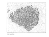
- 238000007490 hematoxylin and eosin (H&E) staining Methods 0.000 description 6
- 238000011081 inoculation Methods 0.000 description 6
- 241000588747 Klebsiella pneumoniae Species 0.000 description 5
- 241001465754 Metazoa Species 0.000 description 5
- 241000283973 Oryctolagus cuniculus Species 0.000 description 5
- 150000001540 azides Chemical class 0.000 description 5
- 210000004369 blood Anatomy 0.000 description 5
- 239000008280 blood Substances 0.000 description 5
- 238000006243 chemical reaction Methods 0.000 description 5
- 208000023652 chronic gastritis Diseases 0.000 description 5
- 230000000694 effects Effects 0.000 description 5
- 230000003053 immunization Effects 0.000 description 5
- 239000000203 mixture Substances 0.000 description 5
- 230000026731 phosphorylation Effects 0.000 description 5
- 238000006366 phosphorylation reaction Methods 0.000 description 5
- 102000014914 Carrier Proteins Human genes 0.000 description 4
- 108010078791 Carrier Proteins Proteins 0.000 description 4
- RTZKZFJDLAIYFH-UHFFFAOYSA-N Diethyl ether Chemical compound CCOCC RTZKZFJDLAIYFH-UHFFFAOYSA-N 0.000 description 4
- LFQSCWFLJHTTHZ-UHFFFAOYSA-N Ethanol Chemical compound CCO LFQSCWFLJHTTHZ-UHFFFAOYSA-N 0.000 description 4
- DHMQDGOQFOQNFH-UHFFFAOYSA-N Glycine Chemical compound NCC(O)=O DHMQDGOQFOQNFH-UHFFFAOYSA-N 0.000 description 4
- 241000699666 Mus <mouse, genus> Species 0.000 description 4
- 206010028980 Neoplasm Diseases 0.000 description 4
- FAPWRFPIFSIZLT-UHFFFAOYSA-M Sodium chloride Chemical compound [Na+].[Cl-] FAPWRFPIFSIZLT-UHFFFAOYSA-M 0.000 description 4
- DTQVDTLACAAQTR-UHFFFAOYSA-N Trifluoroacetic acid Chemical compound OC(=O)C(F)(F)F DTQVDTLACAAQTR-UHFFFAOYSA-N 0.000 description 4
- 108010046334 Urease Proteins 0.000 description 4
- 238000004458 analytical method Methods 0.000 description 4
- 201000011510 cancer Diseases 0.000 description 4
- 238000003745 diagnosis Methods 0.000 description 4
- 239000012634 fragment Substances 0.000 description 4
- 230000000968 intestinal effect Effects 0.000 description 4
- 238000002360 preparation method Methods 0.000 description 4
- 238000000746 purification Methods 0.000 description 4
- 239000000523 sample Substances 0.000 description 4
- 238000012360 testing method Methods 0.000 description 4
- 229940124597 therapeutic agent Drugs 0.000 description 4
- 238000002560 therapeutic procedure Methods 0.000 description 4
- FWMNVWWHGCHHJJ-SKKKGAJSSA-N 4-amino-1-[(2r)-6-amino-2-[[(2r)-2-[[(2r)-2-[[(2r)-2-amino-3-phenylpropanoyl]amino]-3-phenylpropanoyl]amino]-4-methylpentanoyl]amino]hexanoyl]piperidine-4-carboxylic acid Chemical compound C([C@H](C(=O)N[C@H](CC(C)C)C(=O)N[C@H](CCCCN)C(=O)N1CCC(N)(CC1)C(O)=O)NC(=O)[C@H](N)CC=1C=CC=CC=1)C1=CC=CC=C1 FWMNVWWHGCHHJJ-SKKKGAJSSA-N 0.000 description 3
- WEVYAHXRMPXWCK-UHFFFAOYSA-N Acetonitrile Chemical compound CC#N WEVYAHXRMPXWCK-UHFFFAOYSA-N 0.000 description 3
- 206010019375 Helicobacter infections Diseases 0.000 description 3
- MHAJPDPJQMAIIY-UHFFFAOYSA-N Hydrogen peroxide Chemical compound OO MHAJPDPJQMAIIY-UHFFFAOYSA-N 0.000 description 3
- 102000003992 Peroxidases Human genes 0.000 description 3
- 239000007983 Tris buffer Substances 0.000 description 3
- 229940024606 amino acid Drugs 0.000 description 3
- 239000012472 biological sample Substances 0.000 description 3
- 230000005847 immunogenicity Effects 0.000 description 3
- 230000007246 mechanism Effects 0.000 description 3
- 230000008506 pathogenesis Effects 0.000 description 3
- 230000007918 pathogenicity Effects 0.000 description 3
- 108040007629 peroxidase activity proteins Proteins 0.000 description 3
- 238000010186 staining Methods 0.000 description 3
- 210000002784 stomach Anatomy 0.000 description 3
- LENZDBCJOHFCAS-UHFFFAOYSA-N tris Chemical compound OCC(N)(CO)CO LENZDBCJOHFCAS-UHFFFAOYSA-N 0.000 description 3
- CSCPPACGZOOCGX-UHFFFAOYSA-N Acetone Chemical compound CC(C)=O CSCPPACGZOOCGX-UHFFFAOYSA-N 0.000 description 2
- 206010003694 Atrophy Diseases 0.000 description 2
- 102000004190 Enzymes Human genes 0.000 description 2
- 108090000790 Enzymes Proteins 0.000 description 2
- WSFSSNUMVMOOMR-UHFFFAOYSA-N Formaldehyde Chemical compound O=C WSFSSNUMVMOOMR-UHFFFAOYSA-N 0.000 description 2
- 239000004471 Glycine Substances 0.000 description 2
- WZUVPPKBWHMQCE-UHFFFAOYSA-N Haematoxylin Chemical compound C12=CC(O)=C(O)C=C2CC2(O)C1C1=CC=C(O)C(O)=C1OC2 WZUVPPKBWHMQCE-UHFFFAOYSA-N 0.000 description 2
- -1 Hydroxysuccinimide ester Chemical class 0.000 description 2
- 108060003951 Immunoglobulin Proteins 0.000 description 2
- 102000004890 Interleukin-8 Human genes 0.000 description 2
- 108090001007 Interleukin-8 Proteins 0.000 description 2
- 102000004022 Protein-Tyrosine Kinases Human genes 0.000 description 2
- 108090000412 Protein-Tyrosine Kinases Proteins 0.000 description 2
- 241000700159 Rattus Species 0.000 description 2
- 102000007056 Recombinant Fusion Proteins Human genes 0.000 description 2
- 108010008281 Recombinant Fusion Proteins Proteins 0.000 description 2
- 238000010171 animal model Methods 0.000 description 2
- 230000037444 atrophy Effects 0.000 description 2
- 230000008033 biological extinction Effects 0.000 description 2
- 238000010170 biological method Methods 0.000 description 2
- 230000015572 biosynthetic process Effects 0.000 description 2
- 239000000872 buffer Substances 0.000 description 2
- 239000007853 buffer solution Substances 0.000 description 2
- 239000000470 constituent Substances 0.000 description 2
- 238000010586 diagram Methods 0.000 description 2
- 230000002183 duodenal effect Effects 0.000 description 2
- 210000001198 duodenum Anatomy 0.000 description 2
- 238000010828 elution Methods 0.000 description 2
- 210000003608 fece Anatomy 0.000 description 2
- 238000002649 immunization Methods 0.000 description 2
- 230000002163 immunogen Effects 0.000 description 2
- 102000018358 immunoglobulin Human genes 0.000 description 2
- 238000011532 immunohistochemical staining Methods 0.000 description 2
- 210000004969 inflammatory cell Anatomy 0.000 description 2
- 229940096397 interleukin-8 Drugs 0.000 description 2
- XKTZWUACRZHVAN-VADRZIEHSA-N interleukin-8 Chemical compound C([C@H](NC(=O)[C@H](CC(O)=O)NC(=O)[C@H](CC=1C2=CC=CC=C2NC=1)NC(=O)[C@@H](NC(C)=O)CCSC)C(=O)N[C@@H](CC(O)=O)C(=O)N[C@@H](CC(O)=O)C(=O)N[C@@H](CC(C)C)C(=O)N[C@@H](CC(N)=O)C(=O)N[C@@H](CC=1C=CC=CC=1)C(=O)N[C@@H]([C@@H](C)O)C(=O)NCC(=O)N[C@@H](CCSC)C(=O)N1[C@H](CCC1)C(=O)N1[C@H](CCC1)C(=O)N[C@@H](C)C(=O)N[C@H](CC(O)=O)C(=O)N[C@H](CCC(O)=O)C(=O)N[C@H](CC(O)=O)C(=O)N[C@H](CC=1C=CC(O)=CC=1)C(=O)N[C@H](CO)C(=O)N1[C@H](CCC1)C(N)=O)C1=CC=CC=C1 XKTZWUACRZHVAN-VADRZIEHSA-N 0.000 description 2
- 108010045069 keyhole-limpet hemocyanin Proteins 0.000 description 2
- 210000003205 muscle Anatomy 0.000 description 2
- 230000001717 pathogenic effect Effects 0.000 description 2
- 230000008569 process Effects 0.000 description 2
- 238000012545 processing Methods 0.000 description 2
- 230000002829 reductive effect Effects 0.000 description 2
- 238000011160 research Methods 0.000 description 2
- 239000011780 sodium chloride Substances 0.000 description 2
- 238000006467 substitution reaction Methods 0.000 description 2
- 230000008685 targeting Effects 0.000 description 2
- 125000003088 (fluoren-9-ylmethoxy)carbonyl group Chemical group 0.000 description 1
- BDNKZNFMNDZQMI-UHFFFAOYSA-N 1,3-diisopropylcarbodiimide Chemical compound CC(C)N=C=NC(C)C BDNKZNFMNDZQMI-UHFFFAOYSA-N 0.000 description 1
- OXEUETBFKVCRNP-UHFFFAOYSA-N 9-ethyl-3-carbazolamine Chemical compound NC1=CC=C2N(CC)C3=CC=CC=C3C2=C1 OXEUETBFKVCRNP-UHFFFAOYSA-N 0.000 description 1
- 241000193830 Bacillus <bacterium> Species 0.000 description 1
- 238000009010 Bradford assay Methods 0.000 description 1
- 102000004127 Cytokines Human genes 0.000 description 1
- 108090000695 Cytokines Proteins 0.000 description 1
- 238000002965 ELISA Methods 0.000 description 1
- 108700003822 Helicobacter pylori cagA Proteins 0.000 description 1
- 241000238073 Homarus gammarus Species 0.000 description 1
- 241000282412 Homo Species 0.000 description 1
- IFQSXNOEEPCSLW-DKWTVANSSA-N L-cysteine hydrochloride Chemical compound Cl.SC[C@H](N)C(O)=O IFQSXNOEEPCSLW-DKWTVANSSA-N 0.000 description 1
- 241000699670 Mus sp. Species 0.000 description 1
- 102000008300 Mutant Proteins Human genes 0.000 description 1
- 108010021466 Mutant Proteins Proteins 0.000 description 1
- CTQNGGLPUBDAKN-UHFFFAOYSA-N O-Xylene Chemical compound CC1=CC=CC=C1C CTQNGGLPUBDAKN-UHFFFAOYSA-N 0.000 description 1
- 108010058846 Ovalbumin Proteins 0.000 description 1
- 208000008469 Peptic Ulcer Diseases 0.000 description 1
- 206010035226 Plasma cell myeloma Diseases 0.000 description 1
- 239000004793 Polystyrene Substances 0.000 description 1
- 208000006994 Precancerous Conditions Diseases 0.000 description 1
- 230000003213 activating effect Effects 0.000 description 1
- 230000004913 activation Effects 0.000 description 1
- 230000002238 attenuated effect Effects 0.000 description 1
- 210000003719 b-lymphocyte Anatomy 0.000 description 1
- 229960000074 biopharmaceutical Drugs 0.000 description 1
- 230000000903 blocking effect Effects 0.000 description 1
- 239000002775 capsule Substances 0.000 description 1
- 238000005119 centrifugation Methods 0.000 description 1
- 238000010835 comparative analysis Methods 0.000 description 1
- 238000007796 conventional method Methods 0.000 description 1
- 229960002433 cysteine Drugs 0.000 description 1
- 235000018417 cysteine Nutrition 0.000 description 1
- XUJNEKJLAYXESH-UHFFFAOYSA-N cysteine Natural products SCC(N)C(O)=O XUJNEKJLAYXESH-UHFFFAOYSA-N 0.000 description 1
- 210000000805 cytoplasm Anatomy 0.000 description 1
- 238000012217 deletion Methods 0.000 description 1
- 230000037430 deletion Effects 0.000 description 1
- 238000011161 development Methods 0.000 description 1
- BGRWYRAHAFMIBJ-UHFFFAOYSA-N diisopropylcarbodiimide Natural products CC(C)NC(=O)NC(C)C BGRWYRAHAFMIBJ-UHFFFAOYSA-N 0.000 description 1
- LOKCTEFSRHRXRJ-UHFFFAOYSA-I dipotassium trisodium dihydrogen phosphate hydrogen phosphate dichloride Chemical compound P(=O)(O)(O)[O-].[K+].P(=O)(O)([O-])[O-].[Na+].[Na+].[Cl-].[K+].[Cl-].[Na+] LOKCTEFSRHRXRJ-UHFFFAOYSA-I 0.000 description 1
- 229940079593 drug Drugs 0.000 description 1
- 210000000981 epithelium Anatomy 0.000 description 1
- 230000008029 eradication Effects 0.000 description 1
- 230000002550 fecal effect Effects 0.000 description 1
- 238000000684 flow cytometry Methods 0.000 description 1
- 108010074605 gamma-Globulins Proteins 0.000 description 1
- 210000001156 gastric mucosa Anatomy 0.000 description 1
- 208000017215 gastric mucosa-associated lymphoid tissue lymphoma Diseases 0.000 description 1
- 108060003552 hemocyanin Proteins 0.000 description 1
- 238000004128 high performance liquid chromatography Methods 0.000 description 1
- 230000036732 histological change Effects 0.000 description 1
- 230000028993 immune response Effects 0.000 description 1
- 230000016784 immunoglobulin production Effects 0.000 description 1
- 238000001727 in vivo Methods 0.000 description 1
- 230000002757 inflammatory effect Effects 0.000 description 1
- 238000002347 injection Methods 0.000 description 1
- 239000007924 injection Substances 0.000 description 1
- 238000007689 inspection Methods 0.000 description 1
- 210000002490 intestinal epithelial cell Anatomy 0.000 description 1
- 239000003446 ligand Substances 0.000 description 1
- 210000003141 lower extremity Anatomy 0.000 description 1
- 201000003265 lymphadenitis Diseases 0.000 description 1
- 210000004698 lymphocyte Anatomy 0.000 description 1
- 238000001840 matrix-assisted laser desorption--ionisation time-of-flight mass spectrometry Methods 0.000 description 1
- 239000012120 mounting media Substances 0.000 description 1
- 210000004877 mucosa Anatomy 0.000 description 1
- 210000004400 mucous membrane Anatomy 0.000 description 1
- 201000000050 myeloid neoplasm Diseases 0.000 description 1
- 210000000440 neutrophil Anatomy 0.000 description 1
- 238000011587 new zealand white rabbit Methods 0.000 description 1
- 238000011275 oncology therapy Methods 0.000 description 1
- 229940092253 ovalbumin Drugs 0.000 description 1
- 239000012188 paraffin wax Substances 0.000 description 1
- 230000036961 partial effect Effects 0.000 description 1
- 239000002245 particle Substances 0.000 description 1
- 125000002467 phosphate group Chemical group [H]OP(=O)(O[H])O[*] 0.000 description 1
- 229920002223 polystyrene Polymers 0.000 description 1
- 239000003755 preservative agent Substances 0.000 description 1
- 230000002335 preservative effect Effects 0.000 description 1
- 239000003223 protective agent Substances 0.000 description 1
- 239000002516 radical scavenger Substances 0.000 description 1
- 230000035484 reaction time Effects 0.000 description 1
- 230000006798 recombination Effects 0.000 description 1
- 239000011347 resin Substances 0.000 description 1
- 229920005989 resin Polymers 0.000 description 1
- 238000004007 reversed phase HPLC Methods 0.000 description 1
- 150000003839 salts Chemical class 0.000 description 1
- 230000019491 signal transduction Effects 0.000 description 1
- 238000002741 site-directed mutagenesis Methods 0.000 description 1
- 239000007787 solid Substances 0.000 description 1
- 238000010532 solid phase synthesis reaction Methods 0.000 description 1
- 241000894007 species Species 0.000 description 1
- 210000000952 spleen Anatomy 0.000 description 1
- 230000000638 stimulation Effects 0.000 description 1
- 239000000725 suspension Substances 0.000 description 1
- HNKJADCVZUBCPG-UHFFFAOYSA-N thioanisole Chemical compound CSC1=CC=CC=C1 HNKJADCVZUBCPG-UHFFFAOYSA-N 0.000 description 1
- 238000001269 time-of-flight mass spectrometry Methods 0.000 description 1
- 210000004881 tumor cell Anatomy 0.000 description 1
- 210000002700 urine Anatomy 0.000 description 1
- 230000001018 virulence Effects 0.000 description 1
- 230000007923 virulence factor Effects 0.000 description 1
- 239000000304 virulence factor Substances 0.000 description 1
- 230000000007 visual effect Effects 0.000 description 1
- XLYOFNOQVPJJNP-UHFFFAOYSA-N water Substances O XLYOFNOQVPJJNP-UHFFFAOYSA-N 0.000 description 1
- 239000008096 xylene Substances 0.000 description 1
Images
Landscapes
- Medicines That Contain Protein Lipid Enzymes And Other Medicines (AREA)
- Medicines Containing Antibodies Or Antigens For Use As Internal Diagnostic Agents (AREA)
- Peptides Or Proteins (AREA)
- Preparation Of Compounds By Using Micro-Organisms (AREA)
Abstract
Description
本発明は、ヘリコバクター・ピロリ菌(Helicobacter pylori)由来のチロシンリン酸化CagAタンパク質を認識する抗体、及びその利用法に関するものである。 The present invention relates to an antibody that recognizes a tyrosine-phosphorylated CagA protein derived from Helicobacter pylori, and a method of using the same.
ヘリコバクター・ピロリ菌(以下、H.ピロリ菌と称する)は、ヒトの胃内から分離されたグラム陰性桿菌であり、ヒト等の動物の胃上皮細胞に接着し、感染することが知られている。そして、H.ピロリ菌の感染は、慢性胃炎、胃潰瘍、十二指腸潰瘍の主な原因と考えられており、さらに、胃癌との関連性も指摘されている。 Helicobacter pylori (hereinafter, referred to as H. pylori) is a Gram-negative bacillus isolated from the human stomach and is known to adhere to and infect gastric epithelial cells of animals such as humans. . And H. Helicobacter pylori infection is thought to be a major cause of chronic gastritis, gastric ulcer, and duodenal ulcer, and has also been linked to gastric cancer.
このようなH.ピロリ菌は、病原性関連因子として、ウレアーゼ、空胞化サイトトキシンVacA(vaculoating cytotoxin)タンパク質、空胞化サイトトキシン関連遺伝子CagA(cytotoxin-associated gene A)タンパク質等を産生することが知られており、これらの病原性因子が、H.ピロリ菌の感染により引き起こされる種々の疾患の原因の一つと考えられている(例えば、非特許文献1及び非特許文献2参照。)。 HH. H. pylori is known to produce urease, a vacuolated cytotoxin VacA (vaculoating cytotoxin) protein, a vacuolated cytotoxin-related gene CagA (cytotoxin-associated gene A) protein, and the like as pathogenicity-related factors. Is a virulence factor of H. It is considered to be one of the causes of various diseases caused by H. pylori infection (for example, see Non-Patent Documents 1 and 2).
このように、H.ピロリ菌の感染が種々の疾患の原因となるため、上記疾患の早期発見、早期治療を行うために、H.ピロリ菌の感染を検出、診断する方法がいくつか考えられている。例えば、上記病原性関連因子の一つであるウレアーゼを標的としたウレアーゼ試験がある。具体的には、内視鏡的に採取した生検組織を用いて、ウレアーゼ活性を指標としてH.ピロリ菌が感染しているか否かを診断する方法である。その他には、血中、尿中のH.ピロリ菌に対するIgG抗体を測定する方法等も利用されている(例えば、非特許文献3参照。)。 HH. Since infection with H. pylori causes various diseases, H. pneumoniae is required for early detection and early treatment of the diseases. Several methods for detecting and diagnosing H. pylori infection have been considered. For example, there is a urease test targeting urease, which is one of the above-mentioned virulence-related factors. Specifically, using a biopsy tissue sampled endoscopically, U.S.P. This is a method to diagnose whether H. pylori is infected. Others include H. in blood and urine. A method of measuring an IgG antibody against H. pylori has also been used (for example, see Non-Patent Document 3).
上記方法によって、H.ピロリ菌が感染しているか否かを簡便に診断することができる。
しかし、H.ピロリ菌の感染者の全てに上記疾患が発生するわけではない。例えば、胃・十二指腸潰瘍等の消化性潰瘍は、H.ピロリ菌感染者のほんの一部、約2〜3%、さらに胃癌は1〜2%にしか発生しないことが知られている。 However, H. Not all persons infected with H. pylori develop the disease. For example, peptic ulcers such as gastric and duodenal ulcers are described in H. et al. It is known that only a small portion of H. pylori-infected persons, about 2-3%, and gastric cancer occur in only 1-2%.
このため、上述した従来の方法による診断では、網羅的にH.ピロリ菌の感染を調べることはできるが、慢性胃炎、胃潰瘍、十二指腸潰瘍、さらに、胃癌といった疾患と密接に関連するH.ピロリ菌の感染を効率的に精度よく調べることはできない。即ち、慢性胃炎、胃潰瘍、十二指腸潰瘍、さらに、胃癌等の疾患を効率的に、早期に検出、診断、治療することはできないという問題がある。 Therefore, in the diagnosis by the conventional method described above, H. Although H. pylori infection can be examined, H. pylori is closely associated with diseases such as chronic gastritis, gastric ulcer, duodenal ulcer, and gastric cancer. H. pylori infection cannot be examined efficiently and accurately. That is, there is a problem that diseases such as chronic gastritis, gastric ulcer, duodenal ulcer, and gastric cancer cannot be detected, diagnosed, and treated efficiently and early.
本発明は、上記の問題点に鑑みなされたものであり、その目的は、疾患と密接に関連する、チロシンリン酸化CagAタンパク質を認識する抗体を提供するとともに、さらにその抗体を用いた、胃潰瘍、胃癌等の疾患と密接に関連したH.ピロリ菌の感染を検出する方法、H.ピロリ菌の感染検出剤、疾患の発症または発症危険性の判定方法、治療方法、及びワクチンを提供することにある。 The present invention has been made in view of the above problems, and its object is to provide an antibody that recognizes tyrosine phosphorylated CagA protein, which is closely related to disease, and further uses the antibody to produce gastric ulcer, H. is closely associated with diseases such as gastric cancer. H. H. pylori infection detection method; It is to provide an agent for detecting H. pylori infection, a method for determining the onset or risk of onset of a disease, a treatment method, and a vaccine.
本発明者は、上記の課題に鑑み鋭意検討した結果、H.ピロリ菌に感染した宿主細胞に見出される、チロシンリン酸化されたCagAタンパク質を特異的に認識する抗体を作製し、ヒトの萎縮性胃炎、胃潰瘍、十二指腸潰瘍、MALTリンパ腫、胃癌等の患者から得られた生検標本に該抗体を作用させることにより、これらの疾患部位からチロシンリン酸化されたCagAタンパク質を検出できることを独自に見出し、本発明を完成させるに至った。 As a result of intensive studies in view of the above problems, the present inventor found that An antibody specifically recognizing tyrosine-phosphorylated CagA protein found in H. pylori-infected host cells was produced and obtained from human patients with atrophic gastritis, gastric ulcer, duodenal ulcer, MALT lymphoma, gastric cancer, etc. The inventors of the present invention have uniquely found that the tyrosine-phosphorylated CagA protein can be detected from these diseased sites by causing the antibody to act on the biopsy specimen obtained, thereby completing the present invention.
即ち、本発明に係る抗体は、(a)ヘリコバクター・ピロリ菌が感染している宿主細胞に見出される、チロシンリン酸化されているCagAタンパク質を特異的に認識する抗体である。 That is, the antibody according to the present invention is (a) an antibody that specifically recognizes a tyrosine-phosphorylated CagA protein found in a host cell infected with Helicobacter pylori.
上記抗体は、本発明者が今回新たに、ヘリコバクター・ピロリ菌が感染している宿主細胞に特異的に見出される、チロシンリン酸化されているCagAタンパク質を認識する抗体として生産したものである。 The above-mentioned antibody was newly produced by the present inventors as an antibody recognizing a tyrosine-phosphorylated CagA protein specifically found in host cells infected with Helicobacter pylori.
なお、本発明で「抗体」とは、モノクローナル抗体、ポリクローナル抗体を含むものである。 抗体 In the present invention, “antibody” includes a monoclonal antibody and a polyclonal antibody.
また、本発明に係る抗体は、(b)配列番号1に示すアミノ酸配列のうち、1番目のシステイン残基を除く2〜16番目のアミノ酸配列からなるポリペプチドであって、10番目のチロシン残基がリン酸化されているポリペプチドを含むリン酸化CagAタンパク質を特異的に認識する抗体である。 Further, the antibody according to the present invention is (b) a polypeptide consisting of the amino acid sequence of the 2nd to 16th positions excluding the 1st cysteine residue in the amino acid sequence shown in SEQ ID NO: 1; This is an antibody that specifically recognizes a phosphorylated CagA protein containing a polypeptide whose group is phosphorylated.
上記「ポリペプチド」とは、後述するように、H.ピロリ菌由来のCagAタンパク質の抗原決定基(エピトープ)領域であり、その10番目のチロシン残基がリン酸化されているものである。配列番号1中、1番目のシステイン残基は抗体作製時にキャリアタンパク質との結合のため付加されたものであり、CagAタンパク質の配列には存在しない。 The “polypeptide” is, as described later, H. It is an antigenic determinant (epitope) region of the H. pylori-derived CagA protein, and its tenth tyrosine residue is phosphorylated. In SEQ ID NO: 1, the first cysteine residue was added for binding to a carrier protein when the antibody was prepared, and is not present in the CagA protein sequence.
即ち、本発明に係る抗体は、10番目のチロシン残基がリン酸化されている場合のみ、上記ポリペプチドを含むリン酸化CagAタンパク質を特異的に認識する。実際、本発明者が作製した抗体は、胃潰瘍患者等から採取した胃上皮組織において、リン酸化CagAタンパク質を特異的に認識することが示された(後述の実施例参照)。 That is, the antibody according to the present invention specifically recognizes a phosphorylated CagA protein containing the above polypeptide only when the 10th tyrosine residue is phosphorylated. In fact, it was shown that the antibody prepared by the present inventors specifically recognized phosphorylated CagA protein in gastric epithelial tissue collected from patients with gastric ulcer and the like (see Examples described later).
また、本発明に係るヘリコバクター・ピロリ菌の宿主細胞への感染検出剤は、上記(a)または(b)の抗体を含有するものであり、本発明に係るヘリコバクター・ピロリ菌の宿主細胞への感染を検出する方法は、上記(a)または(b)の抗体を用いて行うものである。上述したように、上記(a)または(b)の抗体は、高精度でチロシンリン酸化されたCagAタンパク質を認識することができる。このため、本発明に係るヘリコバクター・ピロリ菌の感染検出剤、感染検出方法を用いることにより、高精度で、効率よく疾患と密接に関連するH.ピロリ菌の宿主細胞への感染を検出することができる。 The agent for detecting infection of Helicobacter pylori host cells according to the present invention contains the antibody (a) or (b) described above, and the agent for detecting infection of the host cells of Helicobacter pylori cells according to the present invention is used. The method for detecting infection is performed using the antibody (a) or (b). As described above, the antibody (a) or (b) can recognize the tyrosine-phosphorylated CagA protein with high accuracy. Therefore, by using the agent and the method for detecting Helicobacter pylori infection according to the present invention, H. pylori closely related to the disease with high accuracy and efficiency can be used. H. pylori infection to host cells can be detected.
また、本発明に係る疾患の発症または発症危険性判定方法は、上記(a)または(b)の抗体を用いて、萎縮性胃炎、胃潰瘍、十二指腸潰瘍、MALTリンパ腫、および胃癌のうち、いずれかの疾患の発症または発症危険性を判定する方法である。この判定方法を用いることにより、容易に、上記疾患の発症または発症危険性を判定することができ、上記疾患の早期診断、早期治療または予防を行うことができる。 In addition, the method for determining the onset or risk of onset of a disease according to the present invention uses the antibody of the above (a) or (b) to treat any of atrophic gastritis, gastric ulcer, duodenal ulcer, MALT lymphoma, and gastric cancer. This is a method for determining the onset or risk of onset of the disease. By using this determination method, the onset or risk of onset of the above-mentioned disease can be easily determined, and early diagnosis, early treatment or prevention of the above-mentioned disease can be performed.
また、本発明に係る疾患の治療方法は、上記抗体を用いて、萎縮性胃炎、胃潰瘍、十二指腸潰瘍、MALTリンパ腫、および胃癌のうち、いずれかの疾患を治療する方法である。 The method for treating a disease according to the present invention is a method for treating any one of atrophic gastritis, gastric ulcer, duodenal ulcer, MALT lymphoma, and gastric cancer using the above antibody.
また、本発明に係るワクチンは、配列番号4に示すアミノ酸配列からなるポリペプチドであって、10番目のチロシン残基がリン酸化されているポリペプチドを含有するワクチンである。 ワ ク チ ン The vaccine according to the present invention is a polypeptide comprising the amino acid sequence shown in SEQ ID NO: 4 and containing a polypeptide in which the 10th tyrosine residue is phosphorylated.
本発明のワクチンを用いることにより、H.ピロリ菌の感染、または萎縮性胃炎、胃潰瘍、十二指腸潰瘍、MALTリンパ腫、および胃癌のうち、いずれかの疾患を治療、または予防することができる。 に よ り By using the vaccine of the present invention, Helicobacter pylori infection or any of atrophic gastritis, gastric ulcer, duodenal ulcer, MALT lymphoma, and gastric cancer can be treated or prevented.
上述したように、本発明に係る抗体は、H.ピロリ菌が感染している宿主細胞に見出される、チロシンリン酸化されたCagAタンパク質を精度良く認識することができる。このため、本発明に係る抗体は、高精度で種々の疾患と密接に関連しているH.ピロリ菌の感染を、高精度、かつ効率的に検出できるという効果を奏する。 抗体 As described above, the antibody according to the present invention is The tyrosine-phosphorylated CagA protein found in host cells infected with H. pylori can be accurately recognized. Therefore, the antibody according to the present invention is highly accurate and closely related to various diseases. It is effective in detecting infection with H. pylori with high accuracy and efficiency.
また、本発明に係るH.ピロリ菌の感染検出剤、H.ピロリ菌の感染検出方法を用いることにより、高精度、かつ効率的に種々の疾患と密接に関連しているH.ピロリ菌の感染を検出することができるという効果を奏する。 {H. H. pylori infection detection agent; By using the H. pylori infection detection method, H. pylori is closely related to various diseases with high accuracy and efficiency. It is effective in detecting infection with H. pylori.
さらに、本発明に係る疾患の発症または発症危険性判定方法を用いることにより、萎縮性胃炎、胃潰瘍、十二指腸潰瘍、MALTリンパ腫、胃癌等の疾患の発症または発症危険性を高精度、かつ効率的に判断することができ、上記疾患の予防、早期診断、早期治療に利用することができるという効果を奏する。 Furthermore, by using the method for determining the onset or risk of onset of the disease according to the present invention, atrophy gastritis, gastric ulcer, duodenal ulcer, MALT lymphoma, highly accurate and efficient onset or risk of onset of disease such as gastric cancer. It is possible to make a judgment and to use the present invention for prevention, early diagnosis and early treatment of the above-mentioned diseases.
また、本発明に係る疾患の治療方法を用いることにより、萎縮性胃炎、胃潰瘍、十二指腸潰瘍、MALTリンパ腫、胃癌等の疾患の発症または発症危険性を高精度、かつ効率的に治療することができるという効果を奏する。 Further, by using the method for treating a disease according to the present invention, it is possible to treat the onset or risk of developing atrophy such as atrophic gastritis, gastric ulcer, duodenal ulcer, MALT lymphoma, and gastric cancer with high precision and efficiency. This has the effect.
また、本発明に係るワクチンを用いることにより、効果的にH.ピロリ菌の感染を治療することができ、ひいては、H.ピロリ菌の感染により引き起こされる種々の疾患を予防、治療することができるという効果を奏する。 H Moreover, by using the vaccine according to the present invention, H. H. pylori infections can be treated, and thus H. pylori. It is effective in preventing and treating various diseases caused by H. pylori infection.
本発明の実施の一形態について説明すれば、以下の通りである。なお、本発明はこれに限定されるものではない。 れ ば One embodiment of the present invention will be described below. Note that the present invention is not limited to this.
本発明は、萎縮性胃炎、胃潰瘍、十二指腸潰瘍、MALTリンパ腫、胃癌等の疾患と密接に関連するチロシンリン酸化されたCagAタンパク質を特異的に認識する新規な抗体を提供するとともに、その利用方法を提案するものである。 The present invention provides a novel antibody that specifically recognizes a tyrosine-phosphorylated CagA protein closely related to diseases such as atrophic gastritis, gastric ulcer, duodenal ulcer, MALT lymphoma, and gastric cancer, and a method of using the same. It is a suggestion.
CagAタンパク質が、ヒトの胃上皮細胞内に注入され、チロシンリン酸化されると、胃上皮細胞内のシグナル伝達を阻害することが知られている。このことが胃上皮細胞に与える影響についての詳細は、未だ明らかとなっていない。しかし、CagAタンパク質等の病原性関連因子は、胃上皮細胞に様々な影響を与えることが予想される。例えば、これら病原性関連因子の刺激により、胃上皮細胞からIL−8(interleukin-8)をはじめとした種々のサイトカインが分泌され、好中球やその他の炎症細胞が粘膜固有層に遊走する。そして胃上皮細胞が障害を受け、さらに炎症細胞が遊走し、免疫反応が賦活されると考えられる。 It is known that CagA protein, when injected into human gastric epithelial cells and tyrosine phosphorylated, inhibits signal transduction in gastric epithelial cells. The details of the effect of this on gastric epithelial cells have not yet been elucidated. However, pathogenicity-related factors such as CagA protein are expected to have various effects on gastric epithelial cells. For example, by stimulation of these pathogenicity-related factors, various cytokines including IL-8 (interleukin-8) are secreted from gastric epithelial cells, and neutrophils and other inflammatory cells migrate to the lamina propria. It is thought that the gastric epithelial cells are damaged, the inflammatory cells migrate, and the immune response is activated.
そのため、本発明者は、CagAタンパク質のチロシンリン酸化は、H.ピロリ菌の感染が引き起こす種々の疾患(例えば、慢性胃炎、萎縮性胃炎、胃潰瘍、十二指腸潰瘍、MALTリンパ腫、胃癌等)に特異的な現象であって、これら疾患の発症機構の1つではないかと注目した。 た め Therefore, the present inventor has reported that tyrosine phosphorylation of CagA protein is described in H. et al. It is a phenomenon specific to various diseases caused by H. pylori infection (eg, chronic gastritis, atrophic gastritis, gastric ulcer, duodenal ulcer, MALT lymphoma, gastric cancer, etc.), and is one of the mechanisms of the pathogenesis of these diseases. noticed.
そこで、本発明者は、種々の疾患における、チロシンリン酸化されたCagAタンパク質の作用に注目し、H.ピロリ菌が感染している宿主細胞に特異的に見出される、チロシンリン酸化されているCagAタンパク質を認識する抗体を生産し、該抗体を利用して、疾患と密接に関連するH.ピロリ菌の感染を高精度で、効率よく検出する手段を独自に開発した。 Therefore, the present inventors focused on the effect of tyrosine phosphorylated CagA protein in various diseases, It produces antibodies that recognize tyrosine-phosphorylated CagA protein, which is specifically found in host cells infected with H. pylori, and uses these antibodies to make H. pylori closely related to diseases. We have independently developed a means to detect H. pylori infection with high precision and efficiency.
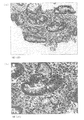
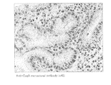
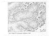
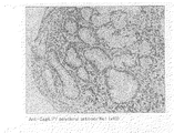
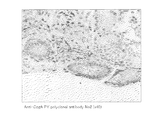
即ち、後述する実施例に示すように、本発明に係る抗体を用いることにより、萎縮性胃炎(腸上皮化性が見られる)の生検標本において、チロシンリン酸化されたCagAタンパク質が検出されることが実験的に示された。この萎縮性胃炎、腸上皮化性は、胃癌や胃潰瘍の発生母地(前癌状態)と一般的に考えられている。このため、チロシンリン酸化されたCagAタンパク質は、萎縮性胃炎、胃潰瘍、十二指腸潰瘍、MALTリンパ腫、胃癌等の疾患と密接に関連していることがin vivoにて初めて示唆された。即ち、チロシンリン酸化されたCagAタンパク質が検出されることは、萎縮性胃炎、胃潰瘍、十二指腸潰瘍、MALTリンパ腫、及び胃癌等が発症している、あるいは発症する危険性が高いことを示すと考えられる。 That is, as shown in Examples described later, tyrosine phosphorylated CagA protein is detected in a biopsy specimen of atrophic gastritis (intestinal epithelialization is observed) by using the antibody according to the present invention. This was shown experimentally. The atrophic gastritis and intestinal epithelialization are generally considered to be the origin of stomach cancer and gastric ulcer (pre-cancerous state). For this reason, it was suggested for the first time in vivo that tyrosine-phosphorylated CagA protein is closely related to diseases such as atrophic gastritis, gastric ulcer, duodenal ulcer, MALT lymphoma, and gastric cancer. That is, the detection of tyrosine-phosphorylated CagA protein is considered to indicate that atrophic gastritis, gastric ulcer, duodenal ulcer, MALT lymphoma, and gastric cancer have been developed, or that there is a high risk of developing gastric cancer. .
従って、この手段、即ち、チロシンリン酸化されたCagAタンパク質を検出する手段は、慢性胃炎、萎縮性胃炎、胃潰瘍、十二指腸潰瘍、MALTリンパ腫、胃癌等のH.ピロリ菌の感染によって引き起こされる種々の疾患の予防、早期発見、早期治療を行うための重要な手段となる可能性が高い。 Therefore, this means, that is, means for detecting tyrosine-phosphorylated CagA protein, is not suitable for the treatment of chronic gastritis, atrophic gastritis, gastric ulcer, duodenal ulcer, MALT lymphoma, gastric cancer, etc. It is likely to be an important means for prevention, early detection and early treatment of various diseases caused by H. pylori infection.
そこで以下に、まず本発明に係る抗体の特徴について説明し、次いで上記抗体の生産方法、機能、及び利用方法について説明することとする。 Therefore, the characteristics of the antibody according to the present invention will be described first, and then the production method, function, and utilization method of the antibody will be described.
(1)本発明に係る抗体の特徴
本発明者は、以前、胃の上皮細胞に感染したH.ピロリ菌によって、CagAタンパク質が宿主細胞(例えば、ヒトの胃上皮細胞)の細胞質内に注入された後、宿主細胞内のタンパク質チロシンキナーゼ(PTKs)によって、リン酸化されることを明らかにした(上記非特許文献2:Asahi. M., et al. J. Exp. Med. 191, No.4: 2000 593-602)。そして、この現象に着目し、この現象を利用して効率的に、上記疾患と密接に関連するH.ピロリ菌の感染を検出する方法等を開発しようと鋭意努力を重ねた。
(1) Characteristics of the Antibody According to the Present Invention Helicobacter pylori revealed that the CagA protein was injected into the cytoplasm of host cells (eg, human gastric epithelial cells) and then phosphorylated by protein tyrosine kinases (PTKs) in the host cells (see above). Non-Patent Document 2: Asahi. M., et al. J. Exp. Med. 191, No. 4: 2000 593-602). By paying attention to this phenomenon, it is possible to efficiently utilize this phenomenon to efficiently associate H. pneumoniae with the above-mentioned disease. We worked diligently to develop methods for detecting H. pylori infection.
その結果、本発明者は、効率的に、上記疾患と密接に関連するH.ピロリ菌の宿主細胞への感染を検出することを目的とし、H.ピロリ菌が感染している宿主細胞に見出される、チロシンリン酸化されているCagAタンパク質を特異的に認識する抗体を生産することに成功し、本発明を完成するに至った。なお、本発明でいう「チロシンリン酸化されたCagAタンパク質」とは、CagAタンパク質のチロシン残基にリン酸基が結合しているものをいう。H.ピロリ菌由来のCagAタンパク質には、その893番目と912番目と965番目と999番目と1033番目とにチロシン残基が存在するが、これら5つのチロシン残基の少なくとも一つのチロシン残基がリン酸化されていればよい。即ち、本発明に係る抗体は、上記5つのチロシン残基のリン酸化を認識するものである。なお、上記5つのチロシン残基のうち、965番目、999番目および1033番目に存在するチロシン残基はいずれも、ITAM(immunoreceptor tyrosine-based activation motif)とよばれるモチーフと非常に類似する領域(ITAM様モチーフ)に存在する。そのため、上記965番目、999番目および1033番目のチロシン残基のリン酸化を特異的に認識する抗体であることがより好ましい。 結果 As a result, the present inventor efficiently and highly closely related to the above-mentioned diseases. H. pylori, for the purpose of detecting infection of host cells; The present inventors have succeeded in producing an antibody specifically recognizing a tyrosine-phosphorylated CagA protein found in a host cell infected with H. pylori, and completed the present invention. The term “Tyrosine-phosphorylated CagA protein” used in the present invention refers to a protein in which a phosphate group is bonded to a tyrosine residue of the CagA protein. H. H. pylori-derived CagA protein has tyrosine residues at positions 893, 912, 965, 999, and 1033, and at least one of these five tyrosine residues is phosphorylated. It should just be done. That is, the antibody according to the present invention recognizes phosphorylation of the above five tyrosine residues. Of the five tyrosine residues, the tyrosine residues at positions 965, 999 and 1033 are all very similar to a motif called ITAM (immunoreceptor tyrosine-based activation motif) (ITAM). Motif). Therefore, an antibody that specifically recognizes phosphorylation of the tyrosine residues at positions 965, 999, and 1033 is more preferable.
また、上記「宿主細胞」とは、例えば、H.ピロリ菌が感染することができる細胞であればよく、その細胞の由来生物種、器官、組織等は特に限定されるものではない。上記宿主細胞として、例えば、ヒトの胃上皮細胞や十二指腸上皮細胞等が挙げられる。 In addition, the “host cell” is, for example, H. The cells may be any cells that can be infected by H. pylori, and the species, organs, tissues, and the like from which the cells are derived are not particularly limited. Examples of the host cells include human gastric epithelial cells and duodenal epithelial cells.
また、本発明に係る抗体は、チロシンリン酸化されたCagAタンパク質の抗原決定基(エピトープ)領域である、配列番号1に示すアミノ酸配列からなるポリペプチドであって、10のチロシン残基がリン酸化されているポリペプチドを特異的に認識するものであればよい。なお、上記「ポリペプチド」とは、チロシンリン酸化されたCagAタンパク質のエピトープ領域(H.ピロリ菌由来のCagAタンパク質の957番目〜971番目のアミノ酸残基、991番目〜1005番目のアミノ酸残基、1025番目〜1039番目のアミノ酸残基)に相当し、さらに、配列番号1に示すアミノ酸配列のうち、10番目のチロシン残基がリン酸化されているものをいう。 The antibody according to the present invention is a polypeptide having the amino acid sequence shown in SEQ ID NO: 1, which is an antigenic determinant (epitope) region of a tyrosine-phosphorylated CagA protein, wherein 10 tyrosine residues are phosphorylated. Any polypeptide that specifically recognizes a given polypeptide may be used. The “polypeptide” refers to an epitope region of a tyrosine-phosphorylated CagA protein (the 957th to 971th amino acid residues, the 991th to 1005th amino acid residues of the CagA protein derived from H. pylori, (1025th to 1039th amino acid residues), and the amino acid sequence shown in SEQ ID NO: 1 in which the 10th tyrosine residue is phosphorylated.
本実施の形態では、本発明に係る抗体の一例として、本発明者がH.ピロリ菌由来のチロシンリン酸化されたCagAタンパク質のエピトープ領域(H.ピロリ菌由来のCagAタンパク質の957番目〜971番目のアミノ酸残基、991番目〜1005番目のアミノ酸残基、1025番目〜1039番目のアミノ酸残基)に相当し、さらに、配列番号1に示すアミノ酸配列のうち、10番目のチロシン残基がリン酸化されているポリペプチドを抗原として生産したポリクローナル抗体を例に挙げて説明する。 で は In the present embodiment, the present inventor has described H. as an example of the antibody according to the present invention. H. pylori-derived tyrosine phosphorylated CagA protein epitope region (H. H. pylori-derived CagA protein at 957th to 971th amino acid residues, 991th to 1005th amino acid residues, 1025th to 1039th amino acid residues) (Amino acid residue), and a polyclonal antibody produced using, as an antigen, a polypeptide in which the 10th tyrosine residue is phosphorylated in the amino acid sequence shown in SEQ ID NO: 1.
なお、上記本実施の形態に係る抗体は、後述する実施例に示すように、配列番号3に示すアミノ酸配列からなり、8番目のチロシン残基がリン酸化されているポリペプチドとアフィニティ結合するポリクローナル抗体として精製されたものである。このため、配列番号4に示すアミノ酸配列からなるポリペプチドであって、4番目のチロシン残基がリン酸化されているポリペプチドを特異的に認識することができる。 The antibody according to the present embodiment has an amino acid sequence represented by SEQ ID NO: 3 and has a polyclonal affinity binding with a polypeptide having a phosphorylated tyrosine residue at the eighth position, as shown in Examples described later. It was purified as an antibody. For this reason, it is possible to specifically recognize a polypeptide having the amino acid sequence shown in SEQ ID NO: 4, in which the fourth tyrosine residue is phosphorylated.
さらに、例えば、後述する実施例のアフィニティ精製において、配列番号1に示すアミノ酸配列からなるポリペプチドであって、10番目のチロシン残基がリン酸化されているポリペプチドとアフィニティ結合するポリクローナル抗体を精製することで、CagAタンパク質の965番目、999番目および1033番目のチロシン残基のリン酸化を特異的に認識する抗体を取得することもできる。 Furthermore, for example, in the affinity purification of the Examples described later, a polyclonal antibody that binds affinity to a polypeptide having the amino acid sequence shown in SEQ ID NO: 1 and whose 10th tyrosine residue is phosphorylated is purified. By doing so, an antibody that specifically recognizes the phosphorylation of tyrosine residues at positions 965, 999, and 1033 of the CagA protein can be obtained.
また、本発明に係る抗体は、ポリクローナル抗体のみでなく、モノクローナル抗体も含むものである。ポリクローナル抗体、及びモノクローナル抗体は、免疫グロブリンクラス(immunoglobulin class)が、IgGのモノクローナル抗体であることが好ましいが、それ以外の免疫グロブリンクラス、例えば、IgMやIgA等であってもよく、特に限られるものではない。 抗体 The antibodies according to the present invention include not only polyclonal antibodies but also monoclonal antibodies. The polyclonal antibody and the monoclonal antibody preferably have an immunoglobulin class of an IgG monoclonal antibody, but may have other immunoglobulin classes such as IgM and IgA, and are particularly limited. Not something.
(2)本発明に係る抗体の生産方法
(2−1)ポリクローナル抗体の生産方法
本発明に係るポリクローナル抗体を生産する方法は、特に限定されるものではなく、例えば、抗原タンパク質で動物を免疫した後、その動物から血清を採取し、ポリクローナル抗体を得ればよい。なお、「生産」とは、この抗体の生合成に加えて各種人為的な作用を含む、本発明に係る抗体(ポリクローナル抗体、モノクローナル抗体を含む)を得るための全体的な流れを指すものとする。
(2) Method for Producing Antibody According to the Present Invention (2-1) Method for Producing Polyclonal Antibody The method for producing the polyclonal antibody according to the present invention is not particularly limited. For example, an animal was immunized with an antigen protein. Thereafter, serum may be collected from the animal to obtain a polyclonal antibody. In addition, “production” refers to the overall flow of obtaining the antibody (including polyclonal antibody and monoclonal antibody) of the present invention, including various artificial actions in addition to the biosynthesis of this antibody. I do.
本実施の形態に係るポリクローナル抗体は、後述する実施例に示すように、10番目のチロシン残基をリン酸化した配列番号1に示すアミノ酸配列からなるポリペプチドを抗原として、ウサギを免疫した後、そのウサギの血液から血清を得て、さらに、この血清からIgGを精製して取得された。 The polyclonal antibody according to the present embodiment, as shown in the examples below, after immunizing rabbits with a polypeptide consisting of the amino acid sequence of SEQ ID NO: 1 phosphorylated at the 10th tyrosine residue as an antigen, Serum was obtained from the blood of the rabbit, and IgG was purified from the serum and obtained.
なお、本実施形態では、免疫させる動物としてウサギを用いたが、マウス、ラット等のその他の実験動物を用いてもよく、特に限定されるものではない。 In the present embodiment, a rabbit was used as an animal to be immunized, but other experimental animals such as a mouse and a rat may be used, and there is no particular limitation.
さらに、本発明に係るポリクローナル抗体は、例えば、チロシンリン酸化されたCagAタンパク質、その抗原決定基(エピトープ)を含むフラグメントまたはその誘導体、あるいはそれらのアナログ、またはそれらの変異体、もしくはそれらを発現する細胞を免疫源(抗原)として用いることにより産生することができる。 Furthermore, the polyclonal antibody according to the present invention expresses, for example, a tyrosine-phosphorylated CagA protein, a fragment or a derivative thereof containing an antigenic determinant (epitope), an analog thereof, a mutant thereof, or an expression thereof. It can be produced by using cells as an immunogen (antigen).
即ち、本発明に係るポリクローナル抗体は、例えば、チロシンリン酸化されたCagAタンパク質、その抗原決定基(エピトープ)を含むフラグメントまたはその誘導体、あるいはそれらのアナログ、またはそれらの変異体、もしくはそれらを発現する細胞を認識することができる。 That is, the polyclonal antibody according to the present invention expresses, for example, a tyrosine-phosphorylated CagA protein, a fragment or a derivative thereof containing an antigenic determinant (epitope), an analog thereof, a mutant thereof, or an expression thereof. Cells can be recognized.
なお、ここにいう「変異体」は、部位特異的突然変異誘発法等の公知の変異タンパク質作製法により置換、欠失、挿入、及び/または付加できる程度の数のアミノ酸が置換、欠失、挿入、及び/または付加されたものをいう。 The “mutant” used herein refers to a substitution, deletion, or substitution of amino acids in a number that can be substituted, deleted, inserted, and / or added by a known method for producing a mutant protein such as site-directed mutagenesis. Inserted and / or added.
また、上記抗原としては、ポリペプチドであれば特に限定されるものではないが、抗原決定基とする物質をキャリアタンパク質に結合してなる抗原タンパク質が用いられてもよい。 The antigen is not particularly limited as long as it is a polypeptide, but an antigen protein obtained by binding a substance serving as an antigenic determinant to a carrier protein may be used.
具体的には、上記抗原がハプテンであれば、抗体の産生等を誘導する能力をもたないため、抗体を産生することができないが、抗原を異種由来のタンパク質などの生体高分子からなる担体と共有結合させて抗原タンパク質を得て、これで免疫すれば、抗体産生を誘導することができる。上記担体としては、特に限定されるものではなく、オボアルブミン、γグロブリン、ヘモシアニン等、この分野で従来公知の各種タンパク質を好適に用いることができる。 Specifically, if the antigen is a hapten, the antibody cannot be produced because it does not have the ability to induce the production of an antibody, etc. When an antigen protein is obtained by covalently binding to the antibody and immunizing with the antigen protein, antibody production can be induced. The carrier is not particularly limited, and various proteins conventionally known in this field, such as ovalbumin, γ globulin, and hemocyanin, can be suitably used.
なお、ここでいう「ポリペプチド」とは、アミノ酸が数個結合した短いペプチド、及び組換えタンパク質を含むものである。また、ポリペプチドは、天然に存在するもの、化学的、生物学的方法にて合成されたものを含む。 In addition, the "polypeptide" as used herein includes a short peptide in which several amino acids are bonded and a recombinant protein. In addition, polypeptides include those naturally occurring and those synthesized by chemical and biological methods.
また、本発明に係るポリクローナル抗体の生産においては、抗血清によって得られたポリクローナル抗体を、従来公知の方法で精製したり、修飾したりする工程が含まれていてもよい。 生産 In addition, production of the polyclonal antibody according to the present invention may include a step of purifying or modifying the polyclonal antibody obtained by the antiserum by a conventionally known method.
(2−2)モノクローナル抗体の生産方法
本発明に係るモノクローナル抗体を生産する方法は、特に限定されるものではなく、例えば、抗原でマウスを免疫した後、そのマウス脾臓リンパ球とマウス由来のミエローマ細胞とを融合させてなる抗体産生ハイブリドーマにより、モノクローナル抗体を得ればよい。なお、本実施の形態における「産生」とは、ハイブリドーマによる抗体の生合成を指すものとする。
(2-2) Method for Producing Monoclonal Antibody The method for producing the monoclonal antibody according to the present invention is not particularly limited. For example, after immunizing a mouse with an antigen, the mouse spleen lymphocytes and mouse-derived myeloma A monoclonal antibody may be obtained using an antibody-producing hybridoma obtained by fusing with a cell. Note that “production” in the present embodiment refers to antibody biosynthesis by hybridomas.
ハイブリドーマの生産方法は、従来公知の方法、例えば、ハイブリドーマ法(Kohler, G. and Milstein, C., Nature 256, 495-497(1975))、トリオーマ法、ヒトB−細胞ハイブリドーマ法(Kozbor, Immunology Today 4, 72(1983))、及びEBV−ハイブリドーマ法(Monoclonal Antibodies and Cancer Therapy, Alan R Liss, Inc., 77-96(1985))等を利用することが可能であり、特に限定されるものではない。
Hybridoma production methods include hitherto known methods, for example, the hybridoma method (Kohler, G. and Milstein, C., Nature 256, 495-497 (1975)), the trioma method, and the human B-cell hybridoma method (Kozbor, Immunology).
例えば、本発明に係るモノクローナル抗体は、10番目のチロシン残基をリン酸化した配列番号1に示すアミノ酸配列からなるポリペプチドを抗原として、ハイブリドーマを作製して取得することができる。 For example, the monoclonal antibody according to the present invention can be obtained by preparing a hybridoma using a polypeptide having the amino acid sequence of SEQ ID NO: 1 phosphorylated at the 10th tyrosine residue as an antigen.
なお、免疫させる動物としては、マウスの他にも、ラット、ウサギ等のその他の実験動物を用いてもよく、特に限定されるものではない。また、モノクローナル抗体は遺伝子組換え技術等によっても生産できる。 動物 As the animal to be immunized, other experimental animals such as rats and rabbits may be used in addition to mice, and are not particularly limited. Monoclonal antibodies can also be produced by genetic recombination techniques and the like.
このようにして得られたハイブリドーマクローンは、本発明に係るモノクローナル抗体を大量に産生することができるものである。 ハ イ The thus obtained hybridoma clone is capable of producing the monoclonal antibody according to the present invention in a large amount.
さらに、本発明に係るモノクローナル抗体は、上記ポリクローナル抗体と同様に、抗原として種々の物質を用いて生産することができる。例えば、チロシンリン酸化されたCagAタンパク質、その抗原決定基(エピトープ)を含むフラグメントまたはその誘導体等を、免疫源(抗原)として用いることにより産生することができる。 Furthermore, the monoclonal antibody according to the present invention can be produced using various substances as antigens, similarly to the above-mentioned polyclonal antibody. For example, it can be produced by using a tyrosine-phosphorylated CagA protein, a fragment containing an antigenic determinant (epitope) or a derivative thereof as an immunogen (antigen).
従って、本発明に係るモノクローナル抗体は、例えば、チロシンリン酸化されたCagAタンパク質、その抗原決定基(エピトープ)を含むフラグメントまたはその誘導体、あるいはそれらのアナログ、またはそれらの変異体、もしくはそれらを発現する細胞を認識することができる。 Therefore, the monoclonal antibody according to the present invention expresses, for example, a tyrosine-phosphorylated CagA protein, a fragment containing the antigenic determinant (epitope) or a derivative thereof, an analog thereof, or a mutant thereof, or expresses them. Cells can be recognized.
また、上記抗原としては、上記ポリクローナル抗体と同様に、ポリペプチド、ハプテン等を利用することができる。 As the antigen, a polypeptide, a hapten, or the like can be used as in the case of the polyclonal antibody.
また、本発明に係るモノクローナル抗体の生産においては、ハイブリドーマによって産生されたモノクローナル抗体を、従来公知の方法で精製したり、修飾したりする工程が含まれていてもよい。 In addition, the production of the monoclonal antibody according to the present invention may include a step of purifying or modifying the monoclonal antibody produced by the hybridoma by a conventionally known method.
(3)本発明に係る抗体の機能
後述する実施例に示すように、本発明に係る抗体を用いて、H.ピロリ菌の感染により引き起こされた胃潰瘍、胃癌のそれぞれの患者から取り出した胃の生検組織の免疫染色を行った結果、萎縮性胃炎を裏付ける腸上皮化性細胞において、チロシンリン酸化されたCagAタンパク質を精度よく検出することができた。
(3) Function of Antibody According to the Present Invention As shown in Examples described later, H. Immunostaining of gastric biopsies taken from patients with gastric ulcer and gastric cancer caused by H. pylori infection revealed that tyrosine-phosphorylated CagA protein in intestinal epithelial cells supporting atrophic gastritis Was accurately detected.
従って、本発明に係る抗体は、チロシンリン酸化されているCagAタンパク質を精度良く認識できることが実験的に示された。さらに、上述したように、CagAタンパク質のチロシンリン酸化は、H.ピロリ菌の感染によって引き起こされる種々の疾患と密接に関連していると考えられる。 Accordingly, it was experimentally shown that the antibody according to the present invention can accurately recognize tyrosine-phosphorylated CagA protein. Further, as described above, tyrosine phosphorylation of the CagA protein is described by H. et al. It is thought to be closely related to various diseases caused by H. pylori infection.
このため、本発明に係る抗体は、胃や十二指腸における種々の疾患と密接に関連しているH.ピロリ菌の宿主細胞への感染を、精度よく、効率的に検出することができる。即ち、本発明に係る抗体は、H.ピロリ菌の感染によって引き起こされる種々の疾患の発症、または発症危険性(可能性)を判断する際の指標に利用(換言すれば、疾患の診断方法に利用)することができる。 Therefore, the antibody according to the present invention is closely related to various diseases in the stomach and duodenum. H. pylori infection to host cells can be detected accurately and efficiently. That is, the antibody according to the present invention is H. It can be used as an index for determining the onset or risk of onset (possibility) of various diseases caused by H. pylori infection (in other words, used for a method of diagnosing the disease).
(4)本発明に係る抗体の利用法
上述したように、本発明に係る抗体は、チロシンリン酸化されたCagAタンパク質を特異的に認識する新規な抗体である。また、本発明者は、H.ピロリ菌が感染したヒトの胃上皮細胞において、チロシン残基がリン酸化されたCagAタンパク質が見出されることを明らかにしており、CagAタンパク質のチロシンリン酸化は、H.ピロリ菌の感染によって引き起こされる種々の疾患と密接に関連していると考えられる。そのため、本発明の抗体は、以下に示すように利用することができる。
(4) Use of Antibody According to the Present Invention As described above, the antibody according to the present invention is a novel antibody that specifically recognizes a tyrosine-phosphorylated CagA protein. In addition, the present inventor described in H. It has been shown that a CagA protein in which tyrosine residues are phosphorylated is found in human gastric epithelial cells infected with H. pylori. It is thought to be closely related to various diseases caused by H. pylori infection. Therefore, the antibody of the present invention can be used as described below.
(4−1)本発明に係るH.ピロリ菌の感染検出剤
本発明に係る抗体は、H.ピロリ菌の宿主細胞への感染を検出するためのH.ピロリ菌の感染検出剤として用いることができる。即ち、本発明に係るH.ピロリ菌の感染検出剤の具体的な組成は、上記抗体を含んでいればよく、特に限定されるものではない。具体的には、使用(検査)対象の生物個体、器官、組織、細胞塊、糞便、あるいは細胞等の種類に応じて、H.ピロリ菌の感染を検出するために、本発明に係る抗体の機能が発揮できるような緩衝液等が含まれていればよい。
(4-1) The H.2 according to the present invention. H. pylori infection detecting agent H. pylori for detecting infection of host cells with H. pylori; It can be used as an agent for detecting infection of H. pylori. That is, according to the H.264 of the present invention. The specific composition of the agent for detecting H. pylori infection is not particularly limited as long as it contains the above antibody. Specifically, depending on the type of living individual, organ, tissue, cell mass, stool, cell or the like to be used (examined), In order to detect H. pylori infection, a buffer or the like that can exert the function of the antibody according to the present invention may be included.
即ち、本発明に係るH.ピロリ菌の感染検出剤中に含まれる上記抗体は、特異的な抗原抗体反応によって、チロシンリン酸化されたCagAタンパク質を認識、検出する。このため、上記抗体を、顕微鏡等による目視で、または機械的に識別できる手段を用いて標識する等の処理を施すと、抗体が結合したチロシンリン酸化されたCagAタンパク質も高精度で標識される。その結果、種々の疾患と密接に関連したH.ピロリ菌の感染を高精度で、効率よく検出可能となる。 That is, the H.3 according to the present invention. The antibody contained in the H. pylori infection detection agent recognizes and detects tyrosine phosphorylated CagA protein by a specific antigen-antibody reaction. Therefore, when the antibody is subjected to a process such as labeling with a microscope or the like visually or using a means capable of mechanically distinguishing, the tyrosine-phosphorylated CagA protein to which the antibody is bound is also labeled with high precision. . As a result, H. pneumoniae is closely related to various diseases. H. pylori infection can be detected with high accuracy and efficiency.
従って、チロシンリン酸化されたCagAタンパク質を指標に、H.ピロリ菌の感染によって引き起こされる種々の疾患についての診断を行う際に、容易に疾患と密接に関連するH.ピロリ菌の感染を検出することができ、上記疾患に対する早期診断、早期治療に用いることができる。 Therefore, using the tyrosine phosphorylated CagA protein as an indicator, In diagnosing various diseases caused by H. pylori infection, H. pylori is easily associated with the diseases. It can detect infection with H. pylori and can be used for early diagnosis and early treatment of the above diseases.
上記抗体の標識手段としては、従来公知の方法を用いることができ、特に限定されるものではない。例えば、後述する実施例に示すように、従来公知の抗原抗体反応を利用して、免疫染色により標識する方法が挙げられる。即ち、チロシンリン酸化されたCagAタンパク質を認識する抗体を1次抗体とし、この1次抗体に対する抗体を2次抗体として、この2次抗体を例えば、ペルオキシダーゼ等の酵素等により標識しておけば、染色処理により、目視的にチロシンリン酸化されたCagAタンパク質を検出できる。その結果、種々の疾患と密接に関連したH.ピロリ菌の感染を高精度で、効率よく検出可能となる。 手段 As a means for labeling the antibody, a conventionally known method can be used and is not particularly limited. For example, as described in Examples described later, a method of labeling by immunostaining using a conventionally known antigen-antibody reaction may be mentioned. That is, if an antibody that recognizes the tyrosine-phosphorylated CagA protein is used as a primary antibody, an antibody against the primary antibody is used as a secondary antibody, and the secondary antibody is labeled with an enzyme such as peroxidase, for example, By the staining treatment, tyrosine phosphorylated CagA protein can be visually detected. As a result, H. pneumoniae is closely related to various diseases. H. pylori infection can be detected with high accuracy and efficiency.
その他にも、例えば、蛍光物質で本発明の抗体を直接標識する方法が挙げられる。即ち、上記抗体を蛍光物質で標識した場合、チロシンリン酸化されたCagAタンパク質は蛍光物質により標識される。従って、その蛍光を指標に顕微鏡等を用いて観察することにより、H.ピロリ菌の感染を検出することができる。なお、上記抗体を標識する方法は蛍光物質によるものに限られない。 Other examples include a method of directly labeling the antibody of the present invention with a fluorescent substance. That is, when the above antibody is labeled with a fluorescent substance, the tyrosine phosphorylated CagA protein is labeled with the fluorescent substance. Therefore, by observing the fluorescence as an index using a microscope or the like, H. H. pylori infection can be detected. The method of labeling the antibody is not limited to the method using a fluorescent substance.
(4−2)本発明に係るH.ピロリ菌の感染検出方法
また、本発明に係る抗体は、H.ピロリ菌の宿主細胞への感染を検出するための手段として用いることができる。即ち、本発明に係るH.ピロリ菌の感染検出方法は、上記抗体を用いていればよく、その他の構成手段は特に限定されるものではない。
(4-2) H.2 according to the present invention. Method for detecting H. pylori infection It can be used as a means for detecting infection of host cells with H. pylori. That is, according to the H.264 of the present invention. The method for detecting H. pylori infection may be any method as long as the above-mentioned antibody is used, and other constituent means are not particularly limited.
本発明に係るH.ピロリ菌の感染を検出する方法の一例をより具体的に説明すれば、検体取得工程、標識工程、検出工程を含んでいればよく、特に限定されるものではない。 << H. More specifically, an example of a method for detecting H. pylori infection is not particularly limited as long as the method includes a sample obtaining step, a labeling step, and a detecting step.
検体取得工程とは、目的とする組織、細胞塊、糞便、細胞等の生検標本を取得する工程であればよく、特に限定されるものではない。目的の生検標本を取得するための手段としては、例えば、生物の個体そのものから、または生物の器官や組織等から目的の生検標本を採取する工程であればよく、従来公知の方法を利用でき、特に限定されるものではない。例えば、生物個体、器官や組織等から内視鏡を用いて細胞を取得する工程等が挙げられる。糞便は、防腐剤の入った溶液に縣濁させる工程などが挙げられる。 The specimen obtaining step is not particularly limited as long as it is a step of obtaining a biopsy specimen of a target tissue, cell mass, feces, cells, and the like. Means for obtaining the target biopsy specimen may be, for example, a step of collecting the target biopsy specimen from the individual of the organism or from an organ or tissue of the organism, using a conventionally known method. Yes, it is not particularly limited. For example, a step of obtaining cells from an individual organism, an organ, a tissue, or the like using an endoscope may be mentioned. The step of suspending feces in a solution containing a preservative may be mentioned.
標識工程とは、上記検体取得工程にて取得した生検標本を用いて、本発明に係る抗体により、チロシンリン酸化されたCagAタンパク質を標識する工程であればよく、このときの反応条件、例えば、細胞等の濃度、抗体の濃度、反応温度や反応時間、塩の濃度(使用するバッファーの種類)等については特に限定されるものではない。また、標識手段も従来公知の方法であればよく、例えば、目視で、または機械的に識別できるように標識すればよい。具体的には、後述する実施例に示すように、従来公知の抗原抗体反応や蛍光物質等を用いることができるが、特に限定されるものではない。 The labeling step may be a step of labeling the tyrosine-phosphorylated CagA protein with the antibody according to the present invention using the biopsy specimen obtained in the specimen obtaining step, and the reaction conditions at this time, for example, The concentration of the cells, the concentration of the antibody, the concentration of the antibody, the reaction temperature and the reaction time, the concentration of the salt (the type of buffer used), and the like are not particularly limited. Also, the marking means may be any conventionally known method. For example, the marking means may be visually or mechanically identified. Specifically, as shown in Examples described later, conventionally known antigen-antibody reactions, fluorescent substances, and the like can be used, but are not particularly limited.
検出方法とは、上記標識工程で標識されたチロシンリン酸化CagAタンパク質を検出する工程であればよく、特に限定されるものではない。具体的には、顕微鏡等による目視観察により、または、蛍光等の情報を感知できる機器(例えば、フローサイトメトリー機器)により機械的にチロシンリン酸化CagAタンパク質を検出する工程等が挙げられる。糞便縣濁液は、抗体を固定したマイクロプレートを用いた酵素抗体法で検出する工程が挙げられる。 The detection method is not particularly limited as long as it is a step of detecting the tyrosine phosphorylated CagA protein labeled in the labeling step. Specific examples include a step of visually detecting tyrosine phosphorylated CagA protein by visual observation using a microscope or the like, or mechanically using a device capable of sensing information such as fluorescence (eg, a flow cytometry device). The step of detecting the fecal suspension by an enzyme antibody method using a microplate on which an antibody is immobilized may be mentioned.
また、本発明に係る抗体は、例えば、後述する実施例に示すように、本発明に係る抗体で標識した生検標本と、CagAタンパク質を特異的に認識するモノクローナル抗体で標識した生検標本とを、比較分析することにより、より高精度で、胃や十二指腸において発生する種々の疾患と密接に関連するH.ピロリ菌の宿主細胞への感染を検出、診断することもできる。 Further, the antibody according to the present invention includes, for example, a biopsy specimen labeled with the antibody according to the present invention and a biopsy specimen labeled with a monoclonal antibody that specifically recognizes the CagA protein, as described in Examples below. By performing a comparative analysis of H. pneumoniae with higher accuracy, it is closely related to various diseases occurring in the stomach and duodenum. H. pylori infection to host cells can also be detected and diagnosed.
また、上記抗体を用いて、所定の領域がチロシンリン酸化されたCagAタンパク質の検出剤を開発することも可能であり、また、上記抗体を用いた、所定の領域がチロシンリン酸化CagAタンパク質を検出する方法を開発することもできる。なお、「所定の領域」とは、CagAタンパク質の893番目、912番目、965番目、999番目、1033番目のチロシン残基のうち、少なくとも一つのチロシン残基をいい、より好ましくは、965番目、999番目、1033番目のチロシン残基をいう。 It is also possible to develop a detection agent for CagA protein in which a predetermined region is tyrosine-phosphorylated using the above-mentioned antibody, and to detect a tyrosine-phosphorylated CagA protein in a predetermined region using the above-described antibody. You can also develop ways to do it. The “predetermined region” refers to at least one tyrosine residue among the tyrosine residues at positions 893, 912, 965, 999, and 1033 of the CagA protein, and more preferably, 965, It refers to the 999th and 1033th tyrosine residues.
(4−3)本発明に係る疾患の発症または発症危険性判定方法(診断方法)
また、本発明に係る抗体は、萎縮性胃炎、胃潰瘍、MALTリンパ腫、および胃癌等のH.ピロリ菌の感染によって引き起こされる種々の疾患の発症または発症危険性を判定するための手段として用いることができる。即ち、本発明に係る上記疾患の発症または発症危険性判定方法(診断方法)は、上記抗体を用いていればよく、その他の構成手段は特に限定されるものではない。
(4-3) Method for judging onset or risk of onset of disease according to the present invention (diagnosis method)
In addition, the antibody according to the present invention can be used for H. adenitis, such as atrophic gastritis, gastric ulcer, MALT lymphoma and gastric cancer. It can be used as a means for determining the onset or risk of onset of various diseases caused by H. pylori infection. That is, the onset or onset risk determination method (diagnosis method) of the disease according to the present invention may use the above-mentioned antibody, and other constituent means are not particularly limited.
本発明に係る上記疾患の発症または発症危険性を判定する方法の一例をより具体的に説明すれば、上述したH.ピロリ菌の感染検出方法と同様の検体取得工程、標識工程、検出工程に加えて、さらに判定工程を含んでいればよく、特に限定されるものではない。 例 An example of a method for determining the onset or risk of onset of the disease according to the present invention will be described more specifically. The method is not particularly limited as long as it further includes a determination step in addition to the sample acquisition step, the labeling step, and the detection step similar to the method for detecting H. pylori infection.
検体取得工程、標識工程、および検出工程は、上記(4−2)で述べたものと同様の工程であればよい。 The sample acquisition step, the labeling step, and the detection step may be the same steps as those described in the above (4-2).
判定方法とは、上記検出工程において得られた、チロシンリン酸化CagAタンパク質の検出の有無およびH.ピロリ菌の感染状態等の情報に基づき、用いた生検標本において萎縮性胃炎、胃潰瘍、十二指腸潰瘍、MALTリンパ腫、及び胃癌のうち、いずれかの疾患が発症しているか、あるいは発症する危険性(可能性)があるか否かを判定する工程であればよい。具体的には、例えば、チロシンリン酸化CagAタンパク質が一定量以上検出された場合、またはチロシンリン酸化CagAタンパク質が器官や組織等の特定の部位において検出された場合、上記疾患が発症している、あるいは発症する危険性があると判断することができるが、特に限られるものではない。 The determination method refers to the presence or absence of detection of tyrosine-phosphorylated CagA protein obtained in the above detection step, Based on information such as the status of H. pylori infection, the biopsy specimen used has developed or is at risk of developing any of atrophic gastritis, gastric ulcer, duodenal ulcer, MALT lymphoma, and gastric cancer ( It may be a step of determining whether or not there is (possibility). Specifically, for example, when the tyrosine-phosphorylated CagA protein is detected in a certain amount or more, or when the tyrosine-phosphorylated CagA protein is detected in a specific site such as an organ or tissue, the above-mentioned disease has developed. Alternatively, it can be determined that there is a risk of developing, but it is not particularly limited.
この疾患の発症または発症危険性の判定方法を用いることにより、例えば、チロシンリン酸化されたCagAタンパク質を指標に、H.ピロリ菌の感染によって引き起こされる種々の疾患の発症または発症危険性(可能性)を容易に判定することができる。従って、本発明に係る上記疾患の発症または発症危険性判定方法は、上記疾患に対する予防、早期診断、早期治療に用いることができる。 こ と By using the method for determining the onset or risk of onset of this disease, for example, H. tyrosin phosphorylated CagA protein is used as an index to The onset or the risk (possibility) of various diseases caused by H. pylori infection can be easily determined. Therefore, the method for determining the onset or onset risk of the disease according to the present invention can be used for prevention, early diagnosis, and early treatment of the disease.
(4−4)本発明に係る抗体を用いた治療方法等
本発明に係る抗体は、さらに、H.ピロリ菌の感染によって引き起こされる種々の疾患の治療に利用できる可能性がある。即ち、上記疾患の原因の一つと考えられるチロシンリン酸化されたCagAタンパク質を標的として、本発明に係る抗体を用いた治療剤または治療方法等を開発することができる。
(4-4) Therapeutic method using the antibody according to the present invention, etc. It can be used to treat various diseases caused by H. pylori infection. That is, a therapeutic agent or a therapeutic method using the antibody according to the present invention can be developed by targeting the tyrosine-phosphorylated CagA protein, which is considered to be one of the causes of the above-mentioned diseases.
上記治療剤としては、本発明に係る抗体を含んでいればよい。即ち、例えば、使用(検査)対象の生物個体、器官、組織、細胞塊、あるいは細胞等の種類に応じて、チロシンリン酸化されたCagAタンパク質と結合するために、本発明に係る抗体の機能が発揮できるような緩衝液等を含んでいればよい。 The therapeutic agent only needs to contain the antibody according to the present invention. That is, for example, in order to bind to tyrosine-phosphorylated CagA protein in accordance with the type of living individual, organ, tissue, cell mass, cell, or the like to be used (tested), the function of the antibody according to the present invention is What is necessary is just to include a buffer solution or the like that can be exerted.
また、上記治療方法は、本発明に係る抗体を用いるものであればよい。即ち、具体的には、緩衝液等と混合した上記抗体を注射等により、直接患部(器官、組織等)に接種する方法や、上記抗体を可溶性カプセルに内包させ、経口的に患者に与えて治療する方法等が挙げられるが、特に限られるものではない。 上 記 In addition, the above-mentioned treatment method may be any method using the antibody according to the present invention. That is, specifically, a method in which the antibody mixed with a buffer solution or the like is directly inoculated into an affected part (organ, tissue, etc.) by injection or the like, or the antibody is encapsulated in a soluble capsule and given to a patient orally. Examples of the method include a treatment method, but the method is not particularly limited.
即ち、上記治療剤または治療方法は、本発明に係る抗体とチロシンリン酸化されたCagAタンパク質とを結合させて、CagAタンパク質の機能を阻害するようになっていればよい。 That is, the therapeutic agent or the therapeutic method only needs to bind the antibody according to the present invention to the tyrosine-phosphorylated CagA protein to inhibit the function of the CagA protein.
従って、上記治療剤または治療方法を用いることにより、H.ピロリ菌の感染によって引き起こされる種々の疾患の治療を行うことができる。 Therefore, by using the above therapeutic agent or method, H. Various diseases caused by H. pylori infection can be treated.
また、CagAタンパク質、またはその一部を含むワクチンを作製することができる。このワクチンは、H.ピロリ菌の感染やH.ピロリ菌の感染により引き起こされる種々の疾患の治療または予防に用いることができるものである。 ワ ク チ ン Also, a vaccine containing the CagA protein or a part thereof can be prepared. This vaccine is H. H. pylori infection and H. pylori infection. It can be used for treating or preventing various diseases caused by H. pylori infection.
このワクチンの具体的な構成としては、免疫原性を損なわないように低毒化または無毒化したCagAタンパク質、またはその一部の領域を含んでいればよく、その他の構成は特に限定されるものではない。より具体的には、ワクチンは、例えば、配列番号4に示すアミノ酸配列からなるポリペプチドであって、4番目のチロシン残基がリン酸化されているポリペプチドを含んでいることが好ましい。即ち、前記ワクチンは、ITAM様モチーフを含む領域からなるポリペプチドを含んでいることが好ましい。なお、配列番号1に示すアミノ酸配列のうち、1番目のシステイン残基を除く2〜16番目のアミノ酸配列からなるポリペプチドであって、10番目のチロシン残基がリン酸化されているポリペプチドを含んでいることがより好ましい。 The specific configuration of this vaccine may include the CagA protein which has been attenuated or detoxified so as not to impair immunogenicity, or a partial region thereof, and other configurations are particularly limited. is not. More specifically, the vaccine preferably contains, for example, a polypeptide consisting of the amino acid sequence shown in SEQ ID NO: 4, wherein the fourth tyrosine residue is phosphorylated. That is, the vaccine preferably contains a polypeptide consisting of a region containing an ITAM-like motif. In addition, in the amino acid sequence shown in SEQ ID NO: 1, a polypeptide consisting of the 2nd to 16th amino acid sequences excluding the 1st cysteine residue, wherein the 10th tyrosine residue is phosphorylated More preferably, it is included.
また、本発明のワクチンには、H.ピロリ菌が産生する他の病原性因子、例えば、ウレアーゼやVacA等を、免疫原性を損なわないように低毒化または無毒化処理を施したもの(例えば、ポリペプチド等)を含んでいてもよい。この場合、複数の病原性因子を同時に標的としているため、より効率的に、H.ピロリ菌の感染やH.ピロリ菌の感染により引き起こされる種々の疾患の治療や予防に用いることができる。 ワ ク チ ン Further, the vaccine of the present invention includes H. Other pathogenic factors produced by H. pylori, for example, urease, VacA, etc., which have been reduced or detoxified so as not to impair immunogenicity (for example, polypeptides) may be contained. Good. In this case, since multiple pathogenic factors are targeted at the same time, H. e. H. pylori infection and H. pylori infection. It can be used for treatment and prevention of various diseases caused by H. pylori infection.
なお、ここでいう「ポリペプチド」とは、アミノ酸が数個結合した短いペプチド、及び組換えタンパク質を含むものである。また、ポリペプチドは、天然に存在するもの、化学的、生物学的方法にて合成、生産されたものを含む。 In addition, the "polypeptide" as used herein includes a short peptide in which several amino acids are bonded and a recombinant protein. In addition, polypeptides include naturally occurring polypeptides, and those synthesized and produced by chemical and biological methods.
前記ワクチンを用いることにより、より効率的にH.ピロリ菌の感染やH.ピロリ菌の感染により引き起こされる種々の疾患の治療や予防に用いることができる。 H By using the vaccine, the H. vulgaris can be more efficiently used. H. pylori infection and H. pylori infection. It can be used for treatment and prevention of various diseases caused by H. pylori infection.
上述したように、本発明に係る抗体、H.ピロリ菌の宿主細胞への感染検出剤、H.ピロリ菌の宿主細胞への感染を検出する方法、または萎縮性胃炎等の疾患の発症または発症危険性判定方法、治療方法、ワクチン(以下、本発明に係る抗体等と称する)は、H.ピロリ菌の感染によって引き起こされる種々の疾患の効率的な早期診断、早期治療、または予防等に利用することができる。さらに、本発明に係る抗体等を利用し、未だ明らかとなっていないH.ピロリ菌の感染によって引き起こされる種々の疾患の発生メカニズムについて詳細な解析を行うこともでき、基礎研究的な利用もできる。 、 As described above, the antibody according to the present invention, An agent for detecting infection of H. pylori into host cells; A method for detecting infection of host cells with H. pylori, a method for determining the onset or risk of onset of a disease such as atrophic gastritis, a therapeutic method, and a vaccine (hereinafter referred to as the antibody etc. according to the present invention) are described in It can be used for efficient early diagnosis, early treatment, prevention or the like of various diseases caused by H. pylori infection. Furthermore, by utilizing the antibody and the like according to the present invention, H. Detailed analysis can be performed on the mechanism of the occurrence of various diseases caused by H. pylori infection, and it can also be used for basic research.
即ち、本発明に係る抗体等は、チロシンリン酸化されたCagAタンパク質の検出を通して、H.ピロリ菌の感染により引き起こされる疾患、特に萎縮性胃炎、胃潰瘍、十二指腸潰瘍、MALTリンパ腫、胃癌等の早期診断、予防、臨床検査、及び治療等に利用できる可能性があるだけでなく、H.ピロリ菌の感染により引き起こされる種々の疾患の発病機序解明という基礎的研究への利用も利用可能である。 That is, the antibody and the like according to the present invention can be used to detect H. tyrosin-phosphorylated CagA protein. H. pylori can be used for early diagnosis, prevention, clinical examination, treatment, etc. of diseases caused by H. pylori infection, particularly atrophic gastritis, gastric ulcer, duodenal ulcer, MALT lymphoma, gastric cancer and the like. It can also be used for basic research to elucidate the pathogenesis of various diseases caused by H. pylori infection.
以下添付した図面に沿って実施例を示し、本発明の実施の形態についてさらに詳しく説明する。もちろん、本発明は以下の実施例に限定されるものではなく、細部については様々な態様が可能であることはいうまでもない。 Hereinafter, embodiments will be described with reference to the accompanying drawings to explain embodiments of the present invention in further detail. Of course, the present invention is not limited to the following examples, and it goes without saying that various aspects are possible in detail.
以下に、本発明の抗体のうち、ポリクローナル抗体について具体的に説明する。 ポ リ Hereinafter, among the antibodies of the present invention, polyclonal antibodies will be specifically described.
〔実施例1〕ポリクローナル抗体の作製
以下にポリクローナル抗体の作製方法を説明する。
[Example 1] Preparation of polyclonal antibody A method for preparing a polyclonal antibody will be described below.
(1-1)抗原調製
具体的には、以下の方法で行った。まず、配列番号1に示すアミノ酸配列からなるポリペプチドであって、10番目のチロシン残基がリン酸化されている、リン酸化ペプチドを合成した。より詳細には、アミノ酸残基を活性化させるHBTU、あるいはジイソプロピルカルボジイミドを用いた、Fmoc-based化学固相法を利用した。合成したポリペプチドは保護剤(プロテクト)を外し、チオアニソール等からなるスカベンジャーを含んだトリフルオロ酢酸を用いて、レジンベースのポリスチレンからなる固相支持体から切り離した。その後、抽出したポリペプチドは、エーテル内で沈殿させ、5,000rpmにて遠心回収し、エーテルで洗浄した。その後、HPLCにて精製を行った。
(1-1) Preparation of antigen Specifically, the following method was used. First, a phosphorylated peptide having the amino acid sequence shown in SEQ ID NO: 1 and having a phosphorylated tyrosine residue at position 10 was synthesized. More specifically, an Fmoc-based chemical solid phase method using HBTU or diisopropylcarbodiimide for activating amino acid residues was used. The synthesized polypeptide was removed from the solid support made of resin-based polystyrene using trifluoroacetic acid containing a scavenger made of thioanisole or the like after removing the protective agent (protection). Thereafter, the extracted polypeptide was precipitated in ether, collected by centrifugation at 5,000 rpm, and washed with ether. Thereafter, purification was performed by HPLC.
逆相HPLCによるポリペプチドの精製には、粒子型10μmカラムであるC18カラムを用いて、BioCad 60装置(Perkin-Elmer, Foster City CA)にて行った。流速20ml/分で、45分間にわたって、直線勾配を10%〜80%までかけた(溶液A:0.05%TFA溶液、溶液B:0.05%TFA含有アセトニトリル)。主ピークを、Lasermat 2000(Finnigan Ma, San Jose, CA)にて、タイムオブフライトマススペクトメトリー(MALDI-TOF)により解析した。フラクションは、正確な重量で集めて、抗原として凍結乾燥した。 ポ リ Purification of polypeptide by reverse phase HPLC was performed using a C18 column, a particle type 10 μm column, with a BioCad 60 apparatus (Perkin-Elmer, Foster City CA). A linear gradient was applied from 10% to 80% over 45 minutes at a flow rate of 20 ml / min (solution A: 0.05% TFA solution, solution B: acetonitrile with 0.05% TFA). The main peak was analyzed by Laser-Mat 2000 (Finnigan Ma, San Jose, CA) by time-of-flight mass spectrometry (MALDI-TOF). Fractions were collected at the correct weight and lyophilized as antigen.
(1−2)免疫処理
上記方法により得られた合成リン酸化ポリペプチドを抗原として、メスのニュージーランド白ウサギ(平均年齢:6ヶ月、平均重量:5-6 lbs)を用いて免疫処理を行った。
(1-2) Immunization A female New Zealand white rabbit (average age: 6 months, average weight: 5-6 lbs) was immunized with the synthetic phosphorylated polypeptide obtained by the above method as an antigen. .
抗原と補助剤との混合物を、ウサギ背中皮下と後ろ足の筋肉とに接種した。接種量は、1箇所につき0.1ml以下で行った。最初の接種では、1.0mlの抗原に対して、同量の完全補助剤(Complete Freund’s Adjuvant)を混合して用い、それ以外の接種では、抗原と同量の非完全補助剤(Incomplete Freund’s Adjuvant)を混合して用いた。接種は、背中皮下に10箇所、後ろ足筋肉に10箇所行った。 混合 A mixture of the antigen and the adjuvant was inoculated subcutaneously into the back of the rabbit and into the muscles of the hind legs. The inoculation volume was 0.1 ml or less per site. In the first inoculation, 1.0 ml of antigen was mixed with the same amount of complete adjuvant (Complete Freund's Adjuvant), and in the other inoculations, the same amount of antigen as incomplete adjuvant (Incomplete Freund's Adjuvant) was used. Was used as a mixture. The inoculation was performed at 10 places under the back skin and 10 places at the hind leg muscles.
より詳細には、1日目に抗原200μgを結合溶液0.5mlに溶解させ、同量の完全補助剤と混ぜたものを0.1mlずつ10箇所に接種した。その14日後に抗原200μgを結合溶液0.5mlに溶解させ、同量の非完全補助剤と混ぜたものを0.1mlずつ10箇所に接種した。さらに、最初の接種日から28日後、42日後、56日後に抗原溶液を接種した。 More specifically, on the first day, 200 μg of the antigen was dissolved in 0.5 ml of the binding solution, and 0.1 ml of the mixture mixed with the same amount of the complete adjuvant was inoculated at 10 sites. Fourteen days later, 200 μg of the antigen was dissolved in 0.5 ml of the binding solution, and 0.1 ml of the mixture mixed with the same amount of incomplete adjuvant was inoculated into 10 portions. Further, the antigen solution was inoculated 28 days, 42 days, and 56 days after the first inoculation date.
そして、最初の接種日から42日後、56日後、70日後にそれぞれ20mlずつ血清採取できるように採血した。採血した血液は、室温に2時間放置し、その後、1晩冷蔵庫に放置した。翌日、この血液を5,000rpm、15分遠心分離し、血清を得た。得られた血清は、さらにクロット塊を取り除き、さらに5,000rpm、5分遠心を行った。最終的に得られた血清はバイアルに入れられ、−20℃にて保存された。 42 Then, blood was collected at 42 days, 56 days, and 70 days after the first inoculation date so that 20 ml of each serum could be collected. The collected blood was left at room temperature for 2 hours, and then left in a refrigerator overnight. The next day, this blood was centrifuged at 5,000 rpm for 15 minutes to obtain serum. The obtained serum was further removed from the clot mass and centrifuged at 5,000 rpm for 5 minutes. The final serum was stored in vials and stored at -20 ° C.
なお、免疫する物質が十分に免疫原性をもつには、分子量が10,000以上必要であるため、本発明者は、N末端またはC末端にシステイン残基を導入し、m−マレイミドベンゾイル−N−ヒドロキシスクシニミドエステル(MBS)を用いてキャリアタンパク質に結合させて、抗原タンパク質として免疫に用いた。キャリアタンパク質としては、KLH(Keyhole Limpet Hemocyanin)を用いた。 In order for a substance to be immunized to have sufficient immunogenicity, a molecular weight of 10,000 or more is required. Hydroxysuccinimide ester (MBS) was used to bind to the carrier protein and used as an antigen protein for immunization. KLH (Keyhole Limpet Hemocyanin) was used as a carrier protein.
(1−3)アフィニティ精製用カラム作製
以下にアフィニティ精製用カラム作製について説明する。
(1-3) Preparation of Column for Affinity Purification Preparation of a column for affinity purification will be described below.
まず、ポリペプチド結合ゲルカラム(アフィニティ精製用カラム)は以下のように作製した。10mlの硫酸結合ゲル(Sulfo-Link Coupling Gel)を加えて、2mlのカラムを一度に5本作製した。次いで、カップリング溶液(50mM Tris溶液、pH 8.5)により平衡化し、カラムの底部にキャップをした。次に、カラムに結合させるポリペプチド(抗原等)がシステインを有するか否かを調べた。 First, a polypeptide-bound gel column (affinity purification column) was prepared as follows. 10 ml of Sulfo-Link Coupling Gel was added to make 5 2 ml columns at a time. It was then equilibrated with a coupling solution (50 mM Tris solution, pH 8.5) and capped at the bottom of the column. Next, it was examined whether the polypeptide (such as an antigen) to be bound to the column had cysteine.
そして、上記ポリペプチドをカップリング溶液1mlに溶解させた(5mgのポリペプチドを1mlに溶解させた)。 Then, the polypeptide was dissolved in 1 ml of the coupling solution (5 mg of the polypeptide was dissolved in 1 ml).
次いで、4〜5mgのTCEP-HCLを用いて、上記ポリペプチドを減らした。溶液をpH 5.0に調製し、30分間放置した。 Next, the polypeptide was reduced using 4 to 5 mg of TCEP-HCL. The solution was adjusted to pH 5.0 and left for 30 minutes.
2倍量のカップリング溶液を、上記ポリペプチド溶液に添加し、pH7.0〜7.5に調製した。カラムに、このポリペプチド溶液を添加し、カラムの上部と底部とにキャップした。4℃で1晩、カラムを静置してゲルを安定化させた。その後、1時間カラムを室温に放置し、リガンド溶液をチューブに回収した。その後、2mlのカップリング溶液をカラムに添加し、同じチューブに回収した(この溶液は、ポリペプチドがゲルに結合していない場合は保存しておく)。その後、15mlのカップリング溶液をカラムに流し、洗浄し、アフィニティ精製用カラムを得た。 A 2-fold amount of the coupling solution was added to the polypeptide solution to adjust the pH to 7.0 to 7.5. The polypeptide solution was added to the column and capped on the top and bottom of the column. The column was allowed to stand at 4 ° C. overnight to stabilize the gel. Thereafter, the column was left at room temperature for 1 hour, and the ligand solution was collected in a tube. Thereafter, 2 ml of the coupling solution was added to the column and collected in the same tube (this solution was saved if the polypeptide was not bound to the gel). Thereafter, 15 ml of the coupling solution was flowed through the column and washed to obtain an affinity purification column.
なお、上記カラムに結合させるポリペプチドとして、配列番号2に示すアミノ酸配列からなるポリペプチド(非リン酸化ポリペプチド)と、配列番号3に示すアミノ酸配列からなるポリペプチドであって、8番目のチロシン残基がリン酸化されているポリペプチドとを用いた。即ち、配列番号2に示すアミノ酸配列からなるポリペプチド(非リン酸化ポリペプチド)を結合させたカラムAと、配列番号3に示すアミノ酸配列からなるポリペプチドであって、8番目のチロシン残基がリン酸化されているポリペプチドを結合させたカラムBとの2種類のアフィニティ精製用カラムを作製した。 The polypeptide to be bound to the column is a polypeptide having the amino acid sequence shown in SEQ ID NO: 2 (non-phosphorylated polypeptide) and a polypeptide having the amino acid sequence shown in SEQ ID NO: 3, wherein the 8th tyrosine A polypeptide having a phosphorylated residue was used. That is, a column A to which a polypeptide (non-phosphorylated polypeptide) consisting of the amino acid sequence shown in SEQ ID NO: 2 is bound, and a polypeptide consisting of the amino acid sequence shown in SEQ ID NO: 3, wherein the tyrosine residue at position 8 is Two types of affinity purification columns were prepared, including column B to which the phosphorylated polypeptide was bound.
(1−4)アフィニティ精製用カラムのブロッキング
上記カラムの底部にキャップがされていることを確認した後、15.8mgのL-システイン-HClを2mlのカップリング溶液に添加し、上記ゲルカラムにアプライした。そして、1時間室温にてインキュベートした。その後、10〜15mlの1M NaClにて洗浄し、フィルターをセットした。次いで、10〜15mlの0.5%アザイドNaにて洗浄し、3〜4mlのアザイドNaにてカラム内を満たし、キャップをしっかり閉めて4℃にて保存した。
(1-4) Blocking of affinity purification column After confirming that the bottom of the column was capped, 15.8 mg of L-cysteine-HCl was added to 2 ml of the coupling solution and applied to the gel column. . And it incubated at room temperature for 1 hour. Thereafter, the filter was washed with 10 to 15 ml of 1 M NaCl, and a filter was set. Next, the column was washed with 10 to 15 ml of 0.5% azide Na, and the column was filled with 3 to 4 ml of azide Na. The cap was tightly closed and stored at 4 ° C.
(1−5)血清精製
上記2種類のアフィニティ精製用カラムA、Bを用いて、血清精製を行った。具体的には、まず、配列番号2に示すアミノ酸配列からなるポリペプチドを結合させたアフィニティ精製用カラムAに血清を通した。次いで、前記カラムAからパスしてきた溶液を、配列番号3に示すアミノ酸配列からなるポリペプチドであって、8番目のチロシン残基がリン酸化されているポリペプチドが結合しているアフィニティ精製用カラムBに通した。そして、前記カラムBに結合した抗体を、チロシンリン酸化CagAタンパク質を特異的に認識する抗体として取得した。
(1-5) Serum purification Serum was purified using the above two types of affinity purification columns A and B. Specifically, first, the serum was passed through an affinity purification column A to which a polypeptide having the amino acid sequence represented by SEQ ID NO: 2 was bound. Next, the solution passed from the column A is used as an affinity purification column to which a polypeptide having an amino acid sequence represented by SEQ ID NO: 3 and a polypeptide having a phosphorylated tyrosine residue at the eighth position is bound. B. Then, the antibody bound to the column B was obtained as an antibody that specifically recognizes the tyrosine phosphorylated CagA protein.
具体的な方法は以下のように行った。まず、カラムを10〜15mlの1×PBS溶液(phosphate-buffered saline;137mM NaCl、2.7mM KCl、1.5mM KH2PO4、8.0mM Na2HPO4、pH7.2)で洗浄した。次に、血清 1mlとPBS 1mlとを混合し、上記カラムにアプライした。そして、カラムの底部にキャップをし、1〜2時間室温にてインキュベートした。 The specific method was performed as follows. First, the column was washed with 10 to 15 ml of 1 × PBS solution (phosphate-buffered saline; 137 mM NaCl, 2.7 mM KCl, 1.5 mM KH 2 PO 4 , 8.0 mM Na 2 HPO 4 , pH 7.2). Next, 1 ml of serum and 1 ml of PBS were mixed and applied to the column. The bottom of the column was capped and incubated for 1-2 hours at room temperature.
次に、再びカラムを10〜15mlの1×PBS溶液にて洗浄した。次いで、溶出溶液(100mM Glycine溶液、pH 2.8)を1ml添加し、カラムから溶出してきた回収液をチューブに集めた。この処理を6回繰り返した。得られた各回収液から50μl採取し、10μlのBioRad溶液でコーティングしたマイクロウェルプレートに添加し試験した。その結果、BioRad溶液が青色に変化した回収液1mlに対して、1M Tris(pH 9.5)溶液を50μlずつ加えた。そして、その回収液を4リットルの1×PBS溶液で、24時間透析を行った。そして、スペクトロメーターを用いて、280nmのODを測定した(消滅係数は1.4)。使用し終わったカラムは10〜15mlのアザイドNa溶液にて洗浄した後、約4mlのアザイドNa溶液でカラムを満たし、4℃にて保存した。 Next, the column was washed again with 10 to 15 ml of 1 × PBS solution. Next, 1 ml of an elution solution (100 mM Glycine solution, pH 2.8) was added, and the recovered solution eluted from the column was collected in a tube. This process was repeated six times. From each of the collected solutions, 50 μl was collected, added to a microwell plate coated with 10 μl of BioRad solution, and tested. As a result, 50 μl of a 1 M Tris (pH 9.5) solution was added to 1 ml of the recovered solution in which the BioRad solution turned blue. The recovered solution was dialyzed against 4 liters of a 1 × PBS solution for 24 hours. The OD at 280 nm was measured using a spectrometer (the extinction coefficient was 1.4). After the used column was washed with 10 to 15 ml of azide Na solution, the column was filled with about 4 ml of azide Na solution and stored at 4 ° C.
上述の処理により、配列番号4に示すアミノ酸配列からなるポリペプチドであって、4番目のチロシンがリン酸化されているポリペプチドを特異的に認識するポリクローナル抗体を取得できた。即ち、チロシンリン酸化されたCagAタンパク質を特異的に認識するポリクローナル抗体を取得できた。 (4) By the above-mentioned processing, a polyclonal antibody which specifically recognizes the polypeptide having the amino acid sequence shown in SEQ ID NO: 4 and in which the fourth tyrosine is phosphorylated was obtained. That is, a polyclonal antibody that specifically recognizes the tyrosine-phosphorylated CagA protein could be obtained.
(1−6)IgG精製
IgG精製は以下の手順で行った。まず、カラムを10mlの1×PBS(pH7.3)溶液で洗浄した。次に、上記血清 3mlとPBS 3mlとを2本のIgGカラムにアプライした。カラムを透過した血清をテストチューブに集めた。この血清を再びカラムにかけるという工程を5〜6回繰り返した。
(1-6) Purification of IgG IgG was purified by the following procedure. First, the column was washed with 10 ml of 1 × PBS (pH 7.3) solution. Next, 3 ml of the above serum and 3 ml of PBS were applied to two IgG columns. The serum that passed through the column was collected in a test tube. The step of applying the serum again to the column was repeated 5 to 6 times.
次に、再びカラムを10mlの1×PBS溶液にて洗浄した。そして、1M Tris(pH 9.5)溶液を50μlずつ加えた1mlチューブを準備した。次いで、溶出溶液(100mM Glycine溶液、pH 2.8)を1ml添加し、カラムから溶出してきた回収液を前記チューブに集めた。この処理を繰り返した後、得られた各回収液から50μl採取し、10μlのBradford Assay溶液の入ったELISAプレートに添加して、試験した。その結果、陽性だったサンプル1mlを4リットルの1×PBS溶液(pH 7.2)内で、24時間透析を行った。そして、溶液中のIgGの濃度を280nmのスペクトロメーターを用いて測定した(消滅係数は1.4)。得られたIgG溶液は、−20〜−4℃にて保存した。なお、ゲルの容量は、各カラム1mlのゲルを用いた。 Next, the column was washed again with 10 ml of a 1 × PBS solution. Then, a 1 ml tube to which 50 μl of a 1 M Tris (pH 9.5) solution was added was prepared. Next, 1 ml of an elution solution (100 mM Glycine solution, pH 2.8) was added, and the recovered solution eluted from the column was collected in the tube. After repeating this treatment, 50 μl was collected from each of the collected solutions, and added to an ELISA plate containing 10 μl of the Bradford Assay solution for testing. As a result, 1 ml of a positive sample was dialyzed for 24 hours in 4 liters of a 1 × PBS solution (pH 7.2). Then, the concentration of IgG in the solution was measured using a 280 nm spectrometer (extinction coefficient was 1.4). The obtained IgG solution was stored at −20 to −4 ° C. The gel volume used was 1 ml of gel in each column.
〔実施例2〕ポリクローナル抗体の機能解析
上記実施例1に記載した方法により得たポリクローナル抗体の機能を免疫組織染色法により、以下のように解析した。
[Example 2] Function analysis of polyclonal antibody The function of the polyclonal antibody obtained by the method described in Example 1 was analyzed by immunohistochemical staining as follows.
まず、生検標本を、10%のホルマリン溶液にて固定した。これらは、処理した後、端を揃えてパラフィンにはめ込んだ。そして、4〜5mm角に切断した。生検標本は、インフォームド・コンセントの後に、福井病院(日本)の患者から集められた。胃粘膜の生検標本は、H.ピロリ菌が感染している患者から、計15標本(胃潰瘍:3、胃癌:9、十二指腸潰瘍:2、MALTリンパ腫:1)、H.ピロリ菌に感染していない患者から2標本を得られた。 First, the biopsy specimen was fixed with a 10% formalin solution. After processing, they were snapped into paraffin. And it cut | disconnected to 4-5 mm square. Biopsy specimens were collected from patients at Fukui Hospital (Japan) after informed consent. Biopsy specimens of gastric mucosa are described in From patients infected with H. pylori, a total of 15 specimens (gastric ulcer: 3, gastric cancer: 9, duodenal ulcer: 2, MALT lymphoma: 1). Two specimens were obtained from patients not infected with H. pylori.
そして、これらの粘膜層は、以下に示す免疫組織染色に用いられた。脱パラフィンされた生検標本の画分を、室温にてキシレン(Xylene)と5分間インキュベートし(2回繰り返す)、そして100%エタノールと3分間インキュベートし(3回繰り返す)、80%エタノールと3分間インキュベートした。 These mucosal layers were used for immunohistochemical staining shown below. The deparaffinized biopsy fractions were incubated at room temperature with Xylene for 5 minutes (repeat twice), and with 100% ethanol for 3 minutes (repeat 3 times), 80% ethanol and 3% Incubated for minutes.
その後、前記画分を水で洗浄した後、内在性のペルオキシダーゼをブロックするために、0.2%アザイドNa含有0.03%過酸化水素溶液にて20分間インキュベートした。そして、前記画分をPBS溶液で洗浄した後、室温にて、チロシンリン酸化CagAタンパク質に対するポリクローナル抗体、またはCagAタンパク質に対するモノクローナル抗体とともに、60分間インキュベートした。 Thereafter, the fraction was washed with water and then incubated with a 0.03% hydrogen peroxide solution containing 0.2% azide Na for 20 minutes to block endogenous peroxidase. Then, the fraction was washed with a PBS solution and then incubated at room temperature for 60 minutes with a polyclonal antibody against tyrosine phosphorylated CagA protein or a monoclonal antibody against CagA protein.
なお、CagAタンパク質に対するモノクローナル抗体は、リン酸化されていないCagAタンパク質とリン酸化されているCagAタンパク質の両方を認識するものであり、市販のものを使用した(AUSTRAL Biologicals, HPM-5001-5)。 The monoclonal antibody against the CagA protein recognizes both the non-phosphorylated CagA protein and the phosphorylated CagA protein, and a commercially available one was used (AUSTRAL Biologicals, HPM-5001-5).
そして、前記抗体処理した画分を、ペルオキシダーゼでラベルした抗ウサギ抗体、または抗マウス抗体(DAKO Envision System, DAKO Corp., Carpinteria, CA)とともに30分間インキュベートした。その後、過酸化水素(DAKO Envision System)含有3-アミノ-9-エチルカルバゾール溶解アセトン溶液にて染色した。また、ヘマトキシリンにてカウンター染色した後、水溶性のマウント培地にのせて顕微鏡観察した。 Then, the antibody-treated fraction was incubated for 30 minutes with a peroxidase-labeled anti-rabbit antibody or anti-mouse antibody (DAKO Envision System, DAKO Corp., Carpinteria, CA). Thereafter, staining was carried out with an acetone solution containing 3-amino-9-ethylcarbazole containing hydrogen peroxide (DAKO Envision System). After counterstaining with hematoxylin, the cells were mounted on a water-soluble mounting medium and observed under a microscope.
その結果を図1〜図7に示す。図1(a)は、H.ピロリ菌に感染している胃潰瘍患者から採取した生検標本をHE(ヘマトキシリン−エオシン)染色し、倍率20倍にて顕微鏡観察した結果を示し、図1(b)は、図1(a)の生検標本を倍率40倍にて顕微鏡観察した結果を示すものである。 The results are shown in FIGS. FIG. A biopsy specimen collected from a gastric ulcer patient infected with H. pylori was stained with HE (hematoxylin-eosin) and observed under a microscope at a magnification of 20 times. FIG. 1 (b) shows the results of FIG. 1 shows the results of microscopic observation of a biopsy specimen at a magnification of 40 times.
図2は、CagAタンパク質を認識するモノクローナル抗体にて、H.ピロリ菌に感染している胃潰瘍患者から採取した生検標本を免疫染色し、倍率40倍にて顕微鏡観察した結果を示すものである。図3は、チロシンリン酸化されているCagAタンパク質を認識するポリクローナル抗体にて、H.ピロリ菌に感染している胃潰瘍患者から採取した生検標本を免疫染色し、倍率40倍にて顕微鏡観察した結果を示すものである。 FIG. 2 shows a monoclonal antibody recognizing the CagA protein. 1 shows the results of immunostaining a biopsy specimen collected from a gastric ulcer patient infected with H. pylori and microscopic observation at a magnification of 40 times. FIG. 3 shows a polyclonal antibody that recognizes tyrosine-phosphorylated CagA protein. 1 shows the results of immunostaining a biopsy specimen collected from a gastric ulcer patient infected with H. pylori and microscopic observation at a magnification of 40 times.
また、図4は、H.ピロリ菌に感染している胃癌患者から採取した生検標本をHE(ヘマトキシリン−エオシン)染色し、倍率4倍にて顕微鏡観察した結果を示すものである。図5は、チロシンリン酸化されているCagAタンパク質を認識するポリクローナル抗体にて、H.ピロリ菌に感染している胃癌患者から採取した生検標本を免疫染色し、倍率40倍にて顕微鏡観察した結果を示すものである。そして、図6は、チロシンリン酸化されているCagAタンパク質を認識するポリクローナル抗体にて、他のH.ピロリ菌に感染している胃癌患者から採取した生検標本を免疫染色し、倍率40倍にて顕微鏡観察した結果を示すものである。 図 FIG. 1 shows the results of HE (hematoxylin-eosin) staining of a biopsy specimen collected from a gastric cancer patient infected with H. pylori, and microscopic observation at a magnification of 4 times. FIG. 5 shows a polyclonal antibody recognizing tyrosine-phosphorylated CagA protein. 1 shows the results of immunostaining of a biopsy specimen collected from a gastric cancer patient infected with H. pylori and microscopic observation at a magnification of × 40. FIG. 6 shows a polyclonal antibody that recognizes the tyrosine-phosphorylated CagA protein. 1 shows the results of immunostaining of a biopsy specimen collected from a gastric cancer patient infected with H. pylori and microscopic observation at a magnification of × 40.
また、図7(a)は、H.ピロリ菌に感染している胃癌の患者から採取した癌細胞の生体標本(Tumor Cells)を、HE染色した結果を示し、図7(b)は、前記癌細胞の生体標本を、リン酸化CagAタンパク質を認識するポリクローナル抗体を用いて免疫染色した結果を示す図であり、図7(c)は、前記癌細胞の生体標本を、CagAタンパク質を認識するモノクローナル抗体を用いて免疫染色した結果を示した図である。また、図7(d)は、H.ピロリ菌に感染している胃癌の患者から採取した通常の細胞の生体標本(Normal Cells)を、HE染色した結果を示す図であり、図7(e)は、前記通常の細胞の生体標本を、リン酸化CagAタンパク質を認識するポリクローナル抗体を用いて免疫染色した結果を示す図であり、図7(f)は、前記通常の細胞の生体標本を、CagAタンパク質を認識するモノクローナル抗体により免疫染色した結果を示した図である。 FIG. 7 (a) shows H.264. FIG. 7 (b) shows the results of HE staining of a biological sample (Tumor Cells) of a cancer cell collected from a patient with gastric cancer infected with H. pylori. FIG. 7 (c) shows the result of immunostaining of a biological specimen of the cancer cells using a monoclonal antibody recognizing the CagA protein. FIG. Further, FIG. FIG. 7E is a diagram showing the results of HE staining of a normal cell biological sample (Normal Cells) collected from a gastric cancer patient infected with H. pylori. FIG. 7 (e) shows the normal cell biological sample. FIG. 7 (f) shows the result of immunostaining using a polyclonal antibody recognizing phosphorylated CagA protein. It is a figure showing a result.
図1(a)(b)、図4に示すように、H.ピロリ菌に感染している胃潰瘍患者、及び胃癌患者から採取した生検標本は、HE染色の結果、萎縮性胃炎を裏付ける腸上皮化性が起こっていることがわかった。 H As shown in FIGS. 1A and 1B and FIG. HE staining of biopsy specimens collected from gastric ulcer patients infected with H. pylori and gastric cancer patients showed that intestinal epithelialization supporting atrophic gastritis had occurred.
そして、図2に示すように、胃上皮細胞の近傍で、CagAタンパク質が検出された。さらに、図3、図5、及び図6に示すように、その腸上皮化性細胞において、チロシンリン酸化されたCagAタンパク質を検出することができた(図中の黒い点がCagAタンパク質)。なお、結果は示さないが、その他のMALTリンパ腫や、十二指腸潰瘍等の患者から採取した生検標本でも同様に、チロシンリン酸化されたCagAタンパク質を検出することができた。 (4) As shown in FIG. 2, CagA protein was detected near gastric epithelial cells. Furthermore, as shown in FIG. 3, FIG. 5, and FIG. 6, in the intestinal epithelializing cells, tyrosine-phosphorylated CagA protein could be detected (black dots in the figures are CagA proteins). Although no results are shown, tyrosine-phosphorylated CagA protein could be similarly detected in biopsy specimens collected from other patients such as MALT lymphoma and duodenal ulcer.
また、図7(a)〜(c)に示すように、癌細胞において、チロシンリン酸化されたCagAタンパク質が検出された。なお、図7(d)〜(f)に示すように、癌化していない細胞においてもチロシンリン酸化されたCagAタンパク質が検出されたが、これら癌化していない細胞は、癌前段階の胃縮性胃炎や胃潰瘍の細胞と相関があると考えられる。 Further, as shown in FIGS. 7A to 7C, tyrosine-phosphorylated CagA protein was detected in cancer cells. As shown in FIGS. 7 (d) to 7 (f), tyrosine-phosphorylated CagA protein was detected in non-cancerous cells. It is thought that there is a correlation with inflammatory gastritis and gastric ulcer cells.
これらの結果から、H.ピロリ菌の感染により引き起こされた萎縮性胃炎、胃潰瘍、十二指腸潰瘍、MALTリンパ腫、胃癌等の疾患と、チロシンリン酸化されたCagAタンパク質とは、密接に関連していることが示された。そして、この知見は、H.ピロリ菌の感染と関連している疾患の発生機構の解明に繋がるだけでなく、H.ピロリ菌の感染の防止や、分析、検査等に応用可能である。 か ら From these results, H. Diseases such as atrophic gastritis, gastric ulcer, duodenal ulcer, MALT lymphoma, and gastric cancer caused by H. pylori infection were shown to be closely related to tyrosine-phosphorylated CagA protein. And this finding is based on H. In addition to elucidating the mechanism of the development of diseases associated with H. pylori infection, H. pylori infection is also known. It can be applied to prevention of H. pylori infection, analysis, and inspection.
本発明は、H.ピロリ菌の感染の予防、診断、分析、検査といった医学的、生理学的な分野だけでなく、製薬産業への利用も可能である。 The present invention relates to It can be used not only in medical and physiological fields such as prevention, diagnosis, analysis and testing of H. pylori infection but also in the pharmaceutical industry.
Claims (7)
Priority Applications (1)
| Application Number | Priority Date | Filing Date | Title |
|---|---|---|---|
| JP2003317412A JP2004123737A (en) | 2002-09-10 | 2003-09-09 | Novel antibody and its use |
Applications Claiming Priority (2)
| Application Number | Priority Date | Filing Date | Title |
|---|---|---|---|
| JP2002264825 | 2002-09-10 | ||
| JP2003317412A JP2004123737A (en) | 2002-09-10 | 2003-09-09 | Novel antibody and its use |
Publications (1)
| Publication Number | Publication Date |
|---|---|
| JP2004123737A true JP2004123737A (en) | 2004-04-22 |
Family
ID=32301548
Family Applications (1)
| Application Number | Title | Priority Date | Filing Date |
|---|---|---|---|
| JP2003317412A Pending JP2004123737A (en) | 2002-09-10 | 2003-09-09 | Novel antibody and its use |
Country Status (1)
| Country | Link |
|---|---|
| JP (1) | JP2004123737A (en) |
Cited By (3)
| Publication number | Priority date | Publication date | Assignee | Title |
|---|---|---|---|---|
| JP2008189648A (en) * | 2006-04-27 | 2008-08-21 | Toyama Univ | A novel antigen derived from Helicobacter pylori, an antigen composition, and a method for detecting an antibody against H. pylori. |
| JP2016528499A (en) * | 2013-08-13 | 2016-09-15 | テクニシェ ユニバーシタット ミュンヘン | H. How to detect H. pylori infection |
| WO2018153991A1 (en) * | 2017-02-24 | 2018-08-30 | Biotome Pty Ltd | Novel peptides and their use in diagnosis |
-
2003
- 2003-09-09 JP JP2003317412A patent/JP2004123737A/en active Pending
Cited By (8)
| Publication number | Priority date | Publication date | Assignee | Title |
|---|---|---|---|---|
| JP2008189648A (en) * | 2006-04-27 | 2008-08-21 | Toyama Univ | A novel antigen derived from Helicobacter pylori, an antigen composition, and a method for detecting an antibody against H. pylori. |
| JP2016528499A (en) * | 2013-08-13 | 2016-09-15 | テクニシェ ユニバーシタット ミュンヘン | H. How to detect H. pylori infection |
| WO2018153991A1 (en) * | 2017-02-24 | 2018-08-30 | Biotome Pty Ltd | Novel peptides and their use in diagnosis |
| US11401308B2 (en) | 2017-02-24 | 2022-08-02 | Biotome Pty Ltd. | Peptides and their use in diagnosis |
| JP2023049050A (en) * | 2017-02-24 | 2023-04-07 | バイオトム ピーティーワイ リミテッド | Novel peptides and their use in diagnosis |
| US12037365B2 (en) | 2017-02-24 | 2024-07-16 | Biotome Pty Ltd. | Peptides and their use in diagnosis |
| US12459976B2 (en) | 2017-02-24 | 2025-11-04 | Biotome Pty Ltd. | Peptides and their use in diagnosis |
| AU2024219396B2 (en) * | 2017-02-24 | 2026-03-26 | Biotome Pty Ltd | Novel Peptides and Their Use in Diagnosis |
Similar Documents
| Publication | Publication Date | Title |
|---|---|---|
| JPH10502366A (en) | Helicobacter pylori antigen protein preparation and immunoassay | |
| US5866690A (en) | Detection of malignant tumor cells | |
| JP2000502070A (en) | Treatment and diagnosis of infections by Helicobacter pylori | |
| US12203934B2 (en) | Method of detecting Mycobacterium avium subspecies paratuberculosis infection | |
| EP3423838A1 (en) | Methods and compositions for the detection and diagnosis of renal disease and periodontal disease | |
| Neumann et al. | In vivo deposition of myosin-specific autoantibodies in the hearts of mice with experimental autoimmune myocarditis | |
| JPH04506562A (en) | How to prevent papilloma virus (HPV) in the human body for medical purposes | |
| BRPI0912982B1 (en) | method for detecting the presence or absence of one or more nematode antigens in a sample, isolated polypeptide, device for detecting the presence or absence of nematode antigens from a sample, and kit for detecting one or more nematode antigens in a sample; mammalian sample | |
| JP2020090477A (en) | Diagnosis of blistering autoimmune diseases | |
| PT97150A (en) | NEW METHODS FOR TUBERCULOSIS DIAGNOSIS | |
| CA2109088A1 (en) | Methods to detect and treat diseases caused by bacterial allergens | |
| KR101062437B1 (en) | Hepatomegaly diagnostic kit comprising hepatitis specific antigen and monoclonal antibodies for the production of said hepatitis specific antigen | |
| KR20190121820A (en) | New Peptides and Their Uses in Diagnostics | |
| KR101647159B1 (en) | Compositions, methods and kits | |
| ES2358804T3 (en) | CAMPYLOBACTER JEJUNI POLIPEPTIDES LOCATED IN SURFACE. | |
| KR101777254B1 (en) | Specific monoclonal antibody from a specific antigen EN2 protein or composition comprising the same for diagnosis of prostate cancer | |
| JP2004123737A (en) | Novel antibody and its use | |
| KR102202082B1 (en) | Monoclonal antibody with specificity for the envelope protein domain Ⅱ of chikungunya virus, hybridoma cell line producing the same and use thereof | |
| US20110020800A1 (en) | Characterization of BBK07 antigen of Borrelia burgdorferi and methods of use | |
| JP5565723B2 (en) | Anti-DCD monoclonal antibody | |
| KR101777085B1 (en) | Immunogenic segment peptide of EN2 protein, or antibody composition specifically detecting the peptide | |
| KR102168417B1 (en) | Monoclonal antibody with specificity for the toxin of Corynebacterium diphtheriae, hybridoma cell line producing the same and use thereof | |
| CN113087802B (en) | Kit for detecting cancer by combining nucleic acid detection with antibody | |
| KR101779443B1 (en) | Kit for fluorescence-linked immunochromatographic assay to diagnose brucellosis | |
| RU2281511C1 (en) | Polyclonal antibody specifically reacting with prionic protein in cerebral tissue of mammalians and diagnostic reagent upon its basis |
Legal Events
| Date | Code | Title | Description |
|---|---|---|---|
| A621 | Written request for application examination |
Free format text: JAPANESE INTERMEDIATE CODE: A621 Effective date: 20060907 |
|
| RD02 | Notification of acceptance of power of attorney |
Free format text: JAPANESE INTERMEDIATE CODE: A7422 Effective date: 20060907 |
|
| A131 | Notification of reasons for refusal |
Free format text: JAPANESE INTERMEDIATE CODE: A131 Effective date: 20091006 |
|
| A521 | Written amendment |
Free format text: JAPANESE INTERMEDIATE CODE: A523 Effective date: 20091130 |
|
| A02 | Decision of refusal |
Free format text: JAPANESE INTERMEDIATE CODE: A02 Effective date: 20100112 |