JP2004242583A - Foreign insert selection marker - Google Patents
Foreign insert selection marker Download PDFInfo
- Publication number
- JP2004242583A JP2004242583A JP2003035786A JP2003035786A JP2004242583A JP 2004242583 A JP2004242583 A JP 2004242583A JP 2003035786 A JP2003035786 A JP 2003035786A JP 2003035786 A JP2003035786 A JP 2003035786A JP 2004242583 A JP2004242583 A JP 2004242583A
- Authority
- JP
- Japan
- Prior art keywords
- gene
- translation
- dna
- foreign
- marker
- Prior art date
- Legal status (The legal status is an assumption and is not a legal conclusion. Google has not performed a legal analysis and makes no representation as to the accuracy of the status listed.)
- Granted
Links
Images
Landscapes
- Measuring Or Testing Involving Enzymes Or Micro-Organisms (AREA)
Abstract
【課題】外来挿入遺伝子断片を保持する組換え体において、該外来挿入遺伝子断片がコードするペプチド、ポリペプチドあるいはタンパク質の方向および読み枠が正しく翻訳される組換え体のみを、効率的にスクリーニング可能な手段を提供する。
【解決手段】トランスレーショナルカップリングを利用し、挿入する外来遺伝子の翻訳が正しく行われたときのみマーカー遺伝子を発現させることにより、目的とする外来遺伝子が正しく保持された組換体のみを、極めて効率的にスクリーニングする。[PROBLEMS] To efficiently screen only those recombinants having a foreign inserted gene fragment that correctly translate the direction and reading frame of the peptide, polypeptide or protein encoded by the foreign inserted gene fragment. Provide a means.
SOLUTION: By using a translational coupling and expressing a marker gene only when translation of a foreign gene to be inserted is performed correctly, only a recombinant in which a foreign gene of interest is properly retained can be extremely isolated. Screen efficiently.
Description
【0001】
【発明の属する技術分野】
本発明は、外来挿入遺伝子が正しく翻訳される組換え体のみを選択的にスクリーニングするためのDNAあるいはRNA断片、これらの断片を有するベクター、およびこれら断片あるいはベクターを少なくとも含むスクリーニング試薬キットに関する。
【0002】
【従来の技術】
例えば、従来の大腸菌等を形質転換するためのベクターにおいては、外来挿入遺伝子が挿入された形質転換体をスクリーニングするために、マーカー遺伝子としてβ−ガラクトシダーゼ遺伝子を用いる方法が知られている。この方法では、発現されたガラクトシダーゼによる指示薬の分解により培地上に生育したコロニーの色が青色に着色するが、外来遺伝子の挿入によってβ−ガラクトシダーゼの発現が抑制され、青色に着色しなくなることを指標にする方法がある(非特許文献1)。
【0003】
また、トポイソメラーゼやコリシンE1遺伝子など、大腸菌の致死遺伝子を利用し、翻訳領域に外来DNAが挿入されることによって、該遺伝子の発現が抑制され、外来DNAを保持するクローンのみを選択的に生育させる方法も知られている(特許文献1)。
しかし、βガラクトシダーゼなどを用いた発色による選択では、培地にX−Gal(5−bromo−4−chloro−3−indolyl−beta−D−galactopyranoside)などの発色物質を添加する必要があるばかりでなく、挿入断片を保持しない形質転換体も生育することから、多くの形質転換体を分離するためには広い寒天培地の面積を必要とした。一方、致死遺伝子を用いた場合には、挿入断片を保持しない形質転換体は死滅することから、形質転換体を分離するための培地面積を減少したり、液体培地を用いた選択も可能にする。
【0004】
一方、遺伝子の機能を解析する場合には、遺伝子の発現産物(タンパク質)の性質や活性を解析するか、または遺伝子の産物が発現した状態での宿主の性質の変化を解析する必要がある。この目的のために、多くの場合、タンパク質の発現が可能な所謂発現ベクターが用いられる。また、例えば、多数の遺伝子の機能を解析するために、ゲノムDNAなどの多数の遺伝子を含むDNAを断片化し、ベクターに挿入してライブラリーを作製することがよく行われるが、この場合、タンパク質がコードされない領域が挿入されたり、あるいは遺伝子がコードされている領域であっても6分の5の確率で本来の読み枠とは異なる読み枠でDNA断片が挿入されたクローンが生じてしまう。この様なクローンは、本来の長鎖のペプチドは合成されないが、最初の翻訳終止コドン(読み枠のズレにより生じた終止コドン)が現われるまで翻訳が進み、自然界に存在しないペプチドが合成されることになる。これらのペプチドは、往々にして予期されない生化学的・生理学的活性を有することがあり、正しい生理活性を有しない遺伝子がスクリーニングされるのを妨害したり、偽陽性のバックグラウンドが増大する原因となり、スクリーニングが不成功に終わったり効率を減少させる原因となっていた。
【0005】
【非特許文献1】Sambrook,J. et al. (1989) Molecular Cloning: a laboratory manual. second edition. Cold Spring Harbor Laboratory Press, Cold Spring Harbor, NY
【特許文献1】特開昭57−139095号公報
【0006】
【発明が解決しようとする課題】
本発明の課題は、上記従来技術の問題点を解消することにあり、外来挿入遺伝子断片を保持する組換え体において、該外来挿入遺伝子断片がコードするペプチド、ポリペプチドあるいはタンパク質の方向および読み枠が正しく翻訳される組換え体のみを、効率的にスクリーニング可能な手段を提供するものであって、これにより、本来生体内では産生されることのないペプチドの発現を抑止することを可能とし、また、発現したペプチド、ポリペプチドあるいはタンパク質が有する機能を用いたスクリーニングする場合において、自然界には存在しないあるいは予期されない遺伝子を選択する可能性を極力排除しようとするものである。また、本発明の課題は、不要なクローンを排除することにより、スクリーニングに必要なライブラリーを構成する独立した形質転換体の数を減少させる手段を提供するものである
【0007】
【課題を解決するための手段】
本発明者等は、上記課題を解決すべく鋭意研究の結果、いわゆるトランスレーショナルカップリングを応用し、挿入する外来遺伝子の翻訳が正しく行われたときのみマーカー遺伝子を発現させることにより、目的とする外来遺伝子が正しく保持された組換え体のみを、極めて効率的にスクリーニングすることができることを見いだし本発明を完成させるに至ったものである。
【0008】
すなわち、本発明は以下の(1)〜(10)に係るものである。
(1) マーカー遺伝子と、該マーカー遺伝子の上流に外来遺伝子挿入部位を有するDNAであって、該マーカー遺伝子が、トランスレーショナルカップリングにより、挿入される外来遺伝子全長の翻訳に依存して翻訳されることを特徴とするDNAマーカー材料。
(2) 上記外来遺伝子挿入部位の下流に翻訳終止コドンが配置されることを特徴とする(1)に記載のDNAマーカー材料。
(3)翻訳終止終止コドンを3個以上有し、各終止コドンがそれぞれ読み枠をずらせて配置されていることを特徴とする(2)に記載のDNAマーカー材料。
(4) 外来遺伝子挿入部位が、2個の同一又は異なる制限酵素切断部位からなるとともに、該制限酵素切断部位間の塩基配列中に翻訳終結コドンを有し、該2個の制限酵素切断部位を切断して外来DNAを挿入することによって該翻訳終止コドンが除かれることを特徴とする(1)〜(3)のいずれかに記載のDNAマーカー材料。
(5) マーカー遺伝子が、宿主の生育に必須の栄養素を供給するための遺伝子であるか、あるいは宿主の生育阻害物質の作用を中和、拮抗あるいは失活させる性質を付与する遺伝子であることを特徴とする(1)〜(4)に記載のDNAマーカー材料。
(6) マーカー遺伝子が、コリシンに対するイミュニティー遺伝子である(1)〜(5)に記載のDNAマーカー材料。
(7) マーカー遺伝子が、翻訳開始コドンと翻訳終止コドンを有する(1)〜(6)に記載のDNAマーカー材料。
(8)(1)〜(7)のいずれかに記載のDNAマーカー材料の塩基配列を含むベクター。
(9)(1)〜(7)のいずれかに記載のDNAマーカー材料、又は(8)に記載のベクターの塩基配列に対応する塩基配列を有するRNA。
(10)(1)〜(7)のいずれかに記載のDNAあるいは請求項8に記載のベクターと、これらに含まれるマーカー遺伝子の発現を識別する指示物質とを含む、遺伝子組換え体スクリーニングキット。
【0009】
【発明の実施の形態】
以下、本発明をさらに詳細に説明する。
本発明は、トランスレーショナルカップリングを応用し、挿入する外来遺伝子全長にわたって翻訳が終結されることなく行われたときのみマーカー遺伝子を発現させることにより、目的とする外来遺伝子が正しく保持された組換え体のみスクリーニングすることが可能であるように設計されたDNAマーカー材料あるいは該DNAマーカ材料の塩基配列を有するベクターに関するものである。
【0010】
トランスレーショナルカップリングとは、2つの遺伝子が近接している時に、上流側に位置する遺伝子が翻訳されることによって、下流側の遺伝子も翻訳される現象である。また、上流側の遺伝子がの翻訳レベルが減少すると、下流側の遺伝子の翻訳レベルもこれに伴って減少する。このとき隣り合う2つの遺伝子の翻訳は結合しており、トランスレーショナルカップリングされているという。
【0011】
これまでに、トランスレーショナル・カップリングを用いることにより、外来遺伝子断片にコードされるタンパク質を発現させるためのベクターはあった(Sergey, V. et al., Gene, 88, 121−126, 1990)。しかしこれらは、元々ベクターに組込まれており、トランスレーショナル・カップリングの上流側に位置する遺伝子の翻訳によって、その下流に挿入されたDNA断片にコードされるタンパク質を発現させようとするものであり、従来のHis−tagやGST (Glutathion−S−Transferase)など、ベクター上にコードされるペプチドやタンパク質などと融合させて発現させる場合と基本的に変わることはなく、挿入される遺伝子断片の翻訳読み枠は挿入される位置により特異的に決定されている。
【0012】
以上のように、これまでのトランスレーショナル・カップリングを利用した上記ベクターでは、単に挿入DNA断片にコードされたタンパク質を発現させるために用いただけであったが、本発明は、トランスレーショナル・カップリングを、組換え体のスクリーニングに初めて利用するものであり、極めて革新的かつ極めて実用的である。
【0013】
本発明の上記DNAマーカー材料においては、マーカー遺伝子の上流側に外来遺伝子挿入部位を有し、挿入される外来遺伝子が方向および読み枠において正しく挿入され、その遺伝子全長がペプチドへ翻訳された場合のみ、トランスレーショナルカップリングによりマーカー遺伝子のポリペプチドへの翻訳がなされ、発現されるように設計される。このため外来遺伝子の下流側、マーカー遺伝子の翻訳開始点近傍に終止コドン(TAA,TAG,TGA)を配置し、外来遺伝子が挿入された場合、この外来遺伝子とマーカー遺伝子が、終止コドンを介して接続し、外来遺伝子の翻訳がこのマーカ遺伝子の翻訳開始点近傍でなされた場合にのみ、マーカ遺伝子の翻訳がなされるように設計する。
【0014】
本発明においては、あらかじめこのようなDNAマーカー材料を調製し、これをプラスミド等のベクターに挿入して組換えベクターを得、この組換えベクターに外来遺伝子を導入してもよいが、DNAマーカ材料の調製、そのベクターへの挿入工程を行うことなく、ベクター上で直接上記DNAマーカー材料の塩基配列を有するよう調製し、これに外来遺伝子を導入して組換えベクターを得てもよい。
【0015】
このようにして得られた組換えベクターは、適当な宿主に導入されるが、この際、外来遺伝子が、ベクターの読み枠と一致しないように導入されたものあるいは逆方向に導入されたものは、外来遺伝子の翻訳において、その読み枠のずれ、あるいは塩基配列の逆方向の翻訳により、外来遺伝子の塩基配列中に終止コドンを生じてしまう可能性が高い。例えば、機能ドメインをコードするような数百bp程度のDNA断片では、理論的には501 bpのDNA断片は167アミノ酸をコードするが、全コドンの中で終止コドンをコードする確率は3/64=約0.0469なので、167x0.0469=約7.8となり、平均でおおよを8個の終止コドンが現われることになる。従って、外来遺伝子が、ベクターの読み枠と一致しないように導入されたものあるいは逆方向に導入された場合には、翻訳はほぼ確実に外来挿入断片中でストップし、翻訳はこの偽の終止コドンでストップし、上記マーカー遺伝子の翻訳開始点近傍での外来遺伝子の翻訳がなされないため、トランスレーショナルカップリングが起こらず、マーカ遺伝子の翻訳・発現は起こらない。これに対して、外来遺伝子が、読み枠、方向ともに正しく導入された場合には、挿入された外来遺伝子の全長が翻訳されて、上記マーカー遺伝子の翻訳開始点近傍に設けた終止コドンに至るまで翻訳は継続し、トランスレーショナルカップリングが起こり、マーカー遺伝子が発現する。これらの関係について以下に例示する。
【0016】
〔読み枠が正しい場合〕
【化1】
〔読枠にずれがある場合〕
【化2】
〔外来遺伝子が逆方向に挿入された場合〕
【化3】
このマーカー遺伝子は、その発現の有無により、宿主の生育、増殖、着色、発光、走化性、走磁性等において、指示薬の存在下あるいは非存在下に識別可能な変化をもたらすものであればいずれでもよく、マーカー遺伝子の発現がみられたものは、外来遺伝子が読み枠および方向において正しく挿入された確率が高いものであって、これにより外来遺伝子が読み枠および方向において正しく挿入されたもののみスクリーニングが可能となる。
【0020】
上記例示において、マーカ遺伝子の翻訳開始点近傍に設けた終止コドンは、挿入する外来遺伝子の全長が翻訳された時点で翻訳を終結させるものでありるから、この終止コドンは外来遺伝子の読み枠と一致させる必要があり、このため、挿入する外来遺伝子の全長が明らかであり、終止コドンの読み枠が外来遺伝子の読み枠と一致しない場合には、この外来遺伝子の読み枠と合わせるために付加的な塩基配列を終止コドンの上流に付加する。しかし、そのような操作を省くため、読み枠を各々ずらせた3個以上の終止コドンを設けるのが好ましい。
【0021】
また、これら終止コドンは外来遺伝子の翻訳ポリペプチドにおいて、余分のアミノ酸配列が付加しないよう設けるものであり、読み枠を一致させるための核酸の付加は、外来遺伝子の発現ポリペプチドの構造に変化を生じないようできるだけ少ない方が好ましい。
さらに、これら終止コドンの配置位置は、外来遺伝子の下流側であればよく、マーカー遺伝子の翻訳開始点の上流側、下流側とを問わない。マーカー遺伝子の翻訳開始コドン(ATG)とその近傍に位置する翻訳終止コドンの距離については、マーカー遺伝子の翻訳が、外来挿入遺伝子断片の翻訳とトランスレーショナル・カップリングされていればよく、特に制限はないが、翻訳終止コドンが翻訳開始コドン(ATG)の上流に位置する場合には45 bp以下であることが好ましく、より好ましくは20 bp以下、さらに好ましくは10 bp以下である。また、翻訳終止コドンが翻訳開始コドン(ATG)の下流に位置する場合には、1000 bp以下であることが好ましく、より好ましくは200 bp以下、さらに好ましくは40 bp以下である。該翻訳終止コドンが該翻訳開始コドンの下流に配置する場合には、該終止コドンがマーカー遺伝子の翻訳を終結させてはならない。このためには、例えば、以下に例示するように、該終止コドンがマーカー遺伝子の翻訳読みとは異なる読み枠になるように設計し、該終止コドンの挿入によってマーカー遺伝子の発現タンパク質中のアミノ酸が変化しないように設計される方が好ましい。
【0023】
【化4】
【0024】
なお、この場合、マーカー遺伝子の発現タンパク質の活性に重要ではないアミノ酸であれば、終止コドンの挿入によって該アミノ酸の変化が伴っても良い。さらに、翻訳終止コドンとマーカー遺伝子の翻訳開始コドンはその配列において重複してもよい。
外来遺伝子の挿入部位の設計は特に限定するものはなく、従来と同様に制限酵素により切断されるように設計する。本発明のDNA あるいはこれを有するベクターにおいては、同一若しくは異なる2個の制限酵素に認識され切断される部位を設けるのが好ましく、この制限酵素切断部位間の塩基配列には、終止コドンを設ける。この終止コドンは、プラスミドの読み枠と一致するように設け、外来遺伝子が導入されば、これら2個の切断部位間の塩基配列は取り除かれ、この終止コドンは取り除かれる。
【0025】
したがって、この終止コドンの配置により、外来遺伝子が挿入していない場合、この上流からの翻訳は、この終止コドンで終結し、翻訳はマーカー遺伝子の翻訳開始部位近傍では外来遺伝子の翻訳がなされないため、トランスレーショナルカップリングは起こらず、マーカー遺伝子の翻訳・発現は行われない。したがって、この外来遺伝子の挿入されていないベクターを保有する組換え体も識別可能となり、さらに、この外来遺伝子の全長が翻訳可能領域か否かも判定できる。
また、本発明においては、少なくとも、上記のDNAマーカー材料あるいはこのマーカー材料の塩基配列を有するベクターと、このマーカ材料あるいはベクター中のマーカー遺伝子の発現の有無を識別するための指示薬とを含む遺伝子組換え体スクリーニングキットも提供する。
【0026】
以下に、本発明について、図1を参照しつつ、大腸菌に対する毒性タンパク質であるコリシンを指示薬とし、コリシンの毒性を中和するイミュニティ遺伝子をマーカー遺伝子として用いる場合を例にとり、さらに詳細に説明する。
本発明においては、例えば、外来遺伝子断片を大腸菌に保持させるベクターとしてプラスミドを用い、このプラスミドにイミュニティー遺伝子を挿入しておく。このイミュニティ遺伝子は、外来遺伝子挿入断片の期待される転写・翻訳の方向に対して下流に配置される。また、イミュニティ遺伝子は翻訳開始コドンを有し、その近傍に1個以上の翻訳終止コドン(II)が配置される。この終止コドンの(II)の上流には2個の制限酵素認識部位を配置し、外来遺伝子挿入部位とする。この2個の制限酵素認識部位間には終止コドン(I)をプラスミドの翻訳読み枠と合わせて配置する。外来遺伝子断片が挿入されると、終止コドン(I)は削除される。
【0027】
外来遺伝子挿入断片がコードするペプチド、ポリペプチドあるいはタンパク質は、挿入断片の上流に位置しプラスミドに内在されているか、あるいは挿入断片そのものが保持するプロモーターおよび翻訳開始シグナルより発現される。さらに、外来遺伝子断片の方向および翻訳の読み枠および方向が正しく認識され、自然界の状態と同様に正しくあるいは期待された翻訳がなされた場合には、上記したように、読み枠のずれあるいは挿入方向の誤りによる偽の終止コドンが生じないため、挿入遺伝子断片内で翻訳が終結することなく、マーカー遺伝子の翻訳開始部位近傍に位置する終止コドン(II)まで翻訳が継続し、トランスレーションカップリングが生じ、イミュニティ遺伝子の翻訳・発現が行われる。また、外来遺伝子が挿入されていない場合には、翻訳は終止コドン(I)により、終結し、翻訳は終止コドン(II)まで至らず、イミュニティ遺伝子の翻訳・発現は生じない。
【0028】
ただし、外来遺伝子断片は、完全長cDNAのように本来の遺伝子が備える翻訳終止コドンを有する物ではなく、タンパク質の機能ドメインやエピトープをコードしうる断片化したDNA断片が好んで用いられる。断片化DNAの長さについて特に制限はないが、好ましくは9〜3000 bp、より好ましくは45〜1500 bp、さらに好ましくは200〜1000 bpである。また、断片化に用いられるDNA材料としては、ゲノムDNAであってもcDNAであってもよい。断片化の方法としては、通常用いられるいずれの方法でも良く、細管内を高速に通過させることによる機械的な細断、超音波、DNase I、制限酵素などが利用可能である。また、人為的に設計された合成DNA断片や、人為的に変異を導入したDNA断片も用いることができる。
【0029】
前記イミュニティ遺伝子の翻訳開始コドンの近傍に位置する翻訳終止コドンは、上記したように好ましくは3個であり、翻訳の3種類の読み枠の全てに対して翻訳が終結するように、各終止コドンは、読み枠をずらして配置される。しかし、正しく翻訳されたときの読み枠が1通りあるいは2通りに限定されている場合には、1個あるいは2個でも目的は達成される。また、3個より多くの翻訳終止コドンを配置することも可能であるが、翻訳の原理上から、有効に機能する翻訳終止コドンは3個になる。
【0030】
イミュニティー遺伝子の翻訳開始コドン(ATG)とその近傍に位置する翻訳終止コドン(II)との距離については、上記したように、イミュニティー遺伝子の翻訳と、外来挿入遺伝子断片の翻訳は結合されて(トランスレーショナル・カップリングされて)いればよく、特に制限はないが、翻訳終止コドン(II)が翻訳開始コドン(ATG)の上流に位置する場合には45 bp以下であることが好ましく、より好ましくは20 bp以下、さらに好ましくは10 bp以下である。また、上記説明においては、翻訳終止コドン(II)が翻訳開始コドン(ATG)の上流側に配置される場合について述べたが、本発明においては上記したように、翻訳終止コドンを翻訳開始コドン(ATG)の下流に配置してもよく、この場合には、翻訳開始コドン(ATG)と翻訳終止コドン(II)との距離は、1000 bp以下であることが好ましく、より好ましくは200 bp以下、さらに好ましくは40 bp以下であり、イミュニティー遺伝子の翻訳と、外来挿入遺伝子断片の翻訳は結合されている(トランスレーショナル・カップリングされている)必要がある。また、該翻訳終止コドンが該翻訳開始コドンの下流にある場合には、該終止コドンがイミュニティタンパク質の翻訳を終結させることにより不活性型にしてはならないこと、およびそのための手段は上記したとおりである。
【0031】
本発明において、マーカー遺伝子として、上記コリシンに対するイミュニティー遺伝子を用いたDNA マーカー材料あるいは該マーカーの塩基配列を有するプラスミドを用いて、外来遺伝子による遺伝子組換え操作を行う場合には、選択培地としてコリシン含有培地を用いる。
これにより、外来遺伝子が導入されていないプラスミドあるいはベクターを持つ大腸菌、外来遺伝子の読み枠が本来のものと一致していないプラスミドあるいはベクターを持つ大腸菌、あるいは外来遺伝子の挿入が逆であるプラスミドあるいはベクターを持つ大腸菌においては、イミュニティー遺伝子が発現できず、大腸菌に対する毒性タンパク質であるコリシンにより死滅する。したがって、このコリシン含有培地において増殖した大腸菌は、外来遺伝子が正しく導入され、本来のポリペプチドを発現しているものであり、これを採取することにより極めて効率的にスクリーニングを行うことが可能となる。
【0032】
なお、この例においては、大腸菌に対して致死性を示すタンパク質の致死性を中和する遺伝子をマーカー遺伝子として用いて例示しているが、特に致死性を示すタンパク質を用いる必要はなく、何らかの物質を培地に添加する必要はなく、特定の条件を設定する必要もない。マーカー遺伝子としては、その発現が検出あるいは観察される遺伝子など、該遺伝子が発現していない状態と区別できる遺伝子であればよく、好ましくは該遺伝子を発現する宿主の生育速度が該遺伝子を発現していない宿主に比較して有意に向上させることが可能な遺伝子である。本発明に用いるマーカ遺伝子としては、例えば、アンピシリン、カナマイシン、テトラサイクリン、クロラムフェニコールなどの抗生物質に対する耐性遺伝子(薬剤耐性マーカー)、メチオニン、アルギニン、ヒスチジン、トリプトファン、ウラシルなどのアミノ酸等の合成系に関る必須遺伝子の不活化変異株に対する相補性遺伝子(栄養要求性マーカー)、ラクトースやガラクトースなどの糖の資化性に関する遺伝子などのポジティブセレクションマーカーが好んで用いられるが、β−ガラクトシダーゼ遺伝子などの適当な基質を用いることにより発色反応を触媒する酵素の遺伝子、適当な基質を添加することによって発色や発光を引起こすルシフェラーゼなどの遺伝子、蛍光を発するグリーン・フルオレッセント・プロテイン(GFP)遺伝子等が挙げられる。
【0033】
また、本発明は大腸菌に限定されるものではなく、上記のようなマーカー遺伝子が利用可能であればいかなる生物種にも利用可能である。
さらに、本発明は、以上説明したような生物を用いたスクリーニングに限定されるものではなく、試験管内であってもトランスレーショナル・カップリングが起こる系であればよい。例えば、大腸菌とファージを利用した試験管内転写翻訳系など、生細胞内と類似の翻訳系を試験管内に構築した場合、生細胞内と同様にトランスレーショナルカップリングが起きると推察されるが、本発明はこの様な場合にも利用可能である。この場合には、マーカー遺伝子からのタンパク質の発現の有無によって、外来挿入遺伝子断片が正しく挿入されその全長が翻訳されたベクターあるいは外来遺伝子保持ベクター、DNAあるいはRNAが、これを発現しないベクターあるいは外来遺伝子保持ベクター、DNAあるいはRNAと試験管内で区別できればよく、例えば、マーカー遺伝子として、例えば、蛍光を発するグリーン・フルオレッセント・プロテイン(GFP)や、適当な基質を添加することによって発色や発光を引起こすルシフェラーゼなどのタンパク質をコードする遺伝子を用いることができる。試験管内で実施される場合においては、挿入遺伝子断片を保持するあるいは保持しない単一種あるいは複数種のベクター、DNAあるいはRNAを容器あるいはカプセルなどで隔離することによって、多数の挿入遺伝子断片を保持するあるいは保持しない単一あるいは複数のベクター、DNAあるいはRNAについて、一斉にスクリーニングを行うことができる。ベクターおよび挿入遺伝子断片は、DNAであってもRNAであっても良く、化学合成、PCR、転写、細胞からの抽出など、適当な手段によって用意される。ただし、RNAを用いる場合には、転写翻訳系とともに逆転写系を共存させることが必要である。
以下に本発明の実施例を示すが、本発明は実施例により限定されるものではない。
【0034】
【実施例1】
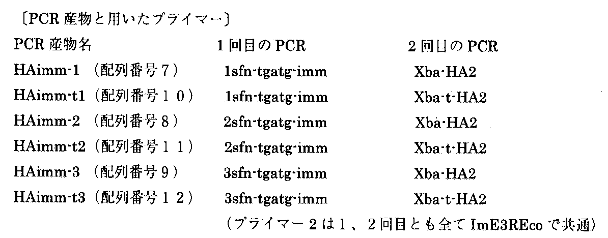
(1)コリシンE3およびイミュニティE3遺伝子(配列番号14)を有するプラスミドpSH350((1)Toba,M., Masaki,H., & Ohta,T.; Primary structure of the ColE2−P9,and ColE3−CA38 lysis genes. J. Biochem., 99,(2),591−596,1986. (2)Akutsu,A., etal. J. Bacteriol., 171, 6430−6436, 1989.)を鋳型として、下記の2回のPCRによって、所定のDNA断片を得た。このPCRにおいては、1sfn−tgatg−imm、2sfn−tgatg−imm、3sfn−tgatg−imm、ImE3REco、Xba−HA2、Xba−t−HA2をそれぞれ合成し、プライマーあるいは鋳型として用いた。これらの塩基配列を、ぞれぞれ、順に配列番号1、2、3、4、5、6として配列表に示した。以下の表1に1回目のPCR条件を示す。
【0035】
【表1】
表2に2回目のPCR条件を示す。
【0036】
【表2】
【0037】
【表3】
次に、上記で得られた6種類のPCR産物をXba IとEco R Iで消化し、pUC19の対応する制限酵素部位にリガーゼを用いて連結した後、大腸菌XLI−Blue MRF’に形質転換した。6種類のそれぞれの形質転換体よりプラスミドを回収し、LI−COR社製Model 4200L DNAシークエンサーを用いて、プラスミドに挿入されたDNA断片の配列を確認した。図2は、上記の結果得られた6種のHAimmシリーズのDNA断片の構造をその塩基配列中に示した図である。また、6種の挿入DNAの構造の模式図を図3に示す。この第3図中、Xba I、Sfi IおよびEcoR Iは、それぞれの制限酵素で切断される配列であり(第2図にその塩基配列が示されている。)、このXba IとSfi Iは外来遺伝子の挿入部位として設けられている。
【0039】
また、第3図中HAはHA tagをコードする配列(第2図中のHA tag領域)を、下線部はイミュニティーE3遺伝子(第2図中、ImmE3領域)の翻訳開始コドンを示す。また、これらの挿入断片は、プラスミドベクターに由来するプロモーターおよび翻訳開始シグナルにより、転写と翻訳によって発現されるが、太文字部は、それぞれのプラスミド上の翻訳読み枠における終止コドン(tga)示す。これら6種の挿入DNA 断片はいずれも終止コドンとして各々を読み枠をずらした終止コドンを3個配置している。また、HAimm−1、HAimm−t1、HAimm−2およびHAimm−t2に存在するccあるいはcは、終止コドン中翻訳読み枠をずらすための付加的な塩基配列である。また、HAimm−t1 、HAimm−t2 、 HAimm−t3のXba IとSfiIとの間に塩基配列中、太字で示したtaaは、終止コドンであ
【0040】
(2)0.1%グルコースあるいは0.2 mM IPTG(isopropyl−b−D−thiogalactopyranoside)あるいはそのいずれも含まない3種類のLB寒天培地を作製し、それぞれに、図4Aに示されたように中央に縦線状にコリシンE3タンパク質を塗布した。この3種類の寒天培地のそれぞれに、図4Aに示されたように各形質転換体を横線上に左から右に向けて塗布した。また、これらの形質転換体以外に、イミュニティーE3遺伝子を保持しないプラスミド(gal4)、およびイミュニティーE3遺伝子が常に発現するように構築されたプラスミド(ImE3)の各形質転換体をコントロールとして、上記の形質転換体と同様に塗布した。
【0041】
上記の形質転換体を塗布した寒天培地を37℃で一昼夜インキュベートし、縦線上に塗布されたコリシンE3タンパク質の両側の各形質転換体の生育を観察した。この結果を図4に示す。イミュニティーE3遺伝子が恒常的に発現しているプラスミド(ImE3)を有する形質転換体は、いずれのプレートにおいても塗布された全領域にわたって旺盛に生育していた。また、イミュニティーE3遺伝子を持たないプラスミド(gal4)を有する形質転換体は、コリシンE3が塗布された領域よりも右側にはほとんど生育が見られなかった。
【0042】
一方、HAimmシリーズのプラスミドを有する各形質転換体については、グルコース、IPTGの存在下・非存在下に関らず、いずれの場合にも、Xba IとSfi Iの間に、プラスミドの読み枠と一致する翻訳終止コドン(taa)が存在しない場合(HAimm−1、HAimm−2およびHAimm−3)の方が、存在する場合(HAimm−t1、HAimm−t2およびHAimm−t3)に比較して、その生育が明確に良好であった。特に、IPTGによって転写を誘導した場合においては、この翻訳終止コドンが存在しない場合にはイミュニティーE3遺伝子を恒常的に発現するプラスミドを保持する形質転換体と同等の生育を示したのに対して、この翻訳終止コドン(taa)が存在する場合にはほとんど生育が見られなかった。
【0043】
以上により、本発明によって製作されたプラスミドは、Xba IおよびSfi Iサイトをクローニング部位として用いることにより、この間に挿入されたDNA断片の全長が翻訳された場合にのみ、生育が可能であることが示された。
【0044】
【実施例2】
pHN301プラスミドを鋳型として、3Sfn−tgatg−imm(配列番号3)とImE3NotEco(配列番号15)のプライマーペアーを用いてPCRを行った。次に、このPCR産物を20倍希釈して鋳型とし、SfiTAA(配列番号16)とImE3NotEco(配列番号15)のプライマーペアーを用いてPCRを行った。さらに、このPCR産物を20倍希釈して鋳型とし、BamSfi(配列番号17)とImE3NotEco(配列番号15)のプライマーペアーを用いてPCRを行った。上記の合計3回のPCRにより、ImE3Sfi2の配列(配列番号18)よりなるDNA断片を得た。なお、3回のPCRは全て下記表4に示す温度条件を用いた。
【0045】
【表4】
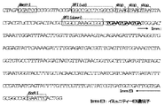
得られたImE3Sfi2(配列番号18)断片の構造を図5に示す。ImE3Sfi2は、その両末端側にBamHIとEcoRIの認識部位を有し、BamHIサイトの下流側に外来遺伝子の挿入部位である2つのSfiI認識部位を有し、この間にそれぞれ読み枠をずらした3個の終止コドン(TAA)を配置している。2個目のSfiIの下流側は3個の終止コドン(TGA)を介してイミュニテイ遺伝子(Immunity E3)に連結している。3番目の終止コドンとイミュニテイ遺伝子(Immunity E3)の開始コドン(ATG)は読み枠をずらせて重複して配置されている。
【0047】
このDNA断片をBamHIとEcoRIで消化し、pUC19の対応する制限酵素部位にリガーゼを用いて連結した後、大腸菌XLI−Blue MRF’に形質転換した。形質転換体よりプラスミドを回収し、LI−COR社製Model 4200L DNAシークエンサーを用いて、プラスミドに挿入されたDNA断片の配列を確認した。こうして得られた正しい塩基配列を有するプラスミドをpTLCと命名した。このプラスミドpTLCの構造を図6に示す。図中の記号は図5と同様である。
【0048】
次に、このベクターの評価用の挿入断片として、アビジン遺伝子を有するDNA断片であって、配列中に停止コドンを有しないAvd1Sfi2(配列番号19)、Avd2Sfi2(配列番号20)、Avd3Sfi2(配列番号21)、および停止コドンを有するAvd1Sfi2−t(配列番号22)、Avd2Sfi2−t(配列番号23)、Avd3Sfi2−t(配列番号24)を作製した。これらAvdSfiシリーズの構造を、図7に示す。すなわち図7中、Sfi IはpTLCベクターに挿入するための制限酵素切断部位を、TAAは挿入断片中で翻訳が集結するように人為的に挿入された終止コドンを、−、C、CCはアビジン遺伝子の翻訳がpTLCベクターに継続される場合に、翻訳が異なる読み枠となるようにするための挿入塩基を示す。以上より、Avd1Sfi2はpTLCベクターに継続される翻訳読み枠が1番目の挿入断片を、Avd1Sfi2−tはpTLCベクターに継続される翻訳読み枠が1番目で上記の人為的終止コドンを有する挿入断片を、Avd2Sfi2はpTLCベクターに継続される翻訳読み枠が2番目の挿入断片を、Avd2Sfi2−tはpTLCベクターに継続される翻訳読み枠が2番目で上記の人為的終止コドンを有する挿入断片を、Avd3Sfi2はpTLCベクターに継続される翻訳読み枠が3番目の挿入断片を、Avd3Sfi2−tはpTLCベクターに継続される翻訳読み枠が3番目で上記の人為的終止コドンを有する挿入断片である。
【0049】
これらの各断片を、pTLCベクターの対応する制限酵素部位にリガーゼを用いて連結した後、Bio−Rad社 Gene Pulserを用いてエレクトロポレーション法によって大腸菌XLI−Blue MRF’に形質転換した。エレクトロポレーション後に、定法通り直ちにSOC培地を添加して37℃で1時間静置した後、最終濃度が1 mMとなるようにIPTGを添加して、さらに37℃で6時間静置した。この形質転換体懸濁液に、最終濃度が1 mg/mlとなるようにコリシンE3を添加し、各形質転換体のそれぞれ等量づつを100 mg/mlのアンピシリンと0.2 mM IPTGを含むLB寒天培地に塗布した。また、対照として、コリシンE3を添加しないで同様の操作を行った。この操作により、6種類のプラスミドによる形質転換体のそれぞれについて、コリシンE3と添加した場合としない場合の2種類、合計12種類の寒天培地による培養の結果を得た。さらに、外来断片を挿入していないpTLCベクターそのものの形質転換体を調製し、コリシンE3を線上に縦方向に塗布した寒天培地上に、これらの形質転換体を横方向に線上に塗布した。図中の1、12、16、47、48はこれらの形質転換体を表す。また、gal4とimmE3は、実施例1におけるgal4とimmE3と同じ形質転換体を表す。この寒天培地を37℃で一昼夜保温し、生育してきたコロニーを観察したところ、図8の結果を得た。これにより、Avd1Sfi2、Avd2Sfi2、Avd3Sfi2を保持する形質転換体は、Avd1Sfi2−t、Avd2Sfi2−t、Avd3Sfi2−tを保持する形質転換体に比較して10〜100倍の数のコロニーを形成した。また、DNA断片を挿入しないpTLCベクターは、同様の操作によってコロニーを形成しなかった。以上より、pTLCベクターは、挿入断片の全長が翻訳された場合にのみ、生育可能な形質転換体を得られることが証明された。
【0050】
【発明の効果】
本発明は、特に発現ライブラリーの構築および利用について、翻訳の方向あるいは読み枠が正しくないことによって生じる自然界に存在しないあるいは発現が好ましくないアミノ酸配列を有するペプチド、ポリペプチドあるいはタンパク質を生産する宿主を排除することにより、正しい生理活性を有しない遺伝子のスクリーニングあるいは偽陽性のバックグラウンドが増大することを防止する。また、指示物質として宿主に対する致死物質、およびマーカー遺伝子として致死物質に対する抵抗遺伝子を用いれば、外来遺伝子断片が挿入されているクローンのみを特異的に生育させることが可能であることから、極めて効率的にスクリーニングを行うことができる。
【0051】
【配列表】
さらに、本発明によれば、試験管内であってもトランスレーショナル・カップリングが起こる系であれば利用することが可能であり、挿入遺伝子断片を保持する単一種あるいは複数種のベクター、DNAあるいはRNAを容器あるいはカプセルなどで隔離することによって、多数の挿入遺伝子断片を保持する単一あるいは複数のベクター、DNAあるいはRNAについて、一斉にスクリーニングを行うことができる。
【図面の簡単な説明】
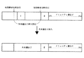
【図1】マーカ遺伝子としてコリシンに対するイミュニティー遺伝子を用いて、本発明のDNAマーカー材料を構築した場合および外来遺伝子を挿入した場合の各構造を示す概念図である。
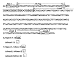
【図2】実施例1において作製したHAimmシリーズの各DNA断片の構造を示す図である。
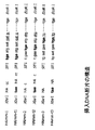
【図3】実施例1において作製した挿入DNA断片の構造を示す模式図である。
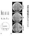
【図4】実施例1において作製された6種の挿入DNA断片を保持する形質転換体のコリシンによる耐性を他の断片と比較して示した培養試験の結果を示す写真である。
【図5】実施例2において作製されたImE3Sfi2の構造を示す図である
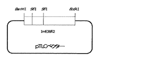
【図6】実施例2において作製されたpTLCベクターの構造を示す図である。
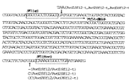
【図7】実施例2において作製されたAvdSfiシリーズの各DNA断片の構造を示す図である。
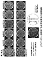
【図8】実施例2において作製されたAvd1Sfi2、Avd2Sfi2、Avd3Sfi2、Avd1Sfi2−t、Avd2Sfi2−t、Avd3Sfi2−tを挿入したpTLCベクターを保持する形質転換体の増殖結果を表す図である。[0001]
TECHNICAL FIELD OF THE INVENTION
The present invention relates to a DNA or RNA fragment for selectively screening only a recombinant in which a foreign inserted gene is correctly translated, a vector having these fragments, and a screening reagent kit containing at least these fragments or vectors.
[0002]
[Prior art]
For example, in a conventional vector for transforming Escherichia coli or the like, a method using a β-galactosidase gene as a marker gene for screening a transformant into which a foreign inserted gene has been inserted is known. In this method, the color of the colony grown on the medium is colored blue due to decomposition of the indicator by the expressed galactosidase, but the expression of β-galactosidase is suppressed by the insertion of the foreign gene, indicating that the color is not colored blue. (Non-Patent Document 1).
[0003]
Further, by using a lethal gene of Escherichia coli such as topoisomerase or colicin E1 gene and inserting a foreign DNA into the translation region, the expression of the gene is suppressed, and only a clone holding the foreign DNA is selectively grown. A method is also known (Patent Document 1).
However, in the selection by color development using β-galactosidase or the like, it is not only necessary to add a color-forming substance such as X-Gal (5-bromo-4-chloro-3-indolyl-beta-D-galactopyranoside) to the medium. Since a transformant that does not retain the inserted fragment also grows, a large area of an agar medium was required to separate many transformants. On the other hand, when a lethal gene is used, the transformant that does not carry the insert is killed, so that the area of the medium for isolating the transformant can be reduced, or selection using a liquid medium is possible. .
[0004]
On the other hand, when analyzing the function of a gene, it is necessary to analyze the properties and activities of the expression product (protein) of the gene, or to analyze the change in the properties of the host when the gene product is expressed. For this purpose, so-called expression vectors capable of expressing proteins are often used. In addition, for example, in order to analyze the functions of a large number of genes, it is common to fragment DNA containing a large number of genes such as genomic DNA and insert it into a vector to prepare a library. Is inserted, or a clone in which a DNA fragment is inserted in a reading frame different from the original reading frame occurs at a probability of 5/6 even in a region where a gene is encoded. In such a clone, the original long-chain peptide is not synthesized, but translation proceeds until the first translation stop codon (a stop codon generated by misalignment of the reading frame) appears, and a peptide that does not exist in nature is synthesized. become. These peptides often have unexpected biochemical and physiological activities, which can prevent genes without the correct bioactivity from being screened or cause an increase in false positive background. This has led to unsuccessful screening and reduced efficiency.
[0005]
[Non-Patent Document 1] Sambrook, J. et al. et al. (1989) Molecular Cloning: a laboratory manual. second edition. Cold Spring Harbor Laboratory Press, Cold Spring Harbor, NY
[Patent Document 1] JP-A-57-139095
[0006]
[Problems to be solved by the invention]
An object of the present invention is to solve the above-mentioned problems of the prior art, and in a recombinant carrying a foreign inserted gene fragment, the orientation and reading frame of the peptide, polypeptide or protein encoded by the foreign inserted gene fragment. Only provides a means that can efficiently screen only those recombinants that can be correctly translated, thereby making it possible to suppress the expression of peptides that are not originally produced in vivo. Further, when screening using the functions of expressed peptides, polypeptides or proteins, it is intended to eliminate as much as possible the possibility of selecting genes that do not exist or are not expected in nature. Another object of the present invention is to provide a means for reducing the number of independent transformants constituting a library required for screening by eliminating unnecessary clones.
[0007]
[Means for Solving the Problems]
The present inventors have conducted intensive studies in order to solve the above-mentioned problems, and have applied so-called translational coupling to express a marker gene only when translation of a foreign gene to be inserted is correctly performed, thereby achieving the object. Thus, the present inventors have found that only a recombinant in which a foreign gene is properly retained can be screened extremely efficiently, and have completed the present invention.
[0008]
That is, the present invention relates to the following (1) to (10).
(1) A DNA having a marker gene and a foreign gene insertion site upstream of the marker gene, wherein the marker gene is translated by translational coupling depending on the translation of the full length of the foreign gene to be inserted. A DNA marker material, characterized in that:
(2) The DNA marker material according to (1), wherein a translation stop codon is arranged downstream of the foreign gene insertion site.
(3) The DNA marker material according to (2), wherein the DNA marker material has three or more translation termination stop codons, and each of the termination codons is arranged so as to shift the reading frame.
(4) The foreign gene insertion site is composed of two identical or different restriction enzyme cleavage sites, has a translation termination codon in the nucleotide sequence between the restriction enzyme cleavage sites, and The DNA marker material according to any one of (1) to (3), wherein the translation termination codon is removed by cutting and inserting a foreign DNA.
(5) that the marker gene is a gene for supplying nutrients essential for the growth of the host, or a gene imparting a property of neutralizing, antagonizing or inactivating the action of a host growth inhibitor. The DNA marker material according to any one of (1) to (4).
(6) The DNA marker material according to (1) to (5), wherein the marker gene is an immunity gene for colicin.
(7) The DNA marker material according to (1) to (6), wherein the marker gene has a translation initiation codon and a translation termination codon.
(8) A vector comprising the nucleotide sequence of the DNA marker material according to any one of (1) to (7).
(9) An RNA having a base sequence corresponding to the base sequence of the DNA marker material according to any one of (1) to (7) or the vector according to (8).
(10) A recombinant screening kit comprising the DNA according to any one of (1) to (7) or the vector according to claim 8, and an indicator identifying the expression of a marker gene contained therein. .
[0009]
BEST MODE FOR CARRYING OUT THE INVENTION
Hereinafter, the present invention will be described in more detail.
The present invention applies translational coupling, and expresses a marker gene only when translation is performed without terminating over the entire length of a foreign gene to be inserted. The present invention relates to a DNA marker material designed so that only a recombinant can be screened or a vector having a base sequence of the DNA marker material.
[0010]
Translational coupling is a phenomenon in which, when two genes are close to each other, a gene located on the upstream side is translated, so that a gene on the downstream side is also translated. When the translation level of the upstream gene decreases, the translation level of the downstream gene also decreases accordingly. At this time, the translations of two adjacent genes are linked, and are said to be translationally coupled.
[0011]
To date, there have been vectors for expressing proteins encoded by foreign gene fragments by using translational coupling (Sergey, V. et al., Gene, 88, 121-126, 1990). ). However, these are originally incorporated into a vector, and are intended to express a protein encoded by a DNA fragment inserted downstream thereof by translation of a gene located upstream of the translational coupling. Yes, it is basically the same as the case of expressing it by fusing it with a peptide or protein encoded on a vector, such as a conventional His-tag or GST (Glutation-S-Transferase). The translation reading frame is specifically determined by the insertion position.
[0012]
As described above, the above-described vector using translational coupling has been used only for simply expressing the protein encoded by the inserted DNA fragment. Couplings are the first to be used for recombinant screening and are very innovative and very practical.
[0013]
The DNA marker material of the present invention has a foreign gene insertion site upstream of the marker gene, and is inserted only when the foreign gene to be inserted is correctly inserted in the direction and reading frame, and the entire gene is translated into a peptide. It is designed so that the translation of the marker gene into the polypeptide is achieved by translational coupling and expressed. Therefore, a stop codon (TAA, TAG, TGA) is placed downstream of the foreign gene and near the translation start point of the marker gene, and when the foreign gene is inserted, the foreign gene and the marker gene are linked via the stop codon. The connection is designed so that the translation of the marker gene is performed only when the translation of the foreign gene is performed near the translation start point of the marker gene.
[0014]
In the present invention, such a DNA marker material is prepared in advance, and inserted into a vector such as a plasmid to obtain a recombinant vector, and a foreign gene may be introduced into the recombinant vector. Alternatively, a recombinant vector may be prepared by directly preparing on the vector the nucleotide sequence of the above DNA marker material without introducing the step of inserting the vector into the vector, and introducing a foreign gene into the vector.
[0015]
The recombinant vector thus obtained is introduced into an appropriate host, and at this time, a foreign gene introduced so as not to match the reading frame of the vector or a gene introduced in the opposite direction is used. In the translation of a foreign gene, there is a high possibility that a stop codon is generated in the nucleotide sequence of the foreign gene due to the shift of the reading frame or the reverse translation of the nucleotide sequence. For example, for a DNA fragment of several hundred bp that encodes a functional domain, a 501 bp DNA fragment theoretically encodes 167 amino acids, but the probability of encoding a stop codon among all codons is 3/64. = Approximately 0.0469, so 167 × 0.0469 = approximately 7.8, resulting in an average of approximately 8 stop codons. Thus, when the foreign gene is introduced in a manner that does not match the reading frame of the vector or is introduced in the reverse direction, translation will almost certainly stop in the foreign insert and translation will stop at this false stop codon. And translation of the foreign gene near the translation start point of the marker gene is not performed, so that translational coupling does not occur and translation / expression of the marker gene does not occur. On the other hand, when the foreign gene is correctly introduced in both the reading frame and the orientation, the entire length of the inserted foreign gene is translated to the stop codon provided near the translation start point of the marker gene. Translation continues, translational coupling occurs, and the marker gene is expressed. Examples of these relationships will be described below.
[0016]
[If the reading frame is correct]
Embedded image
[If the reading frame is misaligned]
Embedded image
[When a foreign gene is inserted in the reverse direction]
Embedded image
The marker gene may be any gene that causes a discernible change in the growth, proliferation, coloring, luminescence, chemotaxis, chemotacticity, etc. of the host depending on the presence or absence of the expression in the presence or absence of the indicator. However, the expression of the marker gene may be observed only when the foreign gene has a high probability of being correctly inserted in the reading frame and direction, and thus, only when the foreign gene has been correctly inserted in the reading frame and direction. Screening becomes possible.
[0020]
In the above example, the stop codon provided near the translation start point of the marker gene terminates translation when the full length of the foreign gene to be inserted is translated, so this stop codon is used as a reading frame for the foreign gene. The length of the foreign gene to be inserted is clear, and if the reading frame of the stop codon does not match the reading frame of the foreign gene, an additional A new base sequence is added upstream of the stop codon. However, in order to eliminate such an operation, it is preferable to provide three or more stop codons whose reading frames are shifted.
[0021]
In addition, these stop codons are provided so as not to add an extra amino acid sequence in the translation polypeptide of the foreign gene, and the addition of a nucleic acid to match the reading frame changes the structure of the expression polypeptide of the foreign gene. It is preferable that the amount be as small as possible so as not to occur.
Furthermore, the location of these stop codons may be on the downstream side of the foreign gene, and may be either upstream or downstream of the translation start point of the marker gene. The distance between the translation initiation codon (ATG) of the marker gene and the translation stop codon located in the vicinity of the translation gene is not particularly limited as long as the translation of the marker gene is translationally coupled with the translation of the foreign inserted gene fragment. However, when the translation termination codon is located upstream of the translation initiation codon (ATG), it is preferably at most 45 bp, more preferably at most 20 bp, even more preferably at most 10 bp. When the translation termination codon is located downstream of the translation initiation codon (ATG), it is preferably at most 1,000 bp, more preferably at most 200 bp, even more preferably at most 40 bp. If the translation stop codon is located downstream of the translation start codon, the stop codon must not terminate the translation of the marker gene. For this purpose, for example, as shown below, the stop codon is designed to be in an open reading frame different from the translational reading of the marker gene, and the insertion of the stop codon reduces the amino acid in the expressed protein of the marker gene. It is preferable to design so as not to change.
[0023]
Embedded image
[0024]
In this case, if the amino acid is not important for the activity of the expressed protein of the marker gene, the amino acid may be changed by inserting a stop codon. Further, the translation stop codon and the translation initiation codon of the marker gene may overlap in their sequence.
There is no particular limitation on the design of the insertion site of the foreign gene, and the insertion site is designed to be cleaved by a restriction enzyme as in the past. In the DNA of the present invention or a vector having the same, it is preferable to provide a site that is recognized and cleaved by two identical or different restriction enzymes, and a termination codon is provided in the nucleotide sequence between the restriction enzyme cleavage sites. This stop codon is provided so as to match the reading frame of the plasmid, and when a foreign gene is introduced, the nucleotide sequence between these two cleavage sites is removed, and the stop codon is removed.
[0025]
Therefore, when a foreign gene is not inserted due to the arrangement of the stop codon, translation from this upstream is terminated at this stop codon, and translation is not performed near the translation start site of the marker gene. No translational coupling occurs, and no translation or expression of the marker gene is performed. Therefore, a recombinant having a vector into which the foreign gene has not been inserted can also be identified, and it can be determined whether the full length of the foreign gene is a translatable region.
Further, in the present invention, a gene set comprising at least the above-mentioned DNA marker material or a vector having the base sequence of the marker material, and an indicator for identifying the presence or absence of the marker gene in the marker material or the vector. A recombinant screening kit is also provided.
[0026]
Hereinafter, the present invention will be described in more detail with reference to FIG. 1 by taking as an example the case where colicin, which is a toxic protein to Escherichia coli, is used as an indicator and an immunity gene that neutralizes the toxicity of colicin is used as a marker gene.
In the present invention, for example, a plasmid is used as a vector for retaining a foreign gene fragment in Escherichia coli, and an immunity gene is inserted into this plasmid. This immunity gene is located downstream of the expected transcription / translation direction of the foreign gene insert. Further, the immunity gene has a translation initiation codon, and one or more translation termination codons (II) are arranged near the translation initiation codon. Two restriction enzyme recognition sites are located upstream of the stop codon (II), and are used as foreign gene insertion sites. A stop codon (I) is arranged between the two restriction enzyme recognition sites in accordance with the translational reading frame of the plasmid. When a foreign gene fragment is inserted, the stop codon (I) is deleted.
[0027]
The peptide, polypeptide or protein encoded by the foreign gene insert is located upstream of the insert and is internal to the plasmid, or is expressed by a promoter and translation initiation signal carried by the insert itself. Further, when the direction of the foreign gene fragment and the reading frame and direction of translation are correctly recognized, and the translation is performed correctly or as expected in the same manner as in the natural world, the displacement or insertion direction of the reading frame is determined as described above. Since no false stop codon is generated due to the above error, translation continues in the inserted gene fragment to the stop codon (II) located near the translation start site of the marker gene, and translation coupling is not performed. The immunity gene is translated and expressed. When a foreign gene is not inserted, translation is terminated by the stop codon (I), translation does not reach the stop codon (II), and translation / expression of the immunity gene does not occur.
[0028]
However, the foreign gene fragment does not have a translation termination codon provided in the original gene as in the case of full-length cDNA, but a fragmented DNA fragment capable of encoding a functional domain or epitope of a protein is preferably used. The length of the fragmented DNA is not particularly limited, but is preferably 9 to 3000 bp, more preferably 45 to 1500 bp, and further preferably 200 to 1000 bp. The DNA material used for fragmentation may be genomic DNA or cDNA. As a method for fragmentation, any method generally used may be used, and mechanical shredding by passing through a narrow tube at high speed, ultrasonic waves, DNase I, restriction enzymes and the like can be used. In addition, artificially designed synthetic DNA fragments and artificially introduced DNA fragments can also be used.
[0029]
The number of translation stop codons located in the vicinity of the translation start codon of the immunity gene is preferably three as described above. Each stop codon is such that translation is terminated for all three reading frames. Are arranged with the reading frame shifted. However, if the number of reading frames when correctly translated is limited to one or two, the object can be achieved with one or two. Although it is possible to arrange more than three translation stop codons, three translation stop codons function effectively in terms of translation.
[0030]
As described above, regarding the distance between the translation initiation codon (ATG) of the immunity gene and the translation termination codon (II) located in the vicinity thereof, the translation of the immunity gene and the translation of the foreign inserted gene fragment are linked (trans There is no particular limitation, as long as the translation stop codon (II) is located upstream of the translation initiation codon (ATG). Is not more than 20 bp, more preferably not more than 10 bp. In the above description, the case where the translation stop codon (II) is arranged upstream of the translation start codon (ATG) has been described. However, in the present invention, as described above, the translation stop codon is replaced with the translation start codon (ATG). ATG), the distance between the translation initiation codon (ATG) and the translation termination codon (II) is preferably 1000 bp or less, more preferably 200 bp or less. More preferably, it is 40 bp or less, and the translation of the immunity gene and the translation of the foreign inserted gene fragment need to be linked (translationally coupled). When the translation stop codon is downstream of the translation start codon, the stop codon must not be rendered inactive by terminating translation of the immunity protein, and the means for that are as described above. is there.
[0031]
In the present invention, when a gene recombination operation using a foreign gene is performed using a DNA marker material using the immunity gene for colicin or a plasmid having the base sequence of the marker as a marker gene, the culture medium containing colicin as a selection medium is used. Use medium.
Thus, Escherichia coli having a plasmid or vector into which a foreign gene has not been introduced, Escherichia coli having a plasmid or vector whose reading frame of the foreign gene does not match the original one, or a plasmid or vector in which the insertion of the foreign gene is reversed , The immunity gene cannot be expressed and is killed by colicin, a toxic protein for E. coli. Therefore, Escherichia coli grown in this colicin-containing medium is one in which the foreign gene has been correctly introduced and expresses the original polypeptide, and by collecting this, it is possible to perform screening very efficiently. .
[0032]
In this example, a gene that neutralizes the lethality of a protein that is lethal to Escherichia coli is used as a marker gene. However, it is not necessary to use a protein that is lethal to Escherichia coli. Does not need to be added to the medium and no specific conditions need to be set. The marker gene may be any gene that can be distinguished from a state in which the gene is not expressed, such as a gene whose expression is detected or observed, and the growth rate of a host that expresses the gene preferably expresses the gene. It is a gene that can be significantly improved as compared to non-hosts. The marker gene used in the present invention includes, for example, a resistance gene (drug resistance marker) for antibiotics such as ampicillin, kanamycin, tetracycline, chloramphenicol, and a synthetic system for amino acids such as methionine, arginine, histidine, tryptophan, uracil, and the like. Inactive essential genes related to the mutant (auxotrophic marker), and positive selection markers such as lactose and galactose-related genes for sugar assimilation are preferably used, but β-galactosidase gene A gene for an enzyme that catalyzes a color reaction by using an appropriate substrate, a gene such as luciferase that causes coloration or luminescence by adding an appropriate substrate, and green fluorescent protein (GFP) that emits fluorescence. Gene, and the like.
[0033]
Further, the present invention is not limited to Escherichia coli, but can be used for any species as long as the above marker gene can be used.
Further, the present invention is not limited to the screening using the organism as described above, and any system may be used as long as translational coupling occurs even in a test tube. For example, when a translation system similar to that in a living cell is constructed in a test tube, such as an in vitro transcription / translation system using Escherichia coli and phage, it is presumed that translational coupling occurs in the same manner as in a living cell. The present invention can be used in such a case. In this case, depending on the presence or absence of expression of the protein from the marker gene, the foreign inserted gene fragment is correctly inserted and its full-length translated vector or foreign gene holding vector, DNA or RNA does not express the vector or foreign gene. It is only necessary that the vector can be distinguished from a holding vector, DNA or RNA in a test tube. For example, a marker gene, for example, green fluorescent protein (GFP) that emits fluorescence or a suitable substrate is added to generate color or luminescence. A gene encoding a protein such as luciferase that causes the luciferase can be used. When carried out in a test tube, a single or plural types of vectors, DNAs or RNAs that retain or do not retain the inserted gene fragment are isolated in a container or a capsule to retain a large number of inserted gene fragments or Screening can be performed simultaneously for single or multiple vectors, DNAs or RNAs that do not retain them. The vector and the inserted gene fragment may be DNA or RNA, and are prepared by appropriate means such as chemical synthesis, PCR, transcription, and extraction from cells. However, when using RNA, it is necessary to allow a reverse transcription system to coexist with the transcription / translation system.
Examples of the present invention will be described below, but the present invention is not limited to the examples.
[0034]
(1) Plasmid pSH350 having colicin E3 and immunity E3 gene (SEQ ID NO: 14) ((1) Toba, M., Masaki, H., & Ohta, T .; Primary structure of the ColE2-P9, and ColE3-CA38) lysgenes. J. Biochem., 99, (2), 591-596, 1986. (2) Using Akutsu, A., et al. J. Bacteriol., 171, 6430-6436, 1989.) as a template, A predetermined DNA fragment was obtained by two PCRs. In this PCR, 1sfn-tgatg-imm, 2sfn-tgatg-imm, 3sfn-tgatg-imm, ImE3REco, Xba-HA2, and Xba-t-HA2 were synthesized and used as primers or templates. These base sequences are shown in the sequence listing as SEQ ID NOs: 1, 2, 3, 4, 5, and 6, respectively. Table 1 below shows the first PCR conditions.
[0035]
[Table 1]
Table 2 shows the second PCR conditions.
[0036]
[Table 2]
[0037]
[Table 3]
Next, the six PCR products obtained above were digested with XbaI and EcoRI, ligated to the corresponding restriction enzyme sites of pUC19 using ligase, and then transformed into Escherichia coli XLI-Blue MRF '. . Plasmids were recovered from each of the six transformants, and the sequence of the DNA fragment inserted into the plasmid was confirmed using a Model 4200L DNA sequencer manufactured by LI-COR. FIG. 2 is a diagram showing the structures of the six types of DNA fragments of the HAimm series obtained as described above in their base sequences. FIG. 3 shows a schematic diagram of the structure of the six types of inserted DNA. In FIG. 3, Xba I, Sfi I and EcoR I are sequences that are cleaved by respective restriction enzymes (the base sequences are shown in FIG. 2), and Xba I and Sfi I are It is provided as a foreign gene insertion site.
[0039]
HA in FIG. 3 indicates the sequence encoding the HA tag (HA tag region in FIG. 2), and the underlined portion indicates the translation initiation codon of the immunity E3 gene (ImmE3 region in FIG. 2). In addition, these inserts are expressed by transcription and translation by a promoter and translation initiation signal derived from a plasmid vector, and the bold letters indicate the stop codon (tga) in the translational reading frame on each plasmid. In each of these six types of inserted DNA fragments, three stop codons whose reading frames are shifted from each other are arranged as stop codons. In addition, cc or c present in HAimm-1, HAimm-t1, HAimm-2 and HAimm-t2 is an additional base sequence for shifting the translational reading frame in the stop codon. In the base sequence between XbaI and SfiI of HAimm-t1, HAimm-t2, and HAimm-t3, taa shown in bold is a termination codon.
[0040]
(2) Three types of LB agar media containing neither 0.1% glucose nor 0.2 mM IPTG (isopropyl-b-D-thiogalactopyranoside) or any of them were prepared, and as shown in FIG. 4A, The colicin E3 protein was applied to the center in a vertical line. As shown in FIG. 4A, each transformant was spread on each of the three types of agar media on a horizontal line from left to right. In addition, in addition to these transformants, a plasmid (gal4) which does not carry the immunity E3 gene and a plasmid (ImE3) which was constructed so that the immunity E3 gene was always expressed were used as controls to obtain the above-mentioned transformants. Coating was carried out in the same manner as the converted body.
[0041]
The agar medium coated with the above transformant was incubated at 37 ° C. overnight, and the growth of each transformant on both sides of the colicin E3 protein applied on the vertical line was observed. The result is shown in FIG. The transformant having the plasmid (ImE3) in which the immunity E3 gene was constantly expressed grew vigorously over the entire area coated on each plate. The transformant having the plasmid (gal4) having no immunity E3 gene hardly grew on the right side of the region coated with colicin E3.
[0042]
On the other hand, for each transformant having the HAimm series plasmid, regardless of the presence or absence of glucose and IPTG, the reading frame of the plasmid was located between Xba I and Sfi I in each case. The absence of a matching translation stop codon (taa) (HAimm-1, HAimm-2, and HAimm-3) is greater than the presence (HAimm-t1, HAimm-t2, and HAimm-t3). Its growth was clearly good. In particular, when transcription was induced by IPTG, the absence of this translation termination codon resulted in growth equivalent to that of a transformant carrying a plasmid that constantly expressed the immunity E3 gene. When this translation termination codon (taa) was present, almost no growth was observed.
[0043]
As described above, the plasmid prepared according to the present invention can grow only when the full length of the DNA fragment inserted therebetween is translated by using the Xba I and Sfi I sites as cloning sites. Indicated.
[0044]
Using the pHN301 plasmid as a template, PCR was performed using a primer pair of 3Sfn-tgatg-imm (SEQ ID NO: 3) and ImE3NotEco (SEQ ID NO: 15). Next, this PCR product was diluted 20-fold as a template, and PCR was performed using a primer pair of SfiTAA (SEQ ID NO: 16) and ImE3NotEco (SEQ ID NO: 15). Further, this PCR product was diluted 20-fold as a template, and PCR was performed using a primer pair of BamSfi (SEQ ID NO: 17) and ImE3NotEco (SEQ ID NO: 15). A DNA fragment consisting of the sequence of ImE3Sfi2 (SEQ ID NO: 18) was obtained by the above three times of PCR. All three PCRs used the temperature conditions shown in Table 4 below.
[0045]
[Table 4]
The structure of the obtained ImE3Sfi2 (SEQ ID NO: 18) fragment is shown in FIG. ImE3Sfi2 has recognition sites for BamHI and EcoRI at both ends, and two SfiI recognition sites, which are insertion sites for foreign genes, downstream of the BamHI site, between which three reading frames are shifted. Is located at the stop codon (TAA). The downstream side of the second SfiI is linked to the immunity gene (Immunity E3) via three stop codons (TGA). The third stop codon and the start codon (ATG) of the immunity gene (Immunity E3) are arranged so as to shift the reading frame.
[0047]
This DNA fragment was digested with BamHI and EcoRI, ligated to the corresponding restriction enzyme site of pUC19 using ligase, and then transformed into E. coli XLI-Blue MRF '. The plasmid was recovered from the transformant, and the sequence of the DNA fragment inserted into the plasmid was confirmed using a Model 4200L DNA sequencer manufactured by LI-COR. The thus obtained plasmid having the correct nucleotide sequence was designated as pTLC. FIG. 6 shows the structure of this plasmid pTLC. The symbols in the figure are the same as in FIG.
[0048]
Next, as an insertion fragment for evaluation of this vector, a DNA fragment having an avidin gene and having no stop codon in its sequence, Avd1Sfi2 (SEQ ID NO: 19), Avd2Sfi2 (SEQ ID NO: 20), Avd3Sfi2 (SEQ ID NO: 21) ), And Avd1Sfi2-t (SEQ ID NO: 22), Avd2Sfi2-t (SEQ ID NO: 23), and Avd3Sfi2-t (SEQ ID NO: 24) having stop codons. The structure of the AvdSfi series is shown in FIG. That is, in FIG. 7, Sfi I is a restriction enzyme cleavage site for insertion into a pTLC vector, TAA is a stop codon artificially inserted so that translation is assembled in the inserted fragment,-, C and CC are avidin. Insertion bases that allow translation to be in a different reading frame when gene translation is continued into the pTLC vector. As described above, Avd1Sfi2 is the first insertion fragment having the translational reading frame continued in the pTLC vector, and Avd1Sfi2-t is the first translational reading frame having the artificial translation codon continued in the pTLC vector. , Avd2Sfi2 is an insertion fragment having the second translational reading frame continued to the pTLC vector, and Avd2Sfi2-t is an insertion fragment having the second translational reading frame continued to the pTLC vector and having the above artificial stop codon, Avd3Sfi2. Is the insert fragment with the third translational reading frame continued to the pTLC vector, and Avd3Sfi2-t is the insert fragment with the third translational reading frame continued to the pTLC vector and having the above artificial stop codon.
[0049]
Each of these fragments was ligated to the corresponding restriction enzyme site of the pTLC vector using ligase, and then transformed into Escherichia coli XLI-Blue MRF 'by electroporation using Bio-Rad Gene Pulser. After electroporation, an SOC medium was immediately added as usual, and the mixture was allowed to stand at 37 ° C for 1 hour. Then, IPTG was added to a final concentration of 1 mM, and the mixture was further allowed to stand at 37 ° C for 6 hours. To this transformant suspension, colicin E3 was added so that the final concentration was 1 mg / ml, and an equal amount of each transformant contained 100 mg / ml of ampicillin and 0.2 mM IPTG. It was spread on LB agar medium. As a control, the same operation was performed without adding colicin E3. By this operation, for each of the transformants with the six types of plasmids, the results of cultivation on a total of 12 types of agar medium, two types with and without the addition of colicin E3, were obtained. Further, transformants of the pTLC vector itself into which no foreign fragment had been inserted were prepared, and these transformants were laterally and linearly spread on an agar medium coated with colicin E3 vertically. 1, 12, 16, 47 and 48 in the figure represent these transformants. Gal4 and immE3 represent the same transformants as gal4 and immE3 in Example 1. The agar medium was kept warm at 37 ° C. for 24 hours, and the colonies that had grown were observed. The results shown in FIG. 8 were obtained. Thus, the transformants carrying Avd1Sfi2, Avd2Sfi2, and Avd3Sfi2 formed 10 to 100 times as many colonies as the transformants carrying Avd1Sfi2-t, Avd2Sfi2-t, and Avd3Sfi2-t. Also, the pTLC vector into which the DNA fragment was not inserted did not form a colony by the same operation. From the above, it was proved that a viable transformant could be obtained from the pTLC vector only when the full length of the inserted fragment was translated.
[0050]
【The invention's effect】
The present invention relates to a host that produces a peptide, polypeptide or protein having an amino acid sequence that is not present in nature or has poor expression due to incorrect translation direction or reading frame, particularly for construction and use of an expression library. Elimination prevents screening of genes that do not have the correct bioactivity or prevents an increase in false positive background. In addition, if a lethal substance against the host is used as the indicator substance and a resistance gene against the lethal substance is used as the marker gene, only the clone into which the foreign gene fragment has been inserted can be specifically grown, which is extremely efficient. Screening can be performed.
[0051]
[Sequence list]
Furthermore, according to the present invention, any system can be used as long as translational coupling occurs even in a test tube, and a single or plural types of vectors, DNAs, or DNAs holding inserted gene fragments can be used. By isolating the RNA in a container or a capsule, screening can be performed simultaneously for single or plural vectors, DNAs or RNAs holding a large number of inserted gene fragments.
[Brief description of the drawings]
FIG. 1 is a conceptual diagram showing the structure when a DNA marker material of the present invention is constructed using an immunity gene for colicin as a marker gene and when a foreign gene is inserted.
FIG. 2 is a view showing the structure of each DNA fragment of the HAimm series prepared in Example 1.
FIG. 3 is a schematic diagram showing the structure of an inserted DNA fragment prepared in Example 1.
FIG. 4 is a photograph showing the results of a culture test in which the transformant holding six types of inserted DNA fragments prepared in Example 1 was compared with other fragments for resistance to colicin.
FIG. 5 is a view showing the structure of ImE3Sfi2 produced in Example 2.
FIG. 6 is a view showing the structure of a pTLC vector prepared in Example 2.
FIG. 7 is a view showing the structure of each DNA fragment of the AvdSfi series produced in Example 2.
FIG. 8 is a diagram showing the growth results of a transformant harboring a pTLC vector into which Avd1Sfi2, Avd2Sfi2, Avd3Sfi2, Avd1Sfi2-t, Avd2Sfi2-t, and Avd3Sfi2-t inserted in Example 2 are inserted.
Claims (10)
Priority Applications (1)
| Application Number | Priority Date | Filing Date | Title |
|---|---|---|---|
| JP2003035786A JP4243680B2 (en) | 2003-02-13 | 2003-02-13 | Foreign insert selection marker |
Applications Claiming Priority (1)
| Application Number | Priority Date | Filing Date | Title |
|---|---|---|---|
| JP2003035786A JP4243680B2 (en) | 2003-02-13 | 2003-02-13 | Foreign insert selection marker |
Publications (2)
| Publication Number | Publication Date |
|---|---|
| JP2004242583A true JP2004242583A (en) | 2004-09-02 |
| JP4243680B2 JP4243680B2 (en) | 2009-03-25 |
Family
ID=33021105
Family Applications (1)
| Application Number | Title | Priority Date | Filing Date |
|---|---|---|---|
| JP2003035786A Expired - Lifetime JP4243680B2 (en) | 2003-02-13 | 2003-02-13 | Foreign insert selection marker |
Country Status (1)
| Country | Link |
|---|---|
| JP (1) | JP4243680B2 (en) |
Cited By (1)
| Publication number | Priority date | Publication date | Assignee | Title |
|---|---|---|---|---|
| EP2228456A1 (en) * | 2007-11-02 | 2010-09-15 | Geneart Ag | Selection of encoding nucleic acid constructs for absence of frameshift mutations |
Families Citing this family (3)
| Publication number | Priority date | Publication date | Assignee | Title |
|---|---|---|---|---|
| CN102304508B (en) * | 2011-07-29 | 2013-05-01 | 上海捷瑞生物工程有限公司 | Method for preparing DL2000 DNA molecular weight marker as well as product and applications thereof |
| KR20210045360A (en) | 2018-05-16 | 2021-04-26 | 신테고 코포레이션 | Methods and systems for guide RNA design and use |
| US11970733B2 (en) * | 2018-11-01 | 2024-04-30 | Synthego Corporation | Methods for analyzing nucleic acid sequences |
-
2003
- 2003-02-13 JP JP2003035786A patent/JP4243680B2/en not_active Expired - Lifetime
Cited By (2)
| Publication number | Priority date | Publication date | Assignee | Title |
|---|---|---|---|---|
| EP2228456A1 (en) * | 2007-11-02 | 2010-09-15 | Geneart Ag | Selection of encoding nucleic acid constructs for absence of frameshift mutations |
| US9115390B2 (en) | 2007-11-02 | 2015-08-25 | Geneart Ag | Method for determining frameshift mutations in coding nucleic acids |
Also Published As
| Publication number | Publication date |
|---|---|
| JP4243680B2 (en) | 2009-03-25 |
Similar Documents
| Publication | Publication Date | Title |
|---|---|---|
| JP2002526108A (en) | Fusion of scaffold protein with random peptide library | |
| EP2115144A1 (en) | Method of cloning at least one nucleic acid molecule of interest using type iis restriction endonucleases, and corresponding cloning vectors, kits and system using type iis restriction endonucleases | |
| WO2002061097A1 (en) | Cloning vectors and vector components | |
| US20230340481A1 (en) | Systems and methods for transposing cargo nucleotide sequences | |
| EP1051502B1 (en) | Highly efficient controlled expression of exogenous genes in e. coli | |
| US6867042B2 (en) | Method for determining and modifying protein/peptide solubility | |
| US9115390B2 (en) | Method for determining frameshift mutations in coding nucleic acids | |
| CN116478264B (en) | Recombinant chromoprotein, its preparation method and application | |
| JP2004242583A (en) | Foreign insert selection marker | |
| CN102382194A (en) | Autophagy concatenated fluorescent probe mTagRFP-mWasabi-LC3 and application thereof | |
| US7297482B2 (en) | Structurally biased random peptide libraries based on different scaffolds | |
| KR101677090B1 (en) | Polypeptide for purification of target protein and use thereof | |
| US9187752B2 (en) | Hybrid portable origin of replication plasmids | |
| US6936421B2 (en) | Structurally biased random peptide libraries based on different scaffolds | |
| US20250223576A1 (en) | Optimized cas protein and use thereof | |
| Huo et al. | Identifying amino acid overproducers using rare-codon-rich markers | |
| Rondot et al. | Epitopes fused to F-pilin are incorporated into functional recombinant pili | |
| JPWO2004053126A1 (en) | Low temperature inducible expression vector | |
| US10442842B2 (en) | Cleavage resistant photoluminescent proteins and applications thereof | |
| KR101067328B1 (en) | Expression vector for fission yeast, Gateway expression vector for fission yeast, and method for producing same | |
| JP4336770B2 (en) | Markers for selection of transformants using lethal genes | |
| US6632638B1 (en) | Enhanced solubility of recombinant proteins using Uracil DNA glycosylase inhibitor | |
| WO1999031266A1 (en) | Method for determining and modifying protein/peptide solubility | |
| JP2006508672A (en) | Optimized protein synthesis | |
| Almashanu et al. | Formation of active bacterial luciferase between interspecific subunits in vivo |
Legal Events
| Date | Code | Title | Description |
|---|---|---|---|
| A621 | Written request for application examination |
Free format text: JAPANESE INTERMEDIATE CODE: A621 Effective date: 20041124 |
|
| A131 | Notification of reasons for refusal |
Free format text: JAPANESE INTERMEDIATE CODE: A131 Effective date: 20071204 |
|
| A521 | Request for written amendment filed |
Free format text: JAPANESE INTERMEDIATE CODE: A523 Effective date: 20080131 |
|
| A02 | Decision of refusal |
Free format text: JAPANESE INTERMEDIATE CODE: A02 Effective date: 20080415 |
|
| A521 | Request for written amendment filed |
Free format text: JAPANESE INTERMEDIATE CODE: A523 Effective date: 20080613 |
|
| A911 | Transfer to examiner for re-examination before appeal (zenchi) |
Free format text: JAPANESE INTERMEDIATE CODE: A911 Effective date: 20080813 |
|
| A131 | Notification of reasons for refusal |
Free format text: JAPANESE INTERMEDIATE CODE: A131 Effective date: 20080902 |
|
| A521 | Request for written amendment filed |
Free format text: JAPANESE INTERMEDIATE CODE: A523 Effective date: 20081104 |
|
| TRDD | Decision of grant or rejection written | ||
| A01 | Written decision to grant a patent or to grant a registration (utility model) |
Free format text: JAPANESE INTERMEDIATE CODE: A01 Effective date: 20081209 |
|
| A01 | Written decision to grant a patent or to grant a registration (utility model) |
Free format text: JAPANESE INTERMEDIATE CODE: A01 |
|
| R150 | Certificate of patent or registration of utility model |
Ref document number: 4243680 Country of ref document: JP Free format text: JAPANESE INTERMEDIATE CODE: R150 Free format text: JAPANESE INTERMEDIATE CODE: R150 |
|
| S533 | Written request for registration of change of name |
Free format text: JAPANESE INTERMEDIATE CODE: R313533 |
|
| R350 | Written notification of registration of transfer |
Free format text: JAPANESE INTERMEDIATE CODE: R350 |
|
| EXPY | Cancellation because of completion of term |