JP2005293045A - Two-dimensional image processing system - Google Patents
Two-dimensional image processing system Download PDFInfo
- Publication number
- JP2005293045A JP2005293045A JP2004105048A JP2004105048A JP2005293045A JP 2005293045 A JP2005293045 A JP 2005293045A JP 2004105048 A JP2004105048 A JP 2004105048A JP 2004105048 A JP2004105048 A JP 2004105048A JP 2005293045 A JP2005293045 A JP 2005293045A
- Authority
- JP
- Japan
- Prior art keywords
- image
- spot
- dimensional
- point
- image processing
- Prior art date
- Legal status (The legal status is an assumption and is not a legal conclusion. Google has not performed a legal analysis and makes no representation as to the accuracy of the status listed.)
- Pending
Links
- 238000012545 processing Methods 0.000 title claims abstract description 76
- 238000012937 correction Methods 0.000 claims abstract description 71
- 238000003384 imaging method Methods 0.000 claims abstract description 18
- 230000008859 change Effects 0.000 claims description 43
- 238000007306 functionalization reaction Methods 0.000 claims description 8
- 238000003702 image correction Methods 0.000 claims description 8
- 238000002360 preparation method Methods 0.000 claims description 6
- 238000000605 extraction Methods 0.000 claims description 3
- 238000003745 diagnosis Methods 0.000 abstract description 23
- 208000037265 diseases, disorders, signs and symptoms Diseases 0.000 abstract description 16
- 201000010099 disease Diseases 0.000 abstract description 15
- 238000000034 method Methods 0.000 description 38
- 239000000523 sample Substances 0.000 description 17
- 210000004027 cell Anatomy 0.000 description 15
- 206010028980 Neoplasm Diseases 0.000 description 14
- 201000011510 cancer Diseases 0.000 description 13
- 238000000539 two dimensional gel electrophoresis Methods 0.000 description 12
- 230000008569 process Effects 0.000 description 10
- 230000002123 temporal effect Effects 0.000 description 10
- 239000000126 substance Substances 0.000 description 9
- 238000006243 chemical reaction Methods 0.000 description 7
- 230000000052 comparative effect Effects 0.000 description 7
- 210000002919 epithelial cell Anatomy 0.000 description 7
- 102000004169 proteins and genes Human genes 0.000 description 7
- 108090000623 proteins and genes Proteins 0.000 description 7
- 238000010586 diagram Methods 0.000 description 5
- 238000005259 measurement Methods 0.000 description 4
- 238000011160 research Methods 0.000 description 4
- 230000000007 visual effect Effects 0.000 description 3
- 230000002159 abnormal effect Effects 0.000 description 2
- 230000008901 benefit Effects 0.000 description 2
- 239000012472 biological sample Substances 0.000 description 2
- 239000003086 colorant Substances 0.000 description 2
- 238000004891 communication Methods 0.000 description 2
- 230000000694 effects Effects 0.000 description 2
- 238000011010 flushing procedure Methods 0.000 description 2
- 201000007270 liver cancer Diseases 0.000 description 2
- 208000014018 liver neoplasm Diseases 0.000 description 2
- 238000010606 normalization Methods 0.000 description 2
- 230000000630 rising effect Effects 0.000 description 2
- 230000036962 time dependent Effects 0.000 description 2
- 238000001269 time-of-flight mass spectrometry Methods 0.000 description 2
- 102000009027 Albumins Human genes 0.000 description 1
- 108010088751 Albumins Proteins 0.000 description 1
- 208000002109 Argyria Diseases 0.000 description 1
- BQCADISMDOOEFD-UHFFFAOYSA-N Silver Chemical compound [Ag] BQCADISMDOOEFD-UHFFFAOYSA-N 0.000 description 1
- 230000005856 abnormality Effects 0.000 description 1
- 238000004458 analytical method Methods 0.000 description 1
- 238000004364 calculation method Methods 0.000 description 1
- 206010009887 colitis Diseases 0.000 description 1
- 238000010835 comparative analysis Methods 0.000 description 1
- 238000012790 confirmation Methods 0.000 description 1
- 238000012888 cubic function Methods 0.000 description 1
- 238000013500 data storage Methods 0.000 description 1
- 238000001962 electrophoresis Methods 0.000 description 1
- 230000008030 elimination Effects 0.000 description 1
- 238000003379 elimination reaction Methods 0.000 description 1
- 230000002708 enhancing effect Effects 0.000 description 1
- 230000006872 improvement Effects 0.000 description 1
- 230000003211 malignant effect Effects 0.000 description 1
- 229920013730 reactive polymer Polymers 0.000 description 1
- 230000035945 sensitivity Effects 0.000 description 1
- 229910052709 silver Inorganic materials 0.000 description 1
- 239000004332 silver Substances 0.000 description 1
- 239000007787 solid Substances 0.000 description 1
- 230000006641 stabilisation Effects 0.000 description 1
- 238000011105 stabilization Methods 0.000 description 1
- 239000010421 standard material Substances 0.000 description 1
- 238000011179 visual inspection Methods 0.000 description 1
Images


Landscapes
- Investigating Or Analysing Biological Materials (AREA)
- Image Processing (AREA)
- Image Analysis (AREA)
Abstract
Description
本発明は、二次元画像処理システムに関するもので、特に二次元電気泳動画像を用いて肝臓癌などの癌の組織を高感度で、かつ簡便に検出する場合に有用である。 The present invention relates to a two-dimensional image processing system, and is particularly useful when detecting a tissue of cancer such as liver cancer with high sensitivity and ease using a two-dimensional electrophoresis image.
近年、病症診断においては、直接対象となる細胞を基に行う診断が重要な地位を占めるようになってきている。特に、癌の診断においては、異常な組織とその周辺の正常組織とを取り出し、その比較解析を行うことで、癌組織の進み具合あるいは良性や悪性の判断が可能な場合あるいは実行される診療機関も増加してきている。 In recent years, in disease diagnosis, diagnosis based on directly targeted cells has come to occupy an important position. In particular, in the diagnosis of cancer, it is possible to determine whether the progress of cancer tissue or benign or malignant can be determined by taking out abnormal tissues and surrounding normal tissues and performing comparative analysis thereof, or a medical institution that is executed. Has also increased.
例えば、固形上皮腫瘍または他の異常性(例えば、直腸結腸炎)の診断の方法として、上皮細胞から間質をサイズ分離する工程、依然として存在するほとんどの他の組織から、上皮細胞に特異的なモノクローナル抗体と該組織とを反応させることによって上皮細胞を分離する工程、抗体に結合した反応細胞を採集し、そして抗体からそれらを放出させることによって上皮細胞を分離する工程であって、それにより該サンプルが、サンプルの全組織の体積で少なくとも90%の上皮細胞を含む、工程、およびサンプルの少なくとも1つの表現型または遺伝子型の特徴を対応する正常上皮細胞と比較する工程を包含する、方法が提案されている。図9は、この方法によって作成された、細胞調製物の正常な結腸直腸上皮細胞に存在するタンパク質の二次元ゲルマップである(例えば特許文献1参照)。 For example, as a method of diagnosing solid epithelial tumors or other abnormalities (eg, colorectal colitis), the process of size-separating stroma from epithelial cells, specific to epithelial cells from most other tissues still present Separating the epithelial cells by reacting the monoclonal antibody with the tissue, collecting the reactive cells bound to the antibody, and separating the epithelial cells by releasing them from the antibody, thereby A method comprising: a sample comprising at least 90% epithelial cells in the total tissue volume of the sample; and comparing at least one phenotypic or genotypic characteristic of the sample with corresponding normal epithelial cells. Proposed. FIG. 9 is a two-dimensional gel map of a protein prepared by this method and present in normal colorectal epithelial cells of a cell preparation (see, for example, Patent Document 1).
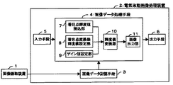
また、組織あるいはたんぱく質を二次元電気泳動により分析し、作成された二次元画像をもとに病症診断を行う方法が提案されている。電気泳動パターンを撮像した電気泳動画像のコントラストを強調させて目視で容易にスポットを確認できるようにしており、図10は、その発明の一実施形態にかかる電気泳動画像処理装置の全体構成を示すブロック図である。つまり、電気泳動画像の画像データを記憶する画像データ記憶手段3と、電気泳動画像のコントラストが強調されるように画像処理する画像データ処理手段4と、入力手段5と、出力手段6とを備え、画像データ処理手段4が、電気泳動画像の画像データから着目点画素の輝度値Lを読み込み、着目点画素の変換後の輝度値dを設定し、ゲイン値kを設定する。そして、電気泳動画像の画像データを構成する各画素の撮像輝度値Xのそれぞれを、着目点画素の輝度値Lと、着目点画素の変換後の輝度値dと、ゲイン値kとに基づいて、式X′=k(X−L)+dによって変換して変換輝度値X′とし、この変換輝度値X′からなる電気泳動画像の画像データを出力手段6へ出力して表示させる方法を提案している(例えば特許文献2参照)。
しかしながら、従来技術で述べた画像処理システムでは、以下のような課題が生じることがある。 However, the image processing system described in the prior art may have the following problems.
つまり、病症診断などにおいては、正常あるいは異常と見られる細胞を処理し画像上で両者を比較することが好ましく、両画像を形成するスポットが明確であることが適切な判断を導く上で非常に重要である一方、作製された画像全体に対して、上記のように一律的に画像強度分割によって境界を認定する、あるいはコントラストを強調することは、正確なスポットを確認していない、との発明者の知見が得られている。 In other words, in disease diagnosis and the like, it is preferable to process normal or abnormal cells and compare them on images, and it is very important to guide the appropriate judgment that the spots forming both images are clear. While it is important, the invention that the whole spot of the produced image does not confirm the exact spot by uniformly identifying the boundary by image intensity division or enhancing the contrast as described above. The knowledge of the person is obtained.
つまり、従来技術で述べた病症診断方法では、こうしたスポットの明確な画像の形成が困難であり、以下のような課題が生じることがあった。
(1)スポットの境界が元々不明瞭な場合の判断はコントラストだけでは正確な認定が困難である。具体的には、癌の病症診断における組織の確認などにおいては、スポットの領域は、細胞自体の造影手段によって表されることから、細胞の平面的な大きさ・高さを表示するとともに、細胞造影の濃淡の相違をも表示することから、画像全体における一律的な画像強度分割による境界の認定は、必ずしも正確な組織の境界を認定していることにならないことがある。
(2)画像自体に経時変化が生じる場合には、対比する画像が同等視できない。つまり、一般には、造影手段を用いて画像を作製するが、このとき組織との反応が安定するまでに所定の時間を要し、かつ組織ごとに安定時間が異なることが、その要因であるとの知見が得られた。
(3)スポット領域同士が重なりをもった場合においては、その境界が不明確になることから、やはり正確な対比が困難となる。特に現実の試料においては組織間の境自体が不明瞭であるため、正確な画像が得られないことから、コントラストだけではスポットの境界の明瞭化が図れない。
(4)画像の撮像時におけるゲルの条件あるいはゲルマップや電気泳動パターンと撮像装置との配置関係によって、画像におけるスポット位置のズレが生じることから、正確な対比が困難となる。
That is, in the disease diagnosis method described in the prior art, it is difficult to form a clear image of such spots, and the following problems may occur.
(1) In the case where the boundary of the spot is originally unclear, accurate recognition is difficult only by contrast. Specifically, in the confirmation of tissue in the diagnosis of cancer disease, the spot area is represented by the contrast means of the cell itself, so that the planar size and height of the cell are displayed, and the cell Since the difference in contrast is also displayed, the recognition of the boundary by uniform image intensity division in the entire image may not necessarily recognize the accurate tissue boundary.
(2) If the image itself changes with time, the images to be compared cannot be regarded as equivalent. In other words, in general, an image is produced using a contrast means, and at this time, it takes a predetermined time for the reaction with the tissue to become stable, and the reason is that the stabilization time differs for each tissue. The knowledge of was obtained.
(3) In the case where the spot areas overlap, the boundary becomes unclear, so that accurate comparison becomes difficult. In particular, in an actual sample, since the boundary between tissues is unclear, an accurate image cannot be obtained. Therefore, the boundary between spots cannot be clarified only by contrast.
(4) Because the position of the gel in the image or the positional relationship between the gel map or electrophoretic pattern and the imaging device is misaligned, the spot position shifts in the image, making accurate comparison difficult.
従って、本発明は、複数画像の比較による同一スポットの特定・比較において、(1)境界の不明確さ、(2)スポットごとの濃淡の相違、(3)画像の経時変化およびスポットごとの変化の相違、(4)領域の重なりによる境界の不明確さ、(5)スポット位置のずれなどによる誤差の排除が課題となる。 Therefore, according to the present invention, in the identification / comparison of the same spot by comparing a plurality of images, (1) unclearness of the boundary, (2) difference in shading for each spot, (3) temporal change of the image and change for each spot (4) Unclearness of boundaries due to overlapping regions, (5) Elimination of errors due to spot position deviations, and the like.
また、画像上に現れるスポットには、病症診断特有の非常に重要なスポットがあり、専門医の判断時にこうした特異点の見過ごしがないように、スポットの重要性に応じた明示も必要となる。 In addition, the spot appearing on the image includes a very important spot peculiar to the diagnosis of the disease, and it is necessary to clearly indicate the importance of the spot so that the singular point is not overlooked at the time of judgment by the specialist.
さらに、現在、癌などの疾病に対しては、現状専門医の経験による判断に頼るしかない状況であるが、こうした専門医の経験による判断に加え、より客観的な判断の指標として二次元画像が求められており、今後、こうした指標を提供しうる画像処理システムが要請されている。 Furthermore, currently, for diseases such as cancer, there is no choice but to rely on judgment based on the experience of specialists at present, but in addition to judgment based on the experience of specialists, a two-dimensional image is required as an index for more objective judgment. In the future, there is a demand for an image processing system that can provide such an index.
つまり、本発明の解決しようとする課題は、例えば、標本をもとに作製された二次元画像を、病症診断に対する客観的な判断の指標として提供できるように、画像の状態あるいは診断に求められる条件に適合した適切な画像処理を行うことができる画像処理システムを提供することである。特に、二次元電気泳動画像を用いて肝臓癌などの癌の組織を検出する場合のように、造影操作あるいは撮像操作によって画像に経時変化を生じる場合には、簡便かつ高精度の画像処理が必要不可欠であり、本発明の課題は、こうした要請に応じることができる画像処理システムを提供することである。 That is, the problem to be solved by the present invention is required for the state or diagnosis of an image so that, for example, a two-dimensional image created based on a specimen can be provided as an index for objective judgment for disease diagnosis. An object of the present invention is to provide an image processing system capable of performing appropriate image processing in conformity with conditions. In particular, when two-dimensional electrophoresis images are used to detect cancerous tissues such as liver cancer, when images change over time due to contrast or imaging operations, simple and highly accurate image processing is required. It is indispensable and an object of the present invention is to provide an image processing system capable of meeting such a demand.
本発明者らは、鋭意研究を重ねた結果、以下に示す画像処理システムにより上記目的を達成できることを見出し、本発明を完成するに到った。 As a result of intensive studies, the present inventors have found that the above object can be achieved by an image processing system described below, and have completed the present invention.
本発明は、画像処理システムであって、経時変化を伴う特定対象を撮像した二次元画像に対するスポットの領域決定において、(1)経時画像の準備、(2)画像内の特定点あるいは特定領域の抽出、(3)特定点あるいは特定領域の画像強度の関数化、(4)バックグランドの認定、(5)バックグランドの画像強度の関数化、(6)補正関数の設定、のステップを有することを特徴とする。 The present invention is an image processing system for determining a spot area for a two-dimensional image obtained by imaging a specific object with a change over time. (1) Preparation of a time-dependent image, (2) Specific point or specific area in an image And (3) functionalization of image intensity at a specific point or area, (4) background recognition, (5) functioning of background image intensity, and (6) setting of a correction function. It is characterized by.
つまり、本発明者は、細胞や蛋白質などの生体標本をもとに造影手段を用いて作製された画像による病症診断など二次元画像の利用においては、対比する画像におけるバックグランドが変化しており特定時間における画像だけでは正確な診断は困難であること、さらに、スポット領域およびバックグランドにおける画像強度の経時変化は各々の画像上の点あるいは領域(以下「ポイント」という。)によって異なること、および、そのポイントにおける画像強度の経時変化に対し所定の関数近似を行うことが可能であることを見出した。従って、スポット領域およびバックグランドにおける画像強度を、こうした関数に用いて自動的に処理し、バックグランドの経時変化の補正を行うことで、より客観的な判断の指標を提供することができる。また、こうした補正によって、上記の課題解決のみならず、重なり合ったスポットが時間を遡ることにより、元は複数スポットからなることを自動的に明らかにすることができる。本方法は、こうした点にも、従来にはない一歩進んだ大きな利点がある。さらに、画像全体に対する一律的な決定方法ではなく、個々のスポットの特性に応じた補正が可能であり、より精度の高い指標の提供を行うことができる。 In other words, the present inventor has changed the background in contrasting images in the use of two-dimensional images such as disease diagnosis based on images prepared using contrast means based on biological specimens such as cells and proteins. It is difficult to make an accurate diagnosis only with an image at a specific time, and further, the temporal change in the image intensity in the spot area and the background varies depending on the point or area on each image (hereinafter referred to as “point”), and The present inventors have found that a predetermined function approximation can be performed with respect to the temporal change in image intensity at that point. Therefore, the image intensity in the spot area and the background is automatically processed using such a function, and the temporal change of the background is corrected, thereby providing a more objective determination index. Further, by such correction, not only the above-described problem can be solved, but it is also possible to automatically clarify that the overlapping spot is originally composed of a plurality of spots by going back in time. In this respect, the present method also has a significant advantage that is one step ahead of the prior art. Furthermore, it is not a uniform determination method for the entire image, but correction according to the characteristics of individual spots is possible, and a more accurate index can be provided.
本発明は、画像処理システムであって、二次元画像に対するスポットの領域決定において、(1)任意の第1基準点あるいは基準領域の選定、(2)第1基準点あるいは基準領域間の画像強度を関数化、(3)第1境界点の設定、(4)中心点の設定、(5)第2基準点あるいは基準領域の選定、(6)第2基準点あるいは基準領域間の画像強度を関数化、(7)第2境界点の設定、(8)前記(4)〜(7)のステップを繰り返しによる第i境界点の設定、(9)スポットの領域の決定、のステップを有することを特徴とする。 The present invention is an image processing system for determining a spot area for a two-dimensional image by (1) selecting an arbitrary first reference point or reference area, and (2) image intensity between the first reference point or reference areas. (3) Setting of the first boundary point, (4) Setting of the center point, (5) Selection of the second reference point or reference area, (6) Image intensity between the second reference point or reference area (7) setting the second boundary point, (8) setting the i-th boundary point by repeating the steps (4) to (7), and (9) determining the spot region. It is characterized by.
本発明者は、二次元画像に対するスポットの領域決定において、画像全体に対する一律的な決定方法ではなく、個々のスポットの特性に応じた決定が必要であるとの知見から、こうした方法によって決定した領域をもとに作製された二次元画像が実際の病症診断において有用であることを見出した。つまり、スポット領域の境界近傍における画像強度の分布は、スポットの大きさあるいは強度(濃淡)に応じた所定の関数近似が可能であり、変曲点など関数において自動的に処理可能な点を領域の境界点として決定することによって、病症診断に対する客観的な判断の指標を提供することができる。また、スポットの境界は閉曲線を形成することから、順次境界点を画定していくこうした画像処理によって、スポットの形状に関係なくスポットの境界設定および領域決定が可能となる。さらに、領域決定と同時に面積の算出が容易である。 The present inventor determined the area determined by such a method from the knowledge that the determination of the area of the spot for the two-dimensional image is not a uniform determination method for the entire image but the determination according to the characteristics of the individual spots. It was found that the two-dimensional image created based on this is useful in actual disease diagnosis. In other words, the distribution of the image intensity in the vicinity of the boundary of the spot area can be approximated to a predetermined function according to the size or intensity (shading) of the spot. By determining as a boundary point, it is possible to provide an index of objective judgment for disease diagnosis. In addition, since the spot boundary forms a closed curve, spot boundary setting and region determination can be performed by such image processing in which boundary points are sequentially defined regardless of the spot shape. Furthermore, the area can be easily calculated simultaneously with the region determination.
さらに、上記処理によって、一次的にはいわゆる2値画像が作製されることになるが、特異点を複数設定すれば、三次元(多次元)画像の作製も同様に行うことができる。 Furthermore, a so-called binary image is primarily created by the above processing, but if a plurality of singular points are set, a three-dimensional (multi-dimensional) image can be similarly produced.
本発明は、上記画像処理システムであって、二次元画像に対するスポットの領域決定において、前記発明において設定された関数に対し、予め外部のあるいは内蔵の、前記関数に相当する正規化された関数を用意し、該正規化された関数をもとに補正あるいは置換する、ステップを有することを特徴とする。 The present invention is the above-described image processing system, wherein in the determination of the spot area for a two-dimensional image, an external or built-in normalized function corresponding to the function is previously set with respect to the function set in the invention. It has a step of preparing and correcting or replacing based on the normalized function.
癌細胞などの生体標本の二次元電気泳動については、最近多くの研究機関で行われており、研究結果としての画像自体は多く存在する。こうした画像について、上記のような、多くの解析関数を作製すれば、同一対象の二次元画像に対する非常に優れたデータベースができあがる。つまり、解析関数についての正規化が可能となる。本発明者は、こうした正規化された関数を本画像処理システムに適用して、スポットの境界点を決定し、あるいは画像強度の経時変化特性の特定や補正を行うことによって、実測時における各種の誤差あるいはノイズの影響を受けずに、客観性の高い領域決定を行うことができることを見出した。 Recently, two-dimensional electrophoresis of biological specimens such as cancer cells has been performed at many research institutions, and there are many images themselves as research results. For such an image, if a large number of analytic functions as described above are prepared, a very excellent database for a two-dimensional image of the same object can be created. That is, normalization for the analytic function is possible. The present inventor applies such a normalized function to the image processing system to determine the boundary point of the spot, or to specify and correct the time-varying characteristics of the image intensity, thereby performing various measurements at the time of actual measurement. It was found that it is possible to determine a highly objective area without being affected by errors or noise.
本発明は、上記画像処理システムであって、同一スポットの撮像を含む複数の二次元画像を比較し画像補正を行うに際し、(1)スポットの領域決定、(2)比較するスポットの選定、(3)同一スポットの重ね合わせによる位置補正、のステップを有することを特徴とする。 The present invention is the image processing system described above, in which when performing image correction by comparing a plurality of two-dimensional images including imaging of the same spot, (1) spot area determination, (2) selection of a spot to be compared, ( 3) It has a step of position correction by superimposing the same spots.
複数の二次元画像におけるいわゆる位置補正に相当する処理である。水平関係または垂直関係にない2つのあるいはそれ以上の、各画像において対応するスポットを特定できれば、画像の平面的な補正が可能となる。この場合も、上記のようにデータベース化された二次元画像から、2以上のスポットの領域を特定することができる正規化された二次元画像の作製が可能であれば、非常に客観的な位置補正を実現することができる。 This is a process corresponding to so-called position correction in a plurality of two-dimensional images. If two or more corresponding spots in each image that are not in a horizontal relationship or a vertical relationship can be identified, the image can be corrected in a planar manner. Also in this case, if it is possible to create a normalized two-dimensional image that can identify two or more spot regions from the two-dimensional image databased as described above, a very objective position Correction can be realized.
本発明は、上記画像処理システムであって、予め1以上の外部標準に対応するスポットの撮像を含む複数の二次元画像を比較し画像補正を行うに際し、前記外部標準に対応するスポットが重なるように、前記比較画像の二次元補正を行う、ことを特徴とする。 The present invention is the image processing system as described above, and when performing image correction by comparing a plurality of two-dimensional images including imaging of spots corresponding to one or more external standards in advance, the spots corresponding to the external standards overlap. Further, the comparison image is subjected to two-dimensional correction.
いわゆる外部標準による補正に相当する処理である。上記のように、通常の二次元画像は位置・濃淡・経時変化の補正を必要とするが、造影操作あるいは撮像操作を経由しても変化がなくかつ画像として二次元位置が分離可能な標準物質を加えて標本を作製し、これをもとに作製された二次元画像によれば、画像の補正における基準スポットとすることができる。この場合も、外部標準に対応するスポットを含むデータベース化され、正規化された二次元画像の作製が可能であれば、非常に客観的な位置補正を実現することができる。 This process corresponds to correction by so-called external standard. As mentioned above, normal two-dimensional images require correction of position, density, and temporal change, but there is no change even through contrast or imaging operations, and a standard substance that can be separated as a two-dimensional position as an image According to the two-dimensional image prepared based on the sample, it can be used as a reference spot for image correction. Also in this case, if a database including spots corresponding to external standards and a normalized two-dimensional image can be produced, very objective position correction can be realized.
本発明は、上記画像処理システムであって、前記スポットを重ねあわせにおいて、スポットの重なり状態に応じた区分を行い、同一スポットにはその間に跨る記号を表示することを特徴とする。 The image processing system according to the present invention is characterized in that in superimposing the spots, classification according to the overlapping state of the spots is performed, and symbols straddling the same spot are displayed on the same spot.
こうした画像処理によって、複数の二次元画像の比較における目視の判断においても速やかに相違点が判り、診断の迅速化を図ることができる。むろん記号に限定されず、対応するスポットがない場合には、フラッシングや色彩を変化させるなど種々の明示方法の適用が可能であることはいうまでもない。ここで、「区分」とは、色彩、濃淡、模様や迷彩による識別を付与することをいう。 By such image processing, the difference can be quickly found even in visual judgment in comparison of a plurality of two-dimensional images, and the diagnosis can be speeded up. Of course, the present invention is not limited to symbols, and when there is no corresponding spot, it is needless to say that various indication methods such as flushing and changing colors can be applied. Here, “classification” refers to assigning identification by color, shading, pattern, or camouflage.
本発明は、上記画像処理システムであって、前記の各ステップにおいて、(1)各画像の全体画面、(2)比較する画像の任意の範囲の原画面、(3)比較する画像を重ね合わせた画面、(4)特定部位の拡大原画面、(5)特定部位を拡大し、比較する画像を重ね合わせた画面、のうちのいずれかの画面を並列的に表示するとともに、各画面間の関連を表記することを特徴とする。 The present invention is the image processing system described above, wherein in each of the above steps, (1) an entire screen of each image, (2) an original screen of an arbitrary range of images to be compared, and (3) an image to be compared are superimposed. One of a screen, (4) an enlarged original screen of a specific part, and (5) a screen in which a specific part is enlarged and the images to be compared are superimposed, It is characterized by describing the relationship.
例えば、癌細胞と正常細胞との比較による病症診断などにおいては、スポットの有無・位置関係・大きさだけではなく、形状あるいは周囲のスポットとの関係など精緻に解析する必要がある。こうした正確な判断のための画像を明示するためには、単に比較する複数の二次元画像だけでなく、部分的拡大画面およびその拡大位置の明示あるいは修正前の画像などを別々にまたは同時に明示できることが好ましい。こうした画像処理によって、より迅速かつ的確な診断が可能となる。 For example, in the diagnosis of disease by comparing cancer cells with normal cells, it is necessary to analyze not only the presence / absence / positional relationship / size of spots but also the relationship with the shape or surrounding spots. In order to specify images for such accurate judgment, it is possible to specify not only a plurality of two-dimensional images to be compared, but also a partial enlarged screen and its enlarged position clearly or separately before or simultaneously. Is preferred. Such image processing enables quicker and more accurate diagnosis.
以上のように、本発明は、病症診断に対する客観的な判断の指標として提供できるように、画像の状態あるいは診断に求められる条件に適合した適切な画像処理を行うことができる画像処理システムを提供することができる。特に、造影操作あるいは撮像操作によって画像に経時変化を生じる場合であっても、こうした要請に応じることができる画像処理システムを提供することができる。 As described above, the present invention provides an image processing system capable of performing appropriate image processing suitable for the state of an image or conditions required for diagnosis so that it can be provided as an index of objective judgment for disease diagnosis. can do. In particular, it is possible to provide an image processing system capable of meeting such a request even when a change with time occurs in an image due to a contrast operation or an imaging operation.
以下、本発明の実施の形態について、図面を参照しながら説明する。なお、本システムの各操作指令は、例えば、演算装置の内部から自動的に行う場合だけではなく、表示部から手動で行う場合や外部の通信手段などによって行うことができる。また、二次元画像データおよび補正に必要な各関数についても、同様に内部にメモリーされたものだけではなく、外部からの入力も可能である。 Hereinafter, embodiments of the present invention will be described with reference to the drawings. In addition, each operation command of this system can be performed not only when it is automatically performed from the inside of the arithmetic unit, but also when manually performed from the display unit or by an external communication unit. Similarly, the two-dimensional image data and each function necessary for correction can be input from the outside as well as those stored therein.
<画像処理システム例1>
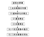
本発明の基本として、1つの対象物に対して複数の二次元画像があるときに、相互に比較し相違点確認するための画像処理を行う場合を例に挙げる。図1にそのフローを示す。ベースおよびスポットの画像強度の算定には、経時変化がある画像については、まず経時変化の補正を行い、次にバックグランドの経時変化量を補正した強度を用いて濃淡補正を行う。経時変化がない画像の場合には、まず、濃淡補正を行い、各スポットの画像領域の設定を行うことで補正した画像の作製ができる。なお、本願にいう「スポット」とは、二次元画像上に表された小領域あるいはその集合体であって、測定対象の特性を示すためにバックグランドと区分された領域をいう。
以下、そのフローに基き、各ステップを詳述する。
<Image processing system example 1>
As a basis of the present invention, when there are a plurality of two-dimensional images with respect to one object, a case where image processing for comparing and confirming a difference is performed as an example. FIG. 1 shows the flow. In calculating the image intensity of the base and the spot, for an image having a change over time, the change over time is first corrected, and then the density correction is performed using the intensity obtained by correcting the background change over time. In the case of an image that does not change with time, it is possible to produce a corrected image by first performing density correction and setting the image area of each spot. The “spot” referred to in the present application is a small area or an aggregate thereof expressed on a two-dimensional image, and is an area that is separated from the background in order to indicate the characteristics of the measurement target.
Hereinafter, each step will be described in detail based on the flow.
(1)二次元画像の経時変化の補正
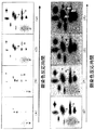
二次元画像の作製においては、例えば、造影時に図2(A)〜(H)に示すような画像の経時変化が生じることがある。これを、同図におけるa〜cのスポットおよびバックグランドgについての濃度の経時変化として捉えると、図3のような、バックグランドの上昇と各スポットの反応に伴う強度変化としての関係が得られる。このとき、各スポットの反応に伴う強度変化は、バックグランドの経時変化を補正すれば、ほぼ相似した曲線となり、近似した関数化が可能である。従って、経時変化の補正方法としては、バックグランドの経時変化量だけでなく、スポットの広がり(面積)の変化についても行うことが可能となる。また、各スポット単位での補正だけでなく、画像全体としての補正を行うことが、さらなる高精度化を図ることとなる。
(1) Correction of change with time of two-dimensional image In the preparation of a two-dimensional image, for example, changes with time of the image as shown in FIGS. If this is regarded as a change with time in the concentrations of the spots a to c and the background g in FIG. 3, the relationship between the background rise and the intensity change accompanying the reaction of each spot as shown in FIG. 3 is obtained. . At this time, the intensity change accompanying the reaction of each spot becomes a substantially similar curve if the background change with time is corrected, and an approximate function can be obtained. Therefore, as a method for correcting the change over time, not only the background change over time but also the change in the spread (area) of the spot can be performed. Further, not only the correction for each spot but also the correction for the entire image can achieve higher accuracy.
二次元画像としては、生体標本についての二次元電気泳動画像だけでなく、ISFETやCCDにおける電荷の分布や電気信号量の分布にも適用することが可能である。
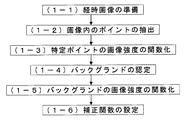
具体的には、スポットの領域設定においては、次の方法が挙げられる。図4にそのフローを示す。
The two-dimensional image can be applied not only to a two-dimensional electrophoresis image of a biological specimen but also to a charge distribution and an electric signal amount distribution in an ISFET or CCD.
Specifically, in the spot area setting, the following method is exemplified. FIG. 4 shows the flow.
(1−1)経時画像の準備
特定対象に対する任意の時間間隔の経時画像を複数準備する。ここでいう「特定対象」とは、時系列的に二次元画像を形成する対象物をいい、上記のように、二次元電気泳動によって二次元画像を形成する対象である細胞や蛋白質などの生体標本などが例示されるが、これに限定されるものではない。
(1-1) Preparation of Time-lapse Images Prepare a plurality of time-lapse images at arbitrary time intervals for a specific object. As used herein, the “specific object” refers to an object that forms a two-dimensional image in a time series. As described above, a biological object such as a cell or protein that is a target for forming a two-dimensional image by two-dimensional electrophoresis. Although a sample etc. are illustrated, it is not limited to this.
(1−2)画像内のポイントの抽出
各ポイントにおける経時変化の情報を得るために、各画像における任意に選択された特定のポイントの画像強度を抽出する。ポイントは画像を区分し順次スキャニングする方法などが採られる。
(1-2) Extraction of points in an image In order to obtain information on changes over time at each point, the image intensity of a specific point arbitrarily selected in each image is extracted. The points are divided into images and scanned sequentially.
(1−3)特定ポイントの画像強度の関数化
上記の各画像の特定ポイントの画像強度を、時間をパラメータとして関数化し、順次、関数Giを作製する。ここで、「画像強度」とは、広く二次元画像の形成が可能なあらゆる対象を、その対象を表現しうる尺度あるいは単位で表したものをいい、具体的には、単色あるいは色彩の強度だけではなく、電荷や電気信号量などの物理量、さらには、化学量あるいはそれを変換した物理量などが該当する。また、「関数」は、特定の点に関する場合にあっては一次元関数で十分であるが、点ではなく特定の領域に関する場合には二次元以上の関数を必要とすることも含まれる。演算速度から低次が好ましく、必要とされる精度および曲線の特性に合った次数の選択が望ましい。また、設定される関数は、1つの式で表される関数だけではなく、時間を区分し2つ以上の関数を結合化したものとすることも可能である。さらに、本項の経時変化を関数化する場合には、時間がパラメータとなるが、位置補正など他項においては、位置つまり座標がパラメータとなることがあり、両方がパラメータとなる場合など、種々の要素がパラメータとなりうる。
(1-3) Functionalization of Image Intensity at Specific Point The image intensity at the specific point of each of the above images is converted into a function using time as a parameter, and a function Gi is sequentially created. Here, “image intensity” refers to any object that can form a wide range of two-dimensional images, expressed in a scale or unit that can represent the object, and specifically, only the intensity of a single color or color. Instead, physical quantities such as electric charges and electrical signal quantities, chemical quantities or physical quantities obtained by converting them are applicable. In addition, as for the “function”, a one-dimensional function is sufficient when it is related to a specific point, but it also includes a need for a function of two or more dimensions when it is related to a specific region instead of a point. A low order is preferable in terms of calculation speed, and it is desirable to select an order suitable for the required accuracy and curve characteristics. In addition, the function to be set is not limited to a function represented by one expression, but may be a function obtained by dividing time and combining two or more functions. Furthermore, time is a parameter when functioning the change over time in this section, but in other sections such as position correction, the position, that is, the coordinates may be a parameter, and both are parameters. Can be a parameter.
(1−4)バックグランドの認定
バックグランドの認定は、画像条件などによって任意に認定方法の設定方法が可能である。例えば、前記(1−2)において取り出された画像強度のうち、経時的に最終画像における画像強度が基準値以下のポイントを、バックグランドと認定する方法が挙げられる。また、画像作製時に予め特定のポイントをバックグランドと認識できる場合には、そのポイントを認定することも可能である。さらに、経時的に最終画像において、画像強度の全平均を求め、それ以上の強度を有するポイントを平均強度に置き換え、さらに全平均を求め、これを繰り返すことでバックグランドとすることが可能なポイントを認定する方法がある。また、経時的に最終画像において、画像強度の高いレベルから低いレベルに段階的に変化させたときに、同一レベルで形成されるスポット領域が画像全体に急激に拡大する場合において、そのレベルまでをバックグランドと認定する方法なども可能である。むろん、バックグランドの認定方法は、これに限定されるものではないことはいうまでもない。
(1-4) Background Certification The background certification can be arbitrarily set by a certification method depending on image conditions and the like. For example, among the image intensities extracted in the above (1-2), there is a method in which the point where the image intensity in the final image is not more than a reference value with time is recognized as the background. In addition, when a specific point can be recognized as a background at the time of image creation, it is also possible to recognize that point. Furthermore, in the final image over time, find the total average of the image intensity, replace the point with higher intensity with the average intensity, further calculate the total average, and repeat this to make the background There is a way to certify. In addition, when a spot area formed at the same level suddenly expands in the entire image when the final image is gradually changed from a high level to a low level over time in the final image, the level is reduced to that level. A method of certifying it as a background is also possible. Needless to say, the background certification method is not limited to this.
(1−5)バックグランドの画像強度の関数化
バックグランドと認定したポイントの画像強度を、時間をパラメータとして関数化し、関数Goを作製する。関数化は、各ポイントごとに行うことも可能であるが、本発明者の知見では、数ポイントにおける画像強度の経時データの平均によって代表させることで十分である。
(1-5) Functionalization of background image intensity A function Go is created by functionalizing the image intensity of a point recognized as background with time as a parameter. The functionalization can be performed for each point, but according to the knowledge of the present inventor, it is sufficient to be represented by the average of the temporal data of the image intensity at several points.
(1−6)補正関数の設定
上記のように補正方法としては、経時データの平均から得られたバックグランドについての関数Goを用い、下式1から、関数Giが作製されたポイントにおける補正関数Aiを求める方法がある。
Ai=Gi−Go ・・式1
(1-6) Setting of Correction Function As described above, as a correction method, the function Go for the background obtained from the average of the time-lapse data is used, and the correction function at the point where the function Gi is created from the following equation 1 There is a method for obtaining Ai.
Ai = Gi-Go .. Formula 1
また、前記バックグランドと認定されたポイントの内、前記関数Giが作製されたポイントに最も近接したポイントのおける関数Goiを選定し、下式2から関数Giが作製されたポイントにおける補正関数Aiを求める方法もある。
Ai=Gi−Goi ・・式2
Further, among the points recognized as the background, a function Goi at a point closest to the point at which the function Gi is created is selected, and the correction function Ai at the point at which the function Gi is created from the following equation 2 is obtained. There is also a way to ask.
Ai = Gi-Goi ..Equation 2
こうした補正関数を各ポイントに適用することによって、各ポイントの経時変化を補正した画像を作製することができるとともに、複数の画像の対比において、各々異なる時間軸での対比となることを回避し、より客観的な判断の指標を提供することができる。 By applying such a correction function to each point, it is possible to create an image in which the change with time of each point is corrected, and in the comparison of a plurality of images, avoiding comparison on different time axes, A more objective indicator of judgment can be provided.
また、こうした補正の最大の利点は、従来近接する複数のスポットが重なり合って形成されていたスポットが、各ポイントでの経時変化を補正し、時間を遡ることにより、元来複数のスポットからなることを自動的に明らかにすることができる点にあるといえる。特に、図2における(E)や(F)あるいは(G)に相当する画像しかない場合であっても、補正関数が想定できる場合には(A)や(B)に相当する各スポットを推定することができ、上記画像のみでは判断できない複数のスポットが重なりを分離し判断することができる。 In addition, the biggest advantage of such correction is that spots that were previously formed by overlapping multiple adjacent spots originally consisted of multiple spots by correcting the changes over time at each point and going back in time. It can be said that it is in the point that can be clarified automatically. In particular, even when there is only an image corresponding to (E), (F), or (G) in FIG. 2, if a correction function can be assumed, each spot corresponding to (A) or (B) is estimated. A plurality of spots that cannot be determined only by the image can be determined by separating the overlap.
(2)二次元画像の濃淡補正
本発明者の研究によって、二次元画像のスポットの境界近傍における画像の濃淡(画像強度)を調べると、次の傾向があることの知見が得られた。つまり、図5に示すように、ベース領域を始点として、比較的フラットな領域Aから、急激に変化する領域Bを経て、濃度の高い領域Cに移行してスポットが形成されている。各スポットの濃淡の差(最高強度DとD’の差)はあっても、その曲線は非常に相似的なものとなる。従って、この曲線は、スポットの位置xをパラメータとする3次関数あるいは4次以上の関数で表現することができる。または、領域ごとに区分し2つ以上の3次以下の低次関数の結合化することも可能である。
具体的には、スポットの領域設定においては、次の方法が挙げられる。図6にそのフローを示す。
(2) Density correction of two-dimensional image According to the study of the present inventor, when the density (image intensity) of an image in the vicinity of the boundary of a spot of a two-dimensional image is examined, it has been found that the following tendency exists. That is, as shown in FIG. 5, a spot is formed by moving from a relatively flat area A to a high density area C through a rapidly changing area B, starting from the base area. Even if there is a difference in density of each spot (difference between the maximum intensity D and D ′), the curves are very similar. Therefore, this curve can be expressed by a cubic function or a function of the fourth or higher order with the spot position x as a parameter. Alternatively, it is possible to combine two or more low-order functions of the third order or lower by dividing each region.
Specifically, in the spot area setting, the following method is exemplified. FIG. 6 shows the flow.
(2−1)第1基準点(領域)の設定
二次元画像の濃淡補正にあたり、スポット内外に、任意の距離を有する任意の2つのポイント(第1基準点)を選定する。2つのポイントは、中間に他のスポットが入り込まないようにできる限り近接することが好ましい一方、境界の判断が可能な所定の距離以上を必要とする。従って、画像全体の画像強度をデータ化してから図5の曲線部に相当する2点を自動的に選定する方法をとることも有効である。
(2-1) Setting of First Reference Point (Region) For density correction of a two-dimensional image, two arbitrary points (first reference point) having an arbitrary distance are selected inside and outside the spot. While it is preferable that the two points be as close as possible so that other spots do not enter in between, the two points require a predetermined distance or more that allows the boundary to be determined. Therefore, it is also effective to adopt a method of automatically selecting two points corresponding to the curved portion in FIG. 5 after converting the image intensity of the entire image into data.
(2−2)第1関数化
位置をパラメータとして、2つの第1基準点間の画像強度を関数化し、関数F1を作製する。関数は上記のように、1つの式で表される関数だけではなく、位置を区分し2つ以上の関数を結合化したものとすることも可能である。
(2-2) First Functionalization Using the position as a parameter, the image intensity between the two first reference points is converted into a function to create a function F1. As described above, the function is not limited to a function represented by one expression, but may be a combination of two or more functions by classifying positions.
(2−3)第1境界点の設定
関数化された画像強度曲線F1の特異点を境界点K1と定める。ここで、「曲線の特異点」とは、予め設定可能な、境界決定の基準となりうる客観的条件を満たす点をいい、具体的には、ピーク値までの分割点(例えば5%立上り点など)や曲線上の変曲点または関数の微分値が基準値を超える点のように傾斜が変化する点などが該当する。手動あるいは自動的に設定することが可能である。
(2-3) Setting of First Boundary Point A singular point of the functioned image intensity curve F1 is defined as a boundary point K1. Here, the “singular point of a curve” refers to a point that can be set in advance and satisfies an objective condition that can serve as a criterion for determining a boundary. Specifically, a dividing point up to a peak value (for example, a rising point of 5%, etc.) ) Or an inflection point on the curve or a point where the slope changes such as a point where the differential value of the function exceeds the reference value. It can be set manually or automatically.
(2−4)第2中心点の設定
境界点K1を中心とする一定範囲を設定し、境界点K1と異なるポイントを任意に選定し、第2中心点とする。ここで、「一定範囲」とは、予め設定可能な、特定の点から原則等距離の範囲をいい、具体的には、設定距離は画像全体から分割される任意の距離(例えば100分割相当値など)や前記分割点の倍数値(例えば2倍の距離)などが該当する。
(2-4) Setting of the second center point A fixed range centering on the boundary point K1 is set, and a point different from the boundary point K1 is arbitrarily selected and set as the second center point. Here, the “certain range” means a range that can be set in advance and in principle equidistant from a specific point. Specifically, the set distance is an arbitrary distance (for example, a value equivalent to 100 divisions) divided from the entire image. Etc.) and a multiple value of the dividing point (for example, a double distance).
(2−5)第2基準点の設定
第2中心点を含むが境界点K1を含まない直線上にあって、第2中心点を中心に略等距離離れた2つのポイント(第2基準点)を選定する。
(2-5) Setting of Second Reference Point Two points (second reference point) that are on a straight line that includes the second center point but does not include the boundary point K1 and that are approximately equidistant from the second center point. ) Is selected.
(2−6)第2関数化
位置をパラメータとして、2つの第2基準点間の画像強度を関数化し、関数F2を作製する。
(2-6) Second Functionalization The function F2 is created by functionalizing the image intensity between the two second reference points using the position as a parameter.
(2−7)第2境界点の設定
関数化された画像強度曲線F2の特異点を境界点K2と定める。
(2-7) Setting of the second boundary point The singular point of the functionalized image intensity curve F2 is defined as the boundary point K2.
(2−8)第i境界点の設定
境界点K2について、前記(2−4)〜(2−7)のステップを繰り返し順次画像強度曲線Fiおよび境界点Kiを確定する。
(2-8) Setting of i-th Boundary Point For the boundary point K2, the steps (2-4) to (2-7) are repeated to sequentially determine the image intensity curve Fi and the boundary point Ki.
(2−9)スポットの領域決定
順次確定する境界点K(n+1)が、前記境界点K1〜Knのいずれかに一致あるいは前記一定範囲内にあるとき、該スポットの領域を決定する。スポットの境界線は、本来、閉曲線を形成することから、順次設定する境界点はいずれ一致点を有することになり、スポットの領域を決定することができる。こうした画像処理によって、スポットの形状に関係なくスポットの境界設定および領域決定が可能となる。
以上の画像処理を、画像上の各スポットについて行うことによって、各スポットの境界設定および領域決定ができ、二次元画像全体のスポット領域の決定を行うことができる。
(2-9) Spot Area Determination When the sequentially determined boundary point K (n + 1) coincides with any of the boundary points K1 to Kn or is within the predetermined range, the spot area is determined. Since the boundary line of the spot originally forms a closed curve, the boundary points to be sequentially set will eventually have coincident points, and the spot area can be determined. Such image processing makes it possible to set a spot boundary and determine a region regardless of the spot shape.
By performing the above image processing for each spot on the image, the boundary setting and area determination of each spot can be performed, and the spot area of the entire two-dimensional image can be determined.
このように、画像強度を関数化することで、個々のスポットの領域を客観的に決定することができるとともに、二次元画像全体の濃淡補正を行うことができる。つまり、個々のスポットの特性に応じた領域の決定が可能となり、有用な二次元画像を提供することができることとなる。 As described above, by converting the image intensity into a function, it is possible to objectively determine the area of each spot and to perform density correction of the entire two-dimensional image. That is, it becomes possible to determine a region according to the characteristics of each spot, and a useful two-dimensional image can be provided.
さらに、上記画像処理によって作製される画像は、いわゆる2値画像であるが、特異点の設定を複数すれば、多次元画像の作製も同様に行うことができる。具体的には、例えば特異点の設定を、特定スポットのピーク値までの分割点とし、立上り点を5%から10%刻みで行い、各ポイントごとに画像強度をパラメータとして関数化すれば、三次元の関数化された画像を作製することができる。 Furthermore, the image created by the image processing is a so-called binary image. However, if a plurality of singular points are set, a multi-dimensional image can be similarly produced. Specifically, for example, if the singularity is set as a dividing point up to the peak value of a specific spot, the rising point is performed in increments of 5% to 10%, and the image intensity is parameterized as a parameter for each point, the third order An original functionalized image can be created.
また、上記三次元の関数化は、複数の重なり合ったスポットを、各々独立した二次元あるいは三次元の画像に分割することができる。つまり、各スポットの重なり合っていない三次元曲面における関数から、重なり合った領域での画像強度を分解・解析することにより、元の複数スポットの三次元の関数化された画像を作製することができる。 The three-dimensional functionalization can divide a plurality of overlapping spots into independent two-dimensional or three-dimensional images. In other words, by decomposing and analyzing the image intensity in the overlapping region from the function of the three-dimensional curved surface where each spot does not overlap, it is possible to produce a three-dimensional functionalized image of the original plurality of spots.
(3)二次元画像の位置補正
対比する2以上の画像におけるスポットの位置補正は、両画像を重ね合せ、比較的相似形で領域の重なりが大きな任意の2以上のスポットを選定し、両画像の同一スポットの特定を行い、両スポットが重なるように、二次元補正を行う。また、位置補正は、基本的には上記の濃淡補正処理を行った後に行うことが好ましい。濃淡補正前では、スポットの領域が不明確であり、領域の重なりの割合を判断することが難しく、結果補正精度の悪化を招くおそれがあるためである。
具体的には、同一スポットの撮像を含む複数の二次元画像を比較し画像補正を行うに際し、以下のステップを有する。
(3) Position correction of two-dimensional image Spot position correction in two or more images to be compared is performed by superimposing both images, selecting any two or more spots having relatively similar shapes and large overlapping areas, and selecting both images. The same spot is identified, and two-dimensional correction is performed so that both spots overlap. Further, it is preferable that the position correction is basically performed after the above-described density correction processing. This is because the spot area is unclear before the density correction, and it is difficult to determine the overlapping ratio of the areas, and the result correction accuracy may be deteriorated.
Specifically, when performing image correction by comparing a plurality of two-dimensional images including imaging of the same spot, the following steps are included.
(3−1)各画像における領域決定
対比する各画像について、前記(2)の画像処理によってスポットの領域決定を行う。領域のデータは、上記の通り2値画像データ、あるいは三次元画像データであることを問うものではない。
(3-1) Region Determination in Each Image For each image to be compared, spot region determination is performed by the image processing of (2). As described above, the region data is not necessarily binary image data or three-dimensional image data.
(3−2)同一スポットの特定
複数の画像の内から特定画像(基準画像)を選定し、その画像内の任意の2以上のスポットを選定し、該特定画像と比較する比較画像の同一スポットの特定を行う。このとき、基準画像は、対象となる複数の画像の内、画像上のスポットが明確なものを選定することが好ましい。選定するスポットは極力大きい方が、重ねたときと補正精度の向上が期待できることから好ましい。また、水平垂直関係にない2つのあるいはそれ以上の、各画像において対応するスポットを特定できれば、画像の平面的な補正が可能となる。つまり、2つ以上のスポットは、画像全体の補正精度が上がるように、画像の対角する端部近傍のスポットあるいはそれに相当するスポットを選択することが好ましい。適切なスポットが不明確な場合には、画像の上端部から対角する端部の点をスキャニングして関数化可能な点を選定することも好ましい。
(3-2) Identification of the same spot A specific image (reference image) is selected from a plurality of images, any two or more spots in the image are selected, and the same spot of the comparison image to be compared with the specific image To identify. At this time, it is preferable to select a reference image having a clear spot on the image among a plurality of target images. It is preferable that the spot to be selected is as large as possible because it can be expected to improve correction accuracy when it is overlapped. Further, if two or more spots that are not in a horizontal / vertical relationship and corresponding spots in each image can be identified, the image can be corrected in a planar manner. That is, for two or more spots, it is preferable to select a spot near the opposite end of the image or a spot corresponding thereto so that the correction accuracy of the entire image is improved. When an appropriate spot is unclear, it is also preferable to select a point that can be functionalized by scanning a point at an end opposite from the upper end of the image.
(3−3)比較画像の二次元補正
両画像の同一スポットが重なるように、比較画像の二次元補正を行う。3以上のスポットを選定し、最も重なりの悪いスポットを選定からはずすことによって、補正精度の向上が期待できる。
(3-3) Two-dimensional correction of comparative image Two-dimensional correction of the comparative image is performed so that the same spot of both images overlap. By selecting three or more spots and removing the least overlapping spot from the selection, an improvement in correction accuracy can be expected.
以上のような画像処理を行うことで、複数の二次元画像におけるいわゆる位置補正が可能となる。造影時あるいは撮像時において、二次元的あるいは3次元的に生じたスポット位置のズレを、適切かつ簡易に補正することが可能となる。 By performing the image processing as described above, so-called position correction in a plurality of two-dimensional images becomes possible. It is possible to appropriately and easily correct the deviation of the spot position generated two-dimensionally or three-dimensionally at the time of imaging or imaging.
(4)二次元画像の表示
上記(1)〜(3)の補正を行うことで、対比すべき二次元画像が完成し、各種の解析が可能となる。なお、上記の補正は、1度行うことで十分精度の向上を図ることができるが、これを繰り返すことで、さらに精度の高い補正が可能となる。また、(1)〜(3)の補正は必ずしもこの順である必要ではなく、任意に選択あるいは繰返しを行うことで、二次元画像の条件に合った補正が可能である。
(4) Display of two-dimensional image By performing the corrections (1) to (3) above, a two-dimensional image to be compared is completed, and various analyzes are possible. Note that the accuracy can be sufficiently improved by performing the above correction once, but the correction can be performed with higher accuracy by repeating this. In addition, the corrections (1) to (3) are not necessarily in this order, and correction that meets the conditions of the two-dimensional image is possible by arbitrarily selecting or repeating.
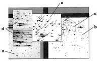
二次元画像の対比に際しては、マルチ画面が好ましく、例えば、図7に示すように(a)各画像の全体画面、(b)比較する画像の任意の範囲の原画面、(c)比較する画像を重ね合わせた画面、(d)特定部位の拡大原画面、(e)特定部位を拡大し、比較する画像を重ね合わせた画面、のように、いずれか任意の画面を並列的に表示する方法が挙げられる。単に比較する複数の二次元画像だけでなく、部分的拡大画面およびその拡大位置の明示あるいは修正前の画像などを別々にまたは同時に明示できることが好ましい。こうした画面の配列、大きさ、色彩などを任意に組合せることで、非常に使い勝手のよい二次元画像の表示を行うことができる。 When comparing two-dimensional images, a multi-screen is preferable. For example, as shown in FIG. 7, (a) an entire screen of each image, (b) an original screen in an arbitrary range of images to be compared, and (c) an image to be compared A method of displaying any arbitrary screen in parallel, such as (d) an enlarged original screen of a specific part, (e) a screen in which a specific part is enlarged and images to be compared are superimposed Is mentioned. It is preferable that not only a plurality of two-dimensional images to be compared, but also a partially enlarged screen and an image of the enlarged position or an image before correction can be specified separately or simultaneously. By arbitrarily combining the arrangement, size, color, and the like of such a screen, it is possible to display a very convenient two-dimensional image.
具体的には、 In particular,
(4−1)基準画像の全体画面(a)と比較画像の全体画面(a)との比較によって外郭的に把握することができる。 (4-1) It is possible to grasp externally by comparing the entire screen (a) of the reference image and the entire screen (a) of the comparative image.
(4−2)基準画像の特定範囲画面(b)と比較画像の特定範囲画面(b)との比較によって詳細比較をすることができる。 (4-2) Detailed comparison can be performed by comparing the specific range screen (b) of the reference image and the specific range screen (b) of the comparative image.
(4−3)比較画像の全体画面(a)と特定範囲画面(b)との比較によって詳細部の全体における位置づけを把握することができる。 (4-3) The position of the entire detailed portion can be grasped by comparing the entire screen (a) of the comparison image with the specific range screen (b).
(4−4)基準画像の特定範囲画面(b)と比較画像の特定範囲画面(b)との重ね合わせ画面(c)によって相違点の抽出を行うことができる。 (4-4) The difference can be extracted by the overlapping screen (c) of the specific range screen (b) of the reference image and the specific range screen (b) of the comparison image.
(4−5)基準画像の特定部位の拡大図(d)と比較画像の特定部位の拡大図(d)との比較によって特定スポットの詳細比較をすることができる。 (4-5) The specific spot can be compared in detail by comparing the enlarged view (d) of the specific part of the reference image with the enlarged view (d) of the specific part of the comparative image.
(4−6)基準画像の特定部位の拡大図(d)と比較画像の特定部位の拡大図(d)との重ね合わせ画面(e)によって、特定スポットにおける相違点の抽出および詳細比較をすることができる。 (4-6) Extraction and detailed comparison of a specific spot are performed by using an overlapping screen (e) of the enlarged view (d) of the specific part of the reference image and the enlarged view (d) of the specific part of the comparative image. be able to.
(4−7)図面(a)〜(e)を全体面で表示可能とすることで、特定スポットにおける相違点の画像全体における位置づけ複数の他の比較画像との比較検証が可能となる。 (4-7) By making it possible to display the drawings (a) to (e) on the entire surface, it is possible to compare and verify the difference between the specific spots in the entire image and a plurality of other comparative images.
また、自動補正では不十分な場合の目視による正確な補正操作のための画像の明示方法としても有用である。こうした画像の明示に対する操作指令は、例えば、演算装置の内部から自動的に行う場合だけではなく、表示部から手動で行う場合や外部の通信手段などによって行うことができる。拡大・縮小画面への変更などについても同様である。 Further, it is also useful as an image specifying method for an accurate correction operation by visual inspection when automatic correction is insufficient. Such an operation command for image specification can be performed not only automatically from the inside of the arithmetic unit, but also manually from the display unit, or by external communication means. The same applies to changes to the enlarged / reduced screen.
(5)二次元画像の対比
上記の二次元画像の表示によって、各画像の対比を行うことができる。具体的には、図8(A)に示すように、両画像内の対応するスポット同士の照合によって行われる。例えば、癌組織標本から得られた二次元画像中のスポットxが、正常組織標本から得られた二次元画像中にはないことが判る。この例によれば、図8(B)のように、二次元画像の比較によって、スポットxに対応する組織あるいはたんぱく質が、発生した癌組織に関連する可能性を示していると判断され、係る部位を取出し、例えば、所定の前処理後にTOF−MASS(時間飛行式質量分析法)によって測定された結果と併せて、病症の判断が行われる。
(5) Comparison of two-dimensional images Each image can be compared by displaying the above-described two-dimensional image. Specifically, as shown in FIG. 8A, the matching is performed by matching corresponding spots in both images. For example, it can be seen that the spot x in the two-dimensional image obtained from the cancer tissue specimen is not in the two-dimensional image obtained from the normal tissue specimen. According to this example, as shown in FIG. 8B, it is determined by comparison of the two-dimensional images that the tissue or protein corresponding to the spot x indicates the possibility of being related to the cancer tissue that has occurred. The site is taken out and, for example, the disease is determined together with the result measured by TOF-MASS (time-of-flight mass spectrometry) after a predetermined pretreatment.
二次元画像の対比においては、図8(B)のように、対応するスポット同士にはその間に跨る記号あるいは引出線などを表示することが好ましい。目視の判断においても速やかに相違点が判り、診断の迅速化を図ることができる。むろん記号に限定されず、対応するスポットがない場合には、フラッシングや色彩を変化させるなど種々の明示方法の適用が可能であることはいうまでもない。 In contrasting two-dimensional images, as shown in FIG. 8B, it is preferable to display symbols or leader lines between corresponding spots between corresponding spots. Differences can be found quickly in visual judgment, and diagnosis can be speeded up. Of course, the present invention is not limited to symbols, and when there is no corresponding spot, it is needless to say that various indication methods such as flushing and changing colors can be applied.
また、スポットの重なり状態に応じた区分としては、例えば、100%に近い重なりの場合には黒色、80〜100%の場合には灰黒色、60〜80%の場合には黒灰色、40〜60%の場合には灰色、20〜40%の場合には淡灰色、20%以下の場合には白色、といったように目視の判断においても速やかに相違点が判ることが好ましい。むろん区分は識別が可能であれば、色彩、濃淡、模様や迷彩など手段を問うものではない。 Further, as the classification according to the overlapping state of the spots, for example, when the overlap is close to 100%, it is black, when it is 80 to 100%, gray black, when it is 60 to 80%, black gray, It is preferable that the difference is quickly understood even in visual judgment such as gray in the case of 60%, light gray in the case of 20 to 40%, and white in the case of 20% or less. Of course, as long as the classification is identifiable, the means such as color, shading, pattern and camouflage are not questioned.
<画像処理例システム2>
上記例では、同一対象に対する経時変化のある場合について説明したが、経時変化のない複数の実測画像による画像処理としては、まず、最初に濃淡補正を行い、以下同様のプロセスによって各スポットの画像領域の設定および補正した画像の作製を行い、二次元画像の表示および対比を行うことができる。つまり、上記の画像処理例1の処理プロセスから、(1)経時変化の補正プロセスを省略して処理することで、同様の客観的な判断の指標を提供することができる。
<Image processing example system 2>
In the above example, the case where there is a change with time for the same target has been described. However, as image processing with a plurality of actually measured images without change over time, first, the density correction is performed first, and then the image area of each spot is processed by the same process. And a corrected image can be generated, and a two-dimensional image can be displayed and compared. In other words, the same objective determination index can be provided by omitting the (1) correction process for change over time from the processing process of the image processing example 1 described above.
<画像処理例システム3>
上記例では、同一対象に対する複数の実測画像による画像処理について説明したが、予め外部標準を加えた標本を用い、複数の実測画像による画像処理を行うことも客観的な判断の指標を得る点において好適である。
<
In the above example, image processing using a plurality of actually measured images for the same object has been described. However, using a sample to which an external standard has been added in advance and performing image processing using a plurality of actually measured images also provides an objective determination index. Is preferred.
つまり、上記のように、通常の二次元画像は、経時変化・位置・濃淡の補正を必要とするが、造影操作あるいは撮像操作を経由しても変化がなくかつ画像として二次元位置が分離可能な標準物質を加えて標本を作製し、これをもとに作製された二次元画像によれば、画像の補正における基準スポットとすることができる。 In other words, as described above, a normal two-dimensional image requires correction of changes over time, position, and shading, but there is no change even through a contrast operation or imaging operation, and the two-dimensional position can be separated as an image. A sample is prepared by adding a standard material, and a two-dimensional image prepared based on the sample can be used as a reference spot for image correction.
例えば、標本として癌細胞およびその周辺の正常細胞、処理手段として二次元電気泳動法、造影手段として銀染色反応を利用し、二次元画像を作製した場合においては、二次元電気泳動法によって分離可能な物質と分離しない物質を添加することで、2つの標準点を含む二次元画像の作製が可能となる。この例では、分離可能な物質として銀反応を生じる特定の標準たんぱく質(例えば、アルブミンなどが候補となる)や反応性ポリマーなどを挙げることができ、分離しない物質として不活性ナポリマーや金属体やセラミックスなどを挙げることができる。
具体的には、図1に示すフローを基本として、各プロセスにおいて以下のような優れた画像処理が可能となる。
For example, when a cancer cell and its surrounding normal cells are used as a specimen, a two-dimensional electrophoresis method is used as a processing means, and a silver staining reaction is used as a contrast means, a two-dimensional image can be separated by two-dimensional electrophoresis. By adding a substance that is not separated from a new substance, a two-dimensional image including two standard points can be produced. In this example, specific standard proteins that cause a silver reaction (for example, albumin and the like are candidates) and reactive polymers can be cited as separable substances. And so on.
Specifically, based on the flow shown in FIG. 1, the following excellent image processing is possible in each process.
(1)経時変化の補正において
予め外部標準を加えた標本を用いた場合には、画像データに対し外部標準に対応したスポットにおいては経時変化つまり画像強度の変化がないことから、該スポットの画像強度の変化をバックグランドの変化とすることができる。つまり、該スポットの画像強度の変化を関数化し、これに基準として他のポイントの画像強度の経時変化を補正することが可能となり、より簡便に精度よく補正することができる。
(1) In the case of using a sample to which an external standard has been added in advance in correction of temporal change, since there is no temporal change, that is, no change in image intensity, in the spot corresponding to the external standard with respect to image data, the image of the spot The change in intensity can be a change in the background. That is, the change in the image intensity of the spot can be converted into a function, and the change with time in the image intensity of other points can be corrected based on the function. This can be corrected more easily and accurately.
(2)濃淡補正において
予め外部標準を加えた標本を用いた場合には、画像データに対し外部標準に対応したスポットの形状および濃淡は一定であることから、該スポットの画像強度を基準値として外部標準に対応しないポイントの画像強度を補正することが可能となり、より簡便に精度よく補正することができる。
(2) In the case of using a specimen to which an external standard has been added in advance, the spot shape and shading corresponding to the external standard are constant with respect to the image data. Therefore, the image intensity of the spot is used as a reference value. It is possible to correct the image intensity at points that do not correspond to the external standard, and it is possible to correct more simply and accurately.
(3)位置補正において
予め外部標準を加えた標本を用いた場合には、画像データに対し外部標準に対応したスポットの位置および大きさは一定であることから、該スポットの位置および大きさを基準値として他のポイントの位置および大きさを補正することが可能となり、より簡便に精度よく補正することができる。特に、例えば、二次元電気泳動法によって分離可能な物質と分離しない物質を添加することが可能な場合には、2つの標準点による補正ができ、さらに精度の向上を図ることができる。
(3) When using a sample to which an external standard has been added in advance for position correction, the position and size of the spot corresponding to the external standard are constant with respect to the image data. The position and size of other points can be corrected as the reference value, and correction can be performed more easily and accurately. In particular, for example, when a substance that can be separated by a two-dimensional electrophoresis method and a substance that cannot be separated can be added, correction by two standard points can be performed, and the accuracy can be further improved.
(4)二次元画像の表示および(5)二次元画像の対比において
予め外部標準を加えた標本を用いた場合には、画像データに対し外部標準を明示することで、それを基準として対比することが可能となり、より正確かつ迅速に判断することができる。
(4) Display of two-dimensional images and (5) Comparison of two-dimensional images When using a sample to which an external standard has been added in advance, the external standard is clearly specified for the image data, and the comparison is made based on that. Can be determined more accurately and quickly.
<画像処理例システム4>
上記例では、同一対象(外部標準を加えた標本を含む)に対する複数の実測画像による画像処理について説明したが、画像処理に際して、予め正規化された関数の準備が可能な場合にあっては、複数の実測画像による画像処理を正規化された関数によって行い、あるいは実測画像による画像処理関数を正規化された関数によって補正することによって、客観的な判断の指標を得る点において好適である。
<Image processing example system 4>
In the above example, image processing using a plurality of actually measured images for the same target (including a sample to which an external standard is added) has been described. However, in the case where a function that has been normalized in advance can be prepared for image processing, This is preferable in that an objective determination index is obtained by performing image processing using a plurality of actually measured images with a normalized function, or correcting an image processing function with the actually measured image with a normalized function.
つまり、上記のように、通常の二次元画像は、経時変化・位置・濃淡の補正を必要とし、それぞれに必要な補正条件を対象となる画像あるいは標準となるスポットに基いて、画像の補正を行っているが、本画像処理システムでは予め正規化された関数の準備が可能で、これをもとに作製された二次元画像によれば、画像の補正における基準関数あるいは標準スポットとすることができる。 In other words, as described above, a normal two-dimensional image requires correction of changes over time, position, and shading, and correction of the image is performed based on the target image or standard spot with correction conditions necessary for each. However, in this image processing system, it is possible to prepare a function that has been normalized in advance, and a two-dimensional image created based on this function can be used as a reference function or standard spot for image correction. it can.
例えば、癌細胞などの生体標本の二次元電気泳動については、最近多くの研究機関で行われており、研究結果としての画像自体は多く存在する。こうした画像について、上記のような、多くの解析関数を作製すれば、同一対象の二次元画像に対する非常に優れたデータベースができあがり、解析関数についての正規化が可能となる。こうした正規化された関数を本画像処理システムに適用して、スポットの境界点を決定し、あるいは画像強度の経時変化特性の特定や補正を行うことによって、実測時における各種の誤差あるいはノイズの影響を受けずに、客観性の高い領域決定を行うことができる。
具体的には、図1に示すフローを基本として、各プロセスにおいて以下のような優れた画像処理が可能となる。
For example, two-dimensional electrophoresis of biological specimens such as cancer cells has been performed recently in many research institutions, and there are many images themselves as research results. If many analytical functions as described above are created for such an image, a very excellent database for the same two-dimensional image can be created, and the analytical function can be normalized. By applying these normalized functions to this image processing system to determine the boundary points of the spots, or to identify and correct the time-varying characteristics of the image intensity, the effects of various errors or noise during measurement It is possible to make a region determination with high objectivity without receiving.
Specifically, based on the flow shown in FIG. 1, the following excellent image processing is possible in each process.
(1)経時変化の補正において
予め造影処理あるいは撮像処理に伴う画像強度の経時変化に対する正規化された関数が準備可能で適用できる場合には、実測画像から個別の補正用関数を演算処理する必要がない。また、正規化された関数をもとに小数の画像データから補正用関数の係数を外挿することが可能となることから、経時画像の数を大幅に減少されることができる。さらに、予め外部標準を加えた標本を用い、外部標準を基準値として画像情報を規格化することができる場合には、規格化された二次元画像情報をもとに、各種の標本を補正することも可能となる。
(1) Correction of temporal change When a normalized function for temporal change in image intensity accompanying contrast processing or imaging processing can be prepared and applied in advance, it is necessary to perform an individual correction function from the measured image. There is no. In addition, since the coefficient of the correction function can be extrapolated from a small number of image data based on the normalized function, the number of time-lapse images can be greatly reduced. Furthermore, when a sample to which an external standard is added in advance and image information can be normalized using the external standard as a reference value, various samples are corrected based on the standardized two-dimensional image information. It is also possible.
(2)濃淡補正において
予め二次元画像におけるスポットの境界点の決定において、特異点設定のための画像強度に対する関数として、正規化された関数が準備可能で適用できる場合には、実測画像から個別の関数を演算処理する必要がないことから、迅速に濃淡補正を行う画像処理が可能となる。さらに、予め外部標準を加えた標本を用い、外部標準を基準値として画像情報を規格化することができる場合には、規格化された二次元画像情報をもとに、各種の標本を補正することも可能となる。つまり、画像データに対し外部標準に対応したスポットの形状および濃淡は一定であることから、該スポットの画像強度を基準値として外部標準に対応しないポイントの画像強度を補正することが可能となり、より簡便に精度よく補正することができる。
(2) In density correction In the determination of spot boundary points in a two-dimensional image in advance, if a normalized function can be prepared and applied as a function for image intensity for singularity setting, it can be applied individually from the measured image. Since it is not necessary to perform an arithmetic processing of the above function, it is possible to perform image processing in which density correction is performed quickly. Furthermore, when a sample to which an external standard is added in advance and image information can be normalized using the external standard as a reference value, various samples are corrected based on the standardized two-dimensional image information. It is also possible. That is, since the spot shape and shading corresponding to the external standard are constant with respect to the image data, it becomes possible to correct the image intensity of the point not corresponding to the external standard with the image intensity of the spot as a reference value. Correction can be performed easily and accurately.
(3)位置補正において
予め生体標本などについての二次元画像の位置情報に対して正規化が可能な場合には、画像データに対応したスポットの位置および大きさに関する正規化された関数が準備することができることから、迅速に位置補正を行う画像処理が可能となる。特に、図2における(E)や(F)あるいは(G)に相当する画像しかない場合であっても、正規化された関数を適用することができる場合には(A)や(B)に相当する各スポットを想定することができ、上記画像のみでは判断できない複数のスポットが重なりを分離し判断することができる。
(3) In the position correction, when normalization is possible with respect to the position information of the two-dimensional image of the biological specimen in advance, a normalized function relating to the position and size of the spot corresponding to the image data is prepared. Therefore, it is possible to perform image processing that promptly corrects the position. In particular, even when there is only an image corresponding to (E), (F), or (G) in FIG. 2, when a normalized function can be applied, (A) and (B) are used. Each corresponding spot can be assumed, and a plurality of spots that cannot be determined only by the image can be determined by separating the overlap.
さらに、予め外部標準を加えた標本を用いた場合には、画像データに対し外部標準を基準値として位置の補正することが可能となり、より簡便に精度よく補正することができる。特に、例えば、二次元電気泳動法によって分離可能な物質と分離しない物質を添加することが可能な場合には、2つの標準点による補正ができ、さらに精度の向上を図ることができる。また、外部標準を基準値として画像情報を規格化することができる場合には、規格化された二次元画像情報をもとに、各種の標本を補正することも可能となる。 Furthermore, when a sample to which an external standard has been added in advance is used, the position of the image data can be corrected using the external standard as a reference value, and correction can be performed more easily and accurately. In particular, for example, when a substance that can be separated by a two-dimensional electrophoresis method and a substance that cannot be separated can be added, correction by two standard points can be performed, and the accuracy can be further improved. When image information can be normalized using an external standard as a reference value, various samples can be corrected based on the normalized two-dimensional image information.
(4)二次元画像の表示および(5)二次元画像の対比において
対比する二次元画像が予め正規化された画像として準備可能な場合には、それを基準として対比することが可能となり、より迅速に判断することができる。また、予め外部標準を加えた標本を用い、外部標準を基準値として画像情報を規格化することが可能となる場合には、規格化された二次元画像情報をもとに、各種の標本と比較することも可能となるという技術的効果を得ることができる。さらに、こうした画像情報をデータベース化しておくことで、標本の適正などの解析プロセスの検証を行うことも可能となる。
(4) Display of two-dimensional image and (5) Comparison of two-dimensional image When the two-dimensional image to be compared can be prepared as a normalized image in advance, it is possible to compare with the reference as a reference. Judgment can be made quickly. In addition, when it is possible to standardize image information using a sample to which an external standard has been added in advance and using the external standard as a reference value, various samples and It is possible to obtain a technical effect that comparison is also possible. Furthermore, by making such image information into a database, it becomes possible to verify the analysis process such as the suitability of the specimen.
以上は、主として癌組織を含む生体試料を中心に述べたが、同様の技術は、癌組織以外の生体病症診断などを目的とした画像処理においても非常に有効であり、上記に限定されるものでないことはいうまでもない。 The above has mainly described biological samples including cancer tissues, but the same technique is very effective in image processing for the purpose of diagnosis of biological diseases other than cancer tissues, and is limited to the above. It goes without saying that it is not.
また、二次元画像の作製において、電気泳動法のように造影操作あるいは撮像操作を必要とする場合を中心に説明したが、これに限定されるものではなく、例えば、生体試料における電荷あるいは抵抗値の分布などを画像処理することによって二次元画像の作製を行う場合においても適用可能である。 Further, in the preparation of a two-dimensional image, the case where contrast operation or imaging operation is required as in the case of electrophoresis has been mainly described, but the present invention is not limited to this. For example, the charge or resistance value in a biological sample The present invention can also be applied to the case of producing a two-dimensional image by performing image processing on the distribution of the image.
a〜c、x スポット
g バックグランド
a to c, x spot g background
Claims (7)
(1)任意の第1基準点あるいは基準領域の選定、(2)第1基準点あるいは基準領域間の画像強度を関数化、(3)第1境界点の設定、(4)中心点の設定、(5)第2基準点あるいは基準領域の選定、(6)第2基準点あるいは基準領域間の画像強度を関数化、(7)第2境界点の設定、(8)前記(4)〜(7)のステップを繰り返しによる第i境界点の設定、(9)スポットの領域の決定、のステップを有することを特徴とする画像処理システム。 In determining the spot area for a two-dimensional image,
(1) Selection of an arbitrary first reference point or reference region, (2) Functionalization of image intensity between the first reference point or reference region, (3) Setting of the first boundary point, (4) Setting of the center point (5) Selection of second reference point or reference area, (6) Function of image intensity between second reference point or reference area, (7) Setting of second boundary point, (8) (4) to (4) to An image processing system comprising steps of setting an i-th boundary point by repeating step (7) and (9) determining a spot region.
(1)スポットの領域決定、(2)比較するスポットの選定、(3)同一スポットの重ね合わせによる位置補正、のステップを有することを特徴とする請求項1〜3のいずれかに記載の画像処理システム。 When comparing multiple two-dimensional images including imaging of the same spot and performing image correction,
The image according to any one of claims 1 to 3, further comprising the steps of (1) spot area determination, (2) selection of spots to be compared, and (3) position correction by overlapping the same spots. Processing system.
In each of the above steps, (1) an entire screen of each image, (2) an original screen of an arbitrary range of images to be compared, (3) a screen in which the images to be compared are superimposed, (4) an enlarged original screen of a specific part (5) A specific part is enlarged, and any one of the screens on which the images to be compared are superimposed is displayed in parallel, and the relationship between the screens is described. The image processing system according to any one of?
Priority Applications (1)
| Application Number | Priority Date | Filing Date | Title |
|---|---|---|---|
| JP2004105048A JP2005293045A (en) | 2004-03-31 | 2004-03-31 | Two-dimensional image processing system |
Applications Claiming Priority (1)
| Application Number | Priority Date | Filing Date | Title |
|---|---|---|---|
| JP2004105048A JP2005293045A (en) | 2004-03-31 | 2004-03-31 | Two-dimensional image processing system |
Publications (1)
| Publication Number | Publication Date |
|---|---|
| JP2005293045A true JP2005293045A (en) | 2005-10-20 |
Family
ID=35325933
Family Applications (1)
| Application Number | Title | Priority Date | Filing Date |
|---|---|---|---|
| JP2004105048A Pending JP2005293045A (en) | 2004-03-31 | 2004-03-31 | Two-dimensional image processing system |
Country Status (1)
| Country | Link |
|---|---|
| JP (1) | JP2005293045A (en) |
Cited By (1)
| Publication number | Priority date | Publication date | Assignee | Title |
|---|---|---|---|---|
| JP2014153172A (en) * | 2013-02-07 | 2014-08-25 | Sharp Corp | Image analysis device, image analysis method and program |
Citations (1)
| Publication number | Priority date | Publication date | Assignee | Title |
|---|---|---|---|---|
| JPH11183435A (en) * | 1997-12-20 | 1999-07-09 | Horiba Ltd | Image processing method in optical scanning two-dimensional concentration distribution measuring apparatus |
-
2004
- 2004-03-31 JP JP2004105048A patent/JP2005293045A/en active Pending
Patent Citations (1)
| Publication number | Priority date | Publication date | Assignee | Title |
|---|---|---|---|---|
| JPH11183435A (en) * | 1997-12-20 | 1999-07-09 | Horiba Ltd | Image processing method in optical scanning two-dimensional concentration distribution measuring apparatus |
Cited By (1)
| Publication number | Priority date | Publication date | Assignee | Title |
|---|---|---|---|---|
| JP2014153172A (en) * | 2013-02-07 | 2014-08-25 | Sharp Corp | Image analysis device, image analysis method and program |
Similar Documents
| Publication | Publication Date | Title |
|---|---|---|
| US8417015B2 (en) | Methods and system for validating sample images for quantitative immunoassays | |
| JP4504203B2 (en) | Scoring of estrogen and progesterone expressions based on image analysis | |
| EP1470411B1 (en) | Method for quantitative video-microscopy and associated system and computer software program product | |
| JP4071186B2 (en) | Method and system for identifying an object of interest in a biological specimen | |
| JP6477878B2 (en) | Data processing device | |
| EP2697770B1 (en) | Method for preparing quantitative video-microscopy and associated system | |
| US11232565B2 (en) | Examining device for processing and analyzing an image | |
| JP5348029B2 (en) | Mass spectrometry data processing method and apparatus | |
| Chopin et al. | RootAnalyzer: a cross-section image analysis tool for automated characterization of root cells and tissues | |
| US20180011000A1 (en) | Methods for quantitative assessment of muscle fibers in muscular dystrophy | |
| US20040023320A1 (en) | Method and system for analyzing cells | |
| JP2016507759A (en) | Cell-based tissue analysis | |
| AU2003236675A1 (en) | Method for quantitative video-microscopy and associated system and computer software program product | |
| US20100279341A1 (en) | Methods and system for analyzing cells | |
| WO2017079212A1 (en) | Cell detection, capture, analysis, aggregation, and output methods and apparatus | |
| JP2005291759A (en) | Disease diagnosis system by two-dimensional image | |
| KR20170128577A (en) | Tissue sample analysis technology | |
| JP2007024612A (en) | Comet assay analysis method, comet assay image analyzer, comet assay analyzer | |
| JP2005293045A (en) | Two-dimensional image processing system | |
| JP6811874B2 (en) | Image creation device, image creation method, image creation program | |
| JP2018017743A (en) | Data processing device | |
| US9122904B2 (en) | Method for optimization of quantitative video-microscopy and associated system | |
| JP7040537B2 (en) | Imaging mass spectrometer | |
| CN115602311B (en) | Auxiliary detection tool for pancreatic cancer based on multivariate parameter analysis of collagen fibers | |
| US11908130B2 (en) | Apparatuses and methods for digital pathology |
Legal Events
| Date | Code | Title | Description |
|---|---|---|---|
| A711 | Notification of change in applicant |
Free format text: JAPANESE INTERMEDIATE CODE: A711 Effective date: 20060613 |
|
| RD03 | Notification of appointment of power of attorney |
Free format text: JAPANESE INTERMEDIATE CODE: A7423 Effective date: 20060613 |
|
| A621 | Written request for application examination |
Free format text: JAPANESE INTERMEDIATE CODE: A621 Effective date: 20070125 |
|
| A977 | Report on retrieval |
Free format text: JAPANESE INTERMEDIATE CODE: A971007 Effective date: 20091126 |
|
| A131 | Notification of reasons for refusal |
Free format text: JAPANESE INTERMEDIATE CODE: A131 Effective date: 20091210 |
|
| A02 | Decision of refusal |
Free format text: JAPANESE INTERMEDIATE CODE: A02 Effective date: 20100413 |