JP2005296052A - MEDICAL IMAGE DISPLAY DEVICE, MEDICAL IMAGE DISPLAY METHOD, AND PROGRAM - Google Patents
MEDICAL IMAGE DISPLAY DEVICE, MEDICAL IMAGE DISPLAY METHOD, AND PROGRAM Download PDFInfo
- Publication number
- JP2005296052A JP2005296052A JP2004112232A JP2004112232A JP2005296052A JP 2005296052 A JP2005296052 A JP 2005296052A JP 2004112232 A JP2004112232 A JP 2004112232A JP 2004112232 A JP2004112232 A JP 2004112232A JP 2005296052 A JP2005296052 A JP 2005296052A
- Authority
- JP
- Japan
- Prior art keywords
- medical image
- displayed
- display
- pixels
- image
- Prior art date
- Legal status (The legal status is an assumption and is not a legal conclusion. Google has not performed a legal analysis and makes no representation as to the accuracy of the status listed.)
- Pending
Links
Images
Landscapes
- User Interface Of Digital Computer (AREA)
- Measuring And Recording Apparatus For Diagnosis (AREA)
- Digital Computer Display Output (AREA)
- Editing Of Facsimile Originals (AREA)
- Image Analysis (AREA)
- Apparatus For Radiation Diagnosis (AREA)
- Magnetic Resonance Imaging Apparatus (AREA)
- Ultra Sonic Daignosis Equipment (AREA)
- Image Processing (AREA)
Abstract
【課題】医用画像の画素数が表示可能画素数を超える場合であっても被写体領域画像の欠落なく医用画像を表示することが可能な医用画像表示装置を提供する。
【解決手段】本発明に係る画像表示装置3によれば、表示画面上に表示すべき医用画像の画素数とモニタの表示画面上に表示可能な表示可能画素数とを比較し、比較の結果、医用画像の画素数が表示可能画素数を超える場合、表示すべき医用画像を複数の画像に分割して、分割された画像のそれぞれを複数のモニタの各表示画面上に表示させる。或いは、分割された画像のそれぞれをモニタの表示画面上に切り替えて表示させる。或いは、表示すべき医用画像内の被写体領域画像を抽出して複数の画像に分割し、分割された複数の画像が同一表示画面上に表示可能か否かを判断し、表示可能である場合に分割された複数の画像を同一表示画面上に分割表示させる。
【選択図】図1Provided is a medical image display device capable of displaying a medical image without omission of a subject area image even when the number of pixels of the medical image exceeds the number of displayable pixels.
According to an image display device 3 of the present invention, the number of pixels of a medical image to be displayed on a display screen is compared with the number of displayable pixels that can be displayed on a display screen of a monitor, and the result of the comparison When the number of pixels of the medical image exceeds the number of displayable pixels, the medical image to be displayed is divided into a plurality of images, and each of the divided images is displayed on each display screen of the plurality of monitors. Alternatively, each of the divided images is switched and displayed on the display screen of the monitor. Alternatively, when a subject area image in a medical image to be displayed is extracted and divided into a plurality of images, it is determined whether or not the plurality of divided images can be displayed on the same display screen, and can be displayed A plurality of divided images are divided and displayed on the same display screen.
[Selection] Figure 1
Description
本発明は、表示画面上に医師が読影診断するための医用画像を表示する医用画像表示装置、医用画像表示方法及びプログラムに関する。 The present invention relates to a medical image display device, a medical image display method, and a program for displaying a medical image for a doctor to perform an interpretation diagnosis on a display screen.
従来、放射線を利用したCT(Computed Tomography)、CR(Computed Radiography)、FPD(Flat Panel Detector)、超音波診断装置、MRI(Magnetic Resonance Imaging)、等の各種モダリティにより生成されたデジタル医用画像は、医師の読影診断の効率化を図るための各種処理が施され、CRT(Cathode Ray Tube)モニタ等の医用画像表示装置に表示され、医師により表示画面上で読影観察される。或いは、各種画像処理が施された後、医用画像は、画像記録装置によりフィルム等の記録媒体上に記録されてハードコピー出力され、医師によりシャーカステン上で読影観察される。 Conventionally, digital medical images generated by various modalities such as CT (Computed Tomography) using radiation, CR (Computed Radiography), FPD (Flat Panel Detector), ultrasonic diagnostic equipment, MRI (Magnetic Resonance Imaging), Various processes for improving the efficiency of doctor's interpretation diagnosis are performed, displayed on a medical image display device such as a CRT (Cathode Ray Tube) monitor, and the doctor interprets the image on the display screen. Alternatively, after various image processing is performed, the medical image is recorded on a recording medium such as a film by an image recording apparatus and is output as a hard copy, and the doctor interprets and interprets the image on the Skasten.
医師が表示画面上で医用画像の読影観察する際、表示しようとする医用画像の画素数が医用画像表示装置の表示画面上に表示可能な画素数を超える場合には、表示可能画素数に合わせて医用画像を縮小し、縮小画像を医用画像表示装置の表示画面上に表示している。 When the doctor interprets the medical image on the display screen, if the number of pixels of the medical image to be displayed exceeds the number of pixels that can be displayed on the display screen of the medical image display device, the number of pixels that can be displayed is adjusted. The medical image is reduced, and the reduced image is displayed on the display screen of the medical image display device.
しかしながら、縮小画像は、もとの医用画像に比べて解像力や視認性が低下するという問題が生じる。 However, the reduced image has a problem that the resolving power and visibility are lower than those of the original medical image.
そこで、このような問題への対策として、例えば、特許文献1には、縮小画像に階調強調を含む画像強調処理を施して表示する技術が開示されている。
ところで、モダリティにおいて生成される、微小な病変の診断に用いるための医用画像、例えば、乳房画像(マンモグラフィ)は、画素数が非常に大きくなっており(例えば、8000×9000画素)、その全画像を表示可能な表示性能を有する表示装置は少ない。そこで、一般的に、画像を縮小表示して画像全体を表示するようにしているが、画像を縮小すると、被写体領域画像の一部が欠落してしまうため、上述した特許文献1のように画像強調処理を施しても診断性能を低下させることとなってしまう。 By the way, a medical image generated in a modality for use in diagnosis of a minute lesion, for example, a breast image (mammography) has a very large number of pixels (for example, 8000 × 9000 pixels), and the entire image thereof. There are few display devices having display performance capable of displaying. Therefore, in general, the image is reduced and displayed so that the entire image is displayed. However, when the image is reduced, a part of the subject area image is lost. Even if the enhancement processing is performed, the diagnostic performance is deteriorated.
本発明の課題は、医用画像の画素数が表示可能画素数を超える場合であっても被写体領域画像の欠落なく医用画像を表示することが可能な医用画像表示装置を提供することである。 An object of the present invention is to provide a medical image display device capable of displaying a medical image without omission of a subject area image even when the number of pixels of the medical image exceeds the number of displayable pixels.
上記課題を解決するため、請求項1に記載の医用画像表示装置は、
表示画面上に医用画像を表示する1以上の表示手段と、
前記表示画面上に表示すべき医用画像の画素数と前記表示画面上に表示可能な表示可能画素数とを比較する比較手段と、
前記比較手段による比較の結果、前記医用画像の画素数が前記表示可能画素数を超える場合、前記表示すべき医用画像を複数の画像に分割して前記表示手段の同一又は異なる表示画面上に表示させる表示制御手段と、
を備えることを特徴としている。
In order to solve the above problem, a medical image display device according to claim 1 is provided.
One or more display means for displaying a medical image on a display screen;
Comparison means for comparing the number of pixels of a medical image to be displayed on the display screen with the number of displayable pixels that can be displayed on the display screen;
When the number of pixels of the medical image exceeds the number of displayable pixels as a result of comparison by the comparison unit, the medical image to be displayed is divided into a plurality of images and displayed on the same or different display screens of the display unit Display control means,
It is characterized by having.
ここで、医用画像の画素数及び表示画面上の表示可能画素数は、それぞれ行方向の画素数及び列方向の画素数からなり、比較手段においては、行方向及び列方向の画素数をそれぞれ比較し、行方向又は列方向の少なくとも一方向の医用画像の画素数が表示画面上に表示可能な表示可能画素数を超える場合は、医用画像の画素数が表示可能画素数を超えると処理される。 Here, the number of pixels of the medical image and the number of displayable pixels on the display screen are respectively composed of the number of pixels in the row direction and the number of pixels in the column direction, and the comparing means compares the number of pixels in the row direction and the column direction, respectively. When the number of pixels of the medical image in at least one direction in the row direction or the column direction exceeds the number of displayable pixels that can be displayed on the display screen, the processing is performed when the number of pixels of the medical image exceeds the number of displayable pixels. .
請求項2に記載の発明は、請求項1に記載の発明において、
前記表示制御手段は、前記比較手段による比較の結果、前記医用画像の画素数が前記表示可能画素数を超える場合であって、かつ前記表示手段が複数の場合、前記表示すべき医用画像を複数の画像に分割し、分割された画像のそれぞれを前記複数の表示手段の各表示画面上に表示させることを特徴としている。
The invention according to
In the case where the number of pixels of the medical image exceeds the number of displayable pixels as a result of the comparison by the comparison unit, and the display unit includes a plurality of display units, the display control unit selects a plurality of medical images to be displayed. And the divided images are displayed on the display screens of the plurality of display means.
請求項3に記載の発明は、請求項1に記載の発明において、
前記表示制御手段は、前記比較手段による比較の結果、前記医用画像の画素数が前記表示可能画素数を超える場合、前記医用画像内の被写体領域画像を抽出して複数の画像に分割し、分割された複数の画像が前記表示手段の同一表示画面上に表示可能か否かを判断した結果、表示可能である場合に、前記分割された複数の画像を前記表示手段の同一表示画面上に分割した状態で表示させることを特徴としている。
The invention according to
If the number of pixels of the medical image exceeds the number of displayable pixels as a result of the comparison by the comparison unit, the display control unit extracts a subject area image in the medical image and divides the image into a plurality of images. As a result of determining whether or not the plurality of images thus displayed can be displayed on the same display screen of the display means, the plurality of divided images are divided on the same display screen of the display means. It is characterized in that it is displayed in the state of being.
請求項4に記載の発明は、請求項1に記載の発明において、
前記表示制御手段は、前記比較手段による比較の結果、前記医用画像の画素数が前記表示可能画素数を超える場合、前記表示すべき医用画像を複数の画像に分割し、分割された複数の画像のそれぞれを前記表示手段の表示画面上に切り替えて表示させることを特徴としている。
The invention according to claim 4 is the invention according to claim 1,
The display control unit divides the medical image to be displayed into a plurality of images when the number of pixels of the medical image exceeds the number of displayable pixels as a result of the comparison by the comparison unit, and the plurality of divided images Are switched and displayed on the display screen of the display means.
請求項5に記載の発明は、請求項1〜4の何れか一項に記載の発明において、
前記表示制御手段は、前記分割された画像の相互に分割境界部近傍の画像が重複して含まれるように分割画像を生成することを特徴としている。
The invention according to claim 5 is the invention according to any one of claims 1 to 4,
The display control means is characterized in that the divided images are generated so that images in the vicinity of the dividing boundary portion are included in the divided images.
請求項6に記載の発明は、請求項5に記載の発明において、
前記表示制御手段は、前記分割された画像の前記分割境界部近傍の画像が重複している領域を識別可能に表示することを特徴としている。
The invention according to claim 6 is the invention according to claim 5,
The display control means is characterized in that an area in which the images in the vicinity of the division boundary portion of the divided images overlap is displayed in an identifiable manner.
請求項7に記載の発明は、
表示画面上に医用画像を表示する1以上の表示手段を備えた医用画像表示装置における医用画像表示方法において、
前記表示画面上に表示すべき医用画像の画素数と前記表示画面上に表示可能な表示可能画素数とを比較する工程と、
前記比較の結果、前記医用画像の画素数が前記表示可能画素数を超える場合、前記表示すべき医用画像を複数の画像に分割して前記表示手段の同一又は異なる表示画面上に表示させる工程と、
を含むことを特徴としている。
The invention described in claim 7
In a medical image display method in a medical image display device comprising one or more display means for displaying a medical image on a display screen,
Comparing the number of pixels of a medical image to be displayed on the display screen with the number of displayable pixels that can be displayed on the display screen;
As a result of the comparison, when the number of pixels of the medical image exceeds the number of displayable pixels, the medical image to be displayed is divided into a plurality of images and displayed on the same or different display screens of the display means; ,
It is characterized by including.
請求項8に記載の発明は、
表示画面上に医用画像を表示する1以上の表示手段を備えた医用画像表示装置を制御するためのコンピュータに、
前記表示画面上に表示すべき医用画像の画素数と前記表示画面上に表示可能な表示可能画素数とを比較する機能と、
前記比較の結果、前記医用画像の画素数が前記表示可能画素数を超える場合、前記表示すべき医用画像を複数の画像に分割して前記表示手段の同一又は異なる表示画面上に表示させる機能と、
を実現させるためのプログラムであることを特徴としている。
The invention according to claim 8 provides:
A computer for controlling a medical image display device comprising one or more display means for displaying a medical image on a display screen;
A function of comparing the number of pixels of a medical image to be displayed on the display screen with the number of displayable pixels that can be displayed on the display screen;
A function of dividing the medical image to be displayed into a plurality of images and displaying them on the same or different display screens when the number of pixels of the medical image exceeds the number of displayable pixels as a result of the comparison; ,
It is characterized by being a program for realizing.
請求項1、7、8に記載の発明によれば、表示画面上に医用画像を表示する1以上の表示手段を備えた医用画像表示装置において、表示画面上に表示すべき医用画像の画素数と表示画面上に表示可能な表示可能画素数とを比較し、比較の結果、医用画像の画素数が表示可能画素数を超える場合、表示すべき医用画像を複数の画像に分割して表示手段の同一又は異なる表示画面上に表示させる。従って、表示画面上に表示すべき医用画像の画素数が表示手段の表示画面上に表示可能な表示可能画素数を超える場合であっても、医用画像を複数の画像に分割して同一又は異なる表示画面上に表示するので、被写体領域画像の欠落なく医用画像を表示することが可能となり、診断性能の低下を防止することが可能となる。 According to the first, seventh, and eighth aspects of the present invention, in the medical image display device including one or more display units that display a medical image on the display screen, the number of pixels of the medical image to be displayed on the display screen. And the number of displayable pixels that can be displayed on the display screen. If the number of pixels of the medical image exceeds the number of displayable pixels as a result of the comparison, the medical image to be displayed is divided into a plurality of images and displayed. Are displayed on the same or different display screens. Therefore, even if the number of pixels of the medical image to be displayed on the display screen exceeds the number of displayable pixels that can be displayed on the display screen of the display means, the medical image is divided into a plurality of images and is the same or different Since it is displayed on the display screen, it is possible to display a medical image without omission of the subject area image, and it is possible to prevent deterioration in diagnostic performance.
請求項2に記載の発明によれば、医用画像の画素数が表示可能画素数を超える場合であって、かつ表示手段が複数の場合、表示すべき医用画像を複数の画像に分割し、分割された画像のそれぞれを複数の表示手段の各表示画面上に表示させる。従って、表示画面上に表示すべき医用画像の画素数が表示手段の表示画面上に表示可能な表示可能画素数を超える場合であっても、被写体領域画像の欠落なく医用画像を表示することが可能となり、診断性能の低下を防止することが可能となる。 According to the second aspect of the present invention, when the number of pixels of the medical image exceeds the number of displayable pixels and there are a plurality of display means, the medical image to be displayed is divided into a plurality of images. Each of the images thus displayed is displayed on each display screen of a plurality of display means. Therefore, even when the number of pixels of the medical image to be displayed on the display screen exceeds the number of displayable pixels that can be displayed on the display screen of the display means, the medical image can be displayed without omission of the subject area image. This makes it possible to prevent deterioration in diagnostic performance.
請求項3に記載の発明によれば、医用画像の画素数が表示可能画素数を超える場合、医用画像内の被写体領域画像を抽出して複数の画像に分割し、分割された複数の画像が表示手段の同一表示画面上に表示可能か否かを判断した結果、表示可能である場合に、分割された複数の画像を表示手段の同一表示画面上に分割した状態で表示させる。従って、表示画面上に表示すべき医用画像の画素数が表示手段の表示画面上に表示可能な表示可能画素数を超える場合であっても被写体領域画像の欠落なく医用画像を表示することが可能となり、診断性能の低下を防止することが可能となる。 According to the third aspect of the present invention, when the number of pixels of the medical image exceeds the number of displayable pixels, the subject region image in the medical image is extracted and divided into a plurality of images. As a result of determining whether or not display is possible on the same display screen of the display means, if display is possible, a plurality of divided images are displayed in a state of being divided on the same display screen of the display means. Therefore, even when the number of pixels of a medical image to be displayed on the display screen exceeds the number of displayable pixels that can be displayed on the display screen of the display means, it is possible to display a medical image without omission of the subject area image. Thus, it is possible to prevent a deterioration in diagnostic performance.
請求項4に記載の発明によれば、医用画像の画素数が表示可能画素数を超える場合、表示すべき医用画像を複数の画像に分割し、分割された複数の画像のそれぞれを表示手段の表示画面上に切り替えて表示させる。従って、表示画面上に表示すべき医用画像の画素数が表示手段の表示画面上に表示可能な表示可能画素数を超える場合であっても被写体領域画像の欠落なく医用画像を表示することが可能となり、診断性能の低下を防止することが可能となる。 According to the invention described in claim 4, when the number of pixels of the medical image exceeds the number of displayable pixels, the medical image to be displayed is divided into a plurality of images, and each of the divided plurality of images is displayed on the display means. Switch to display on the display screen. Therefore, even when the number of pixels of a medical image to be displayed on the display screen exceeds the number of displayable pixels that can be displayed on the display screen of the display means, it is possible to display a medical image without omission of the subject area image. Thus, it is possible to prevent a deterioration in diagnostic performance.
請求項5に記載の発明によれば、分割された画像の相互に分割境界部近傍の画像が重複して含まれるように分割画像を生成するので、分割により被写体領域画像の一部が欠落することがなくなり、診断性能の低下を防止することが可能となる。 According to the fifth aspect of the present invention, since the divided images are generated so that the divided images include the images in the vicinity of the division boundary portion overlapping each other, a part of the subject area image is lost due to the division. Therefore, it is possible to prevent a deterioration in diagnostic performance.
請求項6に記載の発明によれば、分割された画像の分割境界部近傍の画像が重複している領域を識別可能に表示するので、医師が画像が重複している領域を二重に診断してしまうことを防止することが可能となる。 According to the invention described in claim 6, since the region where the images near the division boundary portion of the divided image overlap is displayed so as to be identifiable, the doctor double diagnoses the region where the images overlap. It is possible to prevent this from happening.
まず、本実施の形態の構成について説明する。
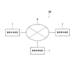
図1は、本実施の形態における医用画像システム100の全体構成を示す概念図である。図1に示すように、医用画像システム100は、画像生成装置1、画像処理装置2及び画像表示装置3がネットワークNを介して、相互にデータ送受信可能なように接続されている。
First, the configuration of the present embodiment will be described.
FIG. 1 is a conceptual diagram showing an overall configuration of a
なお、本実施の形態では、画像生成装置1、画像処理装置2及び画像表示装置3がネットワーク接続された例を説明するが、これに限らず、各装置が直接有線接続されたシステム構成であってもよい。また、各装置の台数及び設置場所は特に限定されない。
In the present embodiment, an example in which the image generation device 1, the
ネットワークNは、LAN(Local Area Network)やWAN(Wide Area Network)、インターネット等の様々な回線形態を適用可能である。なお、病院等の医療機関内で許可されるのであれば、無線通信や赤外線通信であってもよいが、重要な患者情報を含むため、送受信される情報は暗号化することが好ましい。また、病院内の通信方式としては、一般的に、DICOM(Digital Image and Communications in Medicine)規格が用いられており、上述したネットワークN上の各装置間の通信では、DICOM MWM(Modality Worklist Management)やDICOM MPPS(Modality Performed Procedure Step)が用いられる。 As the network N, various line forms such as a LAN (Local Area Network), a WAN (Wide Area Network), and the Internet can be applied. Note that wireless communication or infrared communication may be used as long as it is permitted within a medical institution such as a hospital. However, since important patient information is included, it is preferable to encrypt information to be transmitted and received. Moreover, as a communication system in a hospital, the DICOM (Digital Image and Communications in Medicine) standard is generally used, and in the communication between each device on the network N described above, DICOM MWM (Modality Worklist Management). Or DICOM MPPS (Modality Performed Procedure Step).
画像生成装置1は、例えば、CR(Computed Radiography)、FPD(Flat Panel Detector)、CT(Computed Tomography)、MRI(Magnetic Resonance Imaging)、超音波診断装置等のモダリティから構成され、人体を撮影し、撮影した画像をデジタル変換して、医用画像データを生成する装置である。 The image generation apparatus 1 is composed of modalities such as CR (Computed Radiography), FPD (Flat Panel Detector), CT (Computed Tomography), MRI (Magnetic Resonance Imaging), an ultrasonic diagnostic apparatus, and the like. This is a device that digitally converts a captured image to generate medical image data.
なお、画像生成装置1は上述したDICOM規格に準拠した装置であり、医用画像データに対して、DICOMの画像付帯情報(以下、付帯情報と称する)を入力させたり、自動生成したりすることができる。画像生成装置1は、生成された医用画像データとともにその付帯情報を医用画像データのヘッダ情報としてネットワークNを介して画像処理装置2へ出力するものとするが、DICOM規格に不準拠の場合、図示しないDICOM変換装置を用い、付帯情報を入力させることができる。
Note that the image generation apparatus 1 is an apparatus compliant with the DICOM standard described above, and DICOM image supplementary information (hereinafter referred to as supplementary information) can be input or automatically generated for medical image data. it can. The image generation apparatus 1 outputs the accompanying information along with the generated medical image data to the
医用画像データの付帯情報としては、例えば、撮影された患者の患者氏名、患者ID、年齢、性別等の患者に関する患者情報、撮影日、検査ID、撮影部位、撮影条件(体位、撮影方向等)、画像生成装置(モダリティ種)等の撮影情報、医用画像データの画素数、サンプリングピッチ、ビット数等の画像データ情報が含まれる。 As incidental information of medical image data, for example, patient information about the patient such as the patient name, patient ID, age, and sex of the patient who was photographed, photographing date, examination ID, photographing part, photographing condition (position, photographing direction, etc.) Image data such as imaging information such as an image generation device (modality type), the number of pixels of medical image data, a sampling pitch, and the number of bits are included.
画像処理装置2は、画像生成装置1から供給される医用画像データに対し医師の読影診断の効率化を図るため、階調処理、周波数処理、ダイナミックレンジ圧縮処理を始めとする画像処理を施して画像表示装置3へ送信する装置である。
The
画像表示装置3は、医師が医用画像を表示して読影観察するための医用画像表示装置であり、画像処理装置2において画像処理された医用画像データに基づいて医用画像を表示画面上に表示する。
The
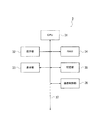
以下、画像表示装置3の内部構成について説明する。
図2は、画像表示装置3の機能的構成を示すブロック図である。図2に示すように、画像表示装置3は、CPU31、操作部32、表示部33、RAM34、記憶部35、通信制御部36等を備えて構成され、各部はバス37により接続されている。
Hereinafter, the internal configuration of the
FIG. 2 is a block diagram illustrating a functional configuration of the
CPU31は、記憶部35に記憶されているシステムプログラムを読み出し、RAM34内に形成されたワークエリアに展開し、該システムプログラムに従って各部を制御する。また、CPU31は、記憶部35に記憶されている表示制御処理プログラムを始めとする各種処理プログラム、各種アプリケーションプログラムを読み出してワークエリアに展開し、後述する表示制御処理(図3参照)を始めとする各種処理を実行する。
The
操作部32は、カーソルキー、数字入力キー、及び各種機能キー等を備えたキーボードと、マウス等のポインティングデバイスを備えて構成され、キーボードに対するキー操作やマウス操作により入力された指示信号をCPU31に出力する。また、操作部32は、表示部33の表示画面にタッチパネルを備えても良く、この場合、タッチパネルを介して入力された指示信号をCPU31に出力する。
The
表示部33は、1以上のLCD(Liquid Crystal Display)やCRT等のモニタにより構成され、CPU21から入力される表示信号の指示に従って、表示画面上に医用画像を表示する。
The
RAM34は、CPU31により実行制御される各種処理において、記憶部35から読み出されたCPU31で実行可能な各種プログラム、入力若しくは出力データ、及びパラメータ等の一時的に記憶するワークエリアを形成する。
The
記憶部35は、HDD(Hard Disc Drive)や不揮発性の半導体メモリ等により構成され、CPU31で実行されるシステムプログラム、当該システムプログラムに対応する、表示制御処理プログラムを始めとする各種処理プログラム、各種アプリケーションプログラム、各種データ等を記憶する。これらの各種プログラムは、読取可能なプログラムコードの形態で格納され、CPU31は、当該プログラムコードに従った動作を逐次実行する。
The
通信制御部36は、LANアダプタやルータやTA(Terminal Adapter)等を備え、ネットワークNに接続された各装置との間の通信を制御する。
The
次に、本実施の形態における動作について説明する。
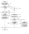
図3は、表示部33のモニタの表示画面上に医用画像データに基づいて医用画像を表示する際にCPU31により実行される表示制御処理を示すフローチャートである。CPU31は、記憶部35に記憶された表示制御処理プログラムとの協働によるソフトウエア処理により当該処理を実行し、比較手段、表示制御手段を実現する。
Next, the operation in this embodiment will be described.
FIG. 3 is a flowchart showing a display control process executed by the
まず、表示すべき医用画像の画素数P1が取得される(ステップS1)。画素数P1は、例えば、医用画像データの付帯情報から取得することが可能であり、画素数P1は、医用画像の行方向の画素数q1及び列方向の画素数q2を示す情報からなる。 First, the number of pixels P1 of the medical image to be displayed is acquired (step S1). The pixel number P1 can be acquired from, for example, supplementary information of medical image data, and the pixel number P1 includes information indicating the number of pixels q1 in the row direction and the number of pixels q2 in the column direction of the medical image.
次いで、画素数P1とモニタの表示画面の表示可能画素数P2が比較される(ステップS2)。画素数P2は、モニタの表示画面の行方向の表示可能画素数r1及び列方向の表示可能画素数r2を示す情報からなる。画素数P1とP2の比較は、行方向及び列方向の画素数の比較で行われ、両方向とも医用画像の画素数がモニタの表示可能画素数以下である場合、即ち、q1がr1以下であり、かつq2がr2以下である場合、比較結果がP1≦P2と処理され、少なくとも一方向の医用画像の画素数がモニタの表示可能画素数を超えた場合、即ち、q1がr1を超えたか或いはq2がr2を超えた場合は、比較結果がP1>P2と処理される。なお、表示部33に複数のモニタが接続されている場合には、表示可能画素数が最大のモニタの表示画面の画素数を表示可能画素数P2とする。
Next, the pixel number P1 is compared with the displayable pixel number P2 on the monitor display screen (step S2). The pixel number P2 includes information indicating the displayable pixel number r1 in the row direction and the displayable pixel number r2 in the column direction on the display screen of the monitor. The number of pixels P1 and P2 are compared by comparing the number of pixels in the row direction and the column direction. When the number of pixels of the medical image is less than or equal to the number of displayable pixels on the monitor in both directions, that is, q1 is less than or equal to r1. When q2 is equal to or less than r2, the comparison result is processed as P1 ≦ P2, and the number of pixels of the medical image in at least one direction exceeds the number of displayable pixels of the monitor, that is, q1 exceeds r1 or When q2 exceeds r2, the comparison result is processed as P1> P2. When a plurality of monitors are connected to the
ステップS2における比較の結果、表示可能画素数P1≦P2である場合、即ち、表示すべき医用画像の画素数P1がモニタの表示画面の表示可能画素数P2以下である場合、モニタの表示画面上に当該医用画像が表示される(ステップS3)。 As a result of the comparison in step S2, if the number of displayable pixels P1 ≦ P2, that is, if the number of pixels P1 of the medical image to be displayed is less than or equal to the number of displayable pixels P2 on the monitor display screen, The medical image is displayed on (Step S3).
一方、P1>P2である場合、即ち、表示すべき医用画像の画素数P1がモニタの表示画面の表示可能画素数P2を超える場合、表示部33に複数のモニタが接続されているか否かが判断され(ステップS4)、複数のモニタが接続されている場合には(ステップS4;YES)、表示すべき医用画像が、その画素数P1と各モニタの画素数に応じて複数の画像に分割され(ステップS5)、分割された画像のそれぞれが複数のモニタの各表示画面上に表示される(ステップS6)。
On the other hand, if P1> P2, that is, if the number of pixels P1 of the medical image to be displayed exceeds the number of displayable pixels P2 on the monitor display screen, whether or not a plurality of monitors are connected to the
ステップS4において、複数のモニタが接続されていない、即ち、接続されているモニタが1つであると判断された場合には(ステップS4;NO)、表示すべき医用画像内の被写体領域画像が抽出され(ステップS7)、この抽出された被写体領域画像が複数の画像に分割され(ステップS8)、この分割された複数の画像が同一表示画面上に表示可能であるか否かが判断され(ステップS9)、表示可能であると判断された場合には(ステップS9;YES)、被写体領域画像が同一表示画面上に分割した状態で表示される(ステップS10)。 If it is determined in step S4 that a plurality of monitors are not connected, that is, one monitor is connected (step S4; NO), the subject area image in the medical image to be displayed is displayed. The extracted subject area image is divided into a plurality of images (step S8), and it is determined whether or not the plurality of divided images can be displayed on the same display screen (step S7). If it is determined in step S9) that display is possible (step S9; YES), the subject area image is displayed in a divided state on the same display screen (step S10).
被写体領域画像の抽出は、例えば、医用画像データをそれぞれ解析し、被写体の輪郭線を抽出する等により行う。例えば、医用画像データのそれぞれの濃度データを適当な閾値を用いて2値化し、「0」と「1」の境界を追跡して輪郭線とし、この輪郭線及び撮影部位/体位、撮影方向に応じて被写体領域画像を決定する。或いは、人体領域又は人体内部の所定の解剖学的構造に対応する領域の輪郭線抽出方法(日本乳癌検診学会誌,Vol.17,No.1,pp87-102,1998、特開昭63−240832号公報)を用いて被写体領域を決定するようにしてもよい。 The extraction of the subject area image is performed, for example, by analyzing the medical image data and extracting the contour line of the subject. For example, each density data of medical image data is binarized by using an appropriate threshold value, and the boundary between “0” and “1” is traced to form a contour line. Accordingly, the subject area image is determined. Alternatively, a method for extracting a contour line of a human body region or a region corresponding to a predetermined anatomical structure inside the human body (Japan Breast Cancer Screening Society Journal, Vol. 17, No. 1, pp 87-102, 1998, JP-A 63-240832 The subject area may be determined by using (
また、抽出された被写体領域画像の分割数や分割方法は特に限定されないが、ここでは、例えば、被写体領域画像を上領域と下領域の2つの領域の画像に等分割し、分割された画像をモニタの表示画面の左右に表示することとする(図5参照)。このとき、ステップS9における、被写体領域画像が分割された複数の画像が同一表示画面上に表示可能であるか否かの判断は、例えば、分割された被写体領域画像の行方向の画素数をq3とすると、q3×2がモニタの表示画面の行方向の画素数r1以下であり、かつ分割された被写体領域画像の列方向の画素数q4がモニタの表示画面の列方向の画素数r2以下である場合に分割された複数の画像が同一表示画面上に表示可能であると判断される。 Further, the number of divisions and the division method of the extracted subject area image are not particularly limited, but here, for example, the subject area image is equally divided into two area images, an upper area and a lower area, and the divided images are divided. They are displayed on the left and right of the monitor display screen (see FIG. 5). At this time, whether or not a plurality of images obtained by dividing the subject area image in step S9 can be displayed on the same display screen is determined by, for example, determining the number of pixels in the row direction of the divided subject area image by q3. Then, q3 × 2 is the number of pixels r1 or less in the row direction of the monitor display screen, and the number of pixels q4 in the column direction of the divided subject area image is less than or equal to the number of pixels r2 in the column direction of the monitor display screen. In some cases, it is determined that a plurality of divided images can be displayed on the same display screen.
一方、ステップS9において、被写体領域画像を分割した複数の画像が同一表示画面上に表示可能ではないと判断された場合(ステップS9;NO)、表示すべき医用画像が複数の画像に分割され(ステップS11)、分割された複数の画像が表示画面上に切り替え可能に表示される(ステップS10)。 On the other hand, when it is determined in step S9 that a plurality of images obtained by dividing the subject area image cannot be displayed on the same display screen (step S9; NO), the medical image to be displayed is divided into a plurality of images ( In step S11), the plurality of divided images are displayed on the display screen in a switchable manner (step S10).
ステップS5、8、11で医用画像又は医用画像内の被写体領域画像が分割される場合、分割された画像の相互に分割境界部近傍の画像が重複して含まれるように分割画像が生成される。これにより、医用画像内の被写体領域画像の一部が分割により欠落することを防止することが可能となる。ステップS6、10、12で分割された医用画像又は医用画像内の被写体領域画像を表示する際には、分割境界部近傍の画像が重複している領域の少なくとも一方に、例えば、重複領域の濃度を変える、重複領域を示す線を表示する、重複領域をハッチングにする等により、重複領域であることが識別可能に表示される。 When the medical image or the subject area image in the medical image is divided in steps S5, S8, and S11, the divided images are generated so that the divided images include the images in the vicinity of the division boundary portion. . As a result, it is possible to prevent a part of the subject area image in the medical image from being lost due to the division. When displaying the medical image divided in Steps S6, 10, and 12 or the subject region image in the medical image, for example, the density of the overlapping region is set in at least one of the regions where the images near the dividing boundary portion overlap. Change, displaying a line indicating the overlapping area, or hatching the overlapping area, the overlapping area is displayed in an identifiable manner.
図4に、ステップS6における画面表示例を示す。図4は、画像表示装置3に2台のモニタが接続されている場合の画面表示例である。図4において、表示しようとする医用画像D1の画素数は表示部33の2つのモニタの画面M1、画面M2の各表示可能画素数より大きい。そこで、医用画像D1は上下に分割され、分割された画像のそれぞれが画面M1、M2のそれぞれに表示される。医用画像D1の分割の際には、分割された画像の相互に分割境界部近傍の画像が重複して含まれるように分割画像が生成される。画面M2のハッチングで示す領域R1は、画面M1と重複して表示された領域であることを示すものである。このように、医用画像の画素数がモニタの表示画面の表示可能画素数を超える場合であっても、医用画像を縮小せずに医用画像全体を被写体領域画像O1の画像の欠落がないように表示するので、医師は、画像生成装置1から得られた被写体領域画像の全てを診断に用いることが可能となり、診断性能の低下を防止することが可能となる。
FIG. 4 shows a screen display example in step S6. FIG. 4 is a screen display example when two monitors are connected to the
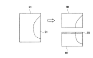
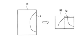
図5に、ステップS10における画面表示例を示す。図5において、表示しようとする医用画像D1の画素数はモニタの画面M3の表示可能画素数より大きい。そこで、医用画像D1内の被写体領域画像が抽出され、抽出された被写体領域画像は、上下に分割され、分割された2つの画像が同一の画面M3の左右に分割表示される。被写体領域画像の分割の際には、分割された画像の相互に分割境界部近傍の画像が重複して含まれるように分割画像が生成される。画面M3の右領域のハッチングで示す領域R2は、左領域に表示された画像と重複して表示された領域であることを示すものである。このように、医用画像の画素数がモニタ表示画面の表示可能画素数を超える場合であっても、医用画像を縮小せずに医用画像全体を被写体領域画像O1の画像の欠落がないように表示するので、医師は、画像生成装置1から得られた被写体領域画像の全てを診断に用いることが可能となり、診断性能の低下を防止することが可能となる。 FIG. 5 shows a screen display example in step S10. In FIG. 5, the number of pixels of the medical image D1 to be displayed is larger than the number of displayable pixels on the monitor screen M3. Therefore, the subject area image in the medical image D1 is extracted, and the extracted subject area image is divided vertically, and the two divided images are divided and displayed on the left and right of the same screen M3. When the subject area image is divided, the divided images are generated so that the divided images include the images in the vicinity of the division boundary portion. A region R2 indicated by hatching in the right region of the screen M3 indicates that the region is displayed overlapping with the image displayed in the left region. As described above, even when the number of pixels of the medical image exceeds the number of displayable pixels on the monitor display screen, the entire medical image is displayed without reducing the image of the subject region image O1 without reducing the medical image. Therefore, the doctor can use all of the subject area images obtained from the image generation device 1 for diagnosis, and can prevent deterioration in diagnostic performance.
図6に、ステップS12における画面表示例を示す。図6において、表示しようとする医用画像D2の画素数はモニタの画面M4の表示可能画素数より大きい。そこで、医用画像D2は上下に分割され、分割された画像のうち上領域画像が画面M4に表示される。被写体領域画像の分割の際には、分割された画像の相互に分割境界部近傍の画像が重複して含まれるように分割画像が生成される。画面M4上には、画面切り替えボタンB1が表示され、このボタンB1が押下されると、分割された画像の下領域画像が画面M5に表示されるとともに、画面切り替えボタンB2が表示される。この画面切り替えボタンB2が押下されると、モニタの表示画面が上領域画像を表示する画面M4の表示に切り替わる。画面M5においてハッチングで示す領域R3は、上領域画像の表示と重複して表示された領域であることを示すものである。このように、医用画像の画素数がモニタ表示画面の表示可能画素数を超える場合であっても、医用画像を縮小せずに医用画像全体を被写体領域画像O2の画像の欠落がないように表示するので、医師は、画像生成装置1から得られた被写体領域画像の全てを診断に用いることが可能となり、診断性能の低下を防止することが可能となる。 FIG. 6 shows a screen display example in step S12. In FIG. 6, the number of pixels of the medical image D2 to be displayed is larger than the number of displayable pixels on the monitor screen M4. Therefore, the medical image D2 is divided into upper and lower parts, and the upper region image among the divided images is displayed on the screen M4. When the subject area image is divided, the divided images are generated so that the divided images include the images in the vicinity of the division boundary portion. A screen switching button B1 is displayed on the screen M4. When this button B1 is pressed, the lower region image of the divided image is displayed on the screen M5, and the screen switching button B2 is displayed. When the screen switching button B2 is pressed, the display screen of the monitor is switched to the display of the screen M4 that displays the upper area image. A region R3 indicated by hatching in the screen M5 indicates that the region is displayed overlapping with the display of the upper region image. As described above, even when the number of pixels of the medical image exceeds the number of displayable pixels on the monitor display screen, the entire medical image is displayed without reducing the image of the subject region image O2 without reducing the medical image. Therefore, the doctor can use all of the subject area images obtained from the image generation device 1 for diagnosis, and can prevent deterioration in diagnostic performance.
以上説明したように、画像表示装置3によれば、表示画面上に表示すべき医用画像の画素数とモニタの表示画面上に表示可能な表示可能画素数とを比較し、比較の結果、医用画像の画素数が表示可能画素数を超える場合、表示すべき医用画像を複数の画像に分割して、分割された画像のそれぞれを複数のモニタの各表示画面上に表示させる。或いは、分割された画像のそれぞれをモニタの表示画面上に切り替えて表示させる。或いは、表示すべき医用画像内の被写体領域画像を抽出して複数の画像に分割し、分割された複数の画像が同一表示画面上に表示可能か否かを判断し、表示可能である場合に分割された複数の画像を同一表示画面上に分割表示させる。
As described above, according to the
従って、表示画面上に表示すべき医用画像の画素数がモニタの表示画面上に表示可能な表示可能画素数を超える場合であっても、被写体領域画像の欠落なく医用画像を表示することが可能となり、診断性能の低下を防止することが可能となる。 Therefore, even when the number of pixels of a medical image to be displayed on the display screen exceeds the number of displayable pixels that can be displayed on the display screen of the monitor, it is possible to display a medical image without omission of the subject area image. Thus, it is possible to prevent a deterioration in diagnostic performance.
また、医用画像を分割する際には、分割された画像の相互に分割境界部近傍の画像が重複して含まれるように分割画像を生成するので、分割により被写体領域画像の一部が欠落することがなくなり、画像生成装置1から得られた被写体領域画像の全てを診断に用いることが可能となる。また、表示が重複している領域については、濃度を変える、重複領域を示す線を表示する、重複領域をハッチングにする等により、重複領域であることを識別可能に表示するので、医師が重複領域を二重に診断してしまうことがない。 In addition, when dividing a medical image, a divided image is generated so that the divided images include overlapping images in the vicinity of the division boundary portion, so that part of the subject region image is lost due to the division. Therefore, all of the subject area images obtained from the image generation apparatus 1 can be used for diagnosis. In addition, for areas where the display overlaps, the doctor displays the overlapping area by identifying the overlapping area by changing the density, displaying a line indicating the overlapping area, or hatching the overlapping area. There is no double diagnosis of the area.
なお、上記実施の形態における記述内容は、本発明に係る医用画像システム100の好適な一例であり、これに限定されるものではない。
例えば、上記実施の形態においては、医用画像又は被写体領域画像を上下の2つに分割する例を説明したが、分割数、分割方法はこれに限定されない。
In addition, the description content in the said embodiment is a suitable example of the
For example, in the above embodiment, the example in which the medical image or the subject area image is divided into the upper and lower parts has been described. However, the number of divisions and the division method are not limited thereto.
その他、医用画像システム100を構成する各装置の細部構成及び細部動作に関しても、本発明の趣旨を逸脱することのない範囲で適宜変更可能である。
In addition, the detailed configuration and detailed operation of each apparatus constituting the
100 医用画像システム
1 画像生成装置
2 画像処理装置
3 画像表示装置
31 CPU
32 操作部
33 表示部
34 RAM
35 記憶部
36 通信制御部
37 バス
N ネットワーク
DESCRIPTION OF
32
35
Claims (8)
前記表示画面上に表示すべき医用画像の画素数と前記表示画面上に表示可能な表示可能画素数とを比較する比較手段と、
前記比較手段による比較の結果、前記医用画像の画素数が前記表示可能画素数を超える場合、前記表示すべき医用画像を複数の画像に分割して前記表示手段の同一又は異なる表示画面上に表示させる表示制御手段と、
を備えることを特徴とする医用画像表示装置。 One or more display means for displaying a medical image on a display screen;
Comparison means for comparing the number of pixels of a medical image to be displayed on the display screen with the number of displayable pixels that can be displayed on the display screen;
When the number of pixels of the medical image exceeds the number of displayable pixels as a result of comparison by the comparison unit, the medical image to be displayed is divided into a plurality of images and displayed on the same or different display screens of the display unit Display control means,
A medical image display device comprising:
前記表示画面上に表示すべき医用画像の画素数と前記表示画面上に表示可能な表示可能画素数とを比較する工程と、
前記比較の結果、前記医用画像の画素数が前記表示可能画素数を超える場合、前記表示すべき医用画像を複数の画像に分割して前記表示手段の同一又は異なる表示画面上に表示させる工程と、
を含むことを特徴とする医用画像表示方法。 In a medical image display method in a medical image display device comprising one or more display means for displaying a medical image on a display screen,
Comparing the number of pixels of a medical image to be displayed on the display screen with the number of displayable pixels that can be displayed on the display screen;
As a result of the comparison, when the number of pixels of the medical image exceeds the number of displayable pixels, the medical image to be displayed is divided into a plurality of images and displayed on the same or different display screens of the display means; ,
A medical image display method comprising:
前記表示画面上に表示すべき医用画像の画素数と前記表示画面上に表示可能な表示可能画素数とを比較する機能と、
前記比較の結果、前記医用画像の画素数が前記表示可能画素数を超える場合、前記表示すべき医用画像を複数の画像に分割して前記表示手段の同一又は異なる表示画面上に表示させる機能と、
を実現させるためのプログラム。 A computer for controlling a medical image display device comprising one or more display means for displaying a medical image on a display screen;
A function of comparing the number of pixels of a medical image to be displayed on the display screen with the number of displayable pixels that can be displayed on the display screen;
A function of dividing the medical image to be displayed into a plurality of images and displaying them on the same or different display screens when the number of pixels of the medical image exceeds the number of displayable pixels as a result of the comparison; ,
A program to realize
Priority Applications (1)
| Application Number | Priority Date | Filing Date | Title |
|---|---|---|---|
| JP2004112232A JP2005296052A (en) | 2004-04-06 | 2004-04-06 | MEDICAL IMAGE DISPLAY DEVICE, MEDICAL IMAGE DISPLAY METHOD, AND PROGRAM |
Applications Claiming Priority (1)
| Application Number | Priority Date | Filing Date | Title |
|---|---|---|---|
| JP2004112232A JP2005296052A (en) | 2004-04-06 | 2004-04-06 | MEDICAL IMAGE DISPLAY DEVICE, MEDICAL IMAGE DISPLAY METHOD, AND PROGRAM |
Publications (1)
| Publication Number | Publication Date |
|---|---|
| JP2005296052A true JP2005296052A (en) | 2005-10-27 |
Family
ID=35328336
Family Applications (1)
| Application Number | Title | Priority Date | Filing Date |
|---|---|---|---|
| JP2004112232A Pending JP2005296052A (en) | 2004-04-06 | 2004-04-06 | MEDICAL IMAGE DISPLAY DEVICE, MEDICAL IMAGE DISPLAY METHOD, AND PROGRAM |
Country Status (1)
| Country | Link |
|---|---|
| JP (1) | JP2005296052A (en) |
Cited By (3)
| Publication number | Priority date | Publication date | Assignee | Title |
|---|---|---|---|---|
| JP2011024622A (en) * | 2009-07-21 | 2011-02-10 | Toshiba Corp | Medical system |
| JP2011072374A (en) * | 2009-09-29 | 2011-04-14 | Fujifilm Corp | Medical diagnostic imaging assistance apparatus |
| WO2019087861A1 (en) * | 2017-10-31 | 2019-05-09 | キヤノン株式会社 | Image processing device, image processing method, and program |
-
2004
- 2004-04-06 JP JP2004112232A patent/JP2005296052A/en active Pending
Cited By (5)
| Publication number | Priority date | Publication date | Assignee | Title |
|---|---|---|---|---|
| JP2011024622A (en) * | 2009-07-21 | 2011-02-10 | Toshiba Corp | Medical system |
| JP2011072374A (en) * | 2009-09-29 | 2011-04-14 | Fujifilm Corp | Medical diagnostic imaging assistance apparatus |
| WO2019087861A1 (en) * | 2017-10-31 | 2019-05-09 | キヤノン株式会社 | Image processing device, image processing method, and program |
| JP2019080813A (en) * | 2017-10-31 | 2019-05-30 | キヤノン株式会社 | IMAGE PROCESSING APPARATUS, IMAGE PROCESSING METHOD, AND PROGRAM |
| US11457877B2 (en) | 2017-10-31 | 2022-10-04 | Canon Kabushiki Kaisha | Image processing apparatus, image processing method, and non-transitory computer-readable medium |
Similar Documents
| Publication | Publication Date | Title |
|---|---|---|
| US7388974B2 (en) | Medical image processing apparatus | |
| CN103957779B (en) | Medical information reading device | |
| JP4911029B2 (en) | Abnormal shadow candidate detection method, abnormal shadow candidate detection device | |
| JP2011123084A (en) | Image display system and image display program | |
| JP2005287927A (en) | Image processing apparatus, image processing method, and medical image system | |
| JP2010057684A (en) | Medical image display, medical image displaying method, and medical image displaying program | |
| JP4635681B2 (en) | Medical image interpretation system | |
| JP4182794B2 (en) | Medical image display method and medical image display system | |
| JP4729860B2 (en) | Image processing apparatus and image processing method | |
| JP2005287750A (en) | Medical image display method, medical image display device and program | |
| JP6146249B2 (en) | Image display device and image display method | |
| JP2006102044A (en) | Medical image processing system and medical image processing method | |
| JP2005296052A (en) | MEDICAL IMAGE DISPLAY DEVICE, MEDICAL IMAGE DISPLAY METHOD, AND PROGRAM | |
| JP4475054B2 (en) | Breast image processing method and breast image output system | |
| US20230401708A1 (en) | Recording medium, information processing apparatus, information processing system, and information processing method | |
| JP2007072649A (en) | Interpretation report creation device | |
| WO2006087895A1 (en) | Medical image system | |
| JP2006271800A (en) | Image processing apparatus, method and program | |
| JP2004305551A (en) | Medical image diagnostic reading system | |
| JP2006061601A (en) | Medical image display device, medical image display system, and medical image display program | |
| JP2005173818A (en) | Medical image diagnostic support system | |
| JP7323989B2 (en) | Computer program, recording medium, display device and display method | |
| JP7428055B2 (en) | Diagnostic support system, diagnostic support device and program | |
| JP2006055419A (en) | Breast image generation system | |
| US12548112B2 (en) | Recording medium, display device, display system and display method that displays lesion candidate areas or display information thereof |