JP2007014351A - Infectious disease detection probe, gene detection probe, infectious disease pathogen amplification reaction primer, infectious disease pathogen amplification reaction primer set, and infectious disease pathogen detection method - Google Patents
Infectious disease detection probe, gene detection probe, infectious disease pathogen amplification reaction primer, infectious disease pathogen amplification reaction primer set, and infectious disease pathogen detection method Download PDFInfo
- Publication number
- JP2007014351A JP2007014351A JP2006257739A JP2006257739A JP2007014351A JP 2007014351 A JP2007014351 A JP 2007014351A JP 2006257739 A JP2006257739 A JP 2006257739A JP 2006257739 A JP2006257739 A JP 2006257739A JP 2007014351 A JP2007014351 A JP 2007014351A
- Authority
- JP
- Japan
- Prior art keywords
- infectious disease
- detecting
- probe
- seq
- nos
- Prior art date
- Legal status (The legal status is an assumption and is not a legal conclusion. Google has not performed a legal analysis and makes no representation as to the accuracy of the status listed.)
- Pending
Links
Classifications
-
- Y—GENERAL TAGGING OF NEW TECHNOLOGICAL DEVELOPMENTS; GENERAL TAGGING OF CROSS-SECTIONAL TECHNOLOGIES SPANNING OVER SEVERAL SECTIONS OF THE IPC; TECHNICAL SUBJECTS COVERED BY FORMER USPC CROSS-REFERENCE ART COLLECTIONS [XRACs] AND DIGESTS
- Y02—TECHNOLOGIES OR APPLICATIONS FOR MITIGATION OR ADAPTATION AGAINST CLIMATE CHANGE
- Y02A—TECHNOLOGIES FOR ADAPTATION TO CLIMATE CHANGE
- Y02A50/00—TECHNOLOGIES FOR ADAPTATION TO CLIMATE CHANGE in human health protection, e.g. against extreme weather
- Y02A50/30—Against vector-borne diseases, e.g. mosquito-borne, fly-borne, tick-borne or waterborne diseases whose impact is exacerbated by climate change
Landscapes
- Measuring Or Testing Involving Enzymes Or Micro-Organisms (AREA)
Abstract
【課題】一度に大量調製することが可能であり、かつ、類似菌種間における菌種の同定が可能な感染症検出用プローブを提供する。
【解決手段】特定の塩基配列及びその相補配列のうちのいずれかを有するオリゴヌクレオチドから、黄色ブドウ球菌由来の遺伝子、表皮ブドウ球菌由来の遺伝子、大腸菌由来の遺伝子、肺炎桿菌由来の遺伝子、緑膿菌由来の遺伝子、セラチア菌由来の遺伝子、肺炎連鎖球菌由来の遺伝子、インフルエンザ菌由来の遺伝子、エンテロバクタークロアカエ菌由来の遺伝子、エンテロコッカウフェカリス菌由来の遺伝子のいずれかを検出可能な感染症検出用プローブが構成される。
【選択図】なしProvided is an infectious disease detection probe that can be prepared in a large amount at one time and can identify bacterial species among similar bacterial species.
From an oligonucleotide having any one of a specific base sequence and a complementary sequence thereof, a gene derived from Staphylococcus aureus, a gene derived from Staphylococcus epidermidis, a gene derived from E. coli, a gene derived from Klebsiella pneumoniae, Pseudomonas aeruginosa Infectious diseases that can detect any of genes derived from fungi, Serratia, Streptococcus pneumoniae, Haemophilus influenzae, Enterobacter cloacae, or Enterococcus faecalis A detection probe is configured.
[Selection figure] None
Description
本発明は、感染症疾患の原因菌である感染症起炎菌の検出及び/又は同定に関する。特に、感染症疾患の原因菌の検出および同定に有用な感染症起因菌由来のプローブ及び、プローブセットならびに担体、遺伝子検査方法に関するものである。 The present invention relates to the detection and / or identification of infectious disease-causing bacteria that are causative bacteria of infectious diseases. In particular, the present invention relates to probes derived from infectious disease-causing bacteria, probe sets and carriers, and genetic testing methods useful for detecting and identifying causative bacteria of infectious diseases.
また、感染症起炎菌の検出及び/又は同定に好適な感染症起炎菌のPCR増幅処理に関する。 In addition, the present invention relates to a PCR amplification process for infectious disease-causing bacteria suitable for detection and / or identification of infectious disease-causing bacteria.
近年、DNAチップ(またはDNAマイクロアレイともいう。以下同じ)を用いた遺伝子発現解析が創薬を初め種々の領域で行なわれている。それは、各種遺伝子セット(プローブ)が配置されたDNAマイクロアレイに、それぞれ異なった検体DNAを反応させ、各検体に存在するそれぞれの遺伝子量を比較して、各ステージで大量に存在する(発現量の高い)遺伝子、或いは逆に不活性化している(発現量の低い)遺伝子を分類し、機能と関連付けて解析するものである。 In recent years, gene expression analysis using DNA chips (also referred to as DNA microarrays; hereinafter the same) has been performed in various areas including drug discovery. This is because DNA microarrays with various gene sets (probes) are reacted with different sample DNAs, and the amount of each gene present in each sample is compared. A gene that is high) or a gene that is inactive (low expression level) is classified and analyzed in association with the function.
感染症の起炎菌検査はその一例であり、江崎らは特許文献1において、DNAプローブとして染色体DNAが固定化されたDNAチップを用いる微生物同定法を提案している。この方法によれば、互いにGC含量の異なる複数の既知微生物由来の染色体DNAと、検体中の未知微生物由来の染色体DNAとを反応させ、生じたハイブリダイゼーション複合体を検出することで検体中の未知微生物を検出することが可能である。 Examination of infectious disease-causing bacteria is one example, and Ezaki et al. In Patent Document 1 propose a microorganism identification method using a DNA chip on which chromosomal DNA is immobilized as a DNA probe. According to this method, a chromosomal DNA derived from a plurality of known microorganisms having different GC contents from each other reacts with a chromosomal DNA derived from an unknown microorganism in the sample, and the resulting hybridization complex is detected to detect the unknown in the sample. It is possible to detect microorganisms.
また、大野らは感染症の起炎菌検査のためのDNAチップに用いるプローブとして、特許文献2で制限酵素断片を利用した真菌の検出用プローブを、特許文献3で緑膿菌の検出用プローブを、特許文献4でEscherichia coli(エシェリキア コリ)菌、klebsiella pneumoniae(クレブシエラ ニューモニエ)菌ならびにEnterobacter cloacae(エンテロバクター クロアカエ)菌の制限酵素断片を利用した検出用プローブをそれぞれ提案している。 Ohno et al., As a probe for use in a DNA chip for infectious disease-causing bacteria testing, disclosed in Patent Document 2 was a fungus detection probe using a restriction enzyme fragment, and Patent Document 3 was a Pseudomonas aeruginosa detection probe. Patent Document 4 proposes detection probes using restriction enzyme fragments of Escherichia coli, klebsiella pneumoniae, and Enterobacter cloacae, respectively.
また、上記マイクロアレイとしては、例えば、スタンフォード法と呼ばれるスタンピング法によるマイクロアレイが知られており、例えば、宝酒造株式会社からがん疾患に関連するヒト由来既知遺伝子のcDNA断片をスポット或いはスタンプにより塗布したDNAチップ、ヒト由来既知遺伝子約1000種類のcDNA断片をスライドガラスに貼り付けたチップが市販されている。 In addition, as the microarray, for example, a microarray by a stamping method called a Stanford method is known. For example, DNA coated with a cDNA fragment of a known gene derived from a human related to cancer disease by a spot or stamp from Takara Shuzo Co., Ltd. Chips and chips in which about 1000 kinds of cDNA fragments of known human-derived genes are attached to a slide glass are commercially available.
一方、Affymetrixのチップは、上記既知遺伝子cDNAを元にオリゴヌクレオチドプローブセットを設計し、基板上の合成によりプローブを配置したもので、一枚のチップ上に高密度にオリゴプローブが配置され、一度に一万を超える遺伝子の発現レベルを解析できるように設計されている。
しかしながら、上記従来技術に示したDNAチップは、染色体DNA或いは制限酵素断片等のDNAプローブを利用するものであり、いずれも微生物から直接取り出したDNAを材料としている。このため、一度に大量に調製することは困難であり、臨床診断用には適さないという問題があった。これは臨床診断用に用いるためには、安価で均質なDNAチップの大量生産が必要であり、このためにプローブ溶液として均質なDNAの大量調製が不可欠となってくるところ、DNAプローブでは、このような大量調製ができないからである。なお、DNAプローブであっても、PCR増幅反応を利用することで当該DNAを徐々に増加させていくことは可能であるが、PCR反応では、一度に大量調製することは困難であることから、臨床診断用に利用することは難しい。 However, the DNA chip shown in the above prior art uses a DNA probe such as a chromosomal DNA or a restriction enzyme fragment, and all use DNA taken directly from a microorganism as a material. For this reason, it was difficult to prepare a large amount at a time, and there was a problem that it was not suitable for clinical diagnosis. This requires mass production of inexpensive and homogeneous DNA chips for use in clinical diagnosis. For this reason, mass production of homogeneous DNA as a probe solution is indispensable. This is because such a large-scale preparation cannot be performed. Note that even with a DNA probe, it is possible to gradually increase the DNA by using a PCR amplification reaction, but it is difficult to prepare a large amount at a time in a PCR reaction. It is difficult to use for clinical diagnosis.
また、DNAプローブは塩基長が長いため、類似菌種間における菌種の同定が困難であり、例えば感染症検出用には適さないという問題があった。これは、感染症の治療においては菌種の特定とそれに応じた抗生剤の選択・投与が必要であり、このために感染症検出用プローブには、同種内の詳細な区別までは必要としないまでも(つまり同種内は一括検出でき)、類似する他の種の細菌は区別して検出できるような機能が求められるからである。一方、例えば特許文献4で示されているエシェリキア コリ菌、クレブシエラ ニューモニエ菌、エンテロバクター クロアカエ菌の制限酵素断片を用いたDNAチップでは、プローブの塩基長が長いために、これら3菌種相互間に交差反応が生じてしまい、類似する個々の菌を区別することができず、感染症検出用に利用することは難しい。 In addition, since the DNA probe has a long base length, it is difficult to identify the bacterial species among similar bacterial species, and there is a problem that it is not suitable for detecting infectious diseases, for example. This means that in the treatment of infectious diseases, it is necessary to identify the bacterial species and select and administer antibiotics accordingly, and therefore the probe for detecting infectious diseases does not require detailed differentiation within the same species. (I.e., it can be detected in the same species in a lump), but the function is required to distinguish and detect other similar bacteria. On the other hand, for example, a DNA chip using restriction enzyme fragments of Escherichia coli, Klebsiella pneumoniae, and Enterobacter cloacae shown in Patent Document 4 has a long base length of the probe. Cross reaction occurs, it is difficult to distinguish similar individual bacteria, and it is difficult to use for detecting infection.
また、以上のようなマイクロアレイの用途として、感染症の起炎菌検査が注目されており、感染症起炎菌検査を目的としたプローブセットの提案もいくつかなされている。 In addition, as a use of the microarray as described above, the examination for infectious disease-causing bacteria is attracting attention, and several probe sets have been proposed for the purpose of examining the infectious disease-causing bacteria.
マイクロアレイを用いた細菌検査における重要なポイントは、感染菌数が少なくても検出できることである。そのために、プライマーを用いたPCR反応等による感染菌のDNAの塩基配列における特定の部位を増幅することが有効である。例えば、16s rRNA遺伝子配列はおよそ1700塩基対の情報中に菌種特有な配列を含み、その配列を利用することによりある程度の分類が可能であると考えられている。したがって、細菌の検出/同定においては、細菌のDNA塩基配列中の16srRNA部分を用いるのが好ましい。したがって、この16s rRNA部分を増幅することが要望されている。 An important point in bacterial testing using microarrays is that detection is possible even with a small number of infectious bacteria. For that purpose, it is effective to amplify a specific site in the base sequence of the DNA of the infecting bacterium by PCR reaction using a primer or the like. For example, a 16s rRNA gene sequence includes a sequence unique to a bacterial species in information of about 1700 base pairs, and it is considered that a certain degree of classification is possible by using the sequence. Therefore, in the detection / identification of bacteria, it is preferable to use the 16s rRNA part in the DNA base sequence of bacteria. Therefore, it is desired to amplify this 16s rRNA portion.
しかしながら、種々の菌の遺伝子配列は部分的には明らかにされていても、16s rRNAの全長が解明されている訳ではなく、PCR増幅反応用のプライマーの設計は容易ではない。 However, even though the gene sequences of various bacteria have been partially clarified, the full length of 16s rRNA has not been elucidated, and the design of primers for PCR amplification reaction is not easy.
本発明は、上記に鑑みてなされたものであり、一度に大量調製することが可能であり、かつ、類似菌種間における菌種の同定が可能な感染症検出用プローブを提供することを目的とする。 The present invention has been made in view of the above, and an object of the present invention is to provide an infectious disease detection probe that can be prepared in a large amount at one time and can identify bacterial species among similar bacterial species. And
より具体的には、感染症の複数種の原因菌の種による分類に適した感染症検出用プローブを提供することを目的とするものである。 More specifically, an object of the present invention is to provide a probe for detecting an infectious disease suitable for classification by the type of a plurality of causative bacteria of the infectious disease.
また、これらの類似菌種間の差異がDNAチップ上で精度良く評価可能であるよう、感染症検出用プローブと検体とのハイブリッド体の安定性も考慮したプローブを提供することを目的とする。 It is another object of the present invention to provide a probe that also considers the stability of a hybrid of an infectious disease detection probe and a specimen so that differences between these similar bacterial species can be accurately evaluated on a DNA chip.
また、これらの感染症検出用プローブと検体との反応を行なう為に、これらの感染症検出用プローブが固定された担体を提供することを目的とする。 It is another object of the present invention to provide a carrier on which these infectious disease detection probes are immobilized in order to react these infectious disease detection probes with a specimen.
さらに、検体溶液との反応の過程で、これらの感染症検出用プローブが安定に担体上に固定され、再現性の高い検出結果を得るために、化学的に固定された担体を提供することを目的とする。 Furthermore, in the process of reaction with the sample solution, these infectious disease detection probes are stably immobilized on the carrier, and in order to obtain highly reproducible detection results, a chemically immobilized carrier is provided. Objective.
また、感染症起炎菌の検出及び/又は同定を目的に、検体中の起炎菌の16s rRNAを増幅する為のPCR反応用プライマーを提供することを目的とする。 It is another object of the present invention to provide a primer for PCR reaction for amplifying 16s rRNA of a pathogenic fungus in a sample for the purpose of detecting and / or identifying the pathogenic fungus.
また、本発明の他の目的は、複数の菌種に共通に利用可能で、菌種が不明な状態でも効果的に起炎菌の16s rRNAを増幅可能なプライマーセットを提供することにある。 Another object of the present invention is to provide a primer set that can be commonly used for a plurality of bacterial species and can effectively amplify the causative 16s rRNA even when the bacterial species is unknown.
更に、本発明の他の目的は、複数種類の起炎菌の16s rRNAを同一PCR条件で増幅可能であるようなプライマーセットを提供することにある。 Furthermore, another object of the present invention is to provide a primer set that can amplify 16s rRNA of multiple types of causative bacteria under the same PCR conditions.
また、ヒト血液検体に対して、該プライマーセットのうちの全てを同時に用いて、PCR反応を行なうことにより、ヒトゲノム由来の遺伝子を増幅することなく、上記菌種のいずれも増幅可能であることを特徴とするプライマーセットを提供する。具体的には、ヒトゲノム遺伝子と3塩基以上異なる配列を持つプライマーセットを提案する。 In addition, it is possible to amplify any of the above bacterial species by amplifying a human blood sample without amplifying a gene derived from the human genome by performing a PCR reaction using all of the primer sets simultaneously. A featured primer set is provided. Specifically, a primer set having a sequence different from the human genome gene by 3 bases or more is proposed.
上記の目的を達成するための本発明の一態様によれば、SEQ ID No.1〜14の塩基配列及びその相補配列のうちのいずれかを有するオリゴヌクレオチドから成ることを特徴とする黄色ブドウ球菌由来の遺伝子を検出可能な感染症検出用プローブが提供される。
また、本発明の他の態様によれば、SEQ ID No.15〜24の塩基配列及びその相補配列のうちのいずれかを有するオリゴヌクレオチドから成ることを特徴とする表皮ブドウ球菌由来の遺伝子を検出可能な感染症検出用プローブが提供される。
また、本発明の他の態様によれば、SEQ ID No.25〜36の塩基配列及びその相補配列のうちのいずれかを有するオリゴヌクレオチドから成る、大腸菌由来の遺伝子を検出可能な感染症検出用プローブが提供される。
また、本発明の他の態様によれば、SEQ ID No.37〜47の塩基配列及びその相補配列のうちのいずれかを有するオリゴヌクレオチドから成ることを特徴とする肺炎桿菌由来の遺伝子を検出可能な感染症検出用プローブが提供される。
また、本発明の他の態様によれば、SEQ ID No.48〜57の塩基配列及びその相補配列のうちのいずれかを有するオリゴヌクレオチドから成ることを特徴とする緑膿菌由来の遺伝子を検出可能な感染症検出用プローブが提供される。
また、本発明の他の態様によれば、SEQ ID No.58〜68の塩基配列及びその相補配列のうちのいずれかを有するオリゴヌクレオチドから成ることを特徴とするセラチア菌由来の遺伝子を検出可能な感染症検出用プローブが提供される。
また、本発明の他の態様によれば、SEQ ID No.69〜77の塩基配列及びその相補配列のうちのいずれかを有するオリゴヌクレオチドから成ることを特徴とする肺炎連鎖球菌由来の遺伝子を検出可能な感染症検出用プローブが提供される。
また、本発明の他の態様によれば、SEQ ID No.78〜85の塩基配列及びその相補配列のうちのいずれかを有するオリゴヌクレオチドから成ることを特徴とするインフルエンザ菌由来の遺伝子を検出可能な感染症検出用プローブが提供される。
また、本発明の他の態様によれば、SEQ ID No.86〜97の塩基配列及びその相補配列のうちのいずれかを有するオリゴヌクレオチドから成ることを特徴とするエンテロバクタークロアカエ菌由来の遺伝子を検出可能な感染症検出用プローブが提供される。
また、本発明の他の態様によれば、SEQ ID No.98〜106の塩基配列及びその相補配列のうちのいずれかを有するオリゴヌクレオチドから成ることを特徴とするエンテロコッカウフェカリス菌由来の遺伝子を検出可能な感染症検出用プローブが提供される。
また、本発明の他の態様によれば、感染症起炎菌の16s rRNA遺伝子配列をPCR増幅させるのに用いられるプライマーであって、
SEQ ID No.73〜78の塩基配列のいずれかを有するオリゴヌクレオチドからなることを特徴とする感染症起炎菌増幅反応用プライマーが提供される。
According to one aspect of the present invention for achieving the above object, the staphylococcus aureus is characterized by comprising an oligonucleotide having any one of the base sequence of SEQ ID Nos. 1 to 14 and its complementary sequence Provided is an infectious disease detection probe capable of detecting a gene derived therefrom.
According to another aspect of the present invention, a gene derived from Staphylococcus epidermidis comprising an oligonucleotide having any one of the nucleotide sequences of SEQ ID Nos. 15 to 24 and its complementary sequence is detected. Possible infectious disease detection probes are provided.
According to another aspect of the present invention, for detecting an infectious disease that can detect an E. coli-derived gene comprising an oligonucleotide having any one of the nucleotide sequences of SEQ ID Nos. 25 to 36 and its complementary sequence A probe is provided.
According to another aspect of the present invention, it is possible to detect a gene derived from K. pneumoniae comprising an oligonucleotide having any one of the base sequence of SEQ ID Nos. 37 to 47 and its complementary sequence A probe for detecting an infectious disease is provided.
According to another aspect of the present invention, a gene derived from Pseudomonas aeruginosa comprising an oligonucleotide having any one of the base sequence of SEQ ID Nos. 48 to 57 and its complementary sequence is detected. Possible infectious disease detection probes are provided.
According to another aspect of the present invention, it is possible to detect a Serratia fungus-derived gene comprising an oligonucleotide having any one of the base sequence of SEQ ID Nos. 58 to 68 and its complementary sequence A probe for detecting an infectious disease is provided.
According to another aspect of the present invention, a gene derived from Streptococcus pneumoniae characterized by comprising an oligonucleotide having any one of the base sequence of SEQ ID Nos. 69 to 77 and its complementary sequence is detected. Possible infectious disease detection probes are provided.
According to another aspect of the present invention, a gene derived from Haemophilus influenzae, comprising an oligonucleotide having any one of the base sequence of SEQ ID Nos. 78 to 85 and its complementary sequence, can be detected A probe for detecting an infectious disease is provided.
According to another aspect of the present invention, a gene derived from Enterobacter cloacae comprising an oligonucleotide having any one of the base sequence of SEQ ID Nos. 86 to 97 and its complementary sequence There is provided an infectious disease detection probe capable of detecting.
According to another aspect of the present invention, a gene derived from Enterococcus faecalis comprising an oligonucleotide having any one of the base sequence of SEQ ID Nos. 98 to 106 and its complementary sequence There is provided an infectious disease detection probe capable of detecting.
According to another aspect of the present invention, a primer used for PCR amplification of the 16s rRNA gene sequence of an infectious disease-causing fungus, comprising:
A primer for amplification reaction of infectious disease-causing bacteria characterized by comprising an oligonucleotide having any one of the nucleotide sequences of SEQ ID Nos. 73 to 78 is provided.
本発明によれば、一度に大量調製することが可能であり、かつ、類似菌種間における菌種の同定が可能な感染症検出用プローブを提供することができる。また、本発明の感染症検出用プローブを用いることにより、感染症の複数種の原因菌の種による分類に適した感染症検出用プローブセットを提供することも可能となる。 According to the present invention, it is possible to provide an infectious disease detection probe that can be prepared in a large amount at one time and can identify bacterial species among similar bacterial species. In addition, by using the infectious disease detection probe of the present invention, it is also possible to provide an infectious disease detection probe set suitable for classification by the species of a plurality of causative bacteria of infectious diseases.
或いは、感染症の原因菌である例えば上記10種類の菌の検出に適した感染症検出用プローブを提供することが可能となる。 Alternatively, it is possible to provide an infectious disease detection probe suitable for detecting, for example, the above-mentioned 10 types of infectious diseases.
また、これらの類似菌種間の差異がDNAチップ上で精度良く評価可能であるよう、感染症検出用プローブと検体とのハイブリッド体の安定性も考慮したプローブを提供することが可能となる。 In addition, it is possible to provide a probe that also considers the stability of a hybrid of an infectious disease detection probe and a specimen so that differences between these similar bacterial species can be accurately evaluated on a DNA chip.
また、これらの感染症検出用プローブと検体との反応を行なう為に、これらの感染症検出用プローブが固定された担体を提供することができる。 In addition, in order to perform a reaction between these infectious disease detection probes and a specimen, a carrier on which these infectious disease detection probes are fixed can be provided.
さらに、検体溶液との反応の過程で、これらの感染症検出用プローブが安定に担体上に固定され、再現性の高い検出結果を得るために、化学的に固定された担体が提供できる。 Furthermore, in the course of reaction with the sample solution, these infectious disease detection probes are stably immobilized on the carrier, and a chemically immobilized carrier can be provided in order to obtain highly reproducible detection results.
また、本発明によれば、感染症起炎菌の検出及び/又は同定を目的とした、検体中の起炎菌の16s rRNAを増幅する為のPCR反応用プライマーが提供される。
また、本発明によれば、複数の菌種に共通に利用可能で、菌種が不明な状態でも効果的に起炎菌の16srRNAを増幅可能なプライマーセットが提供される。
更に、本発明によれば、複数種類の起炎菌の16srRNAを同一PCR条件で増幅可能であるようなプライマーセットが提供される。
In addition, according to the present invention, there is provided a primer for PCR reaction for amplifying 16s rRNA of a pathogenic fungus in a sample for the purpose of detecting and / or identifying the pathogenic fungus.
In addition, according to the present invention, a primer set that can be commonly used for a plurality of bacterial species and can effectively amplify 16srRNA of a causative fungus even when the bacterial species is unknown is provided.
Furthermore, according to the present invention, there is provided a primer set capable of amplifying 16srRNA of a plurality of types of pathogenic bacteria under the same PCR conditions.
以下の実施形態では、感染症の起炎菌同定の為のオリゴヌクレオチドプローブ、より具体的には、黄色ブドウ球菌、表皮ブドウ球菌、大腸菌、肺炎桿菌、緑膿菌、セラチア菌、肺炎連鎖球菌、インフルエンザ菌、エンテロバクター クロアカエ菌、及びエンテロコッカス・フェカリス菌のいずれか、或いは複数菌を検出する為のプローブが示される。すなわち、上記10種類の感染症起炎菌の遺伝子のうちの16s rRNA遺伝子配列を過不足なく検出するための核酸プローブ或いは核酸プローブセットが開示される。 In the following embodiments, oligonucleotide probes for identification of infectious pathogenic bacteria, more specifically, Staphylococcus aureus, Staphylococcus epidermidis, Escherichia coli, Neisseria pneumoniae, Pseudomonas aeruginosa, Serratia, Streptococcus pneumoniae, Probes for detecting any one or more of H. influenzae, Enterobacter cloacae, and Enterococcus faecalis are shown. That is, a nucleic acid probe or a nucleic acid probe set for detecting a 16s rRNA gene sequence among the above-mentioned 10 types of infectious disease-causing bacteria genes without excess or deficiency is disclosed.
本実施形態によれば、上記感染症起炎菌の遺伝子の核酸配列を含む検査溶液と反応せしめるための上記オリゴヌクレオチドプローブは、後述の表示1に示す第1群(添付配列表の配列番号(SEQ ID No.)1〜14)、表2に示す第2群(配列番号15〜24)、表3に示す第3群(配列番号25〜36)、表4に示す第4群(配列番号37〜47)、表5に示す第5群(配列番号48〜57)、表6に示す第6群(配列番号58〜68)、表7に示す第7群(配列番号69〜77)、表8に示す第8群(配列番号78〜85)、表9に示す第9群(配列番号86〜97)、表10に示す第10群(配列番号98〜106)のうちの一つの群に属する1つの塩基配列からなる。ここで、第1群から選択された塩基配列を有するオリゴヌクレオチドプローブは黄色ブドウ球菌を検出し、第2群から選択された塩基配列を有するオリゴヌクレオチドプローブは表皮ブドウ球菌を検出し、第3群から選択された塩基配列を有するオリゴヌクレオチドプローブは大腸菌を検出し、第4群から選択された塩基配列を有するオリゴヌクレオチドプローブは肺炎桿菌を検出し、第5群から選択された塩基配列を有するオリゴヌクレオチドプローブは緑膿菌を検出し、第6群から選択された塩基配列を有するオリゴヌクレオチドプローブはセラチア菌を検出し、第7群から選択された塩基配列を有するオリゴヌクレオチドプローブは肺炎連鎖球菌を検出し、第8群から選択された塩基配列を有するオリゴヌクレオチドプローブはインフルエンザ菌を検出し、第9群から選択された塩基配列を有するオリゴヌクレオチドプローブはエンテロバクター クロアカエ菌(以下、腸球菌)を検出し、第10群から選択された塩基配列を有するオリゴヌクレオチドプローブはエンテロコッカス・フェカリス菌を検出する。 According to this embodiment, the oligonucleotide probes for reacting with the test solution containing the nucleic acid sequence of the gene causing the infectious disease are the first group (SEQ ID NO: ( SEQ ID No.) 1-14), the second group shown in Table 2 (SEQ ID NO: 15-24), the third group shown in Table 3 (SEQ ID NO: 25-36), the fourth group shown in Table 4 (SEQ ID NO: 37-47), the fifth group shown in Table 5 (SEQ ID NOs: 48-57), the sixth group shown in Table 6 (SEQ ID NOs: 58-68), the seventh group shown in Table 7 (SEQ ID NOs: 69-77), One of the eighth group (SEQ ID NOs: 78 to 85) shown in Table 8, the ninth group (SEQ ID NOs: 86 to 97) shown in Table 9, and the tenth group (SEQ ID NOs: 98 to 106) shown in Table 10 Consisting of one base sequence belonging to Here, the oligonucleotide probe having the base sequence selected from the first group detects S. aureus, the oligonucleotide probe having the base sequence selected from the second group detects S. epidermidis, and the third group An oligonucleotide probe having a base sequence selected from E. coli detects E. coli, an oligonucleotide probe having a base sequence selected from Group 4 detects a K. pneumoniae, and an oligonucleotide having a base sequence selected from Group 5 The nucleotide probe detects Pseudomonas aeruginosa, the oligonucleotide probe having a base sequence selected from Group 6 detects Serratia bacteria, and the oligonucleotide probe having a base sequence selected from Group 7 detects Streptococcus pneumoniae. An oligonucleotide probe having a base sequence selected from the eighth group is detected. An oligonucleotide probe having a base sequence selected from Group 9 detects Enza fungus, Enterobacter cloacae (hereinafter enterococcus) is detected, and an oligonucleotide probe having a base sequence selected from Group 10 is Enterococcus faecalis is detected.
なお、これらのプローブ配列の相補的な配列(上記第1群の相補的な配列は添付配列表の配列番号113〜126、第2群の相補的な配列は配列番号127〜136、第3群の相補的な配列は配列番号137〜148、第4群の相補的な配列は配列番号149〜159、第5群の相補的な配列は配列番号160〜169、第6群の相補的な配列は配列番号170〜180、第7群の相補的な配列は配列番号181〜189、第8群の相補的な配列は配列番号190〜197、第9群の相補的な配列は配列番号198〜209、第10群の相補的な配列は配列番号210〜218である)もまた、同じ機能を有するためにプローブ配列として有効である。 The complementary sequences of these probe sequences (the first group of complementary sequences are SEQ ID NOS: 113 to 126 in the attached sequence table, the second group of complementary sequences are SEQ ID NOS: 127 to 136, and the third group. The complementary sequences of SEQ ID NOs: 137 to 148, the fourth group of complementary sequences are SEQ ID NOs: 149 to 159, the fifth group of complementary sequences is SEQ ID NOs: 160 to 169, and the sixth group of complementary sequences. Are the SEQ ID NOs: 170 to 180, the seventh group complementary sequences are SEQ ID NOs: 181 to 189, the eighth group complementary sequences are SEQ ID NOS: 190 to 197, and the ninth group complementary sequences are SEQ ID NOS: 198 to 209, the complementary sequence of group 10 is SEQ ID NO: 210-218), and is also effective as a probe sequence because it has the same function.
各菌のプローブの設計は、16s rRNAをコーディングしているゲノム部分より、当該菌に対し非常に特異性が高く、また、それぞれのプローブ塩基配列でばらつきがなく、十分なハイブリダイゼーション感度が期待できるように行なった。 The probe design of each bacterium is much more specific for the bacterium than the genome part coding for 16s rRNA, and there is no variation in the probe base sequence, and sufficient hybridization sensitivity can be expected. It was done as follows.
これらのオリゴヌクレオチドプローブは、担体上に結合された2種以上のプローブと検体とのハイブリダイゼーション反応において、安定なハイブリッド体を形成し、良好な結果を与えるように設計されている。 These oligonucleotide probes are designed to form stable hybrids and give good results in the hybridization reaction between two or more kinds of probes bound on a carrier and a specimen.
さらに、本発明にかかる感染症検出用プローブが固定された担体は、オリゴヌクレオチドをBJプリンタを用いて吐出し、化学的に結合させることで作製することを特徴としている。これにより、従来法に比べ、プローブがはがれにくくなるうえ、感度が向上するという付帯的な効果も得られる。つまり、従来から一般的に用いられるスタンフォード法と呼ばれるスタンピング法によりDNAチップを生成した場合(例えば、宝酒造は、がん疾患に関連するヒト由来既知遺伝子のcDNA断片をスポット或いはスタンプにより塗布することでDNAチップを生成している)、塗布したDNAがはがれやすいという欠点があった。また、従来のように、DNAチップ上で合成によりプローブを配置した場合(例えば、AffymetrixのDNAチップ等)は、各プローブ配列毎の合成収量が異なる為に、正確な評価ができないという欠点があった。本発明にかかる担体は、かかる点についても考慮して作製されており、従来に比べ安定に固定されはがれにくく、高感度と高精度の検出ができる点を特徴としている。以下、本発明の好適な実施形態について詳細に説明する。 Furthermore, the carrier on which the probe for detecting an infectious disease according to the present invention is immobilized is produced by discharging oligonucleotides using a BJ printer and chemically bonding them. As a result, it is difficult to peel off the probe as compared with the conventional method, and an additional effect that sensitivity is improved can be obtained. In other words, when a DNA chip is generated by a stamping method called the Stanford method that has been generally used in the past (for example, Takara Shuzo is able to apply a cDNA fragment of a known gene derived from a human related to a cancer disease with a spot or a stamp. DNA chip is generated), and the applied DNA has a drawback that it is easy to peel off. Further, when probes are arranged by synthesis on a DNA chip as in the past (for example, Affymetrix DNA chip), there is a drawback that accurate evaluation cannot be performed because the synthesis yield differs for each probe sequence. It was. The carrier according to the present invention is manufactured in consideration of such points, and is characterized in that it is less likely to be fixed and peeled off more stably than in the past, and can be detected with high sensitivity and high accuracy. Hereinafter, preferred embodiments of the present invention will be described in detail.
本実施形態のDNAチップが検査の対象とする検体としては、ヒト、家畜等の動物由来の血液、髄液、喀痰、胃液、膣分泌物、口腔内粘液等の体液、尿及び糞便のような排出物等細菌が存在すると思われるあらゆる物を対象とする。また、食中毒、汚染の対象となる食品、飲料水及び温泉水のような自然環境中の水、空気清浄機や浄水器のフィルタ等、細菌による汚染が引き起こされる可能性のある媒体全てが挙げられる。さらに、輸出入時における検疫等の動植物も検体としてその対象とする。 Samples to be examined by the DNA chip of this embodiment include blood derived from animals such as humans and domestic animals, cerebrospinal fluid, sputum, gastric fluid, vaginal secretions, body fluids such as oral mucus, urine, and feces Targets all substances that may contain bacteria such as effluent. In addition, food poisoning, food subject to contamination, water in the natural environment such as drinking water and hot spring water, air purifiers and filters for water purifiers, all media that can cause contamination by bacteria. . Furthermore, animals and plants such as quarantine at the time of import / export are also considered as specimens.
また、本実施形態のDNAチップが対象とする検体としては、抽出した核酸そのものでも良いが、16s rRNA検出用に設計されたPCR反応用プライマーを用いて調製された増幅検体、或いはPCR増幅物を元にさらにPCR反応等を行なって調製された検体、PCR以外の増幅方法により調製された検体、可視化のために各種標識法により標識された検体等、いずれの調製法により調製された検体をも含む。 The sample targeted by the DNA chip of this embodiment may be the extracted nucleic acid itself, but an amplified sample prepared using a PCR reaction primer designed for 16s rRNA detection or a PCR amplified product may be used. Samples prepared by any preparation method such as samples prepared by further PCR reactions, samples prepared by amplification methods other than PCR, samples labeled by various labeling methods for visualization, etc. Including.
また、本実施形態のDNAチップに用いられる担体は、ガラス基板、プラスチック基板、シリコンウェハー等の平面基板、凹凸のある三次元構造体、ビーズのような球状のもの、棒状、紐状、糸状のもの等あらゆるものを含む。さらに、その基板の表面をプローブDNAの固定化が可能なように処理したものも含む。特に、表面に化学反応が可能となるように官能基を導入したものは、ハイブリダイゼーション反応の過程でプローブが安定に結合している為に、再現性の点で好ましい形態である。 The carrier used in the DNA chip of this embodiment is a flat substrate such as a glass substrate, a plastic substrate, or a silicon wafer, a three-dimensional structure with irregularities, a spherical shape such as a bead, a rod shape, a string shape, a thread shape Including everything. Further, the substrate surface is treated so that the probe DNA can be immobilized. In particular, those in which a functional group is introduced so that a chemical reaction can be performed on the surface are preferable in terms of reproducibility because the probe is stably bound in the course of the hybridization reaction.
本発明に用いられる固定化方法としては、例えば、マレイミド基とチオール(−SH)基との組合わせを用いる例が挙げられる。即ち核酸プローブの末端にチオール(−SH)基を結合させておき、固相表面がマレイミド基を有するように処理しておくことで、固相表面に供給された核酸プローブのチオール基と固相表面のマレイミド基とが反応して核酸プローブを固定化する。 Examples of the immobilization method used in the present invention include an example using a combination of a maleimide group and a thiol (-SH) group. That is, the thiol (-SH) group is bonded to the end of the nucleic acid probe, and the solid phase surface is treated so as to have a maleimide group, so that the thiol group of the nucleic acid probe supplied to the solid phase surface and the solid phase The maleimide group on the surface reacts to immobilize the nucleic acid probe.
マレイミド基の導入方法としては、まず、ガラス基板にアミノシランカップリング剤を反応させ、次にそのアミノ基とEMCS試薬(N-(6-Maleimidocaproyloxy)succinimide :Dojin社製)との反応によりマレイミド基を導入する。DNAへのSH基の導入は、DNA自動合成機でDNAを合成する際に、5'-Thiol-ModifierC6(GlenResearch社製)を用いることにより行なうことができる。 As a method for introducing the maleimide group, first, an aminosilane coupling agent is reacted with a glass substrate, and then the maleimide group is reacted by reacting the amino group with an EMCS reagent (N- (6-Maleimidocaproyloxy) succinimide: Dojin). Introduce. The introduction of SH groups into DNA can be performed by using 5′-Thiol-Modifier C6 (manufactured by GlenResearch) when DNA is synthesized by an automatic DNA synthesizer.
固定化に利用する官能基の組合わせとしては、上記したチオール基とマレイミド基の組合わせ以外にも、例えばエポキシ基(固相上)とアミノ基(核酸プローブ末端)の組合わせ等が挙げられる。また、各種シランカップリング剤による表面処理も有効であり、該シランカップリング剤により導入された官能基と反応可能な官能基を導入したオリゴヌクレオチドが用いられる。さらに、官能基を有する樹脂をコーティングする方法も利用可能である。 Examples of combinations of functional groups used for immobilization include combinations of epoxy groups (on the solid phase) and amino groups (ends of nucleic acid probes) in addition to the combinations of thiol groups and maleimide groups described above. . Further, surface treatment with various silane coupling agents is also effective, and oligonucleotides into which functional groups capable of reacting with functional groups introduced with the silane coupling agents are used. Furthermore, a method of coating a resin having a functional group can also be used.
以下、上述した10種の感染症起炎菌のそれぞれを検出するための感染症起炎菌検出用プローブを用いた実施例により、更に詳細に説明する。 Hereinafter, it demonstrates still in detail by the Example using the probe for infectious disease pathogenic bacteria detection for detecting each of 10 types of infectious pathogenic bacteria mentioned above.
<実施例1> 1 Step PCR法を用いた微生物の検出
[1.プローブDNAの準備]
上記10種の起炎菌の株検出用プローブとして表1〜10に示す核酸配列を設計した。具体的には、各菌の16s rRNAをコーディングしているゲノム部分より、以下に示したプローブ塩基配列を選んだ。これらのプローブ塩基配列群は、当該菌に対し非常に特異性が高く、それぞれのプローブ塩基配列でばらつきのない、十分なハイブリダイゼーション感度が期待できるように設計されている。なお、表1〜10に示す各プローブ塩基配列はこれに完全に一致したものに限定される必要はなく、各プローブ塩基配列を含む20から30程度の塩基長を有するプローブ塩基配列も各表に示す各プローブ塩基配列に含まれるものとする。また、上述のように各表に示した塩基配列の相補的な配列(相補鎖)を用いてもよい。
<Example 1> Detection of microorganisms using 1 Step PCR method [1. Preparation of probe DNA]
Nucleic acid sequences shown in Tables 1 to 10 were designed as probes for detecting strains of the above 10 types of causative bacteria. Specifically, the probe base sequences shown below were selected from the genome part coding 16s rRNA of each bacterium. These probe base sequence groups are designed so as to be highly specific to the bacterium and to be expected to have sufficient hybridization sensitivity without variation among the probe base sequences. In addition, each probe base sequence shown in Tables 1 to 10 is not necessarily limited to that completely matching this, and probe base sequences having a base length of about 20 to 30 including each probe base sequence are also included in each table. It shall be included in each probe base sequence shown. Further, as described above, a complementary sequence (complementary chain) of the base sequence shown in each table may be used.
なお、以下の各表において、Probe No.は便宜的に割り当てた符号であり、配列番号は添付の配列表における配列番号と一致している。また、上述したように、配列番号1〜106に対応する相補鎖配列は配列番号113〜218に示してある。 In each table below, Probe No. is a code assigned for convenience, and the sequence number is identical to the sequence number in the attached sequence table. Further, as described above, the complementary strand sequences corresponding to SEQ ID NOs: 1 to 106 are shown in SEQ ID NOs: 113 to 218.
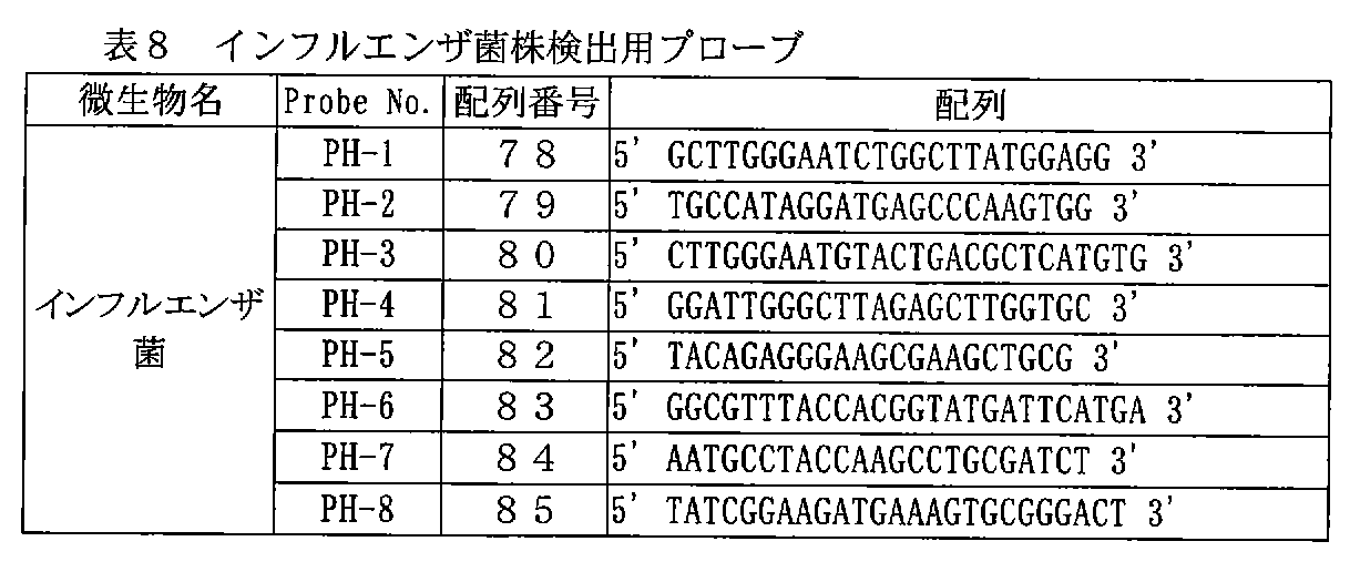
上記各表中に示したプローブは、DNAマイクロアレイに固定するための官能基として、合成後、定法に従って核酸の5'末端にチオール基を導入した。官能基の導入後、精製し、凍結乾燥した。凍結乾燥したプローブは、−30℃の冷凍庫に保存した。 In the probes shown in the above tables, a thiol group was introduced into the 5 ′ end of the nucleic acid after synthesis as a functional group for immobilization on the DNA microarray according to a conventional method. After introduction of the functional group, it was purified and lyophilized. The freeze-dried probe was stored in a −30 ° C. freezer.
[2.検体増幅用PCRプライマーの準備]
上記の起炎菌検出用の為の16s rRNA遺伝子(標的遺伝子)増幅用PCRPrimerとして以下の表11に示す核酸配列を設計した。具体的には、16s rRNAをコーディングしているゲノム部分を特異的に増幅するプローブセット、つまり約1400〜1700塩基長の16srRNAコーディング領域の両端部分で、特異的な融解温度をできるだけ揃えたプライマーを設計した。なお、変異株や、ゲノム上に複数存在する16s rRNAコーディング領域も同時に増幅できるように複数種類のプライマーを設計した。なお、起炎菌の16srRNAコーディング領域に対して共通にほぼ全長を増幅できるプライマーセットであれば、下記の表11に挙げたプライマセットに限定する必要はないのは言うまでもない。
[2. Preparation of PCR primers for sample amplification]
The nucleic acid sequences shown in Table 11 below were designed as PCR Primers for amplification of the 16s rRNA gene (target gene) for detection of the above-mentioned pathogenic bacteria. Specifically, a probe set that specifically amplifies the 16s rRNA-encoding genomic region, that is, a primer with a specific melting temperature aligned as much as possible at both ends of the 16srRNA coding region of about 1400 to 1700 bases in length. Designed. Multiple primers were designed to simultaneously amplify mutant strains and multiple 16s rRNA coding regions on the genome. Needless to say, the primer sets need not be limited to the primer sets listed in Table 11 below as long as they can amplify almost the entire length in common with the 16srRNA coding region of the causative fungus.
表11中に示したPrimerは、合成後、高速液体クロマトグラフィー(HPLC)により精製し、ForwardPrimerを3種、Reverse Primerを3種を混合し、それぞれのプライマー濃度が、最終濃度10pmol/μlとなるようにTE緩衝液に溶解した。 The primers shown in Table 11 were purified by high performance liquid chromatography (HPLC) after synthesis, and 3 kinds of ForwardPrimer and 3 kinds of Reverse Primer were mixed, and the respective primer concentrations were 10 pmol / μl final concentration. So that it was dissolved in TE buffer.
[3.各起炎菌Genome DNA(モデル検体)の抽出]
[3-1]微生物の培養 & Genome DNA 抽出の前処理
まず、上記各起炎菌の標準株(黄色ブドウ球菌標準株(ATCC12600)、表皮ブドウ球菌標準株(ATCC14990)、大腸菌標準株(ATCC11775)、肺炎桿菌標準株(ATCC13883)、緑膿菌標準株(ATCC10145)、セラチア菌株、肺炎連鎖球菌標準株、インフルエンザ菌株、腸球菌標準株(ATCC13047)、エンテロコッカス フェカリス菌標準株(ATCC19433))を定法に従って培養し、微生物培養液を生成した。これらの微生物培養液のそれぞれを1.5ml容量のマイクロチューブに1.0ml(OD600=0.7)採取し、遠心分離で菌体を回収した(8500rpm、5min、4℃)。次に、上精を捨てた後、EnzymeBuffer(50mM Tris-HCl:p.H. 8.0、25mM EDTA)300μlを加え、ミキサーを用いて再縣濁した。再縣濁した菌液は、再度、遠心分離で菌体を回収した(8500rpm、5min、4℃)。上精を捨てた後、回収された菌体に、以下の酵素溶液を加え、ミキサーを用いて再縣濁した。
[3. Extraction of Genome DNA (Model Specimens)]
[3-1] Microbial culture & pre-treatment of Genome DNA extraction First, the above-mentioned standard strains of the respective pathogenic bacteria (Staphylococcus aureus standard strain (ATCC12600), Staphylococcus epidermidis standard strain (ATCC14990), Escherichia coli standard strain (ATCC11775)) , Standard strain of K. pneumoniae (ATCC13883), Pseudomonas aeruginosa standard strain (ATCC10145), Serratia strain, Streptococcus pneumoniae standard strain, influenza strain, enterococcus standard strain (ATCC13047), Enterococcus faecalis standard strain (ATCC19433)) Cultured to produce a microbial culture. 1.0 ml (OD 600 = 0.7) of each of these microorganism culture solutions was collected in a 1.5 ml capacity microtube, and the cells were collected by centrifugation (8500 rpm, 5 min, 4 ° C.). Next, after discarding the supernatant, 300 μl of EnzymeBuffer (50 mM Tris-HCl: pH 8.0, 25 mM EDTA) was added and resuspended using a mixer. The resuspended bacterial solution was recovered again by centrifugation (8500 rpm, 5 min, 4 ° C.). After discarding the upper fine particles, the following enzyme solution was added to the collected cells and resuspended using a mixer.
次に、酵素溶液を加え再縣濁した菌液を、37℃のインキュベーター内で30分間静置し、細胞壁の溶解処理を行った。 Next, the bacterial solution resuspended by adding the enzyme solution was allowed to stand for 30 minutes in a 37 ° C. incubator to lyse the cell wall.
[3-2]Genome抽出
以下に示す微生物のGenome DNA抽出は、核酸精製キット(MagExtractor-Genome-:TOYOBO社製)を用いて行った。
[3-2] Genome extraction Genome DNA extraction of the microorganisms shown below was performed using a nucleic acid purification kit (MagExtractor-Genome-: manufactured by TOYOBO).
具体的には、まず、前処理した微生物縣濁液に溶解・吸着液750μlと磁性ビーズ40μlを加え、チューブミキサーを用いて、10分間激しく攪拌した(ステップ1)。
次に、分離用スタンド(Magical Trapper)にマイクロチューブをセットし、30秒間静置して磁性粒子をチューブの壁面に集め、スタンドにセットした状態のまま、上精を捨てた(ステップ2)。
次に、洗浄液900μlを加え、ミキサーで5sec程度攪拌して再縣濁を行った(ステップ3)。
次に、分離用スタンド(Magical Trapper)にマイクロチューブをセットし、30秒間静置して磁性粒子をチューブの壁面に集め、スタンドにセットした状態のまま、上精を捨てた(ステップ4)。
Specifically, first, 750 μl of the dissolving / adsorbing solution and 40 μl of magnetic beads were added to the pretreated microorganism suspension and vigorously stirred for 10 minutes using a tube mixer (step 1).
Next, the microtube was set on a separation stand (Magical Trapper), left to stand for 30 seconds, the magnetic particles were collected on the wall of the tube, and the upper fine was discarded while being set on the stand (step 2).
Next, 900 μl of the washing solution was added and re-suspended by stirring for about 5 seconds with a mixer (step 3).
Next, a microtube was set on a separation stand (Magical Trapper), left still for 30 seconds, magnetic particles were collected on the wall surface of the tube, and the upper fine was discarded while being set on the stand (step 4).
上記ステップ3、4を繰り返して2度目の洗浄を行なう(ステップ5)。その後、70%エタノール900μlを加え、ミキサーで5sec程度攪拌して再縣濁した(ステップ6)。
次に、分離用スタンド(Magical Trapper)にマイクロチューブをセットし、30秒間静置して磁性粒子をチューブの壁面に集め、スタンドにセットした状態のまま、上精を捨てた(ステップ7)。
ステップ6、7を繰り返して70%エタノールによる2度目の洗浄(ステップ8)を行った後、回収された磁性粒子に純水100μlを加え、チューブミキサーで10分間攪拌を行った(ステップ9)。
次に分離用スタンド(Magical Trapper)にマイクロチューブをセットし、30sec静置して磁性粒子をチューブ壁面に集め、スタンドにセットした状態のまま、上精を新しいチューブに回収した。
The above steps 3 and 4 are repeated to perform the second cleaning (step 5). Thereafter, 900 μl of 70% ethanol was added, and the mixture was stirred for about 5 seconds with a mixer and resuspended (step 6).
Next, the microtube was set on a separation stand (Magical Trapper), left to stand for 30 seconds, the magnetic particles were collected on the wall of the tube, and the upper fine was discarded while being set on the stand (step 7).
Steps 6 and 7 were repeated and the second washing with 70% ethanol (Step 8) was performed. Then, 100 μl of pure water was added to the collected magnetic particles, and the mixture was stirred with a tube mixer for 10 minutes (Step 9).
Next, the microtube was set on a separation stand (Magical Trapper), left still for 30 seconds, and magnetic particles were collected on the wall surface of the tube, and the supernatant was collected in a new tube while being set on the stand.
[3-3]回収したGenome DNAの検査
回収された微生物(各起炎菌株)のGenome DNAは、定法に従って、アガロース電気泳動と260/280nmの吸光度測定を行い、その品質(低分子核酸の混入量、分解の程度)と回収量を検定した。本実施例では、それぞれの菌において、約9〜10μgのGenomeDNA が回収され、Genome DNAのデグラデーションやrRNAの混入は認められなかった。回収したGenome DNAは、最終濃度50ng/μlとなるようにTE緩衝液に溶解し、以下の実施例に使用した。
[3-3] Examination of recovered Genome DNA Genome DNA of recovered microorganisms (each inflammatory strain) is subjected to agarose electrophoresis and absorbance measurement at 260/280 nm according to a conventional method to determine its quality (contamination with low molecular nucleic acids). Volume, degree of degradation) and recovery. In this example, about 9-10 μg of Genome DNA was recovered in each bacterium, and no degradation of Genome DNA or contamination with rRNA was observed. The recovered Genome DNA was dissolved in TE buffer so as to have a final concentration of 50 ng / μl and used in the following examples.
[4.DNAマイクロアレイの作製]
DNAマイクロアレイを、特開平11−187900号公報に開示された方法に従って用意した。
[4-1]ガラス基板の洗浄
合成石英のガラス基板(サイズ:25mm×75mm×1mm、飯山特殊ガラス社製)を耐熱、耐アルカリのラックに入れ、所定の濃度に調製した超音波洗浄用の洗浄液に浸した。一晩洗浄液中で浸した後、20分間超音波洗浄を行った。続いて基板を取り出し、軽く純水ですすいだ後、超純水中で20分超音波洗浄をおこなった。次に80℃に加熱した1N水酸化ナトリウム水溶液中に10分間基板を浸した。再び純水洗浄と超純水洗浄を行い、DNAマイクロアレイ用の石英ガラス基板を用意した。
[4. Preparation of DNA microarray]
A DNA microarray was prepared according to the method disclosed in JP-A-11-187900.
[4-1] Cleaning of glass substrate A glass substrate made of synthetic quartz (size: 25 mm x 75 mm x 1 mm, manufactured by Iiyama Special Glass Co., Ltd.) is placed in a heat-resistant and alkali-resistant rack and used for ultrasonic cleaning prepared to a predetermined concentration. Immerse in the cleaning solution. After soaking in the cleaning solution overnight, ultrasonic cleaning was performed for 20 minutes. Subsequently, the substrate was taken out, rinsed lightly with pure water, and then ultrasonically cleaned in ultrapure water for 20 minutes. Next, the substrate was immersed in a 1N sodium hydroxide aqueous solution heated to 80 ° C. for 10 minutes. Pure water cleaning and ultrapure water cleaning were performed again to prepare a quartz glass substrate for DNA microarray.
[4-2]表面処理
シランカップリング剤KBM-603(信越シリコーン社製)を、1%の濃度となるように純水中に溶解させ、2時間室温で攪拌した。続いて、先に洗浄したガラス基板をシランカップリング剤水溶液に浸し、20分間室温で放置した。ガラス基板を引き上げ、軽く純水で表面を洗浄した後、窒素ガスを基板の両面に吹き付けて乾燥させた。次に乾燥した基板を120℃に加熱したオーブン中で1時間ベークし、カップリング剤処理を完結させ、基板表面にアミノ基を導入した。次いで同仁化学研究所社製のN−マレイミドカプロイロキシスクシイミド(N-(6-Maleimidocaproyloxy)succinimido)(以下EMCSと略す)を、ジメチルスルホキシドとエタノールの1:1混合溶媒中に最終濃度が0.3mg/mlとなるように溶解したEMCS溶液を用意した。
[4-2] Surface treatment A silane coupling agent KBM-603 (manufactured by Shin-Etsu Silicone) was dissolved in pure water to a concentration of 1% and stirred at room temperature for 2 hours. Subsequently, the previously cleaned glass substrate was immersed in an aqueous silane coupling agent solution and allowed to stand at room temperature for 20 minutes. The glass substrate was pulled up and the surface was lightly washed with pure water, and then nitrogen gas was blown onto both sides of the substrate to dry it. Next, the dried substrate was baked in an oven heated to 120 ° C. for 1 hour to complete the coupling agent treatment, and amino groups were introduced onto the substrate surface. Next, N- (6-Maleimidocaproyloxy) succinimido (hereinafter abbreviated as EMCS) manufactured by Dojindo Laboratories Ltd. was added to a 1: 1 mixed solvent of dimethyl sulfoxide and ethanol. An EMCS solution dissolved to 0.3 mg / ml was prepared.
ベークの終了したガラス基板を放冷し、調製したEMCS溶液中に室温で2時間浸した。この処理により、シランカップリング剤によって表面に導入されたアミノ基とEMCSのスクシイミド基が反応し、ガラス基板表面にマレイミド基が導入された。EMCS溶液から引き上げたガラス基板を、先述のEMCSを溶解した混合溶媒を用いて洗浄し、さらにエタノールにより洗浄した後、窒素ガス雰囲気下で乾燥させた。 The glass substrate after baking was allowed to cool and immersed in the prepared EMCS solution for 2 hours at room temperature. By this treatment, the amino group introduced on the surface by the silane coupling agent and the succinimide group of EMCS reacted to introduce a maleimide group on the glass substrate surface. The glass substrate pulled up from the EMCS solution was washed with the above-mentioned mixed solvent in which EMCS was dissolved, further washed with ethanol, and then dried in a nitrogen gas atmosphere.
[4-3]プローブDNA
実施例1で作製した微生物検出用プローブを純水に溶解し、それぞれ、最終濃度(インク溶解時)10μMとなるように分注した後、凍結乾燥を行い、水分を除いた。
[4-3] Probe DNA
The microorganism detection probe prepared in Example 1 was dissolved in pure water, dispensed to a final concentration (at the time of ink dissolution) of 10 μM, and then freeze-dried to remove moisture.
[4-4]BJプリンタによるDNA吐出、および基板への結合
グリセリン7.5wt%、チオジグリコール7.5wt%、尿素7.5wt%、アセチレノールEH(川研ファインケミカル社製)1.0wt%を含む水溶液を用意した。続いて、先に用意した7種類のプローブ(表1)の夫々を上記の混合溶媒に規定濃度なるように溶解した。得られたDNA溶液をバブルジェット(登録商標)プリンタ(商品名:BJF-850 キヤノン社製)用インクタンクに充填し、印字ヘッドに装着した。
[4-4] DNA ejection by BJ printer and binding to substrate Prepare an aqueous solution containing glycerin 7.5wt%, thiodiglycol 7.5wt%, urea 7.5wt%, acetylenol EH (manufactured by Kawaken Fine Chemicals) 1.0wt% did. Subsequently, each of the seven types of probes prepared in advance (Table 1) was dissolved in the above mixed solvent to a specified concentration. The obtained DNA solution was filled in an ink tank for a bubble jet (registered trademark) printer (trade name: BJF-850 manufactured by Canon Inc.) and mounted on a print head.
なおここで用いたバブルジェット(登録商標)プリンタは平板への印刷が可能なように改造を施したものである。またこのバブルジェット(登録商標)プリンタは、所定のファイル作成方法に従って印字パターンを入力することにより、約5ピコリットルのDNA溶液を約120μmピッチでスポッティングすることが可能となっている。 The bubble jet (registered trademark) printer used here is modified so that printing on a flat plate is possible. This Bubble Jet (registered trademark) printer can spot a DNA solution of about 5 picoliters at a pitch of about 120 μm by inputting a print pattern according to a predetermined file creation method.
続いて、この改造バブルジェット(登録商標)プリンタを用いて、1枚のガラス基板に対して、印字操作を行い、DNAマイクロアレイを作製した。印字が確実に行われていることを確認した後、30分間加湿チャンバー内に静置し、ガラス基板表面のマレイミド基と核酸プローブ末端のチオール基とを反応させた。 Subsequently, using this modified bubble jet (registered trademark) printer, a printing operation was performed on one glass substrate to produce a DNA microarray. After confirming that printing was performed reliably, the sample was left in a humidified chamber for 30 minutes to react the maleimide group on the surface of the glass substrate with the thiol group at the end of the nucleic acid probe.
[4-5]洗浄
30分間の反応後、100mMのNaClを含む10mMのリン酸緩衝液(pH7.0)により表面に残ったDNA溶液を洗い流し、ガラス基板表面に一本鎖DNAが固定した遺伝子チップ(DNAマイクロアレイ)を得た。
[4-5] Cleaning
After the reaction for 30 minutes, the DNA solution remaining on the surface was washed away with 10 mM phosphate buffer (pH 7.0) containing 100 mM NaCl, and a gene chip (DNA microarray) in which single-stranded DNA was immobilized on the glass substrate surface Obtained.
[5.検体の増幅と標識化(PCR増幅&蛍光標識の取り込み)]
検体となる微生物遺伝子の増幅、および、標識化反応を以下に示す。
[5. Sample amplification and labeling (PCR amplification & incorporation of fluorescent labels)]
Amplification and labeling reaction of a microbial gene as a specimen are shown below.
上記組成の反応液を以下のプロトコールに従って、市販のサーマルサイクラーで増幅反応を行った。 The reaction solution having the above composition was subjected to an amplification reaction using a commercially available thermal cycler according to the following protocol.
反応終了後、精製用カラム(QIAGEN QIAquick PCRPurification Kit)を用いてPrimerを除去(精製)した後、増幅産物の定量を行い、標識化検体とした。 After completion of the reaction, Primer was removed (purified) using a purification column (QIAGEN QIAquick PCRPurification Kit), and then the amplification product was quantified to obtain a labeled sample.
[6.ハイブリダイゼーション]
上述の[4.DNAマイクロアレイの作製]で作製した遺伝子チップと[5.検体の増幅と標識化(PCR増幅&蛍光標識の取り込み)]で作製した標識化検体を用いて検出反応を行った。
[6. Hybridization]
[4. Gene chip prepared in [Preparation of DNA microarray] and [5. Detection reaction was carried out using the labeled sample prepared in [Amplification and labeling of sample (PCR amplification & incorporation of fluorescent label)].
[6-1]DNAマイクロアレイのブロッキング
BSA(牛血清アルブミンFraction V:Sigma社製)を1wt%となるように100mMNaCl/10mM Phosphate Bufferに溶解し、この溶液に[4.DNAマイクロアレイの作製]で作製した遺伝子チップを室温で2時間浸し、ブロッキングを行った。ブロッキング終了後、0.1wt%SDS(ドデシル硫酸ナトリウム)を含む2xSSC溶液(NaCl300mM 、Sodium Citrate (trisodium citrate dihydrate, C6H5Na3・2H2O)30mM、pH 7.0)で洗浄を行った後、純水でリンスしてからスピンドライ装置で水切りを行った。
[6-1] DNA microarray blocking
BSA (bovine serum albumin Fraction V: manufactured by Sigma) was dissolved in 100 mM NaCl / 10 mM Phosphate Buffer to 1 wt%, and [4. The gene chip prepared in [Preparation of DNA microarray] was immersed at room temperature for 2 hours for blocking. After the end of blocking, washing was performed with 0.1 wt% SDS 2 × SSC containing (sodium dodecyl sulfate) solution (NaCl300mM, Sodium Citrate (trisodium citrate dihydrate, C 6 H 5 Na 3 · 2H 2 O) 30mM, pH 7.0), After rinsing with pure water, draining was performed with a spin dryer.
[6-2]ハイブリダイゼーション
水切りした遺伝子チップをハイブリダイゼーション装置(GenomicSolutions Inc. Hybridization Station)にセットし、以下に示すハイブリダイゼーション溶液、条件でハイブリダイゼーション反応を行った。
[6-2] Hybridization The drained gene chip was set in a hybridization apparatus (Genomic Solutions Inc. Hybridization Station), and a hybridization reaction was performed with the following hybridization solution and conditions.
[6-3]ハイブリダイゼーション溶液
6xSSPE / 10% Form amide / Target(2nd PCR Products 全量)
(6xSSPE: NaCl 900mM、NaH2PO4・H2O60mM、EDTA 6mM、p.H. 7.4)。
[6-3] Hybridization solution
6xSSPE / 10% Form amide / Target (2nd PCR Products total amount)
(6xSSPE: NaCl 900 mM, NaH 2 PO 4 · H 2 O 60 mM, EDTA 6 mM, pH 7.4).
[6-4]ハイブリダイゼーション条件
65℃ 3min → 92℃ 2min → 45℃ 3hr → Wash 2xSSC/ 0.1% SDS at 25℃ → Wash 2xSSC at 20℃ → (Rinse with H2O : Manual) → Spin dry(65℃で3分、92度で2分、45℃で3時間ハイブリダイゼーション反応させた後、2xSSC/ 0.1% SDS、25℃で洗浄、2xSSC、20℃で洗浄後、純水でリンスしスピンドライした)。
[6-4] Hybridization conditions
65 ℃ 3min → 92 ℃ 2min → 45 ℃ 3hr → Wash 2xSSC / 0.1% SDS at 25 ℃ → Wash 2xSSC at 20 ℃ → (Rinse with H 2 O: Manual) → Spin dry (3 minutes at 65 ℃, at 92 degrees) After 2 minutes of hybridization at 45 ° C. for 3 hours, 2 × SSC / 0.1% SDS, washed at 25 ° C., washed 2 × SSC at 20 ° C., rinsed with pure water and spin-dried).
[7.微生物の検出(蛍光測定)]
上記ハイブリダイゼーション反応終了後の遺伝子チップを遺伝子チップ用蛍光検出装置(Axon社製、GenePix 4000B)を用いて蛍光測定を行った。その結果、以下の表12〜21に示すように、各菌において、再現性良く、十分なシグナルで各菌を検出することができた。また、他の菌のプローブに対するハイブリッド体は検出されなかった。
[7. Detection of microorganisms (fluorescence measurement)]
The gene chip after completion of the hybridization reaction was subjected to fluorescence measurement using a gene chip fluorescence detector (Axon, GenePix 4000B). As a result, as shown in Tables 12 to 21 below, it was possible to detect each bacterium with a sufficient signal with good reproducibility. Moreover, the hybrid body with respect to the probe of another microbe was not detected.
なお、本実施例では、2回ずつ蛍光測定を実施しており、それぞれの結果を以下に示した。 In this example, fluorescence measurement was performed twice, and the results are shown below.
表12〜21の蛍光輝度の数値(フォトマル電圧400V)は、ピクセル平均輝度(解像度5μm)を示した。また、S/N比は、測定機付属の解析ソフト(Axon社製、GenePixPro Ver.3.0)で測定したバックグラウンド平均値で蛍光輝度を除したものを示した。 The numerical values of fluorescence luminance (photomal voltage 400 V) in Tables 12 to 21 indicate pixel average luminance (resolution 5 μm). Moreover, S / N ratio showed what remove | divided the fluorescence luminance by the background average value measured with the analysis software (Axon company make, GenePixPro Ver.3.0) attached to the measuring machine.
表12〜21で明らかなように、再現性良く、十分なシグナルで各起炎菌を検出することができる。 As apparent from Tables 12 to 21, each causative bacterium can be detected with a sufficient signal with good reproducibility.
<実施例2> 2Step PCR法を用いた微生物の検出
実施例1同様に、プローブDNA、検体増幅用PCRプライマー、各起炎菌のGenome DNA、DNAマイクロアレイを用意し、以下の実験を行った。
<Example 2> Detection of microorganisms using 2Step PCR method In the same manner as in Example 1, probe DNA, PCR primers for sample amplification, Genome DNA of each pathogenic bacterium, and DNA microarray were prepared, and the following experiments were performed.
[1.検体の増幅と標識化(PCR増幅&蛍光標識の取り込み)]
検体となる微生物遺伝子の増幅(1st PCR)、および、標識化(2ndPCR)反応を以下に示す。
[1. Sample amplification and labeling (PCR amplification & incorporation of fluorescent labels)]
The amplification (1st PCR) and labeling (2nd PCR) reaction of a microbial gene as a sample are shown below.
[2.増幅反応液組成:1st PCR] [2. Amplification reaction composition: 1st PCR]
上記組成の反応液を以下のプロトコールに従って、市販のサーマルサイクラーで増幅反応を行った。 The reaction solution having the above composition was subjected to an amplification reaction using a commercially available thermal cycler according to the following protocol.
反応終了後、精製用カラム(QIAGEN QIAquick PCRPurification Kit)を用いて精製した後、増幅産物の定量を行った。 After completion of the reaction, the product was purified using a purification column (QIAGEN QIAquick PCRPurification Kit), and the amplification product was quantified.
[3.標識化反応液組成:2nd PCR] [3. Labeling reaction solution composition: 2nd PCR]
上記組成の反応液を以下のプロトコールに従って、市販のサーマルサイクラーで増幅反応を行った。 The reaction solution having the above composition was subjected to an amplification reaction using a commercially available thermal cycler according to the following protocol.
反応終了後、精製用カラム(QIAGEN QIAquick PCRPurification Kit)を用いて精製し、標識化検体とした。 After completion of the reaction, the reaction mixture was purified using a purification column (QIAGEN QIAquick PCRPurification Kit) to obtain a labeled sample.
[4.ハイブリダイゼーション]
実施例1と同様に行った。
[4. Hybridization]
The same operation as in Example 1 was performed.
[5.微生物の検出(蛍光測定)]
ハイブリダイゼーション反応終了後のDNAマイクロアレイをDNAマイクロアレイ用蛍光検出装置(Axon社製、GenePix 4000B)を用いで蛍光測定を行った。測定結果を表22〜31に示す。
[5. Detection of microorganisms (fluorescence measurement)]
After completion of the hybridization reaction, the DNA microarray was subjected to fluorescence measurement using a fluorescence detector for DNA microarray (Axon, GenePix 4000B). The measurement results are shown in Tables 22 to 31.
なお、本実施例においても、2回ずつ蛍光測定を実施し、それぞれの結果を表22〜31に示した。 In this example also, fluorescence measurements were performed twice, and the results are shown in Tables 22-31.
表22〜31の蛍光輝度の数値(フォトマル電圧400V)は、ピクセル平均輝度(解像度5μm)を示した。また、S/N比は、測定機付属の解析ソフト(Axon社製、GenePixPro Ver.3.0)で測定したバックグラウンド平均値で蛍光輝度を除したものを示した。 The numerical values of fluorescence luminance (photomal voltage 400 V) in Tables 22 to 31 indicate pixel average luminance (resolution 5 μm). Moreover, S / N ratio showed what remove | divided fluorescence luminance by the background average value measured with the analysis software (Axon company make, GenePixPro Ver.3.0) attached to the measuring machine.
表22〜31で明らかなように、再現性良く、十分なシグナルで各種起炎菌を検出することができる。 As is apparent from Tables 22 to 31, various infectious bacteria can be detected with a sufficient signal with good reproducibility.
<実施例3> 2Step PCR法を用いた微生物の検出
実施例1、2同様に、プローブDNA、検体増幅用PCRプライマー、各起炎菌のGenome DNA、DNAマイクロアレイを用意し、以下の実験を行った。
<Example 3> Detection of microorganisms using 2Step PCR method In the same manner as in Examples 1 and 2, probe DNA, PCR primers for sample amplification, Genome DNA of each pathogenic fungus, and DNA microarray were prepared, and the following experiment was conducted. It was.
[1.検体の増幅と標識化(蛍光標識付きPCRプライマーの使用)]
検体となる微生物遺伝子の増幅(1st PCR)、および、標識化(2nd PCR)反応を以下に示す。
[1. Sample amplification and labeling (use of fluorescently labeled PCR primers)]
Amplification (1st PCR) and labeling (2nd PCR) reaction of a microbial gene as a specimen are shown below.
[2.増幅反応液組成;1st PCR] [2. Amplification reaction solution composition: 1st PCR]
上記組成の反応液を以下のプロトコールに従って、市販のサーマルサイクラーで増幅反応を行なった。 The reaction solution having the above composition was subjected to an amplification reaction using a commercially available thermal cycler according to the following protocol.
反応終了後、精製用カラム(QIAGEN QIAquick PCR PurificationKit)を用いて精製した後、増幅産物の定量を行なった。 After completion of the reaction, the product was purified using a purification column (QIAGEN QIAquick PCR Purification Kit), and the amplification product was quantified.
[3.標識化反応組成:2nd PCR] [3. Labeling reaction composition: 2nd PCR]
上記組成の反応液を以下のプロトコールに従って、市販のサーマルサイクラーで増幅反応を行った。 The reaction solution having the above composition was subjected to an amplification reaction using a commercially available thermal cycler according to the following protocol.
反応終了後、精製用カラム(QIAGEN QIAquick PCR PurificationKit)を用いて精製し、標識化検体とした。 After completion of the reaction, the product was purified using a purification column (QIAGEN QIAquick PCR Purification Kit) to prepare a labeled sample.
[4.ハイブリダイゼーション]
実施例1と同様に行った。
[4. Hybridization]
The same operation as in Example 1 was performed.
[5.微生物の検出(蛍光測定)]
ハイブリダイゼーション反応終了後のDNAマイクロアレイをDNAマイクロアレイ用蛍光検出装置(Axon社製、GenePix 4000B)を用いで蛍光測定を行った。測定結果を表32〜41に示す。
[5. Detection of microorganisms (fluorescence measurement)]
After completion of the hybridization reaction, the DNA microarray was subjected to fluorescence measurement using a fluorescence detector for DNA microarray (Axon, GenePix 4000B). The measurement results are shown in Tables 32-41.
なお、本実施例においては、2回ずつ蛍光測定を実施し、それぞれの結果を表32〜41に示した。 In this example, fluorescence measurements were performed twice, and the results are shown in Tables 32-41.
表22〜31で明らかなように、再現性良く、十分なシグナルで各種起炎菌を検出することができる。 As is apparent from Tables 22 to 31, various infectious bacteria can be detected with a sufficient signal with good reproducibility.
以上説明したように、上記実施例によれば、黄色ブドウ球菌、表皮ブドウ球菌、大腸菌、肺炎桿菌、緑膿菌、セラチア菌、肺炎連鎖球菌、インフルエンザ菌、腸球菌、及びエンテロコッカス・フェカリス菌の10種菌を検出可能なプローブセットをそれぞれ別々に、或いは組み合わせて固定したマクロアレイを用いて、感染症起炎菌を同定することが可能になり、微生物由来のDNAプローブの問題を解決した。すなわち、オリゴヌクレオチドプローブは、その塩基数の少なさゆえに、化学的に大量合成が可能であり、精製や濃度のコントロールが可能である。また、細菌の種による分類を目的に、同じ種の菌種は一括検出が可能で、しかも、他の種の細菌は区別して検出できるようなプローブセットが提供できた。 As described above, according to the above examples, 10 of Staphylococcus aureus, Staphylococcus epidermidis, Escherichia coli, Neisseria pneumoniae, Pseudomonas aeruginosa, Serratia, Streptococcus pneumoniae, Haemophilus influenzae, Enterococcus, and Enterococcus faecalis Infectious disease-causing bacteria can be identified using a macroarray in which probe sets capable of detecting inoculums are fixed separately or in combination, and the problem of DNA probes derived from microorganisms has been solved. That is, oligonucleotide probes can be chemically synthesized in large quantities due to their small number of bases, and purification and concentration control are possible. In addition, for the purpose of classification based on bacterial species, a probe set that can simultaneously detect bacterial species of the same species and distinguish and detect other types of bacteria can be provided.
また、これらの差異がDNAマイクロアレイ上で精度良く評価できるよう、プローブと検体とのハイブリッド体の安定性も考慮したプローブセットを提供することができる。さらにはこれらのプローブDNAと検体との反応を行なう為に、これらのプローブDNAが固定された担体を提供することができる。このプローブ及びプローブセットと検体溶液とを反応せしめる過程で、これらのプローブDNAが安定に担体上に固定され、再現性の高い検出結果を得るために、化学的に固定された担体を提供することができる。 In addition, a probe set that also considers the stability of the hybrid of the probe and the specimen can be provided so that these differences can be accurately evaluated on the DNA microarray. Furthermore, a carrier on which these probe DNAs are immobilized can be provided for the reaction between these probe DNAs and the specimen. In the process of reacting the probe and probe set with the sample solution, these probe DNAs are stably immobilized on the carrier, and a chemically immobilized carrier is provided to obtain highly reproducible detection results. Can do.
また、上記実施形態によれば、感染症起炎菌遺伝子の16s rRNA遺伝子配列を過不足なく検出することにより、該感染症起炎菌の存在を効率良く、また高い精度で判定することができる。 Further, according to the above embodiment, by detecting the 16s rRNA gene sequence of the infectious disease-causing fungus gene without excess or deficiency, the presence of the infectious disease-causing fungus can be determined efficiently and with high accuracy. .
<実施例4> プライマーセットについて
上記実施例において、黄色ブドウ球菌、表皮ブドウ球菌、大腸菌、肺炎桿菌、緑膿菌、セラチア菌、肺炎連鎖球菌、インフルエンザ菌、エンテロバクター・クロアカエ菌、及びエンテロコッカス・フェカリス菌のいずれか、或いは複数の16s rRNA遺伝子配列を増幅するのに用いられたプライマーセット(表11)を説明する。
<Example 4> About a primer set In the said Example, Staphylococcus aureus, Staphylococcus epidermidis, Escherichia coli, Neisseria pneumoniae, Pseudomonas aeruginosa, Serratia bacteria, Streptococcus pneumoniae, Haemophilus influenzae, Enterobacter cloacae, Enterococcus faecalis A primer set (Table 11) used to amplify any of the fungi or a plurality of 16s rRNA gene sequences will be described.
本実施形態のプライマーセットは、感染症起炎菌同定のために行なわれるPCR反応において良好な増幅結果を与えるように設計されている。ここで「良好な」とは、目的の16s rRNAが十分増幅されていることのみならず、16s rRNA以外の産物が無いことを意味する。 The primer set of this embodiment is designed to give a good amplification result in a PCR reaction performed for identification of infection-causing bacteria. Here, “good” means not only that the target 16s rRNA is sufficiently amplified, but also that there is no product other than 16s rRNA.
また、ここで「良好な」とは、検体中に含まれる検体由来のヒトゲノム遺伝子を増幅せずに、感染症起炎菌16srRNAのみを増幅することをも意味する。 In addition, “good” herein also means that only the infectious disease-causing fungus 16srRNA is amplified without amplifying the human genome gene derived from the sample contained in the sample.
本実施形態に用いられる検体としては、ヒト、家畜等の動物由来の血液、喀痰、胃液、膣分泌物、口腔内粘液等の体液、尿及び糞便のような排出物等、細菌が存在すると思われるあらゆる物を対象とする。また、食中毒、汚染の対象となる食品、飲料水及び温泉水のような自然環境中の水、空気清浄機や浄水器のフィルタ等、細菌による汚染が引き起こされる可能性のある媒体全てが挙げられる。さらに、輸出入時における検疫等の動植物も検体としてその対象となり得る。 As specimens used in this embodiment, bacteria such as blood derived from animals such as humans and domestic animals, sputum, gastric fluid, vaginal secretions, bodily fluids such as oral mucus, effluents such as urine and stool are considered to exist. Targeting everything In addition, food poisoning, food subject to contamination, water in the natural environment such as drinking water and hot spring water, air purifiers and filters for water purifiers, all media that can cause contamination by bacteria. . Furthermore, animals and plants such as quarantine at the time of import / export can also be targeted.
また、本実施形態に用いられるPCR反応とは、抽出した核酸そのものを鋳型として用いたPCR反応、或いは該PCR増幅物を鋳型として、さらに配列番号107〜109(表11のF1〜F3)、或いは配列番号110〜112(表11のR1〜R3)の片側のプライマーを用いた非対称PCR反応、可視化のために標識物を取り込ませるPCR法等いずれの反応をも含む。 The PCR reaction used in the present embodiment is a PCR reaction using the extracted nucleic acid itself as a template, or the PCR amplification product as a template, and further SEQ ID NOs: 107 to 109 (F1 to F3 in Table 11), or It includes any reaction such as an asymmetric PCR reaction using primers on one side of SEQ ID NOs: 110 to 112 (R1 to R3 in Table 11) and a PCR method for incorporating a label for visualization.
[1.検体増幅用PCR Primer の準備]
起炎菌検出用の為の16s rRNA遺伝子(標的遺伝子)増幅用PCR Primerとして表11に示す核酸配列を設計した。
[1. Preparation of PCR Primer for sample amplification]
The nucleic acid sequences shown in Table 11 were designed as PCR primers for amplifying 16s rRNA gene (target gene) for detection of pathogenic bacteria.
具体的には、16s rRNAをコーディングしているゲノム部分を特異的に増幅するプローブセット、つまり約1500塩基長の16srRNAコーディング領域の両端部分で、特異的な融解温度をできるだけ揃えたプライマーを設計した。なお、変異株や、ゲノム上に複数存在する16s rRNAコーディング領域も同時に増幅できるように複数種類のプライマーを設計した。 Specifically, a probe set that specifically amplifies the 16s rRNA-encoding genomic region, that is, a primer with the same melting temperature as possible at both ends of the approximately 1500 base length 16s rRNA coding region was designed. . Multiple primers were designed to simultaneously amplify mutant strains and multiple 16s rRNA coding regions on the genome.
表11中に示したプライマーは、合成後、高速液体クロマトグラフィー(HPLC)により精製され、3種のForwardPrimerと3種のReverse Primerの全てを混合し、それぞれのプライマー濃度が、最終濃度10 pmol/μl となるようにTE緩衝液に溶解した。なお、本実施例では全てのForwardPrimerとReverse Primerを用いたが、Forward PrimerとReverse Primerの夫々から1〜3種類ずつを用いるようにしてもよい。 The primers shown in Table 11 were purified by high performance liquid chromatography (HPLC) after synthesis, and all three kinds of ForwardPrimer and three kinds of Reverse Primers were mixed, and each primer concentration was 10 pmol / final concentration. It dissolved in TE buffer so that it might become microliter. In this embodiment, all ForwardPrimers and Reverse Primers are used, but one to three types may be used from each of Forward Primer and Reverse Primer.
以上により生成したForward PrimerとReverse Primerの溶液(ForwardPrimer mix及びReverse Primer mix)を用いて、[3.各起炎菌Genome DNA(モデル検体)の抽出]で説明した方法で抽出したGenome DNAを、[5.検体の増幅と標識化(PCR増幅&蛍光標識の取り込み)]で説明した手法により増幅する。 Using the solution of Forward Primer and Reverse Primer generated as described above (ForwardPrimer mix and Reverse Primer mix), [3. Genome DNA extracted by the method described in [Extraction of each causative bacteria Genome DNA (model specimen)] [5. Amplification and labeling of specimen (PCR amplification & incorporation of fluorescent label)].
反応終了後、精製用カラム(QIAGEN QIAquick PCR Purification Kit)を用いてPrimerを除去した後、増幅産物をゲル電気泳動により検討した。1500塩基対領域に1バンドが検出され、良好にPCR反応が行なわれていることが確認できた。また、副産物は無かった。 After completion of the reaction, the primer was removed using a purification column (QIAGEN QIAquick PCR Purification Kit), and the amplification product was examined by gel electrophoresis. One band was detected in the 1500 base pair region, and it was confirmed that the PCR reaction was carried out satisfactorily. There were no by-products.
なお、表11のプライマーを用いることにより、例えば上述した10種の感染症起炎菌(黄色ブドウ球菌、表皮ブドウ球菌、大腸菌、肺炎桿菌、緑膿菌、セラチア菌、肺炎連鎖球菌、インフルエンザ菌、腸球菌、及びエンテロコッカス・フェカリス菌)の全てにおいて、良好なPCR増幅の増幅結果を得ることができた。 In addition, by using the primers of Table 11, for example, the above-mentioned 10 infectious disease-causing fungi (S. aureus, Staphylococcus epidermidis, Escherichia coli, Neisseria pneumoniae, Pseudomonas aeruginosa, Serratia, Streptococcus pneumoniae, Haemophilus influenzae, In all of enterococci and Enterococcus faecalis), good amplification results of PCR amplification could be obtained.
<実施例5> 血液と菌液の混合物からの16sRNA遺伝子の増幅
ヒトの血液200μl(採血EDTA血)に実施例1で説明し手順で培養した腸球菌を103、104、105加え、菌血症モデル系とした。この溶液のそれぞれにN-アセチルムラミダーゼ溶液(0.2mg/ml in Enzyme Buffer)を加え、37℃30分加温した後、Qiamp Blood mini Kit(キアゲン社製)を用いてDNAを抽出し、PCR反応用のテンプレートとした。
<Example 5> Amplification of 16sRNA gene from a mixture of blood and fungus solution 10 3 , 10 4 , 10 5 of enterococci cultured in the procedure described in Example 1 and added to 200 μl of human blood (collected EDTA blood), A bacteremia model system was used. N-acetylmuramidase solution (0.2 mg / ml in Enzyme Buffer) was added to each of these solutions, heated at 37 ° C. for 30 minutes, DNA was extracted using Qiamp Blood mini Kit (Qiagen), and PCR was performed. It was set as the template for reaction.
これらのDNAに対して実施例4と同様、表11のプライマーセットを用いてPCR反応を行なった。 PCR was performed on these DNAs using the primer sets shown in Table 11 in the same manner as in Example 4.
その結果、実施例4と同様、1500塩基対長領域にバンドが検出され、良好にPCR反応が行なわれていることが確認できた。また、副産物はなく、そのバンドから求めたPCR増幅産物の量は、加えた菌量に比例していた。このことは、このプライマーセットを用いた際に、ヒトゲノムのPCR産物はなく、腸球菌の16sRNAのみが増幅されたことを示している。 As a result, as in Example 4, a band was detected in the 1500 base pair long region, and it was confirmed that the PCR reaction was carried out satisfactorily. Moreover, there were no by-products, and the amount of PCR amplification product determined from the band was proportional to the amount of added bacteria. This indicates that when this primer set was used, there was no human genome PCR product, and only enterococcal 16sRNA was amplified.
以上説明したように、本実施形態によれば、複数種類の感染症起炎菌の遺伝子中の16s rRNA部分を効率良く、また高い純度で増幅することができる。また、ヒトゲノムDNA存在時においても感染症起炎菌の16srRNAのみを効率よく増幅することができる。 As described above, according to the present embodiment, the 16s rRNA portion in the genes of multiple types of infectious disease-causing bacteria can be efficiently amplified with high purity. In addition, even when human genomic DNA is present, only 16s rRNA of infectious disease-causing bacteria can be efficiently amplified.
Claims (26)
SEQ ID No.73〜78の塩基配列のいずれかを有するオリゴヌクレオチドからなることを特徴とする感染症起炎菌増幅反応用プライマー。 A primer used to PCR amplify the 16s rRNA gene sequence of an infectious agent,
A primer for amplification reaction of infectious disease-causing bacteria characterized by comprising an oligonucleotide having any one of the nucleotide sequences of SEQ ID Nos. 73 to 78.
SEQ ID No.107〜109の塩基配列のうちの少なくとも一つ以上と、SEQ ID No.110〜112の塩基配列のうちの少なくとも一つ以上の複数の塩基配列の各々を有するオリゴヌクレオチドからなる複数のプライマーを含むことを特徴とする感染症起炎菌増幅反応用プライマーセット。 A primer set used for PCR amplification of 16s rRNA gene sequence of infectious disease-causing bacteria,
A plurality of oligonucleotides each having at least one of the base sequences of SEQ ID Nos. 107 to 109 and at least one or more of the base sequences of SEQ ID Nos. 110 to 112 A primer set for amplification reaction of infectious disease-causing bacteria.
Priority Applications (1)
| Application Number | Priority Date | Filing Date | Title |
|---|---|---|---|
| JP2006257739A JP2007014351A (en) | 2003-04-02 | 2006-09-22 | Infectious disease detection probe, gene detection probe, infectious disease pathogen amplification reaction primer, infectious disease pathogen amplification reaction primer set, and infectious disease pathogen detection method |
Applications Claiming Priority (13)
| Application Number | Priority Date | Filing Date | Title |
|---|---|---|---|
| JP2003099455 | 2003-04-02 | ||
| JP2003099463 | 2003-04-02 | ||
| JP2003099452 | 2003-04-02 | ||
| JP2003099453 | 2003-04-02 | ||
| JP2003099458 | 2003-04-02 | ||
| JP2003099454 | 2003-04-02 | ||
| JP2003099461 | 2003-04-02 | ||
| JP2003099460 | 2003-04-02 | ||
| JP2003099462 | 2003-04-02 | ||
| JP2003099459 | 2003-04-02 | ||
| JP2003099457 | 2003-04-02 | ||
| JP2003099456 | 2003-04-02 | ||
| JP2006257739A JP2007014351A (en) | 2003-04-02 | 2006-09-22 | Infectious disease detection probe, gene detection probe, infectious disease pathogen amplification reaction primer, infectious disease pathogen amplification reaction primer set, and infectious disease pathogen detection method |
Related Parent Applications (1)
| Application Number | Title | Priority Date | Filing Date |
|---|---|---|---|
| JP2004077045A Division JP2004313181A (en) | 2003-04-02 | 2004-03-17 | Probe and probe set for detecting causative bacteria of infectious disease, carrier and gene testing method |
Publications (1)
| Publication Number | Publication Date |
|---|---|
| JP2007014351A true JP2007014351A (en) | 2007-01-25 |
Family
ID=37752033
Family Applications (1)
| Application Number | Title | Priority Date | Filing Date |
|---|---|---|---|
| JP2006257739A Pending JP2007014351A (en) | 2003-04-02 | 2006-09-22 | Infectious disease detection probe, gene detection probe, infectious disease pathogen amplification reaction primer, infectious disease pathogen amplification reaction primer set, and infectious disease pathogen detection method |
Country Status (1)
| Country | Link |
|---|---|
| JP (1) | JP2007014351A (en) |
Cited By (3)
| Publication number | Priority date | Publication date | Assignee | Title |
|---|---|---|---|---|
| WO2009016770A1 (en) | 2007-08-02 | 2009-02-05 | Canon Kabushiki Kaisha | Hybridization method and apparatus |
| JP2009039046A (en) * | 2007-08-09 | 2009-02-26 | Yamaguchi Univ | Primer set for detecting pneumonia |
| EP3625358A4 (en) * | 2017-05-17 | 2021-02-24 | Microbio Pty Ltd | BIOMARKERS AND USES OF THE LATEST |
Citations (6)
| Publication number | Priority date | Publication date | Assignee | Title |
|---|---|---|---|---|
| EP0957175A1 (en) * | 1998-04-20 | 1999-11-17 | Academisch Ziekenhuis Groningen | Method for the rapid determination of bacteria |
| JP2000135085A (en) * | 1998-10-29 | 2000-05-16 | Canon Inc | Ps. Using the 16S rRNA gene and the DNA sequence in the gene. alcaligenes KB2 strain detection method |
| JP2002051783A (en) * | 2000-08-09 | 2002-02-19 | Denso Corp | Detection and quantification of eubacteria |
| JP2002238563A (en) * | 2001-01-31 | 2002-08-27 | Univ Catholique De Louvain | Identification of mycobacterial-specific nucleotide sequences and development of differential diagnostic methods for mycobacterial species |
| JP2002355084A (en) * | 2001-03-27 | 2002-12-10 | National Institute Of Advanced Industrial & Technology | Novel nucleic acid probe and novel nucleic acid measurement method using the same |
| JP2002542808A (en) * | 1999-05-03 | 2002-12-17 | ジェン−プローブ・インコーポレーテッド | Microbial identification method based on polynucleotide matrix |
-
2006
- 2006-09-22 JP JP2006257739A patent/JP2007014351A/en active Pending
Patent Citations (6)
| Publication number | Priority date | Publication date | Assignee | Title |
|---|---|---|---|---|
| EP0957175A1 (en) * | 1998-04-20 | 1999-11-17 | Academisch Ziekenhuis Groningen | Method for the rapid determination of bacteria |
| JP2000135085A (en) * | 1998-10-29 | 2000-05-16 | Canon Inc | Ps. Using the 16S rRNA gene and the DNA sequence in the gene. alcaligenes KB2 strain detection method |
| JP2002542808A (en) * | 1999-05-03 | 2002-12-17 | ジェン−プローブ・インコーポレーテッド | Microbial identification method based on polynucleotide matrix |
| JP2002051783A (en) * | 2000-08-09 | 2002-02-19 | Denso Corp | Detection and quantification of eubacteria |
| JP2002238563A (en) * | 2001-01-31 | 2002-08-27 | Univ Catholique De Louvain | Identification of mycobacterial-specific nucleotide sequences and development of differential diagnostic methods for mycobacterial species |
| JP2002355084A (en) * | 2001-03-27 | 2002-12-10 | National Institute Of Advanced Industrial & Technology | Novel nucleic acid probe and novel nucleic acid measurement method using the same |
Cited By (6)
| Publication number | Priority date | Publication date | Assignee | Title |
|---|---|---|---|---|
| WO2009016770A1 (en) | 2007-08-02 | 2009-02-05 | Canon Kabushiki Kaisha | Hybridization method and apparatus |
| US8323892B2 (en) | 2007-08-02 | 2012-12-04 | Canon Kabushiki Kaisha | Hybridization method and apparatus |
| JP2009039046A (en) * | 2007-08-09 | 2009-02-26 | Yamaguchi Univ | Primer set for detecting pneumonia |
| EP3625358A4 (en) * | 2017-05-17 | 2021-02-24 | Microbio Pty Ltd | BIOMARKERS AND USES OF THE LATEST |
| US11739389B2 (en) | 2017-05-17 | 2023-08-29 | Microbio Pty Ltd | Biomarkers and uses thereof |
| US12344904B2 (en) | 2017-05-17 | 2025-07-01 | Microbio Pty Ltd | Biomarkers and uses thereof |
Similar Documents
| Publication | Publication Date | Title |
|---|---|---|
| JP5596891B2 (en) | Probe set, probe fixing carrier, and genetic testing method | |
| JP5596893B2 (en) | Probe set, probe fixing carrier, and genetic testing method | |
| JP5596892B2 (en) | Probe set, probe fixing carrier, and genetic testing method | |
| JP5201818B2 (en) | Probe set, probe fixing carrier, and genetic testing method | |
| KR100834587B1 (en) | Probe Set, Carrier and Gene Identification Method for Infectious Disease Pathogen Detection | |
| JP2008118904A (en) | Probe, probe set, probe fixing carrier, and genetic testing method | |
| JP2008118914A (en) | Probe, probe set, probe fixing carrier, and genetic testing method | |
| JP5037905B2 (en) | Probe, probe set, probe fixing carrier, and genetic testing method | |
| JP2007159411A (en) | Probe set, probe fixing carrier, and genetic testing method | |
| JP5037906B2 (en) | Probe, probe set, probe fixing carrier, and genetic testing method | |
| JP5596894B2 (en) | Probe set, probe fixing carrier, and genetic testing method | |
| JP2006129810A (en) | Probe set and carrier for detecting infectious disease-causing bacteria and genetic testing method | |
| JP2008118907A (en) | Probe, probe set, probe fixing carrier, and genetic testing method | |
| JP2008118908A (en) | Probe, probe set, probe fixing carrier, and genetic testing method | |
| JP2006129828A (en) | Probe set and carrier for detecting infectious disease-causing bacteria and genetic testing method | |
| JP2006055028A (en) | Nucleic acid preparation method, and nucleic acid amplification method, nucleic acid labeling method and nucleic acid detection method using the same | |
| JP2006217897A (en) | Base sequence for control probe and design method thereof | |
| JP2007014351A (en) | Infectious disease detection probe, gene detection probe, infectious disease pathogen amplification reaction primer, infectious disease pathogen amplification reaction primer set, and infectious disease pathogen detection method | |
| JP5094181B2 (en) | Probe set, probe fixing carrier, and inspection method | |
| JP5078409B2 (en) | Probe set, probe fixing carrier, and inspection method | |
| JP5137442B2 (en) | Probe set, probe fixing carrier, and inspection method | |
| JP5137441B2 (en) | Probe set, probe fixing carrier, and inspection method | |
| JP5137443B2 (en) | Probe set, probe fixing carrier, and genetic testing method | |
| JP5121281B2 (en) | Probe set, probe fixing carrier, and inspection method | |
| JP5089221B2 (en) | Probe set, probe fixing carrier, and inspection method |
Legal Events
| Date | Code | Title | Description |
|---|---|---|---|
| A621 | Written request for application examination |
Free format text: JAPANESE INTERMEDIATE CODE: A621 Effective date: 20070319 |
|
| A131 | Notification of reasons for refusal |
Free format text: JAPANESE INTERMEDIATE CODE: A131 Effective date: 20091218 |
|
| A521 | Written amendment |
Free format text: JAPANESE INTERMEDIATE CODE: A523 Effective date: 20100215 |
|
| A131 | Notification of reasons for refusal |
Free format text: JAPANESE INTERMEDIATE CODE: A131 Effective date: 20100326 |
|
| A521 | Written amendment |
Free format text: JAPANESE INTERMEDIATE CODE: A523 Effective date: 20100525 |
|
| A02 | Decision of refusal |
Free format text: JAPANESE INTERMEDIATE CODE: A02 Effective date: 20101022 |