JP2010142498A - 眼底画像表示装置及びその制御方法、コンピュータプログラム - Google Patents
眼底画像表示装置及びその制御方法、コンピュータプログラム Download PDFInfo
- Publication number
- JP2010142498A JP2010142498A JP2008324709A JP2008324709A JP2010142498A JP 2010142498 A JP2010142498 A JP 2010142498A JP 2008324709 A JP2008324709 A JP 2008324709A JP 2008324709 A JP2008324709 A JP 2008324709A JP 2010142498 A JP2010142498 A JP 2010142498A
- Authority
- JP
- Japan
- Prior art keywords
- fundus
- transfer function
- blood vessel
- fundus image
- distance
- Prior art date
- Legal status (The legal status is an assumption and is not a legal conclusion. Google has not performed a legal analysis and makes no representation as to the accuracy of the status listed.)
- Granted
Links
Images

Classifications
-
- A—HUMAN NECESSITIES
- A61—MEDICAL OR VETERINARY SCIENCE; HYGIENE
- A61B—DIAGNOSIS; SURGERY; IDENTIFICATION
- A61B3/00—Apparatus for testing the eyes; Instruments for examining the eyes
- A61B3/10—Objective types, i.e. instruments for examining the eyes independent of the patients' perceptions or reactions
- A61B3/102—Objective types, i.e. instruments for examining the eyes independent of the patients' perceptions or reactions for optical coherence tomography [OCT]
-
- A—HUMAN NECESSITIES
- A61—MEDICAL OR VETERINARY SCIENCE; HYGIENE
- A61B—DIAGNOSIS; SURGERY; IDENTIFICATION
- A61B3/00—Apparatus for testing the eyes; Instruments for examining the eyes
- A61B3/10—Objective types, i.e. instruments for examining the eyes independent of the patients' perceptions or reactions
- A61B3/12—Objective types, i.e. instruments for examining the eyes independent of the patients' perceptions or reactions for looking at the eye fundus, e.g. ophthalmoscopes
-
- G—PHYSICS
- G06—COMPUTING OR CALCULATING; COUNTING
- G06T—IMAGE DATA PROCESSING OR GENERATION, IN GENERAL
- G06T7/00—Image analysis
- G06T7/10—Segmentation; Edge detection
- G06T7/11—Region-based segmentation
-
- G—PHYSICS
- G06—COMPUTING OR CALCULATING; COUNTING
- G06T—IMAGE DATA PROCESSING OR GENERATION, IN GENERAL
- G06T2207/00—Indexing scheme for image analysis or image enhancement
- G06T2207/10—Image acquisition modality
- G06T2207/10072—Tomographic images
- G06T2207/10101—Optical tomography; Optical coherence tomography [OCT]
-
- G—PHYSICS
- G06—COMPUTING OR CALCULATING; COUNTING
- G06T—IMAGE DATA PROCESSING OR GENERATION, IN GENERAL
- G06T2207/00—Indexing scheme for image analysis or image enhancement
- G06T2207/30—Subject of image; Context of image processing
- G06T2207/30004—Biomedical image processing
- G06T2207/30041—Eye; Retina; Ophthalmic
-
- G—PHYSICS
- G06—COMPUTING OR CALCULATING; COUNTING
- G06T—IMAGE DATA PROCESSING OR GENERATION, IN GENERAL
- G06T2207/00—Indexing scheme for image analysis or image enhancement
- G06T2207/30—Subject of image; Context of image processing
- G06T2207/30004—Biomedical image processing
- G06T2207/30101—Blood vessel; Artery; Vein; Vascular
Landscapes
- Health & Medical Sciences (AREA)
- Life Sciences & Earth Sciences (AREA)
- Engineering & Computer Science (AREA)
- Physics & Mathematics (AREA)
- Surgery (AREA)
- Public Health (AREA)
- Biomedical Technology (AREA)
- Heart & Thoracic Surgery (AREA)
- Medical Informatics (AREA)
- Molecular Biology (AREA)
- Biophysics (AREA)
- Animal Behavior & Ethology (AREA)
- General Health & Medical Sciences (AREA)
- Ophthalmology & Optometry (AREA)
- Veterinary Medicine (AREA)
- Computer Vision & Pattern Recognition (AREA)
- General Physics & Mathematics (AREA)
- Theoretical Computer Science (AREA)
- Nuclear Medicine, Radiotherapy & Molecular Imaging (AREA)
- Radiology & Medical Imaging (AREA)
- Eye Examination Apparatus (AREA)
- Image Analysis (AREA)
Abstract
【解決手段】網膜の断層像を示す眼底画像において網膜層のいずれかの境界位置を特定する特定手段と、不透明度のピーク位置が前記網膜内の所定位置となるように、特定された前記境界位置からの距離を不透明度を表すパラメータに変換するための距離伝達関数を設定する距離伝達関数設定手段と、前記眼底画像の輝度値を、不透明度を表すパラメータに変換する輝度伝達関数を設定する輝度伝達関数設定手段と、前記距離伝達関数と前記輝度伝達関数とを用いて、前記断層像の各位置の不透明度を算出し、ボリュームレンダリングにより半透明表示画像を生成する可視化手段とを備える。
【選択図】図5
Description
網膜の断層像を示す眼底画像において網膜層のいずれかの境界位置を特定する特定手段と、
不透明度のピーク位置が前記網膜内の所定位置となるように、特定された前記境界位置からの距離を不透明度を表すパラメータに変換するための距離伝達関数を設定する距離伝達関数設定手段と、
前記眼底画像の輝度値を、不透明度を表すパラメータに変換する輝度伝達関数を設定する輝度伝達関数設定手段と、
前記距離伝達関数と前記輝度伝達関数とを用いて、前記断層像の各位置の不透明度を算出し、ボリュームレンダリングにより半透明表示画像を生成する可視化手段と
を備える。
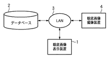
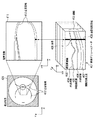
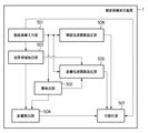
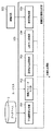
図3は、本実施形態に係る眼底画像表示システムの構成の一例を示す図である。本実施形態において、眼底画像表示装置1は、LAN3を介してデータベース2から眼底画像を読み出すことができる。或いは、眼底画像表示装置1に記憶装置、例えばFDD、CD−RWドライブ、MOドライブ、ZIPドライブ等を接続し、それらのドライブから眼底画像を読み込むようにしても良い。また、LAN3を経由して眼底画像撮影装置4から直接に医用画像等を取得してもよい。データベース2には、眼底画像の他にも、患者の名前や所見情報、図4に示す網膜403、神経線維層405、外網状層406、網膜色素上皮407の厚み情報が格納されている。図4は、眼底写真、網膜ボリュームデータ及び積算画像の関係を説明するための図である。
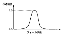
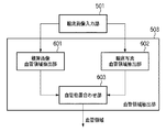
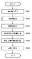
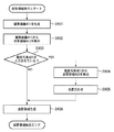
なお、βは、表示対象毎にあらかじめ設定しておいても良い。血管を可視化したい場合、血管付近ではコントラストが低いことから、βを低く設定し、距離伝達関数の重みを重くすることが考えられる。最後に、可視化部507は、ボリュームレンダリングの結果画像として、半透明表示画像を生成する。
上述の第1実施形態では、眼底画像から血管領域と層境界を抽出し、層境界からの距離と網膜ボリュームデータの輝度値から算出した不透明度を用いて血管をボリュームレンダリングすることによって、眼底血管の3次元走行を可視化した。これに対して、第2実施形態では、血管ではなく、眼底の特徴的な病変である白斑を可視化することを目的とする。
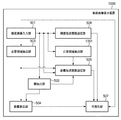
本実施形態に係る眼底画像表示装置及びそれに接続される機器の構成は、図2に示す第1実施形態の構成と同様である。また、本実施形態に係る眼底画像表示装置の各部の機能をソフトウェアで実現するためのコンピュータの基本構成も、図12に示す第1実施形態の構成と同様である。
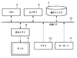
次に、第1実施形態、第2実施形態及び第3実施形態の眼底画像表示装置の各ブロックの機能をコンピュータプログラムで実現するためのコンピュータの基本構成を、図12を用いて説明する。
2 データベース
3 LAN
4 眼底画像表示装置
5 CPU
6 主メモリ
7 磁気ディスク
8 表示メモリ
9 モニタ
10 マウス
11 キーボード
12 共通バス
Claims (9)
- 網膜の断層像を示す眼底画像において網膜層のいずれかの境界位置を特定する特定手段と、
不透明度のピーク位置が前記網膜内の所定位置となるように、特定された前記境界位置からの距離を不透明度を表すパラメータに変換するための距離伝達関数を設定する距離伝達関数設定手段と、
前記眼底画像の輝度値を、不透明度を表すパラメータに変換する輝度伝達関数を設定する輝度伝達関数設定手段と、
前記距離伝達関数と前記輝度伝達関数とを用いて、前記断層像の各位置の不透明度を算出し、ボリュームレンダリングにより半透明表示画像を生成する可視化手段と
を備えることを特徴とする眼底画像表示装置。 - 前記眼底画像において、前記網膜内の眼底血管が存在する領域を抽出する血管領域抽出手段をさらに備え、
前記所定位置は、前記網膜内の眼底血管の存在する領域の位置であることを特徴とする請求項1に記載の眼底画像表示装置。 - 前記眼底画像には、眼底写真が更に含まれ、
前記血管領域抽出手段は前記眼底写真において眼底血管の位置を特定し、該特定した眼底血管の位置を利用して、前記眼底画像において前記網膜内の眼底血管が存在する領域を抽出することを特徴とする請求項2に記載の眼底画像表示装置。 - 前記眼底画像において、前記網膜内の白斑が存在する領域を抽出する白斑領域抽出手段をさらに備え、
前記所定位置は、前記網膜内の白斑の存在する領域の位置であることを特徴とする請求項1に記載の眼底画像表示装置。 - 前記眼底画像には、眼底写真が更に含まれ、
前記白斑領域抽出手段は前記眼底写真において白斑の位置を特定し、該特定した白斑の位置を利用して、前記眼底画像において前記網膜内の白斑が存在する領域を抽出することを特徴とする請求項4に記載の眼底画像表示装置。 - 前記網膜層を構成する各層の厚みを登録するデータベースをさらに備え、
前記距離伝達関数設定手段は、前記網膜内の所定位置を前記各層の厚みにより特定し、該所定位置が不透明度のピークとなるように、前記特定された境界位置からの距離を不透明度を表すパラメータに変換することを特徴とする請求項1に記載の眼底画像表示装置。 - 前記所定位置は、前記網膜内の他の境界の位置であることを特徴とする請求項1に記載の眼底画像表示装置。
- 網膜の断層像を示す眼底画像において網膜層のいずれかの境界位置を特定する特定工程と、
不透明度のピーク位置が前記網膜内の所定位置となるように、特定された前記境界位置からの距離を不透明度を表すパラメータに変換するための距離伝達関数を設定する距離伝達関数設定工程と、
前記眼底画像の輝度値を、不透明度を表すパラメータに変換する輝度伝達関数を設定する輝度伝達関数設定工程と、
前記距離伝達関数と前記輝度伝達関数とを用いて、前記断層像の各位置の不透明度を算出し、ボリュームレンダリングにより半透明表示画像を生成する可視化工程と
を備えることを特徴とする眼底画像表示装置の制御方法。 - コンピュータを請求項1乃至請求項7のいずれか1項に記載の眼底画像表示装置を機能させるためのコンピュータプログラム。
Priority Applications (6)
| Application Number | Priority Date | Filing Date | Title |
|---|---|---|---|
| JP2008324709A JP4850892B2 (ja) | 2008-12-19 | 2008-12-19 | 眼底画像表示装置及びその制御方法、コンピュータプログラム |
| CN200980150006.4A CN102245082B (zh) | 2008-12-19 | 2009-12-08 | 图像处理装置、其控制方法和图像处理系统 |
| KR1020117016012A KR101277040B1 (ko) | 2008-12-19 | 2009-12-08 | 화상 처리 장치, 그 제어 방법, 화상 처리 시스템 및 컴퓨터 판독가능 저장 매체 |
| PCT/JP2009/070795 WO2010071091A1 (en) | 2008-12-19 | 2009-12-08 | Fundus image display apparatus, control method thereof and computer program |
| EP09833394.1A EP2378950A4 (en) | 2008-12-19 | 2009-12-08 | APPARATUS FOR DISPLAYING EYE BACKGROUND PICTURES, CONTROL PROCEDURES THEREFOR AND COMPUTER POGRAM |
| US13/130,319 US8687863B2 (en) | 2008-12-19 | 2009-12-08 | Image processing apparatus, control method thereof and computer program |
Applications Claiming Priority (1)
| Application Number | Priority Date | Filing Date | Title |
|---|---|---|---|
| JP2008324709A JP4850892B2 (ja) | 2008-12-19 | 2008-12-19 | 眼底画像表示装置及びその制御方法、コンピュータプログラム |
Related Child Applications (1)
| Application Number | Title | Priority Date | Filing Date |
|---|---|---|---|
| JP2011231091A Division JP5484425B2 (ja) | 2011-10-20 | 2011-10-20 | 眼科装置及びその制御方法、コンピュータプログラム |
Publications (2)
| Publication Number | Publication Date |
|---|---|
| JP2010142498A true JP2010142498A (ja) | 2010-07-01 |
| JP4850892B2 JP4850892B2 (ja) | 2012-01-11 |
Family
ID=42268761
Family Applications (1)
| Application Number | Title | Priority Date | Filing Date |
|---|---|---|---|
| JP2008324709A Expired - Fee Related JP4850892B2 (ja) | 2008-12-19 | 2008-12-19 | 眼底画像表示装置及びその制御方法、コンピュータプログラム |
Country Status (6)
| Country | Link |
|---|---|
| US (1) | US8687863B2 (ja) |
| EP (1) | EP2378950A4 (ja) |
| JP (1) | JP4850892B2 (ja) |
| KR (1) | KR101277040B1 (ja) |
| CN (1) | CN102245082B (ja) |
| WO (1) | WO2010071091A1 (ja) |
Cited By (12)
| Publication number | Priority date | Publication date | Assignee | Title |
|---|---|---|---|---|
| JP2010279438A (ja) * | 2009-06-02 | 2010-12-16 | Canon Inc | 画像処理装置、画像処理方法及びコンピュータプログラム |
| JP2012011258A (ja) * | 2011-10-20 | 2012-01-19 | Canon Inc | 画像処理装置、画像処理方法及びコンピュータプログラム |
| JP2012019958A (ja) * | 2010-07-14 | 2012-02-02 | Canon Inc | 画像処理装置、プログラムおよび記憶媒体 |
| JP2012045298A (ja) * | 2010-08-30 | 2012-03-08 | Canon Inc | 画像処理装置、画像処理方法、画像処理システム及びプログラム |
| JP2012045226A (ja) * | 2010-08-27 | 2012-03-08 | Canon Inc | 眼科用画像処理装置及びその方法 |
| JP2012071113A (ja) * | 2010-08-31 | 2012-04-12 | Canon Inc | 画像処理装置、画像処理装置の制御方法及びプログラム |
| JP2012071043A (ja) * | 2010-09-29 | 2012-04-12 | Canon Inc | 情報処理装置、その表示処理方法及びプログラム |
| JP2015515894A (ja) * | 2012-05-10 | 2015-06-04 | カール ツアイス メディテック アクチエンゲゼルシャフト | Oct血管造影データの解析および可視化 |
| US9113779B2 (en) | 2010-08-30 | 2015-08-25 | Canon Kabushiki Kaisha | Image processing apparatus, image processing method, and program recording medium |
| JP2017074273A (ja) * | 2015-10-16 | 2017-04-20 | 国立大学法人 鹿児島大学 | 画像処理装置、画像処理方法、診断システム及びプログラム |
| JP2017104309A (ja) * | 2015-12-10 | 2017-06-15 | 株式会社トプコン | 眼科画像表示装置及び眼科撮影装置 |
| JP2021079205A (ja) * | 2021-03-01 | 2021-05-27 | 株式会社トプコン | 眼科画像表示装置 |
Families Citing this family (12)
| Publication number | Priority date | Publication date | Assignee | Title |
|---|---|---|---|---|
| JP5794664B2 (ja) * | 2011-01-20 | 2015-10-14 | キヤノン株式会社 | 断層画像生成装置及び断層画像生成方法 |
| US9226654B2 (en) * | 2011-04-29 | 2016-01-05 | Carl Zeiss Meditec, Inc. | Systems and methods for automated classification of abnormalities in optical coherence tomography images of the eye |
| DE102011076929A1 (de) * | 2011-06-03 | 2012-12-06 | Siemens Ag | Verfahren und Vorrichtung zur Darstellung von Volumendaten für eine Untersuchung von Dichteeigenschaften |
| US8433393B2 (en) | 2011-07-07 | 2013-04-30 | Carl Zeiss Meditec, Inc. | Inter-frame complex OCT data analysis techniques |
| JP2013075035A (ja) * | 2011-09-30 | 2013-04-25 | Canon Inc | 光断層像撮像方法、光断層像撮像装置およびプログラム |
| JP5926533B2 (ja) * | 2011-10-27 | 2016-05-25 | キヤノン株式会社 | 眼科装置 |
| WO2014031961A1 (en) * | 2012-08-23 | 2014-02-27 | University Of Washington Through Its Center For Commercialization | Methods and systems for determining volumetric properties of a tissue |
| US9778021B2 (en) | 2013-08-29 | 2017-10-03 | Carl Zeiss Meditec, Inc. | Evaluation of optical coherence tomographic data prior to segmentation |
| US10398302B2 (en) | 2014-05-02 | 2019-09-03 | Carl Zeiss Meditec, Inc. | Enhanced vessel characterization in optical coherence tomograogphy angiography |
| WO2017050863A1 (en) | 2015-09-24 | 2017-03-30 | Carl Zeiss Meditec, Inc. | Methods for high sensitivity flow visualization |
| US10896490B2 (en) * | 2016-12-23 | 2021-01-19 | Oregon Health & Science University | Systems and methods for reflectance-based projection-resolved optical coherence tomography angiography |
| KR102431173B1 (ko) * | 2020-06-30 | 2022-08-10 | 연세대학교 산학협력단 | 진단 영상에서 심층신경망을 이용한 국소종양진행을 진단하기 위한 장치 및 국소종양진행 진단에 필요한 정보를 제공하는 방법 |
Citations (3)
| Publication number | Priority date | Publication date | Assignee | Title |
|---|---|---|---|---|
| JP2008267891A (ja) * | 2007-04-18 | 2008-11-06 | Topcon Corp | 光画像計測装置 |
| WO2008146457A1 (ja) * | 2007-05-24 | 2008-12-04 | Kabushiki Kaisha Topcon | 眼底観察装置及びそれを制御するプログラム |
| JP2009119107A (ja) * | 2007-11-16 | 2009-06-04 | Kowa Co | 画像処理方法および画像処理装置 |
Family Cites Families (22)
| Publication number | Priority date | Publication date | Assignee | Title |
|---|---|---|---|---|
| US4423931A (en) * | 1980-09-09 | 1984-01-03 | Shapiro Jerrold M | Fundus camera accessory for analysis of the ocular fundus contour |
| US5029220A (en) * | 1990-07-31 | 1991-07-02 | The United States Of America As Represented By The Administrator Of The National Aeronautics And Space Administration | Optical joint correlator for real-time image tracking and retinal surgery |
| US5220360A (en) * | 1990-10-24 | 1993-06-15 | Ophthalmic Imaging Systems, Inc. | Apparatus and method for topographical analysis of the retina |
| US5751395A (en) * | 1997-01-10 | 1998-05-12 | Thall; Edmond H. | Retinal diagnostic device |
| US6563626B1 (en) * | 1999-02-25 | 2003-05-13 | Brother Kogyo Kabushiki Kaisha | Display device |
| CA2413872C (en) * | 2000-06-16 | 2009-05-12 | The University Of British Columbia | Analysis of optic nerve head shape |
| US7522174B2 (en) * | 2003-06-30 | 2009-04-21 | Canon Kabushiki Kaisha | Image processing apparatus, image processing method, program for implementing image processing method, and recording medium recording program |
| JP4916779B2 (ja) | 2005-09-29 | 2012-04-18 | 株式会社トプコン | 眼底観察装置 |
| CN100535926C (zh) * | 2006-01-06 | 2009-09-02 | 佳能株式会社 | 数据处理,图像处理和图像分类方法及设备 |
| US8118752B2 (en) * | 2006-02-16 | 2012-02-21 | The Board Of Trustees Of The University Of Illinois | Apparatus and methods for mapping retinal function |
| US7768652B2 (en) * | 2006-03-16 | 2010-08-03 | Carl Zeiss Meditec, Inc. | Methods for mapping tissue with optical coherence tomography data |
| JP5049654B2 (ja) | 2006-05-29 | 2012-10-17 | 株式会社東芝 | 医用画像処理装置、及び医用画像処理方法 |
| JP4855150B2 (ja) | 2006-06-09 | 2012-01-18 | 株式会社トプコン | 眼底観察装置、眼科画像処理装置及び眼科画像処理プログラム |
| WO2008091401A2 (en) * | 2006-09-15 | 2008-07-31 | Retica Systems, Inc | Multimodal ocular biometric system and methods |
| JP5095167B2 (ja) | 2006-09-19 | 2012-12-12 | 株式会社トプコン | 眼底観察装置、眼底画像表示装置及び眼底観察プログラム |
| US8223143B2 (en) * | 2006-10-27 | 2012-07-17 | Carl Zeiss Meditec, Inc. | User interface for efficiently displaying relevant OCT imaging data |
| JP5138977B2 (ja) | 2007-05-24 | 2013-02-06 | 株式会社トプコン | 光画像計測装置 |
| JP5148928B2 (ja) * | 2007-05-31 | 2013-02-20 | 株式会社トプコン | 眼底検査装置及びプログラム |
| US8363783B2 (en) * | 2007-06-04 | 2013-01-29 | Oraya Therapeutics, Inc. | Method and device for ocular alignment and coupling of ocular structures |
| JP4940069B2 (ja) * | 2007-09-10 | 2012-05-30 | 国立大学法人 東京大学 | 眼底観察装置、眼底画像処理装置及びプログラム |
| US8401246B2 (en) * | 2007-11-08 | 2013-03-19 | Topcon Medical Systems, Inc. | Mapping of retinal parameters from combined fundus image and three-dimensional optical coherence tomography |
| US8366271B2 (en) * | 2010-01-20 | 2013-02-05 | Duke University | Systems and methods for surgical microscope and optical coherence tomography (OCT) imaging |
-
2008
- 2008-12-19 JP JP2008324709A patent/JP4850892B2/ja not_active Expired - Fee Related
-
2009
- 2009-12-08 US US13/130,319 patent/US8687863B2/en not_active Expired - Fee Related
- 2009-12-08 CN CN200980150006.4A patent/CN102245082B/zh not_active Expired - Fee Related
- 2009-12-08 KR KR1020117016012A patent/KR101277040B1/ko not_active Expired - Fee Related
- 2009-12-08 EP EP09833394.1A patent/EP2378950A4/en not_active Withdrawn
- 2009-12-08 WO PCT/JP2009/070795 patent/WO2010071091A1/en not_active Ceased
Patent Citations (3)
| Publication number | Priority date | Publication date | Assignee | Title |
|---|---|---|---|---|
| JP2008267891A (ja) * | 2007-04-18 | 2008-11-06 | Topcon Corp | 光画像計測装置 |
| WO2008146457A1 (ja) * | 2007-05-24 | 2008-12-04 | Kabushiki Kaisha Topcon | 眼底観察装置及びそれを制御するプログラム |
| JP2009119107A (ja) * | 2007-11-16 | 2009-06-04 | Kowa Co | 画像処理方法および画像処理装置 |
Cited By (21)
| Publication number | Priority date | Publication date | Assignee | Title |
|---|---|---|---|---|
| JP2010279438A (ja) * | 2009-06-02 | 2010-12-16 | Canon Inc | 画像処理装置、画像処理方法及びコンピュータプログラム |
| JP2012019958A (ja) * | 2010-07-14 | 2012-02-02 | Canon Inc | 画像処理装置、プログラムおよび記憶媒体 |
| KR101405997B1 (ko) * | 2010-08-27 | 2014-06-11 | 캐논 가부시끼가이샤 | 안과용 화상 처리장치 및 그 방법 |
| US9373172B2 (en) | 2010-08-27 | 2016-06-21 | Canon Kabushiki Kaisha | Ophthalmic-image processing apparatus and method therefor |
| JP2012045226A (ja) * | 2010-08-27 | 2012-03-08 | Canon Inc | 眼科用画像処理装置及びその方法 |
| JP2012045298A (ja) * | 2010-08-30 | 2012-03-08 | Canon Inc | 画像処理装置、画像処理方法、画像処理システム及びプログラム |
| US9113779B2 (en) | 2010-08-30 | 2015-08-25 | Canon Kabushiki Kaisha | Image processing apparatus, image processing method, and program recording medium |
| US8994753B2 (en) | 2010-08-30 | 2015-03-31 | Canon Kabushiki Kaisha | Displaying extracted structures from an OCT image superimposed on an eyeground image |
| JP2012071113A (ja) * | 2010-08-31 | 2012-04-12 | Canon Inc | 画像処理装置、画像処理装置の制御方法及びプログラム |
| KR101405153B1 (ko) * | 2010-09-29 | 2014-06-10 | 캐논 가부시끼가이샤 | 의료 시스템 |
| US8638992B2 (en) | 2010-09-29 | 2014-01-28 | Canon Kabushiki Kaisha | Medical system |
| US9084563B2 (en) | 2010-09-29 | 2015-07-21 | Canon Kabushiki Kaisha | Medical system |
| JP2012071043A (ja) * | 2010-09-29 | 2012-04-12 | Canon Inc | 情報処理装置、その表示処理方法及びプログラム |
| JP2012011258A (ja) * | 2011-10-20 | 2012-01-19 | Canon Inc | 画像処理装置、画像処理方法及びコンピュータプログラム |
| JP2015515894A (ja) * | 2012-05-10 | 2015-06-04 | カール ツアイス メディテック アクチエンゲゼルシャフト | Oct血管造影データの解析および可視化 |
| JP2017074273A (ja) * | 2015-10-16 | 2017-04-20 | 国立大学法人 鹿児島大学 | 画像処理装置、画像処理方法、診断システム及びプログラム |
| JP2017104309A (ja) * | 2015-12-10 | 2017-06-15 | 株式会社トプコン | 眼科画像表示装置及び眼科撮影装置 |
| US11058296B2 (en) | 2015-12-10 | 2021-07-13 | Topcon Corporation | Ophthalmologic image display device and ophthalmologic imaging device |
| JP2021079205A (ja) * | 2021-03-01 | 2021-05-27 | 株式会社トプコン | 眼科画像表示装置 |
| JP2022093539A (ja) * | 2021-03-01 | 2022-06-23 | 株式会社トプコン | 眼科画像表示装置 |
| JP7432652B2 (ja) | 2021-03-01 | 2024-02-16 | 株式会社トプコン | 眼科画像表示装置 |
Also Published As
| Publication number | Publication date |
|---|---|
| CN102245082A (zh) | 2011-11-16 |
| US20110243408A1 (en) | 2011-10-06 |
| EP2378950A4 (en) | 2015-11-11 |
| WO2010071091A1 (en) | 2010-06-24 |
| JP4850892B2 (ja) | 2012-01-11 |
| CN102245082B (zh) | 2014-03-26 |
| EP2378950A1 (en) | 2011-10-26 |
| US8687863B2 (en) | 2014-04-01 |
| KR20110104019A (ko) | 2011-09-21 |
| KR101277040B1 (ko) | 2013-06-24 |
Similar Documents
| Publication | Publication Date | Title |
|---|---|---|
| JP4850892B2 (ja) | 眼底画像表示装置及びその制御方法、コンピュータプログラム | |
| JP6073971B2 (ja) | 医用画像処理装置 | |
| JP6396310B2 (ja) | 第一レンダリング投影と第二レンダリング投影との間のトランジションをユーザーに表示するための方法および装置 | |
| JP5312801B2 (ja) | 医用画像ビューイングプロトコル | |
| US7796835B2 (en) | Computer readable medium for image processing and image processing method | |
| JP2016198447A (ja) | 眼科画像表示装置及び眼科撮影装置 | |
| JP5588291B2 (ja) | 情報処理装置、情報処理方法、情報処理システム、及びプログラム | |
| JP5566370B2 (ja) | 医用画像処理装置及び方法 | |
| CA2975089A1 (en) | Touchless advanced image processing and visualization | |
| JP4492645B2 (ja) | 医用画像表示装置及びプログラム | |
| JP2012045226A (ja) | 眼科用画像処理装置及びその方法 | |
| JP3989896B2 (ja) | 医用画像処理装置、関心領域抽出方法、ならびに、プログラム | |
| JP2012045299A (ja) | 画像処理装置、画像処理方法、プログラム及びプログラム記録媒体 | |
| JP5380231B2 (ja) | 医用画像表示装置および方法ならびにプログラム | |
| JP6892234B2 (ja) | 眼科撮影装置 | |
| JP7386949B2 (ja) | 眼科情報処理装置 | |
| JP5484425B2 (ja) | 眼科装置及びその制御方法、コンピュータプログラム | |
| JP2011212099A (ja) | 解剖図生成方法、装置及びプログラム | |
| JPWO2020054019A1 (ja) | 画像処理装置、画像処理方法、及びプログラム | |
| US10977792B2 (en) | Quantitative evaluation of time-varying data | |
| JP2013013824A (ja) | 眼科用画像処理装置及びその方法 | |
| JP2018068807A (ja) | 画像診断支援装置、画像診断支援方法および画像診断支援プログラム |
Legal Events
| Date | Code | Title | Description |
|---|---|---|---|
| A621 | Written request for application examination |
Free format text: JAPANESE INTERMEDIATE CODE: A621 Effective date: 20110830 |
|
| A871 | Explanation of circumstances concerning accelerated examination |
Free format text: JAPANESE INTERMEDIATE CODE: A871 Effective date: 20110830 |
|
| TRDD | Decision of grant or rejection written | ||
| A975 | Report on accelerated examination |
Free format text: JAPANESE INTERMEDIATE CODE: A971005 Effective date: 20110916 |
|
| A01 | Written decision to grant a patent or to grant a registration (utility model) |
Free format text: JAPANESE INTERMEDIATE CODE: A01 Effective date: 20110922 |
|
| A01 | Written decision to grant a patent or to grant a registration (utility model) |
Free format text: JAPANESE INTERMEDIATE CODE: A01 |
|
| A61 | First payment of annual fees (during grant procedure) |
Free format text: JAPANESE INTERMEDIATE CODE: A61 Effective date: 20111019 |
|
| R151 | Written notification of patent or utility model registration |
Ref document number: 4850892 Country of ref document: JP Free format text: JAPANESE INTERMEDIATE CODE: R151 |
|
| FPAY | Renewal fee payment (event date is renewal date of database) |
Free format text: PAYMENT UNTIL: 20141028 Year of fee payment: 3 |
|
| LAPS | Cancellation because of no payment of annual fees |