JP2010142546A - Endoscope apparatus and control method therefor - Google Patents
Endoscope apparatus and control method therefor Download PDFInfo
- Publication number
- JP2010142546A JP2010142546A JP2008325256A JP2008325256A JP2010142546A JP 2010142546 A JP2010142546 A JP 2010142546A JP 2008325256 A JP2008325256 A JP 2008325256A JP 2008325256 A JP2008325256 A JP 2008325256A JP 2010142546 A JP2010142546 A JP 2010142546A
- Authority
- JP
- Japan
- Prior art keywords
- complexity
- shooting
- close
- subject
- endoscope apparatus
- Prior art date
- Legal status (The legal status is an assumption and is not a legal conclusion. Google has not performed a legal analysis and makes no representation as to the accuracy of the status listed.)
- Abandoned
Links
Images
Classifications
-
- A—HUMAN NECESSITIES
- A61—MEDICAL OR VETERINARY SCIENCE; HYGIENE
- A61B—DIAGNOSIS; SURGERY; IDENTIFICATION
- A61B1/00—Instruments for performing medical examinations of the interior of cavities or tubes of the body by visual or photographical inspection, e.g. endoscopes; Illuminating arrangements therefor
- A61B1/00002—Operational features of endoscopes
- A61B1/00004—Operational features of endoscopes characterised by electronic signal processing
- A61B1/00009—Operational features of endoscopes characterised by electronic signal processing of image signals during a use of endoscope
- A61B1/000094—Operational features of endoscopes characterised by electronic signal processing of image signals during a use of endoscope extracting biological structures
Landscapes
- Life Sciences & Earth Sciences (AREA)
- Health & Medical Sciences (AREA)
- Surgery (AREA)
- Engineering & Computer Science (AREA)
- Biophysics (AREA)
- Medical Informatics (AREA)
- Nuclear Medicine, Radiotherapy & Molecular Imaging (AREA)
- Optics & Photonics (AREA)
- Pathology (AREA)
- Radiology & Medical Imaging (AREA)
- Signal Processing (AREA)
- Biomedical Technology (AREA)
- Heart & Thoracic Surgery (AREA)
- Physics & Mathematics (AREA)
- Molecular Biology (AREA)
- Animal Behavior & Ethology (AREA)
- General Health & Medical Sciences (AREA)
- Public Health (AREA)
- Veterinary Medicine (AREA)
- Instruments For Viewing The Inside Of Hollow Bodies (AREA)
- Endoscopes (AREA)
Abstract
Description
本発明は、内視鏡装置の各種撮影モードを制御する内視鏡装置およびその制御方法に関するものである。 The present invention relates to an endoscope apparatus that controls various imaging modes of the endoscope apparatus and a control method thereof.
食道、胃、大腸等の体腔の状態を画像診断するために、体腔内にスコープを挿入し、体腔内の画像を取得する内視鏡装置が用いられている。近年、この内視鏡による画像診断を行う際、スコープにより撮影されている内視鏡画像がリアルタイムにモニタに表示され、医師はモニタを見ながら撮影場所を確認して画像診断を行う。上記画像診断を容易にするために、内視鏡を用いた観察モードには白色光を照射したときの被写体を観察する通常観察モード、蛍光を照射したときに被写体から発光する蛍光を観察する蛍光観察モード、狭帯域の光を照射したときの被写体を観察する狭帯域モード等がある。 In order to perform image diagnosis of the state of body cavities such as the esophagus, stomach, and large intestine, an endoscope apparatus is used in which a scope is inserted into a body cavity and an image in the body cavity is acquired. In recent years, when performing an image diagnosis with this endoscope, an endoscopic image captured by a scope is displayed on a monitor in real time, and a doctor confirms an imaging location while viewing the monitor to perform an image diagnosis. In order to facilitate the above image diagnosis, the observation mode using an endoscope is a normal observation mode for observing a subject when irradiated with white light, and a fluorescence for observing fluorescence emitted from the subject when irradiated with fluorescence. There are an observation mode, a narrow band mode for observing an object when narrow band light is irradiated, and the like.
ここで、医師による画像診断を効率的に行うために、上述した各種観察モードを自動的に切り替えることが提案されている(たとえば特許文献1参照)。特許文献1において、レンズの倍率に応じて上述した通常観察モード、狭帯域観察モード、蛍光観察モードの各種撮影モードを自動的に切り替えることが提案されている。 Here, in order to efficiently perform image diagnosis by a doctor, it has been proposed to automatically switch between the various observation modes described above (see, for example, Patent Document 1). In Patent Document 1, it is proposed to automatically switch various imaging modes such as the normal observation mode, the narrow band observation mode, and the fluorescence observation mode described above according to the magnification of the lens.
また、各種撮影モードによってスコープの先端に取り付けられた撮像レンズの倍率を自動的に切り替えることも提案されている(たとえば特許文献2参照)。具体的には、特許文献2において、画像の明るさを一定に保つために光源ユニットの絞りを制御するAEC(Auto Exposure Control)制御下において、物体までの距離が変化したときには絞りが変化することを利用して、絞りの状態から物体までの距離が把握して近接撮影であるか遠景撮影であるかを判別し、近接撮影された画像であるときには高周波成分を強調処理し、遠景撮影された画像であるときには低周波成分を強調処理することが提案されている。
上述した特許文献2のようなAECを利用してスコープと被写体との距離を計測する場合の他に、撮影部位に応じて近接撮影と遠景撮影とを自動的に切り替えて画像診断を効率的に行うことが望まれている。
In addition to the case where the distance between the scope and the subject is measured using the AEC as described in
そこで、本発明は、被写体の種類に応じて撮影モードを自動的に切り替えることができる内視鏡装置およびその制御方法を提供することを目的とするものである。 Accordingly, an object of the present invention is to provide an endoscope apparatus capable of automatically switching a photographing mode according to the type of a subject and a control method thereof.
本発明の内視鏡装置は、被写体を撮影して内視鏡画像を取得するスコープと、スコープにより取得された内視鏡画像のエッジ成分を抽出するエッジ抽出手段と、エッジ抽出手段により抽出されたエッジ成分の複雑度を検出する複雑度検出手段と、複雑度検出手段において検出された複雑度に応じて、被写体を近接撮影したものであるか遠景撮影したものであるかを判定する撮影状態判定手段と、撮影状態判定手段において判定された近接撮影もしくは遠景撮影に応じて撮影条件を切り替える条件切替手段とを備えたことを特徴とするものである。 The endoscope apparatus according to the present invention is extracted by a scope that captures an object and obtains an endoscope image, an edge extraction unit that extracts an edge component of the endoscope image acquired by the scope, and an edge extraction unit. A complexity detection means for detecting the complexity of the edge component, and a shooting state for determining whether the subject is a close-up shot or a distant shot according to the complexity detected by the complexity detection means The image processing apparatus includes a determination unit and a condition switching unit that switches a shooting condition according to the close-up shooting or the far-field shooting determined by the shooting state determination unit.
本発明の内視鏡装置の制御方法は、スコープにより被写体を撮影して内視鏡画像を取得する内視鏡装置の制御方法であって、スコープにより取得された内視鏡画像のエッジ成分を抽出し、抽出したエッジ成分の複雑度を検出し、検出した複雑度に応じて、被写体を近接撮影したものであるか遠景撮影したものであるかを判定し、判定した近接撮影もしくは遠景撮影に応じて撮影条件を切り替えることを特徴とするものである。 An endoscopic apparatus control method according to the present invention is an endoscopic apparatus control method for acquiring an endoscopic image by photographing a subject with a scope, wherein an edge component of the endoscopic image acquired by the scope is obtained. Extracting, detecting the complexity of the extracted edge component, and determining whether the subject is a close-up shot or a long-distance shot according to the detected complexity, and for the determined close-up shot or long-range shot The imaging conditions are switched accordingly.
ここで、内視鏡画像は、スコープを用いて取得した画像であればよく、たとえば被写体に白色光を照射し撮影した通常観察画像、励起光を照射し被写体から発光する蛍光を撮影した蛍光画像、狭帯域光を照射し撮影した狭帯域画像あるいは分光画像のいずれの画像であってもよい。 Here, the endoscopic image may be an image acquired using a scope. For example, a normal observation image obtained by irradiating a subject with white light, or a fluorescence image obtained by irradiating excitation light with fluorescence emitted from the subject. The image may be either a narrow band image or a spectral image captured by irradiating narrow band light.
また、近接撮影とは、被写体とスコープ先端もしくはスコープ先端に取り付けられたフードとが接触しもしくは極めて接触に近い位置で撮影することを意味し、遠景撮影とは、スコープ先端と被写体とが離れた位置で撮影することを意味する。 Close-up photography means that the subject and the scope tip or the hood attached to the scope tip are in contact with each other or that the subject is very close to the subject. It means shooting at a position.
さらに、撮影条件は、近接撮影もしくは遠景撮影に適した条件に切り替えられるものであればよく、たとえば撮影倍率、電子ズームの倍率、被写体に照射する光の光量、画像処理条件等公知の撮影条件を意味する。 Furthermore, the shooting conditions may be any conditions that can be switched to conditions suitable for close-up shooting or distant shooting. For example, known shooting conditions such as shooting magnification, electronic zoom magnification, amount of light applied to the subject, and image processing conditions may be used. means.
なお、撮影状態判定手段は、複雑度が設定しきい値以上であるとき近接撮影であると判定し、設定しきい値未満であるとき遠景撮影であると判定するものであってもよい。 Note that the shooting state determination unit may determine that the close-up shooting is performed when the complexity is equal to or greater than the set threshold value, and may determine that the distant shooting is performed when the complexity is less than the set threshold value.
また、複雑度検出手段は、エッジ成分の複雑度を検出するものであればその方法を問わず、たとえばエッジ成分のフラクタル次元を複雑度として検出するものであってもよいし、エッジ成分からピットパターンを抽出し、抽出したピットパターンの複雑度を検出するものであってもよい。 The complexity detection means may detect any complexity of the edge component, and may detect, for example, the fractal dimension of the edge component as complexity, or may detect pits from the edge component. A pattern may be extracted and the complexity of the extracted pit pattern may be detected.
また、スコープが撮影倍率を変更する撮像レンズを有するものであり、条件切替手段が、撮影条件を撮像レンズの倍率として制御するものであって、近接撮影のときに撮像レンズの倍率を大きくし、遠景撮影のときに撮像レンズの倍率を小さくするものであってもよい。 Further, the scope has an imaging lens for changing the photographing magnification, and the condition switching means controls the photographing condition as the magnification of the imaging lens, and increases the magnification of the imaging lens at the time of close-up photography, It is also possible to reduce the magnification of the imaging lens when shooting a distant view.
さらに、スコープが被写体に光を照射する光照射手段を有するものであり、条件切替手段が、撮影条件を光の光量として制御するものであって、近接撮影のときに光照射手段から照射される光の光量を小さくし、遠景撮影に切り替えたときに光照射手段から照射される光の光量を大きくするものであってもよい。 Further, the scope has a light irradiating means for irradiating the subject with light, and the condition switching means controls the photographing condition as the amount of light, and is irradiated from the light irradiating means at the time of close-up photographing. The amount of light emitted from the light irradiating means may be increased when the amount of light is reduced and switching to distant shooting.
本発明の内視鏡装置およびその制御方法によれば、スコープにより取得された内視鏡画像のエッジ成分を抽出し、抽出したエッジ成分の複雑度を検出し、検出した複雑度に応じて、被写体を近接撮影したものであるか遠景撮影したものであるかを判定し、判定した近接撮影もしくは遠景撮影に応じて撮影条件を切り替えることにより、内視鏡画像から操作者が観察したい被写体を推定し、観察したい部位に適した撮影モードに切り替えることができるため、操作者が撮影モードを切り替える必要がなく効率的な画像診断を行うことができる。 According to the endoscope apparatus and its control method of the present invention, the edge component of the endoscopic image acquired by the scope is extracted, the complexity of the extracted edge component is detected, and according to the detected complexity, Estimate the subject that the operator wants to observe from the endoscopic image by determining whether the subject is a close-up shot or a long-range shot, and switching the shooting conditions according to the determined close-up shot or far-field shot And since it can switch to the imaging | photography mode suitable for the site | part which wants to observe, an operator does not need to switch imaging | photography mode and can perform an efficient image diagnosis.
なお、撮影状態判定手段が、複雑度が設定しきい値以上であるとき近接撮影であると判定し、設定しきい値未満であるとき遠景撮影であると判定するものであるとき、近接撮影のときには内視鏡画像に密集した微細な血管像や被写体表面の凹凸等が映し出され複雑度が高くなることを利用し、精度良く近接撮影であるか否かを判定することができる。 When the shooting state determining means determines that the close-up shooting is performed when the complexity is equal to or higher than the set threshold, and the close-up shooting is determined when the shooting state determination means determines that the distant shooting is performed when the complexity is less than the set threshold Sometimes, it is possible to accurately determine whether close-up photography is performed by utilizing the fact that minute blood vessel images and irregularities on the surface of the subject are projected on the endoscopic image and the complexity increases.
また、複雑度検出手段が、エッジ成分のフラクタル次元を複雑度として検出するものであれば、精度良く複雑度を検出することができる。 Further, if the complexity detection means detects the fractal dimension of the edge component as the complexity, the complexity can be detected with high accuracy.
さらに、複雑度検出手段が、エッジ成分からピットパターンを抽出し、抽出したピットパターンの複雑度を検出するものであるとき、精度良く複雑度を検出することができる。 Furthermore, when the complexity detection means extracts a pit pattern from the edge component and detects the complexity of the extracted pit pattern, the complexity can be detected with high accuracy.
また、スコープが撮影倍率を変更する撮像レンズを有するものであり、条件切替手段が、近接撮影のときに撮像レンズの倍率を大きくし、遠景撮影のときに撮像レンズの倍率を小さくするものであれば、近接撮影および遠景撮影のそれぞれに適した撮影倍率に自動的に設定することができるため、効率的に画像診断を行うことができる。 In addition, the scope has an imaging lens for changing the shooting magnification, and the condition switching means increases the magnification of the imaging lens for close-up shooting and reduces the magnification of the imaging lens for shooting a distant view. For example, it is possible to automatically set the imaging magnification suitable for each of the close-up shooting and the far-field shooting, so that the image diagnosis can be performed efficiently.
さらに、スコープが被写体に光を照射する光照射手段を有するものであり、条件切替手段が、近接撮影のときに光照射手段から照射される光の光量をも小さくし、遠景撮影に切り替えたときに光照射手段から照射される光の光量を大きくするものであるとき、近接撮影および遠景撮影のそれぞれに適した光量に自動的に設定することができるため、効率的に画像診断を行うことができる。 Furthermore, the scope has a light irradiating means for irradiating the subject with light, and the condition switching means reduces the amount of light emitted from the light irradiating means at the time of close-up photography, and switches to distant photography. When the amount of light emitted from the light irradiating means is increased, it is possible to automatically set the light amount suitable for each of the close-up shooting and the distant shooting so that the image diagnosis can be performed efficiently. it can.
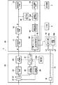
以下、図面を参照して本発明の実施形態を詳細に説明する。図1は本発明の内視鏡装置の一例を示すブロック図である。内視鏡装置1は、光源ユニット10、スコープ20、画像処理装置30を備えている。光源ユニット10は内視鏡による観察を行うために被写体に光を照射するものであって、キセノンランプ等の通常観察を行うための通常光源10aと、蛍光観察を行うための励起光源10bとを備えており、装置コントローラ80により動作が制御されている。通常光源10aは白色光を射出するものであり、励起光源10bは上述したようにたとえば682nmもしくは747nmの励起光を射出するものである。この光源ユニット10はスコープ20のライトガイド15に光学的に接続されており、光源ユニット10から射出された白色光L1および励起光L2はライトガイド15内に入射され観察窓(光照射手段)16から被写体に照射される。したがって、観察窓(光照射手段)16から照射される光の光量は装置コントローラ80によって制御されることになる。
Hereinafter, embodiments of the present invention will be described in detail with reference to the drawings. FIG. 1 is a block diagram showing an example of an endoscope apparatus of the present invention. The endoscope apparatus 1 includes a
スコープ20は、撮像レンズ21、撮像素子22、CDS/AGC回路23、A/D変換器24、CCD駆動部25、レンズ駆動部26等を有しており、各構成要素はスコープコントローラ27により制御されている。撮像レンズ21はたとえば複数のレンズ群から構成されており、レンズ駆動部26の駆動により撮影倍率が変更する。撮像素子22はたとえばCCDやCMOS等からなり、撮像レンズ21により結像された被写体像を光電変換して画像を取得するものである。この撮像素子22としては、例えば撮像面にMg(マゼンタ),Ye(イエロー),Cy(シアン),G(グリーン)の色フィルタを有する補色型、あるいはRGBの色フィルタを有する原色型が用いられる。なお、撮像素子22の動作はCCD駆動部25により制御されている。撮像素子22が画像(映像)信号を取得したとき、CDS/AGC(相関二重サンプリング/自動利得制御)回路23がサンプリングして増幅し、A/D変換器24がCDS/AGC回路17から出力された内視鏡画像をA/D変換し、画像処理装置30に出力される。
The
画像処理装置30は、スコープ20を用いて取得された内視鏡画像を処理するものであって、たとえばDSP等により構成されている。画像処理装置30は、画像取得手段31、前処理手段32、画像処理手段33、表示制御手段34を備えている。画像取得手段31は、スコープ20の撮像素子22により撮影された内視鏡画像Pを取得するものである。前処理手段32は、画像取得手段31において取得された内視鏡画像Pに対し前処理を施すものであって、たとえば内視鏡画像PがYCC表色系からなっている場合にはRGB表色系に変換し、さらにガンマ変換機能、階調を調整する機能等を有している。画像処理手段33は前処理された内視鏡画像Pに対し強調処理等を施すものであり、表示制御手段34は画像処理手段33において画像処理された内視鏡画像Pをキャラクタ情報等とともに表示装置3に表示する機能を有している。
The
さらに、画像処理装置30は、エッジ抽出手段40、複雑度検出手段50、撮影状態判定手段60、条件切替手段70を備えている。エッジ抽出手段40は、前処理手段32により前処理された内視鏡画像から血管像を抽出するものである。ここで、エッジ抽出手段40は、たとえば微分フィルタ処理等の公知の技術を用いてエッジ成分を検出する。
Further, the
複雑度検出手段50は、エッジ抽出手段40により抽出されたエッジ成分EPの複雑度CDを検出するものである。具体的には、複雑度検出手段50は、エッジ成分EPを2値化し、2値化されたエッジ成分についてたとえばボックスカウンティング法等公知の技術を用いてフラクタル次元を複雑度CDとして検出する。この複雑度(フラクタル次元)CDはエッジ成分EPが複雑な構造になればなるほど高い値になり、単純な構造になればなるほど低い値になる(図2、図3参照)。
The
撮影状態判定手段60は、複雑度検出手段50において検出された複雑度CDに応じて被写体を近接撮影したものであるか遠景撮影したものであるかを判定するものである。この近接撮影とはスコープ20の先端もしくはスコープ20の先端に取り付けられたフードが被写体に接触しもしくは僅かに離れた状態で撮影を行うことを意味し、遠景撮影とはスコープ20の先端が被写体から離れた状態で撮影を行うことを意味する。ここで、撮影状態判定手段60は、複雑度CDが設定しきい値CDref以上であるとき近接撮影であると判定し、設定しきい値CDref未満であるとき遠景撮影であると判定する。このように、近接撮影のときには内視鏡画像Pに密集した微細な血管像や被写体表面の凹凸等が映し出され複雑度が高くなることを利用し、精度良く近接撮影であるか否かを判定することができる。
The shooting
なお、複雑度検出手段50は、フラクタル解析を行う場合のみならず、ピットパターンを検出することにより複雑度CDを算出するようにしてもよい。具体的には、複雑度検出手段50は、エッジ成分EPにおいて公知の技術を用いて領域統合処理を行うことによりピットパターンを検出する。そして、ピットパターンの面積、周囲長、主軸長、円形度等を特徴量として抽出し、これらをベクトル成分とする特徴ベクトルを複雑度CDとして生成する。そして、撮影状態判定手段60は、この特徴ベクトルからベクターサポートマシンやニューラルネットワーク等の公知の識別アルゴリズムにより、ピットパターンの複雑度CDが設定しきい値CDref以上であるか否かを判断する。この場合であっても、近接撮影のときには内視鏡画像Pに密集した微細な血管像や被写体表面の凹凸等が映し出され複雑度が高くなることを利用し、精度良く近接撮影であるか否かを判定することができる。
The complexity detection means 50 may calculate the complexity CD not only when performing fractal analysis but also by detecting a pit pattern. Specifically, the
条件切替手段70は、撮影状態判定手段60により判定された撮影状態に応じて撮影条件を自動的に切り替えるものである。具体的には、近接撮影であると判定されたとき、条件切替手段70はスコープ20の撮像レンズ21の倍率を標準倍率(たとえば20倍)よりも大きくする(たとえば60倍以上)。さらに、条件切替手段70は光照射手段16から照射される光の光量を小さくなるように光源ユニット10を制御する。一方、遠景撮影であると判定されたとき、条件切替手段70はスコープ20の撮像レンズの倍率を標準倍率に比べて小さくする(たとえば等倍)。さらに、条件切替手段70は光照射手段16から照射される光の光量を大きくなるように光源ユニット10を制御する。
The
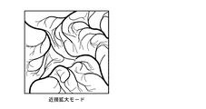
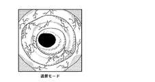
このように、内視鏡画像Pの複雑度CDに応じて撮影モードを自動的に切り替えることにより、操作者が観察したい被写体の部位に適した撮影条件に自動的に設定することができるため、従来のように操作者が撮影倍率を設定する必要がなく、効率的な内視鏡観察を行うことができる。つまり、一般的に、被写体を近接撮影は局所的な部位を撮影するものであるため、図2に示すように、内視鏡画像には密集した微細な血管像や生体組織の微細な凹凸やピットパターン等が映し出されることになる。したがって、内視鏡画像の複雑度CDは高いものとなる。一方、スコープ20の先端を被写体から離して撮影した内視鏡画像には、図3に示すように、太い血管や毛細血管あるいは生体組織の輪郭等が映し出されるものであって、上述した近接拡大撮影をした場合に比べて複雑度CDは低いものとなる。そこで、複雑度CDに基づいて操作者が観察したい部位を推定し、推定した部位に最適な撮影モードに自動的に切り替える。これにより、操作者の負担を軽減し効率的な内視鏡観察を行うことができる。
Thus, by automatically switching the imaging mode according to the complexity CD of the endoscopic image P, it is possible to automatically set the imaging conditions suitable for the part of the subject that the operator wants to observe. It is not necessary for the operator to set the photographing magnification as in the prior art, and efficient endoscopic observation can be performed. That is, in general, close-up imaging of a subject is an imaging of a local site, and as shown in FIG. 2, an endoscopic image has a dense blood vessel image, a minute unevenness of a living tissue, A pit pattern or the like will be projected. Therefore, the complexity CD of the endoscopic image is high. On the other hand, as shown in FIG. 3, the endoscope image obtained by shooting the distal end of the
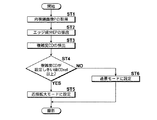
図4は本発明の内視鏡装置の制御方法の好ましい実施形態を示すフローチャートであり、図1から図4を参照して内視鏡装置の制御方法について説明する。まず、スコープ20が体腔内に挿入さたた状態で撮影を行うことにより、内視鏡画像が取得される(ステップST1)。そして、エッジ抽出手段40により内視鏡画像のエッジ成分が抽出される(ステップST2)。その後、エッジ成分EPの複雑度CDが検出され(ステップST3)、条件切替手段70において複雑度CDが設定しきい値以上であるかが判断される(ステップST4)。複雑度CDが設定しきい値CDref以上であるとき、条件切替手段70により撮影モードが近接拡大モードに設定され、装置コントローラ80により撮像レンズ21の倍率が等倍に設定されると共に光源ユニット10から照射される光の光量が小さくなる(ステップST5)。一方、複雑度CDが設定しきい値CDref未満であるとき、条件切替手段70により撮影モードが遠景モードに設定され、装置コントローラ80により撮像レンズ21の倍率が30倍〜60倍に設定されると共に光源ユニット10から照射される光の光量が大きくなる(ステップST6)。
FIG. 4 is a flowchart showing a preferred embodiment of the method for controlling an endoscope apparatus according to the present invention. The method for controlling an endoscope apparatus will be described with reference to FIGS. First, an endoscope image is acquired by performing imaging while the
上記実施の形態によれば、スコープ20により取得された内視鏡画像Pのエッジ成分EPを抽出し、抽出したエッジ成分EPの複雑度CDを検出し、検出した複雑度CDに応じて、被写体を近接撮影したものであるか遠景撮影したものであるかを判定し、判定した近接撮影もしくは遠景撮影に応じて撮影条件を切り替えることにより、内視鏡画像Pから操作者が観察したい被写体を推定し、観察したい部位に適した撮影モードに切り替えることができるため、操作者が撮影モードを切り替える必要がなく効率的な画像診断を行うことができる。
According to the above embodiment, the edge component EP of the endoscopic image P acquired by the
なお、撮影状態判定手段60が、複雑度CDが設定しきい値CDref以上であるとき近接撮影であると判定し、設定しきい値CDref未満であるとき遠景撮影であると判定するものであるとき、近接撮影のときには内視鏡画像Pに密集した微細な血管像や被写体表面の凹凸等が映し出され複雑度が高くなることを利用し、精度良く近接撮影であるか否かを判定することができる。
When the shooting
また、複雑度検出手段50が、エッジ成分EPのフラクタル次元を複雑度CDとして検出するものであれば、精度良く複雑度CDを検出することができる。 Further, if the complexity detection means 50 detects the fractal dimension of the edge component EP as the complexity CD, the complexity CD can be detected with high accuracy.
さらに、複雑度検出手段50が、エッジ成分EPからピットパターンを抽出し、抽出したピットパターンの複雑度CDを検出するものであるとき、精度良く複雑度CDを検出することができる。 Furthermore, when the complexity detection means 50 extracts a pit pattern from the edge component EP and detects the complexity CD of the extracted pit pattern, the complexity CD can be detected with high accuracy.
また、スコープ20が撮影倍率を変更する撮像レンズ21を有するものであり、条件切替手段70が、近接撮影のときに撮像レンズ21の倍率を大きくし、遠景撮影のときに撮像レンズ21の倍率を小さくするものであれば、近接撮影に適した撮影倍率に自動的に設定することができるため、効率的に画像診断を行うことができる。
The
さらに、スコープ20が被写体に光を照射する光照射手段16を有するものであり、条件切替手段70が、近接撮影のときに光照射手段16から照射される光の光量を小さくし、遠景撮影に切り替えたときに光照射手段16から照射される光の光量をも大きくするものであるとき、近接撮影に適した光量に自動的に設定することができるため、効率的に画像診断を行うことができる。
Further, the
本発明の実施形態は、上記実施形態に限定されない。たとえば、上記実施の形態において、被写体に白色光を照射したときに取得された通常観察画像を用いて複雑度CDを検出する場合について例示しているが、励起光を照射したときに被写体から発光する蛍光を撮影した蛍光画像、被写体に狭帯域な光を照射したときい撮影した狭帯域画像あるいは分光画像を用いて複雑度CDを検出するようにしてもよい。 The embodiment of the present invention is not limited to the above embodiment. For example, in the above embodiment, the case where the complexity CD is detected using the normal observation image acquired when the subject is irradiated with white light is illustrated, but the subject emits light when the excitation light is irradiated. The complexity CD may be detected using a fluorescent image obtained by photographing the fluorescent light, a narrow-band image obtained when the subject is irradiated with narrow-band light, or a spectral image.
また、上記実施形態において、条件切替手段70は、撮影条件として撮影倍率や光量を切り替える場合について例示しているが、近接撮影の場合には高周波成分を強調し、遠景撮影の場合には低周波成分を強調する、もしくは近接撮影の場合は電子ズームの倍率を大きくし、遠景撮影の場合は電子ズームの倍率を小さくする、というように画像処理手段33における画像処理条件を切り替えるようにしてもよい。
In the above embodiment, the condition switching means 70 is exemplified for switching the shooting magnification and the light amount as shooting conditions. However, the high frequency component is emphasized for close-up shooting, and the low frequency is used for far-field shooting. The image processing conditions in the
また、条件切替手段70が複雑度CDが設定しきい値未満であるときに遠景撮影に適した撮影条件に設定する場合について例示しているが、遠景モード内において複雑度が低くなるにつれて倍率を大きくするように撮像レンズ21を制御するようにしてもよい。
Further, although the case where the
1 内視鏡装置
10 光源ユニット
16 光照射手段
20 スコープ
21 撮像レンズ
30 画像処理装置
31 画像取得手段
32 前処理手段
33 画像処理手段
40 エッジ抽出手段
50 複雑度検出手段
60 撮影状態判定手段
70 条件切替手段
80 装置コントローラ
CD 複雑度
CDref 設定しきい値
EP エッジ成分
P 内視鏡画像
DESCRIPTION OF SYMBOLS 1
Claims (7)
該スコープにより取得された前記内視鏡画像のエッジ成分を抽出するエッジ抽出手段と、
該エッジ抽出手段により抽出された前記エッジ成分の複雑度を検出する複雑度検出手段と、
該複雑度検出手段において検出された前記複雑度に応じて、前記被写体を近接撮影したものであるか遠景撮影したものであるかを判定する撮影状態判定手段と、
該撮影状態判定手段において判定された前記近接撮影もしくは前記遠景撮影に応じて撮影条件を切り替える条件切替手段と
を備えたことを特徴とする内視鏡装置。 A scope that captures the subject and obtains an endoscopic image;
Edge extraction means for extracting an edge component of the endoscopic image acquired by the scope;
Complexity detecting means for detecting the complexity of the edge component extracted by the edge extracting means;
Shooting state determination means for determining whether the subject is a close-up shot or a distant view shot according to the complexity detected by the complexity detection means;
An endoscope apparatus comprising: a condition switching unit that switches a shooting condition according to the close-up shooting or the distant shooting determined by the shooting state determination unit.
前記条件切替手段が、前記近接撮影のときに前記撮像レンズの倍率を大きくし、前記遠景撮影のときに前記撮像レンズの倍率を小さくするものであることを特徴とする請求項1から4のいずれか1項記載の内視鏡装置。 The scope has an imaging lens for changing a photographing magnification;
5. The condition switching unit is configured to increase the magnification of the imaging lens during the close-up shooting and decrease the magnification of the imaging lens during the distant shooting. The endoscope apparatus according to claim 1.
前記条件切替手段が、前記近接撮影のときに前記光照射手段から照射される前記光の光量を小さくし、前記遠景撮影のときに前記光照射手段から照射される前記光の光量を大きくするものであることを特徴とする請求項1から5のいずれか1項記載の内視鏡装置。 The scope has a light irradiation means for irradiating the subject with light,
The condition switching unit reduces the amount of light emitted from the light irradiation unit during the close-up photography, and increases the amount of light emitted from the light irradiation unit during the far-field photography. The endoscope apparatus according to any one of claims 1 to 5, wherein the endoscope apparatus is any one of the following.
前記スコープにより取得された前記内視鏡画像のエッジ成分を抽出し、
抽出した前記エッジ成分の複雑度を検出し、
検出した前記複雑度に応じて、前記被写体を近接撮影したものであるか遠景撮影したものであるかを判定し、
判定した前記近接撮影もしくは遠景撮影に応じて撮影条件を切り替える
ことを特徴とする内視鏡装置の制御方法。 An endoscopic device control method for capturing an endoscope image by photographing a subject with a scope,
Extracting an edge component of the endoscopic image acquired by the scope;
Detecting the complexity of the extracted edge component;
In accordance with the detected complexity, it is determined whether the subject is a close-up shot or a distant shot,
A method for controlling an endoscope apparatus, wherein the imaging condition is switched according to the determined close-up shooting or far-field shooting.
Priority Applications (4)
| Application Number | Priority Date | Filing Date | Title |
|---|---|---|---|
| JP2008325256A JP2010142546A (en) | 2008-12-22 | 2008-12-22 | Endoscope apparatus and control method therefor |
| EP09013303A EP2179687B1 (en) | 2008-10-22 | 2009-10-21 | Endoscope apparatus and control method therefor |
| US12/603,187 US8553075B2 (en) | 2008-10-22 | 2009-10-21 | Endoscope apparatus and control method therefor |
| EP11158020.5A EP2335551B1 (en) | 2008-10-22 | 2009-10-21 | Endoscope apparatus and control method therefor |
Applications Claiming Priority (1)
| Application Number | Priority Date | Filing Date | Title |
|---|---|---|---|
| JP2008325256A JP2010142546A (en) | 2008-12-22 | 2008-12-22 | Endoscope apparatus and control method therefor |
Publications (1)
| Publication Number | Publication Date |
|---|---|
| JP2010142546A true JP2010142546A (en) | 2010-07-01 |
Family
ID=42563579
Family Applications (1)
| Application Number | Title | Priority Date | Filing Date |
|---|---|---|---|
| JP2008325256A Abandoned JP2010142546A (en) | 2008-10-22 | 2008-12-22 | Endoscope apparatus and control method therefor |
Country Status (1)
| Country | Link |
|---|---|
| JP (1) | JP2010142546A (en) |
Cited By (5)
| Publication number | Priority date | Publication date | Assignee | Title |
|---|---|---|---|---|
| JP2012152332A (en) * | 2011-01-25 | 2012-08-16 | Fujifilm Corp | Endoscope system and method of assisting operation thereof |
| JP2012152279A (en) * | 2011-01-24 | 2012-08-16 | Fujifilm Corp | Endoscope system |
| JP2012213552A (en) * | 2011-04-01 | 2012-11-08 | Fujifilm Corp | Endoscope system, processor device of endoscope system, and image processing method |
| JP2012239815A (en) * | 2011-05-24 | 2012-12-10 | Fujifilm Corp | Endoscope system and method for displaying endoscope image |
| JPWO2021060159A1 (en) * | 2019-09-27 | 2021-04-01 |
Citations (1)
| Publication number | Priority date | Publication date | Assignee | Title |
|---|---|---|---|---|
| JP2002028126A (en) * | 2000-07-14 | 2002-01-29 | Olympus Optical Co Ltd | Endoscope device |
-
2008
- 2008-12-22 JP JP2008325256A patent/JP2010142546A/en not_active Abandoned
Patent Citations (1)
| Publication number | Priority date | Publication date | Assignee | Title |
|---|---|---|---|---|
| JP2002028126A (en) * | 2000-07-14 | 2002-01-29 | Olympus Optical Co Ltd | Endoscope device |
Cited By (8)
| Publication number | Priority date | Publication date | Assignee | Title |
|---|---|---|---|---|
| JP2012152279A (en) * | 2011-01-24 | 2012-08-16 | Fujifilm Corp | Endoscope system |
| JP2012152332A (en) * | 2011-01-25 | 2012-08-16 | Fujifilm Corp | Endoscope system and method of assisting operation thereof |
| JP2012213552A (en) * | 2011-04-01 | 2012-11-08 | Fujifilm Corp | Endoscope system, processor device of endoscope system, and image processing method |
| JP2012239815A (en) * | 2011-05-24 | 2012-12-10 | Fujifilm Corp | Endoscope system and method for displaying endoscope image |
| JPWO2021060159A1 (en) * | 2019-09-27 | 2021-04-01 | ||
| WO2021060159A1 (en) * | 2019-09-27 | 2021-04-01 | 富士フイルム株式会社 | Image processing device, endoscopic system, and operation method of image processing |
| CN114521122A (en) * | 2019-09-27 | 2022-05-20 | 富士胶片株式会社 | Image processing apparatus, endoscope system, and image processing method |
| JP7196330B2 (en) | 2019-09-27 | 2022-12-26 | 富士フイルム株式会社 | Image processing device, endoscope system, and method of operating image processing device |
Similar Documents
| Publication | Publication Date | Title |
|---|---|---|
| US8553075B2 (en) | Endoscope apparatus and control method therefor | |
| CN110663251B (en) | Medical image processing apparatus | |
| CN110381807B (en) | Endoscope system, processor device, and method for operating endoscope system | |
| JP5271062B2 (en) | Endoscope apparatus and method of operating the same | |
| CN112689469B (en) | Endoscope device, endoscope processor, and method for operating endoscope device | |
| CN102131449B (en) | Imaging system and endoscope system | |
| JP5435746B2 (en) | Endoscope device | |
| CN110325100A (en) | Endoscopic system and its operating method | |
| JP5358368B2 (en) | Endoscope system | |
| WO2022037397A1 (en) | Endoscope system and method for detecting when tail end of endoscope comes into contact with tissue | |
| JP4098402B2 (en) | Fluorescent imaging device | |
| JP2016158682A (en) | Image processing device | |
| JP6132901B2 (en) | Endoscope device | |
| JP4566754B2 (en) | Image processing device | |
| JPWO2016088628A1 (en) | Image evaluation apparatus, endoscope system, operation method of image evaluation apparatus, and operation program of image evaluation apparatus | |
| JP5422180B2 (en) | Endoscope apparatus and method of operating the same | |
| JP2010142547A (en) | Endoscopic image processing apparatus and method, and program. | |
| JP4895750B2 (en) | Endoscope processor, autofluorescence image display program, and endoscope system | |
| JP5371366B2 (en) | Endoscope apparatus and method for operating endoscope apparatus | |
| JP6128989B2 (en) | Image processing apparatus, endoscope apparatus, and operation method of image processing apparatus | |
| WO2022230607A1 (en) | Medical image processing device, endoscope system, and operation method for medical image processing device | |
| JP2010279526A (en) | Endoscopic image processing apparatus and method, and program | |
| JP2025074589A (en) | Endoscope system and method of operation thereof | |
| JP6184928B2 (en) | Endoscope system, processor device | |
| JP6168878B2 (en) | Image processing apparatus, endoscope apparatus, and image processing method |
Legal Events
| Date | Code | Title | Description |
|---|---|---|---|
| RD15 | Notification of revocation of power of sub attorney |
Free format text: JAPANESE INTERMEDIATE CODE: A7435 Effective date: 20110215 |
|
| A621 | Written request for application examination |
Free format text: JAPANESE INTERMEDIATE CODE: A621 Effective date: 20110707 |
|
| A977 | Report on retrieval |
Free format text: JAPANESE INTERMEDIATE CODE: A971007 Effective date: 20130131 |
|
| A131 | Notification of reasons for refusal |
Free format text: JAPANESE INTERMEDIATE CODE: A131 Effective date: 20130205 |
|
| A762 | Written abandonment of application |
Free format text: JAPANESE INTERMEDIATE CODE: A762 Effective date: 20130307 |