JP2011152425A - Inflatable porous implant and method for drug delivery - Google Patents
Inflatable porous implant and method for drug delivery Download PDFInfo
- Publication number
- JP2011152425A JP2011152425A JP2011036448A JP2011036448A JP2011152425A JP 2011152425 A JP2011152425 A JP 2011152425A JP 2011036448 A JP2011036448 A JP 2011036448A JP 2011036448 A JP2011036448 A JP 2011036448A JP 2011152425 A JP2011152425 A JP 2011152425A
- Authority
- JP
- Japan
- Prior art keywords
- graft
- therapeutic agent
- agent
- channel
- host polymer
- Prior art date
- Legal status (The legal status is an assumption and is not a legal conclusion. Google has not performed a legal analysis and makes no representation as to the accuracy of the status listed.)
- Granted
Links
Images





Classifications
-
- A—HUMAN NECESSITIES
- A61—MEDICAL OR VETERINARY SCIENCE; HYGIENE
- A61F—FILTERS IMPLANTABLE INTO BLOOD VESSELS; PROSTHESES; DEVICES PROVIDING PATENCY TO, OR PREVENTING COLLAPSING OF, TUBULAR STRUCTURES OF THE BODY, e.g. STENTS; ORTHOPAEDIC, NURSING OR CONTRACEPTIVE DEVICES; FOMENTATION; TREATMENT OR PROTECTION OF EYES OR EARS; BANDAGES, DRESSINGS OR ABSORBENT PADS; FIRST-AID KITS
- A61F2/00—Filters implantable into blood vessels; Prostheses, i.e. artificial substitutes or replacements for parts of the body; Appliances for connecting them with the body; Devices providing patency to, or preventing collapsing of, tubular structures of the body, e.g. stents
- A61F2/02—Prostheses implantable into the body
- A61F2/04—Hollow or tubular parts of organs, e.g. bladders, tracheae, bronchi or bile ducts
- A61F2/06—Blood vessels
- A61F2/07—Stent-grafts
-
- A—HUMAN NECESSITIES
- A61—MEDICAL OR VETERINARY SCIENCE; HYGIENE
- A61F—FILTERS IMPLANTABLE INTO BLOOD VESSELS; PROSTHESES; DEVICES PROVIDING PATENCY TO, OR PREVENTING COLLAPSING OF, TUBULAR STRUCTURES OF THE BODY, e.g. STENTS; ORTHOPAEDIC, NURSING OR CONTRACEPTIVE DEVICES; FOMENTATION; TREATMENT OR PROTECTION OF EYES OR EARS; BANDAGES, DRESSINGS OR ABSORBENT PADS; FIRST-AID KITS
- A61F2/00—Filters implantable into blood vessels; Prostheses, i.e. artificial substitutes or replacements for parts of the body; Appliances for connecting them with the body; Devices providing patency to, or preventing collapsing of, tubular structures of the body, e.g. stents
- A61F2/02—Prostheses implantable into the body
- A61F2/04—Hollow or tubular parts of organs, e.g. bladders, tracheae, bronchi or bile ducts
- A61F2/06—Blood vessels
-
- A—HUMAN NECESSITIES
- A61—MEDICAL OR VETERINARY SCIENCE; HYGIENE
- A61L—METHODS OR APPARATUS FOR STERILISING MATERIALS OR OBJECTS IN GENERAL; DISINFECTION, STERILISATION OR DEODORISATION OF AIR; CHEMICAL ASPECTS OF BANDAGES, DRESSINGS, ABSORBENT PADS OR SURGICAL ARTICLES; MATERIALS FOR BANDAGES, DRESSINGS, ABSORBENT PADS OR SURGICAL ARTICLES
- A61L27/00—Materials for grafts or prostheses or for coating grafts or prostheses
- A61L27/50—Materials characterised by their function or physical properties, e.g. injectable or lubricating compositions, shape-memory materials, surface modified materials
- A61L27/507—Materials characterised by their function or physical properties, e.g. injectable or lubricating compositions, shape-memory materials, surface modified materials for artificial blood vessels
-
- A—HUMAN NECESSITIES
- A61—MEDICAL OR VETERINARY SCIENCE; HYGIENE
- A61L—METHODS OR APPARATUS FOR STERILISING MATERIALS OR OBJECTS IN GENERAL; DISINFECTION, STERILISATION OR DEODORISATION OF AIR; CHEMICAL ASPECTS OF BANDAGES, DRESSINGS, ABSORBENT PADS OR SURGICAL ARTICLES; MATERIALS FOR BANDAGES, DRESSINGS, ABSORBENT PADS OR SURGICAL ARTICLES
- A61L27/00—Materials for grafts or prostheses or for coating grafts or prostheses
- A61L27/50—Materials characterised by their function or physical properties, e.g. injectable or lubricating compositions, shape-memory materials, surface modified materials
- A61L27/54—Biologically active materials, e.g. therapeutic substances
-
- A—HUMAN NECESSITIES
- A61—MEDICAL OR VETERINARY SCIENCE; HYGIENE
- A61F—FILTERS IMPLANTABLE INTO BLOOD VESSELS; PROSTHESES; DEVICES PROVIDING PATENCY TO, OR PREVENTING COLLAPSING OF, TUBULAR STRUCTURES OF THE BODY, e.g. STENTS; ORTHOPAEDIC, NURSING OR CONTRACEPTIVE DEVICES; FOMENTATION; TREATMENT OR PROTECTION OF EYES OR EARS; BANDAGES, DRESSINGS OR ABSORBENT PADS; FIRST-AID KITS
- A61F2/00—Filters implantable into blood vessels; Prostheses, i.e. artificial substitutes or replacements for parts of the body; Appliances for connecting them with the body; Devices providing patency to, or preventing collapsing of, tubular structures of the body, e.g. stents
- A61F2/0095—Packages or dispensers for prostheses or other implants
-
- A—HUMAN NECESSITIES
- A61—MEDICAL OR VETERINARY SCIENCE; HYGIENE
- A61F—FILTERS IMPLANTABLE INTO BLOOD VESSELS; PROSTHESES; DEVICES PROVIDING PATENCY TO, OR PREVENTING COLLAPSING OF, TUBULAR STRUCTURES OF THE BODY, e.g. STENTS; ORTHOPAEDIC, NURSING OR CONTRACEPTIVE DEVICES; FOMENTATION; TREATMENT OR PROTECTION OF EYES OR EARS; BANDAGES, DRESSINGS OR ABSORBENT PADS; FIRST-AID KITS
- A61F2/00—Filters implantable into blood vessels; Prostheses, i.e. artificial substitutes or replacements for parts of the body; Appliances for connecting them with the body; Devices providing patency to, or preventing collapsing of, tubular structures of the body, e.g. stents
- A61F2/82—Devices providing patency to, or preventing collapsing of, tubular structures of the body, e.g. stents
- A61F2/86—Stents in a form characterised by the wire-like elements; Stents in the form characterised by a net-like or mesh-like structure
- A61F2/89—Stents in a form characterised by the wire-like elements; Stents in the form characterised by a net-like or mesh-like structure the wire-like elements comprising two or more adjacent rings flexibly connected by separate members
-
- A—HUMAN NECESSITIES
- A61—MEDICAL OR VETERINARY SCIENCE; HYGIENE
- A61F—FILTERS IMPLANTABLE INTO BLOOD VESSELS; PROSTHESES; DEVICES PROVIDING PATENCY TO, OR PREVENTING COLLAPSING OF, TUBULAR STRUCTURES OF THE BODY, e.g. STENTS; ORTHOPAEDIC, NURSING OR CONTRACEPTIVE DEVICES; FOMENTATION; TREATMENT OR PROTECTION OF EYES OR EARS; BANDAGES, DRESSINGS OR ABSORBENT PADS; FIRST-AID KITS
- A61F2/00—Filters implantable into blood vessels; Prostheses, i.e. artificial substitutes or replacements for parts of the body; Appliances for connecting them with the body; Devices providing patency to, or preventing collapsing of, tubular structures of the body, e.g. stents
- A61F2/02—Prostheses implantable into the body
- A61F2/04—Hollow or tubular parts of organs, e.g. bladders, tracheae, bronchi or bile ducts
- A61F2/06—Blood vessels
- A61F2/07—Stent-grafts
- A61F2002/075—Stent-grafts the stent being loosely attached to the graft material, e.g. by stitching
-
- A—HUMAN NECESSITIES
- A61—MEDICAL OR VETERINARY SCIENCE; HYGIENE
- A61F—FILTERS IMPLANTABLE INTO BLOOD VESSELS; PROSTHESES; DEVICES PROVIDING PATENCY TO, OR PREVENTING COLLAPSING OF, TUBULAR STRUCTURES OF THE BODY, e.g. STENTS; ORTHOPAEDIC, NURSING OR CONTRACEPTIVE DEVICES; FOMENTATION; TREATMENT OR PROTECTION OF EYES OR EARS; BANDAGES, DRESSINGS OR ABSORBENT PADS; FIRST-AID KITS
- A61F2250/00—Special features of prostheses classified in groups A61F2/00 - A61F2/26 or A61F2/82 or A61F9/00 or A61F11/00 or subgroups thereof
- A61F2250/0003—Special features of prostheses classified in groups A61F2/00 - A61F2/26 or A61F2/82 or A61F9/00 or A61F11/00 or subgroups thereof having an inflatable pocket filled with fluid, e.g. liquid or gas
-
- A—HUMAN NECESSITIES
- A61—MEDICAL OR VETERINARY SCIENCE; HYGIENE
- A61F—FILTERS IMPLANTABLE INTO BLOOD VESSELS; PROSTHESES; DEVICES PROVIDING PATENCY TO, OR PREVENTING COLLAPSING OF, TUBULAR STRUCTURES OF THE BODY, e.g. STENTS; ORTHOPAEDIC, NURSING OR CONTRACEPTIVE DEVICES; FOMENTATION; TREATMENT OR PROTECTION OF EYES OR EARS; BANDAGES, DRESSINGS OR ABSORBENT PADS; FIRST-AID KITS
- A61F2250/00—Special features of prostheses classified in groups A61F2/00 - A61F2/26 or A61F2/82 or A61F9/00 or A61F11/00 or subgroups thereof
- A61F2250/0014—Special features of prostheses classified in groups A61F2/00 - A61F2/26 or A61F2/82 or A61F9/00 or A61F11/00 or subgroups thereof having different values of a given property or geometrical feature, e.g. mechanical property or material property, at different locations within the same prosthesis
- A61F2250/0023—Special features of prostheses classified in groups A61F2/00 - A61F2/26 or A61F2/82 or A61F9/00 or A61F11/00 or subgroups thereof having different values of a given property or geometrical feature, e.g. mechanical property or material property, at different locations within the same prosthesis differing in porosity
-
- A—HUMAN NECESSITIES
- A61—MEDICAL OR VETERINARY SCIENCE; HYGIENE
- A61F—FILTERS IMPLANTABLE INTO BLOOD VESSELS; PROSTHESES; DEVICES PROVIDING PATENCY TO, OR PREVENTING COLLAPSING OF, TUBULAR STRUCTURES OF THE BODY, e.g. STENTS; ORTHOPAEDIC, NURSING OR CONTRACEPTIVE DEVICES; FOMENTATION; TREATMENT OR PROTECTION OF EYES OR EARS; BANDAGES, DRESSINGS OR ABSORBENT PADS; FIRST-AID KITS
- A61F2250/00—Special features of prostheses classified in groups A61F2/00 - A61F2/26 or A61F2/82 or A61F9/00 or A61F11/00 or subgroups thereof
- A61F2250/0014—Special features of prostheses classified in groups A61F2/00 - A61F2/26 or A61F2/82 or A61F9/00 or A61F11/00 or subgroups thereof having different values of a given property or geometrical feature, e.g. mechanical property or material property, at different locations within the same prosthesis
- A61F2250/0035—Special features of prostheses classified in groups A61F2/00 - A61F2/26 or A61F2/82 or A61F9/00 or A61F11/00 or subgroups thereof having different values of a given property or geometrical feature, e.g. mechanical property or material property, at different locations within the same prosthesis differing in release or diffusion time
-
- A—HUMAN NECESSITIES
- A61—MEDICAL OR VETERINARY SCIENCE; HYGIENE
- A61F—FILTERS IMPLANTABLE INTO BLOOD VESSELS; PROSTHESES; DEVICES PROVIDING PATENCY TO, OR PREVENTING COLLAPSING OF, TUBULAR STRUCTURES OF THE BODY, e.g. STENTS; ORTHOPAEDIC, NURSING OR CONTRACEPTIVE DEVICES; FOMENTATION; TREATMENT OR PROTECTION OF EYES OR EARS; BANDAGES, DRESSINGS OR ABSORBENT PADS; FIRST-AID KITS
- A61F2250/00—Special features of prostheses classified in groups A61F2/00 - A61F2/26 or A61F2/82 or A61F9/00 or A61F11/00 or subgroups thereof
- A61F2250/0058—Additional features; Implant or prostheses properties not otherwise provided for
- A61F2250/0067—Means for introducing or releasing pharmaceutical products into the body
- A61F2250/0068—Means for introducing or releasing pharmaceutical products into the body the pharmaceutical product being in a reservoir
-
- A—HUMAN NECESSITIES
- A61—MEDICAL OR VETERINARY SCIENCE; HYGIENE
- A61L—METHODS OR APPARATUS FOR STERILISING MATERIALS OR OBJECTS IN GENERAL; DISINFECTION, STERILISATION OR DEODORISATION OF AIR; CHEMICAL ASPECTS OF BANDAGES, DRESSINGS, ABSORBENT PADS OR SURGICAL ARTICLES; MATERIALS FOR BANDAGES, DRESSINGS, ABSORBENT PADS OR SURGICAL ARTICLES
- A61L2300/00—Biologically active materials used in bandages, wound dressings, absorbent pads or medical devices
- A61L2300/40—Biologically active materials used in bandages, wound dressings, absorbent pads or medical devices characterised by a specific therapeutic activity or mode of action
- A61L2300/404—Biocides, antimicrobial agents, antiseptic agents
-
- A—HUMAN NECESSITIES
- A61—MEDICAL OR VETERINARY SCIENCE; HYGIENE
- A61L—METHODS OR APPARATUS FOR STERILISING MATERIALS OR OBJECTS IN GENERAL; DISINFECTION, STERILISATION OR DEODORISATION OF AIR; CHEMICAL ASPECTS OF BANDAGES, DRESSINGS, ABSORBENT PADS OR SURGICAL ARTICLES; MATERIALS FOR BANDAGES, DRESSINGS, ABSORBENT PADS OR SURGICAL ARTICLES
- A61L2300/00—Biologically active materials used in bandages, wound dressings, absorbent pads or medical devices
- A61L2300/40—Biologically active materials used in bandages, wound dressings, absorbent pads or medical devices characterised by a specific therapeutic activity or mode of action
- A61L2300/41—Anti-inflammatory agents, e.g. NSAIDs
-
- A—HUMAN NECESSITIES
- A61—MEDICAL OR VETERINARY SCIENCE; HYGIENE
- A61L—METHODS OR APPARATUS FOR STERILISING MATERIALS OR OBJECTS IN GENERAL; DISINFECTION, STERILISATION OR DEODORISATION OF AIR; CHEMICAL ASPECTS OF BANDAGES, DRESSINGS, ABSORBENT PADS OR SURGICAL ARTICLES; MATERIALS FOR BANDAGES, DRESSINGS, ABSORBENT PADS OR SURGICAL ARTICLES
- A61L2300/00—Biologically active materials used in bandages, wound dressings, absorbent pads or medical devices
- A61L2300/40—Biologically active materials used in bandages, wound dressings, absorbent pads or medical devices characterised by a specific therapeutic activity or mode of action
- A61L2300/412—Tissue-regenerating or healing or proliferative agents
-
- A—HUMAN NECESSITIES
- A61—MEDICAL OR VETERINARY SCIENCE; HYGIENE
- A61L—METHODS OR APPARATUS FOR STERILISING MATERIALS OR OBJECTS IN GENERAL; DISINFECTION, STERILISATION OR DEODORISATION OF AIR; CHEMICAL ASPECTS OF BANDAGES, DRESSINGS, ABSORBENT PADS OR SURGICAL ARTICLES; MATERIALS FOR BANDAGES, DRESSINGS, ABSORBENT PADS OR SURGICAL ARTICLES
- A61L2300/00—Biologically active materials used in bandages, wound dressings, absorbent pads or medical devices
- A61L2300/40—Biologically active materials used in bandages, wound dressings, absorbent pads or medical devices characterised by a specific therapeutic activity or mode of action
- A61L2300/416—Anti-neoplastic or anti-proliferative or anti-restenosis or anti-angiogenic agents, e.g. paclitaxel, sirolimus
-
- A—HUMAN NECESSITIES
- A61—MEDICAL OR VETERINARY SCIENCE; HYGIENE
- A61L—METHODS OR APPARATUS FOR STERILISING MATERIALS OR OBJECTS IN GENERAL; DISINFECTION, STERILISATION OR DEODORISATION OF AIR; CHEMICAL ASPECTS OF BANDAGES, DRESSINGS, ABSORBENT PADS OR SURGICAL ARTICLES; MATERIALS FOR BANDAGES, DRESSINGS, ABSORBENT PADS OR SURGICAL ARTICLES
- A61L2300/00—Biologically active materials used in bandages, wound dressings, absorbent pads or medical devices
- A61L2300/40—Biologically active materials used in bandages, wound dressings, absorbent pads or medical devices characterised by a specific therapeutic activity or mode of action
- A61L2300/42—Anti-thrombotic agents, anticoagulants, anti-platelet agents
Landscapes
- Health & Medical Sciences (AREA)
- Life Sciences & Earth Sciences (AREA)
- Transplantation (AREA)
- Public Health (AREA)
- Oral & Maxillofacial Surgery (AREA)
- Veterinary Medicine (AREA)
- Animal Behavior & Ethology (AREA)
- General Health & Medical Sciences (AREA)
- Medicinal Chemistry (AREA)
- Engineering & Computer Science (AREA)
- Biomedical Technology (AREA)
- Chemical & Material Sciences (AREA)
- Vascular Medicine (AREA)
- Heart & Thoracic Surgery (AREA)
- Gastroenterology & Hepatology (AREA)
- Cardiology (AREA)
- Pulmonology (AREA)
- Dermatology (AREA)
- Epidemiology (AREA)
- Molecular Biology (AREA)
- Prostheses (AREA)
- Materials For Medical Uses (AREA)
- Media Introduction/Drainage Providing Device (AREA)
Abstract
Description
本発明は一般に、医療器具及び方法に関する。より詳細には、本発明は、例えばグラフト、ステントグラフト、及び嚢体(bladders)等の膨張可能な多孔性インプラント、並びに薬物送達のための方法及びキットを提供する。 The present invention generally relates to medical devices and methods. More particularly, the present invention provides expandable porous implants, such as grafts, stent grafts, and bladders, and methods and kits for drug delivery.
脈管及び器官の疾患は、罹患した脈管及び器官の機能を修復又は代替する補綴具であるグラフト又はステントグラフトを必要としてきた。本発明は特に、身体内の流路の完全性を損なう可能性がある疾病、又は損傷を治療するための補綴具に関する。例えば、補綴具は、胸部及び腹部大動脈瘤、動脈解離(例えば外傷を原因とするもの)等を含む心血管系における適応症の治療、並びに消化器系(胆管、食道、及び胃腸管内の同様の構造)、呼吸器系(気管支、気管、及び同様物)、生殖器系(卵管、子宮、及び同様物)、泌尿器系(尿道、尿管、及び同様物)、及び人間の身体の他の系内における適応症の治療に有用である。補綴具としてのグラフト又はステントグラフトは、修復、代替、又はバイパス形成される脈管又は器官の自然な流れを再建、又は再形成する。 Vascular and organ diseases have required grafts or stent grafts that are prosthetics to repair or replace the function of affected vessels and organs. In particular, the present invention relates to a prosthesis for treating diseases or injuries that may compromise the integrity of the flow path in the body. For example, prosthetics can be used to treat indications in the cardiovascular system, including thoracic and abdominal aortic aneurysms, arterial dissections (such as those caused by trauma), and the like in the digestive system (bile, esophagus, and gastrointestinal tract) Structure), respiratory system (bronchi, trachea, and the like), genital system (oviduct, uterus, and the like), urinary system (urethra, ureter, and the like), and other systems in the human body It is useful for the treatment of indications in the hospital. A graft or stent graft as a prosthetic device reconstructs or reforms the natural flow of a vessel or organ that is being repaired, replaced, or bypassed.
補綴具としてのグラフト又はステントグラフトは、ある程度の成功を収めているが、これらの埋め込み型器具を改良することは有益であると思われる。より詳細には、改良点の一つとして、埋め込み型補綴具を介して、治療薬を身体の流路内に直接送達することがある。腹部大動脈瘤又は他の血管の動脈瘤に使用されるグラフトは、グラフトを閉塞させ得る血栓形成等の合併症を起こすことがある。また、一般にステントグラフトは肥厚化を引き起こす場合があり、この肥厚化によりグラフトが破損し得る。このような場合、これら合併症を治療又は予防する治療薬を送達することが特に有用である。 Although grafts or stent grafts as prosthetic devices have had some success, it would be beneficial to improve these implantable devices. More particularly, one improvement is the delivery of therapeutic agents directly into the body flow path via an implantable prosthesis. Grafts used for abdominal aortic aneurysms or other vascular aneurysms can cause complications such as thrombus formation that can occlude the graft. In general, a stent graft may cause thickening, and this thickening may damage the graft. In such cases, it is particularly useful to deliver therapeutic agents that treat or prevent these complications.
この必要性に対処する目的で、治療薬を身体の流路内に直接送達する種々の埋め込み型器具が設計されている。治療薬の局所送達は、いくつかの理由により全身送達よりも優れている。第一に、局所送達は、標的としない他の脈管又は器官をその薬剤に晒すことなく、標的部位に適切な量の治療薬を送達することができる。第二に、治療薬の局所濃度は、全身投与によって通常達成され得る濃度と比較して、相当高くすることができる。第三に、局所送達は、通常であれば治療薬の吸収が遅い標的組織に、薬剤を集中させることができる。 To address this need, a variety of implantable devices have been designed that deliver therapeutic agents directly into the body's flow path. Local delivery of therapeutic agents is superior to systemic delivery for several reasons. First, local delivery can deliver the appropriate amount of therapeutic agent to the target site without exposing other untargeted vessels or organs to the agent. Secondly, the local concentration of the therapeutic agent can be considerably higher than that which can usually be achieved by systemic administration. Third, local delivery can focus the drug on a target tissue that is normally slow to absorb the therapeutic drug.
治療薬の直接投与は、主としてカテーテル注入又は被覆補綴器具の使用により達成されている。カテーテルを介した治療薬の直接投与では、通常、薬物送達の継続時間全体に亘ってカテーテルが身体内の定位置に配置されている必要がある。したがって、カテーテル注入による治療は、主として治療継続時間が短いものに適している。被覆補綴器具、例えばステント、グラフト、又は他のインプラント等も幅広く利用されている。これらの被覆器具は様々な程度にて成功を収めているが、いくつかの明白な欠点が存在する。すなわち、被覆補綴器具は、その製造工程が複雑であり、場合によっては器具の組立て中(例えばステントを搬送カテーテル又はシースに通過させる時)又は留置中に被覆が剥がれ落ちることがある。被覆補綴器具に関する別の懸念の一つは、薬物の投与量、送達の正確さ及び継続時間が、インプラント全体に対して通常小さい部分を占める被覆自体によって、制限され得ることである。 Direct administration of therapeutic agents has been achieved primarily through the use of catheter injection or coated prosthetic devices. Direct administration of a therapeutic agent via a catheter typically requires the catheter to be in place in the body for the entire duration of drug delivery. Therefore, the treatment by catheter injection is mainly suitable for treatment with a short treatment duration. Coated prosthetic devices such as stents, grafts, or other implants are also widely used. Although these coating devices have been successful to varying degrees, there are some obvious drawbacks. That is, the coated prosthetic device is complex in its manufacturing process, and in some cases, the coating may peel off during assembly of the device (eg, when passing the stent through a delivery catheter or sheath) or during placement. Another concern with coated prosthetic devices is that drug dosage, delivery accuracy and duration can be limited by the coating itself, which usually occupies a small portion of the entire implant.
これらの理由により、身体の流路内に治療薬を送達するための改良された埋め込み型器具、及び方法を提供することが望ましい。特に、より多量に、且つより正確な量で、より長い投与時間に亘って、薬物を身体内に直接送達する改良された埋め込み型器具及び方法を提供することが望ましい。また、留置が容易なように、柔軟性を有する小型の埋め込み型器具を提供することがさらに望ましい。また、薬物送達に加えて、機械的又は構造的機能を果たす一体化された埋め込み型器具を提供することがさらに望ましい。 For these reasons, it is desirable to provide improved implantable devices and methods for delivering therapeutic agents within the body flow path. In particular, it would be desirable to provide improved implantable devices and methods for delivering drugs directly into the body in larger amounts and in more accurate amounts over longer administration times. It is further desirable to provide a small implantable device that is flexible so that it can be easily placed. It is further desirable to provide an integrated implantable device that performs a mechanical or structural function in addition to drug delivery.
活性物質を埋め込み型器具、及び他の器具から放出するための方法及び装置は、特許文献1、特許文献2、特許文献3、特許文献4、特許文献5、特許文献6、特許文献7、特許文献8、特許文献9、特許文献10、特許文献11、特許文献12、特許文献13、特許文献14、特許文献15、特許文献16、特許文献17、特許文献18、特許文献19、特許文献20、特許文献21、特許文献22、特許文献23、特許文献24、特許文献25、特許文献26、特許文献27、特許文献28、特許文献29、特許文献30、特許文献31、特許文献32、特許文献33、特許文献34、特許文献35、特許文献36、特許文献37、特許文献38、特許文献39、特許文献40、特許文献41、特許文献42、特許文献43、特許文献44、特許文献45、特許文献46、特許文献47、特許文献48、特許文献49、特許文献50、特許文献51、特許文献52、特許文献53、特許文献54、特許文献55、特許文献56、特許文献57、特許文献58、特許文献59、特許文献60、特許文献61、特許文献62、特許文献63、特許文献64、特許文献65、特許文献66、特許文献67、特許文献68に記載されている。
Methods and devices for releasing active substances from implantable devices and other devices are described in Patent Document 1, Patent Document 2, Patent Document 3, Patent Document 4, Patent Document 5, Patent Document 6, Patent Document 7, Patent Literature 8, Patent Literature 9,
上記の各引用の開示全体は、参照により本願に援用される。 The entire disclosure of each of the above citations is incorporated herein by reference.
本発明は、膨張可能な多孔性のインプラント、例えばグラフト、ステントグラフト、及び嚢体、並びに薬物送達のための方法及びキットを提供する。特に、本発明のグラフト及びステントグラフトは、身体の流路内に治療薬及び/又は診断薬を送達する。この膨張可能な多孔性インプラントは、薬物をより多量に、且つより正確な投与量で、より長い投与時間に亘って、身体内に直接送達することができる。さらに、この膨張可能な多孔性インプラントは、多くの場合、挿入時には柔軟であり、留置が容易なように送達形態は小型化されている。以下に詳細に説明するように、本発明のインプラントは、一体化された単一構造にて、薬物送達に加えて機械的又は構造的機能をさらに提供し得る。 The present invention provides expandable porous implants such as grafts, stent grafts, and sac, and methods and kits for drug delivery. In particular, the grafts and stent grafts of the present invention deliver therapeutic and / or diagnostic agents within the body flow path. This expandable porous implant can deliver the drug directly into the body in larger amounts and with more precise dosages over longer administration times. Furthermore, the expandable porous implant is often flexible upon insertion and the delivery configuration is miniaturized to facilitate placement. As described in detail below, the implants of the present invention may further provide mechanical or structural functions in addition to drug delivery in an integrated unitary structure.
本発明の膨張可能な多孔性のグラフト及びステントグラフトは、身体内の罹患した流路又はその一部を修復、代替、又はバイパス形成することが可能である。通常、このようなグラフト及びステントグラフトは、動脈(例えば大動脈)、静脈、並びに過去に移植したグラフト、シャント、フィステル及び同様物を含む、患者の脈管構造における任意の血管内に移植することが可能である。このような場合、その補綴器具は、胸部及び腹部大動脈瘤、動脈解離、胸部若しくは末梢動脈瘤、他の血管の動脈瘤、血管疾患、他の罹患した若しくは異常な血管、冠動脈内の罹患した伏在静脈グラフト等を含む、脈管構造内におけるいくつかの適応症の治療に有用である。しかしながら、本発明の器具は、脈管構造内への留置及び/又は移植に限定されないことが理解されよう。例えば、本発明のグラフト及びステントグラフトは、消化器系(胆管、食道、及び胃腸管内の同様の構造)、呼吸器系(気管支、気管、及び同様物)、生殖器系(卵管、子宮、及び同様物)、泌尿器系(尿道、尿管、及び同様物)、並びに人間の身体の他の系内における他の適応症の治療にも有用であり得る。 The expandable porous grafts and stent grafts of the present invention are capable of repairing, substituting, or bypassing the affected flow path or a portion thereof within the body. Typically, such grafts and stent grafts can be implanted into any vessel in the patient's vasculature, including arteries (eg, aorta), veins, and previously grafted grafts, shunts, fistulas and the like. It is. In such cases, the prosthetic device can be used for thoracic and abdominal aortic aneurysms, arterial dissections, thoracic or peripheral aneurysms, aneurysms of other blood vessels, vascular diseases, other affected or abnormal blood vessels, affected saphenous in coronary arteries. It is useful for the treatment of several indications within the vasculature, including venous grafts and the like. However, it will be appreciated that the device of the present invention is not limited to placement and / or implantation within the vasculature. For example, the grafts and stent grafts of the present invention can be used in the digestive system (similar structures in the bile duct, esophagus, and gastrointestinal tract), respiratory system (bronchi, trachea, and the like), reproductive system (oviduct, uterus, and the like). ), The urinary system (urethra, ureters, and the like), and other indications within other systems of the human body.
本発明の一側面において、グラフトはグラフト本体部及び膨張媒体を備える。グラフト本体部は、近位端及び遠位端を有し、少なくとも一個の膨張可能な多孔性チャネルすなわち空洞を画成する。膨張媒体は、膨張可能なチャネル内に導入されるように構成された、少なくとも一種の治療薬を含有する。本発明にて使用され得るグラフトの例は、米国特許第6,395,019号明細書、及び同時係属の米国特許出願第10/327,711号明細書に詳細に記載されており、これら双方は本願の譲受人に譲渡され、参照により本願に援用される。本発明では、例えば、Holmanらに付与された米国特許第5,871,537号明細書、Kwan−Gettに付与された米国特許第5,151,105号明細書、Pigottに付与された米国特許第5,156,620号明細書、Samuelsに付与された米国特許第6,007,575号明細書、McDermottらに付与された米国特許第6,312,462号明細書、及びSherryによる米国特許出願第09/978,383号明細書(2001年10月16日出願)に記載されている他のグラフトを使用してもよく、それぞれその全体が参照により本願に援用される。 In one aspect of the invention, the graft comprises a graft body and an inflation medium. The graft body portion has a proximal end and a distal end and defines at least one inflatable porous channel or cavity. The inflation medium contains at least one therapeutic agent configured to be introduced into the inflatable channel. Examples of grafts that can be used in the present invention are described in detail in US Pat. No. 6,395,019 and co-pending US patent application Ser. No. 10 / 327,711, both Is assigned to the assignee of the present application and is incorporated herein by reference. In the present invention, for example, U.S. Pat. No. 5,871,537 granted to Holman et al., U.S. Pat. No. 5,151,105 granted to Kwan-Gett, U.S. Patent granted to Pigott. US Pat. No. 5,156,620, US Pat. No. 6,007,575 to Samuels, US Pat. No. 6,312,462 to McDermott et al., And US Pat. Other grafts described in application Ser. No. 09 / 978,383 (filed Oct. 16, 2001) may be used, each incorporated herein by reference in its entirety.
少なくとも一種の治療薬は、膨張媒体から多孔性チャネルの壁を介して輸送され、例えば患者の脈管構造内の血管のような身体管腔内に放出され得る。少なくとも一種の治療薬は、グラフト本体部の管腔側及び反管腔側のいずれかの表面から身体内に放出されるように構成され得る。少なくとも一種の治療薬は、グラフトによって治療される特定の適応症に応じた任意の治療目的を果たす様々な薬剤からなり得る。例えば、治療薬は、内皮形成促進薬、血管新生促進薬、抗血栓薬、抗動脈瘤薬、抗感染薬、抗炎症薬、抗再狭窄薬、化学療法剤、及び抗癌剤からなる群から選択された一種又は二種以上の薬剤からなり得る。 At least one therapeutic agent can be transported from the inflation medium through the walls of the porous channel and released into a body lumen, such as a blood vessel in the patient's vasculature. At least one therapeutic agent may be configured to be released into the body from either the luminal or anti-luminal surface of the graft body. The at least one therapeutic agent can consist of a variety of agents that serve any therapeutic purpose depending on the particular indication being treated by the graft. For example, the therapeutic agent is selected from the group consisting of an endothelium-promoting agent, angiogenesis-promoting agent, antithrombotic agent, anti-aneurysm agent, anti-infective agent, anti-inflammatory agent, anti-restenosis agent, chemotherapeutic agent, and anticancer agent. Or one or more drugs.
多孔性チャネルは様々な多孔度を有しており、身体管腔内への制御及び/又はプログラムされた薬物送達を実現し得る。グラフト本体部は、その表面を介した輸送を可能にする任意の生体適合性材料からなり得る。例えば、グラフト本体部は、フルオロポリマー、ポリエチレンテレフタレート、ポリ塩化ビニル、ポリウレタン、ポリオレフィン、及びポリアミドからなる群から選択された一種又は二種以上の材料からなり得る。グラフト本体部は、特に発泡ポリテトラフルオロエチレン、有孔ポリテトラフルオロエチレン、又は他の合成材料からなっていてもよい。このようなグラフトの製造方法及び装置は、本願の譲受人に譲渡され、参照により本願に援用される国際特許出願第PCT/US02/40997号明細書に、より詳細に記載されている。 Porous channels have varying porosity and can provide controlled and / or programmed drug delivery into a body lumen. The graft body can be made of any biocompatible material that allows transport through its surface. For example, the graft body can be made of one or more materials selected from the group consisting of fluoropolymer, polyethylene terephthalate, polyvinyl chloride, polyurethane, polyolefin, and polyamide. The graft body may in particular consist of foamed polytetrafluoroethylene, perforated polytetrafluoroethylene or other synthetic material. A method and apparatus for making such a graft is described in more detail in International Patent Application No. PCT / US02 / 40997, assigned to the assignee of the present application and incorporated herein by reference.
少なくとも一個のチャネルは、螺旋、長手方向すなわち直線状のチャネル、及びグラフトの端部間においてグラフトの長さの一部分のみ、又はグラフトの遠位端若しくは近位端の周囲のみに延び得る周方向のリングから選択された一種又は二種以上の構成を有し得る。例えば、相互に連結する格子又はリング、鋸歯状パターン、互い違い又は不連続パターン、非直線状又は波形パターンのような他の配置も、単独で、又は上述のチャネル構成と組合せて適切であり得る。さらに、グラフトはグラフト本体部の近位端、遠位端又はその双方に配置された少なくとも一個の膨張可能な多孔性のカフを備えてもよく、このカフは、必要に応じて上述の少なくとも一個のチャネルと流体が流れるように連通し、且つ膨張媒体を収容していてもよい。一個又は二個以上のチャネル及びカフは、膨張時に十分堅い構造となり、それによってグラフト本体部を支持して、身体内の自然な流路の再形成又は再建に役立つ。さらに、一個又は二個以上の膨張したチャネル及びカフは、グラフトを密着させていかなる洩れをも防ぐに十分な表面を提供し、また器具のねじれを防止することができる。ねじれ抵抗型グラフト及びエンドリークの取り扱いは、同時係属の米国特許出願第10/384,103号明細書及び米国特許出願第10/691,849号明細書にそれぞれより詳細に記載されており、これら双方は本発明の譲受人に譲渡され、参照により本願に援用される。 At least one channel is a helix, a longitudinal or straight channel, and a circumferential direction that can extend only a portion of the length of the graft between the ends of the graft, or only around the distal or proximal end of the graft It may have one or more configurations selected from rings. Other arrangements such as interconnected grids or rings, serrated patterns, staggered or discontinuous patterns, non-linear or corrugated patterns may be appropriate alone or in combination with the channel configurations described above. In addition, the graft may comprise at least one inflatable porous cuff disposed at the proximal end, the distal end or both of the graft body portion, the cuff optionally including at least one of the above-described cuffs. The channel may be in fluid communication with the channel and may contain an inflation medium. One or more channels and cuffs are sufficiently stiff when inflated, thereby supporting the graft body and helping to reform or rebuild the natural flow path within the body. In addition, one or more inflated channels and cuffs can provide a sufficient surface to adhere to the graft and prevent any leaks and prevent twisting of the instrument. The handling of torsion resistant grafts and endoleaks is described in more detail in co-pending US patent application Ser. No. 10 / 384,103 and US patent application Ser. No. 10 / 691,849, respectively. Both are assigned to the assignee of the present invention and incorporated herein by reference.
膨張媒体は、通常、治療薬を収容するホストポリマーからなるが、必ずしもそうでなくてもよい。治療薬はポリマーから徐放的に放出される。治療薬は、ホストポリマーを介した放散により放出させるか、又は代替的にホストポリマーの減成又は分解により放出させることが可能である。ポリマーの減成による放出の場合、グラフト本体部はホストポリマー塊の輸送を阻止することが可能な生体適合性材料から形成される。一般的には、治療薬の所望の放出速度を達成するために、チャネルの多孔度、及び/又はホストポリマーの放散若しくは減成特性を調節し得る。通常、身体管腔内に放出可能な治療薬の量は、μg〜mgレベルの範囲内にあり、おおよそ約10μg〜約100mg又はそれ以上である。治療薬は、約1週間未満〜約2、3ヶ月又は多数ヶ月間、おおよそ約7日間〜12ヶ月間、身体管腔内に輸送されるように構成されている。 The inflation medium usually consists of a host polymer that contains the therapeutic agent, but this need not be the case. The therapeutic agent is released slowly from the polymer. The therapeutic agent can be released by release through the host polymer, or alternatively by release or degradation of the host polymer. In the case of release by polymer degradation, the graft body is formed from a biocompatible material capable of preventing transport of the host polymer mass. In general, the porosity of the channel and / or the release or degradation characteristics of the host polymer can be adjusted to achieve the desired release rate of the therapeutic agent. Typically, the amount of therapeutic agent that can be released into the body lumen is in the range of the μg to mg level, approximately from about 10 μg to about 100 mg or more. The therapeutic agent is configured to be delivered into the body lumen for less than about 1 week to about 2, 3 months or many months, approximately about 7 days to 12 months.
ホストポリマーは一般に、グラフトの留置及び/又は移植の前、留置及び/又は移植の間、又は留置及び/又は移植の後に、膨張可能なチャネル内に導入されることが可能な、任意の生体適合性材料からなり得る。例えば、ホストポリマーは、グラフトが搬送カテーテルから留置された後であって、その部位にグラフトが実際に移植される前に、グラフトを膨張させるよう使用され得る。ホストポリマーは、ポリエチレングリコール、ポリエチレングリコールジアクリレート、エトキシル化トリメチロールプロパントリアクリレート、プルロニックポリオキシマー(polyoxymer)、アクリルアミド、ポリエチレンオキシド、ポリビニルアルコール、ポリエチレン/ビニルアルコール共重合体、ポリアクリル酸、ポリエチレン/アクリル酸共重合体、ポリエチルオキサゾリン、ポリビニルピロリドン、ポリエチレン/ビニルピロリドン共重合体、ポリマレイン酸、ポリエチレン/マレイン酸共重合体、ポリアクリルアミド、及びポリエチレンオキシド/ポリプロピレンオキシド共重合体からなる群から選択された一種又は二種以上の材料からなり得る。膨張媒体は液体からなってもよく、その液体は注入後に液体のままであってもよく、相変化又は架橋形成によって固化してもよい。後者の場合、膨張媒体は約3分〜約12分の範囲の硬化時間、及び約345kPa〜2758kPa(約50psi〜約400psi)の範囲の硬化後の弾性係数を有する硬化性の液体とすることができる。 The host polymer is generally any biocompatible that can be introduced into the inflatable channel prior to implantation and / or implantation, during implantation and / or implantation, or after implantation and / or implantation. It can be made of a sex material. For example, the host polymer can be used to inflate the graft after it has been placed from the delivery catheter and before the graft is actually implanted at that site. Host polymers are polyethylene glycol, polyethylene glycol diacrylate, ethoxylated trimethylolpropane triacrylate, pluronic polyoxymer, acrylamide, polyethylene oxide, polyvinyl alcohol, polyethylene / vinyl alcohol copolymer, polyacrylic acid, polyethylene / Selected from the group consisting of acrylic acid copolymer, polyethyloxazoline, polyvinylpyrrolidone, polyethylene / vinylpyrrolidone copolymer, polymaleic acid, polyethylene / maleic acid copolymer, polyacrylamide, and polyethylene oxide / polypropylene oxide copolymer Or one or more materials. The expansion medium may consist of a liquid, which may remain liquid after injection or may solidify by phase change or cross-linking. In the latter case, the expansion medium may be a curable liquid having a cure time in the range of about 3 minutes to about 12 minutes and a post-cure modulus of elasticity in the range of about 345 kPa to 2758 kPa (about 50 psi to about 400 psi). it can.
本発明の別の一側面では、近位端及び遠位端を有し、該近位端と遠位端との間に少なくとも一個の膨張可能な多孔性チャネルを画成するグラフト本体部を備えるステントグラフトを提供する。グラフト本体部の近位端又は遠位端には接続部材が取り付けられ、同接続部材は一個又は二個以上の接続要素を有している。接続部材の一個又は二個以上の接続要素には、もう一個の近位端側ステント接続要素を有するステントが連結されている。少なくとも一種の治療薬を含有する膨張媒体は、膨張可能なチャネル内に導入されるように構成されている。 In another aspect of the present invention, a graft body having a proximal end and a distal end and defining at least one inflatable porous channel between the proximal end and the distal end is provided. A stent graft is provided. A connecting member is attached to the proximal end or the distal end of the graft body, and the connecting member has one or more connecting elements. Coupled to one or more connecting elements of the connecting member is a stent having another proximal end stent connecting element. An inflation medium containing at least one therapeutic agent is configured to be introduced into the inflatable channel.
本発明のさらなる別の一側面では、治療薬を送達する方法を提供する。一つの方法は、近位端及び遠位端を有し、且つ少なくとも一個の膨張可能な多孔性チャネルを画成するグラフト本体部を提供する工程を含む。グラフト本体部は、身体管腔内に移植される。多孔性チャネルが膨張され、すなわち少なくとも一種の治療薬を含有する膨張媒体が注入される。グラフトは標準的な外科的手法を用いて移植されてもよく、管腔内又は他の送達モードにより移植されてもよい。通常、多孔性チャネルは、グラフトの留置及び/又は移植前、留置及び/又は移植中、又は留置及び/又は移植後に膨張される。次に、本願の譲受人に譲渡され、参照により本願に援用される同時係属の米国特許出願第10/686,863号明細書に、その例がより詳細に記載されているグラフト送達システムを分離して身体から除去し、インプラントをその位置に残留させた後、身体内に一定期間薬剤を放出させる。治療薬は膨張媒体から多孔性チャネルを介して輸送されて、グラフト本体部の管腔側又は反管腔側の表面から身体管腔内に放出される。多孔性チャネルは、様々な多孔度を有する発泡又は有孔ポリテトラフルオロエチレンから形成されている。膨張媒体は、治療薬を収容するホストポリマーからなり、該ポリマーは放散又は減成プロセスにより治療薬を放出する。一般にグラフト本体部は、ホストポリマー塊の輸送を阻止する。本発明によれば、有用なホストポリマーは、液体として注入され、約3分〜約12分の範囲の硬化時間を有し、約345kPa〜2758kPa(約50psi〜約400psi)の範囲の硬化後の弾性係数を有するポリエチレングリコールからなり得る。 In yet another aspect of the invention, a method of delivering a therapeutic agent is provided. One method includes providing a graft body having a proximal end and a distal end and defining at least one inflatable porous channel. The graft body is implanted in the body lumen. The porous channel is expanded, that is, an expansion medium containing at least one therapeutic agent is injected. The graft may be implanted using standard surgical techniques and may be implanted intraluminally or by other delivery modes. Typically, the porous channel is inflated before, during and / or after graft placement and / or implantation. The separation of the graft delivery system, an example of which is described in more detail in co-pending US patent application Ser. No. 10 / 686,863, assigned to the assignee of the present application and incorporated herein by reference. After removing from the body and leaving the implant in place, the drug is released into the body for a period of time. The therapeutic agent is transported from the inflation medium through the porous channel and released into the body lumen from the luminal or antiluminal surface of the graft body. The porous channel is formed from foamed or perforated polytetrafluoroethylene having various porosities. The inflation medium consists of a host polymer that contains the therapeutic agent, which releases the therapeutic agent by a diffusion or degradation process. In general, the graft body blocks the transport of the host polymer mass. In accordance with the present invention, useful host polymers are injected as a liquid, have a cure time in the range of about 3 minutes to about 12 minutes, and after cure in the range of about 345 kPa to 2758 kPa (about 50 psi to about 400 psi). It can consist of polyethylene glycol with an elastic modulus.
本発明のさらなる別の一側面では、グラフトと、治療薬を送達するために該グラフトを移植及び膨張させる方法に関する使用説明書とを備えたキットを提供する。このキットは、少なくとも一種の治療薬を含有する膨張媒体の原料も備え得る。グラフトは、本願に記載されている任意の送達構造を有してよく、使用説明書には、一般に上述した方法の一つ又は二つ以上を実行するステップが列挙されている。使用説明書は多くの場合印刷され、場合によっては少なくとも一部が包装上に記述される。あるいは、使用説明書は、上述した任意の方法を説明する、及び/又は列挙するビデオテープ、CD、DVD、他の機械判読可能なフォーマット若しくはコード、グラフィック描写、又は同様物に含められていてもよい。 In yet another aspect of the invention, a kit is provided that includes a graft and instructions regarding how to implant and expand the graft to deliver a therapeutic agent. The kit can also include a source of inflation medium containing at least one therapeutic agent. The graft may have any of the delivery structures described herein, and the instructions for use enumerate steps that generally perform one or more of the methods described above. Instructions for use are often printed and in some cases at least partially written on the package. Alternatively, instructions for use may be included in videotapes, CDs, DVDs, other machine-readable formats or codes, graphic depictions, or the like that describe and / or enumerate any of the methods described above. Good.
本発明の薬物送達機構は、多数の異なる埋め込み型器具に導入することができる。上記の説明はグラフト及びステントグラフトに関するものであるが、治療薬送達のために他の膨張可能な多孔性のインプラント器具を使用し得ることが理解されるであろう。より詳細には、嚢状又は管状の形態を有する膨張可能な多孔性の嚢体を使用することもできる。嚢体は、不定期間且つ潜在的に長期間、治療部位に移植可能である点で永久性を有する。嚢体は、脈管構造(例えば血管)、又は他の身体管腔、臓器、及び嚢体の存在がいずれの生体機能にも悪影響を及ぼさない身体内組織構造に適用され得る。また通常、嚢体は薬物送達のプラットフォームとしての役割を果たす上に、いくつかの機械的機能を発揮する。嚢体はさらに、例えば卵管を遮断する等の閉塞性を有し、又は例えば組織充填、尿失禁の治療、癌の治療、受胎調節処置、サイナス移植、美容若しくは形成外科における補綴、脊椎構造の支持、管腔又は管路内の流量若しくは開閉弁の制御、及び同様の用途等の他の能力が利用される。 The drug delivery mechanism of the present invention can be introduced into a number of different implantable devices. While the above description relates to grafts and stent grafts, it will be understood that other expandable porous implant devices may be used for therapeutic drug delivery. More particularly, an inflatable porous sac having a sac-like or tubular form may be used. The sac is permanent in that it can be implanted at the treatment site for an indefinite and potentially long period of time. The sac can be applied to vasculature (eg, blood vessels), or other body lumens, organs, and tissue structures within the body where the presence of the sac does not adversely affect any biological function. Also, the capsule usually serves as a drug delivery platform and performs several mechanical functions. The sac can also have occlusive properties, such as blocking the fallopian tube, or for example tissue filling, urinary incontinence treatment, cancer treatment, fertility control treatment, sinus transplantation, prosthesis in cosmetic or plastic surgery, spinal structure Other capabilities such as support, control of flow rate or open / close valves in lumens or lines, and similar applications are utilized.
一実施態様において、人間の身体内に移植するのに適した膨張可能な多孔性インプラントは、一般的に膨張可能な多孔性の嚢体を備え、この嚢体は、その容積を画成する実質的に非弾性の壁を有し得る。少なくとも一種の治療薬を含有する膨張媒体を導入するために、少なくとも一つの嚢体壁開口部が形成されている。少なくとも一つの嚢体開口部の各々には、少なくとも一つの嚢体壁閉鎖具も設けられて、容積内に膨張媒体を保持する。嚢体壁及び膨張媒体は、移植部位に所望の治療薬を送達するよう選択され得る。本発明にて使用され得る典型的な膨張可能なインプラントは、本願の譲受人に譲渡され、参照により本願に援用される同時係属の米国特許出願第10/461,853号明細書に詳細に記載されている。膨張可能な多孔性嚢体は、膨張可能な多孔性グラフトチャネルに関して上記した任意の材料からなり得、またこのインプラント材料の輸送及び多孔度に関しても上記した任意の特性を採用し得る。同様に、本実施態様においては、前述したような膨張媒体、治療薬、種々の放出モード、薬剤の投与量及び継続時間を適用し得る。 In one embodiment, an inflatable porous implant suitable for implantation within a human body generally comprises an inflatable porous sac, wherein the sac defines a volume defining its volume. May have inelastic walls. At least one bladder wall opening is formed to introduce an inflation medium containing at least one therapeutic agent. Each of the at least one bladder opening is also provided with at least one bladder wall closure to hold the inflation medium in the volume. The bladder wall and inflation medium can be selected to deliver the desired therapeutic agent to the implantation site. Exemplary inflatable implants that may be used in the present invention are described in detail in co-pending US patent application Ser. No. 10 / 461,853, assigned to the assignee of the present application and incorporated herein by reference. Has been. The expandable porous sac can be composed of any material described above with respect to the expandable porous graft channel and can also employ any of the properties described above with respect to transport and porosity of the implant material. Similarly, in this embodiment, inflation media, therapeutic agents, various release modes, drug dosages and durations as described above may be applied.
別の一実施態様において、閉塞嚢体は、カテーテル、外科移植又は他の移植技術を用いて、患者の身体の治療部位に留置することができる。本実施例では、膨張媒体は、嚢体を膨張させて脈管を閉鎖すると共に、治療薬を送達するために使用され得る。治療薬は、閉塞具の周囲の洩れの危険を低減するために血栓薬からなってもよく、閉塞具により提供された止血シールの永久性を高めるために細胞成長剤からなってもよく、及び/又は、嚢体の周囲の組織に対して他の治療的利点を提供するために他の治療薬からなってもよい。膨張媒体は身体管腔内の流体の流れを減少させ、又は閉塞するために機械的障害物として働き、また標的塞栓部位の領域へ治療薬を局所送達するために治療薬のリザーバとして働くといった二役を果たす塞栓組成物をさらに含有し得る。塞栓組成物は、ポリエチレングリコールジアクリレート、エトキシル化トリメチロールプロパントリアクリレート、又はポリプロピレングリコールジアクリレートの、ペンタエリスリトールテトラ3(メルカプトプロピオネート)及び生理学的に許容される緩衝液との組合せからなり得る。本発明と関連して使用され得る塞栓物質は、“Methods、Compositions、and Devices for Embolizing Body Lumens”と題された2004年1月7日出願の、同時係属の米国特許出願第10/ 明細書(代理人整理番号:021630−005700US)に詳細に記載されており、その開示全体は参照により本願に援用される。 In another embodiment, the occlusion sac can be placed at a treatment site on the patient's body using a catheter, surgical implantation or other implantation technique. In this example, the inflation medium can be used to inflate the bladder to close the vessel and deliver a therapeutic agent. The therapeutic agent may consist of a thrombotic drug to reduce the risk of leakage around the obturator, may consist of a cell growth agent to increase the permanence of the hemostatic seal provided by the obturator, and Alternatively, it may consist of other therapeutic agents to provide other therapeutic benefits to the tissue surrounding the sac. The inflation medium acts as a mechanical obstruction to reduce or occlude fluid flow in the body lumen and as a reservoir of therapeutic agent for local delivery of the therapeutic agent to the area of the target embolization site. It may further contain an embolic composition that serves. The embolic composition may consist of a combination of polyethylene glycol diacrylate, ethoxylated trimethylolpropane triacrylate, or polypropylene glycol diacrylate with pentaerythritol tetra-3 (mercaptopropionate) and a physiologically acceptable buffer. . An embolic material that may be used in connection with the present invention is a copending US patent application Ser. No. 10/10, filed Jan. 7, 2004 entitled “Methods, Compositions, and Devices for Embroughing Body Lumens”. It is described in detail in the specification (Attorney Docket No: 0216630-005700US), the entire disclosure of which is incorporated herein by reference.
この二役の実施態様において、最初に治療薬は塞栓組成物の容積全体に収容され得る。治療薬は、懸濁物、混合物のいずれかとして、又は塞栓組成物の構成要素の一つに化学結合されることによって収容され得る。治療薬は、塞栓組成物の構成要素の主鎖、又は側鎖に結合され得る。例えば、治療薬はポリエチレングリコールの主鎖に結合されてもよい。いくつかの典型的な薬物、及び該薬物を塞栓組成物に結合させる方法は、J.M.Harris、“Laboratory Synthesis of Polyethylene Glycol Derivatives、”Journal of Macromolecular Science−Reviews in Macromolecular Chemistry、vol.C−25、No.3、pp.325−373、Jan.1、1985、M.Harris、Ed.、“Biomedical and Biotechnical Applications of Poly(Ethylene Glycol) Chemistry”、Plenum、NewYork、pp.1−14、1992、Greenwaldら、“Highly Water Soluble Taxol Derivatives:7−Polyethylene Glycol Carbamates and Carbonates:”、J.Org.Chem.、vol.60、No.2、pp.331−336、1995、Matsushimaら、“Modification of E.Coli Asparaginase with 2、4−Bis(O−Methoxypolyethylene Glycol)−6−Chloro−S−Triazine(Activated PEG.sub.2);Disappearance of Binding Ability Towards Anti−Serum and retention of Enzymic Activity”、Chemistry Letters、pp.773−776、1980、及びNathanら、“Copolymers of Lysine and Polyethylene Glycol:A New Family of Functionalized Drug Carriers、”Bioconjugate Chem.4,54−62(1993)に記載されており、これら各々は参照により本願に援用される。治療薬は、製造中、構成要素の一つと共によく混合されるか、又は別々に保管されて使用前に他方のポリマー構成要素と混合され得る。塞栓組成物は、その開示全体が参照により本願に援用される、Hubbellによる“Biomaterials formed by Nucleophilic Addition Reaction to Conjugated Unsaturated Groups”と題された国際公開第00/44808号パンフレット、及びHubbell等による“Conjugate Addition Reactions for the Controlled Delivery of Pharmaceutically Active Compounds”と題された国際公開第01/92584号パンフレットに記載されているマイケル付加反応によるインビボでの重合により形成され得る。 In this dual role embodiment, initially the therapeutic agent may be contained within the entire volume of the embolic composition. The therapeutic agent can be contained either as a suspension, a mixture, or by being chemically bonded to one of the components of the embolic composition. The therapeutic agent can be attached to the main chain, or side chain, of the component of the embolic composition. For example, the therapeutic agent may be bound to a polyethylene glycol backbone. Some exemplary drugs and methods for attaching the drugs to embolic compositions are described in J. Org. M.M. Harris, "Laboratory Synthesis of Polyethylene Glycol Derivatives," Journal of Macromolecular Science-Reviews in Macromolecular Chemistry. C-25, No. 3, pp. 325-373, Jan. 1, 1985, M.M. Harris, Ed. "Biomedical and Biotechnical Applications of Poly (Ethylene Glycol) Chemistry", Plenum, New York, pp. 1-14, 1992, Greenwald et al., “Highly Water Soluble Taxol Derivatives: 7-Polyethylene Glycol Carbamates and Carbonates:”, J. Am. Org. Chem. , Vol. 60, no. 2, pp. 331-336, 1995, Matsushima et al., “Modification of E. Coli Asparaginase with 2, 4-Bis (O-Methoxypolyethylene glycol) -6-Chloro-S-Triazinep. Anti-Serum and retention of Enzymatic Activity ", Chemistry Letters, pp. 773-776, 1980, and Nathan et al., "Copolymers of Lysine and Polyethylene Glycol: A New Family of Functionalized Carriers," Bioconjugate Chem. 4, 54-62 (1993), each of which is incorporated herein by reference. The therapeutic agent may be well mixed with one of the components during manufacture or stored separately and mixed with the other polymer component prior to use. An embolic composition is disclosed in International Publication Nos. 00 / 44808b, et al., “Biomaterials formed by Nucleophilic Addition Reaction to Conjugated United Groups” by Hubbell, which is incorporated herein by reference in its entirety. It can be formed by in vivo polymerization by the Michael addition reaction described in WO 01/92584 entitled “Addition Reactions for the Controlled Deliverable of Pharmaceutical Active Compounds”.
本発明によれば、薬剤を収容するホストポリマーを含む多孔性の容積(例、グラフトチャネル又は嚢体)を有する膨張可能なインプラントは、薬剤がより多量に、且つより正確な投与量で、より長い投与時間に亘って、身体内に送達される可能性を有している。これは、薬剤収容ホストポリマーがインプラント全体のかなりの部分を占め得ることによるが、必ずしもそれが理由とは限らない。また、本発明の埋め込み型グラフト及び嚢体器具は、インプラントの留置中、又は留置後にホストポリマーをチャネル又は嚢体内に充填することができるため、器具を柔軟且つ小型の状態で挿入することができ、したがって身体内への留置又は移植がより容易であり得る。インプラントは、一旦留置されると、膨張媒体の注入によってより堅くなり得る。さらに、これら薬物送達のための膨張可能なチャネル又は嚢体は、留置中に薬剤が剥がれ落ちるという、いかなる懸念も低下させる。さらに、本発明のグラフト及び嚢体インプラントは、一体化された単一構造にて、薬物リザーバの機能に加えて、機械的又は構造的な機能を提供する。例えば、グラフト本体部の膨張したチャネル及び/又はカフは、身体内で流体管路を提供し得る。嚢体の場合、膨張媒体が注入されると、身体管腔を閉塞する役割を果たすか、又は組織の充填に使用され得る。 In accordance with the present invention, an expandable implant having a porous volume (eg, a graft channel or sac) containing a host polymer that contains a drug can be used in a larger amount of drug and at a more accurate dosage. It has the potential to be delivered into the body over long administration times. This is due to the fact that the drug-containing host polymer can occupy a significant portion of the entire implant, but this is not necessarily the reason. In addition, since the implantable graft and capsular device of the present invention can be filled with the host polymer into the channel or the capsular body during or after the implantation of the implant, the device can be inserted in a flexible and compact state. Thus, placement or implantation within the body may be easier. Once in place, the implant can be made stiffer by injection of the inflation medium. In addition, these inflatable channels or capsules for drug delivery reduce any concern that the drug will fall off during placement. Furthermore, the graft and capsular implants of the present invention provide mechanical or structural functions in addition to drug reservoir functions in a single unitary structure. For example, the inflated channel and / or cuff of the graft body can provide a fluid line within the body. In the case of a sac, when inflated, it can serve to occlude body lumens or can be used to fill tissue.
本発明の性質及び利点のさらなる理解は、明細書の残りの部分及び図面を参照することにより、より明らかとなるであろう。 A further understanding of the nature and advantages of the present invention will become more apparent with reference to the remaining portions of the specification and drawings.
以下の図面の説明は、詳細な説明を参照して読むべきである。異なる図面中の同様の符号は、同様の要素を指す。必ずしも等尺でない図面は、本発明の実施態様を具体的に説明するために示したものであり、本発明の範囲を限定することを意図するものではない。 The following description of the drawings should be read with reference to the detailed description. Like reference symbols in the different drawings indicate like elements. The drawings, which are not necessarily to scale, are presented to specifically illustrate embodiments of the invention and are not intended to limit the scope of the invention.
図1において、膨張可能な多孔性グラフト10は、留置形態にて示されている。別に述べない限り、本願にて用語“グラフト”又は“ステントグラフト”は、修復、代替、又はバイパス形成するべき脈管又は器官の疾患部内に、自然な流路を再形成、又は再建することが可能な補綴具を意味し、一般的には管状又は二叉の器具と、そこに取り付けられ、又はそれと一体化された任意の構成要素とを含む。解説を目的として、以下に説明するグラフトの実施態様は、腹部大動脈瘤(AAA)の血管内治療に最も有用であると考えられる。本願のために、グラフト装置を参照して、用語“近位端”は、器具が身体通路内に留置された際に、通常は血液である体液が流入する側に向けられるグラフト端部を意味する。したがって、用語“遠位端”は、近位端と反対側のグラフト端部を意味する。この説明は例証のみを目的としており、必ずしもグラフト10の実際の形状、大きさ、寸法、又は特定の構成(カフ、チャネル、グラフト形態、接続部材、ステント、充填孔ブリッジ、近位端頸部及び遠位端頸部等、及びそれらの個々の構成)を反映するものではない。このようなものとして、本願に図示し且つ説明する実施態様は、単なる例示であり、本発明の範囲内で多様に変更され得る。加えて、本願に示した本発明のステントグラフトの実施態様は、膨張可能なチャネル(一個又は複数)の封止性、剛性付与、及び他の機械的及び臨床的な利点を示すが、これらの利点は本発明の範囲内に含まれるものと理解する必要はない。例えば、接続部材30,36及びステント32,38を有するグラフト10が示されているが、これらの構成は任意である。このことは本願の全ての説明及び実施態様に適用される。
In FIG. 1, an expandable
図1に示すように、グラフト10は、近位端12及び遠位端14を有し、管状構造、すなわちグラフト本体部16を備えている。グラフト本体部16は、近位端13及び遠位端15を有し、内部の流体の流れを制限するように構成された長手方向に延びる管腔18を形成しており、その長さは約5cm〜約30cm、特に約10cm〜約20cmの範囲内にあり得る。グラフト本体部16は、さらに、少なくとも一個の膨張可能な多孔性チャネル20すなわち空洞を画成し、該チャネル20すなわち空洞は、本実施例では長手方向に延びるチャネル、すなわち背骨を有し、これらチャネルすなわち背骨は、周方向に延びる一連の平行なチャネル、すなわちリングと流体が流れるように連通している。少なくとも一種の治療薬46を含有する膨張媒体22は、膨張可能なチャネル20内に導入されるように構成されている(図2参照)。
As shown in FIG. 1, the
チャネル20又は複数のチャネルは、膨張するとグラフト本体部16の剛性を高め、グラフト本体部16のねじれを防止することができ、また患者の身体通路内へのグラフト10の留置を容易にもし得る。膨張可能なチャネル20の長手方向及び径方向の寸法は、グラフト10による治療を意図される適応症に依存して、異なるグラフト本体部間において、及び同一のグラフト本体部においてさえも、必要に応じて変更され得る。さらに、膨張可能なチャネル20は、グラフト本体部16の長手方向軸25に関して様々な角度に向けられてよく、チャネルの間隔は、必要に応じて変更されてよい。
The
膨張可能なチャネル20又は複数のチャネルは、図示するように、グラフト本体部に関して長手方向及び/又は周方向に延びる一種又は複数の構成を任意の組合せにて採用し得る。例えば螺旋、又は相互に連結する格子若しくはリングのような他の構成もまた、単独で、又は任意の他の構成との組合せにて適当であり得る。膨張可能な一つ又は複数のチャネルは、ねじれ又は折れに対する抵抗性を付与するために鋸歯状のパターンを有してもよい。鋸歯状の膨張可能なチャネルは、螺旋状に、周方向に、環状リブ及び背骨の構成で、又は同様にて配置され得る。このような膨張可能なチャネルのねじれ抵抗性は、鋸歯が蝶番式に動くことができて長手方向の折れが防止されるために向上され得る。場合により、鋸歯は異なる内側半径及び外側半径を有し得る。さらに、周方向リングを接続するチャネルは、代替的に、互い違った又は不連続の長手方向チャネルすなわち背骨を有して、グラフト本体部、すなわち肢の可撓性を向上し得る。膨張可能なチャネルを相互に連結する長手方向チャネルすなわち背骨はまた、非線形又は波形の構成を採用することによって、グラフトの長手方向の圧縮を改善し得る。このような構成はさらに、圧縮時のグラフトのねじれの可能性を低減し得る。
The
近位端側の膨張可能な多孔性カフ26、及び遠位端側の膨張可能な多孔性カフ28を任意にてさらに設けてもよい。近位端側カフ26及び遠位端側カフ28は、膨張可能なチャネル20と流体が流れるように連通しており、膨張可能なカフ及びチャネルが互いに流体が流れるように連通するネットワークを形成している。充填孔24は、近位端側カフ26、遠位端側カフ28、及び膨張可能なチャネル20と流体が流れるように連通し、膨張媒体22のグラフト本体部16内への導入部を該ネットワークに付加している。カフ26及びカフ28は、膨張時に十分堅い構造を提供してグラフト本体部16を支持し、グラフトが留置された脈管の内面に対してグラフト10を十分に密着させる表面を提供するように構成され得る。密着によって、血流力学的圧力が動脈瘤へ伝達されることが防止され、またグラフト本体部16の外側表面の周囲における血液等の流体の流れが防止される。
A proximal end inflatable
膨張可能なカフ26及びカフ28は、任意の数の構成にて配置され得る。例えば、カフ26及びカフ28は、グラフト本体部の近位端及び/又は遠位端の周囲に、軸対照の円筒パターンにて配置され得る。あるいは、近位端側及び遠位端側の密封カフは、鋸歯状の構成を採用してもよい。鋸歯状の膨張可能なカフは、圧縮時に折れにくい利点を有するため、グラフトに対する身体管腔径の変化による影響が少ない。鋸歯状の膨張可能なカフは、複数の頂部を規定するジグザグのチャネルからなり得る。膨張時、本発明による鋸歯状の膨張可能なカフは、グラフトと身体管腔との正反対の相互作用が原因となり得る内側への折れが少ない。さらに、鋸歯状の膨張可能なカフは、様々な半径の鋸歯を有することにより、内側に折れる可能性のある臨床用途にて、望ましくない内側折れの可能性をさらに低減し得る。
複数の多孔性チャネル、カフ、又は多孔性チャネル若しくはカフ及び非多孔性チャネル若しくはカフの任意の組合せが同一のグラフト本体部に存在し、これらの各々又は全部が同一又は異なる膨張媒体によって別々に膨張可能であり得る構成も、本発明の範囲内に包含される。例えば、本発明のグラフト本体部の代替的な実施態様(図示せず)は、治療薬を含有しない第一の膨張媒体によって膨張可能な一個又は二個以上の比較的非多孔性のチャネルを有し得る。このような一個又は複数のチャネルは、グラフト本体部内で、例えばグラフト密封、ねじれ抵抗等の他の機能を果たす。この同一のグラフト本体部は、一個又は複数の非多孔性チャネルと流体が流れるように連通していない一個又は二個以上の比較的多孔性のチャネルをさらに有してもよく、この比較的多孔性のチャネルは、本願に記載する、標的領域への送達のための治療薬を収容する異なる膨張媒体によって、(例えば、一個又は二個以上の別個の充填孔を介して)別個に膨張可能である。 Multiple porous channels, cuffs, or any combination of porous channels or cuffs and non-porous channels or cuffs are present in the same graft body, each of which is inflated separately by the same or different inflation media Configurations that may be possible are also encompassed within the scope of the invention. For example, an alternative embodiment (not shown) of the graft body of the present invention has one or more relatively non-porous channels that can be expanded by a first expansion medium that does not contain a therapeutic agent. Can do. One or more such channels perform other functions within the graft body, such as graft sealing, torsional resistance, and the like. The same graft body may further include one or more relatively porous channels that are not in fluid communication with the one or more non-porous channels. Sex channels can be separately inflated (eg, via one or more separate fill holes) by different inflation media containing therapeutic agents for delivery to a target area as described herein. is there.
このようなグラフト本体部が、一個又は複数の多孔性及び/又は非多孔性チャネルの一個又は二個以上と流体が流れるように連通し、又は連通していない、一個又は複数の多孔性及び/又は非多孔性カフの複数の組合せを有する他の構成もまた、本発明の範囲内に包含される。例えば、グラフト本体部は、背骨すなわちチャネルを介して互いに流体が流れるように連通する、治療薬を含有しない膨張媒体で膨張され得る非多孔性の近位端カフ及び遠位端カフを有してもよい。このようなカフは、例えば腹部大動脈瘤治療のための留置時に、主として密封の機能を果たすと想定される。あるいは、近位端カフを多孔性を有するように、また非多孔性の遠位端カフと流体が流れるように連通させずに形成して、治療薬を含有する異なる膨張媒体をグラフト本体部の基端領域内に(別個の充填孔を介して)別個に送達して、標的の身体管腔部内を治療し得る。このグラフト本体部は、さらに又は代替的に、例えば、カフの一個又は複数のネットワークと流体が流れるように連通していない、別個に膨張可能な二個の多孔性チャネルを有してもよい。これら多孔性チャネルの各々は、異なる膨張媒体によって二種の異なる治療薬を送達するように構成され得る。あるいは、チャネルの一個をカフと流体が流れるように連通しない非多孔性にし、治療薬を含有しないが、硬化時に異なる機械的性質を有して、例えばねじれ抵抗性を最大にする等、機械的機能を調節する第三の膨張媒体によって膨張させてもよい。 Such a graft body may be in communication with one or more porous and / or non-porous channels in fluid communication with or without fluid communication with one or more porous and / or porous and / or non-porous channels. Or other configurations having multiple combinations of non-porous cuffs are also encompassed within the scope of the present invention. For example, the graft body has a non-porous proximal end cuff and a non-porous proximal end cuff that can be inflated with a therapeutic agent-free inflation medium that are in fluid communication with each other through the spine or channel. Also good. Such a cuff is assumed to mainly perform a sealing function, for example, when placed for treatment of an abdominal aortic aneurysm. Alternatively, the proximal end cuff is formed to be porous and not in fluid communication with the non-porous distal end cuff so that different inflation media containing the therapeutic agent can be applied to the graft body. It can be delivered separately into the proximal region (via a separate fill hole) to treat within the target body lumen. The graft body may additionally or alternatively have two separately inflatable porous channels that are not in fluid communication with one or more networks of the cuff, for example. Each of these porous channels can be configured to deliver two different therapeutic agents by different inflation media. Alternatively, make one of the channels non-porous so that fluid does not flow with the cuff and contain no therapeutic agents, but have different mechanical properties when cured, for example to maximize torsional resistance The expansion may be performed by a third expansion medium that adjusts the function.
複数の、相互に連結した、又は別個にネットワークが形成された多孔性及び非多孔性のカフ、チャネル、並びにそれらの膨張媒体等の他の組合せも、本発明の範囲内に包含される。 Other combinations such as multiple, interconnected or separately networked porous and non-porous cuffs, channels, and their expansion media are also encompassed within the scope of the present invention.
図1に示すグラフト10の特定の構成は、以下のような任意の構成、すなわち、12個の冠部すなわち12個の頂部を有する近位端側接続部材30、6個及び3個の冠部を有する2段の近位端側ステント32、近位端側頸部31、遠位端側頸部34、遠位端側接続部材36、及び遠位端側ステント38も有している。遠位端側接続部材36及び遠位端側ステント38は、遠位端側ステントが1段であり、且つその任意の突起が近位端側ステント32の突起40に関して反対方向、すなわち近位端方向に向かう以外は、接続部材30及び近位端側ステント32と同様である。遠位端側接続部材36は、遠位端側ステント38に固着又は接続され、近位端側接続部材30は、ステント32に固着又は接続されている。次に、近位端側接続部材30及び遠位端側接続部材36は、管状構造すなわちグラフト本体部16に対して、より一般的には、各々、近位端側頸部31及び遠位端側頸部34に対して取り付けられ、固着され、一体形成され得る。遠位端側接続部材36は、任意の充填孔ブリッジ42をさらに有する。近位端側頸部31は、グラフト本体部の長手方向軸25に関して角度αをなす入口軸27を有している。この角度をなす入口軸27は、角度を有する脈管形態を有する患者の脈管構造に対してグラフトをより一致させる。任意の接続部材30及び接続部材36並びにステント32及びステント38は、従来の医療等級材料から製造され得る。
The particular configuration of the
膨張可能な多孔性チャネル20並びにカフ26及びカフ28のネットワークは、一種又は二種以上の固体、流体(気体及び/又は液体)、ゲル、又は他の媒体からなり得る材料の導入又は注入によって、インビボで膨張されることが最も有益である。例えば、膨張媒体22は、グラフト10の留置中又は留置後にチャネル20内に充填され得るように、液体からなり得る。膨張媒体22は、注入後に、液体のまま残留するか、又は例えば相変化若しくは架橋形成により固化して、柔軟な弾性材料に変化するかのいずれかであり得る。後者の場合、膨張媒体22は、約3分〜約12分の範囲の硬化時間、及び約345kPa〜約2758kPa(約50psi〜約400psi)の範囲の硬化後の弾性係数を有する硬化性液体からなり得る。
The expandable
本発明によれば、有用な膨張媒体22は、治療薬を収容するホストポリマーからなる。ホストポリマー22は、ポリエチレングリコール、ポリエチレングリコールジアクリレート、エトキシル化トリメチロールプロパントリアクリレート、プルロニックポリオキシマー、アクリルアミド、ポリエチレンオキシド、ポリビニルアルコール、ポリエチレン/ビニルアルコール共重合体、ポリアクリル酸、ポリエチレン/アクリル酸共重合体、ポリエチルオキサゾリン、ポリビニルピロリドン、ポリエチレン/ビニルピロリドン共重合体、ポリマレイン酸、ポリエチレン/マレイン酸共重合体、ポリアクリルアミド、ポリプロピレンオキシド、ポリエチレンオキシド/ポリプロピレンオキシド共重合体、又はそれらの官能基化された誘導体を含む同様の材料からなる群から選択されたもう一種の材料を含む。膨張媒体22は、さらに、グリシルグリシン又はN−[2−ヒドロキシエチル]ピペラジン−N’−[2−エタンスルホン酸](HEPES)のような緩衝液と、チオールからなる群、又チオールを含む群から選択された強い求核試薬とを含有し得る。この3成分からなる膨張媒体22に、生理食塩水、又は他の不活性な生体適合性の液体を、膨張媒体の全容積の約60%まで加えてもよい。膨張媒体は、様々な画像及び/又は探知モダリティを用いて、例えば、膨張媒体又はその一部を、磁気共鳴(MR)、超音波、X線透視、又は他の画像化モダリティの下で可視化する薬剤を加えることによって、視覚化又は別様に探知することが可能であり得る。例えば、膨張媒体22をX線透視下で可視化するために、タンタル、ヨウ素造影剤、硫酸バリウム等の放射線不透過性材料を3成分媒体に、通常は緩衝液中に加え得る。
In accordance with the present invention, a
治療薬を収容するホストポリマー22は、一般にグラフト10の留置及び/又は移植の前、留置及び/又は移植の間、又は留置及び/又は移植の後に、膨張可能なチャネル20内に導入されることが可能な任意の生体適合性材料からなり得る。例えば、グラフトが外科的に移植される場合、治療薬46を含有するホストポリマー22は、製造中、使用前に病院内で、移植中、又は移植後であって外科処置部が閉じられる前に、器具内に充填され得る。器具が管腔内手法により移植される場合、治療薬46を含有するホストポリマー22は、留置工程の間又は後に注入され得る。
The
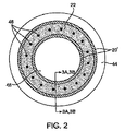
図2を参照すると、管腔48を有する脈管壁44内の周方向のリングチャネル20’を備えた埋め込み型グラフト10の断面図が示されている。治療薬46の送達は、二つの放出モードを有し得る。第一に、治療薬46はホストポリマー22から徐放的に放出され得る。例えば、治療薬46はホストポリマー22を介した拡散により放出され得る。あるいは、治療薬46は、例えばエステル又は他の結合の加水分解による、減成又は分解プロセスによって放出され得る。ホストポリマー22は、減成すると同時に治療薬46を放出する一方で、グラフト本体部16の機械的強度及び/又は剛性を低下させてグラフト10にさらなる軟性又は可撓性を付与することが好ましいと思われる。この場合、ホストポリマー22は、結合が切断されて薬剤が放出されるとポリマーもまた切断されるように、単鎖ポリマーとして調製され得る。別の場合では、ホストポリマー22の塊が多孔性チャネル20’の内部に残留して、有意に減成しない(例、部分減成)ことが好ましいと思われる。例えば、減成のプロセスは、多孔性チャネル20’の隙間に位置する治療薬とホストポリマーとの結合部位において優位に行われて、薬剤の放出がホストポリマー22に必ずしも有意な変化をもたらさない。さらに、ポリマーの減成による放出の場合、グラフト本体部16、及び特にチャネル20’は、ホストポリマー22の塊の輸送を阻止することが可能な生体適合性の材料からなり得る。一般的には、減成プロセスは比較的大きい分子の送達に適切であり、拡散は比較的小さい分子の送達に適切である。しかしながら、一方のプロセスが他方のプロセスと比較して所望される場合、他の要因も寄与し得ることが理解されよう。
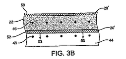
Referring to FIG. 2, a cross-sectional view of an
第二に、多孔性チャネル20’は、細孔からの薬剤の溶出を介して脈管壁44又は管腔48内へ、制御及び/又はプログラムされた薬物送達を提供し得るように、特定のカフ、チャネル、又はカフ/チャネル部分内で、又はそれら間で様々な多孔度を有するように構成される。より詳細には、治療薬46は、膨張媒体22から多孔性チャネル20’の壁を通って輸送されて、脈管壁44又は管腔48内に放出され得る。治療薬46は、図3Aの矢印51に示すように、多孔性チャネル20’の管腔側の壁50から脈管腔48内に放出されるように構成され得る。あるいは、治療薬46は、図3Bの矢印53に示すように、多孔性チャネル20’の反管腔側の壁52から脈管壁44内に放出されるように構成されてもよい。本発明により想定される別の一実施例(図示せず)としては、特定のカフ又はチャネル20’の寸法(例えば、長さ、幅、高さ又はこれらの任意の組合せ)に沿って多孔性が変化し得るもの(図示せず)がある。例えば、このようにあつらえた構成は、特定の臨床的必要性や、その装置に想定される適応症のために、グラフト又はステントグラフトの薬物送達の速度及び他の性質(例、機械的性質)が調整され得る用途において有用であろう。加えて、多孔度は、特定のカフ又はチャネル内で均一で、任意の所定のチャネル及び/又はカフ間で異なっていてもよい。所定のカフ、チャネル、又はカフ/チャネル部分内、又はそれら間における多孔度の相違の任意の組合せが、本発明により想定される。
Second, the
多孔性チャネル20’並びにカフ26及びカフ28を含むグラフト本体部16は、その表面を介した輸送を可能にする任意の生体適合性材料からなり得る。例えば、多孔性チャネル20’は、フルオロポリマー、ポリエチレンテレフタレート、ポリ塩化ビニル、ポリウレタン、ポリオレフィン、及びポリアミドからなる群より選択された材料から形成された一つ又は二つ以上の層を有し得る。多孔性チャネル20’は、特にポリテトラフルオロエチレン、発泡ポリテトラフルオロエチレン、有孔ポリテトラフルオロエチレン、多孔性ポリテトラフルオロエチレン又は他の合成材料の任意の組合せから形成され得る。多孔性チャネル20’は、必ずしも機械的に多孔性を有する必要がないことが理解されよう。例えば、チャネル20’は治療薬46に対する透過性を有することにより、治療薬46の輸送を可能にしてもよい。
The
したがって、本発明によれば、治療薬46の所望の放出速度を達成するように、チャネル20’の多孔度、及び/又はホストポリマー22の放散若しくは減成特性を調節し得る。例えば、所望の治療薬46の輸送及びグラフト10の機械的性質を得るために、ホストポリマー22の官能基及び主鎖の分子量が選択され得る。本発明によれば、利便性の高いホストポリマー22は、官能基化ポリエチレングリコールのファミリーからなる。ポリエチレングリコールジアクリレートを、例えばペンタエリスリトールテトラ3(メルカプトプロピオネート)等のチオールからなる、又はチオールを含む化合物と架橋させて安定なゲルを形成し得る。このゲルは多孔性チャネル20’の細孔から治療薬46を管腔48内に直接放散又は溶出させて流体と直接接触させ、又は例えば脈管壁44内に直接放散又は溶出させて組織と直接接触させ得る。さらに、ポリエチレングリコールの主鎖に別の官能基を加えて、選択的減成プロセスを介した治療薬46の放出を可能にし得る。またさらに、エトキシル化トリメチロールプロパントリアクリレートからなるホストポリマー22を、チオールからなる、又はチオールを含む化合物と架橋させ、官能基化して治療薬46を収容及び放出してもよい。
Thus, according to the present invention, the porosity of the
上述したように、ホストポリマー22は、液体として多孔性チャネル20’内に注入された後、相変化又は架橋形成によって固化し得る(すなわち、硬化性液体)。このようなホストポリマー22の例には、例えばBASFF−127のようなプルロニックポリオキシマーのファミリーに属するポリマー、又は適切な下限臨界溶液温度を有する、例えばn−イソプロピルポリアクリルアミドのようなアクリルアミドがある。硬化性液体と関連して、治療薬46は、ホストポリマー22のモノマーに結合されてもよく、これが架橋して固体を形成する。次に、時間の経過と共に、薬剤とポリマー主鎖とを結合している結合が、例えばホストポリマーの材料の選択、及び治療薬の取付けに使用された結合の種類等のいくつかの基準に基づき、所望の速度で減成して治療薬を放出させる。あるいは、治療薬46は、いずれの化学結合を有することなく、固化して薬剤分子をホストポリマーのネットワーク22内に閉じ込める、もう一種の液体からなる膨張媒体中に混合され得る。この場合、治療薬は、ある時間に亘り、放散によってホストポリマーネットワークの外部へ放出され得る。放散の速度は、例えばホストポリマーの材料、密度、調製、ホストポリマーネットワークの極性等のいくつかの基準に基づく。
As described above, the
少なくとも一種の治療薬46は、グラフトにより治療される特定の適応症に応じて任意の治療目的を果たす、様々な薬剤からなり得る。治療薬46は、内皮形成促進薬(例、血管内皮細胞増殖因子)、血管新生促進薬、抗血栓薬(例、ヘパリン)、抗動脈瘤薬、抗感染薬、抗炎症薬、抗再狭窄薬、化学療法剤、及び坑癌剤からなる群から選択された一種又は二種以上の薬剤からなり得る。通常、身体管腔内に放出可能な治療薬の量は、μg〜mgレベルの範囲内にあり、おおよそ約10μg〜約100mgの範囲内ある。治療薬は、約1週間未満〜約2、3ヶ月間又は多数ヶ月間、おおよそ約7日間〜約12ヶ月間、身体管腔内に輸送されるように構成されている。
The at least one
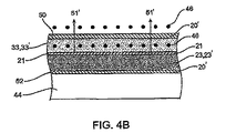
図4A〜4Cに、本発明のさらなる実施態様を示す。図4Aは、図2、図3A及び図3Bの実施態様と同様の、管腔48を有する脈管壁44内の周方向リングチャネルを備えた埋め込み型グラフト10の断面図である。しかしながら、本実施態様では、周方向リングチャネル20’は、チャネル20’を2個のチャンバ23及びチャンバ33に分割する比較的非多孔性のフィルム層21(例えば、ポリテトラフルオロエチレン又は他の適切な材料から形成された)によって特徴付けられる二重チャンバのチャネルである。一個のインプラント又はグラフト本体部内に複数の膨張媒体を含めることができる、本願に解説した他の実施態様と同様、二種の異なる膨張媒体23’及び膨張媒体33’を、各々、チャネル20’のチャンバ23及びチャンバ33内に別々に導入することができる。
4A-4C illustrate a further embodiment of the present invention. FIG. 4A is a cross-sectional view of an
例えば、図4Bの実施態様では、治療薬を含有しない第一の膨張媒体23’をチャンバ23内に送ることができ、膨張媒体23が’チャンバ23内に入った後、又は入っている間、媒体23’は硬化しながら、又は硬化した後に制御下にて膨張し、それによって媒体23’が層21上に圧力を加え、次にこの層が圧力をチャンバ33内に配置された第二の膨張媒体33’に伝達する。媒体33’は、治療薬46を含有するホスト材料からなる。適切なホスト材料は、治療薬がチャンバ33の壁を介して輸送されることが可能な材料であり、本実施態様ではホストポリマーである必要はなく、治療薬を含有する液体、固体、ゲル、及び他の物質形態を有する。第一の膨張媒体23’の拡張又は膨張により、層21を介してホスト材料33’へ圧力を加えることによって、治療薬46はホストポリマー33’中での移動を開始し、前述し、且つ矢印51’に示したように、チャネル20’の細孔を通って脈管腔48内に溶出する。図4Cに示すように、これと同一の機構は他方の方向にも働き得る。治療薬46、並びに媒体23’及び媒体33’は、代替的に、矢印53’で示すように、チャネル20’の反管腔側壁52を通った溶出による治療薬46の脈管壁44内への溶出を促進するように構成することができる。
For example, in the embodiment of FIG. 4B, a first inflation medium 23 ′ that does not contain a therapeutic agent can be delivered into the chamber 23, after the inflation medium 23 has entered or entered the chamber 23. The medium 23 ′ expands under control, either after curing or after curing, so that the medium 23 ′ applies pressure on the
この治療薬46を脈管腔48又は脈管壁44内に移動させる“ポンプ”作用は、非常によく制御されるように調整することができ、治療薬46を所望のように放出させることができる。例えば、膨張工程と最終的な治療薬46の放出が、遅延して開始されるように、媒体23’及び媒体33’の調製、硬化時間又は固化時間を、個別に又は集合的に設計し得る。また、膨張の速度を調節してホストポリマーに付与される駆動力の速度を調節することによって、薬剤の放出速度を制御することができる。例えば、媒体23’及び/又は媒体33’は、媒体をインビボ環境に対して浸透勾配を有するように調製することで膨張させ得る。別の一実施例として、媒体23’及び/又は媒体33’は、これらの材料を固化後に吸水するヒドロゲルとして調製することによって膨張させ得る。この機構により、10%〜500%、又はそれ以上の膨張を達成し得る。媒体23’及び/又は33’を膨張させるために、他の機構も使用し得ることは勿論である。
The “pump” action that moves the
図4A〜図4Cの二重チャンバの実施態様は、周方向に配置された層21を有する周方向リングチャネルの構成を示しているが、本発明の他の実施態様と同様、チャネル及び層21は図示されているように完全に環状である必要はなく、また周方向に配向されている必要もない。二重チャンバ構成は、本発明の一個又は二個以上のカフ又は他の構成を含めることができる。加えて、本発明の他の実施態様に関して本願に記載されているグラフト、膨張材料、治療薬、構成、及び方法を、図4A〜4Cの実施態様中で任意に組合せて使用することができる。
The dual chamber embodiment of FIGS. 4A-4C shows the configuration of a circumferential ring channel with circumferentially arranged
図5を参照すると、グラフト10’、該グラフト10’の使用説明書56、及び治療薬を含有する膨張媒体の原料58を備えたキット54が示されている。グラフト10’は、ステントグラフトを含む本願に記載されている任意の構造を有することができ、原料は本願に記載されている任意の媒体からなり、使用説明書56は、上述した方法の一つ又は二つ以上を実行するためのステップが列挙され得る。使用説明書62は多くの場合印刷物であるが、代替的に、上述した任意の方法を説明する、及び/又は列挙するビデオテープ、CD、DVD、他の機械判読可能なフォーマット又はコード、グラフィック描写、又は同様物に含められていてもよい。
Referring to FIG. 5, there is shown a
理解を明確にするために、また例示として、特定の典型的な実施態様及び方法を詳細に説明してきたが、本発明の趣旨及び範囲から逸脱せずに、これら実施態様及び方法を変化させ、修正し、変更し、また適合させ得ることが当業者には明らかであろう。したがって、上記の説明は、添付の特許請求の範囲によって定義される本発明の範囲を限定するものであると解釈すべきではない。 Although certain exemplary embodiments and methods have been described in detail for purposes of clarity of understanding and by way of example, these embodiments and methods may be varied without departing from the spirit and scope of the present invention, and It will be apparent to those skilled in the art that modifications, changes, and adaptations can be made. Therefore, the above description should not be taken as limiting the scope of the invention which is defined by the appended claims.
Claims (35)
前記膨張可能なチャネル内に導入されるように構成された、少なくとも一種の治療薬を含有する膨張媒体と、
を備えるグラフト。 A graft body having a proximal end and a distal end and defining at least one inflatable porous channel;
An inflation medium containing at least one therapeutic agent configured to be introduced into the inflatable channel;
Graft comprising.
前記グラフト本体部の近位端又は遠位端に固着され、一個又は二個以上の接続要素を有する接続部材と、
前記接続部材の前記一個又は二個以上の接続要素に連結したもう一個の近位端側ステント接続要素を有するステントと、
前記膨張可能なチャネル内に導入されるように構成された、少なくとも一種の治療薬を含有する膨張媒体と、
を備えるグラフト。 A graft body having a proximal end and a distal end, and defining at least one inflatable porous channel between the proximal end and the distal end;
A connection member secured to the proximal or distal end of the graft body and having one or more connection elements;
A stent having another proximal stent connecting element coupled to the one or more connecting elements of the connecting member;
An inflation medium containing at least one therapeutic agent configured to be introduced into the inflatable channel;
Graft comprising.
前記グラフト本体部を身体の管腔内に移植する工程と、
少なくとも一種の治療薬を含有する膨張媒体によって前記多孔性チャネルを膨張させる工程と、
を含む、治療薬を送達する方法。 Providing a graft body having a proximal end and a distal end and defining at least one inflatable porous channel;
Transplanting the graft body into a body lumen;
Inflating the porous channel with an inflation medium containing at least one therapeutic agent;
A method of delivering a therapeutic agent comprising:
Applications Claiming Priority (2)
| Application Number | Priority Date | Filing Date | Title |
|---|---|---|---|
| US10/769,532 US7803178B2 (en) | 2004-01-30 | 2004-01-30 | Inflatable porous implants and methods for drug delivery |
| US10/769,532 | 2004-01-30 |
Related Parent Applications (1)
| Application Number | Title | Priority Date | Filing Date |
|---|---|---|---|
| JP2006551504A Division JP5207629B2 (en) | 2004-01-30 | 2005-01-27 | Inflatable porous implant |
Publications (2)
| Publication Number | Publication Date |
|---|---|
| JP2011152425A true JP2011152425A (en) | 2011-08-11 |
| JP5468030B2 JP5468030B2 (en) | 2014-04-09 |
Family
ID=34808154
Family Applications (2)
| Application Number | Title | Priority Date | Filing Date |
|---|---|---|---|
| JP2006551504A Expired - Fee Related JP5207629B2 (en) | 2004-01-30 | 2005-01-27 | Inflatable porous implant |
| JP2011036448A Expired - Fee Related JP5468030B2 (en) | 2004-01-30 | 2011-02-22 | Expandable porous implant and method for drug delivery |
Family Applications Before (1)
| Application Number | Title | Priority Date | Filing Date |
|---|---|---|---|
| JP2006551504A Expired - Fee Related JP5207629B2 (en) | 2004-01-30 | 2005-01-27 | Inflatable porous implant |
Country Status (5)
| Country | Link |
|---|---|
| US (2) | US7803178B2 (en) |
| EP (2) | EP2332493A1 (en) |
| JP (2) | JP5207629B2 (en) |
| CA (1) | CA2546525A1 (en) |
| WO (1) | WO2005074547A2 (en) |
Cited By (7)
| Publication number | Priority date | Publication date | Assignee | Title |
|---|---|---|---|---|
| JP2019519264A (en) * | 2016-04-27 | 2019-07-11 | サイナーズ メディカル,インコーポレイティド | Anchorless intragastric device for treatment of obesity |
| US11135078B2 (en) | 2010-06-13 | 2021-10-05 | Synerz Medical, Inc. | Intragastric device for treating obesity |
| US11351050B2 (en) | 2010-06-13 | 2022-06-07 | Synerz Medical, Inc. | Intragastric device for treating obesity |
| US11596538B2 (en) | 2010-06-13 | 2023-03-07 | Synerz Medical, Inc. | Intragastric device for treating obesity |
| US11607329B2 (en) | 2010-06-13 | 2023-03-21 | Synerz Medical, Inc. | Intragastric device for treating obesity |
| JP7438681B2 (en) | 2012-04-06 | 2024-02-27 | トリバスキュラー インコーポレイテッド | Low profile stent graft and delivery system |
| US12453627B2 (en) | 2012-04-06 | 2025-10-28 | Trivascular, Inc. | Low profile stent graft and delivery system |
Families Citing this family (98)
| Publication number | Priority date | Publication date | Assignee | Title |
|---|---|---|---|---|
| WO2005044361A1 (en) | 2003-11-07 | 2005-05-19 | Merlin Md Pte Ltd | Implantable medical devices with enhanced visibility, mechanical properties and biocompatibility |
| US8915952B2 (en) | 2004-03-31 | 2014-12-23 | Merlin Md Pte Ltd. | Method for treating aneurysms |
| US8715340B2 (en) | 2004-03-31 | 2014-05-06 | Merlin Md Pte Ltd. | Endovascular device with membrane |
| US8500751B2 (en) | 2004-03-31 | 2013-08-06 | Merlin Md Pte Ltd | Medical device |
| US8349001B2 (en) * | 2004-04-07 | 2013-01-08 | Medtronic, Inc. | Pharmacological delivery implement for use with cardiac repair devices |
| GB0415152D0 (en) * | 2004-07-06 | 2004-08-11 | Anson Medical Ltd | Interconnected tubular structures |
| US20070100321A1 (en) * | 2004-12-22 | 2007-05-03 | Leon Rudakov | Medical device |
| US20060233991A1 (en) | 2005-04-13 | 2006-10-19 | Trivascular, Inc. | PTFE layers and methods of manufacturing |
| CA2642250A1 (en) * | 2006-02-13 | 2007-08-23 | Merlin Md Pte Ltd | Endovascular device with membrane |
| US20080082036A1 (en) * | 2006-04-25 | 2008-04-03 | Medtronic, Inc. | Cerebrospinal fluid shunt having long term anti-occlusion agent delivery |
| US8216297B2 (en) * | 2006-08-14 | 2012-07-10 | Trivascular, Inc. | Dual chamber cuff structure |
| US10094836B2 (en) | 2007-01-08 | 2018-10-09 | The United States Of America, As Represented By The Secretary, Department Of Health & Human Services | SLCO1B3 genotype |
| ES2530862T3 (en) | 2007-02-07 | 2015-03-06 | Cook Medical Technologies Llc | Coatings of medical devices to release a therapeutic agent at different rates |
| US20080234809A1 (en) * | 2007-03-23 | 2008-09-25 | Medtronic Vascular, Inc. | Stent Graft System With Injection Tube |
| CA2978267A1 (en) | 2007-08-23 | 2009-02-23 | Dfm, Llc | Translumenally implantable heart valve with formed in place support |
| US8226701B2 (en) | 2007-09-26 | 2012-07-24 | Trivascular, Inc. | Stent and delivery system for deployment thereof |
| US8663309B2 (en) | 2007-09-26 | 2014-03-04 | Trivascular, Inc. | Asymmetric stent apparatus and method |
| US8066755B2 (en) | 2007-09-26 | 2011-11-29 | Trivascular, Inc. | System and method of pivoted stent deployment |
| AU2008308474B2 (en) | 2007-10-04 | 2014-07-24 | Trivascular, Inc. | Modular vascular graft for low profile percutaneous delivery |
| CA2702000C (en) * | 2007-10-09 | 2017-03-28 | Universite Rene Descartes - Paris V | Device for administering cells and cell-therapy methods using said device |
| US8328861B2 (en) | 2007-11-16 | 2012-12-11 | Trivascular, Inc. | Delivery system and method for bifurcated graft |
| US8083789B2 (en) | 2007-11-16 | 2011-12-27 | Trivascular, Inc. | Securement assembly and method for expandable endovascular device |
| US8016880B2 (en) * | 2007-11-16 | 2011-09-13 | Medtronic Vascular, Inc. | Stent having spiral channel for drug delivery |
| EP2106820A1 (en) * | 2008-03-31 | 2009-10-07 | Torsten Heilmann | Expansible biocompatible coats comprising a biologically active substance |
| WO2009137384A2 (en) * | 2008-05-05 | 2009-11-12 | Boston Scientific Scimed, Inc. | Medical devices having a bioresorbable coating layer with a pre-determined pattern for fragmentation |
| US8262692B2 (en) * | 2008-09-05 | 2012-09-11 | Merlin Md Pte Ltd | Endovascular device |
| US20100274276A1 (en) * | 2009-04-22 | 2010-10-28 | Ricky Chow | Aneurysm treatment system, device and method |
| US10772717B2 (en) | 2009-05-01 | 2020-09-15 | Endologix, Inc. | Percutaneous method and device to treat dissections |
| EP2424447A2 (en) | 2009-05-01 | 2012-03-07 | Endologix, Inc. | Percutaneous method and device to treat dissections |
| US8870938B2 (en) | 2009-06-23 | 2014-10-28 | Endospan Ltd. | Vascular prostheses for treating aneurysms |
| WO2011004374A1 (en) | 2009-07-09 | 2011-01-13 | Endospan Ltd. | Apparatus for closure of a lumen and methods of using the same |
| ES2549000T3 (en) | 2009-07-27 | 2015-10-22 | Endologix, Inc. | Endoprosthesis |
| WO2011064782A2 (en) | 2009-11-30 | 2011-06-03 | Endospan Ltd. | Multi-component stent-graft system for implantation in a blood vessel with multiple branches |
| CA2783554C (en) | 2009-12-08 | 2016-02-16 | Endospan Ltd. | Endovascular stent-graft system with fenestrated and crossing stent-grafts |
| ES2362228B1 (en) * | 2009-12-11 | 2013-06-03 | Universidad De Zaragoza | BONE IMPLANT, EQUIPPED WITH POROUS LIMITS FOR THE CONTROLLED RELEASE OF THERAPEUTICALLY ACTIVE COMPOUNDS. |
| US9295541B2 (en) | 2009-12-31 | 2016-03-29 | Neograft Technologies, Inc. | Graft devices and methods of fabrication |
| EP2533722B1 (en) | 2010-02-08 | 2017-03-29 | Endospan Ltd. | Thermal energy application for prevention and management of endoleaks in stent-grafts |
| US20110208289A1 (en) * | 2010-02-25 | 2011-08-25 | Endospan Ltd. | Flexible Stent-Grafts |
| US8500819B2 (en) * | 2010-03-05 | 2013-08-06 | Biomet Manufacturing, Llc | Drug delivery and diagnostic system for orthopedic implants |
| CA2794630A1 (en) * | 2010-05-14 | 2011-11-17 | Liliana Atanasoska | Endoprosthesis |
| US9433501B2 (en) | 2010-05-19 | 2016-09-06 | Direct Flow Medical, Inc. | Inflation media for implants |
| US9603708B2 (en) | 2010-05-19 | 2017-03-28 | Dfm, Llc | Low crossing profile delivery catheter for cardiovascular prosthetic implant |
| US8696738B2 (en) * | 2010-05-20 | 2014-04-15 | Maquet Cardiovascular Llc | Composite prosthesis with external polymeric support structure and methods of manufacturing the same |
| WO2012012407A2 (en) | 2010-07-19 | 2012-01-26 | Neograft Technologies, Inc. | Graft devices and methods of use |
| WO2012068298A1 (en) | 2010-11-17 | 2012-05-24 | Endologix, Inc. | Devices and methods to treat vascular dissections |
| AU2012207387B2 (en) | 2011-01-17 | 2017-02-16 | Artio Medical, Inc. | Ballstent device and methods of use |
| US11484318B2 (en) | 2011-01-17 | 2022-11-01 | Artio Medical, Inc. | Expandable body device and method of use |
| CN103561682A (en) | 2011-01-28 | 2014-02-05 | 梅瑞特医药体系股份有限公司 | Electrospun PTFE coated stents and methods of use thereof |
| US9526638B2 (en) | 2011-02-03 | 2016-12-27 | Endospan Ltd. | Implantable medical devices constructed of shape memory material |
| WO2012111006A1 (en) | 2011-02-17 | 2012-08-23 | Endospan Ltd. | Vascular bands and delivery systems therefor |
| EP2680788A4 (en) | 2011-03-02 | 2014-12-10 | Endospan Ltd | Reduced-strain extra- vascular ring for treating aortic aneurysm |
| US8574287B2 (en) | 2011-06-14 | 2013-11-05 | Endospan Ltd. | Stents incorporating a plurality of strain-distribution locations |
| WO2012174389A1 (en) | 2011-06-15 | 2012-12-20 | Phraxis Inc. | Anastomotic connector and system for delivery |
| US8951298B2 (en) | 2011-06-21 | 2015-02-10 | Endospan Ltd. | Endovascular system with circumferentially-overlapping stent-grafts |
| WO2013005207A1 (en) | 2011-07-07 | 2013-01-10 | Endospan Ltd. | Stent fixation with reduced plastic deformation |
| US9839510B2 (en) | 2011-08-28 | 2017-12-12 | Endospan Ltd. | Stent-grafts with post-deployment variable radial displacement |
| US9381112B1 (en) | 2011-10-06 | 2016-07-05 | William Eric Sponsell | Bleb drainage device, ophthalmological product and methods |
| WO2013065040A1 (en) | 2011-10-30 | 2013-05-10 | Endospan Ltd. | Triple-collar stent-graft |
| WO2013084235A2 (en) | 2011-12-04 | 2013-06-13 | Endospan Ltd. | Branched stent-graft system |
| US8632489B1 (en) | 2011-12-22 | 2014-01-21 | A. Mateen Ahmed | Implantable medical assembly and methods |
| KR102037543B1 (en) | 2012-01-16 | 2019-10-28 | 메리트 메디컬 시스템즈, 인크. | Rotational spun material covered medical appliances and methods of manufacture |
| JP6356612B2 (en) | 2012-01-17 | 2018-07-11 | メタクティブ・メディカル・インコーポレイテッドMetactive Medical, Inc. | Expandable device and usage |
| KR20140133822A (en) * | 2012-02-14 | 2014-11-20 | 네오그래프트 테크놀로지스, 아이엔씨. | Kink resistant graft devices and related systems and methods |
| US8992595B2 (en) | 2012-04-04 | 2015-03-31 | Trivascular, Inc. | Durable stent graft with tapered struts and stable delivery methods and devices |
| US9498363B2 (en) | 2012-04-06 | 2016-11-22 | Trivascular, Inc. | Delivery catheter for endovascular device |
| JP6324371B2 (en) | 2012-04-06 | 2018-05-16 | マーリン エムディー プライベート リミテッド | Devices and methods for treating aneurysms |
| US9445897B2 (en) | 2012-05-01 | 2016-09-20 | Direct Flow Medical, Inc. | Prosthetic implant delivery device with introducer catheter |
| WO2013171730A1 (en) | 2012-05-15 | 2013-11-21 | Endospan Ltd. | Stent-graft with fixation elements that are radially confined for delivery |
| JP6105722B2 (en) | 2012-06-15 | 2017-03-29 | フラクシス インコーポレイテッド | Arterial and venous anchor devices forming an anastomotic connector and system for delivery |
| US11541154B2 (en) | 2012-09-19 | 2023-01-03 | Merit Medical Systems, Inc. | Electrospun material covered medical appliances and methods of manufacture |
| AU2013329077A1 (en) * | 2012-10-11 | 2015-04-23 | Tufts University | Compositions and methods for sustained delivery of glucagon-like peptide (GLP-1) receptor agonist therapeutics |
| US9668892B2 (en) | 2013-03-11 | 2017-06-06 | Endospan Ltd. | Multi-component stent-graft system for aortic dissections |
| WO2014159710A1 (en) | 2013-03-13 | 2014-10-02 | Merit Medical Systems, Inc. | Serially deposited fiber materials and associated devices and methods |
| US9827703B2 (en) | 2013-03-13 | 2017-11-28 | Merit Medical Systems, Inc. | Methods, systems, and apparatuses for manufacturing rotational spun appliances |
| US9182420B2 (en) * | 2013-03-15 | 2015-11-10 | Palo Alto Research Center Incorporated | Phase-change enabled flow field visualization |
| US10603197B2 (en) | 2013-11-19 | 2020-03-31 | Endospan Ltd. | Stent system with radial-expansion locking |
| AU2015231110B2 (en) | 2014-03-21 | 2019-03-07 | University Of Pittsburgh - Of The Commonwealth System Of Higher Education | Methods for preparation of a terminally sterilized hydrogel derived from extracellular matrix |
| DE102014109343A1 (en) * | 2014-07-03 | 2016-01-07 | Gert Rothe | Gastrointestinalstützvorrichtung |
| RU2721288C2 (en) | 2014-09-17 | 2020-05-18 | Метэктив Медикал, Инк. | Medical device for saccular aneurysm treatment |
| US10166126B2 (en) | 2014-12-08 | 2019-01-01 | Boston Scientific Scimed, Inc. | Inflatable balloon stent |
| EP3068339B1 (en) | 2014-12-18 | 2017-11-01 | Endospan Ltd. | Endovascular stent-graft with fatigue-resistant lateral tube |
| EP3261589B1 (en) | 2015-02-26 | 2020-09-16 | Merit Medical Systems, Inc. | Layered medical appliances |
| US10594128B2 (en) * | 2015-11-10 | 2020-03-17 | Te Connectivity Corporation | Holdout devices and cover assemblies and methods incorporating the same |
| CA3011294A1 (en) | 2016-01-13 | 2017-07-20 | University Of Pittsburgh - Of The Commonwealth System Of Higher Education | Vascular extracellular matrix hydrogel |
| WO2018089419A1 (en) * | 2016-11-09 | 2018-05-17 | Boston Scientific Scimed, Inc. | Deployable sleeves and related methods |
| WO2018176064A2 (en) | 2017-03-24 | 2018-09-27 | Metactive Medical, Inc. | Medical devices comprising detachable balloons and methods of manufacturing and use |
| US11883273B2 (en) * | 2017-04-28 | 2024-01-30 | Regents Of The University Of Minnesota | Compliant aortic stent grafts and related systems and methods |
| JP7275097B2 (en) | 2017-07-07 | 2023-05-17 | エンドーロジックス リミテッド ライアビリティ カンパニー | Endovascular graft system and method for deployment in main arteries and arterial branches |
| BR112020009444A2 (en) * | 2017-11-30 | 2020-11-17 | Anatoli D. Dosta | local exposure method for biological tissues, tissue substitute applicator and use of porous polytetrafluoroethylene |
| EP3803866A4 (en) | 2018-05-24 | 2022-03-16 | Nureva Inc. | METHOD, DEVICE AND COMPUTER-READABLE MEDIA FOR MANAGING SEMI-CONSTANT (PERSISTENT) SOUND SOURCES IN MICROPHONE RECORDING/FOCUS ZONES |
| MX2020012459A (en) | 2018-05-24 | 2021-04-28 | Celanese Eva Performance Polymers Llc | IMPLANTABLE DEVICE FOR SUSTAINED RELEASE OF A MACROMOLECULAR DRUG COMPOUND. |
| KR20260007295A (en) | 2018-05-24 | 2026-01-13 | 셀라니즈 이브이에이 퍼포먼스 폴리머스 엘엘씨 | Implantable device for sustained release of a macromolecular drug compound |
| GB2577052B (en) | 2018-09-11 | 2021-04-28 | Strait Access Tech Holdings Pty Ltd | Expandable sleeved stent and method of making such stent |
| US11896508B2 (en) * | 2018-09-22 | 2024-02-13 | Transit Scientific, LLC | Expandable exoskeleton devices |
| WO2020097339A1 (en) | 2018-11-08 | 2020-05-14 | Dfine, Inc. | Tumor ablation device and related systems and methods |
| EP4297813A4 (en) | 2021-02-26 | 2025-01-15 | Merit Medical Systems, Inc. | FIBROUS CONSTRUCTIONS WITH THERAPEUTIC MATERIAL PARTICLES |
| BR112023022439A2 (en) | 2021-04-26 | 2023-12-26 | Celanese Eva Performance Polymers Llc | IMPLANTABLE DEVICE FOR SUSTAINED RELEASE OF A MACROMOLECULAR DRUG COMPOUND |
| USD1061891S1 (en) * | 2023-01-09 | 2025-02-11 | Polimerbio, S.L. | Stent |
Citations (2)
| Publication number | Priority date | Publication date | Assignee | Title |
|---|---|---|---|---|
| WO2001067991A1 (en) * | 2000-03-13 | 2001-09-20 | Jun Yang | Stent having cover with drug delivery capability |
| WO2003032869A1 (en) * | 2001-10-16 | 2003-04-24 | Scimed Life Systems, Inc. | Aortic artery aneurysm endovascular prosthesis |
Family Cites Families (393)
| Publication number | Priority date | Publication date | Assignee | Title |
|---|---|---|---|---|
| US3540431A (en) | 1968-04-04 | 1970-11-17 | Kazi Mobin Uddin | Collapsible filter for fluid flowing in closed passageway |
| US3631854A (en) | 1969-05-19 | 1972-01-04 | Robert Howard Fryer | Inflatable medical assemblies |
| CA962021A (en) | 1970-05-21 | 1975-02-04 | Robert W. Gore | Porous products and process therefor |
| US3818511A (en) | 1972-11-17 | 1974-06-25 | Medical Prod Corp | Medical prosthesis for ducts or conduits |
| US3991767A (en) | 1973-11-02 | 1976-11-16 | Cutter Laboratories, Inc. | Tubular unit with vessel engaging cuff structure |
| US3900027A (en) | 1974-01-02 | 1975-08-19 | Pall Corp | Process for preparing integral absorbent pad bandages and product |
| US3902198A (en) | 1974-05-20 | 1975-09-02 | Gore & Ass | Method of replacing a body part with expanded porous polytetrafluoroethylene |
| US4374669A (en) | 1975-05-09 | 1983-02-22 | Mac Gregor David C | Cardiovascular prosthetic devices and implants with porous systems |
| US4208745A (en) | 1976-01-21 | 1980-06-24 | Sumitomo Electric Industries, Ltd. | Vascular prostheses composed of polytetrafluoroethylene and process for their production |
| DE2714810A1 (en) | 1976-04-05 | 1977-10-13 | Anvar | PROCESS FOR MANUFACTURING ORGAN DENTALS PRODUCED ACCORDING TO THE PROCESS |
| US4140126A (en) | 1977-02-18 | 1979-02-20 | Choudhury M Hasan | Method for performing aneurysm repair |
| US4183102A (en) | 1977-09-08 | 1980-01-15 | Jacques Guiset | Inflatable prosthetic device for lining a body duct |
| DE2806030C2 (en) | 1978-02-14 | 1984-02-02 | B. Braun Melsungen Ag, 3508 Melsungen | Process for the production of a tubular blood vessel prosthesis |
| US4439585A (en) * | 1980-11-12 | 1984-03-27 | Tyndale Plains-Hunter, Ltd. | Polyurethane diacrylate compositions as carrier for pharmacological agents |
| GB2121286B (en) | 1982-06-02 | 1985-11-06 | Ethicon Inc | Improvements in synthetic vascular grafts, and methods of manufacturing such grafts |
| US4647416A (en) | 1983-08-03 | 1987-03-03 | Shiley Incorporated | Method of preparing a vascular graft prosthesis |
| US4550447A (en) | 1983-08-03 | 1985-11-05 | Shiley Incorporated | Vascular graft prosthesis |
| US4592754A (en) | 1983-09-09 | 1986-06-03 | Gupte Pradeep M | Surgical prosthetic vessel graft and catheter combination and method |
| US4665906A (en) | 1983-10-14 | 1987-05-19 | Raychem Corporation | Medical devices incorporating sim alloy elements |
| US5749920A (en) | 1983-12-09 | 1998-05-12 | Endovascular Technologies, Inc. | Multicapsule intraluminal grafting system and method |
| US6221102B1 (en) | 1983-12-09 | 2001-04-24 | Endovascular Technologies, Inc. | Intraluminal grafting system |
| US5104399A (en) | 1986-12-10 | 1992-04-14 | Endovascular Technologies, Inc. | Artificial graft and implantation method |
| US5669936A (en) | 1983-12-09 | 1997-09-23 | Endovascular Technologies, Inc. | Endovascular grafting system and method for use therewith |
| US4787899A (en) | 1983-12-09 | 1988-11-29 | Lazarus Harrison M | Intraluminal graft device, system and method |
| US5275622A (en) | 1983-12-09 | 1994-01-04 | Harrison Medical Technologies, Inc. | Endovascular grafting apparatus, system and method and devices for use therewith |
| US5108424A (en) | 1984-01-30 | 1992-04-28 | Meadox Medicals, Inc. | Collagen-impregnated dacron graft |
| US4617932A (en) | 1984-04-25 | 1986-10-21 | Elliot Kornberg | Device and method for performing an intraluminal abdominal aortic aneurysm repair |
| US4562596A (en) | 1984-04-25 | 1986-01-07 | Elliot Kornberg | Aortic graft, device and method for performing an intraluminal abdominal aortic aneurysm repair |
| US4580568A (en) | 1984-10-01 | 1986-04-08 | Cook, Incorporated | Percutaneous endovascular stent and method for insertion thereof |
| US4705517A (en) | 1985-09-03 | 1987-11-10 | Becton, Dickinson And Company | Percutaneously deliverable intravascular occlusion prosthesis |
| US4733665C2 (en) | 1985-11-07 | 2002-01-29 | Expandable Grafts Partnership | Expandable intraluminal graft and method and apparatus for implanting an expandable intraluminal graft |
| JPH0696023B2 (en) | 1986-11-10 | 1994-11-30 | 宇部日東化成株式会社 | Artificial blood vessel and method for producing the same |
| US4816028A (en) | 1987-07-01 | 1989-03-28 | Indu Kapadia | Woven vascular graft |
| CA1322628C (en) | 1988-10-04 | 1993-10-05 | Richard A. Schatz | Expandable intraluminal graft |
| US4902423A (en) | 1989-02-02 | 1990-02-20 | W. L. Gore & Associates, Inc. | Highly air permeable expanded polytetrafluoroethylene membranes and process for making them |
| US6146358A (en) | 1989-03-14 | 2000-11-14 | Cordis Corporation | Method and apparatus for delivery of therapeutic agent |
| US4957669A (en) | 1989-04-06 | 1990-09-18 | Shiley, Inc. | Method for producing tubing useful as a tapered vascular graft prosthesis |
| US5100422A (en) | 1989-05-26 | 1992-03-31 | Impra, Inc. | Blood vessel patch |
| US5152782A (en) | 1989-05-26 | 1992-10-06 | Impra, Inc. | Non-porous coated ptfe graft |
| US5104400A (en) | 1989-05-26 | 1992-04-14 | Impra, Inc. | Blood vessel patch |
| US4955899A (en) | 1989-05-26 | 1990-09-11 | Impra, Inc. | Longitudinally compliant vascular graft |
| US5609626A (en) | 1989-05-31 | 1997-03-11 | Baxter International Inc. | Stent devices and support/restrictor assemblies for use in conjunction with prosthetic vascular grafts |
| US5207695A (en) | 1989-06-19 | 1993-05-04 | Trout Iii Hugh H | Aortic graft, implantation device, and method for repairing aortic aneurysm |
| GB8927282D0 (en) | 1989-12-01 | 1990-01-31 | Univ Strathclyde | Vascular surgical devices |
| US5674192A (en) | 1990-12-28 | 1997-10-07 | Boston Scientific Corporation | Drug delivery |
| US6004346A (en) | 1990-02-28 | 1999-12-21 | Medtronic, Inc. | Intralumenal drug eluting prosthesis |
| US5123917A (en) | 1990-04-27 | 1992-06-23 | Lee Peter Y | Expandable intraluminal vascular graft |
| US5360443A (en) | 1990-06-11 | 1994-11-01 | Barone Hector D | Aortic graft for repairing an abdominal aortic aneurysm |
| US5578071A (en) | 1990-06-11 | 1996-11-26 | Parodi; Juan C. | Aortic graft |
| US5106424A (en) * | 1990-07-16 | 1992-04-21 | Polar Marine Inc. | Decontamination method and use of contaminant |
| EP0549590A1 (en) | 1990-07-26 | 1993-07-07 | LANE, Rodney James | Self expanding vascular endoprosthesis for aneurysms |
| US5122154A (en) | 1990-08-15 | 1992-06-16 | Rhodes Valentine J | Endovascular bypass graft |
| AR246020A1 (en) | 1990-10-03 | 1994-03-30 | Hector Daniel Barone Juan Carl | A ball device for implanting an intraluminous aortic prosthesis, for repairing aneurysms. |
| ATE135555T1 (en) | 1990-10-09 | 1996-04-15 | Cook Inc | PERCUTANE STENT ARRANGEMENT |
| US5156620A (en) | 1991-02-04 | 1992-10-20 | Pigott John P | Intraluminal graft/stent and balloon catheter for insertion thereof |
| DE4104702C2 (en) | 1991-02-15 | 1996-01-18 | Malte Neuss | Implants for organ pathways in spiral form |
| US5282847A (en) | 1991-02-28 | 1994-02-01 | Medtronic, Inc. | Prosthetic vascular grafts with a pleated structure |
| CA2065634C (en) | 1991-04-11 | 1997-06-03 | Alec A. Piplani | Endovascular graft having bifurcation and apparatus and method for deploying the same |
| US5628783A (en) | 1991-04-11 | 1997-05-13 | Endovascular Technologies, Inc. | Bifurcated multicapsule intraluminal grafting system and method |
| WO1992022604A1 (en) | 1991-06-14 | 1992-12-23 | W.L. Gore & Associates, Inc. | Surface modified porous expanded polytetrafluoroethylene and process for making |
| NL9101159A (en) | 1991-07-03 | 1993-02-01 | Industrial Res Bv | FORMATTABLE EXPANDABLE RING, CYLINDER OR SLEEVE. |
| US5411550A (en) | 1991-09-16 | 1995-05-02 | Atrium Medical Corporation | Implantable prosthetic device for the delivery of a bioactive material |
| US5500013A (en) | 1991-10-04 | 1996-03-19 | Scimed Life Systems, Inc. | Biodegradable drug delivery vascular stent |
| US5151105A (en) | 1991-10-07 | 1992-09-29 | Kwan Gett Clifford | Collapsible vessel sleeve implant |
| US5662713A (en) | 1991-10-09 | 1997-09-02 | Boston Scientific Corporation | Medical stents for body lumens exhibiting peristaltic motion |
| US5366504A (en) | 1992-05-20 | 1994-11-22 | Boston Scientific Corporation | Tubular medical prosthesis |
| US5456713A (en) | 1991-10-25 | 1995-10-10 | Cook Incorporated | Expandable transluminal graft prosthesis for repairs of aneurysm and method for implanting |
| US5387235A (en) | 1991-10-25 | 1995-02-07 | Cook Incorporated | Expandable transluminal graft prosthesis for repair of aneurysm |
| CA2081424C (en) | 1991-10-25 | 2008-12-30 | Timothy A. Chuter | Expandable transluminal graft prosthesis for repair of aneurysm |
| US5720776A (en) | 1991-10-25 | 1998-02-24 | Cook Incorporated | Barb and expandable transluminal graft prosthesis for repair of aneurysm |
| US5693084A (en) | 1991-10-25 | 1997-12-02 | Cook Incorporated | Expandable transluminal graft prosthesis for repair of aneurysm |
| CA2380683C (en) | 1991-10-28 | 2006-08-08 | Advanced Cardiovascular Systems, Inc. | Expandable stents and method for making same |
| US5316023A (en) | 1992-01-08 | 1994-05-31 | Expandable Grafts Partnership | Method for bilateral intra-aortic bypass |
| US5683448A (en) | 1992-02-21 | 1997-11-04 | Boston Scientific Technology, Inc. | Intraluminal stent and graft |
| FR2688401B1 (en) | 1992-03-12 | 1998-02-27 | Thierry Richard | EXPANDABLE STENT FOR HUMAN OR ANIMAL TUBULAR MEMBER, AND IMPLEMENTATION TOOL. |
| US5383928A (en) | 1992-06-10 | 1995-01-24 | Emory University | Stent sheath for local drug delivery |
| US5562725A (en) | 1992-09-14 | 1996-10-08 | Meadox Medicals Inc. | Radially self-expanding implantable intraluminal device |
| US5716395A (en) | 1992-12-11 | 1998-02-10 | W.L. Gore & Associates, Inc. | Prosthetic vascular graft |
| US5443458A (en) | 1992-12-22 | 1995-08-22 | Advanced Cardiovascular Systems, Inc. | Multilayered biodegradable stent and method of manufacture |
| US5370691A (en) | 1993-01-26 | 1994-12-06 | Target Therapeutics, Inc. | Intravascular inflatable stent |
| US5354310A (en) | 1993-03-22 | 1994-10-11 | Cordis Corporation | Expandable temporary graft |
| NL9300500A (en) | 1993-03-22 | 1994-10-17 | Industrial Res Bv | Expandable hollow sleeve for locally supporting and / or strengthening a body vessel, as well as a method for manufacturing it. |
| US5843167A (en) | 1993-04-22 | 1998-12-01 | C. R. Bard, Inc. | Method and apparatus for recapture of hooked endoprosthesis |
| AU689094B2 (en) | 1993-04-22 | 1998-03-26 | C.R. Bard Inc. | Non-migrating vascular prosthesis and minimally invasive placement system therefor |
| ATE164056T1 (en) | 1993-04-23 | 1998-04-15 | Schneider Europ Ag | STENT HAVING A COATING OF ELASTIC MATERIAL AND METHOD FOR APPLYING THE COATING TO THE STENT |
| US5370682A (en) | 1993-04-26 | 1994-12-06 | Meadox Medicals, Inc. | Solid woven tubular prosthesis |
| US20020055710A1 (en) | 1998-04-30 | 2002-05-09 | Ronald J. Tuch | Medical device for delivering a therapeutic agent and method of preparation |
| US5464449A (en) | 1993-07-08 | 1995-11-07 | Thomas J. Fogarty | Internal graft prosthesis and delivery system |
| GR930100328A (en) | 1993-07-30 | 1995-03-31 | Grigorios Skalkeas | Vascular graft for the replacement of the aortic arch and the superior part of the abdominal aorta. |
| CA2125258C (en) | 1993-08-05 | 1998-12-22 | Dinah B Quiachon | Multicapsule intraluminal grafting system and method |
| JPH09501583A (en) | 1993-08-18 | 1997-02-18 | ダブリュ.エル.ゴア アンド アソシエイツ,インコーポレイティド | Tubular endoluminal implant |
| US5735892A (en) | 1993-08-18 | 1998-04-07 | W. L. Gore & Associates, Inc. | Intraluminal stent graft |
| US6025044A (en) | 1993-08-18 | 2000-02-15 | W. L. Gore & Associates, Inc. | Thin-wall polytetrafluoroethylene tube |
| US6159565A (en) | 1993-08-18 | 2000-12-12 | W. L. Gore & Associates, Inc. | Thin-wall intraluminal graft |
| DE69332644T2 (en) | 1993-08-20 | 2003-09-25 | Kanji Inoue | IMPLANT AND METHOD FOR FOLDING IT UP |
| DE69433617T2 (en) | 1993-09-30 | 2005-03-03 | Endogad Research Pty Ltd. | INTRALUMINAL TRANSPLANT |
| AU8012394A (en) | 1993-10-01 | 1995-05-01 | Emory University | Self-expanding intraluminal composite prosthesis |
| AU7621794A (en) | 1993-10-01 | 1995-05-01 | Imperial College Of Science, Technology And Medicine | Vascular prostheses |
| US5609624A (en) | 1993-10-08 | 1997-03-11 | Impra, Inc. | Reinforced vascular graft and method of making same |
| US5632772A (en) | 1993-10-21 | 1997-05-27 | Corvita Corporation | Expandable supportive branched endoluminal grafts |
| US5723004A (en) | 1993-10-21 | 1998-03-03 | Corvita Corporation | Expandable supportive endoluminal grafts |
| US5855598A (en) | 1993-10-21 | 1999-01-05 | Corvita Corporation | Expandable supportive branched endoluminal grafts |
| US5639278A (en) | 1993-10-21 | 1997-06-17 | Corvita Corporation | Expandable supportive bifurcated endoluminal grafts |
| US5389106A (en) | 1993-10-29 | 1995-02-14 | Numed, Inc. | Impermeable expandable intravascular stent |
| ES2135520T3 (en) | 1993-11-04 | 1999-11-01 | Bard Inc C R | NON-MIGRANT VASCULAR PROSTHESIS. |
| WO1995013033A1 (en) | 1993-11-08 | 1995-05-18 | Lazarus Harrison M | Intraluminal vascular graft and method |
| US5527353A (en) | 1993-12-02 | 1996-06-18 | Meadox Medicals, Inc. | Implantable tubular prosthesis |
| DE9319267U1 (en) | 1993-12-15 | 1994-02-24 | Günther, Rudolf W., Prof. Dr., 52074 Aachen | Aortic endoprosthesis |
| US5789047A (en) | 1993-12-21 | 1998-08-04 | Japan Gore-Tex, Inc | Flexible, multilayered tube |
| US6165213A (en) | 1994-02-09 | 2000-12-26 | Boston Scientific Technology, Inc. | System and method for assembling an endoluminal prosthesis |
| US6051020A (en) | 1994-02-09 | 2000-04-18 | Boston Scientific Technology, Inc. | Bifurcated endoluminal prosthesis |
| US5507769A (en) | 1994-10-18 | 1996-04-16 | Stentco, Inc. | Method and apparatus for forming an endoluminal bifurcated graft |
| US5423851A (en) | 1994-03-06 | 1995-06-13 | Samuels; Shaun L. W. | Method and apparatus for affixing an endoluminal device to the walls of tubular structures within the body |
| US5653746A (en) | 1994-03-08 | 1997-08-05 | Meadox Medicals, Inc. | Radially expandable tubular prosthesis |
| US6165210A (en) | 1994-04-01 | 2000-12-26 | Gore Enterprise Holdings, Inc. | Self-expandable helical intravascular stent and stent-graft |
| US6001123A (en) | 1994-04-01 | 1999-12-14 | Gore Enterprise Holdings Inc. | Folding self-expandable intravascular stent-graft |
| JP3129327B2 (en) | 1994-04-01 | 2001-01-29 | プログラフト メディカル,インコーポレイテッド | Self-expandable stents and stent-grafts and methods of using them |
| US5824044A (en) | 1994-05-12 | 1998-10-20 | Endovascular Technologies, Inc. | Bifurcated multicapsule intraluminal grafting system |
| US5824041A (en) | 1994-06-08 | 1998-10-20 | Medtronic, Inc. | Apparatus and methods for placement and repositioning of intraluminal prostheses |
| US5683451A (en) | 1994-06-08 | 1997-11-04 | Cardiovascular Concepts, Inc. | Apparatus and methods for deployment release of intraluminal prostheses |
| DE69530842T2 (en) | 1994-06-13 | 2004-01-22 | Endomed, Inc., Phoenix | EXPANDABLE ENDOVASCULAR TRANSPLANT AND METHOD FOR THE PRODUCTION THEREOF |
| US5522881A (en) | 1994-06-28 | 1996-06-04 | Meadox Medicals, Inc. | Implantable tubular prosthesis having integral cuffs |
| US5636641A (en) | 1994-07-25 | 1997-06-10 | Advanced Cardiovascular Systems, Inc. | High strength member for intracorporeal use |
| US5575817A (en) | 1994-08-19 | 1996-11-19 | Martin; Eric C. | Aorto femoral bifurcation graft and method of implantation |
| US6331188B1 (en) | 1994-08-31 | 2001-12-18 | Gore Enterprise Holdings, Inc. | Exterior supported self-expanding stent-graft |
| US5843170A (en) | 1994-09-02 | 1998-12-01 | Ahn; Sam Seunghae | Apparatus and method for performing aneurysm repair |
| US5707378A (en) | 1994-09-02 | 1998-01-13 | Sam S. Ahn | Apparatus and method for performing aneurysm repair |
| US5527355A (en) | 1994-09-02 | 1996-06-18 | Ahn; Sam S. | Apparatus and method for performing aneurysm repair |
| US6015429A (en) | 1994-09-08 | 2000-01-18 | Gore Enterprise Holdings, Inc. | Procedures for introducing stents and stent-grafts |
| US5649977A (en) | 1994-09-22 | 1997-07-22 | Advanced Cardiovascular Systems, Inc. | Metal reinforced polymer stent |
| US5617932A (en) * | 1994-09-29 | 1997-04-08 | Stuart; Bobby R. | Tree stand shroud |
| US5534024A (en) | 1994-11-04 | 1996-07-09 | Aeroquip Corporation | Intraluminal stenting graft |
| JP3611578B2 (en) | 1994-11-09 | 2005-01-19 | エンドテックス インターベンショナル システムズ,インコーポレイテッド | Delivery catheter and graft for the treatment of aneurysms |
| AU3783195A (en) | 1994-11-15 | 1996-05-23 | Advanced Cardiovascular Systems Inc. | Intraluminal stent for attaching a graft |
| US5891558A (en) | 1994-11-22 | 1999-04-06 | Tissue Engineering, Inc. | Biopolymer foams for use in tissue repair and reconstruction |
| US5507770A (en) | 1994-11-23 | 1996-04-16 | Aeroquip Corporation | Intraluminal grafting stent and method for implanting same in a blood vessel |
| US5571079A (en) | 1994-12-30 | 1996-11-05 | Algos Pharmaceutical Corporation | Wound dressing |
| US5755770A (en) | 1995-01-31 | 1998-05-26 | Boston Scientific Corporatiion | Endovascular aortic graft |
| US5683449A (en) | 1995-02-24 | 1997-11-04 | Marcade; Jean Paul | Modular bifurcated intraluminal grafts and methods for delivering and assembling same |
| US7204848B1 (en) | 1995-03-01 | 2007-04-17 | Boston Scientific Scimed, Inc. | Longitudinally flexible expandable stent |
| US6039755A (en) | 1997-02-05 | 2000-03-21 | Impra, Inc., A Division Of C.R. Bard, Inc. | Radially expandable tubular polytetrafluoroethylene grafts and method of making same |
| US6451047B2 (en) | 1995-03-10 | 2002-09-17 | Impra, Inc. | Encapsulated intraluminal stent-graft and methods of making same |
| US6264684B1 (en) | 1995-03-10 | 2001-07-24 | Impra, Inc., A Subsidiary Of C.R. Bard, Inc. | Helically supported graft |
| EP0813397A4 (en) | 1995-03-10 | 1999-10-06 | Cardiovascular Concepts Inc | Tubular endoluminar prosthesis having oblique ends |
| US6124523A (en) | 1995-03-10 | 2000-09-26 | Impra, Inc. | Encapsulated stent |
| US6053943A (en) | 1995-12-08 | 2000-04-25 | Impra, Inc. | Endoluminal graft with integral structural support and method for making same |
| JP3507503B2 (en) | 1995-03-10 | 2004-03-15 | インプラ・インコーポレーテッド | Sealable stent for body cavity, method for producing the same, and method for introducing the same into body cavity |
| US5676671A (en) | 1995-04-12 | 1997-10-14 | Inoue; Kanji | Device for introducing an appliance to be implanted into a catheter |
| US5641373A (en) | 1995-04-17 | 1997-06-24 | Baxter International Inc. | Method of manufacturing a radially-enlargeable PTFE tape-reinforced vascular graft |
| US5609628A (en) | 1995-04-20 | 1997-03-11 | Keranen; Victor J. | Intravascular graft and catheter |
| US5667523A (en) | 1995-04-28 | 1997-09-16 | Impra, Inc. | Dual supported intraluminal graft |
| US5628786A (en) | 1995-05-12 | 1997-05-13 | Impra, Inc. | Radially expandable vascular graft with resistance to longitudinal compression and method of making same |
| WO1996036297A1 (en) | 1995-05-19 | 1996-11-21 | Kanji Inoue | Transplantation instrument, method of bending same and method of transplanting same |
| WO1996038101A1 (en) | 1995-06-01 | 1996-12-05 | Meadox Medicals, Inc. | Implantable intraluminal prosthesis |
| WO1996039993A1 (en) | 1995-06-07 | 1996-12-19 | Gore Hybrid Technologies, Inc. | An implantable containment apparatus for a therapeutical device and method for loading and reloading the device therein |
| US7550005B2 (en) | 1995-06-07 | 2009-06-23 | Cook Incorporated | Coated implantable medical device |
| US7611533B2 (en) | 1995-06-07 | 2009-11-03 | Cook Incorporated | Coated implantable medical device |
| US5609629A (en) | 1995-06-07 | 1997-03-11 | Med Institute, Inc. | Coated implantable medical device |
| JPH096720A (en) | 1995-06-15 | 1997-01-10 | Canon Inc | Information transmission method and information transmission system |
| US5554180A (en) | 1995-07-07 | 1996-09-10 | Aeroquip Corporation | Intraluminal stenting graft |
| US5785679A (en) | 1995-07-19 | 1998-07-28 | Endotex Interventional Systems, Inc. | Methods and apparatus for treating aneurysms and arterio-venous fistulas |
| US5697968A (en) | 1995-08-10 | 1997-12-16 | Aeroquip Corporation | Check valve for intraluminal graft |
| EP0850030B1 (en) | 1995-08-24 | 2004-07-21 | Bard Peripheral Vascular, Inc. | Method of assembly of a covered endoluminal stent |
| AU6905696A (en) | 1995-09-01 | 1997-03-27 | Emory University | Endovascular support device and method of use |
| EP0851746A1 (en) | 1995-09-18 | 1998-07-08 | W.L. Gore & Associates, Inc. | A delivery system for intraluminal vascular grafts |
| US6193745B1 (en) | 1995-10-03 | 2001-02-27 | Medtronic, Inc. | Modular intraluminal prosteheses construction and methods |
| US5591195A (en) | 1995-10-30 | 1997-01-07 | Taheri; Syde | Apparatus and method for engrafting a blood vessel |
| US6929659B2 (en) | 1995-11-07 | 2005-08-16 | Scimed Life Systems, Inc. | Method of preventing the dislodgment of a stent-graft |
| US5628788A (en) | 1995-11-07 | 1997-05-13 | Corvita Corporation | Self-expanding endoluminal stent-graft |
| ES2131253T3 (en) | 1995-11-14 | 1999-07-16 | Schneider Europ Gmbh | DEVICE FOR THE IMPLEMENTATION OF AN ENDOPROTESIS. |
| US5788626A (en) | 1995-11-21 | 1998-08-04 | Schneider (Usa) Inc | Method of making a stent-graft covered with expanded polytetrafluoroethylene |
| US5665117A (en) | 1995-11-27 | 1997-09-09 | Rhodes; Valentine J. | Endovascular prosthesis with improved sealing means for aneurysmal arterial disease and method of use |
| US5824040A (en) | 1995-12-01 | 1998-10-20 | Medtronic, Inc. | Endoluminal prostheses and therapies for highly variable body lumens |
| US6576009B2 (en) | 1995-12-01 | 2003-06-10 | Medtronic Ave, Inc. | Bifurcated intraluminal prostheses construction and methods |
| US6042605A (en) | 1995-12-14 | 2000-03-28 | Gore Enterprose Holdings, Inc. | Kink resistant stent-graft |
| JP2000503559A (en) | 1995-12-14 | 2000-03-28 | ゴア エンタープライズ ホールディングス,インコーポレイティド | Apparatus and method for deploying a stent-graft |
| WO1997022371A1 (en) * | 1995-12-18 | 1997-06-26 | Collagen Corporation | Crosslinked polymer compositions and methods for their use |
| US5843158A (en) | 1996-01-05 | 1998-12-01 | Medtronic, Inc. | Limited expansion endoluminal prostheses and methods for their use |
| US5800512A (en) | 1996-01-22 | 1998-09-01 | Meadox Medicals, Inc. | PTFE vascular graft |
| US5747128A (en) | 1996-01-29 | 1998-05-05 | W. L. Gore & Associates, Inc. | Radially supported polytetrafluoroethylene vascular graft |
| US5871537A (en) | 1996-02-13 | 1999-02-16 | Scimed Life Systems, Inc. | Endovascular apparatus |
| US5607478A (en) | 1996-03-14 | 1997-03-04 | Meadox Medicals Inc. | Yarn wrapped PTFE tubular prosthesis |
| US5843160A (en) | 1996-04-01 | 1998-12-01 | Rhodes; Valentine J. | Prostheses for aneurysmal and/or occlusive disease at a bifurcation in a vessel, duct, or lumen |
| US5824042A (en) | 1996-04-05 | 1998-10-20 | Medtronic, Inc. | Endoluminal prostheses having position indicating markers |
| US20010029349A1 (en) | 1996-04-12 | 2001-10-11 | Boris Leschinsky | Method and apparatus for treating aneurysms |
| BE1010183A3 (en) | 1996-04-25 | 1998-02-03 | Dereume Jean Pierre Georges Em | Luminal endoprosthesis FOR BRANCHING CHANNELS OF A HUMAN OR ANIMAL BODY AND MANUFACTURING METHOD THEREOF. |
| JP4636634B2 (en) | 1996-04-26 | 2011-02-23 | ボストン サイエンティフィック サイムド,インコーポレイテッド | Intravascular stent |
| US5718159A (en) | 1996-04-30 | 1998-02-17 | Schneider (Usa) Inc. | Process for manufacturing three-dimensional braided covered stent |
| FR2748199B1 (en) | 1996-05-02 | 1998-10-09 | Braun Celsa Sa | TRANSCUTANEOUS SURGICAL ANASTOMOSABLE VASCULAR PROSTHESIS |
| US6440165B1 (en) | 1996-05-03 | 2002-08-27 | Medinol, Ltd. | Bifurcated stent with improved side branch aperture and method of making same |
| US6251133B1 (en) | 1996-05-03 | 2001-06-26 | Medinol Ltd. | Bifurcated stent with improved side branch aperture and method of making same |
| UA58485C2 (en) | 1996-05-03 | 2003-08-15 | Медінол Лтд. | Method for manufacture of bifurcated stent (variants) and bifurcated stent (variants) |
| US5709701A (en) | 1996-05-30 | 1998-01-20 | Parodi; Juan C. | Apparatus for implanting a prothesis within a body passageway |
| US6599275B1 (en) | 1996-06-04 | 2003-07-29 | Cook Incorporated | Implantable medical device |
| BR9709867A (en) | 1996-06-20 | 2000-01-11 | Sulzer Vascutek Ltda | Device for retaining a prosthesis in a passage of the body device for fixing a prosthesis on an internal surface of a body passage, prosthetic device, prosthesis and process for fixing a prosthetic device, for repairing a vessel and for inserting a prosthesis in a passage of the body. |
| US5928279A (en) | 1996-07-03 | 1999-07-27 | Baxter International Inc. | Stented, radially expandable, tubular PTFE grafts |
| US20020042645A1 (en) | 1996-07-03 | 2002-04-11 | Shannon Donald T. | Drug eluting radially expandable tubular stented grafts |
| US5676697A (en) | 1996-07-29 | 1997-10-14 | Cardiovascular Dynamics, Inc. | Two-piece, bifurcated intraluminal graft for repair of aneurysm |
| WO1998006355A1 (en) | 1996-08-09 | 1998-02-19 | Edoga John K | Endoluminal graft replacement of abdominal aortic aneurysms |
| US6325819B1 (en) | 1996-08-19 | 2001-12-04 | Cook Incorporated | Endovascular prosthetic device, an endovascular graft prothesis with such a device, and a method for repairing an abdominal aortic aneurysm |
| US6306165B1 (en) | 1996-09-13 | 2001-10-23 | Meadox Medicals | ePTFE small caliber vascular grafts with significant patency enhancement via a surface coating which contains covalently bonded heparin |
| US5954764A (en) | 1996-09-20 | 1999-09-21 | Parodi; Juan Carlos | Device for concurrently placing an endovascular expander with an endovascular prosthesis |
| ATE309762T1 (en) | 1996-09-26 | 2005-12-15 | Scimed Life Systems Inc | COMBINED MEDICAL DEVICE CONSISTING OF A SUPPORT STRUCTURE AND A MEMBRANE |
| US5824046A (en) | 1996-09-27 | 1998-10-20 | Scimed Life Systems, Inc. | Covered stent |
| US6036702A (en) | 1997-04-23 | 2000-03-14 | Vascular Science Inc. | Medical grafting connectors and fasteners |
| US6315791B1 (en) | 1996-12-03 | 2001-11-13 | Atrium Medical Corporation | Self-expanding prothesis |
| US5925074A (en) | 1996-12-03 | 1999-07-20 | Atrium Medical Corporation | Vascular endoprosthesis and method |
| US6015431A (en) | 1996-12-23 | 2000-01-18 | Prograft Medical, Inc. | Endolumenal stent-graft with leak-resistant seal |
| US6352561B1 (en) | 1996-12-23 | 2002-03-05 | W. L. Gore & Associates | Implant deployment apparatus |
| US6551350B1 (en) | 1996-12-23 | 2003-04-22 | Gore Enterprise Holdings, Inc. | Kink resistant bifurcated prosthesis |
| US6494904B1 (en) | 1996-12-27 | 2002-12-17 | Ramus Medical Technologies | Method and apparatus for forming vascular prostheses |
| FR2757941B1 (en) | 1996-12-30 | 1999-01-22 | Commissariat Energie Atomique | PROCESS FOR PRODUCING A SUSPENDED ELEMENT IN A MICRO-MACHINED STRUCTURE |
| US5827310A (en) | 1997-01-14 | 1998-10-27 | Endovascular Systems, Inc. | Apparatus for forming custom length grafts after endoluminal insertion |
| BE1010858A4 (en) | 1997-01-16 | 1999-02-02 | Medicorp R & D Benelux Sa | Luminal endoprosthesis FOR BRANCHING. |
| US5961545A (en) | 1997-01-17 | 1999-10-05 | Meadox Medicals, Inc. | EPTFE graft-stent composite device |
| US5957974A (en) | 1997-01-23 | 1999-09-28 | Schneider (Usa) Inc | Stent graft with braided polymeric sleeve |
| US6152956A (en) | 1997-01-28 | 2000-11-28 | Pierce; George E. | Prosthesis for endovascular repair of abdominal aortic aneurysms |
| US6030415A (en) | 1997-01-29 | 2000-02-29 | Endovascular Technologies, Inc. | Bell-bottom modular stent-graft |
| US5707970A (en) * | 1997-02-12 | 1998-01-13 | Nutrition 21 | Arginine silicate complex and use thereof |
| US6090128A (en) | 1997-02-20 | 2000-07-18 | Endologix, Inc. | Bifurcated vascular graft deployment device |
| ATE287679T1 (en) | 1997-03-05 | 2005-02-15 | Boston Scient Ltd | COMPLIANT MULTI-LAYER STENT DEVICE |
| AU1918397A (en) | 1997-03-14 | 1998-10-12 | Harry Bernard Joseph Spoelstra | Arrangement for the endovascular repair of a blood vessel section |
| US6302908B1 (en) | 1997-03-24 | 2001-10-16 | Juan Carlos Parodi | Arterial graft device and method of positioning the same |
| GR970100134A (en) | 1997-04-10 | 1998-12-31 | Bifurcated inravascular implant for the intravascular treatment of aneurysms of the abdominal aorta and implanting technique | |
| US6240616B1 (en) | 1997-04-15 | 2001-06-05 | Advanced Cardiovascular Systems, Inc. | Method of manufacturing a medicated porous metal prosthesis |
| US6162245A (en) | 1997-05-07 | 2000-12-19 | Iowa-India Investments Company Limited | Stent valve and stent graft |
| US6245102B1 (en) | 1997-05-07 | 2001-06-12 | Iowa-India Investments Company Ltd. | Stent, stent graft and stent valve |
| US6143015A (en) | 1997-05-19 | 2000-11-07 | Cardio Medical Solutions, Inc. | Device and method for partially occluding blood vessels using flow-through balloon |
| US5906641A (en) | 1997-05-27 | 1999-05-25 | Schneider (Usa) Inc | Bifurcated stent graft |
| US6007575A (en) | 1997-06-06 | 1999-12-28 | Samuels; Shaun Laurence Wilkie | Inflatable intraluminal stent and method for affixing same within the human body |
| US6102938A (en) | 1997-06-17 | 2000-08-15 | Medtronic Inc. | Endoluminal prosthetic bifurcation shunt |
| CA2241558A1 (en) | 1997-06-24 | 1998-12-24 | Advanced Cardiovascular Systems, Inc. | Stent with reinforced struts and bimodal deployment |
| GB9713624D0 (en) | 1997-06-28 | 1997-09-03 | Anson Medical Ltd | Expandable device |
| US6316522B1 (en) | 1997-08-18 | 2001-11-13 | Scimed Life Systems, Inc. | Bioresorbable hydrogel compositions for implantable prostheses |
| US6063114A (en) | 1997-09-04 | 2000-05-16 | Kensey Nash Corporation | Connector system for vessels, ducts, lumens or hollow organs and methods of use |
| US6306164B1 (en) | 1997-09-05 | 2001-10-23 | C. R. Bard, Inc. | Short body endoprosthesis |
| US5968090A (en) | 1997-09-08 | 1999-10-19 | United States Surgical Corp. | Endovascular graft and method |
| US5984955A (en) | 1997-09-11 | 1999-11-16 | Wisselink; Willem | System and method for endoluminal grafting of bifurcated or branched vessels |
| US6206910B1 (en) | 1997-09-11 | 2001-03-27 | Wake Forest University | Compliant intraluminal stents |
| DE69838256T2 (en) | 1997-09-24 | 2008-05-15 | Med Institute, Inc., West Lafayette | RADIAL EXPANDABLE STENT |
| US5972027A (en) | 1997-09-30 | 1999-10-26 | Scimed Life Systems, Inc | Porous stent drug delivery system |
| US5984956A (en) | 1997-10-06 | 1999-11-16 | Heartstent Corporation | Transmyocardial implant |
| US6193991B1 (en) * | 1997-10-29 | 2001-02-27 | Atul J. Shukla | Biodegradable delivery systems of biologically active substances |
| US6248116B1 (en) | 1997-12-16 | 2001-06-19 | B. Braun Celsa | Medical treatment of a diseased anatomical duct |
| CA2318829C (en) | 1998-02-02 | 2007-11-13 | Impra, Inc. | Encapsulated intraluminal stent-graft and methods of making same |
| US6395018B1 (en) | 1998-02-09 | 2002-05-28 | Wilfrido R. Castaneda | Endovascular graft and process for bridging a defect in a main vessel near one of more branch vessels |
| US6395019B2 (en) * | 1998-02-09 | 2002-05-28 | Trivascular, Inc. | Endovascular graft |
| US6488701B1 (en) | 1998-03-31 | 2002-12-03 | Medtronic Ave, Inc. | Stent-graft assembly with thin-walled graft component and method of manufacture |
| FR2775182B1 (en) | 1998-02-25 | 2000-07-28 | Legona Anstalt | DEVICE FORMING INTRACORPOREAL ENDOLUMINAL ANDOPROTHESIS, IN PARTICULAR AORTIC ABDOMINAL |
| US5938697A (en) | 1998-03-04 | 1999-08-17 | Scimed Life Systems, Inc. | Stent having variable properties |
| US6077296A (en) | 1998-03-04 | 2000-06-20 | Endologix, Inc. | Endoluminal vascular prosthesis |
| US6129756A (en) | 1998-03-16 | 2000-10-10 | Teramed, Inc. | Biluminal endovascular graft system |
| US6224609B1 (en) | 1998-03-16 | 2001-05-01 | Teramed Inc. | Bifurcated prosthetic graft |
| US6558415B2 (en) | 1998-03-27 | 2003-05-06 | Intratherapeutics, Inc. | Stent |
| US7208010B2 (en) | 2000-10-16 | 2007-04-24 | Conor Medsystems, Inc. | Expandable medical device for delivery of beneficial agent |
| US6290731B1 (en) | 1998-03-30 | 2001-09-18 | Cordis Corporation | Aortic graft having a precursor gasket for repairing an abdominal aortic aneurysm |
| US6364856B1 (en) | 1998-04-14 | 2002-04-02 | Boston Scientific Corporation | Medical device with sponge coating for controlled drug release |
| US6436133B1 (en) | 1998-04-15 | 2002-08-20 | Joseph G. Furst | Expandable graft |
| DE69935716T2 (en) | 1998-05-05 | 2007-08-16 | Boston Scientific Ltd., St. Michael | STENT WITH SMOOTH ENDS |
| US5989287A (en) | 1998-05-06 | 1999-11-23 | Av Healing Llc | Vascular graft assemblies and methods for implanting same |
| US6093203A (en) | 1998-05-13 | 2000-07-25 | Uflacker; Renan | Stent or graft support structure for treating bifurcated vessels having different diameter portions and methods of use and implantation |
| EP1077654B1 (en) | 1998-05-15 | 2004-03-24 | Shlomo Gabbay | Intra vascular implant and method of manufacture thereof |
| CA2335333C (en) | 1998-06-19 | 2009-05-05 | Endologix, Inc. | Self expanding bifurcated endovascular prosthesis |
| US20020007193A1 (en) | 1998-07-01 | 2002-01-17 | Howard Tanner | Method and apparatus for the surgical repair of aneurysms |
| US6652581B1 (en) | 1998-07-07 | 2003-11-25 | Boston Scientific Scimed, Inc. | Medical device with porous surface for controlled drug release and method of making the same |
| US6159239A (en) | 1998-08-14 | 2000-12-12 | Prodesco, Inc. | Woven stent/graft structure |
| US6238432B1 (en) | 1998-08-25 | 2001-05-29 | Juan Carlos Parodi | Stent graft device for treating abdominal aortic aneurysms |
| AUPQ302999A0 (en) | 1999-09-23 | 1999-10-21 | Endogad Research Pty Limited | Pre-shaped intraluminal graft |
| US6368345B1 (en) | 1998-09-30 | 2002-04-09 | Edwards Lifesciences Corporation | Methods and apparatus for intraluminal placement of a bifurcated intraluminal garafat |
| US6358276B1 (en) | 1998-09-30 | 2002-03-19 | Impra, Inc. | Fluid containing endoluminal stent |
| US6273909B1 (en) | 1998-10-05 | 2001-08-14 | Teramed Inc. | Endovascular graft system |
| US6361780B1 (en) | 1998-11-12 | 2002-03-26 | Cardiac Pacemakers, Inc. | Microporous drug delivery system |
| US6540780B1 (en) | 1998-11-23 | 2003-04-01 | Medtronic, Inc. | Porous synthetic vascular grafts with oriented ingrowth channels |
| US6336937B1 (en) | 1998-12-09 | 2002-01-08 | Gore Enterprise Holdings, Inc. | Multi-stage expandable stent-graft |
| US6187036B1 (en) | 1998-12-11 | 2001-02-13 | Endologix, Inc. | Endoluminal vascular prosthesis |
| DE69927055T2 (en) | 1998-12-11 | 2006-06-29 | Endologix, Inc., Irvine | ENDOLUMINAL VASCULAR PROSTHESIS |
| US6197049B1 (en) | 1999-02-17 | 2001-03-06 | Endologix, Inc. | Articulating bifurcation graft |
| DE29822381U1 (en) | 1998-12-16 | 1999-03-18 | Fumedica Intertrade AG, Hünenberg | Device for inserting an aortic endoprosthesis |
| EP1020166A1 (en) | 1999-01-12 | 2000-07-19 | Orbus Medical Technologies, Inc. | Expandable intraluminal endoprosthesis |
| US6517571B1 (en) | 1999-01-22 | 2003-02-11 | Gore Enterprise Holdings, Inc. | Vascular graft with improved flow surfaces |
| AU768071B2 (en) | 1999-01-22 | 2003-12-04 | W.L. Gore & Associates, Inc. | Low profile stent and graft combination |
| EP1121066B1 (en) | 1999-01-22 | 2006-05-17 | Gore Enterprise Holdings, Inc. | Covered endoprosthesis |
| DK1181323T3 (en) | 1999-02-01 | 2011-10-17 | Eidgenoess Tech Hochschule | Biomaterials formed by nucleophilic addition reaction with conjugated unsaturated groups |
| US6558414B2 (en) | 1999-02-02 | 2003-05-06 | Impra, Inc. | Partial encapsulation of stents using strips and bands |
| US6398803B1 (en) | 1999-02-02 | 2002-06-04 | Impra, Inc., A Subsidiary Of C.R. Bard, Inc. | Partial encapsulation of stents |
| US6187054B1 (en) | 1999-02-04 | 2001-02-13 | Endomed Inc. | Method of making large diameter vascular prosteheses and a vascular prosthesis made by said method |
| US6162246A (en) | 1999-02-16 | 2000-12-19 | Barone; Hector Daniel | Aortic graft and method of treating abdominal aortic aneurysms |
| GB9904722D0 (en) | 1999-03-03 | 1999-04-21 | Murch Clifford R | A tubular intraluminal graft |
| US6261316B1 (en) | 1999-03-11 | 2001-07-17 | Endologix, Inc. | Single puncture bifurcation graft deployment system |
| US6312457B1 (en) | 1999-04-01 | 2001-11-06 | Boston Scientific Corporation | Intraluminal lining |
| US6325825B1 (en) | 1999-04-08 | 2001-12-04 | Cordis Corporation | Stent with variable wall thickness |
| US6287335B1 (en) | 1999-04-26 | 2001-09-11 | William J. Drasler | Intravascular folded tubular endoprosthesis |
| US6328762B1 (en) | 1999-04-27 | 2001-12-11 | Sulzer Biologics, Inc. | Prosthetic grafts |
| US6245101B1 (en) | 1999-05-03 | 2001-06-12 | William J. Drasler | Intravascular hinge stent |
| JP2003530900A (en) | 1999-06-25 | 2003-10-21 | モレキュラー・ディアグノスティックス・インコーポレイテッド | Cervical cell collector for personal use |
| US6364904B1 (en) | 1999-07-02 | 2002-04-02 | Scimed Life Systems, Inc. | Helically formed stent/graft assembly |
| US6652570B2 (en) | 1999-07-02 | 2003-11-25 | Scimed Life Systems, Inc. | Composite vascular graft |
| US6554857B1 (en) | 1999-07-20 | 2003-04-29 | Medtronic, Inc | Transmural concentric multilayer ingrowth matrix within well-defined porosity |
| US6569193B1 (en) | 1999-07-22 | 2003-05-27 | Advanced Cardiovascular Systems, Inc. | Tapered self-expanding stent |
| FR2797389B1 (en) | 1999-08-09 | 2001-11-30 | Novatech Inc | BIFURCED AORTIC PROSTHESIS |
| DE60032151T2 (en) | 1999-09-01 | 2007-09-27 | Boston Scientific Scimed, Inc., Maple Grove | TUBULAR STENT FABRIC COMPOSITE DEVICE AND MANUFACTURING METHOD THEREFOR |
| US6293968B1 (en) | 1999-09-02 | 2001-09-25 | Syde A. Taheri | Inflatable intraluminal vascular stent |
| US6379381B1 (en) | 1999-09-03 | 2002-04-30 | Advanced Cardiovascular Systems, Inc. | Porous prosthesis and a method of depositing substances into the pores |
| US6409757B1 (en) | 1999-09-15 | 2002-06-25 | Eva Corporation | Method and apparatus for supporting a graft assembly |
| US6312462B1 (en) | 1999-09-22 | 2001-11-06 | Impra, Inc. | Prosthesis for abdominal aortic aneurysm repair |
| AUPQ302899A0 (en) | 1999-09-23 | 1999-10-21 | Endogad Research Pty Limited | Implants for the use in the treatment of aneurysmal disease |
| US20020123790A1 (en) | 1999-09-28 | 2002-09-05 | White Geoffrey Hamilton | Enhanced engagement member for anchoring prosthetic devices in body lumen |
| ATE301426T1 (en) | 1999-10-08 | 2005-08-15 | Gen Hospital Corp | PERCUTANE STENT PROSTHESIS |
| JP2004500043A (en) | 1999-10-13 | 2004-01-08 | ユニバーシティ・オブ・フロリダ | Non-edible feed matrix inserts for subterranean termite control |
| WO2001026584A1 (en) | 1999-10-14 | 2001-04-19 | United Stenting, Inc. | Stents with multilayered struts |
| US6193750B1 (en) | 1999-10-15 | 2001-02-27 | Medevec Licensing, B.V. | Collars for lens loops |
| US6319279B1 (en) | 1999-10-15 | 2001-11-20 | Edwards Lifesciences Corp. | Laminated self-sealing vascular access graft |
| US6325823B1 (en) | 1999-10-29 | 2001-12-04 | Revasc Corporation | Endovascular prosthesis accommodating torsional and longitudinal displacements and methods of use |
| US6521284B1 (en) | 1999-11-03 | 2003-02-18 | Scimed Life Systems, Inc. | Process for impregnating a porous material with a cross-linkable composition |
| US20020055768A1 (en) | 1999-11-24 | 2002-05-09 | Kathy Hess | Method of manufacturing a thin-layered, endovascular, polymer-covered stent device |
| US6280466B1 (en) | 1999-12-03 | 2001-08-28 | Teramed Inc. | Endovascular graft system |
| US6251136B1 (en) | 1999-12-08 | 2001-06-26 | Advanced Cardiovascular Systems, Inc. | Method of layering a three-coated stent using pharmacological and polymeric agents |
| US6428506B1 (en) | 1999-12-22 | 2002-08-06 | Advanced Cardiovascular Systems, Inc. | Medical device formed of ultrahigh molecular weight polyethylene |
| US6471721B1 (en) | 1999-12-30 | 2002-10-29 | Advanced Cardiovascular Systems, Inc. | Vascular stent having increased radiopacity and method for making same |
| US6355058B1 (en) * | 1999-12-30 | 2002-03-12 | Advanced Cardiovascular Systems, Inc. | Stent with radiopaque coating consisting of particles in a binder |
| US6312458B1 (en) | 2000-01-19 | 2001-11-06 | Scimed Life Systems, Inc. | Tubular structure/stent/stent securement member |
| US6355063B1 (en) | 2000-01-20 | 2002-03-12 | Impra, Inc. | Expanded PTFE drug delivery graft |
| US6409756B1 (en) | 2000-01-24 | 2002-06-25 | Edward G. Murphy | Endovascular aortic graft |
| US6296661B1 (en) | 2000-02-01 | 2001-10-02 | Luis A. Davila | Self-expanding stent-graft |
| SE0000363L (en) | 2000-02-04 | 2001-08-05 | Zoucas Kirurgkonsult Ab | Coated medical device |
| US6423090B1 (en) | 2000-02-11 | 2002-07-23 | Advanced Cardiovascular Systems, Inc. | Stent pattern with staged expansion |
| US6344044B1 (en) | 2000-02-11 | 2002-02-05 | Edwards Lifesciences Corp. | Apparatus and methods for delivery of intraluminal prosthesis |
| EP1127890A1 (en) | 2000-02-22 | 2001-08-29 | Schering Aktiengesellschaft | Steroid derivatives with an additional ring annulated to ring A and use of these derivatives for the preparation of meiosis regulating medicaments |
| ATE255860T1 (en) | 2000-03-03 | 2003-12-15 | Cook Inc | ENDOVASCULAR DEVICE WITH STENT |
| US6319278B1 (en) | 2000-03-03 | 2001-11-20 | Stephen F. Quinn | Low profile device for the treatment of vascular abnormalities |
| ATE317247T1 (en) | 2000-03-09 | 2006-02-15 | Design & Performance Cyprus Lt | INTRALUMINAL PROSTHESIS |
| US6613082B2 (en) | 2000-03-13 | 2003-09-02 | Jun Yang | Stent having cover with drug delivery capability |
| KR20020082872A (en) | 2000-03-14 | 2002-10-31 | 쿡 인코포레이티드 | Edovascular stent graft |
| ATE459305T1 (en) | 2000-03-30 | 2010-03-15 | Teramed Corp | BRANCHED TRANSPLANT |
| US20020049490A1 (en) | 2000-04-11 | 2002-04-25 | Pollock David T. | Single-piece endoprosthesis with high expansion ratios |
| US6361556B1 (en) | 2000-04-27 | 2002-03-26 | Endovascular Tech Inc | System and method for endovascular aneurysm repair in conjuction with vascular stabilization |
| US6451050B1 (en) | 2000-04-28 | 2002-09-17 | Cardiovasc, Inc. | Stent graft and method |
| US6520984B1 (en) | 2000-04-28 | 2003-02-18 | Cardiovasc, Inc. | Stent graft assembly and method |
| CA2403276C (en) | 2000-05-04 | 2009-10-20 | Oregon Health Sciences University | Endovascular stent graft |
| US6454796B1 (en) | 2000-05-05 | 2002-09-24 | Endovascular Technologies, Inc. | Vascular graft |
| MXPA02011891A (en) | 2000-06-02 | 2004-04-02 | Eidgenoess Tech Hochschule | Conjugate addition reactions for the controlled delivery of pharmaceutically active compounds. |
| DE60124113T2 (en) | 2000-06-27 | 2007-06-06 | Mitsubishi Rayon Co., Ltd. | PLASTIC LIGHTING WAVE, LIGHT WAVE CABLE AND OPTICAL TRANSMISSION DEVICE |
| US20020077693A1 (en) | 2000-12-19 | 2002-06-20 | Barclay Bruce J. | Covered, coiled drug delivery stent and method |
| AU2001273446B2 (en) | 2000-07-14 | 2006-03-30 | Cook Medical Technologies Llc | Medical device with braid and coil |
| KR100461734B1 (en) * | 2000-07-24 | 2004-12-13 | 유미특허법인 | System for providing contents through internet and method thereof |
| US6939377B2 (en) | 2000-08-23 | 2005-09-06 | Thoratec Corporation | Coated vascular grafts and methods of use |
| DE10044043A1 (en) | 2000-08-30 | 2002-03-14 | Biotronik Mess & Therapieg | Repositionable stent |
| US6699278B2 (en) | 2000-09-22 | 2004-03-02 | Cordis Corporation | Stent with optimal strength and radiopacity characteristics |
| ATE415916T1 (en) | 2000-09-23 | 2008-12-15 | Boston Scient Scimed Inc | INTRAVASCULAR STENT |
| WO2002026164A2 (en) | 2000-09-25 | 2002-04-04 | Jang G David | Intravascular stent apparatus |
| EP1328212B1 (en) | 2000-09-25 | 2007-01-03 | Boston Scientific Scimed, Inc. | Intravascular stent apparatus |
| US6254632B1 (en) | 2000-09-28 | 2001-07-03 | Advanced Cardiovascular Systems, Inc. | Implantable medical device having protruding surface structures for drug delivery and cover attachment |
| US20020042644A1 (en) | 2000-10-10 | 2002-04-11 | Greenhalgh E. Skott | Bifurcated fabric sleeve stent graft with junction region strengthening elements |
| EP1343435A4 (en) | 2000-10-31 | 2006-05-24 | Prodesco | Graft having region for biological seal formation |
| KR20030045847A (en) | 2000-10-31 | 2003-06-11 | 쿡 인코포레이티드 | Coated implantable medical device |
| AU780015B2 (en) | 2000-11-16 | 2005-02-24 | Cordis Corporation | A stent graft having an improved means for attaching a stent to a graft |
| US6494909B2 (en) | 2000-12-01 | 2002-12-17 | Prodesco, Inc. | Endovascular valve |
| US8402068B2 (en) | 2000-12-07 | 2013-03-19 | Half.Com, Inc. | System and method for collecting, associating, normalizing and presenting product and vendor information on a distributed network |
| US6545097B2 (en) | 2000-12-12 | 2003-04-08 | Scimed Life Systems, Inc. | Drug delivery compositions and medical devices containing block copolymer |
| US20030033007A1 (en) | 2000-12-22 | 2003-02-13 | Avantec Vascular Corporation | Methods and devices for delivery of therapeutic capable agents with variable release profile |
| US6824559B2 (en) | 2000-12-22 | 2004-11-30 | Advanced Cardiovascular Systems, Inc. | Ethylene-carboxyl copolymers as drug delivery matrices |
| US6471980B2 (en) | 2000-12-22 | 2002-10-29 | Avantec Vascular Corporation | Intravascular delivery of mycophenolic acid |
| US6939375B2 (en) | 2000-12-22 | 2005-09-06 | Avantac Vascular Corporation | Apparatus and methods for controlled substance delivery from implanted prostheses |
| US6663662B2 (en) * | 2000-12-28 | 2003-12-16 | Advanced Cardiovascular Systems, Inc. | Diffusion barrier layer for implantable devices |
| US6752829B2 (en) | 2001-01-30 | 2004-06-22 | Scimed Life Systems, Inc. | Stent with channel(s) for containing and delivering a biologically active material and method for manufacturing the same |
| WO2002076346A1 (en) | 2001-03-23 | 2002-10-03 | Hassan Tehrani | Branched aortic arch stent graft |
| DE60104647T2 (en) | 2001-03-27 | 2005-08-11 | William Cook Europe Aps | Vascular graft for the aorta |
| KR20030094304A (en) | 2001-03-28 | 2003-12-11 | 쿡 인코포레이티드 | Modular stent graft assembly and use thereof |
| US20040138734A1 (en) | 2001-04-11 | 2004-07-15 | Trivascular, Inc. | Delivery system and method for bifurcated graft |
| US20030009212A1 (en) | 2001-07-06 | 2003-01-09 | Andrew Kerr | Axially-connected stent/graft assembly |
| US7175651B2 (en) | 2001-07-06 | 2007-02-13 | Andrew Kerr | Stent/graft assembly |
| US6613083B2 (en) | 2001-05-02 | 2003-09-02 | Eckhard Alt | Stent device and method |
| US6537195B2 (en) | 2001-05-07 | 2003-03-25 | Xoft, Microtube, Inc. | Combination x-ray radiation and drug delivery devices and methods for inhibiting hyperplasia |
| US6656506B1 (en) * | 2001-05-09 | 2003-12-02 | Advanced Cardiovascular Systems, Inc. | Microparticle coated medical device |
| ATE303170T1 (en) | 2001-06-11 | 2005-09-15 | Boston Scient Ltd | COMPOSITE EPTFE/TEXTIL PROSTHESIS |
| AU2002316254A1 (en) | 2001-06-18 | 2003-01-02 | Eva Corporation | Prosthetic graft assembly and method of use |
| US6716239B2 (en) | 2001-07-03 | 2004-04-06 | Scimed Life Systems, Inc. | ePTFE graft with axial elongation properties |
| US6994722B2 (en) | 2001-07-03 | 2006-02-07 | Scimed Life Systems, Inc. | Implant having improved fixation to a body lumen and method for implanting the same |
| EP1273314A1 (en) | 2001-07-06 | 2003-01-08 | Terumo Kabushiki Kaisha | Stent |
| US6827737B2 (en) | 2001-09-25 | 2004-12-07 | Scimed Life Systems, Inc. | EPTFE covering for endovascular prostheses and method of manufacture |
| US20030065377A1 (en) | 2001-09-28 | 2003-04-03 | Davila Luis A. | Coated medical devices |
| US7033389B2 (en) | 2001-10-16 | 2006-04-25 | Scimed Life Systems, Inc. | Tubular prosthesis for external agent delivery |
| AUPR847201A0 (en) | 2001-10-26 | 2001-11-15 | Cook Incorporated | Endoluminal graft |
| US6719784B2 (en) | 2001-11-21 | 2004-04-13 | Scimed Life Systems, Inc. | Counter rotational layering of ePTFE to improve mechanical properties of a prosthesis |
| US7147661B2 (en) * | 2001-12-20 | 2006-12-12 | Boston Scientific Santa Rosa Corp. | Radially expandable stent |
| AU2002360765C1 (en) | 2001-12-20 | 2009-06-11 | Trivascular, Inc. | Advanced endovascular graft |
| CA2468969A1 (en) | 2001-12-20 | 2003-07-03 | Trivascular, Inc. | Method and apparatus for manufacturing an endovascular graft section |
| US6939374B2 (en) * | 2001-12-21 | 2005-09-06 | Scimed Life Systems, Inc. | Stents, stenting systems, and related methods for agent delivery |
| JP4820057B2 (en) | 2002-03-29 | 2011-11-24 | ボストン サイエンティフィック リミテッド | Drug delivery particle and production method |
| US7351256B2 (en) | 2002-05-10 | 2008-04-01 | Cordis Corporation | Frame based unidirectional flow prosthetic implant |
| EP1507492A1 (en) | 2002-05-10 | 2005-02-23 | Cordis Corporation | Method of making a medical device having a thin wall tubular membrane over a structural frame |
| US7314484B2 (en) | 2002-07-02 | 2008-01-01 | The Foundry, Inc. | Methods and devices for treating aneurysms |
| EP1636585B2 (en) * | 2003-05-20 | 2012-06-13 | Bayer HealthCare LLC | Diaryl ureas with kinase inhibiting activity |
-
2004
- 2004-01-30 US US10/769,532 patent/US7803178B2/en not_active Expired - Fee Related
-
2005
- 2005-01-27 JP JP2006551504A patent/JP5207629B2/en not_active Expired - Fee Related
- 2005-01-27 WO PCT/US2005/002741 patent/WO2005074547A2/en not_active Ceased
- 2005-01-27 EP EP11153209A patent/EP2332493A1/en not_active Withdrawn
- 2005-01-27 CA CA002546525A patent/CA2546525A1/en not_active Abandoned
- 2005-01-27 EP EP05706137A patent/EP1708647A4/en not_active Withdrawn
-
2010
- 2010-08-20 US US12/860,364 patent/US8267989B2/en not_active Expired - Fee Related
-
2011
- 2011-02-22 JP JP2011036448A patent/JP5468030B2/en not_active Expired - Fee Related
Patent Citations (2)
| Publication number | Priority date | Publication date | Assignee | Title |
|---|---|---|---|---|
| WO2001067991A1 (en) * | 2000-03-13 | 2001-09-20 | Jun Yang | Stent having cover with drug delivery capability |
| WO2003032869A1 (en) * | 2001-10-16 | 2003-04-24 | Scimed Life Systems, Inc. | Aortic artery aneurysm endovascular prosthesis |
Cited By (8)
| Publication number | Priority date | Publication date | Assignee | Title |
|---|---|---|---|---|
| US11135078B2 (en) | 2010-06-13 | 2021-10-05 | Synerz Medical, Inc. | Intragastric device for treating obesity |
| US11351050B2 (en) | 2010-06-13 | 2022-06-07 | Synerz Medical, Inc. | Intragastric device for treating obesity |
| US11596538B2 (en) | 2010-06-13 | 2023-03-07 | Synerz Medical, Inc. | Intragastric device for treating obesity |
| US11607329B2 (en) | 2010-06-13 | 2023-03-21 | Synerz Medical, Inc. | Intragastric device for treating obesity |
| JP7438681B2 (en) | 2012-04-06 | 2024-02-27 | トリバスキュラー インコーポレイテッド | Low profile stent graft and delivery system |
| US12453627B2 (en) | 2012-04-06 | 2025-10-28 | Trivascular, Inc. | Low profile stent graft and delivery system |
| JP2019519264A (en) * | 2016-04-27 | 2019-07-11 | サイナーズ メディカル,インコーポレイティド | Anchorless intragastric device for treatment of obesity |
| US10779980B2 (en) | 2016-04-27 | 2020-09-22 | Synerz Medical, Inc. | Intragastric device for treating obesity |
Also Published As
| Publication number | Publication date |
|---|---|
| US7803178B2 (en) | 2010-09-28 |
| US8267989B2 (en) | 2012-09-18 |
| WO2005074547A3 (en) | 2005-10-13 |
| EP1708647A2 (en) | 2006-10-11 |
| EP1708647A4 (en) | 2008-09-03 |
| EP2332493A1 (en) | 2011-06-15 |
| WO2005074547A2 (en) | 2005-08-18 |
| US20050171593A1 (en) | 2005-08-04 |
| JP5207629B2 (en) | 2013-06-12 |
| US20110137404A1 (en) | 2011-06-09 |
| JP2007519491A (en) | 2007-07-19 |
| JP5468030B2 (en) | 2014-04-09 |
| CA2546525A1 (en) | 2005-08-18 |
Similar Documents
| Publication | Publication Date | Title |
|---|---|---|
| JP5468030B2 (en) | Expandable porous implant and method for drug delivery | |
| JP2007509651A5 (en) | ||
| JP2007509651A (en) | Endoleak management of endoluminal prosthesis | |
| US8801768B2 (en) | Graft systems having semi-permeable filling structures and methods for their use | |
| JP4891240B2 (en) | Methods and systems for endovascular aneurysm treatment | |
| US5769882A (en) | Methods and apparatus for conformably sealing prostheses within body lumens | |
| US20050049691A1 (en) | Polymeric reconstrainable, repositionable, detachable, percutaneous endovascular stentgraft | |
| JP5070373B2 (en) | Graft system having a filling structure supported by a framework and method of use thereof | |
| US20100094409A1 (en) | Improvements in and relating to medical devices | |
| US20070255388A1 (en) | Endovascular device with membrane | |
| US20160194425A1 (en) | Highly expandable hydrogels in medical device sealing technology | |
| US20080228259A1 (en) | Endovascular devices and methods to protect aneurysmal wall | |
| JP2004515271A (en) | How to treat aneurysms | |
| CN102088927A (en) | Device and method for treating abdominal aortic aneurysm | |
| US20050245891A1 (en) | Method and apparatus for decompressing aneurysms | |
| EP1663345A2 (en) | Application of a therapeutic substance to a tissue location using a porous medical device | |
| Cohn et al. | In situ generated medical devices | |
| JP2023182568A (en) | Intraluminal devices and polymers | |
| WO2016075691A1 (en) | Ready-made biomedical devices for in vivo welding | |
| Doorschodt et al. | In vivo evaluation of customized aortic repair using a novel survival model | |
| EP3338820A1 (en) | In situ preparation of drug eluting stents with biocompatible photo crosslinked hydrogels | |
| Duncan et al. | Initial Experience with Intracranial Stent-Graft Use: Technical Notes | |
| JP2004527268A (en) | Methods for vascular reconstruction of diseased arteries |
Legal Events
| Date | Code | Title | Description |
|---|---|---|---|
| A521 | Request for written amendment filed |
Free format text: JAPANESE INTERMEDIATE CODE: A523 Effective date: 20110322 |
|
| A621 | Written request for application examination |
Free format text: JAPANESE INTERMEDIATE CODE: A621 Effective date: 20110322 |
|
| A131 | Notification of reasons for refusal |
Free format text: JAPANESE INTERMEDIATE CODE: A131 Effective date: 20121204 |
|
| A601 | Written request for extension of time |
Free format text: JAPANESE INTERMEDIATE CODE: A601 Effective date: 20130304 |
|
| A602 | Written permission of extension of time |
Free format text: JAPANESE INTERMEDIATE CODE: A602 Effective date: 20130307 |
|
| A521 | Request for written amendment filed |
Free format text: JAPANESE INTERMEDIATE CODE: A523 Effective date: 20130531 |
|
| TRDD | Decision of grant or rejection written | ||
| A01 | Written decision to grant a patent or to grant a registration (utility model) |
Free format text: JAPANESE INTERMEDIATE CODE: A01 Effective date: 20140107 |
|
| A61 | First payment of annual fees (during grant procedure) |
Free format text: JAPANESE INTERMEDIATE CODE: A61 Effective date: 20140128 |
|
| R150 | Certificate of patent or registration of utility model |
Free format text: JAPANESE INTERMEDIATE CODE: R150 Ref document number: 5468030 Country of ref document: JP Free format text: JAPANESE INTERMEDIATE CODE: R150 |
|
| R250 | Receipt of annual fees |
Free format text: JAPANESE INTERMEDIATE CODE: R250 |
|
| R250 | Receipt of annual fees |
Free format text: JAPANESE INTERMEDIATE CODE: R250 |
|
| R250 | Receipt of annual fees |
Free format text: JAPANESE INTERMEDIATE CODE: R250 |
|
| R250 | Receipt of annual fees |
Free format text: JAPANESE INTERMEDIATE CODE: R250 |
|
| R250 | Receipt of annual fees |
Free format text: JAPANESE INTERMEDIATE CODE: R250 |
|
| LAPS | Cancellation because of no payment of annual fees |