JP2012108155A - Soluble human m-csf receptor and uses thereof - Google Patents
Soluble human m-csf receptor and uses thereof Download PDFInfo
- Publication number
- JP2012108155A JP2012108155A JP2012043540A JP2012043540A JP2012108155A JP 2012108155 A JP2012108155 A JP 2012108155A JP 2012043540 A JP2012043540 A JP 2012043540A JP 2012043540 A JP2012043540 A JP 2012043540A JP 2012108155 A JP2012108155 A JP 2012108155A
- Authority
- JP
- Japan
- Prior art keywords
- cancer
- csf
- soluble
- human
- csfr
- Prior art date
- Legal status (The legal status is an assumption and is not a legal conclusion. Google has not performed a legal analysis and makes no representation as to the accuracy of the status listed.)
- Pending
Links
Images








Classifications
-
- G—PHYSICS
- G01—MEASURING; TESTING
- G01N—INVESTIGATING OR ANALYSING MATERIALS BY DETERMINING THEIR CHEMICAL OR PHYSICAL PROPERTIES
- G01N33/00—Investigating or analysing materials by specific methods not covered by groups G01N1/00 - G01N31/00
- G01N33/48—Biological material, e.g. blood, urine; Haemocytometers
- G01N33/50—Chemical analysis of biological material, e.g. blood, urine; Testing involving biospecific ligand binding methods; Immunological testing
-
- G—PHYSICS
- G01—MEASURING; TESTING
- G01N—INVESTIGATING OR ANALYSING MATERIALS BY DETERMINING THEIR CHEMICAL OR PHYSICAL PROPERTIES
- G01N33/00—Investigating or analysing materials by specific methods not covered by groups G01N1/00 - G01N31/00
- G01N33/48—Biological material, e.g. blood, urine; Haemocytometers
- G01N33/50—Chemical analysis of biological material, e.g. blood, urine; Testing involving biospecific ligand binding methods; Immunological testing
- G01N33/68—Chemical analysis of biological material, e.g. blood, urine; Testing involving biospecific ligand binding methods; Immunological testing involving proteins, peptides or amino acids
- G01N33/6893—Chemical analysis of biological material, e.g. blood, urine; Testing involving biospecific ligand binding methods; Immunological testing involving proteins, peptides or amino acids related to diseases not provided for elsewhere
-
- G—PHYSICS
- G01—MEASURING; TESTING
- G01N—INVESTIGATING OR ANALYSING MATERIALS BY DETERMINING THEIR CHEMICAL OR PHYSICAL PROPERTIES
- G01N33/00—Investigating or analysing materials by specific methods not covered by groups G01N1/00 - G01N31/00
- G01N33/48—Biological material, e.g. blood, urine; Haemocytometers
-
- G—PHYSICS
- G01—MEASURING; TESTING
- G01N—INVESTIGATING OR ANALYSING MATERIALS BY DETERMINING THEIR CHEMICAL OR PHYSICAL PROPERTIES
- G01N33/00—Investigating or analysing materials by specific methods not covered by groups G01N1/00 - G01N31/00
- G01N33/48—Biological material, e.g. blood, urine; Haemocytometers
- G01N33/483—Physical analysis of biological material
- G01N33/487—Physical analysis of biological material of liquid biological material
-
- G—PHYSICS
- G01—MEASURING; TESTING
- G01N—INVESTIGATING OR ANALYSING MATERIALS BY DETERMINING THEIR CHEMICAL OR PHYSICAL PROPERTIES
- G01N33/00—Investigating or analysing materials by specific methods not covered by groups G01N1/00 - G01N31/00
- G01N33/48—Biological material, e.g. blood, urine; Haemocytometers
- G01N33/50—Chemical analysis of biological material, e.g. blood, urine; Testing involving biospecific ligand binding methods; Immunological testing
- G01N33/53—Immunoassay; Biospecific binding assay; Materials therefor
- G01N33/575—Immunoassay; Biospecific binding assay; Materials therefor for cancer
-
- G—PHYSICS
- G01—MEASURING; TESTING
- G01N—INVESTIGATING OR ANALYSING MATERIALS BY DETERMINING THEIR CHEMICAL OR PHYSICAL PROPERTIES
- G01N33/00—Investigating or analysing materials by specific methods not covered by groups G01N1/00 - G01N31/00
- G01N33/48—Biological material, e.g. blood, urine; Haemocytometers
- G01N33/50—Chemical analysis of biological material, e.g. blood, urine; Testing involving biospecific ligand binding methods; Immunological testing
- G01N33/53—Immunoassay; Biospecific binding assay; Materials therefor
- G01N33/575—Immunoassay; Biospecific binding assay; Materials therefor for cancer
- G01N33/5758—Immunoassay; Biospecific binding assay; Materials therefor for cancer involving compounds serving as markers for tumours, cancers or neoplasias, e.g. cellular determinants, receptors, heat shock/stress proteins, A-protein, oligosaccharides or metabolites
- G01N33/57585—Immunoassay; Biospecific binding assay; Materials therefor for cancer involving compounds serving as markers for tumours, cancers or neoplasias, e.g. cellular determinants, receptors, heat shock/stress proteins, A-protein, oligosaccharides or metabolites involving compounds identifiable in body fluids
-
- G—PHYSICS
- G01—MEASURING; TESTING
- G01N—INVESTIGATING OR ANALYSING MATERIALS BY DETERMINING THEIR CHEMICAL OR PHYSICAL PROPERTIES
- G01N33/00—Investigating or analysing materials by specific methods not covered by groups G01N1/00 - G01N31/00
- G01N33/48—Biological material, e.g. blood, urine; Haemocytometers
- G01N33/50—Chemical analysis of biological material, e.g. blood, urine; Testing involving biospecific ligand binding methods; Immunological testing
- G01N33/68—Chemical analysis of biological material, e.g. blood, urine; Testing involving biospecific ligand binding methods; Immunological testing involving proteins, peptides or amino acids
- G01N33/689—Chemical analysis of biological material, e.g. blood, urine; Testing involving biospecific ligand binding methods; Immunological testing involving proteins, peptides or amino acids related to pregnancy or the gonads
-
- G—PHYSICS
- G01—MEASURING; TESTING
- G01N—INVESTIGATING OR ANALYSING MATERIALS BY DETERMINING THEIR CHEMICAL OR PHYSICAL PROPERTIES
- G01N2333/00—Assays involving biological materials from specific organisms or of a specific nature
- G01N2333/435—Assays involving biological materials from specific organisms or of a specific nature from animals; from humans
- G01N2333/52—Assays involving cytokines
- G01N2333/53—Colony-stimulating factor [CSF]
- G01N2333/535—Granulocyte CSF; Granulocyte-macrophage CSF
Landscapes
- Health & Medical Sciences (AREA)
- Life Sciences & Earth Sciences (AREA)
- Engineering & Computer Science (AREA)
- Immunology (AREA)
- Biomedical Technology (AREA)
- Chemical & Material Sciences (AREA)
- Molecular Biology (AREA)
- Hematology (AREA)
- Urology & Nephrology (AREA)
- Physics & Mathematics (AREA)
- Food Science & Technology (AREA)
- Medicinal Chemistry (AREA)
- Analytical Chemistry (AREA)
- Biochemistry (AREA)
- General Health & Medical Sciences (AREA)
- General Physics & Mathematics (AREA)
- Pathology (AREA)
- Microbiology (AREA)
- Cell Biology (AREA)
- Biotechnology (AREA)
- Proteomics, Peptides & Aminoacids (AREA)
- Gynecology & Obstetrics (AREA)
- Pregnancy & Childbirth (AREA)
- Reproductive Health (AREA)
- Biophysics (AREA)
- Peptides Or Proteins (AREA)
- Medicines That Contain Protein Lipid Enzymes And Other Medicines (AREA)
- Medicines Containing Antibodies Or Antigens For Use As Internal Diagnostic Agents (AREA)
- Pharmaceuticals Containing Other Organic And Inorganic Compounds (AREA)
- Investigating Or Analysing Biological Materials (AREA)
Abstract
【課題】可溶性ヒトM−CSF受容体およびその使用の提供。
【解決手段】可溶性ヒトM−CSF受容体、ならびにこのような受容体を含む医薬組成物、医薬組成物を含むキット、および溶骨性疾患に罹患した被験体における骨量減少などのM−CSFに関連する疾患および障害を診断および治療する方法が提供される。本発明の癌を診断する方法は、(a)患者由来の流体試料を可溶性M−CSF受容体のレベルに関して分析する工程を含み、閾値を上回る可溶性M−CSF受容体のレベルは癌の存在と相関し、前記閾値未満のレベルは患者が癌を有する可能性が低いことを示す。
【選択図】なしSoluble human M-CSF receptor and use thereof.
Soluble human M-CSF receptors, and pharmaceutical compositions comprising such receptors, kits comprising the pharmaceutical compositions, and M-CSF, such as bone loss in subjects suffering from osteolytic disease Methods of diagnosing and treating diseases and disorders associated with the present invention are provided. The method of diagnosing cancer of the present invention comprises (a) analyzing a fluid sample from a patient for the level of soluble M-CSF receptor, wherein the level of soluble M-CSF receptor above a threshold is determined by the presence of cancer. Correlated and a level below the threshold indicates that the patient is unlikely to have cancer.
[Selection figure] None
Description
本発明は、M−CSFを結合できるヒトM−CSFの受容体の天然に存在する可溶性フラグメント、治療にこの可溶性フラグメントを使用する方法、および患者試料中のこの可溶性フラグメントを検出することにより疾患および病態を診断する方法に関する。 The present invention relates to naturally occurring soluble fragments of the receptor for human M-CSF capable of binding M-CSF, methods of using the soluble fragments for therapy, and detecting this soluble fragment in patient samples and The present invention relates to a method for diagnosing a disease state.
発明の背景
毎年およそ140万の癌の新規症例が生じ、毎年およそ60万人が癌で死亡する。検出と治療方法の改善によって、これらの患者の多くは、かなりの長期間に亙って生存する。2002年1月1日現在で、人口の約3.4%に相当するおよそ1010万人の癌生存者が存在していた。これらの癌生存者のうちで、乳房(22%)、前立腺(18%)、結腸直腸(10%)および婦人科(10%)に関すものが、最も一般的な癌の部位である。
BACKGROUND OF THE INVENTION Approximately 1.4 million new cases of cancer occur each year and approximately 600,000 people die from cancer each year. With improved detection and treatment methods, many of these patients survive for a considerable length of time. As of January 1, 2002, there were approximately 10.1 million cancer survivors, representing approximately 3.4% of the population. Of these cancer survivors, those related to the breast (22%), prostate (18%), colorectal (10%) and gynecology (10%) are the most common sites of cancer.
一般に癌の罹患率および死亡率は、癌の進行の初期に検出されない場合著しく増加する。治療方法の決定は、診断時の癌の病期に関連することが多い。したがって、診断、病期分類、予後の予測、および治療期間を通じて癌の進行または退縮をモニタリングするための高感度で精確な方法が非常に必要とされている。 In general, cancer morbidity and mortality increases significantly if not detected early in the progression of cancer. Treatment decisions are often related to the stage of the cancer at the time of diagnosis. Therefore, there is a great need for sensitive and accurate methods for monitoring cancer progression or regression throughout diagnosis, staging, prognosis prediction, and treatment period.
マクロファージコロニー刺激因子(M−CSFまたはCSF−1)は、マクロファージおよびそれらの前駆体ならびに破骨細胞およびそれらの前駆体の生存、増殖、および分化を制御する。M−CSF活性は、M−CSFおよびその膜結合受容体(M−CSFR)の遺伝子発現を調節するメカニズムによって、ならびに受容体依存性エンドサイトーシス、代謝的プロセシング、および下流シグナル伝達の阻害によって厳密に制御されている。 Macrophage colony stimulating factor (M-CSF or CSF-1) controls the survival, proliferation and differentiation of macrophages and their precursors and osteoclasts and their precursors. M-CSF activity is rigorously driven by mechanisms that regulate gene expression of M-CSF and its membrane-bound receptor (M-CSFR), and by inhibition of receptor-dependent endocytosis, metabolic processing, and downstream signaling. Is controlled.
M−CSFは、間質細胞、骨芽細胞およびその他の細胞で発現する。M−CSFはまた、乳房、子宮、および卵巣腫瘍細胞で発現する。これらの腫瘍で発現の程度は、高悪性度および予後不良に相関する(非特許文献1;非特許文献2)。乳癌において、M−CSFの発現は、腺管内(浸潤前)癌とは対照的に浸潤性腫瘍細胞でよく見られる(非特許文献3)。さらに、M−CSFは、悪性に乳癌の進行を促進することが示されている(非特許文献4)。
M-CSF is expressed on stromal cells, osteoblasts and other cells. M-CSF is also expressed in breast, uterus, and ovarian tumor cells. The degree of expression in these tumors correlates with high malignancy and poor prognosis (Non-patent
ヒト膜結合型M−CSF受容体は、c−fms癌原遺伝子によってコードされ、骨髄および分化した血液単核細胞で発現されたタイプI膜タンパク質であるタンパク質チロシンキナーゼ膜貫通受容体である。M−CSF受容体は、TyrプロテインキナーゼファミリーおよびCSF−1/PDGF受容体サブファミリーに属している。これは5つのIg様C2タイプ(免疫グロブリン様)ドメイン、1つの膜貫通ドメイン、および2つのキナーゼドメインを有する細胞内で中断されたSrc関連ドメインを含む。M−CSFは典型的には、生物学的効果を発揮するためにその受容体と結合する。c−fmsに対するM−CSFの結合は、細胞質のキナーゼドメインを活性化して自己リン酸化および他の細胞タンパク質のリン酸化を生じる受容体のホモ二量体化をもたらす。これらの現象は、多様な細胞応答、すなわち有糸分裂、サイトカインの分泌、膜の波打ち、およびM−CSFRの転写調節を引き起こすJAK/STAT、P13K、ならびにERK経路を含むシグナル伝達経路のカスケードを開始する。(非特許文献5;非特許文献6;非特許文献7)。
The human membrane-bound M-CSF receptor is a protein tyrosine kinase transmembrane receptor that is a type I membrane protein encoded by the c-fms proto-oncogene and expressed in bone marrow and differentiated blood mononuclear cells. The M-CSF receptor belongs to the Tyr protein kinase family and the CSF-1 / PDGF receptor subfamily. This includes an Src-related domain interrupted in cells with five Ig-like C2 type (immunoglobulin-like) domains, one transmembrane domain, and two kinase domains. M-CSF typically binds to its receptor to exert a biological effect. Binding of M-CSF to c-fms results in receptor homodimerization that activates the cytoplasmic kinase domain resulting in autophosphorylation and phosphorylation of other cellular proteins. These phenomena initiate a cascade of signaling pathways including JAK / STAT, P13K, and ERK pathways that cause diverse cellular responses, namely mitosis, cytokine secretion, membrane rippling, and transcriptional regulation of M-CSFR To do. (Non-patent
要旨
本発明の材料および方法は、当技術分野における前述の必要性および他の関連する必要性を満たす。本発明の一態様では、ヒトM−CSF(CSF−1)受容体(膜貫通ドメインを欠く)の天然に存在する可溶性フラグメント(複数可)の発見が、M−CSF活性の中和が望まれる治療的使用のためにそのような天然に存在するフラグメントの組み換え型の産生を可能にする。
SUMMARY The materials and methods of the present invention meet the aforementioned needs and other related needs in the art. In one aspect of the invention, the discovery of naturally occurring soluble fragment (s) of the human M-CSF (CSF-1) receptor (which lacks the transmembrane domain) is desired to neutralize M-CSF activity. Allows recombinant production of such naturally occurring fragments for therapeutic use.
関連する実施形態では、本発明は、前述のポリペチドおよび第2のポリペチドを含む融合タンパク質を提供し、前述のポリペチドおよび第2のポリペチドは、前述のポリペチドの正常な生物活性が損なわれないように結合している。典型的な融合タンパク質は、免疫グロブリンFc融合を含む。 In a related embodiment, the present invention provides a fusion protein comprising the aforementioned polypeptide and a second polypeptide, such that said polypeptide and said second polypeptide do not impair the normal biological activity of said polypeptide. Are connected. Exemplary fusion proteins include immunoglobulin Fc fusions.
本発明のさらに別の実施形態では、可溶性M−CSF受容体の特異的な天然に存在する形態または融合タンパク質を含む医薬組成物、ならびに癌または破骨細胞活性の増加と関連した代謝的骨疾患を治療するために、そのような医薬組成物を使用する方法が提供される。 In yet another embodiment of the invention, a pharmaceutical composition comprising a specific naturally occurring form of a soluble M-CSF receptor or a fusion protein, and metabolic bone disease associated with increased cancer or osteoclast activity Methods of using such pharmaceutical compositions to treat are provided.
別の態様では、本発明は、癌を診断する方法であって、(a)患者由来の流体試料を可溶性M−CSF受容体のレベルに関して分析する工程を含み、閾値を上回る可溶性M−CSF受容体のレベルは癌の存在と相関し、前記閾値未満のレベルは患者が癌を有する可能性が低いことを示す方法を提供する。本発明の別の典型的な実施形態では、癌に罹患した被験体の予後を判定する方法であって、(a)患者由来の流体試料を可溶性M−CSF受容体のレベルに関して分析する工程を含み、閾値を上回る可溶性M−CSF受容体のレベルは患者が予後不良である可能性が高いことを示し、前記閾値未満のレベルは患者が予後良好である可能性が高いことを示す方法が提供される。さらに別の実施形態では、癌に罹患した被験体において癌治療をモニタリングする方法であって、(a)癌治療薬による治療の開始前に、患者由来の流体試料を可溶性M−CSF受容体のレベルに関して分析する工程と;(b)癌治療薬による治療の開始後に、流体試料を分析する工程とを含み、癌治療薬による治療の開始後の可溶性M−CSF受容体のレベルの減少は、患者が治療有効量の癌治療薬を受容していることを示す方法が提供される。 In another aspect, the invention is a method of diagnosing cancer comprising the steps of: (a) analyzing a fluid sample from a patient for levels of soluble M-CSF receptor, wherein soluble M-CSF receptor above a threshold Body levels correlate with the presence of cancer, and levels below the threshold provide a way to indicate that a patient is unlikely to have cancer. In another exemplary embodiment of the present invention, a method for determining the prognosis of a subject afflicted with cancer, comprising: (a) analyzing a fluid sample from a patient for levels of soluble M-CSF receptor. Including, a level of soluble M-CSF receptor above a threshold indicates that the patient is likely to have a poor prognosis, and a level below the threshold is provided to indicate that the patient is likely to have a good prognosis Is done. In yet another embodiment, a method of monitoring cancer treatment in a subject afflicted with cancer, comprising: (a) prior to initiation of treatment with a cancer therapeutic agent, a fluid sample from a patient is analyzed for soluble M-CSF receptor. Analyzing the level; and (b) analyzing the fluid sample after initiation of treatment with the cancer therapeutic agent, wherein the reduction in the level of soluble M-CSF receptor after initiation of treatment with the cancer therapeutic agent comprises: A method is provided for indicating that a patient is receiving a therapeutically effective amount of a cancer therapeutic.
本発明は、診断、予後または癌治療のモニタリングを含む前記方法はいずれも任意の癌について実施できることを企図する。典型的な癌には、乳癌、肺癌、腎臓癌、多発性骨髄腫、甲状腺癌、前立腺癌、腺癌、白血病およびリンパ腫を含む血球悪性疾患;頭頚部癌;食道癌、胃癌、結腸癌、腸癌、結腸直腸癌、直腸癌、膵臓癌、肝臓癌、胆管癌または胆嚢癌を含む消化管癌;卵巣癌腫、子宮内膜癌、膣癌、および子宮頸癌を含む女性生殖管の悪性疾患;膀胱癌;神経芽細胞腫を含む脳癌;肉腫、骨肉腫;ならびに悪性黒色腫または扁平上皮癌を含む皮膚癌が含まれる。 The present invention contemplates that any of the above methods, including diagnosis, prognosis or monitoring of cancer treatment, can be performed on any cancer. Typical cancers include breast cancer, lung cancer, kidney cancer, multiple myeloma, thyroid cancer, prostate cancer, adenocarcinoma, leukemia and lymphoma, blood cell malignancies; head and neck cancer; esophageal cancer, stomach cancer, colon cancer, intestine Gastrointestinal cancer including cancer, colorectal cancer, rectal cancer, pancreatic cancer, liver cancer, bile duct cancer or gallbladder cancer; malignant diseases of female reproductive tract including ovarian carcinoma, endometrial cancer, vaginal cancer, and cervical cancer; Examples include bladder cancer; brain cancer including neuroblastoma; sarcoma, osteosarcoma; and skin cancer including malignant melanoma or squamous cell carcinoma.
本発明の別の実施形態では、女性における月経周期をモニタリングする方法であって、(a)女性患者由来の流体試料を可溶性M−CSF受容体のレベルに関して分析する工程を含み、閾値を上回る可溶性M−CSF受容体のレベルは患者が妊娠している可能性が高いことを示し、閾値未満のレベルは患者が妊娠している可能性が低いことを示す方法が提供される。関連する実施形態では、女性の子宮内膜増殖を検出する方法であって、同様の工程を含み、閾値を上回る可溶性M−CSF受容体のレベルは異常な子宮内膜増殖、たとえば子宮内膜症と相関し、閾値未満のレベルは患者が正常な子宮内膜増殖状態を有する可能性が高いことを示す方法が提供される。 In another embodiment of the invention, a method of monitoring the menstrual cycle in a woman, comprising: (a) analyzing a fluid sample from a female patient for levels of soluble M-CSF receptor, wherein the solubility exceeds a threshold M-CSF receptor levels indicate that the patient is likely to be pregnant, and sub-threshold levels are provided to indicate that the patient is unlikely to be pregnant. In a related embodiment, a method of detecting endometrial proliferation in a woman, comprising similar steps, wherein the level of soluble M-CSF receptor above a threshold is abnormal endometrial proliferation, eg, endometriosis Correlating with a level below the threshold is provided that indicates that the patient is likely to have a normal endometrial proliferative condition.
癌または子宮内膜増殖に関して前記方法のいずれに従っても分析することができる典型的な流体試料には、尿、血漿、または血清が含まれる。 Typical fluid samples that can be analyzed according to any of the methods described above for cancer or endometrial proliferation include urine, plasma, or serum.
関連する実施形態では、本発明はまた、プロセッサ、コンピュータ可読メモリ、ならびにコンピュータ可読メモリに保存され、プロセッサで実行されるように適合されたルーチンを提供して、前記方法のいずれかを実施し、かつ/または「正常」範囲外のレベルが上記の病態のうちの1つまたは複数と相関するように、可溶性M−CSF受容体の検出されたレベルおよび「正常」とみなされた閾値または閾値レベルの範囲をアウトプットとして生成する。本発明はさらに、プログラムまたは同様の機能を実施するルーチンを含むコンピュータで読出し可能な媒体を提供する。コンピュータの適切なシステム、環境および/または構成の例には、パーソナルコンピュータ、サーバコンピュータ、ハンドヘルドもしくはラップトップ装置、マルチプロセッサシステム、マイクロプロセッサをベースとしたシステム、セットトップボックス、プログラム可能な家庭用電化製品、ネットワークPC、ミニコンピュータ、メインフレームコンピュータ、上記のシステムもしくは装置のいずれかを含む分散コンピュータ環境、または当技術分野で知られている他の任意のシステムが含まれる。 In a related embodiment, the present invention also provides a processor, computer readable memory, and a routine stored in the computer readable memory and adapted to be executed by the processor to perform any of the above methods, And / or the detected level of soluble M-CSF receptor and the threshold or threshold level considered “normal” so that a level outside of the “normal” range correlates with one or more of the above pathologies Is generated as output. The present invention further provides a computer readable medium containing a program or routine for performing similar functions. Examples of suitable computer systems, environments and / or configurations include personal computers, server computers, handheld or laptop devices, multiprocessor systems, microprocessor based systems, set top boxes, programmable consumer electronics Products, network PCs, minicomputers, mainframe computers, distributed computing environments including any of the above systems or devices, or any other system known in the art.
本発明のさらに別の実施形態では、(a)可溶性M−CSF受容体に特異的に結合する第1抗体と;(b)既知量のM−CSF受容体を含むM−CSF受容体標準物質とを含むキットが提供される。関連する態様では、第1抗体が検出可能な標識に結合している前述のキットが提供される。さらに関連する態様では、この標識は酵素である。さらに別の態様では、キットは、酵素が検出可能なシグナル放出する基質をさらに含む。別の関連する態様では、前述のキットは、可溶性M−CSF受容体に結合する第2抗体をさらに含む。さらに別の関連する態様では、前述のキットは、第1抗体に結合する第2抗体をさらに含む。さらに別の関連する態様では、前述のキットは、可溶性M−CSF受容体に結合する第2抗体をさらに含む。 In yet another embodiment of the invention, (a) a first antibody that specifically binds to a soluble M-CSF receptor; and (b) an M-CSF receptor standard comprising a known amount of M-CSF receptor. And a kit is provided. In a related aspect, the aforementioned kit is provided wherein the first antibody is conjugated to a detectable label. In a further related aspect, the label is an enzyme. In yet another embodiment, the kit further comprises a substrate that emits a detectable signal to the enzyme. In another related aspect, the aforementioned kit further comprises a second antibody that binds to a soluble M-CSF receptor. In yet another related aspect, the aforementioned kit further comprises a second antibody that binds to the first antibody. In yet another related aspect, the aforementioned kit further comprises a second antibody that binds to a soluble M-CSF receptor.
本発明の他の実施形態では、可溶性M−CSF受容体レベルは、代謝的骨疾患、内分泌障害(コルチゾン過剰症、性腺機能不全、原発性または二次性副甲状腺機能亢進症、甲状腺機能亢進症)、高カルシウム血症、欠乏症(くる病/骨軟化症、壊血病、栄養失調)、慢性疾患(吸収不良症候群、慢性腎不全(腎性骨形成異常症)、慢性肝疾患(肝性骨形成異常症))、薬剤(グルココルチコイド(グルココルチコイド誘発骨粗鬆症)、ヘパリン、アルコール)、ならびに遺伝性疾患(骨形成不全症、ホモシスチン尿症)、癌、骨粗鬆症、プロステーシス周囲骨量減少(たとえば、骨中への外来インプラント、または全人工股関節置換術を含む人工器官を用いる関節置換に起因する)、関節炎および関節リウマチに伴う骨の炎症、歯周疾患、線維異形成、および/またはパジェット病を含めた、破骨細胞活性の相対的増大または骨再形成に必要とされる破骨/造骨細胞のプロセスに伴う溶骨性疾患から成る群から選択される疾患の存在を検出し、予後または重篤度を予測し、または進行をモニタリングする。
例えば、本願発明は以下の項目を提供する。
(項目1)
癌を診断する方法であって、
(a)患者由来の流体試料を可溶性M−CSF受容体のレベルに関して分析する工程を含み、閾値を上回る可溶性M−CSF受容体のレベルは癌の存在と相関し、該閾値未満のレベルは該患者が癌を有する可能性が低いことを示す、方法。
(項目2)
前記流体試料が、尿、血漿または血清から成る群から選択される、項目1に記載の方法。
(項目3)
癌に罹患した被験体の予後を判定する方法であって、
(a)患者由来の流体試料を可溶性M−CSF受容体のレベルに関して分析する工程を含み、閾値を上回る可溶性M−CSF受容体のレベルは該患者が予後不良である可能性が高いことを示し、該閾値未満のレベルは該患者が予後良好である可能性が高いことを示す方法。
(項目4)
前記流体試料が、尿、血漿または血清から成る群から選択される、項目3に記載の方法。
(項目5)
癌に罹患した被験体において癌治療をモニタリングする方法であって、
(a)癌治療薬による治療の開始前に、患者由来の流体試料を可溶性M−CSF受容体のレベルに関して分析する工程と;
(b)該癌治療薬による治療の開始後に、該流体試料を分析する工程とを含み、
該癌治療薬による治療の開始後での可溶性M−CSF受容体レベルの減少は、該患者が治療有効量の該癌治療薬を受容していることを示す、方法。
(項目6)
前記流体試料が、尿、血漿または血清から成る群から選択される、項目5に記載の方法。
(項目7)
前記癌が、乳癌、肺癌、腎臓癌、多発性骨髄腫、甲状腺癌、前立腺癌、腺癌、白血病およびリンパ腫を含む血球悪性疾患;頭頚部癌;食道癌、胃癌、大腸癌、腸癌、結腸直腸癌、直腸癌、膵臓癌、肝臓癌、胆管癌または胆嚢癌を含む消化管癌;卵巣癌腫、子宮内膜癌、膣癌、および子宮頸癌を含む女性生殖管の悪性疾患;膀胱癌;神経芽細胞腫を含む脳癌;肉腫、骨肉腫;ならびに悪性黒色腫または扁平上皮癌を含む皮膚癌から成る群から選択される、項目1、3または5に記載の方法。
(項目8)
前記癌が乳癌である、項目7に記載の方法。
(項目9)
女性における月経周期をモニタリングする方法であって、
(a)女性患者由来の流体試料を可溶性M−CSF受容体のレベルに関して分析する工程を含み、閾値を上回る可溶性M−CSF受容体のレベルは該患者が妊娠している可能性が高いことを示し、該閾値未満のレベルは該患者が妊娠している可能性が低いことを示す方法。
(項目10)
前記流体試料が、尿、血漿または血清から成る群から選択される、項目9に記載の方法。
(項目11)
(a)shM−CSFRに特異的に結合する第1抗体と、
(b)既知量のM−CSFRを含むM−CSFR標準物質と
を含むキット。
(項目12)
前記第1抗体が検出可能な標識に結合している、項目11に記載のキット。
(項目13)
前記標識が酵素である、項目12に記載のキット。
(項目14)
前記酵素が検出可能なシグナルを放出する基質をさらに含む、項目13に記載のキット。
(項目15)
shM−CSFRに結合する第2抗体をさらに含む、項目11に記載のキット。
(項目16)
前記第1抗体に結合する第2抗体をさらに含む、項目11に記載のキット。
(項目17)
shM−CSFRに結合する第2抗体をさらに含む、項目11に記載のキット。
In other embodiments of the present invention, soluble M-CSF receptor levels are measured in metabolic bone disease, endocrine disorders (cortisone excess, gonadal dysfunction, primary or secondary hyperparathyroidism, hyperthyroidism). ), Hypercalcemia, deficiency (rickets / osteomalacia, scurvy, malnutrition), chronic diseases (malabsorption syndrome, chronic renal failure (renal dysplasia)), chronic liver disease (hepatic bone) Dysplasia)), drugs (glucocorticoids (glucocorticoid-induced osteoporosis), heparin, alcohol), as well as genetic disorders (bone dysplasia, homocystinuria), cancer, osteoporosis, bone loss around prosthesis (eg bone Due to joint replacement using an external implant in or prosthesis including total hip replacement, bone inflammation associated with arthritis and rheumatoid arthritis, periodontal disease, lines Selected from the group consisting of osteolytic diseases associated with osteoclast / osteoblastic processes required for relative increases in osteoclast activity or osteogenesis, including dysplasia and / or Paget's disease Detect the presence of disease, predict prognosis or severity, or monitor progression.
For example, the present invention provides the following items.
(Item 1)
A method of diagnosing cancer,
(A) analyzing a fluid sample from a patient for soluble M-CSF receptor levels, wherein soluble M-CSF receptor levels above a threshold correlate with the presence of cancer, and levels below the threshold are A method indicating that the patient is unlikely to have cancer.
(Item 2)
(Item 3)
A method for determining the prognosis of a subject suffering from cancer, comprising:
(A) analyzing a fluid sample from a patient for soluble M-CSF receptor levels, wherein a soluble M-CSF receptor level above a threshold indicates that the patient is likely to have a poor prognosis. , A level below the threshold indicates that the patient is likely to have a good prognosis.
(Item 4)
4. The method of
(Item 5)
A method of monitoring cancer treatment in a subject afflicted with cancer, comprising:
(A) analyzing a fluid sample from the patient for levels of soluble M-CSF receptor prior to initiating treatment with a cancer therapeutic;
(B) analyzing the fluid sample after initiation of treatment with the cancer therapeutic agent,
A method wherein a decrease in soluble M-CSF receptor levels after initiation of treatment with the cancer therapeutic agent indicates that the patient is receiving a therapeutically effective amount of the cancer therapeutic agent.
(Item 6)
6. The method of
(Item 7)
Hematological malignancies including breast cancer, lung cancer, kidney cancer, multiple myeloma, thyroid cancer, prostate cancer, adenocarcinoma, leukemia and lymphoma; head and neck cancer; esophageal cancer, stomach cancer, colon cancer, intestinal cancer, colon Gastrointestinal cancer including rectal cancer, rectal cancer, pancreatic cancer, liver cancer, bile duct cancer or gallbladder cancer; malignant diseases of female reproductive tract including ovarian carcinoma, endometrial cancer, vaginal cancer, and cervical cancer; bladder cancer; 6. The method of
(Item 8)
(Item 9)
A method for monitoring the menstrual cycle in women,
(A) analyzing a fluid sample from a female patient for soluble M-CSF receptor levels, wherein a soluble M-CSF receptor level above a threshold indicates that the patient is likely to be pregnant. And a level below the threshold indicates that the patient is unlikely to be pregnant.
(Item 10)
10. The method of item 9, wherein the fluid sample is selected from the group consisting of urine, plasma or serum.
(Item 11)
(A) a first antibody that specifically binds to shM-CSFR;
(B) an M-CSFR reference material containing a known amount of M-CSFR;
Including kit.
(Item 12)
(Item 13)
(Item 14)
14. The kit according to
(Item 15)
(Item 16)
(Item 17)
詳細な説明
本発明は、通常は膜結合型の受容体であるヒトM−CSF受容体の天然に存在する可溶性形態の発見に基づく。受容体のこの可溶性形態(shM−CSFR)の濃度は、癌、破骨細胞分化および月経周期と相関していることが示されている。shM−CSFRの濃度増加は、癌の重篤度および細胞の増殖状態と相関していることが示されている。
DETAILED DESCRIPTION The present invention is based on the discovery of a naturally occurring soluble form of the human M-CSF receptor, which is usually a membrane-bound receptor. The concentration of this soluble form of the receptor (shM-CSFR) has been shown to correlate with cancer, osteoclast differentiation and the menstrual cycle. Increased concentrations of shM-CSFR have been shown to correlate with cancer severity and cell growth status.
したがって、本発明は、細胞の増殖状態、特に癌で認められる増殖状態、子宮内膜症、および女性の妊孕期間の子宮内層の蓄積を検出またはモニタリングする方法を提供する。本方法は、診断、病期分類、予後の予測または重篤度、ならびにそのような増殖状態の進行または退縮のモニタリング、たとえば癌化学療法の間および/または癌化学療法後、子宮内膜症治療または月経周期間をモニタリングする方法を含む。 Accordingly, the present invention provides a method for detecting or monitoring the proliferative state of cells, particularly the proliferative state observed in cancer, endometriosis, and the accumulation of the uterine lining during female fertility. The method can be used for diagnosis, staging, prognosis or severity, and monitoring the progression or regression of such proliferative conditions, eg during and / or after cancer chemotherapy, treating endometriosis Or a method of monitoring between menstrual cycles.
別の態様では、本発明はまた、破骨細胞増殖状態、特に破骨細胞分化を含む増殖状態の検出またはモニタリングする方法を提供する。本方法は、診断、病期分類、予後の予測または重篤度、ならびに治療の間および/または治療後の代謝的骨疾患の進行または退縮をモニタリングする方法を含む。 In another aspect, the present invention also provides a method for detecting or monitoring an osteoclast proliferative condition, particularly a proliferative condition involving osteoclast differentiation. The methods include methods of monitoring diagnosis, staging, prognostic prediction or severity, and progression or regression of metabolic bone disease during and / or after treatment.
別の態様では、本発明はまた、精製されたshM−CSFRを提供する。このshM−CSFRは、ヒト血清または尿から精製され、当技術分野で知られている方法に従って配列決定される。たとえばc−fmsに対する特異的抗体を用いるアフィニティークロマトグラフィーが親和性精製に使用される。本明細書に記載される(たとえば、実施例1を参照)可溶性受容体ELISAアッセイで使用された抗体(Duo Set Elisa development system hM−CSF R(R&D systems、カタログ番号DY329))が使用される。これとは別に、またはこれに加えて、shM−CSFRはグリコシル化されることが見出されたので、グリコシル化されたタンパク質を精製するレクチン親和性クロマトグラフィー工程が使用される。またイオン交換クロマトグラフィーが、shM−CSFRをさらに精製するために使用され得る。精製されたshM−CSFRをタンパク質配列決定に供する前に、当技術分野で知られており本明細書に記載された方法を使用する脱グリコシル反応が実施される(たとえば、実施例1を参照)。N末端配列決定、ペプチドマッピング、および質量分析法を組み合わせた方法が、shM−CSFRの全配列の決定に使用される。 In another aspect, the present invention also provides purified shM-CSFR. This shM-CSFR is purified from human serum or urine and sequenced according to methods known in the art. For example, affinity chromatography using specific antibodies against c-fms is used for affinity purification. The antibodies used in the soluble receptor ELISA assay described herein (see, eg, Example 1) (Duo Set Elisa development system hM-CSF R (R & D systems, catalog number DY329)) are used. Alternatively or in addition, shM-CSFR was found to be glycosylated, so a lectin affinity chromatography step to purify the glycosylated protein is used. Ion exchange chromatography can also be used to further purify shM-CSFR. Prior to subjecting the purified shM-CSFR to protein sequencing, a deglycosylation reaction is performed using methods known in the art and described herein (see, eg, Example 1). . A combination of N-terminal sequencing, peptide mapping, and mass spectrometry is used to determine the entire sequence of shM-CSFR.
患者試料中の可溶性タンパク質を検出するいずれかの方法が、いずれかの上記の疾患または病態と関連したshM−CSFRの存在または相対的量を検出するために使用され得る。特に好ましい方法には、当技術分野で知られている任意の抗体に基づくイムノアッセイ、たとえばELISA(酵素免疫測定法)、RIA(ラジオイムノアッセイ)、LIA(ルミネセンスイムノアッセイ)、およびFLA(蛍光イムノアッセイ)が含まれる。第1または第2抗体のどちらの抗体も、放射性同位元素(たとえば、125I)、蛍光色素(たとえば、FITC)または蛍光もしくは発光反応を触媒する酵素(たとえば、HRPまたはAP)を含む、当技術分野で知られている任意の標識で標識することができる。 Any method of detecting soluble protein in a patient sample can be used to detect the presence or relative amount of shM-CSFR associated with any of the above diseases or conditions. Particularly preferred methods include any antibody-based immunoassay known in the art, such as ELISA (enzyme immunoassay), RIA (radioimmunoassay), LIA (luminescence immunoassay), and FLA (fluorescence immunoassay). included. The antibody of either the first or second antibody comprises a radioisotope (eg, 125 I), a fluorescent dye (eg, FITC) or an enzyme that catalyzes a fluorescent or luminescent reaction (eg, HRP or AP). It can be labeled with any label known in the art.
そのような方法の工程は、患者由来の流体試料をshM−CSF受容体の存在または相対的レベルに関して分析することを含み、閾値を上回る可溶性M−CSF受容体レベルは、患者が病状または病態を有する可能性が高いことを示し、前記閾値未満のレベルは、患者がそのような病状または病態を有する可能性が低いことを示す。感受性および特異性は、それぞれ任意の所望のレベル、たとえば855、90%、95%、96%、97%、98%、99%またはそれ以上に設定することができる。 The steps of such a method include analyzing a fluid sample from the patient for the presence or relative level of shM-CSF receptor, where a soluble M-CSF receptor level above a threshold is indicative of the patient's condition or condition. A level below the threshold indicates that the patient is unlikely to have such a medical condition or condition. Sensitivity and specificity can each be set at any desired level, eg, 855, 90%, 95%, 96%, 97%, 98%, 99% or more.
任意の患者試料を使用することができるが、流体試料、たとえば血液(全血、血漿または血清を含む)、尿、唾液、脳脊髄液、腹水、胸膜液、吸引液、または他のあらゆる身体の滲出液もしくは分泌物が好ましい。 Any patient sample can be used, but fluid samples such as blood (including whole blood, plasma or serum), urine, saliva, cerebrospinal fluid, ascites, pleural fluid, aspirate, or any other body Exudates or secretions are preferred.
本明細書で用いる用語「診断」とは、疾患もしくは障害を検出すること、または疾患もしくは障害の病期もしくは程度を決定することを意味する。用語「診断」はまた、疾患もしくは障害に対する素因を検出すること、薬剤治療の治療効果を測定すること、または薬剤治療に対する応答パターンを予測することを包含する。本発明の診断方法は、独立に使用し、または特定の疾患または障害に関して医療分野で知られている他の診断および/または病期分類方法と組み合わせて使用することができる。したがって一実施形態では、診断の方法は、本明細書で記載された方法のいずれか1つを使用して、患者で本発明のshM−CSFRの濃度を検出し、患者がこのタンパク質の異常な濃度を有するがどうかを測定することにより実施される。典型的な実施形態では、この診断方法は、疾患の他の臨床的徴候または症状のため疾患または病態を有する疑いがある患者のサブセットに適用される。 As used herein, the term “diagnosis” means detecting a disease or disorder or determining the stage or extent of a disease or disorder. The term “diagnosis” also includes detecting a predisposition to a disease or disorder, measuring the therapeutic effect of a drug treatment, or predicting a response pattern to a drug treatment. The diagnostic methods of the present invention can be used independently or in combination with other diagnostic and / or staging methods known in the medical field for a particular disease or disorder. Thus, in one embodiment, the method of diagnosis detects the concentration of shM-CSFR of the invention in a patient using any one of the methods described herein, and the patient This is done by measuring whether it has a concentration. In an exemplary embodiment, the diagnostic method is applied to a subset of patients suspected of having a disease or condition due to other clinical signs or symptoms of the disease.
「予後」とは、発病または疾患の予想される転帰に関する予測、および/または疾患の性質および症状により示されるような疾患からの回復に関する見込みである。例としては、予後不良は回復の可能性が低く、場合によれば死亡することを予測するが、予後良好は回復の可能性があり、完全な健康状態への回復の可能性のあることを予測する。 “Prognosis” is the prediction regarding the onset or expected outcome of the disease and / or the recovery from the disease as indicated by the nature and symptoms of the disease. For example, a poor prognosis is unlikely to recover and predicts death in some cases, but a good prognosis is likely to recover and may be recovered to full health. Predict.
用語「閾値」とは、病態の存在または欠如と相関する所定の条件下で、既知の物質(たとえば、分子、化合物など)の定量的または定性的な測定単位(たとえば、濃度、レベル、活性、その他)を意味する。このようにして、既知の物質の測定単位を種々の条件下で、患者がその病態に罹患しているかどうかの決定を助けるために閾値測定単位と比べることができる。 The term “threshold” refers to a quantitative or qualitative unit of measurement (eg, concentration, level, activity, etc.) of a known substance (eg, molecule, compound, etc.) under predetermined conditions that correlate with the presence or absence of a disease state. Other). In this way, a unit of measure for a known substance can be compared to a threshold unit of measure to help determine whether a patient is afflicted with the condition under various conditions.
用語「標準物質」とは、所定の条件下で既知の物質(たとえば、M−CSFR)の特異的な測定単位の設定範囲(たとえば、濃度)を表す。 The term “standard substance” represents a set range (for example, concentration) of a specific measurement unit of a known substance (for example, M-CSFR) under a predetermined condition.
用語「分析する」とは、複合体物質(たとえば、血清のような流体)中の成分を検出または測定することを意味する。「モニタリングする」とは、処置(たとえば、癌に罹患した患者を治療する処置)の効果を定期的に観察または調べるプロセスである。「病期分類」とは、認められた医業基準で決定した臨床病期と相関しているマーカーレベルを分析することによる癌または他の疾患の臨床病期(たとえば、初期または進行期)の予測を表す。 The term “analyzing” means detecting or measuring a component in a complex material (eg, a fluid such as serum). “Monitoring” is the process of periodically observing or examining the effects of a treatment (eg, a treatment that treats a patient suffering from cancer). “Staging” is the prediction of the clinical stage (eg, early or advanced) of cancer or other disease by analyzing marker levels that are correlated with clinical stages determined by accepted medical practice Represents.
本明細書で用いる「腫瘍」とは、悪性または良性にかかわらず全ての腫瘍性細胞増殖および増殖、ならびに全ての前癌性および癌性の細胞と組織を表す。 As used herein, “tumor” refers to all neoplastic cell growth and proliferation, whether malignant or benign, and all precancerous and cancerous cells and tissues.
用語「癌」および「癌性の」とは、典型的には無秩序な細胞増殖を特徴とする哺乳動物における生理学的病態を表すかまたは意味する。癌の例には限定されるものではないが、癌、;リンパ腫、芽細胞腫、肉腫、および白血病が含まれる。このような癌のより特定の例には、乳癌、前立腺癌、結腸癌、扁平上皮癌、小細胞肺癌、非小細胞肺癌、消化管癌、膵臓癌、グリア芽細胞腫、子宮頸癌、卵巣癌腫、肝臓癌、膀胱癌、肝細胞癌、結腸直腸癌、子宮内膜癌、唾液腺癌、腎臓癌、肝臓癌、外陰部癌、甲状腺癌、肝癌および種々のタイプの頭頚部癌が含まれる。 The terms “cancer” and “cancerous” refer to or mean the physiological condition in mammals that is typically characterized by unregulated cell growth. Examples of cancer include, but are not limited to, cancer; lymphoma, blastoma, sarcoma, and leukemia. More specific examples of such cancers include breast cancer, prostate cancer, colon cancer, squamous cell carcinoma, small cell lung cancer, non-small cell lung cancer, gastrointestinal cancer, pancreatic cancer, glioblastoma, cervical cancer, ovary Includes carcinoma, liver cancer, bladder cancer, hepatocellular carcinoma, colorectal cancer, endometrial cancer, salivary gland cancer, kidney cancer, liver cancer, vulva cancer, thyroid cancer, liver cancer and various types of head and neck cancer.
「治療」とは、発症を阻止するかまたは障害の病理を変更することを意図して実施される介入である。したがって「治療」とは、治療処置および予防的または防止的処置の両方を表す。治療を必要とする被験体には、すでに障害を有する被験体ならびに障害を予防しようとする被験体が含まれる。腫瘍(たとえば、癌)治療において、治療剤は腫瘍細胞の病理を直接に減少させ得るか、または腫瘍細胞を他の治療剤、たとえば放射線および/または化学療法による治療により感受性にし得る。骨変性などの疾患の臨床的、生化学的、放射線学的または自覚的症状に罹患している患者の治療は、そのような症状の一部または全てを緩和することまたは疾患に対する素因を低減することを含み得る。癌の「病理」には、患者の健康を損なう全ての現象が含まれる。これには限定されるものではないが、異常かまたは制御不能な細胞増殖、転移、隣接した細胞の正常機能に対する干渉、異常なレベルのサイトカインまたは他の分泌産物の放出、炎症性もしくは免疫学的応答の抑制または悪化その他が含まれる。したがって治療後の改善は、腫瘍の大きさの減少、腫瘍増殖速度の低下、既存の腫瘍細胞もしくは転移性細胞の破壊および/または転移の大きさもしくは数の減少として表し得る。 “Treatment” is an intervention performed with the intention of preventing the onset or altering the pathology of a disorder. "Treatment" thus refers to both therapeutic treatment and prophylactic or preventative measures. A subject in need of treatment includes a subject already having a disorder as well as a subject seeking to prevent a disorder. In tumor (eg, cancer) treatment, the therapeutic agent may directly reduce the pathology of the tumor cells or may make the tumor cells more sensitive to treatment with other therapeutic agents, eg, radiation and / or chemotherapy. Treatment of patients suffering from clinical, biochemical, radiological or subjective symptoms of diseases such as bone degeneration alleviates some or all of such symptoms or reduces the predisposition to the disease Can include. The “pathology” of cancer includes all phenomena that impair the health of the patient. This includes, but is not limited to, abnormal or uncontrollable cell growth, metastasis, interference with normal function of neighboring cells, release of abnormal levels of cytokines or other secreted products, inflammatory or immunological Includes suppression or worsening of responses, etc. Thus, post-treatment improvement may be expressed as a decrease in tumor size, a decrease in tumor growth rate, destruction of existing tumor cells or metastatic cells and / or a decrease in the size or number of metastases.
治療目的の「哺乳動物」とは、ヒト、家畜および動物園、スポーツまたは愛玩動物、たとえばイヌ、ウマ、ネコ、ウシなどを含む哺乳動物として分類された任意の動物を表す。哺乳動物はヒトであるのが好ましい。 “Mammal” for therapeutic purposes refers to any animal classified as a mammal, including humans, farm animals and zoos, sports or companion animals such as dogs, horses, cats, cows and the like. The mammal is preferably a human.
本明細書で用いる「転移癌」という語句は、身体の他の領域、特に骨に転移する可能性を有する癌として定義される。種々の癌が骨に転移することができるが、最も一般的な転移性の癌は、乳癌、肺癌、腎臓癌、多発性骨髄腫、甲状腺癌および前立腺癌である。例として、骨に転移する可能性を有する他の癌には、限定されるものではないが、腺癌、白血病およびリンパ腫を含む血球悪性疾患;頭頚部癌;食道癌、胃癌、結腸癌、腸癌、結腸直腸癌、直腸癌、膵臓癌、肝臓癌、胆管癌または胆嚢癌を含む消化管癌;卵巣癌腫、子宮内膜癌、膣癌、および子宮頸癌を含む女性生殖管の悪性疾患;膀胱癌;神経芽細胞腫を含む脳癌;肉腫、骨肉腫;ならびに悪性黒色腫および扁平上皮癌を含む皮膚癌が含まれる。本発明は、特に骨における腫瘍誘導性骨溶解病変の予防および治療を意図する。 As used herein, the phrase “metastatic cancer” is defined as a cancer that has the potential to metastasize to other areas of the body, particularly bone. Although various cancers can metastasize to the bone, the most common metastatic cancers are breast cancer, lung cancer, kidney cancer, multiple myeloma, thyroid cancer and prostate cancer. By way of example, other cancers with the potential to metastasize to bone include, but are not limited to, hematologic malignancies including, but not limited to, adenocarcinoma, leukemia and lymphoma; head and neck cancer; esophageal cancer, stomach cancer, colon cancer, intestine Gastrointestinal cancer including cancer, colorectal cancer, rectal cancer, pancreatic cancer, liver cancer, bile duct cancer or gallbladder cancer; malignant diseases of female reproductive tract including ovarian carcinoma, endometrial cancer, vaginal cancer, and cervical cancer; Examples include bladder cancer; brain cancer including neuroblastoma; sarcoma, osteosarcoma; and skin cancer including malignant melanoma and squamous cell carcinoma. The present invention contemplates the prevention and treatment of tumor-induced osteolytic lesions, particularly in bone.
本明細書で用いる「治療有効量」という語句は、所望の治療法に従って投与された場合、所望の治療的または予防的効果または応答を誘発する本発明の実施形態に適切であると考えられる治療的または予防的M−CSF受容体の可溶性フラグメントの量を表す。 As used herein, the phrase “therapeutically effective amount” refers to a treatment that is considered appropriate for embodiments of the invention that elicit the desired therapeutic or prophylactic effect or response when administered according to the desired treatment modality. Represents the amount of soluble fragment of the M-CSF receptor that is protective or prophylactic.
本明細書で用いるヒト「M−CSF」は、各々が参考として本明細書中に組み込まれる、Kawasakiら、Science230巻:291頁(1985年)、Cerrettiら、Molecular Immunology25巻:761頁(1988年)、またはLadnerら、EMBO Journal6巻:2693頁(1987年)に記載されている成熟ヒトM−CSFα、M−CSFβ、またはM−CSFγポリペチドと実質的に同じアミノ酸配列を有するヒトポリペチドを表す。そのような用語は、上記のようにこの3つの成熟M−CSFは異なるアミノ酸配列を有し、M−CSFの活性型はジスルフィド結合した二量体であるという知識を反映しており、したがって用語「M−CSF」が生物学的活性型を表す場合、二量体型を意味する。「M−CSF二量体」は、二量体化し、ホモダイマー(M−CSFモノマーの同じタイプの2つから成る)およびヘテロダイマー(2つの異なるモノマーから成る)の両方を含む2つのM−CSFポリペチドモノマーを表す。参考として本明細書中に組み込まれる米国特許第4,929,700号に記載の通り、M−CSFモノマーは、M−CSF二量体にin vitroで変換することができる。 As used herein, human “M-CSF” refers to Kawasaki et al., Science 230: 291 (1985), Cerretti et al., Molecular Immunology 25: 761 (1988), each of which is incorporated herein by reference. Or a human polypeptide having substantially the same amino acid sequence as the mature human M-CSFα, M-CSFβ, or M-CSFγ polypeptide described in Ladner et al., EMBO Journal 6: 2693 (1987). Such terms reflect the knowledge that the three mature M-CSFs have different amino acid sequences as described above and the active form of M-CSF is a disulfide-linked dimer, and thus the term When “M-CSF” represents a biologically active form, it means a dimeric form. An “M-CSF dimer” is dimerized and includes two M-CSFs that include both homodimers (consisting of two of the same type of M-CSF monomer) and heterodimers (consisting of two different monomers). Represents a polypeptide monomer. As described in US Pat. No. 4,929,700, which is incorporated herein by reference, M-CSF monomers can be converted to M-CSF dimers in vitro.
「可溶性ヒトM−CSF受容体」(shM−CSFR)は、配列番号2のM−CSFRの細胞外部分として実質的に同じアミノ酸配列を有するポリペチドを意味する。 “Soluble human M-CSF receptor” (shM-CSFR) means a polypeptide having substantially the same amino acid sequence as the extracellular portion of M-CSFR of SEQ ID NO: 2.
抗shM−CSFR抗体
モノクローナルまたはポリクローナル抗体を本発明の検出方法で使用することができるが、一貫性および特異性を有するのでモノクローナル抗体が好都合に使用される。マウスのモノクローナル抗体などのげっ歯類の抗体は、診断法に使用するのに好都合である。修飾語「モノクローナル」は、抗体の実質的に均質な集団から得られる抗体の特徴を示し、任意の特定の方法による抗体産生を必要とするものとして解釈されない。
Anti-shM-CSFR antibodies Monoclonal or polyclonal antibodies can be used in the detection methods of the invention, but monoclonal antibodies are advantageously used because of their consistency and specificity. Rodent antibodies such as mouse monoclonal antibodies are convenient for use in diagnostic methods. The modifier “monoclonal” indicates the character of the antibody as being obtained from a substantially homogeneous population of antibodies, and is not to be construed as requiring production of the antibody by any particular method.
抗体産生に使用される抗原は、たとえば完全なshM−CSFRまたは場合によってはエピトープが、その固有のコンフォメーションを表示することができる別のポリペチドに融合された所望のエピトープを保持するshM−CSFRのフラグメントであり得る。あるいは、shM−CSFRを発現する細胞を使用して抗体を作製することができる。そのような細胞は、M−CSFを発現するように形質転換することができ、またはshM−CSFRを発現する他の天然に存在する細胞であってもよい。抗体の作製に有用なshM−CSFRの他の形態は、当業者には明らかであろう。 Antigens used for antibody production are, for example, those of shM-CSFR that retain the desired epitope fused to another polypeptide that is capable of displaying the complete conformation, eg, the complete shM-CSFR or epitope. It can be a fragment. Alternatively, antibodies can be made using cells that express shM-CSFR. Such cells can be transformed to express M-CSF, or may be other naturally occurring cells that express shM-CSFR. Other forms of shM-CSFR useful for generating antibodies will be apparent to those skilled in the art.
shM−CSFRの組み換え産生のために、当技術分野でよく知られているように、shM−CSFRをコードする核酸を単離し、さらにクローニング(DNAの増幅)または発現するために複製可能なベクターに挿入する。モノクローナル抗体をコードするDNAは、容易に単離され、通常の手法(たとえば、抗体の重鎖および軽鎖をコードする遺伝子に特異的に結合できるオリゴヌクレオチドプローブを用いることによる)を用いて配列決定される。多くのベクターが利用できる。ベクター成分には一般に、限定されるものではないが、シグナル配列、複製開始点、1つまたは複数の選択マーカー遺伝子、エンハンサーエレメント、プロモーターおよび転写終結配列のうちの1つまたは複数が含まれる。 For recombinant production of shM-CSFR, a nucleic acid encoding shM-CSFR is isolated and cloned into a replicable vector for further cloning (amplification of DNA) or expression, as is well known in the art. insert. The DNA encoding the monoclonal antibody is easily isolated and sequenced using conventional techniques (eg, by using oligonucleotide probes that can specifically bind to the genes encoding the heavy and light chains of the antibody). Is done. Many vectors are available. Vector components generally include, but are not limited to, one or more of a signal sequence, an origin of replication, one or more selectable marker genes, an enhancer element, a promoter, and a transcription termination sequence.
当技術分野で知られている任意の方法、たとえばKohlerら、Nature256巻:495頁[1975年]により最初に記載されたハイブリドーマ法、または組み換えDNA法(たとえば、米国特許第4,816,567号を参照)を使用して、モノクローナル抗体を作製するために抗原が使用され、またはモノクローナル抗体は、Clacksonら、Nature、352巻:624628頁[1991年]およびMarksら、J.Mol.Biol.222巻:581〜597頁(1991年)に記載されている技術を用いてファージ抗体ライブラリーから単離される。 Any method known in the art, such as the hybridoma method first described by Kohler et al., Nature 256: 495 [1975], or the recombinant DNA method (eg, US Pat. No. 4,816,567). Are used to produce monoclonal antibodies, or monoclonal antibodies are described in Clackson et al., Nature, 352: 624628 [1991] and Marks et al., J. Biol. Mol. Biol. 222: 581-597 (1991). Isolated from a phage antibody library.
ハイブリドーマ法では、マウスまたはハムスターもしくはマカクザルなどの他の適切な宿主動物を、本明細書で記載されるように免疫して、免疫に使用されたタンパク質に特異的に結合する抗体を産生するかまたは産生することができるリンパ球を誘発する。あるいは、リンパ球はin vitroで免疫することもできる。次いで、ポリエチレングリコールなどの適切な融合剤を用いてリンパ球を骨髄腫細胞と融合してハイブリドーマ細胞を形成する(Goding、Monoclonal Antibodies: Principles and Practice、59〜103頁(Academic Press、1986年))。 In hybridoma methods, mice or other suitable host animals such as hamsters or macaques are immunized as described herein to produce antibodies that specifically bind to the proteins used for immunization, or Inducing lymphocytes that can be produced. Alternatively, lymphocytes can be immunized in vitro. The lymphocytes are then fused with myeloma cells using an appropriate fusing agent such as polyethylene glycol to form hybridoma cells (Goding, Monoclonal Antibodies: Principles and Practice, 59-103 (Academic Press, 1986)). .
このように調製されたるハイブリドーマ細胞を、好ましくは融合していない親ミエローマ細胞の増殖または生存を阻害する1つまたは複数の物質を含む適切な培地に接種し増殖する。たとえば、親ミエローマ細胞が酵素ヒポキサンチングアニンホスホリボシルトランスフェラーゼ(HGPRTまたはHPRT)を欠いている場合、ハイブリドーマ用の培地は典型的にはヒポキサンチン、アミノプテリン、およびチミジン(HAT培地)を含み、これらの物質はHGPRT欠損細胞の増殖を阻止する。 The hybridoma cells thus prepared are inoculated and grown in a suitable medium that preferably contains one or more substances that inhibit the growth or survival of the unfused parental myeloma cells. For example, if the parent myeloma cells lack the enzyme hypoxanthine guanine phosphoribosyltransferase (HGPRT or HPRT), the medium for hybridomas typically contains hypoxanthine, aminopterin, and thymidine (HAT medium), The substance prevents the growth of HGPRT deficient cells.
好ましいミエローマ細胞は、効率良く融合して、選択された抗体産生細胞による安定した高レベルの抗体産生を保持し、培地に対して感受性である細胞である。ヒトモノクローナル抗体の産生のための、ヒトミエローマおよびマウスヒトヘテロミエローマ細胞系も記載されている(Kozbor、J.Immunol.133巻:3001頁(1984年);Brodeurら、Monoclonal Antibody Production Techniques and Applications、51〜63頁(Marcel Dekker、Inc.、New York、1987年))。典型的なマウスのミエローマ系には、Salk Institute Cell Distribution Center、San Diego、Calif.USA.から入手可能なMOP−21およびM.C.−11マウス腫瘍に由来する細胞系ならびにAmerican Type Culture Collection、Rockville、Md.USA.から入手可能なSP−2またはX63−Ag8−653細胞が含まれる。 Preferred myeloma cells are those that fuse efficiently, retain stable high levels of antibody production by selected antibody-producing cells, and are sensitive to the medium. Human myeloma and mouse human heteromyeloma cell lines have also been described for the production of human monoclonal antibodies (Kozbor, J. Immunol. 133: 3001 (1984); Brodeur et al., Monoclonal Production Technologies and Applications, Applications). 51-63 (Marcel Dekker, Inc., New York, 1987)). Typical mouse myeloma strains include the Salk Institute Cell Distribution Center, San Diego, Calif. USA. From MOP-21 and M. C. -11 cell lines derived from mouse tumors and American Type Culture Collection, Rockville, Md. USA. SP-2 or X63-Ag8-653 cells available from
ハイブリドーマ細胞が増殖している培地を、抗原に対するモノクローナル抗体の産生についてアッセイする。好ましくは、ハイブリドーマ細胞により産生されたモノクローナル抗体の結合特性を、免疫沈降またはin vitroにおける結合アッセイ、たとえばラジオイムノアッセイ(RIA)もしくは酵素免疫測定法(ELISA)により決定する。たとえばモノクローナル抗体の結合親和性を、スキャッチャード解析により決定することができる(Munsonら、Anal.Biochem107巻:220頁(1980年))。 Culture medium in which hybridoma cells are growing is assayed for production of monoclonal antibodies directed against the antigen. Preferably, the binding properties of monoclonal antibodies produced by hybridoma cells are determined by immunoprecipitation or in vitro binding assays such as radioimmunoassay (RIA) or enzyme immunoassay (ELISA). For example, the binding affinity of a monoclonal antibody can be determined by Scatchard analysis (Munson et al., Anal. Biochem 107: 220 (1980)).
所望の特性、親和性、および/または活性の抗体を産生するハイブリドーマ細胞を同定した後、このクローンを限界希釈法によりサブクローニングし、標準的方法により増殖することもできる(Goding、Monoclonal Antibodies:Principles and Practice、59〜103頁(Academic Press、1986年))。この目的に適した培養培地には、たとえばD−MEMまたはRPMI−1640培地が含まれる。さらに、ハイブリドーマ細胞は、動物の腹水癌としてin vivoで増殖することもできる。サブクローンによって分泌されるモノクローナル抗体は、通常の免疫グロブリン精製法、たとえば、プロテインA−セファロース、ヒドロキシルアパタイトクロマトグラフィー、ゲル電気泳動、透析、またはアフィニティークロマトグラフィーにより、培地、腹水液、または血清から適切に分離される。 After identifying hybridoma cells producing antibodies of the desired properties, affinity, and / or activity, the clones can also be subcloned by limiting dilution and grown by standard methods (Goding, Monoclonal Antibodies: Principles and Practice, 59-103 (Academic Press, 1986)). Suitable culture media for this purpose include, for example, D-MEM or RPMI-1640 medium. In addition, the hybridoma cells can be grown in vivo as ascites cancer in animals. Monoclonal antibodies secreted by subclones are suitable from medium, ascites fluid, or serum by routine immunoglobulin purification methods such as protein A-sepharose, hydroxylapatite chromatography, gel electrophoresis, dialysis, or affinity chromatography. Separated.
用語「抗体」は、最も広い意味で用いられ、完全に組み立てられた抗体、モノクローナル抗体、ポリクローナル抗体、多重特異的抗体(たとえば、二重特異性抗体)、抗原を結合できる抗体フラグメント(たとえばFab’、F’(ab)2、Fv、単鎖抗体、ダイアボディ)、キメラ抗体、ヒト化またはヒト由来の抗体、およびそれらが所望の生物活性を示す限りは前記を含む組み換え型ペプチドを含む。 The term “antibody” is used in the broadest sense and is a fully assembled antibody, monoclonal antibody, polyclonal antibody, multispecific antibody (eg, bispecific antibody), antibody fragment capable of binding an antigen (eg, Fab ′ , F ′ (ab) 2, Fv, single chain antibody, diabody), chimeric antibodies, humanized or human-derived antibodies, and recombinant peptides comprising the above as long as they exhibit the desired biological activity.
キメラまたはヒト化抗体は、ヒトにおいて元のマウスモノクローナル抗体よりも免疫原性が少ないでないので、はるかに低いアナフィラキシーリスクでこれらはヒトの治療に使用することができる。したがって、これらの抗体は、ヒトに対するin vivo投与を含む治療的適用において好適であり得る。 Since chimeric or humanized antibodies are not less immunogenic in humans than the original murine monoclonal antibodies, they can be used to treat humans with a much lower anaphylactic risk. Accordingly, these antibodies may be suitable for therapeutic applications including in vivo administration to humans.
マウスモノクローナル抗体の可変Igドメインがヒト不変Igドメインに融合されたキメラモノクローナル抗体は、当技術分野で知られている標準的な方法を用いて作製することができる(Morrison、S.L.ら、(1984年)Chimeric Human Antibody Molecules;Mouse Antigen Binding Domains with Human Constant Region Domains、Proc.Natl.Acad.Sci.USA81巻、6841〜6855頁;およびBoulianne,G.L.ら、Nature312巻、643〜646頁(1984年)を参照のこと)。一部のキメラモノクローナル抗体は、ヒトにおいて免疫原性が低いと判明したが、まだマウス可変Igドメインは顕著なヒト抗マウス応答を誘導できる。 Chimeric monoclonal antibodies in which the variable Ig domain of a mouse monoclonal antibody is fused to a human invariant Ig domain can be generated using standard methods known in the art (Morrison, SL, et al., (1984) Chimeric Human Antibodies Molecules; Mouse Antigen Binding Domains with Human Constant Region Domains, Proc. Natl. Acad. Sci. USA 81, 684-68. Page (1984)). Although some chimeric monoclonal antibodies have been found to be less immunogenic in humans, mouse variable Ig domains can still induce significant human anti-mouse responses.
ヒト化抗体は、たとえば(1)ヒト以外の相補性決定領域(CDR)を、ヒトフレームワークおよび定常領域上に移植すること(当技術分野で「CDR移植」によりヒト化すると呼ぶ工程)、あるいは(2)ヒト以外の可変ドメイン全体を移植するが、表面残基の置換によりヒト様表面でそれらを「覆い隠す」こと(当技術分野で「被覆(veneering)」と呼ぶ工程)を含む種々の方法によって達成することができる。本発明では、ヒト化抗体は「ヒト化した」抗体および「被覆した」抗体の両方を含む。これらの方法は、たとえばJonesら、Nature321巻:522 525頁(1986年);Morrisonら、Proc.Natl.Acad.Sci.,U.S.A.81巻:6851 6855頁(1984年);MorrisonとOi,Adv.Immunol.44巻:65 92頁(1988年);Verhoeyerら、Science239巻:1534 1536頁(1988年);Padlan、Molec.Immun.28巻:489 498頁(1991年);Padlan、Molec.Immunol.31巻(3号):169 217頁(1994年);およびKettleborough C.A.ら、Protein Eng.4巻(7号):773 83頁(1991年)で開示され、これらの各々は参考として本明細書中に組み込まれる。 Humanized antibodies can be produced, for example, by (1) transplanting non-human complementarity determining regions (CDRs) onto human frameworks and constant regions (steps referred to in the art as humanized by “CDR grafting”), (2) Transplanting entire non-human variable domains, but including “covering” them with human-like surfaces by substitution of surface residues (a step referred to in the art as “veneering”) It can be achieved by the method. In the present invention, humanized antibodies include both “humanized” antibodies and “coated” antibodies. These methods are described, for example, in Jones et al., Nature 321: 522 525 (1986); Morrison et al., Proc. Natl. Acad. Sci. , U. S. A. 81: 6851 6855 (1984); Morrison and Oi, Adv. Immunol. 44:65 92 (1988); Verhoeyer et al., Science 239: 1534 1536 (1988); Padlan, Molec. Immun. 28: 489 498 (1991); Padlan, Molec. Immunol. 31 (3): 169 217 (1994); and Kettleborough C.I. A. Et al., Protein Eng. 4 (7): 773 83 (1991), each of which is incorporated herein by reference.
shM−CSFRを検出する方法
本発明は、生物試料中のshM−CSFRを検出する方法であって、試料を、shM−CSFRに特異的に結合する抗体と接触させること、およびshM−CSFRに結合した抗体または結合していない抗体のいずれかを検出することを含む方法を提供する。どちらの相対的な量も、試料中のshM−CSFRの量に相関する。抗体は、検出可能な標識で直接的にまたは間接的に標識される。
The present invention relates to a method for detecting shM-CSFR in a biological sample, comprising contacting the sample with an antibody that specifically binds to shM-CSFR and binding to shM-CSFR. A method comprising detecting either the purified antibody or the unbound antibody. Both relative amounts correlate with the amount of shM-CSFR in the sample. The antibody is directly or indirectly labeled with a detectable label.
適切な検出可能な標識には、種々の酵素、結合グループ、蛍光物質、発光物質および放射性物質が含まれる。典型的な酵素標識には、ホースラディッシュペルオキシダーゼ、アルカリホスファターゼ、ベータ−ガラクトシダーゼ、またはアセチルコリンエステラーゼが含まれ;典型的な結合グループ複合体には、ストレプトアビジン/ビオチンおよびアビジン/ビオチンが含まれ;典型的な蛍光標識には、ウンベリフェロン、フルオレセイン、フルオレセインイソチオシアネート、ローダミン、ジクロロトリアジニルアミンフルオレセイン、ダンシルクロリドまたはフィコエリトリンが含まれ;典型的な発光性標識にはリミノールが含まれ;典型的な放射性標識には、125I、131I、35Sまたは3Hが含まれる。 Suitable detectable labels include various enzymes, binding groups, fluorescent materials, luminescent materials and radioactive materials. Typical enzyme labels include horseradish peroxidase, alkaline phosphatase, beta-galactosidase, or acetylcholinesterase; typical binding group complexes include streptavidin / biotin and avidin / biotin; Fluorescent labels include umbelliferone, fluorescein, fluorescein isothiocyanate, rhodamine, dichlorotriazinylamine fluorescein, dansyl chloride or phycoerythrin; typical luminescent labels include liminol; typical radioactive labels Includes 125 I, 131 I, 35 S or 3 H.
RIA(ラジオイムノアッセイ)で、検出可能な標識に結合したshM−CSFR標準物質と未標識抗shM−CSFR抗体を用いる競合イムノアッセイにより、shM−CSFRは検出され得る。生体試料、標識shM−CSFR標準物質および抗shM−CSFR抗体を組み合わせ、未標識抗体に結合した標識抗shM−CSFR標準物質の量が決定される。生体試料中のshM−CSFR量は、抗shM−CSFR抗体に結合した標識shM−CSFR標準物質量に反比例する。 ShM-CSFR can be detected by RIA (radioimmunoassay) by competitive immunoassay using shM-CSFR standards and unlabeled anti-shM-CSFR antibody coupled to a detectable label. The biological sample, labeled shM-CSFR standard and anti-shM-CSFR antibody are combined and the amount of labeled anti-shM-CSFR standard bound to the unlabeled antibody is determined. The amount of shM-CSFR in the biological sample is inversely proportional to the amount of labeled shM-CSFR standard substance bound to the anti-shM-CSFR antibody.
ELISAアッセイは、最初にshM−CSFRに特異的な抗体、好ましくはモノクローナル抗体を調製することを含む。さらに、特異的にshM−CSFRに結合するレポーター抗体が一般に調製される。レポーター抗体は、放射性、蛍光または酵素試薬などの検出可能な試薬、たとえばホースラディッシュペルオキシダーゼ酵素(HRP)またはアルカリホスファターゼ(AP)に結合される。ELISAを実施するために、shM−CSFRに特異的な抗体を、抗体を結合する固体支持体、たとえばポリスチレンディッシュ上でインキュベートする。次いで、ディッシュ上の全ての遊離タンパク質結合部位を、ウシ血清アルブミン(BSA)などの非特異的なタンパク質と共にインキュベートすることにより覆う。次に、分析しようとする試料を、shM−CSFRがポリスチレンディッシュに結合した特異的抗体に結合するまでディッシュ中でインキュベートする。結合していない試料を緩衝液で洗浄除去する。shM−CSFRに対して特異的に作られ、ホースラディッシュペルオキシダーゼに結合されたレポーター抗体をディッシュに入れると、shM−CSFRと結合した全てのモノクローナル抗体へのレポーター抗体の結合が生じる。次いで、結合していないレポーター抗体を洗浄する。次いで、発色基質を含むペルオキシダーゼ活性用の試薬をディッシュに添加する。抗shM−CSFR抗体に結合した固定化ペルオキシダーゼは、着色反応生成物を産生する。所与時間内に発現する色の量は、試料中に存在するshM−CSFRタンパク質の量と比例する。定量的結果が、典型的には標準曲線を参照することによって得られる。 The ELISA assay initially comprises preparing an antibody specific to shM-CSFR, preferably a monoclonal antibody. In addition, reporter antibodies that specifically bind shM-CSFR are generally prepared. The reporter antibody is coupled to a detectable reagent such as radioactive, fluorescent or enzymatic reagent, such as horseradish peroxidase enzyme (HRP) or alkaline phosphatase (AP). To perform the ELISA, an antibody specific for shM-CSFR is incubated on a solid support, such as a polystyrene dish, that binds the antibody. All free protein binding sites on the dish are then covered by incubating with a non-specific protein such as bovine serum albumin (BSA). The sample to be analyzed is then incubated in the dish until shM-CSFR binds to the specific antibody bound to the polystyrene dish. Unbound sample is washed away with buffer. When a reporter antibody specifically made for shM-CSFR and bound to horseradish peroxidase is placed in the dish, binding of the reporter antibody to all monoclonal antibodies bound to shM-CSFR occurs. The unbound reporter antibody is then washed. Next, a reagent for peroxidase activity containing a chromogenic substrate is added to the dish. Immobilized peroxidase conjugated to anti-shM-CSFR antibody produces a colored reaction product. The amount of color that develops within a given time is proportional to the amount of shM-CSFR protein present in the sample. Quantitative results are typically obtained by referring to a standard curve.
これらの一般的なアッセイは、以下に記載する装置およびキットを含む種々の形式で利用することができる。 These general assays are available in a variety of formats including the devices and kits described below.
shM−CSFRを検出する装置およびキット
1つの典型的な実施形態では、アッセイ装置はラテラルフロー試験ストリップであり、場合によってハウジング内に入れられている。shM−CSFRに対する第1標識抗体は溶液の中にあるが、shM−CSFRに対する第2抗体は試験ストリップ上に固定化されている。shM−CSFRを含む患者試料が両方の抗体と接触する場合、抗体−標的−抗体サンドイッチ複合体が形成され、固体支持体上に固定化されているこの生じた複合体は標識により検出可能である。次いで試験ストリップを読取機に挿入し、ここで複合体の標識からのシグナルを測定する。結果は、陽性もしくは陰性の結果のいずれであるか、または疾患もしくは障害のリスクもしくは存在を示す結果と相関する試料中のshM−CSFRの濃度の定量であり得る。この全工程は、自動化および/またはコンピュータ制御することもできる。あるいは、試験ストリップは、適切な色の視覚的な基準と比較することにより、目視で読出しを行い得る。この試験はshM−CSFR ELISAと同様であるが、非常に短時間に治療の時点で臨床的な関連情報を提供する。
Apparatus and kit for detecting shM-CSFR In one exemplary embodiment, the assay apparatus is a lateral flow test strip, optionally encased in a housing. The first labeled antibody against shM-CSFR is in solution while the second antibody against shM-CSFR is immobilized on the test strip. When a patient sample containing shM-CSFR comes into contact with both antibodies, an antibody-target-antibody sandwich complex is formed and this resulting complex immobilized on the solid support is detectable by the label. . The test strip is then inserted into a reader where the signal from the label of the complex is measured. The result can be either a positive or negative result, or a quantification of the concentration of shM-CSFR in a sample that correlates with a result indicating a risk or presence of a disease or disorder. This entire process can also be automated and / or computer controlled. Alternatively, the test strip can be read visually by comparison with a visual reference of the appropriate color. This study is similar to the shM-CSFR ELISA but provides clinically relevant information at the time of treatment in a very short time.
別の典型的な実施形態では、装置は、流体試料中のshM−CSFRが限られた標識抗体結合部位を、固定化されたshM−CSFRと競合するイムノアッセイ法を使用するshM−CSFR迅速検出用のテストストリップである。このアッセイ方法では、体液試料は標識抗体−色素コンジュゲートと混合され、多孔性膜に沿って移動する。shM−CSFRの濃度がこの試験の検出限界未満の場合は、結合していない抗体−色素コンジュゲートは膜上に固定化されたshM−CSFRコンジュゲートに結合して、「陰性」試験帯域にカラーバンドを生じる。これとは反対に、shM−CSFRレベルが検出限界またはそれ以上である場合は、遊離shM−CSFRは、膜上に固定化されたshM−CSFRコンジュゲートと、抗体−色素コンジュゲートに対する結合により競合して、抗原抗体複合体を形成し、これによってカラーバンドの発現を阻止する。試料中のshM−CSFRレベルに関係なく、カラーバンドはそれぞれのコントロール帯域に生じ、これは試薬が化学的に活性であることを確かめる品質管理の手段として機能する。 In another exemplary embodiment, the device is for rapid detection of shM-CSFR using an immunoassay that competes with immobilized shM-CSFR for shM-CSFR-limited labeled antibody binding sites in a fluid sample. This is a test strip. In this assay method, a body fluid sample is mixed with a labeled antibody-dye conjugate and moves along a porous membrane. If the concentration of shM-CSFR is below the detection limit of this test, unbound antibody-dye conjugate will bind to the shM-CSFR conjugate immobilized on the membrane and color in the “negative” test zone. Create a band. On the other hand, if the shM-CSFR level is at or above the limit of detection, free shM-CSFR competes with the shM-CSFR conjugate immobilized on the membrane by binding to the antibody-dye conjugate. Thus, an antigen-antibody complex is formed, thereby preventing color band expression. Regardless of the shM-CSFR level in the sample, a color band occurs in each control zone, which serves as a quality control tool to ensure that the reagent is chemically active.
色の強さのグラデーションに基づくshM−CSFRレベルのより定量的な測定に同様の方法を使用することができる。 Similar methods can be used for more quantitative measurements of shM-CSFR levels based on color intensity gradations.
病状、予後などを測定することにより閾値を設定する方法は、当技術分野では周知である。例として、十分に代表的な数の正常被験体(たとえば、検出される病態を伴わない健全集団)に由来する流体試料中のshM−CSFRタンパク質のレベルを、十分に代表的な数の疾患を有する被験体(たとえば、疾患または病態を有すると認められた集団)に由来するshM−CSFRタンパク質レベルに対して分析する。大部分の疾患を有する集団と正常集団を区別するカットオフ閾値を決定することができる。あるいは陰性、不明確および陽性結果に関する有効なエンドポイント値をデータから決定することができる。たとえば大部分の正常集団のshM−CSFRレベルを含むが、ほとんど全ての疾患を有する集団を除外する正常範囲(陰性結果を示す)を決定することができる。同様に、大部分の疾患を有する集団のshM−CSFRレベルを含むが、ほとんど全ての正常集団を除外する陽性の結果を示す範囲を決定することができる。 Methods for setting thresholds by measuring disease states, prognosis, etc. are well known in the art. As an example, the level of shM-CSFR protein in a fluid sample from a sufficiently representative number of normal subjects (eg, a healthy population without the pathology being detected), and a sufficiently representative number of diseases. Analyze against shM-CSFR protein levels from a subject (eg, a population found to have a disease or condition). A cut-off threshold can be determined that distinguishes between a population with most diseases and a normal population. Alternatively, valid endpoint values for negative, unclear and positive results can be determined from the data. For example, a normal range (indicating a negative result) can be determined that includes the shM-CSFR level of the majority of the normal population, but excludes populations with almost any disease. Similarly, a range can be determined that includes a shM-CSFR level of the population with the majority of the disease but shows a positive result that excludes almost all normal populations.
所定の閾値を超える(または適切ならば未満の)shM−CSFRタンパク質の測定されたレベルが、疾患とshM−CSFRタンパク質の関連を示す。この閾値を使用するためには、正常集団に由来する試料は、病的集団に由来する試料と同一のプロトコル(すなわち、調製と保存)を受けなければならない。正常集団からの試料に由来する測定されたshM−CSFRタンパク質レベルの範囲に注目し、病的集団からの試料に由来する測定されたshM−CSFRタンパク質レベルの範囲を考慮に入れた比較のための適切な閾値レベルおよび所望の特異性または感受性、予後ならびに診断に関しては当業者には明らかであろう。 A measured level of shM-CSFR protein that exceeds a predetermined threshold (or less than appropriate) indicates an association of the disease with the shM-CSFR protein. In order to use this threshold, samples from the normal population must undergo the same protocol (ie, preparation and storage) as samples from the pathological population. Note the range of measured shM-CSFR protein levels derived from samples from the normal population and for comparisons taking into account the range of measured shM-CSFR protein levels derived from samples from the pathological population Appropriate threshold levels and the desired specificity or sensitivity, prognosis and diagnosis will be apparent to those skilled in the art.
理想的にはそのような閾値の決定には、総合的な医学的かつ疫学的因子を考慮する。考慮すべき因子には、臨床検査の臨床目的および高い陽性適中率または高い陰性適中率を有する必要があるかどうかということ、ならびに試験集団における疾患の罹患率が含まれる。 Ideally, comprehensive medical and epidemiological factors are considered in determining such a threshold. Factors to consider include the clinical purpose of the laboratory and whether it is necessary to have a high positive predictive value or a high negative predictive value and the prevalence of the disease in the test population.
キットもまた本発明の範囲内に考慮される。典型的なキットは、所望により検出可能な標識に結合されたshM−CSFRに特異的に結合する第1抗体、およびM−CSFRの既知量を含むM−CSFR標準物質を含むことができる。他の成分には、所望によりイムノアッセイ実施用試薬、たとえばshM−CSFRかまたは第1抗体に結合する検出可能な標識に結合された第2抗体を含み得て、標識が酵素である場合、キットはまた酵素が検出可能なシグナルを放出する基質を含み得る。 Kits are also contemplated within the scope of the present invention. A typical kit can include a first antibody that specifically binds shM-CSFR, optionally coupled to a detectable label, and an M-CSFR standard containing a known amount of M-CSFR. Other components may optionally include an immunoassay reagent, such as shM-CSFR or a second antibody coupled to a detectable label that binds to the first antibody, and if the label is an enzyme, the kit is The substrate can also include a substrate that emits a detectable signal.
併用療法
2つ以上のM−CSFアンタゴニスト(たとえば、shM−CSFRおよび抗M−CSF抗体)を混合して、M−CSFを伴う疾患または障害(たとえば、癌、癌転移および/または骨変性)に対してさらに改善された有効性を提供することは有利であり得る。1つまたは複数のM−CSFアンタゴニストを含む組成物を、癌転移および/または癌転移を伴う骨量減少に罹患しているか、または患う素因を有するヒトまたは哺乳動物に投与され得る。2つの治療剤の同時投与は、薬剤がその治療薬効果を発揮している期間に重複部分がある限り、同時にまたは同経路によって薬剤が投与されることを必要としない。同時または連続投与は、異なる日または異なる週の投与が意図されている。またshM−CSFR治療法を化学療法または放射線療法と組み合わせることも実施され得る。
Combination therapy Two or more M-CSF antagonists (eg, shM-CSFR and anti-M-CSF antibody) can be mixed to treat a disease or disorder associated with M-CSF (eg, cancer, cancer metastasis and / or bone degeneration). It may be advantageous to provide further improved efficacy against. A composition comprising one or more M-CSF antagonists can be administered to a human or mammal suffering from or predisposed to cancer metastasis and / or bone loss associated with cancer metastasis. Simultaneous administration of two therapeutic agents does not require that the agents be administered simultaneously or by the same route, as long as there is an overlap in the period during which the agent exerts its therapeutic effect. Simultaneous or sequential administration is intended for administration on different days or different weeks. Combining shM-CSFR therapy with chemotherapy or radiation therapy can also be performed.
投与および調製
本発明の方法の実行において使用されるshM−CSFRは、所望の送達方法に適切な担体を含む医薬組成物に調製することができる。適切な担体はshM−CSFRと組み合わせた場合、shM−CSFRの抗腫瘍機能を保持し、被験体の免疫系に対して非反応性である任意の物質を含む。例として、これらに限定するものではないが、多くの標準の製剤学上の担体のいずれか、たとえば滅菌リン酸緩衝食塩水溶液、静菌水などが含まれる。種々の水性担体、たとえば水、緩衝水、0.4%の生理食塩水、0.3%のグリシンなどを使用することができ、安定性を向上するために他のタンパク質、たとえば軽度の化学的修飾などを付したアルブミン、リポタンパク質、グロブリンなどを含み得る。
Administration and Preparation The shM-CSFR used in carrying out the methods of the invention can be prepared into a pharmaceutical composition comprising a carrier suitable for the desired delivery method. Suitable carriers include any substance that, when combined with shM-CSFR, retains the antitumor function of shM-CSFR and is non-reactive with the subject's immune system. Examples include, but are not limited to, any of a number of standard pharmaceutical carriers, such as sterile phosphate buffered saline, bacteriostatic water, and the like. A variety of aqueous carriers can be used, such as water, buffered water, 0.4% saline, 0.3% glycine, and other proteins such as mild chemicals to improve stability. It may include albumin, lipoprotein, globulin and the like with modifications.
薬剤の治療用製剤は、所望の純度を有する薬剤を、任意の生理学的に許容し得る担体、賦形剤または安定剤(Remington’s Pharmaceutical Sciences第16版、Osol,A.編、(1980年))と混合して、凍結乾燥製剤または水溶液の形態で保存用に調製される。許容し得る担体、賦形剤、または安定剤は、使用される用量および濃度において受容者に対して無毒であり、緩衝液、たとえばリン酸塩、クエン酸塩、および他の有機酸;アスコルビン酸およびメチオニンを含む抗酸化剤;保存剤(たとえばオクタデシルジメチルベンジルアンモニウムクロライド;ヘキサメトニウムクロライド;ベンザルコニウムクロライド、ベンゼトニウムクロライド;フェノール、ブチルもしくはベンジルアルコール;アルキルパラベン、たとえばメチルもしくはプロピルパラベン;カテコール;レゾルシノール;シクロヘキサノール;3−ペンタノール;およびm‐クレゾール);低分子量(約10残基未満)ポリペチド;タンパク質、たとえば血清アルブミン、ゼラチン、もしくは免疫グロブリン;親水性ポリマー、たとえばポリビニルピロリドン;アミノ酸、たとえばグリシン、グルタミン、アスパラギン、ヒスチジン、アルギニン、もしくはリシン;単糖、二糖、およびグルコース、マンノース、もしくはデキストリンを含む他の炭水化物;キレート剤、たとえばEDTA;糖、たとえばシュークロース、マンニトール、トレハロースもしくはソルビトール;塩成形対イオン、たとえばナトリウム;金属錯塩(たとえば、Zn−タンパク質複合体);ならびに/または非イオン性活性剤、たとえばTWEEN(商標)、PLURONICS(商標)またはポリエチレングリコール(PEG)を含む。 A therapeutic formulation of a drug can be obtained by adding a drug of desired purity to any physiologically acceptable carrier, excipient or stabilizer (Remington's Pharmaceutical Sciences 16th edition, Osol, A. Ed. (1980). )) And prepared for storage in the form of a lyophilized formulation or an aqueous solution. Acceptable carriers, excipients, or stabilizers are nontoxic to recipients at the dosages and concentrations used, and buffers such as phosphate, citrate, and other organic acids; ascorbic acid And preservatives (eg octadecyldimethylbenzylammonium chloride; hexamethonium chloride; benzalkonium chloride, benzethonium chloride; phenol, butyl or benzyl alcohol; alkyl parabens such as methyl or propyl paraben; catechol; resorcinol Cyclohexanol; 3-pentanol; and m-cresol); low molecular weight (less than about 10 residues) polypeptide; protein, such as serum albumin, gelatin, or immunoglobulin; hydrophilic polymer For example, polyvinylpyrrolidone; amino acids such as glycine, glutamine, asparagine, histidine, arginine, or lysine; monosaccharides, disaccharides, and other carbohydrates including glucose, mannose, or dextrin; chelating agents such as EDTA; sugars such as sucrose , Mannitol, trehalose or sorbitol; salt-forming counterions such as sodium; metal complex salts (eg Zn-protein complexes); and / or nonionic active agents such as TWEEN ™, PLURONICS ™ or polyethylene glycol ( PEG).
本発明の組成物を、癌、癌転移および/または骨変性の進行を阻止するかまたは少なくとも部分的に阻止するのに十分な量で、哺乳動物に投与する。これを達成するのに適した量は、「治療有効量」として定義される。組成物の1回または複数回の投与が実施でき、用量レベルおよびパターンは治療者により選択することができる。疾患の予防または治療に関して、上記で定義されたように適切な用量は、治療しようとする疾患のタイプ、疾患の重篤度および経過、薬剤が予防のために投与されるのか、治療目的で投与されるのか、過去の治療法、患者の病歴および薬剤に対する応答、ならびに主治医の判断に依存するであろう。 The composition of the invention is administered to a mammal in an amount sufficient to prevent or at least partially block the progression of cancer, cancer metastasis and / or bone degeneration. An amount adequate to accomplish this is defined as a “therapeutically effective amount”. One or more administrations of the composition can be performed and dosage levels and patterns can be selected by the therapist. With regard to the prevention or treatment of a disease, an appropriate dose as defined above is the type of disease to be treated, the severity and course of the disease, whether the drug is administered for prevention or for therapeutic purposes. It will depend on past therapy, patient history and response to medication, and the judgment of the attending physician.
非治療的使用
本発明のshM−CSFRは、M−CSFに対する親和性精製薬剤として、またはたとえば、特異的な細胞、組織もしくは血清におけるその発現を検出する、M−CSFタンパク質用の診断アッセイにおいて使用することもできる。shM−CSFRは、in vivoにおける診断アッセイに使用することもできる。一般にこれらの目的のために、shM−CSFRは、腫瘍がイムノシンチオグラフィーを用いて局所化できるように、放射性核種(たとえば111In、99Tc、14C、131I、125I、3H、32Pまたは35S)で標識される。
Non-Therapeutic Uses The shM-CSFR of the present invention is used as an affinity purification agent for M-CSF or in diagnostic assays for M-CSF protein, eg, detecting its expression in specific cells, tissues or serum You can also shM-CSFR can also be used in diagnostic assays in vivo. Generally for these purposes, shM-CSFR is a radionuclide (eg, 111 In, 99 Tc, 14 C, 131 I, 125 I, 3 H, so that tumors can be localized using immunosynthography. Labeled with 32 P or 35 S).
本発明のshM−CSFRは、任意の既知のアッセイ方法、たとえば競合結合アッセイ、直接および間接サンドイッチアッセイ、たとえばELISA、ならびに免疫沈降アッセイにおいて使用することができる。Zola、Monoclonal Antibodies: A Manual of Techniques、147〜158頁(CRC Press,Inc.1987年)。 The shM-CSFR of the present invention can be used in any known assay method, such as competitive binding assays, direct and indirect sandwich assays, such as ELISA, and immunoprecipitation assays. Zola, Monoclonal Antibodies: A Manual of Technologies, pp. 147-158 (CRC Press, Inc. 1987).
簡便のために、本発明のshM−CSFRは、M−CSFの検出に用いる場合、キット、すなわち既知量のM−CSFを含有するM−CSF標準物質を場合によって含む、所定量の試薬と診断アッセイを実施するための使用説明書とのパッケージ化された組合せとして提供される。さらに、安定剤、緩衝剤(たとえば、ブロック緩衝液または細胞溶解緩衝液)などの他の添加剤を含まれ得る。種々の試薬の相対的な量は、アッセイ感度を十分最適化する試薬溶液中の濃度を提供するために大きく変わり得る。特に試薬は、溶解と同時に適切な濃度を有する試薬溶液を与える賦形剤を含む通常は凍結乾燥された乾燥粉末として提供され得る。 For convenience, the shM-CSFR of the present invention, when used to detect M-CSF, diagnoses with a predetermined amount of reagent, optionally including a kit, ie, an M-CSF standard containing a known amount of M-CSF. Provided as a packaged combination with instructions for performing the assay. In addition, other additives such as stabilizers, buffers (eg, block buffers or cell lysis buffers) may be included. The relative amounts of the various reagents can vary greatly to provide a concentration in the reagent solution that fully optimizes assay sensitivity. In particular, the reagent may be provided as a dry powder, usually lyophilized, containing excipients that provide a reagent solution having an appropriate concentration upon dissolution.
以下の実施例により本発明を例示するが、これによりいかなる意味においても限定されるものではない。 The following examples illustrate the invention but are not intended to limit it in any way.
(実施例1)
この実施例は、ヒトM−CSFに対する可溶性受容体の同定を記載し、この可溶性受容体はM−CSFを結合できることを示す。
Example 1
This example describes the identification of a soluble receptor for human M-CSF and shows that this soluble receptor can bind M-CSF.
I.血清試料中の可溶性ヒトM−CSF受容体の同定
A.材料および方法
マイクロタイタープレート(R&D systems、カタログ番号CP0011)を、PBS中100ng/ml作用希釈の100μl/ウェル捕捉抗体(Duo Set Elisa development system hM−CSF R(R&D systems、カタログ番号DY329))でコートした。プレートをシールし、一夜室温でインキュベートした。マニホルドディスペンサ/ワッシャ(manifold dispenser/washer)を用いて洗浄緩衝液(PBS中、0.05%Tween、pH7.2〜7.4)で3回ウェルを洗浄後、プレートを300μl/ウェルのブロッキング緩衝液(1%BSA、PBS中5%スクロース、pH7.2〜7.4)を用いて、室温で少なくとも1時間ブロックした。
I. Identification of soluble human M-CSF receptor in serum samples Materials and Methods Coat microtiter plates (R & D systems, catalog number CP0011) with 100 μl / well capture antibody (Duo Set Elisa development system hM-CSF R (R & D systems, catalog number DY329)) at 100 ng / ml working dilution in PBS. did. The plate was sealed and incubated overnight at room temperature. Wash
洗浄工程を繰り返した後に、プレートは試料添加の準備が整う。100μl/ウェルの試料または試薬希釈剤中の標準物質のいずれかを添加した。プレートを接着用テープでカバーし、室温で2時間インキュベート後、3サイクルの洗浄を行った。100μlの検出抗体(Duo Set Elisa development system hM−CSFR(R&D systems、カタログ番号DY329))を添加後、プレートをカバーし、室温で2時間インキュベート後、3サイクルの洗浄を行った。 After repeating the washing process, the plate is ready for sample addition. Either 100 μl / well of sample or standard in reagent diluent was added. The plate was covered with adhesive tape and incubated for 2 hours at room temperature, followed by 3 cycles of washing. After the addition of 100 μl of detection antibody (Duo Set Elisa development system hM-CSFR (R & D systems, catalog number DY329)), the plate was covered, incubated at room temperature for 2 hours, and then washed for 3 cycles.
次に、ストレプトアビジンHRP(Duo Set Elisa development system hM−CSFR(R&D systems、カタログ番号DY329))の作用希釈100μlを添加した後、室温、暗所で20分インキュベーションした。3サイクルの洗浄後、ウェル当り100μlの基質溶液(試薬AとBの1:1混合物(R&D systems、カタログ番号DY999))を用いてプレートを5分間発色させた後、50μlの停止液(2N H2SO4)を添加し、完全に混合するためにプレートを軽くたたいた。最終の読出しは、停止液添加の直後に450〜540nmの吸光度であった。 Next, 100 μl of working dilution of streptavidin HRP (Duo Set Elisa development system hM-CSFR (R & D systems, catalog number DY329)) was added, followed by incubation at room temperature in the dark for 20 minutes. After 3 cycles of washing, the plate was developed with 100 μl per well of substrate solution (1: 1 mixture of reagents A and B (R & D systems, catalog number DY999)) for 5 minutes, followed by 50 μl of stop solution (2N H It was added 2 sO 4), and tapped plate to mix thoroughly. The final readout was absorbance at 450-540 nm immediately after addition of stop solution.
B.結果
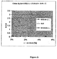
ELISAアッセイを血清試料中の可溶性ヒトM−CSF受容体の存在を測定するために使用した場合、強い陽性シグナルがヒト血清試料で検出された。このELISAアッセイで用いた捕捉抗体および検出抗体は、ヒトM−CSF受容体の細胞外ドメインに特異的であった。図2に示すように、強い陽性シグナルがヒト血清試料で検出された。このシグナルは、ヒト血清の濃度に依存している。捕捉抗体が使用されなかった場合にはシグナルは存在しない。
B. Results When the ELISA assay was used to determine the presence of soluble human M-CSF receptor in a serum sample, a strong positive signal was detected in the human serum sample. The capture and detection antibodies used in this ELISA assay were specific for the extracellular domain of the human M-CSF receptor. As shown in FIG. 2, a strong positive signal was detected in human serum samples. This signal is dependent on the concentration of human serum. There is no signal if no capture antibody was used.
比較タンパク質として組み換え型のヒトM−CSF受容体およびヒト抗体Fcフラグメント融合タンパク質を使用して、標準曲線が可溶性ヒトM−CSF受容体濃度を定量するために確立された(図1)。このELISAアッセイを用いて、ヒト血清中の可溶性受容体の量が、基準としてc−fmsヒトFc融合タンパク質を用いて算出された。約0.5μg/mlの可溶性ヒトM−CSF受容体がヒト血清中に存在することが見出された(図2)。 Using recombinant human M-CSF receptor and human antibody Fc fragment fusion protein as comparative proteins, a standard curve was established to quantify soluble human M-CSF receptor concentration (FIG. 1). Using this ELISA assay, the amount of soluble receptor in human serum was calculated using c-fms human Fc fusion protein as a reference. About 0.5 μg / ml of soluble human M-CSF receptor was found to be present in human serum (FIG. 2).
II.可溶性M−CSF受容体のELISA結合アッセイ
A.材料および方法
マイクロタイタープレート(R&D systems、カタログ番号CP0011)を、PBS中100ng/ml作用希釈の100μl/ウェル捕捉抗体(Duo Set Elisa development system hM−CSF R(R&D systems、カタログ番号DY329))でコートした。プレートをシールして一夜室温でインキュベートした。マニホルドディスペンサ/ワッシャを用いて、洗浄緩衝液(PBS中0.05%Tween、pH7.2〜7.4)で3回ウェルを洗浄後、プレートを300μl/ウェルのブロッキング緩衝液(1%BSA、PBS中5%シュークロース、pH7.2〜7.4)を用いて、室温で少なくとも1時間ブロックした。
II. Soluble M-CSF Receptor ELISA Binding Assay A. Materials and Methods Coat microtiter plates (R & D systems, catalog number CP0011) with 100 μl / well capture antibody (Duo Set Elisa development system hM-CSF R (R & D systems, catalog number DY329)) at 100 ng / ml working dilution in PBS. did. The plate was sealed and incubated overnight at room temperature. After washing the wells three times with wash buffer (0.05% Tween in PBS, pH 7.2-7.4) using a manifold dispenser / washer, the plate was washed with 300 μl / well blocking buffer (1% BSA, Blocked with 5% sucrose in PBS, pH 7.2-7.4) for at least 1 hour at room temperature.
洗浄工程を繰り返した後、プレートは試料添加の準備が整う。100μl/ウェルの試料または試薬希釈剤中の標準物質のいずれかを添加した。プレートを接着用テープでカバーし、室温で2時間インキュベート後、3サイクルの洗浄を行った。1μg/mLのヒトM−CSFをプレートに添加し、室温で1時間インキュベートした。ヒトM−CSFに対する100μlの検出抗体(HRP結合ポリクローナル抗ヒトM−CSF抗体:R&D Systems、Cat DMC00,part#890154)を添加後、プレートをカバーし、室温で2時間インキュベート後、3サイクルの洗浄を行った。プレートを、ウェル当り100μlの基質溶液(試薬AとBの1:1混合物(R&D systems、カタログ番号DY999))を用いて5分間発色させた後、50μlの停止液(2N H2SO4)を添加し、完全に混合するためにプレートを軽くたたいた。最終の読出しは、停止液添加の直後に450〜540nmの吸光度であった。 After repeating the washing process, the plate is ready for sample addition. Either 100 μl / well of sample or standard in reagent diluent was added. The plate was covered with adhesive tape and incubated for 2 hours at room temperature, followed by 3 cycles of washing. 1 μg / mL human M-CSF was added to the plate and incubated for 1 hour at room temperature. After adding 100 μl of detection antibody against human M-CSF (HRP-conjugated polyclonal anti-human M-CSF antibody: R & D Systems, Cat DMC00, part # 890154), the plate is covered, incubated at room temperature for 2 hours, and then washed for 3 cycles Went. Plates were developed with 100 μl per well of substrate solution (1: 1 mixture of reagents A and B (R & D systems, catalog number DY999)) for 5 minutes, followed by 50 μl of stop solution (2N H 2 SO 4 ). Tap and tap the plate to add and mix thoroughly. The final readout was absorbance at 450-540 nm immediately after addition of stop solution.
B.結果
ヒト血清中に存在する可溶性受容体は、M−CSFを結合することができる。図3に示すように、ヒト血清試料の階段希釈を、マウスモノクローナル抗ヒトc−fms抗体でコートしたELISAプレートにアプライした。アルカリホスファターゼ結合ポリクローナル抗ヒトM−CSF抗体によるヒトM−CSFの検出前に、1μg/mlの外来性ヒトM−CSF(四角)またはPBS(ひし形)のいずれかを添加した。明白なM−CSFの結合が約5%のヒト血清に見出された。しかしヒト血清中の可溶性受容体は、組み換え型のc−fms Fc融合タンパク質と比べて低い結合親和性を有することが見出された(図4)[組み換え型c−fms Fc融合タンパク質の結合親和性は、リガンド誘導二量体化のため天然の膜結合型c−fmsの結合親和性に近いと考えられる]。可溶性受容体の低親和性に対する1つの解釈は、この受容体はリガンドに対する1つの結合部位のみを有するが、組み換え型c−fms Fc融合タンパク質は、結合活性効果に関して2つの結合部位を有することである。
B. Results Soluble receptors present in human serum are capable of binding M-CSF. As shown in FIG. 3, serial dilutions of human serum samples were applied to ELISA plates coated with mouse monoclonal anti-human c-fms antibody. Prior to detection of human M-CSF with alkaline phosphatase-conjugated polyclonal anti-human M-CSF antibody, either 1 μg / ml exogenous human M-CSF (square) or PBS (diamond) was added. Obvious M-CSF binding was found in about 5% human serum. However, the soluble receptor in human serum was found to have a lower binding affinity compared to the recombinant c-fms Fc fusion protein (FIG. 4) [binding affinity of the recombinant c-fms Fc fusion protein. Sex is thought to be close to the binding affinity of natural membrane-bound c-fms due to ligand-induced dimerization]. One interpretation for the low affinity of the soluble receptor is that the receptor has only one binding site for the ligand, whereas the recombinant c-fms Fc fusion protein has two binding sites for binding activity effects. is there.
III.血清試料由来ヒトM−CSF受容体の免疫沈降
A.材料および方法
500μlのストレプトアビジンビーズ(#20347、Pierce)を、15mlファルコンチューブ中の10mlの20%ヒト血清(Sigma カタログ番号 S 7023、あらかじめ凍結されている)に添加し、穏やかに振盪して4℃で1時間インキュベートした。試料を400rpmで5分間遠心して上清を新たなチューブに移した。10μgの抗hMCSFR抗体(R&D # BAF 329)を上清に添加し、穏やかに振盪して4℃で1時間インキュベートした。500μlのストレプトアビジンビーズを添加後、試料を4℃で一夜振盪してインキュベートした。試料を400rpmで5分間遠心し、上清を新しいチューブへ移し、そして同時に上清を保存した。ビーズをエッペンドルフチューブへ移し、洗浄緩衝液#1(0.5M LiCL2)、#2(0.5M LiCL2/0.5%Triton)および#3(10mM TRIS pH7.4)のそれぞれ1mlで1回洗浄した。最後の遠心後、大部分の洗浄緩衝液を除去した(ビーズがほぼ乾燥でするまで)。次いでビーズを500μlのPBSに再懸濁した。ゲル(10%Novex)泳動前に、DTTを有する5×SDS試料緩衝液を添加し、試料を100℃で2分間煮沸した。
III. Immunoprecipitation of human M-CSF receptor from serum samples Materials and
免疫沈降したヒトM−CSF受容体を脱グリコシル化するために、上記の免疫沈降方法からの冷凍ビーズ(約200μl)を200μlのPBSを用いて1:1の比率に希釈した。希釈した試料を、100℃で10分間、1×変性緩衝液(PNGase F、NEB# P0705S)中でインキュベートした。G7反応緩衝液(PNGase F、NEB# P0705S)を、1×最終濃度およびNP40(最終濃度1%)になるように添加後、試料を5μlのPNGase Fと共に1時間、37℃でインキュベートした。
To deglycosylate the immunoprecipitated human M-CSF receptor, frozen beads (approximately 200 μl) from the immunoprecipitation method described above were diluted to a 1: 1 ratio with 200 μl PBS. Diluted samples were incubated in 1 × denaturation buffer (PNGase F, NEB # P0705S) at 100 ° C. for 10 minutes. After adding G7 reaction buffer (PNGase F, NEB # P0705S) to 1 × final concentration and NP40 (
B.結果
可溶性ヒトM−CSF受容体は、分子量97KDを有するグリコシル化タンパク質であることが分かった(図5)。脱グリコシル化後、可溶性ヒトM−CSF受容体の分子量は約60KDである(図6)。
B. Results Soluble human M-CSF receptor was found to be a glycosylated protein with a molecular weight of 97 KD (FIG. 5). After deglycosylation, the molecular weight of soluble human M-CSF receptor is approximately 60 KD (FIG. 6).
可溶性ヒトM−CSF受容体は、ヒトM−CSFに対する膜結合受容体であるヒトcfmsタンパク質のタンパク質配列を少なくとも部分的に共有することが分かった。上記の図で示すように、可溶性ヒトM−CSF受容体はまた、ヒトc−ftnsタンパク質を認識する多数の抗体によって認識され得る。可溶性ヒトM−CSF受容体のタンパク質配列をさらに確認するために、タンパク質分析を以下のように行った:脱グリコシル化後の可溶性受容体に対応するタンパク質バンドをSDS−PAGEゲル上で同定した(図7のバンド#4)。このバンド中のタンパク質をトリプシンで消化した。ペプチド混合物であるこの消化産物を分析した。1つのペプチドは、ヒトc−fmsタンパク質の細胞外ドメインのセグメントと同様の、正確な分子量およびアミノ酸の配列(配列番号1)を有することが見出された(図8)。この証拠は、可溶性ヒトM−CSF受容体は、ヒトM−CSFに対する膜結合受容体であるヒトc−fmsタンパク質のタンパク質配列を共有することを示すものである。
Soluble human M-CSF receptor was found to at least partially share the protein sequence of the human cfms protein, which is a membrane-bound receptor for human M-CSF. As shown in the above figure, the soluble human M-CSF receptor can also be recognized by a number of antibodies that recognize the human c-ftns protein. To further confirm the protein sequence of the soluble human M-CSF receptor, protein analysis was performed as follows: the protein band corresponding to the soluble receptor after deglycosylation was identified on an SDS-PAGE gel (
(実施例2)
この実施例は、可溶性M−CSF受容体がヒト尿試料中に認められたことを示す。この実施例は、さらにこの受容体が正常被験体および乳癌患者両方の血清中に存在し、M−CSFレベルと相関した可溶性M−CSF受容体レベルであることを示している。最後にこの実施例は、可溶性M−CSF受容体は霊長類にも認められることを示している。
(Example 2)
This example shows that soluble M-CSF receptor was found in human urine samples. This example further shows that this receptor is present in the serum of both normal subjects and breast cancer patients and is at soluble M-CSF receptor levels correlated with M-CSF levels. Finally, this example shows that soluble M-CSF receptor is also found in primates.
I.可溶性ヒトM−CSF受容体は尿試料にも認められた。 I. Soluble human M-CSF receptor was also found in urine samples.
実施例1に記載のように同じELISA装置を用いて、正常ヒトボランティアからの多数の尿試料を採取し、可溶性ヒトM−CSF受容体の存在についてアッセイした。様々なレベルの可溶性ヒトM−CSF受容体が成人女性(年齢25〜66)、成人男性(年齢35〜70)および年齢3〜9の子供の間で検出された。可溶性ヒトM−CSF受容体濃度の定量は、基準として組み換え型c−fmsヒトFc融合タンパク質を用いて決定した(図9)。 Using the same ELISA device as described in Example 1, multiple urine samples from normal human volunteers were collected and assayed for the presence of soluble human M-CSF receptor. Various levels of soluble human M-CSF receptor were detected among adult females (age 25-66), adult males (age 35-70) and children aged 3-9. Quantification of soluble human M-CSF receptor concentration was determined using recombinant c-fms human Fc fusion protein as a reference (Figure 9).
尿試料を、1人の女性被験体の月経周期の全体を通じて毎日採取した。この試料は朝の最初の排尿として採取し、−20℃に凍結した。全ての試料は、同じ日にアッセイした。可溶性受容体は、本明細書に記載したようにアッセイした。図10で示すように、尿サンプルの可溶性受容体レベルは、月経周期間に変動する。この所見は可溶性受容体を、月経周期または妊娠テストのための潜在的診断マーカーにしている。 Urine samples were collected daily throughout the menstrual cycle of one female subject. This sample was collected as the first urination in the morning and frozen at -20 ° C. All samples were assayed on the same day. Soluble receptors were assayed as described herein. As shown in FIG. 10, the soluble receptor level of the urine sample varies between menstrual cycles. This finding makes soluble receptors a potential diagnostic marker for the menstrual cycle or pregnancy test.
II.乳癌患者におけるM−CSFレベルと可溶性M−CSF受容体レベルの相関
可溶性ヒトM−CSF受容体濃度を、以下のように乳癌血清試料で分析した。ヒト血清試料の階段希釈を、上記のように可溶性ヒトM−CSF受容体の存在に関してELISAでアッセイした。ヒト血清をこのELISA構成(「標準」と呼ぶ)でアッセイした場合、強い陽性シグナルが検出された。対照としての、捕捉抗体または検出抗体のいずれかの除去はシグナルを消去し、このシグナルはこれら抗ヒトc−fms抗体に対して特異的であることを示した。基準として組み換え型c−fmsヒトFc融合タンパク質を用いる可溶性ヒトM−CSF受容体濃度の定量は、0.5μg/mlであると測定された。
II. Correlation between M-CSF levels and soluble M-CSF receptor levels in breast cancer patients Soluble human M-CSF receptor concentrations were analyzed in breast cancer serum samples as follows. Serial dilutions of human serum samples were assayed by ELISA for the presence of soluble human M-CSF receptor as described above. When human serum was assayed in this ELISA configuration (referred to as “standard”), a strong positive signal was detected. As a control, removal of either capture antibody or detection antibody abolished the signal, indicating that this signal was specific for these anti-human c-fms antibodies. The quantification of soluble human M-CSF receptor concentration using recombinant c-fms human Fc fusion protein as a reference was determined to be 0.5 μg / ml.
図11で示すように、可溶性受容体のレベルは患者間で変化する。患者試料の可溶性受容体濃度は上記濃度よりもはるかに低く、血清試料の調製および貯蔵に用いられた異なるプロトコルによる可能性があることに留意されたい(図2)。したがってM−CSFとM−CSF可溶性受容体濃度の比較は、同じプロトコルに従って採取されて処理された試料間でのみ有効である。図12は、同じ試料における乳癌患者のM−CSFレベルに対するM−CSF可溶性受容体濃度間の直線相関を示している。この所見は、M−CSF可溶性受容体を癌進行に関する潜在的バイオマーカーにしている。さらに、M−CSFの直接的測定が困難な場合(たとえば、M−CSF中和抗体が癌治療に用いられる場合)、M−CSF可溶性受容体はさらに有効なバイオマーカーとなる。 As shown in FIG. 11, the level of soluble receptor varies between patients. Note that the soluble receptor concentration of the patient sample is much lower than the above concentration and may be due to the different protocols used for the preparation and storage of serum samples (FIG. 2). Therefore, comparison of M-CSF and M-CSF soluble receptor concentrations is only valid between samples collected and processed according to the same protocol. FIG. 12 shows a linear correlation between M-CSF soluble receptor concentration versus M-CSF level for breast cancer patients in the same sample. This finding makes the M-CSF soluble receptor a potential biomarker for cancer progression. Furthermore, when direct measurement of M-CSF is difficult (for example, when M-CSF neutralizing antibody is used for cancer treatment), M-CSF soluble receptor becomes a more effective biomarker.
III.可溶性M−CSF受容体がまた、他の霊長類で見出される
ヒトM−CSF可溶性受容体に関する上記のELISAアッセイはまた、他の動物種からの血清試料を調べるために使用される(図13)。カニクイザルおよびアカゲザルの血清試料は強い陽性シグナルを示し、可溶性受容体の存在を示す。カニクイザルおよびアカゲザルに関する膜結合M−CSF受容体タンパク質配列は入手不可能である。しかしこれらの配列は、そのヒト対応物と高いレベルのタンパク質相同性を共有すると考えられる。したがって、ヒト膜結合型M−CSF受容体の細胞外ドメインを認識するELISAアッセイで用いた抗体はまた、カニクイザルおよびアカゲザルのこの細胞外ドメインを認識する。
III. Soluble M-CSF receptor is also found in other primates The above ELISA assay for human M-CSF soluble receptor is also used to examine serum samples from other animal species (FIG. 13). . Cynomolgus and rhesus serum samples show strong positive signals, indicating the presence of soluble receptors. Membrane-bound M-CSF receptor protein sequences for cynomolgus and rhesus monkeys are not available. However, these sequences are thought to share a high level of protein homology with their human counterparts. Thus, antibodies used in an ELISA assay that recognizes the extracellular domain of human membrane-bound M-CSF receptor also recognize this cynomolgus and rhesus monkey extracellular domain.
非霊長類血清由来の陽性シグナルの欠如は、M−CSF可溶性受容体の欠如を示さないことに留意されたい。むしろ、可溶性受容体の存在を検出するために用いたELISAアッセイの抗体特異性による可能性が一番高いようである。 Note that the lack of positive signal from non-primate sera does not indicate the lack of M-CSF soluble receptor. Rather, it seems most likely due to the antibody specificity of the ELISA assay used to detect the presence of soluble receptors.
(実施例3)
この実施例は、可溶性ヒトM−CSF受容体の発現を、破骨細胞分化と密接に関連していることを示している。この実施例は、さらに分化した破骨細胞のみが可溶性ヒトM−CSF受容体を発現し、そのレベルがM−CSFの除去により増加することを示している。
(Example 3)
This example shows that the expression of soluble human M-CSF receptor is closely related to osteoclast differentiation. This example shows that only further differentiated osteoclasts express soluble human M-CSF receptor, the level of which increases upon removal of M-CSF.
A.材料および方法
M−CSF可溶性受容体の発現を、膜結合M−CSF受容体の発現が見出されたヒト破骨細胞を用いたin vitro系で研究した。特に可溶性受容体の発現を、破骨細胞分化に関連して調べた。初代のヒト破骨細胞前駆体(Cambrex Bio Science Walkersville、Inc.カタログ番号2T−110)を、30ng/mlのヒトM−CSFおよび100ng/mlのRANKLを含む細胞培養培地中、0.2ml/ウェルの10,000細胞/ウェルで接種した。細胞のプレーティングと同日に、試験抗体を1μg/mLでウェルに添加した。細胞を37℃、5%CO2の加湿雰囲気下でインキュベートした。7日目に、破骨細胞を異常に大きな多核細胞として位相差顕微鏡で同定した。フィーディングして培養をさらに1週間続けることができ、その間に破骨細胞は大きさが増し続ける。培養の最後に、細胞を酒石酸塩耐性酸性ホスファターゼで染色し、この陽性の染色は破骨細胞の存在を示す。各ウェル中の破骨細胞数を光学顕微鏡下で計数し、顕微鏡の視野下の破骨細胞の平均としてグラフに示した。
A. Materials and Methods The expression of M-CSF soluble receptor was studied in an in vitro system using human osteoclasts in which expression of membrane-bound M-CSF receptor was found. In particular, soluble receptor expression was examined in relation to osteoclast differentiation. Primary human osteoclast precursors (Cambrex Bio Science Walkersville, Inc. Catalog No. 2T-110) were added at 0.2 ml / well in cell culture medium containing 30 ng / ml human M-CSF and 100 ng / ml RANKL. Of 10,000 cells / well. On the same day as cell plating, test antibody was added to the wells at 1 μg / mL. Cells were incubated at 37 ° C. in a humidified atmosphere of 5% CO 2 . On
破骨細胞馴化培地のTRAP活性を、SBA−SciencesのBoneTRAP Assayキット(TR201)を用いて検定した。BoneTRAP(登録商標)Assay(TR201)は、ヒト血清試料由来の骨吸収速度の迅速で特異的な判定用の固相免疫固定酵素活性アッセイである。これはヒト破骨細胞培養で用いることができる。 The TRAP activity of the osteoclast conditioned medium was assayed using the SBA-Sciences BONETRAP Assay kit (TR201). BoneTRAP® Assay (TR201) is a solid phase immunofixed enzyme activity assay for rapid and specific determination of bone resorption rate from human serum samples. This can be used in human osteoclast culture.
B.結果
図14で示すように、M−CSF可溶性受容体は破骨細胞で発現した。分化した破骨細胞の馴化培地における可溶性受容体の濃度を測定した。可溶性受容体の発現は破骨細胞に特異的であった。このケースではM−CSF中和抗体であるChir−RX1による破骨細胞分化の阻害は、可溶性受容体の抑制を生じた。
B. Results As shown in FIG. 14, the M-CSF soluble receptor was expressed in osteoclasts. The concentration of soluble receptors in the conditioned medium of differentiated osteoclasts was measured. Soluble receptor expression was specific to osteoclasts. In this case, inhibition of osteoclast differentiation by Chir-RX1, an M-CSF neutralizing antibody, resulted in suppression of soluble receptors.
可溶性受容体は分化した破骨細胞でのみ発現し、破骨細胞前駆体では発現しない。図15aに示した時間経過実験で示すように破骨細胞分化アッセイで、可溶性受容体の発現は10日目に始まり、12日目にプラトーに達する。この発現パターンは、TRAP活性により追跡して図15bに示したように厳密に破骨細胞分化パターンに対応する。
Soluble receptors are expressed only on differentiated osteoclasts and not on osteoclast precursors. In the osteoclast differentiation assay as shown in the time course experiment shown in Figure 15a, soluble receptor expression begins on
興味深いことには、分化した破骨細胞において、M−CSF可溶性受容体の発現はM−CSFの除去により上方制御されている。図16は、IgG1対照抗体またはM−CSF中和抗体であるChir−RX1のいずれかを添加後、24時間以内のM−CSF可溶性受容体の発現を示す。データは、M−CSF活性が中和された場合、可溶性受容体の発現が増加することを明確に示している。発現の増加は8日目に始まり最高レベルに12日目に達し、これは発現の増加が分化した破骨細胞で生じることを示している。
Interestingly, in differentiated osteoclasts, M-CSF soluble receptor expression is upregulated by removal of M-CSF. FIG. 16 shows expression of M-CSF soluble receptor within 24 hours after addition of either IgG1 control antibody or M-CSF neutralizing antibody Chir-RX1. The data clearly shows that soluble receptor expression increases when M-CSF activity is neutralized. Increased expression starts on
分化した破骨細胞において、破骨細胞活性の阻害剤であるZometaは、M−CSF可溶性受容体の発現に及ぼす効果を有してない(図17)。同じ実験で、IgG1対照ではなく、M−CSF中和抗体が発現を刺激した。 In differentiated osteoclasts, Zometa, an inhibitor of osteoclast activity, has no effect on the expression of M-CSF soluble receptors (FIG. 17). In the same experiment, M-CSF neutralizing antibody, but not an IgG1 control, stimulated expression.
本明細書中に挙げられおよび/またはアプリケーションデータシートにリストされた、上記の全ての米国特許、米国特許出願公開、米国特許出願、外国特許、外国特許出願および非特許公開は、その全体が参考として本明細書中に組み込まれる。 All of the above U.S. patents, U.S. patent application publications, U.S. patent applications, foreign patents, foreign patent applications and non-patent publications mentioned herein and / or listed in the application data sheet are incorporated by reference in their entirety. As incorporated herein.
前述より、本発明の特定の実施形態を例示の目的で本明細書中に記載したが、本発明の精神および範囲から逸脱することなく種々の改変がなされ得ることが理解される。 From the foregoing, it will be appreciated that, although specific embodiments of the invention have been described herein for purposes of illustration, various modifications may be made without departing from the spirit and scope of the invention.

Claims (1)
Applications Claiming Priority (2)
| Application Number | Priority Date | Filing Date | Title |
|---|---|---|---|
| US75321805P | 2005-12-22 | 2005-12-22 | |
| US60/753,218 | 2005-12-22 |
Related Parent Applications (1)
| Application Number | Title | Priority Date | Filing Date |
|---|---|---|---|
| JP2008547568A Division JP2009521685A (en) | 2005-12-22 | 2006-12-21 | Soluble human M-CSF receptor and uses thereof |
Publications (1)
| Publication Number | Publication Date |
|---|---|
| JP2012108155A true JP2012108155A (en) | 2012-06-07 |
Family
ID=38609972
Family Applications (2)
| Application Number | Title | Priority Date | Filing Date |
|---|---|---|---|
| JP2008547568A Withdrawn JP2009521685A (en) | 2005-12-22 | 2006-12-21 | Soluble human M-CSF receptor and uses thereof |
| JP2012043540A Pending JP2012108155A (en) | 2005-12-22 | 2012-02-29 | Soluble human m-csf receptor and uses thereof |
Family Applications Before (1)
| Application Number | Title | Priority Date | Filing Date |
|---|---|---|---|
| JP2008547568A Withdrawn JP2009521685A (en) | 2005-12-22 | 2006-12-21 | Soluble human M-CSF receptor and uses thereof |
Country Status (8)
| Country | Link |
|---|---|
| US (2) | US20100099123A1 (en) |
| EP (1) | EP1977238B1 (en) |
| JP (2) | JP2009521685A (en) |
| KR (1) | KR101358600B1 (en) |
| CN (1) | CN101379400A (en) |
| AU (1) | AU2006342119B8 (en) |
| CA (1) | CA2634945A1 (en) |
| WO (1) | WO2007120252A2 (en) |
Families Citing this family (13)
| Publication number | Priority date | Publication date | Assignee | Title |
|---|---|---|---|---|
| US8080246B2 (en) | 2008-11-26 | 2011-12-20 | Five Prime Therapeutics, Inc. | Colony stimulating factor 1 receptor (CSF1R) extracellular domain fusion molecules |
| US8183207B2 (en) | 2008-11-26 | 2012-05-22 | Five Prime Therapeutics, Inc. | Treatment of osteolytic disorders and cancer using CSF1R extracellular domain fusion molecules |
| WO2011007764A1 (en) * | 2009-07-14 | 2011-01-20 | 独立行政法人産業技術総合研究所 | Sugar chain marker as measure of disease conditions of hepatic diseases |
| RU2427323C1 (en) * | 2010-02-25 | 2011-08-27 | Федеральное государственное учреждение "Ростовский научно-исследовательский онкологический институт Федерального агентства по высокотехнологичной медицинской помощи" | Method of predicting process generalisation in case of malignant bone tumours |
| SMT201900074T1 (en) | 2010-05-04 | 2019-02-28 | Five Prime Therapeutics Inc | Antibodies that bind csf1r |
| US20130302322A1 (en) | 2012-05-11 | 2013-11-14 | Five Prime Therapeutics, Inc. | Methods of treating conditions with antibodies that bind colony stimulating factor 1 receptor (csf1r) |
| ES2442991B1 (en) * | 2012-08-14 | 2014-11-25 | Fundación Md Anderson International España | Method for diagnosis and / or prognosis of lymphomas |
| CN104684582A (en) | 2012-08-31 | 2015-06-03 | 戊瑞治疗有限公司 | Methods of treating conditions with antibodies that bind colony stimulating factor 1 receptor (csf1r) |
| CN113350501A (en) | 2014-06-23 | 2021-09-07 | 戊瑞治疗有限公司 | Methods of treating conditions with antibodies that bind colony stimulating factor 1 receptor (CSF1R) |
| AU2015339306B2 (en) | 2014-10-29 | 2021-07-22 | Bristol-Myers Squibb Company | Combination therapy for cancer |
| EA201791421A1 (en) | 2014-12-22 | 2017-10-31 | Файв Прайм Терапьютикс, Инк. | ANTIBODIES AGAINST CSF1R FOR THE TREATMENT OF PVNS |
| BR112017020952A2 (en) | 2015-04-13 | 2018-07-10 | Five Prime Therapeutics Inc | cancer treatment method, composition and use of composition |
| CA3073531A1 (en) | 2017-09-13 | 2019-03-21 | Five Prime Therapeutics, Inc. | Combination anti-csf1r and anti-pd-1 antibody combination therapy for pancreatic cancer |
Citations (3)
| Publication number | Priority date | Publication date | Assignee | Title |
|---|---|---|---|---|
| JPH03500879A (en) * | 1987-10-23 | 1991-02-28 | 森永乳業株式会社 | Compositions and methods for the treatment of cancers characterized by overexpression of the c-fms proto-oncogene |
| JPH0967400A (en) * | 1995-08-31 | 1997-03-11 | Toray Ind Inc | Monoclonal antibody, hybridoma producing the antibody, and use thereof |
| WO2004075775A2 (en) * | 2003-02-24 | 2004-09-10 | Sugen, Inc. | Treatment of excessive osteolyisis with indolinone compounds |
Family Cites Families (4)
| Publication number | Priority date | Publication date | Assignee | Title |
|---|---|---|---|---|
| US4816567A (en) * | 1983-04-08 | 1989-03-28 | Genentech, Inc. | Recombinant immunoglobin preparations |
| US4929700A (en) * | 1987-04-16 | 1990-05-29 | Cetus Corporation | Production of purified, biologically active, bacterially produced recombinant human CSF-1 |
| US6610484B1 (en) * | 1999-01-26 | 2003-08-26 | Cytyc Health Corporation | Identifying material from a breast duct |
| CN1787837A (en) * | 2002-11-15 | 2006-06-14 | 希龙公司 | Methods for preventing and treating cancer metastasis and bone loss associated with cancer metastasis |
-
2006
- 2006-12-21 CA CA002634945A patent/CA2634945A1/en not_active Abandoned
- 2006-12-21 CN CNA2006800519125A patent/CN101379400A/en active Pending
- 2006-12-21 AU AU2006342119A patent/AU2006342119B8/en active Active
- 2006-12-21 WO PCT/US2006/048879 patent/WO2007120252A2/en not_active Ceased
- 2006-12-21 US US12/158,877 patent/US20100099123A1/en not_active Abandoned
- 2006-12-21 JP JP2008547568A patent/JP2009521685A/en not_active Withdrawn
- 2006-12-21 KR KR1020087017904A patent/KR101358600B1/en active Active
- 2006-12-21 EP EP06850597.3A patent/EP1977238B1/en active Active
-
2012
- 2012-02-29 JP JP2012043540A patent/JP2012108155A/en active Pending
-
2013
- 2013-02-20 US US13/771,395 patent/US20140030738A1/en not_active Abandoned
Patent Citations (3)
| Publication number | Priority date | Publication date | Assignee | Title |
|---|---|---|---|---|
| JPH03500879A (en) * | 1987-10-23 | 1991-02-28 | 森永乳業株式会社 | Compositions and methods for the treatment of cancers characterized by overexpression of the c-fms proto-oncogene |
| JPH0967400A (en) * | 1995-08-31 | 1997-03-11 | Toray Ind Inc | Monoclonal antibody, hybridoma producing the antibody, and use thereof |
| WO2004075775A2 (en) * | 2003-02-24 | 2004-09-10 | Sugen, Inc. | Treatment of excessive osteolyisis with indolinone compounds |
Also Published As
| Publication number | Publication date |
|---|---|
| KR101358600B1 (en) | 2014-02-06 |
| CN101379400A (en) | 2009-03-04 |
| US20100099123A1 (en) | 2010-04-22 |
| CA2634945A1 (en) | 2007-10-25 |
| AU2006342119A8 (en) | 2013-05-09 |
| JP2009521685A (en) | 2009-06-04 |
| AU2006342119A1 (en) | 2007-10-25 |
| WO2007120252A2 (en) | 2007-10-25 |
| AU2006342119B8 (en) | 2013-05-09 |
| KR20080085054A (en) | 2008-09-22 |
| WO2007120252A3 (en) | 2008-10-09 |
| EP1977238B1 (en) | 2016-09-28 |
| AU2006342119B2 (en) | 2013-01-10 |
| EP1977238A2 (en) | 2008-10-08 |
| US20140030738A1 (en) | 2014-01-30 |
Similar Documents
| Publication | Publication Date | Title |
|---|---|---|
| US20140030738A1 (en) | Soluble human m-csf receptor and uses thereof | |
| JP6411432B2 (en) | A biomarker for predicting and evaluating the reactivity of thyroid and kidney cancer subjects to lenvatinib compounds | |
| JP6411379B2 (en) | Biomarkers for predicting and assessing responsiveness of endometrial cancer subjects to lenvatinib compounds | |
| KR102057438B1 (en) | Folate receptor alpha as a diagnostic and prognostic marker for folate receptor alpha-expressing cancers | |
| US20250172572A1 (en) | Noninvasive method to quantify kidney function and functional decline | |
| US12298307B2 (en) | PD-ECGF as biomarker of cancer | |
| EP2895863B1 (en) | New biomarkers for the diagnosis and/or prognosis of clear cell renal cell carcinoma | |
| JP2023508023A (en) | CXCL10 binding proteins and uses thereof | |
| KR20180013878A (en) | Biological markers for combination therapy including lenbactinib and everolimus | |
| JP2019518970A (en) | Methods and kits for predicting sensitivity to cancer treatment of subjects suffering from kidney cancer | |
| CN103558391A (en) | Soluble human M-CSF receptor and uses thereof | |
| AU2012261609A9 (en) | Soluble human M-CSF receptor and uses thereof | |
| MX2008007881A (en) | Soluble human m-csf receptor and uses thereof | |
| CN110687284A (en) | Application of reagents for the detection of SIX2 autoantibodies in serum | |
| JP2024523546A (en) | Predicting Cancer Recurrence | |
| JP2008537101A (en) | Use of prostate specific antigen to predict drug response | |
| WO2014023819A1 (en) | Methods for predicting the survival time of a patient suffering from a glioblastoma | |
| US20120301879A1 (en) | Novel use of ca-125 | |
| MX2008010297A (en) | Detection of cancer by elevated levels of bcl-2 |
Legal Events
| Date | Code | Title | Description |
|---|---|---|---|
| A621 | Written request for application examination |
Free format text: JAPANESE INTERMEDIATE CODE: A621 Effective date: 20120229 |
|
| A521 | Request for written amendment filed |
Free format text: JAPANESE INTERMEDIATE CODE: A523 Effective date: 20130128 |
|
| A131 | Notification of reasons for refusal |
Free format text: JAPANESE INTERMEDIATE CODE: A131 Effective date: 20130912 |
|
| A601 | Written request for extension of time |
Free format text: JAPANESE INTERMEDIATE CODE: A601 Effective date: 20131211 |
|
| A602 | Written permission of extension of time |
Free format text: JAPANESE INTERMEDIATE CODE: A602 Effective date: 20131216 |
|
| A601 | Written request for extension of time |
Free format text: JAPANESE INTERMEDIATE CODE: A601 Effective date: 20140110 |
|
| A602 | Written permission of extension of time |
Free format text: JAPANESE INTERMEDIATE CODE: A602 Effective date: 20140116 |
|
| A601 | Written request for extension of time |
Free format text: JAPANESE INTERMEDIATE CODE: A601 Effective date: 20140210 |
|
| A602 | Written permission of extension of time |
Free format text: JAPANESE INTERMEDIATE CODE: A602 Effective date: 20140214 |
|
| A02 | Decision of refusal |
Free format text: JAPANESE INTERMEDIATE CODE: A02 Effective date: 20140905 |