JP2012507331A - Mrスキャンの自動シーケンシャルプランニング - Google Patents
Mrスキャンの自動シーケンシャルプランニング Download PDFInfo
- Publication number
- JP2012507331A JP2012507331A JP2011533913A JP2011533913A JP2012507331A JP 2012507331 A JP2012507331 A JP 2012507331A JP 2011533913 A JP2011533913 A JP 2011533913A JP 2011533913 A JP2011533913 A JP 2011533913A JP 2012507331 A JP2012507331 A JP 2012507331A
- Authority
- JP
- Japan
- Prior art keywords
- volume
- interest
- mri
- image
- anatomical
- Prior art date
- Legal status (The legal status is an assumption and is not a legal conclusion. Google has not performed a legal analysis and makes no representation as to the accuracy of the status listed.)
- Granted
Links
Images



Classifications
-
- G—PHYSICS
- G01—MEASURING; TESTING
- G01R—MEASURING ELECTRIC VARIABLES; MEASURING MAGNETIC VARIABLES
- G01R33/00—Arrangements or instruments for measuring magnetic variables
- G01R33/20—Arrangements or instruments for measuring magnetic variables involving magnetic resonance
- G01R33/44—Arrangements or instruments for measuring magnetic variables involving magnetic resonance using nuclear magnetic resonance [NMR]
- G01R33/48—NMR imaging systems
- G01R33/54—Signal processing systems, e.g. using pulse sequences ; Generation or control of pulse sequences; Operator console
- G01R33/543—Control of the operation of the MR system, e.g. setting of acquisition parameters prior to or during MR data acquisition, dynamic shimming, use of one or more scout images for scan plane prescription
-
- G—PHYSICS
- G01—MEASURING; TESTING
- G01R—MEASURING ELECTRIC VARIABLES; MEASURING MAGNETIC VARIABLES
- G01R33/00—Arrangements or instruments for measuring magnetic variables
- G01R33/20—Arrangements or instruments for measuring magnetic variables involving magnetic resonance
- G01R33/44—Arrangements or instruments for measuring magnetic variables involving magnetic resonance using nuclear magnetic resonance [NMR]
- G01R33/48—NMR imaging systems
- G01R33/54—Signal processing systems, e.g. using pulse sequences ; Generation or control of pulse sequences; Operator console
-
- G—PHYSICS
- G01—MEASURING; TESTING
- G01R—MEASURING ELECTRIC VARIABLES; MEASURING MAGNETIC VARIABLES
- G01R33/00—Arrangements or instruments for measuring magnetic variables
- G01R33/20—Arrangements or instruments for measuring magnetic variables involving magnetic resonance
- G01R33/44—Arrangements or instruments for measuring magnetic variables involving magnetic resonance using nuclear magnetic resonance [NMR]
- G01R33/48—NMR imaging systems
- G01R33/54—Signal processing systems, e.g. using pulse sequences ; Generation or control of pulse sequences; Operator console
- G01R33/546—Interface between the MR system and the user, e.g. for controlling the operation of the MR system or for the design of pulse sequences
-
- G—PHYSICS
- G01—MEASURING; TESTING
- G01R—MEASURING ELECTRIC VARIABLES; MEASURING MAGNETIC VARIABLES
- G01R33/00—Arrangements or instruments for measuring magnetic variables
- G01R33/20—Arrangements or instruments for measuring magnetic variables involving magnetic resonance
- G01R33/44—Arrangements or instruments for measuring magnetic variables involving magnetic resonance using nuclear magnetic resonance [NMR]
- G01R33/48—NMR imaging systems
- G01R33/54—Signal processing systems, e.g. using pulse sequences ; Generation or control of pulse sequences; Operator console
- G01R33/56—Image enhancement or correction, e.g. subtraction or averaging techniques, e.g. improvement of signal-to-noise ratio and resolution
- G01R33/5608—Data processing and visualization specially adapted for MR, e.g. for feature analysis and pattern recognition on the basis of measured MR data, segmentation of measured MR data, edge contour detection on the basis of measured MR data, for enhancing measured MR data in terms of signal-to-noise ratio by means of noise filtering or apodization, for enhancing measured MR data in terms of resolution by means for deblurring, windowing, zero filling, or generation of gray-scaled images, colour-coded images or images displaying vectors instead of pixels
-
- G—PHYSICS
- G01—MEASURING; TESTING
- G01R—MEASURING ELECTRIC VARIABLES; MEASURING MAGNETIC VARIABLES
- G01R33/00—Arrangements or instruments for measuring magnetic variables
- G01R33/20—Arrangements or instruments for measuring magnetic variables involving magnetic resonance
- G01R33/44—Arrangements or instruments for measuring magnetic variables involving magnetic resonance using nuclear magnetic resonance [NMR]
- G01R33/48—NMR imaging systems
- G01R33/54—Signal processing systems, e.g. using pulse sequences ; Generation or control of pulse sequences; Operator console
- G01R33/56—Image enhancement or correction, e.g. subtraction or averaging techniques, e.g. improvement of signal-to-noise ratio and resolution
- G01R33/563—Image enhancement or correction, e.g. subtraction or averaging techniques, e.g. improvement of signal-to-noise ratio and resolution of moving material, e.g. flow contrast angiography
- G01R33/56375—Intentional motion of the sample during MR, e.g. moving table imaging
- G01R33/56383—Intentional motion of the sample during MR, e.g. moving table imaging involving motion of the sample as a whole, e.g. multistation MR or MR with continuous table motion
Landscapes
- Physics & Mathematics (AREA)
- Engineering & Computer Science (AREA)
- Signal Processing (AREA)
- High Energy & Nuclear Physics (AREA)
- Condensed Matter Physics & Semiconductors (AREA)
- General Physics & Mathematics (AREA)
- Magnetic Resonance Imaging Apparatus (AREA)
Abstract
Description
本発明の実施形態がどのように連続移動床撮像(COntinuous Moving Bed Imaging;COMBI)と統合され得るか、又はマルチステーション撮像がどのようにシーケンシャルスキャンプランニングを可能にするかの一例と詳述する。実施形態は、臨床MRI検査のための患者の位置決めについての基本的な前提を自動的且つ一貫して満足することができる。自動スキャンプランニングへの逐次的なアプローチは、作業フロー、並びに臨床画像の収集に関する信頼性、一貫性及び品質における改善を可能にする。
作業フロー:患者の準備が短縮され、患者の位置決めにおける誤差、又はオペレータによるライトバイザーの正しくない使用が回避される;
一貫性/信頼性:FOVについての基本的な前提が自動的に満たされ、第2のサーベイの位置的な一貫性が単一の手動サーベイと比較して向上され、ひいては、このサーベイでの自動スキャンプランニング技術の信頼性も向上される;
品質:上述の利点に加えて、シーケンシャルアプローチは、例えば収集されるべきその後のシーケンスに対して予期される組織応答を指し示すインジケーションなど、患者の特性についての更なる情報を生み出すことができる。これは、許容可能なコントラストを得ることに関して後続のシーケンスを調整する手段となり、故に、許容できないコントラスト又は誤った位置決めに起因してシーケンスを繰り返すことが必要になる可能性が低減される。
この例は、自動化されたサーベイが実行された後に2つの臨床MRI画像の逐次的な自動収集が行われる、本発明の一実施形態を例証するものである。第1の臨床MRI画像は、第2の臨床MRI画像を計画するために使用される。この応用により、完全に自動化された椎間板変性症の収集を詳述する。例えば、椎間板は下部脊椎にあるものとし得る。この異常状態は腰背部の痛みなどヘルニアに至る数多くの問題を生じさせ得るため、これは臨床的に意義のあるものである。高齢者及び肥満者の増加により、この種の検査はますます重要になりつつある。1つ以上の椎間板が病理変化を有することがあり、これらは脊柱内の何れかに位置し得る。
この例は、2つの逐次的な自動サーベイスキャンが実行されて、臨床MRI画像の収集を自動的に計画するために使用される本発明の一実施形態を例証するものである。この例により、完全に自動化された、手関節の臨床MRI画像の収集を詳述する。ライトバイザーを用いた手関節の位置決めは時間を消費し且つ間違いを起こしやすいので、これは臨床的に意義のあるものである。これらの難しさのため、第1のサーベイはしばしば手関節の位置を特定するためにのみ収集される。そして、臨床画像の実際のプランニングに使用される手関節領域を拡大撮像する第2のサーベイが収集される。
手関節領域の第1のサーベイスキャンを収集する。これは大きい視野のサーベイスキャンである;
第1のサーベイスキャンを処理して、手関節の位置を特定する。これは、生体構造の位置を特定するためのマスキング処理を用いて、あるいは生体構造内の細部を前々から明らかにする更に特殊化された処理を用いて実行され得る;
手関節の第2の局所化されたサーベイを収集する;
第2のサーベイを処理して、臨床MRI画像を計画するためのプランニングデータを取得する;
臨床MRI画像を収集する。
102 傾斜磁場コイル制御ユニット
104 傾斜磁場コイル
106 磁石制御ユニット
108 磁石
110 患者支持台
112 患者
114 RF送受信器コイル制御ユニット
116 フェイズドアレイ送受信器コイル
120 制御システム
122 ハードウェアインタフェース
124 マイクロプロセッサ又はコンピュータ
126 ユーザインタフェース
128 コンピュータプログラム製品
130 3Dボリュームモジュール
132 エッジ検出モジュール
134 解剖学的ランドマークモジュール
136 第1のセグメント化モジュール
138 第2のセグメント化モジュール
140 第1のパターン認識モジュール
142 第2のパターン認識モジュール
170 ダイアログボックス
172 選択行為用の領域
174 提案されたスキャンパラメータを承認するためのボタン
176 提案されたスキャンパラメータをやり直すためのボタン
178 オペレータがスキャンパラメータを調整することを可能にするためのボタン
180 提案されたスキャンパラメータを表示する領域
182 MRI画像
184 プランニングデータの図形表示
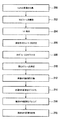
200 マルチ2D画像を収集
202 3Dボリュームを構築
204 エッジ検出
206 解剖学的ランドマークを特定
208 3Dボリュームをセグメント化
210 関心ボリュームを決定
212 準備MRI画像を収集
214 準備MRI画像をセグメント化
216 臨床MRI画像を計画
218 臨床MRI画像を収集
302 心臓を通る冠状MRI画像
304 横隔膜を示す矢状MRI画像
306 ソーベルオペレータで処理された画像302
308 ソーベルオペレータで処理された画像304
310 横隔膜上のメッシュの最終配置を示す画像306
312 横隔膜上のメッシュの最終配置を示す画像308
314 画像304、308及び312に示された矢状面の位置
316 画像302、306及び310に示された冠状面の位置
318 明るい画素
320 メッシュ
400 MRI画像
402 シムボリューム
404 サーベイスタック
406 ナビゲータボリューム
Claims (15)
- 被検体の少なくとも1つの臨床MRI画像を収集する方法であって:
− 第1の視野を有する第1のサーベイ画像を収集するステップであり、該第1のサーベイ画像は第1の空間解像度を有する、ステップと、
− 前記第1のサーベイ画像内で第1の関心領域及び少なくとも1つの解剖学的なランドマークの位置を特定するステップと、
− 前記解剖学的なランドマークを用いて前記第1の関心領域の位置及び向きを決定するステップであり、前記第1の関心領域の該位置及び該向きは、第2のサーベイ画像を計画するために使用される、ステップと、
− 第2の視野を有する前記第2のサーベイ画像を収集するステップであり、前記第2のサーベイ画像は第2の空間解像度を有し、該第2の空間解像度は前記第1の空間解像度より高い、ステップと、
− 前記第2のサーベイ画像を用いて前記解剖学的な関心領域についての配置計画を生成するステップと、
− 前記配置計画を用いて前記解剖学的な関心領域の診断画像を収集するステップと、
を有する方法。 - 第1の視野を有する第1のサーベイ画像を収集する前記ステップは:
− 予め選択されたパラメータセットを用いて前記被検体のマルチ2D MRI画像を収集することを有し、
且つ/或いは、前記第1のサーベイ画像内で第1の関心領域及び少なくとも1つの解剖学的なランドマークの位置を特定する前記ステップは:
− 3Dボリュームモジュールを用いて、前記マルチ2D MRI画像から3Dボリュームを構築することと、
− エッジ検出モジュールを用いて、前記3Dボリューム内でエッジの組を検出することと、
− 前記エッジの組を用い、解剖学的ランドマークモジュールを用いて、前記3Dボリューム内で1つ以上の解剖学的なランドマークを特定することと、
− 第1のセグメント化モジュールを用い、前記1つ以上の解剖学的なランドマークを用いて第1の形状制約変形可能モデルをフィッティングすることによって、前記3Dボリュームを自動的にセグメント化することと、を有し、
且つ/或いは、前記解剖学的なランドマークを用いて前記第1の関心領域の位置及び向きを決定する前記ステップは:
− 第1のパターン認識モジュールを用いて、後のMRIデータの収集に必要な、前記セグメント化された3Dボリューム内の関心ボリュームを決定することを有し、
且つ/或いは、第2の視野を有する前記第2のサーベイ画像を収集する前記ステップは:
− 前記関心ボリュームの1つ以上の準備MRI画像を収集することを有し、
且つ/或いは、前記第2のサーベイ画像を用いて前記解剖学的な関心領域についての配置計画を生成する前記ステップは:
− 第2のセグメント化モジュールを用い、第2の形状制約変形可能モデルを用いて前記1つ以上の準備MRI画像をセグメント化することと、
− 前記セグメント化された1つ以上の準備MRI画像を用い、第2のパターン認識モジュールを用いて、臨床MRI画像を計画するためのプランニングデータの組を生成することと、を有し、
且つ/或いは、前記配置計画を用いて前記解剖学的な関心領域の診断画像を収集する前記ステップは:
− 前記プランニングデータの組を用いて1つ以上の臨床MRI画像を収集することを有する、
請求項1に記載の方法。 - 前記マルチ2D MRI画像、前記3Dボリューム、前記エッジの組、前記1つ以上の解剖学的なランドマーク、前記セグメント化された3Dボリューム、前記関心ボリューム、前記1つ以上の準備MRI画像、及び前記セグメント化された1つ以上の準備MRI画像、のうちの少なくとも1つが、臨床MRI画像を計画するための第2のプランニングデータの組を生成するための再利用のために、コンピュータデータ記憶装置に格納される、請求項2に記載の方法。
- 前記第1のパターン認識モジュールは複数の関心領域を決定し、前記第1のパターン認識モジュールは各関心ボリュームを第1の部分集合、第2の部分集合又は第3の部分集合の何れか内にあるように選択し、前記第1の部分集合の各要素及び前記第3の部分集合の各要素の1つ以上のMRI画像が収集され、前記第2の部分集合の各要素及び前記第3の部分集合の各要素は、磁化プリパレーションパルス、脂肪抑制パルス、血液スピンラベリング(ASL)、リージョナル・サチュレーション・テクニック(REST)、スペクトラル・プレサチュレーション・インバージョン・リカバリー(SPIR)、インバージョン、ナビゲータビーム、B1シミング、及びB0シミングからなる群から選択されたMRI技術を適用され、全ての前記関心ボリュームから収集されたMRIデータが、前記プランニングデータを生成するために前記第2のパターン認識モジュールによって使用される、請求項2又は3に記載の方法。
- 前記第1のパターン認識モジュール及び/又は前記第2のパターン認識モジュールは、訓練型のパターン認識モジュールである、請求項2乃至4の何れか一項に記載の方法。
- 前記解剖学的ランドマークモジュールは、特徴識別アルゴリズムを実装したものである、請求項2乃至5の何れか一項に記載の方法。
- 請求項1乃至6の何れか一項に記載の方法を実行するためのコンピュータ実行可能命令セットを有するコンピュータプログラム。
- 被検体の臨床MRI画像を収集する装置であって:
− 第1の視野を有する第1のサーベイ画像を収集する手段であり、該第1のサーベイ画像は第1の空間解像度を有する、手段と、
− 前記第1のサーベイ画像内で第1の関心領域及び少なくとも1つの解剖学的なランドマークの位置を特定する手段と、
− 前記解剖学的なランドマークを用いて前記第1の関心領域の位置及び向きを決定する手段であり、前記第1の関心領域の該位置及び該向きは、第2のサーベイ画像を計画するために使用される、手段と、
− 第2の視野を有する前記第2のサーベイ画像を収集する手段であり、前記第2のサーベイ画像は第2の空間解像度を有し、該第2の空間解像度は前記第1の空間解像度より高い、手段と、
− 前記第2のサーベイ画像を用いて前記解剖学的な関心領域についての配置計画を生成する手段と、
− 前記配置計画を用いて前記解剖学的な関心領域の診断画像を収集する手段と、
を有する装置。 - 第1の視野を有する第1のサーベイ画像を収集する前記手段は:
− 予め選択されたパラメータセットを用いて前記被検体のマルチ2D MRI画像を収集する手段を有し、
且つ/或いは、前記第1のサーベイ画像内で第1の関心領域及び少なくとも1つの解剖学的なランドマークの位置を特定する前記手段は:
− 3Dボリュームモジュールを用いて、前記マルチ2D MRI画像から3Dボリュームを構築する手段と、
− エッジ検出モジュールを用いて、前記3Dボリューム内でエッジの組を検出する手段と、
− 解剖学的ランドマークモジュールを用いて、前記3Dボリューム内で1つ以上の解剖学的なランドマークを特定する手段と、
− 第1のセグメント化モジュールを用い、前記1つ以上の解剖学的なランドマークを用いて形状制約変形可能モデルをフィッティングすることによって、前記3Dボリュームを自動的にセグメント化する手段と、を有し、
且つ/或いは、前記解剖学的なランドマークを用いて前記第1の関心領域の位置及び向きを決定する前記手段は:
− 解剖学的ランドマークモジュールを用いて、前記3Dボリューム内で1つ以上の解剖学的なランドマークを特定する手段と、
− 第1のセグメント化モジュールを用い、前記1つ以上の解剖学的なランドマークを用いて形状制約変形可能モデルをフィッティングすることによって、前記3Dボリュームを自動的にセグメント化する手段と、
− 第1のパターン認識モジュールを用いて、後のMRIデータの収集に必要な、前記セグメント化された3Dボリューム内の関心ボリュームを決定する手段と、を有し、
且つ/或いは、第2の視野を有する前記第2のサーベイ画像を収集する前記手段は:
− 前記関心ボリュームの1つ以上の準備MRI画像を収集する手段を有し、
且つ/或いは、前記第2のサーベイ画像を用いて前記解剖学的な関心領域についての配置計画を生成する前記手段は:
− 第2のセグメント化モジュールを用い、形状制約変形可能モデルを用いて前記1つ以上の準備MRI画像をセグメント化する手段と、
− 前記セグメント化された1つ以上の準備MRI画像を用い、第2のパターン認識モジュールを用いて、臨床MRI画像を計画するためのプランニングデータの組を生成する手段と、を有し、
且つ/或いは、前記配置計画を用いて前記解剖学的な関心領域の診断画像を収集する前記手段は:
− 前記プランニングデータの組を用いて1つ以上の臨床MRI画像を収集する手段を有する、
請求項8に記載の装置。 - 前記マルチ2D MRI画像、前記3Dボリューム、前記エッジの組、前記1つ以上の解剖学的なランドマーク、前記セグメント化された3Dボリューム、前記関心ボリューム、前記1つ以上の準備MRI画像、及び前記セグメント化された1つ以上の準備MRI画像、のうちの少なくとも1つを、臨床MRI画像を計画するための第2のプランニングデータの組を生成するための再利用のために、コンピュータデータ記憶装置に格納する、ように更に動作する請求項9に記載の装置。
- 前記第1のパターン認識モジュール及び/又は前記第2のパターン認識モジュールは、訓練型のパターン認識モジュールである、請求項9又は10に記載の装置。
- 前記解剖学的ランドマークモジュールは、特徴識別アルゴリズムを実装したものである、請求項9乃至11の何れか一項に記載の装置。
- 前記第1のパターン認識モジュールは複数の関心領域を決定するよう動作し、前記第1のパターン認識モジュールは各関心ボリュームを第1の部分集合、第2の部分集合又は第3の部分集合の何れか内にあるように選択するよう動作し、当該装置は前記第1の部分集合の各要素及び前記第3の部分集合の各要素の1つ以上のMRI画像を収集するよう動作し、当該装置は前記第2の部分集合の各要素及び前記第3の部分集合の各要素に、磁化プリパレーションパルス、脂肪抑制パルス、血液スピンラベリング(ASL)、リージョナル・サチュレーション・テクニック(REST)、スペクトラル・プレサチュレーション・インバージョン・リカバリー(SPIR)、インバージョン、ナビゲータビーム、B1シミング、及びB0シミングからなる群から選択されたMRI技術を適用するよう動作し、全ての前記関心ボリュームから収集されたMRIデータが、前記プランニングデータを生成するために前記第2のパターン認識モジュールによって使用される、請求項9乃至12の何れか一項に記載の装置。
- 前記臨床MRIスキャンが収集される前に前記プランニングデータの組を図形的に表示するよう動作するユーザインタフェースを更に有し、前記ユーザインタフェースは、前記プランニングデータの組を承認あるいは拒絶することを含むオペレータ選択を入力するための入力手段を有し、承認の場合には前記1つ以上の臨床MRI画像の収集が実行され、拒絶の場合には前記ユーザインタフェースは拒絶データの組を表示し、前記拒絶データは、前記マルチ2D画像、前記3Dボリューム、前記エッジ、前記解剖学的なランドマーク、前記セグメント化された3Dボリューム、前記関心ボリューム、前記1つ以上の準備MRI画像、前記セグメント化された1つ以上の準備MRI画像、及び前記プランニングデータ、のうちの少なくとも1つを有する、請求項9乃至13の何れか一項に記載の装置。
- 前記ユーザインタフェースは、前記拒絶データの組の手動変更を受信するよう動作し、変更された拒絶データは、変更されたプランニングデータの組を生成するために使用され、該変更されたプランニングデータの組を用いて1つ以上の臨床MRI画像が収集される、請求項14に記載の装置。
Applications Claiming Priority (3)
| Application Number | Priority Date | Filing Date | Title |
|---|---|---|---|
| EP08168342.7 | 2008-11-05 | ||
| EP08168342 | 2008-11-05 | ||
| PCT/IB2009/054848 WO2010052629A1 (en) | 2008-11-05 | 2009-11-02 | Automated sequential planning of mr scans |
Publications (2)
| Publication Number | Publication Date |
|---|---|
| JP2012507331A true JP2012507331A (ja) | 2012-03-29 |
| JP5667066B2 JP5667066B2 (ja) | 2015-02-12 |
Family
ID=41664549
Family Applications (1)
| Application Number | Title | Priority Date | Filing Date |
|---|---|---|---|
| JP2011533913A Expired - Fee Related JP5667066B2 (ja) | 2008-11-05 | 2009-11-02 | Mrスキャンの自動シーケンシャルプランニング |
Country Status (6)
| Country | Link |
|---|---|
| US (1) | US8565505B2 (ja) |
| EP (1) | EP2347274B1 (ja) |
| JP (1) | JP5667066B2 (ja) |
| CN (1) | CN102203630B (ja) |
| RU (1) | RU2533626C2 (ja) |
| WO (1) | WO2010052629A1 (ja) |
Cited By (10)
| Publication number | Priority date | Publication date | Assignee | Title |
|---|---|---|---|---|
| JP2011194241A (ja) * | 2010-03-18 | 2011-10-06 | General Electric Co <Ge> | Mr撮像スキャンパラメータの自動算定のためのシステム及び方法 |
| JP2013048658A (ja) * | 2011-08-30 | 2013-03-14 | Ge Medical Systems Global Technology Co Llc | 磁気共鳴イメージング装置 |
| JP2013102889A (ja) * | 2011-11-11 | 2013-05-30 | Toshiba Corp | 磁気共鳴イメージング装置 |
| WO2014081022A1 (ja) * | 2012-11-22 | 2014-05-30 | 株式会社東芝 | 磁気共鳴イメージング装置及び磁気共鳴イメージング方法 |
| JP2014108164A (ja) * | 2012-11-30 | 2014-06-12 | Ge Medical Systems Global Technology Co Llc | 検出装置、磁気共鳴装置、検出方法、およびプログラム |
| JP2015000301A (ja) * | 2013-06-18 | 2015-01-05 | 株式会社東芝 | 磁気共鳴イメージング装置及び画像処理プログラム |
| JP2015522374A (ja) * | 2012-07-18 | 2015-08-06 | コーニンクレッカ フィリップス エヌ ヴェ | mDIXON測量からの自動プランニングに基づく効率的な心臓MRのワークフロー |
| JP2016087023A (ja) * | 2014-10-31 | 2016-05-23 | ジーイー・メディカル・システムズ・グローバル・テクノロジー・カンパニー・エルエルシー | 磁気共鳴装置およびプログラム |
| JP2017507755A (ja) * | 2014-03-19 | 2017-03-23 | コーニンクレッカ フィリップス エヌ ヴェKoninklijke Philips N.V. | 磁気共鳴画像診断法を使用して解剖学的細部の壁厚を決定するためのスキャン・ジオメトリ・プランニング方法 |
| JP2024041762A (ja) * | 2018-11-20 | 2024-03-27 | コーニンクレッカ フィリップス エヌ ヴェ | 磁気共鳴イメージングにおける他の処理ロケーションの決定 |
Families Citing this family (68)
| Publication number | Priority date | Publication date | Assignee | Title |
|---|---|---|---|---|
| US8361067B2 (en) | 2002-09-30 | 2013-01-29 | Relievant Medsystems, Inc. | Methods of therapeutically heating a vertebral body to treat back pain |
| US7258690B2 (en) | 2003-03-28 | 2007-08-21 | Relievant Medsystems, Inc. | Windowed thermal ablation probe |
| US6907884B2 (en) | 2002-09-30 | 2005-06-21 | Depay Acromed, Inc. | Method of straddling an intraosseous nerve |
| EP1848333B1 (en) | 2005-01-28 | 2013-10-23 | The Regents of The University of California | Systems and methods using nuclear magnetic resonance (nmr) spectroscopy to evaluate pain and degenerative properties of tissue |
| US11315307B1 (en) | 2006-12-28 | 2022-04-26 | Tipping Point Medical Images, Llc | Method and apparatus for performing rotating viewpoints using a head display unit |
| US11228753B1 (en) | 2006-12-28 | 2022-01-18 | Robert Edwin Douglas | Method and apparatus for performing stereoscopic zooming on a head display unit |
| US10795457B2 (en) | 2006-12-28 | 2020-10-06 | D3D Technologies, Inc. | Interactive 3D cursor |
| US11275242B1 (en) | 2006-12-28 | 2022-03-15 | Tipping Point Medical Images, Llc | Method and apparatus for performing stereoscopic rotation of a volume on a head display unit |
| US10028753B2 (en) | 2008-09-26 | 2018-07-24 | Relievant Medsystems, Inc. | Spine treatment kits |
| CA2957010C (en) | 2008-09-26 | 2017-07-04 | Relievant Medsystems, Inc. | Systems and methods for navigating an instrument through bone |
| US8825131B2 (en) | 2009-10-14 | 2014-09-02 | Nocimed, Llc | MR spectroscopy system and method for diagnosing painful and non-painful intervertebral discs |
| JP5575491B2 (ja) * | 2010-01-14 | 2014-08-20 | 株式会社東芝 | 医療画像診断装置 |
| US9341692B2 (en) * | 2010-04-15 | 2016-05-17 | Regents Of The University Of Minnesota | Magnetization transfer and off-resonance protocols in NMR |
| JP2012061074A (ja) * | 2010-09-15 | 2012-03-29 | Ge Medical Systems Global Technology Co Llc | 磁気共鳴イメージング装置 |
| JP5976293B2 (ja) * | 2010-10-08 | 2016-08-23 | 東芝メディカルシステムズ株式会社 | 画像処理装置及び画像処理方法 |
| JP5944645B2 (ja) * | 2010-11-02 | 2016-07-05 | 東芝メディカルシステムズ株式会社 | 磁気共鳴イメージング装置 |
| US9224240B2 (en) * | 2010-11-23 | 2015-12-29 | Siemens Medical Solutions Usa, Inc. | Depth-based information layering in medical diagnostic ultrasound |
| WO2012071566A2 (en) * | 2010-11-24 | 2012-05-31 | Nocimed, Llc | Systems and methods for automated voxelation of regions of interest for magnetic resonance spectroscopy |
| US8922547B2 (en) * | 2010-12-22 | 2014-12-30 | Electronics And Telecommunications Research Institute | 3D model shape transformation method and apparatus |
| DE102011076776A1 (de) * | 2011-05-31 | 2012-12-06 | Siemens Aktiengesellschaft | Verfahren zu einer Bilddatenaufnahme |
| NL2009885C2 (en) * | 2011-11-30 | 2014-12-09 | Gen Electric | System and method for automated landmarking. |
| WO2013101772A1 (en) | 2011-12-30 | 2013-07-04 | Relievant Medsystems, Inc. | Systems and methods for treating back pain |
| US10395420B2 (en) * | 2012-02-28 | 2019-08-27 | Brainlab Ag | Calculation of a medical image using templates |
| DE102012203782B4 (de) * | 2012-03-12 | 2022-08-11 | Siemens Healthcare Gmbh | Verfahren zur Durchführung einer kombinierten Magnetresonanz-Positronenemissions-Tomographie |
| JP6177874B2 (ja) * | 2012-03-26 | 2017-08-09 | コーニンクレッカ フィリップス エヌ ヴェKoninklijke Philips N.V. | 断面通過ナビゲータ |
| US8965094B2 (en) | 2012-04-14 | 2015-02-24 | Nocimed, Llc | Magnetic resonance spectroscopy pulse sequence, acquisition, and processing system and method |
| CN102707425B (zh) * | 2012-06-21 | 2014-04-16 | 爱威科技股份有限公司 | 图像处理方法和装置 |
| US10588691B2 (en) | 2012-09-12 | 2020-03-17 | Relievant Medsystems, Inc. | Radiofrequency ablation of tissue within a vertebral body |
| DE102012217262B4 (de) * | 2012-09-25 | 2023-10-26 | Siemens Healthcare Gmbh | Verfahren zur automatischen Magnetresonanz-Bildgebung und Magnetresonanzanlage |
| CA2889478C (en) | 2012-11-05 | 2020-11-24 | Relievant Medsystems, Inc. | Systems and methods for creating curved paths through bone and modulating nerves within the bone |
| US9113781B2 (en) * | 2013-02-07 | 2015-08-25 | Siemens Aktiengesellschaft | Method and system for on-site learning of landmark detection models for end user-specific diagnostic medical image reading |
| RU2526752C1 (ru) | 2013-03-18 | 2014-08-27 | Корпорация "САМСУНГ ЭЛЕКТРОНИКС Ко., Лтд." | Система и способ для автоматического планирования двухмерных видов в объемных медицинских изображениях |
| US9724151B2 (en) | 2013-08-08 | 2017-08-08 | Relievant Medsystems, Inc. | Modulating nerves within bone using bone fasteners |
| EP3044605B1 (en) * | 2013-09-09 | 2022-11-16 | Koninklijke Philips N.V. | Push-button vessel wall mri with 3d scout scan |
| JP6440990B2 (ja) * | 2013-09-18 | 2018-12-19 | キヤノンメディカルシステムズ株式会社 | 磁気共鳴イメージング装置 |
| WO2015066458A1 (en) * | 2013-11-01 | 2015-05-07 | The Research Foundation For The State University Of New York | Method for measuring the interior three-dimensional movement, stress and strain of an object |
| US9402549B2 (en) * | 2013-11-27 | 2016-08-02 | General Electric Company | Methods and apparatus to estimate ventricular volumes |
| DE102014204381B4 (de) * | 2014-03-11 | 2017-03-23 | Siemens Healthcare Gmbh | Planung einer Brachytherapie-Behandlung aufgrund von Magnetresonanz-Bilddaten mit hyperintens dargestellten Bereichen |
| CN106133789B (zh) * | 2014-03-21 | 2022-11-18 | 皇家飞利浦有限公司 | 用于分割感兴趣区域的图像处理装置和方法 |
| JP6224560B2 (ja) * | 2014-09-12 | 2017-11-01 | ジーイー・メディカル・システムズ・グローバル・テクノロジー・カンパニー・エルエルシー | 磁気共鳴装置およびプログラム |
| CN107077724B (zh) * | 2014-10-20 | 2022-03-29 | 皇家飞利浦有限公司 | 分类的截断补偿 |
| KR101636876B1 (ko) | 2014-11-03 | 2016-07-06 | 삼성전자주식회사 | 의료 영상 처리 장치 및 방법 |
| DE102015200322A1 (de) * | 2015-01-13 | 2016-01-07 | Siemens Aktiengesellschaft | Verfahren und Vorrichtung zur Feststellung einer Position eines anatomischen Bereichs in einer MR-Anlage |
| RU2614545C1 (ru) * | 2016-01-11 | 2017-03-28 | федеральное государственное бюджетное образовательное учреждение высшего образования "Донской государственный технический университет" (ДГТУ) | Устройство объединения медицинских изображений |
| US10463328B2 (en) * | 2016-05-09 | 2019-11-05 | Canon Medical Systems Corporation | Medical image diagnostic apparatus |
| EP3471608B1 (en) | 2016-06-19 | 2023-08-23 | Aclarion, Inc. | Magnetic resonance spectroscopy system and method for diagnosing pain or infection associated with propionic acid |
| US9965863B2 (en) | 2016-08-26 | 2018-05-08 | Elekta, Inc. | System and methods for image segmentation using convolutional neural network |
| US9947102B2 (en) * | 2016-08-26 | 2018-04-17 | Elekta, Inc. | Image segmentation using neural network method |
| DE102017203025A1 (de) * | 2017-02-24 | 2018-08-30 | Siemens Healthcare Gmbh | Verfahren zu einem Unterstützen einer Planung einer Magnetresonanzuntersuchung an einem Patienten mit einer Magnetresonanzvorrichtung sowie eine Magnetresonanzvorrichtung zu einem Ausführen des Verfahrens |
| US10188361B2 (en) * | 2017-03-27 | 2019-01-29 | Siemens Healthcare Gmbh | System for synthetic display of multi-modality data |
| US11903691B2 (en) | 2017-07-18 | 2024-02-20 | Siemens Healthineers Ag | Combined steering engine and landmarking engine for elbow auto align |
| EP3486674A1 (en) * | 2017-11-17 | 2019-05-22 | Koninklijke Philips N.V. | Artificial intelligence-enabled localization of anatomical landmarks |
| US11486950B2 (en) * | 2018-08-01 | 2022-11-01 | General Electric Company | Systems and methods for automated graphical prescription with deep neural networks |
| US12263017B2 (en) * | 2018-08-01 | 2025-04-01 | General Electric Company | Plane selection using localizer images |
| IT201900000583A1 (it) * | 2019-01-14 | 2020-07-14 | Upsurgeon S R L | Dispositivo di apprendimento medico basato sull'integrazione di realtà fisica e virtuale finalizzato allo studio e alla simulazione degli approcci chirurgici ai distretti anatomici |
| EP4027912B1 (en) | 2019-09-12 | 2024-12-18 | Relievant Medsystems, Inc. | Systems for tissue modulation |
| JP2021074275A (ja) * | 2019-11-08 | 2021-05-20 | キヤノンメディカルシステムズ株式会社 | 撮像支援装置 |
| EP3893013A1 (en) * | 2020-04-06 | 2021-10-13 | Koninklijke Philips N.V. | Mr imaging for radiation therapy planning |
| WO2022011115A1 (en) | 2020-07-10 | 2022-01-13 | Relievant Medsystems, Inc. | Vertebral denervation in conjunction with vertebral fusion |
| US12082876B1 (en) | 2020-09-28 | 2024-09-10 | Relievant Medsystems, Inc. | Introducer drill |
| AU2021409967A1 (en) | 2020-12-22 | 2023-08-03 | Relievant Medsystems, Inc. | Prediction of candidates for spinal neuromodulation |
| EP4302112A1 (en) | 2021-03-04 | 2024-01-10 | Hyperfine Operations, Inc. | Systems and methods for performing magnetic resonance imaging with reduced operator interaction |
| EP4075157A1 (en) * | 2021-04-14 | 2022-10-19 | Siemens Healthcare GmbH | Method for correcting object specific inhomogeneities in an mr imaging system |
| CN115707429A (zh) * | 2021-08-20 | 2023-02-21 | 佳能医疗系统株式会社 | 磁共振成像用脏器分割装置及其方法、磁共振成像装置 |
| US12433668B1 (en) | 2021-11-08 | 2025-10-07 | Relievant Medsystems, Inc. | Impedance stoppage mitigation during radiofrequency tissue ablation procedures |
| KR102516945B1 (ko) * | 2022-03-24 | 2023-04-03 | 주식회사 쓰리디오엔에스 | 인공지능 기반의 두경부 랜드마크 검출 방법 및 장치 |
| US12406358B2 (en) * | 2022-07-01 | 2025-09-02 | GE Precision Healthcare LLC | Methods and systems for automated saturation band placement |
| EP4521339A1 (en) * | 2023-09-05 | 2025-03-12 | Koninklijke Philips N.V. | Automatic navigator echo placement detection |
Citations (4)
| Publication number | Priority date | Publication date | Assignee | Title |
|---|---|---|---|---|
| JPH08289888A (ja) * | 1995-04-24 | 1996-11-05 | Ge Yokogawa Medical Syst Ltd | 断層撮影計画方法及び断層撮影装置 |
| US6195409B1 (en) * | 1998-05-22 | 2001-02-27 | Harbor-Ucla Research And Education Institute | Automatic scan prescription for tomographic imaging |
| US20050165294A1 (en) * | 2003-03-18 | 2005-07-28 | Weiss Kenneth L. | Automated brain MRI and CT prescriptions in Talairach space |
| JP2007534411A (ja) * | 2004-04-26 | 2007-11-29 | コーニンクレッカ フィリップス エレクトロニクス エヌ ヴィ | 磁気共鳴イメージングを計画する装置及び方法 |
Family Cites Families (32)
| Publication number | Priority date | Publication date | Assignee | Title |
|---|---|---|---|---|
| US5320102A (en) * | 1992-11-18 | 1994-06-14 | Ciba-Geigy Corporation | Method for diagnosing proteoglycan deficiency in cartilage based on magnetic resonance image (MRI) |
| CN1131906A (zh) * | 1994-06-14 | 1996-09-25 | 菲利浦电子有限公司 | 脉冲序列最佳化的磁共振成像方法及其装置 |
| US5758646A (en) * | 1994-09-12 | 1998-06-02 | U.S. Philips Corporation | Magnetic resonance imaging method with pulse sequence optimization and device for such method |
| US6064208A (en) * | 1998-04-02 | 2000-05-16 | Picker International, Inc. | Two-peak alignment method of field shimming |
| US6396266B1 (en) * | 1998-11-25 | 2002-05-28 | General Electric Company | MR imaging system with interactive MR geometry prescription control |
| JP4979868B2 (ja) * | 1999-12-29 | 2012-07-18 | オルムコ コーポレイション | 注文歯列矯正装置形成方法および装置 |
| CA2474138A1 (en) * | 2002-01-15 | 2003-07-24 | Vanderbilt University | Composition and imaging methods for pharmacokinetic and pharmacodynamic evaluation of therapeutic delivery system |
| US6963768B2 (en) | 2002-05-16 | 2005-11-08 | General Electric Company | Whole body MRI scanning with moving table and interactive control |
| US20030220558A1 (en) | 2002-05-22 | 2003-11-27 | Reed Busse | Automatic field of view optimization for maximization of resolution and elimination of aliasing artifact |
| US6574304B1 (en) * | 2002-09-13 | 2003-06-03 | Ge Medical Systems Global Technology Company, Llc | Computer aided acquisition of medical images |
| US7260249B2 (en) * | 2002-09-27 | 2007-08-21 | Confirma Incorporated | Rules-based approach for processing medical images |
| US6768782B1 (en) * | 2002-12-16 | 2004-07-27 | University Of Notre Dame Du Lac | Iterative method for region-of-interest reconstruction |
| US7078898B2 (en) * | 2003-04-24 | 2006-07-18 | The Regents Of The University Of California | Methods and apparatus for magnetic resonance imaging |
| DE10331412A1 (de) | 2003-07-10 | 2005-01-27 | Bayerische Motoren Werke Ag | Halterung für ein Mobiltelefon |
| CN1929781A (zh) * | 2003-08-21 | 2007-03-14 | 依斯克姆公司 | 用于脉管斑块检测和分析的自动化方法和系统 |
| US20050165295A1 (en) * | 2004-01-23 | 2005-07-28 | Debiao Li | Local magnetic resonance image quality by optimizing imaging frequency |
| CN1926444A (zh) | 2004-03-01 | 2007-03-07 | 皇家飞利浦电子股份有限公司 | 用于优化采集参数的多合一平面扫描成像 |
| US8014575B2 (en) | 2004-03-11 | 2011-09-06 | Weiss Kenneth L | Automated neuroaxis (brain and spine) imaging with iterative scan prescriptions, analysis, reconstructions, labeling, surface localization and guided intervention |
| US7346383B2 (en) * | 2004-07-08 | 2008-03-18 | Mayo Foundation For Medical Education And Research | Method for acquiring MRI data from variable fields of view during continuous table motion |
| US7280862B2 (en) * | 2004-08-18 | 2007-10-09 | General Electric Company | System and method for automatically obtaining a digital image of a heart |
| JP4886186B2 (ja) * | 2004-11-29 | 2012-02-29 | 株式会社東芝 | 医用機器及び医用データアクセス制御方法 |
| US8913830B2 (en) * | 2005-01-18 | 2014-12-16 | Siemens Aktiengesellschaft | Multilevel image segmentation |
| US7365856B2 (en) * | 2005-01-21 | 2008-04-29 | Carl Zeiss Meditec, Inc. | Method of motion correction in optical coherence tomography imaging |
| CA2610345C (en) * | 2005-06-02 | 2013-12-24 | The Medipattern Corporation | System and method of computer-aided detection |
| RU2297793C2 (ru) * | 2005-06-07 | 2007-04-27 | Государственное учреждение научно-исследовательский институт онкологии Томского научного центра Сибирского отделения Российской академии медицинских наук | Способ компьютерно-томографической диагностики аномалий развития печени |
| DE102005036515B4 (de) * | 2005-08-03 | 2015-07-09 | Siemens Aktiengesellschaft | Verfahren zur Planung einer Untersuchung in einer Magnetresonanzanlage |
| US7876938B2 (en) * | 2005-10-06 | 2011-01-25 | Siemens Medical Solutions Usa, Inc. | System and method for whole body landmark detection, segmentation and change quantification in digital images |
| US20100312734A1 (en) * | 2005-10-07 | 2010-12-09 | Bernard Widrow | System and method for cognitive memory and auto-associative neural network based pattern recognition |
| US8082019B2 (en) * | 2006-09-20 | 2011-12-20 | Siemens Medical Solutions Usa, Inc. | System and method for magnetic resonance brain scan planning |
| US20080146910A1 (en) * | 2006-11-22 | 2008-06-19 | Todd Parrish | Non-invasive method for determining body composition using magnetic resonance (MR) |
| US20100189328A1 (en) * | 2007-05-31 | 2010-07-29 | Koninklijke Philips Electronics N.V. | Method of automatically acquiring magnetic resonance image data |
| EP2194486A1 (en) * | 2008-12-04 | 2010-06-09 | Koninklijke Philips Electronics N.V. | A method, apparatus, and computer program product for acquiring medical image data |
-
2009
- 2009-11-02 WO PCT/IB2009/054848 patent/WO2010052629A1/en not_active Ceased
- 2009-11-02 RU RU2011122651/08A patent/RU2533626C2/ru active
- 2009-11-02 US US13/126,274 patent/US8565505B2/en not_active Expired - Fee Related
- 2009-11-02 EP EP09756846.3A patent/EP2347274B1/en not_active Not-in-force
- 2009-11-02 JP JP2011533913A patent/JP5667066B2/ja not_active Expired - Fee Related
- 2009-11-02 CN CN200980143939.0A patent/CN102203630B/zh not_active Expired - Fee Related
Patent Citations (4)
| Publication number | Priority date | Publication date | Assignee | Title |
|---|---|---|---|---|
| JPH08289888A (ja) * | 1995-04-24 | 1996-11-05 | Ge Yokogawa Medical Syst Ltd | 断層撮影計画方法及び断層撮影装置 |
| US6195409B1 (en) * | 1998-05-22 | 2001-02-27 | Harbor-Ucla Research And Education Institute | Automatic scan prescription for tomographic imaging |
| US20050165294A1 (en) * | 2003-03-18 | 2005-07-28 | Weiss Kenneth L. | Automated brain MRI and CT prescriptions in Talairach space |
| JP2007534411A (ja) * | 2004-04-26 | 2007-11-29 | コーニンクレッカ フィリップス エレクトロニクス エヌ ヴィ | 磁気共鳴イメージングを計画する装置及び方法 |
Non-Patent Citations (1)
| Title |
|---|
| LELLEVELDT B P F ET AL: "Automated Observer-independent Acquisition of Cardiac Short-Axis MR Images: A Pilot Study", RADIOLOGY, vol. 221, no. 2, JPN6014011539, 10 November 2001 (2001-11-10), US, pages 537 - 542, XP002329504, ISSN: 0002772034, DOI: 10.1148/radiol.2212010177 * |
Cited By (14)
| Publication number | Priority date | Publication date | Assignee | Title |
|---|---|---|---|---|
| JP2011194241A (ja) * | 2010-03-18 | 2011-10-06 | General Electric Co <Ge> | Mr撮像スキャンパラメータの自動算定のためのシステム及び方法 |
| JP2013048658A (ja) * | 2011-08-30 | 2013-03-14 | Ge Medical Systems Global Technology Co Llc | 磁気共鳴イメージング装置 |
| JP2013102889A (ja) * | 2011-11-11 | 2013-05-30 | Toshiba Corp | 磁気共鳴イメージング装置 |
| JP2015522374A (ja) * | 2012-07-18 | 2015-08-06 | コーニンクレッカ フィリップス エヌ ヴェ | mDIXON測量からの自動プランニングに基づく効率的な心臓MRのワークフロー |
| JP2018094451A (ja) * | 2012-11-22 | 2018-06-21 | キヤノンメディカルシステムズ株式会社 | 磁気共鳴イメージング装置、磁気共鳴イメージング方法及び画像処理システム |
| WO2014081022A1 (ja) * | 2012-11-22 | 2014-05-30 | 株式会社東芝 | 磁気共鳴イメージング装置及び磁気共鳴イメージング方法 |
| JP2014121597A (ja) * | 2012-11-22 | 2014-07-03 | Toshiba Corp | 磁気共鳴イメージング装置及び磁気共鳴イメージング方法 |
| US20160054417A1 (en) | 2012-11-22 | 2016-02-25 | Kabushiki Kaisha Toshiba | Magnetic resonance imaging apparatus, magnetic resonance imaging method and magnetic resonance imaging system |
| US10338177B2 (en) | 2012-11-22 | 2019-07-02 | Toshiba Medical Systems Corporation | Magnetic resonance imaging apparatus, magnetic resonance imaging method and magnetic resonance imaging system |
| JP2014108164A (ja) * | 2012-11-30 | 2014-06-12 | Ge Medical Systems Global Technology Co Llc | 検出装置、磁気共鳴装置、検出方法、およびプログラム |
| JP2015000301A (ja) * | 2013-06-18 | 2015-01-05 | 株式会社東芝 | 磁気共鳴イメージング装置及び画像処理プログラム |
| JP2017507755A (ja) * | 2014-03-19 | 2017-03-23 | コーニンクレッカ フィリップス エヌ ヴェKoninklijke Philips N.V. | 磁気共鳴画像診断法を使用して解剖学的細部の壁厚を決定するためのスキャン・ジオメトリ・プランニング方法 |
| JP2016087023A (ja) * | 2014-10-31 | 2016-05-23 | ジーイー・メディカル・システムズ・グローバル・テクノロジー・カンパニー・エルエルシー | 磁気共鳴装置およびプログラム |
| JP2024041762A (ja) * | 2018-11-20 | 2024-03-27 | コーニンクレッカ フィリップス エヌ ヴェ | 磁気共鳴イメージングにおける他の処理ロケーションの決定 |
Also Published As
| Publication number | Publication date |
|---|---|
| US20110206260A1 (en) | 2011-08-25 |
| RU2533626C2 (ru) | 2014-11-20 |
| CN102203630B (zh) | 2014-07-02 |
| EP2347274B1 (en) | 2019-08-07 |
| RU2011122651A (ru) | 2012-12-20 |
| EP2347274A1 (en) | 2011-07-27 |
| US8565505B2 (en) | 2013-10-22 |
| WO2010052629A1 (en) | 2010-05-14 |
| CN102203630A (zh) | 2011-09-28 |
| JP5667066B2 (ja) | 2015-02-12 |
Similar Documents
| Publication | Publication Date | Title |
|---|---|---|
| JP5667066B2 (ja) | Mrスキャンの自動シーケンシャルプランニング | |
| CN102239495B (zh) | 用于采集医学图像数据的方法、装置和计算机程序产品 | |
| US12029543B2 (en) | Medical image diagnostic apparatus and medical image display apparatus for MRI or X-ray CT volume image correlations | |
| JP5230809B2 (ja) | 医用画像撮影装置 | |
| CA2926958C (en) | Flow analysis in 4d mr image data | |
| JP4717427B2 (ja) | 磁気共鳴断層撮影装置の作動方法および制御装置 | |
| JP5944645B2 (ja) | 磁気共鳴イメージング装置 | |
| KR101652387B1 (ko) | 영상 데이터를 생성하는 방법 | |
| JP5971682B2 (ja) | 磁気共鳴イメージング装置 | |
| EP3605548B1 (en) | SYSTEMS AND METHODS FOR AUTOMATED GRAPHIC PRESCRIPTION WITH DEEP NERICAL NETWORKS | |
| EP3477324A1 (en) | Improving left ventricle segmentation in contrast-enhanced cine mri datasets | |
| US8781552B2 (en) | Localization of aorta and left atrium from magnetic resonance imaging | |
| JP2005169118A (ja) | 断層画像データのセグメント化方法および画像処理システム | |
| CN1804866B (zh) | 在对象的平面图像上对齐图形对象的方法 | |
| EP3798661B1 (en) | Mri method to determine a susceptibility distribution of an examination subject | |
| US7961920B2 (en) | Method for the computer-assisted visualization of diagnostic image data | |
| US12406358B2 (en) | Methods and systems for automated saturation band placement |
Legal Events
| Date | Code | Title | Description |
|---|---|---|---|
| A621 | Written request for application examination |
Free format text: JAPANESE INTERMEDIATE CODE: A621 Effective date: 20121101 |
|
| A977 | Report on retrieval |
Free format text: JAPANESE INTERMEDIATE CODE: A971007 Effective date: 20140116 |
|
| A131 | Notification of reasons for refusal |
Free format text: JAPANESE INTERMEDIATE CODE: A131 Effective date: 20140325 |
|
| A521 | Request for written amendment filed |
Free format text: JAPANESE INTERMEDIATE CODE: A523 Effective date: 20140617 |
|
| TRDD | Decision of grant or rejection written | ||
| A01 | Written decision to grant a patent or to grant a registration (utility model) |
Free format text: JAPANESE INTERMEDIATE CODE: A01 Effective date: 20141118 |
|
| A61 | First payment of annual fees (during grant procedure) |
Free format text: JAPANESE INTERMEDIATE CODE: A61 Effective date: 20141211 |
|
| R150 | Certificate of patent or registration of utility model |
Ref document number: 5667066 Country of ref document: JP Free format text: JAPANESE INTERMEDIATE CODE: R150 |
|
| R250 | Receipt of annual fees |
Free format text: JAPANESE INTERMEDIATE CODE: R250 |
|
| R250 | Receipt of annual fees |
Free format text: JAPANESE INTERMEDIATE CODE: R250 |
|
| R250 | Receipt of annual fees |
Free format text: JAPANESE INTERMEDIATE CODE: R250 |
|
| R250 | Receipt of annual fees |
Free format text: JAPANESE INTERMEDIATE CODE: R250 |
|
| R250 | Receipt of annual fees |
Free format text: JAPANESE INTERMEDIATE CODE: R250 |
|
| R250 | Receipt of annual fees |
Free format text: JAPANESE INTERMEDIATE CODE: R250 |
|
| R250 | Receipt of annual fees |
Free format text: JAPANESE INTERMEDIATE CODE: R250 |
|
| LAPS | Cancellation because of no payment of annual fees |