JP2014207902A - Peptide for apoptotic cell target and application thereof - Google Patents
Peptide for apoptotic cell target and application thereof Download PDFInfo
- Publication number
- JP2014207902A JP2014207902A JP2014082162A JP2014082162A JP2014207902A JP 2014207902 A JP2014207902 A JP 2014207902A JP 2014082162 A JP2014082162 A JP 2014082162A JP 2014082162 A JP2014082162 A JP 2014082162A JP 2014207902 A JP2014207902 A JP 2014207902A
- Authority
- JP
- Japan
- Prior art keywords
- peptide
- cancer
- present
- composition
- group
- Prior art date
- Legal status (The legal status is an assumption and is not a legal conclusion. Google has not performed a legal analysis and makes no representation as to the accuracy of the status listed.)
- Granted
Links
Images



Classifications
-
- C—CHEMISTRY; METALLURGY
- C07—ORGANIC CHEMISTRY
- C07K—PEPTIDES
- C07K7/00—Peptides having 5 to 20 amino acids in a fully defined sequence; Derivatives thereof
- C07K7/04—Linear peptides containing only normal peptide links
- C07K7/06—Linear peptides containing only normal peptide links having 5 to 11 amino acids
-
- A—HUMAN NECESSITIES
- A61—MEDICAL OR VETERINARY SCIENCE; HYGIENE
- A61K—PREPARATIONS FOR MEDICAL, DENTAL OR TOILETRY PURPOSES
- A61K31/00—Medicinal preparations containing organic active ingredients
- A61K31/33—Heterocyclic compounds
- A61K31/335—Heterocyclic compounds having oxygen as the only ring hetero atom, e.g. fungichromin
- A61K31/35—Heterocyclic compounds having oxygen as the only ring hetero atom, e.g. fungichromin having six-membered rings with one oxygen as the only ring hetero atom
- A61K31/352—Heterocyclic compounds having oxygen as the only ring hetero atom, e.g. fungichromin having six-membered rings with one oxygen as the only ring hetero atom condensed with carbocyclic rings, e.g. methantheline
-
- A—HUMAN NECESSITIES
- A61—MEDICAL OR VETERINARY SCIENCE; HYGIENE
- A61K—PREPARATIONS FOR MEDICAL, DENTAL OR TOILETRY PURPOSES
- A61K47/00—Medicinal preparations characterised by the non-active ingredients used, e.g. carriers or inert additives; Targeting or modifying agents chemically bound to the active ingredient
- A61K47/50—Medicinal preparations characterised by the non-active ingredients used, e.g. carriers or inert additives; Targeting or modifying agents chemically bound to the active ingredient the non-active ingredient being chemically bound to the active ingredient, e.g. polymer-drug conjugates
- A61K47/51—Medicinal preparations characterised by the non-active ingredients used, e.g. carriers or inert additives; Targeting or modifying agents chemically bound to the active ingredient the non-active ingredient being chemically bound to the active ingredient, e.g. polymer-drug conjugates the non-active ingredient being a modifying agent
- A61K47/62—Medicinal preparations characterised by the non-active ingredients used, e.g. carriers or inert additives; Targeting or modifying agents chemically bound to the active ingredient the non-active ingredient being chemically bound to the active ingredient, e.g. polymer-drug conjugates the non-active ingredient being a modifying agent the modifying agent being a protein, peptide or polyamino acid
- A61K47/64—Drug-peptide, drug-protein or drug-polyamino acid conjugates, i.e. the modifying agent being a peptide, protein or polyamino acid which is covalently bonded or complexed to a therapeutically active agent
-
- A—HUMAN NECESSITIES
- A61—MEDICAL OR VETERINARY SCIENCE; HYGIENE
- A61K—PREPARATIONS FOR MEDICAL, DENTAL OR TOILETRY PURPOSES
- A61K49/00—Preparations for testing in vivo
- A61K49/001—Preparation for luminescence or biological staining
- A61K49/0013—Luminescence
- A61K49/0017—Fluorescence in vivo
- A61K49/0019—Fluorescence in vivo characterised by the fluorescent group, e.g. oligomeric, polymeric or dendritic molecules
- A61K49/0021—Fluorescence in vivo characterised by the fluorescent group, e.g. oligomeric, polymeric or dendritic molecules the fluorescent group being a small organic molecule
- A61K49/0032—Methine dyes, e.g. cyanine dyes
-
- A—HUMAN NECESSITIES
- A61—MEDICAL OR VETERINARY SCIENCE; HYGIENE
- A61K—PREPARATIONS FOR MEDICAL, DENTAL OR TOILETRY PURPOSES
- A61K49/00—Preparations for testing in vivo
- A61K49/001—Preparation for luminescence or biological staining
- A61K49/0013—Luminescence
- A61K49/0017—Fluorescence in vivo
- A61K49/0019—Fluorescence in vivo characterised by the fluorescent group, e.g. oligomeric, polymeric or dendritic molecules
- A61K49/0021—Fluorescence in vivo characterised by the fluorescent group, e.g. oligomeric, polymeric or dendritic molecules the fluorescent group being a small organic molecule
- A61K49/0041—Xanthene dyes, used in vivo, e.g. administered to a mice, e.g. rhodamines, rose Bengal
- A61K49/0043—Fluorescein, used in vivo
-
- A—HUMAN NECESSITIES
- A61—MEDICAL OR VETERINARY SCIENCE; HYGIENE
- A61K—PREPARATIONS FOR MEDICAL, DENTAL OR TOILETRY PURPOSES
- A61K49/00—Preparations for testing in vivo
- A61K49/001—Preparation for luminescence or biological staining
- A61K49/0013—Luminescence
- A61K49/0017—Fluorescence in vivo
- A61K49/005—Fluorescence in vivo characterised by the carrier molecule carrying the fluorescent agent
- A61K49/0056—Peptides, proteins, polyamino acids
-
- A—HUMAN NECESSITIES
- A61—MEDICAL OR VETERINARY SCIENCE; HYGIENE
- A61P—SPECIFIC THERAPEUTIC ACTIVITY OF CHEMICAL COMPOUNDS OR MEDICINAL PREPARATIONS
- A61P35/00—Antineoplastic agents
-
- A—HUMAN NECESSITIES
- A61—MEDICAL OR VETERINARY SCIENCE; HYGIENE
- A61P—SPECIFIC THERAPEUTIC ACTIVITY OF CHEMICAL COMPOUNDS OR MEDICINAL PREPARATIONS
- A61P35/00—Antineoplastic agents
- A61P35/02—Antineoplastic agents specific for leukemia
-
- A—HUMAN NECESSITIES
- A61—MEDICAL OR VETERINARY SCIENCE; HYGIENE
- A61P—SPECIFIC THERAPEUTIC ACTIVITY OF CHEMICAL COMPOUNDS OR MEDICINAL PREPARATIONS
- A61P7/00—Drugs for disorders of the blood or the extracellular fluid
- A61P7/06—Antianaemics
-
- A—HUMAN NECESSITIES
- A61—MEDICAL OR VETERINARY SCIENCE; HYGIENE
- A61P—SPECIFIC THERAPEUTIC ACTIVITY OF CHEMICAL COMPOUNDS OR MEDICINAL PREPARATIONS
- A61P9/00—Drugs for disorders of the cardiovascular system
-
- A—HUMAN NECESSITIES
- A61—MEDICAL OR VETERINARY SCIENCE; HYGIENE
- A61P—SPECIFIC THERAPEUTIC ACTIVITY OF CHEMICAL COMPOUNDS OR MEDICINAL PREPARATIONS
- A61P9/00—Drugs for disorders of the cardiovascular system
- A61P9/10—Drugs for disorders of the cardiovascular system for treating ischaemic or atherosclerotic diseases, e.g. antianginal drugs, coronary vasodilators, drugs for myocardial infarction, retinopathy, cerebrovascula insufficiency, renal arteriosclerosis
-
- A—HUMAN NECESSITIES
- A61—MEDICAL OR VETERINARY SCIENCE; HYGIENE
- A61K—PREPARATIONS FOR MEDICAL, DENTAL OR TOILETRY PURPOSES
- A61K2121/00—Preparations for use in therapy
-
- A—HUMAN NECESSITIES
- A61—MEDICAL OR VETERINARY SCIENCE; HYGIENE
- A61K—PREPARATIONS FOR MEDICAL, DENTAL OR TOILETRY PURPOSES
- A61K38/00—Medicinal preparations containing peptides
-
- C—CHEMISTRY; METALLURGY
- C07—ORGANIC CHEMISTRY
- C07K—PEPTIDES
- C07K2319/00—Fusion polypeptide
- C07K2319/33—Fusion polypeptide fusions for targeting to specific cell types, e.g. tissue specific targeting, targeting of a bacterial subspecies
-
- G—PHYSICS
- G01—MEASURING; TESTING
- G01N—INVESTIGATING OR ANALYSING MATERIALS BY DETERMINING THEIR CHEMICAL OR PHYSICAL PROPERTIES
- G01N2510/00—Detection of programmed cell death, i.e. apoptosis
Landscapes
- Health & Medical Sciences (AREA)
- Life Sciences & Earth Sciences (AREA)
- General Health & Medical Sciences (AREA)
- Chemical & Material Sciences (AREA)
- Engineering & Computer Science (AREA)
- Public Health (AREA)
- Veterinary Medicine (AREA)
- Animal Behavior & Ethology (AREA)
- Medicinal Chemistry (AREA)
- Organic Chemistry (AREA)
- Bioinformatics & Cheminformatics (AREA)
- Proteomics, Peptides & Aminoacids (AREA)
- Pharmacology & Pharmacy (AREA)
- Epidemiology (AREA)
- Chemical Kinetics & Catalysis (AREA)
- General Chemical & Material Sciences (AREA)
- Nuclear Medicine, Radiotherapy & Molecular Imaging (AREA)
- Biomedical Technology (AREA)
- Molecular Biology (AREA)
- Biophysics (AREA)
- Heart & Thoracic Surgery (AREA)
- Cardiology (AREA)
- Genetics & Genomics (AREA)
- Biochemistry (AREA)
- Vascular Medicine (AREA)
- Urology & Nephrology (AREA)
- Hematology (AREA)
- Oncology (AREA)
- Diabetes (AREA)
- Medicines That Contain Protein Lipid Enzymes And Other Medicines (AREA)
- Peptides Or Proteins (AREA)
- Measuring Or Testing Involving Enzymes Or Micro-Organisms (AREA)
- Micro-Organisms Or Cultivation Processes Thereof (AREA)
- Medicinal Preparation (AREA)
- Medicines Containing Antibodies Or Antigens For Use As Internal Diagnostic Agents (AREA)
Abstract
【課題】アポトーシスが進行されているアポトーシス細胞を特異的に標的とし得るペプチド及びアポトーシス細胞の検出並びにこれらの用途の提供。【解決手段】特定のアミノ酸配列を有し、アポトーシス細胞を標的とするペプチド、そのペプチドを有効成分とし発色酵素,放射性同位元素,クロモア、発光物質、常磁性粒子等の標識材により標識されたアポトーシス細胞検出用組成物、薬物伝達用組成物、及び映像化用組成物等。【効果】ペプチドは腫瘍、心筋梗塞、脳卒中及び動脈硬化等の組織に発生するアポトーシスを効果的に標的として、該ペプチドは治療物質と結合して、前記疾患組織に薬物を選択的に伝達する疾患の予防及び治療用組成物等の目的で使用でき、また映像物質と結合して前記疾患の診断及び治療薬物に対する反応の映像化等に広く使用できる。【選択図】なしPROBLEM TO BE SOLVED: To provide a peptide capable of specifically targeting apoptotic cells in which apoptosis is progressing, detection of apoptotic cells, and use thereof. A peptide having a specific amino acid sequence and targeting apoptotic cells, and using the peptide as an active ingredient, apoptosis labeled with a labeling material such as a chromogenic enzyme, a radioisotope, chromoa, a luminescent substance, or a paramagnetic particle Cell detection composition, drug delivery composition, imaging composition and the like. [Effects] Peptides effectively target apoptosis occurring in tissues such as tumors, myocardial infarction, stroke and arteriosclerosis, and the peptides bind to therapeutic substances and selectively transmit drugs to the diseased tissues It can be used for the purpose of prevention and treatment compositions, etc., and can be widely used for diagnosing the disease and imaging the response to therapeutic drugs by combining with imaging substances. [Selection figure] None
Description
本出願は2008年5月14日出願された大韓民国特許出願第10−2008−0044410号を優先権主張し、前記明細書全体は本発明の参考文献である。 This application claims Korean patent application No. 10-2008-0044410 filed on May 14, 2008, and the entire specification is a reference for the present invention.
本発明はアポトーシス(apoptosis)が進行しているアポトーシス細胞(apoptotic cells)を特異的に標的とし得るペプチド及びその用途に関する。より詳しくいえば、配列番号1〜12からなる群より選ばれた配列で示されるアミノ酸配列を有し、アポトーシス細胞を標的とするペプチド、そのペプチドを有効成分として含むアポトーシス細胞検出用組成物、そのペプチドを有効成分として含む薬物伝達用組成物、及びそのペプチドを有効成分として含む映像化用組成物等に関する。 The present invention relates to a peptide capable of specifically targeting apoptotic cells in which apoptosis is progressing and a use thereof. More specifically, a peptide having an amino acid sequence represented by a sequence selected from the group consisting of SEQ ID NOs: 1 to 12 and targeting apoptotic cells, a composition for detecting apoptotic cells containing the peptide as an active ingredient, The present invention relates to a drug delivery composition containing a peptide as an active ingredient, an imaging composition containing the peptide as an active ingredient, and the like.
アポトーシスとは、個体の生命維持のために、不要な細胞または危険な細胞を自ら死なせる現象をいう、アポトーシスとはギリシャ語で“落ちる”ことを意味し、細胞が消失される過程を花から花びらが落ちることに例えて付けられた名称であり、1972年Kerr等により初めて観察された(Kerr et al., Br J Cancer, 1972, 26: 239-257)。アポトーシスは細胞の発生、分化、免疫等の生理学的現象に重要な役割をする(Meier et al., Nature, 2000, 407: 796-801)。一方、多様な病理学的環境及び疾患でもアポトーシスは重要である。例えば、抗癌剤で成功的な治療をする場合、多量のアポトーシスが腫瘍組織内に発生する(Thomson, Science, 1995, 267: 1456-1462)。反面、腫瘍の発生にはアポトーシスの減少が原因として関与する。さらに、別の例としては、脳卒中及び心筋梗塞において、脳及び心臓に流れる血液供給の不足により、脳細胞及び心筋細胞がそれぞれ損傷を受け、アポトーシスが発生する(Du et al., J Cereb Blood Flow Metab, 1996, 16: 195-201; Narula et al., New Engl J Med, 1996, 335: 1182-1189)。さらに、臓器移植後拒否反応、自己免疫疾患、退行性脳神経疾患、動脈硬化及びウィルス感染等の疾患でもアポトーシスが容易に発生する(Thomson, Science, 1995, 267: 1456-1462; Kageyama et al., Ann Thorac Surg, 1998, 65: 1604-1609)。 Apoptosis is a phenomenon in which unnecessary or dangerous cells die by themselves to maintain the life of an individual. Apoptosis means “falling down” in Greek, and the process of cell loss from flowers. The name given to the petal falling, first observed by Kerr et al. In 1972 (Kerr et al., Br J Cancer, 1972, 26: 239-257). Apoptosis plays an important role in physiological phenomena such as cell development, differentiation, and immunity (Meier et al., Nature, 2000, 407: 796-801). On the other hand, apoptosis is important in various pathological environments and diseases. For example, when successfully treated with anticancer drugs, a large amount of apoptosis occurs in the tumor tissue (Thomson, Science, 1995, 267: 1456-1462). On the other hand, tumor development is associated with decreased apoptosis. As another example, in stroke and myocardial infarction, lack of blood supply to the brain and heart damages brain cells and cardiomyocytes, respectively, and apoptosis occurs (Du et al., J Cereb Blood Flow Metab, 1996, 16: 195-201; Narula et al., New Engl J Med, 1996, 335: 1182-1189). Furthermore, apoptosis easily occurs even in diseases such as rejection after organ transplantation, autoimmune disease, degenerative cranial nerve disease, arteriosclerosis and viral infection (Thomson, Science, 1995, 267: 1456-1462; Kageyama et al., Ann Thorac Surg, 1998, 65: 1604-1609).
このようなアポトーシスは、臨床的な診断及び治療の面で極めて重要な意味を有する。つまり、アポトーシスの映像化は、アポトーシスの過度な増加と関連した退行性脳神経疾患(アルツハイマー病及びパーキンソン氏病等)の早期診断、心筋梗塞及び脳卒中における症の進行程度モニタリング、抗癌剤処理による癌の治療効果モニタリング、動脈硬化における動脈硬化プラクの破裂可能性判断等の場合に極めて大きな役割を担う。さらに、アポトーシスが旺盛な部位を標的に、治療剤や細胞を保護する製剤を選択的に伝達できるようになれば副作用を減らしながら治療効果をはるかに高め得る。 Such apoptosis is extremely important in terms of clinical diagnosis and treatment. In other words, imaging of apoptosis is an early diagnosis of degenerative cranial nerve diseases (such as Alzheimer's disease and Parkinson's disease) associated with excessive increase of apoptosis, monitoring of the degree of progression of myocardial infarction and stroke, treatment of cancer by treatment with anticancer drugs It plays an extremely important role in monitoring effects, determining the possibility of atherosclerotic plaque rupture in arteriosclerosis, and the like. Furthermore, if it becomes possible to selectively transmit a therapeutic agent or a preparation that protects cells targeting a site where apoptosis is active, the therapeutic effect can be greatly enhanced while reducing side effects.
アポトーシス細胞で起きる初期事象の一つは、細胞膜を構成するリン脂質(phospholipid)の分布変化である。この内最も特徴的なものはホスファチジルセリン(phosphatidylserine)の細胞膜外側への露出である。正常な状態ではホスファチジルセリンは細胞膜の内部に存在するものの、細胞が死滅信号を受けるかまたは赤血球が老化すると細胞膜の外部に露出される(Fadeel, B. et al., Cell Mol Life Sci, 2003, 60: 2575-2585)。このようなホスファチジルセリンを大食細胞が細胞表面の受容体を介して認識してアポトーシス細胞に対する貪食作用を起こす(Fadok, V. A. et al., J immunol 1992, 148: 2207-2216; Fadok, V. A. et al., Nature 2000, 405: 85-90; Park, S. Y. et al., Cell Death Differ, 2008, 15: 192-201)。特に、多くの腫瘍細胞等が細胞膜の外部にホスファチジルセリンの発現増加を示す(Utsugi, T. et al., Cancer Res. 1991, 15: 3062-3066; Ran, S. et al., Cancer Res. 2002, 62: 6132-6140; Woehlecke, H. et al., Biochem J, 2003, 376: 489-495)。さらに、腫瘍組織内小血管の血管内皮細胞は、ホスファチジルセリンを細胞膜の外部に露出している(Ran, S.et al., Cancer Res. 2002, 62: 6132-6140; Zwaal, R. F. A. et al., Blood. 1997, 89: 1121-1132)。従って、このようなホスファチジルセリンの役割等により、特に腫瘍をはじめとした多様な状況において、ホスファチジルセリンは診断、治療、及び治療追跡のための標的物質と考えられている。 One of the initial events that occurs in apoptotic cells is a change in the distribution of phospholipids that make up the cell membrane. The most characteristic of these is the exposure of phosphatidylserine to the outside of the cell membrane. Under normal conditions, phosphatidylserine exists inside the cell membrane, but is exposed outside the cell membrane when the cell receives a death signal or erythrocytes age (Fadeel, B. et al., Cell Mol Life Sci, 2003, 60: 2575-2585). Macrophages recognize these phosphatidylserine via cell surface receptors and cause phagocytosis on apoptotic cells (Fadok, VA et al., J immunol 1992, 148: 2207-2216; Fadok, VA et al., Nature 2000, 405: 85-90; Park, SY et al., Cell Death Differ, 2008, 15: 192-201). In particular, many tumor cells and the like show increased expression of phosphatidylserine outside the cell membrane (Utsugi, T. et al., Cancer Res. 1991, 15: 3062-3066; Ran, S. et al., Cancer Res. 2002, 62: 6132-6140; Woehlecke, H. et al., Biochem J, 2003, 376: 489-495). Furthermore, vascular endothelial cells of small blood vessels in tumor tissue expose phosphatidylserine outside the cell membrane (Ran, S. et al., Cancer Res. 2002, 62: 6132-6140; Zwaal, RFA et al. , Blood. 1997, 89: 1121-1132). Therefore, phosphatidylserine is considered to be a target substance for diagnosis, treatment, and treatment follow-up, particularly in various situations including tumors, due to the role of phosphatidylserine.
現在アポトーシス細胞表面のホスファチジルセリンの検出に主に使用されるのは、アネキシンV(annexin V)蛋白質である。これは分子量36kDaの蛋白質であり、ホスファチジルセリンと強い親和度(affinity)で結合する(Vermes, I. et al., Immunol Methods. 1995, 184: 39-51)。一方、アネキシンVは試験官(in vitro)内では極めて有用な標的物質またはプローブ(probe)ではあるものの、大きい分子量のため、体外への除去が遅れる等の問題により、生体(in vivo)内の使用は制限的であると報告された(Vermeersch, H., et al., Nucl Med Commun. 2004, 25: 259-263:非特許文献1; Belhocine, T. Z. et al., J Proteome Res. 2004, 3: 345-349:非特許文献2)。
At present, the annexin V protein is mainly used for the detection of phosphatidylserine on the surface of apoptotic cells. This is a protein with a molecular weight of 36 kDa and binds to phosphatidylserine with strong affinity (Vermes, I. et al., Immunol Methods. 1995, 184: 39-51). On the other hand, annexin V is a very useful target substance or probe within the inspector (in vitro), but due to the large molecular weight, removal from the body is delayed, causing problems in vivo (in vivo). Use has been reported to be limited (Vermeersch, H., et al., Nucl Med Commun. 2004, 25: 259-263: Non-Patent
本発明の目的は、アポトーシス細胞を特異的に標的するペプチド及びこれの用途を提供することにある。 It is an object of the present invention to provide peptides that specifically target apoptotic cells and uses thereof.
本発明者等はアポトーシス細胞を特異的に、初期に生体内標的化し得る新たな蛋白質またはその断片を探索するために研究した結果、配列番号1〜12からなる群より選ばれた配列で示されるアミノ酸配列を有するペプチドが、アポトーシス細胞を特異的に標的とできることを確認して、本発明を完成した。
すなわち、本発明は下記のペプチド、ポリヌクレオチド、ベクター、形質転換体、アポトーシス細胞検出用組成物、アポトーシス細胞の検出方法、薬物伝達用組成物及び薬物伝達方法、腫瘍性疾患予防・治療用組成物、脳卒中予防・治療用組成物、心筋梗塞予防・治療用組成物、動脈硬化予防・治療用組成物、疾患部位の映像化用組成物及び映像化方法等を提供する
[1]配列番号1〜12からなる群より選ばれた配列で示されるアミノ酸配列を有し、アポトーシス細胞(apoptotic cell)と特異的に結合するペプチド。
[2]前項1に記載のペプチドをコードする塩基配列を有するポリヌクレオチド。
[3]前項2に記載のペプチドを含むベクター。
[4]前項3に記載のベクターで形質転換された形質転換体。
[5]前項1に記載のペプチドを有効成分として含むアポトーシス細胞検出用組成物。
[6]前記ペプチドが、発色酵素、放射性同位元素、クロモホアー(chromophore)、発光物質、蛍光物質(fluorescer)、常磁性粒子(super paramagnetic particles)及び超常磁性粒子(ultrasuper paramagnetic particles)からなる群より選ばれる一つで標識される前項5に記載の組成物。
[7](a)前項1に記載のペプチドを試料と混合する工程;
(b)未結合かもしくは非特異的に結合した前記ペプチドを除去する工程;及び
(c)前記ペプチドの結合可否及び位置を確認する工程を含むアポトーシス細胞の検出方法。
[8]前項1に記載のペプチドを有効成分として含む薬物伝達用組成物。
[9]腫瘍性疾患、脳卒中、心筋梗塞、及び動脈硬化からなる群より選ばれた疾患に特異的である前項8に記載の組成物。
[10]前記腫瘍性疾患が、大腸癌、肺癌、胃癌、食道癌、膵臓癌、胆嚢癌、腎臓癌、膀胱癌、前立腺癌、睾丸癌、子宮頸部癌、子宮内膜癌、絨毛癌、卵巣癌、乳房癌、甲状腺癌、脳癌、頭頸部癌、悪性黒色腫、皮膚癌、肝臓癌、白血病(leukemia)、リンパ腫(lymphoma)、複合骨髄腫(multiple myeloma)、慢性骨髄性白血病(chronic myelogenous leukemia)、神経芽腫(neuroblastoma)、及び再生不良性貧血からなる群より選ばれる疾患である前項9に記載の組成物。
[11]前記ペプチドが、パクリタキセル、ドキソルビシン、ビンクリスチン、ダウノルビシン(daunorubicin)、ビンブラスチン(vinblastine)、アクチノマイシン−D(actinomycin−D)、ドセタキセル(docetaxel)、エトポシド(etoposide)、テニポシド(teniposide)、ビサントレン(bisantrene)、ホモハリングトニン(homoharringtonine)、グリベック(Gleevec;STI−571)、シスプラチン(cisplatin)、5−フルオウラシル(5−fluouracil)、アドリアマイシン(adriamycin)、メトトレキセート(methotrexate)、ブスルファン(busulfan)、クロラムブシル(chlorambucil)、シクロホスファミド(cyclophosphamide)、メルファラン(melphalan)、ニトロゲンムスタド(nitrogen mustard)、ニトロソウレア(nitrosourea)、ストレプトキナーゼ(streptokinase)、ウロキナーゼ(urokinase)、アルテプラーゼ(alteplase)、アンジオテンシン(angiotensin)II抑制剤、アルドステロン(aldosterone)受容体抑制剤、エリスロポイエチン(erythropoietin)、NMDA(N−methyl−d−aspartate)受容体抑制剤、ロバスタチン(Lovastatin)、ラパマイシン(Rapamycin)、セレブレクス(Celebrex)、チクロピン(Ticropin)、マリマスタト(Marimastat)、及びトロケード(Trocade)からなる群より選ばれる薬剤と結合した前項8に記載の組成物。
[12]前項1に記載のペプチド及びこれと結合した抗腫瘍性疾患製剤を有効成分として含む腫瘍性疾患予防及び/または治療用薬学的組成物。
[13]前記抗腫瘍性疾患製剤が、パクリタキセル、ドキソルビシン、ビンクリスチン、ダウノルビシン(daunorubicin)、ビンブラスチン(vinblastine)、アクチノマイシン−D(actinomycin−D)、ドセタキセル(docetaxel)、エトポシド(etoposide)、テニポシド(teniposide)、ビサントレン(bisantrene)、ホモハリングトニン(homoharringtonine)、グリベック(Gleevec;STI−571)、シスプラチン(cisplatin)、5−フルオウラシル(5−fluouracil)、アドリアマイシン(adriamycin)、メソトレキセート(methotrexate)、ブスルファン(busulfan)、クロラムブシル(chlorambucil)、シクロホスファミド(cyclophosphamide)、メルファラン(melphalan)、ニトロゲンムスタド(nitrogen mustard)、及びニトロソウレア(nitrosourea)からなる群より選ばれ製剤である前項12に記載の組成物。
[14]前項1に記載のペプチドを有効成分として含む腫瘍性疾患部位の映像化用組成物。
[15]前記ペプチドが、発色酵素、放射性同位元素、クロモホア(chromophore)、発光物質、蛍光物質(fluorescer)、常磁性粒子(super paramagnetic particles)、及び超常磁性粒子(ultrasuper paramagnetic particles)からなる群より選ばれる一つで表示され前項14に記載の組成物。
[16]前項1に記載のペプチド及びこれと結合した脳卒中治療剤を有効成分として含む脳卒中予防及び/または治療用薬学的組成物。
[17]前項1に記載のペプチドを有効成分として含む脳卒中部位の映像化用組成物。
[18]前項1に記載のペプチド及びこれと結合した心筋梗塞治療剤を有効成分として含む心筋梗塞予防及び治療用薬学的組成物。
[19]前項1に記載のペプチドを有効成分として含む心筋梗塞部位の映像化用組成物。
[20]前項1に記載のペプチド及びこれと結合した動脈硬化治療剤を有効成分として含む動脈硬化予防及び/または治療用薬学的組成物。
[21]前項1に記載のペプチドを有効成分として含む動脈硬化部位の映像化用組成物。
[22]前項1に記載のペプチドを使用することを特徴とするアポトーシス細胞検出方法。
[23]前項1に記載のペプチドを使用することを特徴とする薬物伝達方法。
[24]前項1に記載のペプチド及びこれと結合した薬剤をこれを必要とする個体に有効量投与することを特徴とする薬物伝達方法。
[25]腫瘍性疾患治療剤の製造のための前項1に記載のペプチド及びこれと結合した抗腫瘍性疾患製剤の使用。
[26]脳卒中治療剤の製造のための前項1に記載のペプチド及びこれと結合した脳卒中治療剤の使用。
[27]心筋梗塞治療剤の製造のための前項1に記載のペプチド及びこれと結合した心筋梗塞治療剤の使用。
[28]動脈硬化治療剤の製造のための前項1に記載のペプチド及びこれと結合した動脈硬化治療剤の使用。
[29]前項1に記載のペプチドを使用することを特徴とする腫瘍性疾患、脳卒中、心筋梗塞及び動脈硬化からなる群より選ばれる疾患部位の映像化方法。
[30]前項1に記載のペプチドをこれを必要とする個体に有効量投与することを特徴とする腫瘍性疾患、脳卒中、心筋梗塞及び動脈硬化からなる群より選ばれた疾患部位の映像化方法。
As a result of researches for searching for new proteins or fragments thereof that can specifically target apoptotic cells in vivo in an early stage, the present inventors have shown a sequence selected from the group consisting of SEQ ID NOs: 1-12. The present invention was completed by confirming that a peptide having an amino acid sequence can specifically target apoptotic cells.
That is, the present invention includes the following peptides, polynucleotides, vectors, transformants, compositions for detecting apoptotic cells, methods for detecting apoptotic cells, drug delivery compositions and drug delivery methods, and compositions for preventing and treating neoplastic diseases. , Providing a composition for preventing and treating stroke, a composition for preventing and treating myocardial infarction, a composition for preventing and treating arteriosclerosis, a composition for imaging a disease site, an imaging method, and the like. A peptide that has an amino acid sequence represented by a sequence selected from the group consisting of 12 and specifically binds to apoptotic cells.
[2] A polynucleotide having a base sequence encoding the peptide according to
[3] A vector comprising the peptide according to
[4] A transformant transformed with the vector according to item 3 above.
[5] A composition for detecting apoptotic cells comprising the peptide according to
[6] The peptide is selected from the group consisting of a chromogenic enzyme, a radioisotope, a chromophore, a luminescent material, a fluorescer, a paramagnetic particle, and a superparamagnetic particle group. 6. The composition according to item 5, which is labeled with one of the above.
[7] (a) A step of mixing the peptide according to
(B) A method for detecting apoptotic cells, comprising a step of removing the unbound or non-specifically bound peptide; and (c) a step of confirming whether or not the peptide is bound.
[8] A composition for drug delivery comprising the peptide according to
[9] The composition according to item 8 above, which is specific for a disease selected from the group consisting of neoplastic diseases, stroke, myocardial infarction, and arteriosclerosis.
[10] The neoplastic disease is colon cancer, lung cancer, gastric cancer, esophageal cancer, pancreatic cancer, gallbladder cancer, kidney cancer, bladder cancer, prostate cancer, testicular cancer, cervical cancer, endometrial cancer, choriocarcinoma, Ovarian cancer, breast cancer, thyroid cancer, brain cancer, head and neck cancer, malignant melanoma, skin cancer, liver cancer, leukemia, lymphoma, multiple myeloma, chronic myelogenous leukemia (chronic) 10. The composition according to item 9 above, which is a disease selected from the group consisting of myelogenous leukemia, neuroblastoma, and aplastic anemia.
[11] The peptide may be paclitaxel, doxorubicin, vincristine, daunorubicin, vinblastine, actinomycin-D, docetaxel, deposito, etoposide bisantrene, homoharringtonine, Gleevec (STI-571), cisplatin, 5-fluuracil, adriamycin, methotrexate, b Rambucil, cyclophosphamide, melphalan, nitrogen mustard, nitrosourea, streptokinase, urokinase, urokinase, urokinase, urokinase Angiotensin II inhibitor, aldosterone receptor inhibitor, erythropoietin, NMDA (N-methyl-d-aspartate) receptor inhibitor, lovastatin, rapemycin (rapamycin) ( elebrex), Chikuropin (Ticropin), Marimasutato (Marimastat), and Torokedo (composition according to item 8 combined with agent selected from the group consisting of TROCADE).
[12] A pharmaceutical composition for preventing and / or treating a neoplastic disease comprising as an active ingredient the peptide according to
[13] The anti-neoplastic disease preparation may be paclitaxel, doxorubicin, vincristine, daunorubicin, vinblastine, actinomycin-D (actinomycin-D), docetaxel (docetaxel), etoposide t ), Bisantrene, homoharringtonine, Gleevec (STI-571), cisplatin, 5-fluoracil, adriamycrine, x busulfa ), Chlorambucil, cyclophosphamide, melphalan, nitrogen mustard, and nitrosourea, which is a preparation selected from the group consisting of nitrosourea Composition.
[14] A composition for imaging a tumorous disease site comprising the peptide according to
[15] The peptide is composed of a chromogenic enzyme, a radioisotope, a chromophore, a luminescent material, a fluorescent material, a superparamagnetic particle, and a superparamagnetic particle group. 15. The composition according to item 14, which is displayed as one selected.
[16] A pharmaceutical composition for preventing and / or treating stroke comprising as an active ingredient the peptide according to
[17] A composition for imaging a stroke site comprising the peptide according to
[18] A pharmaceutical composition for preventing and treating myocardial infarction comprising as an active ingredient the peptide according to
[19] A composition for imaging a myocardial infarction site comprising the peptide according to
[20] A pharmaceutical composition for preventing and / or treating arteriosclerosis comprising as an active ingredient the peptide according to
[21] A composition for imaging an arteriosclerotic site comprising the peptide of
[22] A method for detecting apoptotic cells, comprising using the peptide according to
[23] A drug delivery method using the peptide according to
[24] A drug delivery method comprising administering an effective amount of the peptide according to
[25] Use of the peptide according to
[26] Use of the peptide according to
[27] Use of the peptide according to
[28] Use of the peptide of the
[29] A method for imaging a disease site selected from the group consisting of neoplastic diseases, stroke, myocardial infarction, and arteriosclerosis, which comprises using the peptide according to
[30] A method for imaging a disease site selected from the group consisting of neoplastic diseases, stroke, myocardial infarction, and arteriosclerosis, comprising administering an effective amount of the peptide according to
本発明の配列番号1〜12からなる群より選ばれた配列で示されるアミノ酸配列を有するペプチドは、アポトーシス細胞と特異的に結合することができる。従って、本発明のペプチドはアポトーシス細胞の検出、さらには、腫瘍組織内アポトーシス細胞、心筋梗塞組織内死滅心筋細胞、脳卒中組織内アポトーシス細胞及び動脈硬化部位の検出と、映像診断及び標的薬物伝達等に多様に使用できる。 A peptide having an amino acid sequence represented by a sequence selected from the group consisting of SEQ ID NOs: 1 to 12 of the present invention can specifically bind to apoptotic cells. Therefore, the peptide of the present invention can be used for detection of apoptotic cells, detection of apoptotic cells in tumor tissue, dead myocardial cells in myocardial infarction tissue, apoptotic cells in stroke tissue and arteriosclerosis site, imaging diagnosis and targeted drug transmission, etc. Can be used in various ways.
以下、本発明を詳細に説明する。
本発明は配列番号1〜12からなる群より選ばれた配列で示されるアミノ酸を有するポリペプチドがアポトーシスが進行しているアポトーシス細胞に特異的に結合する点に着目して、配列番号1〜12からなる群より選ばれた配列で示されるアミノ酸を有する、新たな配列のポリペプチドとこれの用途にして、前記ポリペプチドを含むアポトーシス細胞(apoptotic cell)検出用組成物等を提供することを特徴とする。
Hereinafter, the present invention will be described in detail.
The present invention focuses on the point that a polypeptide having an amino acid represented by a sequence selected from the group consisting of SEQ ID NOs: 1-12 specifically binds to apoptotic cells in which apoptosis is progressing. A polypeptide having a new sequence having an amino acid represented by a sequence selected from the group consisting of the above and a composition for detecting apoptotic cells containing the polypeptide for use thereof, and the like. And
本発明のペプチドは、アポトーシスが進行しているアポトーシス細胞に特異的に結合し、配列番号1〜12からなる群より選ばれた配列で示されるアミノ酸配列を有するペプチドをいう。本発明のペプチド断片は、全ての種類のペプチド、蛋白質、ペプチド模造物、化合物及び生物製剤を含み、アポトーシス細胞に特異的に結合できる活性を有することをいう。本発明のペプチドは天然から由来することもあり、公知のペプチド合成方法を利用して合成することもできる。 The peptide of the present invention refers to a peptide that specifically binds to apoptotic cells undergoing apoptosis and has an amino acid sequence represented by a sequence selected from the group consisting of SEQ ID NOs: 1-12. The peptide fragment of the present invention includes all kinds of peptides, proteins, peptide mimetics, compounds and biologics, and has an activity capable of specifically binding to apoptotic cells. The peptide of the present invention may be derived from nature or can be synthesized using a known peptide synthesis method.
また、本発明のペプチドは、天然型アミノ酸配列を持つペプチドだけでなく、そのアミノ酸配列変異体も本発明の範囲に含まれる。本発明のペプチドの変異体とは、配列番号1いう12からなる群より選ばれたアミノ酸配列と一つ以上のアミノ酸残基が欠失、挿入、非保全的または保全的置換、アミノ酸アナログ置換またはこれらの組み合わせによって異なる配列を持つペプチドを意味する。分子の活性を全体的に変更させないアミノ酸交換は、当該分野に公知になっている(H. Neurath, R. L. Hill, The Proteins, Academic Press, New York, 1979)。 Moreover, the peptide of the present invention includes not only peptides having a natural amino acid sequence but also amino acid sequence variants thereof within the scope of the present invention. The variant of the peptide of the present invention is an amino acid sequence selected from the group consisting of 12 of SEQ ID NO: 1 and one or more amino acid residues deleted, inserted, non-conservative or conservative substitution, amino acid analog substitution or Peptides having different sequences depending on these combinations are meant. Amino acid exchanges that do not alter the overall activity of the molecule are known in the art (H. Neurath, R. L. Hill, The Proteins, Academic Press, New York, 1979).
場合によって、本発明のペプチドは、リン酸化(phosphorylation)、硫化(sulfation)、アクリル化(acrylation)、糖化(glycosylation)、メチル化(methylation)、ファルネシル化(farnesylation)などで修飾(modification)できる。 In some cases, the peptides of the present invention can be modified by phosphorylation, sulfation, acrylation, glycosylation, methylation, farnesylation, and the like.
従って、本発明は本発明のペプチドをコードする塩基配列を有するポリヌクレオチドを提供する。前記ポリヌクレオチドには結果的に本発明のペプチドをコードし得るすべての組合わせの塩基配列も含まれる。
従って、本発明は本発明のペプチドを暗号化する塩基配列を有するベクター及び前記ベクターで形質転換された形質転換体を提供する。
Therefore, the present invention provides a polynucleotide having a base sequence encoding the peptide of the present invention. The polynucleotide also includes all combinations of nucleotide sequences that can eventually encode the peptides of the present invention.
Therefore, the present invention provides a vector having a base sequence encoding the peptide of the present invention and a transformant transformed with the vector.
本発明のベクターとしては、プラスミドベクター、コズミドベクター、バクテリオファージベクター及びウイルスベクター等があげられるが、これに制限されない。本発明のベクターは通常のクロニングベクターまたは発現ベクターの場合もあり、発現ベクターはプロモータ、オペレーター、開始コドン、終結コドン、ポリアデニル化シグナル及びインハンサ(促進遺伝子)のような発現調節配列の他にも、膜標的化または分泌のためのシグナル配列またはリーダ配列を含み、目的により多様に製造できる。さらに、前記ベクターはベクターを含有する宿主細胞を選択するための選択マーカーを含み、複製可能なベクターの場合複製起源を含む。 Examples of the vector of the present invention include, but are not limited to, plasmid vectors, cosmid vectors, bacteriophage vectors, and viral vectors. The vector of the present invention may be a normal cloning vector or an expression vector. The expression vector may be used in addition to expression control sequences such as a promoter, an operator, a start codon, a termination codon, a polyadenylation signal, and an enhancer (promoter gene). It contains a signal sequence or leader sequence for membrane targeting or secretion and can be produced in various ways depending on the purpose. Furthermore, the vector contains a selectable marker for selecting a host cell containing the vector, and in the case of a replicable vector, it contains the origin of replication.
前記ベクターで形質転換することは、当業者に公知の形質転換技術により実施できる。好ましくは、微細査出法(microprojectile bombardment)、電気衝撃遺伝子伝達法(electroporation)、リン酸カルシウム(CaPO4)沈澱、塩化カルシウム(CaCl2)沈澱、PEG−媒介融合法(PEG-mediated fusion)、微細注入法(microinjection)及びリポソム媒介法(liposome-mediated method)を利用することができ、前記形質転換体は大腸菌(Escherichia coli)、バシルスサブチリス(Bacillus subtilis)、ストレプトマイセス(Streptomyces)、シュドモナス(Pseudomonas)、プロテウスミラビリス(Proteus mirabilis)、スタフィロコクス(Staphylococcus)、アグロバクテリウムトメファシエンス(Agrobacterium tumefaciens)の場合もあり得るものの、これに制限されるものではない。 Transformation with the vector can be performed by transformation techniques known to those skilled in the art. Preferably, microprojectile bombardment, electroporation gene electroporation, calcium phosphate (CaPO 4 ) precipitation, calcium chloride (CaCl 2 ) precipitation, PEG-mediated fusion, microinjection Microinjection and liposome-mediated method can be used, and the transformant is Escherichia coli, Bacillus subtilis, Streptomyces, Pseudomonas ( Pseudomonas), Proteus mirabilis, Staphylococcus, and Agrobacterium tumefaciens may be used but are not limited thereto.
本発明者等は、アポトーシス細胞に特異的に結合するものに選別された前記ペプチドの機能を確認するために多様な実験を行った結果、本発明のペプチドがアポトーシス過程にある培養状態の腫瘍細胞、正常上皮細胞及びマクロファージを特異的に認識することにより、前記細胞に結合する事実を確認した。さらに、本発明のペプチドが腫瘍組織内のアポトーシス細胞を標的してこれに対する生体内映像(in vivo imaging)及びモニタリングが可能であること分かった。従って、本発明のペプチドをアポトーシス細胞の検出用組成物に利用できることが分かり、さらには、腫瘍組織内にアポトーシスを認識する診断または治療追跡用製剤、または別途の腫瘍治療用製剤と共に、腫瘍性疾患予防及び治療用薬学的組成物等として使用できることが分かった。 The present inventors conducted various experiments to confirm the functions of the peptides selected for those that specifically bind to apoptotic cells, and as a result, the cultured tumor cells in which the peptides of the present invention are in the process of apoptosis. By specifically recognizing normal epithelial cells and macrophages, the fact of binding to the cells was confirmed. Furthermore, it has been found that the peptide of the present invention targets apoptotic cells in tumor tissue and can be used for in vivo imaging and monitoring. Therefore, it can be seen that the peptide of the present invention can be used in a composition for detecting apoptotic cells, and further, a diagnostic or therapeutic follow-up preparation that recognizes apoptosis in a tumor tissue, or a separate tumor therapeutic preparation, together with a neoplastic disease It was found that it can be used as a pharmaceutical composition for prevention and treatment.
より具体的に、本発明の一実施例では商用のT7ファージライブラリを利用して、腫瘍組織より分離したマクロファージ及び血管内皮細胞と、特異的に結合するファージをそれぞれスクリーニングした。その結果、全3ラウンドのスクリーニングを介して前記細胞と特異的に結合するファージをそれぞれ選別することができ、これらの配列を分析した結果、配列番号1で示されるCQRPPR、配列番号2で示されるCSVAPR、配列番号3で示されるCNRPPR、配列番号4で示されるCQKPPR、配列番号5で示されるCQRPPK、配列番号6で示されるCNKPPR、配列番号7で示されるCNRPPK、配列番号8で示されるCQKPPK、配列番号9で示されるCNKPPK、配列番号10で示されるCTVAPR、配列番号11で示されるCSVAPK及び配列番号12で示されるCTVAPKのアミノ酸配列を有するペプチドがそれぞれの細胞に対して主に選別されることが分かった。このような本発明のペプチドは無作為にCX7Cペプチドをコーディングする本発明のファージライブラリでそれぞれ5番目のアミノ酸の後に終止コドンが挿入されたことによって生成するように見える。 More specifically, in one embodiment of the present invention, a commercially available T7 phage library was used to screen for specific binding phages with macrophages and vascular endothelial cells isolated from tumor tissue. As a result, phages that specifically bind to the cells can be selected through all three rounds of screening, and as a result of analyzing these sequences, CQRPPR shown by SEQ ID NO: 1 and SEQ ID NO: 2 show CSVPR, CNRPPR represented by SEQ ID NO: 3, CQKPPPR represented by SEQ ID NO: 4, CQRPPP represented by SEQ ID NO: 5, CNKPPR represented by SEQ ID NO: 6, CNRPPK represented by SEQ ID NO: 7, CQKPPK represented by SEQ ID NO: 8, CNKPPK represented by SEQ ID NO: 9, CVTAPR represented by SEQ ID NO: 10, CSVAPK represented by SEQ ID NO: 11, and peptides having the amino acid sequence of CTVAPK represented by SEQ ID NO: 12 are mainly selected for each cell. I understood. Such a peptide of the present invention appears to be generated by inserting a stop codon after the 5th amino acid in each of the phage libraries of the present invention that randomly encode the CX 7 C peptide.
本発明の他の実施例ではヌードマウス皮下に異種移植された腫瘍部位に、本発明のペプチドが標的されるかの可否及びどんな種類の細胞に結合するかを確認した。その結果、血液内に注入した本発明のペプチドは、組織染色を介した観察において腫瘍組織内に標的され、この際、マクロファージ及び血管内皮細胞でないアポトーシスが進行している腫瘍細胞に主に結合することが分かった。 In another embodiment of the present invention, it was confirmed whether or not the peptide of the present invention is targeted and what kind of cells it binds to a tumor site xenografted subcutaneously in nude mice. As a result, the peptide of the present invention injected into the blood is targeted in the tumor tissue as observed through tissue staining, and mainly binds to tumor cells in which apoptosis is progressing but not macrophages and vascular endothelial cells. I understood that.
本発明のさらに他の実施例では薬剤処理でアポトーシスが誘導された細胞に対する前記選別されたペプチドの結合特異性を確認した。その結果、薬剤を処理しない細胞ではペプチドがほとんど結合しないのに比べて、薬剤を処理したアポトーシス細胞には強く結合することが分かった。従って、選別されたペプチドはアネキシンVを高濃度で先に処理してもアポトーシス細胞に対するペプチドの結合が阻害されないことが分かった。さらに、アポトーシスの初期のみならず、後期段階にある細胞を認識して結合することが分かった。 In yet another embodiment of the present invention, the binding specificity of the selected peptides to cells in which apoptosis was induced by drug treatment was confirmed. As a result, it was found that in cells not treated with the drug, the peptide hardly binds, whereas the cells treated with the drug bind strongly. Therefore, it was found that the selected peptides did not inhibit the binding of the peptides to apoptotic cells even when annexin V was previously treated at a high concentration. Furthermore, it was found that cells not only in the early stage of apoptosis but also in late stages are recognized and bound.
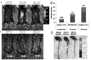
本発明のさらに他の実施例では、ヌードマウス皮下に異種移植された腫瘍部位に本発明のペプチドを標的として、これを映像化できるか否かを確認した。その結果、ドキソルビシンを処理した後、本発明のペプチドを血液内に注入した群では腫瘍組織に本発明のペプチドを標的として、これを蛍光表示を介して映像を確認することができた。反面、薬剤を処理しない群に、本発明のペプチドを注入した群及び薬剤を処理した後、対照群ペプチドを注入した群ではペプチドの標的を観察することができなかった。 In yet another embodiment of the present invention, it was confirmed whether or not the peptide of the present invention can be targeted at a tumor site xenografted subcutaneously in nude mice and visualized. As a result, after treatment with doxorubicin, in the group in which the peptide of the present invention was injected into the blood, the peptide of the present invention was targeted to the tumor tissue, and an image could be confirmed through fluorescent display. On the other hand, the peptide target could not be observed in the group in which the peptide of the present invention was injected into the group not treated with the drug and the group in which the control group peptide was injected after treatment with the drug.
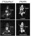
本発明のさらに他の実施例では、ヌードマウス皮下に、異種移植された腫瘍部位に放射性同位元素が標識された本発明のペプチドを標的として、これを陽電子放出単層撮影(positron emission tomography、PET)で映像化できるか否かを確認した。その結果、ドキソルビシンを処理した後、I−123(放射性ヨウ素123ではないか)が標識された本発明のペプチドを血液内に注入した群では、腫瘍部位にPET映像信号が増加することを通じて本発明のペプチドが標的されることを確認することができた。反面、現在PETに多く使用されているF−18が標識されたfluorodeoxy glucose(FDG)を注入した群では薬剤を処理した後、腫瘍部位にPET映像信号が減少した。 In still another embodiment of the present invention, a peptide of the present invention in which a radioisotope is labeled at a tumor site transplanted into a xenograft subcutaneously in a nude mouse is used as a target, which is subjected to positron emission tomography (PET). ) To see if it can be visualized. As a result, in the group in which the peptide of the present invention labeled with I-123 (not radioactive iodine 123) was injected into blood after treating doxorubicin, the PET image signal increased at the tumor site. It was possible to confirm that the peptide of the target was targeted. On the other hand, in the group injected with F-18-labeled fluorodeoxy glucose (FDG), which is currently widely used in PET, the PET video signal decreased at the tumor site after treatment with the drug.
本発明のさらに他の実施例では、動脈硬化を誘導させたマウスの大動脈に本発明のペプチドを標的として、これを映像化できるか否かを確認した。その結果、動脈硬化マウスに本発明のペプチドを血液内に注入した群では、大動脈に本発明のペプチドが標的され、これを蛍光標識を介して映像を確認することができた。反面動脈硬化マウスに対照群ペプチドを注入した群及び正常マウスに本発明のペプチドを注入した群ではペプチドの標的を観察することができなかった。 In yet another embodiment of the present invention, it was confirmed whether the peptide of the present invention was targeted to the aorta of a mouse in which arteriosclerosis was induced, and this could be visualized. As a result, in the group in which the peptide of the present invention was injected into blood into arteriosclerotic mice, the peptide of the present invention was targeted to the aorta, and an image could be confirmed through the fluorescent label. On the other hand, in the group in which the control group peptide was injected into arteriosclerotic mice and the group in which the peptide of the present invention was injected into normal mice, the peptide target could not be observed.
本発明のさらに他の実施例では、脳卒中を誘導させたラットの脳組織に本発明のペプチドを標的として、これを映像化できるか否かを確認した。その結果、脳卒中誘導ラットに本発明のペプチドを血液内に注入した群では、損傷された脳組織に本発明のペプチドが標的され、これを蛍光標識を介して映像を確認することができた。反面脳卒中誘導ラットに対照群ペプチドを注入した群及び正常ラットに本発明のペプチドを注入した群ではペプチドの標的を観察することができなかった。 In yet another embodiment of the present invention, it was confirmed whether or not the peptide of the present invention was targeted to the brain tissue of a rat in which stroke was induced, and this could be visualized. As a result, in the group in which the peptide of the present invention was injected into the blood of a stroke-induced rat, the peptide of the present invention was targeted to the damaged brain tissue, and the image could be confirmed through the fluorescent label. On the other hand, the target of the peptide could not be observed in the group in which the control group peptide was injected into the stroke induction rat and the group in which the peptide of the present invention was injected into the normal rat.
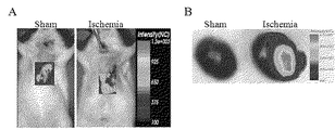
本発明のさらに他の実施例では、心筋虚血(myocardial ischemia)または心筋梗塞(myocardial infarction)を誘導させたラットの心筋組織に本発明のペプチドが標的され、これを映像化できるか否かを確認した。その結果、心筋虚血誘導ラットに本発明のペプチドを血液内に注入した群では、損傷された心筋組織に本発明のペプチドを標的として、これを蛍光標識を介して映像を確認することができた。反面、対照群ラットに本発明のペプチドを注入した群ではペプチドの標的を観察することができなかった。 In yet another embodiment of the present invention, it is determined whether the peptide of the present invention is targeted to the myocardial tissue of a rat in which myocardial ischemia or myocardial infarction has been induced and can be visualized. confirmed. As a result, in the group in which the peptide of the present invention was injected into the blood of myocardial ischemia-induced rats, it was possible to target the peptide of the present invention to damaged myocardial tissue and confirm the image through a fluorescent label. It was. On the other hand, in the group in which the peptide of the present invention was injected into control group rats, the peptide target could not be observed.
結論的に、本発明のペプチドがアポトーシス細胞と特異的に結合して生体内でアポトーシスを認識し、及び腫瘍の標的となることが分かった。 In conclusion, it has been found that the peptides of the present invention specifically bind to apoptotic cells to recognize apoptosis in vivo and become a tumor target.
前記に言及したヌクレオチド及び蛋白質の作業には下記の文献を参照することができる(Maniatis et al., Molecular Cloning: A Laboratory Manual, Cold Spring Harbor Laboratory, Cold Spring Harbor, N. Y. (1982); Sambrook et al., Molecular Cloning: A Laboratory Manual, 2d Ed., Cold Spring Harbor Laboratory Press (1989); Deutscher, M., Guide to Protein Purification Methods Enzymology, vol. 182. Academic Press. Inc., San Diego, CA (1990))。 Reference can be made to the following references for work on nucleotides and proteins referred to above (Maniatis et al., Molecular Cloning: A Laboratory Manual, Cold Spring Harbor Laboratory, Cold Spring Harbor, NY (1982); Sambrook et al. ., Molecular Cloning: A Laboratory Manual, 2d Ed., Cold Spring Harbor Laboratory Press (1989); Deutscher, M., Guide to Protein Purification Methods Enzymology, vol. 182. Academic Press. Inc., San Diego, CA (1990 )).
従って、本発明の配列番号1〜12からなる群より選ばれた配列で示されるアミノ酸配列を有する、本発明のペプチドを有効成分として含むアポトーシス細胞検出用組成物を提供する。 Accordingly, there is provided a composition for detecting apoptotic cells, which comprises the peptide of the present invention as an active ingredient and having an amino acid sequence represented by a sequence selected from the group consisting of SEQ ID NOs: 1 to 12 of the present invention.
本発明のペプチドのアポトーシス細胞結合可否の確認、検出及び定量を容易にするため、本発明のペプチドは標識された状態で提供できる。つまり、検出可能な標識にリンク(例:共有結合または架橋)されて提供できる。前記検出可能な標識は発色酵素(例:ファオキシターゼ、アルカラインホスファターゼ)、放射性同位元素(例:18F,124I,125I,32P,35S)、クロモホアー(chromophore)、発光物質または蛍光物質(例:FITC,RITC,蛍光蛋白質(GFP(Green Fluorescent Protein);EGFP(Enhanced Green Fluorescent Protein),RFP(Red Fluorescent Protein);DsRed(Discosoma sp.Red Fluorescent Protein);CFP(Cyan Fluorescent Protein),CGFP(Cyan Green Fluorescent Protein),YFP(Yellow Fluorescent Protein),Cy3,Cy5及びCy7.5)、常磁性粒子(super paramagnetic particles)または超常磁性粒子(ultrasuper paramagnetic particles)でもあり得る。 In order to facilitate the confirmation, detection and quantification of apoptotic cell binding of the peptide of the present invention, the peptide of the present invention can be provided in a labeled state. That is, it can be provided linked (eg, covalently linked or cross-linked) to a detectable label. The detectable label can be a chromogenic enzyme (eg, phaoxidase, alkaline phosphatase), a radioisotope (eg, 18 F, 124 I, 125 I, 32 P, 35 S), a chromophore, a luminescent substance or a fluorescent substance. Substance (example: FITC, RITC, fluorescent protein (GFP (Green Fluorescent Protein); EGFP (Enhanced Green Fluorescent Protein), RFP (Red Fluorescent Protein)); DsRed (Discosoma sp. Red Fluorescent Protein); CFP (Cyan Fluorescent Protein), It can also be CGFP (Cyan Green Fluorescent Protein), YFP (Yellow Fluorescent Protein), Cy3, Cy5 and Cy7.5), superparamagnetic particles or ultrasuperparamagnetic particles.
標識に伴う検出方法は当業界に広く知られており、例えば、下記のような方法により実施し得る。検出可能な標識として蛍光物質を利用する場合には、免疫蛍光染色法を利用できる。例えば、蛍光物質で標識された本発明のペプチドを試料と反応させ、未結合または非特異的な結合産物を除去し、蛍光顕微鏡下でペプチドによる蛍光を観察できる。さらに、検出可能性な標識として酵素を利用する場合には、酵素反応を介した基質の発色反応により吸光度を測定し、放射線物質の場合には放射線放出量を測定することにより行われる。従って、検出された結果は、検出標識に伴う公知の映像化方法により映像化できる。 The detection method associated with the label is widely known in the art, and can be carried out, for example, by the following method. When a fluorescent substance is used as a detectable label, an immunofluorescent staining method can be used. For example, the peptide of the present invention labeled with a fluorescent substance can be reacted with a sample, unbound or non-specific binding products can be removed, and fluorescence by the peptide can be observed under a fluorescence microscope. Further, when an enzyme is used as a detectable label, the absorbance is measured by a color development reaction of the substrate via an enzyme reaction, and in the case of a radioactive substance, the radiation emission is measured. Therefore, the detected result can be visualized by a known imaging method associated with the detection label.
さらに、本発明は(a)本発明のポリペプチドを試料と混合する工程;(b)未結合か若しくは非特異的に結合した前記ポリペプチドを除去する工程;及び(c)前記ポリペプチドの結合可否及び位置を確認する工程を含むアポトーシス細胞の検出方法を提供する。この際、本発明のポリペプチド及びアポトーシス細胞と結合した本発明のポリペプチドの検出方法は前記にて記載した通り、または公知の方法により実施し得る。 The invention further comprises (a) mixing the polypeptide of the invention with a sample; (b) removing the unbound or non-specifically bound polypeptide; and (c) binding of the polypeptide. Provided is a method for detecting apoptotic cells, comprising the step of confirming availability and location. In this case, the method for detecting the polypeptide of the present invention bound to the polypeptide of the present invention and apoptotic cells can be carried out as described above or by a known method.
本発明は本発明のポリペプチドのアポトーシス細胞検出用途を提供する。この際、本発明のポリペプチド及びアポトーシス細胞と結合した本発明のポリペプチドの検出方法は前記にて記載した通り、または公知の方法により実施し得る。 The present invention provides the use of the polypeptide of the present invention for detecting apoptotic cells. In this case, the method for detecting the polypeptide of the present invention bound to the polypeptide of the present invention and apoptotic cells can be carried out as described above or by a known method.
さらに、本発明のペプチドはアポトーシス細胞と特異的に結合できるので、薬物を前記細胞に選択的に伝達する知能型薬物伝達体として使用できる。従って、本発明のペプチドを有効成分として含む薬物伝達用組成物を提供する。さらに、本発明のペプチドの薬物伝達用途を提供する。従って、本発明のペプチド及びこれと結合した薬剤を、これを必要とする個体に有効量投与することを特徴とする薬物伝達方法を提供する。 Furthermore, since the peptide of the present invention can specifically bind to apoptotic cells, it can be used as an intelligent drug transmitter that selectively transmits a drug to the cells. Accordingly, a drug delivery composition comprising the peptide of the present invention as an active ingredient is provided. Furthermore, the drug delivery use of the peptide of this invention is provided. Therefore, the present invention provides a drug delivery method characterized by administering an effective amount of the peptide of the present invention and a drug bound thereto to an individual in need thereof.
前記の通り、アポトーシスは多様な種類の腫瘍細胞ばかりでなく、脳卒中、心筋梗塞及び動脈硬化が進行される細胞で表れる(Thomson, Science, 1995, 67: 1456-1462; Du et al., J Cereb Blood Flow Metab, 1996, 16: 195-201; Narula et al., New Engl J Med, 1996, 335: 1182-1189)。従って、前記薬物伝達用組成物は、腫瘍性疾患、脳卒中、心筋梗塞または動脈硬化に特異的であり得る。腫瘍性疾患とは、悪性腫瘍により病理的症状を呈する疾患であり、その例は特に限定されるのではないが、大腸癌、肺癌、胃癌、食道癌、膵臓癌、胆嚢癌、腎臓癌、膀胱癌、前立腺癌、睾丸癌、子宮頸部癌、子宮内膜癌、絨毛癌、卵巣癌、乳房癌、甲状腺癌、脳癌、頭頸部癌、悪性黒色腫、皮膚癌、肝臓癌、白血病(leukemia)、リンパ腫(lymphoma)、複合骨髄腫(multiple myeloma)、慢性骨髄性白血病(chronic myelogenous leukemia)、神経芽腫(neuroblastoma)、再生不良性貧血が挙げられる。 As described above, apoptosis is expressed not only in various types of tumor cells but also in cells in which stroke, myocardial infarction and arteriosclerosis are advanced (Thomson, Science, 1995, 67: 1456-1462; Du et al., J Cereb Blood Flow Metab, 1996, 16: 195-201; Narula et al., New Engl J Med, 1996, 335: 1182-1189). Thus, the drug delivery composition can be specific for neoplastic disease, stroke, myocardial infarction or arteriosclerosis. A neoplastic disease is a disease that exhibits pathological symptoms due to a malignant tumor, and examples thereof include, but are not limited to, colon cancer, lung cancer, stomach cancer, esophageal cancer, pancreatic cancer, gallbladder cancer, kidney cancer, bladder Cancer, prostate cancer, testicular cancer, cervical cancer, endometrial cancer, choriocarcinoma, ovarian cancer, breast cancer, thyroid cancer, brain cancer, head and neck cancer, malignant melanoma, skin cancer, liver cancer, leukemia ), Lymphoma, multiple myeloma, chronic myelogenous leukemia, neuroblastoma, aplastic anemia.
本発明の薬物伝達用組成物に含まれる本発明のペプチドを、従来の抗腫瘍性疾患製剤、心筋梗塞治療剤、脳卒中治療剤及び動脈硬化治療剤等の薬剤と連結して治療に利用すれば本発明のペプチドにより、前記製剤が腫瘍細胞、心筋梗塞部位、脳卒中部位、動脈硬化部位等の疾患部位にのみ選択的に伝達されるので、薬物の効能を高めることができ、同時に正常組織に及ぼす副作用を大幅に減らし得る。 If the peptide of the present invention contained in the drug delivery composition of the present invention is used for treatment in combination with conventional antitumor disease preparations, myocardial infarction therapeutic agents, stroke therapeutic agents, arteriosclerosis therapeutic agents and the like. By the peptide of the present invention, the preparation can be selectively transmitted only to diseased sites such as tumor cells, myocardial infarction sites, stroke sites, arteriosclerosis sites, etc., so that the efficacy of the drug can be enhanced and at the same time exerted on normal tissues Side effects can be greatly reduced.
本発明のペプチドに連結し得る抗腫瘍性疾患製剤は、従来、腫瘍治療に使用できるものであれば制限なく使用可能である。例えば、パクリタキセル、ドキソルビシン、ビンクリスチン、ダウノルビシン(daunorubicin)、ビンブラスチン(vinblastine)、アクチノマイシン−D(actinomycin-D)、ドセタキセル(docetaxel)、エトポシド(etoposide)、テニポシド(teniposide)、ビサントレン(bisantrene)、ホモハリングトニン(homoharringtonine)、グリーベック(Gleevec;STI-571)、シスプラチン(cisplatin)、5−フルオラシル(5-fluouracil)、アドリアマイシン(adriamycin)、メトトレキセート(methotrexate)、ブスルファン(busulfan)、クロラムブシル(chlorambucil)、シクロホスファミド(cyclophosphamide)、メルファラン(melphalan)、ニトロゲンムスタド(nitrogen mustard)、ニトロソウレア(nitrosourea)等がある。 The antitumor disease preparation that can be linked to the peptide of the present invention can be used without limitation as long as it can be used for tumor treatment. For example, paclitaxel, doxorubicin, vincristine, daunorubicin, vinblastine, actinomycin-D, docetaxel, etoposide, teniposide, bisantren, bisantren Tonin (homoharringtonine), Gleevec (STI-571), cisplatin (5-platinum), 5-fluouracil (adult), adriamycin, methotrexate, busulfan, chlorambucil, cycloambucil There are cyclophosphamide, melphalan, nitrogen mustard, nitrosourea and the like.
心筋梗塞治療剤、脳卒中治療剤及び動脈硬化治療剤は、従来これらの疾患の治療に使用できるものであれば制限なく使用することができ、例えば、前記脳卒中及び心筋梗塞疾患で血管を塞いでいる血栓を除去するために使用されている血栓溶解剤(thrombolytic)であるストレプトキナーゼ(streptokinase)、ウロキナーゼ(urokinase)、アルテプラーゼ(alteplase)等の薬物、心筋細胞保護剤であるアンジオテンシン(angiotensin)II抑制剤、アルドステロン(aldosterone)受容体抑制剤及びエリスロポイエチン(erythropoietin)等の薬物、脳神経細胞保護剤であるNMDA(N-methyl-d-aspartate)受容体抑制剤等の薬物がある。さらに、コレステロール合成抑制及び血中コレステロール濃度を下げる薬物であるロバスタチン(Lovastatin)、血管平滑筋細胞の増殖を減らす薬物であるラパマイシン(Rapamycin)、抗炎症薬物であるセレブレクス(Celebrex)、血小板凝集抑制薬物であるチクロピン(Ticlopin)、基質金属蛋白質分解酵素(matrix metalloprotease)抑制薬物であるマリマスタト(Marimastat)及びトロカド(Trocade)等がある。 The therapeutic agent for myocardial infarction, the therapeutic agent for stroke and the therapeutic agent for arteriosclerosis can be used without limitation as long as they can be used for the treatment of these diseases. For example, the blood vessel is blocked by the stroke and myocardial infarction disease. Drugs such as streptokinase, urokinase, alteplase, which are thrombolytic agents used to remove blood clots, angiotensin II inhibitors, which are cardiomyocyte protective agents And drugs such as aldosterone receptor inhibitors and erythropoietin, and drugs such as NMDA (N-methyl-d-aspartate) receptor inhibitors which are cranial nerve cell protecting agents. In addition, Lovastatin, a drug that suppresses cholesterol synthesis and lowers blood cholesterol levels, Rapamycin, a drug that reduces proliferation of vascular smooth muscle cells, Celebrex, an anti-inflammatory drug, and platelet aggregation inhibitor There are ticropin (Ticlopin), matrix metalloprotease inhibitor drugs, marimastat and trocade.
前記製剤と本発明のペプチドの連結は、当業界に公知の方法、例えば、共有結合、架橋等を介して行える。このために本発明のペプチドは必要に応じてその活性が消失されない範囲で化学的に修飾(modification)できる。本発明の組成物に含まれる本発明のペプチドの量は結合される抗癌剤の種類及び量によって異なり得る。 The preparation and the peptide of the present invention can be linked through a method known in the art, for example, covalent bonding, cross-linking and the like. Therefore, the peptide of the present invention can be chemically modified as long as the activity is not lost. The amount of the peptide of the present invention contained in the composition of the present invention may vary depending on the type and amount of anticancer agent bound.
本発明で“有効量”とは、本発明における疾患治療剤が投与対象である個体内で対象疾患の治療効果を表す量をいい、前記“個体(subject)”とは、哺乳動物、特に人間を含む動物を意味する。前記個体には、疾患治療を必要とする人間も含まれる。 In the present invention, the “effective amount” refers to an amount representing the therapeutic effect of the target disease in the individual to which the disease therapeutic agent in the present invention is administered, and the “subject” refers to a mammal, particularly a human. Means animals containing Such individuals also include humans in need of disease treatment.
一方、本発明は本発明のペプチド及びこれと結合した抗腫瘍性疾患製剤を、有効成分として腫瘍性疾患の予防及び治療用薬学的組成物を提供する。さらに、本発明は腫瘍性疾患治療剤の製造のための本発明のペプチド及びこれと結合した抗腫瘍性疾患製剤の用途を提供する。 On the other hand, the present invention provides a pharmaceutical composition for the prevention and treatment of neoplastic diseases by using the peptide of the present invention and an antitumor disease preparation bound thereto as an active ingredient. Furthermore, the present invention provides the use of the peptide of the present invention and an antitumor disease preparation bound thereto for the production of a therapeutic agent for neoplastic disease.
この際、前記薬学的組成物における抗腫瘍性疾患製剤、結合方法及び腫瘍性疾患については前記記載の通りである。 At this time, the antitumor disease preparation, binding method and neoplastic disease in the pharmaceutical composition are as described above.
一方、本発明に伴う薬学的組成物は前記ペプチドの純粋な形態または薬学的に許容される担体と共に、適切な形態に剤形化することにより提供できる。“薬学的に許容される”とは、生理学的に許容され、人間に投与される時、通常的に胃腸障碍、眩気症等のようなアレルギー反応またはこれと類似した反応を起こさない非毒性の組成物をいう。前記担体には全ての種類の溶媒、分散媒質、水中油または油中水エマルジョン、水性組成物、リポソーム、ミクロビード及びミクロソーム、生分解性ナノ粒子等が含まれる。 On the other hand, the pharmaceutical composition according to the present invention can be provided by formulating into an appropriate form together with a pure form of the peptide or a pharmaceutically acceptable carrier. “Pharmaceutically acceptable” means non-toxic that is physiologically acceptable and does not normally cause allergic reactions or similar reactions such as gastrointestinal disorders, dizziness, etc. when administered to humans. Of the composition. Such carriers include all types of solvents, dispersion media, oil-in-water or water-in-oil emulsions, aqueous compositions, liposomes, microbeads and microsomes, biodegradable nanoparticles, and the like.
一方、本発明に伴う薬学的組成物は、投与経路により適切な担体と共に剤形化できる。前記本発明に伴う薬学的組成物の投与経路としては特に限定されないものの、経口的または非経口的に投与できる。非経口的投与経路には例えば、経皮、鼻腔、腹腔、筋肉、皮下または静脈等の多様な経路が含まれる。 On the other hand, the pharmaceutical composition according to the present invention can be formulated with an appropriate carrier according to the administration route. Although the administration route of the pharmaceutical composition according to the present invention is not particularly limited, it can be administered orally or parenterally. Parenteral routes of administration include various routes such as transdermal, nasal, abdominal, muscle, subcutaneous or intravenous.
本発明の薬学的組成物を経口投与する場合、本発明の薬学的組成物は、適切な経口投与用担体と共に、公知の方法により粉末、顆粒、錠剤、丸剤、糖衣錠剤、カプセル剤、液剤、ゲル剤、シロップ剤、懸濁液、ウェハー等の形態で剤形化でき得る。適切な担体の例には、ラクトース、デキストロース、スクロース、ソルビトール、マンニトール、キシリトール、エリスロトール及びマルチトール等を含む糖類とトウモロコシ澱粉、小麦澱粉、お米澱粉及び馬鈴薯澱粉等を含む澱粉類、セルロース、メチールセルロース、ナトリウムカルボキシメチルセルロース及びヒドロキシプロピルメチールセルロース等を含むセルロース類、ゼラチン、ポリビニルピロリドン等のような充填剤が含まれ得る。さらに、場合によって架橋結合ポリビニルピロリドン、寒天、アルギン酸またはナトリウムアルギネート等を崩解剤として添加し得る。さらには、前記薬学的組成物は抗凝集剤、潤滑剤、湿潤剤、香料、乳化剤及び防腐剤等を追加して含め得る。 When the pharmaceutical composition of the present invention is orally administered, the pharmaceutical composition of the present invention is mixed with a suitable carrier for oral administration by a known method in the form of powder, granules, tablets, pills, sugar-coated tablets, capsules, liquids , Gels, syrups, suspensions, wafers and the like. Examples of suitable carriers include sugars and corn starch including lactose, dextrose, sucrose, sorbitol, mannitol, xylitol, erythritol and maltitol, starches including wheat starch, rice starch and potato starch, cellulose, Fillers such as celluloses including methyl cellulose, sodium carboxymethyl cellulose and hydroxypropyl methyl cellulose, gelatin, polyvinylpyrrolidone and the like may be included. Further, in some cases, cross-linked polyvinyl pyrrolidone, agar, alginic acid, sodium alginate or the like can be added as a disintegrating agent. Furthermore, the pharmaceutical composition may additionally contain anti-aggregating agents, lubricants, wetting agents, perfumes, emulsifiers and preservatives.
さらに、非経口的に投与する場合、本発明の薬学的組成物は適切な非経口用担体と共に、注射剤、経皮投与剤及び鼻腔吸込み剤の形態で当業界に公知の方法により剤形化可能である。前記注射剤の場合には必ず滅菌しなければならず、バクテリア及び眞菌のような微生物の汚染から保護されなければならない。注射剤の場合、適切な担体の例は、特に限定はされないものの、水、エタノール、ポリオール(例えば、グリセロール、プロピレングリコール及び液体ポリエチレングリコール等)、これらの混合物及び/または植物油を含む溶媒または分散媒質を使用し得る。より好ましくは、適切な担体にはハンクス溶液、リンゲル溶液、トリエタノールアミンが含まれたPBS(phosphate buffered saline)または注射用滅菌水、10%エタノール、40%プロピレングリコール及び5%テキトローズのような等張溶液等を使用し得る。前記注射剤を微生物の汚染から保護するためには、パラベン、クロロブタノール、フェノール、ソルビサン、チメロサール等のような多様な抗菌剤及び抗眞菌剤を追加して含め得る。さらに、前記注射剤は大部分の場合、糖またはナトリウムクロライドのような等張化剤を追加して含め得る。これらの剤形は、製薬学で一般的に知られた処方書である文献(Remington's Pharmaceutical Science, 15th Edition, 1975, Mack Publishing Company, EASTON, Pennsylvania)に記述されている。 Furthermore, when administered parenterally, the pharmaceutical composition of the present invention is formulated by methods known in the art in the form of injections, transdermal administration agents, and nasal inhalants, together with suitable parenteral carriers. Is possible. Such injections must be sterilized and protected from contamination by microorganisms such as bacteria and bacilli. In the case of injections, examples of suitable carriers include, but are not limited to, solvents or dispersion media containing water, ethanol, polyols (eg, glycerol, propylene glycol and liquid polyethylene glycol, etc.), mixtures thereof and / or vegetable oils Can be used. More preferably, suitable carriers include Hank's solution, Ringer's solution, PBS (phosphate buffered saline) containing triethanolamine, or sterile water for injection, such as 10% ethanol, 40% propylene glycol and 5% textrose. A tonic solution or the like can be used. In order to protect the injection from microbial contamination, various antibacterial and antifungal agents such as parabens, chlorobutanol, phenol, sorbisan, thimerosal and the like may be additionally included. In addition, the injection may in most cases additionally contain isotonic agents such as sugars or sodium chloride. These dosage forms are described in the literature (Remington's Pharmaceutical Science, 15th Edition, 1975, Mack Publishing Company, EASTON, Pennsylvania), which is a commonly known formulation in pharmacy.
吸込み投与剤の場合、本発明により使用される化合物は適切な推進剤、例えば、ジクロロフルオロメタン、トリクロロフルオロメタン、ジクロロテトラフルオロエタン、二酸化炭素または他の適切な基体を使用して、加圧パックまたは煙霧器からエアロゾルスプレー形態で容易に伝達できる。加圧エアロゾルの場合、投薬単位は計量された量を伝達する弁を提供して決定できる。例えば、吸込み器または取込み器に使用されるゼラチンカプセル及びカトリッジは化合物、及びラクトースまたは澱粉のような適切な粉末基体の粉末混合物を含有するように剤形化することができる。 In the case of inhalation dosages, the compounds used in accordance with the present invention are pressurized packs using a suitable propellant such as dichlorofluoromethane, trichlorofluoromethane, dichlorotetrafluoroethane, carbon dioxide or other suitable substrate. Or it can be easily transmitted in the form of an aerosol spray from an atomizer. In the case of a pressurized aerosol, the dosage unit can be determined by providing a valve that delivers a metered amount. For example, gelatin capsules and cartridges used in inhalers or ingestors can be formulated to contain a compound and a powder mixture of a suitable powder base such as lactose or starch.
その他に、薬学的に許容される担体としては、下記の文献に記載されているものを参考にできる(Remington's Pharmaceutical Sciences, 19th ed., Mack Publishing Company, Easton, PA, 1995)。 In addition, as pharmaceutically acceptable carriers, those described in the following documents can be referred to (Remington's Pharmaceutical Sciences, 19th ed., Mack Publishing Company, Easton, PA, 1995).
さらに、本発明の薬学的組成物は一つ以上の緩衝剤(例えば、食塩水またはPBS)、カーボハイトレート(例えば、グルコース、マンノス、スクロースまたはテキトラン)、安定化剤(亜硫酸水素ナトリウム、亜硫酸ナトリウムまたはアスコロビン酸)、抗酸化剤、静菌剤、キレート化剤(例えば、EDTAまたはグルタチオン)、アジュバント(例えば、アルミニウムヒドロキシド)、懸濁剤、濃厚剤及び/または保存剤(ベンズアルコニウムクロライド、メチルパラベン、プロピルパラベン及びクロロブタノール)を追加して含め得る。 In addition, the pharmaceutical composition of the present invention may comprise one or more buffers (eg, saline or PBS), carbohydrate (eg, glucose, mannos, sucrose or textran), stabilizers (sodium bisulfite, sodium sulfite). Or ascorbic acid), antioxidants, bacteriostatic agents, chelating agents (eg EDTA or glutathione), adjuvants (eg aluminum hydroxide), suspending agents, thickeners and / or preservatives (benzalkonium chloride, Methylparaben, propylparaben and chlorobutanol) may be additionally included.
さらに、本発明の薬学的組成物は哺乳動物に投与された後、活性成分の迅速、遅速または遅延された放出を提供できるように公知の方法により剤形化できる。 Furthermore, the pharmaceutical compositions of the invention can be formulated by known methods so as to provide rapid, slow or delayed release of the active ingredient after administration to a mammal.
前記のような方法により、剤形化された薬学的組成物は有効量で経口、経皮、皮下、静脈または筋肉を含む多くの経路を介して投与できる。前記の“有効量”とは、患者に投与した時、診断または治療効果の追跡を可能にする化合物または抽出物の量をいう。本発明に伴う薬学的組成物の投与量は投与経路、投与対象、対象疾患及びこれの重症度、年令、性別、体重、個人差及び疾病状態によって適切に選択できる。好ましくは、本発明のペプチドを含む薬学的組成物は、疾患の程度によって有効成分の含量を異にし得るものの、通常、成人を基準にする時、1回投与の際、10μg〜10mgの有効容量で1日複数回繰り返し投与できる。 By such methods, the formulated pharmaceutical composition can be administered in an effective amount via a number of routes including oral, transdermal, subcutaneous, intravenous, or muscle. The “effective amount” refers to the amount of a compound or extract that, when administered to a patient, allows for the tracking of diagnostic or therapeutic effects. The dosage of the pharmaceutical composition according to the present invention can be appropriately selected according to the administration route, the subject to be administered, the target disease and the severity thereof, age, sex, body weight, individual difference and disease state. Preferably, the pharmaceutical composition containing the peptide of the present invention may have different contents of the active ingredient depending on the degree of the disease, but normally, when based on an adult, an effective dose of 10 μg to 10 mg when administered once Can be administered multiple times a day.
本発明のペプチドはアポトーシス細胞と特異的に結合するので、腫瘍性疾患の発病部位の映像化及び診断にも有効に使用できる。従って、本発明は前記ペプチドを有効成分として含む、腫瘍性疾患の映像化及び診断用組成物を提供する。さらに、本発明は本発明のペプチドの腫瘍性疾患部位の映像化用途を提供する。従って、本発明は本発明のペプチドを必要とする個体に有効量で投与することを特徴とする腫瘍性疾患部位の映像化方法を提供する。この際、腫瘍性疾患の映像化及び診断は、特に限定はされないものの、腫瘍性疾患の初診目的ばかりでなく、進行結果、腫瘍治療に対する治療経過、治療剤に対する反応モニタリング等を包括して使用できる。前記ペプチドは結合可否の確認、検出及び定量を容易にするために、標識された状態で提供することができるが、これについては前記にて記述した通りである。 Since the peptide of the present invention specifically binds to apoptotic cells, it can be used effectively for imaging and diagnosis of the onset site of a neoplastic disease. Accordingly, the present invention provides a composition for imaging and diagnosing neoplastic diseases comprising the peptide as an active ingredient. Furthermore, the present invention provides the use of the peptide of the present invention for imaging a tumorous disease site. Accordingly, the present invention provides a method for imaging a tumorous disease site, comprising administering an effective amount to an individual in need of the peptide of the present invention. At this time, imaging and diagnosis of neoplastic disease are not particularly limited, but can be used not only for the purpose of initial diagnosis of neoplastic disease, but also comprehensively including progress results, treatment course for tumor treatment, reaction monitoring for therapeutic agent, etc. . The peptide can be provided in a labeled state in order to facilitate the confirmation, detection and quantification of binding, as described above.
さらに、本発明のペプチドはアポトーシス細胞と特異的に結合するので、腫瘍にのみ限定されずアポトーシスが大きく増加している脳卒中、心筋梗塞及び動脈硬化でも本発明のペプチドは薬物伝達をすることができる。つまり、本発明は本発明のペプチド及びこれと結合した脳卒中治療剤を有効成分として含む脳卒中予防及び治療用薬学的組成物を提供する。従って、本発明は本発明のペプチド及びこれと結合した心筋梗塞治療剤を有効成分として含む心筋梗塞予防及び治療用薬学的組成物を提供する。従って、本発明は本発明のペプチド及びこれと結合した動脈硬化治療剤を有効成分として含む動脈硬化予防及び治療用薬学的組成物を提供する。 Furthermore, since the peptide of the present invention specifically binds to apoptotic cells, the peptide of the present invention is capable of drug transmission even in stroke, myocardial infarction and arteriosclerosis, which are not limited to tumors but have greatly increased apoptosis. . That is, the present invention provides a pharmaceutical composition for preventing and treating stroke comprising the peptide of the present invention and a therapeutic agent for stroke combined therewith as an active ingredient. Accordingly, the present invention provides a pharmaceutical composition for preventing and treating myocardial infarction comprising the peptide of the present invention and a therapeutic agent for myocardial infarction bound thereto as an active ingredient. Accordingly, the present invention provides a pharmaceutical composition for preventing and treating arteriosclerosis comprising the peptide of the present invention and an arteriosclerosis therapeutic agent bound thereto as an active ingredient.
一方、本発明は脳卒中治療剤の製造のための本発明のペプチド及びこれと結合した脳卒中治療剤の用途を提供する。
一方、本発明は心筋梗塞治療剤の製造のための本発明のペプチド及びこれと結合した心筋梗塞治療剤の用途を提供する。
一方、本発明は動脈硬化治療剤の製造のための本発明のペプチド及びこれと結合した動脈硬化治療剤の用途を提供する。
On the other hand, the present invention provides the use of the peptide of the present invention and the therapeutic agent for stroke combined therewith for the production of a therapeutic agent for stroke.
On the other hand, the present invention provides the use of the peptide of the present invention and the therapeutic agent for myocardial infarction combined therewith for the production of a therapeutic agent for myocardial infarction.
On the other hand, the present invention provides the use of the peptide of the present invention and the arteriosclerosis therapeutic agent combined therewith for the production of an arteriosclerosis therapeutic agent.
本発明のペプチドを従来脳卒中治療剤、心筋梗塞治療剤及び動脈硬化治療剤と連結して治療に利用すれば本発明のペプチドにより前記製剤が疾病部位の細胞にのみ選択的に伝達されるので、薬物の効能を増加させ、同時に正常組織に及ぼす副作用を大幅に減らし得る。 If the peptide of the present invention is used for treatment by linking with a conventional therapeutic agent for stroke, therapeutic agent for myocardial infarction and therapeutic agent for arteriosclerosis, the above-mentioned preparation is selectively transmitted only to cells at the disease site by the peptide of the present invention. It can increase drug efficacy and at the same time greatly reduce the side effects on normal tissues.
本発明のペプチドに連結できる心筋梗塞治療剤、脳卒中治療剤及び動脈硬化治療剤は従来これらの疾患の治療に使用されるものであれば制限なく使用することができ、例えば、前記に言及したストレプトキナーゼ(streptokinase)、ウロキナーゼ(urokinase)、アルテプラーゼ(alteplase)、アンジオテンシン(angiotensin)II抑制剤、アルドステロン(aldosterone)受容体抑制剤、エリスロポイエチン(erythropoietin)、NMDA(N-methyl-d-aspartate)受容体抑制剤、ロバスタチン(Lovastatin)、ラパマイシン(Rapamycin)、セレブレキス(Celebrex)、チクロピン(Ticlopin)、マリマスタト(Marimastat)及びトロカド(Trocade)等が使用できる。前記製剤と本発明のペプチドの連結は当業界に公知された方法、例えば、共有結合、架橋等を介して行える。このため、本発明のペプチドは必要であれば、その活性が消失されない範囲で化学的に修飾(modification)できる。本発明の組成物に含まれる本発明のペプチドの量は、結合される前記治療剤の種類及び量によって異なる。 The therapeutic agent for myocardial infarction, the therapeutic agent for stroke, and the therapeutic agent for arteriosclerosis that can be linked to the peptide of the present invention can be used without limitation as long as it is conventionally used for the treatment of these diseases. Kinase (streptokinase), urokinase (urokinase), alteplase (alteplase), angiotensin II inhibitor, aldosterone receptor inhibitor, erythropoietin, NMDA (N-methyl-d-aspartate) receptor A body suppressor, Lovastatin, Rapamycin, Celebrex, ticlopin, Ticlopin, Marimastat, Trocade and the like can be used. The preparation and the peptide of the present invention can be linked through a method known in the art, for example, covalent bonding, cross-linking and the like. Therefore, if necessary, the peptide of the present invention can be chemically modified as long as its activity is not lost. The amount of the peptide of the present invention contained in the composition of the present invention varies depending on the kind and amount of the therapeutic agent to be bound.
本発明のペプチドはアポトーシス細胞と特異的に結合することにより、脳卒中、心筋梗塞及び動脈硬化の発病部位の映像化及び診断にも有用に使用できる。従って、本発明は本発明のペプチドを有効成分として含む脳卒中部位の映像化用組成物を提供する。従って、本発明は本発明のペプチドを有効成分として含む心筋梗塞部位の映像化用組成物を提供する。さらに、本発明は本発明のペプチドを有効成分として含む動脈硬化部位の映像化用組成物を提供する。 The peptide of the present invention binds specifically to apoptotic cells, and thus can be usefully used for imaging and diagnosis of the onset site of stroke, myocardial infarction and arteriosclerosis. Accordingly, the present invention provides a composition for imaging a stroke site comprising the peptide of the present invention as an active ingredient. Accordingly, the present invention provides a composition for imaging a myocardial infarction site comprising the peptide of the present invention as an active ingredient. Furthermore, the present invention provides a composition for imaging an arteriosclerosis site comprising the peptide of the present invention as an active ingredient.
一方、本発明は本発明のペプチドの腫瘍性疾患、脳卒中、心筋梗塞及び動脈硬化からなる群より選ばれた疾患部位の映像化用途を提供する。本発明は本発明のペプチドをこれを必要とする個体に有効量で投与することを特徴とする腫瘍性疾患、脳卒中、心筋梗塞及び動脈硬化からなる群より選ばれた疾患部位の映像化方法を提供する。 On the other hand, the present invention provides a use for imaging a disease site selected from the group consisting of neoplastic disease, stroke, myocardial infarction and arteriosclerosis of the peptide of the present invention. The present invention provides a method for imaging a disease site selected from the group consisting of neoplastic diseases, stroke, myocardial infarction and arteriosclerosis, which comprises administering the peptide of the present invention to an individual in need thereof in an effective amount. provide.
この際、疾患の映像化及び診断は、特に限定はされないものの、疾患の初診目的ばかりでなく、進行結果、治療に対する治療経過、治療剤に対する反応モニタリング等を包括して使用できる。前記ペプチドは結合可否の確認、検出及び定量を容易にするために、標識された状態で提供することができる。これについては前記に記述した通りである。 At this time, imaging and diagnosis of the disease are not particularly limited, but can be used not only for the purpose of initial diagnosis of the disease, but also comprehensively including progress results, treatment course for treatment, reaction monitoring for the therapeutic agent, and the like. The peptide can be provided in a labeled state in order to facilitate confirmation, detection and quantification of binding. This is as described above.
以下、本発明を実施例により詳細に説明する。
ただし、下記実施例は本発明を例示したのみであって、本発明の内容が下記実施例に限定されるものではない。
Hereinafter, the present invention will be described in detail with reference to examples.
However, the following examples only illustrate the present invention, and the content of the present invention is not limited to the following examples.
実施例1:腫瘍組織の細胞に対する結合特異性を有するペプチドの探索
<1−1>ファージペプチドライブラリの製造
本発明者等は腫瘍組織を構成する各種細胞に、特異的なペプチドを探し出すために、ファージペプチドディスプレイ技術を利用した(Smith, Science, 228: 1315-1317, 1985)。ファージペプチドディスプレイは数個〜数十個のアミノ酸で構成されたペプチドをバクテリオファージ表面にディスプレイすることである。最大109種類のペプチドを有するファージライブラリを製造できるので、一度に多くの種類のペプチドを探索して望む組織または腫瘍に標的されるペプチドを探出すに有用な技術である。
本発明に使用したファージペプチドライブラリは次のような方法により製造した。まず両末端にシステインがあって、その間に任意の7個アミノ酸を含むCX7Cペプチドをコーディング(coding)するオリゴヌクレオチド(oligonucleotide)を無作為で合成した。前記オリゴヌクレオチドの合成はマクロゼン社(大韓民国)に依頼して行った。以降、合成されたオリゴヌクレオチドをノバゲン(Novagen,米国)社のT7Select(登録商標)ファージクローニングキットを利用して製造社の指針に従い前記合成されたオリゴヌクレオチドをT7 415−1bファージの表面を構成するカプシッド(capsid)蛋白質遺伝子内にクローニングしてファージペプチドライブラリを製造した。製造されたファージペプチドライブラリの多様性は約5×10⁸pfu程度に測定された。
Example 1: Search for peptides having binding specificity for tumor tissue cells <1-1> Production of phage peptide library The present inventors searched for various peptides constituting tumor tissues in order to search for specific peptides. Phage peptide display technology was used (Smith, Science, 228: 1315-1317, 1985). Phage peptide display is to display a peptide composed of several to tens of amino acids on the bacteriophage surface. Since a phage library having a maximum of 10 9 types of peptides can be produced, it is a useful technique for searching for peptides targeted to a desired tissue or tumor by searching many types of peptides at once.
The phage peptide library used in the present invention was produced by the following method. First, oligonucleotides were randomly synthesized that encode a CX 7 C peptide having cysteines at both ends and any 7 amino acids between them. The synthesis of the oligonucleotide was performed by Macrozen Co. (South Korea). Thereafter, the synthesized oligonucleotide is constructed on the surface of the T7 415-1b phage according to the manufacturer's guidelines using the Novagen (USA) T7Select (registered trademark) phage cloning kit. A phage peptide library was prepared by cloning into the capsid protein gene. The diversity of the prepared phage peptide library was measured to be about 5 × 10 8 pfu.
<1−2>ファージペプチドライブラリ探索
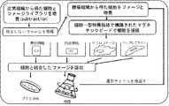
腫瘍治療の目的で施行された手術的切除の結果として得た腫瘍組織及び腫瘍周辺部位の正常組織をそれぞれ刃物で細切して、組織粉砕機を利用して粉砕し、細胞懸濁液(cell suspension)を調製した。前記実施例1の<1−1>で作製したファージライブラリを正常組織由来の細胞懸濁液と混合し、4℃で2時間反応させた。反応終了後上層液のみを採り正常細胞に結合しないファージを回収し、宿主であるBL21大腸菌を利用してタイターを増幅した。その後、腫瘍組織由来の細胞懸濁液と同一な条件で反応させた。腫瘍細胞に非特異的に弱くついているファージを1%の牛血清アルブミン(bovine serum albumin;BSA)が含まれたDMEM溶液(Dulbeco's modified Eagle's medium)1mlで室温で5分間、全3回洗浄して除去した。洗浄後マクロファージに対する抗体(抗−CD14抗体、Dynal社)または血管内皮細胞に対する抗体(抗−CD31抗体,Dynal社)が表面に付いている磁性粒子を細胞懸濁液と4℃で30分間反応させ、磁石を利用してそれぞれの磁性粒子に結合した細胞を分離した。分離されたマクロファージまたは血管内皮細胞に1%NP−40が含まれたDMEM溶液100μlを4℃で10分間処理した後、宿主であるBL21大腸菌培養液900μlを添加して細胞に結合したファージを検出した。検出されたファージの一部は公知の方法(Phage display, Clackson T and Lowman HB, p171, 2004, Oxford University Press, New York)によりタイターを測定し、残りは増幅して数を増やしてそれぞれの細胞に結合するファージを探索する過程を前記方法により、全3回繰返し実施した。その結果、腫瘍組織に由来するマクロファージ及び内皮細胞に結合したファージタイターが著しく増加し、全体的に成功裏に探索がなされたことがわかった。前記探索過程の模式図を図1に示した。
<1-2> Phage peptide library search Tumor tissue obtained as a result of surgical excision performed for the purpose of tumor treatment and normal tissue around the tumor are each chopped with a blade, and a tissue grinder is used. By grinding, a cell suspension was prepared. The phage library prepared in <1-1> of Example 1 was mixed with a cell suspension derived from normal tissue and reacted at 4 ° C. for 2 hours. After the reaction was completed, only the upper layer solution was collected to collect phages that did not bind to normal cells, and the titer was amplified using BL21 E. coli as a host. Then, it was made to react on the same conditions as the cell suspension derived from a tumor tissue. Wash phages nonspecifically weakly on tumor cells with 1 ml of DMEM solution (Dulbeco's modified Eagle's medium) containing 1% bovine serum albumin (BSA) for 5 minutes at room temperature for a total of 3 times. Removed. After washing, magnetic particles with an antibody against macrophages (anti-CD14 antibody, Dynal) or vascular endothelial cells (anti-CD31 antibody, Dynal) are reacted with the cell suspension at 4 ° C. for 30 minutes. Then, the cells bound to the respective magnetic particles were separated using a magnet. Treat the isolated macrophages or vascular endothelial cells with 100 μl of DMEM solution containing 1% NP-40 at 4 ° C. for 10 minutes, and then add 900 μl of BL21 E. coli culture medium as a host to detect phages bound to the cells. did. A part of the detected phage was measured for titer by a known method (Phage display, Clackson T and Lowman HB, p171, 2004, Oxford University Press, New York), and the rest was amplified to increase the number of each cell. The process of searching for phages that bind to was repeated a total of three times by the method described above. As a result, it was found that phage titers bound to the macrophages and endothelial cells derived from the tumor tissue were remarkably increased, and the overall search was successful. A schematic diagram of the search process is shown in FIG.
<1−3>ファージクロンの塩基配列判読及びアミノ酸配列分析
前記実施例1の<1−2>を介して探索したファージにどんなペプチドがディスプレイされたかを調査するために、それぞれの細胞に対して無作為で30個のファージクロンを選択してファージに挿入されたDNAをPCRで増幅した後、配列分析(sequencing)を行った。この際、5’−プライマーとして、オリゴヌクレオチド(AGCGGACCAGATTATCGCTA,配列番号13)及び3’−プライマーとしてオリゴヌクレオチド(AACCCCTCAAGACCCGTTTA,配列番号14)をそれぞれ使用した。PCRは95℃で5分間加熱して鋳型DNAを前変性させた後、94℃で50秒;50℃で1分;及び72℃で1分を1サイクルにして全35回繰返して行い、72℃で6分間最終的に反応させて行った。
以降、PCR反応産物の塩基配列を塩基配列分析会社(バイオニア社)に依頼して判読した。判読された塩基配列をもとにアミノ酸配列を推定した。推定されたアミノ酸配列をClustalWプログラムで分析した結果、マクロファージ及び内皮細胞に対してそれぞれ最もありふれた頻度で示された代表的なファージクロンのペプチドを得ることができ、これを配列番号1(ApoPep−1と命名、CQRPPR,マクロファージで選別)、配列番号2(ApoPep−2と命名、CSVAPR,内皮細胞で選別)、配列番号3(CNRPPR)、配列番号4(CQKPPR)、配列番号5(CQRPPK)、配列番号6(CNKPPR)、配列番号7(CNRPPK)、配列番号8(CQKPPK)、配列番号9(CNKPPK)、配列番号10(CTVAPR)、配列番号11(CSVAPK)及び配列番号12(CTVAPK)でそれぞれ示した。
<1-3> Reading of base sequence and amino acid sequence analysis of phage clone In order to investigate what peptides were displayed on the phage searched through <1-2> of Example 1, for each cell, Randomly, 30 phage clones were selected and the DNA inserted into the phage was amplified by PCR, followed by sequence analysis. At this time, oligonucleotide (AGCGGACCAGATTATCCTCA, SEQ ID NO: 13) was used as the 5′-primer and oligonucleotide (AACCCCCTCAAGACCCGTTTA, SEQ ID NO: 14) was used as the 3′-primer. PCR was performed at 95 ° C. for 5 minutes to pre-denature the template DNA, followed by 35 cycles of 94 ° C. for 50 seconds; 50 ° C. for 1 minute; and 72 ° C. for 1 minute. The final reaction was carried out at 6 ° C. for 6 minutes.
Thereafter, the base sequence of the PCR reaction product was read at the request of a base sequence analysis company (Bionia). The amino acid sequence was estimated based on the read base sequence. As a result of analyzing the deduced amino acid sequence by the ClustalW program, a typical phage clone peptide shown with the most common frequency for macrophages and endothelial cells, respectively, can be obtained, which is represented by SEQ ID NO: 1 (ApoPep- No. 1, CQRPPR, selected by macrophages), SEQ ID NO: 2 (named ApoPep-2, CSVAPR, selected by endothelial cells), SEQ ID NO: 3 (CNRPPR), SEQ ID NO: 4 (CQKPRPR), SEQ ID NO: 5 (CQRPPPK), SEQ ID NO: 6 (CNKPPR), SEQ ID NO: 7 (CNRPPK), SEQ ID NO: 8 (CQKPPK), SEQ ID NO: 9 (CNKPPK), SEQ ID NO: 10 (CTVAPPR), SEQ ID NO: 11 (CSVAPK), and SEQ ID NO: 12 (CTVAPK), respectively Indicated.
実施例2:本発明のペプチドの生体内腫瘍標的に対する組織学的評価
<2−1>腫瘍異種移植ヌードマウスの製造
全ての動物実験は所属機関の動物実験倫理委員会のガイドラインにより実施された。腫瘍異種移植(tumor xenografts)をするために、6週令BALB/c雄ヌードマウス(孝昌サイエンス社)にRMPI−1640培地に浮遊させたA549人間肺癌細胞株(1×107個)を右側上肢または下肢部位に皮下注射した。その後、3週間腫瘍細胞が0.5〜1cmの大きさに育つようにした。本実験に使用したA549細胞株は抗生剤(ペニシリン及びストレプトマイシン)が添加された10%牛胎児血清(FBS,Fetal bovine serum)を含むRMPI−1640培地で培養し、3〜4日毎に改代培養した。
Example 2: Histological evaluation of the peptide of the present invention against an in vivo tumor target <2-1> Production of tumor xenograft nude mice All animal experiments were performed according to the guidelines of the animal experiment ethics committee of the affiliated institution. In order to perform tumor xenografts, A549 human lung cancer cell lines (1 × 10 7 cells) suspended in RMPI-1640 medium in 6-week-old BALB / c male nude mice (Kochang Science Co., Ltd.) Alternatively, it was injected subcutaneously into the lower limb region. Thereafter, the tumor cells were allowed to grow to a size of 0.5 to 1 cm for 3 weeks. The A549 cell line used in this experiment was cultured in RMPI-1640 medium containing 10% fetal bovine serum (FBS) supplemented with antibiotics (penicillin and streptomycin), and subcultured every 3-4 days. did.
<2−2>腫瘍標的に対する組織学的分析
本発明で使用されるペプチドはN−末端にフルオレシン(fluorecein)が結合した形態にして、標準Fmoc方法により合成した後HPLC機器で分離した。本ペプチドの合成は専門会社(Peptron社)に依頼して施行した。
フルオレシンが標識された本発明のペプチド(ApoPep−1)または対照群ペプチド(アミノ酸配列、NSSVDK)をイソフルラン麻酔(isoflurane anesthesia)下でマウスの尻尾静脈を介して最終50μM濃度で投入した後、2時間循環させた。
組織学的実験のため、前記マウスを麻酔して開腹した。心臓を介してphosphate-buffered saline(PBS)洗浄液及び4%パラホルムアルデヒド固定液を順次に灌流させ、腫瘍組織及び器官を除去した。各組織を凍結切断(Cryosections)した後、蛍光顕微鏡(fluorescence microscopy,Zeiss社)で本発明のペプチドを観察した。腫瘍組織におけるアポトーシスは製造会社(Chemicon社)の指針に従いTUNEL(in vitro terminal deoxynucleotidyl transferase-mediated dUTP nick-end labeling)分析法により確認した。フィブリノゲン(fibrinogen)染色は、これに対する抗体(Abcam社)及び赤色の蛍光試薬であるアレキサー568で標識された2次抗体を利用して免疫組織化学法(immunohistochemistry)で施行した。
その結果、図2に示した通り、H&E染色を介して腫瘍が確認され(図2A)、本発明のペプチド(ApoPep−1)を注入した場合、腫瘍組織においてペプチドが観察された(図2B)、対照群ペプチドの場合、ほとんど観察されなかったことが分かった(図2C)。一方、図2Aでアステリスクで表示した部位に対するペプチド蛍光(図2D)、TUNEL染色(図2E)及びこれの映像合成(図2F)をすることにより、TUNEL染色ができる細胞、つまり、アポトーシス細胞にペプチドが結合することが分かった。さらに、図2Aにおいて三角形で表示した部位に対するペプチド蛍光(図2G)、TUNEL染色(図2H)及びこれの映像合成(図2I)をすることにより、TUNEL染色できない細胞にもペプチドが結合し、この部位に対するペプチド蛍光(図2J)、フィブリノゲン染色(図2K)及びこれの映像合成(図2L)を介して細胞の凝固壊死(coagulation necrosis)部位であることが分かった。
従って、配列番号2のペプチド(ApoPep−2)を使用して前記と類似した実験をしてペプチド蛍光(図2M)、TUNEL染色(図2N)及びこれの映像合成(図2O)をすることにより、腫瘍組織内アポトーシス細胞にペプチドが結合することが分かった。
<2-2> Histological analysis for tumor target Peptides used in the present invention were conjugated with fluorescein at the N-terminus, synthesized by a standard Fmoc method, and then separated by HPLC instrument. The synthesis of this peptide was carried out at the request of a specialized company (Peptron).
Fluorescin-labeled peptide of the present invention (ApoPep-1) or control peptide (amino acid sequence, NSSVDK) was injected at a final concentration of 50 μM via the tail vein of mice under isoflurane anesthesia for 2 hours It was circulated.
For histological experiments, the mice were anesthetized and opened. A phosphate-buffered saline (PBS) washing solution and a 4% paraformaldehyde fixing solution were sequentially perfused through the heart to remove tumor tissues and organs. After each tissue was frozen and cut (Cryosections), the peptide of the present invention was observed with a fluorescence microscope (Zeiss). Apoptosis in the tumor tissue was confirmed by a TUNEL (in vitro terminal deoxynucleotidyl transferase-mediated dUTP nick-end labeling) analysis method according to the guidelines of the manufacturer (Chemicon). Fibrinogen staining was performed by immunohistochemistry using an antibody against this (Abcam) and a secondary antibody labeled with Alexer 568, a red fluorescent reagent.
As a result, as shown in FIG. 2, the tumor was confirmed through H & E staining (FIG. 2A), and when the peptide of the present invention (ApoPep-1) was injected, the peptide was observed in the tumor tissue (FIG. 2B). In the case of the control group peptide, it was found that it was hardly observed (FIG. 2C). On the other hand, peptide fluorescence (FIG. 2D), TUNEL staining (FIG. 2E) and video synthesis (FIG. 2F) of the site indicated by an asterisk in FIG. Was found to combine. Furthermore, by peptide fluorescence (FIG. 2G), TUNEL staining (FIG. 2H) and image synthesis (FIG. 2I) for the site indicated by a triangle in FIG. 2A, the peptide also binds to cells that cannot be TUNEL stained. It was found to be a coagulation necrosis site of the cell through peptide fluorescence (FIG. 2J), fibrinogen staining (FIG. 2K) and video synthesis (FIG. 2L) thereof.
Therefore, by conducting an experiment similar to the above using the peptide of SEQ ID NO: 2 (ApoPep-2), peptide fluorescence (FIG. 2M), TUNEL staining (FIG. 2N), and video synthesis of this (FIG. 2O) The peptide was found to bind to apoptotic cells in the tumor tissue.
実施例3:培養状態のアポトーシス細胞に本発明のペプチドの結合
<3−1>アポトーシス細胞に対するペプチド結合の顕微鏡的観察
細胞をチャンバースライド(Nalgen Nunc社)で培養し、50μM濃度のエトポシド(etoposide、Sigma社)を定められた時間に処理してアポトーシスを誘導した(A549及びHeLa細胞15時間、H460細胞24時間、L132細胞3時間、RAW細胞6時間)。前記細胞は抗生剤(ペニシリン及びストレプトマイシン)と10%牛胎児血清を含む、RMPI−1640培地(A549及びH460細胞)及びDMEM培地(HeLa,L132及びRAW細胞)で培養した。一方、全ての細胞は3〜4日毎に継代培養した。アポトーシスが誘導されたアポトーシス細胞をPBSで洗浄し、1%BSAで37℃で30分間ブロッキング(brocking)を行い、10μMのフルオレシンで標識されたペプチドと細胞を4℃で1時間反応させた。細胞を洗浄してアレキサー594蛍光試薬で標識されたアネキシンV(Molecular Probes社)を添加したアネキシンV反応緩衝液と細胞を室温で15分間反応させた。細胞をPBSで洗浄して4%パラホルムアルデヒドで5分間固定した。以降、核染色剤である4’,6’−ジアミジノ−2−フェニルインドル(4',6'-diamidino-2-phenylindole,DAPI)で対応染色(counterstain)した後、マウンティング液(Molecular Probes社)を処理して蛍光顕微鏡(Zeiss社)で撮影した。
その結果、図3に示した通り、エトポシドを処理しない正常細胞に本発明のペプチド(ApoPep−1)及びアネキシンVを処理した場合、標識がされなかった(左側より一番目の列、図3のA,E,I,M,Q)。これに比べてエトポシドを処理したアポトーシス細胞に本発明のペプチド(ApoPep−1)(左側より二番目の列、図3のB,F,J,N,R)またはアネキシンV(左側より三番目の列、図3のC,G,K,O,S)を処理した場合、両方全てに標識がなされ、コンピュータープログラムを利用して二つの写真を合成した結果、本発明のペプチドとアネキシンVが互いに同一な部位に結合することが分かった(左側より四番目の列、図3のD,H,L,P,T)。
Example 3 Binding of Peptide of the Present Invention to Apoptotic Cells in Culture <3-1> Microscopic Observation of Peptide Binding to Apoptotic Cells Cells were cultured in a chamber slide (Nalgen Nunc), and 50 μM etoposide (etoposide, (Sigma) was treated at the prescribed times to induce apoptosis (A549 and
As a result, as shown in FIG. 3, when normal cells not treated with etoposide were treated with the peptide of the present invention (ApoPep-1) and annexin V, no labeling was performed (the first column from the left side, FIG. 3). A, E, I, M, Q). In contrast, apoptotic cells treated with etoposide were treated with the peptide of the present invention (ApoPep-1) (second column from the left, B, F, J, N, R in FIG. 3) or annexin V (third from the left). When the column, C, G, K, O, S) in FIG. 3 is processed, both are labeled, and as a result of synthesizing two photographs using a computer program, the peptide of the present invention and annexin V are mutually linked. It was found to bind to the same site (fourth column from the left side, D, H, L, P, T in FIG. 3).
<3−2>アネキシンVの処理に伴うアポトーシス細胞に対するペプチド結合の競争的阻害試験
アポトーシス細胞に対する本発明のペプチド(ApoPep−1)の結合特性をより一層調査するために、アネキシンVとの競争的阻害可否を測定した。このため、まず、A549アポトーシス細胞に蛍光が標識されないアネキシンVを0.200及び1000μMの濃度で処理し、蛍光標識されたアネキシンVを前記実施例3の<3−1>に記載された通りの条件で細胞と反応させ、その結合可否を蛍光顕微鏡で観察した。
その結果、図4A〜Cに示した通り、蛍光が標識されないアネキシンVを高濃度で前処理した場合、蛍光が標識されたアネキシンVの結合が競争的に阻害され、これによる蛍光が大きく減少することが分かった。
一方、A549アポトーシス細胞に蛍光が標識されないアネキシンVを1000μMの濃度で前処理し、蛍光標識されたペプチドを前記実施例3の<3−1>に記載された通りの条件で細胞と反応させ、その結合可否を蛍光顕微鏡で観察した。
その結果、図4D〜Fに示した通り、本発明のペプチド(ApoPep−1)の結合は高濃度のアネキシンVを前処理しても阻害されなかった。
<3-2> Competitive inhibition test of peptide binding to apoptotic cells associated with treatment of annexin V In order to further investigate the binding properties of the peptide of the present invention (ApoPep-1) to apoptotic cells, competitive testing with annexin V The inhibition was measured. For this reason, first, annexin V in which fluorescence is not labeled on A549 apoptotic cells was treated at concentrations of 0.200 and 1000 μM, and the fluorescently labeled annexin V was treated as described in <3-1> of Example 3 above. The cells were allowed to react under conditions, and the binding was observed with a fluorescence microscope.
As a result, as shown in FIGS. 4A to 4C, when annexin V not labeled with fluorescence is pretreated at a high concentration, the binding of annexin V labeled with fluorescence is competitively inhibited, and the fluorescence due to this is greatly reduced. I understood that.
On the other hand, Annexin V in which fluorescence is not labeled on A549 apoptotic cells was pretreated at a concentration of 1000 μM, and the fluorescently labeled peptide was reacted with the cells under the conditions described in <3-1> of Example 3 above. The binding was observed with a fluorescence microscope.
As a result, as shown in FIGS. 4D to F, the binding of the peptide of the present invention (ApoPep-1) was not inhibited even by pretreatment with a high concentration of annexin V.
<3−3>FACS分析を介したアポトーシス細胞に対する本発明のペプチドの結合確認
アポトーシス細胞に対する本発明のペプチドの結合を確認するための、さらに別の方法でアポトーシス細胞にフルオレシンが標識された本発明のペプチドを処理した後、結合可否をFACSの分析で確認した。まず、50μM濃度のエトポシドをA549細胞に6〜15時間処理して、アポトーシスを誘導した。フルオレシンが標識された本発明のApoPep−1ペプチド(10μM)またはApoPep−2ペプチド(10μM)及び同一濃度の対照群ペプチドを正常またはアポトーシス細胞と4℃で1時間反応した。一方、フルオレシン標識されたアネキシンVは室温で15分間細胞と反応した。前記細胞をpropodium iodide(PI)で同時に染色した後PBSで洗浄し、FACS機器(Becton Dickinson社)を使用してFACS分析を行った。
その結果、図5に示した通り、エトポシドを処理したA549アポトーシス細胞にアネキシンVとPIを染色した場合、アネキシンV染色のみがなされる細胞(Q4分画、アポトーシス初期段階)及びアネキシンVとPIが両方同時に染色される細胞(Q2分画、アポトーシス後期段階)の百分率は15時間の場合にそれぞれ64.3%と9.4%で6時間処理した場合に比べて多くなったことが分かった(図5A)。15時間エトポシド処理した細胞に本発明のApoPep−1ペプチドを処理した場合、アポトーシス初期段階と後期段階の細胞にペプチドがそれぞれ90.3%と7.2%で結合することが分かった(図5B)。アポトーシス細胞に対照群ペプチドを処理した場合及び正常細胞に本発明のペプチド(ApoPep−1)を処理した場合は結合する細胞がほとんどなかった。
従って、20時間エトポシド処理したA549アポトーシス細胞に本発明のApoPep−2ペプチドを4℃及び37℃の温度条件で1時間処理した場合にも正常細胞に比べてアポトーシス細胞により良く結合することが分かった(図5C)。
<3-3> Confirmation of binding of the peptide of the present invention to apoptotic cells via FACS analysis The present invention wherein apoptotic cells are labeled with fluorescein by another method for confirming the binding of the peptide of the present invention to apoptotic cells. After the peptide was treated, the binding was confirmed by FACS analysis. First, A549 cells were treated with 50 μM concentration of etoposide for 6 to 15 hours to induce apoptosis. ApoPep-1 peptide (10 μM) or ApoPep-2 peptide (10 μM) of the present invention labeled with fluorescein and a control group peptide of the same concentration were reacted with normal or apoptotic cells at 4 ° C. for 1 hour. On the other hand, fluorescein-labeled annexin V reacted with the cells for 15 minutes at room temperature. The cells were simultaneously stained with propodium iodide (PI), washed with PBS, and FACS analysis was performed using a FACS instrument (Becton Dickinson).
As a result, as shown in FIG. 5, when annexin V and PI were stained on A549 apoptotic cells treated with etoposide, cells that only undergo annexin V staining (Q4 fraction, early stage of apoptosis) and annexins V and PI It was found that the percentage of cells stained simultaneously (Q2 fraction, late stage of apoptosis) increased at 15 hours compared to 6 hours at 64.3% and 9.4%, respectively (see FIG. FIG. 5A). It was found that when the ApoPep-1 peptide of the present invention was treated on cells treated with etoposide for 15 hours, the peptides bound to cells in early and late stages of apoptosis at 90.3% and 7.2%, respectively (FIG. 5B). ). When apoptotic cells were treated with the control group peptide and normal cells were treated with the peptide of the present invention (ApoPep-1), there were almost no cells that bound.
Therefore, it was found that even when ApoPep-2 peptide of the present invention was treated with ApoPep-2 peptide of the present invention for 1 hour at 4 ° C. and 37 ° C. for 1 hour, it binds to apoptotic cells better than normal cells. (FIG. 5C).
実施例4:本発明のペプチドの腫瘍内アポトーシス細胞標的検証及び映像化
<4−1>ペプチドの腫瘍内アポトーシス細胞標的検証及び映像化
前記実施例2の<2−1>での通り、A549肺癌細胞株で腫瘍が移植されたヌードマウスを準備した。これらマウスをそれぞれ抗癌剤であるドキソルビシン(doxorubicin,Sigma社)処理群(+Dox)及び未処理群(−Dox)に分けた後、処理群にはペプチド注入1週間前に、48時間間隔で3回ドキソルビシン(10mg/kg)を処理した。その後、それぞれのマウスにフルオレシンが標識された本発明のペプチドまたは対照群ペプチドをイソフルラン麻酔下で尻尾静脈を介して最終50μM濃度で注入した。注入1時間後から6時間まで毎時間毎にOptix exPlore機器(GE Healthcare社)を使用して生体蛍光映像を撮影した。さらに、機器自体のソフトウェアを使用して各映像を標準化した。
腫瘍がかなり育った場合(径1cm以上)には、ドキソルビシンを処理しない状態で本発明のペプチドを前記と同じ濃度で注入して一定時間毎にIVIS蛍光映像システム(Chemipro社)を使用して生体蛍光映像を撮影した。
その結果、図6Aに示した通り、ドキソルビシン処理群に本発明のApoPep−1ペプチドを注入した場合に、腫瘍組織でフルオレシンによる蛍光信号が2時間目に最も強く測定され、5時間まで測定されたことが分かった。反面、ドキソルビシン未処理群に本発明のペプチドを注入するか、もしくはドキソルビシン処理群に対照群ペプチドを注入した場合、蛍光信号が微弱に検出されるか、ほとんど検出されないことが分かった。
さらに、図6Bに示した通り、本発明濃度ApoPep−2ペプチドをドキソルビシンを処理しない、径1cm以上の大きい腫瘍があるマウスに注入した場合、2時間目から腫瘍より蛍光が発見され、5時間目に強い蛍光映像が測定された。これは大きい腫瘍の場合、薬剤を処理しなくとも腫瘍組織内相当部分アポトーシスが起きたことを示唆するものである。
Example 4: Intratumoral Apoptotic Cell Target Verification and Imaging of Peptide of the Present Invention <4-1> Intratumoral Apoptotic Cell Target Verification and Imaging of Peptide As described in <2-1> of Example 2, A549 lung cancer Nude mice in which tumors were transplanted with cell lines were prepared. Each of these mice was divided into a doxorubicin (doxorubicin, Sigma) treatment group (+ Dox) and an untreated group (−Dox) as an anticancer agent, and then the treatment group was doxorubicin three times at 48 hour intervals one week before peptide injection. (10 mg / kg) was processed. Thereafter, the peptide of the present invention labeled with fluorescin or the control group peptide was injected into each mouse at a final concentration of 50 μM via the tail vein under isoflurane anesthesia. Biofluorescence images were taken using the Optix exPlore instrument (GE Healthcare) every hour from 1 hour to 6 hours after injection. In addition, each video was standardized using the software of the device itself.
When the tumor grows considerably (1 cm or more in diameter), the peptide of the present invention is injected at the same concentration as above without treatment with doxorubicin, and the IVIS fluorescence imaging system (Chemipro) is used every time. A fluorescent image was taken.
As a result, as shown in FIG. 6A, when the ApoPep-1 peptide of the present invention was injected into the doxorubicin-treated group, the fluorescence signal due to fluorescin was measured most strongly in the tumor tissue at 2 hours and was measured up to 5 hours. I understood that. On the other hand, it was found that when the peptide of the present invention was injected into the doxorubicin-untreated group or the control group peptide was injected into the doxorubicin-treated group, the fluorescence signal was weakly detected or hardly detected.
Furthermore, as shown in FIG. 6B, when the ApoPep-2 peptide of the present invention was injected into a mouse having a large tumor with a diameter of 1 cm or more that was not treated with doxorubicin, fluorescence was found from the tumor from the second hour, and the fifth hour. A strong fluorescence image was measured. This suggests that in the case of a large tumor, substantial partial apoptosis within the tumor tissue occurred without treatment with the drug.
<4−2>ペプチドの腫瘍内アポトーシス細胞標的に対する核医学映像
前記実施例2の<2−1>での通り、H460肺癌細胞株に腫瘍が異種移植されたヌードマウスを準備した。これらマウスをそれぞれ抗癌剤であるドキソルビシン処理群及び非処理群に分け、処理群にはペプチド注入1週間前に48時間間隔で3回ドキソルビシン(10mg/kg)を処理した。その後、それぞれのマウスに放射性同位元素I−124標識された本発明のペプチド(ApoPep−1)をイソフルラン麻酔下で尻尾静脈を介して注入した(処理群、91μCi;非処理群、93μCi)。一方、現在陽電子放出単層撮影(PET)に主に使用されるF−18標識されたFDGをドキソルビシン処理群及び非処理群にそれぞれ300μCi及び304μCi注入した。[124I]ApoPep−1ペプチド注入後5時間及び[18F]FDG注入1時間後にmicro PET機器(Concorde MicroSystems社)を使用して核医学映像をそれぞれ撮影した。その結果、図7に示した通り、[124I]ApoPep−1ペプチド注入した場合、ドキソルビシン処理群において非処理群に比べて一層強化された映像信号が腫瘍部位で観察された。反面、[18F]FDGの場合は、ドキソルビシン処理群において非処理群に比べてさらに弱くなった映像信号が観察された。これは抗癌剤処理により腫瘍細胞のアポトーシスが誘導されたことを本発明のペプチドが認識してモニタリングできることを示唆する。
<4-2> Nuclear Medicine Video for Intratumoral Apoptotic Cell Target of Peptide As described in <2-1> of Example 2 above, a nude mouse in which a tumor was xenografted into an H460 lung cancer cell line was prepared. Each of these mice was divided into an anticancer drug doxorubicin treatment group and a non-treatment group, and the treatment group was treated with doxorubicin (10 mg / kg) three times at 48 hour intervals one week before peptide injection. Thereafter, the peptide of the present invention (ApoPep-1) labeled with the radioisotope I-124 was injected into each mouse through the tail vein under isoflurane anesthesia (treated group, 91 μCi; untreated group, 93 μCi). On the other hand, F-18-labeled FDG mainly used for positron emission single layer imaging (PET) was injected into the doxorubicin-treated group and the non-treated group at 300 μCi and 304 μCi, respectively. Nuclear medicine images were taken using a micro PET device (Concorde MicroSystems) 5 hours after [ 124 I] ApoPep-1 peptide injection and 1 hour after [ 18 F] FDG injection, respectively. As a result, as shown in FIG. 7, when [ 124 I] ApoPep-1 peptide was injected, a video signal further enhanced in the doxorubicin-treated group than in the non-treated group was observed at the tumor site. On the other hand, in the case of [ 18 F] FDG, a video signal that was weaker in the doxorubicin-treated group than in the non-treated group was observed. This suggests that the peptide of the present invention can recognize and monitor that apoptosis of tumor cells is induced by treatment with an anticancer agent.
実施例5:本発明のペプチドを利用した動脈硬化の分子映像
動脈硬化動物モデルは低密度脂質蛋白質(low density lipoprotein,LDL)に対する受容体を遺伝的に欠乏(Ldlr(−/−))させたマウスに高コレステロール食餌を8週間実施して誘導した。動脈硬化及び正常マウスにCy7.5近赤外線蛍光が標識された配列番号1のペプチド(ApoPep−1)または対照群ペプチドをイソフルラン麻酔下で尻尾静脈を介して最終50μM濃度で注入した。2時間後にマウスを麻酔させた状態で近赤外線蛍光をOptix exPlore機器を使用して生体撮影した。生体撮影後には大動脈を隔離して体外で近赤外線蛍光を測定した。
その結果、背後から撮影した際、ApoPep−1ペプチドを注入した動脈硬化マウスにおいて対照群ペプチドに比べて一層強化された近赤外線蛍光が観察され、正常マウスにはApoPep−1ペプチドを注入しても蛍光がほとんど観察されなかった(図8A)。この際、蛍光の強度を数値で換算すれば、本発明のペプチドの蛍光が対照群ペプチドの約2倍程度であった(図8B)。一方、腹部を割いて大動脈を露出した状態で撮影した時には、ApoPep−1ペプチドを注入した動脈硬化マウスでの近赤外線蛍光信号が対照群ペプチドに比べて遥かに強かった(図8C)。さらに、大動脈を隔離して体外で蛍光を測定しても類似した結果を示した(図8D)。
Example 5: Molecular image of arteriosclerosis using the peptide of the present invention In an atherosclerotic animal model, a receptor for low density lipoprotein (LDL) was genetically deficient (Ldlr (-/-)). Mice were induced with a high cholesterol diet for 8 weeks. Arteriosclerosis and normal mice were injected with a peptide of SEQ ID NO: 1 labeled with Cy7.5 near infrared fluorescence (ApoPep-1) or a control group peptide through the tail vein under isoflurane anesthesia at a final concentration of 50 μM. Two hours later, the mouse was anesthetized, and near-infrared fluorescence was imaged using an Optix exPlore device. After in vivo imaging, the aorta was isolated and near-infrared fluorescence was measured outside the body.
As a result, near-infrared fluorescence was further enhanced in the arteriosclerotic mice injected with ApoPep-1 peptide when compared with the control group peptide, and normal mice were injected with ApoPep-1 peptide. Little fluorescence was observed (FIG. 8A). At this time, if the fluorescence intensity was converted into a numerical value, the fluorescence of the peptide of the present invention was about twice that of the control group peptide (FIG. 8B). On the other hand, when photographing with the abdomen broken and the aorta exposed, the near-infrared fluorescence signal in the arteriosclerotic mouse injected with the ApoPep-1 peptide was much stronger than the control group peptide (FIG. 8C). Furthermore, similar results were obtained when fluorescence was measured outside the body with the aorta isolated (FIG. 8D).
実施例6:本発明のペプチドを利用した脳卒中の分子映像
脳卒中モデルはラット(rat)の左側中間大脳動脈(middle cerebral artery)を2時間結紮後血流を再循環させて誘導した。血流を再循環させて2時間後に、脳卒中ラット及び正常ラットにCy7.5近赤外線蛍光が標識された配列番号1のペプチド(ApoPep−1)及び対照群ペプチドをイソフルラン麻酔下で、尻尾静脈を介して最終50μM濃度で注入した。静脈注射1時間及び3時間後、頭部位の近赤外線蛍光を撮影した。一方、3時間グループのラットよりそれぞれ脳を隔離して体外で脳の近赤外線蛍光を撮影した。
その結果、本発明のペプチド(ApoPep−1)を注入した脳卒中ラットより、1時間目から頭部位に近赤外線蛍光が観察され、3時間目は極めて強烈な蛍光が観察された。反面、対照群ペプチド(NSSSVDK)を注入した脳卒中ラット及びApoPep−1ペプチドを注入した正常ラットでは、蛍光がほとんど観察されなかった(図9A)。さらに、それぞれのラットより脳を隔離して体外で蛍光を測定した時も類似した結果を示し、特に、左側脳血管を結紮したことにより、脳卒中ラットの左側半球(hemisphere)で蛍光が観察された(図9B)。
Example 6: Molecular image of stroke using the peptide of the present invention The stroke model was induced by ligating the left middle cerebral artery of a rat for 2 hours and then recirculating blood flow. Two hours after recirculation of blood flow, the peptide of SEQ ID NO: 1 labeled with Cy7.5 near-infrared fluorescence (ApoPep-1) and the control group peptide in the stroke rat and normal rat were subjected to the tail vein under isoflurane anesthesia. At a final concentration of 50 μM. Near-infrared fluorescence of the head region was photographed 1 hour and 3 hours after intravenous injection. On the other hand, the brain was isolated from the 3-hour group of rats and the near-infrared fluorescence of the brain was photographed outside the body.
As a result, near-infrared fluorescence was observed in the head region from the 1st hour, and extremely intense fluorescence was observed in the 3rd hour from the stroke rat injected with the peptide of the present invention (ApoPep-1). On the other hand, almost no fluorescence was observed in the stroke rats injected with the control group peptide (NSSSVDK) and the normal rats injected with the ApoPep-1 peptide (FIG. 9A). In addition, when the brain was isolated from each rat and the fluorescence was measured outside the body, similar results were shown, and in particular, fluorescence was observed in the left hemisphere of stroke rats by ligating the left cerebral blood vessels. (FIG. 9B).
実施例7:本発明のペプチドを利用した心筋虚血の分子映像
心筋虚血(myocardial ischemia)はラットの冠状動脈を結紮し、血流を再循環させる虚血/再循環(ischemia/reperfusion)モデルを利用して誘導した。前記施術のため、ラットをフェノバルビタル(phenobarbital)10mgを腹腔内に注入して麻酔させた状態で、気管支挿管及び人工呼吸器(ventilator)を連結した。その後、胸郭を切開して心臓を露出させ、左前下方冠状動脈(left anterior descending coronary artery)を縫合糸で30分間結紮した後、再び緩めて血流を再循環させた。対照群(Sham)は同一の施術をしたものの、血管を結紮しない場合である。血流を再循環させた2時間後に、心筋損傷及び対照群ラットにCy7.5近赤外線蛍光が標識された配列番号1のペプチド(ApoPep−1)を静脈注射した。ペプチドを注入した2時間後、心臓部位の近赤外線を撮影し、それぞれの心臓を隔離して近赤外線撮影をした。
その結果、心筋虚血が起こったラットの心臓部位で対照群ラットに比べて遥かに強力な近赤外線蛍光が観察され(図10A)、それぞれの心臓を隔離して体外で蛍光を測定した場合にも類似した結果を示した(図10B)。
Example 7: Molecular image of myocardial ischemia using the peptide of the present invention Myocardial ischemia is an ischemia / reperfusion model that ligates the coronary artery of the rat and recirculates blood flow I was guided using. For the treatment, bronchial intubation and a ventilator were connected in a state where rats were anesthetized by injecting 10 mg of phenobarbital intraperitoneally. Subsequently, the thorax was opened to expose the heart, and the left anterior descending coronary artery was ligated with a suture for 30 minutes and then loosened again to recirculate blood flow. The control group (Sham) does the same procedure but does not ligate blood vessels. Two hours after the blood flow was recirculated, the peptide of SEQ ID NO: 1 (ApoPep-1) labeled with Cy7.5 near-infrared fluorescence was intravenously injected into myocardial injury and control rats. Two hours after the peptide was injected, near-infrared images of the heart region were taken, and each heart was isolated and taken by near-infrared imaging.
As a result, far stronger near-infrared fluorescence was observed in the heart region of the rat in which myocardial ischemia occurred compared to the control group rat (FIG. 10A), and when the fluorescence was measured outside the body with each heart isolated. Also showed similar results (FIG. 10B).
以上の通り、本発明のペプチドはアポトーシス細胞と特異的に結合できる。従って、本発明のペプチドは腫瘍におけるアポトーシス細胞の検出及び心筋梗塞、脳卒中または動脈硬化部位でのアポトーシス細胞の検出と映像診断または標的薬物伝達等に多様に使用できる。 As described above, the peptide of the present invention can specifically bind to apoptotic cells. Therefore, the peptide of the present invention can be used in various ways for detection of apoptotic cells in tumors, detection of apoptotic cells at myocardial infarction, stroke or arteriosclerosis, imaging diagnosis or targeted drug delivery.
Claims (26)
(b)未結合かもしくは非特異的に結合した前記ペプチドを除去する工程;及び
(c)前記ペプチドの結合可否及び位置を確認する工程を含むアポトーシス細胞の検出方法。 (A) mixing the peptide of claim 1 with a sample;
(B) A method for detecting apoptotic cells, comprising a step of removing the unbound or non-specifically bound peptide; and (c) a step of confirming whether or not the peptide is bound.
Applications Claiming Priority (2)
| Application Number | Priority Date | Filing Date | Title |
|---|---|---|---|
| KR10-2008-0044410 | 2008-05-14 | ||
| KR20080044410 | 2008-05-14 |
Related Parent Applications (1)
| Application Number | Title | Priority Date | Filing Date |
|---|---|---|---|
| JP2009116678A Division JP5789078B2 (en) | 2008-05-14 | 2009-05-13 | Peptide for targeting apoptotic cells and use thereof |
Publications (2)
| Publication Number | Publication Date |
|---|---|
| JP2014207902A true JP2014207902A (en) | 2014-11-06 |
| JP5823561B2 JP5823561B2 (en) | 2015-11-25 |
Family
ID=41316362
Family Applications (2)
| Application Number | Title | Priority Date | Filing Date |
|---|---|---|---|
| JP2009116678A Active JP5789078B2 (en) | 2008-05-14 | 2009-05-13 | Peptide for targeting apoptotic cells and use thereof |
| JP2014082162A Active JP5823561B2 (en) | 2008-05-14 | 2014-04-11 | Peptide for targeting apoptotic cells and use thereof |
Family Applications Before (1)
| Application Number | Title | Priority Date | Filing Date |
|---|---|---|---|
| JP2009116678A Active JP5789078B2 (en) | 2008-05-14 | 2009-05-13 | Peptide for targeting apoptotic cells and use thereof |
Country Status (6)
| Country | Link |
|---|---|
| US (1) | US8217012B2 (en) |
| EP (1) | EP2279201B1 (en) |
| JP (2) | JP5789078B2 (en) |
| KR (2) | KR100952841B1 (en) |
| CN (1) | CN102177174B (en) |
| WO (1) | WO2009139570A1 (en) |
Families Citing this family (10)
| Publication number | Priority date | Publication date | Assignee | Title |
|---|---|---|---|---|
| KR101077618B1 (en) | 2009-11-13 | 2011-10-27 | 경북대학교 산학협력단 | Peptide that cross blood-brain barrier and target apoptosis in neurodegenerative diseases and uses thereof |
| WO2011059262A2 (en) * | 2009-11-13 | 2011-05-19 | 경북대학교 산학협력단 | Uses of apoptotic cell-targeting peptides, label substances and liposomes containing a therapeutic agent for preventing, treating or therapeutically diagnosing apoptosis-related diseases |
| KR20110053128A (en) * | 2009-11-13 | 2011-05-19 | 경북대학교 산학협력단 | A composition for preventing, treating or diagnosing apoptosis-related diseases comprising a liposome comprising an apoptotic target peptide, a label, and a therapeutic agent as an active ingredient |
| KR101656758B1 (en) * | 2013-04-30 | 2016-09-13 | 경북대학교 산학협력단 | peptides binding histone H1 specifically and uses thereof |
| CN112472699A (en) * | 2013-07-26 | 2021-03-12 | 种族肿瘤学公司 | Combination methods for improving the therapeutic benefit of bisantrene and derivatives |
| KR101705353B1 (en) * | 2013-10-17 | 2017-02-13 | 경북대학교 산학협력단 | Reagent for Diagnosis of Osteoarthritis Comprising Peptide Probe of ApoPep-1 |
| KR20160068345A (en) * | 2014-12-05 | 2016-06-15 | 경북대학교 산학협력단 | Cyclic peptide for binding apoptotic cells specifically and uses thereof |
| CN108181214B (en) * | 2018-02-09 | 2020-06-30 | 北京爱泰浦生物医药科技有限责任公司 | Methods and compositions for treating diseases using ATAP peptides |
| KR102189574B1 (en) * | 2019-01-28 | 2020-12-11 | 조선대학교산학협력단 | Cell-killing peptide eMTD derived from Noxa |
| US20250154201A1 (en) * | 2022-02-10 | 2025-05-15 | Kyungpook National University Industry-Academic Cooperation Foundation | Peptide binding to mesothelin, and use thereof |
Family Cites Families (3)
| Publication number | Priority date | Publication date | Assignee | Title |
|---|---|---|---|---|
| US5595756A (en) * | 1993-12-22 | 1997-01-21 | Inex Pharmaceuticals Corporation | Liposomal compositions for enhanced retention of bioactive agents |
| ATE458000T1 (en) * | 2005-11-18 | 2010-03-15 | Dabur Pharma Ltd | TARGETED FUSION PROTEINS FOR CANCER THERAPY |
| US8389207B2 (en) * | 2006-06-08 | 2013-03-05 | Salk Institute For Biological Studies | Methods for identifying candidate fat-mobilizing agents |
-
2009
- 2009-05-12 WO PCT/KR2009/002491 patent/WO2009139570A1/en not_active Ceased
- 2009-05-12 CN CN200980116187.9A patent/CN102177174B/en not_active Expired - Fee Related
- 2009-05-12 EP EP09746749.2A patent/EP2279201B1/en active Active
- 2009-05-13 KR KR1020090041634A patent/KR100952841B1/en active Active
- 2009-05-13 JP JP2009116678A patent/JP5789078B2/en active Active
- 2009-05-13 US US12/465,007 patent/US8217012B2/en active Active
-
2010
- 2010-03-02 KR KR1020100018726A patent/KR101107616B1/en not_active Expired - Fee Related
-
2014
- 2014-04-11 JP JP2014082162A patent/JP5823561B2/en active Active
Non-Patent Citations (5)
| Title |
|---|
| JPN6014000965; J. Cell. Mol. Med., 2008-03-14, Vol. 12, No. 5A, pp. 1649-1660 * |
| JPN6014000968; J. Biomol. Screen., 2006, Vol. 11, No. 5, pp. 537-545 * |
| JPN6014000970; Eur. J. Radiol., 2005, Vol. 56, No. 2, pp. 143-153 * |
| JPN6014000972; J. Control. Release, 2010, Vol. 148, No. 3, pp. 283-291 * |
| JPN6014000974; J. Control. Release, 2011, Vol. 154, No. 3, pp. 214-217 * |
Also Published As
| Publication number | Publication date |
|---|---|
| KR101107616B1 (en) | 2012-01-25 |
| CN102177174A (en) | 2011-09-07 |
| US8217012B2 (en) | 2012-07-10 |
| JP2009273462A (en) | 2009-11-26 |
| JP5823561B2 (en) | 2015-11-25 |
| EP2279201A1 (en) | 2011-02-02 |
| EP2279201B1 (en) | 2016-07-13 |
| EP2279201A4 (en) | 2012-12-26 |
| KR20100029232A (en) | 2010-03-16 |
| CN102177174B (en) | 2015-06-03 |
| KR100952841B1 (en) | 2010-04-15 |
| KR20090118868A (en) | 2009-11-18 |
| US20090285754A1 (en) | 2009-11-19 |
| JP5789078B2 (en) | 2015-10-07 |
| WO2009139570A1 (en) | 2009-11-19 |
Similar Documents
| Publication | Publication Date | Title |
|---|---|---|
| JP5823561B2 (en) | Peptide for targeting apoptotic cells and use thereof | |
| Wang et al. | In vivo imaging of tumor apoptosis using histone H1-targeting peptide | |
| US10695396B2 (en) | Malignant tumor target peptide | |
| Ngambenjawong et al. | Serum stability and affinity optimization of an M2 macrophage-targeting peptide (M2pep) | |
| JP5055438B2 (en) | Polypeptides that specifically bind to phosphatidylserine and uses thereof | |
| KR101750549B1 (en) | Peptides for targeting tumor cells and uses thereof | |
| KR101844571B1 (en) | Peptides for targeting autophagic cell and uses thereof | |
| CN108697693B (en) | Peptide conjugated nanoparticles for targeting, imaging and treating prostate cancer | |
| US20120258038A1 (en) | Uses of apoptotic cell-targeting peptides, label substances and liposomes containing a therapeutic agent for preventing, treating or therapeutically diagnosing apoptosis-related diseases | |
| US8993721B2 (en) | Peptides for targeting apoptotic cells and uses thereof | |
| KR101656758B1 (en) | peptides binding histone H1 specifically and uses thereof | |
| KR101556391B1 (en) | Peptides for targeting apoptotic cells and uses thereof | |
| US20250073355A1 (en) | Cd44-binding peptide reagents and methods | |
| KR20160068345A (en) | Cyclic peptide for binding apoptotic cells specifically and uses thereof | |
| LUO et al. | A new non-muscle-invasive bladder tumor-homing peptide identified by phage displayin vivo | |
| WO2011059262A2 (en) | Uses of apoptotic cell-targeting peptides, label substances and liposomes containing a therapeutic agent for preventing, treating or therapeutically diagnosing apoptosis-related diseases |
Legal Events
| Date | Code | Title | Description |
|---|---|---|---|
| A131 | Notification of reasons for refusal |
Free format text: JAPANESE INTERMEDIATE CODE: A131 Effective date: 20150403 |
|
| A601 | Written request for extension of time |
Free format text: JAPANESE INTERMEDIATE CODE: A601 Effective date: 20150630 |
|
| A521 | Request for written amendment filed |
Free format text: JAPANESE INTERMEDIATE CODE: A523 Effective date: 20150702 |
|
| TRDD | Decision of grant or rejection written | ||
| A01 | Written decision to grant a patent or to grant a registration (utility model) |
Free format text: JAPANESE INTERMEDIATE CODE: A01 Effective date: 20150908 |
|
| A61 | First payment of annual fees (during grant procedure) |
Free format text: JAPANESE INTERMEDIATE CODE: A61 Effective date: 20151007 |
|
| R150 | Certificate of patent or registration of utility model |
Ref document number: 5823561 Country of ref document: JP Free format text: JAPANESE INTERMEDIATE CODE: R150 |
|
| R250 | Receipt of annual fees |
Free format text: JAPANESE INTERMEDIATE CODE: R250 |
|
| R250 | Receipt of annual fees |
Free format text: JAPANESE INTERMEDIATE CODE: R250 |
|
| R250 | Receipt of annual fees |
Free format text: JAPANESE INTERMEDIATE CODE: R250 |
|
| R250 | Receipt of annual fees |
Free format text: JAPANESE INTERMEDIATE CODE: R250 |
|
| R250 | Receipt of annual fees |
Free format text: JAPANESE INTERMEDIATE CODE: R250 |
|
| R250 | Receipt of annual fees |
Free format text: JAPANESE INTERMEDIATE CODE: R250 |
|
| R250 | Receipt of annual fees |
Free format text: JAPANESE INTERMEDIATE CODE: R250 |
|
| R250 | Receipt of annual fees |
Free format text: JAPANESE INTERMEDIATE CODE: R250 |