JP2014226376A - X線ct装置 - Google Patents
X線ct装置 Download PDFInfo
- Publication number
- JP2014226376A JP2014226376A JP2013109143A JP2013109143A JP2014226376A JP 2014226376 A JP2014226376 A JP 2014226376A JP 2013109143 A JP2013109143 A JP 2013109143A JP 2013109143 A JP2013109143 A JP 2013109143A JP 2014226376 A JP2014226376 A JP 2014226376A
- Authority
- JP
- Japan
- Prior art keywords
- ray
- detection
- data
- detector
- column direction
- Prior art date
- Legal status (The legal status is an assumption and is not a legal conclusion. Google has not performed a legal analysis and makes no representation as to the accuracy of the status listed.)
- Ceased
Links
Images





Classifications
-
- A—HUMAN NECESSITIES
- A61—MEDICAL OR VETERINARY SCIENCE; HYGIENE
- A61B—DIAGNOSIS; SURGERY; IDENTIFICATION
- A61B6/00—Apparatus or devices for radiation diagnosis; Apparatus or devices for radiation diagnosis combined with radiation therapy equipment
- A61B6/02—Arrangements for diagnosis sequentially in different planes; Stereoscopic radiation diagnosis
- A61B6/03—Computed tomography [CT]
- A61B6/032—Transmission computed tomography [CT]
- A61B6/035—Mechanical aspects of CT
-
- A—HUMAN NECESSITIES
- A61—MEDICAL OR VETERINARY SCIENCE; HYGIENE
- A61B—DIAGNOSIS; SURGERY; IDENTIFICATION
- A61B6/00—Apparatus or devices for radiation diagnosis; Apparatus or devices for radiation diagnosis combined with radiation therapy equipment
- A61B6/02—Arrangements for diagnosis sequentially in different planes; Stereoscopic radiation diagnosis
- A61B6/03—Computed tomography [CT]
- A61B6/032—Transmission computed tomography [CT]
-
- A—HUMAN NECESSITIES
- A61—MEDICAL OR VETERINARY SCIENCE; HYGIENE
- A61B—DIAGNOSIS; SURGERY; IDENTIFICATION
- A61B6/00—Apparatus or devices for radiation diagnosis; Apparatus or devices for radiation diagnosis combined with radiation therapy equipment
- A61B6/06—Diaphragms
-
- A—HUMAN NECESSITIES
- A61—MEDICAL OR VETERINARY SCIENCE; HYGIENE
- A61B—DIAGNOSIS; SURGERY; IDENTIFICATION
- A61B6/00—Apparatus or devices for radiation diagnosis; Apparatus or devices for radiation diagnosis combined with radiation therapy equipment
- A61B6/40—Arrangements for generating radiation specially adapted for radiation diagnosis
- A61B6/4007—Arrangements for generating radiation specially adapted for radiation diagnosis characterised by using a plurality of source units
- A61B6/4014—Arrangements for generating radiation specially adapted for radiation diagnosis characterised by using a plurality of source units arranged in multiple source-detector units
-
- A—HUMAN NECESSITIES
- A61—MEDICAL OR VETERINARY SCIENCE; HYGIENE
- A61B—DIAGNOSIS; SURGERY; IDENTIFICATION
- A61B6/00—Apparatus or devices for radiation diagnosis; Apparatus or devices for radiation diagnosis combined with radiation therapy equipment
- A61B6/42—Arrangements for detecting radiation specially adapted for radiation diagnosis
-
- A—HUMAN NECESSITIES
- A61—MEDICAL OR VETERINARY SCIENCE; HYGIENE
- A61B—DIAGNOSIS; SURGERY; IDENTIFICATION
- A61B6/00—Apparatus or devices for radiation diagnosis; Apparatus or devices for radiation diagnosis combined with radiation therapy equipment
- A61B6/42—Arrangements for detecting radiation specially adapted for radiation diagnosis
- A61B6/4208—Arrangements for detecting radiation specially adapted for radiation diagnosis characterised by using a particular type of detector
-
- A—HUMAN NECESSITIES
- A61—MEDICAL OR VETERINARY SCIENCE; HYGIENE
- A61B—DIAGNOSIS; SURGERY; IDENTIFICATION
- A61B6/00—Apparatus or devices for radiation diagnosis; Apparatus or devices for radiation diagnosis combined with radiation therapy equipment
- A61B6/42—Arrangements for detecting radiation specially adapted for radiation diagnosis
- A61B6/4208—Arrangements for detecting radiation specially adapted for radiation diagnosis characterised by using a particular type of detector
- A61B6/4233—Arrangements for detecting radiation specially adapted for radiation diagnosis characterised by using a particular type of detector using matrix detectors
-
- A—HUMAN NECESSITIES
- A61—MEDICAL OR VETERINARY SCIENCE; HYGIENE
- A61B—DIAGNOSIS; SURGERY; IDENTIFICATION
- A61B6/00—Apparatus or devices for radiation diagnosis; Apparatus or devices for radiation diagnosis combined with radiation therapy equipment
- A61B6/42—Arrangements for detecting radiation specially adapted for radiation diagnosis
- A61B6/4266—Arrangements for detecting radiation specially adapted for radiation diagnosis characterised by using a plurality of detector units
-
- A—HUMAN NECESSITIES
- A61—MEDICAL OR VETERINARY SCIENCE; HYGIENE
- A61B—DIAGNOSIS; SURGERY; IDENTIFICATION
- A61B6/00—Apparatus or devices for radiation diagnosis; Apparatus or devices for radiation diagnosis combined with radiation therapy equipment
- A61B6/44—Constructional features of apparatus for radiation diagnosis
- A61B6/4429—Constructional features of apparatus for radiation diagnosis related to the mounting of source units and detector units
- A61B6/4452—Constructional features of apparatus for radiation diagnosis related to the mounting of source units and detector units the source unit and the detector unit being able to move relative to each other
-
- A—HUMAN NECESSITIES
- A61—MEDICAL OR VETERINARY SCIENCE; HYGIENE
- A61B—DIAGNOSIS; SURGERY; IDENTIFICATION
- A61B6/00—Apparatus or devices for radiation diagnosis; Apparatus or devices for radiation diagnosis combined with radiation therapy equipment
- A61B6/52—Devices using data or image processing specially adapted for radiation diagnosis
-
- A—HUMAN NECESSITIES
- A61—MEDICAL OR VETERINARY SCIENCE; HYGIENE
- A61B—DIAGNOSIS; SURGERY; IDENTIFICATION
- A61B6/00—Apparatus or devices for radiation diagnosis; Apparatus or devices for radiation diagnosis combined with radiation therapy equipment
- A61B6/52—Devices using data or image processing specially adapted for radiation diagnosis
- A61B6/5205—Devices using data or image processing specially adapted for radiation diagnosis involving processing of raw data to produce diagnostic data
-
- A—HUMAN NECESSITIES
- A61—MEDICAL OR VETERINARY SCIENCE; HYGIENE
- A61B—DIAGNOSIS; SURGERY; IDENTIFICATION
- A61B6/00—Apparatus or devices for radiation diagnosis; Apparatus or devices for radiation diagnosis combined with radiation therapy equipment
- A61B6/52—Devices using data or image processing specially adapted for radiation diagnosis
- A61B6/5211—Devices using data or image processing specially adapted for radiation diagnosis involving processing of medical diagnostic data
-
- G—PHYSICS
- G16—INFORMATION AND COMMUNICATION TECHNOLOGY [ICT] SPECIALLY ADAPTED FOR SPECIFIC APPLICATION FIELDS
- G16H—HEALTHCARE INFORMATICS, i.e. INFORMATION AND COMMUNICATION TECHNOLOGY [ICT] SPECIALLY ADAPTED FOR THE HANDLING OR PROCESSING OF MEDICAL OR HEALTHCARE DATA
- G16H50/00—ICT specially adapted for medical diagnosis, medical simulation or medical data mining; ICT specially adapted for detecting, monitoring or modelling epidemics or pandemics
- G16H50/20—ICT specially adapted for medical diagnosis, medical simulation or medical data mining; ICT specially adapted for detecting, monitoring or modelling epidemics or pandemics for computer-aided diagnosis, e.g. based on medical expert systems
Landscapes
- Health & Medical Sciences (AREA)
- Life Sciences & Earth Sciences (AREA)
- Engineering & Computer Science (AREA)
- Medical Informatics (AREA)
- Public Health (AREA)
- Biomedical Technology (AREA)
- Physics & Mathematics (AREA)
- Pathology (AREA)
- General Health & Medical Sciences (AREA)
- Surgery (AREA)
- Veterinary Medicine (AREA)
- Optics & Photonics (AREA)
- Heart & Thoracic Surgery (AREA)
- Molecular Biology (AREA)
- Nuclear Medicine, Radiotherapy & Molecular Imaging (AREA)
- Animal Behavior & Ethology (AREA)
- High Energy & Nuclear Physics (AREA)
- Biophysics (AREA)
- Radiology & Medical Imaging (AREA)
- Computer Vision & Pattern Recognition (AREA)
- Pulmonology (AREA)
- Theoretical Computer Science (AREA)
- Mathematical Physics (AREA)
- Data Mining & Analysis (AREA)
- Databases & Information Systems (AREA)
- Epidemiology (AREA)
- Primary Health Care (AREA)
- Apparatus For Radiation Diagnosis (AREA)
Abstract
Description
第1実施形態のX線CT装置は、2個のX線検出器の回転軌道を検出素子の列方向にずらす構成を有し、それぞれのX線検出器の1検出素子を1検出(計数)領域とするものである。
…(1)
エレメントP31の演算値=検出素子E11の検出値−エレメントP21の演算値
…(2)
エレメントP41の演算値=検出素子e21の検出値とエレメントP31の演算値
…(3)
第2実施形態のX線CT装置は、第1実施形態のX線CT装置と異なり、2個のX線検出器の回転軌道を一致させる構成を有し、それぞれのX線検出器の複数の検出素子を1検出領域とするものである。
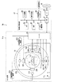
11,11a スキャナ装置
12 画像処理装置
31A,31B,31C X線管
32A,32B,32C 絞り
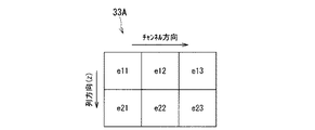
33A,33B,33C X線検出器
34A,34B,34C DAS
36A,36C 加算・スイッチ回路
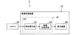
41 CPU
45 表示装置
51 前処理手段
52 画像生成手段
Claims (6)
- 第1のX線を発する第1のX線源と、
チャンネル方向及び列方向に複数の第1検出素子を備え、前記第1のX線を検出する第1の検出器と、
第2のX線を発する第2のX線源と、
チャンネル方向及び列方向に複数の第2検出素子を備え、前記第2のX線を検出する第2の検出器と、
前記第1及び第2のX線源と前記第1及び第2の検出器とを制御してスキャンを実行し、1又は列方向に複数の第1検出素子からなる複数の第1検出領域と、1又は列方向に複数の第2検出素子からなる複数の第2検出領域とでそれぞれ対応する検出領域同士が前記列方向に前記検出領域の1/2ずれた、前記第1検出領域の第1のデータと前記第2検出領域の第2のデータを取得するスキャン実行手段と、
前記取得された第1及び第2のデータに基づいて画像データを生成する画像生成手段と、
を有するX線CT装置。 - 前記第1及び第2検出領域が前記1の第1及び第2検出素子である場合、
前記第2の検出器の回転軌道は、前記第1の検出器に対して前記列方向に、前記検出素子の1/2ずれるように配置される請求項1に記載のX線CT装置。 - 同一ビューにおける前記第1及び第2の検出器の列方向の重複部分に前記第1のX線及び前記第2のX線が入射されるようにそれぞれ制御する第1及び第2の絞りをさらに有し、
前記スキャン実行手段は、前記スキャンとしてコンベンショナルスキャンを実行し、
前記画像生成手段は、前記コンベンショナルスキャンによって同一ビューにおける前記取得された第1及び第2のデータに基づいて1検出素子を列方向に2分割したエレメント毎のデータを演算し、そのデータに基づいて前記画像データを生成する請求項2に記載のX線CT装置。 - 前記スキャン実行手段は、前記スキャンとしてコンベンショナルスキャン又はヘリカルスキャンを実行し、
前記画像生成手段は、前記スキャンによって同一ビューにおける前記取得された第1のデータに基づいて第1の画像データを生成し、前記第2のデータに基づいて第2の画像データを生成し、前記第1及び第2の画像データを加算して第3の画像データを生成する請求項2に記載のX線CT装置。 - 前記第1及び第2検出領域が前記複数の第1及び第2検出素子である場合、
前記第2の検出器の回転軌道は、前記第1の検出器の回転軌道と一致するように配置される請求項1に記載のX線CT装置。 - 第1のX線を発する第1のX線源と、
チャンネル方向及び列方向に複数の第1検出素子を備え、前記第1のX線を検出する第1の検出器と、
第2のX線を発する第2のX線源と、
チャンネル方向及び列方向に複数の第2検出素子を備え、前記第2のX線を検出する第2の検出器と、
前記第1及び第2のX線源と前記第1及び第2の検出器とを制御してスキャンを実行し、チャンネル方向に複数の第1検出素子からなる複数の第1検出領域と、チャンネル方向に複数の第2検出素子からなる複数の第2検出領域とでそれぞれ対応する検出領域同士が前記チャンネル方向に前記検出領域の1/2ずれた、前記第1検出領域の第1のデータと前記第2検出領域の第2のデータを取得するスキャン実行手段と、
前記取得された第1及び第2のデータに基づいて画像データを生成する画像生成手段と、
を有するX線CT装置。
Priority Applications (3)
| Application Number | Priority Date | Filing Date | Title |
|---|---|---|---|
| JP2013109143A JP2014226376A (ja) | 2013-05-23 | 2013-05-23 | X線ct装置 |
| PCT/JP2014/063714 WO2014189133A1 (ja) | 2013-05-23 | 2014-05-23 | X線ct装置 |
| US14/877,311 US10258296B2 (en) | 2013-05-23 | 2015-10-07 | X-ray CT apparatus including processing circuitry to improve a spatial resolution in a row direction and a channel direction |
Applications Claiming Priority (1)
| Application Number | Priority Date | Filing Date | Title |
|---|---|---|---|
| JP2013109143A JP2014226376A (ja) | 2013-05-23 | 2013-05-23 | X線ct装置 |
Publications (1)
| Publication Number | Publication Date |
|---|---|
| JP2014226376A true JP2014226376A (ja) | 2014-12-08 |
Family
ID=51933683
Family Applications (1)
| Application Number | Title | Priority Date | Filing Date |
|---|---|---|---|
| JP2013109143A Ceased JP2014226376A (ja) | 2013-05-23 | 2013-05-23 | X線ct装置 |
Country Status (3)
| Country | Link |
|---|---|
| US (1) | US10258296B2 (ja) |
| JP (1) | JP2014226376A (ja) |
| WO (1) | WO2014189133A1 (ja) |
Families Citing this family (3)
| Publication number | Priority date | Publication date | Assignee | Title |
|---|---|---|---|---|
| EP2914179A1 (en) * | 2012-11-02 | 2015-09-09 | Analogic Corporation | Volumetric and projection image generation |
| JP2014230600A (ja) * | 2013-05-28 | 2014-12-11 | 株式会社東芝 | X線ct装置およびx線ct装置用x線検出器 |
| CN115105110B (zh) | 2021-10-15 | 2024-11-08 | 清华大学 | 用于射线检查的成像系统和方法 |
Citations (2)
| Publication number | Priority date | Publication date | Assignee | Title |
|---|---|---|---|---|
| JP2004325183A (ja) * | 2003-04-23 | 2004-11-18 | M & C:Kk | 放射線検出方法、放射線検出器、及び、この検出器を搭載した放射線撮像システム |
| JP2006187453A (ja) * | 2005-01-06 | 2006-07-20 | Ge Medical Systems Global Technology Co Llc | X線ct装置 |
Family Cites Families (75)
| Publication number | Priority date | Publication date | Assignee | Title |
|---|---|---|---|---|
| JP4054402B2 (ja) * | 1997-04-25 | 2008-02-27 | 株式会社東芝 | X線断層撮影装置 |
| JPH06169911A (ja) | 1992-12-04 | 1994-06-21 | Toshiba Corp | X線コンピュータトモグラフィ装置 |
| JP3340203B2 (ja) | 1993-09-17 | 2002-11-05 | 株式会社東芝 | X線ct装置 |
| US6421412B1 (en) * | 1998-12-31 | 2002-07-16 | General Electric Company | Dual cardiac CT scanner |
| US6363134B1 (en) * | 1999-01-13 | 2002-03-26 | Kabushiki Kaisha Toshiba | X-ray computed tomography apparatus |
| JP4267180B2 (ja) * | 2000-06-07 | 2009-05-27 | 株式会社日立メディコ | X線ct装置 |
| WO2002026134A1 (en) * | 2000-09-28 | 2002-04-04 | Philips Medical Systems Technologies Ltd. | Ct scanner for time-coherent large coverage |
| JP2002301056A (ja) * | 2001-04-04 | 2002-10-15 | Toshiba Medical System Co Ltd | X線ct装置およびx線検出器 |
| US6834097B2 (en) * | 2001-10-05 | 2004-12-21 | Kabushiki Kaisha Toshiba | X-ray CT apparatus and X-ray CT imaging method |
| JP4088058B2 (ja) * | 2001-10-18 | 2008-05-21 | 株式会社東芝 | X線コンピュータ断層撮影装置 |
| US7085343B2 (en) * | 2001-10-18 | 2006-08-01 | Kabushiki Kaisha Toshiba | X-ray computed tomography apparatus |
| US6922457B2 (en) * | 2001-11-29 | 2005-07-26 | Kabushiki Kaisha Toshiba | Computer tomography apparatus |
| JP2005527800A (ja) * | 2002-03-19 | 2005-09-15 | ブレークアウェイ・イメージング・エルエルシー | 大視野の対象物を画像化するシステムおよび方法 |
| JP2004000356A (ja) | 2002-06-03 | 2004-01-08 | Hitachi Medical Corp | マルチスライスx線ct装置および方法 |
| US6819738B2 (en) * | 2002-08-15 | 2004-11-16 | Ge Medical Systems Global Technology Company, Llc | Hybrid scintillator/photo sensor & direct conversion detector |
| JP4314008B2 (ja) * | 2002-10-01 | 2009-08-12 | 株式会社東芝 | X線ctスキャナ |
| DE10302565A1 (de) * | 2003-01-22 | 2004-08-12 | Siemens Ag | Bildgebendes Tomographiegerät mit wenigstens zwei Strahler-Detektor-Kombinationen |
| DE602004027358D1 (de) * | 2003-03-13 | 2010-07-08 | Philips Intellectual Property | Computertomographisches bildgebungssystem |
| DE602004024682D1 (de) * | 2003-07-15 | 2010-01-28 | Koninkl Philips Electronics Nv | Ung |
| JP3961468B2 (ja) * | 2003-09-19 | 2007-08-22 | ジーイー・メディカル・システムズ・グローバル・テクノロジー・カンパニー・エルエルシー | 放射線計算断層画像装置およびそれに用いる放射線検出器 |
| DE10354214A1 (de) * | 2003-11-20 | 2005-06-02 | Siemens Ag | Verfahren zur Erzeugung von tomographischen Schnittbildern eines sich periodisch bewegenden Objektes mit mehreren Fokus-Detektor-Kombinationen |
| DE10354900A1 (de) * | 2003-11-24 | 2005-06-30 | Siemens Ag | Verfahren zur Erzeugung von tomographischen Schnittbildern eines sich periodisch bewegenden Objektes mit mehreren Fokus-Detektor-Kombinationen |
| US7280631B2 (en) * | 2003-11-26 | 2007-10-09 | General Electric Company | Stationary computed tomography system and method |
| DE102004028124B4 (de) * | 2004-06-09 | 2008-08-07 | Siemens Ag | Bildgebendes Tomografiegerät mit mehrfachen Betriebsarten sowie ein Verfahren zum Wechsel der Betriebsart des Tomografiegeräts |
| DE102004030550A1 (de) * | 2004-06-24 | 2006-01-19 | Siemens Ag | Bildgebendes Tomographiegerät mit zumindest zwei unter Systemwinkel angeordneten Aufnahmesystemen und Verfahren für ein derartiges Tomographiegerät zur Bestimmung der Systemwinkel der Aufnahmesysteme |
| DE102004030549A1 (de) * | 2004-06-24 | 2006-01-19 | Siemens Ag | Bildgebendes Tomographiegerät mit zumindest zwei zueinander fest angeordneten Aufnahmesystemen und Verfahren für ein derartiges Tomographiegerät zur Bestimmung der Anordnung der Aufnahmesysteme |
| US7215733B2 (en) * | 2004-07-23 | 2007-05-08 | Kabushiki Kaisha Toshiba | X-ray computed tomography apparatus |
| US7103138B2 (en) * | 2004-08-24 | 2006-09-05 | The Board Of Trustees Of The Leland Stanford Junior University | Sampling in volumetric computed tomography |
| DE102004042491B4 (de) * | 2004-08-31 | 2009-07-09 | Siemens Ag | Verfahren zur Erzeugung von tomographischen Schnittbildern von einem Untersuchungsobjekt mit mindestens zwei winkelversetzten Strahlenbündeln und Computertomographie-Gerät zur Durchführung dieses Verfahrens |
| US7194061B2 (en) * | 2004-09-14 | 2007-03-20 | Kabushiki Kaisha Toshiba | X-ray computer tomography apparatus |
| JP4594699B2 (ja) * | 2004-10-29 | 2010-12-08 | 株式会社東芝 | X線コンピュータ断層撮影装置 |
| DE602006013733D1 (de) * | 2005-03-07 | 2010-06-02 | Toshiba Kk | Röntgen-CT-Gerät und Datendetektionsverfahren des Röntgen-CT-Geräts |
| US7440547B2 (en) * | 2005-04-15 | 2008-10-21 | Kabushiki Kaisha Toshiba | CT scanner |
| WO2006123581A1 (ja) * | 2005-05-18 | 2006-11-23 | Hitachi Medical Corporation | 放射線撮影装置及び画像処理プログラム |
| US7535987B2 (en) * | 2005-06-30 | 2009-05-19 | Kabushiki Kaisha Toshiba | X-ray CT apparatus |
| DE102005034876B3 (de) * | 2005-07-26 | 2007-04-05 | Siemens Ag | Verfahren zur Erstellung von computertomographischen Darstellungen durch ein CT mit mindestens zwei winkelversetzten Strahlenquellen |
| DE102005037368A1 (de) * | 2005-08-08 | 2007-02-15 | Siemens Ag | Verfahren zur Berechnung computertomographischer Aufnahmen aus Detektordaten eines CT's mit mindestens zwei Strahlenquellen |
| JP4701038B2 (ja) * | 2005-08-12 | 2011-06-15 | ジーイー・メディカル・システムズ・グローバル・テクノロジー・カンパニー・エルエルシー | X線ct装置 |
| DE102005048397A1 (de) * | 2005-10-10 | 2007-04-12 | Siemens Ag | Verfahren zur Strahlungskorrektur eines CT-Systems |
| US7831012B2 (en) * | 2006-02-09 | 2010-11-09 | L-3 Communications Security and Detection Systems Inc. | Radiation scanning systems and methods |
| US8340241B2 (en) * | 2006-02-27 | 2012-12-25 | Kabushiki Kaisha Toshiba | Image display apparatus and X-ray computed tomography apparatus |
| DE102006019923A1 (de) * | 2006-04-28 | 2007-11-15 | Siemens Ag | Verfahren zur Streustrahlungskorrektur bei einem Röntgen-CT und Röntgen-CT zur Anwendung dieses Verfahrens |
| DE102006019920B4 (de) * | 2006-04-28 | 2008-04-10 | Siemens Ag | Verfahren zur Streustrahlungskorrektur eines CT-Systems mit mindestens zwei winkelversetzt angeordneten Fokus-Detektor-Systemen und Röntgen-CT-System |
| DE102006027221B4 (de) * | 2006-06-12 | 2008-12-24 | Siemens Ag | Gerät zur medizinischen Bildgebung mit zwei Detektorsystemen |
| US7835486B2 (en) * | 2006-08-30 | 2010-11-16 | General Electric Company | Acquisition and reconstruction of projection data using a stationary CT geometry |
| US7706499B2 (en) * | 2006-08-30 | 2010-04-27 | General Electric Company | Acquisition and reconstruction of projection data using a stationary CT geometry |
| US7616731B2 (en) * | 2006-08-30 | 2009-11-10 | General Electric Company | Acquisition and reconstruction of projection data using a stationary CT geometry |
| US8488736B2 (en) * | 2006-09-19 | 2013-07-16 | General Electric Company | Stacked flat panel x-ray detector assembly and method of making same |
| US8483352B2 (en) * | 2006-09-19 | 2013-07-09 | General Electric Company | Stacked x-ray detector assembly and method of making same |
| DE102006051475A1 (de) * | 2006-10-31 | 2008-05-08 | Siemens Ag | Verfahren zur Bestimmung von Bewegungs- und Ruhephasen eines sich zeitweise bewegenden Teilobjektes bei einer CT-Untersuchung und CT-System |
| US7388940B1 (en) * | 2006-11-24 | 2008-06-17 | General Electric Company | Architectures for cardiac CT based on area x-ray sources |
| US7428292B2 (en) * | 2006-11-24 | 2008-09-23 | General Electric Company | Method and system for CT imaging using multi-spot emission sources |
| DE102006056884A1 (de) * | 2006-12-01 | 2008-06-05 | Siemens Ag | Verfahren und CT-System zur Durchführung einer Cardio-CT-Untersuchung eines Patienten |
| DE102007008118B4 (de) * | 2007-02-19 | 2011-02-03 | Siemens Ag | Verfahren zur Erzeugung tomographischer Darstellungen mit einem Röntgen-Computertomographie-System mit Streustrahlungskorrektur |
| JP2008237886A (ja) * | 2007-02-28 | 2008-10-09 | Toshiba Corp | X線ct装置及びその制御方法 |
| WO2008122971A1 (en) * | 2007-04-10 | 2008-10-16 | Arineta Ltd. | Cone-beam ct |
| JP5371758B2 (ja) * | 2007-08-06 | 2013-12-18 | 株式会社日立メディコ | X線ct装置 |
| US7433443B1 (en) * | 2007-08-29 | 2008-10-07 | General Electric Company | System and method of CT imaging with second tube/detector patching |
| JP5052281B2 (ja) * | 2007-10-02 | 2012-10-17 | 株式会社東芝 | X線ctにおける散乱線強度分布の推定方法およびx線ct装置 |
| US7801265B2 (en) * | 2007-11-23 | 2010-09-21 | Mayo Foundation For Medical Education And Research | System and method for creating mixed image from dual-energy CT data |
| US7949089B2 (en) * | 2008-04-10 | 2011-05-24 | Arineta Ltd. | Apparatus and method for tracking feature's position in human body |
| DE102008030552A1 (de) * | 2008-06-27 | 2009-12-31 | Siemens Aktiengesellschaft | Verfahren zur Erzeugung von Bilddaten zu einer virtuell vorgebbaren Röntgenröhrenspannung aus ersten und zweiten CT-Bilddaten |
| JP5159543B2 (ja) * | 2008-09-30 | 2013-03-06 | 株式会社東芝 | X線ct装置 |
| JP5537132B2 (ja) * | 2008-12-11 | 2014-07-02 | 株式会社東芝 | X線コンピュータ断層撮影装置、医用画像処理装置、及び医用画像処理プログラム |
| JP2011104075A (ja) * | 2009-11-17 | 2011-06-02 | Toshiba Corp | X線ct装置 |
| US8594272B2 (en) * | 2010-03-19 | 2013-11-26 | Triple Ring Technologies, Inc. | Inverse geometry volume computed tomography systems |
| CN102665564B (zh) * | 2010-11-12 | 2015-04-15 | 株式会社东芝 | 图像诊断装置及位置校正方法 |
| JP6033086B2 (ja) * | 2010-12-21 | 2016-11-30 | タカラテレシステムズ株式会社 | 放射線検出器、及び、この検出器を備えた放射線撮像装置 |
| US8774351B2 (en) * | 2011-04-05 | 2014-07-08 | Triple Ring Technologies, Inc. | Method and apparatus for advanced X-ray imaging systems |
| WO2013089154A1 (ja) * | 2011-12-12 | 2013-06-20 | 株式会社 日立メディコ | X線ct装置 |
| JP6195337B2 (ja) * | 2012-02-24 | 2017-09-13 | 東芝メディカルシステムズ株式会社 | X線ct装置 |
| WO2013191001A1 (ja) * | 2012-06-20 | 2013-12-27 | 株式会社日立メディコ | X線ct装置 |
| WO2014034888A1 (ja) * | 2012-08-30 | 2014-03-06 | 株式会社東芝 | X線ct装置、画像処理装置、及び画像処理方法 |
| WO2014106956A1 (ja) * | 2013-01-07 | 2014-07-10 | 株式会社東芝 | X線コンピュータ断層撮影装置及び医用画像処理装置 |
| DE102013203541A1 (de) * | 2013-03-01 | 2014-09-04 | Siemens Aktiengesellschaft | Röntgen-CT-Abtastung und Dual-Source-CT-System |
-
2013
- 2013-05-23 JP JP2013109143A patent/JP2014226376A/ja not_active Ceased
-
2014
- 2014-05-23 WO PCT/JP2014/063714 patent/WO2014189133A1/ja not_active Ceased
-
2015
- 2015-10-07 US US14/877,311 patent/US10258296B2/en active Active
Patent Citations (2)
| Publication number | Priority date | Publication date | Assignee | Title |
|---|---|---|---|---|
| JP2004325183A (ja) * | 2003-04-23 | 2004-11-18 | M & C:Kk | 放射線検出方法、放射線検出器、及び、この検出器を搭載した放射線撮像システム |
| JP2006187453A (ja) * | 2005-01-06 | 2006-07-20 | Ge Medical Systems Global Technology Co Llc | X線ct装置 |
Also Published As
| Publication number | Publication date |
|---|---|
| WO2014189133A1 (ja) | 2014-11-27 |
| US20160022237A1 (en) | 2016-01-28 |
| US10258296B2 (en) | 2019-04-16 |
Similar Documents
| Publication | Publication Date | Title |
|---|---|---|
| US10219775B2 (en) | Photon-counting X-ray CT apparatus and image processing apparatus | |
| US11244480B2 (en) | Medical information processing apparatus | |
| JP3961468B2 (ja) | 放射線計算断層画像装置およびそれに用いる放射線検出器 | |
| JP5761972B2 (ja) | X線ct装置 | |
| JP6494982B2 (ja) | X線コンピュータ断層撮影装置、画像処理装置及び画像処理プログラム | |
| JP2007300964A (ja) | 放射線撮影装置および放射線撮影方法 | |
| JP2020103571A (ja) | 医用処理装置及びx線診断システム | |
| Ji et al. | Task-driven optimization of the non-spectral mode of photon counting CT for intracranial hemorrhage assessment | |
| JP2024178147A (ja) | X線ct装置及びデータ処理方法 | |
| US20240065654A1 (en) | Photon counting x-ray image diagnosis apparatus and method for generating calibration data for pileup correction | |
| US20210052236A1 (en) | X-ray imaging apparatus, medical information processing apparatus, x-ray detector, and correction method of x-ray detector | |
| JP2014226376A (ja) | X線ct装置 | |
| US20250182881A1 (en) | Medical image processing apparatus and x-ray computed tomography apparatus | |
| JP2015160139A (ja) | X線コンピュータ断層撮像装置、及びx線コンピュータ断層撮像装置におけるレジストレーション方法 | |
| JP5872212B2 (ja) | マルチスライスct装置及びデータ前処理方法 | |
| US20230404514A1 (en) | Medical data processing method, model generating method, and medical data processing apparatus | |
| EP4092617B1 (en) | METHOD FOR PROCESSING MEDICAL INFORMATION AND APPARATUS FOR PROCESSING MEDICAL IMAGES | |
| JP2024082589A (ja) | 医用画像処理装置、および医用画像処理方法 | |
| JP7399720B2 (ja) | X線ct装置 | |
| JP2015123302A (ja) | 医用画像診断装置及び画像処理装置 | |
| US11478203B2 (en) | X-ray computed tomography apparatus and imaging condition management apparatus | |
| JP2025071076A (ja) | 光子計数型放射線診断装置、方法及びプログラム | |
| JP2020065842A (ja) | 医用画像処理装置 | |
| JP6643019B2 (ja) | 画像処理装置、画像処理方法およびプログラム | |
| JP2024066111A (ja) | 医用画像処理装置及び方法 |
Legal Events
| Date | Code | Title | Description |
|---|---|---|---|
| A711 | Notification of change in applicant |
Free format text: JAPANESE INTERMEDIATE CODE: A711 Effective date: 20160506 |
|
| A621 | Written request for application examination |
Free format text: JAPANESE INTERMEDIATE CODE: A621 Effective date: 20160520 |
|
| A131 | Notification of reasons for refusal |
Free format text: JAPANESE INTERMEDIATE CODE: A131 Effective date: 20161025 |
|
| A521 | Request for written amendment filed |
Free format text: JAPANESE INTERMEDIATE CODE: A523 Effective date: 20161226 |
|
| A01 | Written decision to grant a patent or to grant a registration (utility model) |
Free format text: JAPANESE INTERMEDIATE CODE: A01 Effective date: 20170131 |
|
| A045 | Written measure of dismissal of application [lapsed due to lack of payment] |
Free format text: JAPANESE INTERMEDIATE CODE: A045 Effective date: 20170530 |