JP2017129593A - 試料の表示およびレビューのためのシステムおよび方法 - Google Patents
試料の表示およびレビューのためのシステムおよび方法 Download PDFInfo
- Publication number
- JP2017129593A JP2017129593A JP2017043947A JP2017043947A JP2017129593A JP 2017129593 A JP2017129593 A JP 2017129593A JP 2017043947 A JP2017043947 A JP 2017043947A JP 2017043947 A JP2017043947 A JP 2017043947A JP 2017129593 A JP2017129593 A JP 2017129593A
- Authority
- JP
- Japan
- Prior art keywords
- cell
- cells
- image
- images
- sample
- Prior art date
- Legal status (The legal status is an assumption and is not a legal conclusion. Google has not performed a legal analysis and makes no representation as to the accuracy of the status listed.)
- Granted
Links
Images







Classifications
-
- G—PHYSICS
- G01—MEASURING; TESTING
- G01N—INVESTIGATING OR ANALYSING MATERIALS BY DETERMINING THEIR CHEMICAL OR PHYSICAL PROPERTIES
- G01N15/00—Investigating characteristics of particles; Investigating permeability, pore-volume or surface-area of porous materials
- G01N15/10—Investigating individual particles
- G01N15/14—Optical investigation techniques, e.g. flow cytometry
- G01N15/1429—Signal processing
- G01N15/1433—Signal processing using image recognition
-
- G—PHYSICS
- G06—COMPUTING OR CALCULATING; COUNTING
- G06T—IMAGE DATA PROCESSING OR GENERATION, IN GENERAL
- G06T7/00—Image analysis
- G06T7/0002—Inspection of images, e.g. flaw detection
- G06T7/0012—Biomedical image inspection
-
- G—PHYSICS
- G06—COMPUTING OR CALCULATING; COUNTING
- G06T—IMAGE DATA PROCESSING OR GENERATION, IN GENERAL
- G06T7/00—Image analysis
- G06T7/10—Segmentation; Edge detection
- G06T7/136—Segmentation; Edge detection involving thresholding
-
- G—PHYSICS
- G01—MEASURING; TESTING
- G01N—INVESTIGATING OR ANALYSING MATERIALS BY DETERMINING THEIR CHEMICAL OR PHYSICAL PROPERTIES
- G01N15/00—Investigating characteristics of particles; Investigating permeability, pore-volume or surface-area of porous materials
- G01N15/10—Investigating individual particles
- G01N15/14—Optical investigation techniques, e.g. flow cytometry
- G01N15/1429—Signal processing
-
- G—PHYSICS
- G01—MEASURING; TESTING
- G01N—INVESTIGATING OR ANALYSING MATERIALS BY DETERMINING THEIR CHEMICAL OR PHYSICAL PROPERTIES
- G01N15/00—Investigating characteristics of particles; Investigating permeability, pore-volume or surface-area of porous materials
- G01N15/01—Investigating characteristics of particles; Investigating permeability, pore-volume or surface-area of porous materials specially adapted for biological cells, e.g. blood cells
- G01N2015/012—Red blood cells
-
- G—PHYSICS
- G01—MEASURING; TESTING
- G01N—INVESTIGATING OR ANALYSING MATERIALS BY DETERMINING THEIR CHEMICAL OR PHYSICAL PROPERTIES
- G01N15/00—Investigating characteristics of particles; Investigating permeability, pore-volume or surface-area of porous materials
- G01N15/10—Investigating individual particles
- G01N15/14—Optical investigation techniques, e.g. flow cytometry
- G01N15/1468—Optical investigation techniques, e.g. flow cytometry with spatial resolution of the texture or inner structure of the particle
- G01N2015/1472—Optical investigation techniques, e.g. flow cytometry with spatial resolution of the texture or inner structure of the particle with colour
-
- G—PHYSICS
- G01—MEASURING; TESTING
- G01N—INVESTIGATING OR ANALYSING MATERIALS BY DETERMINING THEIR CHEMICAL OR PHYSICAL PROPERTIES
- G01N35/00—Automatic analysis not limited to methods or materials provided for in any single one of groups G01N1/00 - G01N33/00; Handling materials therefor
- G01N35/00584—Control arrangements for automatic analysers
- G01N35/00722—Communications; Identification
- G01N2035/00891—Displaying information to the operator
- G01N2035/0091—GUI [graphical user interfaces]
-
- G—PHYSICS
- G06—COMPUTING OR CALCULATING; COUNTING
- G06T—IMAGE DATA PROCESSING OR GENERATION, IN GENERAL
- G06T2207/00—Indexing scheme for image analysis or image enhancement
- G06T2207/10—Image acquisition modality
- G06T2207/10056—Microscopic image
-
- G—PHYSICS
- G06—COMPUTING OR CALCULATING; COUNTING
- G06T—IMAGE DATA PROCESSING OR GENERATION, IN GENERAL
- G06T2207/00—Indexing scheme for image analysis or image enhancement
- G06T2207/30—Subject of image; Context of image processing
- G06T2207/30004—Biomedical image processing
- G06T2207/30024—Cell structures in vitro; Tissue sections in vitro
Landscapes
- Engineering & Computer Science (AREA)
- Physics & Mathematics (AREA)
- General Physics & Mathematics (AREA)
- Computer Vision & Pattern Recognition (AREA)
- Theoretical Computer Science (AREA)
- Health & Medical Sciences (AREA)
- General Health & Medical Sciences (AREA)
- Quality & Reliability (AREA)
- Radiology & Medical Imaging (AREA)
- Medical Informatics (AREA)
- Nuclear Medicine, Radiotherapy & Molecular Imaging (AREA)
- Chemical & Material Sciences (AREA)
- Life Sciences & Earth Sciences (AREA)
- Dispersion Chemistry (AREA)
- Signal Processing (AREA)
- Analytical Chemistry (AREA)
- Biochemistry (AREA)
- Immunology (AREA)
- Pathology (AREA)
- Investigating Or Analysing Biological Materials (AREA)
- Investigating Or Analysing Materials By Optical Means (AREA)
- Apparatus Associated With Microorganisms And Enzymes (AREA)
Abstract
Description
本出願は、米国特許法第119条(e)の下で、2011年6月17日に出願された米国仮特許出願第61/498,456号、2011年7月22日に出願された米国仮特許出願第61/510,614号、2011年7月22日に出願された米国仮特許出願番号第61/510,696号、および2011年7月22日に出願された米国仮特許出願第61/510,710号に基づく優先権を主張する。これらの出願の開示内容の全ては、参照により本明細書に組み込まれる。
本開示は、情報を整理および表示することに関し、試料画像を表示することを含む。
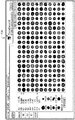
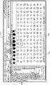
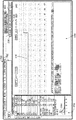
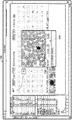
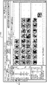
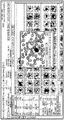
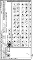
図7は、赤血球画像の表示のための、グラフィカルユーザインタフェース700の一実施形態の画像を示す。インタフェース700は、識別子ブロック702、画像ブロック704、ソートブロック706、およびデータブロック708を含んでいる。識別子ブロック702は、技術者によってレビューされている試料についての、様々な情報を表示するために用いられる。この情報は、患者の氏名、関連する診療記録番号、試料番号、患者の生年月日、年齢、性別および人種、予備的診断、主治医の氏名および科、病院または検査室の名前、ならびに血液試料の抜き取り時間を含んでいる。
(a)106/マイクロリットルの単位で記録され得る、試料中の赤血球数(RBC)
(b)赤血球数と平均細胞ヘモグロビン濃度との積から計算でき、g/dLの単位で記録され得る、試料中のヘモグロビン濃度(Hgb)
(c)平均細胞体積と試料体積で割られた赤血球の合計数との積から計算でき、百分率で記録され得る、試料のヘマトクリット(Hct)
(d)fLの単位で記録され得る、平均細胞体積(MCV)
(e)pgの単位で記録され得る、平均細胞ヘモグロビン(MCH)
(f)MCH/MCVの比率として計算でき、赤血球中のヘモグロビンの濃度に対応する、g/dLの単位で記録され得る平均細胞ヘモグロビン濃度(MCHC)
(g)試料中で特定され調べられた個別の赤血球の体積平均で割られた、個別の赤血球体積の標準偏差から計算でき、百分率で記録され得る赤血球分布幅(RDW−CV)
(h)試料中の赤血球体積の分布のヒストグラムに基づき決定され得る、赤血球分布幅(RDW−SD)
(i)試料中の有核赤血球の数に対応し、103/マイクロリットルの単位で記録され得る、有核赤血球数(NRBC)
(j)試料中で特定された有核細胞、すなわち赤血球の割合と対応する、有核赤血球の割合(NRBC%)
(k)試料中の網状赤血球の数に対応し、106/マイクロリットルの単位で記録され得る、網状赤血球(Retic)
(l)網状赤血球と特定された試料中の全ての赤血球の割合に対応する、網状赤血球の割合(Retic%)
(m)網状赤血球中のヘモグロビン濃度に対応し、g/dLの単位で記録され得る、網状赤血球ヘモグロビン(RetHE)
(a)103/マイクロリットルの単位で記録され得る、試料の白血球数(WBC)
(b)103/マイクロリットルの単位、および/または、試料中の全ての白血球の中での好中血球の割合(%NEUT)で記録され得る、試料の好中血球数(NEUT)
(c)103/マイクロリットルの単位、および/または、試料中の全ての白血球の中でのリンパ球の割合(%LYMPH)で記録され得る、試料のリンパ球数(LYMPH)
(d)103/マイクロリットルの単位、および/または、試料中の全ての白血球の中での単球の割合(%MONO)で記録され得る、試料の単球数(MONO)
(e)103/マイクロリットルの単位、および/または、試料中の全ての白血球の中での好酸球の割合(%EOS)で記録され得る、試料の好酸球数(EOS)
(f)103/マイクロリットルの単位、および/または、試料中の全ての白血球の中での好塩基球の割合(%BASO)で記録され得る、試料の好塩基球数(BASO)、および
(g)103/マイクロリットルの単位、および/または、試料中の全ての白血球の中での分類されていない細胞の割合(%UC)で記録され得る、分類されていない細胞数(UC)

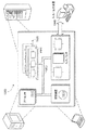
図1は、CBC分析の実行を含む、血液試料の調製および検査のための、自動化されたシステム1000の概略図を示す。システム1000は、基板を保管し、基板上に試料を配置し、基板上に調製された試料を検査し、調製された試料を保管するために、複数のサブシステムを含んでいる。
本明細書に記載される方法ステップおよび工程は、ハードウェアまたはソフトウェア、もしくはそれらの組み合わせに実装され得る。詳細には、電子プロセッサ114は上述した方法ステップのいずれかを行うために、ソフトウェアおよび/またはハードウェアの命令を含み得る。この方法は、本明細書に開示する方法ステップおよび図面に付随する、標準のプログラミング技術を用いて、コンピュータプログラムに実装され得る。本明細書に記載する機能を行うために、プログラムコードが入力データに加えられる。出力情報は、プリンタなどの出力装置、表示装置、または、例えば遠隔監視のためにウェブサイトへのアクセスを有するコンピュータモニタ上のウェブページなどの、1つまたは2つ以上に用いられる。
多数の実施形態が記載されている。それでもなお、本開示の要旨および範囲を逸脱しない限りは、様々な変更が行われ得ることを理解されたい。詳細には、特定の実施形態に関連する本明細書に開示した特徴は、一般に、他の実施形態に含まれてもよく、本明細書に開示する特定の特徴は、一般に、本明細書に開示する実施形態の他のいずれの特徴と組み合わせて用いられてもよい。したがって、他の実施形態は以下の特許請求の範囲内にある。
Claims (19)
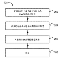
- 試料中の細胞画像を表示するための方法であって、該方法が、
それぞれが前記試料中の細胞の1つに対応している、前記試料中の複数の細胞画像を取得すること、
複数の前記画像に基づき、前記細胞それぞれに関する少なくとも1つの特性の値を測定すること、
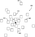
複数の前記画像を順不同に配列し、第1の画像アレイを形成すること、
前記第1の画像アレイを表示すること、
複数の前記画像を配列し、前記画像が前記少なくとも1つの特性の値に基づいて順序付けされている、第2の画像アレイを形成すること、および
前記第2の画像アレイを表示すること
を含む方法。 - 前記試料が血液を含み、前記細胞が赤血球を含むことを特徴とする請求項1記載の方法。
- 前記試料が血液を含み、前記細胞が血小板を含むことを特徴とする請求項1記載の方法。
- 前記第2の画像アレイの各画像が、ユーザが選択可能な制御として表示されることを特徴とする請求項1記載の方法。
- ユーザが選択可能な制御のそれぞれが、作動すると、対応する前記試料中の細胞についての情報が表示されるように構成されていることを特徴とする請求項4記載の方法。
- 前記少なくとも1つの特性の値が、前記細胞それぞれの細胞ヘモグロビン量を含んでいることを特徴とする請求項1記載の方法。
- 前記少なくとも1つの特性の値が、前記細胞それぞれの細胞体積を含んでいることを特徴とする請求項1記載の方法。
- 前記少なくとも1つの特性の値が、前記細胞それぞれの細胞の大きさを含んでいることを特徴とする請求項1記載の方法。
- 前記少なくとも1つの特性の値が、前記細胞それぞれの光学密度を含んでいることを特徴とする請求項1記載の方法。
- 前記少なくとも1つの特性の値が、前記細胞それぞれの形状を含んでいることを特徴とする請求項1記載の方法。
- 複数の前記画像のそれぞれに対して、前記画像に関連する前記細胞には対応していない、画像の背景画素のセットを特定することをさらに備え、
前記第2の画像アレイの表示が、複数の前記画像それぞれに対して、前記背景画素のセットのそれぞれの要素に色を割り当てることを含み、
前記色が、前記画像に関連する前記細胞に関する、前記少なくとも1つの特性の値に基づいて割り当てられていることを特徴とする請求項1記載の方法。 - 細胞の視覚的な検査のためのシステムであって、該システムが、
試料中の細胞を照射するように構成される光源と、
それぞれが前記試料中の細胞の1つに対応する、複数の前記細胞の画像を取得するように構成される検出器と、
前記細胞の画像を表示するように構成される表示装置と、
複数の前記画像に基づき、前記細胞のそれぞれに関する少なくとも1つの特性の値を測定し、
複数の前記画像を順不同に配列して、第1の画像アレイを形成し、
前記第1の画像アレイを表示し、
複数の前記画像を配列して、前記画像が前記少なくとも1つの特性の値に基づいて順序付けられている、第2の画像アレイを形成し、
前記第2の画像アレイを前記表示装置上に表示するように構成される電子処理装置と
を備えるシステム。 - 前記表示装置が、前記第2の画像アレイの各画像を、ユーザが選択可能な制御として表示するように構成されていることを特徴とする請求項12記載のシステム。
- 前記電子処理装置が、前記ユーザが選択可能な制御のうちの1つが作動すると、作動した前記制御に対応する前記細胞についての情報を、前記表示装置上に表示するように構成されることを特徴とする請求項13記載のシステム。
- 前記少なくとも1つの特性の値が、前記細胞それぞれの細胞ヘモグロビン量を含むことを特徴とする請求項12記載のシステム。
- 前記少なくとも1つの特性の値が、前記細胞それぞれの細胞体積を含むことを特徴とする請求項12記載のシステム。
- 前記少なくとも1つの特性の値が、前記細胞それぞれの細胞の大きさ、前記細胞それぞれの光学密度、および前記細胞それぞれの形状からなる群のうち、少なくとも1つの要素を含んでいることを特徴とする請求項12記載のシステム。
- 前記電子処理装置がさらに、前記細胞中の封入体を特定するために複数の前記画像を分析するように構成され、前記少なくとも1つの特性が前記封入体に関する特性であり、前記封入体の大きさ、前記封入体の形状、および前記封入体の数の、少なくとも1つを含んでいることを特徴とする請求項12記載のシステム。
- 前記電子処理装置がさらに、複数の前記画像のそれぞれに対して、
前記画像に関連する前記細胞には対応していない、前記画像中の背景画素のセットを特定し、および
前記背景画素のセットのそれぞれの要素に色を割り当てることにより、前記第2の画像アレイの画像を表示するように構成されており、
前記色は、前記画像に関連する前記細胞に関する、前記少なくとも1つの特性の値に基づいて割り当てられていることを特徴とする請求項12記載のシステム。
Priority Applications (1)
| Application Number | Priority Date | Filing Date | Title |
|---|---|---|---|
| JP2019076632A JP6926141B2 (ja) | 2011-06-17 | 2019-04-12 | 試料の表示およびレビューのためのシステムおよび方法 |
Applications Claiming Priority (8)
| Application Number | Priority Date | Filing Date | Title |
|---|---|---|---|
| US201161498456P | 2011-06-17 | 2011-06-17 | |
| US61/498,456 | 2011-06-17 | ||
| US201161510614P | 2011-07-22 | 2011-07-22 | |
| US201161510696P | 2011-07-22 | 2011-07-22 | |
| US201161510710P | 2011-07-22 | 2011-07-22 | |
| US61/510,696 | 2011-07-22 | ||
| US61/510,710 | 2011-07-22 | ||
| US61/510,614 | 2011-07-22 |
Related Parent Applications (1)
| Application Number | Title | Priority Date | Filing Date |
|---|---|---|---|
| JP2014516079A Division JP6109822B2 (ja) | 2011-06-17 | 2012-06-18 | 試料の表示およびレビューのためのシステムおよび方法 |
Related Child Applications (1)
| Application Number | Title | Priority Date | Filing Date |
|---|---|---|---|
| JP2019076632A Division JP6926141B2 (ja) | 2011-06-17 | 2019-04-12 | 試料の表示およびレビューのためのシステムおよび方法 |
Publications (2)
| Publication Number | Publication Date |
|---|---|
| JP2017129593A true JP2017129593A (ja) | 2017-07-27 |
| JP6514726B2 JP6514726B2 (ja) | 2019-05-15 |
Family
ID=46465289
Family Applications (3)
| Application Number | Title | Priority Date | Filing Date |
|---|---|---|---|
| JP2014516079A Active JP6109822B2 (ja) | 2011-06-17 | 2012-06-18 | 試料の表示およびレビューのためのシステムおよび方法 |
| JP2017043947A Active JP6514726B2 (ja) | 2011-06-17 | 2017-03-08 | 試料の表示およびレビューのためのシステムおよび方法 |
| JP2019076632A Active JP6926141B2 (ja) | 2011-06-17 | 2019-04-12 | 試料の表示およびレビューのためのシステムおよび方法 |
Family Applications Before (1)
| Application Number | Title | Priority Date | Filing Date |
|---|---|---|---|
| JP2014516079A Active JP6109822B2 (ja) | 2011-06-17 | 2012-06-18 | 試料の表示およびレビューのためのシステムおよび方法 |
Family Applications After (1)
| Application Number | Title | Priority Date | Filing Date |
|---|---|---|---|
| JP2019076632A Active JP6926141B2 (ja) | 2011-06-17 | 2019-04-12 | 試料の表示およびレビューのためのシステムおよび方法 |
Country Status (8)
| Country | Link |
|---|---|
| US (2) | US11047791B2 (ja) |
| EP (1) | EP2721392B1 (ja) |
| JP (3) | JP6109822B2 (ja) |
| CN (2) | CN103930762B (ja) |
| AU (1) | AU2012271257B2 (ja) |
| CA (1) | CA2839531A1 (ja) |
| ES (1) | ES2882791T3 (ja) |
| WO (1) | WO2012174542A2 (ja) |
Cited By (2)
| Publication number | Priority date | Publication date | Assignee | Title |
|---|---|---|---|---|
| CN108776085A (zh) * | 2018-05-11 | 2018-11-09 | 柘城惠丰钻石科技股份有限公司 | 一种超硬材料微粉专用光学检测仪 |
| JP2020056623A (ja) * | 2018-09-28 | 2020-04-09 | シスメックス株式会社 | 表示方法、検体分析装置、コンピュータプログラムおよび記録媒体 |
Families Citing this family (46)
| Publication number | Priority date | Publication date | Assignee | Title |
|---|---|---|---|---|
| US9164112B2 (en) * | 2008-09-03 | 2015-10-20 | Hitachi High-Technologies Corporation | Automatic analyzer |
| US9075225B2 (en) | 2009-10-28 | 2015-07-07 | Alentic Microscience Inc. | Microscopy imaging |
| US20140152801A1 (en) | 2009-10-28 | 2014-06-05 | Alentic Microscience Inc. | Detecting and Using Light Representative of a Sample |
| JP2013509618A (ja) | 2009-10-28 | 2013-03-14 | アレンティック マイクロサイエンス インコーポレイテッド | 顕微撮像法 |
| JP5997256B2 (ja) | 2011-04-15 | 2016-09-28 | コンスティテューション・メディカル・インコーポレイテッドConstitution Medical, Inc. | 細胞の体積および成分の計測 |
| US9026531B2 (en) * | 2012-04-17 | 2015-05-05 | Cerner Innovation, Inc. | Associating multiple data sources into a web-accessible framework |
| JP5333635B1 (ja) * | 2012-08-23 | 2013-11-06 | 富士ゼロックス株式会社 | 画像処理装置、プログラム及び画像処理システム |
| JP5698208B2 (ja) * | 2012-11-30 | 2015-04-08 | 株式会社Screenホールディングス | 画像処理装置、画像処理方法、および画像処理プログラム |
| EP2954310B1 (en) * | 2013-02-06 | 2024-01-03 | Alentic Microscience Inc. | Detecting and using light representative of a sample |
| US10502666B2 (en) | 2013-02-06 | 2019-12-10 | Alentic Microscience Inc. | Sample processing improvements for quantitative microscopy |
| AU2014249011A1 (en) * | 2013-03-11 | 2015-09-24 | Roche Diagnostics Hematology, Inc. | Imaging blood cells |
| US9646376B2 (en) * | 2013-03-15 | 2017-05-09 | Hologic, Inc. | System and method for reviewing and analyzing cytological specimens |
| JP6455829B2 (ja) * | 2013-04-01 | 2019-01-23 | キヤノン株式会社 | 画像処理装置、画像処理方法、およびプログラム |
| WO2014161585A1 (en) * | 2013-04-05 | 2014-10-09 | Bastidas García Óscar | Particle counting system adaptable to an optical instrument |
| USD802608S1 (en) * | 2013-04-25 | 2017-11-14 | Life Technologies Corporation | Display screen with graphical user interface |
| SE537088C2 (sv) * | 2013-05-08 | 2015-01-07 | Cellavision Ab | Grafiskt användargränssnitt för analys av röda blodkroppar |
| WO2014183049A1 (en) * | 2013-05-09 | 2014-11-13 | Abbott Point Of Care, Inc. | Method and apparatus for determining hemoglobin based parameters in an unlysed blood sample |
| WO2014205576A1 (en) | 2013-06-26 | 2014-12-31 | Alentic Microscience Inc. | Sample processing improvements for microscopy |
| WO2016080442A1 (ja) * | 2014-11-21 | 2016-05-26 | 住友電気工業株式会社 | 品質評価方法及び品質評価装置 |
| CN113763377B (zh) * | 2015-01-31 | 2025-10-31 | 文塔纳医疗系统公司 | 用于使用载片缩略图图像的感兴趣区域检测的系统和方法 |
| US9928403B2 (en) * | 2016-02-09 | 2018-03-27 | Molecular Devices, Llc | System and method for image analysis of multi-dimensional data |
| JP6776672B2 (ja) * | 2016-07-08 | 2020-10-28 | 住友電気工業株式会社 | 品質評価方法及び品質評価装置 |
| US10489633B2 (en) * | 2016-09-27 | 2019-11-26 | Sectra Ab | Viewers and related methods, systems and circuits with patch gallery user interfaces |
| JP6832165B2 (ja) * | 2017-01-16 | 2021-02-24 | オリンパス株式会社 | 観察システム |
| JP6847204B2 (ja) * | 2017-04-14 | 2021-03-24 | 株式会社日立ハイテク | 荷電粒子線装置および細胞評価方法 |
| US11321585B2 (en) * | 2017-04-14 | 2022-05-03 | Hitachi High-Tech Corporation | Imaging device and morphological feature data display method |
| JP7510742B2 (ja) * | 2018-11-19 | 2024-07-04 | アークレイ株式会社 | 情報処理装置、測定システム、及びプログラム |
| JP7293907B2 (ja) * | 2019-06-25 | 2023-06-20 | オムロン株式会社 | 外観検査管理システム、外観検査管理装置、外観検査管理方法及びプログラム |
| JP2021040590A (ja) * | 2019-09-13 | 2021-03-18 | シスメックス株式会社 | 細胞画像解析方法、細胞画像解析装置、プログラム、及び細胞画像解析システム |
| WO2021097630A1 (zh) * | 2019-11-18 | 2021-05-27 | 深圳迈瑞生物医疗电子股份有限公司 | 一种血液细胞检测结果处理方法、系统及存储介质 |
| WO2021152089A1 (en) * | 2020-01-30 | 2021-08-05 | Vitadx International | Systematic characterization of objects in a biological sample |
| EP3876193A1 (de) | 2020-03-02 | 2021-09-08 | Euroimmun Medizinische Labordiagnostika AG | Bildverarbeitungsverfahren zum anzeigen von zellen mehrerer gesamtbilder |
| CN111583186A (zh) * | 2020-04-14 | 2020-08-25 | 山东省千佛山医院 | 面向临床应用的病理er/pr细胞核计数方法及系统 |
| US11749569B2 (en) * | 2020-05-06 | 2023-09-05 | Taiwan Semiconductor Manufacturing Company, Ltd. | Method for non-destructive inspection of cell etch redeposition |
| DE102020125929A1 (de) * | 2020-05-06 | 2021-11-11 | Taiwan Semiconductor Manufacturing Co., Ltd. | Verfahren zur nicht destruktiven überprüfung parasitärer ätzabscheidungen auf zellen |
| JP6864279B1 (ja) * | 2020-08-13 | 2021-04-28 | 株式会社宮本製作所 | 情報収集方法、情報収集システムおよび情報収集装置 |
| CN114255833A (zh) * | 2020-09-25 | 2022-03-29 | 深圳迈瑞生物医疗电子股份有限公司 | 一种样本分析系统 |
| CN114660057A (zh) * | 2020-12-23 | 2022-06-24 | 深圳迈瑞生物医疗电子股份有限公司 | 生成用于细胞分析的图形用户界面的方法、装置和系统 |
| CN117043866A (zh) * | 2021-02-08 | 2023-11-10 | Cdx医疗Ip公司 | 按照细胞和组织的诊断重要性顺序动态优先处理、组织和显示细胞和组织 |
| JP7552491B2 (ja) * | 2021-04-22 | 2024-09-18 | 株式会社島津製作所 | 粒子画像解析装置 |
| WO2022235564A1 (en) * | 2021-05-03 | 2022-11-10 | Memorial Sloan Kettering Cancer Center | Detection of labels in biomedical images |
| CN115700791A (zh) * | 2021-07-30 | 2023-02-07 | 深圳市瑞图生物技术有限公司 | 精子分析结果显示方法、装置、计算机设备和存储介质 |
| JP2023131196A (ja) * | 2022-03-09 | 2023-09-22 | シスメックス株式会社 | 精度管理情報の生成方法、精度管理情報の生成装置、及びプログラム |
| AU2024251002B2 (en) * | 2023-04-11 | 2025-11-20 | Marquette University | Image cytometry-based non-invasive white blood cell count from fingertip videos |
| WO2025122591A1 (en) * | 2023-12-06 | 2025-06-12 | Idexx Laboratories, Inc. | Digital microscopy data visualiation systems and methods for using the same |
| WO2025131829A1 (en) | 2023-12-18 | 2025-06-26 | F. Hoffmann-La Roche Ag | Method and system for applying a liquid sample onto a substrate for image analysis |
Citations (9)
| Publication number | Priority date | Publication date | Assignee | Title |
|---|---|---|---|---|
| JPH0829317A (ja) * | 1994-07-18 | 1996-02-02 | Hitachi Ltd | 粒子自動分類システム |
| JP2000353246A (ja) * | 1999-06-11 | 2000-12-19 | Hitachi Ltd | パターン認識装置 |
| JP2003298995A (ja) * | 2002-02-22 | 2003-10-17 | Eastman Kodak Co | ユーザの選択可能なタスクのリストを供給する画像アプリケーションソフトウェア |
| JP3916395B2 (ja) * | 1997-07-17 | 2007-05-16 | アキュメッド インターナショナル インコーポレイテッド | 検体の予備処理機能を備えた検査体系 |
| JP2008064534A (ja) * | 2006-09-06 | 2008-03-21 | Olympus Corp | 細胞画像処理装置および細胞画像処理方法 |
| JP2009212626A (ja) * | 2008-03-03 | 2009-09-17 | Sysmex Corp | 細胞画像表示システム、細胞画像表示装置およびコンピュータプログラム |
| US20100023675A1 (en) * | 2008-07-23 | 2010-01-28 | Phison Electronics Corp. | Wear leveling method, and storage system and controller using the same |
| JP2010151647A (ja) * | 2008-12-25 | 2010-07-08 | Sysmex Corp | 細胞画像表示装置 |
| US20110070606A1 (en) * | 2008-04-25 | 2011-03-24 | Constitution Medical, Inc. | Systems and methods for analyzing body fluids |
Family Cites Families (47)
| Publication number | Priority date | Publication date | Assignee | Title |
|---|---|---|---|---|
| US4612614A (en) * | 1980-09-12 | 1986-09-16 | International Remote Imaging Systems, Inc. | Method of analyzing particles in a fluid sample |
| GB8313739D0 (en) | 1983-05-18 | 1983-06-22 | Analytical Instr Ltd | Image analysers |
| US4965725B1 (en) | 1988-04-08 | 1996-05-07 | Neuromedical Systems Inc | Neural network based automated cytological specimen classification system and method |
| US5740270A (en) | 1988-04-08 | 1998-04-14 | Neuromedical Systems, Inc. | Automated cytological specimen classification system and method |
| US5544650A (en) | 1988-04-08 | 1996-08-13 | Neuromedical Systems, Inc. | Automated specimen classification system and method |
| US5123055A (en) | 1989-08-10 | 1992-06-16 | International Remote Imaging Systems, Inc. | Method and an apparatus for differentiating a sample of biological cells |
| EP0556286B1 (en) | 1990-11-07 | 1999-04-07 | Neuromedical Systems, Inc | Method with inspection auditing for images presented on a display |
| US5655029A (en) | 1990-11-07 | 1997-08-05 | Neuromedical Systems, Inc. | Device and method for facilitating inspection of a specimen |
| CA2084099C (en) | 1991-12-06 | 1996-06-18 | James V. Bacus | Method and apparatus for automated cell analysis |
| US6026174A (en) * | 1992-10-14 | 2000-02-15 | Accumed International, Inc. | System and method for automatically detecting malignant cells and cells having malignancy-associated changes |
| JPH0785168A (ja) | 1993-09-10 | 1995-03-31 | Toa Medical Electronics Co Ltd | 臨床検査結果用表示装置 |
| CA2132269C (en) | 1993-10-12 | 2000-02-01 | Rainer Hermann Doerrer | Interactive automated cytology method and system |
| JPH07286954A (ja) * | 1994-04-19 | 1995-10-31 | Hitachi Ltd | 細胞自動分類装置 |
| US5625705A (en) | 1994-06-03 | 1997-04-29 | Neuromedical Systems, Inc. | Intensity texture based classification system and method |
| WO1996020456A1 (en) * | 1994-12-23 | 1996-07-04 | International Remote Imaging Systems, Inc. | Method and apparatus of analyzing particles in a fluid sample and displaying same |
| US5889880A (en) | 1995-06-07 | 1999-03-30 | Autocyte, Inc. | Interactive automated cytology method incorporating both manual and automatic determinations |
| US6091842A (en) | 1996-10-25 | 2000-07-18 | Accumed International, Inc. | Cytological specimen analysis system with slide mapping and generation of viewing path information |
| US6148096A (en) | 1995-09-15 | 2000-11-14 | Accumed International, Inc. | Specimen preview and inspection system |
| DE69627183T2 (de) | 1995-11-30 | 2004-01-29 | Chromavision Med Sys Inc | Verfahren zur automatischen bildanalyse biologischer proben |
| US6718053B1 (en) | 1996-11-27 | 2004-04-06 | Chromavision Medical Systems, Inc. | Method and apparatus for automated image analysis of biological specimens |
| JPH11132932A (ja) * | 1997-10-29 | 1999-05-21 | Hitachi Ltd | 生体に関する粒子画像分類システムおよび粒子の再分類方法 |
| US6235536B1 (en) | 1998-03-07 | 2001-05-22 | Robert A. Levine | Analysis of quiescent anticoagulated whole blood samples |
| JPH11271209A (ja) | 1998-03-24 | 1999-10-05 | Olympus Optical Co Ltd | 走査型サイトメータ |
| JP3871456B2 (ja) | 1998-12-10 | 2007-01-24 | シスメックス株式会社 | 粒子画像分析装置 |
| US8406498B2 (en) * | 1999-01-25 | 2013-03-26 | Amnis Corporation | Blood and cell analysis using an imaging flow cytometer |
| US7383134B2 (en) | 2002-01-15 | 2008-06-03 | Piper James R | Method and/or system for analyzing biological samples using a computer system |
| US7368080B2 (en) | 2002-01-18 | 2008-05-06 | Sysmex Corporation | Smear preparing apparatus |
| JP4299597B2 (ja) | 2002-07-29 | 2009-07-22 | シスメックス株式会社 | 血液分析装置及び方法 |
| US7925070B2 (en) | 2004-03-30 | 2011-04-12 | Sysmex Corporation | Method for displaying virtual slide and terminal device for displaying virtual slide |
| US8263414B2 (en) | 2005-05-23 | 2012-09-11 | Siemens Healthcare Diagnostics Inc. | Dispensing of a diagnostic liquid onto a diagnostic reagent |
| JP4964446B2 (ja) | 2005-09-14 | 2012-06-27 | シスメックス株式会社 | 分析装置及び検体情報処理プログラム |
| JP4808492B2 (ja) | 2005-12-28 | 2011-11-02 | シスメックス株式会社 | 標本画像撮像装置、標本画像撮像システムおよび標本スライド供給装置 |
| JP4871618B2 (ja) | 2006-03-14 | 2012-02-08 | 株式会社日立ハイテクノロジーズ | 精度管理システム |
| SE530750C2 (sv) * | 2006-07-19 | 2008-09-02 | Hemocue Ab | En mätapparat, en metod och ett datorprogram |
| WO2008052258A1 (en) | 2006-10-31 | 2008-05-08 | Inivai Technologies Pty Ltd | A system and method for processing flow cytometry data |
| US20080144945A1 (en) * | 2006-12-19 | 2008-06-19 | Siemens Computer Aided Diagnosis Ltd. | Clusterization of Detected Micro-Calcifications in Digital Mammography Images |
| US9322834B2 (en) | 2007-05-30 | 2016-04-26 | Sysmex Corporation | Sample analyzer, blood analyzer and displaying method |
| JP2009128267A (ja) * | 2007-11-27 | 2009-06-11 | Toyobo Co Ltd | 画像処理方法 |
| CN101910842B (zh) | 2007-12-27 | 2014-08-20 | 西门子医疗保健诊断公司 | 用于远程多进程图形监视的方法和装置 |
| JP5466825B2 (ja) * | 2008-01-23 | 2014-04-09 | シスメックス株式会社 | 細胞画像処理システム、細胞画像表示システムおよび細胞画像表示方法 |
| JP4991586B2 (ja) | 2008-01-31 | 2012-08-01 | 株式会社日立ハイテクノロジーズ | 自動分析装置 |
| WO2009135271A1 (en) | 2008-05-08 | 2009-11-12 | Inivai Technologies Pty Ltd | A system and method for processing flow cytometry data |
| JP5420203B2 (ja) | 2008-06-30 | 2014-02-19 | シスメックス株式会社 | 試料分析装置、粒子分布図表示方法、及びコンピュータプログラム |
| JP5301232B2 (ja) | 2008-09-30 | 2013-09-25 | シスメックス株式会社 | 血球画像表示装置、検体分析システム、血球画像表示方法、及びコンピュータプログラム |
| JP2010151566A (ja) * | 2008-12-25 | 2010-07-08 | Hitachi High-Technologies Corp | 粒子画像解析方法及び装置 |
| JP5426181B2 (ja) | 2009-01-21 | 2014-02-26 | シスメックス株式会社 | 検体処理システム、細胞画像分類装置、及び検体処理方法 |
| JP2011095182A (ja) | 2009-10-30 | 2011-05-12 | Sysmex Corp | 細胞分析装置及び細胞分析方法 |
-
2012
- 2012-06-18 AU AU2012271257A patent/AU2012271257B2/en active Active
- 2012-06-18 EP EP12732906.8A patent/EP2721392B1/en active Active
- 2012-06-18 ES ES12732906T patent/ES2882791T3/es active Active
- 2012-06-18 US US13/526,223 patent/US11047791B2/en active Active
- 2012-06-18 WO PCT/US2012/042972 patent/WO2012174542A2/en not_active Ceased
- 2012-06-18 JP JP2014516079A patent/JP6109822B2/ja active Active
- 2012-06-18 CN CN201280040121.8A patent/CN103930762B/zh active Active
- 2012-06-18 CN CN201810145754.4A patent/CN108318409A/zh active Pending
- 2012-06-18 CA CA2839531A patent/CA2839531A1/en not_active Abandoned
-
2017
- 2017-03-08 JP JP2017043947A patent/JP6514726B2/ja active Active
-
2019
- 2019-04-12 JP JP2019076632A patent/JP6926141B2/ja active Active
-
2020
- 2020-10-20 US US17/075,631 patent/US11933711B2/en active Active
Patent Citations (9)
| Publication number | Priority date | Publication date | Assignee | Title |
|---|---|---|---|---|
| JPH0829317A (ja) * | 1994-07-18 | 1996-02-02 | Hitachi Ltd | 粒子自動分類システム |
| JP3916395B2 (ja) * | 1997-07-17 | 2007-05-16 | アキュメッド インターナショナル インコーポレイテッド | 検体の予備処理機能を備えた検査体系 |
| JP2000353246A (ja) * | 1999-06-11 | 2000-12-19 | Hitachi Ltd | パターン認識装置 |
| JP2003298995A (ja) * | 2002-02-22 | 2003-10-17 | Eastman Kodak Co | ユーザの選択可能なタスクのリストを供給する画像アプリケーションソフトウェア |
| JP2008064534A (ja) * | 2006-09-06 | 2008-03-21 | Olympus Corp | 細胞画像処理装置および細胞画像処理方法 |
| JP2009212626A (ja) * | 2008-03-03 | 2009-09-17 | Sysmex Corp | 細胞画像表示システム、細胞画像表示装置およびコンピュータプログラム |
| US20110070606A1 (en) * | 2008-04-25 | 2011-03-24 | Constitution Medical, Inc. | Systems and methods for analyzing body fluids |
| US20100023675A1 (en) * | 2008-07-23 | 2010-01-28 | Phison Electronics Corp. | Wear leveling method, and storage system and controller using the same |
| JP2010151647A (ja) * | 2008-12-25 | 2010-07-08 | Sysmex Corp | 細胞画像表示装置 |
Cited By (4)
| Publication number | Priority date | Publication date | Assignee | Title |
|---|---|---|---|---|
| CN108776085A (zh) * | 2018-05-11 | 2018-11-09 | 柘城惠丰钻石科技股份有限公司 | 一种超硬材料微粉专用光学检测仪 |
| CN108776085B (zh) * | 2018-05-11 | 2021-01-22 | 柘城惠丰钻石科技股份有限公司 | 一种超硬材料微粉专用光学检测仪 |
| JP2020056623A (ja) * | 2018-09-28 | 2020-04-09 | シスメックス株式会社 | 表示方法、検体分析装置、コンピュータプログラムおよび記録媒体 |
| US12241904B2 (en) | 2018-09-28 | 2025-03-04 | Susmex Corporation | Display method, sample analyzer, and recording medium |
Also Published As
| Publication number | Publication date |
|---|---|
| JP2014520266A (ja) | 2014-08-21 |
| JP6926141B2 (ja) | 2021-08-25 |
| CA2839531A1 (en) | 2012-12-20 |
| JP6109822B2 (ja) | 2017-04-05 |
| AU2012271257A1 (en) | 2013-05-02 |
| WO2012174542A3 (en) | 2013-04-04 |
| EP2721392A2 (en) | 2014-04-23 |
| US20130002847A1 (en) | 2013-01-03 |
| WO2012174542A9 (en) | 2013-02-14 |
| CN103930762B (zh) | 2018-03-20 |
| JP2019144262A (ja) | 2019-08-29 |
| WO2012174542A2 (en) | 2012-12-20 |
| CN103930762A (zh) | 2014-07-16 |
| US20210239595A1 (en) | 2021-08-05 |
| US11933711B2 (en) | 2024-03-19 |
| EP2721392B1 (en) | 2021-05-19 |
| AU2012271257B2 (en) | 2016-01-14 |
| US11047791B2 (en) | 2021-06-29 |
| JP6514726B2 (ja) | 2019-05-15 |
| ES2882791T3 (es) | 2021-12-02 |
| CN108318409A (zh) | 2018-07-24 |
Similar Documents
| Publication | Publication Date | Title |
|---|---|---|
| JP6926141B2 (ja) | 試料の表示およびレビューのためのシステムおよび方法 | |
| JP6224583B2 (ja) | 血液分析器の較正および評価 | |
| US10585032B2 (en) | Urine analysis system, image capturing apparatus, urine analysis method | |
| CN104094118B (zh) | 通过显微图像自动识别全血样品中血小板的方法和设备 | |
| JP5663089B2 (ja) | 顕微鏡画像からの自動全血試料分析のための方法および装置 | |
| US9459196B2 (en) | Blood analyzer calibration and assessment | |
| JP2010223833A (ja) | 動物用血球測定装置 | |
| AU2015271917B2 (en) | Systems and methods for sample display and review | |
| JP7374943B2 (ja) | 表示方法および検体分析装置 | |
| HK1195122A (en) | Systems and methods for sample display and review | |
| AU2015261699B2 (en) | Blood analyzer calibration and assessment | |
| JP5058320B2 (ja) | 血液撮像装置および血液撮像方法 | |
| HK1193472A (en) | Blood analyzer calibration and assessment | |
| CN121702980A (zh) | 检体分析装置及检体分析方法 | |
| WO2025075824A1 (en) | Urine sediment interface with probabilistic classification binning and methods of producing and using same | |
| JP2024075125A (ja) | 有形成分検出方法、有形成分検出装置およびプログラム |
Legal Events
| Date | Code | Title | Description |
|---|---|---|---|
| A131 | Notification of reasons for refusal |
Free format text: JAPANESE INTERMEDIATE CODE: A131 Effective date: 20180313 |
|
| A601 | Written request for extension of time |
Free format text: JAPANESE INTERMEDIATE CODE: A601 Effective date: 20180613 |
|
| A601 | Written request for extension of time |
Free format text: JAPANESE INTERMEDIATE CODE: A601 Effective date: 20180810 |
|
| A521 | Request for written amendment filed |
Free format text: JAPANESE INTERMEDIATE CODE: A523 Effective date: 20180913 |
|
| TRDD | Decision of grant or rejection written | ||
| A01 | Written decision to grant a patent or to grant a registration (utility model) |
Free format text: JAPANESE INTERMEDIATE CODE: A01 Effective date: 20190212 |
|
| A601 | Written request for extension of time |
Free format text: JAPANESE INTERMEDIATE CODE: A601 Effective date: 20190313 |
|
| A61 | First payment of annual fees (during grant procedure) |
Free format text: JAPANESE INTERMEDIATE CODE: A61 Effective date: 20190412 |
|
| R150 | Certificate of patent or registration of utility model |
Ref document number: 6514726 Country of ref document: JP Free format text: JAPANESE INTERMEDIATE CODE: R150 |
|
| R250 | Receipt of annual fees |
Free format text: JAPANESE INTERMEDIATE CODE: R250 |
|
| R250 | Receipt of annual fees |
Free format text: JAPANESE INTERMEDIATE CODE: R250 |
|
| R250 | Receipt of annual fees |
Free format text: JAPANESE INTERMEDIATE CODE: R250 |
|
| R250 | Receipt of annual fees |
Free format text: JAPANESE INTERMEDIATE CODE: R250 |
|
| R250 | Receipt of annual fees |
Free format text: JAPANESE INTERMEDIATE CODE: R250 |