JP2017169455A - Signal detection method from bacteria in foods using fluorescence in situ hybridization combined with culture - Google Patents
Signal detection method from bacteria in foods using fluorescence in situ hybridization combined with culture Download PDFInfo
- Publication number
- JP2017169455A JP2017169455A JP2016056271A JP2016056271A JP2017169455A JP 2017169455 A JP2017169455 A JP 2017169455A JP 2016056271 A JP2016056271 A JP 2016056271A JP 2016056271 A JP2016056271 A JP 2016056271A JP 2017169455 A JP2017169455 A JP 2017169455A
- Authority
- JP
- Japan
- Prior art keywords
- fluorescence
- noise
- brightness
- signal
- food
- Prior art date
- Legal status (The legal status is an assumption and is not a legal conclusion. Google has not performed a legal analysis and makes no representation as to the accuracy of the status listed.)
- Pending
Links
Images






Landscapes
- Investigating, Analyzing Materials By Fluorescence Or Luminescence (AREA)
- Investigating Or Analysing Materials By The Use Of Chemical Reactions (AREA)
- Measuring Or Testing Involving Enzymes Or Micro-Organisms (AREA)
Abstract
【課題】畜産物、水産物は、比較的蛍光ノイズが低いが、農産物、取り分け緑黄色野菜は、色素をふくむため、蛍光ノイズの問題が大きい。そこで、より広汎な種類の食品においてノイズの発生しない新たな蛍光シグナル自動検出方法の開発を課題とした。
【解決手段】培養併用蛍光インサイチューハイブリダイゼーション法を用いて、増殖された微生物を、低倍率光学系を用いて、メンブレンフィルター上のDNAプローブ由来蛍光シグナルを特定の色と明るさで検出する手段により検出することにより、ノイズの発生しない新たな蛍光シグナルを自動検出する微生物検査方法を提供する。
【選択図】図1[Problem] Livestock products and marine products have relatively low fluorescence noise, but agricultural products, especially green and yellow vegetables, contain pigments, and thus have a problem of fluorescence noise. Therefore, the development of a new fluorescent signal automatic detection method that does not generate noise in a wider variety of foods was an issue.
By means of detecting a fluorescence signal derived from a DNA probe on a membrane filter with a specific color and brightness using a low-magnification optical system using a fluorescence in situ hybridization method combined with culture. Provided is a microorganism testing method for automatically detecting a new fluorescent signal free from noise by detection.
[Selection] Figure 1
Description
本発明は、食品製造業や環境分析などの産業分野で、培養併用インサイチューハイブリダイゼーション法を用いて、対象微生物のマイクロコロニーの蛍光シグナルを、色、明るさ、大きさに基づきノイズと区別して検出する方法に関する。 The present invention detects the fluorescence signal of the micro-colony of the target microorganism by distinguishing it from noise based on color, brightness, and size using in-situ hybridization with culture in industrial fields such as food manufacturing and environmental analysis. On how to do.
大腸菌、サルモネラ、リステリア、腸炎ビブリオなど特定微生物の検出あるいは計数する微生物検査は、食品や環境中の衛生評価のために必須である。一般に、これらの検査は、培養法により行われ、陽性確定に至るまで複数の項目(増菌培養、分離培養、推定試験、確認試験)の実施と、それに伴う1日〜8日間の日数を要し、手間と時間とがかかった。 Microbiological tests that detect or count specific microorganisms such as E. coli, Salmonella, Listeria, Vibrio parahaemolyticus are essential for sanitary evaluation in food and the environment. In general, these tests are performed by a culture method, and multiple items (enrichment culture, isolation culture, estimation test, confirmation test) must be performed and the number of days required for the test must be 1-8 days until positive confirmation is obtained. It took time and effort.
本発明者らは、上述の産業分野における微生物検査に、メンブレンフィルターを用いる培養併用蛍光インサイチューハイブリダイゼーション法を開発した(特許文献1 特許第4785449号)。
The present inventors have developed a culture combined fluorescence in situ hybridization method using a membrane filter for the above-mentioned microorganism inspection in the industrial field (
この方法は、食品0.1g当たりの測定ができることから混釈平板培養法と同等の検出可能限界値(10 CFU/g)をもち、食品中の生きた特定細菌の検出、計数が迅速(7時間〜24時間以内)に可能である。また、メンブレンフィルター全域に渡って取得した蛍光画像を画像処理手段に入力して画像処理し、メンブレンフィルター上の検出対象微生物の個数を自動的に計数可能なように構成した。画像処理方法は、多くの生鮮食品の場合には、マイクロコロニーの蛍光像がノイズとなる介在物等の蛍光像よりも光量や発光面積の点で大きく勝り、簡単に弁別可能なため、安価な画像処理機器でもリアルタイムに実現可能な一般的な粒子計数プロセスで計数可能とした。例えば、TAMRAを腸内細菌科検出用DNAプローブに標識し、これを用いて培養併用蛍光インサイチューハイブリダイゼーション法を行ったとき、大腸菌を添加した食品((菌数:約103 CFU/g)豚肉、いか、サバ、チキンカツ等)の腸内細菌科を正確に計数した。 Since this method can measure per 0.1 g of food, it has the same limit of detection (10 CFU / g) as the pour plate culture method, and it can detect and count live specific bacteria in food quickly (7 hours) Within 24 hours). In addition, a fluorescence image acquired over the entire membrane filter was input to the image processing means and subjected to image processing, so that the number of microorganisms to be detected on the membrane filter could be automatically counted. In many fresh foods, the image processing method is cheaper because the fluorescence image of the microcolony is much better in terms of light quantity and light emission area than the fluorescence image of inclusions and the like that cause noise, and can be easily distinguished. Counting is possible with a general particle counting process that can be realized in real time even with image processing equipment. For example, when TAMRA is labeled on a DNA probe for detection of Enterobacteriaceae and the fluorescence in situ hybridization method combined with culture is used, food with E. coli added ((Bacterial count: approx. 10 3 CFU / g) pork , Squid, mackerel, chicken cutlet, etc.) were accurately counted.
また、培養併用蛍光インサイチューハイブリダイゼーション法を簡易にする検査キットとして高さのある外枠付メンブレンフィルターを発明した(特許文献2 特許第4950433号)。 In addition, a membrane filter with a high outer frame was invented as a test kit that simplifies the fluorescence in situ hybridization method combined with culture (Patent Document 2, Patent No. 4950433).
なお、食品からの微生物標準試験法としては、腸内細菌科菌群集落計数法NIHSJ-16(非特許文献1)がある。 As a standard test method for microorganisms from food, there is NIHSJ-16 (Non-patent Document 1).
培養併用蛍光インサイチューハイブリダイゼーション法は、食品製造業などの産業分野での畜産物と水産物の微生物検査への応用の道を拓いた。特許文献1の手法は、その実施例2に記載の通り、検出対象を腸内細菌科とし、供試細菌として大腸菌を最終濃度1.0×10²以上CFU/gを添加した食品試料(キャベツ、レタス、ビールオレンジ、豆腐、豚肉、チキンカツ、いか等)において、腸内細菌科を正確に計数した。
Fluorescence in situ hybridization combined with culture has paved the way for application to microbial testing of livestock and marine products in industrial fields such as food manufacturing. As described in Example 2, the method of
ところで、種々の食品の中には、自家蛍光を発する成分があることが知られており、特に、農産物、取り分け緑黄色野菜は色素をふくむため、自家蛍光が計測に影響を与えることが懸念された。そこで、発明者等は、特許文献1の手法の広範な食品への適用可能性をさらに検討するため特許文献1記載の培養併用蛍光インサイチューハイブリダイゼーション法について、食品の自家蛍光ノイズが蛍光シグナル自動計数に及ぼす影響を試験した。試料には表1の12種類の食品を用いた。また、試験には、簡易検査キットを試作し用いた。このキット構成は、直径55mmで高さ15mmのプラスチックシャーレに、直径50mmの濾紙を5枚入れ、その上に、特許文献2記載の高さのある外枠付メンブレンフィルター(直径47mm、ポアサイズ0.8μm以下のディスク型メンブレンフィルターと外径47mm内径42mm高さ15mmのリング状アクリルを貼り合わせた)を、フィルター面を下にして、置き、これに直径60mm高さ10mmのプラスチックシャーレのフタを被せるものとする。
By the way, it is known that there are components that emit autofluorescence in various foods. In particular, since agricultural products, especially green and yellow vegetables contain pigments, there is a concern that autofluorescence may affect measurement. . In order to further examine the applicability of the technique of
各々の食品試料25gをストマッカー用袋に入れ、さらに細菌検査希釈液(リン酸緩衝液)225mlを加え懸濁し試料10倍希釈液(試料0.1g/ml)を調製した。その1mlを簡易検査キットに添加した。希釈液がフィルター面から濾紙へろ過された後、その高さのある外枠付メンブレンフィルターをエタノールに20分浸して固定化し乾燥させた。次に、その高さのある外枠付メンブレンフィルターを直径55mmで高さ15mmのプラスチックシャーレに置き、ハイブリダイゼーション緩衝液(20%ホルムアミド、0.01%ドデシル硫酸ナトリウム[SDS]、0.9M 塩化ナトリウム、20mM Tris-HCl [pH 7.4])1.5mlを添加し、46℃20分置いた後、液を排水した。次いで、洗浄液(0.01%ドデシル硫酸ナトリウム[SDS]、180mM 塩化ナトリウム、20mM Tris-HCl [pH 7.4])を添加し46℃15分置いた後、排液した(洗浄)。蒸留水で高さのある外枠付メンブレンフィルターをすすいだ後乾燥した。これにより、食品試料0.1gを含み、かつ、細菌マイクロコロニー(大きさ20μm乃至150μm)を含まない食品試料標本を調製した。各種食品試料標本のメンブレンフィルター全面について特許文献1記載の自動計数(光量や発光面積の点で、マイクロコロニーとノイズを弁別する手段)を行った。
25 g of each food sample was placed in a stomacher bag, and further 225 ml of a bacterial test diluent (phosphate buffer) was added and suspended to prepare a 10-fold diluted sample (sample 0.1 g / ml). 1 ml of that was added to a simple test kit. After the diluted solution was filtered from the filter surface onto the filter paper, the membrane filter with an outer frame having the height was immersed in ethanol for 20 minutes to be fixed and dried. Next, the membrane filter with the outer frame with the height is placed in a plastic petri dish with a diameter of 55 mm and a height of 15 mm, and a hybridization buffer (20% formamide, 0.01% sodium dodecyl sulfate [SDS], 0.9 M sodium chloride, 20 mM). 1.5 ml of Tris-HCl [pH 7.4]) was added and the mixture was allowed to stand at 46 ° C. for 20 minutes. Next, a washing solution (0.01% sodium dodecyl sulfate [SDS], 180 mM sodium chloride, 20 mM Tris-HCl [pH 7.4]) was added and left at 46 ° C. for 15 minutes, and then drained (washing). The membrane filter with an outer frame having a height was rinsed with distilled water and dried. As a result, a food sample specimen containing 0.1 g of the food sample and containing no bacterial microcolony (size 20 μm to 150 μm) was prepared. Automatic counting (means for discriminating microcolony from noise in terms of light intensity and light emitting area) described in
蛍光検出光学条件は、第1の蛍光検出光学条件としては励起波長460−500 nm、蛍光波長515 nm以上、第2の蛍光検出光学条件としては励起波長520−560 nm、蛍光波長570nm以上、第3の蛍光検出光学条件は励起波長590−650nm、蛍光波長660nm以上とした。蛍光ラベルしたDNAプローブを用いない場合、前記条件下で計数される蛍光は、すべてノイズ(光量や発光面積がマイクロコロニーと同等である食品由来自家蛍光)となる。その結果、多くの食品は、ノイズを含むことが判明した。ノイズ数は、試料の種類、蛍光検出条件により異なるが、概して、第1の蛍光検出光学条件は、12種類全ての食品試料でノイズが甚だしかった。第2の蛍光検出条件では、キャベツ、ニンジン、豆腐とメンチカツでノイズが検出されたが、その他の食品から検出されなかった。第3の蛍光検出条件では、緑色農産物のレタスとインゲンから100個台/gのノイズが検出された(表1)。 The fluorescence detection optical conditions are the excitation wavelength 460-500 nm and the fluorescence wavelength 515 nm or more as the first fluorescence detection optical condition, the excitation wavelength 520-560 nm, the fluorescence wavelength 570 nm or more as the second fluorescence detection optical condition, The fluorescence detection optical conditions of 3 were an excitation wavelength of 590-650 nm and a fluorescence wavelength of 660 nm or more. When a fluorescently labeled DNA probe is not used, all the fluorescence counted under the above conditions is noise (a food-derived autofluorescence whose light amount and light emitting area are equivalent to a microcolony). As a result, many foods were found to contain noise. Although the number of noises varies depending on the type of sample and the fluorescence detection conditions, in general, the first fluorescence detection optical condition was very noisy for all 12 types of food samples. Under the second fluorescence detection condition, noise was detected in cabbage, carrots, tofu and mentaka, but not in other foods. Under the third fluorescence detection condition, 100 units / g of noise was detected from lettuce and green beans of green produce (Table 1).
培養併用蛍光インサイチューハイブリダイゼーション法の検出限界は10CFU/gとするためには、食品試料10倍希釈液1mlの測定においてノイズは検出されてはならない。特許文献1記載の検出限界10CFU/gとするインサイチューハイブリダイゼーション法の自動計数手段は、第2蛍光検出手段においては、黄赤色農産加工品、畜産物、水産物に適用でき、第3の蛍光検出手段では、黄赤色農産物、黄赤色農産加工品、白色農産物、畜産物、畜産加工品、水産物に適用できるものだった。
In order to set the detection limit of the combined fluorescence in situ hybridization method to 10 CFU / g, noise must not be detected in the measurement of 1 ml of a 10-fold diluted food sample. The automatic counting means of the in situ hybridization method with a detection limit of 10 CFU / g described in
産業の現場で応用するためには、できるだけ広汎な種類の食品試料に適用できることが望ましい。そこで、より広汎な種類の食品においてノイズの発生しない新たな蛍光シグナル自動検出方法の開発を課題とした。 For application in the industrial field, it is desirable to be able to apply to as wide a variety of food samples as possible. Therefore, the development of a new fluorescent signal automatic detection method that does not generate noise in a wider variety of foods was an issue.

上記表1に示されるように、畜産物、水産物は、比較的蛍光ノイズが低いが、農産物、取り分け緑黄色野菜は、色素をふくむため、蛍光ノイズの問題が大きいと考えられる。 As shown in Table 1 above, livestock products and marine products have relatively low fluorescence noise, but agricultural products, especially green and yellow vegetables, contain pigments, so it is considered that the problem of fluorescence noise is large.
本発明者は、培養併用蛍光インサイチューハイブリダイゼーション法の蛍光シグナルと食品由来の蛍光ノイズを区別する新たなシグナルの自動検出手法を開発するため、様々の食品の自家蛍光とDNAプローブの標識に用いる蛍光物質の蛍光シグナルを比較した。 The present inventor has developed a new automatic signal detection method for distinguishing between fluorescence signals in culture combined fluorescence in situ hybridization and food-derived fluorescence noise, so that autofluorescence of various foods and fluorescence used for labeling of DNA probes are used. The fluorescence signals of the substances were compared.
通常蛍光測定は、励起波長に対して、特定の波長範囲の蛍光強度(明るさ)を測定することで行われるが、検討の結果、本願発明者らは、培養併用蛍光インサイチューハイブリダイゼーション法の蛍光シグナルと食品由来の蛍光ノイズを区別するためには、観察用の蛍光の限定された波長範囲の明るさのみではノイズとの区別が困難であるが、観察対象の色と明るさを計測し、両者が一定範囲に入るシグナルを蛍光プローブ由来のシグナルであると評価することで自動化計測できることを見出した。 Usually, the fluorescence measurement is performed by measuring the fluorescence intensity (brightness) in a specific wavelength range with respect to the excitation wavelength. As a result of the examination, the inventors of the present application have studied the fluorescence of the combined fluorescence in situ hybridization method. In order to distinguish fluorescent noise from signal and food, it is difficult to distinguish it from noise only by the brightness of the limited wavelength range of the fluorescence for observation, but the color and brightness of the observation object are measured, It was found that automated measurement can be performed by evaluating a signal that falls within a certain range as a signal derived from a fluorescent probe.
より具体的には、本願発明者らは、培養併用蛍光インサイチューハイブリダイゼーション法を用いる食品等の微生物検査方法において、有効ろ過面積が直径24mmの円の面積と同等又はそれ以上の面積を有するメンブレンフィルターを用いてろ過し、マイクロコロニーが大きさ20μm乃至150μmとなるように培養し、培養併用蛍光インサイチューハイブリダイゼーション法により増殖された特定微生物を、メンブレンフィルターの低倍率光学系で蛍光シグナルを特定の色と明るさで、固体素子により、自動検出することにより、食品衛生基準で必要とされる精度で細菌を自動検出できることを見出して、本願発明を完成させた。 More specifically, the inventors of the present invention provide a membrane filter having an effective filtration area equal to or greater than the area of a circle having a diameter of 24 mm in a microorganism testing method such as food using a combined fluorescence in situ hybridization method. Filtered using, cultured so that the microcolonies are 20 μm to 150 μm in size, and propagated the specific microorganisms grown by the fluorescence in situ hybridization method combined with the culture using the low-magnification optical system of the membrane filter with a specific color. Thus, the present invention has been completed by finding that bacteria can be automatically detected with the accuracy required by food hygiene standards by automatic detection with a solid state element with brightness and brightness.
ここで、信号(シグナル)とノイズの弁別条件は、一般的には次のように決めることができる。すなわち、ノイズ及び信号(シグナル)が既知のサンプルを用いて、ノイズ及び信号(シグナル)の色相・明度を実測し、信号(シグナル)とノイズの光学的特徴から色相と明度による弁別条件を決定することができる。具体的には、大腸菌等細菌を添加した試料、大腸菌等細菌を添加しない試料それぞれについて腸内細菌科計数を蛍光測定による本願発明の方法より計数し、同時に、標準法(対照法)により計数を実施する。試料には、腸内細菌科が含まれる場合と含まれない場合がある。 Here, the signal (signal) and noise discrimination conditions can be generally determined as follows. That is, using a sample with known noise and signal (signal), the hue and brightness of the noise and signal (signal) are actually measured, and the discrimination condition based on the hue and brightness is determined from the optical characteristics of the signal (signal) and noise. be able to. Specifically, the Enterobacteriaceae count is counted from the method of the present invention by fluorescence measurement for each of the sample added with bacteria such as E. coli and the sample not added with bacteria such as E. coli, and at the same time, the count is performed by the standard method (control method). carry out. The sample may or may not include Enterobacteriaceae.
各試料について本発明の方法による装置計数の値が対照法の計数と一致するよう、装置計測条件(明度、色相)を検討する。両者の計数値がほぼ一致することによって、装置計測においてノイズとシグナルが区別できることとなり適切な明度と色相範囲を決めることができる。開発した本願発明の方法の妥当性は、食品試料の測定を本願発明で開発した手法と標準的な対照法とで行い両者の結果を比較し、評価することができる。 For each sample, the device measurement conditions (lightness, hue) are examined so that the value of the device count according to the method of the present invention matches the count of the control method. When the count values of the two substantially match, noise and signal can be distinguished in apparatus measurement, and appropriate brightness and hue range can be determined. The validity of the developed method of the present invention can be evaluated by measuring food samples using the method developed in the present invention and a standard control method, and comparing the results of both.
1.第1の蛍光検出光学条件
本願発明者らは、始めに第1の蛍光検出光学条件(励起波長460−500 nm、蛍光波長515 nm以上)で検討した。また、この条件で蛍光を示し、その蛍光輝度(%)が最適蛍光波長で得られる最大蛍光輝度の90%以上である物質であればいずれの蛍光物質も第1の蛍光物質として用いることができるが、例えば、Alexa fluor488、FITC、FluorX、Dylight488、5-FITC、FAM(5'FAM)、DY-495-X5、DY-495、Fluorescein、ATTO495、ECD、DY-485XL、PE、Quantum Red、ParCP、0regon green488、 spectrum green等を用いることができる。具体的には、Alexa fluor488を挙げることができる。ここでは、Alexa fluor488で標識した腸内細菌科DNAプローブを用いた。食品試料として、3色ミックス野菜(インゲン、トウモロコシ、ニンジン)、メンチカツ、ひらめを用いた。各食品試料は、大腸菌を添加した(シグナル+ノイズ区)と添加しない(ノイズ区)の2つの区分を設けた。食品試料(シグナル+ノイズ区、または、ノイズ区)に細菌検査用希釈液を加え混和して10倍希釈液(0.1g/ml)を調製した。各々の10倍希釈液1mlとマイクロコロニー形成培地1mlを混和して、これを前述の簡易検査キットに添加した。
1. First Fluorescence Detection Optical Condition The inventors of the present application first studied on the first fluorescence detection optical condition (excitation wavelength: 460-500 nm, fluorescence wavelength: 515 nm or more). In addition, any fluorescent substance can be used as the first fluorescent substance as long as it exhibits fluorescence under these conditions and the fluorescent luminance (%) is 90% or more of the maximum fluorescent luminance obtained at the optimum fluorescent wavelength. Is, for example, Alexa fluor488, FITC, FluorX, Dylight488, 5-FITC, FAM (5'FAM), DY-495-X5, DY-495, Fluorescein, ATTO495, ECD, DY-485XL, PE, Quantum Red, ParCP , 0regon green488, spectrum green, etc. can be used. Specific examples include Alexa fluor488. Here, an Enterobacteriaceae DNA probe labeled with Alexa fluor488 was used. As food samples, three-color mixed vegetables (green beans, corn, carrots), mentakatsu, and hirame were used. Each food sample was provided with two sections: E. coli added (signal + noise section) and no addition (noise section). A 10-fold diluted solution (0.1 g / ml) was prepared by adding a diluent for a bacterial test to a food sample (signal + noise group or noise group) and mixing. 1 ml of each 10-fold diluted solution and 1 ml of microcolony formation medium were mixed and added to the above-described simple test kit.
シグナル+ノイズ区の場合は、37℃で6.5時間培養し、細菌のマイクロコロニーを形成させた(マイクロコロニー形成培養)。その後、100%エタノール2mlを簡易検査キットに加え、10分固定した(固定化)。次に、高さのある外枠付メンブレンフィルターを取り出し乾燥後、これに各種の高さのある外枠付メンブレンフィルターにAlexa fluor488標識腸内細菌科検出プローブを含むハイブリダイゼーション液2mlを添加し46℃で30分反応させ、次に洗浄液10mlを添加し、46℃で15分静置した後、排水して洗浄し、高さのある外枠付メンブレンフィルターを蒸留水ですすいだのち乾燥させた。 In the case of the signal + noise group, the cells were cultured at 37 ° C. for 6.5 hours to form bacterial microcolony (microcolony formation culture). Thereafter, 2 ml of 100% ethanol was added to the simple test kit and fixed for 10 minutes (immobilization). Next, after removing the membrane filter with a high outer frame and drying it, 2 ml of hybridization solution containing Alexa fluor488-labeled Enterobacteriaceae detection probe is added to the membrane filter with various outer frames. Reacted for 30 minutes at 10 ° C, then added 10 ml of washing solution, allowed to stand at 46 ° C for 15 minutes, then drained and washed, rinsed the high membrane filter with outer frame with distilled water and dried .
次に、ノイズ区の場合は、食品試料10倍希釈液1mlと2倍濃度細菌培養液体培地1mlを混和して、簡易検査キットに添加後、マイクロコロニー形成培養は行わず、後の操作は、シグナル+ノイズ区と同様に行った。これら、培養併用蛍光インサイチューハイブリダイゼーション法標本、すなわち、シグナル+ノイズ区、ノイズ区の高さのある外枠付メンブレンフィルターに対して、前述の第1の蛍光検出光学条件にて、カラー撮影した。各区分の撮影画像の輝点の色相と明度を測定した。測定データーを比較し、シグナルとノイズの弁別条件を検討した。 Next, in the case of the noise zone, mix 1 ml of the 10-fold diluted food sample and 1 ml of the 2-fold concentration bacterial culture liquid medium, add it to the simple test kit, do not perform microcolony formation culture, It carried out like signal + noise section. A color photograph was taken on these culture combined fluorescence in situ hybridization specimens, that is, a membrane filter with an outer frame having a height of a signal + noise section and a noise section under the first fluorescence detection optical conditions described above. The hue and brightness of the bright spots of the captured images of each category were measured. The measurement data were compared and the signal and noise discrimination conditions were examined.
その結果、第1の蛍光検出光学条件にて、第1の蛍光物質のシグナルを特徴する色相と明度を見出した。具体的には、色相範囲:H64-H98、明度:V150以上だった。 As a result, the hue and brightness characteristic of the signal of the first fluorescent substance were found under the first fluorescence detection optical condition. Specifically, hue range: H64-H98, brightness: V150 or more.
2.第2の蛍光検出光学条件
次に、第2の蛍光検出光学条件(励起波長520−560 nm、蛍光波長570nm以上)で検討した。この条件で、蛍光を示し、その蛍光輝度(%)が最適蛍光波長で得られる最大蛍光輝度の90%以上である物質であればいずれの蛍光物質も第2の蛍光物質として用いることができるが、例えば、PhycoerythrinB、TRITC、Cy3、DY521XL、Alexa fluor555、Dylight547、Rhodamine、DY-548、DY-554、Alexa fluor546、DY-556、Northernlight557、Oyster550、5-TAMRA、DY-505-X5、DY-547、Oyster556、DY-549、ATTO550、B-PE、DY-560、Spectrum Orange、TAMRA等を用いることができる。具体的には、TAMRAを挙げることができる。ここでは、TAMRAで標識した腸内細菌科DNAプローブを用いた。食品試料として、3色野菜(インゲン、トウモロコシ、ニンジン)、メンチカツ、ひらめを用いた。各食品試料は、大腸菌を添加した(シグナル+ノイズ区)と添加しない(ノイズ区)2つの区分を設けた。培養併用蛍光インサイチューハイブリダイゼーション法標本作成は、前述の第1の蛍光検出光学条件と同様に行った。次いで、標本に対して、前述の第2の蛍光検出光学条件にて、カラー撮影した。各区分の撮影画像の輝点の色相と明度を測定した。測定データーを比較し、シグナルとノイズの弁別条件を検討した。
2. Second fluorescence detection optical condition Next, the second fluorescence detection optical condition (excitation wavelength: 520-560 nm, fluorescence wavelength: 570 nm or more) was examined. Any fluorescent substance can be used as the second fluorescent substance as long as the substance exhibits fluorescence under this condition and the fluorescent brightness (%) is 90% or more of the maximum fluorescent brightness obtained at the optimum fluorescent wavelength. For example, PhycoerythrinB, TRITC, Cy3, DY521XL, Alexa fluor555, Dylight547, Rhodamine, DY-548, DY-554, Alexa fluor546, DY-556, Northernlight557, Oyster550, 5-TAMRA, DY-505-X5, DY-547 Oyster556, DY-549, ATTO550, B-PE, DY-560, Spectrum Orange, TAMRA, and the like can be used. Specific examples include TAMRA. Here, Enterobacteriaceae DNA probe labeled with TAMRA was used. As food samples, three-color vegetables (green beans, corn, carrots), mentakatsu, and hirame were used. Each food sample was provided with two sections with addition of E. coli (signal + noise section) and without addition (noise section). Preparation of a culture combined fluorescence in situ hybridization method specimen was performed in the same manner as the first fluorescence detection optical condition described above. Next, a color image of the specimen was taken under the second fluorescence detection optical condition described above. The hue and brightness of the bright spots of the captured images of each category were measured. The measurement data were compared and the signal and noise discrimination conditions were examined.
その結果、第2の蛍光検出光学条件にて、第2の蛍光物質のシグナルを特徴する色相と明度を見出した。具体的には、色相範囲:H44-H54、明度:V150以上及び色相範囲:H54-H74、明度:V100以上だった。 As a result, the hue and brightness characteristic of the signal of the second fluorescent substance were found under the second fluorescence detection optical condition. Specifically, hue range: H44-H54, brightness: V150 or more, hue range: H54-H74, brightness: V100 or more.
3.第3の蛍光検出光学条件
次に、第3の蛍光検出光学条件(励起波長 590−650nm、蛍光波長660nm以上)で検討した。この条件で、蛍光を示し、その蛍光輝度(%)が最適蛍光波長で得られる最大蛍光輝度の90%以上である物質であれば、いずれの蛍光物質も第3の蛍光物質として用いることができるが、例えば、Allophycocyanin(APC)、Cy5、APC-XL、Alexa Fluor 647、Oyster 645、DY 635、DY-636、DYLight 649、HILytePlus 647等を用いることができる。具体的には、Cy5を挙げることができる。ここでは、Cy5で標識した腸内細菌科DNAプローブを用いた。食品試料として、3色野菜(インゲン、トウモロコシ、ニンジン)、メンチカツ、ひらめを用いた。各食品試料は、大腸菌を添加した(シグナル+ノイズ区)と添加しない(ノイズ区)2つの区分を設けた。培養併用蛍光インサイチューハイブリダイゼーション法標本作成は、前述の第1の蛍光検出光学条件と同様に行った。次いで、標本に対して、前述の第3の蛍光検出光学条件にて、カラー撮影した。各区分の撮影画像の輝点の色相と明度を測定した。測定データーを比較し、シグナルとノイズの弁別条件を検討した。
3. Third fluorescence detection optical condition Next, the third fluorescence detection optical condition (excitation wavelength: 590-650 nm, fluorescence wavelength: 660 nm or more) was examined. Any fluorescent substance can be used as the third fluorescent substance as long as the substance exhibits fluorescence under this condition and the fluorescent luminance (%) is 90% or more of the maximum fluorescent luminance obtained at the optimum fluorescent wavelength. However, for example, Allophycocyanin (APC), Cy5, APC-XL, Alexa Fluor 647, Oyster 645, DY 635, DY-636, DYLight 649, HILytePlus 647, and the like can be used. Specifically, Cy5 can be mentioned. Here, an enterobacteriaceae DNA probe labeled with Cy5 was used. As food samples, three-color vegetables (green beans, corn, carrots), mentakatsu, and hirame were used. Each food sample was provided with two sections with addition of E. coli (signal + noise section) and without addition (noise section). Preparation of a culture combined fluorescence in situ hybridization method specimen was performed in the same manner as the first fluorescence detection optical condition described above. Next, a color image was taken on the specimen under the third fluorescence detection optical condition described above. The hue and brightness of the bright spots of the captured images of each category were measured. The measurement data were compared and the signal and noise discrimination conditions were examined.
その結果、第3の蛍光検出光学条件にて、第3の蛍光物質のシグナルを特徴する色相と明度を見出した。具体的には、色相範囲:H340-H360、明度:V80以上、及び、色相範囲:H0-H20、明度:V80以上だった。 As a result, the hue and brightness characteristic of the signal of the third fluorescent substance were found under the third fluorescence detection optical condition. Specifically, hue range: H340-H360, brightness: V80 or more, hue range: H0-H20, brightness: V80 or more.
これら第1、第2、又は、第3の蛍光条件で用いる特定の蛍光物質のシグナルにおいて固有の色調と明度をそれぞれ明らかとした。これらをシグナルの検出条件として用いて、自動検出する手法を設構築し、これらの広汎な種類の食品への適用性を調査した。その結果、第1、第2の、第3の蛍光検出条件が広汎な食品へ適用できることが明らかとなった。 The specific color tone and lightness were clarified in the signal of the specific fluorescent material used in the first, second, or third fluorescence conditions. Using these as signal detection conditions, a method for automatic detection was established and investigated for applicability to a wide variety of foods. As a result, it has become clear that the first, second, and third fluorescence detection conditions can be applied to a wide variety of foods.
なお、以下の実施例では、第1、第2、又は、第3の蛍光条件で、それぞれ代表的な蛍光物質を用いて検討したが、他の蛍光色素を使った場合も、励起波長と蛍光波長が類似のものは、色相・明度が大きく変化する事がないことは、当業者には明らかである。 In the following examples, examination was performed using typical fluorescent substances under the first, second, and third fluorescence conditions, but the excitation wavelength and fluorescence were also obtained when other fluorescent dyes were used. It will be apparent to those skilled in the art that hues and brightnesses of similar wavelengths do not change significantly.
これらのことから、培養併用インサイチューハイブリダイゼーション法を用いて、第1、第2、又は第3の蛍光検出条件において、対象微生物のマイクロコロニーの蛍光シグナルを、色及び、明るさに基づきノイズと区別して検出する方法を構築した。 Based on these facts, the fluorescence signal of the microcolonies of the target microorganisms is classified into noise and noise based on color and brightness under the first, second, or third fluorescence detection conditions using the culture combined in situ hybridization method. A detection method was constructed separately.
培養併用蛍光インサイチューハイブリダイゼーションにおいて、特許文献1記載の細菌のシグナル自動検出方法は、食品試料の種類によってノイズが発生し測定に誤りを起こすことのあることが明らかとなり、ノイズを発生する食品には、適用できない問題があったが、本発明の細菌のシグナル検出方法は、食品から検出対象細菌をノイズと区別して正確に自動計数することができ、広汎な種類の食品に適用できる。また、食品製造業、食品加工業などの産業分野での、製造工程中での検査への利用や、製品出荷前の検査など、正確性と迅速性を要求される産業上の多くの工程で利用可能で、産業上きわめて有益なものである。
In the fluorescence in situ hybridization combined with culture, it has been clarified that the automatic signal detection method for bacteria described in
<本願発明の概要>
本願発明は、培養併用蛍光インサイチューハイブリダイゼーション法を用いる食品等の微生物検査方法において、有効ろ過面積が直径24mmの円の面積と同等又はそれ以上の面積を有するメンブレンフィルターを用いてろ過し、マイクロコロニーの大きさが20μm乃至150μmとなるように培養することを含み、前記検査が、培養併用蛍光インサイチューハイブリダイゼーション法により増殖された特定微生物を、メンブレンフィルターの低倍率光学系で蛍光シグナルを特定の色と明るさで、固体素子により、自動検出する手段を特徴とする細菌の検出方法を含有する。
<Outline of the present invention>
The present invention relates to a microbe colony in a microorganism testing method such as food using a combined fluorescence in situ hybridization method, using a membrane filter having an effective filtration area equal to or greater than the area of a circle having a diameter of 24 mm. Culturing to a size of 20 μm to 150 μm, and the above-mentioned test is performed by using a low-magnification optical system of a membrane filter for a specific color of a specific microorganism grown by a culture combined fluorescence in situ hybridization method. And a method for detecting bacteria characterized by means of automatic detection by a solid state device with brightness.
(1)培養併用蛍光インサイチューハイブリダイゼーション法
本願発明において、培養併用蛍光インサイチューハイブリダイゼーション法は、微生物を含有する試料を培地で培養しマイクロコロニーを形成させた後に、微生物検出用の蛍光プローブを用いて検出することを含有する。蛍光プローブとしては、rRNAを標的とするものが使用でき、特許第4785449号に記載のものを用いることもできる。
(1) Culture combined fluorescence in situ hybridization method In the present invention, the culture combined fluorescence in situ hybridization method uses a fluorescent probe for detecting microorganisms after culturing a sample containing microorganisms in a medium to form microcolony. Contains detecting. As the fluorescent probe, those targeting rRNA can be used, and those described in Japanese Patent No. 4785449 can also be used.
蛍光シグナルを特定の色、例えば、上記第1、第2、又は第3の蛍光検出光学条件で、色と明るさを自動で検出する手段を備えた培養併用蛍光インサイチューハイブリダイゼーション法を使用する装置を利用することができる。具体的には、例えば、次のように行うことができる。 Apparatus using fluorescence in situ hybridization combined with culture provided with means for automatically detecting the color and brightness of a fluorescent signal under a specific color, for example, the first, second, or third fluorescence detection optical conditions Can be used. Specifically, for example, it can be performed as follows.
(イ)試料のろ過
メンブレンフィルターを吸引濾過フィルターユニットに組み込み、試料液を濾過する。
特許文献2記載の高さのある外枠つきメンブレンフィルターを用いる場合は、吸引濾過用フィルターベースに置き、試料液を濾過する。または、試料液1mlとマイクロコロニー形成培地1mlを混合して、簡易検査キット(構成:直径55mmで高さ15mmのプラスチックシャーレに、直径50mmの濾紙を5枚入れ、その上に、特許文献2記載の高さのある外枠付メンブレンフィルター(直径47mm、ポアサイズ0.8μm以下のディスク型メンブレンフィルターと外径47mm内径42mm高さ15mmのリング状アクリルを貼り合わせた)を、フィルター面を下にして置き、これに直径60mm高さ10mmのプラスチックシャーレのフタを被せる)に添加してろ過を行ってもよい。
(B) Filtration of sample A membrane filter is incorporated in the suction filtration filter unit, and the sample solution is filtered.
When using the membrane filter with an outer frame described in Patent Document 2, the sample solution is filtered by placing it on the filter base for suction filtration. Alternatively, 1 ml of the sample solution and 1 ml of the microcolony formation medium are mixed, and a simple test kit (configuration: 5 sheets of filter paper with a diameter of 55 mm and a height of 15 mm in a petri dish with a diameter of 55 mm and a height of 15 mm is described on Patent Document 2. An outer frame membrane filter with a height of 47 mm (diameter 47 mm, pore type 0.8 μm or smaller disk-type membrane filter and 47 mm outer diameter 42 mm inner diameter 42 mm height 15 mm height acrylic laminated together) is placed with the filter face down This may be added to a plastic petri dish lid having a diameter of 60 mm and a height of 10 mm) for filtration.
(ロ)培養
ベース上のメンブレンフィルターを固形の培地(ゲル状培地、あるいは液体培地を含む繊維状パット)の上に置き、フィルターを他方の面(裏側面)と培地を接触させる。所定条件で培養し、マイクロコロニーをフィルターの表側の面上に形成させる。例えば、高さのある外枠つきメンブレンフィルターを寒天培地上に置き、検出対象細菌の至適温度下にて、約5〜6時間程度培養することにより、マイクロコロニーをメンブレンフィルターの表側に形成させることができる。または、前述の試料液を供した簡易検査キットをそのまま培養してもよい。
(B) Culture The membrane filter on the base is placed on a solid medium (gel medium or fibrous pad containing liquid medium), and the filter is brought into contact with the other side (back side) and the medium. Culture is performed under predetermined conditions, and microcolony is formed on the surface on the front side of the filter. For example, by placing a membrane filter with a high outer frame on an agar medium and culturing for about 5 to 6 hours at the optimum temperature of the bacteria to be detected, a microcolony is formed on the front side of the membrane filter. be able to. Or you may culture the simple test kit which provided the above-mentioned sample solution as it is.
(ハ)固定
メンブレンフィルターを適切な固定液(エタノールなど)に浸し、その後乾燥させる。高さのある外枠つきフィルターを用いる場合は、例えば、メンブレンフィルターを、エタノールを含んだ濾紙の上におくことにより固定することもできる。
(C) Fixation Immerse the membrane filter in an appropriate fixative (such as ethanol) and then dry it. When using a filter with a high outer frame, for example, the membrane filter can be fixed by placing it on a filter paper containing ethanol.
(ニ)ハイブリダイゼーション
例えば、ハイブリダイゼーション緩衝液と特定細菌検出用蛍光標識プローブを入れた専用の容器にフィルターを移し、ハイブリダイゼーションさせることができる。高さのある外枠つきフィルターを用いる場合は、フィルター上に特定細菌検出用蛍光標識プローブとハイブリダイゼーション緩衝液を添加し、所定の温度で反応させる。
(D) Hybridization For example, the filter can be transferred to a dedicated container containing a hybridization buffer and a fluorescently labeled probe for detecting a specific bacterium for hybridization. When using a filter with a high outer frame, a fluorescent labeling probe for detecting a specific bacterium and a hybridization buffer are added to the filter and reacted at a predetermined temperature.
(ホ)プローブの標識蛍光物質
第1の蛍光検出光学条件(励起波長460−500 nm、蛍光波長515 nm以上)を用いる培養併用蛍光インサイチューハイブリダイゼーションにおいては、この条件で蛍光を示し、その蛍光輝度(%)が最適蛍光波長で得られる最大蛍光輝度の90%以上である蛍光を示す物質を第1の蛍光物質とし、これを対象細菌検出用DNAプローブの標識蛍光物質に用いる。例えば、Alexa fluor488やFITC等を用いることができる。
(E) Probe labeled fluorescent substance In fluorescence in situ hybridization combined with culture using the first fluorescence detection optical conditions (excitation wavelength: 460-500 nm, fluorescence wavelength: 515 nm or more), fluorescence is emitted under these conditions, and the fluorescence brightness A substance exhibiting fluorescence whose (%) is 90% or more of the maximum fluorescence luminance obtained at the optimum fluorescence wavelength is used as a first fluorescent substance, and this is used as a labeled fluorescent substance of a DNA probe for detecting target bacteria. For example, Alexa fluor488 or FITC can be used.
第2の蛍光検出光学条件(励起波長520−560 nm、蛍光波長570nm以上)を用いる培養併用蛍光インサイチューハイブリダイゼーションにおいては、この条件で蛍光を示し、その蛍光輝度(%)が最適蛍光波長で得られる最大蛍光輝度の90%以上である蛍光を示す物質を第2の蛍光物質とし、これを対象細菌検出用DNAプローブの標識蛍光物質に用いる。例えばTAMRAを用いることができる。 In fluorescence in situ hybridization combined with culture using the second fluorescence detection optical condition (excitation wavelength: 520-560 nm, fluorescence wavelength: 570 nm or more), fluorescence is displayed under this condition, and the fluorescence intensity (%) is obtained at the optimum fluorescence wavelength. A substance exhibiting fluorescence that is 90% or more of the maximum fluorescence brightness is used as the second fluorescent substance, and this is used as the labeled fluorescent substance of the DNA probe for detecting the target bacteria. For example, TAMRA can be used.
第3の蛍光検出光学条件(励起波長590−650nm、蛍光波長660nm以上)を用いる培養併用蛍光インサイチューハイブリダイゼーションにおいては、この条件で蛍光を示し、その蛍光輝度(%)が最適蛍光波長で得られる最大蛍光輝度の90%以上である蛍光を示す物質を第3の蛍光物質とし、これを対象細菌検出用DNAプローブの標識蛍光物質に用いる。例えばCy5を用いることができる。 In the fluorescence in situ hybridization combined with culture using the third fluorescence detection optical condition (excitation wavelength: 590-650 nm, fluorescence wavelength: 660 nm or more), fluorescence is exhibited under this condition, and the fluorescence brightness (%) is obtained at the optimum fluorescence wavelength. A substance exhibiting fluorescence that is 90% or more of the maximum fluorescence luminance is used as a third fluorescent substance, and this is used as a labeled fluorescent substance for the DNA probe for detecting the target bacteria. For example, Cy5 can be used.
(ヘ)洗浄
例えば、メンブレンフィルターを、ピンセットを用いて、専用の容器に移し、その中でフィルターを洗浄液に浸して洗浄できる。また、高さのある外枠つきメンブレンフィルターを用いる場合は、フィルター上に洗浄液を添加し、所定の温度で反応させることもできる。
(F) Cleaning For example, the membrane filter can be transferred to a dedicated container using tweezers, and the filter can be immersed in a cleaning solution for cleaning. When a membrane filter with a high outer frame is used, a washing solution can be added on the filter and reacted at a predetermined temperature.
(ト)標本作成
例えば、メンブレンフィルターをスライドガラス上に置き、その上に封入剤を加え、次いでカバーガラスを被せこれを標本とする。しかし、高さのある外枠つきメンブレンフィルターを用いる場合は、その作業は不要となる。
(G) Preparation of specimen For example, a membrane filter is placed on a slide glass, an encapsulant is added thereon, and then a cover glass is put on to form a specimen. However, when a membrane filter with a high outer frame is used, the work is not necessary.
(チ)蛍光シグナル計数
第1、第2、第3の蛍光検出光学条件を備え、蛍光検出部に付随した低倍率光学系(倍率1倍乃至6倍)で、メンブレンフィルター上のDNAプローブ由来蛍光シグナルを特定の色と明るさで検出する手段を有するシグナル自動検出方法を実行できる装置に、標本を供し、蛍光シグナルを測定することができる。
(H) Fluorescence signal counting Fluorescence signal counting The first, second, and third fluorescence detection optical conditions are provided, and the fluorescence derived from the DNA probe on the membrane filter is obtained with a low magnification optical system (1 to 6 times magnification) attached to the fluorescence detection unit. A specimen can be provided in an apparatus that can execute an automatic signal detection method having means for detecting a signal with a specific color and brightness, and a fluorescence signal can be measured.
(2)メンブレンフィルター上のDNAプローブ由来蛍光シグナルを特定の色と明るさで検出する手段を有するシグナル自動検出方法における各種条件の設定
本願発明において、メンブレンフィルター上のDNAプローブ由来蛍光シグナルを特定の色と明るさで検出する手段を有するシグナル自動検出方法の条件を次の通り調整する。
(2) Setting of various conditions in a signal automatic detection method having means for detecting a fluorescent signal derived from a DNA probe on a membrane filter with a specific color and brightness In the present invention, a fluorescent signal derived from a DNA probe on a membrane filter is specified The conditions of the automatic signal detection method having means for detecting by color and brightness are adjusted as follows.
(イ)メンブレンフィルター
具体的には、食品等の試料0.1gを一時に検査できるようにすればよく、そのためには、例えば、メンブレンフィルターとして、有効濾過面積が直径24mmの円の面積と同等又はそれ以上の面積を有するメンブレンフィルター、例えば、直径47mmのメンブレンフィルターを用いることができる。
(A) Membrane filter Specifically, it is only necessary to be able to inspect a sample of food such as 0.1 g at a time. For this purpose, for example, as a membrane filter, the effective filtration area is equivalent to the area of a circle with a diameter of 24 mm or A membrane filter having an area larger than that, for example, a membrane filter having a diameter of 47 mm can be used.
メンブレンフィルターの素材は、通常市販されているいずれのものでよく、例えば、ポリカーボネート又はセルロースアセテートを用いることができる。平均ポアサイズは、細菌を捕集できるように、0.4μm〜0.8μmが望ましい。またメンブレンフィルターの底面は、吸引濾過フィルターベースに設置でき、固定培養時に固定培地に密接でき、顕微鏡の操作台に設置できる形状であればよい。 The material of the membrane filter may be any commercially available material, and for example, polycarbonate or cellulose acetate can be used. The average pore size is preferably 0.4 μm to 0.8 μm so that bacteria can be collected. The bottom surface of the membrane filter may be any shape that can be placed on the suction filtration filter base, can be in close contact with the fixed medium during fixed culture, and can be placed on the operation console of the microscope.
必要に応じて、メンブレンフィルターの表側の面に、その外部に高さのある外枠(以下外枠と言う。)を設けることもできる。 If necessary, an outer frame having a height (hereinafter referred to as an outer frame) can be provided on the outer surface of the membrane filter.
外枠をメンブレンフィルターに付けることにより、一定量の液体をメンブレン上に収容可能となり、メンブレンフィルター上にハイブリダイゼーション用及び洗浄用の溶液が保持できるようになる。また、この外枠により、メンブレンフィルターはたるみのない状態で保持できる。外枠は、例えば、メンブレンフィルターに隙間なく直接に接着することもできるが、メンブレンフィルターの周囲にメンブレンフィルターをたるまないように保持する保持枠を設けその上に高さを持った外枠を設けることもできる。外枠の素材は、プラスチック、金属、セルロースなど、一定の硬さとフィルターをたるみなく張らせるための一定のテンションに耐えられる素材で液体が透過しない素材であればよい。外枠の高さは、対物レンズ(1倍〜10倍)を用いて外枠付フィルター面に焦点を合わせたとき、対物レンズが該外枠に接触しない高さで、通常の蛍光顕微鏡用であれば、例えば、3mm〜30mmとすることができる。外枠の形状は、例えば、円柱状、四角などに形成でき任意であるが、円柱状の外枠とすることが望ましい。円柱状の外枠は、メンブレンフィルターの面積よりも開口部の面積が大きくなるように、例えば、ファンネル状、漏斗状に形成することもできる。高さのある外枠付きメンブレンフィルターとしては、メンブレンフィルターに外壁を設けた市販品、例えば、パーミアブルサポートトランスウェルを用いることもできる。 By attaching the outer frame to the membrane filter, a certain amount of liquid can be accommodated on the membrane, and the solution for hybridization and washing can be held on the membrane filter. Further, the outer frame allows the membrane filter to be held without sagging. For example, the outer frame can be directly bonded to the membrane filter without any gaps, but a holding frame is provided around the membrane filter to hold the membrane filter so that it does not sag. You can also. The material of the outer frame may be a material that can withstand a certain tension and a certain tension for stretching the filter without slack, such as plastic, metal, and cellulose, and that does not allow liquid to permeate. The height of the outer frame is the height at which the objective lens does not contact the outer frame when the objective lens (1 to 10 times) is used to focus on the filter surface with the outer frame. If it exists, it can be set to 3 mm-30 mm, for example. The shape of the outer frame can be arbitrarily formed, for example, in a cylindrical shape, a square, or the like, but is preferably a cylindrical outer frame. The columnar outer frame can be formed, for example, in a funnel shape or a funnel shape so that the area of the opening is larger than the area of the membrane filter. As the membrane filter with a high outer frame, a commercially available product in which an outer wall is provided on the membrane filter, for example, a permeable support transwell can be used.
(ロ)DNAプローブ
DNAプローブとしては、種々の蛍光プローブを用いることができる。具体的には、蛍光標識したrRNAに対するオリゴヌクレオチドが挙げられる。腸内細菌科 (TGCTCTCGCGAGGTCGCTTCTCTT) やCl. Perfringens (AATGATGATGCCATCTTTCAACA)等を用いることができる(特許文献1)。
(B) DNA probe
Various fluorescent probes can be used as the DNA probe. Specific examples include oligonucleotides to fluorescently labeled rRNA. Enterobacteriaceae (TGCTCTCGCGAGGTCGCTTCTCTT), Cl. Perfringens (AATGATGATGCCATCTTTCAACA), etc. can be used (patent document 1).
(ハ)自動計測における低倍率蛍光検出光学系
本願発明では、試料中の検出目的の特定細菌を培養併用蛍光インサイチューハイブリダイゼーション法により増殖させるため、低倍率蛍光検出光学系を用いることができる。微生物の蛍光シグナルは、倍率が1倍から6倍の低倍率蛍光検出光学系により検出できるが、直径47mmの高さのある外枠付メンブレンフィルターを標本とする自動計測においては、倍率が2倍から6倍の低倍率蛍光検出系では、1視野面積が標本のメンブレンフィルター全面の一部であるため、標本の2次元スキャン測定を要し、移動試料ステージが必要となる。倍率が1倍の低倍率蛍光検出系では、1視野面積が標本全面をカバーでき、1視野測定のみで標本全面測定が完了し、試料移動ステージが不要となり、測定が短時間となる。
(C) Low-magnification fluorescence detection optical system in automatic measurement In the present invention, a low-magnification fluorescence detection optical system can be used in order to proliferate a specific bacterium for detection in a sample by a combined fluorescence in situ hybridization method. Microorganism fluorescence signals can be detected by a low-magnification fluorescence detection optical system with a magnification of 1 to 6 times. However, in automatic measurement using a membrane filter with an outer frame with a diameter of 47 mm, the magnification is 2 times. In the low-magnification fluorescence detection system of 6 times, since one field of view is a part of the whole surface of the membrane filter of the sample, a two-dimensional scan measurement of the sample is required and a moving sample stage is required. In a low-magnification fluorescence detection system with a magnification of 1 ×, one field of view covers the entire surface of the sample, and the entire surface of the sample is measured with only one field of view measurement.
(ニ)自動計測における蛍光検出光学条件
自動計測装置では、上述した第1の蛍光検出光学条件、第2の蛍光検出光学条件、及び第3の蛍光検出光学条件を備え、それぞれの条件に簡単に切り替え、計測できる。
(D) Fluorescence detection optical conditions in automatic measurement The automatic measurement device has the first fluorescence detection optical condition, the second fluorescence detection optical condition, and the third fluorescence detection optical condition described above. Can be switched and measured.
(3)本発明で使用する自動化計測装置
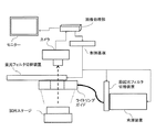
本願発明では、倍率1倍乃至6倍の高解像度低倍率光学系を用いる事でメンブレンフィルター全体の蛍光画像を少ない回数の観測で取得でき、カラー撮像素子で撮像する事でシグナル蛍光と自家蛍光などのノイズ成分を色相・明度で自動判別することが可能である。
以下本発明で使用する装置の具体的様態を図1に基づいて説明する。
(3) Automated measuring device used in the present invention In the present invention, a fluorescence image of the entire membrane filter can be obtained with a small number of observations by using a high-resolution low-magnification optical system with a magnification of 1 to 6 times, and a color imaging device It is possible to automatically distinguish noise components such as signal fluorescence and autofluorescence based on hue and brightness.
Hereinafter, a specific mode of the apparatus used in the present invention will be described with reference to FIG.
(イ)試料ステージ
高解像度低倍率光学系の使用により、倍率1倍光学系では試料ステージの移動が不要となっている。試料ステージは、装置内の完全暗室内に設置されており、励起光以外の入光がない状態になっている。また、2倍乃至6倍の光学系を用いる場合には、少ない撮影回数でメンブレンフィルター全体を撮影できるよう移動ステージを採用する。
(B) Sample stage The use of a high-resolution, low-magnification optical system makes it unnecessary to move the sample stage in a 1x optical system. The sample stage is installed in a completely dark room in the apparatus and is in a state where there is no incident light other than excitation light. In addition, when a 2 to 6 times optical system is used, a moving stage is adopted so that the entire membrane filter can be imaged with a small number of imaging times.
(ロ)励起光源
ハロゲンランプ光源からの光を、ファイバーを経由して、励起波長フィルターに通して励起光とし、試料ステージ上の試料に対し、ライトガイドを通して均一の励起光を照射している。
(B) Excitation light source Light from a halogen lamp light source is passed through an excitation wavelength filter via a fiber as excitation light, and the sample on the sample stage is irradiated with uniform excitation light through a light guide.
(ハ)光学フィルター切りかえ装置(励起フィルター用、蛍光用吸収フィルター用)
励起光源からの励起光(白色)を表示部での選択蛍光標識にあわせ、励起光フィルターへ自動的に切替を行う。また、蛍光検出部の前段にある蛍光用吸収フィルターについても、対応する吸収フィルターへ自動的に切り替わる。
(C) Optical filter changer (excitation filter, fluorescent absorption filter)
The excitation light (white) from the excitation light source is automatically switched to the excitation light filter according to the selected fluorescent label on the display unit. In addition, the absorption filter for fluorescence in the previous stage of the fluorescence detection unit is automatically switched to the corresponding absorption filter.
第1蛍光検出条件には、励起光フィルターとしてはバンドパスフィルター460-500nmを、蛍光シグナル用吸収フィルターとしてロングパスフィルター515nm以上を用いることができる。 As the first fluorescence detection condition, a band-pass filter 460-500 nm can be used as the excitation light filter, and a long-pass filter 515 nm or more can be used as the fluorescent signal absorption filter.
第2蛍光検出条件には、励起光フィルターとしてはバンドパスフィルター 520-560nmを、蛍光シグナル用吸収フィルターとしてはロングパスフィルター570nm以上を用いることができる。 As the second fluorescence detection condition, a band-pass filter 520-560 nm can be used as an excitation light filter, and a long-pass filter 570 nm or more can be used as an absorption filter for a fluorescence signal.
第3蛍光検出条件には、励起光フィルターとしてはバンドパスフィルター590-650nmを、蛍光シグナル用吸収フィルターとしてはバンドパスフィルター660nm以上を用いることができる。 As the third fluorescence detection condition, a bandpass filter 590-650 nm can be used as the excitation light filter, and a bandpass filter 660 nm or more can be used as the fluorescent signal absorption filter.
(ニ)蛍光検出部(カメラ)
デジタルカラーカメラとしては、例えば、CMOS素子サイズが35mmフルサイズ(35.9×24.0mm、画総数7360×4912)のものを、蛍光検出部には、高解像度のデジタルカラーカメラを使用し、細菌のマイクロコロニーを検出できるものを用いることができる。カメラには低倍率光学系:倍率1倍乃至6倍を使用することができる。これは、マイクロコロニーの形成と高解像度蛍光検出部により、低倍率光学系を使用する事ができている。光学系倍率を1倍とすることにより、一回の観測によるフィルターデバイス全体の計測が可能になり、大幅な観測時間短縮に寄与している。
(D) Fluorescence detector (camera)
As a digital color camera, for example, a CMOS device with a 35 mm full size (35.9 x 24.0 mm, total number of images 7360 x 4912) is used, and a high-resolution digital color camera is used for the fluorescence detection unit. What can detect a colony can be used. The camera can use a low magnification optical system: 1 to 6 times magnification. This is because a low-magnification optical system can be used due to the formation of microcolony and the high-resolution fluorescence detection section. Setting the optical system magnification to 1 makes it possible to measure the entire filter device with a single observation, contributing to a significant reduction in observation time.
(ホ)画像処理部
蛍光検出部で撮像された蛍光カラー画像に対して色相・明度を基にした画像処理判別を行い、判別結果をモニターへ表示する。また、制御基板への制御命令も行う。
(E) Image processing unit Image processing discrimination based on hue and brightness is performed on the fluorescent color image captured by the fluorescence detection unit, and the discrimination result is displayed on the monitor. It also issues control commands to the control board.
(ヘ)システム制御基板
画像処理部からの命令を受け、光源の制御(ON/OFF、光量調整)やフィルター切替装置の制御を行う。また、各種機器からのエラー信号を受信し、画像処理部へ伝達する。
(F) System control board Upon receiving a command from the image processing unit, it controls the light source (ON / OFF, light amount adjustment) and the filter switching device. Also, error signals from various devices are received and transmitted to the image processing unit.
(ト)モニター
画像処理部の処理結果の表示を行い、装置の操作状況などの表示も行う。
(G) Monitor The processing result of the image processing unit is displayed, and the operation status of the device is also displayed.
(チ)装置の特徴
第1の蛍光検出条件、第2の蛍光検出条件、又は第3の蛍光検出条件をモニター画面から選択し、自動で切り替えることができる。撮影条件(光源強度、ISO、露出、シャッター速度)は、モニター画面から選択入力できる。計測範囲はフィルターデバイス全体とする。
(H) Device characteristics The first fluorescence detection condition, the second fluorescence detection condition, or the third fluorescence detection condition can be selected from the monitor screen and automatically switched. Shooting conditions (light source intensity, ISO, exposure, shutter speed) can be selected and input from the monitor screen. The measurement range is the entire filter device.
(4)具体的な試料の自動化測定
上記(1)の培養併用蛍光インサイチューハイブリダイゼーション法により調整された測定試料を(2)のメンブレンフィルター上のDNAプローブ由来蛍光シグナルを特定の色と明るさで検出する手段を有するシグナル自動検出方法における各種条件に調整し、(3)記載の測定装置を使用し、以下のように行うことができる。
(4) Automated measurement of a specific sample The measurement sample prepared by the fluorescence in situ hybridization method combined with the culture in (1) above is a fluorescence signal derived from the DNA probe on the membrane filter in (2) with a specific color and brightness. It can adjust as various conditions in the signal automatic detection method which has a means to detect, and can perform as follows using the measuring apparatus of (3) description.
具体的には、培養併用蛍光インサイチューハイブリダイゼーション法で、標本を高さのある外枠付メンブレンフィルターに対して、前述の第1の蛍光検出光学条件にて、カラーカメラ撮影する。撮影画像を、まず、(i)撮影画像全体の画素に対して、色相及び明度の条件に当てはまるかを判定し、色相及び明度の条件に当てはまらない画素は、例えば、マスク処理することにより、除外するができる。次に、(ii)当てはまった画素の中からコロニーである画素の集合体(輝点)を探索し、検出する画像処理を行う。画像処理においては、撮像画像からの、円形、楕円、三角、四角形、など形状の判別は一般化されているところ、コロニーは、基本的に円形・楕円形をしているので、画素集合体が円形または楕円形の形状で、大きさ、つまり直径又は長径及び短径が20〜150μmである画素集合体を自動検出の対象とすることができる。 Specifically, the sample is photographed with a color camera using a culture combined fluorescence in situ hybridization method with respect to a membrane filter with a frame having a height under the first fluorescence detection optical conditions described above. First, (i) determine whether the conditions of the hue and lightness are met for the pixels of the entire photographed image, and exclude pixels that do not meet the conditions of hue and lightness by, for example, masking. I can do it. Next, (ii) image processing for searching for and detecting an aggregate (bright spot) of pixels that are colonies from the applied pixels is performed. In image processing, discriminating shapes such as a circle, an ellipse, a triangle, and a rectangle from a captured image is generalized. However, since a colony is basically a circle or an ellipse, A pixel aggregate having a circular or elliptical shape and a size, that is, a diameter or a major axis and a minor axis of 20 to 150 μm can be automatically detected.
ここで、第1の蛍光検出光学条件で測定する場合は、例えば、撮影条件をハロゲン光源出力10%、カメラ設定露出時間2.0秒、露出F3.5、ISO感度 12800とする。次に、撮影画像の画素集合体(輝点)の色相範囲はH64-H98、明度はV150以上であって、円形または楕円形の形状で、大きさが、直径又は長径及び短径20μm乃至150μmである画素集合体を自動検出の対象とすることができる。第2の蛍光検出光学条件で測定する場合には、例えば、撮影条件をハロゲン光源出力10%、カメラ設定露出時間0.6秒、露出F3.5、ISO感度 25600とする。次に、撮影画像の輝点が色相範囲はH44-H54、明度はV150以上、又は色相範囲はH54-H74、明度はV100以上であって、円形または楕円形の形状で、直径又は長径及び短径が20μm乃至150μmである画素集合体を自動検出の対象とすることができる。第3の蛍光検出光学条件で測定する場合は、例えば、撮影条件をハロゲン光源出力10%、カメラ設定露出時間1.0秒、露出F3.5、ISO感度 1250とする。次に、撮影画像の画素集合体(輝点)の色相範囲はH340-H360、明度はV80以上、又は、色相範囲はH0-H20、明度はV80以上であって、円形または楕円形の形状で、直径又は長径及び短径が20μm乃至150μmである画素集合体を自動検出の対象とすることができる。なお、光源の強度を変化させても、色相は大きく影響を受ける事はない。また、光源の強度にあわせ、カメラの露光時間を短くする事で、同様に判別する事が可能である。 Here, when the measurement is performed under the first fluorescence detection optical condition, for example, the photographing conditions are a halogen light source output 10%, a camera setting exposure time 2.0 seconds, an exposure F3.5, and an ISO sensitivity 12800. Next, the hue range of the pixel aggregate (bright spot) of the photographed image is H64-H98, the brightness is V150 or more, the shape is circular or elliptical, and the size is the diameter or major axis and minor axis 20 μm to 150 μm. Can be the target of automatic detection. For measurement under the second fluorescence detection optical condition, for example, the photographing conditions are a halogen light source output of 10%, a camera setting exposure time of 0.6 seconds, an exposure of F3.5, and an ISO sensitivity of 25600. Next, the bright spot of the photographed image has a hue range of H44-H54, lightness of V150 or higher, or a hue range of H54-H74, lightness of V100 or higher, circular or elliptical shape, diameter or major axis and short axis. A pixel aggregate having a diameter of 20 μm to 150 μm can be an object of automatic detection. When measurement is performed under the third fluorescence detection optical condition, for example, the photographing condition is a halogen light source output 10%, camera setting exposure time 1.0 second, exposure F3.5, and ISO sensitivity 1250. Next, the hue range of the pixel aggregate (bright spot) of the photographed image is H340-H360, the brightness is V80 or more, or the hue range is H0-H20, the brightness is V80 or more, and it has a circular or elliptical shape. In addition, a pixel aggregate having a diameter or a major axis and a minor axis of 20 μm to 150 μm can be an object of automatic detection. Note that even if the intensity of the light source is changed, the hue is not greatly affected. It is also possible to make a similar determination by shortening the exposure time of the camera in accordance with the intensity of the light source.
本願発明は、上記した実際の色相・明度範囲に限定されず、色相と明度条件を調整することにより、食品由来の蛍光ノイズと蛍光プローブ由来のシグナルとを弁別することが可能であり、様々な食品ノイズにも対応できるものである。 The present invention is not limited to the actual hue / brightness ranges described above, and by adjusting the hue and lightness conditions, it is possible to discriminate between food-derived fluorescent noise and fluorescent probe-derived signals. It can cope with food noise.
(5)蛍光検出光学条件の組み合わせ
また、上記(4)の試料の自動化測定において、第1の蛍光物質を用いたDNAプローブを使用し第1の蛍光検出光学条件での測定、第2の蛍光物質を用いたプローブを使用し第2の蛍光検出光学条件での測定、及び第3の蛍光物質を用いたDNAプローブを使用し第3の蛍光検出光学条件での測定、からなる3種類の蛍光検出光学条件から選ばれる任意の2以上の蛍光検出光学条件を組み合わせて、より精度よく測定することもできる。
(5) Combination of fluorescence detection optical conditions In addition, in the automated measurement of the sample of (4) above, measurement under the first fluorescence detection optical condition using a DNA probe using the first fluorescent substance, second fluorescence Three types of fluorescence consisting of measurement under the second fluorescence detection optical condition using a probe using a substance and measurement under a third fluorescence detection optical condition using a DNA probe using a third fluorescence substance It is also possible to measure more accurately by combining two or more fluorescence detection optical conditions selected from the detection optical conditions.
[実施例1]
新規自動蛍光シグナル検出法の開発
培養併用蛍光インサイチューハイブリダイゼーション法の蛍光シグナルと食品由来のノイズを区別する新たな自動計数手法を開発するために様々の食品の自家蛍光とDNAプローブの標識に用いる蛍光物質の蛍光シグナルを下記の通り比較した。
[Example 1]
Development of a new automatic fluorescence signal detection method Autofluorescence of various foods and fluorescence used for labeling of DNA probes to develop a new automatic counting method to distinguish fluorescence signals from food-borne fluorescence in situ hybridization methods and food-derived noise The fluorescence signals of the substances were compared as follows.
始めに第1の蛍光検出光学条件(励起波長460−500 nm、蛍光波長515 nm以上)で検討した。また、この条件で蛍光を示し、その蛍光輝度(%)が最適蛍光波長で得られる最大蛍光輝度の90%以上である物質を第1の蛍光物質とし、Alexa fluor488を用いた。Alexa fluor488で標識した腸内細菌科細菌検出用DNAプローブ((TGCTCTCGCGAGGTCGCTTCTCTT) 特許文献1 特許第4785449号)を用いた。また、簡易検査キットを試作し用いた。このキット構成は、直径55mmで高さ15mmのプラスチックシャーレ内に、直径50mmの濾紙を5枚入れ、その上に、特許文献2記載の高さのある外枠付メンブレンフィルター(直径47mm、ポアサイズ0.4μm以下のディスク型メンブレンフィルターと外径47mm内径42mm高さ15mmのリング状アクリルを貼り合わせた)をフィルター面を下にして置き、さらに、プラスチックシャーレ蓋(直径60mm、高さ10mm)を高さのある外枠付メンブレンフィルターの外枠とプラスチックシャーレの外縁に被せる。
First, investigation was performed under the first fluorescence detection optical conditions (excitation wavelength: 460-500 nm, fluorescence wavelength: 515 nm or more). In addition, Alexa fluor488 was used as a first fluorescent substance that exhibited fluorescence under these conditions and whose fluorescence brightness (%) was 90% or more of the maximum fluorescence brightness obtained at the optimum fluorescence wavelength. A DNA probe ((TGCTCTCGCGAGGTCGCTTCTCTT)
食品試料は、ひらめ、3色ミックス野菜(インゲン、ニンジン、トウモロコシ)、メンチカツを用いた。 As food samples, inspiration, three-color mixed vegetables (green beans, carrots, corn), and chili cutlet were used.
各食品試料は、大腸菌を添加した試料(シグナル+ノイズ区)と添加しない試料(ノイズ区)の2つの区分を設けた。 Each food sample was provided with two sections: a sample added with E. coli (signal + noise section) and a sample not added (noise section).
食品試料(シグナル+ノイズ区、または、ノイズ区)25gをストマッカー用袋に採取し、細菌検査用希釈液225mlを加え混和して10倍希釈液(0.1g/ml)を調製した。食品試料希釈液1mlと一般細菌マイクロコロニー形成培地(カゼインペプトン34g、ソイペプトン6g、酵母エキス12g、グルコース 5g、塩化ナトリウム 10g、ピルビン酸ナトリウム2.2g、リン酸二水素カリウム2.7g、リン酸水素二カリウム5g、リン酸水素二ナトリウム19.2g、胆汁酸塩No.3 1.12g、精製水1L を滅菌(121℃、15分)する。)1mlを混和して、これを前述の簡易検査キットに添加した。
25 g of a food sample (signal + noise group or noise group) was collected in a stomacher bag, mixed with 225 ml of a diluent for bacterial testing, and mixed to prepare a 10-fold diluted solution (0.1 g / ml). 1 ml of food sample diluent and general bacterial microcolony formation medium (casein peptone 34 g, soy peptone 6 g, yeast extract 12 g, glucose 5 g, sodium chloride 10 g, sodium pyruvate 2.2 g, potassium dihydrogen phosphate 2.7 g, dipotassium hydrogen phosphate Sterilize 5 g, disodium hydrogenphosphate 19.2 g, bile salt No. 3 1.12 g, and purified water 1 L (121 ° C, 15 minutes).
シグナル+ノイズ区の場合は、次に、37℃で6.5時間培養し、細菌のマイクロコロニーを形成させた(マイクロコロニー形成培養)。その後、100%エタノール2mlを簡易検査キットに加え、10分固定した(固定化)。次に、高さのある外枠付メンブレンフィルターを取り出し乾燥後、直径55mmで高さ15mmのプラスチックシャーレ内に移し、これに各種の蛍光物質で標識した10μM腸内細菌科細菌検出用プローブ5μlを含むハイブリダイゼーション緩衝液(20%ホルムアミド、0.01%ドデシル硫酸ナトリウム[SDS]、0.9M 塩化ナトリウム、20mM Tris-HCl [pH 7.4])1.5mlを添加し46℃で30分反応させ(ハイブリダイゼーション)、排水した。次に洗浄液(0.01%ドデシル硫酸ナトリウム[SDS]、180mM 塩化ナトリウム、20mM Tris-HCl [pH 7.4])10mlを添加し、46℃で15分置いて排水して(洗浄)、その高さのある外枠付メンブレンフィルターを蒸留水ですすいだのち乾燥させた。 In the case of the signal + noise group, the cells were then cultured at 37 ° C. for 6.5 hours to form bacterial microcolony (microcolony formation culture). Thereafter, 2 ml of 100% ethanol was added to the simple test kit and fixed for 10 minutes (immobilization). Next, the membrane filter with a high outer frame is taken out, dried, transferred to a plastic petri dish with a diameter of 55 mm and a height of 15 mm, and 5 μl of a probe for detecting 10 μM Enterobacteriaceae bacteria labeled with various fluorescent substances. Hybridization buffer solution (20% formamide, 0.01% sodium dodecyl sulfate [SDS], 0.9 M sodium chloride, 20 mM Tris-HCl [pH 7.4]) 1.5 ml was added and reacted at 46 ° C. for 30 minutes (hybridization). Drained. Next, add 10 ml of washing solution (0.01% sodium dodecyl sulfate [SDS], 180 mM sodium chloride, 20 mM Tris-HCl [pH 7.4]), leave it at 46 ° C. for 15 minutes and drain (wash), and then the height The membrane filter with outer frame was rinsed with distilled water and dried.
ノイズ区の場合は、食品試料10倍希釈液1mlとマイクロコロニー形成培地1mlを混和して、簡易検査キットに添加後、マイクロコロニー形成培養は行わず、後の操作は、シグナル+ノイズ区と同様に行った。 In the case of the noise group, mix 1 ml of the 10-fold diluted food sample and 1 ml of the microcolony formation medium, add it to the simple test kit, do not perform the microcolony formation culture, and the subsequent operations are the same as the signal + noise group Went to.
これら、培養併用蛍光インサイチューハイブリダイゼーション法標本、すなわち、シグナル+ノイズ区、ノイズ区の高さのある外枠付メンブレンフィルターに対して、前述の第1の蛍光検出光学条件にて、カラーカメラ撮影した。装置としてはデジタルカメラ(CMOS素子サイズが35mmフルサイズ(35.9×24.0mm、画素数7360×4912))とハロゲンライト、フィルター切りかえ装置(励起光側:小型回転式フィルター切替装置(6枚切替)、蛍光側:薄型回転式フィルター切替装置(8枚切替))等を材料として自動化計測装置を前記の通り試作して用い、ハロゲン光源出力は10%とし、Alexa fluor488用のフィルター(励起:バンドパスフィルター 460−500 nm、吸収:ロングパスフィルター515 nm以上)を用いた。カメラ設定は、露出時間:2.0秒 F:3.5 ISO12800とした。各区分の撮影画像をモニター画面に映し出し、様々な色や明るさ、大きさの輝点の集合体(画素の集合)について、目視で、シグナルの画素集合体とノイズの画素集合体を区別した。次に、目視で見分けたノイズである画素の集合体(大きさ20-150μm相当)をランダムに10点以上で色相・明度を測定した。同様にシグナルである画素集合体(大きさ20-150μm相当)をランダムに10点で色相・明度を測定した。 These culture combined fluorescence in situ hybridization method specimens, that is, the signal + noise section, the membrane filter with an outer frame having a noise section height, were taken with a color camera under the first fluorescence detection optical conditions described above. . The equipment is a digital camera (CMOS element size 35mm full size (35.9 x 24.0mm, pixel number 7360 x 4912)), halogen light, filter switching device (excitation light side: small rotary filter switching device (6 sheets switching), Fluorescent side: Thin-type rotary filter switching device (8-sheet switching) etc. is used as a material, and an automated measuring device is prototyped and used as described above. Halogen light source output is 10%, Alexa fluor488 filter (excitation: bandpass filter) 460-500 nm, absorption: long pass filter 515 nm or more). The camera settings were: exposure time: 2.0 seconds F: 3.5 ISO12800. Projected images of each section are displayed on the monitor screen, and the signal pixel aggregates and noise pixel aggregates are visually distinguished for the aggregates (pixel groups) of bright spots of various colors, brightness, and size. . Next, hue / lightness was measured at 10 points or more at random on an aggregate of pixels (corresponding to a size of 20 to 150 μm), which was noise identified visually. Similarly, hue / lightness was measured at 10 points on a pixel aggregate (equivalent to a size of 20 to 150 μm) as a signal.
(結果)
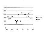
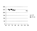
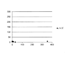
第1の蛍光検出光学条件での測定結果を、表2から表10及び図2から図7に示す。3色ミックス野菜の結果(ノイズ区(表2、図2)、シグナル+ノイズ区(表3、表4、図3)、ひらめの結果(ノイズ区(表5、図4)、シグナル+ノイズ区(表6、表7、図5))、メンチカツの結果(ノイズ区(表8、図6)、シグナル+ノイズ区(表9、表10、図7))から、第1の蛍光検出光学条件で第1の蛍光物質の弁別条件を検討した。色相の範囲は、コロニーに含まれている蛍光標識の蛍光が強い場合と微弱な場合で見え方が違うため、色相に範囲を持たせた。その結果、色相範囲がH64-H98であって明度がV150以上の範囲内の輝点をシグナルと弁別する弁別条件とし、これを自動計測条件とした。
(result)
The measurement results under the first fluorescence detection optical condition are shown in Tables 2 to 10 and FIGS. 2 to 7. Three-color mixed vegetable results (noise group (Table 2, Fig. 2), signal + noise group (Table 3, Table 4, Fig. 3), inspiration results (noise group (Table 5, Fig. 4), signal + noise group) (Table 6, Table 7, and Fig. 5)), Menchikatsu results (noise zone (Table 8, Fig. 6), signal + noise zone (Table 9, Table 10, Fig. 7)), the first fluorescence detection optical conditions In order to distinguish the first fluorescent substance, the hue range was different because the appearance of the fluorescent label contained in the colony differs depending on whether it is strong or weak. As a result, a bright spot having a hue range of H64-H98 and a brightness of V150 or higher was set as a discrimination condition for discriminating from a signal, and this was set as an automatic measurement condition.
よって、3色ミックス野菜シグナル+ノイズ区の測定結果のうち、表3は、シグナル、表4はノイズとなり、合わせて、図3に示す。次に、ひらめのシグナル+ノイズ区のうち、表6はシグナル、表7はノイズとなり、合わせて、図5に示す。さらに、メンチカツのシグナル+ノイズ区のうち、表9はシグナル、表10はノイズとなり、合わせて図7に示す。 Therefore, among the measurement results of the three-color mixed vegetable signal + noise section, Table 3 shows the signal and Table 4 shows the noise, and these are shown in FIG. Next, in the signal / noise section of the inflection, Table 6 is a signal and Table 7 is a noise. Further, in the signal + noise section of Menchikatsu, Table 9 is a signal, and Table 10 is a noise, which are collectively shown in FIG.
次に、第2の蛍光検出光学条件(励起波長520−560 nm、蛍光波長570nm以上)で検討した。この条件で、蛍光を示し、その蛍光輝度(%)が最適蛍光波長で得られる最大蛍光輝度の90%以上である物質を第2の蛍光物質とし、TAMRAを用いた。TAMRAで標識した腸内細菌科細菌検出用DNAプローブ((TGCTCTCGCGAGGTCGCTTCTCTT) 特許文献1 特許第4785449号)を用いた。食品試料として、3色野菜(インゲン、トウモロコシ、ニンジン)、メンチカツ、ひらめを用いた。各食品試料は、大腸菌を添加した(シグナル+ノイズ区)と添加しない(ノイズ区)2つの区分を設けた。培養併用蛍光インサイチューハイブリダイゼーション法標本作成は、前述の第1の蛍光検出光学条件と同様に行った。次いで、標本に対して、前述の第2の蛍光検出光学条件にて、カラー撮影した。装置としてはデジタルカメラ(CMOS素子サイズが35mmフルサイズ(35.9×24.0mm、画総数7360×4912))とハロゲンライト、フィルター切りかえ装置(励起光側:小型回転式フィルター切替装置(6枚切替)、蛍光側:薄型回転式フィルター切替装置(8枚切替))等を材料として自動化計測装置を前記の通り試作して用い、ハロゲン光源出力は10%とし、TAMRA用のフィルター(励起:バンドパスフィルター 520−560nm、吸収:ロングパスフィルター 570nm以上)を用いた。カメラ設定は、露出時間:0.6秒 F:3.5 ISO25600とした。
Next, the second fluorescence detection optical conditions (excitation wavelength 520-560 nm, fluorescence wavelength 570 nm or more) were studied. Under these conditions, TAMRA was used with a substance exhibiting fluorescence and having a fluorescence brightness (%) of 90% or more of the maximum fluorescence brightness obtained at the optimum fluorescence wavelength as the second fluorescent substance. A DNA probe ((TGCTCTCGCGAGGTCGCTTCTCTT)
撮像をモニター画面に映し出し、様々な色や明るさ、大きさの輝点の集合体(画素の集合)について、目視で、シグナルの画素集合体とノイズの画素集合体を区別した。次に、目視で見分けたノイズである画素の集合体(大きさ20-150μm相当)をランダムに10点及びシグナルである画素集合体(大きさ20-150μm相当)をランダムに10点で色相・明度を測定した。測定データーを比較し、シグナルとノイズの弁別条件を検討した。 The image was projected on a monitor screen, and a signal pixel aggregate and a noise pixel aggregate were visually distinguished for a collection (pixel collection) of bright spots of various colors, brightness, and size. Next, the pixel group (equivalent to a size of 20-150μm), which is the noise identified by visual observation, is randomly assigned to 10 points, and the pixel group, which is a signal (equivalent to a size of 20-150μm), is randomly assigned to the hue / The brightness was measured. The measurement data were compared and the signal and noise discrimination conditions were examined.
第2の蛍光検出光学条件の測定結果は、表11から表19及び図8から図13に示す。3色ミックス野菜の結果(ノイズ区(表11、図8)、シグナル+ノイズ区(表12、表13、図9))、ひらめの結果(ノイズ区(表14、図10)、シグナル+ノイズ区(表15、表16、図11))、メンチカツの結果(ノイズ区(表17、図12)、シグナル+ノイズ区(表18、表19、図13))から、第2の蛍光検出光学条件で第2の蛍光物質の弁別条件を検討した。検討した。色相の範囲は、コロニーに含まれている蛍光標識の蛍光が強い場合と微弱な場合で見え方が違うため、色相に範囲を持たせた。その結果、色相範囲がH44-H54であって明度をV150以上の範囲、及び色相範囲がH54-H74であって、明度がV100以上の範囲内の輝点をシグナルとする弁別条件とし、これを自動計測条件とした。 The measurement results of the second fluorescence detection optical condition are shown in Tables 11 to 19 and FIGS. Three-color mixed vegetable results (noise group (Table 11, Fig. 8), signal + noise group (Table 12, Table 13, Fig. 9)), inspiration results (noise group (Table 14, Fig. 10), signal + noise 2nd fluorescence detection optics from ward (Table 15, Table 16, Figure 11)), Machikatsu results (Noise (Table 17, Figure 12), Signal + Noise (Table 18, Table 19, Figure 13)) The conditions for discriminating the second fluorescent material were examined. investigated. The range of the hue was given to the hue because the appearance differs depending on whether the fluorescent label contained in the colony is strong or weak. As a result, the hue range is H44-H54 and the brightness is in the range of V150 or higher, and the hue range is H54-H74, and the bright spot in the range of the brightness is V100 or higher is set as the discrimination condition. Automatic measurement conditions were used.
なお、表11から表19までの測定では、よって、3色ミックス野菜シグナル+ノイズ区の測定結果のうち、表12は、シグナル、表13はノイズとなり、合わせて図9に示す。次に、ひらめのシグナル+ノイズ区のうち、表15はシグナル、表16はノイズとなり、合わせて、図11に示す。さらに、メンチカツのシグナル+ノイズ区のうち、表18はシグナル、表19はノイズとなり、合わせて図13に示す。 In the measurements from Table 11 to Table 19, therefore, among the measurement results of the three-color mixed vegetable signal + noise section, Table 12 is a signal and Table 13 is a noise, and these are shown in FIG. Next, in the signal / noise section of the inflection, Table 15 is a signal and Table 16 is a noise. Further, in the signal + noise section of Menchikatsu, Table 18 is a signal, and Table 19 is a noise.
次に、第3の蛍光検出光学条件(励起波長 590−650nm、蛍光波長660nm以上)で検討した。この条件で、蛍光を示し、その蛍光輝度(%)が最適蛍光波長で得られる最大蛍光輝度の90%以上である物質を第3の蛍光物質とし、Cy5を用いた。Cy5で標識した腸内細菌科細菌検出用DNAプローブ((TGCTCTCGCGAGGTCGCTTCTCTT) 特許文献1 特許第4785449号)を用いた。食品試料として、3色野菜(インゲン、トウモロコシ、ニンジン)、メンチカツ、ひらめを用いた。各食品試料は、大腸菌を添加した(シグナル+ノイズ区)と添加しない(ノイズ区)2つの区分を設けた。培養併用蛍光インサイチューハイブリダイゼーション法標本作成は、前述の第1の蛍光検出光学条件と同様に行った。次いで、標本に対して、前述の第3の蛍光検出光学条件にて、カラー撮影した。装置としてはデジタルカメラ(CMOS素子サイズが35mmフルサイズ(35.9×24.0mm、画総数7360×4912))とハロゲンライト、フィルター切りかえ装置(励起光側:小型回転式フィルター切替装置(6枚切替)、蛍光側:薄型回転式フィルター切替装置(8枚切替))等を材料として自動化計測装置を前記の通り試作して用い、ハロゲン光源出力は10%とし、Cy5用のフィルター(励起:バンドパスフィルター590-650nm、吸収:ロングパスフィルター660nm以上)をもちいた。カメラ設定は、露出時間:1.0秒 F:3.5 ISO1250とした。
Next, examination was performed under the third fluorescence detection optical condition (excitation wavelength: 590-650 nm, fluorescence wavelength: 660 nm or more). Under this condition, a substance that exhibits fluorescence and whose fluorescence brightness (%) is 90% or more of the maximum fluorescence brightness obtained at the optimum fluorescence wavelength was used as a third fluorescent substance, and Cy5 was used. A DNA probe for detecting Enterobacteriaceae bacteria labeled with Cy5 ((TGCTCTCGCGAGGTCGCTTCTCTT)
撮像をモニター画面に映し出し、様々な色や明るさ、大きさの輝点の集合体(画素の集合)について、目視で、シグナルの画素集合体とノイズの画素集合体を区別した。次に、目視で見分けたノイズである画素の集合体(大きさ20-150μm相当)をランダムに10点及びシグナルである画素集合体(大きさ20-150μm相当)をランダムに10点以上で色相・明度を測定した。測定データーを比較し、シグナルとノイズの弁別条件を検討した。 The image was projected on a monitor screen, and a signal pixel aggregate and a noise pixel aggregate were visually distinguished for a collection (pixel collection) of bright spots of various colors, brightness, and size. Next, the hue of the pixel aggregate (equivalent to a size of 20-150μm), which is the noise identified visually, is randomly assigned to 10 points, and the pixel group (signal equivalent to the size of 20-150μm), which is a signal, is randomly assigned to 10 or more -The brightness was measured. The measurement data were compared and the signal and noise discrimination conditions were examined.
第3の蛍光検出光学条件での測定結果は、表20から表28及び図14から図19に示す。3色ミックス野菜(ノイズ区(表20、図14)、シグナル+ノイズ区(表21、表22、図15))、ひらめ(ノイズ区(表23、図16)、シグナル+ノイズ区(表24、表25、図17))、メンチカツ(ノイズ区(表26、図18)、シグナル+ノイズ区(表27、表28、図19))から、第3の蛍光検出光学条件で第3の蛍光物質の弁別条件を検討した。色相範囲がH340-H360であって、明度がV80以上、及び、色相範囲がH0-H20であって、明度がV80以上の範囲内にある輝点をシグナルと弁別する条件とし、これを自動計測条件とした。 The measurement results under the third fluorescence detection optical condition are shown in Tables 20 to 28 and FIGS. 14 to 19. Three-color mixed vegetables (noise group (Table 20, Figure 14), signal + noise group (Table 21, Table 22, Figure 15)), inflection (noise group (Table 23, Figure 16), signal + noise group (Table 24) , Table 25, Fig. 17)), Machikatsu (Noise section (Table 26, Figure 18), Signal + Noise section (Table 27, Table 28, Figure 19)), the third fluorescence under the third fluorescence detection optical conditions. Substance discrimination conditions were investigated. The brightness range is H340-H360, the brightness is V80 or more, and the hue range is H0-H20, and the brightness point is in the range of V80 or more. Condition.
よって、3色ミックス野菜シグナル+ノイズ区の測定結果のうち、表21は、シグナル、表22はノイズとなり、合わせて、図15に示す。次に、ひらめのシグナル+ノイズ区のうち、表24はシグナル、表25はノイズとなり、合わせて、図17に示す。さらに、メンチカツのシグナル+ノイズ区のうち、表27はシグナル、表28はノイズとなり、合わせて、図19に示す。 Therefore, among the measurement results of the three-color mixed vegetable signal + noise section, Table 21 shows the signal and Table 22 shows the noise, and these are shown in FIG. Next, in the signal / noise section of the inflection, Table 24 is a signal and Table 25 is a noise. Further, in the signal + noise section of Menchikatsu, Table 27 is a signal, and Table 28 is a noise.
[実施例2]
本発明の蛍光シグナル計測法による食品の蛍光ノイズ測定
特許文献1記載の蛍光計測法は、表1記載の食品試料に対し、蛍光ノイズを示した。同じ食品試料を、本発明手法により、次の通り計測した。
[Example 2]
Fluorescence noise measurement of food by the fluorescence signal measurement method of the present invention The fluorescence measurement method described in
試料に表1の13種類の食品を用いた。また、試験には、簡易検査キットを試作し用いた。このキット構成は、直径55mmで高さ15mmのプラスチックシャーレに、直径50mmの濾紙を5枚入れ、その上に、特許文献2記載の高さのある外枠付メンブレンフィルター(但し、直径47mm、ポアサイズ0.8μm以下のディスク型メンブレンフィルターと外径47mm、内径42mm、高さ15mmのリング状アクリルを貼り合わせた)をフィルター面を下にして置くものとした。 Thirteen kinds of foods shown in Table 1 were used as samples. In the test, a simple test kit was prototyped and used. This kit consists of a 55 mm diameter plastic petri dish with a height of 15 mm and 5 filter papers with a diameter of 50 mm, and a membrane filter with an outer frame as described in Patent Document 2 (however, 47 mm in diameter, pore size) A disc-type membrane filter of 0.8 μm or less and a ring-shaped acrylic having an outer diameter of 47 mm, an inner diameter of 42 mm, and a height of 15 mm were attached together with the filter face down.
各々の食品試料25gをストマッカー用袋に入れ、さらに細菌検査用リン酸緩衝液225mlを加え懸濁し試料10倍希釈液(試料0.1g/ml)を調製した。その1mlを簡易検査キットに供した。希釈液がろ過された後、その高さのある外枠付メンブレンフィルターをエタノールに20分浸し固定化し乾燥させ、ハイブリダイゼーション緩衝液(20%ホルムアミド、0.01%ドデシル硫酸ナトリウム[SDS]、0.9M 塩化ナトリウム、20mM Tris-HCl [pH 7.4])1.5mlを添加し、46℃で20分置いた。次いで、洗浄液(0.01%ドデシル硫酸ナトリウム[SDS]、180mM 塩化ナトリウム、20mM Tris-HCl [pH 7.4])を添加し46℃で15分置いた後、排液して洗浄した。蒸留水で高さのある外枠付メンブレンフィルターをすすいだ後乾燥した。これにより食品試料0.1gを含み、かつ、細菌マイクロコロニー(円形のコロニーで、直径20μm乃至150μm)を含まない食品試料標本を調製した。次に、高さのある外枠付きメンブレンフィルターの蛍光を特定の2色以上の色と明るさで検出する手段により、第1、第2、第3の蛍光物質の蛍光シグナル自動検出条件で測定した。ここで計数される蛍光は、すべてノイズ(光量や発光面積がマイクロコロニーと同等である食品由来自家蛍光)となる。使用する装置としては、実施例1で使用した同じ自動化計測装置を使用し、倍率が1倍の低倍率蛍光検出系を用いた。蛍光シグナル検出条件は次の通りである。 25 g of each food sample was placed in a stomacher bag, and further 225 ml of a phosphate buffer for bacterial testing was added and suspended to prepare a 10-fold diluted sample (sample 0.1 g / ml). The 1 ml was used for a simple test kit. After the diluted solution has been filtered, the membrane filter with a high outer frame is immersed in ethanol for 20 minutes, fixed and dried, and then a hybridization buffer (20% formamide, 0.01% sodium dodecyl sulfate [SDS], 0.9M chloride) 1.5 ml of sodium, 20 mM Tris-HCl [pH 7.4] was added and placed at 46 ° C. for 20 minutes. Next, a washing solution (0.01% sodium dodecyl sulfate [SDS], 180 mM sodium chloride, 20 mM Tris-HCl [pH 7.4]) was added and left at 46 ° C. for 15 minutes, and then drained and washed. The membrane filter with an outer frame having a height was rinsed with distilled water and dried. As a result, a food sample specimen containing 0.1 g of food sample and not containing bacterial microcolony (circular colony, diameter 20 μm to 150 μm) was prepared. Next, by measuring the fluorescence of the membrane filter with a high outer frame with two or more specific colors and brightness, it is measured under the conditions for automatic detection of the fluorescence signal of the first, second and third fluorescent substances. did. The fluorescence counted here becomes noise (a food-derived autofluorescence whose light amount and light emitting area are equivalent to those of a microcolony). As an apparatus to be used, the same automated measurement apparatus used in Example 1 was used, and a low-magnification fluorescence detection system having a magnification of 1 was used. The fluorescence signal detection conditions are as follows.
第1の蛍光物質の蛍光シグナル自動検出条件
ハロゲン光源出力10%、Alexa fluor488用のフィルター(励起:バンドパスフィルター 460−500 nm、吸収:ロングパスフィルター515 nm以上)を使用。カメラ設定は、露出時間:2.0秒 F:3.5 ISO12800とする。色相範囲:H64-H98、明度:V150以上とする。なお、本件実施例では、カメラの解度と撮像されたエリアから、1画素あたりの大きさは約10μmであるから、縦横2画素ずつ以上かつ15画素以下の画素集合体は、大きさ20-150μmに相当するので、縦横2画素ずつ以上かつ15画素以下の画素集合体で、色相範囲:H64-H98、明度:V150以上のものを、シグナルとして自動検出した。
Fluorescent signal automatic detection condition for the first fluorescent substance Halogen light source output 10%, Alexa fluor488 filter (excitation: bandpass filter 460-500 nm, absorption: longpass filter 515 nm or more). The camera setting is exposure time: 2.0 seconds F: 3.5 ISO12800. Hue range: H64-H98, brightness: V150 or higher. In this embodiment, because of the resolution of the camera and the imaged area, the size per pixel is about 10 μm, so a pixel aggregate of 2 pixels vertically and horizontally and 15 pixels or less is 20- Since it corresponds to 150 μm, a pixel aggregate of 2 pixels vertically and horizontally and 15 pixels or less, with a hue range of H64-H98 and lightness of V150 or more was automatically detected as a signal.
第2の蛍光物質の蛍光シグナル自動検出条件
ハロゲン光源出力10%、TAMRA用のフィルター(励起:バンドパスフィルター 520−560nm、吸収:ロングパスフィルター 570nm以上)を使用。カメラ設定は、露出時間:0.6秒 F:3.5 ISO25600とする。色相範囲:H44-H54、明度:V150以上、又は、色相範囲:H54-H74、明度:V100以上とする。なお、本件実施例では、カメラの解度と撮像されたエリアから、1画素あたりの大きさは約10μmであるから、縦横2画素ずつ以上かつ15画素以下の画素集合体は、大きさ20-150μmに相当するので、縦横2画素ずつ以上かつ15画素以下の画素集合体で、色相範囲:H44-H54、明度:V150以上、又は、色相範囲:H54-H74、明度:V100以上の画素集合体を、シグナルとして自動検出した。
Fluorescent signal automatic detection condition for the second fluorescent substance Halogen light source output 10%, TAMRA filter (excitation: bandpass filter 520-560nm, absorption: longpass filter 570nm or more). The camera setting is exposure time: 0.6 seconds F: 3.5 ISO25600. Hue range: H44-H54, brightness: V150 or higher, or hue range: H54-H74, brightness: V100 or higher. In this embodiment, because of the resolution of the camera and the imaged area, the size per pixel is about 10 μm, so a pixel aggregate of 2 pixels vertically and horizontally and 15 pixels or less is 20- Since it corresponds to 150μm, it is a pixel aggregate of 2 pixels vertically and horizontally and 15 pixels or less. Hue range: H44-H54, Brightness: V150 or higher, or Hue range: H54-H74, Brightness: Pixel aggregate of 100 or more Was automatically detected as a signal.
第3の蛍光物質の蛍光シグナル自動検出条件
ハロゲン光源出力10%、Cy5用のフィルター(励起:バンドパスフィルター 590-650nm、吸収:ロングパスフィルター 660nm以上)を使用。カメラ設定は、露出時間:1.0秒 F:3.5 ISO1250とする。色相範囲:H340-H360、明度:V80以上、又は、色相範囲:H0-H20、明度:V80以上とする。なお、本件実施例では、カメラの解度と撮像されたエリアから、1画素あたりの大きさは約10μmであるから、縦横2画素ずつ以上かつ15画素以下の縦横2画素ずつ以上かつ15画素以下の画素集合体は、大きさ20-150μmに相当するので、縦横2画素ずつ以上かつ15画素以下の画素集合体で、色相範囲:H340-H360、明度:V80以上、又は、色相範囲:H0-H20、明度:V80以上シグナルとしての画素集合体を、シグナルとして自動検出した。
Fluorescent signal automatic detection conditions for the third fluorescent substance Halogen light source output 10%, Cy5 filter (excitation: bandpass filter 590-650nm, absorption: longpass filter 660nm or more). The camera setting is exposure time: 1.0 sec. F: 3.5 ISO1250. Hue range: H340-H360, brightness: V80 or higher, or hue range: H0-H20, brightness: V80 or higher. In this embodiment, from the resolution of the camera and the imaged area, the size per pixel is about 10 μm, so the vertical and horizontal pixels are 2 pixels or more and 15 pixels or less. The vertical and horizontal pixels are 2 pixels or more and 15 pixels or less. Since the pixel assembly corresponds to a size of 20 to 150 μm, it is a pixel assembly of 2 pixels in the vertical and horizontal directions and 15 pixels or less in hue range: H340-H360, brightness: V80 or more, or hue range: H0- H20, lightness: V80 or higher A pixel aggregate as a signal was automatically detected as a signal.
その結果、第1、第2及び第3の蛍光検出光学条件いずれにおいても、細菌検出限界10CFU/gにおいて、ノイズを発生しなかった(表29)。 As a result, no noise was generated at the bacterial detection limit of 10 CFU / g under any of the first, second and third fluorescence detection optical conditions (Table 29).
[実施例3]
培養併用蛍光インサイチューハイブリダイゼーションのシグナル検出方法による食品中の特定細菌の計数への適用評価
本発明の食品中の特定細菌の計数への適用性を検討した。
特定細菌を腸内細菌科とした。同一食品試料に対し培養併用蛍光インサイチューハイブリダイゼーション法と腸内細菌科計数対照法による計数を行い、両手法の値の比較を次の通り行った。
[Example 3]
Evaluation of Applicability to Counting Specific Bacteria in Foods by Signal Detection Method of Fluorescence In Situ Hybridization Combined with Culture The applicability of the present invention to the counting of specific bacteria in foods was examined.
The specific bacterium was designated Enterobacteriaceae. The same food sample was counted by the combined fluorescence in situ hybridization method and the Enterobacteriaceae count control method, and the values of both methods were compared as follows.
材料
供試菌株
腸内細菌科細菌としてEscherichia coli ATCC11775を用いた。
material
Test strain
Escherichia coli ATCC 11775 was used as an enterobacteriaceae bacterium.
VRBG寒天
ペプトン7g、胆汁酸塩No.3 1.5g、食塩5.0g、クリスタルバイオレッド 0.002g 、酵母エキス3.0g、ブドウ糖10g、ニュートラルレッド0.03g、寒天15g、精製水1Lを煮沸溶解後、47℃に冷却して使用した。
VRBG Agar Peptone 7g, Bile Salt No.3 1.5g, Salt 5.0g , Crystal Bio Red 0.002g, Yeast Extract 3.0g, Glucose 10g, Neutral Red 0.03g, Agar 15g, Purified Water 1L Used after cooling.
食品試料
ひらめ、3色ミックス野菜(インゲン、ニンジン、トウモロコシ)、メンチカツを用いた。各食品試料は、大腸菌を添加した(菌添加区)と添加しない(非添加区)の2つの区分を設けた。
A food sample was used, three-color mixed vegetables (green beans, carrots, corn), and chili cutlet. Each food sample was divided into two sections: E. coli added (bacteria added) and no addition (non-added).
マイクロコロニー形成培地
カゼインペプトン34g、ソイペプトン6g、酵母エキス12g、グルコース 5g、塩化ナトリウム 10g、ピルビン酸ナトリウム2.2g、リン酸二水素カリウム2.7g、リン酸水素二カリウム5g、リン酸水素二ナトリウム19.2g、胆汁酸塩No.3 1.12g、精製水1L を低温殺菌(80〜100℃、30分)し、室温まで冷却した。
Microcolony formation medium casein peptone 34g, soy peptone 6g, yeast extract 12g, glucose 5g, sodium chloride 10g, sodium pyruvate 2.2g, potassium dihydrogen phosphate 2.7g, dipotassium hydrogen phosphate 5g, disodium hydrogen phosphate 19.2g Then, 1.12 g of bile salt No. 3 and 1 L of purified water were pasteurized (80-100 ° C., 30 minutes) and cooled to room temperature.
検査キット
直径55mmで高さ15mmのプラスチックシャーレ(底面を下に置く)に、直径50mmの濾紙(No.2定量用濾紙 アドバンテック社)を5枚入れ、その上に、特許文献2記載の高さのある外枠付メンブレンフィルター(直径47mm、ポアサイズ0.8μm以下のディスク型メンブレンフィルターと外径47mm、内径42mm、高さ15mmのリング状アクリルを貼り合わせた)をフィルター面を下にして置くものとする。さらに、プラスチックシャーレ蓋(直径60mm、高さ10mm)を高さのある外枠付メンブレンフィルターの外枠とプラスチックシャーレの外縁に被せる。
A plastic dish of height 15mm with
腸内細菌科細菌検出用DNAプローブ(特許文献1 特許第4785449号)
腸内細菌科細菌検出用DNAプローブとして、蛍光物質で標識した次の塩基配列のDNAプローブを用いる(TGCTCTCGCGAGGTCGCTTCTCTT) 。
DNA probe for detecting Enterobacteriaceae bacteria (
As a DNA probe for detecting Enterobacteriaceae bacteria, a DNA probe having the following base sequence labeled with a fluorescent substance is used (TGCTCTCGCGAGGTCGCTTCTCTT).
DNAプローブの標識蛍光物質
メンブレンフィルターの蛍光シグナルを特定の色と明るさで検出する手段が、第1の蛍光検出光学条件(励起波長460−500 nm、蛍光波長515 nm以上)の場合は、DNAプローブを標識する第1の蛍光物質をAlexa Fluor 488とする。
If the means for detecting the fluorescence signal of the fluorescent substance membrane filter of the DNA probe with a specific color and brightness is the first fluorescence detection optical condition (excitation wavelength: 460-500 nm, fluorescence wavelength: 515 nm or more), DNA The first fluorescent substance for labeling the probe is called Alexa Fluor 488.
メンブレンフィルターの蛍光シグナルを特定の色と明るさで検出する手段が、第2の蛍光検出光学条件(励起波長520−560 nm、蛍光波長570nm以上)の場合は、DNAプローブを標識する第2の蛍光物質をTAMRAとする。 When the means for detecting the fluorescence signal of the membrane filter with a specific color and brightness is the second fluorescence detection optical condition (excitation wavelength: 520-560 nm, fluorescence wavelength: 570 nm or more), the second probe for labeling the DNA probe The fluorescent substance is TAMRA.
メンブレンフィルターの蛍光シグナルを特定の色と明るさで検出する手段が、第3の蛍光検出光学条件(励起波長590−650nm、蛍光波長660nm以上)の場合は、DNAプローブを標識する第3の蛍光物質をCy5とする。 If the means for detecting the fluorescence signal of the membrane filter with a specific color and brightness is under the third fluorescence detection optical condition (excitation wavelength: 590-650 nm, fluorescence wavelength: 660 nm or more), the third fluorescence that labels the DNA probe Let the material be Cy5.
ハイブリダイゼーション緩衝液
20%ホルムアミド、0.01%ドデシル硫酸ナトリウム[SDS]、0.9M 塩化ナトリウム、20mM Tris-HCl [pH 7.4]
Hybridization buffer
20% formamide, 0.01% sodium dodecyl sulfate [SDS], 0.9 M sodium chloride, 20 mM Tris-HCl [pH 7.4]
洗浄液
(0.01%ドデシル硫酸ナトリウム[SDS]、180mM 塩化ナトリウム、20mM Tris-HCl [pH 7.4])10ml
Washing solution (0.01% sodium dodecyl sulfate [SDS], 180 mM sodium chloride, 20 mM Tris-HCl [pH 7.4]) 10 ml
蛍光シグナル自動検出
使用する装置としては、実施例1で使用した同じ自動化計測装置を使用し、倍率が1倍の低倍率蛍光検出系を用いた。
As an apparatus to be used for automatic detection of fluorescent signals, the same automated measuring apparatus used in Example 1 was used, and a low-magnification fluorescence detection system having a magnification of 1 was used.
メンブレンフィルターの蛍光シグナルを特定の色と明るさで検出する手段において、第1、第2、第3の蛍光物質の蛍光シグナル検出条件を次の通りとする。 In the means for detecting the fluorescent signal of the membrane filter with a specific color and brightness, the fluorescent signal detection conditions of the first, second and third fluorescent substances are as follows.
第1の蛍光物質由来のマイクロコロニー蛍光シグナル自動検出条件
ハロゲン光源出力10%、Alexa fluor488用のフィルター(励起:バンドパスフィルター 460−500 nm、吸収:ロングパスフィルター515 nm以上)を使用。カメラ設定は、露出時間:2.0秒 F:3.5 ISO12800とする。色相範囲:H64-H98、明度:V150以上とする。
Conditions for automatic detection of microcolony fluorescence signal derived from the first fluorescent substance Halogen light source output 10%, Alexa fluor488 filter (excitation: bandpass filter 460-500 nm, absorption: longpass filter 515 nm or more). The camera setting is exposure time: 2.0 seconds F: 3.5 ISO12800. Hue range: H64-H98, brightness: V150 or higher.
なお、本件実施例では、カメラの解度と撮像されたエリアから、1画素あたりの大きさは約10μmであるから、縦横2画素ずつ以上かつ15画素以下の画素集合体は、大きさ20-150μmに相当するので、縦横2画素ずつ以上かつ15画素以下の画素集合体で、色相範囲:H64-H98、明度:V150以上の画素集合体を、シグナルとして自動検出した。 In this embodiment, because of the resolution of the camera and the imaged area, the size per pixel is about 10 μm, so a pixel aggregate of 2 pixels vertically and horizontally and 15 pixels or less is 20- Since it corresponds to 150 μm, a pixel aggregate of 2 pixels in the vertical and horizontal directions and 15 pixels or less in the hue range: H64-H98, brightness: V150 or more was automatically detected as a signal.
第2の蛍光物質由来のマイクロコロニー蛍光シグナル自動検出条件
ハロゲン光源出力10%、TAMRA用のフィルター(励起:バンドパスフィルター 520−560nm、吸収:ロングパスフィルター 570nm以上)を使用。カメラ設定は、露出時間:0.6秒 F:3.5 ISO25600とする。色相範囲:H44-H54、明度:V150以上、又は、色相範囲:H54-H74、明度:V100以上とする。なお、上記と同様に、縦横2画素ずつ以上かつ15画素以下の画素集合体は、大きさ20-150μmに相当するので、縦横2画素ずつ以上かつ15画素以下の画素集合体で、色相範囲:H44-H54、明度:V150以上、又は、色相範囲:H54-H74、明度:V100以上の画素集合体を、シグナルとして自動検出した。
。
Automatic detection of microcolony fluorescence signal derived from the second fluorescent substance Halogen light source output 10%, TAMRA filter (excitation: bandpass filter 520-560nm, absorption: longpass filter 570nm or more). The camera setting is exposure time: 0.6 seconds F: 3.5 ISO25600. Hue range: H44-H54, brightness: V150 or higher, or hue range: H54-H74, brightness: V100 or higher. Similarly to the above, a pixel aggregate of 2 pixels vertically and horizontally and 15 pixels or less corresponds to a size of 20 to 150 μm. Therefore, a pixel aggregate of 2 pixels vertically and horizontally and 15 pixels or less has a hue range: A pixel aggregate having H44-H54, lightness: V150 or higher, or hue range: H54-H74, lightness: V100 or higher was automatically detected as a signal.
.
第3の蛍光物質由来のマイクロコロニー蛍光シグナル自動検出条件
ハロゲン光源出力10%、Cy5用のフィルター (励起:バンドパスフィルター590-650nm、吸収:ロングパスフィルター 660nm以上)を使用。カメラ設定は、露出時間:1.0秒 F:3.5 ISO1250とする。色相範囲:H340-H360、明度:V80以上、又は、色相範囲:H0-H20、明度:V80以上とする。なお、上記と同様に、縦横2画素ずつ以上かつ15画素以下の縦横2画素ずつ以上かつ15画素以下の画素集合体は、大きさ20-150μmに相当するので、縦横2画素ずつ以上かつ15画素以下の画素集合体で、色相範囲:H340-H360、明度:V80以上、又は、色相範囲:H0-H20、明度:V80以上シグナルとしての画素集合体を、シグナルとして自動検出した。
Automatic detection of micro colony fluorescence signal derived from third fluorescent substance Halogen light source output 10%, Cy5 filter (excitation: bandpass filter 590-650nm, absorption: longpass filter 660nm or more). The camera setting is exposure time: 1.0 sec. F: 3.5 ISO1250. Hue range: H340-H360, brightness: V80 or higher, or hue range: H0-H20, brightness: V80 or higher. In the same way as above, a pixel aggregate of 2 pixels vertically and horizontally and 15 pixels or less and 2 pixels vertically and horizontally and 15 pixels or less corresponds to a size of 20-150 μm, so it is 2 pixels vertically and horizontally and 15 pixels. In the following pixel group, a pixel group having a hue range: H340-H360, lightness: V80 or higher, or hue range: H0-H20, lightness: V80 or higher was automatically detected as a signal.
培養併用蛍光インサイチューハイブリダイゼーション法
食品試料25gをストマッカー用袋に採取し、細菌検査用希釈液225mlを加え混和して10倍希釈液(0.1g/ml)を調製した。食品試料希釈液1mlをマイクロコロニー形成培地1mlと混和して、これを簡易検査キットに添加した。
Fluorescence in situ hybridization method combined with culture 25 g of food sample was collected in a stomacher bag, mixed with 225 ml of dilution for bacterial test, and mixed to prepare a 10-fold dilution (0.1 g / ml). 1 ml of the food sample dilution was mixed with 1 ml of the microcolony formation medium and added to the simple test kit.
シグナル+ノイズ区の場合は、次に、37℃で6.5時間培養し、細菌のマイクロコロニーを形成させた(マイクロコロニー形成培養)。その後、100%エタノール2mlを簡易検査キットに加え、10分固定した(固定化)。次に、高さのある外枠付メンブレンフィルターを取り出し乾燥後、これに各種の蛍光物質で標識した腸内細菌科検出プローブ液を添加し46℃で30分反応させ、次に洗浄液を添加し、46℃で15分洗浄し、高さのある外枠付メンブレンフィルターを蒸留水ですすいだのち乾燥させた。 In the case of the signal + noise group, the cells were then cultured at 37 ° C. for 6.5 hours to form bacterial microcolony (microcolony formation culture). Thereafter, 2 ml of 100% ethanol was added to the simple test kit and fixed for 10 minutes (immobilization). Next, the membrane filter with a high outer frame is taken out and dried, and then enterobacteriaceae detection probe solution labeled with various fluorescent substances is added and reacted at 46 ° C. for 30 minutes, and then the washing solution is added. The membrane filter was washed for 15 minutes at 46 ° C, and the membrane filter with a high outer frame was rinsed with distilled water and dried.
ノイズ区の場合は、食品試料10倍希釈液1mlをマイクロコロニー形成培地1mlと混和して、これを簡易検査キットに添加した。次に、マイクロコロニー形成培養は行わず、後の操作は、シグナル+ノイズ区と同様に行った。次にメンブレンフィルターの蛍光シグナルを特定の色と明るさで検出する手段において、第1、第2、第3の蛍光物質の蛍光シグナルを前記検出条件で測定した。 In the case of the noise section, 1 ml of the 10-fold diluted food sample was mixed with 1 ml of the microcolony forming medium and added to the simple test kit. Next, the microcolony formation culture was not performed, and the subsequent operation was performed in the same manner as the signal + noise section. Next, in the means for detecting the fluorescence signal of the membrane filter with a specific color and brightness, the fluorescence signals of the first, second and third fluorescent substances were measured under the detection conditions.
腸内細菌科計数対照法
上記した非特許文献1(腸内細菌科菌群集落計数法NIHSJ-16)記載の腸内細菌科の標準法(集落計数法)を参考に次の通り実施した。
Enterobacteriaceae count control method The standard method (colon count method) of Enterobacteriaceae described in Non-Patent Document 1 (Enterobacteriaceae community settlement method NIHSJ-16) described above was performed as follows.
培養併用蛍光インサイチューハイブリダイゼーション法で調製した食品試料希釈液を滅菌シャーレに1ml加え溶解VRBG寒天を注ぎ、重層固化した。これを35℃で24時間培養後、腸内細菌科菌群の典型的コロニーを計数した。 1 ml of a diluted food sample prepared by the fluorescence in situ hybridization method combined with culture was added to a sterile petri dish, poured into dissolved VRBG agar, and solidified. After culturing at 35 ° C. for 24 hours, typical colonies of Enterobacteriaceae were counted.
結果
食品の腸内細菌科計数の結果(表30)、すべての食品試料(菌添加区)において、第1、第2および第3の蛍光検出光学条件のいずれにおいても、培養併用蛍光インサイチューハイブリダイゼーション法の値は、腸内細菌科計数対照法の値と比べ、1/10倍から10倍以内で、測定誤差の許容範囲にあると考えられ、細菌の計数値として妥当な結果だった。食品試料(非添加区)については、第1、第2および第3の蛍光検出光学条件のいずれにおいても、培養併用蛍光インサイチューハイブリダイゼーション法と対照法は腸内細菌科を検出せず(<10 CFU/g)、妥当な結果だった。
Results Results of Enterobacteriaceae count of food (Table 30), Fluorescence in situ hybridization combined with culture in all food samples (bacteria added group) under any of the first, second and third fluorescence detection optical conditions The value of the method was within 1/10 to 10 times the value of the Enterobacteriaceae count control method, and was considered to be within the tolerance of measurement error. For food samples (non-added section), the fluorescence in situ hybridization method combined with culture and the control method did not detect Enterobacteriaceae in any of the first, second and third fluorescence detection optical conditions (<10 CFU / g), a reasonable result.
よって、DNAプローブ由来蛍光シグナルを特定の2色以上の色と明るさで検出する手段による検出可能限界を10CFU/gとする培養併用蛍光インサイチューハイブリダイゼーション法(UFU/g)は、第1、第2及び第3の蛍光検出光学条件において正確な計数ができ広汎な種類の食品検査に用いることができることが示された。 Therefore, the combined fluorescence in situ hybridization method (UFU / g) with a detection limit of 10 CFU / g by means of detecting two or more specific colors and brightness of fluorescent signals derived from DNA probes is the first, It was shown that accurate counting was possible under the second and third fluorescence detection optical conditions, and it could be used for a wide variety of food inspections.
本実施例では、食品の代表として、上記3種の試料を用いたが、食品は、大きく農産物、畜産物、水産物の3つに分類され、これらが単独あるいは複合して構成されているので、広汎な食品におけるノイズに関わる要素は、これら3種に含まれ、これらに適用出来れば他の食品にも適用できるものである。特に、使用した3種野菜は、3種の色(緑、黄、橙〜赤)をもつ野菜試料で、緑黄色野菜の代表であり、この緑黄色野菜のノイズを判別できれば、本願発明が、広汎な食品に適用できることは明らかであるが、本実施例では、念のため畜産物、水産物のノイズも確認し、以下の表30に示されるように正確な計数ができることが確認できた。 In this example, the above three types of samples were used as representative foods, but foods are roughly classified into three categories: agricultural products, livestock products, and marine products, and these are configured individually or in combination. Elements related to noise in a wide range of foods are included in these three types, and if applicable to these, they can be applied to other foods. In particular, the three kinds of vegetables used are vegetable samples having three kinds of colors (green, yellow, orange to red), which are representative of green-yellow vegetables. If the noise of these green-yellow vegetables can be discriminated, the present invention is extensive. Although it is clear that it can be applied to food, in this example, noise of livestock products and fishery products was also confirmed just in case, and it was confirmed that accurate counting was possible as shown in Table 30 below.
こうして、DNAプローブ由来蛍光シグナルを特定の2色以上の色と明るさで検出する手段による培養併用蛍光インサイチューハイブリダイゼーション法(UFU/g)は、広汎な種類の食品検査に用いることができることが示されたので、食品製造業、食品加工業などの産業分野で利用可能な自動微生物検査装置をきわめて安価に提供可能となり、食品の安全・安心な供給にも大きな貢献が可能である。 Thus, it is shown that the combined fluorescence in situ hybridization method (UFU / g) using a means for detecting a DNA probe-derived fluorescent signal with two or more specific colors and brightness can be used for a wide variety of food inspections. As a result, automatic microbiological testing devices that can be used in industrial fields such as the food manufacturing industry and the food processing industry can be provided at a very low cost, which can greatly contribute to the safe and reliable supply of food.
Claims (6)
前記培養併用蛍光インサイチューハイブリダイゼーション法が、(1)食品の0.1g以上の試料を含む液体を、有効ろ過面積が直径24mmの円の面積と同等又はそれ以上の面積を有するメンブレンフィルターを用いてろ過し、(2)マイクロコロニーの大きさを20μm乃至150μmとなるように培養することを含み、
前記検査が、培養併用蛍光インサイチューハイブリダイゼーション法により増殖された特定微生物を、低倍率光学系を用いて、メンブレンフィルター上のDNAプローブ由来蛍光シグナルを特定の色と明るさで検出する手段により検出することを特徴とする、前記食品の微生物検査方法。 A microbiological test method for foods, which is examined by fluorescence in situ hybridization combined with culture so that the detectable limit value of a specific microorganism is at least 10 equivalents per 10 g of food sample (10 CFU / g) or less,
According to the fluorescence in situ hybridization method used in combination with culture, (1) a liquid containing 0.1 g or more of food sample is filtered using a membrane filter having an effective filtration area equal to or larger than the area of a circle having a diameter of 24 mm. And (2) culturing the microcolony to a size of 20 μm to 150 μm,
The test detects a specific microorganism grown by the fluorescence in situ hybridization method combined with culture by means of detecting a fluorescence signal derived from a DNA probe on a membrane filter with a specific color and brightness using a low magnification optical system. A method for testing microorganisms for foods.
Priority Applications (1)
| Application Number | Priority Date | Filing Date | Title |
|---|---|---|---|
| JP2016056271A JP2017169455A (en) | 2016-03-18 | 2016-03-18 | Signal detection method from bacteria in foods using fluorescence in situ hybridization combined with culture |
Applications Claiming Priority (1)
| Application Number | Priority Date | Filing Date | Title |
|---|---|---|---|
| JP2016056271A JP2017169455A (en) | 2016-03-18 | 2016-03-18 | Signal detection method from bacteria in foods using fluorescence in situ hybridization combined with culture |
Publications (1)
| Publication Number | Publication Date |
|---|---|
| JP2017169455A true JP2017169455A (en) | 2017-09-28 |
Family
ID=59969520
Family Applications (1)
| Application Number | Title | Priority Date | Filing Date |
|---|---|---|---|
| JP2016056271A Pending JP2017169455A (en) | 2016-03-18 | 2016-03-18 | Signal detection method from bacteria in foods using fluorescence in situ hybridization combined with culture |
Country Status (1)
| Country | Link |
|---|---|
| JP (1) | JP2017169455A (en) |
Cited By (4)
| Publication number | Priority date | Publication date | Assignee | Title |
|---|---|---|---|---|
| KR20200048291A (en) * | 2018-10-29 | 2020-05-08 | 재단법인대구경북과학기술원 | Bio material detecting system and detecting method |
| CN111197002A (en) * | 2018-11-19 | 2020-05-26 | 武汉菲思特生物科技有限公司 | High-throughput hybridization recognizing and reading integrated machine |
| CN114435850A (en) * | 2022-01-19 | 2022-05-06 | 重庆周义食品有限公司 | Aseptic dried tofu conveying equipment |
| WO2024176626A1 (en) * | 2023-02-24 | 2024-08-29 | 浜松ホトニクス株式会社 | Image acquisition device and image acquisition method |
-
2016
- 2016-03-18 JP JP2016056271A patent/JP2017169455A/en active Pending
Cited By (7)
| Publication number | Priority date | Publication date | Assignee | Title |
|---|---|---|---|---|
| KR20200048291A (en) * | 2018-10-29 | 2020-05-08 | 재단법인대구경북과학기술원 | Bio material detecting system and detecting method |
| KR102134343B1 (en) * | 2018-10-29 | 2020-07-16 | 재단법인대구경북과학기술원 | Bio material detecting system and detecting method |
| CN111197002A (en) * | 2018-11-19 | 2020-05-26 | 武汉菲思特生物科技有限公司 | High-throughput hybridization recognizing and reading integrated machine |
| CN114435850A (en) * | 2022-01-19 | 2022-05-06 | 重庆周义食品有限公司 | Aseptic dried tofu conveying equipment |
| CN114435850B (en) * | 2022-01-19 | 2024-02-09 | 重庆周义食品有限公司 | Aseptic dried tofu conveying equipment |
| WO2024176626A1 (en) * | 2023-02-24 | 2024-08-29 | 浜松ホトニクス株式会社 | Image acquisition device and image acquisition method |
| JP7640821B1 (en) * | 2023-02-24 | 2025-03-05 | 浜松ホトニクス株式会社 | Image acquisition device and image acquisition method |
Similar Documents
| Publication | Publication Date | Title |
|---|---|---|
| RU2517618C2 (en) | Method and system for determining quality of cultivated cells | |
| JP2012511905A (en) | Method for characterizing microorganisms on solid or semi-solid media | |
| Pettipher et al. | Semi‐automated counting of bacteria and somatic cells in milk using epifluorescence microscopy and television image analysis | |
| TWI720102B (en) | Device for judging the life and death state of bacteria, and method for judging the life and death state of bacteria using the device | |
| Baird et al. | Evaluation and optimization of multiple fluorophore analysis of a Pseudomonas aeruginosa biofilm | |
| JP2017169455A (en) | Signal detection method from bacteria in foods using fluorescence in situ hybridization combined with culture | |
| CN112877396B (en) | Method for evaluating migration risk of resistance genes | |
| JP2011223924A (en) | Method and device for counting plankton | |
| KR20040047775A (en) | Microbe examining device and method | |
| Buchanan et al. | Smartphone-based autofluorescence imaging to detect bacterial species on laboratory surfaces | |
| JP3734080B2 (en) | Method and apparatus for immediate discrimination of fungi | |
| Park et al. | Acousto-optic tunable filter hyperspectral microscope imaging method for characterizing spectra from foodborne pathogens | |
| JP2008187935A (en) | Microorganism counting device and inspection chip | |
| JP2008187935A5 (en) | ||
| JP4590902B2 (en) | Filamentous fungus measurement method | |
| CN102346107A (en) | Activity detection method for fusarium virguliforme | |
| WO2006084552A1 (en) | Method for identifying germs | |
| JP2019082789A (en) | Method for quickly reporting result of bacteria test | |
| JP2007071742A (en) | Fluorescence reader and microorganism counting device | |
| JP6692075B2 (en) | Microorganism detection device, microorganism detection program, and microorganism detection method | |
| JPH0430798A (en) | Counting of viable microorganism and device therefor | |
| JP4487985B2 (en) | Microorganism weighing device | |
| JPH0383598A (en) | Rapid method for inspecting microorganism | |
| JP4810871B2 (en) | Microorganism detection method | |
| WO2007029821A1 (en) | Apparatus for counting the number of microorganisms |