JP2017516100A - Selective analysis of modified biological molecules using solid state nanopores - Google Patents
Selective analysis of modified biological molecules using solid state nanopores Download PDFInfo
- Publication number
- JP2017516100A JP2017516100A JP2016567412A JP2016567412A JP2017516100A JP 2017516100 A JP2017516100 A JP 2017516100A JP 2016567412 A JP2016567412 A JP 2016567412A JP 2016567412 A JP2016567412 A JP 2016567412A JP 2017516100 A JP2017516100 A JP 2017516100A
- Authority
- JP
- Japan
- Prior art keywords
- biological molecule
- complexing
- nanopore
- transfer agent
- nucleic acid
- Prior art date
- Legal status (The legal status is an assumption and is not a legal conclusion. Google has not performed a legal analysis and makes no representation as to the accuracy of the status listed.)
- Granted
Links
Images





Classifications
-
- G—PHYSICS
- G01—MEASURING; TESTING
- G01N—INVESTIGATING OR ANALYSING MATERIALS BY DETERMINING THEIR CHEMICAL OR PHYSICAL PROPERTIES
- G01N33/00—Investigating or analysing materials by specific methods not covered by groups G01N1/00 - G01N31/00
- G01N33/48—Biological material, e.g. blood, urine; Haemocytometers
- G01N33/483—Physical analysis of biological material
- G01N33/487—Physical analysis of biological material of liquid biological material
- G01N33/48707—Physical analysis of biological material of liquid biological material by electrical means
- G01N33/48721—Investigating individual macromolecules, e.g. by translocation through nanopores
-
- C—CHEMISTRY; METALLURGY
- C12—BIOCHEMISTRY; BEER; SPIRITS; WINE; VINEGAR; MICROBIOLOGY; ENZYMOLOGY; MUTATION OR GENETIC ENGINEERING
- C12Q—MEASURING OR TESTING PROCESSES INVOLVING ENZYMES, NUCLEIC ACIDS OR MICROORGANISMS; COMPOSITIONS OR TEST PAPERS THEREFOR; PROCESSES OF PREPARING SUCH COMPOSITIONS; CONDITION-RESPONSIVE CONTROL IN MICROBIOLOGICAL OR ENZYMOLOGICAL PROCESSES
- C12Q1/00—Measuring or testing processes involving enzymes, nucleic acids or microorganisms; Compositions therefor; Processes of preparing such compositions
- C12Q1/68—Measuring or testing processes involving enzymes, nucleic acids or microorganisms; Compositions therefor; Processes of preparing such compositions involving nucleic acids
-
- C—CHEMISTRY; METALLURGY
- C12—BIOCHEMISTRY; BEER; SPIRITS; WINE; VINEGAR; MICROBIOLOGY; ENZYMOLOGY; MUTATION OR GENETIC ENGINEERING
- C12Q—MEASURING OR TESTING PROCESSES INVOLVING ENZYMES, NUCLEIC ACIDS OR MICROORGANISMS; COMPOSITIONS OR TEST PAPERS THEREFOR; PROCESSES OF PREPARING SUCH COMPOSITIONS; CONDITION-RESPONSIVE CONTROL IN MICROBIOLOGICAL OR ENZYMOLOGICAL PROCESSES
- C12Q1/00—Measuring or testing processes involving enzymes, nucleic acids or microorganisms; Compositions therefor; Processes of preparing such compositions
- C12Q1/68—Measuring or testing processes involving enzymes, nucleic acids or microorganisms; Compositions therefor; Processes of preparing such compositions involving nucleic acids
- C12Q1/6813—Hybridisation assays
- C12Q1/6816—Hybridisation assays characterised by the detection means
Landscapes
- Engineering & Computer Science (AREA)
- Health & Medical Sciences (AREA)
- Life Sciences & Earth Sciences (AREA)
- Chemical & Material Sciences (AREA)
- Physics & Mathematics (AREA)
- Biomedical Technology (AREA)
- Organic Chemistry (AREA)
- Analytical Chemistry (AREA)
- General Health & Medical Sciences (AREA)
- Molecular Biology (AREA)
- Biophysics (AREA)
- Immunology (AREA)
- Biochemistry (AREA)
- Zoology (AREA)
- Wood Science & Technology (AREA)
- Proteomics, Peptides & Aminoacids (AREA)
- Medicinal Chemistry (AREA)
- Nanotechnology (AREA)
- Spectroscopy & Molecular Physics (AREA)
- Pathology (AREA)
- Food Science & Technology (AREA)
- Hematology (AREA)
- Urology & Nephrology (AREA)
- General Physics & Mathematics (AREA)
- Microbiology (AREA)
- Biotechnology (AREA)
- Bioinformatics & Cheminformatics (AREA)
- General Engineering & Computer Science (AREA)
- Genetics & Genomics (AREA)
- Measuring Or Testing Involving Enzymes Or Micro-Organisms (AREA)
- Investigating Or Analyzing Materials By The Use Of Electric Means (AREA)
Abstract
一態様において、生物学的分子組成物の選択的検出および定量分析のための方法が本明細書に記載される。本明細書に記載の方法は、別のDNAまたはタンパク質などの移行剤と複合体を形成する、DNA、RNAまたはタンパク質などの生物学的分子、および非複合体形成生物学的分子を含む混合物を提供することを含む。混合物を少なくとも1つのナノ細孔を含む膜と接触させ、電界をナノ細孔に印加し、少なくとも1つのナノ細孔を通じて、移行剤と複合体を形成する生物学的分子を選択的に移行させる。複合体形成生物学的分子の濃度は前記分子の移行速度に基づいて決定される。In one aspect, methods for selective detection and quantitative analysis of biological molecular compositions are described herein. The methods described herein comprise a mixture comprising a biological molecule, such as DNA, RNA or protein, and a non-complexing biological molecule that forms a complex with a transfer agent such as another DNA or protein. Including providing. The mixture is contacted with a membrane containing at least one nanopore, an electric field is applied to the nanopore, and the biological molecules forming a complex with the migrating agent are selectively transferred through the at least one nanopore. . The concentration of the complexing biological molecule is determined based on the migration rate of the molecule.
Description
(関連出願データ)
本出願は、2014年5月13日に出願された米国仮特許出願第61/992,814号および2015年3月13日に出願された米国仮特許出願第62/132,989号への米国特許法第119条(e)に基づく利益を主張し、これらはそれぞれ参照によりその全体が本明細書に組み込まれる。
(Related application data)
This application is a U.S. application to U.S. Provisional Patent Application No. 61 / 992,814 filed May 13, 2014 and U.S. Provisional Patent Application No. 62 / 132,989 filed March 13, 2015. Alleged benefit under 35 USC 119 (e), each of which is incorporated herein by reference in its entirety.
本発明は生物学的分子組成物の分析に関し、特に、固体状態ナノ細孔を用いた生物学的分子組成物の選択的検出および定量に関する。 The present invention relates to the analysis of biological molecular compositions, and more particularly to the selective detection and quantification of biological molecular compositions using solid state nanopores.
免疫沈降およびプルダウンアッセイは生化学において主力である。異種混合物中の特定の物質を識別する能力によって、それらはプロテオミクス、エピジェノミクスおよびトランスクリプトミクスを含む、幅広い分野で重要な役割を果たす。しかしながら、幅広い有用性にもかかわらず、これらの確立された戦略には限界がある。大きな試料サイズが必要であることに加え、多くの人手を要し、本質的に定量的ではなく、それに続くPCRまたは下流分析の充実が概ね必要である。これらの理由により、単一分子の感度がある定量技術は重要な利点を提供し得る。 Immunoprecipitation and pull-down assays are major in biochemistry. Due to their ability to identify specific substances in heterogeneous mixtures, they play an important role in a wide range of fields, including proteomics, epigenomics and transcriptomics. However, despite their broad utility, these established strategies have limitations. In addition to the need for large sample sizes, it requires a lot of manpower, is not quantitative in nature, and is generally required to be enriched for subsequent PCR or downstream analysis. For these reasons, single molecule sensitive quantification techniques can provide significant advantages.
一態様において、生物学的分子組成物の選択的検出および定量分析のための方法が本明細書に記載される。本明細書に記載の方法は、移行剤(translocating agent)と複合体を形成する生物学的分子、および非複合体形成生物学的分子を含む混合物を提供することを含む。混合物を少なくとも1つのナノ細孔を含む膜と接触させ、電界をナノ細孔に印加し、少なくとも1つのナノ細孔を通じて移行剤と複合体を形成する生物学的分子を移行し、複合体形成生物学的分子の移行が選択的に検出される。いくつかの実施形態において、本明細書に記載の方法は、複合体形成生物学的分子の1つまたは複数の移行事象の間に、ナノ細孔を通る電流の変化を測定することをさらに含む。さらに、該方法は、複合体形成生物学的分子の移行事象の速度を測定すること、および移行事象の速度から複合体形成生物学的分子の濃度を決定することをさらに含むことができる。重要なのは、移行された複合体形成生物学的分子を溶液から回収でき、よって最初の混合物の非複合体形成生物学的分子から分離されることである。本明細書に記載の方法による分析に適する生物学的分子は、核酸およびタンパク質を含む。いくつかの実施形態において、例えば、生物学的分子は一本鎖および二本鎖デオキシリボ核酸(dexoxyribonucleic acid)(DNA)、ならびにリボ核酸(RNA)および鎖内二重らせんを有するRNAを含む。 In one aspect, methods for selective detection and quantitative analysis of biological molecular compositions are described herein. The methods described herein include providing a mixture comprising a biological molecule that is complexed with a translocating agent and a non-complexing biological molecule. The mixture is contacted with a membrane containing at least one nanopore, an electric field is applied to the nanopore, and a biological molecule that forms a complex with the transfer agent is transferred through the at least one nanopore to form a complex. Biological molecule migration is selectively detected. In some embodiments, the methods described herein further comprise measuring a change in current through the nanopore during one or more transition events of the complexing biological molecule. . Further, the method can further include measuring the rate of the transition event of the complexing biological molecule and determining the concentration of the complexing biological molecule from the rate of the transition event. What is important is that the transferred complexing biological molecules can be recovered from the solution and thus separated from the non-complexing biological molecules of the initial mixture. Biological molecules suitable for analysis by the methods described herein include nucleic acids and proteins. In some embodiments, for example, biological molecules include single and double stranded dexoxyribonucleic acid (DNA), and ribonucleic acid (RNA) and RNA with an intrastrand duplex.
これらおよび他の実施形態は下記の詳細な説明においてより詳細に記載される。 These and other embodiments are described in more detail in the detailed description below.
(詳細な説明)
本明細書に記載の実施形態は、下記の詳細な説明および実施例、ならびにそれらの前後の説明を参照することにより、より容易に理解することができる。しかしながら、本明細書で記載される要素、装置および方法は、詳細な説明および実施例に示される特定の実施形態に制限されない。これらの実施形態は、本発明の原理の例示にすぎないことが認識されるべきである。多くの改変および適合は本発明の精神および範囲から逸脱せず、当業者には容易に明らかであろう。
(Detailed explanation)
The embodiments described herein can be understood more readily by reference to the following detailed description and examples, and their previous and following descriptions. However, the elements, devices and methods described herein are not limited to the specific embodiments shown in the detailed description and examples. It should be appreciated that these embodiments are merely illustrative of the principles of the present invention. Many modifications and adaptations will be readily apparent to those skilled in the art without departing from the spirit and scope of the invention.
一態様において、生物学的分子組成物の選択的検出および定量分析のための方法が本明細書に記載される。本明細書に記載の方法は、移行剤と複合体を形成する生物学的分子、および非複合体形成生物学的分子を含む混合物を提供することを含む。混合物を少なくとも1つのナノ細孔を含む膜と接触させ、電界をナノ細孔に印加し、少なくとも1つのナノ細孔を通じて移行剤と複合体を形成する生物学的分子を移行し、複合体形成生物学的分子の移行が選択的に検出される。いくつかの実施形態において、本明細書に記載の方法は、複合体形成生物学的分子の1つまたは複数の移行事象の間に、ナノ細孔を通る電流の変化を測定することをさらに含む。さらに、該方法は、複合体形成生物学的分子の移行事象の速度を測定すること、および移行事象の速度から複合体形成生物学的分子の濃度を決定することをさらに含むことができる。重要なのは、移行された複合体形成生物学的分子を溶液から回収でき、よって最初の混合物の非複合体形成生物学的分子から分離されることである。 In one aspect, methods for selective detection and quantitative analysis of biological molecular compositions are described herein. The methods described herein include providing a mixture comprising a biological molecule that is complexed with a transfer agent and a non-complexing biological molecule. The mixture is contacted with a membrane containing at least one nanopore, an electric field is applied to the nanopore, and a biological molecule that forms a complex with the transfer agent is transferred through the at least one nanopore to form a complex. Biological molecule migration is selectively detected. In some embodiments, the methods described herein further comprise measuring a change in current through the nanopore during one or more transition events of the complexing biological molecule. . Further, the method can further include measuring the rate of the transition event of the complexing biological molecule and determining the concentration of the complexing biological molecule from the rate of the transition event. What is important is that the transferred complexing biological molecules can be recovered from the solution and thus separated from the non-complexing biological molecules of the initial mixture.
次に特定の工程を検討すると、本明細書に記載の方法は、移行剤と複合体を形成する生物学的分子、および非複合体形成生物学的分子を含む混合物を提供することを含む。本明細書に記載の方法によって、本発明の目的と矛盾しない任意の生物学的分子を選択的に検出および定量できる。本明細書に記載の方法による分析に適する生物学的分子は核酸を含む。いくつかの実施形態において、例えば、生物学的分子はss−DNA、ds−DNA、ならびにRNAおよび鎖内二重らせんを有するRNAを含む。RNAはmRNA、miRNA、rRNA、tRNA、tmRNA、aRNAまたはそれらの混合物を含むことができる。核酸は本発明の目的と矛盾しない、任意の所望の数のヌクレオチドを有することができる。いくつかの実施形態において、例えば、分析のための核酸は25から1000のヌクレオチドを有する。加えて、核酸は表Iから選択される数多くのヌクレオチドを有することができる。 Considering now specific steps, the methods described herein include providing a mixture comprising a biological molecule that forms a complex with a transfer agent and a non-complexing biological molecule. The methods described herein can selectively detect and quantify any biological molecule not inconsistent with the objectives of the present invention. Biological molecules suitable for analysis by the methods described herein include nucleic acids. In some embodiments, for example, biological molecules include ss-DNA, ds-DNA, and RNA and RNA with an intrastrand duplex. The RNA can include mRNA, miRNA, rRNA, tRNA, tmRNA, aRNA or mixtures thereof. The nucleic acid can have any desired number of nucleotides consistent with the objectives of the present invention. In some embodiments, for example, the nucleic acid for analysis has between 25 and 1000 nucleotides. In addition, the nucleic acid can have a number of nucleotides selected from Table I.

混合物の核酸は真核生物、原核生物および/またはウイルス源に由来し得る。さらに、生物学的分子は核酸断片またはオリゴヌクレオチドを含むことができる。本明細書に記載の方法によって、本発明の目的と矛盾しない任意の長さのオリゴヌクレオチドを選択的に検出および定量できる。さらに、分析のための生物学的分子は単一ヌクレオチドおよび/またはその誘導体を含むことができる。 The nucleic acid of the mixture can be derived from eukaryotic, prokaryotic and / or viral sources. In addition, biological molecules can include nucleic acid fragments or oligonucleotides. By the methods described herein, oligonucleotides of any length not inconsistent with the objectives of the present invention can be selectively detected and quantified. Furthermore, the biological molecule for analysis can comprise a single nucleotide and / or a derivative thereof.
核酸に加えて、本明細書に記載の方法による分析に適する生物学的分子はタンパク質を含む。本発明の目的と矛盾しない任意のタンパク質を本明細書に記載の方法に用いることができる。 In addition to nucleic acids, biological molecules suitable for analysis by the methods described herein include proteins. Any protein not inconsistent with the objectives of the present invention can be used in the methods described herein.
本明細書に記載のとおり、混合物の生物学的分子は移行剤と選択的に複合体を形成する。移行剤は、印加電圧により設定された印加電界を用いた複合体形成生物学的分子の移行の選択的検出を可能にする十分な電荷および/または構造がある化学種である。いくつかの実施形態において、例えば、移行剤はタンパク質、核酸または核酸断片を含む生物学的分子である。生物学的分子は天然に存在する状態であり得る。代替的に、複合体形成生物学的分子の移行の選択的検出を可能にする特定の結合、好適な電荷および/または構造を立証するために、生物学的分子を改変することができる。いくつかの実施形態において、移行剤は、複合体形成生物学的分子の移行前に1つまたは複数の分子種と結合するためのアフィニティータグで修飾された一本鎖核酸を含む。例えば、移行剤は、一本鎖核酸がタンパク質タグで修飾された、公知の特定の配列である一本鎖核酸を含むことができる。タンパク質タグは、移行剤と複合体を形成する生物学的分子の移行前に生物学的分子の混合物中の好適なタンパク質と結合することができる。一実施形態において、一本鎖核酸の移行剤は、相補(complimentary)配列の標的核酸の移行前にストレプトアビジンと結合するためにビオチニル化されている。 As described herein, the biological molecules of the mixture selectively form a complex with the transfer agent. A transfer agent is a chemical species with sufficient charge and / or structure that allows selective detection of the migration of complexed biological molecules using an applied electric field set by an applied voltage. In some embodiments, for example, the migrating agent is a biological molecule comprising a protein, nucleic acid or nucleic acid fragment. The biological molecule can be in a naturally occurring state. Alternatively, biological molecules can be modified to demonstrate specific binding, suitable charges and / or structures that allow selective detection of the migration of complexed biological molecules. In some embodiments, the transfer agent comprises a single stranded nucleic acid modified with an affinity tag for binding to one or more molecular species prior to transfer of the complexing biological molecule. For example, the transfer agent can include a single-stranded nucleic acid that is a known specific sequence in which the single-stranded nucleic acid is modified with a protein tag. The protein tag can bind to a suitable protein in the mixture of biological molecules prior to the transfer of the biological molecule that forms a complex with the transfer agent. In one embodiment, the single stranded nucleic acid transfer agent is biotinylated to bind to streptavidin prior to transfer of the complementary sequence target nucleic acid.
他の実施形態において、移行剤はナノ粒子のような非生物化学種である。本発明の目的と矛盾しない任意のナノ粒子を用いることができる。ナノ粒子は、例えば、カーボンナノ粒子(カーボンナノチューブ、グラフェン、フラーレン、等)を含む有機ナノ粒子を含むことができる。ナノ粒子は半導体ナノ粒子、セラミックナノ粒子および/または金属ナノ粒子のような無機ナノ粒子も含むことができる。 In other embodiments, the transfer agent is a non-biochemical species such as nanoparticles. Any nanoparticle not inconsistent with the objectives of the present invention can be used. The nanoparticles can include, for example, organic nanoparticles including carbon nanoparticles (carbon nanotubes, graphene, fullerenes, etc.). The nanoparticles can also include inorganic nanoparticles such as semiconductor nanoparticles, ceramic nanoparticles, and / or metal nanoparticles.
移行剤を生物学的分子混合物に加え、移行剤は生物学的分子と選択的に結合し、複合体形成生物学的分子を提供する。移行剤により選択的に結合しない生物学的分子は、混合物の非複合体形成生物学的分子を形成する。 A transfer agent is added to the biological molecule mixture, and the transfer agent selectively binds to the biological molecule to provide a complexed biological molecule. Biological molecules that are not selectively bound by the transfer agent form a non-complexed biological molecule of the mixture.
選択的な移行検出のために生物学的分子に複合体を形成させるために、移行剤は部位特異的結合領域を含むことができる。複合体が形成される生物学的分子の同一性によって、部位特異的結合領域はDNAまたはRNA結合ドメインであり得る。例えば、いくつかの実施形態において、移行剤は核酸結合ドメインの使用を必要とする核酸に直接結合することができる。 To allow the biological molecule to form a complex for selective translocation detection, the translocation agent can include a site-specific binding region. Depending on the identity of the biological molecule with which the complex is formed, the site-specific binding region can be a DNA or RNA binding domain. For example, in some embodiments, the transfer agent can bind directly to a nucleic acid that requires the use of a nucleic acid binding domain.
一本鎖核酸の移行剤は、生物学的分子の混合物中の相補配列の標的一本鎖核酸と選択的にハイブリダイズすることができる。選択的ハイブリダイゼーションに続く移行は、混合物中の標的一本鎖核酸の定量を可能にし得る。いくつかの実施形態において、配列が異なる複数の一本鎖核酸の移行剤を、混合物中のいくつかの標的一本鎖核酸の存在の同定および/または定量に用いることができる。高度に保存された配列を、例えば、様々な細菌種のような、混合物中の特定種の同定が可能である移行剤に用いることができる。1つまたは複数の塩基対の不一致が分析結果に示される場合、相同性をモニターすることもできる。さらに、DNAの融解および/またはアニーリング特性は、一本鎖核酸の移行剤を用いることによって解明することができる。一本鎖型(移行剤と複合体を形成していない)および二本鎖型(移行剤と複合体を形成している)間の移行は、移行事象と一時的に相関があり得る。 Single stranded nucleic acid transfer agents are capable of selectively hybridizing to complementary sequence target single stranded nucleic acids in a mixture of biological molecules. Transition following selective hybridization may allow quantification of target single-stranded nucleic acids in the mixture. In some embodiments, multiple single-stranded nucleic acid transfer agents with different sequences can be used to identify and / or quantify the presence of several target single-stranded nucleic acids in a mixture. Highly conserved sequences can be used for migrating agents that are capable of identifying specific species in a mixture, such as, for example, various bacterial species. Homology can also be monitored if one or more base pair mismatches are indicated in the analysis results. Furthermore, the melting and / or annealing properties of DNA can be elucidated by using single-stranded nucleic acid transfer agents. Transition between the single-stranded form (not complexed with the transition agent) and the double-stranded form (complexed with the transition agent) can be temporally correlated with the transition event.
代替的に、部位特異的結合領域はタンパク質結合ドメインであり得る。他の実施形態において、移行剤に結合するためのアフィニティータグを核酸に提供でき、それによって、タンパク質結合ドメインを必要とする。 Alternatively, the site-specific binding region can be a protein binding domain. In other embodiments, the nucleic acid can be provided with an affinity tag for binding to the transfer agent, thereby requiring a protein binding domain.
移行剤は、複合体形成生物学的分子の同一性によって、いくつかのメカニズムによる複合体形成生物学的分子の移行の選択的検出を可能にし得る。いくつかの実施形態において、例えば、目的の生物学的分子は、印加電界の選択された条件および/または他の溶液条件でのナノ細孔を通じた移行に十分な電荷を持たない。そのような実施形態において、移行剤は目的の生物学的分子に十分な電荷を提供して、選択された印加電界および/または他の溶液条件での移行を起こす。したがって、混合物の非複合体形成生物学的分子が選択された条件下で移行しないので、複合体形成生物学的分子の移行を選択的に検出することができる。 The transfer agent may allow selective detection of the transfer of the complexing biological molecule by several mechanisms, depending on the identity of the complexing biological molecule. In some embodiments, for example, the biological molecule of interest does not have sufficient charge for migration through the nanopore under selected conditions of applied electric field and / or other solution conditions. In such embodiments, the transfer agent provides sufficient charge to the biological molecule of interest to cause transfer at the selected applied electric field and / or other solution conditions. Thus, the migration of complexing biological molecules can be selectively detected since the non-complexing biological molecules of the mixture do not migrate under the selected conditions.
代替的に、目的の生物学的分子は高電荷であり、ナノ細孔分析で用いられる従来の電子技術では検出できない速度で移行を起こす。タンパク質は、例えば、従来の電子技術では検出できない速度で度々移行し、それによって、ナノ細孔装置はタンパク質の検出および定量に適さない。そのような実施形態において、移行剤は十分な対立電荷および/またはサイズのものであり、従来のナノ細孔システムおよび電子技術での検出および定量のため、タンパク質移行速度を遅らせることができる。同様に、一本鎖核酸は、従来の電子技術では検出できない速度で度々移行し、それによってナノ細孔装置は核酸の検出および定量に適さない。そのような実施形態において、生物学的分子の混合物中の標的一本鎖核酸と相補的な配列を有する一本鎖核酸の移行剤が用いられる。一本鎖核酸の移行剤は、標的一本鎖核酸と形成する複合体の検出を可能にする構造を示す。本明細書に記載のとおり、一本鎖核酸の移行剤は、複合体形成標的一本鎖核酸の移行前に1つまたは複数の分子種と結合するためのタンパク質タグのようなアフィニティータグで修飾され得る。さらに、移行剤は、混合物中の他の種と十分に区別できる移行速度またはナノ細孔の滞留時間を提供し得る。 Alternatively, the biological molecule of interest is highly charged and undergoes migration at a rate that cannot be detected by conventional electronic techniques used in nanopore analysis. Proteins frequently migrate, for example, at rates that cannot be detected by conventional electronic techniques, so that nanopore devices are not suitable for protein detection and quantification. In such embodiments, the transfer agent is of sufficiently opposite charge and / or size and can slow the protein transfer rate for detection and quantification with conventional nanopore systems and electronics. Similarly, single-stranded nucleic acids frequently migrate at a rate that cannot be detected by conventional electronic techniques, thereby making the nanopore device unsuitable for nucleic acid detection and quantification. In such embodiments, a single-stranded nucleic acid transfer agent having a sequence complementary to the target single-stranded nucleic acid in the mixture of biological molecules is used. Single-stranded nucleic acid transfer agents exhibit a structure that allows detection of complexes formed with a target single-stranded nucleic acid. As described herein, a single-stranded nucleic acid transfer agent is modified with an affinity tag, such as a protein tag, for binding to one or more molecular species prior to transfer of the complexed target single-stranded nucleic acid. Can be done. In addition, the transfer agent may provide a migration rate or nanopore residence time that is sufficiently distinguishable from other species in the mixture.
本明細書に記載のとおり、移行剤と複合体を形成する生物学的分子および非複合体形成生物学的分子を含む混合物は、少なくとも1つのナノ細孔を含む膜と接触する。いくつかの実施形態において、膜はナノ細孔の配列を含む。膜は任意の厚さを有し、本発明の目的と矛盾しない任意の材料から形成され得る。いくつかの実施形態において、膜は非金属性である。非金属性膜は、セラミック材料を含む1つまたは複数の電気絶縁材料を含むことができる。好適なセラミックは、金属酸化物、金属窒化物、金属炭化物もしくは金属炭窒化物またはそれらの組合せを含む。いくつかの実施形態において、膜としての使用に適するセラミックは窒化ケイ素(SiN、Si3N4)である。加えて、膜セラミックは酸化ケイ素、炭化ケイ素、酸化アルミニウムまたは遷移金属酸化物を含むことができる。 As described herein, a mixture comprising a biological molecule complexed with a transfer agent and a non-complexed biological molecule is contacted with a membrane comprising at least one nanopore. In some embodiments, the membrane comprises an array of nanopores. The membrane can have any thickness and can be formed from any material consistent with the objectives of the present invention. In some embodiments, the membrane is non-metallic. The non-metallic film can include one or more electrically insulating materials including a ceramic material. Suitable ceramics include metal oxides, metal nitrides, metal carbides or metal carbonitrides or combinations thereof. In some embodiments, a ceramic suitable for use as a film is silicon nitride (SiN, Si 3 N 4 ). In addition, the membrane ceramic can comprise silicon oxide, silicon carbide, aluminum oxide or transition metal oxide.
いくつかの実施形態において、セラミック膜は本質的に多結晶である。いくつかの実施形態において、セラミック膜は本質的に単結晶である。さらに、セラミック膜は多層であり得る。多層膜の個別の層は、同じ材料または異なる材料を含むことができる。いくつかの実施形態において、セラミック膜の個別の層は遷移金属炭化物、遷移金属窒化物、遷移金属炭窒化物、遷移金属酸化物、アルミナ、シリカおよび窒化ケイ素からなる群から独立して選択される。 In some embodiments, the ceramic membrane is essentially polycrystalline. In some embodiments, the ceramic membrane is essentially single crystal. Furthermore, the ceramic membrane can be multilayer. The individual layers of the multilayer film can comprise the same material or different materials. In some embodiments, the individual layers of the ceramic membrane are independently selected from the group consisting of transition metal carbide, transition metal nitride, transition metal carbonitride, transition metal oxide, alumina, silica, and silicon nitride. .
さらに、膜は1つまたは複数の半導体の材料を含むことができる。いくつかの実施形態において、好適な半導体の材料はII/VI半導体、IV族半導体またはIII/V半導体を含む。いくつかの実施形態において、半導体材料は三元半導体または四元半導体を含む。好適な半導体材料は非晶構造、結晶構造またはそれらの混合物を有することができる。結晶半導体材料は多結晶または単結晶であり得る。 Further, the film can include one or more semiconductor materials. In some embodiments, suitable semiconductor materials include II / VI semiconductors, Group IV semiconductors or III / V semiconductors. In some embodiments, the semiconductor material comprises a ternary semiconductor or a quaternary semiconductor. Suitable semiconductor materials can have an amorphous structure, a crystalline structure, or a mixture thereof. The crystalline semiconductor material can be polycrystalline or single crystal.
いくつかの実施形態において、膜は金属性である。そのような実施形態において、膜は金属または様々な金属合金であり得る。いくつかの実施形態において、例えば、好適な金属は、金のような貴金属を含む遷移金属である。代替的に、膜は、いくつかの実施形態においては金ではない。金属性膜を、本明細書に記載の方法で用いられる誘電または電気絶縁材料で覆うことができる。 In some embodiments, the membrane is metallic. In such embodiments, the membrane can be a metal or various metal alloys. In some embodiments, for example, suitable metals are transition metals including noble metals such as gold. Alternatively, the membrane is not gold in some embodiments. The metallic film can be covered with a dielectric or electrically insulating material used in the methods described herein.
いくつかの実施形態において、膜は有機材料を含む。例えば、膜は1つまたは複数の高分子材料を含むことができる。好適な高分子材料は、熱可塑性物質、熱硬化性物質またはエラストマーを含む。高分子材料は、いくつかの実施形態において、ポリエチレン、ポリプロピレンおよびポリカーボネートの1つまたは複数を含む。 In some embodiments, the film includes an organic material. For example, the membrane can include one or more polymeric materials. Suitable polymeric materials include thermoplastics, thermosets or elastomers. The polymeric material includes, in some embodiments, one or more of polyethylene, polypropylene, and polycarbonate.
本明細書に記載の方法の使用に適する膜は任意の所望の厚さを有することができる。いくつかの実施形態において、膜は、一本鎖核酸セグメントを含む1つまたは複数の核酸セグメントの検出および/または分析の実行に適する厚さを有する。いくつかの実施形態において、膜は約200nm未満または約100nm未満の平均厚さを有する。いくつかの実施形態において、膜は表IIの平均厚さを有することができる。 Membranes suitable for use with the methods described herein can have any desired thickness. In some embodiments, the membrane has a thickness suitable for performing detection and / or analysis of one or more nucleic acid segments comprising single stranded nucleic acid segments. In some embodiments, the film has an average thickness of less than about 200 nm or less than about 100 nm. In some embodiments, the membrane can have an average thickness of Table II.
さらに、膜は原子スケールの厚さを有することができる。いくつかの実施形態において、膜は、0.1nmから0.9nmのような、1nm未満の厚さを有する。いくつかの実施形態において、膜の厚さを、本明細書に記載の方法によるナノ細孔形成前に測定する。 Further, the film can have an atomic scale thickness. In some embodiments, the film has a thickness of less than 1 nm, such as 0.1 nm to 0.9 nm. In some embodiments, the thickness of the membrane is measured prior to nanopore formation by the methods described herein.
加えて、本明細書に記載される膜のナノ細孔は、本発明の目的と矛盾しない任意のサイズおよび形を有することができる。いくつかの実施形態において、例えば、少なくとも1つのナノ細孔は約1nmより大きいまたは約5nmより大きい直径を有する。本明細書に記載される膜のナノ細孔は表IIIの直径を有する。 In addition, the nanopores of the membranes described herein can have any size and shape consistent with the objectives of the present invention. In some embodiments, for example, the at least one nanopore has a diameter greater than about 1 nm or greater than about 5 nm. The nanopores of the membranes described herein have the diameters in Table III.
さらに、ナノ細孔は、膜の平均厚さと同じ厚さを有することができる。したがって、いくつかの実施形態において、ナノ細孔は本明細書の表IIから選択される厚さを有することができる。代替的に、ナノ細孔は膜の平均厚さより薄い厚さを有する。 Furthermore, the nanopores can have the same thickness as the average thickness of the membrane. Thus, in some embodiments, the nanopore can have a thickness selected from Table II herein. Alternatively, the nanopore has a thickness that is less than the average thickness of the membrane.
さらに、ナノ細孔の直径および/または厚さを、移行事象と関連がある電流測定のような、本明細書に記載される所望の信号対雑音比(SNR)の測定に基づいて選択することができる。移行事象のSNRは、いくつかの実施形態において、大きい直径のナノ細孔ほどより高く、小さい直径のナノ細孔ほどより低い。加えて、いくつかの実施形態において、ナノ細孔の直径および/または厚さは、ナノ細孔中の移行された種の所望の滞留時間または移行事象の所望の持続期間に基づいて選択される。いくつかの実施形態において、滞留時間および/または移行事象の持続期間は、薄いナノ細孔よりも厚いナノ細孔でより長い。滞留時間は、いくつかの実施形態において、ナノ細孔での最初のコンダクタンス低下から基礎値に戻るまでの経過時間である。 Further, selecting the nanopore diameter and / or thickness based on a desired signal to noise ratio (SNR) measurement described herein, such as a current measurement associated with a transition event. Can do. The SNR of the transition event is, in some embodiments, higher for larger diameter nanopores and lower for smaller diameter nanopores. In addition, in some embodiments, the nanopore diameter and / or thickness is selected based on the desired residence time of the migrated species in the nanopore or the desired duration of the transition event. . In some embodiments, the residence time and / or duration of the transition event is longer for thick nanopores than for thin nanopores. The residence time, in some embodiments, is the elapsed time from the initial decrease in conductance at the nanopore to the return to the baseline value.
本明細書に記載される膜を本発明の目的と矛盾しない任意の方法で形成することができる。いくつかの実施形態において、例えば、膜を特許協力条約(PCT)出願公開WO2012/170,499に記載される方法に従って形成し、参照によりその全体が本明細書に組み込まれる。 The films described herein can be formed by any method consistent with the objectives of the present invention. In some embodiments, for example, the membrane is formed according to the method described in Patent Cooperation Treaty (PCT) Application Publication WO 2012 / 170,499, which is incorporated herein by reference in its entirety.
本明細書に記載のとおり、電界をナノ細孔に印加させ、少なくとも1つのナノ細孔を通じて移行剤と複合体を形成する生物学的分子が移行し、移行事象が選択的に検出される。電界は、本発明の目的と矛盾しない任意の印加電圧に従って設定され得る。好適な印加電圧は、例えば、1mVから5Vまでであり得る。いくつかの実施形態において、印加電圧は、10mVから1Vまたは50mVから500mVまでである。 As described herein, an electric field is applied to the nanopore, and a biological molecule that forms a complex with the migrating agent is translocated through at least one nanopore, and a translocation event is selectively detected. The electric field can be set according to any applied voltage not inconsistent with the objectives of the present invention. A suitable applied voltage may be, for example, from 1 mV to 5V. In some embodiments, the applied voltage is from 10 mV to 1 V or from 50 mV to 500 mV.
いくつかの実施形態において、本明細書に記載の方法は、複合体形成生物学的分子の1つまたは複数の移行事象の間に、ナノ細孔を通る電流の変化を測定することをさらに含む。さらに、該方法は、複合体形成生物学的分子の移行事象の速度を測定すること、および移行事象の速度から複合体形成生物学的分子の濃度を決定することをさらに含む。いくつかの実施形態において、例えば、複合体形成生物学的分子の濃度。 In some embodiments, the methods described herein further comprise measuring a change in current through the nanopore during one or more transition events of the complexing biological molecule. . Further, the method further includes measuring the rate of the transition event of the complexing biological molecule and determining the concentration of the complexing biological molecule from the rate of the transition event. In some embodiments, for example, the concentration of the complexing biological molecule.
重要なのは、移行された複合体形成生物学的分子は溶液から回収することができ、よって最初の混合物の非複合体形成生物学的分子から分離される。 Importantly, the transferred complexing biological molecule can be recovered from the solution and thus separated from the non-complexing biological molecule of the initial mixture.
これらおよび他の実施形態はさらに、下記の非限定的な実施例で示される。 These and other embodiments are further illustrated by the following non-limiting examples.
(実施例1)
二本鎖DNAの選択的検出および定量
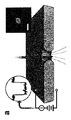
一価のストレプトアビジン(MS)の移行剤と結合するためのビオチンで修飾されたds−DNAの選択的検出および定量が本実施例に詳細に記載される。本実施例で用いられるモノビオチニル化されたds−DNAのSS−ナノ細孔の識別を図1に示す。図1aにおいて、電解質溶液に浸した単一のナノ細孔を有する薄膜に電気バイアスを印加する。これにより、孔を通じて分子(または分子複合体)の動電移行が容易になり、そのそれぞれがイオン電流事象を生じることができる。この技術はMS(図1b左)およびモノビオチニル化された90bpのdsDNA(bio90、図1b中央)測定に用い、それぞれ8μMおよび1μMの濃度で個々に測定した。50〜200mVの範囲にわたって、どちらの分子についてもほとんど事象が同定されなかった。しかしながら、MSとbio90を測定前にモル比8:1(MS:bio90)で一緒にインキュベートすると、単位時間当たりの移行事象数の著しい増加(図1b右)が観察された。混合物の事象速度は常に、どちらか一方の構成分子単独と比べて1桁より大きかった;200mVの印加電圧では、例えば、MS−bio90複合体は23.3±0.9s−1の速度をもたらした一方、MSおよびbio90個々の事象速度はそれぞれ0.09±0.04s−1および1.1±0.2s−1だった。複合体とナノ細孔との間の確率論的な相互作用よりも実際の移行にMS−bio90事象が対応することを立証するために、測定中に印加電圧の極性を逆にし、再捕獲事象を観察した。
Example 1
Selective Detection and Quantification of Double-Stranded DNA Selective detection and quantification of bios-modified ds-DNA for binding to monovalent streptavidin (MS) transfer agent is described in detail in this example . The identification of SS-nanopores of monobiotinylated ds-DNA used in this example is shown in FIG. In FIG. 1a, an electrical bias is applied to a thin film having a single nanopore immersed in an electrolyte solution. This facilitates electrokinetic transfer of molecules (or molecular complexes) through the pores, each of which can generate an ionic current event. This technique was used to measure MS (FIG. 1b left) and monobiotinylated 90 bp dsDNA (bio90, FIG. 1b middle) and measured individually at concentrations of 8 μM and 1 μM, respectively. Over the range of 50-200 mV, few events were identified for either molecule. However, when MS and bio90 were incubated together at a molar ratio of 8: 1 (MS: bio90) prior to measurement, a significant increase in the number of transition events per unit time (FIG. 1b right) was observed. The event rate of the mixture was always greater than an order of magnitude compared to either component alone; for example, at an applied voltage of 200 mV, the MS-bio90 complex yielded a rate of 23.3 ± 0.9 s −1. On the other hand, the individual event rates of MS and bio90 were 0.09 ± 0.04 s −1 and 1.1 ± 0.2 s −1 , respectively. To demonstrate that the MS-bio90 event corresponds to an actual transition rather than a stochastic interaction between the complex and the nanopore, the polarity of the applied voltage is reversed during the measurement and the recapture event Was observed.
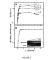
さらにシステムを調査するため、一連のSS−ナノ細孔測定を、MSをbio90の一定量(1μM)に対して滴定して行った。全ての調査した電圧にわたって、測定された事象速度はモル比1:1まで著しく上昇した(図2a)。しかしながら、単一体からモル比8:1(MS:bio90)まで、MSの追加により事象速度はさらに上昇しなかった。これは、核タンパク質複合体の形成に必要なds−DNAの供給が限られた結果であり;タンパク質はかなり低い速度(約10−5s−1)であり、各オリゴヌクレオチドは単一のビオチン部分のみ含有するので、溶液中のほとんど全てのbio90が当モル濃度以上で結合したと予想された。同じ化学量論的範囲にわたってMSおよびbio90で行った電気泳動移動度シフトアッセイ(EMSA)と移行結果を比較すると、非常に類似した傾向が観察された(図2b)。これらのデータは、混合物に対して実質的に観察された全ての移行事象がMS−bio90複合体と対応したという結論を支持した。このアプローチの特異性が高いさらなる証拠は、MSとインキュベートしたビオチニル化されていないdsDNAが、bio90単独と等しい、ごく小さい事象速度をもたらした対照測定によって提供された。 To further investigate the system, a series of SS-nanopore measurements were performed by titrating MS against a certain amount of bio90 (1 μM). Over all investigated voltages, the measured event rate increased significantly to a molar ratio of 1: 1 (FIG. 2a). However, the addition of MS did not further increase the event rate from a single entity to a molar ratio of 8: 1 (MS: bio90). This is a result of the limited supply of ds-DNA required for formation of the nucleoprotein complex; the protein is at a much lower rate (approximately 10 −5 s −1 ) and each oligonucleotide is a single biotin. Since only a portion was contained, it was expected that almost all bio90 in the solution was bound above the equimolar concentration. When comparing the migration results with electrophoretic mobility shift assay (EMSA) performed on MS and bio90 over the same stoichiometric range, a very similar trend was observed (FIG. 2b). These data supported the conclusion that virtually all transition events observed for the mixture corresponded to the MS-bio90 complex. Further evidence of the high specificity of this approach was provided by a control measurement in which non-biotinylated dsDNA incubated with MS resulted in a very small event rate equal to bio90 alone.
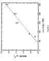
修飾オリゴヌクレオチドの選択的定量:図3において、モル比1:1までの事象速度を検証し、印加電圧の一次従属性を見出した。これは、MS−bio90複合体の捕獲過程が孔との相互作用よりも拡散によって制御されていることを暗示し、以前の研究と一致した。重要なのは、事象頻度が分子濃度によって変化し得るので、観察された傾向が溶液中のMS−bio90複合体の定量への道を提供した。本システムで観察されたほとんど全ての事象は専ら複合体の移行によるものだったので、図3の直線フィットは、溶液中のMS−bio90の濃度を、所与の電圧で生じた特定の事象速度と関連付ける。よって、これまでに記載された測定は、タンパク質に限定されたレジーム(MS:bio90<1:1)で行い、その結果、測定された事象速度はコンジュゲートされていないbio90のバックグラウンドでMS−bio90複合体の定量を容易にした。しかしながら、同じアプローチは原則として、ビオチニル化されていないDNAを有する異種溶液中でのビオチニル化されたオリゴヌクレオチドの定量にも用いることができる。 Selective quantification of modified oligonucleotides: In FIG. 3, the event rate up to a molar ratio of 1: 1 was verified and a first order dependence of the applied voltage was found. This suggested that the capture process of the MS-bio90 complex was controlled by diffusion rather than interaction with the pores, consistent with previous studies. Importantly, since the event frequency can vary with molecular concentration, the observed trend provided a way to quantitate MS-bio90 complex in solution. Since almost all events observed with this system were exclusively due to complex migration, the linear fit in FIG. 3 shows the concentration of MS-bio90 in solution at a given event rate at a given voltage. Associate with. Thus, the measurements described thus far are performed in a protein-only regime (MS: bio90 <1: 1), so that the measured event rate is MS-in the background of unconjugated bio90. Quantification of the bio90 complex was facilitated. However, the same approach can in principle be used for the quantification of biotinylated oligonucleotides in heterogeneous solutions with non-biotinylated DNA.
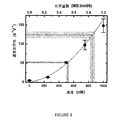
この可能性を調査するために、第三者によって調製された2つの試料で盲検試験を行った。これらのサンプルはそれぞれ、総濃度1μM(上述の測定濃度と等しい)に混合した、ビオチニル化されたおよびビオチニル化されていない90bpのds−DNAの異なる混合物を含有した。全てのbio90が複合体を形成したことを確認するために、両溶液をMSと共に4μMの濃度でインキュベートする。以前のセクションに記載したように、MS単独では測定できる事象はごく少数しか生じなかったため、過剰のタンパク質は測定を不安定にしなかった。SS−ナノ細孔分析は、予想されたように、両試料で印加電圧と事象速度との間で直線関係を示した。2つの盲検試料から得られた事象速度と以前の測定との比較(図4)は、それぞれのbio90濃度の値を導いた:850±35nM(試料1)および520±20nM(試料2)。注目すべきことに、これらの実験で決定された濃度はそれぞれ調製された値である800±20および480±20nMに非常に一致していた。これらの結果は、我々のSS−ナノ細孔アプローチは、混合試料中においても、単一ヌクレオチドビオチン修飾を選択的に有するDNAを一意的に定量できることを立証する。 To investigate this possibility, a blinded study was conducted with two samples prepared by a third party. Each of these samples contained a different mixture of biotinylated and non-biotinylated 90 bp ds-DNA mixed to a total concentration of 1 μM (equal to the measured concentration described above). Both solutions are incubated with MS at a concentration of 4 μM to confirm that all bio90 has formed a complex. As described in the previous section, the excess protein did not destabilize the measurement, since only a few events were measurable with MS alone. SS-nanopore analysis showed a linear relationship between applied voltage and event rate for both samples, as expected. Comparison of the event rate obtained from two blind samples with the previous measurement (FIG. 4) led to the respective bio90 concentration values: 850 ± 35 nM (Sample 1) and 520 ± 20 nM (Sample 2). Notably, the concentrations determined in these experiments were very consistent with the prepared values of 800 ± 20 and 480 ± 20 nM, respectively. These results demonstrate that our SS-nanopore approach can uniquely quantify DNA with selective single nucleotide biotin modifications even in mixed samples.
(方法)
SS−ナノ細孔デバイスの製造および電気的測定
ナノ細孔を、他にWO2012/170,499に記載されているような技術を用いて製造した。簡単には、走査型ヘリウムイオン顕微鏡(Carl Zeiss Orion Plus)のビームの焦点を、シリコン支持チップの吊るされたシリコン窒化物薄膜(厚さ30nm)に合わせた。較正された露出時間を用いて、7.3〜7.7nmの直径のナノ細孔を摩砕した。そして、個別の孔を含有する支持チップを膜の両側への流体通路を有するカスタムフローセルに配置した。測定溶液(900mMのNaClおよび6mMのPBS緩衝液)をフローセルの両側に導入し、Ag/AgCl電極を溶液に浸した。デバイスが低いRMSノイズ(一般的には、<20pA)および較正されたナノ細孔の直径と一致する直線電流−電圧特性を示すことを立証するために、電気的測定(Axopatch 200B)を用いた。移行の測定は、生体分子を含有する測定溶液を有するデバイスの一方の側の溶液を交換することによって行った。コンダクタンスは200kHzの帯域幅で記録し、四極ベッセルフィルターを用いて100kHzでろ過した。分析は全ての測定に対して25kHzの追加のローパスフィルターを適用したカスタムソフトウェアを用いて行った。分析の事象閾値について4つの標準偏差を設定し、12〜2000μsの持続期間での事象のみについて検討した。
(Method)
Fabrication and electrical measurements of SS-nanopore devices Nanopores were fabricated using other techniques such as those described in WO2012 / 170,499. Briefly, the beam of a scanning helium ion microscope (Carl Zeiss Orion Plus) was focused on a suspended silicon nitride film (
生体分子
Bio90のオリゴヌクレオチド配列は購入した(Integrated DNA Technologies、Coralville、IA):TGT ATA CCA TGG CCA GGA TCC TGG GCC ATC TGG TATB GTA ATT CAT AAA GAA TTC TCA TTC TGC AGG TGC ACA TGT TAA CAC TAG TCG TGA。TBは、内部がビオチニル化された単一のdTを表す。対立鎖(dsDNAを形成する)は修飾されていないヌクレオチドを含有していた。混合物(盲検測定)に用いたビオチニル化されていないオリゴヌクレオチドは同じ配列を有していたが、ビオチン部分は有していなかった。用いたストレプトアビジン変異体(SAe1D3)は1つの活性ビオチン結合部位を含有し、Howarth lab(Oxford University)から供給された。この変異タンパク質(54.5kDa)は野生型ストレプトアビジンと類似の結合アフィニティーおよび安定性を保持し、同等のpH条件下で−17.1eの負電荷を与える絶縁で用いられるヘキサグルタミン酸タグを含有する。
Oligonucleotide sequences of biomolecules Bio90 was purchased (Integrated DNA Technologies, Coralville, IA ): TGT ATA CCA TGG CCA GGA TCC TGG GCC ATC TGG TAT B GTA ATT CAT AAA GAA TTC TCA TTC TGC AGG TGC ACA TGT TAA CAC TAG TCG TGA. T B represents a single dT which inside is biotinylated. The opposite strand (forming dsDNA) contained unmodified nucleotides. The non-biotinylated oligonucleotide used for the mixture (blind measurement) had the same sequence but no biotin moiety. The streptavidin variant used (SAe1D3) contained one active biotin binding site and was supplied by Howarth lab (Oxford University). This mutant protein (54.5 kDa) contains a hexaglutamate tag used in insulation that retains similar binding affinity and stability as wild-type streptavidin and gives a negative charge of -17.1e under equivalent pH conditions .
電気泳動移動度シフトアッセイ
MSをbio90と0:1から8:1までのモル比(MS:bio90)で、室温で20分インキュベートした。そして混合物を可視化のためのエチジウムブロマイドを含む1.5%アガロースゲルにロードした。電気泳動ユニットの緩衝液槽を氷浴に浸し、タンパク質−DNA複合体の解離を最小限にした。
Electrophoretic mobility shift assay MS was incubated with bio90 at a molar ratio of 0: 1 to 8: 1 (MS: bio90) at room temperature for 20 minutes. The mixture was then loaded onto a 1.5% agarose gel containing ethidium bromide for visualization. The buffer bath of the electrophoresis unit was immersed in an ice bath to minimize dissociation of the protein-DNA complex.
(実施例2)
二本鎖DNAの選択的検出および定量
2種のDNA分子により与えられる牽引力の変動によって、ビオチニル化されたdsDNAおよびssDNAを区別する能力を立証した。この効果はMSの存在下、ナノ細孔による一本鎖(ss)および二本鎖(ds)バーションの34塩基対(bp)モノビオチニル化されたオリゴヌクレオチドの両方を分析することにより立証した。図5に示すように、事象速度の顕著な増加がdsDNA−MSで観察され、ssDNA−MSでは増進が観察されなかった。この結果に基づいて、ビオチニル化されたssDNAは、生物学的分子の混合物中の相補標的配列と結合することができる配列特異的移行剤として用いることができる。34bpのビオチニル化されたssDNAの配列特異的移行能力を、相同性配列の一本鎖核酸および4つの非相同性ssDNA配列のバックグラウンドを含む混合物で試験した。MSを混合物中に導入し、熱サイクルを行い、アニーリングを促進した。図6は、ビオチニル化されたdsDNA−MSのハイブリダイズされた複合体の移行が観察された結果を示す。重要なのは、非複合体形成ssDNAおよび非複合体形成ビオチニル化されたssDNA−MS移行剤の移行が観察されなかったことである。
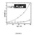
(Example 2)
Selective Detection and Quantification of Double-Stranded DNA The ability to distinguish between biotinylated dsDNA and ssDNA was demonstrated by the variation in traction provided by the two DNA molecules. This effect was demonstrated by analyzing both single-stranded (ss) and double-stranded (ds) versions of 34 base pair (bp) monobiotinylated oligonucleotides in the presence of MS. . As shown in FIG. 5, a significant increase in event rate was observed with dsDNA-MS, and no enhancement was observed with ssDNA-MS. Based on this result, biotinylated ssDNA can be used as a sequence-specific transfer agent that can bind to complementary target sequences in a mixture of biological molecules. The sequence-specific translocation ability of 34 bp biotinylated ssDNA was tested in a mixture containing a single-stranded nucleic acid of homologous sequence and a background of four heterologous ssDNA sequences. MS was introduced into the mixture and a thermal cycle was performed to facilitate annealing. FIG. 6 shows the results of observed migration of the hybridized complex of biotinylated dsDNA-MS. Importantly, no transfer of non-complexed ssDNA and non-complexed biotinylated ssDNA-MS transfer agent was observed.
(実施例3)
ds−DNAの移行の特徴付け
一連のSS−ナノ細孔の測定は、ビオチンで酵素的に標識され、続いてMSに結合した単一ヒドロキシメチルシトシンを有する150bpのds−DNA鎖の移行事象速度を、ナノ細孔の印加電圧および150bpのds−DNA鎖の濃度に対して測定して行った。ナノ細孔の構築および操作パラメーターは、50〜600mVまでの印加電圧である実施例1で得たもの一致した。図7は、ds−DNA−ビオチン−MSの移行事象速度が電圧の上昇と共に増加した試験結果を示す。比較のために、ビオチン−MS移行剤構造を用いていない150bpのds−DNA鎖を試験し、移行反応は記録されなかった。ds−DNA−ビオチン−MS濃度への移行事象速度の依存性もまた調査した。図8に示すように、移行事象速度は、200mVの一定の印加電圧でd−sDNA−ビオチン−MS濃度に対して概ね直線的に変化した。図8の影の部分はノイズフロアを表す。ds−DNAの長さへの移行事象速度の依存性もまた調査した。図9に示すように、移行率は、ds−DNAの長さへの実質的な一次従属性を示した。これらの立証された関係によって、本明細書に記載の方法は、核酸を含む生物学的分子の特徴付けおよび定量に対して強力なツールを提供する。
(Example 3)
Characterization of ds-DNA translocation A series of SS-nanopore measurements were measured for the translocation event rate of a 150 bp ds-DNA strand enzymatically labeled with biotin, followed by a single hydroxymethylcytosine bound to MS. Was measured with respect to the applied voltage of the nanopore and the concentration of ds-DNA strand of 150 bp. The nanopore construction and operating parameters were consistent with those obtained in Example 1 with applied voltages up to 50-600 mV. FIG. 7 shows the test results in which the ds-DNA-biotin-MS transition event rate increased with increasing voltage. For comparison, a 150 bp ds-DNA strand without biotin-MS transfer agent structure was tested and no transfer reaction was recorded. The dependence of the transition event rate on ds-DNA-biotin-MS concentration was also investigated. As shown in FIG. 8, the transition event rate varied approximately linearly with the d-sDNA-biotin-MS concentration at a constant applied voltage of 200 mV. The shaded portion in FIG. 8 represents the noise floor. The dependence of transition event rate on ds-DNA length was also investigated. As shown in FIG. 9, the migration rate showed a substantial first order dependency on the length of the ds-DNA. Due to these proven relationships, the methods described herein provide a powerful tool for the characterization and quantification of biological molecules, including nucleic acids.
本発明の様々な実施形態は、本発明の様々な目的の履行として記載されている。これらの実施形態は、本発明の原理の単なる例示であることを理解されたい。発明の精神および範囲から逸脱することなく、当業者には多くの修正および適応が容易に明らかであろう。 Various embodiments of the invention have been described as fulfilling the various objectives of the invention. It should be understood that these embodiments are merely illustrative of the principles of the present invention. Many modifications and adaptations will be readily apparent to those skilled in the art without departing from the spirit and scope of the invention.
Claims (17)
混合物を、少なくとも1つのナノ細孔を含む膜と接触させるステップと;
ナノ細孔に電界を印加するステップと;
少なくとも1つのナノ細孔を通じて、移行剤と複合体を形成する生物学的分子を移行させるステップ
とを含む、生物学的分子組成物の分析方法であって、
複合体形成生物学的分子の移行が選択的に検出される、方法。 Providing a mixture comprising a biological molecule complexed with a transfer agent and a non-complexed biological molecule;
Contacting the mixture with a membrane comprising at least one nanopore;
Applying an electric field to the nanopore;
Transferring the biological molecule that forms a complex with the transfer agent through at least one nanopore, the method comprising analyzing a biological molecule composition comprising:
A method wherein the translocation of complexing biological molecules is selectively detected.
Applications Claiming Priority (5)
| Application Number | Priority Date | Filing Date | Title |
|---|---|---|---|
| US201461992814P | 2014-05-13 | 2014-05-13 | |
| US61/992,814 | 2014-05-13 | ||
| US201562132989P | 2015-03-13 | 2015-03-13 | |
| US62/132,989 | 2015-03-13 | ||
| PCT/US2015/030531 WO2015175638A1 (en) | 2014-05-13 | 2015-05-13 | Selective analysis of modified biological molecules with solid-state nanopores |
Publications (2)
| Publication Number | Publication Date |
|---|---|
| JP2017516100A true JP2017516100A (en) | 2017-06-15 |
| JP6513705B2 JP6513705B2 (en) | 2019-05-15 |
Family
ID=53267651
Family Applications (1)
| Application Number | Title | Priority Date | Filing Date |
|---|---|---|---|
| JP2016567412A Expired - Fee Related JP6513705B2 (en) | 2014-05-13 | 2015-05-13 | Selective analysis of modified biological molecules using solid state nanopores |
Country Status (11)
| Country | Link |
|---|---|
| US (1) | US10634658B2 (en) |
| EP (1) | EP3143398A1 (en) |
| JP (1) | JP6513705B2 (en) |
| KR (1) | KR20170005090A (en) |
| CN (1) | CN106662568A (en) |
| AU (1) | AU2015259187A1 (en) |
| CA (1) | CA2947978A1 (en) |
| IL (1) | IL248618A0 (en) |
| MX (1) | MX2016014798A (en) |
| WO (1) | WO2015175638A1 (en) |
| ZA (1) | ZA201607693B (en) |
Citations (4)
| Publication number | Priority date | Publication date | Assignee | Title |
|---|---|---|---|---|
| US20060073489A1 (en) * | 2004-10-05 | 2006-04-06 | Gangqiang Li | Nanopore separation devices and methods of using same |
| WO2013016486A1 (en) * | 2011-07-27 | 2013-01-31 | The Board Of Trustees Of The University Of Illinois | Nanopore sensors for biomolecular characterization |
| JP2014173936A (en) * | 2013-03-07 | 2014-09-22 | Toshiba Corp | Specimen detector and detection method |
| US20140378331A1 (en) * | 2013-06-25 | 2014-12-25 | Two Pore Guys, Inc. | Multiplexed biomarker quantitation by nanopore analysis of biomarker-polymer complexes |
Family Cites Families (12)
| Publication number | Priority date | Publication date | Assignee | Title |
|---|---|---|---|---|
| US20030104428A1 (en) * | 2001-06-21 | 2003-06-05 | President And Fellows Of Harvard College | Method for characterization of nucleic acid molecules |
| WO2006137891A2 (en) * | 2004-09-29 | 2006-12-28 | University Of Florida Research Foundation, Inc. | Membrane with nanochannels for detection of molecules |
| US20060210995A1 (en) * | 2005-03-15 | 2006-09-21 | Joyce Timothy H | Nanopore analysis systems and methods of using nanopore devices |
| TWI427440B (en) * | 2005-04-06 | 2014-02-21 | Kodak Graphic Comm Canada Co | Methods and apparatus for correcting banding of imaged regular patterns |
| JP2012507565A (en) * | 2008-10-30 | 2012-03-29 | グオ・ペイシュエン | Viral DNA packaging motor protein connector biosensor embedded in membrane for DNA sequencing and other applications |
| JP5838474B2 (en) * | 2010-02-02 | 2016-01-06 | アリゾナ ボード オブ リージェンツ オン ビハーフ オブ アリゾナ ステート ユニバーシティ | Controlled tunnel gap device for determining polymer alignment |
| US20130256118A1 (en) * | 2010-05-11 | 2013-10-03 | Trustees Of Boston University | Use of Nanopore Arrays For Multiplex Sequencing of Nucleic Acids |
| US8894796B2 (en) | 2011-06-06 | 2014-11-25 | The University Of North Carolina At Greensboro | Nanopore fabrication and applications thereof |
| EP2734840B3 (en) | 2011-07-20 | 2017-06-07 | The Regents of the University of California | Dual-pore device |
| US9347900B2 (en) * | 2011-10-14 | 2016-05-24 | Pacific Biosciences Of California, Inc. | Real-time redox sequencing |
| GB201120910D0 (en) * | 2011-12-06 | 2012-01-18 | Cambridge Entpr Ltd | Nanopore functionality control |
| WO2014182634A1 (en) | 2013-05-06 | 2014-11-13 | Two Pore Guys, Inc. | A method of biological target detection using a nanopore and a fusion protein binding agent |
-
2015
- 2015-05-13 JP JP2016567412A patent/JP6513705B2/en not_active Expired - Fee Related
- 2015-05-13 KR KR1020167034752A patent/KR20170005090A/en not_active Withdrawn
- 2015-05-13 MX MX2016014798A patent/MX2016014798A/en unknown
- 2015-05-13 CA CA2947978A patent/CA2947978A1/en not_active Abandoned
- 2015-05-13 WO PCT/US2015/030531 patent/WO2015175638A1/en not_active Ceased
- 2015-05-13 EP EP15724906.1A patent/EP3143398A1/en not_active Withdrawn
- 2015-05-13 US US15/310,630 patent/US10634658B2/en active Active
- 2015-05-13 AU AU2015259187A patent/AU2015259187A1/en not_active Abandoned
- 2015-05-13 CN CN201580025211.3A patent/CN106662568A/en active Pending
-
2016
- 2016-10-30 IL IL248618A patent/IL248618A0/en unknown
- 2016-11-08 ZA ZA2016/07693A patent/ZA201607693B/en unknown
Patent Citations (6)
| Publication number | Priority date | Publication date | Assignee | Title |
|---|---|---|---|---|
| US20060073489A1 (en) * | 2004-10-05 | 2006-04-06 | Gangqiang Li | Nanopore separation devices and methods of using same |
| JP2006113057A (en) * | 2004-10-05 | 2006-04-27 | Agilent Technol Inc | Nanopore separation device and method of using the same |
| WO2013016486A1 (en) * | 2011-07-27 | 2013-01-31 | The Board Of Trustees Of The University Of Illinois | Nanopore sensors for biomolecular characterization |
| JP2014521956A (en) * | 2011-07-27 | 2014-08-28 | ザ ボード オブ トラスティーズ オブ ザ ユニヴァーシティー オブ イリノイ | Nanopore sensors for characterizing biomolecules |
| JP2014173936A (en) * | 2013-03-07 | 2014-09-22 | Toshiba Corp | Specimen detector and detection method |
| US20140378331A1 (en) * | 2013-06-25 | 2014-12-25 | Two Pore Guys, Inc. | Multiplexed biomarker quantitation by nanopore analysis of biomarker-polymer complexes |
Also Published As
| Publication number | Publication date |
|---|---|
| KR20170005090A (en) | 2017-01-11 |
| JP6513705B2 (en) | 2019-05-15 |
| CN106662568A (en) | 2017-05-10 |
| WO2015175638A1 (en) | 2015-11-19 |
| EP3143398A1 (en) | 2017-03-22 |
| MX2016014798A (en) | 2017-08-02 |
| US10634658B2 (en) | 2020-04-28 |
| AU2015259187A1 (en) | 2016-11-17 |
| ZA201607693B (en) | 2018-05-30 |
| US20170082599A1 (en) | 2017-03-23 |
| IL248618A0 (en) | 2017-01-31 |
| CA2947978A1 (en) | 2015-11-19 |
Similar Documents
| Publication | Publication Date | Title |
|---|---|---|
| JP6679106B2 (en) | Nanopore sensors for characterizing biomolecules | |
| US9863912B2 (en) | Dual-pore device | |
| JP6541114B2 (en) | Target detection method by nanopore | |
| KR20190127983A (en) | Nanopore detection of target polynucleotides from sample background | |
| US20140099726A1 (en) | Device for characterizing polymers | |
| KR20170003619A (en) | Target detection with nanopore | |
| JP2018500905A (en) | Detection of conformational change of nucleic acid polymerase using nanotubes | |
| CN104271762A (en) | Versatile and Sensitive Biosensors | |
| CN106471369B (en) | Target detection using nanopores | |
| Deshmukh et al. | A capacitive DNA sensor for sensitive detection of Escherichia coli O157: H7 in potable water based on the z3276 genetic marker: Fabrication and analytical performance | |
| Ge et al. | Electrochemical investigation of DNA-modified surfaces: From quantitation methods to experimental conditions | |
| JP2021536261A (en) | Sensing compositions, methods, and devices for detecting molecules using nanopore devices | |
| Luchian et al. | Single‐molecule, hybridization‐based strategies for short nucleic acids detection and recognition with nanopores | |
| JP6513705B2 (en) | Selective analysis of modified biological molecules using solid state nanopores | |
| Singer et al. | Detection of urea-induced internal denaturation of dsDNA using solid-state nanopores | |
| Mir et al. | Electrokinetic techniques applied to electrochemical DNA biosensors | |
| García‐Mendiola et al. | Screening of specific gene mutations associated with cystic fibrosis | |
| HK1234825B (en) | Target detection with nanopore | |
| Radadiya | The Graphene Base DNA Sensing |
Legal Events
| Date | Code | Title | Description |
|---|---|---|---|
| A621 | Written request for application examination |
Free format text: JAPANESE INTERMEDIATE CODE: A621 Effective date: 20180404 |
|
| A977 | Report on retrieval |
Free format text: JAPANESE INTERMEDIATE CODE: A971007 Effective date: 20190228 |
|
| TRDD | Decision of grant or rejection written | ||
| A01 | Written decision to grant a patent or to grant a registration (utility model) |
Free format text: JAPANESE INTERMEDIATE CODE: A01 Effective date: 20190319 |
|
| A61 | First payment of annual fees (during grant procedure) |
Free format text: JAPANESE INTERMEDIATE CODE: A61 Effective date: 20190410 |
|
| R150 | Certificate of patent or registration of utility model |
Ref document number: 6513705 Country of ref document: JP Free format text: JAPANESE INTERMEDIATE CODE: R150 |
|
| LAPS | Cancellation because of no payment of annual fees |