JP2017526367A - Methods and compositions for the treatment of cancer - Google Patents
Methods and compositions for the treatment of cancer Download PDFInfo
- Publication number
- JP2017526367A JP2017526367A JP2017511674A JP2017511674A JP2017526367A JP 2017526367 A JP2017526367 A JP 2017526367A JP 2017511674 A JP2017511674 A JP 2017511674A JP 2017511674 A JP2017511674 A JP 2017511674A JP 2017526367 A JP2017526367 A JP 2017526367A
- Authority
- JP
- Japan
- Prior art keywords
- epcam
- asic
- cells
- cancer
- cell
- Prior art date
- Legal status (The legal status is an assumption and is not a legal conclusion. Google has not performed a legal analysis and makes no representation as to the accuracy of the status listed.)
- Pending
Links
Images






















Classifications
-
- C—CHEMISTRY; METALLURGY
- C12—BIOCHEMISTRY; BEER; SPIRITS; WINE; VINEGAR; MICROBIOLOGY; ENZYMOLOGY; MUTATION OR GENETIC ENGINEERING
- C12N—MICROORGANISMS OR ENZYMES; COMPOSITIONS THEREOF; PROPAGATING, PRESERVING, OR MAINTAINING MICROORGANISMS; MUTATION OR GENETIC ENGINEERING; CULTURE MEDIA
- C12N15/00—Mutation or genetic engineering; DNA or RNA concerning genetic engineering, vectors, e.g. plasmids, or their isolation, preparation or purification; Use of hosts therefor
- C12N15/09—Recombinant DNA-technology
- C12N15/11—DNA or RNA fragments; Modified forms thereof; Non-coding nucleic acids having a biological activity
- C12N15/113—Non-coding nucleic acids modulating the expression of genes, e.g. antisense oligonucleotides; Antisense DNA or RNA; Triplex- forming oligonucleotides; Catalytic nucleic acids, e.g. ribozymes; Nucleic acids used in co-suppression or gene silencing
-
- C—CHEMISTRY; METALLURGY
- C12—BIOCHEMISTRY; BEER; SPIRITS; WINE; VINEGAR; MICROBIOLOGY; ENZYMOLOGY; MUTATION OR GENETIC ENGINEERING
- C12N—MICROORGANISMS OR ENZYMES; COMPOSITIONS THEREOF; PROPAGATING, PRESERVING, OR MAINTAINING MICROORGANISMS; MUTATION OR GENETIC ENGINEERING; CULTURE MEDIA
- C12N15/00—Mutation or genetic engineering; DNA or RNA concerning genetic engineering, vectors, e.g. plasmids, or their isolation, preparation or purification; Use of hosts therefor
- C12N15/09—Recombinant DNA-technology
- C12N15/11—DNA or RNA fragments; Modified forms thereof; Non-coding nucleic acids having a biological activity
- C12N15/115—Aptamers, i.e. nucleic acids binding a target molecule specifically and with high affinity without hybridising therewith ; Nucleic acids binding to non-nucleic acids, e.g. aptamers
-
- A—HUMAN NECESSITIES
- A61—MEDICAL OR VETERINARY SCIENCE; HYGIENE
- A61K—PREPARATIONS FOR MEDICAL, DENTAL OR TOILETRY PURPOSES
- A61K31/00—Medicinal preparations containing organic active ingredients
- A61K31/33—Heterocyclic compounds
- A61K31/335—Heterocyclic compounds having oxygen as the only ring hetero atom, e.g. fungichromin
- A61K31/337—Heterocyclic compounds having oxygen as the only ring hetero atom, e.g. fungichromin having four-membered rings, e.g. taxol
-
- A—HUMAN NECESSITIES
- A61—MEDICAL OR VETERINARY SCIENCE; HYGIENE
- A61K—PREPARATIONS FOR MEDICAL, DENTAL OR TOILETRY PURPOSES
- A61K48/00—Medicinal preparations containing genetic material which is inserted into cells of the living body to treat genetic diseases; Gene therapy
-
- A—HUMAN NECESSITIES
- A61—MEDICAL OR VETERINARY SCIENCE; HYGIENE
- A61P—SPECIFIC THERAPEUTIC ACTIVITY OF CHEMICAL COMPOUNDS OR MEDICINAL PREPARATIONS
- A61P35/00—Antineoplastic agents
-
- C—CHEMISTRY; METALLURGY
- C07—ORGANIC CHEMISTRY
- C07H—SUGARS; DERIVATIVES THEREOF; NUCLEOSIDES; NUCLEOTIDES; NUCLEIC ACIDS
- C07H21/00—Compounds containing two or more mononucleotide units having separate phosphate or polyphosphate groups linked by saccharide radicals of nucleoside groups, e.g. nucleic acids
-
- C—CHEMISTRY; METALLURGY
- C07—ORGANIC CHEMISTRY
- C07K—PEPTIDES
- C07K14/00—Peptides having more than 20 amino acids; Gastrins; Somatostatins; Melanotropins; Derivatives thereof
- C07K14/435—Peptides having more than 20 amino acids; Gastrins; Somatostatins; Melanotropins; Derivatives thereof from animals; from humans
- C07K14/705—Receptors; Cell surface antigens; Cell surface determinants
-
- C—CHEMISTRY; METALLURGY
- C12—BIOCHEMISTRY; BEER; SPIRITS; WINE; VINEGAR; MICROBIOLOGY; ENZYMOLOGY; MUTATION OR GENETIC ENGINEERING
- C12N—MICROORGANISMS OR ENZYMES; COMPOSITIONS THEREOF; PROPAGATING, PRESERVING, OR MAINTAINING MICROORGANISMS; MUTATION OR GENETIC ENGINEERING; CULTURE MEDIA
- C12N15/00—Mutation or genetic engineering; DNA or RNA concerning genetic engineering, vectors, e.g. plasmids, or their isolation, preparation or purification; Use of hosts therefor
- C12N15/09—Recombinant DNA-technology
- C12N15/11—DNA or RNA fragments; Modified forms thereof; Non-coding nucleic acids having a biological activity
- C12N15/113—Non-coding nucleic acids modulating the expression of genes, e.g. antisense oligonucleotides; Antisense DNA or RNA; Triplex- forming oligonucleotides; Catalytic nucleic acids, e.g. ribozymes; Nucleic acids used in co-suppression or gene silencing
- C12N15/1137—Non-coding nucleic acids modulating the expression of genes, e.g. antisense oligonucleotides; Antisense DNA or RNA; Triplex- forming oligonucleotides; Catalytic nucleic acids, e.g. ribozymes; Nucleic acids used in co-suppression or gene silencing against enzymes
-
- A—HUMAN NECESSITIES
- A61—MEDICAL OR VETERINARY SCIENCE; HYGIENE
- A61K—PREPARATIONS FOR MEDICAL, DENTAL OR TOILETRY PURPOSES
- A61K39/00—Medicinal preparations containing antigens or antibodies
- A61K2039/51—Medicinal preparations containing antigens or antibodies comprising whole cells, viruses or DNA/RNA
- A61K2039/515—Animal cells
- A61K2039/5152—Tumor cells
-
- C—CHEMISTRY; METALLURGY
- C12—BIOCHEMISTRY; BEER; SPIRITS; WINE; VINEGAR; MICROBIOLOGY; ENZYMOLOGY; MUTATION OR GENETIC ENGINEERING
- C12N—MICROORGANISMS OR ENZYMES; COMPOSITIONS THEREOF; PROPAGATING, PRESERVING, OR MAINTAINING MICROORGANISMS; MUTATION OR GENETIC ENGINEERING; CULTURE MEDIA
- C12N2310/00—Structure or type of the nucleic acid
- C12N2310/10—Type of nucleic acid
- C12N2310/14—Type of nucleic acid interfering nucleic acids [NA]
-
- C—CHEMISTRY; METALLURGY
- C12—BIOCHEMISTRY; BEER; SPIRITS; WINE; VINEGAR; MICROBIOLOGY; ENZYMOLOGY; MUTATION OR GENETIC ENGINEERING
- C12N—MICROORGANISMS OR ENZYMES; COMPOSITIONS THEREOF; PROPAGATING, PRESERVING, OR MAINTAINING MICROORGANISMS; MUTATION OR GENETIC ENGINEERING; CULTURE MEDIA
- C12N2310/00—Structure or type of the nucleic acid
- C12N2310/10—Type of nucleic acid
- C12N2310/16—Aptamers
-
- C—CHEMISTRY; METALLURGY
- C12—BIOCHEMISTRY; BEER; SPIRITS; WINE; VINEGAR; MICROBIOLOGY; ENZYMOLOGY; MUTATION OR GENETIC ENGINEERING
- C12N—MICROORGANISMS OR ENZYMES; COMPOSITIONS THEREOF; PROPAGATING, PRESERVING, OR MAINTAINING MICROORGANISMS; MUTATION OR GENETIC ENGINEERING; CULTURE MEDIA
- C12N2310/00—Structure or type of the nucleic acid
- C12N2310/30—Chemical structure
- C12N2310/32—Chemical structure of the sugar
- C12N2310/322—2'-R Modification
-
- C—CHEMISTRY; METALLURGY
- C12—BIOCHEMISTRY; BEER; SPIRITS; WINE; VINEGAR; MICROBIOLOGY; ENZYMOLOGY; MUTATION OR GENETIC ENGINEERING
- C12N—MICROORGANISMS OR ENZYMES; COMPOSITIONS THEREOF; PROPAGATING, PRESERVING, OR MAINTAINING MICROORGANISMS; MUTATION OR GENETIC ENGINEERING; CULTURE MEDIA
- C12N2310/00—Structure or type of the nucleic acid
- C12N2310/30—Chemical structure
- C12N2310/35—Nature of the modification
- C12N2310/351—Conjugate
- C12N2310/3519—Fusion with another nucleic acid
-
- C—CHEMISTRY; METALLURGY
- C12—BIOCHEMISTRY; BEER; SPIRITS; WINE; VINEGAR; MICROBIOLOGY; ENZYMOLOGY; MUTATION OR GENETIC ENGINEERING
- C12N—MICROORGANISMS OR ENZYMES; COMPOSITIONS THEREOF; PROPAGATING, PRESERVING, OR MAINTAINING MICROORGANISMS; MUTATION OR GENETIC ENGINEERING; CULTURE MEDIA
- C12N2310/00—Structure or type of the nucleic acid
- C12N2310/30—Chemical structure
- C12N2310/35—Nature of the modification
- C12N2310/353—Nature of the modification linked to the nucleic acid via an atom other than carbon
- C12N2310/3533—Halogen
Landscapes
- Health & Medical Sciences (AREA)
- Life Sciences & Earth Sciences (AREA)
- Genetics & Genomics (AREA)
- Chemical & Material Sciences (AREA)
- Engineering & Computer Science (AREA)
- Molecular Biology (AREA)
- Organic Chemistry (AREA)
- Biomedical Technology (AREA)
- Biotechnology (AREA)
- General Health & Medical Sciences (AREA)
- Zoology (AREA)
- Bioinformatics & Cheminformatics (AREA)
- General Engineering & Computer Science (AREA)
- Biochemistry (AREA)
- Wood Science & Technology (AREA)
- Biophysics (AREA)
- Physics & Mathematics (AREA)
- Plant Pathology (AREA)
- Microbiology (AREA)
- Medicinal Chemistry (AREA)
- Veterinary Medicine (AREA)
- Public Health (AREA)
- Animal Behavior & Ethology (AREA)
- Pharmacology & Pharmacy (AREA)
- Epidemiology (AREA)
- Virology (AREA)
- Gastroenterology & Hepatology (AREA)
- Cell Biology (AREA)
- Immunology (AREA)
- Toxicology (AREA)
- Proteomics, Peptides & Aminoacids (AREA)
- Chemical Kinetics & Catalysis (AREA)
- General Chemical & Material Sciences (AREA)
- Nuclear Medicine, Radiotherapy & Molecular Imaging (AREA)
- Pharmaceuticals Containing Other Organic And Inorganic Compounds (AREA)
- Medicines That Contain Protein Lipid Enzymes And Other Medicines (AREA)
- Medicinal Preparation (AREA)
Abstract
本明細書において、例えばアプタマー-siRNAキメラ分子を使用する、癌、例えば乳癌の処置に関連する方法および組成物を記載する。Described herein are methods and compositions related to the treatment of cancer, eg, breast cancer, using, for example, aptamer-siRNA chimeric molecules.
Description
関連出願の相互参照
本願は、35 U.S.C.§119(e)の下で、2014年8月29日に出願された米国仮出願第62/043,803号の恩典を主張し、その内容は全体として参照により本明細書に組み入れられる。
This application claims the benefit of US Provisional Application No. 62 / 043,803, filed August 29, 2014, under 35 USC § 119 (e), the contents of which are incorporated by reference in their entirety It is incorporated herein.
政府による支援
本発明は、国防総省から授与された助成金第W81XWH-09-1-0058号の下で政府の財政支援を受けてなされたものである。米国政府は、本発明に関して一定の権利を保有する。
Government Support This invention was made with government financial support under Grant No. W81XWH-09-1-0058 awarded by the Department of Defense. The US government has certain rights in this invention.
配列表
本願は、ASCII形式で電子的に提出され、全体として参照により本明細書に組み入れられる配列表を含んでいる。2015年8月28日に作成されたこのASCIIコピーは、701039-082401-PCT_SL.txtと命名され、8,984バイトのサイズである。
SEQUENCE LISTING This application contains a Sequence Listing submitted electronically in ASCII format and incorporated herein by reference in its entirety. This ASCII copy, created on August 28, 2015, is named 701039-082401-PCT_SL.txt and is 8,984 bytes in size.
技術分野
本明細書に記載される技術は、EpCAM結合分子および阻害性核酸を含むキメラ分子、ならびに癌、例えば上皮癌の処置のためにそのような組成物を使用する方法に関する。
TECHNICAL FIELD The technology described herein relates to chimeric molecules comprising EpCAM binding molecules and inhibitory nucleic acids, and methods of using such compositions for the treatment of cancer, eg, epithelial cancer.
背景
RNA干渉(RNAi)は、肝臓での遺伝子発現を減少させることにおける治療的使用のために検討されてきた。しかし、肝臓は、RNAi分子でトランスフェクトすることが容易な点で特有である。他の組織における低分子RNAの送達および結果として生じる遺伝子ノックダウンは、非効率的で、かつ最終的に無効であり続けている。特に、送達妨害は、癌を処置するためにRNAiを利用することの大きな障害である。
background
RNA interference (RNAi) has been investigated for therapeutic use in reducing gene expression in the liver. However, the liver is unique in that it is easy to transfect with RNAi molecules. Delivery of small RNAs in other tissues and the resulting gene knockdown continues to be inefficient and ultimately ineffective. In particular, delivery hindrance is a major obstacle to utilizing RNAi to treat cancer.
概要
本明細書に記載されるように、本発明者らは、新規なアプタマー-siRNAキメラ分子(AsiC)を開発した。これらのAsiCは、癌細胞マーカーを標的としてsiRNAを癌細胞に特異的に方向付け、送達効力および治療有効性を増加させる一方で、副作用の潜在能を減少させる。
Overview As described herein, the inventors have developed a novel aptamer-siRNA chimeric molecule (AsiC). These AsiCs target cancer cell markers to specifically direct siRNA to cancer cells, increasing delivery efficacy and therapeutic efficacy while reducing the potential for side effects.
1つの局面において、癌マーカー結合アプタマードメインおよび阻害性核酸ドメインを含むキメラ分子が、本明細書において記載されている。いくつかの態様において、癌マーカーは、EpCAMまたはEphA2である。いくつかの態様において、阻害性核酸は、癌細胞においてアップレギュレートされる遺伝子産物と特異的に結合する。いくつかの態様において、阻害性核酸は、Plk1;MCL1;EphA2;PsmA2;MSI1;BMI1;XBP1;PRPF8;PFPF38A;RBM22;USP39;RAN;NUP205;およびNDC80からなる群より選択される遺伝子の発現を阻害する。いくつかの態様において、癌マーカーはEpCAMであり、阻害性核酸ドメインは、Plk1の発現を阻害する。 In one aspect, chimeric molecules comprising a cancer marker binding aptamer domain and an inhibitory nucleic acid domain are described herein. In some embodiments, the cancer marker is EpCAM or EphA2. In some embodiments, the inhibitory nucleic acid specifically binds to a gene product that is upregulated in cancer cells. In some embodiments, the inhibitory nucleic acid exhibits expression of a gene selected from the group consisting of Plk1; MCL1; EphA2; PsmA2; MSI1; BMI1; XBP1; PRPF8; PFPF38A; RBM22; USP39; RAN; Inhibit. In some embodiments, the cancer marker is EpCAM and the inhibitory nucleic acid domain inhibits Plk1 expression.
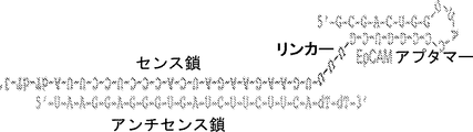
いくつかの態様において、前記分子は、アプタマー-siRNAキメラ(AsiC)である。いくつかの態様において、癌マーカー結合アプタマードメインは、SEQ ID NO:33の配列を含む。いくつかの態様において、癌マーカー結合アプタマードメインは、本質的に、SEQ ID NO:33の配列からなる。いくつかの態様において、阻害性核酸ドメインは、SEQ ID NO:2の配列を含む。いくつかの態様において、阻害性核酸ドメインは、本質的にSEQ ID NO:2の配列からなる。いくつかの態様において、前記分子は、SEQ ID NO:1〜3のうちの1つの配列を含む。いくつかの態様において、前記分子は、本質的にSEQ ID NO:1〜3のうちの1つの配列からなる。 In some embodiments, the molecule is an aptamer-siRNA chimera (AsiC). In some embodiments, the cancer marker binding aptamer domain comprises the sequence of SEQ ID NO: 33. In some embodiments, the cancer marker binding aptamer domain consists essentially of the sequence of SEQ ID NO: 33. In some embodiments, the inhibitory nucleic acid domain comprises the sequence of SEQ ID NO: 2. In some embodiments, the inhibitory nucleic acid domain consists essentially of the sequence of SEQ ID NO: 2. In some embodiments, the molecule comprises a sequence of one of SEQ ID NOs: 1-3. In some embodiments, the molecule consists essentially of the sequence of one of SEQ ID NOs: 1-3.
いくつかの態様において、前記分子の3'末端は、dTdTを含む。いくつかの態様において、前記分子は、少なくとも1つの2'-Fピリミジンを含む。 In some embodiments, the 3 ′ end of the molecule comprises dTdT. In some embodiments, the molecule comprises at least one 2′-F pyrimidine.
1つの局面において、本明細書に記載されるキメラ分子および薬学的に許容される担体を含む薬学的組成物が、本明細書に記載されている。いくつかの態様において、組成物は、本明細書に記載される少なくとも2種類のキメラ分子を含み、その際、キメラ分子は、異なるアプタマードメインおよび/または阻害性核酸ドメインを有する。いくつかの態様において、異なるアプタマードメインまたは阻害性核酸ドメインは、異なる標的を認識する。いくつかの態様において、異なるアプタマードメインまたは阻害性核酸ドメインは、配列を有し、同じ標的を認識する。 In one aspect, a pharmaceutical composition comprising a chimeric molecule described herein and a pharmaceutically acceptable carrier is described herein. In some embodiments, the composition comprises at least two types of chimeric molecules described herein, wherein the chimeric molecules have different aptamer domains and / or inhibitory nucleic acid domains. In some embodiments, different aptamer domains or inhibitory nucleic acid domains recognize different targets. In some embodiments, different aptamer domains or inhibitory nucleic acid domains have sequences and recognize the same target.
1つの局面において、癌を処置する方法であって、本明細書に記載されるキメラ分子および/または組成物を投与する工程を含む方法が、本明細書において記載されている。いくつかの態様において、癌は、上皮癌または乳癌である。いくつかの態様において、乳癌は、トリプルネガティブ乳癌である。いくつかの態様において、投与は皮下である。いくつかの態様において、対象には、追加的な癌処置がさらに施される。いくつかの態様において、癌処置は、パクリタキセルである。 In one aspect, a method of treating cancer, comprising the step of administering a chimeric molecule and / or composition described herein is described herein. In some embodiments, the cancer is epithelial cancer or breast cancer. In some embodiments, the breast cancer is triple negative breast cancer. In some embodiments, administration is subcutaneous. In some embodiments, the subject is further administered additional cancer treatment. In some embodiments, the cancer treatment is paclitaxel.
詳細な説明
本発明者らは、癌の処置におけるAsiC(アプタマー-siRNAキメラ分子)の驚くべき効力を実証した。本明細書に記載されるAsiCは、キメラ分子を癌細胞に特異的に標的指向化するアプタマーを利用し、siRNAの標的とされる遺伝子の効果的で的を射た抑制をもたらす。
DETAILED DESCRIPTION The inventors have demonstrated the surprising efficacy of AsiC (aptamer-siRNA chimeric molecule) in the treatment of cancer. AsiC as described herein utilizes aptamers that specifically target chimeric molecules to cancer cells, resulting in effective and targeted suppression of genes targeted by siRNA.
特に、本明細書に記載されるアプタマー、例えばEpCAMおよびEphA2を標的とするものは、癌源細胞(tumor-initiating cell)(癌幹細胞とも呼ばれる)を標的とする治療を可能にする。これらの細胞は、発がんイニシエーション、再発、および転移を担うだけでなく、従来の細胞傷害性療法に相対的に耐性でもある。このように、本明細書に記載される組成物および方法は、既存の治療が行えない方法で基礎となる病理の有効な処置を可能にする。抗体を用いたEpCAMの直接ターゲティングが以前に研究されており、有効性が欠如していることが見出されていた点で、本明細書に記載されるAsiCの成功は、特に驚くべきことである。 In particular, the aptamers described herein, such as those targeting EpCAM and EphA2, allow treatment targeting tumor-initiating cells (also called cancer stem cells). These cells are not only responsible for carcinogenic initiation, recurrence, and metastasis, but are also relatively resistant to conventional cytotoxic therapies. Thus, the compositions and methods described herein allow for effective treatment of underlying pathologies in ways that are incapable of existing therapies. The success of AsiC described herein was particularly surprising in that direct targeting of EpCAM with antibodies has been previously studied and found to lack efficacy. is there.
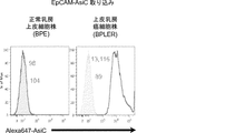
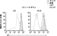
そのうえ、本明細書に記載されるAsiCは、上皮癌、例えば乳癌(例えばトリプルネガティブ乳癌(TNBC))の処置に驚くほど効果的であることが実証されている。TNBCに対する目下の標的指向化された療法はなく、利用できる処置は、典型的には3年以内に転移を招き、死に至る。本明細書に記載されるAsiCは、健康な細胞と比較して管腔型および基底A型TNBC細胞に特異的な、有効な遺伝子ノックダウンを示し、コロニー形成およびマンモスフィア形成をインビトロで抑制し、エクスビボで発がんイニシエーションを打ち消した。AsiCを用いたインビトロ処置は、治療薬の標的指向化された送達および迅速な腫瘍退縮を招いた。 Moreover, AsiC described herein has been shown to be surprisingly effective in the treatment of epithelial cancers, such as breast cancer (eg, triple negative breast cancer (TNBC)). There are currently no targeted therapies for TNBC and available treatments typically lead to metastasis and death within 3 years. AsiC described herein exhibits effective gene knockdown specific to luminal and basal type TNBC cells compared to healthy cells and suppresses colony formation and mammosphere formation in vitro. , Canceled the carcinogenesis initiation ex vivo. In vitro treatment with AsiC led to targeted delivery of therapeutic agents and rapid tumor regression.
1つの局面において、癌マーカー結合ドメインおよび阻害性核酸ドメインを含むキメラ分子が、本明細書において記載されている。本明細書で使用される場合、「癌マーカー結合ドメイン」は、同じ型の健康な細胞と比較して癌細胞の表面により高く発現される分子(癌マーカー)と特異的に結合できるドメインおよび/または分子を表す。いくつかの態様において、癌マーカーは、タンパク質および/またはポリペプチドであることができる。いくつかの態様において、癌マーカーは、EpCAMまたはEphA2より選択することができる。いくつかの態様において、癌マーカー結合ドメインは、アプタマーであることができる。 In one aspect, chimeric molecules comprising a cancer marker binding domain and an inhibitory nucleic acid domain are described herein. As used herein, a “cancer marker binding domain” is a domain that can specifically bind to a molecule (cancer marker) that is highly expressed on the surface of a cancer cell as compared to healthy cells of the same type and / or Or represents a molecule. In some embodiments, the cancer marker can be a protein and / or polypeptide. In some embodiments, the cancer marker can be selected from EpCAM or EphA2. In some embodiments, the cancer marker binding domain can be an aptamer.
本明細書で使用される場合、「EpCAM」または「上皮細胞接着分子」は、上皮細胞におけるCa2+非依存性ホモタイプ細胞間接着を媒介する膜貫通糖タンパク質を表す。EpCAMについての配列は、多様な種、例えばヒトEpCAM(例えばNCBI Gene ID:4072;タンパク質配列:NCBI Ref Seq: NP_002345.2を参照されたい)について公知である。 As used herein, “EpCAM” or “epithelial cell adhesion molecule” refers to a transmembrane glycoprotein that mediates Ca2 + -independent homotypic cell-cell adhesion in epithelial cells. The sequence for EpCAM is known for a variety of species, such as human EpCAM (see eg NCBI Gene ID: 4072; protein sequence: NCBI Ref Seq: NP — 002345.2).
本明細書で使用される場合、「EphA2」または「EPH受容体A2」は、エフリン型タンパク質-チロシンキナーゼ受容体を表す。EphA2は、エフリン-Aリガンドに結合し、宿主細胞へのカポジ肉腫関連ヘルペスウイルスの侵入を許す。EphA2についての配列は、多様な種、例えばヒトEphA2(例えばNCBI Gene ID:1969;タンパク質配列:NCBI Ref Seq: NP_004422.2を参照されたい)について公知である。 As used herein, “EphA2” or “EPH receptor A2” refers to the ephrin-type protein-tyrosine kinase receptor. EphA2 binds to ephrin-A ligand and allows the entry of Kaposi's sarcoma-associated herpesvirus into the host cell. The sequence for EphA2 is known for a variety of species, such as human EphA2 (see eg NCBI Gene ID: 1969; protein sequence: NCBI Ref Seq: NP — 004422.2).
本明細書で使用される場合、「阻害性核酸ドメイン」は、阻害性核酸を含むドメインを表す。いくつかの態様において、阻害性核酸はsiRNAであることができる。 As used herein, “inhibitory nucleic acid domain” refers to a domain comprising an inhibitory nucleic acid. In some embodiments, the inhibitory nucleic acid can be an siRNA.
阻害性核酸ドメインは、癌細胞においてアップレギュレートされる遺伝子産物の発現ならびに/または細胞の成長および/もしくは生存に必要とされる遺伝子の発現を阻害、例えばそれを標的とすることができる。いくつかの態様において、阻害性核酸ドメインは、Plk1(例えば「polo様キナーゼ1」;NCBI Gene ID:5347);MCL1(例えば骨髄細胞白血病配列1;NCBI Gene ID:4170);EphA2(NCBI Gene ID:1969);PsmA2(例えばプロテアソームサブユニットα2;NCBI Gene ID:5683);MSI1(例えば、musashi RNA結合タンパク質1;NCBI Gene ID:4440);BMI1(例えば、Bリンパ腫Mo-MLV挿入配列1、NCBI Gene ID:648);XBP1(X-boxn結合タンパク質1;NCBI Gene ID:7494);PRPF8(例えば、プレmRNAプロセシング因子8;NCBI Gene ID:10594)、PFPF38A(例えば、プレmRNAプロセシング因子38A;NCBI Gene ID:84950)、RBM22(例えば、RNA結合モチーフタンパク質22;NCBI Gene ID:55696)、USP39(例えば、ユビキチン特異的ペプチダーゼ39;NCBI Gene ID:10713);RAN(例えば、ras関連核タンパク質;NCBI Gene ID:5901);NUP205(例えば、ヌクレオポリン205kDa;NCBI Gene ID:23165)、およびNDC80(例えば、NDC80キネトコア複合体成分;NCBI Gene ID:10403)より選択される遺伝子の発現を阻害することができる。これらの遺伝子、例えばヒトmRNAの配列は、NCBIデータベースから容易に得られ、阻害性核酸を設計するために当業者によって使用されることができる。さらに、例示的な阻害性核酸ドメイン、例えばSEQ ID NO:2の配列を有する核酸が、本明細書に提供される。
Inhibitory nucleic acid domains can inhibit, eg target, the expression of gene products that are up-regulated in cancer cells and / or the expression of genes that are required for cell growth and / or survival. In some embodiments, the inhibitory nucleic acid domain is Plk1 (eg “polo-
いくつかの態様において、本明細書に記載される組成物は、アプタマーを含む癌マーカー結合ドメインおよびsiRNAを含む阻害性核酸ドメインを含むことができ、例えば、組成物は、アプタマー-siRNAキメラ(AsiC)を含むことができる。 In some embodiments, a composition described herein can include a cancer marker binding domain comprising an aptamer and an inhibitory nucleic acid domain comprising siRNA, eg, the composition comprises an aptamer-siRNA chimera (AsiC ) Can be included.
いくつかの態様において、本明細書に記載される方法は、癌を有するかまたは癌を有すると診断された対象を、本明細書に記載される組成物で処置することに関する。癌を有する対象は、癌を診断する現行方法を使用して医師によって同定されることができる。これらの状態を特徴づけ、かつ診断を助ける、癌の症状および/または合併症は、当技術分野において周知であり、それらには非限定的に、例えば乳癌の場合、乳房組織のしこりもしくは塊、乳房のすべてもしくは一部の腫脹、皮膚のかぶれ、乳房のくぼみ、乳房もしくは乳頭の疼痛、乳頭退縮、発赤、鱗屑(scaliness)、または乳房もしくは乳頭のかぶれ、および乳頭分泌が含まれる。例えば乳癌の診断の助けとなり得る試験には、非限定的にマンモグラム、X線、MRI、超音波、乳管造影図、生検、および乳管洗浄細胞診が含まれる。癌の家族歴または癌についてのリスク因子への曝露(例えば喫煙、放射線、汚染物、BRCA1変異など)も、対象が癌を有する可能性があるかの判定または癌の診断を助けることができる。 In some embodiments, the methods described herein relate to treating a subject having or diagnosed with cancer with a composition described herein. A subject with cancer can be identified by a physician using current methods of diagnosing cancer. Cancer symptoms and / or complications that characterize these conditions and aid in diagnosis are well known in the art and include, but are not limited to, lump or mass of breast tissue, for example in the case of breast cancer, Includes swelling of all or part of the breast, skin irritation, breast depression, breast or nipple pain, teat retraction, redness, scaliness, or breast or nipple rash, and nipple discharge. For example, tests that can help diagnose breast cancer include, but are not limited to, mammograms, x-rays, MRI, ultrasound, ductography, biopsy, and ductal lavage cytology. Exposure to a family history of cancer or risk factors for cancer (eg, smoking, radiation, contaminants, BRCA1 mutations, etc.) can also help determine whether a subject may have cancer or diagnose cancer.
本明細書で使用される場合、「悪性腫瘍」、「悪性状態」、「癌」、または「腫瘍」という用語は、身体器官および身体システムの正常な機能を妨害する、細胞の無制御な成長を表す。 As used herein, the terms “malignant tumor”, “malignant condition”, “cancer”, or “tumor” refer to the uncontrolled growth of cells that interfere with the normal functioning of body organs and systems. Represents.
本明細書で使用される場合、「癌」という用語は、一般的に、異常な細胞が制御されずに分裂し、隣接組織に浸潤する可能性がある疾患または状態のうちの1つのクラスに関する。癌細胞は、また、血液およびリンパ系を経由して身体の他の部分に拡散する可能性がある。 As used herein, the term “cancer” generally relates to one class of diseases or conditions in which abnormal cells can divide uncontrolled and invade adjacent tissues. . Cancer cells can also spread to other parts of the body via the blood and lymphatic system.
「癌細胞」または「腫瘍細胞」は、癌性成長または癌性組織の個別の細胞を表す。腫瘍は、一般的に、良性、前悪性、または悪性であり得る細胞の異常成長によって形成される腫脹または病変を表す。大部分の癌細胞は腫瘍を形成するが、一部、例えば白血病は、必ずしも腫瘍を形成しない。腫瘍を形成する癌細胞について、癌(細胞)および腫瘍(細胞)という用語は、互換的に使用される。 “Cancer cells” or “tumor cells” refer to individual cells of cancerous growth or cancerous tissue. A tumor generally represents a swelling or lesion formed by an abnormal growth of cells that can be benign, pre-malignant, or malignant. Most cancer cells form tumors, but some, such as leukemia, do not necessarily form tumors. For cancer cells that form a tumor, the terms cancer (cell) and tumor (cell) are used interchangeably.
癌または腫瘍を有する対象は、対象の体内に存在する客観的に測定可能な癌細胞を有する対象である。悪性の、活発に増殖する癌と同様に潜在的に休眠状態の腫瘍または微小転移が、この定義に含まれる。その本来の位置から移動して、他の重要器官に播種する癌は、最終的には、罹患器官の機能的悪化を通じて対象を死亡させる可能性がある。白血病のような造血癌は、対象における正常な造血区画を打ち負かす(out-compete)ことによって、造血不全(貧血、血小板減少症および好中球減少症の形態)に繋がり、最終的に死亡を引き起す可能性がある。 A subject having a cancer or tumor is a subject having objectively measurable cancer cells present in the subject's body. Potentially dormant tumors or micrometastases as well as malignant, actively growing cancers are included in this definition. Cancers that migrate from their original location and disseminated to other vital organs can ultimately cause the subject to die through functional deterioration of the affected organs. Hematopoietic cancers such as leukemia can lead to hematopoietic failure (forms of anemia, thrombocytopenia and neutropenia) and ultimately death by defeating the normal hematopoietic compartment in the subject. May cause.
癌の例には、非限定的に、癌腫、リンパ腫、芽細胞腫、肉腫、白血病、基底細胞癌、胆道癌;膀胱癌;骨癌;脳およびCNS癌;乳癌;腹膜癌;子宮頚癌;絨毛癌;結腸直腸癌;結合組織癌;消化器癌;子宮内膜癌;食道癌;眼癌;頭頚部癌;胃癌(胃腸癌を含む);膠芽腫(GBM);肝癌;肝細胞癌;上皮内新生物;腎臓または腎癌;喉頭癌;白血病;肝臓癌;肺癌(例えば、小細胞肺癌、非小細胞肺癌、肺腺癌、および肺扁平上皮癌);ホジキンリンパ腫および非ホジキンリンパ腫を含むリンパ腫;黒色腫;骨髄腫;神経芽細胞腫;口腔癌(例えば唇、舌、口、および咽頭);卵巣癌;膵臓癌;前立腺癌;網膜芽細胞腫;横紋筋肉腫;直腸癌;呼吸器癌;唾液腺癌;肉腫;皮膚癌;扁平上皮癌;胃癌;精巣癌;甲状腺癌;子宮または子宮内膜癌;泌尿器癌;外陰癌;ならびに他の癌腫および肉腫;ならびにB細胞リンパ腫(低悪性度/濾胞性非ホジキンリンパ腫(NHL);小リンパ球性(SL)NHL;中悪性度/濾胞性NHL;中悪性度びまん性NHL;高悪性度免疫芽球性NHL;高悪性度リンパ芽球性NHL;高悪性度小型非開裂細胞性NHL;かさばり病変性NHL;マントル細胞リンパ腫;AIDS関連リンパ腫;およびワルデンストレーム高ガンマグロブリン血症を含む);慢性リンパ性白血病(CLL);急性リンパ芽球性白血病(ALL);ヘアリー細胞白血病;慢性骨髄芽球性白血病;および移植後リンパ増殖性障害(PTLD)、ならびに母斑に関連する異常血管増殖、浮腫(たとえば脳腫瘍に関連する浮腫)、およびメージュ症候群が含まれる。いくつかの態様において、癌は、上皮癌であることができる。いくつかの態様において、癌は、乳癌であることができる。いくつかの態様において、癌は、トリプルネガティブ乳癌であることができる。 Examples of cancer include, but are not limited to, carcinoma, lymphoma, blastoma, sarcoma, leukemia, basal cell cancer, biliary tract cancer; bladder cancer; bone cancer; brain and CNS cancer; breast cancer; peritoneal cancer; Choriocarcinoma; colorectal cancer; connective tissue cancer; gastrointestinal cancer; endometrial cancer; esophageal cancer; eye cancer; head and neck cancer; gastric cancer (including gastrointestinal cancer); glioblastoma (GBM); Intraepithelial neoplasia; kidney or kidney cancer; laryngeal cancer; leukemia; liver cancer; lung cancer (eg, small cell lung cancer, non-small cell lung cancer, lung adenocarcinoma, and lung squamous cell carcinoma); Hodgkin lymphoma Melanoma; myeloma; neuroblastoma; oral cancer (eg lips, tongue, mouth, and pharynx); ovarian cancer; pancreatic cancer; prostate cancer; retinoblastoma; rhabdomyosarcoma; rectal cancer; Respiratory cancer; Salivary gland cancer; Sarcoma; Skin cancer; Squamous cell carcinoma; Gastric cancer; Testicular cancer; Thyroid cancer; Uterine or endometrial cancer Urinary cancer; vulvar cancer; and other carcinomas and sarcomas; and B-cell lymphoma (low grade / follicular non-Hodgkin lymphoma (NHL); small lymphocytic (SL) NHL; moderate grade / follicular NHL; moderate grade Diffuse NHL; high-grade immunoblastic NHL; high-grade lymphoblastic NHL; high-grade small non-cleavable cellular NHL; bulky lesioned NHL; mantle cell lymphoma; AIDS-related lymphoma; and Waldenstray Chronic lymphocytic leukemia (CLL); acute lymphoblastic leukemia (ALL); hairy cell leukemia; chronic myeloblastic leukemia; and post-transplant lymphoproliferative disorder (PTLD), As well as abnormal vascular proliferation associated with nevus, edema (eg, edema associated with brain tumors), and Meige syndrome. In some embodiments, the cancer can be epithelial cancer. In some embodiments, the cancer can be breast cancer. In some embodiments, the cancer can be triple negative breast cancer.
「癌細胞」は、新たな遺伝子物質の取り込みを必ずしも伴わない、自発的または誘導された表現型の変化を有する、インビボ、エクスビボまたは組織培養のいずれかにおける、癌性、前癌性、または形質転換された細胞である。形質転換は、形質転換ウイルスによる感染、および新たなゲノム核酸の組み込み、または外因性核酸の取り込みにより起こることができるが、それはまた、自発的にまたは発癌物質への曝露後にも起こることができ、それにより内因性遺伝子を変異させる。形質転換/癌は、例えば、形態学的変化、細胞の不死化、異常な成長制御、病巣形成、足場非依存性、悪性腫瘍、成長の接触阻止および密度制限の喪失、成長因子もしくは血清非依存性、腫瘍特異的マーカー、侵襲性もしくは転移、ならびにヌードマウスのような適当な動物宿主における腫瘍成長に関連する。例えば、Freshney, CULTURE ANIMAL CELLS: MANUAL BASIC TECH. (3rd ed., 1994)を参照されたい。 A “cancer cell” is a cancerous, precancerous, or trait in vivo, ex vivo, or tissue culture that has a spontaneous or induced phenotypic change that is not necessarily accompanied by the uptake of new genetic material. It is a transformed cell. Transformation can occur by infection with a transforming virus and the incorporation of new genomic nucleic acids, or the incorporation of exogenous nucleic acids, but it can also occur spontaneously or after exposure to carcinogens, Thereby mutating the endogenous gene. Transformation / cancer includes, for example, morphological changes, cell immortalization, abnormal growth control, lesion formation, anchorage independence, malignancy, growth contact inhibition and loss of density restriction, growth factor or serum independence Related to sex, tumor specific markers, invasiveness or metastasis, and tumor growth in suitable animal hosts such as nude mice. See, for example, Freshney, CULTURE ANIMAL CELLS: MANUAL BASIC TECH. (3rd ed., 1994).
本明細書に記載される組成物および方法は、癌を有するかまたは癌を有すると診断された対象に投与することができる。いくつかの態様において、本明細書に記載される方法は、癌の症状を軽減するために、対象に有効量の本明細書に記載される組成物を投与する工程を含む。本明細書で使用される場合、「癌の症状を軽減する」は、癌に関連する任意の状態または症状を改善することである。同等の未処置対照と比較して、そのような減少は、任意の標準的な技術によって測定される少なくとも5%、10%、20%、40%、50%、60%、80%、90%、95%、99%、またはそれを上回る減少である。本明細書に記載される組成物を対象に投与するための様々な手段が、当業者に公知である。そのような方法は、経口、非経口、静脈内、筋肉内、皮下、経皮、気道(エアロゾル)、肺、皮膚、局所、注射または腫瘍内投与を含むことができるが、それらに限定されるわけではない。投与は、局所または全身であることができる。いくつかの態様において、投与は皮下である。いくつかの態様において、本明細書に記載されるAsiCの投与は皮下である。 The compositions and methods described herein can be administered to a subject who has or has been diagnosed with cancer. In some embodiments, the methods described herein comprise administering to a subject an effective amount of a composition described herein to alleviate symptoms of cancer. As used herein, “reducing the symptoms of cancer” is to ameliorate any condition or symptom associated with cancer. Compared to an equivalent untreated control, such a decrease is at least 5%, 10%, 20%, 40%, 50%, 60%, 80%, 90% as measured by any standard technique , 95%, 99% or more. Various means for administering the compositions described herein to a subject are known to those skilled in the art. Such methods can include, but are not limited to, oral, parenteral, intravenous, intramuscular, subcutaneous, transdermal, airway (aerosol), pulmonary, dermal, topical, injection or intratumoral administration. Do not mean. Administration can be local or systemic. In some embodiments, administration is subcutaneous. In some embodiments, the administration of AsiC described herein is subcutaneous.
本明細書で使用される「有効量」という用語は、疾患または障害の少なくとも1つまたは複数の症状を軽減するのに必要とされる組成物の量を表し、所望の効果を提供する薬理学的組成物の十分量に関連する。したがって「治療有効量」という用語は、典型的な対象に投与された場合に特定の抗癌性効果を提供するのに十分な量を表す。本明細書において様々な文脈で使用される有効量はまた、疾患の症状の進展を遅らせる、疾患の症状の経過を変化させる(例えば、しかしこれに限定されないが、疾患の症状の進行を鈍化させる)または疾患の症状を好転させるのに十分な量を含む。したがって、正確な「有効量」を指定することは通常現実的でない。しかし、任意の特定例ごとに、適当な「有効量」は、慣用的実験のみを用いて当業者によって決定され得る。 As used herein, the term “effective amount” refers to the amount of composition required to alleviate at least one or more symptoms of a disease or disorder and provides a desired effect. Related to a sufficient amount of the target composition. Thus, the term “therapeutically effective amount” refers to an amount sufficient to provide a particular anticancer effect when administered to a typical subject. Effective amounts used in various contexts herein also alter the course of disease symptoms, slowing the progression of disease symptoms (eg, but not limited to slowing the progression of disease symptoms) Or an amount sufficient to improve disease symptoms. Therefore, it is usually not practical to specify an exact “effective amount”. However, for any particular example, an appropriate “effective amount” can be determined by one of ordinary skill in the art using only routine experimentation.
有効量、毒性および治療効果は、例えばLD50(集団の50%に対して致死性の用量)およびED50(集団の50%において治療的に有効な用量)を決定するために、細胞培養物または実験動物において標準的な薬学的手順により決定され得る。用量は、用いられる投薬形態および利用される投与経路に依存して異なり得る。毒性と治療効果の間の用量比が治療指数であり、LD50/ED50の比として表され得る。大きな治療指数を示す組成物および方法が好ましい。治療有効用量は、最初に細胞培養アッセイから概算され得る。また、用量は、細胞培養物においてまたは適当な動物モデルにおいて決定されたIC50(すなわち、症状の最大半量阻害を達成する組成物の濃度)を含む循環血漿濃度範囲を達成するよう動物モデルにおいて決定され得る。血漿中レベルは、例えば、高速液体クロマトグラフィーによって測定され得る。任意の特定の用量の効果は、適当なバイオアッセイ、例えば、特に腫瘍サイズに関するアッセイによってモニタリングされ得る。用量は、医師によって決定され得、必要な場合、観察される処置効果に適合するよう調節され得る。 Effective doses, toxicities, and therapeutic effects can be determined by, for example, cell cultures or experiments to determine LD50 (a dose that is lethal to 50% of the population) and ED50 (a dose that is therapeutically effective in 50% of the population). It can be determined by standard pharmaceutical procedures in animals. The dosage may vary depending on the dosage form used and the route of administration utilized. The dose ratio between toxic and therapeutic effects is the therapeutic index and it can be expressed as the ratio LD50 / ED50. Compositions and methods that exhibit large therapeutic indices are preferred. A therapeutically effective dose can be estimated initially from cell culture assays. Also, the dose is determined in the animal model to achieve a circulating plasma concentration range that includes the IC50 (ie, the concentration of the composition that achieves half-maximal inhibition of symptoms) determined in cell culture or in an appropriate animal model. obtain. Plasma levels can be measured, for example, by high performance liquid chromatography. The effect of any particular dose can be monitored by an appropriate bioassay, for example, an assay specifically regarding tumor size. The dose can be determined by a physician and, if necessary, adjusted to suit the observed treatment effect.
いくつかの態様において、本明細書に記載される技術は、本明細書に記載される薬学的組成物、および任意で薬学的に許容される担体に関連する。薬学的に許容される担体および希釈剤は、生理食塩水、水性緩衝溶液、溶媒および/または分散媒を含む。そのような担体および希釈剤の使用は、当技術分野で周知である。薬学的に許容される担体として機能し得る物質のいくつかの非限定的な例は、(1)糖、例えばラクトース、グルコースおよびスクロース;(2)デンプン、例えばトウモロコシデンプンおよびジャガイモデンプン;(3)セルロースおよびその誘導体、例えばカルボキシメチルセルロースナトリウム、メチルセルロース、エチルセルロース、微結晶セルロースおよび酢酸セルロース;(4)粉末トラガカント;(5)麦芽;(6)ゼラチン;(7)滑沢剤、例えばステアリン酸マグネシウム、ラウリル硫酸ナトリウムおよびタルク;(8)賦形剤、例えばココアバターおよび坐剤ワックス;(9)油、例えばピーナツ油、綿実油、サフラワー油、ゴマ油、オリーブ油、トウモロコシ油およびダイズ油;(10)グリコール、例えばポリエチレングリコール;(11)ポリオール、例えばグリセリン、ソルビトール、マンニトールおよびポリエチレングリコール(PEG);(12)エステル、例えばオレイン酸エチルおよびラウリン酸エチル;(13)寒天;(14)緩衝剤、例えば水酸化マグネシウムおよび水酸化アルミニウム;(15)アルギン酸;(16)発熱物質非含有水;(17)等張性生理食塩水;(18)リンガー溶液;(19)エチルアルコール;(20)pH緩衝溶液;(21)ポリエステル、ポリカーボネートおよび/またはポリ無水物;(22)バルク剤、例えばポリペプチドおよびアミノ酸(23)血清成分、例えば血清アルブミン、HDLおよびLDL;(22)C2〜C12アルコール、例えばエタノール;ならびに(23)薬学的配合物において用いられる他の非毒性適合物質を含む。湿潤剤、着色剤、放出剤、コーティング剤、甘味剤、フレーバー剤、芳香剤、保存剤および抗酸化物質もまた、配合物中に存在し得る。「賦形剤」、「担体」、「薬学的に許容される担体」等の用語は、本明細書で言い換え可能に使用される。いくつかの態様において、担体は、例えば本明細書に記載される活性剤の分解を阻害する。 In some embodiments, the techniques described herein relate to the pharmaceutical compositions described herein, and optionally to a pharmaceutically acceptable carrier. Pharmaceutically acceptable carriers and diluents include saline, aqueous buffer solutions, solvents and / or dispersion media. The use of such carriers and diluents is well known in the art. Some non-limiting examples of substances that can function as pharmaceutically acceptable carriers are: (1) sugars such as lactose, glucose and sucrose; (2) starches such as corn starch and potato starch; Cellulose and its derivatives, such as sodium carboxymethylcellulose, methylcellulose, ethylcellulose, microcrystalline cellulose and cellulose acetate; (4) powdered tragacanth; (5) malt; (6) gelatin; (7) lubricants such as magnesium stearate, lauryl Sodium sulfate and talc; (8) excipients such as cocoa butter and suppository wax; (9) oils such as peanut oil, cottonseed oil, safflower oil, sesame oil, olive oil, corn oil and soybean oil; (10) glycol; For example, polyethylene glycol (11) polyols such as glycerin, sorbitol, mannitol and polyethylene glycol (PEG); (12) esters such as ethyl oleate and ethyl laurate; (13) agar; (14) buffers such as magnesium hydroxide and hydroxylated (15) alginic acid; (16) pyrogen-free water; (17) isotonic saline; (18) Ringer's solution; (19) ethyl alcohol; (20) pH buffer solution; (21) polyester; polycarbonates and / or polyanhydrides; (22) bulking agents, such as polypeptides and amino acids (23) serum components, such as serum albumin, HDL and LDL; (22) C 2 -C 12 alcohols, such as ethanol; and (23) Including other non-toxic compatible materials used in pharmaceutical formulations. Wetting agents, coloring agents, release agents, coating agents, sweetening agents, flavoring agents, fragrances, preservatives and antioxidants may also be present in the formulation. Terms such as “excipient”, “carrier”, “pharmaceutically acceptable carrier” and the like are used interchangeably herein. In some embodiments, the carrier inhibits degradation of, for example, an active agent described herein.
いくつかの態様において、本明細書に記載される薬学的組成物は、非経口投薬形態であり得る。非経口投薬形態の投与は典型的に混入物質に対する患者の自然防御を迂回するので、非経口投薬形態は好ましくは滅菌性であるかまたは患者への投与前に滅菌することができるものである。非経口投薬形態の例は、注射可能な溶液、注射用の薬学的に許容されるビヒクルに溶解または懸濁可能な粉末物、注射可能な懸濁物および乳濁物を含むがこれらに限定されない。加えて、DUROS(登録商標)型投薬形態およびドーズ・ダンピング(dose-dumping)を含むがこれらに限定されない制御放出非経口投薬形態が、患者の投与のために調製され得る。 In some embodiments, the pharmaceutical compositions described herein can be in parenteral dosage forms. Since administration of parenteral dosage forms typically bypasses the patient's natural defenses against contaminants, parenteral dosage forms are preferably sterile or capable of being sterilized prior to administration to a patient. Examples of parenteral dosage forms include, but are not limited to, injectable solutions, powders that can be dissolved or suspended in pharmaceutically acceptable vehicles for injection, injectable suspensions and emulsions. . In addition, controlled release parenteral dosage forms can be prepared for patient administration, including but not limited to DUROS® type dosage forms and dose-dumping.
開示される非経口投薬形態を提供するために使用され得る適当なビヒクルは、当業者に周知である。例は、非限定的に、滅菌水;注射用水USP;生理食塩水溶液;グルコース溶液;水性ビヒクル、例えば、非限定的に、塩化ナトリウム注射液、リンガー注射液、デキストロース注射液、デキストロースおよび塩化ナトリウム注射液ならびに硫酸加リンガー注射液;水混和性ビヒクル、例えば、非限定的に、エチルアルコール、ポリエチレングリコールおよびプロピレングリコール;ならびに非水性ビヒクル、例えば、非限定的に、トウモロコシ油、綿実油、ピーナツ油、ゴマ油、オレイン酸エチル、ミリスチン酸イソプロピルおよび安息香酸ベンジルを含む。薬学的に許容される塩の溶解性を変化させるまたは調整する化合物もまた、従来型および制御放出非経口投薬形態を含む本開示の非経口投薬形態に組み入れられ得る。 Suitable vehicles that can be used to provide the disclosed parenteral dosage forms are well known to those skilled in the art. Examples include, but are not limited to, sterile water; water for injection USP; saline solution; glucose solution; aqueous vehicle such as, but not limited to, sodium chloride injection, Ringer injection, dextrose injection, dextrose and sodium chloride injection Solutions and sulfated Ringer injections; water-miscible vehicles such as, but not limited to, ethyl alcohol, polyethylene glycol and propylene glycol; and non-aqueous vehicles such as, but not limited to, corn oil, cottonseed oil, peanut oil, sesame oil , Ethyl oleate, isopropyl myristate and benzyl benzoate. Compounds that alter or modulate the solubility of pharmaceutically acceptable salts can also be incorporated into the parenteral dosage forms of the present disclosure, including conventional and controlled release parenteral dosage forms.
薬学的組成物は、経口投与に適するように、例えば非限定的に、錠剤(非限定的に分割錠またはコーティング錠を含む)、丸剤、カプレット、カプセル剤、咀嚼錠、分包散剤(powder packets)、カシェ剤、トローチ、ウェーハー、エアロゾル噴霧剤、または非限定的に、水性液体、非水性液体、水中油型乳剤、もしくは油中水型乳剤中のシロップ剤、エリキシル剤、液剤もしくは懸濁剤のような液体のような別個の投薬形態として製剤化することもできる。そのような組成物は、開示された化合物の薬学的に許容される塩を所定量含有し、当業者に周知の薬学的方法によって調製され得る。一般的に、Remington: The Science and Practice of Pharmacy, 21st Ed., Lippincott, Williams, and Wilkins, Philadelphia PA. (2005)を参照されたい。 The pharmaceutical composition may be suitable for oral administration, eg, without limitation, tablets (including but not limited to split tablets or coated tablets), pills, caplets, capsules, chewable tablets, powders (powder). packets), cachets, troches, wafers, aerosol sprays, or, without limitation, syrups, elixirs, solutions or suspensions in aqueous liquids, non-aqueous liquids, oil-in-water emulsions, or water-in-oil emulsions It can also be formulated as a separate dosage form such as a liquid such as an agent. Such compositions contain a predetermined amount of a pharmaceutically acceptable salt of the disclosed compounds and can be prepared by pharmaceutical methods well known to those skilled in the art. See generally Remington: The Science and Practice of Pharmacy, 21st Ed., Lippincott, Williams, and Wilkins, Philadelphia PA. (2005).
従来的な投薬形態は、一般に、配合物からの迅速または即時の薬物放出を提供する。薬物の薬理学および薬物動態に依存して、従来型投薬形態の使用は、患者の血液および他の組織において薬物の濃度を大きく変動させ得る。これらの変動は、多くのパラメータ、例えば、投薬頻度、作用発現、効果期間、治療的血中レベルの維持、毒性、副作用等に影響し得る。制御放出配合物は、薬物の作用発現、作用期間、治療ウィンドウ内の血漿レベルおよび最大血中レベルを制御するために使用することができる利点を有する。特に、制御放出または延長放出投薬形態または配合物は、薬物の過少投薬(すなわち、最小治療レベル以下への降下)およびその薬物の毒性レベルの超過の両方から起こり得る、潜在的副作用および安全性の懸念を最小限に抑えつつ薬物の最大効果を達成することを確実にするために使用され得る。いくつかの態様において、組成物は、持続放出配合物として投与され得る。 Conventional dosage forms generally provide rapid or immediate drug release from the formulation. Depending on the pharmacology and pharmacokinetics of the drug, the use of conventional dosage forms can greatly vary the concentration of the drug in the patient's blood and other tissues. These variations can affect many parameters such as dosing frequency, onset of action, duration of effect, maintenance of therapeutic blood levels, toxicity, side effects and the like. Controlled release formulations have the advantage that they can be used to control drug onset of action, duration of action, plasma levels and maximum blood levels within the treatment window. In particular, controlled release or extended release dosage forms or formulations are of potential side effects and safety that can result from both underdose of a drug (i.e., below a minimum therapeutic level) and exceeding the drug's toxicity level. It can be used to ensure that the maximum effect of the drug is achieved while minimizing concerns. In some embodiments, the composition can be administered as a sustained release formulation.
制御放出性薬品は、それらの非制御放出対応物により達成されるよりも薬物治療を改善するという共通の目標を有する。理想的には、薬物処置における最適に設計された制御放出調製物の使用は、最小限の時間内にその状態を治癒または制御するために用いられる最小量の薬物物質により特徴付けられる。制御放出配合物の利点は、1)薬物の活性の長期化;2)投薬頻度の低下;3)患者のコンプライアンスの向上;4)より少ない総薬物量の使用;5)局所または全身性の副作用の減少;6)薬物蓄積の最小化;7)血中レベルの変動の減少;8)処置効果の改善;9)薬物活性の相乗作用または喪失の減少;および10)疾患または状態の制御速度の改善を含む。Kim, Cherng-ju, Controlled Release Dosage Form Design, 2 (Technomic Publishing, Lancaster, Pa.:2000)。 Controlled release drugs have a common goal of improving drug therapy over that achieved by their uncontrolled release counterparts. Ideally, the use of an optimally designed controlled release preparation in drug treatment is characterized by the minimal amount of drug substance used to cure or control the condition within a minimum amount of time. Advantages of controlled-release formulations are: 1) longer drug activity; 2) lower dosing frequency; 3) improved patient compliance; 4) use of less total drug dose; 5) local or systemic side effects 6) Minimization of drug accumulation; 7) Reduction in fluctuations in blood levels; 8) Improvement of treatment effect; 9) Reduction of drug activity synergy or loss; and 10) Control of the rate of disease or condition Includes improvements. Kim, Cherng-ju, Controlled Release Dosage Form Design, 2 (Technomic Publishing, Lancaster, Pa.:2000).
ほとんどの制御放出配合物は、所望の治療効果を即座に生じる薬物(活性成分)量を最初に放出し、そして徐々にかつ継続的に、この治療または予防効果のレベルを維持する別の薬物量を長期間にわたって放出するよう設計される。体内でのこの一定レベルの薬物を維持するために、薬物は、代謝され体内から排出される薬物量と置き換わる速度で投薬形態から放出されなければならない。活性成分の制御放出は、pH、イオン強度、浸透圧、温度、酵素、水および他の生理学的条件または化合物を含むがこれらに限定されない様々な条件によって刺激され得る。 Most controlled release formulations first release the amount of drug (active ingredient) that immediately produces the desired therapeutic effect, and another drug amount that gradually and continuously maintains this therapeutic or prophylactic effect level. Designed to release over a long period of time. In order to maintain this constant level of drug in the body, the drug must be released from the dosage form at a rate that will replace the amount of drug being metabolized and excreted from the body. Controlled release of the active ingredient can be stimulated by a variety of conditions including but not limited to pH, ionic strength, osmotic pressure, temperature, enzymes, water and other physiological conditions or compounds.
様々な公知の制御放出または延長放出投薬形態、配合物およびデバイスが、本開示の塩および組成物との使用に適合され得る。例は、各々参照により本明細書に組み入れられる、米国特許第3,845,770号;同第3,916,899号;同第3,536,809号;同第3,598,123号;同第4,008,719号;同第5674,533号;同第5,059,595号;同第5,591,767号;同第5,120,548号;同第5,073,543号;同第5,639,476号;同第5,354,556号;同第5,733,566号;および同第6,365,185号B1に記載されるものを含むがこれらに限定されない。これらの投薬形態は、例えば、ヒドロキシプロピルメチルセルロース、他のポリマーマトリクス、ゲル、透過性メンブレン、浸透圧システム(例えば、OROS(登録商標)(Alza Corporation, Mountain View, Calif. USA)または様々な比率で所望の放出プロフィールを提供するそれらの組み合わせを用いた、1つまたは複数の活性成分の緩やかなまたは制御された放出を提供するために使用され得る。 A variety of known controlled or extended release dosage forms, formulations and devices may be adapted for use with the salts and compositions of the present disclosure. Examples are incorporated herein by reference, U.S. Pat. Nos. 3,845,770; 3,916,899; 3,536,809; 3,598,123; 4,008,719; 5,674,533; 5,059,595. No. 5,591,767; No. 5,120,548; No. 5,073,543; No. 5,639,476; No. 5,354,556; No. 5,733,566; and No. 6,365,185 B1. These dosage forms can be, for example, hydroxypropyl methylcellulose, other polymer matrices, gels, permeable membranes, osmotic systems (eg, OROS® (Alza Corporation, Mountain View, Calif. USA) or various ratios. It can be used to provide slow or controlled release of one or more active ingredients, using combinations thereof that provide the desired release profile.
本明細書に記載される方法は、さらに、例えば併用療法の一部として、対象に第2の薬剤を投与するおよび/または処置を施す工程を含むことができる。第2の薬剤および/または処置の非限定的な例は、放射線療法、外科手術、ゲムシタビン、シスプラチン、パクリタキセル、カルボプラチン、ボルテゾミブ、AMG479、ボリノスタット、リツキシマブ、テモゾロミド、ラパマイシン、ABT-737、PI-103;チオテパおよびCYTOXAN(登録商標)シクロホスファミドのようなアルキル化剤;ブスルファン、イムプロスルファンおよびピポスルファンのようなアルキルスルホネート;ベンゾドーパ、カルボコン、メツレドーパ、およびウレドーパのようなアジリジン;アルトレタミン、トリエチレンメラミン、トリエチレンホスホラミド、トリエチレンチオホスホラミド、およびトリメチロロメラミンを含むエチレンイミンおよびメチラメラミン;アセトゲニン(特にブラタシンおよびブラタシノン);カンプトテシン(合成アナログであるトポテカンを含む);ブリオスタチン;カリスタチン;CC-1065(そのアドゼレシン、カルゼレシン、およびビゼレシン合成アナログを含む);クリプトフィシン(特にクリプトフィシン1およびクリプトフィシン8);ドラスタチン;デュオカルマイシン(合成アナログ、KW-2189およびCB1-TM1を含む);エレウテロビン;パンクラチスタチン;サルコジクチン;スポンジスタチン;クロラムブシル、クロルナファジン、クロロホスファミド、エストラムスチン、イフォスファミド、メクロレタミン、メクロレタミンオキシド塩酸塩、メルファラン、ノベンビシン、フェネステリン、プレドニムスチン、トロホスファミド、ウラシルマスタードのようなナイトロジェンマスタード;カルムスチン、クロロゾトシン、フォテムスチン、ロムスチン、ニムスチン、およびラニムスチンのようなニトロソウレア;エンジイン抗生物質(例えば、カリケアマイシン、特にカリケアマイシンガンマ1I、およびカリケアマイシンオメガI1(例えば、Agnew, Chem. Intl. Ed. Engl., 33: 183-186 (1994)を参照されたい)のような抗生物質;ジネミシンAを含むジネミシン;クロドロネートのようなビスホスホネート;エスペラマイシン;ならびにネオカルジノスタチンクロモフォアおよび関連クロモタンパク質エンジイン抗生物質クロモフォア)、アクラシノマイシン、アクチノマイシン、オースラマイシン、アザセリン、ブレオマイシン、カクチノマイシン、カラビシン、カミノマイシン、カルジノフィリン、クロモマイシン、ダクチノマイシン、ダウノルビシン、デトルビシン、6-ジアゾ-5-オキソ-L-ノルロイシン、ADRIAMYCIN(登録商標)ドキソルビシン(モルホリノ-ドキソルビシン、シアノモルフォリノ-ドキソルビシン、2-ピロリノ-ドキソルビシンおよびデオキシドキソルビシンを含む)、エピルビシン、エソルビシン、イダルビシン、マルセロマイシン、マイトマイシンCのようなマイトマイシン、ミコフェノール酸、ノガラマイシン、オリボマイシン、ペプロマイシン、ポトフィロマイシン、ピューロマイシン、クエラマイシン、ロドルビシン、ストレプトニグリン、ストレプトゾシン、ツベルシジン、ウベニメクス、ジノスタチン、ゾルビシン;メトトレキサートおよび5-フルオロウラシル(5-FU)のような代謝拮抗剤;デノプテリン、メトトレキサート、プテロプテリン、トリメトレキサートのような葉酸アナログ;フルダラビン、6-メルカプトプリン、チアミプリン、チオグアニンのようなプリンアナログ;アンシタビン、アザシチジン、6-アザウリジン、カルモフール、シタラビン、ジデオキシウリジン、ドキシフルリジン、エノシタビン、フロクスウリジンのようなピリミジンアナログ;カルステロン、プロピオン酸ドロモスタノロン、エピチオスタノール、メピチオスタン、テストラクトンのようなアンドロゲン;アミノグルテチミド、ミトタン、トリロスタンのような抗副腎剤;フロリン酸(frolinic acid)のような葉酸補給剤;アセグラトン;アルドホスファミドグルコシド;アミノレブリン酸;エニルウラシル;アムサクリン;ベストラブシル;ビサントレン;エダトラキサート;デフォファミン;デメコルシン;ジアジコン;エルフォルミチン;酢酸エリプチニウム;エポチロン;エトグルシド;硝酸ガリウム;ヒドロキシウレア;レンチナン;ロニダイニン;メイタンシンおよびアンサミトシンのようなメイタンシノイド;ミトグアゾン;ミトキサントロン;モピダモール;ニトラエリン;ペントスタチン;フェナメット;ピラルビシン;ロソキサントロン;ポドフィリン酸;2-エチルヒドラジド;プロカルバジン;PSK(登録商標)多糖類複合体(JHS Natural Products, Eugene, Oreg.);ラゾキサン;リゾキシン;シゾフラン;スピロゲルマニウム;テヌアゾン酸;トリアジコン;2,2',2"-トリクロロトリエチルアミン;トリコテセン(特にT-2毒素、ベラクリンA、ロリジンA、およびアングイジン);ウレタン;ビンデシン;ダカルバジン;マンノムスチン;ミトブロニトール;ミトラクトール;ピポブロマン;ガシトシン;アラビノシド(「Ara-C」);シクロホスファミド;チオテパ;タキソイド、例えばTAXOL(登録商標)パクリタキセル(Bristol-Myers Squibb Oncology, Princeton, N.J.)、ABRAXANE(登録商標)クレモフォアフリー、パクリタキセルのアルブミンナノ粒子製剤(American Pharmaceutical Partners, Schaumberg, Ill.)、およびTAXOTERE(登録商標)ドキセタキセル(Rhone-Poulenc Rorer, Antony, France);クロラムブシル;GEMZAR(登録商標)ゲムシタビン;6-チオグアニン;メルカプトプリン;メトトレキサート;シスプラチン、オキサリプラチンおよびカルボプラチンのような白金アナログ;ビンブラスチン;白金;エトポシド(VP-16);イフォスファミド;ミトキサントロン;ビンクリスチン;NAVELBINE(商標);ビノレルビン;ノバントロン;テニポシド;エダトレキサート;ダウノマイシン;アミノプテリン;ゼローダ;イバンドロネート;イリノテカン(Camptosar、CPT-11)(5-FUおよびロイコボリンを伴うイリノテカンの処置レジメンを含む);トポイソメラーゼ阻害剤RFS2000;ジフルオロメチルオルニチン(DMFO);レチノイン酸のようなレチノイド;カペシタビン;コンブレタスタチン;ロイコボリン(LV);オキサリプラチン処置レジメン(FOLFOX)を含むオキサリプラチン;ラパチニブ(Tykerb(商標));細胞増殖を減少させる、PKC-α、Raf、H-Ras、EGFR(例えば、エルロチニブ(Tarceva(登録商標)))およびVEGF-Aの阻害剤、ならびに上記のいずれかの薬学的に許容される塩、酸、または誘導体を含むことができる。 The methods described herein can further comprise administering a second agent and / or administering treatment to the subject, eg, as part of a combination therapy. Non-limiting examples of second agents and / or treatments include radiation therapy, surgery, gemcitabine, cisplatin, paclitaxel, carboplatin, bortezomib, AMG479, vorinostat, rituximab, temozolomide, rapamycin, ABT-737, PI-103; Alkylating agents such as thiotepa and CYTOXAN® cyclophosphamide; alkyl sulfonates such as busulfan, improsulfan and piperosulphane; aziridines such as benzodopa, carbocone, metresorpa, and ureodopa; altretamine, triethylenemelamine , Ethyleneimine and methylelamelamine, including triethylenephosphoramide, triethylenethiophosphoramide, and trimethylolomelamine; acetogenin (especially bratacin and bratacinone); campto Syn (including topotecan, which is a synthetic analog); Bryostatin; Calistatin; CC-1065 (including its adzelesin, calzeresin, and biresin synthetic analogs); Cryptophysin (especially cryptophycin 1 and cryptophycin 8); Dolastatin; Myel (including synthetic analogs, KW-2189 and CB1-TM1); eleuterobin; panclastatin; sarcodictin; sponge statins; Nitrogen mustards such as oxide hydrochloride, melphalan, nobenbicin, phenesterine, predonimustine, trophosphamide, uracil mustard; carmustine, chlorozotocin, photem Nitrosourea, such as stine, lomustine, nimustine, and ranimustine; enediyne antibiotics (eg, calicheamicin, especially calicheamicin gamma 1I, and calicheamicin omega I1 (eg, Agnew, Chem. Intl. Ed. Engl. , 33: 183-186 (1994)); Dynemicins including Dynemicin A; Bisphosphonates such as Clodronate; Esperamycin; and the neocardinostatin chromophore and related chromoprotein enediyne antibiotic chromophore ), Aclacinomycin, Actinomycin, Ausramycin, Azaserine, Bleomycin, Kactinomycin, Carabicin, Caminomycin, Cardinophilin, Chromomycin, Dactinomycin, Daunorubicin, Detorubicin, 6-diazo-5-oki Mitomycins such as so-L-norleucine, ADRIAMYCIN® doxorubicin (including morpholino-doxorubicin, cyanomorpholino-doxorubicin, 2-pyrrolino-doxorubicin and deoxyxorubicin), epirubicin, esorubicin, idarubicin, marcellomycin, mitomycin C , Mycophenolic acid, nogaramycin, olivomycin, pepromycin, potophilomycin, puromycin, cueramycin, rhodorubicin, streptonigrin, streptozocin, tubercidin, ubenimex, dinostatin, zorubicin; Antimetabolites; folic acid analogs such as denopterine, methotrexate, pteropterin, trimetrexate; fludarabine , Purine analogs such as 6-mercaptopurine, thiaminpurine, thioguanine; pyrimidine analogs such as ancitabine, azacitidine, 6-azauridine, carmofur, cytarabine, dideoxyuridine, doxyfluridine, enocitabine, floxuridine; Androgens such as thiostanol, mepithiostane, test lactone; anti-adrenal drugs such as aminoglutethimide, mitotane, trilostane; folic acid supplements such as frolinic acid; acegraton; aldophosphamide glucoside; aminolevulinic acid Eniluracil; amsacrine; vestlabcil; bisantrene; edatraxate; defofamine; demecorsin; diazicon; Gallium nitrate; hydroxyurea; lentinin; maytansinoids such as maytansine and ansamitocin; mitoguazone; mitoxantrone; mopidamol; nitraerine; Prohydrazine; PSK® polysaccharide complex (JHS Natural Products, Eugene, Oreg.); Razoxan; Rhizoxin; Sizofuran; Spirogermanium; Tenuazonic acid; Triadicon; 2,2 ', 2 "-Trichlorotriethylamine; Trichothecene (especially T-2 toxin, veracrine A, loridine A, and anguidine); urethane; vindesine; dacarbazine; mannomustine; mitoblonitol; Gacytosine; arabinoside ("Ara-C"); cyclophosphamide; thiotepa; taxoids such as TAXOL (R) paclitaxel (Bristol-Myers Squibb Oncology, Princeton, NJ), ABRAXANE (R) Cremophor Free, paclitaxel Albumin nanoparticle formulation (American Pharmaceutical Partners, Schaumberg, Ill.) And TAXOTERE® Doxetaxel (Rhone-Poulenc Rorer, Antony, France); chlorambucil; GEMZAR® gemcitabine; 6-thioguanine; mercaptopurine; methotrexate Platinum analogs such as cisplatin, oxaliplatin and carboplatin; vinblastine; platinum; etoposide (VP-16); ifosfamide; mitoxantrone; vincristine; NAVELBINE ™; vinorelbine; nobantron; teniposide; A; Daunomycin; Aminopterin; Xeloda; Ibandronate; Irinotecan (Camptosar, CPT-11) (including irinotecan treatment regimen with 5-FU and leucovorin); Topoisomerase inhibitor RFS2000; Difluoromethylornithine (DMFO); Retinoin Acid retinoids; capecitabine; combretastatin; leucovorin (LV); oxaliplatin including oxaliplatin treatment regimen (FOLFOX); lapatinib (Tykerb ™); reduce cell proliferation, PKC-α, Raf, H -Ras, EGFR (eg, erlotinib (Tarceva®)) and inhibitors of VEGF-A, as well as any of the pharmaceutically acceptable salts, acids, or derivatives described above.
加えて処置方法は、さらに、放射線または放射線療法の使用を含むことができる。さらに処置方法は、さらに外科的処置の使用を含むことができる。 In addition, the treatment methods can further include the use of radiation or radiation therapy. Further, the treatment method can further include the use of a surgical procedure.
本明細書に記載される任意の局面のいくつかの態様において、本明細書に記載されるキメラ分子は、タキサン(例えばドセタキセルまたはパクリタキセル)と組み合わせて投与することができる。本明細書に記載される任意の局面のいくつかの態様において、本明細書に記載されるキメラ分子は、パクリタキセルと組み合わせて投与することができる。本明細書に記載される任意の局面のいくつかの態様において、本明細書に記載されるAsiCは、タキサンと組み合わせて投与することができる。本明細書に記載される任意の局面のいくつかの態様において、本明細書に記載されるAsiCは、パクリタキセルと組み合わせて投与することができる。 In some embodiments of any aspect described herein, the chimeric molecule described herein can be administered in combination with a taxane (eg, docetaxel or paclitaxel). In some embodiments of any aspect described herein, the chimeric molecule described herein can be administered in combination with paclitaxel. In some embodiments of any aspect described herein, AsiC described herein can be administered in combination with a taxane. In some embodiments of any aspect described herein, AsiC described herein can be administered in combination with paclitaxel.
特定の態様において、有効用量の本明細書に記載される組成物は、患者に一度投与され得る。特定の態様において、有効用量の組成物は、患者に繰り返し投与され得る。全身投与の場合、対象は、例えば0.1 mg/kg、0.5 mg/kg、1.0 mg/kg、2.0 mg/kg、2.5 mg/kg、5 mg/kg、10 mg/kg、15 mg/kg、20 mg/kg、25 mg/kg、30 mg/kg、40 mg/kg、50 mg/kgまたはそれ以上を含む治療量の組成物を投与され得る。 In certain embodiments, an effective dose of a composition described herein can be administered to a patient once. In certain embodiments, the effective dose of the composition can be repeatedly administered to the patient. For systemic administration, the subject is, for example, 0.1 mg / kg, 0.5 mg / kg, 1.0 mg / kg, 2.0 mg / kg, 2.5 mg / kg, 5 mg / kg, 10 mg / kg, 15 mg / kg, 20 A therapeutic amount of the composition comprising mg / kg, 25 mg / kg, 30 mg / kg, 40 mg / kg, 50 mg / kg or more can be administered.
いくつかの態様において、初回処置レジメンの後に、処置はより低頻度で実施され得る。例えば、3ヶ月の間の隔週の処置の後に、処置は、6ヶ月または1年またはそれ以上の間、1ヶ月に1度で反復され得る。本明細書に記載される方法にしたがう処置は、状態のマーカーまたは症状のレベルを、例えば少なくとも10%、少なくとも15%、少なくとも20%、少なくとも25%、少なくとも30%、少なくとも40%、少なくとも50%、少なくとも60%、少なくとも70%、少なくとも80%または少なくとも90%またはそれ以上、減少させ得る。 In some embodiments, treatment may be performed less frequently after the initial treatment regimen. For example, after biweekly treatment for 3 months, treatment can be repeated once a month for 6 months or 1 year or more. Treatment in accordance with the methods described herein provides a level of condition marker or symptom, such as at least 10%, at least 15%, at least 20%, at least 25%, at least 30%, at least 40%, at least 50%. , At least 60%, at least 70%, at least 80% or at least 90% or more.
本明細書に記載される組成物の用量は、医師によって決定され得、そして必要な場合、観察される処置の効果に適するよう調節され得る。処置の期間および頻度に関して、処置がいつ治療的利益を提供するかを決定するために、および用量を増加もしくは減少させるかどうか、投与頻度を増加もしくは減少させるかどうか、処置を中断するかどうか、処置を再開するかどうかまたはその処置レジメンに対する他の代替処置レジメンを作成するかどうかを決定するために、臨床医が対象をモニタリングするのが典型的である。投薬スケジュールは、多くの臨床的因子、例えば組成物に対する対象の感受性に依存して、週に1回から毎日まで様々であり得る。所望の用量または活性化量は、一度に投与され得、または分割量、例えば2〜4分割量に分割され、一定期間にわたって、例えば適切な間隔で一日を通じてもしくは他の適当なスケジュールで投与され得る。いくつかの態様において、投与は、長期的、例えば週または月にまたがる毎日の1または複数の投薬および/または処置、であり得る。投薬および/または処置スケジュールの例は、1週、2週、3週、4週、1ヶ月、2ヶ月、3ヶ月、4ヶ月、5ヶ月または6ヶ月またはそれを上回る期間にわたる、毎日、1日2回、1日3回、または1日4回もしくはそれ以上の回数の投与である。組成物は、一定期間にわたり、例えば5分間、10分間、15分間、20分間または25分間の時間にわたり投与され得る。 The dosage of the compositions described herein can be determined by a physician and, if necessary, adjusted to suit the effects of the observed treatment. In terms of duration and frequency of treatment, to determine when the treatment provides a therapeutic benefit, whether to increase or decrease the dose, whether to increase or decrease the frequency of administration, whether to discontinue treatment, Typically, the clinician monitors the subject to determine whether to resume treatment or create another alternative treatment regimen for that treatment regimen. The dosing schedule can vary from once a week to daily, depending on a number of clinical factors, such as the subject's sensitivity to the composition. The desired dose or activation amount can be administered at once, or divided into divided doses, e.g. 2-4 divided doses, administered over a period of time, e.g. at appropriate intervals throughout the day or on other suitable schedules obtain. In some embodiments, administration can be long-term, eg, daily dosing and / or treatment over a week or month. Examples of medication and / or treatment schedules are 1 day, 2 weeks, 3 weeks, 4 weeks, 1 month, 2 months, 3 months, 4 months, 5 months or 6 months or more, daily, 1 day Dosing twice, three times a day, or four times a day or more. The composition may be administered over a period of time, for example over a period of 5 minutes, 10 minutes, 15 minutes, 20 minutes or 25 minutes.
利便性を考慮して、本明細書、実施例および添付の特許請求の範囲で使用されるいくつかの用語およびフレーズの意味を以下に提供する。それ以外のことが示されていない限り、または文脈から暗示されていない限り、以下の用語およびフレーズは、以下に提供される意味を含む。定義は、具体的実施態様の説明を補助するために提供され、特許請求の範囲に記載の発明を限定することは意図されておらず、本発明の範囲は、特許請求の範囲によってのみ限定される。それ以外のことが定義されていない限り、本明細書で使用されるすべての技術および科学用語は、本発明の属する技術の分野における通常の知識を有する者によって広く理解されているのと同じ意味を有する。当技術分野における用語の用法と本明細書に提供されるその定義の間に明らかな食い違いがあった場合、本明細書内に提供される定義が優先される。 For convenience, the meaning of some terms and phrases used in the specification, examples, and appended claims are provided below. Unless otherwise stated or implied from context, the following terms and phrases include the meanings provided below. The definitions are provided to aid in the description of specific embodiments and are not intended to limit the invention as claimed, and the scope of the invention is limited only by the scope of the claims. The Unless defined otherwise, all technical and scientific terms used herein have the same meaning as commonly understood by one of ordinary skill in the art to which this invention belongs. Have In the event of a clear discrepancy between the usage of a term in the art and its definition provided herein, the definition provided herein shall prevail.
利便性を考慮して、本明細書、実施例および添付の特許請求の範囲で用いられる特定の用語をここにまとめる。 For convenience, certain terms employed in the specification, examples, and appended claims are collected here.
「減少(decrease)」、「減少(reduced)」、「減少(reduction)」または「阻害」という用語はすべて、統計的に有意な量の減少を意味するよう本明細書で使用される。いくつかの態様において、「減少(reduce)」、「減少(reduction)」または「減少(decrease)」または「阻害(inhibit)」は典型的に、参照レベル(例えば、特定の処置の非存在)と比較して少なくとも10%の減少を意味し、例えば、少なくとも約10%、少なくとも約20%、少なくとも約25%、少なくとも約30%、少なくとも約35%、少なくとも約40%、少なくとも約45%、少なくとも約50%、少なくとも約55%、少なくとも約60%、少なくとも約65%、少なくとも約70%、少なくとも約75%、少なくとも約80%、少なくとも約85%、少なくとも約90%、少なくとも約95%、少なくとも約98%、少なくとも約99%またはそれを上回る減少を含み得る。本明細書で使用される場合、「減少」または「阻害」は、参照レベルと比較しての完全な阻害または減少を包含しない。「完全な阻害」は、参照レベルと比較しての100%阻害である。減少は好ましくは、特定の障害を有しない個体にとって正常範囲内として受け入れられるレベルへの低下であり得る。 The terms “decrease”, “reduced”, “reduction” or “inhibition” are all used herein to mean a statistically significant amount of reduction. In some embodiments, “reduce”, “reduction” or “decrease” or “inhibit” is typically a reference level (eg, absence of a particular treatment). Means a reduction of at least 10%, such as at least about 10%, at least about 20%, at least about 25%, at least about 30%, at least about 35%, at least about 40%, at least about 45%, At least about 50%, at least about 55%, at least about 60%, at least about 65%, at least about 70%, at least about 75%, at least about 80%, at least about 85%, at least about 90%, at least about 95%, It may comprise a reduction of at least about 98%, at least about 99% or more. As used herein, “decrease” or “inhibition” does not encompass complete inhibition or reduction compared to a reference level. “Complete inhibition” is 100% inhibition compared to a reference level. The reduction may preferably be a reduction to a level that is acceptable for an individual who does not have a particular disorder as being within the normal range.
「増加(increased)」、「増加(increase)」、「増強」または「活性化」という用語はすべて、統計的に有意な量の増加を意味するよう本明細書で使用される。いくつかの態様において、「増加(increased)」、「増加(increase)」、「増強」または「活性化」という用語は、参照レベルと比較して少なくとも10%の増加、例えば、参照レベルと比較して少なくとも約20%、もしくは少なくとも約30%、もしくは少なくとも約40%、もしくは少なくとも約50%、もしくは少なくとも約60%、もしくは少なくとも約70%、もしくは少なくとも約80%、もしくは少なくとも約90%、もしくは最大100%の増加、もしくは10〜100%の間の任意の増加、または参照レベルと比較して少なくとも約2倍、もしくは少なくとも約3倍、もしくは少なくとも約4倍、もしくは少なくとも約5倍、もしくは少なくとも約10倍の増加、もしくは2倍〜10倍の間の任意の増加もしくはそれ以上を意味し得る。マーカーまたは症状との関係で、「増加」は、そのようなレベルの統計的に有意な増加である。 The terms “increased”, “increase”, “enhancement” or “activation” are all used herein to mean a statistically significant amount of increase. In some embodiments, the terms “increased”, “increase”, “enhancement” or “activation” increase by at least 10% compared to a reference level, eg, compared to a reference level At least about 20%, or at least about 30%, or at least about 40%, or at least about 50%, or at least about 60%, or at least about 70%, or at least about 80%, or at least about 90%, or Up to 100% increase, or any increase between 10-100%, or at least about 2-fold, or at least about 3-fold, or at least about 4-fold, or at least about 5-fold, or at least compared to the reference level It can mean about a 10-fold increase, or any increase between 2 and 10-fold or more. An “increase” in relation to a marker or symptom is a statistically significant increase of such a level.
本明細書で使用される場合、「対象」は、ヒトまたは動物を意味する。通常、動物は脊椎動物、例えば霊長類、げっ歯類、家畜動物または狩猟動物である。霊長類は、チンパンジー、カニクイザル、クモザル、およびマカク、例えばアカゲザルを含む。げっ歯類は、マウス、ラット、ウッドチャック、フェレット、ウサギおよびハムスターを含む。家畜および狩猟動物は、ウシ、ウマ、ブタ、シカ、バイソン、バッファロー、ネコ種、例えばイエネコ、イヌ種、例えばイヌ、キツネ、オオカミ、トリ種、例えばニワトリ、エミュー、ダチョウ、およびサカナ、例えばマス、ナマズおよびサケを含む。いくつかの態様において、対象は哺乳動物、例えば霊長類、例えばヒトである。「個体」、「患者」および「対象」という用語は、本明細書において言い換え可能に使用される。 As used herein, “subject” means a human or animal. Usually the animal is a vertebrate, such as a primate, rodent, domestic animal or hunting animal. Primates include chimpanzees, cynomolgus monkeys, spider monkeys, and macaques such as rhesus monkeys. Rodents include mice, rats, woodchucks, ferrets, rabbits and hamsters. Livestock and game animals include cattle, horses, pigs, deer, bison, buffalo, cat species such as domestic cats, dog species such as dogs, foxes, wolves, bird species such as chickens, emu, ostriches and fish, such as trout, Includes catfish and salmon. In some embodiments, the subject is a mammal, such as a primate, such as a human. The terms “individual”, “patient” and “subject” are used interchangeably herein.
好ましくは、対象は哺乳動物である。哺乳動物は、ヒト、非ヒト霊長類、マウス、ラット、イヌ、ネコ、ウマまたはウシであり得るが、これらの例に限定されない。ヒト以外の哺乳動物は、癌の動物モデルに相当する対象として使用することができる利点がある。対象は、雄(男性)または雌(女性)であり得る。 Preferably, the subject is a mammal. The mammal can be a human, non-human primate, mouse, rat, dog, cat, horse or cow, but is not limited to these examples. Mammals other than humans have the advantage that they can be used as subjects corresponding to animal models of cancer. The subject can be male (male) or female (female).
対象は、処置を必要とする状態(例えば、癌)またはそのような状態に関係する1つもしくは複数の合併症を有すると以前に診断されているか、あるいはそれらを患っているもしくは有すると特定されており、かつ任意で、癌または癌に関係する1つもしくは複数の合併症に対する処置を既に受けた対象であり得る。あるいは、対象はまた、癌または癌に関係する1つもしくは複数の合併症を有すると以前に診断されていない対象であり得る。例えば、対象は、癌または癌に関係する1つもしくは複数の合併症に関する1つまたは複数のリスク因子を示す対象またはリスク因子を示さない対象であり得る。 The subject has been previously diagnosed with, or identified as having, or suffering from a condition in need of treatment (eg, cancer) or one or more complications associated with such condition. And optionally may be a subject who has already been treated for cancer or one or more complications related to cancer. Alternatively, the subject can also be a subject that has not been previously diagnosed as having cancer or one or more complications associated with cancer. For example, a subject can be a subject that exhibits one or more risk factors for cancer or one or more complications associated with cancer, or a subject that does not exhibit risk factors.
特定の状態に対する処置を「必要とする対象」は、その状態を有する対象、その状態を有すると診断された対象、またはその状態を発生させるリスクがある対象であり得る。 A “subject in need” of treatment for a particular condition can be a subject having the condition, a subject diagnosed with the condition, or a subject at risk of developing the condition.
本明細書で使用される場合、「タンパク質」および「ポリペプチド」という用語は、本明細書において言い換え可能に使用され、隣接する残基のアルファ-アミノ基とカルボキシ基との間のペプチド結合によって相互に接続された一連のアミノ酸残基を示す。「タンパク質」および「ポリペプチド」という用語は、そのサイズまたは機能に関わらず、修飾アミノ酸(例えばリン酸化、糖化、グリコシル化など)およびアミノ酸アナログを含むアミノ酸のポリマーを表す。「タンパク質」および「ポリペプチド」は、多くの場合、比較的大きなポリペプチドに対して使用され、一方「ペプチド」という用語は、多くの場合、小さなポリペプチドに対して使用されるが、当技術分野におけるこれらの用語の使用は重複している。遺伝子産物およびそのフラグメントを表す場合、「タンパク質」および「ポリペプチド」という用語は、本明細書において言い換え可能に使用される。したがって、例示的なポリペプチドまたはタンパク質は、遺伝子産物、天然タンパク質、ホモログ、オルソログ、パラログ、フラグメント、ならびに前記の他の等価物、変種、フラグメントおよびアナログを含む。 As used herein, the terms “protein” and “polypeptide” are used interchangeably herein and by a peptide bond between the alpha-amino and carboxy groups of adjacent residues. A series of amino acid residues connected to each other is shown. The terms “protein” and “polypeptide” refer to polymers of amino acids, including modified amino acids (eg, phosphorylated, glycated, glycosylated, etc.) and amino acid analogs, regardless of their size or function. “Protein” and “polypeptide” are often used for relatively large polypeptides, while the term “peptide” is often used for small polypeptides, although The use of these terms in the field overlaps. When referring to gene products and fragments thereof, the terms “protein” and “polypeptide” are used interchangeably herein. Thus, exemplary polypeptides or proteins include gene products, natural proteins, homologs, orthologs, paralogs, fragments, and other equivalents, variants, fragments and analogs of the foregoing.
本明細書で使用される場合、「核酸」または「核酸配列」という用語は、リボ核酸、デオキシリボ核酸またはそのアナログのユニットが組み込まれた任意の分子、好ましくはポリマー分子を表す。核酸は、一本鎖または二本鎖のいずれかであり得る。一本鎖核酸は、変性された二本鎖DNAの一方の核酸鎖であり得る。あるいは、それは、任意の二本鎖DNA由来でない一本鎖核酸であり得る。1つの局面において、核酸は、DNAであり得る。別の局面において、核酸はRNAであり得る。適当な核酸分子は、ゲノムDNAまたはcDNAを含むDNAである。他の適当な核酸分子は、mRNAを含むRNAである。 As used herein, the term “nucleic acid” or “nucleic acid sequence” refers to any molecule, preferably a polymer molecule, that incorporates a unit of ribonucleic acid, deoxyribonucleic acid or an analog thereof. Nucleic acids can be either single stranded or double stranded. A single-stranded nucleic acid can be one nucleic acid strand of denatured double-stranded DNA. Alternatively, it can be a single stranded nucleic acid not derived from any double stranded DNA. In one aspect, the nucleic acid can be DNA. In another aspect, the nucleic acid can be RNA. Suitable nucleic acid molecules are DNA, including genomic DNA or cDNA. Other suitable nucleic acid molecules are RNA, including mRNA.
特定の遺伝子の発現の阻害剤は、阻害性核酸または阻害性オリゴヌクレオチドであり得る。いくつかの態様において、阻害性核酸は、阻害性RNA(iRNA)である。いくつかの態様において、阻害性核酸は阻害性DNA(iDNA)である。二本鎖RNA分子(dsRNA)は、RNA干渉(RNAi)として公知の高度に保存された調節メカニズムで遺伝子発現を阻止することが示されている。本明細書に記載される阻害性核酸は、標的遺伝子転写物の前駆体または成熟形態の少なくとも一部分に実質的に相補的である、30ヌクレオチド長またはそれ未満、すなわち8〜30ヌクレオチド長、通常19〜24ヌクレオチド長の領域を有するRNA鎖またはDNA鎖(アンチセンス鎖)を含み得る。これらの阻害性オリゴヌクレオチドの使用は、標的遺伝子の標的指向化された分解による標的遺伝子の発現および/または活性の減少を可能にする。 Inhibitors of the expression of particular genes can be inhibitory nucleic acids or inhibitory oligonucleotides. In some embodiments, the inhibitory nucleic acid is an inhibitory RNA (iRNA). In some embodiments, the inhibitory nucleic acid is inhibitory DNA (iDNA). Double-stranded RNA molecules (dsRNA) have been shown to block gene expression with a highly conserved regulatory mechanism known as RNA interference (RNAi). The inhibitory nucleic acids described herein are 30 nucleotides in length or less, i.e., 8-30 nucleotides in length, usually 19 which is substantially complementary to at least a portion of the precursor or mature form of the target gene transcript. An RNA strand or DNA strand (antisense strand) having a region of ˜24 nucleotides in length can be included. The use of these inhibitory oligonucleotides allows for the reduction of target gene expression and / or activity due to targeted degradation of the target gene.
本明細書で使用される場合、「阻害性オリゴヌクレオチド」、「阻害性核酸」、または「アンチセンスオリゴヌクレオチド」(ASO)という用語は、RNA転写物の標的指向化された切断を媒介するオリゴヌクレオチド、例えばDNAまたはRNA分子を含む作用物質を表す。1つの態様において、本明細書に記載される阻害性オリゴヌクレオチドは、標的遺伝子の発現および/または活性の阻害をもたらす。本方法および組成物に有用な阻害性核酸には、アンチセンスオリゴヌクレオチド、リボザイム、外部ガイド配列(EGS)オリゴヌクレオチド、siRNA化合物、一本鎖または二本鎖RNA干渉(RNAi)化合物、たとえばsiRNA化合物、修飾塩基/ロックド核酸(LNA)、アンタゴmir(antagomir)、ペプチド核酸(PNA)、および標的核酸の少なくとも一部とハイブリダイズしてその機能を調節する他のオリゴマー性化合物またはオリゴヌクレオチド模倣体が含まれる。いくつかの態様において、阻害性核酸には、アンチセンスRNA、アンチセンスDNA、キメラアンチセンスオリゴヌクレオチド、修飾された結合を含むアンチセンスオリゴヌクレオチド、干渉RNA(RNAi)、低分子干渉RNA(siRNA);マイクロ干渉RNA(miRNA);低分子テンポラルRNA(stRNA);または低分子ヘアピン型RNA(shRNA);低分子RNA誘導遺伝子活性化(RNAa);低分子活性化RNA(saRNA)、またはそれらの組み合わせが含まれる。阻害性核酸に関するさらなる開示については、US2010/0317718(アンチセンスオリゴ);US2010/0249052(二本鎖リボ核酸(dsRNA));US2009/0181914およびUS2010/0234451(LNA);US2007/0191294(siRNAアナログ);US2008/0249039(修飾siRNA);ならびにWO2010/129746およびWO2010/040112(阻害性核酸)を参照されたい。 As used herein, the terms “inhibitory oligonucleotide”, “inhibitory nucleic acid”, or “antisense oligonucleotide” (ASO) are oligos that mediate targeted cleavage of an RNA transcript. Represents an agent comprising a nucleotide, such as a DNA or RNA molecule. In one embodiment, the inhibitory oligonucleotides described herein provide for inhibition of target gene expression and / or activity. Inhibitory nucleic acids useful in the present methods and compositions include antisense oligonucleotides, ribozymes, external guide sequence (EGS) oligonucleotides, siRNA compounds, single or double stranded RNA interference (RNAi) compounds, such as siRNA compounds , Modified base / locked nucleic acid (LNA), antagomir, peptide nucleic acid (PNA), and other oligomeric compounds or oligonucleotide mimics that hybridize with at least a portion of the target nucleic acid and modulate its function included. In some embodiments, inhibitory nucleic acids include antisense RNA, antisense DNA, chimeric antisense oligonucleotides, antisense oligonucleotides containing modified linkages, interfering RNA (RNAi), small interfering RNA (siRNA) Microinterfering RNA (miRNA); small temporal RNA (stRNA); or small hairpin RNA (shRNA); small RNA-induced gene activation (RNAa); small molecule activating RNA (saRNA), or a combination thereof Is included. For further disclosure regarding inhibitory nucleic acids, see US2010 / 0317718 (antisense oligo); US2010 / 0249052 (double-stranded ribonucleic acid (dsRNA)); US2009 / 0181914 and US2010 / 0234451 (LNA); US2007 / 0191294 (siRNA analog) See US2008 / 0249039 (modified siRNA); and WO2010 / 129746 and WO2010 / 040112 (inhibitory nucleic acids);
特定の態様において、細胞を阻害剤(例えば阻害性オリゴヌクレオチド)と接触させる工程は、細胞中の標的RNAレベルにおいて、阻害性オリゴヌクレオチドの不在下で細胞中に見出される標的mRNAレベルの少なくとも約5%、約10%、約20%、約30%、約40%、約50%、約60%、約70%、約80%、約90%、約95%、約99%、および最大100%に相当する減少を招く。 In certain embodiments, the step of contacting the cell with an inhibitor (eg, an inhibitory oligonucleotide) comprises at least about 5 of the target mRNA level found in the cell in the absence of the inhibitory oligonucleotide at the target RNA level in the cell. %, About 10%, about 20%, about 30%, about 40%, about 50%, about 60%, about 70%, about 80%, about 90%, about 95%, about 99%, and up to 100% A corresponding reduction.
本明細書で使用される場合、「iRNA」という用語は、本明細書で定義されるところのRNAを含み、かつ、RNA誘導サイレンシング複合体(RISC)経路を通じたRNA転写物の標的指向化された切断を媒介する作用物質を表す。1つの態様において、本明細書に記載されるiRNAは、標的遺伝子の発現の阻害および/または活性の阻害をもたらす。1つの局面において、RNA干渉作用物質には、標的RNA配列と相互作用して標的RNAの切断を導く一本鎖RNAが含まれる。理論に縛られることを望むわけではないが、植物および無脊椎動物細胞内に導入された長い二本鎖RNAは、Dicerとして公知のIII型エンドヌクレアーゼによってsiRNAに破壊される(Sharp et al., Genes Dev. 2001, 15:485)。リボヌクレアーゼ-III様酵素であるDicerは、dsRNAを、特徴的な2つの3'オーバーハングを有する19〜23塩基対の低分子干渉RNAにプロセシングする(Bernstein, et al., (2001) Nature 409:363)。次に、siRNAは、1種類または複数種類のヘリカーゼがsiRNA二重鎖を巻き戻し、相補的アンチセンス鎖が標的認識をガイドできるようにするRNA誘導サイレンシング複合体(RISC)に組み入れられる(Nykanen, et al., (2001) Cell 107:309)。適当な標的mRNAに結合すると、RISC内の1種類または複数種類のエンドヌクレアーゼが標的を切断してサイレンシングを誘導する(Elbashir, et al., (2001) Genes Dev. 15:188)。したがって、1つの局面において、RNA干渉作用物質は、標的転写物の標的領域での切断のためにRISC複合体をガイドして標的遺伝子のサイレンシングをもたらすRNAの一本鎖を含む該複合体の形成を促進する二本鎖RNAに関する。 As used herein, the term “iRNA” includes RNA as defined herein and targets RNA transcripts through the RNA-induced silencing complex (RISC) pathway. Represents an agent that mediates cleavage. In one embodiment, the iRNA described herein results in inhibition of target gene expression and / or inhibition of activity. In one aspect, the RNA interference agent comprises a single stranded RNA that interacts with the target RNA sequence to direct cleavage of the target RNA. Without wishing to be bound by theory, long double-stranded RNA introduced into plant and invertebrate cells is destroyed into siRNA by a type III endonuclease known as Dicer (Sharp et al., Genes Dev. 2001, 15: 485). Dicer, a ribonuclease-III-like enzyme, processes dsRNA into 19-23 base pair small interfering RNAs with two characteristic 3 'overhangs (Bernstein, et al., (2001) Nature 409: 363). The siRNA is then incorporated into an RNA-induced silencing complex (RISC) that allows one or more helicases to unwind the siRNA duplex and allow the complementary antisense strand to guide target recognition (Nykanen , et al., (2001) Cell 107: 309). When bound to the appropriate target mRNA, one or more endonucleases in RISC cleave the target and induce silencing (Elbashir, et al., (2001) Genes Dev. 15: 188). Thus, in one aspect, an RNA interference agent comprises a single strand of RNA that guides the RISC complex for cleavage at the target region of the target transcript and results in silencing of the target gene. It relates to double-stranded RNA that promotes formation.
いくつかの態様において、阻害性オリゴヌクレオチドは、二本鎖核酸(例えばdsRNA)であることができる。二本鎖核酸は、二本鎖核酸が使用される条件でハイブリダイズして二重鎖構造を形成するために十分に相補的な2つの核酸鎖を含む。二本鎖核酸の一方の鎖(アンチセンス鎖)は、標的配列に実質的に相補的な、一般的に完全に相補的な相補性領域を含む。標的配列は、標的遺伝子の発現の間に形成されるmRNAおよび/または成熟miRNAの配列から得ることができる。もう一方の鎖(センス鎖)は、アンチセンス鎖に相補的な領域を含み、その結果、2本の鎖は適当な条件下で組み合わせるとハイブリダイズし、二重鎖構造を形成する。一般的に、二重鎖構造は、8個〜30個(記載の数字を含む)、より一般的には18個〜25個(記載の数字を含む)、さらにより一般的には19個〜24個(記載の数字を含む)、最も一般的には19個〜21個(記載の数字を含む)の塩基対の長さである。同様に、標的配列に相補的な領域は、8個〜30個(記載の数字を含む)のヌクレオチド、より一般的には18個〜25個(記載の数字を含む)のヌクレオチド、さらにより一般的には19個〜24個(記載の数字を含む)のヌクレオチド、最も一般的には19個〜21個(記載の数字を含む)のヌクレオチドの長さである。いくつかの態様において、dsRNAは15個〜20個(記載の数字を含む)のヌクレオチドの長さであり、他の態様において、dsRNAは25個〜30個(記載の数字を含む)のヌクレオチドの長さである。当業者が認識するように、切断の標的とされたRNAの標的とされた領域は、最も多くの場合、より大きなRNA分子、多くの場合mRNA分子の一部分である。該当する場合、mRNAおよび/またはmiRNA標的の「一部分」は、アンチセンス鎖を対象とした切断(例えばRISC経路を通じた切断)の基質となるのに十分な長さのmRNA標的の連続配列である。8塩基対と短い二重鎖を有する二本鎖核酸は、いくつかの状況下で、アンチセンス鎖を対象としたRNA切断を媒介することができる。最も多くの場合、標的は、少なくとも15ヌクレオチド長、好ましくは15〜30ヌクレオチド長である。 In some embodiments, inhibitory oligonucleotides can be double stranded nucleic acids (eg, dsRNA). A double stranded nucleic acid comprises two nucleic acid strands that are sufficiently complementary to hybridize to form a double stranded structure under conditions in which the double stranded nucleic acid is used. One strand of the double-stranded nucleic acid (antisense strand) contains a complementary region that is substantially complementary to the target sequence, generally completely complementary. The target sequence can be derived from the sequence of mRNA and / or mature miRNA formed during expression of the target gene. The other strand (sense strand) contains a region complementary to the antisense strand, so that the two strands hybridize when combined under appropriate conditions to form a duplex structure. Generally, the duplex structure has 8 to 30 (including the indicated numbers), more usually 18 to 25 (including the indicated numbers), and even more typically 19 to 24 (including the indicated numbers), most commonly 19 to 21 (including the indicated numbers) base pairs in length. Similarly, a region complementary to a target sequence is 8-30 nucleotides (including the indicated numbers), more usually 18-25 (including the indicated numbers) nucleotides, and even more generally Typically 19 to 24 (including the indicated number) nucleotides, most commonly 19 to 21 (including the indicated number) nucleotides in length. In some embodiments, the dsRNA is 15-20 nucleotides in length (including the indicated numbers), and in other embodiments, the dsRNA is 25-30 inclusive (including the indicated numbers) nucleotides. Length. As one skilled in the art will recognize, the targeted region of the RNA targeted for cleavage is most often a larger RNA molecule, often a portion of an mRNA molecule. Where applicable, a “part” of an mRNA and / or miRNA target is a contiguous sequence of mRNA target that is long enough to serve as a substrate for cleavage targeting the antisense strand (eg, cleavage through the RISC pathway). . Double-stranded nucleic acids with 8 base pairs and short duplexes can mediate RNA cleavage targeted to the antisense strand under some circumstances. Most often the target is at least 15 nucleotides long, preferably 15-30 nucleotides long.
当業者は、二重鎖領域、例えば8〜36、例えば15〜30塩基対の二重鎖領域が、二本鎖阻害性核酸の主な機能的部分であることも認識している。したがって、1つの態様において、プロセシングされて所望のRNAを切断の標的とする、例えば15〜30塩基対の機能的二重鎖になる限り、30塩基対よりも大きな二重鎖領域を有する阻害性核酸分子または阻害性核酸分子の複合体は、二本鎖核酸である。このように当業者は、次に、1つの態様においてmiRNAがdsRNAであることを認識している。別の態様において、dsRNAは、非天然miRNAである。別の態様において、標的遺伝子の発現を標的とするために有用な阻害性核酸作用物質は、標的細胞において、より大きな二本鎖核酸分子の切断によって産生されることはない。 Those skilled in the art also recognize that double-stranded regions, such as 8-36, such as 15-30 base pair double-stranded regions, are the main functional parts of double-stranded inhibitory nucleic acids. Thus, in one embodiment, an inhibitor having a duplex region larger than 30 base pairs as long as it is processed to target the desired RNA for cleavage, e.g., 15-30 base pairs functional duplex A complex of nucleic acid molecules or inhibitory nucleic acid molecules is a double-stranded nucleic acid. Thus, the skilled artisan will then recognize that in one embodiment the miRNA is a dsRNA. In another embodiment, the dsRNA is a non-natural miRNA. In another embodiment, inhibitory nucleic acid agents useful for targeting expression of a target gene are not produced by cleavage of larger double stranded nucleic acid molecules in the target cell.
標的配列は、一般的に15〜30ヌクレオチド長であるが、任意の所与の標的RNAの切断を導くことに関して、この範囲内の特定の配列の適合性には、幅広い多様性がある。miRNAが標的指向化される場合、標的配列は、「シード」領域(例えばヌクレオチド2〜8つ))を含めてヌクレオチド8つほどの短さであることができる。本明細書において記載される様々なソフトウェアパッケージおよびガイドラインは、任意の所与の遺伝子標的にとって最適な標的配列を同定するためのガイダンスを提供するが、所与のサイズ(非限定的な例として、ヌクレオチド21個)の「ウィンドウ」または「マスク」が標的RNA配列上に文字通りまたは比喩的(例えばインシリコを含む)に配置されて、標的配列として役立ち得るサイズ範囲の配列を同定する、経験的アプローチも採用することができる。選択された任意の所与の標的サイズについて可能な配列の完全なセットが同定されるまで、配列「ウィンドウ」を最初の標的配列の位置から上流または下流に1つのヌクレオチドだけ連続的に移動させることによって、次の潜在的標的配列を同定することができる。同定された配列の系統的合成および最適に機能する配列を同定するための試験(本明細書に記載される、または当技術分野において公知のアッセイを使用する)を伴う上記のプロセスは、阻害性核酸作用物質を用いて標的指向化したときに標的遺伝子発現の最良の阻害を媒介するRNA配列を同定することができる。 Target sequences are generally 15-30 nucleotides in length, but there is a wide variety in the suitability of a particular sequence within this range with respect to directing cleavage of any given target RNA. If the miRNA is targeted, the target sequence can be as short as 8 nucleotides, including the “seed” region (eg, 2-8 nucleotides). The various software packages and guidelines described herein provide guidance for identifying the optimal target sequence for any given gene target, but for a given size (as a non-limiting example, An empirical approach where a “window” or “mask” of 21 nucleotides) is placed literally or figuratively (including in silico, for example) on the target RNA sequence to identify a range of sizes that can serve as the target sequence. Can be adopted. Continuously move the sequence “window” by one nucleotide upstream or downstream from the position of the first target sequence until a complete set of possible sequences is identified for any given target size chosen Can identify the next potential target sequence. The above processes involving systematic synthesis of identified sequences and tests to identify optimally functioning sequences (described herein or using assays known in the art) are inhibitory. RNA sequences that mediate the best inhibition of target gene expression when targeted with a nucleic acid agent can be identified.
本明細書に記載される二本鎖阻害性核酸は、さらに、1つまたは複数の一本鎖ヌクレオチドオーバーハングを含むことができる。二本鎖阻害性核酸は、例えばさらに下に論じるように当技術分野において公知の標準的な方法によって、例えばBiosearch、Applied Biosystems, Inc. から市販されているように自動DNA合成装置を使用することによって、合成することができる。1つの態様において、二本鎖阻害性核酸のアンチセンス鎖は、3'末端および/または5'末端にヌクレオチド1〜10個のオーバーハングを有する。1つの態様において、二本鎖阻害性核酸のセンス鎖は、3'末端および/または5'末端にヌクレオチド1〜10個のオーバーハングを有する。1つの態様において、二本鎖阻害性核酸の少なくとも一端は、ヌクレオチド1〜4つ、一般的に1または2つの一本鎖ヌクレオチドオーバーハングを有する。少なくとも1つのヌクレオチドオーバーハングを有する二本鎖阻害性核酸は、それらの平滑末端対応物と比較して予想外に優れた阻害特性を有する。 The double stranded inhibitory nucleic acids described herein can further comprise one or more single stranded nucleotide overhangs. Double-stranded inhibitory nucleic acids can be obtained using standard DNA synthesizers, such as those commercially available from Biosearch, Applied Biosystems, Inc., for example, by standard methods known in the art, as discussed further below. Can be synthesized. In one embodiment, the antisense strand of the double stranded inhibitory nucleic acid has an overhang of 1-10 nucleotides at the 3 ′ end and / or the 5 ′ end. In one embodiment, the sense strand of the double stranded inhibitory nucleic acid has an overhang of 1-10 nucleotides at the 3 ′ end and / or the 5 ′ end. In one embodiment, at least one end of the double-stranded inhibitory nucleic acid has a single-stranded nucleotide overhang of 1-4 nucleotides, generally 1 or 2 nucleotides. Double stranded inhibitory nucleic acids having at least one nucleotide overhang have unexpectedly superior inhibitory properties compared to their blunt end counterparts.
別の態様において、オーバーハング中の1つまたは複数のヌクレオチドは、ヌクレオシドチオリン酸と置換されている。 In another embodiment, one or more nucleotides in the overhang are replaced with a nucleoside thiophosphate.
本明細書で使用される場合、「ヌクレオチドオーバーハング」という用語は、阻害性核酸、例えばdsRNAの二重鎖構造から突出する少なくとも1つの不対ヌクレオチドを表す。例えば、二本鎖阻害性核酸の一方の鎖の3'末端がもう一方の鎖の5'末端を超えて伸長する場合、またはその逆の場合、ヌクレオチドオーバーハングがある。二本鎖阻害性核酸は、少なくとも1つのヌクレオチドのオーバーハングを含むことができ;あるいは、オーバーハングは、少なくとも2つのヌクレオチド、少なくとも3つのヌクレオチド、少なくとも4つのヌクレオチド、少なくとも5つのヌクレオチド、またはそれ以上含むことができる。ヌクレオチドオーバーハングは、デオキシヌクレオチド/ヌクレオシドを含むヌクレオチド/ヌクレオシドアナログを含む、またはそれからなることができる。オーバーハングは、センス鎖、アンチセンス鎖またはその任意の組み合わせ上にあり得る。さらに、オーバーハングのヌクレオチドは、二本鎖阻害性核酸のアンチセンス鎖またはセンス鎖のいずれかの5'末端、3'末端または両端に存在することができる。 As used herein, the term “nucleotide overhang” refers to at least one unpaired nucleotide that protrudes from the duplex structure of an inhibitory nucleic acid, eg, dsRNA. For example, if the 3 ′ end of one strand of a double-stranded inhibitory nucleic acid extends beyond the 5 ′ end of the other strand, or vice versa, there is a nucleotide overhang. The double-stranded inhibitory nucleic acid can comprise an overhang of at least one nucleotide; alternatively, the overhang is at least 2 nucleotides, at least 3 nucleotides, at least 4 nucleotides, at least 5 nucleotides, or more Can be included. Nucleotide overhangs can comprise or consist of nucleotide / nucleoside analogs including deoxynucleotides / nucleosides. The overhang can be on the sense strand, the antisense strand, or any combination thereof. Furthermore, overhanging nucleotides can be present at the 5 ′ end, 3 ′ end or both ends of either the antisense strand or the sense strand of a double-stranded inhibitory nucleic acid.
二本鎖阻害性核酸に関連して本明細書で使用される「平滑末端化する」または「平滑末端」という用語は、dsRNAの所与の末端に不対のヌクレオチドもヌクレオチドアナログもないこと、すなわちヌクレオチドオーバーハングがないことを意味する。二本鎖阻害性核酸の一端または両端は、平滑末端化されることができる。二本鎖阻害性核酸の両端が平滑末端の場合、二本鎖阻害性核酸は、平滑末端化されていると言われる。明確にするために言うと、「平滑末端化」された二本鎖阻害性核酸は、両端で平滑な、すなわち分子のどちらの末端にもヌクレオチドオーバーハングがない、二本鎖阻害性核酸である。ほとんどの場合、そのような分子は、その全長にわたり二本鎖である。 The term “blunt ended” or “blunt ended” as used herein in reference to a double-stranded inhibitory nucleic acid means that there is no unpaired nucleotide or nucleotide analog at a given end of the dsRNA; That is, there is no nucleotide overhang. One or both ends of the double-stranded inhibitory nucleic acid can be blunt ended. When both ends of a double-stranded inhibitory nucleic acid are blunt ends, the double-stranded inhibitory nucleic acid is said to be blunt ended. For clarity, a “blunt ended” double stranded inhibitory nucleic acid is a double stranded inhibitory nucleic acid that is blunt at both ends, ie, has no nucleotide overhangs at either end of the molecule. . In most cases, such molecules are double stranded over their entire length.
この局面において、2つの鎖の一方は、2つの鎖のもう一方と相補的であり、鎖の一方は、標的遺伝子前駆体または成熟miRNAの配列と実質的に相補的である。そのように、この局面において、二本鎖阻害性核酸は、一方のオリゴヌクレオチドがセンス鎖として記載され、第2のオリゴヌクレオチドが、センス鎖に対応するアンチセンス鎖として記載される、2つのオリゴヌクレオチドを含む。本明細書のどこか他に記載されるように、および当技術分野に公知のように、別々のオリゴヌクレオチド上にあるのとは対照的に、二本鎖阻害性核酸の相補的配列は、単一核酸分子の自己相補性領域としても含まれることができる。 In this aspect, one of the two strands is complementary to the other of the two strands, and one of the strands is substantially complementary to the sequence of the target gene precursor or mature miRNA. As such, in this aspect, a double-stranded inhibitory nucleic acid comprises two oligonucleotides, one oligonucleotide being described as the sense strand and the second oligonucleotide being described as the antisense strand corresponding to the sense strand. Contains nucleotides. As described elsewhere herein and as known in the art, the complementary sequence of a double-stranded inhibitory nucleic acid, as opposed to being on separate oligonucleotides, is It can also be included as a self-complementary region of a single nucleic acid molecule.
当業者は、20から23の間であるが、具体的には21塩基対の二重鎖構造を有する阻害性核酸が、アンチセンス媒介阻害を誘導するのに特に有効であると認められていることを十分に承知している(Elbashir et al., EMBO 2001, 20:6877-6888)。しかし、他の当業者は、より短いまたはより長い阻害性核酸が同様に有効であり得ることを見出している。 Those skilled in the art recognize that inhibitory nucleic acids between 20 and 23, but specifically having a 21 base pair duplex structure, are particularly effective in inducing antisense-mediated inhibition. I am fully aware of this (Elbashir et al., EMBO 2001, 20: 6877-6888). However, other skilled artisans have found that shorter or longer inhibitory nucleic acids may be effective as well.
さらに、同定された任意の配列について、ヌクレオチドを系統的に付加または除去することにより、より長いまたはより短い配列を産生させ、それらの配列、およびその点から標的RNAの上流または下流により長いまたは短いサイズのウィンドウをウォーキングすることによって産生された配列を試験することによって、さらなる最適化を達成することもできることが、考察されている。また、新たな候補標的を産生するためのこのアプローチを、当技術分野において公知のまたは本明細書に記載される阻害アッセイでそれらの標的配列に基づく阻害性核酸の有効性の試験と結合させることで、阻害効率でのさらなる改善に繋げることができる。なおさらに、そのような最適化された配列を、例えば本明細書に記載される、もしくは当技術分野において公知の修飾ヌクレオチドの導入、オーバーハングの付加もしくは変化、または分子をさらに最適化する(例えば、血清安定性もしくは循環半減期を増加させる、熱安定性を増加させる、膜貫通送達を高める、特定の位置もしくは細胞型に対して標的指向化する、サイレンシング経路の酵素との相互作用を増加させる、エンドソームからの放出を増加させるなど)ための当技術分野において公知および/もしくは本明細書において論じられる他の修飾によって発現阻害剤として調整することができる。 Furthermore, for any identified sequence, nucleotides are systematically added or removed to produce longer or shorter sequences, and from that point, and in that respect, longer or shorter upstream or downstream of the target RNA. It has been considered that further optimization can also be achieved by examining the sequences produced by walking the size window. Also, combining this approach to generate new candidate targets with testing of the effectiveness of inhibitory nucleic acids based on their target sequences in inhibition assays known in the art or described herein. This can lead to further improvement in inhibition efficiency. Still further, such optimized sequences can be further optimized, for example, by introducing modified nucleotides, adding or changing overhangs, or molecules described herein or known in the art (eg, Increases serum stability or circulating half-life, increases thermal stability, enhances transmembrane delivery, targets to specific locations or cell types, increases interaction with enzymes in the silencing pathway Can be tailored as expression inhibitors by other modifications known in the art and / or discussed herein to increase release from endosomes, etc.
本明細書に記載される阻害性核酸は、標的配列に対する1つまたは複数のミスマッチを含むことができる。1つの態様において、本明細書に記載される阻害性核酸は、3つ以下のミスマッチを含む。阻害性核酸のアンチセンス鎖が標的配列とのミスマッチを含むならば、ミスマッチの区域が相補性領域の中心に位置しないことが好ましい。阻害性核酸のアンチセンス鎖が標的配列とのミスマッチを含むならば、ミスマッチが相補性領域の5'または3'末端のいずれかから最後の5ヌクレオチド内に限定されることが好ましい。例えば、標的遺伝子またはその前駆体の領域に相補的な23ヌクレオチドの阻害性核酸作用物質鎖について、該鎖は、一般的に、中心の13ヌクレオチド内にミスマッチを全く含まない。本明細書に記載される方法または当技術分野において公知の方法は、標的配列とのミスマッチを含む阻害性核酸が標的遺伝子の発現を阻害するのに有効かどうかを判定するために使用することができる。標的遺伝子の発現阻害における、ミスマッチを有する阻害性核酸の効力を考慮することは、特に標的遺伝子中の特定の相補性領域が集団内で多型配列変動を有することが公知ならば、重要である。 The inhibitory nucleic acids described herein can include one or more mismatches to the target sequence. In one embodiment, the inhibitory nucleic acids described herein comprise no more than 3 mismatches. If the antisense strand of the inhibitory nucleic acid contains a mismatch with the target sequence, it is preferred that the mismatched region is not located in the center of the complementary region. If the antisense strand of the inhibitory nucleic acid contains a mismatch with the target sequence, it is preferred that the mismatch be limited to either the 5 ′ or 3 ′ end of the complementary region and within the last 5 nucleotides. For example, for a 23 nucleotide inhibitory nucleic acid strand that is complementary to a region of the target gene or its precursor, the strand generally does not contain any mismatch within the central 13 nucleotides. The methods described herein or methods known in the art can be used to determine whether an inhibitory nucleic acid containing a mismatch with a target sequence is effective to inhibit expression of the target gene. it can. It is important to consider the potency of inhibitory nucleic acids with mismatches in inhibiting target gene expression, especially if it is known that certain complementary regions in the target gene have polymorphic sequence variations within the population .
さらに別の態様において、阻害性核酸の核酸、例えばdsRNAは、安定性または他の有益な特徴を増強するために化学的に修飾される。本発明において特徴とされる核酸は、当技術分野において十分に確立された方法、例えば、参照により本明細書に組み入れられる"Current protocols in nucleic acid chemistry," Beaucage, S.L. et al. (Edrs.), John Wiley & Sons, Inc., New York, NY, USAに記載される方法によって合成および/または修飾され得る。修飾は、例えば(a)末端修飾、例えば5'末端修飾(リン酸化、コンジュゲーション、逆結合等)、3'末端修飾(コンジュゲーション、DNAヌクレオチド、逆結合等)、(b)塩基修飾、例えば安定化塩基、不安定化塩基、もしくは拡張された相手方のレパートリーと塩基対形成する塩基との置換、塩基の除去(脱塩基ヌクレオチド)または共役塩基、(c)糖修飾(例えば、2'位もしくは4'位における修飾)または糖の置換、および(d)ホスホジエステル結合の修飾または置換を含む骨格修飾を含む。本明細書に記載される態様において有用な核酸化合物の具体例は、非限定的に、修飾骨格を含むまたは天然のヌクレオシド間結合を含まない核酸を含む。修飾骨格を有する核酸は、特に、骨格にリン原子を有しない核酸を含む。本明細書の目的上、およびときどき当技術分野において参照されるように、ヌクレオシド間骨格にリン原子を有しない修飾核酸も、オリゴヌクレオシドであるとみなされ得る。特定の態様では、修飾核酸は、そのヌクレオシド骨格にリン原子を有する。 In yet another embodiment, the inhibitory nucleic acid, eg, dsRNA, is chemically modified to enhance stability or other beneficial characteristics. Nucleic acids characterized in the present invention may be obtained by methods well established in the art, such as "Current protocols in nucleic acid chemistry," Beaucage, SL et al. (Edrs.), Incorporated herein by reference. , John Wiley & Sons, Inc., New York, NY, USA. Modifications include, for example, (a) terminal modification, such as 5 ′ terminal modification (phosphorylation, conjugation, reverse bond, etc.), 3 ′ terminal modification (conjugation, DNA nucleotide, reverse bond, etc.), (b) base modification, such as Substitutive base, destabilizing base, or replacement with an extended counterpart repertoire, base-removing base, removal of base (basic nucleotide) or conjugate base, (c) sugar modification (eg, 2 'position or Modification at the 4 ′ position) or sugar substitutions, and (d) backbone modifications including phosphodiester bond modifications or substitutions. Specific examples of nucleic acid compounds useful in the embodiments described herein include, but are not limited to, nucleic acids that include a modified backbone or that do not include natural internucleoside linkages. The nucleic acid having a modified backbone particularly includes a nucleic acid having no phosphorus atom in the backbone. For the purposes of this specification, and as sometimes referred to in the art, modified nucleic acids that do not have a phosphorus atom in the internucleoside backbone can also be considered to be oligonucleosides. In certain embodiments, the modified nucleic acid has a phosphorus atom in its nucleoside backbone.
修飾骨格は、例えば、通常の3'-5'結合を有するホスホロチオエート、キラルホスホロチオエート、ホスホロジチオエート、ホスホトリエステル、アミノアルキルホスホトリエステル、3'-アルキレンホスホネートおよびキラルホスホネートを含むメチルおよび他のアルキルホスホネート、ホスフィネート、3'-アミノホスホルアミデートおよびアミノアルキルホスホルアミデートを含むホスホルアミデート、チオノホスホルアミデート、チオノアルキルホスホネート、チオノアルキルホスホトリエステル、およびボラノホスフェート、これらの2'-5'結合アナログ、ならびに隣接するヌクレオシドユニットの対が3'-5'から5'-3'に、または2'-5'から5'-2'に連結した逆極性を有するものを含み得る。様々な塩、混合塩および遊離酸形態も含まれる。 Modified backbones include, for example, phosphorothioates with conventional 3'-5 'linkages, chiral phosphorothioates, phosphorodithioates, phosphotriesters, aminoalkyl phosphotriesters, methyl and other phosphonates, including 3'-alkylene phosphonates and chiral phosphonates. Alkylphosphonates, phosphinates, phosphoramidates including 3'-aminophosphoramidates and aminoalkylphosphoramidates, thionophosphoramidates, thionoalkylphosphonates, thionoalkylphosphotriesters, and boranophosphates , These 2'-5 'linked analogs, as well as pairs of adjacent nucleoside units linked from 3'-5' to 5'-3 'or from 2'-5' to 5'-2 ' You can include what you have. Various salts, mixed salts and free acid forms are also included.
上記のリン含有連結の形成を教示する代表的な米国特許は、各々参照により本明細書に組み入れられる、米国特許第3,687,808号;同第4,469,863号;同第4,476,301号;同第5,023,243号;同第5,177,195号;同第5,188,897号;同第5,264,423号;同第5,276,019号;同第5,278,302号;同第5,286,717号;同第5,321,131号;同第5,399,676号;同第5,405,939号;同第5,453,496号;同第5,455,233号;同第5,466,677号;同第5,476,925号;同第5,519,126号;同第5,536,821号;同第5,541,316号;同第5,550,111号;同第5,563,253号;同第5,571,799号;同第5,587,361号;同第5,625,050号;同第6,028,188号;同第6,124,445号;同第6,160,109号;同第6,169,170号;同第6,172,209号;同第6,239,265号;同第6,277,603号;同第6,326,199号;同第6,346,614号;同第6,444,423号;同第6,531,590号;同第6,534,639号;同第6,608,035号;同第6,683,167号;同第6,858,715号;同第6,867,294号;同第6,878,805号;同第7,015,315号;同第7,041,816号;同第7,273,933号;同第7,321,029号;および米国再発行特許第39464号を含むがこれらに限定されない。 Representative U.S. patents that teach the formation of the above phosphorus-containing linkages are each incorporated herein by reference: U.S. Pat. Nos. 3,687,808; 4,469,863; 4,476,301; 5,023,243; No. 5,177,195; No. 5,188,897; No. 5,264,423; No. 5,276,019; No. 5,278,302; No. 5,286,717; No. 5,321,131; No. 5,399,676; No. 5,405,939; No. 5,453,496; No. 5,455,233; No. 5,466,677; No. 5,476,925; No. 5,519,126; No. 5,536,821; No. 5,541,316; No. 5,550,111; No. 5,563,253; No. 5,571,799; No. 5,587,361; No. 6,625,050; No. 6,028,188; No. 6,124,445; No. 6,160,109; No. 6,169,170; No. 6,172,209; No. 6,239,265; No. 6,277,603; No. 6,326,199; No. 6,346,614; No. 6,444,423; No. 6,531,590; No. 6,534,639; No. 6,608,035; No. 6,683,167; No. 6,858,715; No. 6,86 No. 7,294; No. 6,878,805; No. 7,015,315; No. 7,041,816; No. 7,273,933; No. 7,321,029; and US Reissue Patent No. 39464.
リン原子を含まない修飾骨格は、短鎖アルキルもしくはシクロアルキルヌクレオシド間結合、ヘテロ原子とアルキルもしくはシクロアルキルヌクレオシド間結合の組み合わせ、または1つもしくは複数の短鎖ヘテロ原子性もしくは複素環性ヌクレオシド間結合によって形成された骨格を有する。これらは、モルホリノ結合(部分的にヌクレオシドの糖部分から形成される);シロキサン骨格;スルフィド、スルホキシドおよびスルホン骨格;ホルムアセチルおよびチオホルムアセチル骨格;メチレンホルムアセチルおよびチオホルムアセチル骨格;アルケン含有骨格;スルファメート骨格;メチレンイミノおよびメチレンヒドラジノ骨格;スルホネートおよびスルホンアミド骨格;アミド骨格を有するもの;ならびにN、O、SおよびCH2要素部分の組み合わせを有するその他を含む。 Modified backbones that do not contain a phosphorus atom include short chain alkyl or cycloalkyl internucleoside linkages, combinations of heteroatoms and alkyl or cycloalkyl internucleoside bonds, or one or more short chain heteroatomic or heterocyclic internucleoside linkages Having a skeleton formed by These are morpholino linkages (partially formed from the sugar portion of the nucleoside); siloxane backbone; sulfide, sulfoxide and sulfone backbone; formacetyl and thioformacetyl backbone; methyleneformacetyl and thioformacetyl backbone; alkene-containing backbone; Sulfamate skeletons; methyleneimino and methylenehydrazino skeletons; sulfonate and sulfonamide skeletons; those having amide skeletons; and others having combinations of N, O, S and CH 2 elemental moieties.
上記オリゴヌクレオシドの調製を教示する代表的な米国特許は、各々参照により本明細書に組み入れられる、米国特許第5,034,506号;同第5,166,315号;同第5,185,444号;同第5,214,134号;同第5,216,141号;同第5,235,033号;同第5,64,562号;同第5,264,564号;同第5,405,938号;同第5,434,257号;同第5,466,677号;同第5,470,967号;同第5,489,677号;同第5,541,307号;同第5,561,225号;同第5,596,086号;同第5,602,240号;同第5,608,046号;同第5,610,289号;同第5,618,704号;同第5,623,070号;同第5,663,312号;同第5,633,360号;同第5,677,437号;および同第5,677,439号を含むがこれらに限定されない。 Representative US patents that teach the preparation of the above oligonucleosides are US Pat. Nos. 5,034,506; 5,166,315; 5,185,444; 5,214,134; 5,216,141, each incorporated herein by reference. No. 5,235,033; No. 5,64,562; No. 5,264,564; No. 5,405,938; No. 5,434,257; No. 5,466,677; No. 5,470,967; No. 5,489,677; No. 5,541,307; 5,561,225; 5,596,086; 5,602,240; 5,608,046; 5,610,289; 5,618,704; 5,623,070; 5,663,312; 5,663,312; 5,633,360; 5,677,437; Including but not limited to 5,677,439.
阻害性核酸における使用に適したまたはその使用が想定されている他の核酸模倣体では、糖およびヌクレオシド間結合の両方、すなわちヌクレオチドユニットの骨格が、新しい基と置換される。塩基ユニットは、適切な核酸標的化合物とのハイブリダイゼーションのために維持される。優れたハイブリダイゼーション特性を有することが示されている1つのそのようなオリゴマー化合物である核酸模倣体は、ペプチド核酸(PNA)と称される。PNA化合物においては、核酸の糖骨格が、アミド含有骨格、特にアミノエチルグリシン骨格で置換される。核酸塩基は、保持され、かつ骨格のアミド部分のアザ窒素原子と直接または間接的に結合している。PNA化合物の調製を教示する代表的な米国特許は、各々参照により本明細書に組み入れられる、米国特許第5,539,082号;同第5,714,331号;および同第5,719,262号を含むがこれらに限定されない。PNA化合物のさらなる教示は、例えば、Nielsen et al., Science, 1991, 254, 1497-1500において見出すことができる。 In other nucleic acid mimetics suitable for or envisioned for use in inhibitory nucleic acids, both the sugar and the internucleoside linkage, ie the backbone of the nucleotide unit, are replaced with new groups. Base units are maintained for hybridization with the appropriate nucleic acid target compound. One such oligomeric compound, a nucleic acid mimetic that has been shown to have excellent hybridization properties, is referred to as a peptide nucleic acid (PNA). In the PNA compound, the sugar skeleton of the nucleic acid is replaced with an amide-containing skeleton, particularly an aminoethylglycine skeleton. The nucleobase is retained and is bound directly or indirectly to the aza nitrogen atom of the backbone amide moiety. Representative US patents that teach the preparation of PNA compounds include, but are not limited to, US Pat. Nos. 5,539,082; 5,714,331; and 5,719,262, each incorporated herein by reference. Further teachings of PNA compounds can be found, for example, in Nielsen et al., Science, 1991, 254, 1497-1500.
本発明において特徴とされるいくつかの態様は、ホスホロチオエート骨格を有する核酸およびヘテロ原子骨格、特に上記参照の米国特許第5,489,677号の--CH2--NH--CH2--、--CH2--N(CH3)--O--CH2--[メチレン(メチルイミノ)またはMMI骨格として公知]、--CH2--O--N(CH3)--CH2--、--CH2--N(CH3)--N(CH3)--CH2--および--N(CH3)--CH2--CH2--[ネイティブのホスホジエステル骨格は--O--P--O--CH2--で表される]および上記参照の米国特許第5,602,240号のアミド骨格を有するオリゴヌクレオシドを含む。いくつかの態様において、本明細書において特徴とされる阻害性核酸は、上記参照の米国特許第5,034,506号のモルホリノ骨格構造を有する。 Some of the embodiments characterized in the present invention, nucleic acids and heteroatom backbones with phosphorothioate backbones, especially --CH 2 of U.S. Patent No. 5,489,677 referenced above --NH - CH 2 -, - CH 2 --N (CH 3 )-O--CH 2- [known as methylene (methylimino) or MMI skeleton], --CH 2 --O--N (CH 3 )-CH 2- , --CH 2 --N (CH 3) - N (CH 3) - CH 2 - and --N (CH 3) - CH 2 --CH 2 - [ native phosphodiester backbone - -O--P--O--CH 2- ] and oligonucleosides having an amide skeleton of the above-referenced US Pat. No. 5,602,240. In some embodiments, the inhibitory nucleic acids featured herein have the morpholino backbone structure of US Pat. No. 5,034,506, referenced above.
修飾核酸はまた、1つまたは複数の置換糖部分を含み得る。本明細書において特徴とされる阻害性核酸は、2'位に、以下:OH;F;O-、S-もしくはN-アルキル;O-、S-もしくはN-アルケニル;O-、S-もしくはN-アルキニル;またはO-アルキル-O-アルキル、のうちの1つを含み得、アルキル、アルケニルおよびアルキニルは、置換または非置換のC1〜C10アルキルまたはC2〜C10アルケニルおよびアルキニルであり得る。例示的な適切な修飾は、nおよびmが1から約10である、O[(CH2)nO]mCH3、O(CH2)nOCH3、O(CH2)nNH2、O(CH2)nCH3、O(CH2)nONH2およびO(CH2)nON[(CH2)nCH3)]2を含む。他の態様において、dsRNAは、2'位に以下:C1〜C10低級アルキル、置換低級アルキル、アルカリル、アラルキル、O-アルカリルもしくはO-アラルキル、SH、SCH3、OCN、Cl、Br、CN、CF3、OCF3、SOCH3、SO2CH3、ONO2、NO2、N3、NH2、ヘテロシクロアルキル、ヘテロシクロアルカリル、アミノアルキルアミノ、ポリアルキルアミノ、置換シリル、RNA切断基、レポーター基、インターカレーター、阻害性核酸の薬物動態特性を改善するための基または阻害性核酸の薬力学特性を改善するための基、および類似の特性を有する他の置換基のうちの1つを含む。いくつかの態様において、修飾は、2'-メトキシエトキシ(2'-O-(2-メトキシエチル)または2'-MOEとしても公知の2'-O--CH2CH2OCH3)(Martin et al., Helv. Chim. Acta, 1995, 78:486-504)、すなわちアルコキシ-アルコキシ基を含む。別の例示的な修飾は、2'-ジメチルアミノオキシエトキシ、すなわち本明細書後述の実施例に記載される2'-DMAOEとしても公知のO(CH2)2ON(CH3)2基、および2'-ジメチルアミノエトキシエトキシ(当技術分野において2'-O-ジメチルアミノエトキシエチルまたは2'-DMAEOEとしても公知)、すなわち本明細書後述の実施例にも記載される2'-O--CH2--O--CH2--N(CH2)2である。 Modified nucleic acids can also include one or more substituted sugar moieties. Inhibitory nucleic acids characterized herein are in the 2 'position: OH; F; O-, S- or N-alkyl; O-, S- or N-alkenyl; O-, S- or N- alkynyl; comprise or O- alkyl -O- alkyl, one of the alkyl, alkenyl and alkynyl, substituted or unsubstituted C 1 -C 10 alkyl or C 2 -C 10 alkenyl and alkynyl possible. Exemplary suitable modifications are O [(CH 2 ) n O] m CH 3 , O (CH 2 ) n OCH 3 , O (CH 2 ) n NH 2 , wherein n and m are from 1 to about 10. O (CH 2 ) n CH 3 , O (CH 2 ) n ONH 2 and O (CH 2 ) n ON [(CH 2 ) n CH 3 )] 2 . In other embodiments, the dsRNA has the following in the 2 ′ position: C 1 -C 10 lower alkyl, substituted lower alkyl, alkaryl, aralkyl, O-alkaryl or O-aralkyl, SH, SCH 3 , OCN, Cl, Br, CN , CF 3 , OCF 3 , SOCH 3 , SO 2 CH 3 , ONO 2 , NO 2 , N 3 , NH 2 , heterocycloalkyl, heterocycloalkaryl, aminoalkylamino, polyalkylamino, substituted silyl, RNA cleavage group , Reporter groups, intercalators, groups for improving the pharmacokinetic properties of inhibitory nucleic acids or groups for improving the pharmacodynamic properties of inhibitory nucleic acids, and other substituents with similar properties including. In some embodiments, the modification is 2′-methoxyethoxy (2′-O—CH 2 CH 2 OCH 3 ), also known as 2′-O- (2-methoxyethyl) or 2′-MOE (Martin et al., Helv. Chim. Acta, 1995, 78: 486-504), ie containing alkoxy-alkoxy groups. Another exemplary modification is 2′-dimethylaminooxyethoxy, an O (CH 2 ) 2 ON (CH 3 ) 2 group, also known as 2′-DMAOE, described in the Examples herein below, And 2'-dimethylaminoethoxyethoxy (also known in the art as 2'-O-dimethylaminoethoxyethyl or 2'-DMAEOE), i.e. 2'-O-, which is also described in the examples herein below -CH 2 --O - a CH 2 --N (CH 2) 2 .
他の修飾は、2'-メトキシ(2'-OCH3)、2'-アミノプロポキシ(2'-OCH2CH2CH2NH2)および2'-フルオロ(2'-F)を含む。同様の修飾は、阻害性核酸の核酸上の他の位置、特に3'末端ヌクレオチド上または2'-5'連結dsRNA中の糖の3'位および5'末端ヌクレオチドの5'位においてもなされ得る。阻害性核酸はまた、ペントフラノシル糖の一に糖模倣体、例えばシクロブチル部分を有し得る。そのような修飾糖構造の調製を教示する代表的な米国特許は、米国特許第4,981,957号;同第5,118,800号;同第5,319,080号;同第5,359,044号;同第5,393,878号;同第5,446,137号;同第5,466,786号;同第5,514,785号;同第5,519,134号;同第5,567,811号;同第5,576,427号;同第5,591,722号;同第5,597,909号;同第5,610,300号;同第5,627,053号;同第5,639,873号;同第5,646,265号;同第5,658,873号;同第5,670,633号;および同第5,700,920号を含むがこれらに限定されず、それらの中のいくつかは本出願と共に共有されており、これらの各々は参照により本明細書に組み入れられる。 Other modifications include 2′-methoxy (2′-OCH 3 ), 2′-aminopropoxy (2′-OCH 2 CH 2 CH 2 NH 2 ) and 2′-fluoro (2′-F). Similar modifications can also be made at other positions on the nucleic acid of the inhibitory nucleic acid, particularly on the 3 ′ terminal nucleotide or in the 2′-5 ′ linked dsRNA at the 3 ′ position of the sugar and the 5 ′ position of the 5 ′ terminal nucleotide. . Inhibitory nucleic acids can also have a sugar mimic, such as a cyclobutyl moiety, on one of the pentofuranosyl sugars. Representative US patents that teach the preparation of such modified sugar structures are US Pat. Nos. 4,981,957; 5,118,800; 5,319,080; 5,359,044; 5,393,878; 5,446,137; No. 5,466,786; No. 5,514,785; No. 5,519,134; No. 5,567,811; No. 5,576,427; No. 5,591,722; No. 5,597,909; No. 5,610,300; No. 5,627,053; No. 5,639,873; 5,646,265; 5,658,873; 5,670,633; and 5,700,920, some of which are shared with this application, each of which is incorporated by reference Incorporated into the specification.
阻害性核酸はまた、核酸塩基(当技術分野においてしばしば単に「塩基」と称される)の修飾または置換を含み得る。本明細書で使用される場合、「非修飾」または「天然」核酸塩基は、プリン塩基であるアデニン(A)およびグアニン(G)ならびにピリミジン塩基であるチミン(T)、シトシン(C)およびウラシル(U)を含む。修飾核酸塩基は、他の合成および天然核酸塩基、例えば、5-メチルシトシン(5-me-C)、5-ヒドロキシメチルシトシン、キサンチン、ヒポキサンチン、2-アミノアデニン、アデニンおよびグアニンの6-メチルおよび他のアルキル誘導体、アデニンおよびグアニンの2-プロピルおよび他のアルキル誘導体、2-チオウラシル、2-チオチミンおよび2-チオシトシン、5-ハロウラシルおよびシトシン、5-プロピニルウラシルおよびシトシン、6-アゾウラシル、シトシンおよびチミン、5-ウラシル(プソイドウラシル)、4-チオウラシル、8-ハロ、8-アミノ、8-チオール、8-チオアルキル、8-ヒドロキシルおよび他の8-置換アデニンおよびグアニン、5-ハロ、特に5-ブロモ、5-トリフルオロメチルおよび他の5-置換ウラシルおよびシトシン、7-メチルグアニンおよび7-メチルアデニン、8-アザグアニンおよび8-アザアデニン、7-デアザグアニンおよび7-デアザ(daaza)アデニンならびに3-デアザグアニンおよび3-デアザアデニンを含む。さらなる核酸塩基は、米国特許第3,687,808号に開示されるもの、Modified Nucleosides in Biochemistry, Biotechnology and Medicine, Herdewijn, P. ed. Wiley-VCH, 2008に開示されるもの;The Concise Encyclopedia Of Polymer Science And Engineering, pages 858-859, Kroschwitz, J. L, ed. John Wiley & Sons, 1990に開示されるもの、Englisch et al., Angewandte Chemie, International Edition, 1991, 30, 613によって開示されるこれら、およびSanghvi, Y S., Chapter 15, dsRNA Research and Applications, pages 289-302, Crooke, S. T. and Lebleu, B., Ed., CRC Press, 1993によって開示されるものを含む。これらの核酸塩基のいくつかは、本発明において特徴とされるオリゴマー化合物の結合親和性を増加させるのに特に有用である。これらは、2-アミノプロピルアデニン、5-プロピニルウラシルおよび5-プロピニルシトシンを含む、5-置換ピリミジン、6-アザピリミジンならびにN-2、N-6および0-6置換プリンを含む。5-メチルシトシン置換は、核酸二重鎖の安定性を0.6〜1.2℃増加させることが示されており(Sanghvi, Y. S., Crooke, S. T. and Lebleu, B., Eds., dsRNA Research and Applications, CRC Press, Boca Raton, 1993, pp. 276-278)、これらは、より詳細には2'-O-メトキシエチル糖修飾と組み合わせる場合の、例示的な塩基置換である。
Inhibitory nucleic acids can also include modifications or substitutions of nucleobases (often referred to in the art simply as “bases”). As used herein, “unmodified” or “natural” nucleobases are the purine bases adenine (A) and guanine (G) and the pyrimidine bases thymine (T), cytosine (C) and uracil. (U) is included. Modified nucleobases include other synthetic and natural nucleobases such as 5-methylcytosine (5-me-C), 5-hydroxymethylcytosine, xanthine, hypoxanthine, 2-aminoadenine, adenine and guanine 6-methyl. And other alkyl derivatives, 2-propyl and other alkyl derivatives of adenine and guanine, 2-thiouracil, 2-thiothymine and 2-thiocytosine, 5-halouracil and cytosine, 5-propynyluracil and cytosine, 6-azouracil, cytosine and Thymine, 5-uracil (pseudouracil), 4-thiouracil, 8-halo, 8-amino, 8-thiol, 8-thioalkyl, 8-hydroxyl and other 8-substituted adenines and guanines, 5-halo, especially 5-bromo 5-trifluoromethyl and other 5-substituted uracils and cytosines, 7-methylguanine and 7-methy Adenine, including 8-azaguanine and 8-azaadenine, 7-deazaguanine and 7-deaza (daaza) adenine and 3- deazaguanine and 3-deazaadenine. Additional nucleobases are those disclosed in US Pat. No. 3,687,808, those disclosed in Modified Nucleosides in Biochemistry, Biotechnology and Medicine, Herdewijn, P. ed. Wiley-VCH, 2008; The Concise Encyclopedia Of Polymer Science And Engineering , pages 858-859, Kroschwitz, J. L, ed. John Wiley & Sons, 1990, those disclosed by Englisch et al., Angewandte Chemie, International Edition, 1991, 30, 613, and Sanghvi , Y S.,
上述の修飾核酸塩基および他の修飾核酸塩基のいくつかの調製を教示する代表的な米国特許は、上記の米国特許第3,687,808号、ならびに各々参照により本明細書に組み入れられる、米国特許第4,845,205号;同第5,130,30号;同第5,134,066号;同第5,175,273号;同第5,367,066号;同第5,432,272号;同第5,457,187号;同第5,459,255号;同第5,484,908号;同第5,502,177号;同第5,525,711号;同第5,552,540号;同第5,587,469号;同第5,594,121号、同第5,596,091号;同第5,614,617号;同第5,681,941号;同第6,015,886号;同第6,147,200号;同第6,166,197号;同第6,222,025号;同第6,235,887号;同第6,380,368号;同第6,528,640号;同第6,639,062号;同第6,617,438号;同第7,045,610号;同第7,427,672号;および同第7,495,088号、ならびに同様に参照により本明細書に組み入れられる米国特許第5,750,692号を含むがこれらに限定されない。 Representative U.S. patents that teach the preparation of some of the above-described modified nucleobases and other modified nucleobases are U.S. Pat.No. 3,687,808, and U.S. Pat. No. 5,130,30; No. 5,134,066; No. 5,175,273; No. 5,367,066; No. 5,432,272; No. 5,457,187; No. 5,459,255; No. 5,484,908; No. 5,502,177; No. 5,525,711; No. 5,552,540; No. 5,587,469; No. 5,594,121; No. 5,596,091; No. 5,614,617; No. 5,681,941; No. 6,015,886; No. 6,147,200; No. 6,166,197 No. 6,222,025; No. 6,235,887; No. 6,380,368; No. 6,528,640; No. 6,639,062; No. 6,617,438; No. 7,045,610; No. 7,427,672; and No. 7,495,088 Including, but not limited to, U.S. Pat. There.
阻害性核酸の核酸はまた、1つまたは複数のロックド核酸(LNA)を含むよう修飾され得る。ロックド核酸は、リボース部分が2'炭素と4'炭素を接続する追加の架橋を含む修飾リボース部分を有するヌクレオチドである。この構造は、リボースを3'-エンド立体構造内に効果的に「ロック」する。siRNAへのロックド核酸の付加は、血清中でのsiRNAの安定性を増加させ、オフターゲット効果を減少させることが示されている(Elmen, J. et al., (2005) Nucleid Acids Research 33(1):439-447; Mook, OR. et al., (2007) Mol Canc Ther 6(3):833-843; Grunweller, A. et al., (2003) Nucleic Acids Research 31(12):3185-3193)。 Inhibitory nucleic acid nucleic acids can also be modified to include one or more locked nucleic acids (LNA). A locked nucleic acid is a nucleotide having a modified ribose moiety that includes an additional bridge connecting the 2 'and 4' carbons. This structure effectively “locks” the ribose within the 3′-end conformation. Addition of locked nucleic acids to siRNA has been shown to increase the stability of siRNA in serum and reduce off-target effects (Elmen, J. et al., (2005) Nucleid Acids Research 33 ( 1): 439-447; Mook, OR. Et al., (2007) Mol Canc Ther 6 (3): 833-843; Grunweller, A. et al., (2003) Nucleic Acids Research 31 (12): 3185 -3193).
ロックド核酸ヌクレオチドの調製を教示する代表的な米国特許は、各々全体が参照により本明細書に組み入れられる以下が含まれるがこれらに限定されない:米国特許第6,268,490号;同第6,670,461号;同第6,794,499号;同第6,998,484号;同第7,053,207号;同第7,084,125号;および同第7,399,845号。 Representative US patents that teach the preparation of locked nucleic acid nucleotides include, but are not limited to: US Pat. Nos. 6,268,490; 6,670,461; 6,794,499, each of which is incorporated herein by reference in its entirety. No. 6,998,484; 7,053,207; 7,084,125; and 7,399,845.
本発明において特徴とされる阻害性核酸の別の核酸修飾は、阻害性核酸の活性、細胞分布、薬物動態特性または細胞取り込みを増強する1つまたは複数のリガンド、部分またはコンジュゲートを核酸に化学的に連結することを伴う。そのような部分は、脂質部分、例えば、コレステロール部分(Letsinger et al., Proc. Natl. Acid. Sci. USA, 1989, 86: 6553-6556)、コール酸(Manoharan et al., Biorg. Med. Chem. Let., 1994, 4:1053-1060)、チオエーテル、例えばベリル-S-トリチルチオール(Manoharan et al., Ann. N.Y. Acad. Sci., 1992, 660:306-309; Manoharan et al., Biorg. Med. Chem. Let., 1993, 3:2765-2770)、チオコレステロール(Oberhauser et al., Nucl. Acids Res., 1992, 20:533-538)、脂肪族鎖、例えばドデカンジオールまたはウンデシル残基(Saison-Behmoaras et al., EMBO J, 1991, 10:1111-1118; Kabanov et al., FEBS Lett., 1990, 259:327-330; Svinarchuk et al., Biochimie, 1993, 75:49-54)、リン脂質、例えばジ-ヘキサデシル-rac-グリセロールまたはトリエチル-アンモニウム1,2-ジ-O-ヘキサデシル-rac-グリセロ-3-ホスホネート(Manoharan et al., Tetrahedron Lett., 1995, 36:3651-3654; Shea et al., Nucl. Acids Res., 1990, 18:3777-3783)、ポリアミンもしくはポリエチレングリコール鎖(Manoharan et al., Nucleosides & Nucleotides, 1995, 14:969-973)、またはアダマンタン酢酸(Manoharan et al., Tetrahedron Lett., 1995, 36:3651-3654)、パルミチル部分(Mishra et al., Biochim. Biophys. Acta, 1995, 1264:229-237)、またはオクタデシルアミンもしくはヘキシルアミノ-カルボニルオキシコレステロール部分(Crooke et al., J. Pharmacol. Exp. Ther., 1996, 277:923-937)を含むがこれらに限定されない。
Another nucleic acid modification of the inhibitory nucleic acid featured in the present invention is the chemistry of one or more ligands, moieties or conjugates that enhance the activity, cell distribution, pharmacokinetic properties or cellular uptake of the inhibitory nucleic acid. Entails linking them together. Such moieties include lipid moieties such as cholesterol moieties (Letsinger et al., Proc. Natl. Acid. Sci. USA, 1989, 86: 6553-6556), cholic acid (Manoharan et al., Biorg. Med. Chem. Let., 1994, 4: 1053-1060), thioethers such as beryl-S-tritylthiol (Manoharan et al., Ann. NY Acad. Sci., 1992, 660: 306-309; Manoharan et al., Biorg. Med. Chem. Let., 1993, 3: 2765-2770), thiocholesterol (Oberhauser et al., Nucl. Acids Res., 1992, 20: 533-538), aliphatic chains such as dodecanediol or undecyl Residues (Saison-Behmoaras et al., EMBO J, 1991, 10: 1111-1118; Kabanov et al., FEBS Lett., 1990, 259: 327-330; Svinarchuk et al., Biochimie, 1993, 75:49 -54), phospholipids such as di-hexadecyl-rac-glycerol or triethyl-
1つの態様において、リガンドは、それが組み入れられる阻害性核酸作用物質の分布、ターゲティングまたは寿命を変化させる。好ましい態様において、リガンドは、例えばそのようなリガンドを欠く種と比較して、選択された標的、例えば分子、細胞または細胞型、区画、例えば細胞区画もしくは器官区画、組織、器官または身体領域に対する増強した親和性を提供する。好ましいリガンドは、二重鎖核酸における二重鎖対形成に関与しない。 In one embodiment, the ligand alters the distribution, targeting or longevity of the inhibitory nucleic acid agent into which it is incorporated. In preferred embodiments, the ligand is potentiated against a selected target, eg, molecule, cell or cell type, compartment, eg, cell compartment or organ compartment, tissue, organ or body region, eg, compared to a species lacking such ligand. Provide a good affinity. Preferred ligands do not participate in duplex pairing in duplex nucleic acids.
リガンドは、タンパク質(例えば、ヒト血清アルブミン(HSA)、低密度リポタンパク質(LDL)、もしくはグロブリン);糖質(例えば、デキストラン、プルラン、キチン、キトサン、イヌリン、シクロデキストリンまたはヒアルロン酸);または脂質のような天然物質を含むことができる。リガンドは、また、合成ポリマー、例えば、合成ポリアミノ酸のような組み換えまたは合成分子であり得る。ポリアミノ酸の例には、ポリリシン(PLL)、ポリLアスパラギン酸、ポリL-グルタミン酸、スチレン-無水マレイン酸コポリマー、ポリ(L-ラクチド-co-グリコリド)コポリマー、ジビニルエーテル-無水マレイン酸コポリマー、N-(2-ヒドロキシプロピル)メタクリルアミドコポリマー(HMPA)、ポリエチレングリコール(PEG)、ポリビニルアルコール(PVA)、ポリウレタン、ポリ(2-エチルアクリル酸)、N-イソプロピルアクリルアミドポリマー、またはポリホスファジンが含まれる。ポリアミンの例には、ポリエチレンイミン、ポリリシン(PLL)、スペルミン、スペルミジン、ポリアミン、シュードペプチド-ポリアミン、ペプチド模倣体ポリアミン、デンドリマーポリアミン、アルギニン、アミジン、プロタミン、陽イオン脂質、陽イオンポルフィリン、ポリアミンの4級塩、またはαヘリックスペプチドが含まれる。
The ligand is a protein (eg, human serum albumin (HSA), low density lipoprotein (LDL), or globulin); a carbohydrate (eg, dextran, pullulan, chitin, chitosan, inulin, cyclodextrin or hyaluronic acid); or lipid Natural materials such as The ligand can also be a synthetic polymer, eg, a recombinant or synthetic molecule such as a synthetic polyamino acid. Examples of polyamino acids include polylysine (PLL), poly L aspartic acid, poly L-glutamic acid, styrene-maleic anhydride copolymer, poly (L-lactide-co-glycolide) copolymer, divinyl ether-maleic anhydride copolymer, N -(2-hydroxypropyl) methacrylamide copolymer (HMPA), polyethylene glycol (PEG), polyvinyl alcohol (PVA), polyurethane, poly (2-ethylacrylic acid), N-isopropylacrylamide polymer, or polyphosphazine. Examples of polyamines include polyethyleneimine, polylysine (PLL), spermine, spermidine, polyamine, pseudopeptide-polyamine, peptidomimetic polyamine, dendrimer polyamine, arginine, amidine, protamine, cationic lipid, cationic porphyrin,
リガンドは、また、ターゲティング基、例えば、細胞または組織を標的とする作用物質、例えば、レクチン、糖タンパク質、脂質またはタンパク質、例えばとりわけ肝細胞またはマクロファージのような特定化された細胞型に結合する抗体を含むことができる。ターゲティング基は、甲状腺刺激ホルモン、メラニン細胞刺激ホルモン、レクチン、糖タンパク質、サーファクタントタンパク質A、ムチン糖質、多価ラクトース、多価ガラクトース、N-アセチル-ガラクトサミン、N-アセチル-グルコサミン多価マンノース、多価フコース、グリコシル化ポリアミノ酸、多価ガラクトース、トランスフェリン、ビスホスホネート、ポリグルタミン酸エステル、ポリアスパラギン酸エステル、脂質、コレステロール、ステロイド、胆汁酸、葉酸塩、ビタミンB12、ビタミンA、ビオチン、またはRGDペプチドもしくはRGDペプチド模倣体であることができる。 A ligand can also be a targeting group, eg, an agent that targets a cell or tissue, eg, a lectin, glycoprotein, lipid or protein, such as an antibody that binds to a specialized cell type such as, among others, hepatocytes or macrophages Can be included. Targeting groups include thyroid stimulating hormone, melanocyte stimulating hormone, lectin, glycoprotein, surfactant protein A, mucin carbohydrate, polyvalent lactose, polyvalent galactose, N-acetyl-galactosamine, N-acetyl-glucosamine polyvalent mannose, many Valuated fucose, glycosylated polyamino acid, polyvalent galactose, transferrin, bisphosphonate, polyglutamate, polyaspartate, lipid, cholesterol, steroid, bile acid, folate, vitamin B12, vitamin A, biotin, or RGD peptide or RGD It can be a peptidomimetic.
リガンドの他の例には、色素、挿入剤(例えばアクリジン)、架橋剤(例えばソラレン、マイトマイシンC)、ポルフィリン(TPPC4、テキサフィリン、サフィリン)、多環式芳香族炭化水素(例えばフェナジン、ジヒドロフェナジン)、人工エンドヌクレアーゼ(例えばEDTA)、親油性分子、例えばコレステロール、コール酸、アダマンタン酢酸、1-ピレン酪酸、ジヒドロテストステロン、1,3-ビス-O(ヘキサデシル)グリセロール、ゲラニルオキシヘキシル基、ヘキサデシルグリセロール、ボルネオール、メントール、1,3-プロパンジオール、ヘプタデシル基、パルミチン酸、ミリスチン酸、O3-(オレオイル)リトコール酸、O3-(オレオイル)コレン酸、ジメトキシトリチル、またはフェノキサジン)およびペプチドコンジュゲート(例えばアンテナペディアペプチド、Tatペプチド)、アルキル化剤、リン酸エステル、アミノ、メルカプト、PEG(例えば、PEG-40K)、MPEG、[MPEG]2、ポリアミノ、アルキル、置換アルキル、放射標識マーカー、酵素、ハプテン(例えばビオチン)、輸送/吸収促進因子(例えば、アスピリン、ビタミンE、葉酸)、合成リボヌクレアーゼ(例えば、イミダゾール、ビスイミダゾール、ヒスタミン、イミダゾールクラスター、アクリジン-イミダゾールコンジュゲート、テトラアザ大環状Eu3+錯体)、ジニトロフェニル、HRP、またはAPが含まれる。 Other examples of ligands include dyes, intercalating agents (eg acridine), cross-linking agents (eg psoralen, mitomycin C), porphyrins (TPPC4, texaphyrin, saphirin), polycyclic aromatic hydrocarbons (eg phenazine, dihydrophenazine) , Artificial endonucleases (eg EDTA), lipophilic molecules such as cholesterol, cholic acid, adamantaneacetic acid, 1-pyrenebutyric acid, dihydrotestosterone, 1,3-bis-O (hexadecyl) glycerol, geranyloxyhexyl group, hexadecylglycerol , Borneol, menthol, 1,3-propanediol, heptadecyl group, palmitic acid, myristic acid, O3- (oleoyl) lithocholic acid, O3- (oleoyl) cholenic acid, dimethoxytrityl, or phenoxazine) and peptide conjugates (E.g. Ante Napedia peptide, Tat peptide), alkylating agent, phosphate ester, amino, mercapto, PEG (eg, PEG-40K), MPEG, [MPEG] 2, polyamino, alkyl, substituted alkyl, radiolabeled marker, enzyme, hapten (Eg biotin), transport / absorption enhancers (eg aspirin, vitamin E, folic acid), synthetic ribonucleases (eg imidazole, bisimidazole, histamine, imidazole clusters, acridine-imidazole conjugates, tetraaza macrocyclic Eu3 + complexes), dinitro Phenyl, HRP, or AP is included.
リガンドは、タンパク質、例えば糖タンパク質、またはペプチド、例えば、共リガンドに対して特異的親和性を有する分子、または抗体、例えば肝細胞またはマクロファージのような特異的細胞型に結合する抗体であることができる。リガンドは、また、ホルモンおよびホルモン受容体を含み得る。それらはまた、脂質、レクチン、糖質、ビタミン、補助因子、多価ラクトース、多価ガラクトース、N-アセチル-ガラクトサミン、N-アセチル-グルコサミン多価マンノース、または多価フコースのような非ペプチド種を含むこともできる。 The ligand may be a protein, such as a glycoprotein, or a peptide, such as a molecule having specific affinity for a co-ligand, or an antibody, such as an antibody that binds to a specific cell type such as hepatocytes or macrophages. it can. The ligand can also include hormones and hormone receptors. They also include non-peptidic species such as lipids, lectins, carbohydrates, vitamins, cofactors, multivalent lactose, multivalent galactose, N-acetyl-galactosamine, N-acetyl-glucosamine multivalent mannose, or multivalent fucose It can also be included.
リガンドは、例えば細胞の細胞骨格を破壊することによって、例えば細胞の微小管、微小線維、および/もしくは中間径フィラメントを破壊することによって、細胞内への阻害性核酸作用物質の取り込みを増加させることができる物質、例えば薬物であることができる。薬物は、例えば、タクソン、ビンクリスチン、ビンブラスチン、サイトカラシン、ノコダゾール、ジャプラキノリド、ラトランクリンA、ファロイジン、スウィンホリドA、インダノシン、またはミオセルビンであることができる。 Ligand increases the uptake of inhibitory nucleic acid agents into cells, for example, by disrupting the cytoskeleton of the cell, for example by disrupting the microtubules, microfibers, and / or intermediate filaments of the cell It can be a substance that can be treated, such as a drug. The drug can be, for example, taxon, vincristine, vinblastine, cytochalasin, nocodazole, japraquinolide, latrunculin A, phalloidin, swinholide A, indanosine, or myoserbin.
いくつかの態様において、本明細書に記載される阻害性核酸に結合したリガンドは、薬物動態(PK)モジュレーターとして作用する。本明細書で使用される場合、「PKモジュレーター」は、薬物動態モジュレーターを表す。PKモジュレーターには、親油性物質(lipophile)、胆汁酸、ステロイド、リン脂質アナログ、ペプチド、タンパク質結合作用因子、PEG、ビタミンなどが含まれる。例示的なPKモジュレーターには、非限定的に、コレステロール、脂肪酸、コール酸、リトコール酸、ジアルキルグリセリド、ジアシルグリセリド、リン脂質、スフィンゴ脂質、ナプロキセン、イブプロフェン、ビタミンE、ビオチンなどが含まれる。いくつかのホスホロチオエート結合を含むオリゴヌクレオチドも、血清タンパク質と結合することが公知であり、従って、低分子オリゴヌクレオチド、例えば、主鎖に複数のホスホロチオエート結合を含む、約5塩基、10塩基、15塩基または20塩基のオリゴヌクレオチドも、リガンドとして本発明に適用できる(例えばPKを調節するリガンドとして)。加えて、血清成分(例えば血清タンパク質)と結合するアプタマーも、本明細書に記載される態様においてPKを調節するリガンドとしての使用に適当である。 In some embodiments, a ligand bound to an inhibitory nucleic acid described herein acts as a pharmacokinetic (PK) modulator. As used herein, “PK modulator” refers to a pharmacokinetic modulator. PK modulators include lipophiles, bile acids, steroids, phospholipid analogs, peptides, protein binding agents, PEG, vitamins, and the like. Exemplary PK modulators include, but are not limited to, cholesterol, fatty acids, cholic acid, lithocholic acid, dialkyl glycerides, diacyl glycerides, phospholipids, sphingolipids, naproxen, ibuprofen, vitamin E, biotin, and the like. Oligonucleotides containing several phosphorothioate linkages are also known to bind serum proteins, and thus small oligonucleotides, for example, about 5 bases, 10 bases, 15 bases containing multiple phosphorothioate linkages in the backbone Alternatively, 20-base oligonucleotides can also be applied to the present invention as ligands (eg, as ligands that modulate PK). In addition, aptamers that bind serum components (eg, serum proteins) are also suitable for use as ligands that modulate PK in the embodiments described herein.
二重膜を容易に通過できない高分子薬および親水性薬物分子について、細胞のエンドソーム/リソソーム区画内への捕捉が、その作用部位への効果的な送達のための最大のハードルであると考えられる。いくつかのアプローチおよび戦略が、この問題に取り組むように考案されている。リポソーム製剤について、製剤における融合誘発性脂質の使用が、最もよく見られるアプローチである(Singh, R. S., Goncalves, C. et al. (2004). On the Gene Delivery Efficacies of pH-Sensitive Cationic Lipids via Endosomal Protonation. A Chemical Biology Investigation. Chem. Biol. 11, 713-723.)。プロトン化および/またはpH誘導性立体配座変化によりpH感受性エンドソーム溶解活性を示す他の成分には、荷電ポリマーおよびペプチドが含まれる。例は、Hoffman, A. S., Stayton, P. S. et al. (2002). Design of "smart" polymers that can direct intracellular drug delivery. Polymers Adv. Technol. 13, 992-999; Kakudo, Chaki, T., S. et al. (2004). Transferrin-Modified Liposomes Equipped with a pH-Sensitive Fosogenic Peptide: An Artificial Viral-like Delivery System. Biochemistry 436, 5618-5628; Yessine, M. A. and Leroux, J. C. (2004). Membrane-destabilizing polyanions: interaction with lipid bilayers and endosomal escape of biomacromolecules. Adv. Drug Deliv. Rev. 56, 999-1021; Oliveira, S., van Rooy, I. et al. (2007). Fusogenic peptides enhance endosomal escape improving inhibitory nucleic acid-induced silencing of oncogenes. Int. J. Pharm. 331, 211-4から見出され得る。それらは、一般的に、リポソームまたはリポプレックスのような薬物送達システムに関連して使用されている。リポソーム製剤を使用する葉酸塩受容体媒介送達について、例えばpH感受性融合誘発性ペプチドがリポソームに組み入れられ、取り込みプロセスの間に薬物の荷下ろしを改善することによって活性を高めることが示されている(Turk, M. J., Reddy, J. A. et al. (2002). Characterization of a novel pH-sensitive peptide that enhances drug release from folate-targeted liposomes at endosomal pHs is described in Biochim. Biophys. Acta 1559, 56-68)。 For macromolecular and hydrophilic drug molecules that cannot easily cross the bilayer, trapping of cells within the endosomal / lysosomal compartment appears to be the greatest hurdle for effective delivery to their site of action. . Several approaches and strategies have been devised to address this issue. For liposome formulations, the use of fusion-inducing lipids in the formulation is the most common approach (Singh, RS, Goncalves, C. et al. (2004). On the Gene Delivery Efficacies of pH-Sensitive Cationic Lipids via Endosomal Protonation. A Chemical Biology Investigation. Chem. Biol. 11, 713-723.). Other components that exhibit pH-sensitive endosomal lytic activity by protonation and / or pH-induced conformational changes include charged polymers and peptides. Examples are Hoffman, AS, Stayton, PS et al. (2002) .Design of "smart" polymers that can direct intracellular drug delivery. Polymers Adv. Technol. 13, 992-999; Kakudo, Chaki, T., S. et al. (2004). Transferrin-Modified Liposomes Equipped with a pH-Sensitive Fosogenic Peptide: An Artificial Viral-like Delivery System. Biochemistry 436, 5618-5628; Yessine, MA and Leroux, JC (2004). Membrane-destabilizing polyanions : interaction with lipid bilayers and endosomal escape of biomacromolecules.Adv.Drug Deliv.Rev. 56, 999-1021; Oliveira, S., van Rooy, I. et al. (2007) .Fusogenic peptides enhance endosomal escape improving inhibitory nucleic acid -induced silencing of oncogenes. Int. J. Pharm. 331, 211-4. They are commonly used in connection with drug delivery systems such as liposomes or lipoplexes. For folate receptor-mediated delivery using liposomal formulations, for example, pH-sensitive fusogenic peptides have been shown to be incorporated into liposomes to enhance activity by improving drug unloading during the uptake process ( Turk, MJ, Reddy, JA et al. (2002). Characterization of a novel pH-sensitive peptide that enhances drug release from folate-targeted molecules at endosomal pHs is described in Biochim. Biophys. Acta 1559, 56-68).
特定の態様において、エンドソーム溶解成分は、pH依存性膜活性および/または融合誘発性を示すポリアニオン性ペプチドまたはペプチド模倣体であることができる。ペプチド模倣体は、ペプチドを模倣するように設計された低分子タンパク質様鎖であることができる。ペプチド模倣体は、分子特性を改変するために既存のペプチドの修飾または非天然アミノ酸もしくはそのアナログを使用するペプチド様分子の合成から生じることができる。特定の態様において、それらは、ペプチドと比較して向上した安定性および/または生物学的活性を有する。特定の態様において、エンドソーム溶解成分は、エンドソームのpH(例えばpH5〜6)でその活性立体配座を想定している。「活性」立体配座は、エンドソーム溶解成分がエンドソームの溶解および/または細胞のエンドソームから細胞質への本発明のモジュール組成物もしくはその成分のそのいずれか(例えば核酸)の輸送を促進する立体配座である。 In certain embodiments, the endosomal lytic component can be a polyanionic peptide or peptidomimetic that exhibits pH-dependent membrane activity and / or fusogenicity. A peptidomimetic can be a small protein-like chain designed to mimic a peptide. Peptidomimetics can result from the modification of existing peptides or the synthesis of peptide-like molecules that use unnatural amino acids or analogs thereof to alter molecular properties. In certain embodiments, they have improved stability and / or biological activity compared to peptides. In certain embodiments, the endosomal lytic component assumes its active conformation at the pH of the endosome (eg, pH 5-6). An “active” conformation is a conformation in which an endosomal lytic component promotes endosomal lysis and / or transport of the module composition of the invention or any of its components (eg, nucleic acids) from the endosome of the cell to the cytoplasm. It is.
例示的なエンドソーム溶解成分には、GALAペプチド(Subbarao et al., Biochemistry, 1987, 26: 2964-2972)、EALAペプチド(Vogel et al., J. Am. Chem. Soc., 1996, 118: 1581-1586)、およびそれらの誘導体(Turk et al., Biochem. Biophys. Acta, 2002, 1559: 56-68)が含まれる。特定の態様において、エンドソーム溶解成分は、pHの変化に応答して電荷またはプロトン化の変化を受ける化学基(例えばアミノ酸)を含むことができる。エンドソーム溶解成分は、直鎖または分枝であり得る。例示的なエンドソーム溶解成分の一次配列には、
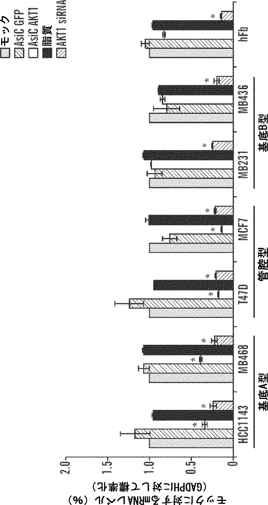
特定の態様において、複数のエンドソーム溶解成分を、本発明の阻害性核酸作用物質に組み入れられることができる。いくつかの態様において、これは、阻害性核酸作用物質に複数の同じエンドソーム溶解成分を組み入れることを含む。他の態様において、これは、阻害性核酸作用物質に2種類以上の異なるエンドソーム溶解成分を組み入れることを含む。 In certain embodiments, multiple endosomal lytic components can be incorporated into the inhibitory nucleic acid agents of the invention. In some embodiments, this includes incorporating multiple identical endosomal lytic components into the inhibitory nucleic acid agent. In other embodiments, this involves incorporating two or more different endosomal lytic components into the inhibitory nucleic acid agent.
これらのエンドソーム溶解成分は、エンドソームのpHで立体配座を変化させることによってエンドソーム脱出を媒介することができる。特定の態様において、エンドソーム溶解成分は、中性pHにおいてランダムコイル型立体配座で存在し、エンドソームのpHで両親媒性らせんに再配列することができる。この立体配座の転移の結果として、これらのペプチドは、エンドソームの脂質膜に入り込み、エンドソームの内容物を細胞質中に漏出させ得る。立体配座の転移はpH依存性であるので、エンドソーム溶解成分は、血液(pH約7.4)中で循環する間に融合誘発活性をほとんどまたは全く示さないことができる。本明細書で使用される場合、「融合誘発活性」は、エンドソーム溶解成分による脂質膜の破壊を招く活性として定義される。融合誘発活性の一例は、エンドソームの溶解、またはエンドソームから細胞質への本発明のモジュール組成物(例えば核酸)のうちの1つもしくは複数の成分の漏出および輸送に繋がる、エンドソーム溶解成分によるエンドソーム膜の破壊である。 These endosomal lytic components can mediate endosomal escape by changing the conformation at endosomal pH. In certain embodiments, the endosomal lytic component is present in a random coil conformation at neutral pH and can be rearranged into an amphipathic helix at the endosomal pH. As a result of this conformational change, these peptides can enter the lipid membrane of the endosome and leak the endosomal contents into the cytoplasm. Since the conformational transition is pH dependent, the endosomal lytic component can exhibit little or no fusion-inducing activity while circulating in the blood (pH about 7.4). As used herein, “fusion-inducing activity” is defined as an activity that results in disruption of the lipid membrane by endosomal lytic components. One example of fusion-inducing activity is endosomal lysis, or endosomal membrane-mediated by endosomal lytic components that lead to leakage and transport of one or more components of the modular composition (eg, nucleic acid) of the invention from endosomes to the cytoplasm. It is destruction.
当業者ならば、適当なエンドソーム溶解成分を試験および同定することができる。例えば、化合物がpH環境に依存して応答する、例えば電荷を変化させる能力は、日常的な方法によって、例えば細胞アッセイで試験することができる。特定の態様において、被験化合物を、細胞と混合または接触させて、細胞に、例えばエンドサイトーシスによって該被験化合物を取り込ませる。次に、接触された細胞からエンドソーム調製物を作り、このエンドソーム調製物を、対照細胞からのエンドソーム調製物と比較することができる。対照細胞と比較した、接触させた細胞からのエンドソーム画分における変化、例えば減少は、被験化合物が融合誘発性作用因子として機能できることを示している。あるいは、接触させた細胞および対照細胞を、例えば顕微鏡法、例えば光学または電子顕微鏡法によって評価して、細胞中のエンドソーム集団における差を判定することができる。被験化合物および/またはエンドソームを標識して、例えばエンドソームの漏出を定量することができる。 One skilled in the art can test and identify suitable endosomal lytic components. For example, the ability of a compound to respond depending on the pH environment, eg, change charge, can be tested by routine methods, eg, in a cellular assay. In certain embodiments, a test compound is mixed or contacted with a cell and the cell is taken up by, for example, endocytosis. An endosome preparation can then be made from the contacted cells and this endosome preparation can be compared to an endosome preparation from a control cell. A change, such as a decrease, in the endosomal fraction from the contacted cell compared to the control cell indicates that the test compound can function as a fusogenic agent. Alternatively, contacted cells and control cells can be evaluated, for example, by microscopy, such as light or electron microscopy, to determine differences in the endosomal population in the cells. Test compounds and / or endosomes can be labeled to quantify, for example, endosomal leakage.
別の種類のアッセイでは、本明細書に記載される阻害性核酸作用物質は、1種類または複数種類の被験作用因子または推定上の融合誘発性作用因子を使用して構築される。阻害性核酸作用物質は、容易な視覚化のために標識することができる。阻害性核酸作用物質が細胞によって取り込まれた後、エンドソーム溶解成分がエンドソーム脱出を促進する能力を、例えばエンドソーム調製物の調製によって、または細胞の細胞質中の標識阻害性核酸作用物質を視覚化できるようにする顕微鏡技術によって評価することができる。特定の他の態様において、遺伝子発現の阻害または任意の他の生理学的パラメータがエンドソーム脱出の代替マーカーとして使用され得る。 In another type of assay, the inhibitory nucleic acid agents described herein are constructed using one or more test agents or putative fusion-inducing agents. Inhibitory nucleic acid agents can be labeled for easy visualization. After the inhibitory nucleic acid agent is taken up by the cell, the ability of the endosomal lysis component to promote endosomal escape can be visualized, for example, by preparing an endosomal preparation or by labeling the inhibitory nucleic acid agent in the cytoplasm of the cell Can be evaluated by microscopic techniques. In certain other embodiments, inhibition of gene expression or any other physiological parameter can be used as an alternative marker for endosomal escape.
他の態様において、pH依存性構造転移を示す化合物を同定するために円偏光二色性分光法を使用することができる。第1のアッセイが、被験化合物が単独でpH変化に応答する能力を評価し、第2のアッセイが、被験化合物を含むモジュール組成物がpH変化に応答する能力を評価する、二段階アッセイも行うことができる。 In other embodiments, circular dichroism spectroscopy can be used to identify compounds that exhibit a pH-dependent structural transition. A two-step assay is also performed in which the first assay evaluates the ability of the test compound to respond to pH changes alone, and the second assay assesses the ability of the module composition containing the test compound to respond to pH changes. be able to.
本明細書に記載される局面の1つの態様において、リガンドまたはコンジュゲートは、脂質または脂質ベースの分子である。そのような脂質または脂質ベースの分子は、好ましくは血清タンパク質、例えばヒト血清アルブミン(HSA)と結合する。HSA結合リガンドは、標的組織、例えば身体の非腎臓標的組織へのコンジュゲートの分布を可能にする。HSAと結合できる他の分子もリガンドとして使用することができる。例えば、ネプロキシンまたはアスピリンを使用することができる。脂質または脂質ベースのリガンドは、(a)コンジュゲートの分解に対する耐性を増加させることができ、(b)標的細胞もしくは細胞膜へのターゲティングもしくは輸送を増加させることができ、かつ/または(c)血清タンパク質、例えばHSAへの結合を調整するために使用することができる。 In one embodiment of the aspects described herein, the ligand or conjugate is a lipid or lipid-based molecule. Such lipids or lipid-based molecules preferably bind to serum proteins such as human serum albumin (HSA). HSA binding ligands allow distribution of the conjugate to target tissues, eg, non-renal target tissues of the body. Other molecules that can bind HSA can also be used as ligands. For example, neproxin or aspirin can be used. Lipids or lipid-based ligands can (a) increase resistance to degradation of the conjugate, (b) increase targeting or transport to target cells or cell membranes, and / or (c) serum It can be used to modulate binding to proteins such as HSA.
別の局面において、リガンドは、細胞透過作用物質、好ましくはヘリカル細胞透過作用物質である。好ましくは、そのような作用物質は両親媒性である。例示的な作用物質は、tatまたはアンテナペディアのようなペプチドである。作用物質がペプチドの場合、ペプチジル模倣体、インバートマー、非ペプチドまたは擬似ペプチド結合、およびD-アミノ酸の使用でそれを修飾することができる。ヘリカル作用物質は、好ましくはα-ヘリカル作用物質であり、α-ヘリカル作用物質は、好ましくは、親油相および疎油相を有する。 In another aspect, the ligand is a cell permeation agent, preferably a helical cell permeation agent. Preferably such agents are amphiphilic. Exemplary agents are peptides such as tat or antennapedia. If the agent is a peptide, it can be modified with the use of peptidyl mimetics, invertmers, non-peptide or pseudopeptide bonds, and D-amino acids. The helical agent is preferably an α-helical agent, and the α-helical agent preferably has a lipophilic phase and an oleophobic phase.
本発明での使用に適当なペプチドは、天然ペプチド、例えばtatまたはアンテナペディアペプチド、合成ペプチド、またはペプチド模倣体であることができる。さらに、ペプチドは、修飾ペプチドであることができ、例えばペプチドは、非ペプチドまたは擬似ペプチド結合、およびD-アミノ酸を含むことができる。ペプチド模倣体(本明細書においてオリゴペプチド模倣体とも呼ばれる)は、天然ペプチドに類似した所定の三次元構造にフォールディング可能な分子である。阻害性核酸作用物質へのペプチドおよび模倣体の結合は、例えば細胞認識および吸収を高めることによって、阻害性核酸の薬物動態的分布に影響することができる。ペプチドまたはペプチド模倣体部分は、約5〜50アミノ酸長、例えば約5、10、15、20、25、30、35、40、45、または50アミノ酸長であることができる。 Peptides suitable for use in the present invention can be natural peptides such as tat or antennapedia peptides, synthetic peptides, or peptidomimetics. Further, the peptide can be a modified peptide, eg, the peptide can include non-peptide or pseudo-peptide bonds, and D-amino acids. A peptidomimetic (also referred to herein as an oligopeptidomimetic) is a molecule that can fold into a defined three-dimensional structure similar to a natural peptide. Binding of peptides and mimetics to inhibitory nucleic acid agents can affect the pharmacokinetic distribution of inhibitory nucleic acids, for example, by enhancing cell recognition and absorption. The peptide or peptidomimetic moiety can be about 5-50 amino acids long, eg, about 5, 10, 15, 20, 25, 30, 35, 40, 45, or 50 amino acids long.
ペプチドまたはペプチド模倣体は、例えば、細胞透過性ペプチド、陽イオンペプチド、両親媒性ペプチド、または疎水性ペプチド(例えば、主としてTyr、TrpまたはPheからなる)であることができる。ペプチド部分は、デンドリマーペプチド、束縛されたペプチドまたは架橋ペプチドであることができる。別の代替において、ペプチド部分は、疎水性膜移行配列(MTS)を含むことができる。例示的な疎水性MTS含有ペプチドは、アミノ酸配列
「細胞透過性ペプチド」は、細胞、例えば細菌もしくは真菌細胞のような微生物細胞、またはヒト細胞のような哺乳類細胞を透過することが可能である。微生物細胞透過性ペプチドは、例えば、α-ヘリカル直鎖ペプチド(例えばLL-37、またはセロピンP1)、ジスルフィド結合含有ペプチド(例えば、α-デフェンシン、β-デフェンシンもしくはバクテネシン)、または1種類もしくは2種類のみの支配的アミノ酸を含むペプチド(例えばPR-39またはインドリシジン)であることができる。細胞透過性ペプチドはまた、核局在化シグナル(NLS)を含むことができる。例えば、細胞透過性ペプチドは、HIV-1 gp41とSV40のラージT抗原のNLSとの融合ペプチドドメイン由来のMPGなどの、二分両親媒性ペプチドであることができる(Simeoni et al., Nucl. Acids Res. 31:2717-2724, 2003)。 A “cell penetrating peptide” is capable of penetrating cells, eg, microbial cells such as bacterial or fungal cells, or mammalian cells such as human cells. The microbial cell-penetrating peptide is, for example, an α-helical linear peptide (for example, LL-37 or cellopin P1), a disulfide bond-containing peptide (for example, α-defensin, β-defensin or bactenecin), or one or two types Can be peptides containing only the dominant amino acid (eg PR-39 or indolicidin). The cell penetrating peptide can also include a nuclear localization signal (NLS). For example, the cell penetrating peptide can be a bipartite amphipathic peptide such as MPG derived from the fusion peptide domain of HIV-1 gp41 and N40 of SV40 large T antigen (Simeoni et al., Nucl. Acids Res. 31: 2717-2724, 2003).
いくつかの態様において、本明細書に記載される阻害性核酸オリゴヌクレオチドは、さらに、糖質コンジュゲートを含む。糖質コンジュゲートは、核酸のインビボ送達および本明細書に記載されるインビボ治療用途に適当な組成物にとって有利である。本明細書で使用される場合、「糖質」は、酸素、窒素もしくは硫黄原子が各炭素原子に結合した少なくとも6つの炭素原子(直鎖、分枝もしくは環状であり得る)を有する1つもしくは複数の単糖ユニットから作られた糖質自体;または酸素、窒素もしくは硫黄原子が各炭素原子に結合した少なくとも6つの炭素原子(直鎖、分枝もしくは環状であり得る)をそれぞれ有する1つもしくは複数の単糖ユニットから作られた糖質部分をその一部として有する化合物のいずれかである化合物を表す。代表的な糖質には、糖(単糖、二糖、三糖および約4〜9つの単糖ユニットを含むオリゴ糖)、ならびにデンプン、グリコーゲン、セルロースおよび多糖ガムのような多糖が含まれる。特定の単糖には、C5以上(好ましくはC5〜C8)の糖が含まれ;二糖および三糖には、2または3つの単糖ユニット(好ましくはC5〜C8)を有する糖が含まれる。いくつかの態様において、糖質コンジュゲートは、さらに、非限定的にPKモジュレーター、エンドソーム溶解リガンド、および細胞透過性ペプチドのような他のリガンドを含む。 In some embodiments, the inhibitory nucleic acid oligonucleotides described herein further comprise a carbohydrate conjugate. Carbohydrate conjugates are advantageous for compositions suitable for in vivo delivery of nucleic acids and in vivo therapeutic applications described herein. As used herein, a “carbohydrate” is one having at least 6 carbon atoms (which may be straight, branched or cyclic) having an oxygen, nitrogen or sulfur atom attached to each carbon atom, or A carbohydrate itself made from multiple monosaccharide units; or one each having at least 6 carbon atoms (which may be linear, branched, or cyclic) with an oxygen, nitrogen or sulfur atom bonded to each carbon atom, or It represents a compound that is one of the compounds having a carbohydrate moiety made from a plurality of monosaccharide units as a part thereof. Exemplary carbohydrates include sugars (monosaccharides, disaccharides, trisaccharides and oligosaccharides containing about 4-9 monosaccharide units), and polysaccharides such as starch, glycogen, cellulose and polysaccharide gum. Certain monosaccharides include C5 or higher (preferably C5-C8) sugars; disaccharides and trisaccharides include sugars having 2 or 3 monosaccharide units (preferably C5-C8) . In some embodiments, the carbohydrate conjugate further comprises other ligands such as, but not limited to, PK modulators, endosomal lytic ligands, and cell penetrating peptides.
いくつかの態様において、本明細書に記載されるコンジュゲートは、切断可能または切断不可能であることができる様々なリンカーを用いて阻害性核酸オリゴヌクレオチドに結合させることができる。「リンカー」または「連結基」という用語は、化合物の2つの部分を結合する有機部分を意味する。リンカーは、典型的には、直接結合または酸素もしくは硫黄のような原子、NR8、C(O)、C(O)NH、SO、SO2、SO2NHのようなユニット、または非限定的に、置換もしくは非置換アルキル、置換もしくは非置換アルケニル、置換もしくは非置換アルキニル、アリールアルキル、アリールアルケニル、アリールアルキニル、ヘテロアリールアルキル、ヘテロアリールアルケニル、ヘテロアリールアルキニル、ヘテロシクリルアルキル、ヘテロシクリルアルケニル、ヘテロシクリルアルキニル、アリール、ヘテロアリール、ヘテロシクリル、シクロアルキル、シクロアルケニル、アルキルアリールアルキル、アルキルアリールアルケニル、アルキルアリールアルキニル、アルケニルアリールアルキル、アルケニルアリールアルケニル、アルケニルアリールアルキニル、アルキニルアリールアルキル、アルキニルアリールアルケニル、アルキニルアリールアルキニル、アルキルヘテロアリールアルキル、アルキルヘテロアリールアルケニル、アルキルヘテロアリールアルキニル、アルケニルヘテロアリールアルキル、アルケニルヘテロアリールアルケニル、アルケニルヘテロアリールアルキニル、アルキニルヘテロアリールアルキル、アルキニルヘテロアリールアルケニル、アルキニルヘテロアリールアルキニル、アルキルヘテロシクリルアルキル、アルキルヘテロシクリルアルケニル、アルキルヘテロシクリルアルキニル、アルケニルヘテロシクリルアルキル、アルケニルヘテロシクリルアルケニル、アルケニルヘテロシクリルアルキニル、アルキニルヘテロシクリルアルキル、アルキニルヘテロシクリルアルケニル、アルキニルヘテロシクリルアルキニル、アルキルアリール、アルケニルアリール、アルキニルアリール、アルキルヘテロアリール、アルケニルヘテロアリール、アルキニルヘテロアリールのような原子鎖を含み、その際、1つまたは複数のメチレンは、O、S、S(O)、SO2、N(R8)、C(O)、置換または非置換アリール、置換または非置換ヘテロアリール、置換または非置換複素環によって中断または終結されることができ;式中、R8は、水素、アシル、脂肪族または置換脂肪族である。1つの態様において、リンカーは、1〜24個の原子、好ましくは4〜24個の原子、好ましくは6〜18個の原子、より好ましくは8〜18個の原子、最も好ましくは8〜16個の間の原子である。 In some embodiments, the conjugates described herein can be attached to inhibitory nucleic acid oligonucleotides using a variety of linkers that can be cleavable or non-cleavable. The term “linker” or “linking group” means an organic moiety that connects two parts of a compound. The linker is typically a direct bond or an atom such as oxygen or sulfur, a unit such as, but not limited to, NR8, C (O), C (O) NH, SO, SO2, SO2NH, or Unsubstituted alkyl, substituted or unsubstituted alkenyl, substituted or unsubstituted alkynyl, arylalkyl, arylalkenyl, arylalkynyl, heteroarylalkyl, heteroarylalkenyl, heteroarylalkynyl, heterocyclylalkyl, heterocyclylalkenyl, heterocyclylalkynyl, aryl, heteroaryl , Heterocyclyl, cycloalkyl, cycloalkenyl, alkylarylalkyl, alkylarylalkenyl, alkylarylalkynyl, alkenylarylalkyl, alkenylarylalkenyl, alkenylarylalkenyl Nyl, alkynylarylalkyl, alkynylarylalkenyl, alkynylarylalkynyl, alkylheteroarylalkenyl, alkylheteroarylalkenyl, alkylheteroarylalkynyl, alkenylheteroarylalkyl, alkenylheteroarylalkenyl, alkenylheteroarylalkynyl, alkynylheteroarylalkyl, alkynyl Heteroarylalkenyl, alkynylheteroarylalkynyl, alkylheterocyclylalkyl, alkylheterocyclylalkenyl, alkylheterocyclylalkynyl, alkenylheterocyclylalkyl, alkenylheterocyclylalkenyl, alkenylheterocyclylalkynyl, alkynylheterocyclylalkyl, alkynylheterocyclyl Including atomic chains such as arylalkenyl, alkynylheterocyclylalkynyl, alkylaryl, alkenylaryl, alkynylaryl, alkylheteroaryl, alkenylheteroaryl, alkynylheteroaryl, wherein one or more methylenes are O, S, Can be interrupted or terminated by S (O), SO2, N (R8), C (O), substituted or unsubstituted aryl, substituted or unsubstituted heteroaryl, substituted or unsubstituted heterocycle; Is hydrogen, acyl, aliphatic or substituted aliphatic. In one embodiment, the linker is 1-24 atoms, preferably 4-24 atoms, preferably 6-18 atoms, more preferably 8-18 atoms, most preferably 8-16 atoms. Between the atoms.
切断可能な連結基は、細胞外で十分に安定な基であるが、標的細胞内に進入すると切断されて、リンカーが共に保持している2つの部分を放出する基である。好ましい態様において、切断可能な連結基は、標的細胞中または第1の参照条件(例えば、細胞内条件を模倣または代表するように選択することができる)下で、対象の血中または第2の参照条件(例えば、血液または血清中に見出される条件を模倣または代表するように選択することができる)下よりも少なくとも10倍以上、好ましくは少なくとも100倍速く切断される。 A cleavable linking group is a group that is sufficiently stable outside the cell, but is cleaved upon entry into the target cell, releasing the two moieties held together by the linker. In preferred embodiments, the cleavable linking group is either in the target cell or under a first reference condition (eg, can be selected to mimic or represent an intracellular condition) or in the subject's blood or second Cleavage is at least 10-fold, preferably at least 100-fold faster than under reference conditions (eg, can be selected to mimic or represent conditions found in blood or serum).
切断可能な連結基は、切断作用物質、例えば、pH、酸化還元電位または分解分子の存在に感受性である。一般的に、切断作用物質は、血清もしくは血液中よりも細胞内に豊富に存在するか、またはより高いレベルもしくはより高い活性で見出される。そのような分解作用物質の例には、特定の基質に関して選択される、または基質特異性を有しない酸化還元作用物質、例えば、酸化もしくは還元酵素または酸化還元切断可能な連結基を還元によって分解できる、細胞内に存在するメルカプタンのような還元作用物質を含むもの;エステラーゼ;エンドソームまたは酸性環境を創出できる作用物質、例えばpH5以下を生じるもの;一般酸、ペプチダーゼ(基質特異性であることができる)、およびホスファターゼとして作用することによって酸切断可能な連結基を加水分解または分解することができる酵素が含まれる。 The cleavable linking group is sensitive to the presence of a cleaving agent, such as pH, redox potential or a degrading molecule. In general, cleavage agents are abundant in cells than in serum or blood, or are found at higher levels or higher activity. Examples of such degrading agents are redox agents that are selected for a particular substrate or that have no substrate specificity, such as oxidative or reductase or redox cleavable linking groups that can be degraded by reduction. , Containing reducing agents such as mercaptans present in cells; esterases; agents that can create endosomes or acidic environments, such as those that produce a pH below 5; general acids, peptidases (can be substrate specific) And enzymes capable of hydrolyzing or degrading acid-cleavable linking groups by acting as phosphatases.
ジスルフィド結合のような切断可能な結合基は、pHに感受性であることができる。ヒト血清のpHは7.4であり、一方、平均細胞内pHは約7.1〜7.3の範囲でわずかに低い。エンドソームは、5.5〜6.0の範囲のより酸性のpHを有し、リソソームは、約5.0のなおいっそう酸性のpHを有する。一部のリンカーは、好ましいpHで切断される切断可能な連結基を有することにより、細胞内または細胞の所望の区画内でリガンドから陽イオン脂質を放出する。 A cleavable linking group, such as a disulfide bond, can be sensitive to pH. The pH of human serum is 7.4, while the average intracellular pH is slightly lower in the range of about 7.1-7.3. Endosomes have a more acidic pH in the range of 5.5 to 6.0, and lysosomes have an even more acidic pH of about 5.0. Some linkers have a cleavable linking group that is cleaved at the preferred pH, thereby releasing cationic lipids from the ligand in the cell or in the desired compartment of the cell.
リンカーは、特定の酵素によって切断可能な、切断可能な連結基を含むことができる。リンカー内に組み入れられる切断可能な連結基の種類は、標的とされる細胞に依存し得る。切断可能な連結基のさらなる例には、非限定的に、酸化還元切断可能な連結基(例えばジスルフィド連結基(-S-S-))、リン酸エステルベースの切断可能な結合基、エステルベースの切断可能な連結基、およびペプチドベースの切断可能な連結基が含まれる。RNAコンジュゲートの調製を教示する代表的な米国特許には、非限定的に、米国特許第4,828,979号;同第4,948,882号;同第5,218,105号;同第5,525,465号;同第5,541,313号;同第5,545,730号;同第5,552,538号;同第5,578,717、5,580,731号;同第5,591,584号;同第5,109,124号;同第5,118,802号;同第5,138,045号;同第5,414,077号;同第5,486,603号;同第5,512,439号;同第5,578,718号;同第5,608,046号;同第4,587,044号;同第4,605,735号;同第4,667,025号;同第4,762,779号;同第4,789,737号;同第4,824,941号;同第4,835,263号;同第4,876,335号;同第4,904,582号;同第4,958,013号;同第5,082,830号;同第5,112,963号;同第5,214,136号;同第5,082,830号;同第5,112,963号;同第5,214,136号;同第5,245,022号;同第5,254,469号;同第5,258,506号;同第5,262,536号;同第5,272,250号;同第5,292,873号;同第5,317,098号;同第5,371,241、5,391,723号;同第5,416,203、5,451,463号;同第5,510,475号;同第5,512,667号;同第5,514,785号;同第5,565,552号;同第5,567,810号;同第5,574,142号;同第5,585,481号;同第5,587,371号;同第5,595,726号;同第5,597,696号;同第5,599,923号;同第5,599,928および同第5,688,941号;同第6,294,664号;同第6,320,017号;同第6,576,752号;同第6,783,931号;同第6,900,297号;同第7,037,646号が含まれ;それらのそれぞれは、参照により本明細書に組み入れられる。 The linker can include a cleavable linking group that is cleavable by a particular enzyme. The type of cleavable linking group incorporated within the linker may depend on the targeted cell. Further examples of cleavable linking groups include, but are not limited to, redox cleavable linking groups (eg, disulfide linking group (-SS-)), phosphate ester based cleavable linking groups, ester based cleavage. Possible linking groups and peptide-based cleavable linking groups are included. Representative U.S. patents that teach the preparation of RNA conjugates include, but are not limited to, U.S. Pat. Nos. 4,828,979; 4,948,882; 5,218,105; 5,525,465; 5,541,313; No. 5,552,538; No. 5,578,717, 5,580,731; No. 5,591,584; No. 5,109,124; No. 5,118,802; No. 5,138,045; No. 5,414,077; No. 5,486,603; No. 5,512,439 No. 5,578,718; No. 5,608,046; No. 4,587,044; No. 4,605,735; No. 4,667,025; No. 4,762,779; No. 4,789,737; No. 4,824,941; No. 4,835,263; No. 4,876,335; No. 4,904,582; No. 4,958,013; No. 5,082,830; No. 5,112,963; No. 5,214,136; No. 5,082,830; No. 5,112,963; No. 5,214,136; No. 5,245,022; No. 5,254,469; No. 5,258,506; No. 5,262,536; No. 5,272,250; No. 5,292,873; No. 5,317,098; No. 5,371,241; 5,391,723; No. 5 No. 5,416,203; No. 5,512,667; No. 5,514,785; No. 5,565,552; No. 5,567,810; No. 5,574,142; No. 5,585,481; No. 5,587,371; No. 5,595,726 No. 5,597,696; No. 5,599,923; No. 5,599,928 and No. 5,688,941; No. 6,294,664; No. 6,320,017; No. 6,576,752; No. 6,783,931; No. 6,900,297; Each of which is incorporated herein by reference.
一般に、切断可能な連結基の候補の適合性は、分解作用物質(または分解条件)が連結基の候補を切断する能力を試験することによって評価することができる。また、血中または他の非標的組織と接触したときに切断に抵抗する能力について、切断可能な連結基の候補を試験することも望ましい。したがって、第1の条件が標的細胞内での切断を示すように選択され、かつ第2の条件が他の組織または体液中、例えば血液または血清中での切断を示すように選択される場合の、切断に対する第1の条件および第2の条件間の相対的感受性を決定することができる。評価は、無細胞系、細胞、細胞培養物、器官もしくは組織培養、または動物全体で実施することができる。無細胞条件または培養条件で初期評価を行い、動物全体におけるさらなる評価により確認することが有用であり得る。好ましい態様において、有用な候補化合物は、血液または血清中(または細胞外条件を模倣するように選択されたインビトロ条件)と比較して、細胞(または細胞内条件を模倣するように選択されたインビトロ条件)において少なくとも2、4、10または100倍速く切断される。 In general, the suitability of a cleavable linking group candidate can be assessed by testing the ability of the degradation agent (or degradation conditions) to cleave the linking group candidate. It is also desirable to test cleavable linking group candidates for their ability to resist cleavage when in contact with blood or other non-target tissues. Thus, when the first condition is selected to indicate cleavage in the target cell, and the second condition is selected to indicate cleavage in other tissues or fluids, such as blood or serum. The relative sensitivity between the first condition and the second condition for cleavage can be determined. Evaluation can be performed on cell-free systems, cells, cell cultures, organ or tissue cultures, or whole animals. It may be useful to perform an initial assessment in cell-free or culture conditions and confirm by further assessment in the entire animal. In preferred embodiments, useful candidate compounds are cells (or in vitro selected to mimic intracellular conditions) as compared to blood or serum (or in vitro conditions selected to mimic extracellular conditions). Cutting at least 2, 4, 10 or 100 times faster under (conditions).
所与の化合物におけるすべての位置が一様に修飾される必要はなく、実際には、複数前述の修飾を1つの化合物に、または阻害性核酸内の1つのヌクレオシドにすら、組み入れることができる。本発明は、キメラ化合物である阻害性核酸化合物も含む。本発明の文脈における「キメラ」阻害性核酸化合物または「キメラ」は、それぞれ少なくとも1つのモノマーユニット、すなわちdsRNA化合物の場合はヌクレオチドから作られた2つ以上の化学的に別個の領域を含む阻害性核酸化合物、例えばdsRNAである。これらの阻害性核酸は、典型的には、阻害性核酸にヌクレアーゼ分解への耐性増加、細胞取り込みの増加、および/または標的核酸に対する結合親和性の増加を付与するように核酸が修飾された、少なくとも1つの領域を含む。阻害性核酸の追加的な領域は、RNA:DNAまたはRNA:RNAハイブリッドを切断する能力がある酵素に対する基質として役立ち得る。例として、RNアーゼHは、RNA:DNA二重鎖のRNA鎖を切断する細胞エンドヌクレアーゼである。したがって、RNアーゼHの活性化は、RNA標的の切断を招くことにより、阻害性核酸の遺伝子発現の阻害の効率を大きく高める。その結果、キメラ阻害性核酸が使用される場合、例えば同じ標的領域にハイブリダイズするホスホロチオエートデオキシdsRNAと比較して、より短い阻害性核酸で匹敵する結果が、多くの場合、得られ得る。RNA標的の切断は、ゲル電気泳動法および必要に応じて当技術分野において公知の関連する核酸ハイブリダイゼーション技術によって日常的に検出することができる。 Not all positions in a given compound need to be uniformly modified, and in fact more than one of the above modifications can be incorporated into a single compound or even a single nucleoside within an inhibitory nucleic acid. The present invention also includes inhibitory nucleic acid compounds that are chimeric compounds. A “chimeric” inhibitory nucleic acid compound or “chimera” in the context of the present invention is an inhibitor comprising each of at least one monomer unit, ie two or more chemically distinct regions made up of nucleotides in the case of dsRNA compounds. Nucleic acid compounds, such as dsRNA. These inhibitory nucleic acids are typically modified in the nucleic acid to impart the inhibitory nucleic acid with increased resistance to nuclease degradation, increased cellular uptake, and / or increased binding affinity for the target nucleic acid, Includes at least one region. Additional regions of inhibitory nucleic acids can serve as substrates for enzymes capable of cleaving RNA: DNA or RNA: RNA hybrids. As an example, RNase H is a cellular endonuclease that cleaves the RNA strand of an RNA: DNA duplex. Thus, activation of RNase H greatly increases the efficiency of inhibition of gene expression of inhibitory nucleic acids by inducing cleavage of the RNA target. As a result, comparable results with shorter inhibitory nucleic acids can often be obtained when chimeric inhibitory nucleic acids are used, eg, compared to phosphorothioate deoxy dsRNA that hybridizes to the same target region. Cleavage of the RNA target can be routinely detected by gel electrophoresis and, if necessary, related nucleic acid hybridization techniques known in the art.
場合によっては、阻害性核酸の核酸を、非リガンド基によって修飾することができる。阻害性核酸の活性、細胞分布または細胞取り込みを高めるために、いくつかの非リガンド分子が阻害性核酸にコンジュゲートされており、そのようなコンジュゲーションを行うための手順は、科学文献から入手可能である。そのような非リガンド部分は、コレステロール(Kubo, T. et al., Biochem. Biophys. Res. Comm., 2007, 365(1):54-61; Letsinger et al., Proc. Natl. Acad. Sci. USA, 1989, 86:6553)、コール酸(Manoharan et al., Bioorg. Med. Chem. Lett., 1994, 4:1053)、チオエーテル、例えばヘキシル-S-トリチルチオール(Manoharan et al., Ann. N.Y. Acad. Sci., 1992, 660:306; Manoharan et al., Bioorg. Med. Chem. Let., 1993, 3:2765)、チオコレステロール(Oberhauser et al., Nucl. Acids Res., 1992, 20:533)、脂肪族鎖、例えばドデカンジオールまたはウンデシル残基(Saison-Behmoaras et al., EMBO J., 1991, 10:111; Kabanov et al., FEBS Lett., 1990, 259:327; Svinarchuk et al., Biochimie, 1993, 75:49)、リン脂質、例えばジ-ヘキサデシル-rac-グリセロールまたはトリエチルアンモニウム1,2-ジ-O-ヘキサデシル-rac-グリセロ-3-H-ホスホネート(Manoharan et al., Tetrahedron Lett., 1995, 36:3651; Shea et al., Nucl. Acids Res., 1990, 18:3777)、ポリアミンまたはポリエチレングリコール鎖(Manoharan et al., Nucleosides & Nucleotides, 1995, 14:969)、またはアダマンタン酢酸(Manoharan et al., Tetrahedron Lett., 1995, 36:3651)、パルミチル部分(Mishra et al., Biochim. Biophys. Acta, 1995, 1264:229)、またはオクタデシルアミンもしくはヘキシルアミノ-カルボニル-オキシコレステロール部分(Crooke et al., J. Pharmacol. Exp. Ther., 1996, 277:923)のような脂質部分を含んでいた。そのような核酸コンジュゲートの調製を教示する代表的な米国特許は、上に挙げられている。典型的なコンジュゲーションプロトコールは、配列の1つまたは複数の位置にアミノリンカーを有する核酸の合成を含む。次に、アミノ基が、適当なカップリング試薬または活性化試薬を使用して、コンジュゲートされる分子と反応される。コンジュゲーション反応は、固体支持体に依然として結合している核酸を用いて、または核酸の切断後の液相中のいずれかで行われ得る。HPLCによる核酸コンジュゲートの精製は、典型的には純粋なコンジュゲートをもたらす。
In some cases, the nucleic acid of the inhibitory nucleic acid can be modified with a non-ligand group. Several non-ligand molecules are conjugated to inhibitory nucleic acids to increase the activity, cell distribution or cellular uptake of inhibitory nucleic acids, and procedures for performing such conjugation are available from the scientific literature It is. Such non-ligand moieties are cholesterol (Kubo, T. et al., Biochem. Biophys. Res. Comm., 2007, 365 (1): 54-61; Letsinger et al., Proc. Natl. Acad. Sci. USA, 1989, 86: 6553), cholic acid (Manoharan et al., Bioorg. Med. Chem. Lett., 1994, 4: 1053), thioethers such as hexyl-S-tritylthiol (Manoharan et al., Ann). NY Acad. Sci., 1992, 660: 306; Manoharan et al., Bioorg. Med. Chem. Let., 1993, 3: 2765), thiocholesterol (Oberhauser et al., Nucl. Acids Res., 1992, 20: 533), aliphatic chains such as dodecanediol or undecyl residues (Saison-Behmoaras et al., EMBO J., 1991, 10: 111; Kabanov et al., FEBS Lett., 1990, 259: 327; Svinarchuk et al., Biochimie, 1993, 75:49), phospholipids such as di-hexadecyl-rac-glycerol or
「アプタマー」という用語は、ポリペプチドなどの標的分子と結合する能力がある核酸分子を表す。例えば、本発明のアプタマーは、標的分子、または標的分子の発現および/もしくは活性を調節するシグナル伝達経路内の分子と特異的に結合することができる。アプタマーの産生および治療的使用は、当技術分野において十分に確立されている。例えば米国特許第5,475,096号を参照されたい。 The term “aptamer” refers to a nucleic acid molecule capable of binding a target molecule, such as a polypeptide. For example, the aptamers of the invention can specifically bind to a target molecule or a molecule in a signal transduction pathway that modulates the expression and / or activity of the target molecule. The production and therapeutic use of aptamers is well established in the art. See, for example, US Pat. No. 5,475,096.
本明細書で使用される場合、「特異的結合」という用語は、2つの分子、化合物、細胞および/または粒子の間の化学相互作用を表し、その際、第1の実体は、それが非標的である第3の実体に結合するよりも大きな特異性および親和性で、第2の標的実体と結合する。いくつかの態様において、特異的結合は、第3の非標的実体に対する親和性よりも少なくとも10倍、少なくとも50倍、少なくとも100倍、少なくとも500倍、少なくとも1000倍、またはそれ以上大きい、第2の標的実体に対する第1の実体の親和性を指すことができる。所与の標的に特異的な試薬は、利用されているアッセイの条件下で標的に対する特異的結合を示す試薬である。 As used herein, the term “specific binding” refers to a chemical interaction between two molecules, compounds, cells and / or particles, wherein the first entity is a non- It binds to the second target entity with greater specificity and affinity than it binds to the target third entity. In some embodiments, the specific binding is at least 10, at least 50, at least 100, at least 500, at least 1000, or more greater than the affinity for the third non-target entity, It can refer to the affinity of the first entity for the target entity. A reagent specific for a given target is a reagent that exhibits specific binding to the target under the conditions of the assay being utilized.
本明細書で使用される場合、「処置(treat)」、「処置(treatment)」、「処置(treating)」または「改善」という用語は、その目的が疾患または障害、例えば癌に関連する状態の進行または重篤度を好転させる、緩和する、改善する、阻害する、鈍化させるまたは停止させることである治療的処置を表す。「処置(treating)」という用語は、癌に関連する状態、疾患または障害の少なくとも1つの有害な作用または症状の減少または緩和を含む。処置は、通常、1つまたは複数の症状または臨床マーカーが減少する場合に「有効」である。あるいは、処置は、疾患の進行が減少または停止する場合に「有効」である。すなわち、「処置(treatment)」は、処置の非存在下で予想されるものと比較しての、症状またはマーカーの改善だけでなく、症状の進行または悪化の停止または少なくとも鈍化も含む。有益または望ましい臨床結果は、検出可能か検出不可能かによらず、1つもしくは複数の症状の緩和、疾患の規模の縮小、疾患の安定化(すなわち、悪化しない)状態、疾患の進行の遅延もしくは鈍化、疾患状態の改善もしくは緩和、寛解(部分的か完全かによらず)、および/または死亡率の低下を含むがこれらに限定されない。疾患の「処置(treatment)」という用語はまた、(緩和処置を含む)その疾患の症状または副作用からの解放の提供を含む。 As used herein, the terms “treat”, “treatment”, “treating” or “amelioration” refer to a condition whose purpose is related to a disease or disorder, eg, cancer. Represents a therapeutic treatment that is to improve, alleviate, improve, inhibit, slow down or stop the progression or severity of The term “treating” includes a reduction or alleviation of at least one adverse effect or symptom of a condition, disease or disorder associated with cancer. Treatment is usually “effective” if one or more symptoms or clinical markers are reduced. Alternatively, treatment is “effective” if disease progression is reduced or stopped. That is, “treatment” includes not only the improvement of symptoms or markers as compared to what would be expected in the absence of treatment, but also the cessation or at least slowing of the progression or worsening of symptoms. Beneficial or desirable clinical outcome, whether detectable or undetectable, alleviates one or more symptoms, reduces the size of the disease, stabilizes the disease (ie does not worsen), delays disease progression Or including, but not limited to, slowing, improvement or alleviation of disease state, remission (whether partial or complete), and / or reduced mortality. The term “treatment” of a disease also includes providing relief from the symptoms or side effects of the disease (including palliative treatment).
本明細書で使用される場合、「薬学的組成物」という用語は、薬学的に許容される担体、例えば製薬業において広く使用されている担体と組み合わされた活性な薬剤を表す。「薬学的に許容される」というフレーズは、本明細書において、妥当な医学的判断の範囲で、合理的な利益/リスク比の下、過剰な毒性、刺激、アレルギー反応または他の問題もしくは合併症を伴わずに、ヒトおよび動物の組織と接触させて使用するのに適している化合物、物質、組成物および/または投薬形態を表すために使用される。 As used herein, the term “pharmaceutical composition” refers to an active agent in combination with a pharmaceutically acceptable carrier, such as a carrier widely used in the pharmaceutical industry. The phrase “pharmaceutically acceptable” refers herein to excessive toxicity, irritation, allergic reactions or other problems or complications within reasonable medical judgment and within a reasonable benefit / risk ratio. Used to describe compounds, substances, compositions and / or dosage forms suitable for use in contact with human and animal tissues without symptoms.
本明細書で使用される場合、「投与」という用語は、所望の部位への薬剤の少なくとも部分的な送達を達成する方法または経路による対象への本明細書に開示される化合物の投入を表す。本明細書に開示される化合物を含む薬学的組成物は、対象において有効な処置をもたらす任意の適当な経路によって投与され得る。 As used herein, the term “administration” refers to the input of a compound disclosed herein to a subject by a method or route that achieves at least partial delivery of the agent to the desired site. . A pharmaceutical composition comprising a compound disclosed herein can be administered by any suitable route that results in effective treatment in the subject.
「統計的に有意」または「有意」という用語は、統計的有意性を表し、通常、2標準偏差(2SD)またはそれを上回る差を意味する。 The term “statistically significant” or “significant” refers to statistical significance and usually means a difference of 2 standard deviations (2SD) or more.
実施例を実施する以外では、またはそうでないことが示されていない限り、本明細書で使用される成分の量または反応条件を表すすべての数値は、すべての例において、「約」という用語によって修飾されていると理解されるべきである。「約」という用語は、百分率に関連して使用される場合、±1%を意味し得る。 Unless otherwise stated or indicated otherwise, all numerical values representing amounts of ingredients or reaction conditions used herein are in all examples by the term “about”. It should be understood as being modified. The term “about” when used in relation to a percentage can mean ± 1%.
本明細書で使用される場合、「含む(comprising)」または「含む(comprises)」という用語は、それが必須であろうとなかろうと、指定されていない要素を含む余地を残しつつ、その方法または組成物にとって必須である組成物、方法およびそれらの各要素を参照するために使用される。 As used herein, the terms “comprising” or “comprises”, whether essential or not, leave the room for inclusion of unspecified elements, Used to refer to compositions, methods and their respective elements that are essential to the composition.
「〜からなる」という用語は、その態様の説明の中で言及されていないあらゆる要素に対して排他的である、本明細書に記載される組成物、方法およびそれらの各要素を表す。 The term “consisting of” refers to the compositions, methods and their respective elements described herein that are exclusive to any element not mentioned in the description of that embodiment.
本明細書で使用される場合、「本質的に〜からなる」という用語は、特定の態様に必要とされる要素を表す。この用語は、その態様の基本的かつ新規または機能的な特徴に実質的に影響しない要素の存在を許容する。 As used herein, the term “consisting essentially of” represents an element required for a particular embodiment. The term allows for the presence of elements that do not substantially affect the basic, novel or functional characteristics of the embodiment.
単数形「a」、「an」および「the」は、文脈がそうでないことを明確に示していない限り、複数の参照を含む。同様に、「または、もしくは」という単語は、文脈がそうでないことを明確に示していない限り、「および、ならびに」を含むことが意図されている。本開示を実施または試験する際には本明細書に記載されているのと同様または同等の方法および材料を使用することができるが、以下には適当な方法および材料が記載されている。「例えば(e.g.)」という略語は、ラテン語のexempli gratiaに由来し、本明細書で非限定的な例を示すために使用される。したがって、「例えば(e.g.)」という略語は、「例えば(for example)」という用語と同義である。 The singular forms “a”, “an”, and “the” include plural references unless the context clearly indicates otherwise. Similarly, the word “or” is intended to include “and” unless the context clearly indicates otherwise. Although methods and materials similar or equivalent to those described herein can be used in the practice or testing of the present disclosure, suitable methods and materials are described below. The abbreviation “for example (e.g.)” is derived from the Latin word “exempli gratia” and is used herein to provide a non-limiting example. Thus, the abbreviation “e.g.” is synonymous with the term “for example”.
細胞生物学および分子生物学における一般用語の定義は、「The Merck Manual of Diagnosis and Therapy」、第19版、Merck Research Laboratories出版、2006年(ISBN 0-911910-19-0);Robert S. Porterら(編)、The Encyclopedia of Molecular Biology、Blackwell Science Ltd.出版、1994年(ISBN 0-632-02182-9);Benjamin Lewin、Genes X、Jones & Bartlett Publishing出版、2009年(ISBN-10: 0763766321);Kendrewら(編)、Molecular Biology and Biotechnology: a Comprehensive Desk Reference、VCH Publishers, Inc.出版、1995年(ISBN 1-56081-569-8)およびCurrent Protocols in Protein Sciences 2009, Wiley Intersciences, Coliganら編において見出され得る。 General terms in cell and molecular biology are defined in The Merck Manual of Diagnosis and Therapy, 19th edition, Merck Research Laboratories, 2006 (ISBN 0-911910-19-0); Robert S. Porter (Eds.), The Encyclopedia of Molecular Biology, Blackwell Science Ltd. Publishing, 1994 (ISBN 0-632-02182-9); Benjamin Lewin, Genes X, Jones & Bartlett Publishing, 2009 (ISBN-10: 0763766321) Kendrew et al. (Eds.), Molecular Biology and Biotechnology: a Comprehensive Desk Reference, VCH Publishers, Inc., 1995 (ISBN 1-56081-569-8) and Current Protocols in Protein Sciences 2009, Wiley Intersciences, Coligan et al. Can be found in the knitting.
それ以外のことが言及されていない限り、本発明は、例えば、Sambrook et al., Molecular Cloning: A Laboratory Manual (4 ed.), Cold Spring Harbor Laboratory Press, Cold Spring Harbor, N.Y., USA (2012); Davis et al., Basic Methods in Molecular Biology, Elsevier Science Publishing, Inc., New York, USA (1995);またはMethods in Enzymology: Guide to Molecular Cloning Techniques Vol.152, S.L.Berger and A.R. Kimmel Eds, Academic Press Inc., San Diego, USA (1987); Current Protocols in Protein Science (CPPS)(John E. Coligan, et. al., ed., Johon Wiley and Sons, Inc.)、Current Protocols in Cell Biology (CPCB)(Juan S. Bonifacino et. al. ed., John Wiley and Sons, Inc.)およびCulture of Animal Cells: A Manual of Basic Technique by R. Ian Freshney, Publisher: Wiley-Liss; 5th edition (2005)、Animal Cell Culture Methods (Methods in Cell Biology, Vol.57, Jennie P. Mather and David Barnes editors, Academic Press, 1st edition, 1998)に記載されるような標準的手順を用いて実施され、これらはすべてその全体が参照により本明細書に組み入れられる。 Unless otherwise stated, the present invention is described, for example, in Sambrook et al., Molecular Cloning: A Laboratory Manual (4 ed.), Cold Spring Harbor Laboratory Press, Cold Spring Harbor, NY, USA (2012) ; Davis et al., Basic Methods in Molecular Biology, Elsevier Science Publishing, Inc., New York, USA (1995); or Methods in Enzymology: Guide to Molecular Cloning Techniques Vol. 152, SLBerger and AR Kimmel Eds, Academic Press Inc., San Diego, USA (1987); Current Protocols in Protein Science (CPPS) (John E. Coligan, et.al., ed., Johon Wiley and Sons, Inc.), Current Protocols in Cell Biology (CPCB) (Juan S. Bonifacino et.al.ed., John Wiley and Sons, Inc.) and Culture of Animal Cells: A Manual of Basic Technique by R. Ian Freshney, Publisher: Wiley-Liss; 5th edition (2005), Animal Standard procedures as described in Cell Culture Methods (Methods in Cell Biology, Vol.57, Jennie P. Mather and David Barnes editors, Academic Press, 1st edition, 1998) It is carried out with, in their entirety for all incorporated herein by reference.
他の用語は、本発明の様々な局面の説明の中で本明細書で定義されている。 Other terms are defined herein in the description of various aspects of the invention.
本願を通じて引用されている、参考文献、発行された特許、公開された特許出願および同時係属中の特許出願を含む、すべての特許およびその他の刊行物は、明示的に、例えば、本明細書に開示される技術と関連して使用され得るそのような刊行物に記載される方法論を説明および開示する目的で、参照により本明細書に組み入れられる。これらの刊行物は、それらの開示が本願の出願日より以前であるために提供されるにすぎない。これに関するいかなる事情も、本発明者らが、それが先行発明であるためまたは任意のその他の理由によりそのような開示よりも先の日付を主張する資格を有しないことの承認とみなされるべきではない。これらの書類の日付に関する言及または内容に関する表示はすべて、出願人が入手し得た情報に基づいており、これらの書類の日付または内容の正確性に関する承認とはならない。 All patents and other publications, including references, issued patents, published patent applications and co-pending patent applications cited throughout this application are expressly incorporated herein by reference, for example. For purposes of explaining and disclosing the methodology described in such publications that may be used in connection with the disclosed technology, it is incorporated herein by reference. These publications are provided solely for their disclosure prior to the filing date of the present application. Any circumstances in this regard should not be considered as an admission that the inventors are not entitled to claim a date prior to such disclosure because it is a prior invention or for any other reason. Absent. All references to the date or content of these documents are based on information available to the applicant and do not constitute an approval for the accuracy of the date or content of these documents.
本開示の態様の説明は、排他的であることまたは本開示を開示された正確な形態に限定することを意図していない。本開示の個々の態様および実施例は例示目的で本明細書に記載されており、関連技術分野の当業者が認識するように、様々な等価な変更が本開示の範囲内で可能である。例えば、方法の工程または機能はある順で示されているが、代替の態様は異なる順で機能を実施し得、または機能は実質的に同時に実施され得る。本明細書に提供される開示の技術は、他の手順または方法に適切に適用することができる。本明細書に記載される様々な態様は、さらなる態様を提供するよう組み合わせることができる。本開示の局面は、必要に応じて、本開示のなおさらなる態様を提供するために、上記参考文献および出願の組成、機能およびコンセプトを利用するよう変更され得る。これらおよびその他の変更は、詳細な説明に照らして本開示に対してなされ得る。すべてのそのような変更は、添付の特許請求の範囲内に包含されることが意図されている。 The description of aspects of the disclosure is not intended to be exclusive or to limit the disclosure to the precise form disclosed. Individual aspects and examples of this disclosure are described herein for purposes of illustration, and various equivalent modifications are possible within the scope of this disclosure, as those skilled in the relevant art will recognize. For example, although method steps or functions are shown in a certain order, alternative aspects may perform functions in a different order, or functions may be performed substantially simultaneously. The disclosed techniques provided herein can be suitably applied to other procedures or methods. The various aspects described herein can be combined to provide further aspects. Aspects of the present disclosure can be modified as needed to take advantage of the compositions, functions and concepts of the above references and applications to provide still further embodiments of the present disclosure. These and other changes can be made to the disclosure in light of the detailed description. All such modifications are intended to be included within the scope of the appended claims.
上記態様のいずれかの個々の要素は、他の態様の要素と組み合わせることができまた他の態様の要素で置き換えることができる。さらに、本開示の特定の態様に関連する利点は、これらの態様との関係で記載されているが、他の態様もまたそのような利点を示し得、そしてすべての態様が本開示の範囲に包含されるためにそのような利点を示すことを必要とするものではない。 Individual elements of any of the above aspects can be combined with elements of other aspects and replaced with elements of other aspects. Further, although the advantages associated with certain aspects of the present disclosure are described in relation to these aspects, other aspects may also exhibit such advantages, and all aspects are within the scope of the present disclosure. It is not necessary to show such benefits in order to be included.
本明細書に記載される技術は、以下の実施例によってさらに例証されるが、これらはいかなる事情によってもさらなる限定とみなされるべきでない。 The techniques described herein are further illustrated by the following examples, which should not be construed as further limiting under any circumstances.
本明細書に記載される技術のいくつかの態様は、以下の番号が付された項のいずれかにしたがい定義することができる。
1.癌マーカー結合アプタマードメインおよび阻害性核酸ドメインを含む、キメラ分子。
2.癌マーカーが、EpCAMまたはEphA2である、項1記載の分子。
3.アプタマー-siRNAキメラ(AsiC)である、項1〜2のいずれか一項記載の分子。
4.阻害性核酸が、癌細胞においてアップレギュレートされる遺伝子産物と特異的に結合する、項1〜3のいずれか一項記載の分子。
5.阻害性核酸が、
Plk1;MCL1;EphA2;PsmA2;MSI1;BMI1;XBP1;PRPF8;PFPF38A;RBM22;USP39;RAN;NUP205;およびNDC80
からなる群より選択される遺伝子の発現を阻害する、項1〜4のいずれか一項記載の分子。
6.癌マーカーが、EpCAMであり、阻害性核酸ドメインが、Plk1の発現を阻害する、項1〜5のいずれか一項記載の分子。
7.癌マーカー結合アプタマードメインが、SEQ ID NO:33の配列を含む、項1〜6のいずれか一項記載の分子。
8.癌マーカー結合アプタマードメインが、本質的にSEQ ID NO:33の配列からなる、項1〜6のいずれか一項記載の分子。
9.阻害性核酸ドメインが、SEQ ID NO:2の配列を含む、項1〜8のいずれか一項記載の分子。
10.阻害性核酸ドメインが、本質的にSEQ ID NO:2の配列からなる、項1〜8のいずれか一項記載の分子。
11.SEQ ID NO:1〜3のうちの1つの配列を含む、項1〜10のいずれか一項記載の分子。
12.本質的にSEQ ID NO:1〜3のうちの1つの配列からなる、項1〜11のいずれか一項記載の分子。
13.その3'末端が、dTdTを含む、項1〜12のいずれか一項記載の分子。
14.少なくとも1つの2'-Fピリミジンを含む、項1〜13のいずれか一項記載の分子。
15.項1〜14のいずれか一項記載の分子および薬学的に許容される担体を含む、薬学的組成物。
16.項1〜14のいずれか一項記載の少なくとも2種類のキメラ分子を含む組成物であって、該キメラ分子が、異なるアプタマードメインまたは阻害性核酸ドメインを有する、項15記載の組成物。
17.異なるアプタマーまたは阻害性核酸ドメインが、異なる標的を認識する、項16記載の組成物。
18.異なるアプタマーまたは阻害性核酸ドメインが、配列を有し、かつ同じ標的を認識する、項16記載の組成物。
19.項1〜18のいずれか一項記載の分子または組成物を投与する工程を含む、癌を処置する方法。
20.癌が、上皮癌または乳癌である、項19記載の方法。
21.乳癌が、トリプルネガティブ乳癌である、項20記載の方法。
22.投与が、皮下である、項19〜21のいずれか一項記載の方法。
23.対象に、追加的な癌処置がさらに施される、項19〜22のいずれか一項記載の方法。
24.癌処置が、パクリタキセルである、項23記載の方法。
Several aspects of the techniques described herein can be defined according to any of the following numbered sections:
1. A chimeric molecule comprising a cancer marker binding aptamer domain and an inhibitory nucleic acid domain.
2.
3.
4).
5. Inhibitory nucleic acids
Plk1, MCL1, EphA2, PsmA2, MSI1, BMI1, XBP1, PRPF8, PFPF38A, RBM22, USP39, RAN, NUP205, and NDC80
6).
7).
8).
9.
10.
11. Item 11. The molecule according to any one of
12
13.
14
15.
16.
17.
18.
19. Item 19. A method for treating cancer comprising the step of administering a molecule or composition according to any one of Items 1-18.
20.
21.
22.
23. Item 23. The method of any one of Items 19-22, wherein the subject is further administered an additional cancer treatment.
24.
実施例1:EpCAMアプタマー-siRNAキメラによる遺伝子ノックダウンは、基底細胞様トリプルネガティブ乳癌およびそれらの癌源細胞を阻害する
肝臓外の細胞における遺伝子をノックダウンするインビボsiRNA送達のための有効な治療戦略は、癌の処置にRNA干渉を利用するために必要である。EpCAMは、一般的な上皮癌およびそれらの癌源細胞(T-IC、癌幹細胞としても公知)に高度に発現される腫瘍関連抗原である。アプタマー-siRNAキメラ(AsiC、siRNAセンス鎖に連結し、かつsiRNAアンチセンス鎖にアニーリングさせたEpCAMアプタマー)は、インビトロEpCAM+癌細胞およびヒト癌生検組織において選択的に取り込まれ、遺伝子発現をノックダウンすることが、本明細書において実証されている。PLK1 EpCAM-AsiCは、ヌードマウスにおいて、EpCAM-間葉系基底B型トリプルネガティブ乳癌(TNBC)ではなく、EpCAM+管腔型および基底A型TNBC細胞株による、コロニーおよびマンモスフィア形成(インビトロT-ICアッセイ)ならびに発がんイニシエーションを阻害する。皮下投与されたEpCAM-AsiCはEpCAM+ Her2+およびTNBC腫瘍に集中し、それらの成長を抑制する。したがって、EpCAM-AsiCは、上皮癌を処置するための魅力的なアプローチを提供する。
Example 1: Gene knockdown with EpCAM aptamer-siRNA chimera inhibits basal cell-like triple negative breast cancer and their cancer source cells Effective therapeutic strategy for in vivo siRNA delivery to knock down genes in cells outside the liver Is necessary to utilize RNA interference in the treatment of cancer. EpCAM is a tumor-associated antigen that is highly expressed in common epithelial cancers and their cancer source cells (T-IC, also known as cancer stem cells). Aptamer-siRNA chimera (AsiC, EpCAM aptamer linked to siRNA sense strand and annealed to siRNA antisense strand) is selectively taken up in in vitro EpCAM + cancer cells and human cancer biopsy tissues to knock down gene expression It has been demonstrated herein. PLK1 EpCAM-AsiC induced colony and mammosphere formation in nude mice by EpCAM + luminal and basal A TNBC cell lines (in vitro T-IC), but not EpCAM-mesenchymal basal B triple negative breast cancer (TNBC) Assay) as well as carcinogenic initiation. EpCAM-AsiC administered subcutaneously concentrates on EpCAM + Her2 + and TNBC tumors and suppresses their growth. Thus, EpCAM-AsiC provides an attractive approach for treating epithelial cancer.
緒言
RNA干渉(RNAi)は、病原遺伝子をノックダウンすることによって疾患を処置する機会を提供する1。最近の初期臨床試験から、脂質ナノ粒子に封入したまたはGalNAcにコンジュゲートしたsiRNAを使用して肝臓における一握りの遺伝子標的の強い(75〜95%)、持続性の(数週間または最大数ヶ月持続する)、安全なノックダウンが示された2-5。身体の主要濾過器官である肝臓は粒子を捕捉するので、トランスフェクトが比較的容易である。しかし、大部分の疾患を処置するためにRNAiを利用する大きな障害、すなわち肝臓以外の細胞における低分子RNAの効率的な送達および遺伝子ノックダウンは、まだ解決されていない。特に、送達妨害は、癌の処置にRNAiを利用するための大きな障害である6。
Introduction
RNA interference (RNAi) provides an opportunity to treat disease by knocking down pathogenic genes 1 . From recent initial clinical trials, a handful of gene targets in liver (75-95%), persistent (weeks or months) using siRNA encapsulated in lipid nanoparticles or conjugated to GalNAc 2-5, which shows a safe knockdown). The liver, the main filtering organ of the body, traps the particles and is therefore relatively easy to transfect. However, a major obstacle to utilizing RNAi to treat most diseases, ie efficient delivery and gene knockdown of small RNAs in cells other than the liver, has not been solved. In particular, blockage of delivery is a major obstacle to utilizing RNAi for the treatment of cancer 6 .
エストロゲン、プロゲステロンおよびHer2受容体の発現の欠如によって定義される、低分化型癌の不均一な群であるトリプルネガティブ乳癌(TNBC)は、あらゆる乳癌サブタイプで最悪の予後を有する7-9。大部分のTNBCは、上皮特性を有し、基底細胞様として分類されるか、または基底Aサブタイプに属するが、間葉系(基底Bサブタイプ)は、少数派であるとはいえ、かなりの割合を占める。TNBCは、若齢女性を苦しませるが、BRCA1遺伝子変異に関連するサブタイプである。標的指向化された療法は利用可能ではない。大部分のTNBC患者は化学療法に応答するものの、3年以内に約3分の1が転移を発生し、最終的に死亡する。したがって、新しいアプローチが必要とされる。 Triple-negative breast cancer (TNBC), a heterogeneous group of poorly differentiated cancers, defined by a lack of expression of estrogen, progesterone and Her2 receptors, has the worst prognosis of any breast cancer subtype 7-9 . Most TNBCs have epithelial properties and are classified as basal cell-like or belong to the basal A subtype, but the mesenchymal system (basal B subtype), although minor, is rather Account for TNBC afflicts young women, but is a subtype associated with BRCA1 gene mutations. Targeted therapy is not available. Most TNBC patients respond to chemotherapy, but within about three thirds develop metastases and eventually die within 3 years. New approaches are therefore needed.
大部分の一般的な(上皮)癌に対する治療にも適し得る、基底細胞様TNBCの遺伝子ノックダウンおよび処置のための柔軟性のある標的指向化されたプラットフォームが本明細書に記載されている。本発明者らは、EpCAM(最初に記載された腫瘍抗原であり、基底細胞様TNBCを含む上皮癌に過剰発現される細胞表面受容体である)と結合するRNAアプタマーと低分子干渉RNA(siRNA)を連結することによって、そのsiRNAを上皮癌細胞内に送達する。アプタマー-siRNAキメラ(AsiC)として公知のアプタマー連結型siRNAは、前立腺癌を処置し、HIV感染を予防するために小型動物モデルにおいて使用されている10-18。EpCAMが上皮癌に高発現されるので、本発明者らは、基底細胞様TNBCを標的とするためにEpCAMを選択した。高親和性EpCAMアプタマーは、以前に特定された{Shigdar, 2011 #17903}。EpCAMは、癌源細胞(T-IC、癌幹細胞としても公知)も特徴づける20-27。これらの細胞は、発がんイニシエーションを担うと考えられるだけでなく、従来の細胞傷害性療法にも比較的耐性であり、腫瘍の再発および転移を担うと考えられる。T-ICを消失させるための治療法を考案することは、まだ対処されていない、癌研究の重要な目標である28。 Described herein is a flexible targeted platform for gene knockdown and treatment of basal cell-like TNBC that may be suitable for therapy against most common (epithelial) cancers. We have identified RNA aptamers and small interfering RNAs (siRNAs) that bind to EpCAM (the first described tumor antigen and a cell surface receptor that is overexpressed in epithelial cancers including basal cell-like TNBC). ) To deliver the siRNA into epithelial cancer cells. Aptamer-linked siRNA, known as aptamer-siRNA chimeras (AsiC), has been used in small animal models to treat prostate cancer and prevent HIV infection 10-18 . Since EpCAM is highly expressed in epithelial cancers, we selected EpCAM to target basal cell-like TNBC. A high affinity EpCAM aptamer was previously identified {Shigdar, 2011 # 17903}. EpCAM also characterizes cancer source cells (T-IC, also known as cancer stem cells) 20-27 . These cells are not only thought to be responsible for carcinogenic initiation, but are also relatively resistant to conventional cytotoxic therapies and are thought to be responsible for tumor recurrence and metastasis. Devising a treatment for eliminating the T-IC has not yet been addressed, an important goal in cancer research 28.
正常な上皮において、EpCAMは、薬物が利用できない側底側ギャップ結合にわずかに発現しているだけである29。上皮癌において、EpCAMは、より豊富(数桁)なだけでなく、細胞膜に沿っても分布している。EpCAMのライゲーションは、接着を促進し、細胞増殖および浸潤性を高める。EpCAMのタンパク質分解性切断は、幹細胞因子の転写を増加させる細胞内フラグメントを放出する30,31。EpCAMの発癌特性は、腫瘍細胞がEpCAMを下方調節することによって耐性を発生することを困難にし得る。ある研究では、TNBCの約2/3、おそらく基底Aサブタイプが、EpCAMについて強く染色された25。EpCAM+循環細胞数は、乳癌における予後不良と関連している32-36。EpCAM抗体は、上皮癌について臨床的に評価されているが、それ自体の有効性は限定的であった37-39。転移性乳癌、結腸癌および前立腺癌の処置をモニタリングするためのFDAに承認された方法では、EpCAMの発現が循環腫瘍細胞を同定する32-36。そのうえ、ヒト乳癌の約97%ならびに肺、結腸、膵臓および前立腺を含む他の一般的な上皮癌の実質的に100%が、EpCAMについて明るく染色される。このことは、本明細書で開発されたプラットフォームが一般的な癌のRNAiベースの治療に適応もできることを示唆している。 In normal epithelium, EpCAM is only slightly expressed in basolateral gap junctions where no drug is available 29 . In epithelial cancers, EpCAM is not only richer (several orders of magnitude) but also distributed along the cell membrane. EpCAM ligation promotes adhesion and increases cell proliferation and invasiveness. Proteolytic cleavage of EpCAM releases intracellular fragments that increase transcription of stem cell factor 30,31 . The oncogenic properties of EpCAM can make it difficult for tumor cells to develop resistance by down-regulating EpCAM. In one study, about 2/3 of TNBC, perhaps the basal A subtype, was strongly stained for EpCAM 25 . EpCAM + circulating cell count is associated with poor prognosis in breast cancer 32-36 . EpCAM antibodies have been clinically evaluated for epithelial cancer, but have limited effectiveness per se37-39 . In FDA-approved methods for monitoring the treatment of metastatic breast, colon and prostate cancer, EpCAM expression identifies circulating tumor cells 32-36 . In addition, about 97% of human breast cancers and substantially 100% of other common epithelial cancers including lung, colon, pancreas and prostate are stained brightly for EpCAM. This suggests that the platform developed here can also be adapted for RNAi-based treatment of common cancers.
試験されたすべての上皮性乳癌細胞株がEpCAMについて明るく染色され、一方で不死化正常乳房上皮細胞、線維芽細胞および間葉系腫瘍細胞株は染色されなかったことが、本明細書において実証されている。EpCAM-AsiCは、インビトロで正常上皮細胞、間葉系腫瘍細胞または正常ヒト乳房組織ではなく、管腔型および基底A型TNBC癌細胞およびヒト乳癌組織において標的とされた遺伝子のノックダウンを引き起こした。ノックダウンは、EpCAMの発現と比例した。そのうえ、有糸分裂に必要な遺伝子であるPLK1のEpCAM-AsiC媒介ノックダウンは、上皮性乳癌株のインビトロT-IC機能的アッセイ(コロニーおよびマンモスフィア形成)を抑制した。エクスビボ処置は、発がんイニシエーションを特異的に打ち消した。皮下注射したPLK1 EpCAM-AsiCは、予後不良基底A型およびHer2乳癌であるEpCAM+基底A型トリプルネガティブ乳癌(TNBC)の同所性異種移植片によって特異的に取り込まれ、急速な腫瘍退縮を引き起こした。 It was demonstrated herein that all epithelial breast cancer cell lines tested were brightly stained for EpCAM, while immortalized normal breast epithelial cells, fibroblasts and mesenchymal tumor cell lines were not stained. ing. EpCAM-AsiC caused knockdown of targeted genes in luminal and basal type A TNBC cancer cells and human breast cancer tissue but not normal epithelial cells, mesenchymal tumor cells or normal human breast tissue in vitro . Knockdown was proportional to the expression of EpCAM. Moreover, EpCAM-AsiC-mediated knockdown of PLK1, a gene required for mitosis, suppressed in vitro T-IC functional assays (colony and mammosphere formation) of epithelial breast cancer lines. Ex vivo treatment specifically counteracted carcinogenesis initiation. PLK1 EpCAM-AsiC injected subcutaneously was specifically taken up by orthotopic xenografts of poor prognosis basal type A and Her2 breast cancer, EpCAM + basal type A triple negative breast cancer (TNBC), and caused rapid tumor regression .
EpCAMは、上皮性乳癌細胞株によって高発現される
最初に、乳癌細胞株におけるEpCAMの発現を調査した。Cancer Cell Line Encyclopedia40での遺伝子発現データに基づくと、EpCAM mRNAは、基底A型TNBCおよび管腔型乳癌細胞株において高発現されるが、基底B型(間葉系)TNBCではあまり発現されない(図1A)。フローサイトメトリーによって評価した表面EpCAM染色は、hTERTで不死化させた正常上皮(BPE)41、線維芽細胞または間葉系TNBCよりも、試験したすべての管腔型および基底細胞様細胞株の方が2〜3log明るかった(図1B)。したがって、EpCAMは、正常細胞または間葉系腫瘍と比較して上皮性乳癌細胞株において高発現される。
EpCAM is highly expressed by epithelial breast cancer cell lines First, we investigated the expression of EpCAM in breast cancer cell lines. Based on the gene expression data in Cancer Cell Line Encyclopedia40, EpCAM mRNA is highly expressed in basal A-type TNBC and luminal breast cancer cell lines, but not so much in basal B-type (mesenchymal) TNBC (Fig. 1A). Surface EpCAM staining, assessed by flow cytometry, was better for all luminal and basal cell-like cell lines tested than normal epithelium (BPE) 41 immortalized with hTERT, fibroblasts or mesenchymal TNBC Was 2 to 3 logs brighter (Figure 1B). Thus, EpCAM is highly expressed in epithelial breast cancer cell lines compared to normal cells or mesenchymal tumors.
EpCAM-AsiCは、EpCAM+乳癌細胞での遺伝子発現を選択的にノックダウンする
12nMの親和性でヒトEpCAMと結合するヌクレオチド(nt)19個のアプタマー19をSELEXにより同定した42,43。該アプタマーは、マウスEpCAMには結合しない(データは示さず)。いくつかのリンカーによりアプタマーの3'末端にsiRNAのセンス鎖またはアンチセンス鎖のいずれかを連結した一握りのEpCAM-AsiCを設計し、インビボ安定性を高め、類似配列を有する部分的相補遺伝子のオフターゲットノックダウンを回避し、自然免疫受容体刺激を制限するために2'-フルオロピリミジン置換および3'-dTdTオーバーハングを有するように合成した。RNA送達、遺伝子ノックダウンおよび抗腫瘍効果を試験するために、siRNAを組み入れて、eGFP(有用なマーカー遺伝子として);すべての被験細胞株に発現される内因性遺伝子であり、そのノックダウンが致死的でない、AKT1;および有糸分裂に必要なキナーゼであって、そのノックダウンが致死的である、PLK1を、ノックダウンした(図9)。遺伝子ノックダウンの用量反応試験で最も性能が良好であったAsiCは、U-U-Uリンカーを介して19ntのEpCAMアプタマーをsiRNAのセンス(不活性)鎖に結合させたものであった(図1C)。EpCAM-AsiCは、化学合成した約42〜44nt長の鎖(19ntのアプタマー+リンカー+20〜22ntのsiRNAセンス鎖)を20〜22ntのアンチセンスsiRNA鎖にアニーリングすることによって産生した。2'-フルオロピリミジンを用いて工業的に合成されたので{Jackson, 2003 #11353; Scacheri, 2004 #11912; Jackson, 2006 #13758; Wheeler, 2011 #17906}、これらはRNアーゼ耐性であり、ヒト血清中で非常に安定であり(T1/2≫36時間、図7)、担腫瘍マウスにインビボ注射したときに自然免疫を誘発しない(図7)。
EpCAM-AsiC selectively knocks down gene expression in EpCAM + breast cancer cells
The 19 nucleotide (nt) aptamer 19 that binds to human EpCAM with 12 nM affinity was identified by SELEX 42,43 . The aptamer does not bind to mouse EpCAM (data not shown). Designed a handful of EpCAM-AsiC with several linkers linking either the sense strand or the antisense strand of the siRNA to the 3 'end of the aptamer to enhance in vivo stability and of partially complementary genes with similar sequences Synthesized with 2′-fluoropyrimidine substitution and 3′-dTdT overhangs to avoid off-target knockdown and limit innate immune receptor stimulation. In order to test RNA delivery, gene knockdown and anti-tumor effects, siRNA is incorporated and eGFP (as a useful marker gene); an endogenous gene expressed in all test cell lines that knockdown is lethal Unsuccessful, AKT1; and PLK1, a kinase required for mitosis, whose knockdown is lethal, was knocked down (FIG. 9). AsiC, which performed best in a dose-response study of gene knockdown, had a 19 nt EpCAM aptamer attached to the sense (inactive) strand of siRNA via a UUU linker (FIG. 1C). EpCAM-AsiC was produced by annealing a chemically synthesized strand of about 42-44 nt long (19 nt aptamer + linker + 20-22 nt siRNA sense strand) to a 20-22 nt antisense siRNA strand. Since it was synthesized industrially using 2'-fluoropyrimidine {Jackson, 2003 # 11353; Scacheri, 2004 # 11912; Jackson, 2006 # 13758; Wheeler, 2011 # 17906}, these are RNase resistant and human It is very stable in serum (T 1/2 >> 36 hours, FIG. 7) and does not induce innate immunity when injected in vivo into tumor-bearing mice (FIG. 7).
EpCAM+腫瘍細胞による選択的取り込みを検証するために、共焦点顕微鏡法を使用して、EpCAM+ MDA-MB-468 TNBC細胞およびEpCAMdim不死化乳房上皮細胞であるBPEにおいて、5'末端をCy3で蛍光標識化したEpCAMアプタマーのインターナリゼーションを比較した(データは示さず)。理論に縛られることを望むわけでなく、AsiCはアプタマーを1種類のみ含むので、それらは、それらが認識する受容体と架橋しない。結果として、細胞インターナリゼーションは、おそらく受容体リサイクリングを介して起きるため、より急速な活性化誘導エンドサイトーシスプロセスよりも低速である。 To verify the selective uptake by EpCAM + tumor cells, fluorescence using confocal microscopy, the EpCAM + MDA-MB-468 TNBC cells and EpCAM is a dim immortalized breast epithelial cells BPE, the 5 'end with Cy3 The internalization of labeled EpCAM aptamers was compared (data not shown). Without wishing to be bound by theory, AsiC contains only one aptamer, so they do not crosslink with the receptors they recognize. As a result, cellular internalization is slower than the more rapid activation-induced endocytosis process, presumably through receptor recycling.
MDA-MB-468細胞のみがアプタマーを取り込んだ。取り込みは22時間で明瞭に検出されたが、43時間後に大きく増加した。EpCAM-AsiCがEpCAM bright細胞株によって特異的に取り込まれるかどうかを試験するために、AsiCのアンチセンス鎖の3'末端を蛍光標識した。ヒトTERT、SV40初期領域およびH-RASV12でトランスフェクトすることによってBPEから形質転換された基底A型TNBC細胞株であるEpCAM+ BPLERは、24時間インキュベーション後に分析したとき、Alexa-647 EpCAM-AsiCを取り込んだが、BPE細胞は取り込まなかった(図1D)。以前の研究は、AsiCが細胞内でDicerによってプロセシングされて、アプタマーからsiRNAを放出することを示した(10、12、15)。放出されたsiRNAがRNA誘導サイレンシング複合体(RISC)によって取り込まれたことを検証するために、MDA-MB-468細胞をPLK1 EpCAM-AsiCと共にインキュベートした場合にAgoと一緒に免疫沈降したPLK1 siRNAを、qRT-PCRを利用して増幅した(図33)。同じ細胞をPLK1 siRNAと共にインキュベートした場合、PLK1 siRNAはAgoと結合しなかった。 Only MDA-MB-468 cells took up the aptamer. Uptake was clearly detected at 22 hours but increased significantly after 43 hours. In order to test whether EpCAM-AsiC is specifically taken up by the EpCAM bright cell line, the 3 ′ end of the antisense strand of AsiC was fluorescently labeled. EpCAM + BPLER, a basal A type TNBC cell line transformed from BPE by transfection with human TERT, SV40 early region and H-RASV12, incorporated Alexa-647 EpCAM-AsiC when analyzed after 24 hours incubation. However, BPE cells were not taken up (FIG. 1D). Previous studies have shown that AsiC is processed by Dicer in cells to release siRNA from aptamers (10, 12, 15). To verify that the released siRNA was taken up by the RNA-induced silencing complex (RISC), PLK1 siRNA immunoprecipitated with Ago when MDA-MB-468 cells were incubated with PLK1 EpCAM-AsiC Was amplified using qRT-PCR (FIG. 33). When the same cells were incubated with PLK1 siRNA, PLK1 siRNA did not bind to Ago.
TNBC細胞は、Alexa-467 EpCAM-AsiCを取り込んだが、BPE細胞では取り込みは検出不能であった(図1E)。次に、遺伝子ノックダウンがEpCAM+腫瘍に特異的かどうかを評価するために、eGFP EpCAM-AsiCおよびeGFP siRNAの脂質トランスフェクションによってeGFPを安定発現するこれらの同じ細胞株において、eGFPノックダウンを比較した(図1D)。eGFP siRNAのトランスフェクションは、BPEおよびBPLERにおいて等しく遺伝子発現をノックダウンしたが、いかなるトランスフェクション脂質も不在の状態でのEpCAM-AsiCとのインキュベーションは、BPLERおいてのみ選択的に発現をノックダウンした。AsiCのノックダウンは均質であり、脂質トランスフェクションで達成されたノックダウンに匹敵した。次に、本発明者らは、正常ヒト線維芽細胞と比較した6つの乳癌細胞株において、AKT1 AsiCおよびトランスフェクトされたAKT1 siRNAによる内因性AKT1遺伝子の特異的ノックダウンを比較した(図1E)。間葉系基底B型TNBC、線維芽細胞またはBPE細胞ではなく、EpCAMbright管腔型および基底A型TNBCにおいてのみ、AKT1は、AKT1を標的とするEpCAM-AsiCによって選択的にノックダウンされた(データは示さず)。予想通り、eGFPを標的とするAsiCは、AKT1レベルに効果を有さず、AKT1 siRNAのトランスフェクションは、被験細胞株のすべてで発現を同じ程度にノックダウンした。そのうえ、EpCAM-AsiCによるAKT1のノックダウンは、EpCAMの発現と強く相関した(図1G)。持続的にトランスフェクトされた細胞におけるAKT1タンパク質をフローサイトメトリーによって分析したときに類似の結果が得られた(図1G、1H)。したがって、EpCAM-AsiCによるインビトロノックダウンは有効であり、EpCAMbright腫瘍細胞に特異的である。 TNBC cells took up Alexa-467 EpCAM-AsiC, but uptake was undetectable in BPE cells (FIG. 1E). We next compared eGFP knockdown in these same cell lines that stably express eGFP by lipid transfection of eGFP EpCAM-AsiC and eGFP siRNA to assess whether the gene knockdown is specific for EpCAM + tumors (Figure 1D). Although transfection of eGFP siRNA knocked down gene expression equally in BPE and BPLER, incubation with EpCAM-AsiC in the absence of any transfection lipid selectively knocked down expression only in BPLER . AsiC knockdown was homogeneous and comparable to that achieved with lipid transfection. Next, we compared the specific knockdown of endogenous AKT1 gene by AKT1 AsiC and transfected AKT1 siRNA in 6 breast cancer cell lines compared to normal human fibroblasts (FIG. 1E). . AKT1 was selectively knocked down by EpCAM-AsiC targeting AKT1 only in EpCAM bright luminal and basal A TNBC, but not mesenchymal basal B type TNBC, fibroblasts or BPE cells ( Data not shown). As expected, AsiC targeting eGFP had no effect on AKT1 levels, and transfection of AKT1 siRNA knocked down expression to the same extent in all of the test cell lines. In addition, AKT1 knockdown by EpCAM-AsiC was strongly correlated with EpCAM expression (FIG. 1G). Similar results were obtained when AKT1 protein in the persistently transfected cells was analyzed by flow cytometry (FIGS. 1G, 1H). Thus, in vitro knockdown with EpCAM-AsiC is effective and specific for EpCAM bright tumor cells.
PLK1 EpCAM-AsiCは、インビトロでEpCAMbright腫瘍細胞を選択的に死滅させる
EpCAM-AsiCが乳癌において抗腫瘍剤として使用できるかどうかを調査するために、本発明者らは、CellTiterGloアッセイによって、有糸分裂に必要なキナーゼであるPLK1に対するAsiCが、5つの基底A型TNBC、2つの管腔型細胞株、および3つの基底B型TNBCを含む10種類の乳癌細胞株の生存に及ぼす効果を検討した。eGFPに対する対照AsiCではなく、PLK1を標的とするEpCAM-AsiCは、基底A型および管腔型細胞株における細胞増殖を減少させたが、基底B型細胞を阻害しなかった(図2A)。PLK1 siRNAの脂質トランスフェクションは、すべての細胞株の成長を抑制した。この抗増殖効果は、EpCAMの発現と強く相関した(図2B)。ノックダウン後の生存EpCAM+細胞の減少は、アネキシンV-ヨウ化プロピジウム染色およびカスパーゼ活性化によって評価されたアポトーシス誘導が原因であった(データは示さず)。EpCAMアプタマーの連結がEpCAM-AsiCの抗増殖効果に寄与したかどうかを判定するために、本発明者らは、PLK1 EpCAM-AsiCで処理した細胞の生存を、アプタマーのみで処理した細胞と比較した(図2C)。アプタマー自体は、任意の乳癌細胞株の生存に再現性のある効果を有さなかった。これは、可能性があることには、アプタマーがモノマー性作用物質としてEpCAM受容体と架橋せず、EpCAMシグナル伝達を改変しないからである。このように、PLK1 EpCAM-AsiCは、EpCAM+乳癌細胞に対するその特異的抗腫瘍効果を遺伝子ノックダウンにより行使する。
PLK1 EpCAM-AsiC selectively kills EpCAM bright tumor cells in vitro
To investigate whether EpCAM-AsiC can be used as an anti-tumor agent in breast cancer, we used CellTiterGlo assay to determine that AsiC against PLK1, a kinase required for mitosis, is basal for five basal A-type TNBCs. We examined the effects on survival of 10 breast cancer cell lines, including 2 luminal cell lines and 3 basal type B TNBCs. EpCAM-AsiC targeting PLK1, but not the control AsiC for eGFP, reduced cell proliferation in basal A and luminal cell lines, but did not inhibit basal B cells (FIG. 2A). Lipid transfection of PLK1 siRNA inhibited the growth of all cell lines. This anti-proliferative effect was strongly correlated with EpCAM expression (FIG. 2B). The decrease in viable EpCAM + cells after knockdown was due to apoptosis induction assessed by Annexin V-propidium iodide staining and caspase activation (data not shown). To determine if EpCAM aptamer ligation contributed to the anti-proliferative effect of EpCAM-AsiC, we compared the survival of cells treated with PLK1 EpCAM-AsiC to cells treated with aptamer alone. (Figure 2C). The aptamer itself did not have a reproducible effect on the survival of any breast cancer cell line. This is possibly because aptamers do not crosslink with EpCAM receptors as monomeric agents and do not alter EpCAM signaling. Thus, PLK1 EpCAM-AsiC exercises its specific anti-tumor effect on EpCAM + breast cancer cells by gene knockdown.
EpCAMdim非形質転換上皮細胞と混合したときに、EpCAM-AsiCがEpCAM+細胞を特異的に標的とするかどうかを判定するために、本発明者らは、GFP- TNBC細胞とGFP+ BPE細胞との共培養物をPLK1 EpCAM-AsiCまたは培地と共にインキュベートし、GFP蛍光を使用してそれらの相対生存率をフローサイトメトリーによって3日後に測定した(図2D、2E)。PLK1を標的とするEpCAM-AsiCは、生存EpCAM+基底A型腫瘍細胞の比率を大きく減少させたが、EpCAM-基底B型細胞株の生存に効果を有さなかった。このように、PLK1 EpCAM-AsiCは、正常細胞と混合した場合、EpCAM+腫瘍細胞に対して選択的に細胞傷害性である。 To determine if EpCAM-AsiC specifically targets EpCAM + cells when mixed with EpCAM dim non-transformed epithelial cells, we have determined that GFP-TNBC cells and GFP + BPE cells Co-cultures were incubated with PLK1 EpCAM-AsiC or medium and their relative viability was measured after 3 days by flow cytometry using GFP fluorescence (FIGS. 2D, 2E). EpCAM-AsiC targeting PLK1 greatly reduced the ratio of viable EpCAM + basal type A tumor cells, but had no effect on the survival of the EpCAM-basal type B cell line. Thus, PLK1 EpCAM-AsiC is selectively cytotoxic to EpCAM + tumor cells when mixed with normal cells.
EpCAM-AsiCはEpCAM+乳房腫瘍生検標本中に集中する
次に、EpCAM-AsiCが無傷組織内で正常乳房サンプルと比較してヒト乳房腫瘍中に集中するかどうかを調査した。乳癌患者3人からの正常組織生検および乳房腫瘍生検のペアを、一辺約3mmの立方体に切断し、ペトリ皿に入れた。腫瘍サンプル細胞はすべてEpCAMbrightであり、正常組織細胞はEpCAMdimであった(図3A)。蛍光標識Alexa647-siRNA(正常組織または腫瘍のどちらによっても取り込まれると予想されない)、Alexa647-コレステロールコンジュゲート型siRNA(chol-siRNA、両方によって取り込まれると予想される)、またはCy3-EpCAM-AsiCを培地に添加し、組織を24時間インキュベートした後、回収した。肉眼により視覚化できたAsiCのCy3シグナルは、腫瘍標本のみに集中し、正常組織からは検出されなかった(図3B)。RNAの取り込みを定量するために、組織標本の洗浄済み単細胞懸濁液にフローサイトメトリー分析を行った(代表的な腫瘍-正常組織のペア(図3C)、3つのEpCAMbright乳房腫瘍-正常組織サンプルのペアからの3つ組の生検の平均±SD(図3D))。EpCAM-AsiCは、正常組織ではなく腫瘍によって有意に取り込まれたが、一方、どちらも非コンジュゲート型siRNAを取り込まず、両方がchol-siRNAをある程度取り込んだ。したがって、EpCAM-AsiCは、正常組織と比較して無傷組織内でEpCAMbright腫瘍に選択的に送達される。
EpCAM-AsiC is concentrated in EpCAM + breast tumor biopsy specimens Next, it was investigated whether EpCAM-AsiC was concentrated in human breast tumors in intact tissues compared to normal breast samples. A pair of normal tissue biopsy and breast tumor biopsy from 3 breast cancer patients was cut into cubes approximately 3 mm on each side and placed in a Petri dish. All tumor sample cells were EpCAM bright and normal tissue cells were EpCAM dim (FIG. 3A). Fluorescently labeled Alexa647-siRNA (not expected to be taken up by either normal tissue or tumor), Alexa647-cholesterol conjugated siRNA (chol-siRNA, expected to be taken up by both), or Cy3-EpCAM-AsiC Added to the medium and the tissue was incubated for 24 hours before harvesting. The AsiC Cy3 signal, which could be visualized with the naked eye, was concentrated only in the tumor specimen and was not detected in normal tissues (FIG. 3B). To quantify RNA uptake, a flow cytometric analysis was performed on a washed single cell suspension of tissue specimens (typical tumor-normal tissue pair (Figure 3C), 3 EpCAM bright breast tumors-normal tissue) Mean of triplicate biopsies from sample pairs ± SD (Figure 3D)). EpCAM-AsiC was significantly taken up by tumors but not normal tissues, while neither took up unconjugated siRNA and both took up some chol-siRNA. Thus, EpCAM-AsiC is selectively delivered to EpCAM bright tumors in intact tissue compared to normal tissue.
PLK1 EpCAM-AsiCはEpCAM+腫瘍のT-ICを阻害する
EpCAMがT-ICおよび転移開始細胞(M-IC)を特徴づけることから、標的にEpCAMを選択した20,22,26,27,31。EpCAM-AsiCがT-ICを阻害するかどうかを検討するために、本発明者らは、モック処理、パクリタキセル処理またはeGFPもしくはPLK1に対するEpCAM-AsiC処理後に、コロニーおよびマンモスフィア形成(T-IC機能的代替アッセイ)を比較した。PLK1 EpCAM-AsiCは、EpCAM+基底A型TNBCおよび管腔型細胞株のコロニーおよびマンモスフィア形成をパクリタキセルよりも強く阻害したが、EpCAM-基底B型TNBCに対して不活性であった(図4A〜C)。eGFP AsiCが効果を有さなかったので、T-IC阻害は特異的であった。eGFP AsiCではなく、PLK1 EpCAM-AsiCとのインキュベーションはまた、基底A型および管腔型乳癌細胞株に特異的に、T-ICの表現型を有する細胞の比率、すなわちCD44+CD24low/-およびALDH+細胞数も減少させた(データは示さず)。発がんイニシエーションにおよぼすEpCAM-AsiCの効果を評価するために、ルシフェラーゼを安定的に発現しているEpCAM+ MB468細胞を培地またはPLK1 EpCAM-AsiCでまたはeGFP EpCAM-AsiCで一晩処理し、次に等しい数の生存細胞をヌードマウスにsc移植した。PLK1 EpCAM-AsiCは、インビボ腫瘍細胞発光によって判断される腫瘍形成を完全にブロックした(データは示さず)。対照的に、基底B型MB436細胞の類似の処理は発がんイニシエーションに効果を有さなかった(データは示さず)。したがって、PLK1 EpCAM-AsiCは、EpCAM+乳癌に選択的にインビトロT-ICアッセイおよび発がんイニシエーションを阻害する。
PLK1 EpCAM-AsiC inhibits EpCAM + tumor T-IC
Since EpCAM characterizes T-IC and metastasis-initiating cells (M-IC), EpCAM was selected as the target 20,22,26,27,31 . In order to investigate whether EpCAM-AsiC inhibits T-IC, we determined that colony and mammosphere formation (T-IC function) after mock treatment, paclitaxel treatment or EpCAM-AsiC treatment against eGFP or PLK1. Alternative assay). PLK1 EpCAM-AsiC inhibited EpCAM + basal type TNBC and luminal cell line colonies and mammosphere formation more strongly than paclitaxel, but was inactive against EpCAM-basal type TNBC (FIGS. 4A- C). Since eGFP AsiC had no effect, T-IC inhibition was specific. Incubation with PLK1 EpCAM-AsiC, but not eGFP AsiC, was also specific for basal A and luminal breast cancer cell lines, the proportion of cells with a T-IC phenotype, ie CD44 + CD24 low / -and ALDH + cell numbers were also reduced (data not shown). To evaluate the effect of EpCAM-AsiC on carcinogenic initiation, EpCAM + MB468 cells stably expressing luciferase were treated overnight in medium or PLK1 EpCAM-AsiC or eGFP EpCAM-AsiC and then equal numbers Of living cells were transplanted sc into nude mice. PLK1 EpCAM-AsiC completely blocked tumor formation as judged by in vivo tumor cell luminescence (data not shown). In contrast, similar treatment of basal B-type MB436 cells had no effect on carcinogenic initiation (data not shown). Therefore, PLK1 EpCAM-AsiC selectively inhibits in vitro T-IC assay and carcinogenesis initiation for EpCAM + breast cancer.
皮下投与されたEpCAM-AsiCは、遠隔EpCAM+ TNBCによって選択的に取り込まれる
臨床的に有用であるために、EpCAM-AsiCは、播種性腫瘍細胞によって取り込まれる必要がある。マウスの尾静脈への蛍光EpCAM-AsiCの静脈内注射は、ヌードマウスの側腹部に植え込んだ皮下腫瘍内の顕著なAsiC蓄積に繋がらなかったが(データは示さず)、それはおそらく、そのサイズ(約25kDa)が腎臓濾過の閾値未満であり、それらが急速に排泄されるからである。ポリエチレングリコールとの連結は、前立腺癌のマウス異種移植モデルにおけるPSMA-AsiCの循環半減期、腫瘍蓄積および抗腫瘍治療効果を大きく高めた11。しかし、この修飾を回避できるかを調べるために、本発明者らは、マウス7匹の頸背部(scruff of the neck)へのAlexa750標識eGFP EpCAM-AsiCのsc注射が、各側腹部にsc植え込みした遠隔EpCAM+ MB468 TNBCおよびEpCAM- MB231 TNBCでの蓄積に繋がったかどうかをライブ動物落射蛍光イメージングによって調査した(図5A、5B)。注射当日内は、EpCAM-AsiCは、EpCAM+腫瘍のみに集中し、そこで少なくとも4日間存続した。EpCAM-AsiCは、2日目に注射部位周囲で検出されたが、4日目にはEpCAM+腫瘍内だけに見出された。
EpCAM-AsiC administered subcutaneously is selectively taken up by remote EpCAM + TNBC In order to be clinically useful, EpCAM-AsiC needs to be taken up by disseminated tumor cells. Intravenous injection of fluorescent EpCAM-AsiC into the tail vein of mice did not lead to significant AsiC accumulation in subcutaneous tumors implanted in the flank of nude mice (data not shown), which is probably its size ( Because about 25 kDa) is below the renal filtration threshold and they are rapidly excreted. Conjugation with polyethylene glycol greatly enhanced the circulating half-life, tumor accumulation and antitumor therapeutic effects of PSMA-AsiC in a mouse xenograft model of prostate cancer 11 . However, to see if this modification could be avoided, we inject sc injection of Alexa750-labeled eGFP EpCAM-AsiC into the scruff of the neck of 7 mice sc implanted in each flank. Whether it resulted in accumulation in remote EpCAM + MB468 TNBC and EpCAM-MB231 TNBC was investigated by live animal epifluorescence imaging (FIGS. 5A, 5B). Within the day of injection, EpCAM-AsiC concentrated only in EpCAM + tumors, where it persisted for at least 4 days. EpCAM-AsiC was detected around the injection site on
PLK1 EpCAM AsiCは、基底A型TNBCおよびHer2乳癌異種移植片の退縮を引き起こす
sc注射したEpCAM-AsiCが遠隔EpCAM+腫瘍に集中したので、本発明者らは、次に、PLK1 EpCAM-AsiCのsc注射がEpCAM+ TNBC異種移植腫瘍の成長を選択的に阻害することができるかどうかを調べた。EpCAM+ MB468-luc細胞をマトリゲルに入れてヌードマウスの片方の側腹部に植え込み、EpCAM- MB231-luc-mCherry細胞を反対側の側腹部に植え込んだ。両方の腫瘍のルシフェラーゼシグナルがバックグラウンドを超えてはっきりと検出された後、マウス5〜6匹の群をモック処置するか、またはPLK1もしくはeGFPを標的とする5mg/kg EpCAM-AsiCを3日毎に2週間sc注射した。腫瘍の成長を発光によって経過観察した。PLK1ターゲティングAsiCを受け入れたマウスにおいてのみ、すべてのEpCAM+腫瘍は急速に完全に退縮した(図6A、6B)。eGFPターゲティングAsiCで処置したマウスにおけるEpCAM+ 腫瘍およびすべてのEpCAM- 腫瘍は、成長し続けた。この実験を繰り返し、PLK1 AsiCの注射後に類似の結果が得られた。また、EpCAMアプタマーだけまたはPLK1 siRNAで処置した追加的な群の対照マウスにおいて、および担Her2+ MCF10A-CA1aマウスに投与した場合に、腫瘍は成長し続け、有意な変化を有さなかった(データは示さず)(図34)。このように、sc注射したPLK1 EpCAM-AsiCは、基底A型TNBCおよびEpCAM+ヒト異種移植片に対して特異的抗腫瘍活性を示す。
PLK1 EpCAM AsiC causes regression of basal type TNBC and Her2 breast cancer xenografts
Since sc-injected EpCAM-AsiC concentrated on distant EpCAM + tumors, we next determined whether sc injection of PLK1 EpCAM-AsiC could selectively inhibit the growth of EpCAM + TNBC xenograft tumors I investigated. EpCAM + MB468-luc cells were placed in Matrigel and implanted on one flank of nude mice, and EpCAM-MB231-luc-mCherry cells were implanted on the opposite flank. After the luciferase signal of both tumors is clearly detected above the background, groups of 5-6 mice are mock treated or 5 mg / kg EpCAM-AsiC targeting PLK1 or eGFP every 3 days The sc was injected for 2 weeks. Tumor growth was followed by luminescence. Only in mice that received PLK1-targeting AsiC all EpCAM + tumors rapidly and completely regressed (FIGS. 6A, 6B). EpCAM + tumors and all EpCAM- tumors in mice treated with eGFP targeting AsiC continued to grow. This experiment was repeated and similar results were obtained after injection of PLK1 AsiC. In addition, tumors continued to grow and had no significant changes in the additional group of control mice treated with EpCAM aptamer alone or PLK1 siRNA and when administered to the carrier-bearing Her2 + MCF10A-CA1a mice (data not shown) (Not shown) (Figure 34). Thus sc injected PLK1 EpCAM-AsiC shows specific anti-tumor activity against basal type TNBC and EpCAM + human xenografts.
考察
標的指向化された療法は、これまでのところ発癌キナーゼに対する腫瘍特異的抗体または阻害剤を使用することに依存してきた。これまで誰も、非コンジュゲート型AsiCが強力な抗腫瘍効果を有することができることも、AsiCがsc投与できることも示していない。現在、TNBCまたはT-ICに対する標的指向化された療法はない。TNBCに対する標的指向化された療法を開発することおよびT-ICを消失させる方法を開発することは、癌研究のまだ実現されていない重要な目標である。
Discussion Targeted therapies have so far relied on the use of tumor-specific antibodies or inhibitors against oncogenic kinases. To date, no one has shown that unconjugated AsiC can have a potent antitumor effect nor that AsiC can be administered sc. Currently there are no targeted therapies for TNBC or T-IC. Developing targeted therapies for TNBC and developing methods to eliminate T-IC are important goals that have not yet been realized in cancer research.
正常上皮細胞および間質でなく上皮性乳癌細胞およびそれらの幹細胞に選択的に遺伝子をノックダウンして、腫瘍退縮を引き起こし、発がんイニシエーションを抑制するために、EpCAM-AsiCを使用できることが、本明細書において実証される。1つの非常に攻撃的なTNBC異種移植モデルにおいて、EpCAM-AsiCは、わずか3回の注射後に完全な腫瘍退縮を引き起こした。これは、高レベルのEpCAMを一様に発現する、潜在的にすべての一般的な上皮癌のための、標的指向化された療法のための柔軟性のあるプラットフォームである。 It is possible to use EpCAM-AsiC to knock down genes selectively into epithelial breast cancer cells and their stem cells rather than normal epithelial cells and stroma to cause tumor regression and suppress carcinogenesis initiation Demonstrated in the book. In one highly aggressive TNBC xenograft model, EpCAM-AsiC caused complete tumor regression after only 3 injections. This is a flexible platform for targeted therapies for potentially all common epithelial cancers that uniformly express high levels of EpCAM.
PLK1を標的とするEpCAM-AsiCが本明細書で使用されたが、siRNAを変化させて、個別患者の腫瘍の腫瘍サブタイプまたは分子的特徴にカスタマイズされる任意の腫瘍依存遺伝子をノックダウンすることができる。複数の遺伝子を標的とするAsiCカクテルは、癌治療が薬物耐性を発生する機会を減らすために理想的であろう。標的指向化された癌療法は、これまでのところ発癌キナーゼに対する腫瘍特異的抗体または低分子阻害剤を使用することに依存してきた。AsiCリガンドとしてEpCAMを使用して、癌幹細胞を標的とするためにRNAi療法を開発することは、新規である。これまで誰も、非コンジュゲート型AsiCが強力な抗腫瘍効果を有することができることも、AsiCがsc投与できることも示していない。そのうえ、ヒト化マウスにsc投与されたCD4-AsiCの予備的研究から、脾臓および遠隔リンパ節におけるCD4細胞に強いノックダウンが示されたが、このことは、体内のどこか他の場所に位置する細胞上の受容体を標的とするAsiCもsc投与できることを示唆している。現在、TNBCまたはT-ICに対する標的指向化された療法はない。標的指向化された送達は、バイスタンダー細胞に対する投薬減少および毒性減少という利点を有する。 EpCAM-AsiC targeting PLK1 was used here, but altering the siRNA to knock down any tumor-dependent gene customized to the tumor subtype or molecular characteristics of the individual patient's tumor Can do. AsiC cocktails that target multiple genes would be ideal for reducing the chance that cancer treatment will develop drug resistance. Targeted cancer therapy has so far relied on the use of tumor-specific antibodies or small molecule inhibitors against oncogenic kinases. It is novel to develop RNAi therapies to target cancer stem cells using EpCAM as the AsiC ligand. To date, no one has shown that unconjugated AsiC can have a potent antitumor effect nor that AsiC can be administered sc. In addition, preliminary studies of CD4-AsiC administered sc to humanized mice showed a strong knockdown of CD4 cells in the spleen and distant lymph nodes, which was located elsewhere in the body. This suggests that AsiC that targets receptors on cells can also be administered sc. Currently there are no targeted therapies for TNBC or T-IC. Targeted delivery has the advantage of reduced dosage and reduced toxicity to bystander cells.
癌のためにRNAiを利用するための主な障壁は、播種性細胞に低分子RNAを送達することである。この障害を克服するためのAsiCの使用が、本明細書に記載されている。新しいクラスの強力な抗癌薬が、本明細書に記載されている。AsiCは、異なる細胞表面受容体を標的とし、かつ任意の遺伝子または遺伝子の組み合わせをノックダウンすることができる柔軟性のあるプラットフォームである{Burnett, 2012 #18447; Zhou, 2011 #18448; Thiel, 2010 #18445}。アプタマーを変化させることによって、AsiCプラットフォームは、大部分の疾患へのRNAiベースの治療の適用を邪魔した送達妨害に取り組むことができる。このアプローチは、個別化癌治療のために理想的である。理由は、標的とすべき遺伝子の選択を、腫瘍の分子的特徴に応じて調整できるからである。そのうえ、RNAカクテルは、複数の遺伝子を一度にノックダウンして、薬物耐性を予測し、克服することができる。AsiCは、肝臓外で遺伝子をノックダウンするための最も魅力的な方法である。それらは、RNAを送達する複雑なリポソーム法、ナノ粒子法または複合法よりも優れている。理由は、AsiCが血中で安定で、製造が容易で、非免疫原性で、組織に容易に浸透することができ、かつ濾過器官に捕捉されない、単一の化学実体であるからである。 The main barrier to utilizing RNAi for cancer is the delivery of small RNAs to disseminated cells. The use of AsiC to overcome this obstacle is described herein. A new class of potent anticancer drugs is described herein. AsiC is a flexible platform that targets different cell surface receptors and can knock down any gene or combination of genes {Burnett, 2012 # 18447; Zhou, 2011 # 18448; Thiel, 2010 # 18445}. By altering aptamers, the AsiC platform can address the delivery hindrance that hindered the application of RNAi-based therapies to most diseases. This approach is ideal for individualized cancer treatment. The reason is that the selection of the gene to be targeted can be adjusted according to the molecular characteristics of the tumor. In addition, RNA cocktails can knock down multiple genes at once to predict and overcome drug resistance. AsiC is the most attractive method for knocking down genes outside the liver. They are superior to complex liposome methods, nanoparticle methods or composite methods that deliver RNA. This is because AsiC is a single chemical entity that is stable in blood, easy to manufacture, non-immunogenic, can easily penetrate tissue, and is not trapped in the filtering organ.
重要な癌の研究目標は、T-IC(癌幹細胞)を消失させることである。T-ICは、化学療法に比較的耐性で、腫瘍の再発および転移を担うと考えられる{Federici, 2011 #19371}。本明細書に記載されるAsiCは、(上皮)T-ICを高い効率で標的とする。そのように、それらは、進行性疾患のリスクのある腫瘍内のこの攻撃的亜集団を消失させ得る(図6A、6B参照)。 An important cancer research goal is to eliminate T-IC (cancer stem cells). T-IC is relatively resistant to chemotherapy and is thought to be responsible for tumor recurrence and metastasis {Federici, 2011 # 19371}. AsiC as described herein targets (epithelial) T-IC with high efficiency. As such, they can eliminate this aggressive subpopulation within tumors at risk for progressive disease (see FIGS. 6A, 6B).
<60ntのRNAは効率的に合成できるので、本明細書に使用されるEpCAMアプタマーの小さなサイズは、AsiC薬に理想的である。 Because <60 nt RNA can be synthesized efficiently, the small size of the EpCAM aptamer used herein is ideal for AsiC drugs.
それらの潜在的な治療的使用に加えて、EpCAM-AsiCは、腫瘍およびT-ICの依存遺伝子を同定して、新規な薬物標的を定義するための、強力なインビボ研究ツールでもある。原則として、アプタマーキメラは、siRNAだけでなく、miRNA模倣体もしくはアンタゴmir、RNAi以外の他のメカニズムによって機能するアンチセンスオリゴヌクレオチド、またはより長いmRNAもしくは非コードRNAであっても送達するように設計することもできる(50、51)。それらは、また、複数のアプタマー、複数のsiRNA、または毒素もしくは低分子抗癌薬であっても組み入れるように設計することもできる。 In addition to their potential therapeutic use, EpCAM-AsiC is also a powerful in vivo research tool to identify tumor and T-IC dependent genes and define novel drug targets. In principle, aptamer chimeras are designed to deliver not only siRNA, but also miRNA mimics or antagomir, antisense oligonucleotides that function by other mechanisms other than RNAi, or longer mRNA or non-coding RNA (50, 51). They can also be designed to incorporate multiple aptamers, multiple siRNAs, or even toxins or small molecule anticancer drugs.
その小さなサイズは、AsiC薬に理想的である。理由は、≦60ntのRNAは、効率的に合成できるからである。腫瘍依存遺伝子をノックダウンすることによって、腫瘍のアキレス腱を攻撃するために、腫瘍に標的指向化されたsiRNAだけでなく、薬物標的も選択することができる。この柔軟性は、個別患者の癌の分子脆弱性を標的とする個別化癌治療に利用することができる。 Its small size is ideal for AsiC drugs. The reason is that RNA of ≦ 60 nt can be synthesized efficiently. By knocking down tumor-dependent genes, drug targets as well as siRNA targeted to the tumor can be selected to attack the tumor's Achilles tendon. This flexibility can be exploited for personalized cancer treatments that target the molecular vulnerability of individual patient cancers.
材料および方法
細胞培養。ヒトBPEおよびBPLER細胞をWIT培地(Stemgent)中で成長させた。MB468にルシフェラーゼレポーターを形質導入した。すべての他のヒト細胞株をATCCから入手し、特に示さない限り、すべて10%FBS、1mM L-グルタミンおよびペニシリン/ストレプトマイシン(Gibco)を補充したMEM(MCF7、BT474)、マッコイ5A(SKBR3)、RPMI1640(HCC1806、HCC1143、HCC1937、HCC1954、HCC1187、MB468、T47D)またはDMEM(MB231、BT549、MB436)中で成長させた。4T1マウス乳癌細胞を10%FBS DMEM中で成長させた。インビボイメージングのために、ホタルルシフェラーゼを安定発現しているMB468細胞(MB468-luc)を使用し、pLV-Fluc-mCherry-Puroレンチウイルス(Columbia University, Andrew Kungにより提供)を感染させた後にホタルルシフェラーゼおよびmCherryを安定発現しているMB231細胞(MB231-luc-mCherry)を選択した。MB231細胞をピューロマイシンで選択した。
Materials and Methods Cell culture. Human BPE and BPLER cells were grown in WIT medium (Stemgent). MB468 was transduced with a luciferase reporter. All other human cell lines were obtained from ATCC and unless otherwise indicated, MEM (MCF7, BT474), McCoy 5A (SKBR3), all supplemented with 10% FBS, 1 mM L-glutamine and penicillin / streptomycin (Gibco), Grown in RPMI1640 (HCC1806, HCC1143, HCC1937, HCC1954, HCC1187, MB468, T47D) or DMEM (MB231, BT549, MB436). 4T1 mouse breast cancer cells were grown in 10% FBS DMEM. For in vivo imaging, MB468 cells stably expressing firefly luciferase (MB468-luc) are used and infected with pLV-Fluc-mCherry-Puro lentivirus (provided by Columbia University, Andrew Kung) and then firefly luciferase And MB231 cells stably expressing mCherry (MB231-luc-mCherry) were selected. MB231 cells were selected with puromycin.
取り込みおよびサイレンシング処理のために、細胞を低密度(96ウェルプレート中に細胞10,000個/ウェル)で蒔き、直ちに処理した。すべてのAsiCおよびsiRNA処理をOptiMEMまたはWIT培地中のいずれかで行った。96ウェルプレート中でのCellTiter-Glo(Promega)またはトリパンブルー染色によって細胞生存率を評価した。 For uptake and silencing, cells were seeded at low density (10,000 cells / well in a 96-well plate) and processed immediately. All AsiC and siRNA treatments were performed in either OptiMEM or WIT medium. Cell viability was assessed by CellTiter-Glo (Promega) or trypan blue staining in 96 well plates.
コロニー形成アッセイのために、丸底96ウェルプレート中で生存細胞1,000個を6時間処理し、次に10cmプレートの血清含有培地中に移した。培地を3日毎に交換した。8〜14日後に、細胞をメタノール(-20C)中で固定し、クリスタルバイオレットで染色した。スフィア形成アッセイのために、1,000個/mlの生存細胞を丸底96ウェルプレート中で6時間処理し、次に、EGF(20ng/ml、BD Biosciences)、B27(1:50、Invitrogen)、0.4%ウシ血清アルブミン(Sigma)および4μg/mlインスリン(Sigma)を補充した無血清DMEM/F12 1:1(Invitrogen)中で懸濁培養した。スフィアを1または2週間後に計数した。 For the colony formation assay, 1,000 viable cells were treated for 6 hours in a round bottom 96-well plate and then transferred into serum containing medium in a 10 cm plate. The medium was changed every 3 days. After 8-14 days, cells were fixed in methanol (-20C) and stained with crystal violet. For sphere formation assay, 1,000 / ml viable cells were treated in round bottom 96 well plates for 6 hours, then EGF (20 ng / ml, BD Biosciences), B27 (1:50, Invitrogen), 0.4 Suspension culture in serum-free DMEM / F12 1: 1 (Invitrogen) supplemented with% bovine serum albumin (Sigma) and 4 μg / ml insulin (Sigma). Spheres were counted after 1 or 2 weeks.
siRNAトランスフェクション。製造業者のプロトコールにより、細胞にDharmafect Iをトランスフェクトした。すべてのsiRNA配列については図9を参照されたい。 siRNA transfection. Cells were transfected with Dharmafect I according to the manufacturer's protocol. See Figure 9 for all siRNA sequences.
フローサイトメトリー。フローサイトメトリーのために、以前に記載されたように細胞を染色した(Yu, F. et al (2007). let-7 Regulates Self Renewal and Tumorigenicity of Breast Cancer Cells. Cell 131, 1109-1123.)。簡潔には、1:50希釈したhAbを使用してEpCAMおよびAKT1の直接免疫染色を4℃で30〜60分間行った(BioLegend/BD)。0.5%FCS、1mM EDTA、および25mM HEPESを含有するPBS中で細胞を染色した。サンプルを同じ緩衝液中で2回洗浄した。FACS-Canto II(BD Biosciences)を使用してデータを取得した。3つ組みで分析を行い、10,000回のゲート事象/サンプルを計数した。すべてのデータ解析は、FlowJo(Treestar Inc.)を使用して行った。 Flow cytometry. Cells were stained as previously described for flow cytometry (Yu, F. et al (2007). Let-7 Regulates Self Renewal and Tumorigenicity of Breast Cancer Cells. Cell 131, 1109-1123.) . Briefly, direct immunostaining of EpCAM and AKT1 was performed at 4 ° C. for 30-60 minutes using a 1:50 diluted hAb (BioLegend / BD). Cells were stained in PBS containing 0.5% FCS, 1 mM EDTA, and 25 mM HEPES. Samples were washed twice in the same buffer. Data were acquired using FACS-Canto II (BD Biosciences). Analysis was done in triplicate and 10,000 gated events / sample were counted. All data analysis was performed using FlowJo (Treestar Inc.).
RNA分析。記載されたようにqRT-PCR分析を行った(Petrocca, F., et al. (2008). E2F1-regulated microRNAs impair TGFbeta-dependent cell- cycle arrest and apoptosis in gastric cancer. Cancer Cell 13, 272-286)。簡潔には、総RNAをTrizol(Invitrogen)で抽出し、製造業者のSYBR Green Master Mix(Applied Biosystems)によりThermoscript RTキット(Invitrogen)およびBioRad C1000 Thermal Cycler(Biorad)を使用して、総RNA1000ngからcDNAを調製した。相対CT値をGAPDHに対して標準化し、均等目盛に変換した。
RNA analysis. QRT-PCR analysis was performed as described (Petrocca, F., et al. (2008). E2F1-regulated microRNAs impair TGFbeta-dependent cell-cycle arrest and apoptosis in gastric cancer.
ヒト乳房組織のコラゲナーゼ消化。新鮮乳癌または結腸癌生検および対照生検をUMASS Tissue Bankから受け取り、サンプルを3×3×3mmのサンプルに切断し、RPMI 100ulを有する96ウェルプレートに入れた。サンプルをAlexa647-siRNA-GFP、Alexa647-chol-siRNA-GFPまたはCy3-AsiC-GFPのいずれかで24時間処置した。サンプルを写真撮影し、消化した。各処置からの3つのサンプルをプールし、1mg/mlコラゲナーゼII(Sigma-Aldrich)を含有するRPMI 10ml中に37℃で30分間振盪しながら入れた。コラゲナーゼ消化の前後に、gentleMACS分散装置(Miltenyi)中で、脾臓プログラムを使用して、サンプルを37℃で30分間破壊した。細胞懸濁物に70μm細胞ストレーナー(BD Falcon)を通過させ、細胞懸濁物をRPMI 30mlで洗浄し、フローサイトメトリー用に染色した。 Collagenase digestion of human breast tissue. Fresh or colon cancer biopsies and control biopsies were received from the UMASS Tissue Bank and samples were cut into 3 × 3 × 3 mm samples and placed in 96 well plates with RPMI 100ul. Samples were treated with either Alexa647-siRNA-GFP, Alexa647-chol-siRNA-GFP or Cy3-AsiC-GFP for 24 hours. Samples were photographed and digested. Three samples from each treatment were pooled and placed in 10 ml RPMI containing 1 mg / ml collagenase II (Sigma-Aldrich) with shaking at 37 ° C. for 30 minutes. Before and after collagenase digestion, samples were disrupted at 37 ° C. for 30 minutes using the spleen program in a gentleMACS disperser (Miltenyi). The cell suspension was passed through a 70 μm cell strainer (BD Falcon) and the cell suspension was washed with 30 ml RPMI and stained for flow cytometry.
動物実験。すべての動物手順は、Harvard Medical SchoolおよびBoston Children's Hospitalの実験動物委員会(Animal Care and Use Committee)の承認を得て行った。Jackson Laboratoryからヌードマウスを購入した。 Animal experimentation. All animal procedures were performed with the approval of the Animal Care and Use Committee at Harvard Medical School and Boston Children's Hospital. Nude mice were purchased from Jackson Laboratory.
インビボ実験。発がんイニシエーション研究のために、EpCAM-AsiC-GFP、EpCAM-AsiC-PLK1で24時間前処置した、または未処置の8週齢雌性Nu/Jマウス(Stock # 002019, Jackson Laboratories)にMB468-luc(5×10⁶個)細胞を皮下注射した。細胞をTryple Express(Invitrogen)でトリプシン処理し、WIT培地中に再懸濁し、側腹部に皮下注射した。150mg/kg D-ルシフェリン(Caliper Life Sciences)の腹腔内注射後に、IVIS Spectraシステム(Caliper Life Sciences)を使用して全身の発光画像を5日毎に合計20日間取得した。 In vivo experiments. For carcinogenesis initiation studies, MB468-luc (Stock # 002019, Jackson Laboratories) was used in 8-week-old female Nu / J mice (Stock # 002019, Jackson Laboratories) pre-treated with EpCAM-AsiC-GFP, EpCAM-AsiC-PLK1, or untreated for 24 hours. 5 × 10 6 cells) were injected subcutaneously. Cells were trypsinized with Tryple Express (Invitrogen), resuspended in WIT medium and injected subcutaneously into the flank. Following intraperitoneal injection of 150 mg / kg D-luciferin (Caliper Life Sciences), whole body luminescence images were acquired every 5 days for a total of 20 days using the IVIS Spectra system (Caliper Life Sciences).
AsiC取り込み実験のために、Tryple Express(Invitrogen)でトリプシン処理したMB468-luc(5×10⁶個)およびMB231-luc-mCherry(5×10⁵個)細胞を1:1 WIT-マトリゲル溶液中に再懸濁し、8週齢雌性Nu/Jマウス(Stock # 002019, Jackson Laboratories)の側腹部に皮下注射した。IVIS Spectraシステム(Caliper Life Sciences)を使用して腫瘍のサイズを毎日分析した。腫瘍は、5日後にはっきりと視認可能であり、マウスの頚部領域にAlexa750-EpCAM-AsiC-GFP(0.5mg/kg)を皮下注射した。腫瘍と比較したAsiCの局在を48時間毎に7日間試験した。 For AsiC uptake experiments, tryple express (Invitrogen) trypsinized MB468-luc (5 × 10 6 ) and MB231-luc-mCherry (5 × 10 5 ) cells in 1: 1 WIT-Matrigel solution. Resuspended and injected subcutaneously into the flank of 8 week old female Nu / J mice (Stock # 002019, Jackson Laboratories). Tumor size was analyzed daily using the IVIS Spectra system (Caliper Life Sciences). Tumors were clearly visible after 5 days and Alexa750-EpCAM-AsiC-GFP (0.5 mg / kg) was injected subcutaneously into the neck region of mice. The localization of AsiC compared to the tumor was tested every 48 hours for 7 days.
腫瘍阻害研究のために、Tryple Express(Invitrogen)でトリプシン処理したMB468-luc(5×10⁶個)およびMB231-luc-mCherry(5×10⁵個)細胞を1:1 WIT-マトリゲル溶液中に再懸濁し、8週齢雌性Nu/Jマウス(Stock # 002019, Jackson Laboratories)の側腹部に皮下注射した。IVIS Spectraを使用して腫瘍のサイズを毎日分析し、腫瘍は、5日後にはっきりと視認可能であった。匹敵するサイズの腫瘍を担持するマウスを無作為化して5群にし、5mg/kg EpCAM-AsiC-PLK1、EpCAM-AsiC-GFP、EpCAM-アプタマーもしくはsiRNA-PLK1で処置するか、または未処置とした。マウスを72時間毎に14日間処置した。 For tumor inhibition studies, trypsinized MB468-luc (5 x 10 6 ) and MB231-luc-mCherry (5 x 10 5 ) cells trypsinized (Invitrogen) in 1: 1 WIT-Matrigel solution. Resuspended and injected subcutaneously into the flank of 8 week old female Nu / J mice (Stock # 002019, Jackson Laboratories). Tumor size was analyzed daily using IVIS Spectra and the tumor was clearly visible after 5 days. Mice bearing tumors of comparable size were randomized into 5 groups and treated with 5 mg / kg EpCAM-AsiC-PLK1, EpCAM-AsiC-GFP, EpCAM-aptamer or siRNA-PLK1, or untreated . Mice were treated every 72 hours for 14 days.
Living Image(登録商標)ソフトウェア(Caliper Life Sciences)を使用してすべての画像を解析した。 All images were analyzed using Living Image® software (Caliper Life Sciences).
統計解析。Microsoft Excelを使用して計算したスチューデントのt検定を使用して、処置サンプルと対照との間の有意性を解析した。その際、検定の種類を片側分布(one-tail distribution)および二標本等分散に設定した。自然免疫刺激を評価するために、GraphPad Prizm 4ソフトウェア(GraphPad Software, San Diego, CA)を使用して、ボンフェローニ多重比較検定を伴う一元配置分散分析(ANOVA)を行った。P<0.05を有意と見なした。
Statistical analysis. Student's t-test calculated using Microsoft Excel was used to analyze significance between treated samples and controls. At that time, the test type was set to one-tail distribution and two-sample equal variance. To assess innate immune stimulation, one-way analysis of variance (ANOVA) with Bonferroni multiple comparison test was performed using
自然免疫刺激の測定。マウスにeGFP EpCAM-AsiC(5mg/kg)をsc注射またはポリ(I:C)(5もしくは50mg/kg)を腹腔内注射した。血清サンプルをベースラインならびに処置の6時間後および16時間後に収集し、-80℃で保存し、その後ProcartaPlex Multiplexアッセイ(Affymetrix/eBioscience, San Diego, CA)を使用してIFNβ、IL-6およびIP-10を測定した。処置の16時間後の屠殺時に回収した脾臓をRNAlater(Qiagen)中で保存し、その後TRIZOL(Invitrogen)を使用してgentleMACS分散装置(MACS Miltenyi Biotec, San Diego, CA)を用いてRNAを抽出した。Superscript IIIおよびランダムヘキサマー(Invitrogen)を使用してcDNAを合成し、以下のプライマーを使用して、SsoFast EvaGreen SupermixおよびBio-Rad CFX96リアルタイムPCRシステム(Bio-Rad Laboratories, Hercules, CA)を用いるPCRを行った:
EpCAMは、基底B型乳癌細胞株ではなく、基底A型および管腔型乳癌細胞株に過剰発現される(データは示さず)。8つの異なる乳癌細胞株を用いてFACSを行い、hEpCAM抗体を使用するフローサイトメトリーによってEpCAMの発現レベルを試験した。EpCAMは、基底B型ではなく、すべての基底A型および管腔型細胞株に過剰発現されている。 EpCAM is overexpressed in basal A and luminal breast cancer cell lines but not in basal B breast cancer cell lines (data not shown). Eight different breast cancer cell lines were used for FACS and EpCAM expression levels were tested by flow cytometry using hEpCAM antibodies. EpCAM is overexpressed in all basal A and luminal cell lines, but not basal B.
基底A型乳癌細胞株における細胞生存率の特異的減少は、PLK1依存性である。基底A型、B型および管腔型細胞を代表する10個の異なる乳癌細胞株を、PLK1を標的とするEpCAM-AsiCまたはEpCAM-アプタマーだけのいずれかで処理し、未処理対照と比較した。EpCAM-アプタマーで処理した細胞株はどれも細胞生存率の減少を示さなかったが、一方、基底A型および管腔型細胞株は、PLK1を標的とするEpCAM-AsiCによる処理後に細胞生存率の減少を示した(データは示さず)。 The specific decrease in cell viability in basal type A breast cancer cell lines is PLK1-dependent. Ten different breast cancer cell lines representing basal A, B and luminal cells were treated with either EpCAM-AsiC or EpCAM-aptamer targeting PLK1 alone and compared to untreated controls. None of the cell lines treated with EpCAM-aptamer showed a decrease in cell viability, whereas basal A and luminal cell lines showed cell viability after treatment with EpCAM-AsiC targeting PLK1. There was a decrease (data not shown).
EpCAM-AsiCは、健康生検および結腸癌生検の両方によって取り込まれる。GFPを標的とするCy3-EpCAM-AsiC、Alexa647-siRNA-GFPまたはAlexa647-chol-siRNA-GFP(それぞれ2μM)を結腸癌および対照移植片に添加し、24時間インキュベートし、その後、組織をコラゲナーゼで単細胞懸濁液になるまで消化し、フローサイトメトリーによって分析した。EpCAM-AsiC、siRNAおよびchol-siRNAは腫瘍および健康組織の両方に類似の効力で浸透した。5日目に、腫瘍を取り出し、視覚化して、GFPを標的とするAlexa750標識EpCAM-AsiCが実際に腫瘍に進入したことを検証した。Alexa750の増加したレベルは、mCherryレベルと負の相関関係にある(n=8、*P<0.05、EpCAM+細胞対EpCAM-細胞のt検定)(データは示さず)。
EpCAM-AsiC is taken by both health and colon cancer biopsies. Cy3-EpCAM-AsiC, Alexa647-siRNA-GFP or Alexa647-chol-siRNA-GFP (2 μM each) targeting GFP is added to colon cancer and control grafts and incubated for 24 hours, after which the tissue is collagenase Digested to a single cell suspension and analyzed by flow cytometry. EpCAM-AsiC, siRNA and chol-siRNA penetrated both tumors and healthy tissues with similar potency. On
参考文献
実施例2
細胞表面タンパク質への高親和性結合について選択されたアプタマーと呼ばれる構造化RNAがsiRNAと共有結合したものから構成されるキメラRNAを使用する、標的指向化されたsiRNA送達(アプタマー-siRNAキメラ(AsiC))の開発が、本明細書に記載されている。これらのAsiCは、アプタマーが認識する受容体を発現している細胞によって取り込まれ、細胞内でプロセシングされて、活性siRNAを放出する。これは、特異的細胞表面受容体を標的とすることによって異なる細胞を標的とするように修飾することができ、かつ、任意の遺伝子または遺伝子の組み合わせをノックダウンするように設計することができる、柔軟性のあるプラットフォームである。
Example 2
Targeted siRNA delivery (aptamer-siRNA chimera (AsiC) using a chimeric RNA composed of a structured RNA called aptamer selected for high affinity binding to cell surface proteins covalently linked to siRNA )) Development is described herein. These AsiCs are taken up by cells expressing the receptor that the aptamer recognizes and processed intracellularly to release active siRNA. It can be modified to target different cells by targeting specific cell surface receptors, and can be designed to knock down any gene or combination of genes, It is a flexible platform.
すべての上皮細胞に発現されるが、基底細胞様TNBCなどの低分化型乳癌を含む上皮癌に、より高度に発現されるヒトEpCAM(CD326またはESA)への高親和性結合について、アプタマーを選択した。すべての一般的な癌(肺、膵臓、前立腺、乳房および結腸)は、高いEpCAM発現を有し、潜在的に標的指向化されることができる。 Select aptamers for high-affinity binding to human EpCAM (CD326 or ESA), which is expressed in all epithelial cells but is more highly expressed in epithelial cancers including poorly differentiated breast cancers such as basal cell-like TNBC did. All common cancers (lung, pancreas, prostate, breast and colon) have high EpCAM expression and can potentially be targeted.
間葉系上皮細胞または正常上皮細胞ではなく、上皮性乳癌細胞がEpCAM-AsiCを選択的に取り込み、インビトロで遺伝子ノックダウンを受けることが本明細書において実証されている。そのうえ、ノックダウンの程度は、EpCAMレベルと強く相関する。EpCAM-AsiCを使用して有糸分裂に必要な遺伝子であるPLK1をノックダウンすることは、癌細胞株の成長ならびにコロニーおよびマンモスフィア形成を含む幹細胞の特性ならびに異種移植片における発がんイニシエーションを消失させる。このプラットフォームは、上皮腫瘍内の癌細胞および悪性癌幹細胞を消失させるために使用することができる。 It has been demonstrated herein that epithelial breast cancer cells, rather than mesenchymal or normal epithelial cells, selectively take up EpCAM-AsiC and undergo gene knockdown in vitro. Moreover, the degree of knockdown correlates strongly with EpCAM levels. Knocking down PLK1, a gene required for mitosis using EpCAM-AsiC, abolishes cancer cell line growth and stem cell characteristics including colony and mammosphere formation and carcinogenic initiation in xenografts . This platform can be used to eliminate cancer cells and malignant cancer stem cells in epithelial tumors.
EpCAM AsiCを、基底細胞様腫瘍に特異的に送達し、腫瘍の成長を阻害することができる。これらのAsiCは、T-IC細胞が依存する遺伝子を同定するための強力な研究ツールでもあることができ、それらの遺伝子は、従来の薬物またはRNAiベースの薬物のいずれかについての優れた標的であることもできる。 EpCAM AsiC can be specifically delivered to basal cell-like tumors and inhibit tumor growth. These AsiCs can also be powerful research tools to identify genes on which T-IC cells depend, and these genes are excellent targets for either traditional drugs or RNAi-based drugs There can also be.
実施例3
遺伝子発現を調節するための遍在性メカニズムは、RNA干渉と呼ばれる。それは、低分子相補的配列を有する低分子RNAを使用して、遺伝情報からタンパク質への翻訳をブロックする。この内因性プロセスを利用することは、病原遺伝子の発現をノックダウンすることによって、疾患を処置する刺激的な可能性を提供する。主な障壁は、RNA干渉機構が位置する細胞内に低分子RNAを送達することである。昨年、予備臨床試験によって、肝臓における異常な遺伝子発現によって起こる少数の疾患に顕著な毒性なしに非常に有望な結果が示された。しかし、血中の粒子を捕捉する器官である肝臓への送達の方が、転移腫瘍細胞に薬物を送達するよりも達成が容易である。最も攻撃的な種類の乳癌であるトリプルネガティブ乳癌(TNBC)のターゲティングに特に優れた、上皮癌細胞中にRNAをターゲティングするための戦略が、本明細書に記載されている。そのうえ、この戦略は、癌幹細胞と呼ばれる、大部分の乳癌における最も悪性の亜集団も標的とする。これらの細胞は、化学療法薬に耐性であり、腫瘍の再発および転移を担うと考えられる。現在の癌研究の重要な目標は、癌細胞および正常に分裂している細胞(血液形成細胞および腸管の内面を覆う細胞)の両方に対して毒性である細胞傷害性化学療法薬を、腫瘍に対して、特に腫瘍中の癌幹細胞に対して選択的活性を有する作用物質に置換することである。
Example 3
A ubiquitous mechanism for regulating gene expression is called RNA interference. It uses small RNAs with small complementary sequences to block the translation of genetic information into proteins. Utilizing this endogenous process provides an exciting possibility to treat the disease by knocking down the expression of pathogenic genes. The main barrier is the delivery of small RNAs into the cell where the RNA interference mechanism is located. Last year, preliminary clinical trials showed very promising results without significant toxicity in a few diseases caused by abnormal gene expression in the liver. However, delivery to the liver, the organ that traps blood particles, is easier to achieve than delivering drugs to metastatic tumor cells. Described herein are strategies for targeting RNA in epithelial cancer cells that are particularly good at targeting the most aggressive type of breast cancer, triple negative breast cancer (TNBC). Moreover, this strategy also targets the most malignant subpopulation in most breast cancers, called cancer stem cells. These cells are resistant to chemotherapeutic drugs and are thought to be responsible for tumor recurrence and metastasis. An important goal of current cancer research is to treat tumors with cytotoxic chemotherapeutic drugs that are toxic to both cancer cells and normally dividing cells (blood-forming cells and cells lining the intestinal tract). In contrast, replacement with an agent having a selective activity particularly against cancer stem cells in the tumor.
1種類の乳癌(Her2+)に対する標的指向化された療法は、処置に革命を起こし、生存期間をかなり改善した。現在のところ、TNBCまたは乳癌幹細胞に対する標的指向化された療法はない。 Targeted therapy for one type of breast cancer (Her2 +) revolutionized treatment and significantly improved survival. There are currently no targeted therapies for TNBC or breast cancer stem cells.
干渉性RNAを、細胞表面タンパク質を認識する構造化RNA(アプタマー)に連結しているRNAが、攻撃的乳癌細胞における遺伝子発現をノックダウンできることを実証しているデータが、本明細書に記載されている。乳癌幹細胞および大部分のTNBC細胞に高度に発現するタンパク質に結合するアプタマーは、TNBCの最もよく見られるサブタイプにおいて特異的に、癌細胞の分裂または生存に必要なタンパク質をノックダウンすることができる。これらのRNAは、例えば、組織培養物およびTNBCのマウスモデルの両方で試験することができる。予後不良乳癌を処置するためにRNAベースの薬物を利用し、その効力をマウスモデルで実証するためのプラットフォームが、本明細書に記載されている。 Data demonstrating that RNA linking interfering RNA to structured RNA (aptamers) that recognize cell surface proteins can knock down gene expression in aggressive breast cancer cells is described herein. ing. Aptamers that bind to proteins that are highly expressed on breast cancer stem cells and most TNBC cells can specifically knock down proteins required for cancer cell division or survival in the most common subtype of TNBC . These RNAs can be tested in both tissue cultures and mouse models of TNBC, for example. Described herein is a platform for utilizing RNA-based drugs to treat poor prognosis breast cancer and demonstrating its efficacy in a mouse model.
乳癌を処置するための究極的な適用性(どの患者、患者をどのように助けるか、臨床適用/利益/リスク、患者関連アウトカムまでの予定時間)。この治療が標的とすることができるタンパク質は、すべての上皮癌細胞に発現されるが、最小分化型の、したがって最も悪性な癌細胞に、より強く発現される。このアプローチは、大部分の上皮性乳癌(そして大部分の乳癌細胞は上皮性である)を処置するために使用できるだけでなく、結腸癌、肺癌、膵臓癌、および前立腺癌を含む一般的な癌を処置する潜在能も有する。本発明者らの焦点は、若齢女性および少数集団の女性を優先的に襲う、最も攻撃的で最も予後不良の乳癌であるTNBCに合わせられている。このアプローチは、乳癌治療のための新しいプラットフォームを可能にする。任意の発癌遺伝子もしくは癌促進遺伝子、または遺伝子の組み合わせがノックダウン可能であり、このことは、この戦略を次代の個別化癌治療に理想的なものとし、この戦略において、各患者の治療は、患者の個別の腫瘍の分子的特徴に応じてカスタマイズされる。 Ultimate applicability for treating breast cancer (which patients, how to help patients, clinical application / benefit / risk, estimated time to patient-related outcomes). Proteins that can be targeted by this therapy are expressed in all epithelial cancer cells, but are more strongly expressed in minimally differentiated and therefore most malignant cancer cells. This approach can be used to treat most epithelial breast cancers (and most breast cancer cells are epithelial) as well as common cancers including colon cancer, lung cancer, pancreatic cancer, and prostate cancer It also has the potential to treat Our focus is on TNBC, the most aggressive and poorest prognosis breast cancer that preferentially attacks young women and a small group of women. This approach enables a new platform for breast cancer treatment. Any oncogene or cancer-promoting gene, or combination of genes can be knocked down, making this strategy ideal for the next generation of personalized cancer treatment, where each patient's treatment is: Customized according to the molecular characteristics of the patient's individual tumor.
そのうえ、腫瘍が非応答性であるか、または耐性になる場合、標的遺伝子のカクテルを素早く調整することもできる。正常上皮細胞は、標的指向化に使用されるタンパク質を低レベル発現するので、正常上皮細胞への幾分の取り込みおよび毒性があり得、そのことを、本明細書において評価する。しかし、プラットフォームは柔軟性があるので、その結果、正常細胞に対して最小の毒性を有して腫瘍細胞を死滅させるように、治療用siRNA積み荷を選ぶことができる。 Moreover, if the tumor is non-responsive or resistant, the target gene cocktail can be quickly adjusted. Since normal epithelial cells express low levels of the proteins used for targeting, there may be some uptake and toxicity to normal epithelial cells, which is evaluated herein. However, the platform is flexible so that therapeutic siRNA loads can be chosen to have minimal toxicity to normal cells and kill tumor cells.
マウスTNBCモデルにおいて、腫瘍特異的遺伝子ノックダウンおよび腫瘍抑制を引き起こすことが可能ないくつかの分子を設計および試験することが、本明細書に記載されている。 The design and testing of several molecules capable of causing tumor-specific gene knockdown and tumor suppression in a mouse TNBC model is described herein.
TNBCまたは乳癌内の高悪性癌源細胞亜集団のための標的指向化された療法はない。 There are no targeted therapies for highly malignant cancer source cell subpopulations within TNBC or breast cancer.
トリプルネガティブ乳癌(TNBC)は、乳癌の最悪の予後を有する 1-4。標的指向化された療法はなく、多くの場合TNBCは再発する。基底細胞様(または基底A型)TNBCにおいて腫瘍依存遺伝子をノックダウンする低分子RNAベースの薬物を開発することが、本明細書に記載されている。原則として、RNA干渉(RNAi)を利用して病原遺伝子をノックダウンして、任意の疾患を処置することができる 5-9。しかし、低分子RNAを薬物に変換することは取り組みがいがある。最近の第Iおよび第II相臨床試験により、肝臓において顕著な毒性なしに劇的で持続性の遺伝子ノックダウンが示された(約80〜95%、単回注射後ほぼ1ヶ月間持続する) 10-16。しかし、遺伝子ノックダウンが癌を処置する潜在能を実現することは、肝臓ターゲティングRNAがRNAを送達することができない播種性癌細胞内にRNAを送達するための強靭な方法を必要とする 7。理想的な治療は、正常細胞でなく癌細胞における遺伝子を選択的にノックダウンし、毒性を最小化することである 17。
Triple negative breast cancer (TNBC) has the worst prognosis of breast cancer 1-4. There is no targeted therapy and TNBC often recurs. The development of small RNA-based drugs that knock down tumor-dependent genes in basal cell-like (or basal type A) TNBC is described herein. In principle, RNA interference (RNAi) can be used to knock down pathogenic genes to treat any disease 5-9. However, converting small RNA into drugs is challenging. Recent phase I and phase II clinical trials have shown dramatic and persistent gene knockdown in the liver without significant toxicity (about 80-95%, lasting almost 1 month after a single injection) 10-16. However, realizing the potential for gene knockdown to treat cancer requires a robust method for delivering RNA into disseminated cancer cells where liver targeting RNA cannot deliver
AsiCは、RNAアプタマー(受容体に対して高い親和性を有する構造化RNA)18,19がsiRNAに共有結合したものから構成される(図10A〜10B)。 AsiC is composed of RNA aptamers (structured RNA having high affinity for receptors) 18, 19 covalently bound to siRNA (FIGS. 10A to 10B).
EpCAMアプタマーを使用して上皮癌における遺伝子をノックダウンするためのAsiCの使用が、本明細書に記載されている 37。最初に記載された腫瘍抗原であるEpCAMは、一般的な上皮癌に高発現される 38-45。上皮性乳癌では、EpCAMは、正常乳房組織の約400倍豊富に存在する 46。EpCAM 39,45,47-53は、大部分の上皮癌癌源細胞(T-IC、癌幹細胞としても公知)にも高発現される 39,45,47-53。
The use of AsiC to knock down genes in epithelial cancers using EpCAM aptamers is described herein37. EpCAM, the first tumor antigen described, is highly expressed in common epithelial cancers 38-45. In epithelial breast cancer, EpCAM is approximately 400 times more abundant than normal breast tissue46.
<60ntのRNAは、安価に効率的に合成できるので、EpCAMアプタマーが高親和性(12nM)を有し、かつ短い(19nt)ということは、AsiC薬にとって理想的である。EpCAM-AsiCは、長い42〜44ntの鎖(19ntのアプタマー+3ntのリンカー+20〜22ntのsiRNAセンス鎖)が、20〜22ntのアンチセンス(活性)siRNA鎖(図10B)とアニーリングしたものからなる。それらは、血清安定性(T1/2>3日)を高め、自然免疫認識をブロックする2’-フルオロピリミジンを有するように、工業的に合成される 28,54-56。
Since <60 nt RNA can be synthesized efficiently at low cost, the EpCAM aptamer has high affinity (12 nM) and short (19 nt) is ideal for AsiC drugs. EpCAM-AsiC consists of a long 42-44 nt strand (19 nt aptamer + 3 nt linker + 20-22 nt siRNA sense strand) annealed with a 20-22 nt antisense (active) siRNA strand (FIG. 10B). They are synthesized industrially to have 2'-fluoropyrimidines that increase serum stability (T1 / 2> 3 days) and block innate
EpCAMターゲティングは、正常上皮と比較して基底細胞様TNBCに選択的遺伝子ノックダウンを引き起こすことができる。選択的ノックダウンは、薬物用量および正常組織の毒性の両方を減少させる。正常上皮において、EpCAMは、利用できない側底側ギャップ結合上にのみ発現される。上皮癌において、EpCAMは、より豊富であり、かつ細胞膜に沿って分布している。EpCAMは、接着を促進し、増殖および浸潤性も高める。EpCAMのタンパク質分解切断により、幹細胞因子の転写を増加させる細胞内フラグメントが放出される。EpCAMの発癌特性は、腫瘍細胞がEpCAMを下方調節することによって耐性を発生することを困難にし得る。EpCAM+ 循環細胞数は、乳癌における予後不良と連鎖している。実際、循環EpCAM+ 細胞を数え上げることは、転移性乳癌、結腸癌および前立腺癌の処置をモニタリングするためのFDAに承認された方法の基礎である。本発明者らの研究では、9つの基底A型TNBCおよび管腔型乳癌細胞株の9つが強EpCAM+であり、一方で正常乳癌上皮株および間葉系TNBCは、バックグラウンドレベルに近かった(図1B)。したがって、大部分の基底細胞様TNBCおよび管腔型乳癌は、EpCAM-AsiCによって標的とされる可能性がある。予備データにおいて、EpCAM-AsiCは、正常上皮細胞または間葉系腫瘍細胞ではなく、EpCAM+乳癌細胞株および結腸癌細胞株での発現を選択的にノックダウンし;ノックダウンは、一様であり、脂質トランスフェクションに匹敵したが、脂質トランスフェクションは、すべての細胞株における遺伝子発現を一様にノックダウンした(図3A〜3C)。 EpCAM targeting can cause selective gene knockdown in basal cell-like TNBC compared to normal epithelium. Selective knockdown reduces both drug dose and normal tissue toxicity. In normal epithelium, EpCAM is expressed only on basolateral gap junctions that are not available. In epithelial cancers, EpCAM is more abundant and distributed along the cell membrane. EpCAM promotes adhesion and also increases proliferation and invasiveness. Proteolytic cleavage of EpCAM releases intracellular fragments that increase the transcription of stem cell factor. The oncogenic properties of EpCAM can make it difficult for tumor cells to develop resistance by down-regulating EpCAM. EpCAM + circulating cell counts are linked to poor prognosis in breast cancer. In fact, enumerating circulating EpCAM + cells is the basis of FDA-approved methods for monitoring the treatment of metastatic breast cancer, colon cancer and prostate cancer. In our study, nine basal type TNBC and nine luminal breast cancer cell lines were strong EpCAM +, while normal breast cancer epithelial lines and mesenchymal TNBC were close to background levels (Fig. 1B). Thus, most basal cell-like TNBC and luminal breast cancer can be targeted by EpCAM-AsiC. In preliminary data, EpCAM-AsiC selectively knocks down expression in EpCAM + and colon cancer cell lines, but not normal epithelial cells or mesenchymal tumor cells; knockdown is uniform; Although comparable to lipid transfection, lipid transfection knocked down gene expression uniformly in all cell lines (FIGS. 3A-3C).
有糸分裂に必要なキナーゼであるPLK1に対するEpCAM-AsiCによるAKT1のノックダウンおよび細胞増殖の阻害は、EpCAMレベルと相関した。正常な形質転換上皮細胞(BPE)57を上皮TNBC細胞株と混合した場合、EpCAM-AsiCは、BPE細胞でなく腫瘍細胞においてのみPLK1感受性細胞死を引き起こした(示さず)。そのうえ、腫瘍生検および正常組織生検を蛍光AsiCと同時インキュベートした場合、腫瘍のみがAsiCを取り込み、蛍光を発した(示さず)。これらの結果は、EpCAM-AsiCが正常上皮と比較して上皮腫瘍細胞に特異的であることを示唆している。 Knockdown of AKT1 and inhibition of cell proliferation by EpCAM-AsiC against PLK1, a kinase required for mitosis, correlated with EpCAM levels. When normal transformed epithelial cells (BPE) 57 were mixed with the epithelial TNBC cell line, EpCAM-AsiC caused PLK1-sensitive cell death only in tumor cells but not BPE cells (not shown). Moreover, when tumor biopsies and normal tissue biopsies were co-incubated with fluorescent AsiC, only tumors took up AsiC and fluoresced (not shown). These results suggest that EpCAM-AsiC is specific for epithelial tumor cells compared to normal epithelium.
EpCAMは、T-ICも特徴づける 40,45,58。癌研究の重要な目標は、T-ICを標的とする方法を開発することである。幹細胞仮説に議論の余地があり、すべての癌にあてはまるわけではない可能性があるものの、乳癌がT-IC亜集団を含む十分な証拠がある 59-82。T-Icは、化学療法に相対的に耐性であり、腫瘍の再発および転移を担うとも考えられる。本明細書に記載されるAsiCは、(上皮)T-ICを高い効率で標的とするように設計されている。そのように、それらは、再発のリスクがあり患者内でこの攻撃的亜集団を消失させるために適当であり得る。EpCAM-AsiCがTNBC T-ICを阻害するかどうかを検討するために、本発明者らは、モック処理またはeGFPもしくはPLK1に対するEpCAM-AsiCで処理された乳房腫瘍細胞のマンモスフィアおよびコロニー形成(T-IC機能のインビトロ代替)を比較した。対照GFP AsiCではなく、PLK1 EpCAM-AsiCは、乳房管腔型および基底細胞様TNBC細胞株のマンモスフィアおよびコロニー形成を消失させた(図11D)。PLK1 EpCAM-AsiCは、CD44+ CD24lowおよびAldefluor+細胞も減少させた(示さず)。重要なことに、PLK1 EpCAM-AsiCを用いた処理は、基底細胞様TNBCによる発がんイニシエーションを消失させたが、予想通り、基底B型TNBC発がんイニシエーションに効果を有さなかった(データは示さず)。ルシフェラーゼ発現細胞株を、モック処理またはAsiCで一晩処理した後、乳房脂肪体に同所性に植え込んだ。
EpCAM also characterizes T-
EGF受容体シグナル伝達に重要なEphA2を標的とするAsiC 83-92も、本明細書において考察される。EphA2は、上皮および間葉系(それぞれ基底A型および基底B型)TNBC細胞株(それらのT-ICを含む)上に発現されるが、EpCAMよりも少なく、他の乳癌にはほんの弱く発現される。EphA2の阻害は、複数の癌モデルで腫瘍の成長および血管新生を減少させる。さらに、EphA2は、癌細胞上で選択的に利用可能であるが、正常細胞ではそうではない。 Also contemplated herein is AsiC 83-92, which targets EphA2 which is important for EGF receptor signaling. EphA2 is expressed on epithelial and mesenchymal (basal A and B types) TNBC cell lines (including their T-ICs), but less than EpCAM and only weakly expressed in other breast cancers Is done. Inhibition of EphA2 reduces tumor growth and angiogenesis in multiple cancer models. Furthermore, EphA2 is selectively available on cancer cells but not normal cells.
マウス-ヒト交差反応性AsiCも本明細書において考察され、それらは自然発生マウス腫瘍モデルにおける毒性および有効性の評価を可能にするので、将来的な創薬にとって貴重である。 Mouse-human cross-reactive AsiC is also discussed herein and is valuable for future drug discovery as it allows assessment of toxicity and efficacy in spontaneous mouse tumor models.
EphA2を標的とするAsiCは、EphA2シグナル伝達および細胞増殖の両方を阻害する二重機能RNAを産生し、かつ遺伝子をノックダウンすることができる。 AsiC targeting EphA2 can produce dual function RNA that inhibits both EphA2 signaling and cell proliferation and can knock down genes.
AsiCは個別化癌治療に理想的である。理由は、ノックダウンの標的とされた遺伝子を、腫瘍の分子的特徴に対して調整できるからである。そのうえ、RNAカクテルを集合させて、コンビナトリアル療法のために複数の遺伝子を一度にノックダウンして、薬物耐性を予測し、克服することができる。AsiCは、腫瘍に薬物をターゲティングするだけでなく、siRNAは、腫瘍の特定のアキレス腱を攻撃するように選択することもできる。siRNAは、「薬物化不能(undruggable)」遺伝子を標的とする独特の機会も提供する。正常細胞ではなく、腫瘍の生存に必要な腫瘍依存遺伝子をノックダウンするAsiCは、毒性を減少させたはずである。本発明者らがノックダウンできた基底細胞様TNBCの遺伝的依存性を同定するために、本発明者らは、2つのTNBC細胞株、すなわち基底細胞様BPLERおよび筋上皮HMLER細胞、異なる培地中で同じ発癌遺伝子で形質転換されたヒトの10種類の乳房上皮細胞を比較する、ゲノムワイドなsiRNA致死性スクリーニングを行った 57,93。 AsiC is ideal for personalized cancer treatment. The reason is that the gene targeted for knockdown can be adjusted to the molecular characteristics of the tumor. Moreover, RNA cocktails can be assembled to knock down multiple genes at once for combinatorial therapy to predict and overcome drug resistance. AsiC not only targets drugs to tumors, but siRNAs can also be selected to attack a particular Achilles tendon of the tumor. siRNAs also provide a unique opportunity to target “undruggable” genes. AsiC, which knocks down tumor-dependent genes required for tumor survival but not normal cells, should have reduced toxicity. In order to identify the genetic dependence of the basal cell-like TNBC that we were able to knock down, we used two TNBC cell lines: basal cell-like BPLER and myoepithelial HMLER cells, in different media. A genome-wide siRNA lethality screen comparing 10 human mammary epithelial cells transformed with the same oncogene at
本質的に同質遺伝的であるものの、BPLERは高度に悪性であり、T-ICに富み、細胞50個だけでヌードマウスにおいて腫瘍を形成し、一方でHMLERは、発がんイニシエーションのために細胞>105個を必要とする。スクリーニングは、HMLERではなく、BPLERが依存した154種類の遺伝子を同定した。プロテアソーム遺伝子が、高度に豊富であった(P<10-14)。BPLER依存遺伝子の発現は、肺または結腸癌ではなく、乳癌における予後不良と相関した。TNBCは不均一性であるので1,3,4,94、基底細胞様TNBCに共通の依存性を同定するために、本発明者らは、別のスクリーニングを行って、154種類のBPLER依存遺伝子への依存性について17種類の乳癌細胞株を試験した(未公表)。BPLER依存性の多くは、基底細胞様TNBC細胞株のサブセットのみと共通であったが、プロテアソーム、MCL1、いくつかのスプライシング遺伝子、および少数の他の新規な遺伝子が目立った。理由は、事実上すべて(9種類中少なくとも8種類)の基底細胞様TNBC株がこれらの遺伝子に依存したが、正常細胞は依存しなかったからである。スクリーニングが予測したように、プロテアソーム阻害剤ボルテゾミブは、基底A型TNBCを死滅させ、かつ、コロニーおよびマンモスフィア形成によって評価されたT-IC機能も、同じく基底細胞様TNBCにおいて最も選択的にブロックした。ボルテゾミブへの短時間曝露は、また、マウス上皮TNBC株のコロニー形成および腫瘍阻害を阻害した。ボルテゾミブは、基底B型または管腔型細胞株ではなく、複数のヒト基底A型株およびTp53+/-マウスに自然に発生した初代TNBCの腫瘍の成長を強く阻害した。ボルテゾミブはまた、静脈内注射したTNBC細胞の転移性肺コロニー形成もブロックした。しかし、ボルテゾミブは、固形腫瘍に十分に浸透しない。プロテアソーム活性を阻害し、腫瘍を抑制するために最大耐用量が必要であった。腫瘍への浸透は、開発中のプロテアソーム阻害剤で改善し得るものの、プロテアソーム遺伝子のノックダウンの方が、より持続的で効率的なプロテアソーム阻害を提供し得るであろう。 Although inherently isogenic, BPLER is highly malignant and rich in T-IC, forming tumors in nude mice with only 50 cells, whereas HMLER> 105 cells for carcinogenic initiation I need a piece. Screening identified 154 genes that were dependent on BPLER, but not HMLER. Proteasome genes were highly abundant (P <10-14). BPLER-dependent gene expression correlated with poor prognosis in breast cancer but not lung or colon cancer. Since TNBC is heterogeneous 1,3,4,94, in order to identify a common dependence on basal cell-like TNBC, we performed another screening to determine 154 BPLER-dependent genes. 17 breast cancer cell lines were tested for dependence on (unpublished). Much of the BPLER dependence was common with only a subset of basal cell-like TNBC cell lines, but proteasomes, MCL1, several splicing genes, and a few other novel genes were prominent. The reason is that virtually all (at least 8 out of 9) basal cell-like TNBC strains depended on these genes, but normal cells did not. As the screen predicted, the proteasome inhibitor bortezomib killed basal type TNBC, and T-IC function assessed by colony and mammosphere formation was also most selectively blocked in basal cell-like TNBC . Short-term exposure to bortezomib also inhibited mouse epithelial TNBC strain colonization and tumor inhibition. Bortezomib strongly inhibited the growth of primary TNBC tumors that naturally occurred in multiple human basal A strains and Tp53 +/− mice, but not basal B or luminal cell lines. Bortezomib also blocked metastatic lung colonization of TNBC cells injected intravenously. However, bortezomib does not penetrate well into solid tumors. Maximum tolerated doses were required to inhibit proteasome activity and suppress tumors. Although tumor penetration can be improved with proteasome inhibitors in development, knocking down the proteasome gene could provide more sustained and efficient proteasome inhibition.
EpCAM-AsiCおよびEphA2-AsiCを、正常細胞でなく基底細胞様TNBC癌を処置するための標的指向化された遺伝子ノックダウンのために使用し、その中のT-Icを消失させることができる。EpCAMまたはEphA2を弱く発現する正常上皮細胞にいくらか取り込みがある可能性があるが、遺伝子ノックダウンは、アプタマーリガンド明染腫瘍細胞に集中する。 EpCAM-AsiC and EphA2-AsiC can be used for targeted gene knockdown to treat basal cell-like TNBC cancer but not normal cells, eliminating T-Ic therein. Although there may be some uptake in normal epithelial cells that weakly express EpCAM or EphA2, gene knockdown is concentrated in aptamer ligand-stained tumor cells.
どちらの乳癌サブタイプがEpCAM-AsiCおよびEphA2-AsiCの標的であるかを判定し、アプタマーリガンドレベルがどのように遺伝子サイレンシングに影響するかを判定することができる。正常上皮と比較した癌組織における取り込み/ノックダウンも評価することができる。基底細胞様TNBCにノックダウンを引き起こす有効性についてEpCAM-AsiCをEphA2-AsiCと比較することができる。EpCAM-AsiCおよびEphA2-AsiCがT-ICを標的として発がんイニシエーションを阻害できるかどうかを判定することができる。 It is possible to determine which breast cancer subtype is the target of EpCAM-AsiC and EphA2-AsiC and to determine how aptamer ligand levels affect gene silencing. Uptake / knockdown in cancer tissue compared to normal epithelium can also be evaluated. EpCAM-AsiC can be compared to EphA2-AsiC for efficacy in causing knockdown in basal cell-like TNBC. It can be determined whether EpCAM-AsiC and EphA2-AsiC can target carcinogenesis by targeting T-IC.
EpCAM-AsiCおよびEphA2-AsiCの薬物動態(PK)/薬力学(PD)研究は、同所性TNBC異種移植マウスのライブ動物イメージングを使用して行うことができる。処置後の組織サンプルおよび動物を毒性および自然免疫活性化について調査することができ、必要ならばAsiCを化学修飾してPK/PDを改善または毒性を減少させることができる。原理証明として、PLK1のノックダウンの抗腫瘍効果が評価される。MCL1およびプロテアソーム遺伝子などの最近同定された基底A型TNBC依存遺伝子の抑制を、本明細書に記載される方法に従って実現ことができる。 EpCAM-AsiC and EphA2-AsiC pharmacokinetic (PK) / pharmacodynamic (PD) studies can be performed using live animal imaging of orthotopic TNBC xenograft mice. Tissue samples and animals after treatment can be investigated for toxicity and innate immune activation, and if necessary, AsiC can be chemically modified to improve PK / PD or reduce toxicity. As a proof of principle, the antitumor effect of PLK1 knockdown is evaluated. Suppression of recently identified basal A type TNBC-dependent genes such as MCL1 and proteasome genes can be achieved according to the methods described herein.
本明細書において、以下が考察される:
EpCAMおよびEphA2を認識し、正常上皮細胞と比較して基底A型TNBCに選択的にインターナライズされる種間反応性アプタマー
正常上皮細胞と比較した乳癌細胞株におけるTNBCターゲティングAsiCの選択的取り込み、遺伝子サイレンシングおよびインビトロ細胞傷害効果の検証、それらがトランスフェクトする乳癌細胞株のサブタイプの判定、ならびにそれらが乳房T-Icをトランスフェクトし、消失させる潜在能の評価
インビボ全身送達および腫瘍濃度の評価、PKおよびPDの定義ならびにTNBCターゲティングAsiCの最大耐用量、ならびにPLK1をノックダウンする、最適化されたTNBCターゲティングAsiCの抗腫瘍効果の評価ならびにマウスにおける原発性および転移性癌のヒトTNBC細胞株モデルでの基底細胞様TNBCの依存遺伝子。
In this specification, the following are considered:
Interspecific reactive aptamer that recognizes EpCAM and EphA2 and is selectively internalized to basal type TNBC compared to normal epithelial cells Selective uptake of TNBC targeting AsiC in breast cancer cell lines compared to normal epithelial cells, genes Validation of silencing and in vitro cytotoxic effects, determination of subtypes of breast cancer cell lines they transfect, and assessment of their potential to transfect and eliminate breast T-Ic In vivo systemic delivery and assessment of tumor concentration , The definition of PK and PD and the maximum tolerated dose of TNBC targeting AsiC, as well as the evaluation of anti-tumor effects of optimized TNBC targeting AsiC knocking down PLK1, and the human TNBC cell line model of primary and metastatic cancer in mice Dependent genes for basal cell-like TNBC in
TNBCターゲティングアプタマーの選択。SELEX(試験管内進化法)と呼ばれるプロセスにより膨大な複雑さのコンビナトリアル核酸配列ライブラリー(典型的には1012〜1014種類の別個の配列)の反復スクリーニングによって選ばれた標的に結合するアプタマーを同定する 95,96。古典的方法では、RNAライブラリーをタンパク質標的と共にインキュベートし、結合するRNAを分離し、増幅させて結合RNAのプールを産生する。これらを再び複数のサイクルに適用して、ますます濃縮された高親和性RNAプールを産生する。複数ラウンドのSELEXの後に出現する配列の同定を、<100種類の個別の配列をクローニングおよび配列決定することによって予め達成した。 Selection of TNBC targeting aptamers. Identifies aptamers that bind to selected targets by iterative screening of a hugely complex combinatorial nucleic acid sequence library (typically 1012 to 1014 distinct sequences) through a process called SELEX (in vitro evolution) 95,96. In the classical method, an RNA library is incubated with a protein target, the binding RNA is separated and amplified to produce a pool of bound RNA. These are again applied to multiple cycles to produce an increasingly concentrated high affinity RNA pool. Identification of sequences appearing after multiple rounds of SELEX was previously achieved by cloning and sequencing <100 individual sequences.
これは、多くの場合に、アプタマーを同定するために十分な数の勝利配列を提供したが、一方、分析された配列数は、進化したオリゴヌクレオチドプールの配列複雑さと比較して極めて小さかった。多数の選択サイクルを用いると、効率的に増幅されなかった一部の有効なアプタマー配列が枯渇し、失われる可能性がある。次世代のディープシーケンシング法およびバイオインフォマティクスは、初期サイクルのSELEX配列プール内でより多くの配列の評価を可能にして、より早い選択ラウンドで勝利アプタマー配列を同定し、従って、高親和性アプタマーの完全な同定に必要な時間および資源を減らす 30,97-104。
This often provided a sufficient number of winning sequences to identify aptamers, while the number of sequences analyzed was very small compared to the sequence complexity of the evolved oligonucleotide pool. With multiple selection cycles, some effective aptamer sequences that were not efficiently amplified can be depleted and lost. Next-generation deep sequencing methods and bioinformatics allow the evaluation of more sequences within the early cycle SELEX sequence pool to identify winning aptamer sequences in an earlier selection round, and thus high affinity aptamer sequences. Reduce the time and resources required for
AsiCに組み入れるために有用なアプタマーの重要な特性は、細胞内への効率的なインターナリゼーションである。細胞表面タンパク質のリガンドの中には、細胞表面タンパク質標的と結合した後に効率的にインターナリゼーションされるものもあるが、一方、そうでないものもある。別の戦略(「toggle SELEX」)は、異なる種由来の同じリガンドを認識する交差反応性アプタマーについて選択し、これは、前臨床開発に有用な特質である。オルソロガスなタンパク質リガンド(例えば、マウスおよびヒト形態)を用いた選択の間にサイクルを切り替える(toggle)ことによって、異種間反応性のアプタマーを濃縮することが可能である 105。 An important property of aptamers useful for incorporation into AsiC is efficient internalization into the cell. Some cell surface protein ligands are efficiently internalized after binding to cell surface protein targets, while others are not. Another strategy (“toggle SELEX”) selects for cross-reactive aptamers that recognize the same ligand from different species, a feature that is useful for preclinical development. It is possible to enrich for cross-species reactive aptamers by toggling the cycle during selection with orthologous protein ligands (eg, mouse and human forms) 105.
これらのSELEX技術は、正常不死化上皮細胞株ではなく、ヒト(およびマウス)基底細胞様TNBCにインターナリゼーションされるEpCAMおよびEphA2に対する高親和性異種間反応性アプタマーの同定を可能にすることができる。拮抗活性を有し、かつ/または対応するマウス抗原を交差認識する追加的なEpCAMおよびEphA2アプタマーを選択するために(公表されたEpCAMアプタマーはマウスEpCAMを認識しない(データは示さず))、本発明者らは、2'-フルオロピリミジンを含む1012種類のRNA配列のライブラリーから開始して、市販のマウス精製組み換え標的タンパク質とヒト精製組み換え標的タンパク質との間で切り替えることができる。51nt長オリゴヌクレオチドのこのライブラリーは、各選択ラウンドでPCR増幅するために公知配列の定常領域が隣接するヌクレオチド20個のランダム領域を有するように設計されている。以前に記載された方法を、固定化C末端タグ付きタンパク質に結合する高親和性RNAについて選択するために使用する 37(これは、N末端領域を露出したままにして、細胞外ドメインを認識するアプタマーの選択を促進する)。タグ付き対照タンパク質を使用して、RNAアプタマーライブラリーを予備精製(pre-clear)して、非特異的結合因子を除去することができる 7-10。反復ラウンドのSELEXを行って特異的アプタマーを濃縮することができる。各ラウンド後の濃縮は、表面プラズモン共鳴法によってモニタリングすることができる。特異的結合を示す濃縮されたプールを、高スループットシーケンシング法を用いてシーケンシングすることができる。記載されたように濃縮されたライブラリー配列のバイオインフォマティクス分析を用いて、実験検証用の配列を選ぶことができる 97,98,106。
These SELEX technologies allow the identification of high affinity cross-species reactive aptamers against EpCAM and EphA2 that are internalized into human (and mouse) basal cell-like TNBC, but not normal immortalized epithelial cell lines it can. To select additional EpCAM and EphA2 aptamers that have antagonistic activity and / or cross-recognize the corresponding mouse antigen (published EpCAM aptamers do not recognize mouse EpCAM (data not shown)) Inventors can switch between a commercially available mouse purified recombinant target protein and a human purified recombinant target protein starting from a library of 1012 RNA sequences containing 2'-fluoropyrimidine. This library of 51 nt long oligonucleotides is designed to have a random region of 20 nucleotides flanked by constant regions of known sequences for PCR amplification in each selection round. The previously described method is used to select for high-affinity RNA that binds to immobilized C-terminally tagged proteins 37 (this leaves the N-terminal region exposed and recognizes the extracellular domain Promote aptamer selection). Tagged control proteins can be used to pre-clear RNA aptamer libraries to remove non-specific binding agents 7-10. Iterative rounds of SELEX can be performed to enrich for specific aptamers. Concentration after each round can be monitored by surface plasmon resonance. Enriched pools that exhibit specific binding can be sequenced using high-throughput sequencing methods. Bioinformatics analysis of library sequences enriched as described can be used to select sequences for
比較のために以前に特徴付けられたヒトアプタマーを使用して、ハイスループットシーケンシングおよびバイオインフォマティクス分析からの上位10〜15位の配列を表面プラズモン共鳴法によって評価して、記載されたように 99,106、相対結合親和性を評価することができる。 Using human aptamers previously characterized for comparison, the top 10-15 sequences from high-throughput sequencing and bioinformatics analysis were evaluated by surface plasmon resonance and 99,106 as described The relative binding affinity can be assessed.
高親和性交差反応性アプタマーを同定するための代替的なアプローチは、ヒトまたはマウスEpCAMを発現するようにトランスフェクトされた293T細胞で正の選択をし、対照タンパク質を発現している細胞で予備精製する、細胞インターナリゼーションSELEXである。5種類の最も高親和性のアプタマーがEpCAM/EphA2+細胞にインターナリゼーションされる能力を、以前に記載されたように 37、qRT-PCRおよびフローサイトメトリー(蛍光タグ付きアプタマー使用)によって以前に選択されたアプタマーと比較する。 An alternative approach to identify high-affinity cross-reactive aptamers is to make positive selections in 293T cells transfected to express human or mouse EpCAM and preliminarily in cells expressing control proteins Cell internalization SELEX to be purified. The ability of the five highest-affinity aptamers to be internalized in EpCAM / EphA2 + cells has been previously selected by qRT-PCR and flow cytometry (using fluorescently tagged aptamers) as previously described 37 To the aptamer produced.
これらのアプタマーは、腫瘍細胞株の増殖を特異的に阻害する能力について評価することもできる。この特性を有するアプタマーは、受容体拮抗薬であり得、それは、アプタマーが細胞シグナル伝達に及ぼす効果を調査することによって検証される。ヒトおよびマウスのEphA2細胞外ドメイン間の高い相同性(>90%同一;>90%の構造相同性)を考えれば、ヒトおよびマウスのEphA2と交差反応するアプタマーを同定することは、すでに選択されたアプタマーを、マウスに対する交差反応性について試験するのと同じくらい単純であることができる。したがって、最初に、マウスEphA2と結合する能力について20種類のヒトEphA2アプタマーの既存のセットを評価することができる。あるいは、上記アプローチに従うことができる。少数の上位アプタマーについて、切断型配列(ライブラリーアダプター配列の一方または両方を欠如する)を合成して、結合に必要な最小配列を定義することができる。 These aptamers can also be evaluated for their ability to specifically inhibit the growth of tumor cell lines. Aptamers with this property can be receptor antagonists, which are verified by investigating the effects of aptamers on cell signaling. Given the high homology between human and mouse EphA2 extracellular domains (> 90% identity;> 90% structural homology), identifying aptamers that cross-react with human and mouse EphA2 has already been selected. Aptamers can be as simple as testing for cross-reactivity to mice. Thus, first, an existing set of 20 human EphA2 aptamers can be evaluated for their ability to bind to mouse EphA2. Alternatively, the above approach can be followed. For a small number of top aptamers, a truncated sequence (lacking one or both of the library adapter sequences) can be synthesized to define the minimum sequence required for binding.
化学合成しやすいAsiCに設計することができる約20〜35nt長のアプタマーを各リガンドについて同定することができる。 An aptamer of about 20-35 nt length that can be designed into a chemically easy AsiC can be identified for each ligand.
TNBCターゲティングAsiCおよびT-Icに対するそれらの活性のインビトロ評価。どの乳癌サブタイプがTNBターゲティングAsiCで効率的にトランスフェクトされるかを定義することができ、かつ、正常組織細胞と比較して腫瘍ノックダウンが特異的であるかどうかを、まず細胞株において、次に10種類の腫瘍標本において評価して、細胞株についての結果が10種類の組織に変換されることを検証することができる。本発明者らは、TNBCターゲティングAsiCが乳房T-ICをトランスフェクトするおよび標的とする潜在能も評価することができる。 In vitro evaluation of their activity against TNBC targeting AsiC and T-Ic. It is possible to define which breast cancer subtypes are efficiently transfected with TNB targeting AsiC and whether the tumor knockdown is specific compared to normal tissue cells, first in the cell line, It can then be evaluated in 10 tumor specimens to verify that the results for the cell line are converted to 10 tissues. We can also assess the potential for TNBC targeting AsiC to transfect and target breast T-IC.
AsiCの設計および初期試験。上で同定された最も魅力的なアプタマー(正常細胞と比較した予後不良癌における親和性、結合選択性および発現、長さをより短く切断すること、発癌および幹細胞挙動におけるリガンドの重要性、受容体の拮抗性および異種間反応性を考慮して優先順位を付けた)を、本明細書上記の初期EpCAM-AsiCのために使用された、eGFP、AKT1およびPLK1を標的とするsiRNA(対照のスクランブルsiRNAと比較して)に結合させることによってAsiCとして設計することができる。 AsiC design and initial testing. The most attractive aptamers identified above (affinity, binding selectivity and expression in cancers with poor prognosis compared to normal cells, cutting shorter lengths, importance of ligands in carcinogenesis and stem cell behavior, receptors SiRNA targeting eGFP, AKT1 and PLK1 used for early EpCAM-AsiC as described herein above (prioritized considering antagonism and cross-species reactivity) It can be designed as AsiC by binding to (compared to siRNA).
不安定化(d1)EGFP(タンパク質T1/2が約1時間)を安定発現している基底細胞様NBC細胞株が、以前にレンチウイルスを用いて産生された。GFPの発現は、フローおよびイメージングによって容易に定量することができ、そのノックダウンは、生物学的結果を有しない。短いT1/2は、ノックダウンの迅速で高感度の検出を可能にする。すべての細胞に発現しているAKT1は、そのノックダウンが細胞生存率にあまり影響しないので、研究に好適な内因性遺伝子である。 A basal cell-like NBC cell line stably expressing destabilized (d1) EGFP (protein T1 / 2 for about 1 hour) was previously produced using lentivirus. GFP expression can be easily quantified by flow and imaging, and its knockdown has no biological consequences. The short T1 / 2 allows for quick and sensitive detection of knockdown. AKT1 expressed in all cells is a suitable endogenous gene for research because its knockdown does not significantly affect cell viability.
PLK1のノックダウンは分裂途中の細胞に細胞傷害性であるので、PLK1をその抗腫瘍効果のために使用する。これらの遺伝子のそれぞれを標的とするEpCAM-AsiCを用いた、強く再現性のある遺伝子ノックダウンが本明細書に記載されている。AsiCは、安定性および自然免疫認識阻害のために2'-フルオロピリミジンならびにエキソヌクレアーゼ消化から防御する目的でその3'末端にdT残基を有するように化学合成される。2つの鎖をアニーリングして、最終RNAを産生する(図10A〜10B)。これらのAsiCは、AsiC取り込み(短い鎖の3'末端にコンジュゲーションしたAF-647(AsiCの活性に影響しない)のような蛍光団を使用する)、遺伝子ノックダウンならびに減少した腫瘍細胞株の成長および生存についてのインビトロ用量反応実験で評価して、本来のEpCAM-AsiC(陽性対照として)およびCD4-AsiCまたはPSMA-AsiC(陰性対照として)と比較することができる。少数のヒト基底A型TNBC細胞株(MB468、HCC1937、BPLER 対 不死化上皮細胞)における選択的取り込み、遺伝子ノックダウンおよび抗腫瘍効果を、それぞれフローサイトメトリー;フローサイトメトリーおよびqRT-PCR;ならびにCell-TiterGloおよびアネキシン-PI染色によって定量することができる。これらの実験は、EpCAMおよびEphA2を認識する、一握りの最も性能のよいAsiCの選択を可能にすることができる。 PLK1 is used for its anti-tumor effect because PLK1 knockdown is cytotoxic to cells undergoing division. A strong and reproducible gene knockdown using EpCAM-AsiC targeting each of these genes is described herein. AsiC is chemically synthesized with a dT residue at its 3 ′ end to protect against 2′-fluoropyrimidine and exonuclease digestion for stability and innate immune recognition inhibition. The two strands are annealed to produce the final RNA (Figures 10A-10B). These AsiCs incorporate AsiC (using fluorophores such as AF-647 conjugated to the 3 'end of the short chain (does not affect AsiC activity)), gene knockdown, and reduced tumor cell line growth And can be evaluated in vitro dose response experiments for survival and compared to native EpCAM-AsiC (as a positive control) and CD4-AsiC or PSMA-AsiC (as a negative control). Selective uptake, gene knockdown and anti-tumor effects in a small number of human basal type A TNBC cell lines (MB468, HCC1937, BPLER vs. immortalized epithelial cells), respectively, flow cytometry; flow cytometry and qRT-PCR; and Cell -Can be quantified by TiterGlo and Annexin-PI staining. These experiments can allow selection of a handful of best performing AsiCs that recognize EpCAM and EphA2.
TNBCターゲティングAsiCに応答性の乳癌の種類。どの種類の乳癌が、選択されたAsiCでトランスフェクトでき、かつ、遺伝子ノックダウンが正常上皮細胞と比較して腫瘍においてどのくらい特異的であるかを判定することができる。一般的な乳癌サブタイプを代表するがTNBCに重み付けした20種類のヒト乳癌細胞株(14種類のTNBC株に加えて、管腔型およびHer2+細胞株のサンプリング)における選択されたAsiCによるインビトロノックダウンを評価することができる 93。アプタマーリガンドの発現、蛍光標識AsiCの取り込みおよび遺伝子サイレンシングを、陰性対照としてのBPE57および線維芽細胞株と比較することができる。この大きなパネルの細胞株は、細胞表面EpCAMおよびEphA2レベルがRNAの取り込みおよび遺伝子サイレンシングにどのように影響するのか、および効率的なノックダウンに必要な発現閾値があるかどうかの評価を可能にし得る。用量反応実験は、アプタマーの高親和性がAsiCにおいて維持されていることの検証を可能にすることができる。弱く接着したアプタマーを除去するために酸洗浄を使用すること、および結合が非標識アプタマーによって競合しかつ処置前に細胞をトリプシン処理した場合に消失することを示すことによって、取り込みの特異性(非特異的「付着(sticking)」と比較)を検証する。AsiC媒介トランスフェクションを、陽性対照としての脂質トランスフェクションおよび陰性対照としての裸のsiRNAと比較する。AsiC媒介ノックダウンに最適な時間である5日後に、フローサイトメトリーおよびqRT-PCRによってノックダウンを評価する。取り込みおよび遺伝子サイレンシングは、アプタマーリガンドレベルと相関すると予想される。腫瘍細胞に対する特異性がリガンド+ 非形質転換乳房上皮細胞とリガンドdim/- 非形質転換乳房上皮細胞との混合物中で維持されることを検証するために、本発明者らは、PLK1が、異なるアプタマーリガンドレベルを発現している腫瘍細胞と異なる数のGFP+ BPE細胞との混合物における遺伝子標的である場合の、蛍光AsiCの取り込み、遺伝子ノックダウンおよび生存を、比較することができる。 Types of breast cancer responsive to TNBC targeting AsiC. It can be determined which type of breast cancer can be transfected with the selected AsiC and how specific the gene knockdown is in the tumor compared to normal epithelial cells. In vitro knockdown by selected AsiC in 20 human breast cancer cell lines that represent common breast cancer subtypes but weighted to TNBC (14 TNBC lines plus luminal and Her2 + cell lines) Can be evaluated93. Aptamer ligand expression, fluorescently labeled AsiC incorporation and gene silencing can be compared to BPE57 and fibroblast cell lines as negative controls. This large panel of cell lines allows the assessment of how cell surface EpCAM and EphA2 levels affect RNA uptake and gene silencing and whether there is an expression threshold required for efficient knockdown. obtain. Dose response experiments can allow verification that the high affinity of aptamers is maintained in AsiC. Specificity of uptake by using acid wash to remove weakly attached aptamers and showing that binding competes with unlabeled aptamers and disappears when cells are trypsinized before treatment (non- Verify specific “sticking”). AsiC-mediated transfection is compared to lipid transfection as a positive control and naked siRNA as a negative control. Knockdown is assessed by flow cytometry and qRT-PCR after 5 days, which is the optimal time for AsiC-mediated knockdown. Uptake and gene silencing are expected to correlate with aptamer ligand levels. To verify that specificity for tumor cells is maintained in a mixture of ligand + non-transformed breast epithelial cells and ligand dim /-non-transformed breast epithelial cells, we have determined that PLK1 is different Fluorescent AsiC uptake, gene knockdown and survival can be compared when the gene target is in a mixture of tumor cells expressing aptamer ligand levels and different numbers of GFP + BPE cells.
上皮初代乳癌細胞は、優先的にTNBCターゲティングAsiCを取り込み、組織移植片中の正常上皮細胞と比較してノックダウンを示すか?初代腫瘍の取り込みおよびノックダウンを評価し、正常組織細胞への潜在的毒性を予測するために、本発明者らは、次に、10種類の管腔型、Her2+およびTNBC乳癌ならびに周囲の正常組織の移植片におけるインサイチュートランスフェクションおよび遺伝子ノックダウンを評価することができる。本発明者らは、一般的な腫瘍サブタイプを包括的な目で見るために、約25種類の腫瘍からのサンプルを分析することができる。腫瘍タイピングは、組織像ならびにER、PR、Her2およびE-カドヘリンについての免疫組織化学(IHC)染色によって確認することができる。アプタマーがマウスリガンドを認識する場合、本発明者らは、マウス腫瘍/正常組織を使用して正常上皮に対する潜在的毒性も評価することができる。本発明者らは、大きな競合性腫瘍細胞源を有しない正常組織を、腫瘍細胞を含む組織と比較することができる。これは、治療または外科手術後に低/検出不能の腫瘍量を有する患者にAsiCが与えられる状況で、毒性を予測するために重要であり得る。これらの実験は、また、10種類の腫瘍でのノックダウンが細胞株におけるノックダウンに匹敵するかどうか、組織構造が腫瘍細胞における取り込み/ノックダウンに影響するかどうか、および異なる腫瘍サブタイプがどれほど十分にトランスフェクトされるかの評価も可能にすることができる。本明細書において、上皮性乳癌は、効率的な遺伝子ノックダウンを受けるが、正常上皮細胞は受けないと考察される。 Do primary epithelial breast cancer cells preferentially incorporate TNBC targeting AsiC and show knockdown compared to normal epithelial cells in tissue grafts? In order to assess primary tumor uptake and knockdown and predict potential toxicity to normal tissue cells, we next analyzed 10 luminal types, Her2 + and TNBC breast cancer and surrounding normal tissue. In situ transfection and gene knockdown in the grafts of We can analyze samples from about 25 different tumors to get a comprehensive look at common tumor subtypes. Tumor typing can be confirmed by histology and immunohistochemistry (IHC) staining for ER, PR, Her2 and E-cadherin. If the aptamer recognizes a mouse ligand, we can also assess the potential toxicity to normal epithelium using mouse tumor / normal tissue. We can compare normal tissue that does not have a large competitive tumor cell source with tissue that contains tumor cells. This can be important for predicting toxicity in situations where AsiC is given to patients with low / undetectable tumor burden after treatment or surgery. These experiments also show whether knockdown in 10 tumors is comparable to knockdown in cell lines, whether tissue structure affects uptake / knockdown in tumor cells, and how many different tumor subtypes It can also allow assessment of whether it is fully transfected. Herein, epithelial breast cancer is considered to undergo efficient gene knockdown but not normal epithelial cells.
約3×3×3mm3片に切断された生検を、皮下(SQ)または静脈内注入後のインビボ取り込みを模倣するはずのマイクロタイターウェル中でトランスフェクトすることができる。リポフェクタミン封入siRNAおよびコレステロール-コンジュゲート型siRNAは、両方とも、極性化した円柱および扁平生殖器粘膜における正常上皮細胞の遺伝子ノックダウンに有効であり108,109、一方、裸のsiRNAは取り込まれない。正常乳房上皮組織におけるこれらの対照で類似の結果が予想される。並行して、本発明者らは、コラゲナーゼ消化された10種類の細胞のノックダウンを分析して、組織および癌細胞株で達成されたものとノックダウンを比較することができる。本発明者らは、最初に、siRNAを使用してこれらの対照を検証して、本発明者らが以前にノックダウンした上皮遺伝子(E-カドヘリン、サイトケラチン(CK)-5(基底細胞の優れたマーカー)およびCK-14、ならびにネクチン-1)を標的とすることができる93,108,109。それらの発現は、単離された細胞のIHC、蛍光顕微鏡法(FM)またはフローサイトメトリーによって容易に追跡することができる。標的遺伝子の染色を、表現型マーカーについての染色および蛍光標識siRNAと対比させ、どの細胞型が標的とされているかを判定することができる。汎CK抗体は、間質から上皮細胞(正常および腫瘍)を識別することができる。希少な基底組織幹細胞における送達およびCK5のノックダウンが特に関心対象である。理由は、EpCAM-AsiCがこれらの細胞を標的とし、潜在的に正常組織幹細胞の枯渇に導くことができるからである。組織切片のH&E染色ならびにI型インターフェロンおよび炎症性サイトカイン(IL-1、IL-6、TNF-!)についてのqRT-PCRアッセイによって、組織毒性および炎症を評価する。潜在的に有害な炎症が検出されたならば、RNA配列の追加的な化学修飾(2'-フルオロピリミジン以外)を導入して、それを除去する。
Biopsies cut into approximately 3 × 3 × 3
TNBCターゲティングAsiCは乳房癌源細胞を標的とできるか?本発明者らは、一部はT-ICをトランスフェクトする潜在能から、EpCAMおよびEphA2をアプタマー標的として選んだ。乳房T-ICは、表現型マーカーによって独特には定義されず(また、それらは、実際に不均一性であり得る93,110-113)、実験を取り組みがいのあるものにしている。理由は、T-ICが、継代移植できる腫瘍を少ない数で起こすそれらの能力によって機能的に定義されるからである。異なる組み合わせでのCD44、CD24、EpCAM、CD133、CD49fまたはALDH1についての染色は、T-ICに対して強い 59,72,78,114-121。異なるプロトコールが、重複するが同一ではない潜在的T-ICのサブセットを定義する。理論に縛られることを望むわけではないが、本明細書において、EpCAM-AsiCおよびEphA2-AsiCは、T-ICによって取り込まれ、T-ICに遺伝子サイレンシングを引き起こし、腫瘍内のT-IC潜在能を消失または無能にするための標的指向化された療法のために使用することができると考察される。 Can TNBC targeting AsiC target breast cancer source cells? We chose EpCAM and EphA2 as aptamer targets, in part because of their potential to transfect T-IC. Breast T-ICs are not uniquely defined by phenotypic markers (and they can actually be heterogeneous 93,110-113), making experimentation challenging. The reason is that T-IC is functionally defined by their ability to generate a small number of tumors that can be passaged. Staining for CD44, CD24, EpCAM, CD133, CD49f or ALDH1 in different combinations is strong against T-IC 59,72,78,114-121. Different protocols define subsets of potential T-ICs that overlap but are not identical. While not wishing to be bound by theory, in this specification, EpCAM-AsiC and EphA2-AsiC are taken up by T-IC, causing gene silencing in T-IC, and T-IC potential in tumors. It is contemplated that it can be used for targeted therapies to eliminate or disable the ability.
T-IC亜集団におけるAsiCの取り込みおよび遺伝子サイレンシングを分析するために、乳癌株のパネル(管腔型、Her2+、基底A型およびB型TNBC)におけるEpCAM、EphA2、CD44およびCD24のマルチカラーフローサイトメトリーを使用して、どの乳房細胞株が、EpCAMおよび/またはEphA2について明るく染色される細胞を含む推定上のT-IC集団を有するかを同定することができる。本発明者らは、これらの細胞株から産生されたマンモスフィアおよびAldefluor+細胞のEpCAM/EphA2染色も調査することができる121,123-125。本発明者らは、この副目的で研究するために最も魅力的な細胞株として、最も明るく/最も一様なEpCAM/EphA2発現を有する約4〜5株をT-ICから選択することができ、安定な(d1)GFP発現変種を産生することができる。これらの細胞株、ならびにそれらのマンモスフィアおよびAldefluor+亜集団をGFP siRNAを有するAF647標識AsiC(および陰性対照として非ターゲティングPSMA-AsiC)と共にインキュベートすることができる。AsiCの取り込みを、EpCAMまたはEphA2、CD44およびCD24ならびにAldefluor染色と一緒にAF-647蛍光によって評価する。AsiCは、EpCAM+またはEphA2+ CD44+ CD24-/dim Aldefluor+ 細胞によって取り込まれることができる。T-IC表現型細胞での遺伝子ノックダウンを評価するために、本発明者らは、eGFPまたは対照siRNA担持AsiCによる処理後に、T-IC集団および残りの細胞におけるGFP発現をフローサイトメトリーおよびqRT-PCRによってモニタリングすることができる。本発明者らは、また、内因性PLK1およびAKT1のノックダウンを評価することができる。 Multi-color flow of EpCAM, EphA2, CD44 and CD24 in a panel of breast cancer lines (luminal, Her2 +, basal A and B TNBC) to analyze AsiC uptake and gene silencing in the T-IC subpopulation Cytometry can be used to identify which breast cell lines have a putative T-IC population containing cells that are brightly stained for EpCAM and / or EphA2. We can also investigate EpCAM / EphA2 staining of mammospheres and Aldefluor + cells produced from these cell lines 121,123-125. We can select about 4-5 strains with the brightest / most uniform EpCAM / EphA2 expression from T-IC as the most attractive cell lines to study with this sub-objective. A stable (d1) GFP expression variant can be produced. These cell lines, and their mammospheres and Aldefluor + subpopulations, can be incubated with AF647-labeled AsiC with GFP siRNA (and non-targeting PSMA-AsiC as a negative control). AsiC incorporation is assessed by AF-647 fluorescence along with EpCAM or EphA2, CD44 and CD24 and Aldefluor staining. AsiC can be taken up by EpCAM + or EphA2 + CD44 + CD24− / dim Aldefluor + cells. To assess gene knockdown in T-IC phenotype cells, we treated flow cytometry and qRT for GFP expression in the T-IC population and the remaining cells after treatment with eGFP or control siRNA-loaded AsiC. -Can be monitored by PCR. We can also assess the knockdown of endogenous PLK1 and AKT1.
これらの実験は、T-Icが異なるサブタイプの乳癌においてEpCAM/EphA2-AsiCによって標的とされるかどうかを示すことができる。その後の実験で本発明者らは、T-ICに富む集団で>80%のノックダウンを有する細胞株に焦点を合わせる。ノックダウンが非効率的である場合、本発明者らは、トランスフェクション条件を修正することができる(AsiCの量、細胞数、体積など)。次に、本発明者らは、AsiCがマンモスフィアおよびコロニー形成を阻害し、CD44およびALDH1発現亜集団ならびにサイドポピュレーションのサイズを減少させるかどうかを評価することができる。PLK1をノックダウンすることに加え、本発明者らは、乳房T-ICが自己再生または多能性の維持のために依存する少数の追加的な遺伝子に対するAsiCを設計および評価することができる。基底細胞様TNBCのT-ICは、プロテアソーム阻害に選択的に感受性である 93。 These experiments can show whether T-Ic is targeted by EpCAM / EphA2-AsiC in different subtypes of breast cancer. In subsequent experiments we focus on cell lines with> 80% knockdown in the T-IC rich population. If knockdown is inefficient, we can modify the transfection conditions (AsiC amount, cell number, volume, etc.). Next, we can evaluate whether AsiC inhibits mammospheres and colony formation, reducing the size of CD44 and ALDH1 expression subpopulations and side populations. In addition to knocking down PLK1, we can design and evaluate AsiC for a few additional genes on which breast T-IC relies for self-renewal or maintenance of pluripotency. The T-IC of basal cell-like TNBC is selectively sensitive to proteasome inhibition93.
したがって、本発明者らは、プロテアソーム成分(PSMA2)および潜在的に他の選択的なT-IC依存遺伝子(MSI1(Musashi)、WntおよびNotchシグナル伝達を調節する乳房T-ICにおけるRNA結合タンパク質 126-130、またはBMI1、幹細胞の自己再生に必要なポリコーム成分 131-134)のノックダウンを評価することができる。マンモスフィア細胞においてこれらの遺伝子が発現され、ノックダウンされることを検証した後、本発明者らは、PLK1、MSI1、BMI1もしくはPSMA2を標的とするAsiC、または陰性対照としてeGFPを標的とするAsiCで接着細胞およびマンモスフィアの両方を処理し、5〜7日後にCD44、CD24、EpCAM、CD133、CD49fおよびALDH1で染色することによってT-IC亜集団のサイズを測定することができる。本発明者らは、また、低分子色素を流出させる細胞(「サイドポピュレーション」)を測定することができる。これらの実験は、コロニー形成細胞およびマンモスフィアの頻度を定量する機能的アッセイによって補完することができる。連続再播種は、T-ICをスフィアとして連続的に増殖させる能力が阻害されるかどうかを評価することができる。本明細書において、PLK1、MSI1、BMI1またはPSMA2をノックダウンすることが、一部の細胞株由来のT-ICにおけるT-ICの数、増殖および機能を減少させることができるが、異なる乳房細胞株に関しては異なる遺伝子がより活性であり得ると考察される。例えばプロテアソームの阻害は、基底細胞様TNBCにおけるT-ICを消失させたが、3つの間葉系TNBC細胞株のうちの1つにおいてのみであり、より大きく分化した非TNBC腫瘍では消失させなかった 93。 Thus, we have identified RNA binding proteins in breast T-IC that regulate proteasome components (PSMA2) and potentially other selective T-IC dependent genes (MSI1 (Musashi), Wnt and Notch signaling). -130, or BMI1, the polycomb component 131-134) required for stem cell self-renewal can be assessed. After verifying that these genes are expressed and knocked down in mammosphere cells, we have selected AsiC targeting PLK1, MSI1, BMI1 or PSMA2, or AsiC targeting eGFP as a negative control. T-IC subpopulations can be sized by treating both adherent cells and mammospheres with and staining with CD44, CD24, EpCAM, CD133, CD49f and ALDH1 after 5-7 days. We can also measure cells that shed small molecule dyes (“side population”). These experiments can be complemented by functional assays that quantify the frequency of colony forming cells and mammospheres. Continuous reseeding can assess whether the ability to continuously grow T-IC as a sphere is inhibited. As used herein, knocking down PLK1, MSI1, BMI1 or PSMA2 can reduce the number, proliferation and function of T-IC in T-IC from some cell lines, but different breast cells It is contemplated that different genes may be more active for strains. For example, proteasome inhibition abolished T-IC in basal cell-like TNBC, but only in one of the three mesenchymal TNBC cell lines and not in more differentiated non-TNBC tumors 93.
T-ICを抑制するノックダウンアプローチを、化学阻害剤(例えばボルテゾミブ)が利用可能な場合はそれを使用する実験によって、または同じ経路の他の遺伝子(MSI1についてNOTCH1、β-カテニンもしくはWNT1)をノックダウンすることが抗T-IC活性を有するかどうかを調査することによって、さらに検討することができる。次に、本発明者らは、基底細胞様TNBC株をAsiCに短期間エクスビボ曝露することが、T-IC能力の阻害の最終的な尺度としてのTNBC発がんイニシエーションを阻害するかどうかを、インビトロで有望に見えるAsiCを使用して判定することができる。選ばれたAsiC(および陰性対照としてPSMAアプタマーを使用するAsiC、またはeGFP siRNAを含むAsiC)で一晩処理した細胞株を、生存率について評価する。短期siRNA曝露が生存率に影響しないことを検証した後に、エクスビボ処理した細胞を、ある範囲の細胞数でNOD/scid/"c-/-(NSG)マウス(これらのマウスは腫瘍植え込みに関して最高の生着(take)を有する)に同所性に注射する。24時間のボルテゾミブ処理(この時点で約40%の細胞がまだ生存している)は、陽性対照としての役割を果たす。 Knockdown approaches to suppress T-IC, by experiments using chemical inhibitors (eg bortezomib) if available, or other genes in the same pathway (NOTCH1, β-catenin or WNT1 for MSI1) Further investigation can be done by investigating whether knockdown has anti-T-IC activity. Next, we determined whether in vitro exposure of basal cell-like TNBC strains to AsiC inhibits TNBC carcinogenesis initiation as a final measure of inhibition of T-IC ability in vitro. Can be determined using AsiC that looks promising. Cell lines treated overnight with selected AsiC (and AsiC using PSMA aptamer as a negative control, or AsiC with eGFP siRNA) are evaluated for viability. After verifying that short-term siRNA exposure did not affect survival, ex vivo treated cells were treated with a range of cell numbers in NOD / scid / "c-/-(NSG) mice (these mice were the best for tumor implantation). Orthotopic injections (with take) 24 hours of bortezomib treatment (about 40% of the cells are still alive at this point) serve as a positive control.
TNBCターゲティングAsiCのインビボ評価
次に、一方にMB468またはHCC1187などのアプタマーリガンド+基底A型TNBC株の乳房脂肪体異種移植片を、比較として他方に基底B型MB231のようなリガンド-乳癌細胞株を担持するヌードマウス(これらのモデルでの本発明者らの経験に基づき再現性のある統計を得るために1群マウス約5〜8匹)を使用して、最も性能の良好な少数のAsiCをインビボ評価することができる。インビボイメージングのために、本発明者らは、ルシフェラーゼ-およびmCherry-発現レンチウイルスの感染により安定な発光/蛍光細胞株をすでに作製していた。
In vivo evaluation of TNBC targeting AsiC Next, aptamer ligands such as MB468 or HCC1187 + mammary fat pad xenografts of basal type TNBC strain on one side, and a ligand-breast cancer cell line such as basal B type MB231 on the other side Use the nude mice that carry (about 5-8 mice per group to obtain reproducible statistics based on our experience with these models) and use the few best performing AsiCs It can be evaluated in vivo. For in vivo imaging we have already generated stable luminescent / fluorescent cell lines by infection with luciferase- and mCherry-expressing lentiviruses.
腫瘍細胞における全身送達およびノックダウン。未修飾AsiCは小型(約30kDa)であるので、静脈内または腹腔内注射したとき、それらは腎臓濾過によって急速に消失する。20kDaポリエチレングリコール(PEG)を、siRNAの不活性(パッセンジャー)鎖の5'末端に結合させることができる。21種類の静脈内注射したPEG化PSMA-AsiCは皮下腫瘍中に集中し;PEG化は、腹腔内注射したAsiCの循環T1/2を<35分から≫30時間に延長し、遺伝子サイレンシングの持続時間を約5日に増加させ、有効腫瘍-阻害用量を250pmol×注射5回に8倍減少させた。本発明者らは、5mg/kg未修飾CD4-AsiCの皮下(SQ)注射は、ヒト化マウスの脾臓ならびに近位および遠位リンパ節におけるCD4+細胞での全身特異的ノックダウンを引き起こしたことも見出した(示さず)。したがって、本発明者らは、AF-790結合型AsiCを使用するインビボイメージングおよびIVISスペクトルによって、ならびに血液、尿、肝臓および腫瘍サンプル中の活性鎖のTaqmanアッセイによって、本来のAsiC構築物およびPEG-AsiCの静脈内および皮下(SQ)投与後のAsiCレベルを比較することができる。サンプルは、1日目に頻繁にサンプルを回収して5日間にわたり分析することができる。組織損傷について組織切片を評価することができ、血液学的毒性、肝毒性および腎毒性について血液を血球の計数および血清化学検査によって分析することができる。自然免疫または炎症の誘導に関連する毒性は、血清インターフェロンおよび炎症性サイトカインのELISAアッセイによって評価することができる。循環T1/2および注射後にEpCAM+腫瘍に局在する薬物の比率を計算することができる。CD4-AsiCの皮下(SQ)および静脈内投与を用いた本発明者らの予備的実験ならびにPSMA-AsiCでのインビボ経験 9,21,25に基づき、理論に縛られることを望むわけでなく、本明細書において、非PEG化AsiCは静脈内投与後に迅速に排泄されるが、SQ EpCAM-AsiCおよびIV PEG-AsiCは、腫瘍異種移植片により好都合な局在を有すると考察される。
Systemic delivery and knockdown in tumor cells. Since unmodified AsiC is small (approximately 30 kDa), they disappear rapidly upon renal filtration when injected intravenously or intraperitoneally. 20 kDa polyethylene glycol (PEG) can be attached to the 5 ′ end of the inactive (passenger) strand of the siRNA. Twenty-one intravenously injected PEGylated PSMA-AsiC concentrated in subcutaneous tumors; PEGylation extended circulating T1 / 2 of intraperitoneally injected AsiC from <35 minutes to> 30 hours and sustained gene silencing The time was increased about 5 days and the effective tumor-inhibitory dose was reduced 8-fold to 250 pmol x 5 injections. We also found that subcutaneous (SQ) injection of 5 mg / kg unmodified CD4-AsiC caused systemic specific knockdown in CD4 + cells in the spleen and proximal and distal lymph nodes of humanized mice. Found (not shown). Thus, the inventors determined that the original AsiC construct and PEG-AsiC by in vivo imaging and IVIS spectra using AF-790-conjugated AsiC and by Taqman assay of active chains in blood, urine, liver and tumor samples. AsiC levels can be compared after intravenous and subcutaneous (SQ) administration. Samples can be analyzed over 5 days with frequent sample collection on the first day. Tissue sections can be evaluated for tissue damage and blood can be analyzed for hematological toxicity, hepatotoxicity and nephrotoxicity by blood cell count and serum chemistry. Toxicity associated with the induction of innate immunity or inflammation can be assessed by serum interferon and inflammatory cytokine ELISA assays. The ratio of drugs localized to circulating T1 / 2 and EpCAM + tumors after injection can be calculated. Based on our preliminary experiments with subcutaneous (SQ) and intravenous administration of CD4-AsiC and in vivo experience with PSMA-
ある範囲の濃度のAsiCの単回注射後のmCherryおよびPLK1のノックダウンを、インビボイメージングによって、ならびに処置の4、7および12日後に回収された腫瘍標本のフローサイトメトリー、FM、およびqRT-PCRによって、評価することができる。これらの実験は、腫瘍遺伝子ノックダウンのピークの50、75および90%に必要な有効用量の推定値(ED50、ED75、ED90)ならびに腫瘍におけるノックダウンの耐久性についての推定値(T-KD50=腫瘍の発現がノックダウンのピークから対照に向けて半分戻るための時間として定量される)を提供することができる。これらのパラメータを各構築物について求めることができる。本発明者らは、PLK1構築物についての最大耐用量(MTD)も求めることができる。不十分なPK/PDまたは自然免疫刺激の徴候は、化学修飾を調整する(一部の残基に2'-OMeリボースを付加する)よう、またはより長いPEGポリマーを付加して、直接的な方法(straightforward)を使用してこれらのパラメータを改善するよう本発明者らを導く。 Knockdown of mCherry and PLK1 after a single injection of a range of concentrations of AsiC, by in vivo imaging, and flow cytometry, FM, and qRT-PCR of tumor specimens collected after 4, 7 and 12 days of treatment Can be evaluated. These experiments included an estimate of the effective dose required for 50, 75, and 90% of the oncogene knockdown peak (ED50, ED75, ED90) and an estimate of the knockdown durability in the tumor (T-KD50 = Tumor expression is quantified as the time for the half to return from the knockdown peak to the control). These parameters can be determined for each construct. We can also determine the maximum tolerated dose (MTD) for the PLK1 construct. Signs of inadequate PK / PD or innate immune stimulation are direct, either by adjusting chemical modifications (adding 2'-OMe ribose to some residues) or by adding longer PEG polymers We guide the inventors to improve these parameters using a straightforward method.
抗腫瘍効果。ヌードマウスの乳房脂肪体に植え込まれるかまたは静脈内注射された(転移モデルとして)基底A型腫瘍に対して最も優れたTNBCターゲティングAsiCがどれほど有効であるかを、インビボイメージングによって試験することができる。本発明者らは、原理証明としてPLK1を標的とすることによって開始することができる 21,107。PLK1-AsiCは、PK/PDの結果に基づき選ばれた投薬スケジュールを使用して、基底A型TNBC脂肪体腫瘍を担持するマウス8匹の群(以前の実験に基づき統計的有意性のために選択された群サイズ)に皮下(SQ)および/または静脈内注射することができる。腫瘍が触知可能になったら速やかにマウスを処置することができる。代表的なリガンド+およびリガンド-腫瘍に及ぼす効果を比較する。対照マウスをPBSまたは裸のsiRNAs、スクランブルsiRNAを有するAsiCおよびPLK1 PSMA-AsiCで処置することができる。腫瘍のサイズは、イメージングおよびノギスによって定量することができる。抗腫瘍効果が最適以下であるならば、投薬レジメンを最大耐用レジメンに調整することができる。 Anti-tumor effect. To test by in vivo imaging how effective the best TNBC targeting AsiC is against a basal type A tumor implanted in the mammary fat pad of nude mice or injected intravenously (as a metastasis model) it can. We can start by targeting PLK1 as a proof of principle 21,107. PLK1-AsiC uses a dosing schedule chosen based on PK / PD results, using a group of 8 mice bearing basal type A TNBC fat pad tumors (for statistical significance based on previous experiments). Selected group size) can be injected subcutaneously (SQ) and / or intravenously. Mice can be treated promptly once the tumor becomes palpable. Compare the effects on representative ligand + and ligand-tumors. Control mice can be treated with PBS or naked siRNAs, AsiC with scrambled siRNA and PLK1 PSMA-AsiC. Tumor size can be quantified by imaging and calipers. If the anti-tumor effect is suboptimal, the dosing regimen can be adjusted to the maximum tolerated regimen.
本発明者らは、また、単独および組み合わせて投与されたPLK1ノックダウンおよび標準治療化学療法の効果も比較して、潜在的臨床試験を予測することができる。完全な腫瘍退縮があるならば、本発明者らは、減少した用量を評価することができる。他の少数の基底A型TNBC株を植え込んだマウスにおいても、有効なレジメンを評価して、抗腫瘍応答の一般性を検証することができる。本発明者らは、また、遠隔転移に対する有効性を判定するために、腫瘍細胞を静脈内注射した後のAsiC処置を評価することができる。屠殺時には、マウスを屠殺し、残留した顕微鏡的または肉眼的腫瘍について乳房脂肪体をFM、H&EおよびIHCによって視診することができる。残留腫瘍細胞はまたEpCAM/EphA2の発現についても評価して、腫瘍耐性がアプタマーリガンドのダウンレギュレートの結果として発生した可能性があるかどうかを判定することもできる。毒性の臨床徴候について処置後のマウスを観察することもでき、屠殺時には、血球の計数ならびに腸管、骨髄および脾臓の病理調査によって、腸管および骨髄毒性を慎重に調査することができる。交差反応性アプタマーを用いて設計されたAsiCを使用して、正常上皮の毒性を評価することができる。本発明者らの最も優れたAsiCの設計を使用して、本発明者らは、次に、PLK1のノックダウンと、単独またはPLK1と組み合わせて試験した本発明者らのsiRNAスクリーニング 93で同定されたTNBC依存遺伝子(例えばPSMA2またはMCL1)のノックダウンとの比較を開始することができる。 We can also compare the effects of PLK1 knockdown and standard treatment chemotherapy administered alone and in combination to predict potential clinical trials. If there is complete tumor regression, we can evaluate the reduced dose. In mice implanted with a small number of other basal type A TNBC strains, an effective regimen can also be evaluated to verify the generality of the anti-tumor response. We can also evaluate AsiC treatment after intravenous injection of tumor cells to determine efficacy against distant metastases. At the time of sacrifice, mice can be sacrificed and the mammary fat pad can be inspected by FM, H & E and IHC for residual microscopic or macroscopic tumors. Residual tumor cells can also be evaluated for expression of EpCAM / EphA2 to determine if tumor resistance may have occurred as a result of aptamer ligand down-regulation. The treated mice can also be observed for clinical signs of toxicity, and at the time of sacrifice, gut and bone marrow toxicity can be carefully investigated by counting blood cells and pathological examination of the gut, bone marrow and spleen. AsiC designed with cross-reactive aptamers can be used to assess normal epithelial toxicity. Using our best AsiC design, we next identified with our knockdown of PLK1 and our siRNA screen 93 tested alone or in combination with PLK1. Comparison with knockdown of TNBC dependent genes (eg PSMA2 or MCL1) can be initiated.
引用文献
実施例4
RNA干渉(RNAi)は、病原性遺伝子をノックダウンすることによって疾患を処置する絶好の機会を提供する。最近の初期臨床試験から、有望で持続性の遺伝子ノックダウンおよび/または肝臓での異常な遺伝子発現によって起こる一握りの疾患での臨床的利益が示された。癌の処置にRNAiを利用する主な障壁は、播種性癌細胞への低分子RNAの送達である。それらの中の大部分の上皮癌細胞および癌源細胞(T-IC)は、最初に記載された腫瘍抗原であるEpCAMを高度に発現する。本発明者らが試験したすべての上皮性乳癌細胞株は、EpCAMについて明るく染色され、一方で不死化正常乳房上皮細胞および線維芽細胞は染色されない。上皮癌細胞におけるインビトロでの標的指向化された遺伝子ノックダウンは、EpCAMへの高親和性結合について選択されたアプタマーと呼ばれる構造化RNAをsiRNAと共有結合させたものから構成されるキメラRNAを使用して達成することができる。これらのEpCAMアプタマー-siRNAキメラ(AsiC)は、EpCAM+細胞によって取り込まれ、正常上皮細胞ではなく上皮性乳癌細胞に選択的に遺伝子ノックダウンを引き起こす。そのうえ、EpCAM-AsiCを用いたPLK1のノックダウンは、上皮性乳癌株のコロニーおよびマンモスフィア形成、発がんイニシエーション潜在能のインビトロアッセイ、および発がんイニシエーションを抑制する。
Example 4
RNA interference (RNAi) provides a great opportunity to treat disease by knocking down virulence genes. Recent initial clinical trials have shown clinical benefit in a handful of diseases caused by promising and persistent gene knockdown and / or abnormal gene expression in the liver. The main barrier to utilizing RNAi for cancer treatment is the delivery of small RNAs to disseminated cancer cells. Most of them epithelial cancer cells and cancer source cells (T-IC) highly express EpCAM, the tumor antigen originally described. All epithelial breast cancer cell lines we tested are stained bright for EpCAM, while immortalized normal breast epithelial cells and fibroblasts are not stained. In vitro targeted gene knockdown in epithelial cancer cells uses a chimeric RNA composed of a structured RNA called an aptamer selected for high affinity binding to EpCAM and covalently bound to siRNA Can be achieved. These EpCAM aptamer-siRNA chimeras (AsiC) are taken up by EpCAM + cells and selectively cause gene knockdown in epithelial breast cancer cells rather than normal epithelial cells. Moreover, knockdown of PLK1 using EpCAM-AsiC suppresses colony and mammosphere formation of epithelial breast cancer lines, in vitro assays of carcinogenic initiation potential, and carcinogenic initiation.
皮下注射したPLK1 EpCAM-AsiCは、EpCAM+基底A型トリプルネガティブ乳癌(TNBC)の同所性異種移植片によって特異的に取り込まれ、迅速な腫瘍退縮を引き起こす。TNBCは、あらゆる乳癌の最悪の予後を有し、それについての標的指向化された療法はない。正常細胞でなく上皮(基底細胞様)TNBC癌を処置して、それらの中のT-ICを消失させる標的指向化された遺伝子ノックダウンのためにEpCAM-AsiCを使用できることが、本明細書において具体的に考察されている。どの乳癌サブタイプをEpCAM-AsiCの標的とすることができるかを定義し、EpCAMレベルがどのように取り込みおよび遺伝子サイレンシングに影響するかを判定することができる。EpCAMを発現する癌細胞および正常上皮における相対的取り込み/ノックダウンを、ヒト乳癌組織移植片で評価することができる。EpCAM-AsiCが乳房T-ICを標的として発がんイニシエーションを中断できるかどうかも判定することもできる。 PLK1 EpCAM-AsiC injected subcutaneously is specifically taken up by an orthotopic xenograft of EpCAM + basal type A triple negative breast cancer (TNBC), causing rapid tumor regression. TNBC has the worst prognosis of any breast cancer and there is no targeted therapy for it. It is herein possible to use EpCAM-AsiC for targeted gene knockdown that treats epithelial (basal cell-like) TNBC cancers rather than normal cells and eliminates T-IC in them It is specifically considered. It is possible to define which breast cancer subtypes can be targeted for EpCAM-AsiC and to determine how EpCAM levels affect uptake and gene silencing. Relative uptake / knockdown in EpCAM expressing cancer cells and normal epithelium can be assessed in human breast cancer tissue grafts. It can also be determined whether EpCAM-AsiC can target the breast T-IC and interrupt the carcinogenesis initiation.
EpCAM-AsiCの薬物様特徴を最適化することができる。EpCAM-AsiCを、細胞取り込み、エンドソーム放出、全身送達およびインビボ遺伝子ノックダウンについて最適化することができる。EpCAM-AsiC取り込みの薬物動態(PK)および薬力学(PD)ならびに遺伝子サイレンシングおよび腫瘍抑制を、TNBC細胞株異種移植モデルでのライブ動物イメージングを使用して評価することができる。原理証明として、細胞増殖に必要なPLK1のノックダウンの抗腫瘍効果を評価することができる。加えて、TNBCの遺伝的依存性についてのゲノムワイドsiRNAスクリーニングで同定された新規な遺伝子標的のノックダウンを、マウス異種移植モデルで評価する。最適化されたEpCAM-AsiCならびにそのPK、PDおよび可能な毒性の知識を、さらなる毒性および他の前臨床試験のための実験に使用することができる。 The drug-like characteristics of EpCAM-AsiC can be optimized. EpCAM-AsiC can be optimized for cellular uptake, endosomal release, systemic delivery and in vivo gene knockdown. The pharmacokinetics (PK) and pharmacodynamics (PD) of EpCAM-AsiC uptake and gene silencing and tumor suppression can be assessed using live animal imaging in a TNBC cell line xenograft model. As a proof of principle, the antitumor effect of knockdown of PLK1 necessary for cell proliferation can be evaluated. In addition, knockdown of novel gene targets identified in genome-wide siRNA screens for TNBC genetic dependence is evaluated in a mouse xenograft model. Optimized EpCAM-AsiC and its knowledge of PK, PD and possible toxicity can be used in experiments for further toxicity and other preclinical studies.
基底細胞様トリプルネガティブ乳癌および他の上皮癌ならびにそれらの中の癌源細胞での標的指向化された遺伝子ノックダウンのための方法として、EpCAMアプタマー-siRNAキメラを開発することが、本明細書に記載されている。現在のところ、頻繁に再発するトリプルネガティブ乳癌、または薬剤耐性および再発の一部の場合に関与し得る乳癌内の高悪性癌源細胞亜集団についての標的指向化された療法はない。これらのRNAは、RNAベースの薬物が予後不良乳癌を処置するための可変性で柔軟性のあるプラットフォームを提供する。 Developing EpCAM aptamer-siRNA chimeras as a method for targeted gene knockdown in basal cell-like triple negative breast cancer and other epithelial cancers and cancer source cells therein is described herein. Have been described. At present, there are no targeted therapies for frequently recurrent triple-negative breast cancer, or highly malignant cancer source cell subpopulations in breast cancer that may be involved in some cases of drug resistance and recurrence. These RNAs provide a flexible and flexible platform for RNA-based drugs to treat poor prognosis breast cancer.
実施例5
本明細書において、以下について実証される:(1)EpCAMアプタマーは、単独ではEpCAM+乳房腫瘍細胞株の細胞成長または生存率に影響しない(示さず);(2)正常乳房生検をEpCAM+ TNBCヒト乳房腫瘍組織とインビトロで混合すると、蛍光EpCAM-AsiCは腫瘍のみに集中する(図14);(3)間葉系TNBCではなく、EpCAM+管腔型および基底A型TNBC細胞をPLK1 EpCAM-AsiCで処理すると、癌源細胞(T-IC、コロニーおよびマンモスフィア形成)ならびにインビボ発がんイニシエーション(図15A〜15Cおよび16)のインビトロアッセイがブロックされる;(4)いずれかの側腹部にEpCAM+ TNBCおよびEpCAM- TNBCを担持するマウスのEpCAM+腫瘍中に皮下(sc)注射されたEpCAM-AsiCの集中は、注射部位から離れて位置した(図17A〜18B);ならびに(5)最も重要なことには、PLK1 EpCAM-AsiCの皮下(sc)注射は、触知可能な基底A型TNBC異種移植片の完全退縮を導いた(図18A〜18B)。加えて、(6)新しいsiRNAスクリーニングは、EpCAM-AsiCのノックダウンについて基底A型TNBCの新規な共通遺伝的依存性を同定した(図19)。
Example 5
Herein, it is demonstrated that: (1) EpCAM aptamers alone do not affect cell growth or viability of EpCAM + breast tumor cell lines (not shown); (2) normal breast biopsy to EpCAM + TNBC human When mixed in vitro with breast tumor tissue, fluorescent EpCAM-AsiC concentrates only in the tumor (Figure 14); (3) EpCAM + luminal and basal A TNBC cells, not mesenchymal TNBC, with PLK1 EpCAM-AsiC Treatment blocks in vitro assays of cancer source cells (T-IC, colony and mammosphere formation) and in vivo carcinogenic initiation (FIGS. 15A-15C and 16); (4) EpCAM + TNBC and EpCAM on either flank -The concentration of EpCAM-AsiC injected subcutaneously (sc) in the EpCAM + tumor of mice bearing TNBC was located away from the injection site (Figures 17A-18B); and (5) most importantly, PLK1 EpCAM-AsiC subcutaneous ( sc) injection led to complete regression of palpable basal type TNBC xenografts (FIGS. 18A-18B). In addition, (6) a new siRNA screen identified a novel common genetic dependency of basal A-type TNBC for EpCAM-AsiC knockdown (Figure 19).
理論に縛られることを望むわけではないが、T-ICは、上皮/間葉系遺伝子発現において不均一性でありかつ柔軟性がある。間葉系形質は初期の組織浸潤を促進し得るものの、臨床的に重大な転移の形成(コロニー形成)は、上皮特性を必要とし得る。siRNAのEpCAM媒介送達は、発がんイニシエーションを効果的にブロックするが、これは上皮(基底A型TNBC、管腔型)乳癌についてのみである。 Without wishing to be bound by theory, T-IC is heterogeneous and flexible in epithelial / mesenchymal gene expression. Although mesenchymal traits can promote early tissue invasion, the formation of clinically significant metastases (colony formation) may require epithelial properties. EpCAM-mediated delivery of siRNA effectively blocks carcinogenic initiation, but only for epithelial (basal A TNBC, luminal) breast cancer.
細胞の均質集団および混合集団におけるEpCAMアプタマーの高親和性ならびに本発明者らの取り込み、遺伝子ノックダウン、および増殖実験は、EpCAM+細胞への特異的標的指向化を示している。正常上皮細胞および線維芽細胞は標的とされない。EpCAM-AsiCが正常ヒト乳房生検によって取り込まれないことを示す新しいデータは、説得力がある。 The high affinity of EpCAM aptamers in homogeneous and mixed populations of cells and our uptake, gene knockdown, and proliferation experiments show specific targeting to EpCAM + cells. Normal epithelial cells and fibroblasts are not targeted. New data showing that EpCAM-AsiC is not taken by normal human breast biopsy is compelling.
エストロゲン、プロゲステロンおよびHer2受容体を発現しない、多様な群の高悪性癌であるトリプルネガティブ乳癌(TNBC)は、最悪の乳癌予後を有する。細胞傷害療法後に再発することが多いTNBCに対する標的指向化された療法はない。特異的に標的指向化されたRNA干渉(RNAi)を用いた、基底細胞様TNBCのための遺伝子ノックダウン治療薬用のプラットフォームが、本明細書に記載されている。RNAiは、病原遺伝子を選択的にノックダウンすることができる。しかし、癌を処置するための遺伝子ノックダウンの治療的潜在能を実現することは、播種性癌細胞にRNAを送達する強靭な方法を必要とする。細胞膜を通しておよびエンドソームからRNAを得て、RNAi機構が存在する標的細胞の細胞質に運ぶという2つのボトルネックがある。理想的治療は、大部分の正常細胞でなく、癌細胞における遺伝子を選択的にノックダウンする一方で、毒性を最小限にすることである。 Triple negative breast cancer (TNBC), a diverse group of high-grade cancers that do not express estrogen, progesterone and Her2 receptors, has the worst breast cancer prognosis. There are no targeted therapies for TNBC that often recur after cytotoxic therapy. A platform for gene knockdown therapeutics for basal cell-like TNBC using specifically targeted RNA interference (RNAi) is described herein. RNAi can selectively knock down pathogenic genes. However, realizing the therapeutic potential of gene knockdown to treat cancer requires a robust method of delivering RNA to disseminated cancer cells. There are two bottlenecks: obtaining RNA through the cell membrane and from endosomes and transporting it to the target cell cytoplasm where RNAi machinery exists. The ideal treatment is to selectively knock down genes in cancer cells rather than most normal cells while minimizing toxicity.
最初に記載された腫瘍抗原であるEpCAM(CD326またはESAとしても公知)+に対するアプタマー(標的分子に対して高親和性結合であることから選択された構造化核酸)を使用するキメラRNAを用いた、基底細胞様TNBC(過半数のTNBC)における遺伝子のノックダウンが、本明細書に記載されている。EpCAMは、上皮性乳癌(基底細胞様TNBCを含む)上に、正常乳房組織よりも平均で400倍多く、高発現される。それは、他の上皮癌にも高発現され、「癌幹細胞」(癌源細胞(T-IC)とも呼ばれる)のマーカーである。アプタマー-siRNAキメラ(AsiC)は、ターゲティングアプタマーをsiRNAに共有結合させたものである(図10B)。Dicerは、細胞内のアプタマーからsiRNAを切断する。 Using chimeric RNA using aptamers (structured nucleic acids selected for their high affinity binding to the target molecule) against the first described tumor antigen, EpCAM (also known as CD326 or ESA) + Gene knockdown in basal cell-like TNBC (the majority TNBC) is described herein. EpCAM is highly expressed on epithelial breast cancer (including basal cell-like TNBC) on average 400 times more than normal breast tissue. It is highly expressed in other epithelial cancers and is a marker for “cancer stem cells” (also called cancer source cells (T-IC)). Aptamer-siRNA chimera (AsiC) is a targeting aptamer covalently bound to siRNA (FIG. 10B). Dicer cleaves siRNA from aptamers in cells.
間葉系または正常上皮細胞ではなく、上皮性乳癌細胞は、選択的にEpCAM-AsiCを取り込み、インビトロで遺伝子ノックダウンを受ける。そのうえ、ノックダウンは、EpCAMレベルと強く相関する。EpCAM-AsiCを用いた、有糸分裂に必要な遺伝子であるPLK1のノックダウンは、コロニーおよびマンモスフィア形成(自己再生および発がんイニシエーションと相関するインビトロアッセイ)ならびにインビボ発がんイニシエーションを消失させ、EpCAM-AsiCがT-ICを標的とするために使用され得ることを示唆している。PLK1 EpCAM-AsiCのsc注射は、EpCAM+ TNBC異種移植片の完全退縮を引き起こしたが、EpCAM- 間葉系TNBCに効果を有さなかった。 Epithelial breast cancer cells, rather than mesenchymal or normal epithelial cells, selectively take up EpCAM-AsiC and undergo gene knockdown in vitro. Moreover, knockdown correlates strongly with EpCAM levels. The knockdown of PLK1, a gene required for mitosis using EpCAM-AsiC, abolished colony and mammosphere formation (in vitro assay correlated with self-renewal and carcinogenesis initiation) and in vivo carcinogenesis initiation, and EpCAM-AsiC Suggest that can be used to target T-IC. Sc injection of PLK1 EpCAM-AsiC caused complete regression of EpCAM + TNBC xenografts, but had no effect on EpCAM- mesenchymal TNBC.
EpCAM-AsiCは、正常細胞でなく基底様TNBC癌を処置して、それらの中のT-ICを消失させる標的指向化された遺伝子ノックダウンのために使用できることが、本明細書に記載されている。標的細胞へのそれらの選択的送達とは別に、AsiCは、ナノ粒子、リポソームまたはRNA結合タンパク質によるRNA送達に比べて癌の処置のために重要な利点を有する−(1)それらは、肝臓および肺の捕捉をバイパスし、腫瘍中に集中する;(2)単一のRNA分子として、それらは多成分薬よりも製造が単純で、安価である;(3)それらは、事実上毒性を有さず、かつ自然免疫または炎症を刺激することも、重大なオフターゲット効果を引き起こすこともない;(4)それらは抗体を誘発しないので、繰り返し使用することができる;(5)それらは血清および他の体液中で安定である。 It is described herein that EpCAM-AsiC can be used for targeted gene knockdown that treats basal-like TNBC cancer but not normal cells and eliminates T-IC in them. Yes. Apart from their selective delivery to target cells, AsiC has significant advantages for the treatment of cancer compared to RNA delivery by nanoparticles, liposomes or RNA binding proteins— (1) they are liver and Bypasses lung capture and concentrates in the tumor; (2) as a single RNA molecule, they are simpler and cheaper to manufacture than multi-component drugs; (3) they are virtually toxic And does not stimulate innate immunity or inflammation nor cause significant off-target effects; (4) they do not elicit antibodies and can be used repeatedly; (5) they are serum and Stable in other body fluids.
どの乳癌サブタイプをEpCAM-AsiCによって標的指向化できるかを定義し、EpCAMレベルがどのように取り込みおよび遺伝子サイレンシングに影響するかを判定することができる。正常上皮と比較した癌組織における相対的取り込み/ノックダウンを評価することができる。EpCAM-AsiCが乳房T-ICを標的として発がんイニシエーションを阻害できるかどうかも判定することができる。取り込み、エンドソーム放出、全身送達およびインビボノックダウンのためにEpCAM-AsiCを最適化することが、重要な目標である。EpCAM-AsiCの取り込みの薬物動態(PK)および薬力学(PD)、遺伝子サイレンシングならびに腫瘍抑制は、TNBC同所性異種移植片でのライブ動物イメージングによって評価される。原理証明として、細胞増殖に必要なPLK1のノックダウンの抗腫瘍効果を評価することができる。本発明者らがゲノムワイドRNAiスクリーニングで基底細胞様TNBCの遺伝的依存性として同定した他の遺伝子のノックダウンを評価することができる。最適化されたEpCAM-AsiCの開発ならびにそのPK、PDおよび可能な毒性の知識ならびに標的とする新規な基底様TNBC依存遺伝子の同定が、本明細書に記載されている。 One can define which breast cancer subtypes can be targeted by EpCAM-AsiC and determine how EpCAM levels affect uptake and gene silencing. Relative uptake / knockdown in cancer tissue compared to normal epithelium can be assessed. It can also be determined whether EpCAM-AsiC can target breast T-IC and inhibit carcinogenesis initiation. Optimizing EpCAM-AsiC for uptake, endosome release, systemic delivery and in vivo knockdown is an important goal. EpCAM-AsiC uptake pharmacokinetics (PK) and pharmacodynamics (PD), gene silencing, and tumor suppression are assessed by live animal imaging with TNBC orthotopic xenografts. As a proof of principle, the antitumor effect of knockdown of PLK1 necessary for cell proliferation can be evaluated. We can assess knockdown of other genes that we have identified in the genome-wide RNAi screen as genetic dependence of basal cell-like TNBC. The development of an optimized EpCAM-AsiC and its knowledge of PK, PD and possible toxicity and identification of a novel basal-like TNBC-dependent gene to be targeted are described herein.
正常上皮と比較した上皮性乳癌でのEpCAM-AsiCの選択的活性の検証、ならびにEpCAM-AsiCが乳房T-IC(すなわち癌幹細胞)をトランスフェクトし、消失させる潜在能の評価;上皮TNBC細胞における遺伝子をインビトロでトランスフェクトおよびノックダウンするための、ならびにインビボ全身送達および腫瘍における集中のための、EpCAM-AsiCの最適化、ならびにPKおよびPDおよび最大耐用量を定義すること;マウスにおける原発癌および転移癌のヒト上皮TNBCモデルでのPLK1および基底細胞様TNBCの新規な依存遺伝子を標的とする、最適化されたEpCAM-AsiCの抗腫瘍効果の評価が、本明細書に記載されている。 Validation of the selective activity of EpCAM-AsiC in epithelial breast cancer compared to normal epithelium, and evaluation of the ability of EpCAM-AsiC to transfect and eliminate breast T-IC (ie cancer stem cells); in epithelial TNBC cells Defining EpCAM-AsiC for transfection and knockdown of genes in vitro and for in vivo systemic delivery and concentration in tumors, and defining PK and PD and maximum tolerated doses; Described herein is an evaluation of the optimized anti-tumor effects of EpCAM-AsiC targeting novel dependent genes of PLK1 and basal cell-like TNBC in a human epithelial TNBC model of metastatic cancer.
大部分のTNBC患者は、化学療法に応答するものの、3年以内に約3分の1が転移を発生し、最終的に死亡する。したがって、本発明者らは、新しいアプローチを必要とする。TNBCは、サブタイプ単位で、または個別化治療で処置する必要があり得る不均一性の低分化型腫瘍である 1,3,4,72。大部分のTNBCは、基底細胞様である、または基底A型サブタイプに属する。個別化治療に適した、基底細胞様TNBCを処置するための柔軟性のある標的指向化されたプラットフォームが、本明細書に記載されている。腫瘍依存遺伝子をノックダウンすることによって腫瘍のアキレス腱を攻撃するために、薬物を腫瘍に標的指向化させるだけでなく、薬物標的を選択することもできる。本アプローチは、上皮癌(基底細胞様TNBCを含む)に過剰発現される細胞表面受容体であるEpCAMに結合するRNAアプタマーに低分子干渉RNA(siRNA)を連結することによって(図10B)、上皮癌細胞にsiRNAを送達する。EpCAMは、上皮癌およびそれらのT-Icに高発現される。
Most TNBC patients respond to chemotherapy, but within about three-thirds develop metastases and eventually die within 3 years. Therefore, we need a new approach. TNBC is a heterogeneous poorly differentiated tumor that may need to be treated on a subtype basis or with
EpCAMを標的とすることは、正常上皮ではなく、基底細胞様TNBCに選択的遺伝子ノックダウンを引き起こすことができる。選択的ノックダウンは、薬物用量の減少および組織毒性の減少の両方をもたらす。 Targeting EpCAM can cause selective gene knockdown in basal cell-like TNBC but not normal epithelium. Selective knockdown results in both drug dose reduction and tissue toxicity reduction.
本明細書に記載されるように、9つの基底A型TNBCおよび管腔型乳癌株のうちの9つが強EpCAM+であり、一方で正常乳房上皮細胞株、線維芽細胞および間葉系TNBCは、バックグラウンドに近いEpCAMを有した(図1B)。したがって、事実上すべての基底細胞様TNBC(および恐らく管腔型乳癌)は、EpCAM-AsiCによって標的指向化される。そのうえ、肺、結腸、膵臓および前立腺を含む約100%の上皮癌が、EpCAMについて明るく染色されるので、このプラットフォームは、一般的な癌のRNAiベースの治療のために使用することもできる。 As described herein, nine of the nine basal type A TNBC and luminal breast cancer lines are strong EpCAM +, while the normal breast epithelial cell lines, fibroblasts and mesenchymal TNBC are It had EpCAM close to background (Figure 1B). Thus, virtually all basal cell-like TNBC (and possibly luminal breast cancer) are targeted by EpCAM-AsiC. In addition, since about 100% of epithelial cancers, including lung, colon, pancreas and prostate, are brightly stained for EpCAM, this platform can also be used for RNAi-based treatment of common cancers.
RNAiが、哺乳類から見出されたとき、低分子RNAは次の新薬クラスと呼ばれた。研究者らはすぐに、薬物として研究するためにRNAiを得ることは簡単でないと認識した。しかし、RNA治療の主な障害(細胞取り込み)と取り組んだ後、今ではRNAiベース治療に楽観がある。最近の第I/II相試験から、肝臓の異常な遺伝子発現によって起こる高コレステロール血症、トランスフェリン関連アミロイドーシス、C型肝炎、血友病および肝転移で80〜95%の遺伝子ノックダウンが示された。しかし、癌治療のためにRNAiを適用することは、まだ夢である。癌のためにRNAiを利用する主な障壁は、播種性細胞内に低分子RNAを送達することである。この問題を、例えばAsiCの使用によって克服する方法および組成物が、本明細書に記載されている。 When RNAi was found in mammals, small RNAs were called the next new drug class. Researchers quickly realized that it was not easy to obtain RNAi to study as a drug. However, after tackling the major obstacles (cell uptake) of RNA therapy, there is now optimism for RNAi-based therapy. A recent phase I / II study showed 80-95% gene knockdown in hypercholesterolemia, transferrin-related amyloidosis, hepatitis C, hemophilia, and liver metastasis caused by abnormal gene expression in the liver . However, applying RNAi for cancer treatment is still a dream. The main barrier to utilizing RNAi for cancer is the delivery of small RNAs into disseminated cells. Methods and compositions that overcome this problem, for example, through the use of AsiC are described herein.
AsiCは、異なる細胞表面受容体を標的とし、任意の遺伝子または遺伝子の組み合わせをノックダウンすることができる柔軟性のあるプラットフォームである。アプタマーを変化させることによって、AsiCプラットフォームは、大部分の疾患へのRNAiベースの治療の適用を邪魔した送達妨害と取り組むことができる。このアプローチは、個別化癌治療のために理想的である。理由は、標的とすべき遺伝子の選択を、腫瘍の分子特徴に応じて調整できるからである。そのうえ、RNAカクテルは、複数の遺伝子を一度にノックダウンして、薬物耐性を予測し、克服することができる。 AsiC is a flexible platform that targets different cell surface receptors and can knock down any gene or combination of genes. By altering aptamers, the AsiC platform can address the delivery hindrance that prevented the application of RNAi-based therapies to most diseases. This approach is ideal for individualized cancer treatment. The reason is that the selection of the gene to be targeted can be adjusted according to the molecular characteristics of the tumor. In addition, RNA cocktails can knock down multiple genes at once to predict and overcome drug resistance.
十分に定義されたPK/PDを有する最適化されたEpCAM-AsiCの開発が、本明細書に記載されている。 The development of optimized EpCAM-AsiC with well-defined PK / PD is described herein.
重要な癌研究の目標は、T-IC(癌幹細胞)を消失させることである。T-ICは、化学療法に相対的に耐性であり、腫瘍の再発および転移を担うと考えられる。本明細書に記載されるAsiCは、高い効率で(上皮)T-ICを標的とするように設計される。そのように、それらは、進行性疾患のリスクがある腫瘍内のこの攻撃的亜集団消失させることができる(図16参照)。 An important cancer research goal is to eliminate T-IC (cancer stem cells). T-IC is relatively resistant to chemotherapy and is thought to be responsible for tumor recurrence and metastasis. The AsiC described herein is designed to target (epithelial) T-IC with high efficiency. As such, they can eliminate this aggressive subpopulation within the tumor at risk for progressive disease (see FIG. 16).
EpCAM-AsiCは、また、それらの潜在的治療用途に加えて、腫瘍およびT-ICの依存遺伝子を同定して新規な薬物標的を定義するための強力なインビボ研究ツールであることができる。 EpCAM-AsiC can also be a powerful in vivo research tool to identify tumor and T-IC dependent genes and define novel drug targets, in addition to their potential therapeutic applications.
上皮癌およびそれらのT-ICに広く過剰発現される腫瘍抗原であるEpCAMを標的とすることによる、上皮癌、およびそれらの中のT-ICのための新規な標的指向化された療法が、本明細書に記載されている。今までのところ、標的指向化された療法は、腫瘍特異的抗体または発癌キナーゼに対する阻害剤を使用することに依存していた。以前の誰も、非コンジュゲート型AsiCが潜在的抗腫瘍効果を有し得ることも、AsiCが皮下(sc)投与できることも示していない。現在のところ、TNBCまたはT-ICのための標的指向化された療法はない。TNBCのための標的指向化された療法を開発すること、およびT-ICを消失させる方法を開発することは、癌研究のまだ実現されていない重要な目標である。 A novel targeted therapy for epithelial cancer, and T-IC within them, by targeting EpCAM, a tumor antigen that is widely overexpressed in epithelial cancer and their T-IC, As described herein. So far, targeted therapies have relied on the use of tumor specific antibodies or inhibitors against oncogenic kinases. No one has previously shown that unconjugated AsiC can have a potential anti-tumor effect and that AsiC can be administered subcutaneously (sc). At present, there are no targeted therapies for TNBC or T-IC. Developing targeted therapies for TNBC and developing methods to eliminate T-IC are important goals that have not yet been realized in cancer research.
本明細書に記載される方法は、アプタマーが治療用RNAを腫瘍細胞に特異的に送達し、一方でノックダウンのために選ばれた遺伝子を、標的とされた腫瘍の特異的分子依存性に基づき選択することができるという2つの方法で標的指向化される。インビボノックダウンを試験することによって、基底細胞様TNBCおよびそれらのT-ICが、プロテアソーム、MCL1およびU4/U6-U5トリ-snRNPスプライシング複合体に選択的に依存することを実証することができる。この研究は、従来の薬物およびRNAiベースの薬物の両方に適当な新しいセットの薬物標的を同定することができる。 The methods described herein show that aptamers deliver therapeutic RNA specifically to tumor cells, while the genes selected for knockdown are made dependent on the specific molecular dependence of the targeted tumor. Targeted in two ways that can be selected based on. By testing in vivo knockdown, it can be demonstrated that basal cell-like TNBCs and their T-IC are selectively dependent on the proteasome, MCL1 and U4 / U6-U5 tri-snRNP splicing complex. This study can identify a new set of drug targets that are suitable for both traditional and RNAi-based drugs.
トランスフェクトされた細胞でのsiRNAの輸送を調査することができ、細胞におけるRNAプロセシングの各工程を系統的に最適化してsiRNAの薬物特徴を改善することができる。 The transport of siRNA in transfected cells can be investigated and each step of RNA processing in the cells can be systematically optimized to improve the drug characteristics of siRNA.
CD4-AsiCは、持続的にCD4+ Tリンパ球およびマクロファージでの遺伝子発現をノックダウンし、ヒト化マウスへのHIV感染を阻害する。CD4-AsiCは、特異的に極性化ヒト頚腟部組織移植片およびヒト化マウスの雌性生殖器中のCD4+ T細胞およびマクロファージにおける遺伝子発現を抑制した。それらがモノマー性であり、受容体と架橋しないので、CD4-AsiCは、標的とされた細胞を活性化しなかった。それらは、また、自然免疫を刺激しなかった。ヒト化マウスへのHIV遺伝子および/またはCCR5に対するわずか80pmolのCD4-AsiCの膣内適用は、HIVの性感染を完全にブロックした。インビボのRNAi媒介遺伝子ノックダウンは、数週間継続した。誘発の2日前に適用したCCR5 CD4-AsiCによって感染はブロックされた。曝露を4または6日遅らせたとき、有意であるが不十分な防御も発生した。gag/vifを標的とするCD4-AsiCは、曝露後に投与したときに防御をもたらした。したがって、CD4-AsiCは、HIV殺菌剤への使用に有望である。 CD4-AsiC continually knocks down gene expression in CD4 + T lymphocytes and macrophages and inhibits HIV infection in humanized mice. CD4-AsiC specifically suppressed gene expression in CD4 + T cells and macrophages in polarized human cervical tissue grafts and female genital organs of humanized mice. CD4-AsiC did not activate the targeted cells because they are monomeric and do not crosslink with the receptor. They also did not stimulate innate immunity. Intravaginal application of only 80 pmol of CD4-AsiC against the HIV gene and / or CCR5 to humanized mice completely blocked sexual transmission of HIV. In vivo RNAi-mediated gene knockdown continued for several weeks. Infection was blocked by CCR5 CD4-AsiC applied 2 days before induction. Significant but insufficient protection also occurred when exposure was delayed for 4 or 6 days. CD4-AsiC targeting gag / vif provided protection when administered after exposure. CD4-AsiC is therefore promising for use in HIV fungicides.
HIV感染防御は、生殖器での局所ノックダウンを必要とする。しかし、全身送達はより取り組みがいがあり、癌のために必要である。理由は、AsiCが腎臓によって十分に濾過されるほど小さいので、それらは急速に消失し、遺伝子サイレンシングを効率的に引き起こさない。いくつかの態様において、ポリエチレングリコール(PEG)は、siRNAの不活性(パッセンジャー)鎖の5'末端に結合させることができる。静脈内注射したPEG-AsiCは、皮下(sc)腫瘍に集中する。PEG化は、腹腔内注射したAsiCの循環T1/2を<35分から≫30時間に延長し、遺伝子サイレンシングの持続性を約5日に増加させ、必要用量を8倍減少させた。未修飾CD4-AsiCのsc注射は、ヒト化マウスの脾臓、近位および遠位リンパ節でのCD4+ 細胞において特異的に約80%の遺伝子ノックダウンを引き起こした(示さず)。EpCAM-AsiCのsc注射は、同様に、EpCAM+ 腫瘍における特異的集中/ノックダウンを導いた(下記参照)。 HIV infection protection requires local knockdown in the genitals. However, systemic delivery is more challenging and necessary for cancer. The reason is that AsiC is small enough to be filtered by the kidney, they disappear rapidly and do not cause gene silencing efficiently. In some embodiments, polyethylene glycol (PEG) can be attached to the 5 ′ end of the inactive (passenger) strand of the siRNA. Intravenously injected PEG-AsiC is concentrated in subcutaneous (sc) tumors. PEGylation prolonged circulating T1 / 2 of AsiC injected intraperitoneally from <35 minutes to> 30 hours, increased the duration of gene silencing by approximately 5 days, and reduced the required dose by a factor of 8. Sc injection of unmodified CD4-AsiC caused about 80% gene knockdown specifically in CD4 + cells in the spleen, proximal and distal lymph nodes of humanized mice (not shown). The sc injection of EpCAM-AsiC likewise led to a specific concentration / knockdown in EpCAM + tumors (see below).
EpCAM-AsiCは、EpCAM+ 癌細胞における遺伝子発現を選択的にノックダウンする。EpCAM-AsiCは、約42〜44nt長の鎖(19ntのアプタマー+リンカー+20〜22ntのsiRNA鎖)が20〜22ntの相補性siRNA鎖とアニーリングしたものを有する(図10B)。2'-フルオロピリミジンを有するように工業的に合成されたので、それらは、RNアーゼ耐性であり(血清中でT1/2>3日、データは示さず)、自然免疫を誘発しない 37,91-93。
EpCAM-AsiC selectively knocks down gene expression in EpCAM + cancer cells. EpCAM-AsiC has an approximately 42-44 nt long strand (19 nt aptamer + linker + 20-22 nt siRNA strand) annealed to a 20-22 nt complementary siRNA strand (FIG. 10B). Since they were industrially synthesized to have 2'-fluoropyrimidines, they are RNase resistant (T1 / 2> 3 days in serum, data not shown) and do not induce
表面EpCAMは、試験したすべての管腔型および基底細胞様細胞株で高かったが、hTERTで不死化された正常上皮(BPE) 94、線維芽細胞および間葉系TNBCではバックグラウンドに近かった(図1B)。試験した一握りの設計のいくつか(センスおよびアンチセンス鎖が交換されたもの、ならびに数種類のリンカー)は、EpCAM+ 細胞株に特異的に遺伝子発現をノックダウンしたが、最も有効な設計を図10Bに示す。EpCAM-AsiCによるeGFPおよびAKT1の遺伝子ノックダウンは一様であり、EpCAM+細胞に選択的であり、選択的でなかったsiRNA脂質トランスフェクションと同じように有効であった(図13A〜13C)。8つの乳癌細胞株において、PLK1 EpCAM-AsiCによるAKT1のノックダウンおよび細胞増殖阻害は、EpCAMレベルと強く相関した(図11B〜11C)。EpCAMアプタマーは、単独で細胞増殖に効果を有さなかった(示さず)。EpCAM- BPE細胞を上皮TNBC細胞株と混合したとき、EpCAM-AsiCはAKT1をノックダウンし、正常上皮細胞でなく腫瘍細胞だけにPLK1感受性細胞死を引き起こした(示さず)。生存している腫瘍細胞の比率は3日後に7倍減少した。本発明者らが正常乳房サンプルおよび腫瘍生検サンプルに蛍光AsiC、コレステロール-コンジュゲート型siRNA(chol-siRNA、正常上皮によって取り込まれる)または裸のsiRNAを添加したとき、EpCAM-AsiCは、腫瘍にのみ集中した(図14)。したがって、EpCAM-AsiCは、上皮腫瘍細胞に特異的である。 Surface EpCAM was high in all luminal and basal cell-like cell lines tested, but was near background in normal epithelium (BPE) 94, fibroblasts and mesenchymal TNBC immortalized with hTERT ( Figure 1B). Some of the handful of designs tested (sense and antisense strand exchanged and several linkers) knocked down gene expression specifically in the EpCAM + cell line, but the most effective design is shown in Figure 10B. Shown in The gene knockdown of eGFP and AKT1 by EpCAM-AsiC was uniform, selective for EpCAM + cells and as effective as non-selective siRNA lipid transfection (FIGS. 13A-13C). In eight breast cancer cell lines, AKT1 knockdown and cell growth inhibition by PLK1 EpCAM-AsiC correlated strongly with EpCAM levels (FIGS. 11B-11C). EpCAM aptamer alone had no effect on cell proliferation (not shown). When EpCAM-BPE cells were mixed with the epithelial TNBC cell line, EpCAM-AsiC knocked down AKT1 and caused PLK1-sensitive cell death only in tumor cells but not normal epithelial cells (not shown). The proportion of surviving tumor cells decreased 7-fold after 3 days. When we added fluorescent AsiC, cholesterol-conjugated siRNA (chol-siRNA, taken up by normal epithelium) or naked siRNA to normal breast and tumor biopsy samples, EpCAM-AsiC Only concentrated (Figure 14). Thus, EpCAM-AsiC is specific for epithelial tumor cells.
EpCAM-AsiCは、EpCAM+腫瘍のT-ICを阻害する。EpCAMがT-ICおよび転移開始細胞(M-IC)を特徴づけるという理由で、EpCAMを標的に選んだ。EpCAM-AsiCがT-ICを阻害するかどうかを検討するために、本発明者らは、モック処理、パクリタキセルまたはeGFPもしくはPLK1に対するEpCAM-AsiCを用いた処理の後に、コロニーおよびマンモスフィア形成(T-ICの機能的代替)を比較した。PLK1 EpCAM-AsiCは、複数種類のEpCAM+基底細胞様TNBCおよび管腔型細胞株のコロニーおよびマンモスフィア形成をパクリタキセルよりも強く阻害したが、EpCAM-基底B型TNBCに対して不活性であった(図15A〜15C)。発がんイニシエーションに及ぼすEpCAM-AsiCの効果を評価するために、培地またはPLK1 EpCAM-AsiCまたはGFP EpCAM-AsiCで一晩処理した、生存したluc+ EpCAM+ MB468細胞およびEpCAM- MB231細胞を、ヌードマウスに皮下(sc)植え込みした。PLK1 EpCAM-AsiCは腫瘍形成をブロックしたが、これはEpCAM+ 腫瘍においてだけであった(図16およびデータは示さず)。このように、EpCAM-AsiCはEpCAM+乳癌において発がんイニシエーションを阻害する。 EpCAM-AsiC inhibits EpCAM + tumor T-IC. EpCAM was chosen as the target because EpCAM characterizes T-IC and metastasis-initiating cells (M-IC). To investigate whether EpCAM-AsiC inhibits T-IC, we followed colonization and mammosphere formation (T) after mock treatment, paclitaxel or treatment with EpCAM-AsiC against eGFP or PLK1. -A functional alternative to IC). PLK1 EpCAM-AsiC inhibited colony and mammosphere formation of multiple EpCAM + basal cell-like TNBC and luminal cell lines more strongly than paclitaxel, but was inactive against EpCAM-basal B TNBC ( Figures 15A-15C). To assess the effect of EpCAM-AsiC on carcinogenic initiation, surviving luc + EpCAM + MB468 and EpCAM-MB231 cells treated overnight in medium or PLK1 EpCAM-AsiC or GFP EpCAM-AsiC were subcutaneously administered to nude mice ( sc) Implanted. PLK1 EpCAM-AsiC blocked tumor formation, but only in EpCAM + tumors (FIG. 16 and data not shown). Thus, EpCAM-AsiC inhibits carcinogenic initiation in EpCAM + breast cancer.
EpCAM-AsiCは、EpCAM+ TNBCによって選択的に取り込まれ、腫瘍退縮を引き起こす。EpCAM-AsiCの潜在的臨床有用性を検討するために、本発明者らは、最初に、各側腹部にEpCAM+ TNBCおよびEpCAM- TNBCを担持するマウスの頚背部に皮下(sc)注射したAlexa750標識EpCAM-AsiCの送達を調査した(図17A〜17B)。EpCAM-AsiCは、EpCAM+腫瘍のみに集中した。両側に腫瘍を担持するマウスをモック処置するか、またはPLK1 EpCAM-AsiCもしくはGFP EpCAM-AsiCを週2回(biweekly)注射し、腫瘍の成長を発光により追跡した。PLK1ターゲティングAsiCを受け入れたマウスでのみEpCAM+腫瘍が急速に完全に退縮した(図18A〜18B)。追加的な対照群でこの実験を繰り返し、EpCAM アプタマー単独またはPLK1 siRNAは、どちらもいかなる抗腫瘍活性も有さなかった(データは示さず)。このように、皮下(sc)注射したEpCAM-AsiCは、基底A型TNBCに対して特異的抗腫瘍活性を示す。 EpCAM-AsiC is selectively taken up by EpCAM + TNBC and causes tumor regression. To study the potential clinical utility of EpCAM-AsiC, we first identified Alexa750 label injected subcutaneously (sc) into the back of the neck of mice bearing EpCAM + TNBC and EpCAM-TNBC on each flank. The delivery of EpCAM-AsiC was investigated (Figures 17A-17B). EpCAM-AsiC concentrated only on EpCAM + tumors. Mice carrying tumors on both sides were mock-treated or PLK1 EpCAM-AsiC or GFP EpCAM-AsiC were injected biweekly and tumor growth was followed by luminescence. Only in mice that received PLK1-targeting AsiC, EpCAM + tumors rapidly and completely regressed (FIGS. 18A-18B). This experiment was repeated with an additional control group and neither EpCAM aptamer alone or PLK1 siRNA had any anti-tumor activity (data not shown). Thus, EpCAM-AsiC injected subcutaneously (sc) exhibits specific antitumor activity against basal type A TNBC.
siRNAの取り込み、エンドソーム放出および遺伝子サイレンシングのライブ細胞イメージング。単一分子検出が可能な、最適化スピニングディスク共焦点顕微鏡法を使用して、放出された蛍光RNAの弱いサイトゾルシグナルを検出した。これは、以前は不可能であった。Alexa647-siRNAリポプレックスと共にインキュベートしたHeLa細胞を3秒毎にイメージングした。RNAを含む後期エンドソームは、それらの積み荷RNAをほんのわずか放出し、これは迅速に拡散してサイトゾルを満たした(データは示さず)。放出は、エンドサイトーシスの約15〜20分後という狭い時間枠の間に起こった。典型的な事象で約104のsiRNAが放出された。HeLa細胞では、安定発現しているeGFP-d1、GFP siRNAは、エンドソームの放出後に約2.5時間のT1/2でGFPの発現を急速に減少させた。効率的な遺伝子サイレンシングのためにわずか約1000のサイトゾルsiRNAが必要であった。放出は、オートファジーをトリガーし、それは、RNA含有エンドソームをオートファジー二重膜内に隔離した。その後、放出は起こらなかった。 Live cell imaging of siRNA uptake, endosome release and gene silencing. Optimized spinning disk confocal microscopy capable of single molecule detection was used to detect the weak cytosolic signal of the released fluorescent RNA. This was not possible before. HeLa cells incubated with Alexa647-siRNA lipoplexes were imaged every 3 seconds. Late endosomes containing RNA released only a small amount of their cargo RNA, which diffused rapidly to fill the cytosol (data not shown). Release occurred during a narrow time frame about 15-20 minutes after endocytosis. Approximately 104 siRNAs were released in a typical event. In HeLa cells, stably expressed eGFP-d1, GFP siRNA rapidly reduced GFP expression at T1 / 2 of approximately 2.5 hours after endosome release. Only about 1000 cytosolic siRNAs were required for efficient gene silencing. Release triggered autophagy, which sequestered RNA-containing endosomes within the autophagy bilayer. Thereafter, no release occurred.
本発明者らは、EpCAM+ MB468 TNBCをEpCAM- BPE細胞と比較して、Cy3標識EpCAM-AsiCの取り込み/放出を研究するためにこの方法を適用した。取り込みおよび放出は、BPEでは無視できたが、MB468では明確であった。このイメージング法および本発明者らのsiRNA輸送の理解を使用してEpCAM-AsiCの設計を最適化して、エンドソーム放出およびノックダウンを最適化することができる。 We applied this method to study the uptake / release of Cy3-labeled EpCAM-AsiC, comparing EpCAM + MB468 TNBC with EpCAM-BPE cells. Uptake and release was negligible with BPE, but was clear with MB468. This imaging method and our understanding of siRNA transport can be used to optimize the design of EpCAM-AsiC to optimize endosome release and knockdown.
基底細胞様TNBC依存遺伝子(BDG)の同定。EpCAM-AsiCが標的とすることができる基底細胞様TNBCの遺伝的依存性を同定するために、異なる培地中で同じ癌遺伝子で形質転換されたヒト初代乳房上皮細胞である基底細胞様BPLERと筋上皮HMLER細胞を比較する、ゲノムワイドsiRNA致死性スクリーニングを行った。本質的に同質遺伝的であるものの、BPLERは高悪性であり、T-ICに富み、わずか50個の細胞でヌードマウスに腫瘍を形成し、一方でHMLERは、発がんイニシエーションのために細胞>105個を必要とする。スクリーニングによって遺伝子154個が同定され、それらの遺伝子にHMLERではなくBPLERが依存した。プロテアソーム遺伝子は高度に濃縮されていた(P<10-14)。BPLER依存遺伝子の発現は、肺癌または結腸癌ではなく、乳癌での予後不良と相関した。プロテアソーム阻害剤の感受性は、基底A型TNBCの共通の特徴であり、MCL1依存性と相関した。正常乳房上皮細胞、管腔型乳癌株および間葉系TNBC株は、プロテアソームまたはMCL1に依存しなかった。プロテアソーム阻害は、基底A型TNBCを死滅させただけでなく、今度も基底細胞様TNBCに最も選択的に、コロニーおよびマンモスフィアアッセイによるT-IC機能もブロックした。ボルテゾミブへの短時間暴露は、また、マウス基底様TNBC株の発がんイニシエーションを阻害した。 Identification of basal cell-like TNBC-dependent gene (BDG). To identify the genetic dependence of basal cell-like TNBC that EpCAM-AsiC can target, basal cell-like BPLER and muscle are human primary breast epithelial cells transformed with the same oncogene in different media Genome-wide siRNA lethality screening was performed comparing epithelial HMLER cells. Although inherently isogenic, BPLER is highly malignant, rich in T-IC, and forms tumors in nude mice with only 50 cells, while HMLER> 105 cells for carcinogenic initiation I need a piece. Screening identified 154 genes that depended on BPLER, not HMLER, for those genes. Proteasome genes were highly enriched (P <10-14). BPLER-dependent gene expression correlated with poor prognosis in breast cancer but not lung or colon cancer. Proteasome inhibitor sensitivity is a common feature of basal type TNBC and correlated with MCL1 dependence. Normal breast epithelial cells, luminal breast cancer lines and mesenchymal TNBC lines were not dependent on proteasomes or MCL1. Proteasome inhibition not only killed basal type TNBC, but again, most selectively to basal cell-like TNBC, also blocked T-IC function by colony and mammosphere assays. Short-term exposure to bortezomib also inhibited the carcinogenic initiation of the mouse basal-like TNBC strain.
本発明者らは、次に、プロテアソーム阻害がマウスにおける基底細胞様TNBC腫瘍の成長を阻害するかどうかを試験した。ボルテゾミブは、固形腫瘍に十分に浸透せず、そのことは、その臨床的使用を限定した。最大耐用静脈内量(MTD)は、皮下(sc)腫瘍におけるプロテアソーム活性を阻害するために必要であった。MTDを用いた処理は、3つのヒトおよび1つのマウス基底A型TNBC細胞株;ならびにTp53+/-マウスに自然に発生した10種類のTNBCの腫瘍の成長を強く阻害したが、基底B型または管腔型細胞株に対して不活性であった。カルフィルゾミブで類似の結果が得られた。ボルテゾミブは、また、静脈内注射したマウスTNBC細胞の肺コロニー形成をブロックした。このように、プロテアソームは、上皮TNBCの成長、発がんイニシエーションおよび転移に選択的に必要とされる。腫瘍の浸透およびPDは、より新しいプロテアソーム阻害剤で改善し得るものの、プロテアソーム遺伝子のノックダウンは、より効果的なプロテアソーム阻害を提供し得る。 We next tested whether proteasome inhibition inhibited the growth of basal cell-like TNBC tumors in mice. Bortezomib did not penetrate well into solid tumors, which limited its clinical use. Maximum tolerated intravenous dose (MTD) was necessary to inhibit proteasome activity in subcutaneous (sc) tumors. Treatment with MTD strongly inhibited the growth of 3 human and 1 mouse basal type TNBC cell lines; and 10 naturally occurring TNBC tumors in Tp53 +/− mice, but basal type B or duct It was inactive against the cavity cell line. Similar results were obtained with carfilzomib. Bortezomib also blocked lung colonization of mouse TNBC cells injected intravenously. Thus, the proteasome is selectively required for epithelial TNBC growth, carcinogenesis initiation and metastasis. While tumor penetration and PD can be improved with newer proteasome inhibitors, knockdown of the proteasome gene can provide more effective proteasome inhibition.
TNBCは不均一性であるので 1,3,4,72、本発明者らは、4つの基底A型TNBCおよび3つの管腔型ヒト癌株における154種類のBPLER依存遺伝子を再スクリーニングした。本発明者らの目標は、基底細胞様TNBC細胞株の追加的な共通依存性を潜在的EpCAM-AsiC標的として同定することであった。154種類のBPLER依存遺伝子のうちの21種類のみが、試験した4つの基底A型細胞株の3つで生存率を少なくとも2倍減少させた。これらの推定上のBDGは、4つの機能群、すなわち4種類のプロテアソーム遺伝子およびMCL1(以前に検証済み)、RNAスプライシングに意味づけられている10種類の遺伝子、有糸分裂に意味づけられている2つの遺伝子、ならびに核外搬出に必要な2つの遺伝子にクラスター形成した。新しいsiRNAセットを使用して21種類のBDG遺伝子のうちの20種類を再試験し、14種類の遺伝子を再確認した(その他の「ヒット」は、オフターゲット効果に二次的であった場合があり、またはそれらのノックダウンは、致死性を引き起こすには不十分であった可能性がある)。10種類のスプライシング遺伝子のうちの9種類を再確認したことに留意されたい。それらは、U4/U6-U5トリ-snRNP複合体、PRPF8、PFPF38A、RBM22、USP39という4つのメンバーを含んでいた。他の興味深い共通ヒットは、RAN核外搬出Gタンパク質およびヌクレオポリンNUP205、およびキネトコアを紡錘体にアンカーするキネトコア成分のNDC80であった。(紡錘体チェックポイントにUSP39も必要である)。 Because TNBC is heterogeneous 1,3,4,72, we rescreened 154 BPLER-dependent genes in 4 basal A TNBCs and 3 luminal human cancer lines. Our goal was to identify the additional common dependence of the basal cell-like TNBC cell line as a potential EpCAM-AsiC target. Only 21 of the 154 BPLER-dependent genes reduced survival by at least 2-fold in 3 of the 4 basal A-type cell lines tested. These putative BDGs are implicated in four functional groups: four proteasome genes and MCL1 (previously verified), ten genes implicated in RNA splicing, and mitosis Clustered into two genes, as well as two genes required for nuclear export. Retested 20 of 21 BDG genes using a new siRNA set and reconfirmed 14 genes (other “hits” may be secondary to off-target effects Or their knockdowns may have been insufficient to cause lethality). Note that 9 of the 10 splicing genes have been reconfirmed. They contained four members: U4 / U6-U5 tri-snRNP complex, PRPF8, PFPF38A, RBM22, USP39. Other interesting common hits were the RAN nuclear export G protein and nucleoporin NUP205, and the kinetic core component NDC80 that anchors the kinetic core to the spindle. (USP39 is also required at the spindle checkpoint).
TNBCは、抗有糸分裂剤に特に感受性であることが公知である。USP39は、正常乳房組織と比較して乳癌細胞に過剰発現され、USP39のノックダウンは、管腔型MCF7細胞の増殖およびコロニー形成を阻害した。そのうえゼブラフィッシュにおいて、USP39突然変異は、rb1およびp21のような腫瘍サプレッサー遺伝子のスプライシング欠損に繋がる。基底細胞様TNBCにおけるスプライシングを阻害する治療効果を探究するために、本発明者らは、6つの基底様細胞株および管腔型MCF7細胞における4つのスプライセオソームトリ-snRNP複合体BDG(PRPF8、PRPF38A、RBM22、USP39)をサイレンシングした(図19)。PRPF8、PRPF38AまたはRBM22のノックダウンは、カスパーゼ-3を活性化し、6つの基底細胞様細胞株の6つに致死的であったが、MCF7には致死的でなかった。USP39のノックダウンは、6つの基底細胞様細胞株の3つを死滅させた。スプライセオソームタンパク質は、すべてのサブタイプの乳癌細胞株において多くの場合にアップレギュレートされていた。MCF7細胞ではなく、6つの基底細胞様細胞株すべての生存率は、有糸分裂性キネトコア遺伝子NDC80または核外搬出遺伝子RANもしくはNUP205のノックダウンによって少なくとも2倍減少した。そのうえ、トリ-snRNP複合体遺伝子、RAN、NUP205またはNDC80のそれぞれのノックダウンは、3つの基底細胞様TNBC細胞株のうちの3つでのコロニー形成(T-IC潜在能の代替)をブロックした。 TNBC is known to be particularly sensitive to antimitotic agents. USP39 was overexpressed in breast cancer cells compared to normal breast tissue, and knockdown of USP39 inhibited luminal MCF7 cell proliferation and colony formation. Moreover, in zebrafish, USP39 mutations lead to splicing defects in tumor suppressor genes such as rb1 and p21. In order to explore the therapeutic effect of inhibiting splicing in basal cell-like TNBCs, we have developed four spliceosomal tri-snRNP complex BDG (PRPF8, 4) in 6 basal-like cell lines and luminal MCF7 cells. PRPF38A, RBM22, USP39) were silenced (FIG. 19). Knockdown of PRPF8, PRPF38A or RBM22 activated caspase-3 and was lethal to 6 of 6 basal cell-like cell lines, but not to MCF7. USP39 knockdown killed three of the six basal cell-like cell lines. Spliceosomal proteins were often upregulated in all subtypes of breast cancer cell lines. Viability of all six basal cell-like cell lines, but not MCF7 cells, was reduced at least 2-fold by knockdown of the mitotic kinetocore gene NDC80 or the nuclear export gene RAN or NUP205. Moreover, knockdown of each of the avian-snRNP complex genes, RAN, NUP205 or NDC80 blocked colony formation (an alternative to T-IC potential) in three of the three basal cell-like TNBC cell lines .
EpCAM-AsiCは、EpCAM+腫瘍およびそれらの中のT-ICでの標的指向化された遺伝子ノックダウンを引き起こすことができる。EpCAMを弱く発現する正常上皮細胞において幾分取り込みがあり得るものの、EpCAMbright腫瘍細胞、特にT-ICにおいて、遺伝子ノックダウンが集中する。好都合なPK/PDが、マウスモデルにおいて許容される毒性で基底細胞様TNBCの腫瘍の成長および転移を抑制するために、本明細書に記載されるようにEpCAM-AsiCを最適化することができる。 EpCAM-AsiC can cause targeted gene knockdown at EpCAM + tumors and T-IC within them. Although there may be some uptake in normal epithelial cells that weakly express EpCAM, gene knockdown is concentrated in EpCAMbright tumor cells, particularly T-IC. Convenient PK / PD can optimize EpCAM-AsiC as described herein to inhibit basal cell-like TNBC tumor growth and metastasis with acceptable toxicity in a mouse model .
eGFP、AKT1およびPLK1を標的とするEpCAM-AsiCは、本明細書において、遺伝子ノックダウンを評価し、AsiCの設計を最適化するためのモデルとして使用される。約1時間のタンパク質T1/2で脱安定化(d1)EGFPを安定発現している細胞株を、レンチウイルスを使用して産生することができる。GFPの発現は、フローおよびイメージングによって容易に定量することができ、そのノックダウンは、生物学的結果を有しない。短いT1/2は、ノックダウンの迅速で高感度の検出を可能にする。本発明者らが試験するすべての細胞に発現されるAKT1は、研究するのに優れた内因性遺伝子である。理由は、TNBCでのそのノックダウンが、細胞生存率にあまり影響しないからである。PLK1は、そのノックダウンが分裂途中の細胞に細胞傷害性であるので、その抗腫瘍効果のための概念立証として使用される。本発明者らは、以前に、PLK1のノックダウンが異なる送達戦略を使用してマウスにおけるHer2+乳癌を劇的に抑制したことを示した。最近のスクリーニングで、PLK1は、そのノックダウンが乳房T-ICを消失させたので、キナーゼ遺伝子の中で独特であった。本発明者らは、これらの遺伝子のそれぞれを標的とするEpCAM-AsiCを用いて強くかつ再現性のある遺伝子ノックダウンを達成した。 EpCAM-AsiC targeting eGFP, AKT1 and PLK1 is used herein as a model to assess gene knockdown and optimize AsiC design. A cell line stably expressing (d1) EGFP with protein T1 / 2 for about 1 hour can be produced using lentivirus. GFP expression can be easily quantified by flow and imaging, and its knockdown has no biological consequences. The short T1 / 2 allows for quick and sensitive detection of knockdown. AKT1 expressed in all cells tested by the inventors is an excellent endogenous gene to study. The reason is that its knockdown at TNBC does not significantly affect cell viability. PLK1 is used as a proof-of-concept for its anti-tumor effect because its knockdown is cytotoxic to cells undergoing division. We have previously shown that knockdown of PLK1 dramatically suppressed Her2 + breast cancer in mice using a different delivery strategy. In a recent screen, PLK1 was unique among kinase genes because its knockdown abolished breast T-IC. We achieved strong and reproducible gene knockdown using EpCAM-AsiC targeting each of these genes.
EpCAM-AsiCは、例えばTriLinkまたはNITTO Aveciaから非GMP RNAとして購入することができる。EpCAM-AsiCの各鎖を、2'-フルオロピリミジンおよびエキソヌクレアーゼ消化から防御するためにそれらの3'末端にdT残基を有するように合成し、次にアニーリングさせ、最終RNAを産生させた(図10B)。本発明者らがAsiCを最適化するように、他の化学修飾を置換および試験して、それらが向上した活性を付与するかを判定することができる。アプタマー単独および非ターゲティングsiRNAを有するAsiCは、対照として役立ち得る。AsiCの細胞内取り込みおよび輸送をインビボで定量するために、eGFP EpCAM-AsiCのいくつかも、3'末端が蛍光団で修飾されている(AsiCの活性に影響しない(示さず))アンチセンス鎖とアニーリングさせることができる。 EpCAM-AsiC can be purchased as non-GMP RNA from, for example, TriLink or NITTO Avecia. Each strand of EpCAM-AsiC was synthesized to have a dT residue at their 3 ′ end to protect against 2′-fluoropyrimidine and exonuclease digestion, and then annealed to produce the final RNA ( Figure 10B). As we optimize AsiC, other chemical modifications can be substituted and tested to determine if they confer improved activity. AsiC with aptamer alone and non-targeting siRNA can serve as controls. To quantify AsiC intracellular uptake and transport in vivo, some of the eGFP EpCAM-AsiCs are also modified with a fluorophore at the 3 ′ end (does not affect the activity of AsiC (not shown)) Can be annealed.
正常上皮細胞と比較した上皮性乳癌および乳癌T-ICにおけるEpCAM-AsiCの特異的ノックダウン。どの乳癌サブタイプがEpCAM-AsiCでトランスフェクトされるかを判定し、かつ、腫瘍のノックダウンが癌細胞に特異的かどうかを、まず細胞株において、次に10種類の腫瘍組織において評価して、細胞株についての結果がインサイチュー組織に変換されることを検証することができる。EpCAM-AsiCは、正常組織幹細胞もトランスフェクトし得るので、これらの希少な基底細胞へのノックダウンおよび毒性は、組織実験で評価される。本発明者らは、EpCAM-AsiCが乳房T-ICをトランスフェクトおよび標的とする潜在能も評価することができる。 Specific knockdown of EpCAM-AsiC in epithelial breast cancer and breast cancer T-IC compared to normal epithelial cells. Determine which breast cancer subtypes are transfected with EpCAM-AsiC and assess whether tumor knockdown is specific for cancer cells, first in cell lines and then in 10 different tumor tissues It can be verified that the results for cell lines are converted to in situ tissues. Since EpCAM-AsiC can also transfect normal tissue stem cells, knockdown and toxicity to these rare basal cells is assessed in tissue experiments. We can also evaluate the potential for EpCAM-AsiC to transfect and target breast T-IC.
EpCAM-AsiCに応答性の乳癌の種類。本発明者らは、最初に、どの種類の乳癌をEpCAM-AsiCでトランスフェクトすることができ、遺伝子ノックダウンが正常上皮細胞と比較して腫瘍においてどのくらい特異的であるかを知る必要がある。本発明者らは、一般的な乳癌サブタイプを代表するが、TNBCに重み付けした20種類のヒト乳癌細胞株(14種類のTNBC株に加えて、管腔型およびHer2+細胞株のサンプリング)のパネルにおけるインビトロノックダウンを評価することによって、本発明者らの予備研究を拡張する(図13A〜13Cおよび11B〜11C) 95。EpCAMの発現、Cy3標識AsiCの取り込みおよび腫瘍株での遺伝子サイレンシングを、BPE94および線維芽細胞と比較することができる。この大きな腫瘍パネルは、細胞表面EpCAMレベルが遺伝子サイレンシングにどのように影響するのか、および効率的なノックダウンのためにEpCAMの発現閾値があるかどうかを本発明者らが評価するのを可能にする。本発明者らは、報告されたEpCAMアプタマーの高結合親和性がAsiCにおいて保たれることを、少数のEpCAM+細胞株を使用する用量反応実験においても検証することができる。弱く接着したアプタマーを除去するために酸洗浄を使用すること、および結合が非標識アプタマーによって競合しかつ処理前に細胞をトリプシン処理した場合に消失することを示すことによって、取り込みの特異性(非特異性「付着」と比較)を検証することができる。EpCAM-AsiC媒介トランスフェクションを、対照としての脂質トランスフェクションおよび裸のsiRNAと比較することができる。AsiC媒介ノックダウンに最適な時間である5日後にフローサイトメトリーおよびqRT-PCRによってノックダウンを評価する。本発明者らは、取り込みおよび遺伝子サイレンシングがEpCAMレベルと相関すると予想する。EpCAM+細胞に対する特異性がEpCAM+非形質転換乳房上皮細胞およびEpCAMdim非形質転換乳房上皮細胞の混合物中で維持されることを検証するために、本発明者らは、PLK1が、異なるレベルのEpCAMを発現している腫瘍細胞(100〜1000の間のMFI)と異なる数のGFP+ BPE細胞との混合物における遺伝子標的である場合の、蛍光EpCAM-AsiC取り込み、遺伝子ノックダウンおよび生存を、比較することができる。 Breast cancer responsive to EpCAM-AsiC. We first need to know what kind of breast cancer can be transfected with EpCAM-AsiC and how specific gene knockdown is in tumors compared to normal epithelial cells. We are representative of common breast cancer subtypes, but a panel of 20 human breast cancer cell lines weighted for TNBC (14 TNBC lines plus luminal and Her2 + cell line sampling) Extending our preliminary work by assessing in vitro knockdown in (Figures 13A-13C and 11B-11C) 95. Expression of EpCAM, uptake of Cy3 labeled AsiC and gene silencing in tumor lines can be compared to BPE94 and fibroblasts. This large tumor panel allows us to evaluate how cell surface EpCAM levels affect gene silencing and whether there is an expression threshold for EpCAM for efficient knockdown To. We can also verify that the reported high binding affinity of EpCAM aptamers is maintained in AsiC in dose response experiments using a small number of EpCAM + cell lines. Specificity of uptake by using acid wash to remove weakly attached aptamers and showing that binding competes with unlabeled aptamers and disappears if cells are trypsinized before treatment (non- Specificity (compared to “attachment”). EpCAM-AsiC mediated transfection can be compared to lipid transfection and naked siRNA as controls. Knockdown is assessed by flow cytometry and qRT-PCR after 5 days, which is the optimal time for AsiC-mediated knockdown. We expect uptake and gene silencing to correlate with EpCAM levels. To verify that specificity for EpCAM + cells is maintained in a mixture of EpCAM + non-transformed breast epithelial cells and EpCAMdim non-transformed breast epithelial cells, we express that PLK1 expresses different levels of EpCAM Fluorescent EpCAM-AsiC uptake, gene knockdown and survival when comparing gene targets in a mixture of living tumor cells (MFI between 100-1000) and different numbers of GFP + BPE cells can be compared .
組織移植片において上皮性乳癌細胞は正常上皮細胞と比較して優先的にEpCAM-AsiCを取り込み、ノックダウンを示すか?原発性腫瘍のノックダウンを評価し、正常組織細胞に対する潜在的毒性を予測するために、本発明者らは、乳房切除標本からの10種類の管腔型、Her2+およびTNBC乳癌ならびに周囲の正常組織の移植片におけるインサイチュートランスフェクションおよび遺伝子ノックダウンを評価することができる。腫瘍サブタイプの包括的見解を得るために、約25個の腫瘍からのサンプルを分析することができる。腫瘍のタイピングは、組織像およびER、PR、Her2、E-カドヘリンについてのIHC染色によって確認することができる。本発明者らは、EpCAM+細胞の大きな競合性供給源を有しない正常組織を、腫瘍細胞を含む組織と比較することができる。これは、治療および外科手術後の低/検出不能の腫瘍量を有する患者にAsiCが与えられる状況で毒性を予測するために重要であり得る。これらの実験は、10種類の腫瘍でのノックダウンが細胞株におけるノックダウンに匹敵するかどうか、組織構造が腫瘍細胞における取り込み/ノックダウンに影響するかどうか、および異なる腫瘍サブタイプがどれほど十分にトランスフェクトされるかの評価も可能にすることができる。 Does epithelial breast cancer cells preferentially take up EpCAM-AsiC and show knockdown compared to normal epithelial cells in tissue grafts? In order to assess primary tumor knockdown and predict potential toxicity to normal tissue cells, we used 10 luminal, Her2 + and TNBC breast cancers from mastectomy specimens and surrounding normal tissue. In situ transfection and gene knockdown in the grafts of To obtain a comprehensive view of tumor subtypes, samples from about 25 tumors can be analyzed. Tumor typing can be confirmed by histology and IHC staining for ER, PR, Her2, E-cadherin. We can compare normal tissues that do not have a large competitive source of EpCAM + cells with tissues containing tumor cells. This may be important for predicting toxicity in situations where AsiC is given to patients with low / undetectable tumor burden after treatment and surgery. These experiments show whether knockdown in 10 tumors is comparable to knockdown in cell lines, whether tissue structure affects uptake / knockdown in tumor cells, and how well different tumor subtypes It can also allow assessment of whether it is transfected.
本明細書において提示されたデータ、例えば図14に基づき、正常上皮細胞ではなく、上皮性乳癌が効率的な遺伝子ノックダウンを受けることができると、本明細書において考察される。約3×3×3mm3に切断された組織のサンプルを、マイクロタイターウェルに入れたOptimem溶液中でトランスフェクトすることができる。リポプレックス化siRNAおよびchol-siRNAは、両方とも正常円柱生殖器上皮および扁平生殖器上皮において遺伝子をノックダウンし、一方で裸のsiRNAは、取り込まれない。本発明者らは、最初に、本発明者らが以前にノックダウンし、その発現が、分離された細胞のIHC、蛍光顕微鏡法(FM)またはフローサイトメトリーによって容易に追跡できる、上皮遺伝子(例えばE-カドヘリン、クローディン3、サイトケラチン(CK)-5(基底細胞の優れたマーカー)、およびネクチン-1)を標的とするためにsiRNAを使用して、これらの対照を検証することができる。標的遺伝子産物の染色によって、表現型マーカーについての染色および蛍光siRNAを対比して、組織内のどの細胞型が標的とされるかを判定することができる。汎CK抗体を使用して、上皮細胞(正常および腫瘍)を間質と識別することができる。本発明者らは、コラゲナーゼ消化した10種類の細胞のノックダウンを組織ノックダウンと比較することもできる。理論に縛られることを望むわけでなく、希少な基底組織幹細胞における送達およびCK5のノックダウンを評価することができ、理由は、EpCAM-AsiCがこれらの細胞を標的とし、潜在的に毒性に繋がり得るからである。GI管への毒性は、抗癌薬について用量制限性であることが多いので、本発明者らは、結腸腫瘍標本を使用してこれらの研究を繰り返して、結腸癌細胞、正常腸管上皮および陰窩幹細胞がトランスフェクトされるかどうかを判定することができる。これらの実験は、臨床毒性およびノックダウンすべき遺伝子の選択に関して有用なデータを提供することができ、すなわち本発明者らは、組織幹細胞が効率的にトランスフェクトされるならば、正常幹細胞にとって必須ではない癌依存遺伝子をノックダウンすることができる。(造血細胞はEpCAMを発現しないので、血液毒性は予想されない。)
Based on the data presented herein, eg, FIG. 14, it is considered herein that epithelial breast cancer, rather than normal epithelial cells, can undergo efficient gene knockdown. A sample of tissue cut to approximately 3 × 3 × 3
乳房癌源細胞を標的とするためにEpCAM-AsiCを使用することができるか?本発明者らがEpCAMをアプタマー標的として選んだ1つの理由は、それが、T-IC(「癌幹細胞」)をトランスフェクトする潜在能であるからである。T-ICは、薬物耐性であり、発がんイニシエーション、再発および転移を担うと考えられる。乳房T-ICは、表現型により独特には定義されておらず、実験を取り組み甲斐があるものにしている。理由は、T-ICが、継代移植することができる腫瘍を起こすそれらの能力によって機能的に定義されるからである。異なる組み合わせでのCD44、CD24、EpCAM、CD133、CD49fまたはALDH1についての染色は、T-ICに対して強い 49,61,67,107-111。 Can EpCAM-AsiC be used to target breast cancer source cells? One reason we chose EpCAM as an aptamer target is that it is the potential to transfect T-IC (“cancer stem cells”). T-IC is drug resistant and is thought to be responsible for carcinogenic initiation, recurrence and metastasis. Breast T-IC is not uniquely defined by phenotype, making it challenging to experiment. The reason is that T-IC is functionally defined by their ability to cause tumors that can be passaged. Staining for CD44, CD24, EpCAM, CD133, CD49f or ALDH1 in different combinations is strong against T-IC 49,61,67,107-111.
異なるプロトコールが、潜在的T-ICの重複するが同一ではないサブセットを定義する。T-ICは不均一性であり、それらの間葉系と比較した上皮の特徴に柔軟性を示す(実際、両方の状態の特徴をいくらか有し得る)28,95,112-118。一部の乳房T-ICは、間葉系であり、EpCAMを発現しない。しかし、基底細胞様TNBCが遠隔組織にコロニーを形成し、かつ肉眼的転移を形成する能力(ほぼ間違いなくT-ICの最も臨床的に重要な機能)が、上皮の特性に依存するという証拠がますます増えている。そのうえ、T-ICの機能および発がんイニシエーションに及ぼすEpCAM-AsiCの効果に関する本発明者らの新しいデータ(図15A〜15Cおよび16)は、EpCAM-AsiCが基底A型TNBCに対して抗T-IC活性を有することを示している。本発明者らは、EpCAM-AsiCが基底細胞様TNBC T-ICによって取り込まれ、T-ICを無能にするための標的指向化された療法のために使用することができると仮説を立てている。 Different protocols define overlapping but not identical subsets of potential T-ICs. T-ICs are heterogeneous and show flexibility in epithelial features compared to their mesenchymal system (in fact, they may have some features of both states) 28,95,112-118. Some breast T-ICs are mesenchymal and do not express EpCAM. However, there is evidence that the ability of basal cell-like TNBC to colonize distant tissues and form macroscopic metastases (probably the most clinically important function of T-IC) depends on epithelial properties. Increasingly. Moreover, our new data on the effects of EpCAM-AsiC on T-IC function and carcinogenic initiation (Figures 15A-15C and 16) show that EpCAM-AsiC is anti-T-IC against basal A-type TNBC It shows having activity. We hypothesize that EpCAM-AsiC can be taken up by basal cell-like TNBC T-IC and used for targeted therapy to disable T-IC .
T-ICにおけるEpCAM-AsiCの取り込みおよび遺伝子サイレンシングを分析するために、本発明者らは、まず、乳癌株のパネルのEpCAM、CD44およびCD24を染色して、推定上のT-IC集団がEpCAMについて明るく染色される細胞を含む乳房細胞株を同定することができる。本発明者らは、また、これらの細胞株から産生されたマンモスフィアおよびAldefluor+細胞のEpCAM染色 111,123,124も調査することができる。本発明者らは、この副目標において研究すべき最も魅力的な細胞株として、T-IC内で最も均質にEpCAMを発現する約4〜5株(および対照として、T-ICがEpCAM染色を欠いているであろう1〜2つの基底B型細胞株)を選択することができ、安定なeGFP発現変種を産生させることができる。これらの細胞株、ならびにそれらのマンモスフィアおよびAldefluor+亜集団を蛍光eGFP EpCAM-AsiC(および対照として、非ターゲティングPSMA-AsiC)と共にインキュベートすることができる。AsiCの取り込みは、EpCAM、CD44、CD24およびAldefluor染色と一緒に評価することができる。AsiCは、EpCAM+ CD44+ CD24-/dim Aldefluor+細胞によって取り込まれるはずである。T-IC表現型細胞における遺伝子ノックダウンを評価するために、本発明者らは、eGFP siRNAまたは対照siRNAを有するAsiCによる処理後のT-IC集団および残りの細胞におけるGFPを、(Aldefluor+またはマンモスフィア集団の)フローサイトメトリーおよびqRT-PCRによってモニタリングすることができる。本発明者らはまた、内因性PLK1およびAKT1のノックダウンを評価することもできる。これらの実験は、異なる乳癌サブタイプ中のT-ICがEpCAM-AsiCによって標的とされるかどうかを本発明者らに教えることができる。次に、本発明者らは、AsiCがマンモスフィアおよびコロニー形成を阻害し、表現型T-IC亜集団またはサイドポピュレーションを減少させるかどうかを評価する。 To analyze the uptake and gene silencing of EpCAM-AsiC in T-IC, we first stained EpCAM, CD44 and CD24 from a panel of breast cancer lines, and the putative T-IC population was Breast cell lines containing cells that brightly stain for EpCAM can be identified. We can also investigate the EpCAM staining 111,123,124 of mammospheres and Aldefluor + cells produced from these cell lines. As the most attractive cell line to study in this sub-goal, we have approximately 4-5 strains that express EpCAM most homogeneously within T-IC (and, as a control, T-IC exhibits EpCAM staining). 1-2 basal B-type cell lines that may be lacking) can be selected and stable eGFP expression variants can be produced. These cell lines, and their mammospheres and Aldefluor + subpopulations, can be incubated with fluorescent eGFP EpCAM-AsiC (and non-targeting PSMA-AsiC as a control). AsiC incorporation can be assessed along with EpCAM, CD44, CD24 and Aldefluor staining. AsiC should be taken up by EpCAM + CD44 + CD24− / dim Aldefluor + cells. To assess gene knockdown in T-IC phenotype cells, we used T-IC population after treatment with AsiC with eGFP siRNA or control siRNA and GFP in the remaining cells (Aldefluor + or mammoth). Can be monitored by flow cytometry and qRT-PCR. We can also assess the knockdown of endogenous PLK1 and AKT1. These experiments can teach us whether T-ICs in different breast cancer subtypes are targeted by EpCAM-AsiC. Next, we evaluate whether AsiC inhibits mammospheres and colony formation, reducing phenotypic T-IC subpopulations or side populations.
本発明者らは、また、自己再生または多能性に必要な追加的な遺伝子に対するAsiCを設計および評価することができる。基底細胞様TNBC T-ICはプロテアソーム阻害に感受性であるので、本発明者らは、プロテアソーム成分(PSMA2)のノックダウンを評価することができる。本発明者らが評価する他の潜在的T-IC依存遺伝子は、乳房T-ICにおいて高発現しかつWntおよびNotchシグナル伝達を調節する遺伝子であるMSI1 125-129、自己再生に必要なポリコーム成分であるBMI1 130-133、およびおそらく本発明者らの最近のsiRNAスクリーニングで同定された少数の新規なBDGである(図19)。MSI1のノックダウンは、MCF7およびT47D細胞における幹細胞マーカーおよびマンモスフィア形成を減少させる 129。 We can also design and evaluate AsiC for additional genes required for self-renewal or pluripotency. Since basal cell-like TNBC T-IC is sensitive to proteasome inhibition, we can assess the knockdown of the proteasome component (PSMA2). Other potential T-IC dependent genes that we evaluate are MSI1 125-129, a gene that is highly expressed in breast T-IC and regulates Wnt and Notch signaling, a polycomb component required for self-renewal BMI1 130-133, and possibly a few new BDGs identified in our recent siRNA screen (FIG. 19). MSI1 knockdown reduces stem cell marker and mammosphere formation in MCF7 and T47D cells129.
これらの遺伝子がマンモスフィア細胞において発現およびノックダウンされることを検証した後、本発明者らは、これらの遺伝子または陰性対照としてのeGFPを標的とするAsiCを用いて接着細胞およびマンモスフィアの両方を処理し、5〜7日後にCD44、CD24、EpCAM、CD133、CD49fおよびALDH1について染色することによってT-IC亜集団のサイズを測定することができる。本発明者らはまた、小型分子色素を流出させる細胞(「サイドポピュレーション」)の比率を測定することもできる。これらの実験を、コロニー形成細胞およびマンモスフィアを定量する機能的アッセイによって補完することができる。連続的な再播種により、T-ICのスフィアとしての増殖が阻害されるかどうかを検討することができる。 After verifying that these genes were expressed and knocked down in mammosphere cells, we used both AsiC targeting these genes or eGFP as a negative control to both adherent cells and mammospheres. And the size of the T-IC subpopulation can be determined by staining for CD44, CD24, EpCAM, CD133, CD49f and ALDH1 after 5-7 days. We can also measure the proportion of cells ("side population") that shed small molecule dyes. These experiments can be complemented by functional assays that quantify colony forming cells and mammospheres. It is possible to examine whether continuous reseeding inhibits the growth of T-IC as a sphere.
PLK1、MSI1、BMI1またはPSMA2のノックダウンは、一部の乳癌サブタイプにおけるT-ICの数、増殖および機能を低下させることができるが、異なる遺伝子は、異なる乳房細胞株に対してより活性であり得る(すなわちプロテアソーム阻害は、基底細胞様TNBCでのT-ICを消失させたが、非TNBC腫瘍では消失させず、3つの基底B型TNBCではそのうちの1つだけで消失させた 95)。T-ICを抑制するノックダウンアプローチは、利用可能な化学阻害剤を使用する実験によって、かつ/または同じ経路の他の遺伝子(例えばMSI1についてNOTCH1、β-カテニンまたはWNT1)をノックダウンすることによって、さらに検討することができる。EpCAM-AsiCのT-ICに及ぼす効果を、EpCAMアプタマー単独およびEpCAM抗体アデカツムマブ(adecatumumab)(Amgen)と比較することができる。 Although knockdown of PLK1, MSI1, BMI1 or PSMA2 can reduce the number, proliferation and function of T-IC in some breast cancer subtypes, different genes are more active against different breast cell lines Possible (ie, proteasome inhibition abolished T-IC in basal cell-like TNBC, but not in non-TNBC tumors, but only in one of the three basal B-type TNBCs 95). Knockdown approaches to suppress T-IC are by experimenting with available chemical inhibitors and / or by knocking down other genes in the same pathway (eg NOTCH1, β-catenin or WNT1 for MSI1) Can be further examined. The effect of EpCAM-AsiC on T-IC can be compared with EpCAM aptamer alone and the EpCAM antibody adecatumumab (Amgen).
次に、本発明者らは、EpCAM-AsiCに対する基底細胞様TNBC株の短期エクスビボ曝露が、T-IC阻害の最終的尺度としての発がんイニシエーションを阻害するかどうかを判定する。最も有望なAsiCをインビボ試験することができる。AsiC(および陰性対照として、PSMAアプタマーを使用するかまたはeGFP siRNAを含むAsiC)で一晩処理した細胞株を生存率について評価することができる。短期siRNA曝露が生存率に影響しないことを検証した後、エクスビボ処理した細胞をある範囲の細胞数で、NOD/scid/!c-/-(NSG)マウス(これらのマウスは、腫瘍植え込みについて最高の生着を有する)に同所性注射する。基底細胞様TNBCにおける発がんイニシエーションを減少させたボルテゾミブまたはアデカツムマブを用いた前処理は、対照である。 Next, we determine whether short-term ex vivo exposure of basal cell-like TNBC strains to EpCAM-AsiC inhibits carcinogenic initiation as a final measure of T-IC inhibition. The most promising AsiC can be tested in vivo. Cell lines treated overnight with AsiC (and as a negative control using PSMA aptamers or AsiC with eGFP siRNA) can be assessed for viability. After verifying that short-term siRNA exposure does not affect survival, ex vivo treated cells in a range of cell numbers, NOD / scid /! C-/-(NSG) mice (these mice are the best for tumor implantation) Orthotopic injections). Pretreatment with bortezomib or adecatumumab with reduced carcinogenic initiation in basal cell-like TNBC is a control.
EpCAM-AsiCを最適化する。EpCAM-AsiC薬の特徴を改善するために、本発明者らは、インビトロ遺伝子ノックダウンおよびインビボ送達の各工程を最適化することができる。本発明者らはまた、EpCAM-AsiCの化学的性質を(必要ならば)改変して、オフターゲット効果を最小限にすることもできる。 Optimize EpCAM-AsiC. In order to improve the characteristics of the EpCAM-AsiC drug, we can optimize each step of in vitro gene knockdown and in vivo delivery. We can also modify the EpCAM-AsiC chemistry (if necessary) to minimize off-target effects.
予備的研究および公表された研究において、最適なインビトロノックダウンに必要なAsiC濃度は、脂質トランスフェクションのために使用される約100nM(以下)の濃度よりも何倍も高い約1〜4μMである。ノックダウンのために、EpCAM-AsiCは、以下のステップに従う:(1)細胞受容体の結合、(2)エンドサイトーシス、(3)エンドソーム放出、(4)Dicerによるプロセシング、(5)RNA誘導サイレンシング複合体(RISC)への組み入れ、および(6)標的mRNAの切断。本発明者らは、ステップ(2)および(3)に焦点を合わせて各ステップを系統的に最適化することができ、その場合、本発明者らは、効力に最大の増加が得られると予想する。AsiCの設計上の変動要因は、EpCAMアプタマー(親和性がステップ1および2に影響する);アプタマーとsiRNAとの間のリンカー配列(ステップ4を制御する);siRNA配列(ステップ6を制御する)である。加えて、ホスルアミダイト構成要素からの化学合成のために使用される各残基を化学修飾して、ヌクレアーゼ消化、部分相補的配列のオフターゲット抑制、自然免疫RNAセンサーの結合および刺激を減少させ、かつ細胞取り込みおよびインビボPKを改善することができる。最もよく見られる化学修飾は、リン酸エステル骨格におけるOをSに置換してRNアーゼ耐性ホスホロチオエート(PS)結合を産生させ、リボースの2'-OHを2'-F、2'-O-メチル(2'OMe)、または2'-O-メトキシエチル(2'MOE)に置換することである。PS、2'-Fおよび2'-OMe修飾は、臨床試験において耐容性良好であり、したがって、本発明者らはそれらに集中する。2'-OMeは、rRNAおよびtRNAに自然に存在し、したがって安全であり、2'-Fも耐容性良好である。重度にPs修飾されたヌクレオチドは接着性であり(血清タンパク質への結合を引き起こし、そのことで循環T1/2を改善することができる)、望まれない副作用を引き起こす可能性があり;軽度に修飾されたPS-RNAは無毒である。化学修飾は、ステップ5および6における遺伝子サイレンシングの阻害および増強の両方を行うことができる。これは、反復プロセスであることができる。というのは、修飾が1つのステップで行われるので、以前の候補から学んだ教訓を活かして最も魅力的な修飾候補を他のステップに最適化することができる。本発明者らは、さらなる開発のために選んだ修飾AsiCが自然免疫を刺激せず、または細胞毒性を生じないことを検証することができる。そうした場合、本発明者らは、本発明者らの設計をさらに修飾してこれらの問題を回避することができる。
In preliminary and published studies, the AsiC concentration required for optimal in vitro knockdown is about 1-4 μM, many times higher than the concentration of about 100 nM (below) used for lipid transfection . For knockdown, EpCAM-AsiC follows the following steps: (1) Cell receptor binding, (2) Endocytosis, (3) Endosome release, (4) Dicer processing, (5) RNA induction Incorporation into the silencing complex (RISC), and (6) cleavage of the target mRNA. We can systematically optimize each step, focusing on steps (2) and (3), in which case we get the greatest increase in efficacy. I expect. AsiC design variables include EpCAM aptamer (affinity affects
インビトロノックダウンを最適化する
(1)EpCAM結合性。EpCAMアプタマーは12nMの親和性を有する。この親和性がEpCAM-AsiCにおいて保存されることを検証することができる。AsiCがアプタマーよりも低い親和性を有するならば、本発明者らは、組み換えEpCAMを用いるバイオレイヤー干渉法(OctetRED System, ICCB-Longwood Core)を使用して、アプタマーとAsiCとの親和性を比較することができる。AsiCがより低い結合親和性を有するならば、AsiCは適正にフォールディングしない場合がある。所望の立体配座へのフォールディングを高めるために、本発明者らは、アプタマーとAsiCセンス鎖との間のリンカーの種類および長さを変えてみることができる(すなわち、本発明者らは、より多くの3Cリンカーまたはトリエチレングリコールスペーサーもしくはヘキサエチレングリコールスペーサーを組み入れることができる)。
Optimize in vitro knockdown (1) EpCAM binding. EpCAM aptamer has an affinity of 12 nM. It can be verified that this affinity is conserved in EpCAM-AsiC. If AsiC has a lower affinity than aptamer, we will compare the affinity of aptamer and AsiC using biolayer interferometry (OctetRED System, ICCB-Longwood Core) using recombinant EpCAM can do. If AsiC has a lower binding affinity, AsiC may not fold properly. To enhance folding to the desired conformation, we can try to change the type and length of the linker between the aptamer and the AsiC sense strand (ie, we More 3C linkers or triethylene glycol spacers or hexaethylene glycol spacers can be incorporated).
(2)エンドサイトーシス。モノマー性AsiCは、構成性受容体リサイクリングによってゆっくりと取り込まれる。このステップを、受容体の架橋によって最適化して、アプタマーのマルチマー化を必要とする能動的エンドサイトーシスをトリガーすることができる。アプタマーのマルチマー化(連結したsiRNAが存在または不在)は、(価数を増加させることによって)結合アビディティーを増加させるか、またはシグナル伝達を引き起こさないアプタマーをアゴニスト試薬に変換することができる。アプタマーは、ビオチン化(Bi)アプタマーと結合すべきストレプトアビジン(SA)およびsiRNAを使用すること;複数の相補性配列がフレキシブルリンカーによって結合したものを含む組織化オリゴヌクレオチドと結合するアダプターでアプタマーを伸長すること;または相補性アダプター配列でアプタマーを伸長してダイマーを産生させることによって、マルチマー化することができる。本発明者らは、抗体を誘導しないすべてのRNA設計に焦点を合わせる。本発明者らが試験することができる一部の設計を図20に示す。(本発明者らは、センス鎖およびアンチセンス鎖を交換した被験構築物も試験することができる)。 (2) Endocytosis. Monomeric AsiC is slowly taken up by constitutive receptor recycling. This step can be optimized by receptor cross-linking to trigger active endocytosis requiring aptamer multimerization. Aptamer multimerization (presence or absence of linked siRNA) can increase binding avidity (by increasing valency) or convert aptamers that do not cause signal transduction into agonist reagents. Aptamers should use streptavidin (SA) and siRNA to bind to biotinylated (Bi) aptamers; aptamers with adapters that bind to assembled oligonucleotides, including those with multiple complementary sequences joined by a flexible linker Multimerization can be achieved by extending; or by extending an aptamer with a complementary adapter sequence to produce a dimer. We focus on all RNA designs that do not induce antibodies. Some designs that we can test are shown in FIG. (We can also test test constructs in which the sense and antisense strands have been exchanged).
経時実験および用量反応実験は、蛍光タグ付きマルチマー構築物をモノマー性AsiCと比較して、取り込みおよびGFPノックダウンの程度および迅速性をフローサイトメトリーおよびライブ細胞イメージングによって評価する(データは示さず)。エンドサイトーシスはマルチマー化によって高まるが、ノックダウンは改善しない場合、本発明者らは、Dicerの切断を追跡するためにノーザンブロッティングを使用し、予想されるアンチセンス鎖が産生されるかどうかを判定することができる(下記参照)。そうでないならば、本発明者らは、マルチマー化AsiCが優れたDicer基質(&)であるように、例えば二重鎖領域を21ntから27ntに延長することによってリンカーの設計を改変することができ、Dicer産物の5'末端が意図された塩基で始まることを検証することができる。マルチマー化は、ノックダウンするために必要なAsiC濃度を何倍も減らすはずである。しかし、マルチマー化は、望まれないEpCAMシグナル伝達を引き起こし、腫瘍細胞増殖を促進することもできる。本発明者らは、これは、eGFPを標的とするマルチマー化構築物を使用する場合でないことを検証することができる。マルチマー化の魅力的な特徴は、それが複数種類の異なるsiRNAを連結してコンビナトリアル遺伝子ノックダウンのための単一のRNA分子にして、癌「カクテル」を産生することができることである。 Time course and dose response experiments compare fluorescence-tagged multimeric constructs to monomeric AsiC to assess the extent and rapidity of uptake and GFP knockdown by flow cytometry and live cell imaging (data not shown). If endocytosis is enhanced by multimerization but knockdown does not improve, we use Northern blotting to track Dicer cleavage and determine whether the expected antisense strand is produced. Can be determined (see below). If not, we can modify the linker design, for example by extending the duplex region from 21nt to 27nt so that multimerized AsiC is an excellent Dicer substrate (&). It can be verified that the 5 'end of the Dicer product begins with the intended base. Multimerization should reduce the AsiC concentration required to knock down many times. However, multimerization can also cause unwanted EpCAM signaling and promote tumor cell growth. We can verify that this is not the case when using a multimerization construct targeting eGFP. An attractive feature of multimerization is that it can link multiple different siRNAs into a single RNA molecule for combinatorial gene knockdown to produce a cancer “cocktail”.
これらのマルチマーがどれも機能しないならば、本発明者らは、RNAが小型ナノ粒子に自己集合することを可能にする相補性配列を含むモノマー性AsiCまたはより低免疫刺激性のSA変異体を使用するSA-Bi戦略を試験することができる。 If none of these multimers function, we have added monomeric AsiC or a less immunostimulatory SA variant containing a complementary sequence that allows RNA to self-assemble into small nanoparticles. The SA-Bi strategy used can be tested.
(3)エンドソーム放出。1000個よりも少ないサイトゾルsiRNA分子がノックダウンのために必要と推定されるものの(示さず)、エンドサイトーシスしたリポソーム中のsiRNAの数パーセントだけがサイトゾル中に放出される。EpCAM-AsiCのエンドソーム放出をライブ細胞イメージングによって評価して、エンドサイトーシスしたAsiCのサイトゾル放出の効率性を測定することができる。これが所望未満のエンドソーム放出を示すならば、放出の改善は、薬物用量を実質的に減少させるはずである。可逆的にマスクされた両親媒性陽イオンペプチド(メリチン)またはポリマー(ブチルビニルエーテル)の予備インキュベーションおよびエンドサイトーシスは、サイトゾルへのsiRNA脱出を高めることができる。マスクは、中性pHでペプチドまたはポリマーが非荷電であり、かつ形質膜と相互作用せずそれを損傷しないが、陰性エンドソームpHで、エンドソーム膜を損傷し、かつ共エンドサイトーシスされたオリゴヌクレオチドを放出する陽イオン分子が産生されることを意味する。これらのマスクされたポリマーの静脈内注射は、マウスおよび非ヒト霊長類においてsiRNA送達の2時間以内にchol-siRNAによる肝細胞ノックダウンを500倍増強した。 (3) Endosome release. Although less than 1000 cytosolic siRNA molecules are estimated to be required for knockdown (not shown), only a few percent of siRNA in endocytosed liposomes is released into the cytosol. EpCAM-AsiC endosomal release can be assessed by live cell imaging to determine the efficiency of cytosolic release of endocytosed AsiC. If this indicates less than desired endosomal release, improved release should substantially reduce the drug dose. Preincubation and endocytosis of reversibly masked amphiphilic cationic peptides (melittin) or polymers (butyl vinyl ether) can enhance siRNA escape into the cytosol. The mask is an oligonucleotide in which the peptide or polymer is uncharged at neutral pH and does not interact with and damage the plasma membrane, but at negative endosomal pH it damages the endosomal membrane and is co-endocytosed Means that a cationic molecule is released. Intravenous injection of these masked polymers enhanced hepatocyte knockdown by chol-siRNA by 500-fold within 2 hours of siRNA delivery in mice and non-human primates.
本発明者らは、まず、マスクされた陽イオンポリマーの以前のトランスフェクションがインビトロでEpCAM-AsiC(およびリポプレックスsiRNA)のサイトゾル送達およびeGFPノックダウンを促進するかどうかを、ライブ細胞ビデオ顕微鏡法によって判定することができる。本発明者らはまた、EpCAM-AsiCを塩基性ペプチド/ポリマーと共にインキュベートすることで、プロトンスポンジ理論が予測するように、バフィロマイシンAまたはコンカナマイシンを使用するエンドソーム酸性化の阻害が、EpCAM-AsiCの細胞質放出およびノックダウンを改変するかどうかを判定できるかどうか検討することができる。これらの実験がプロトンスポンジ理論を確認するならば、本発明者らは、EpCAM-AsiCを改変するための戦略を検討することができる。これらには、種々のサイズのポリアルギニン、プロタミン152、メリチン、トランスポータンまたはペネトラチンを含む細胞浸透ペプチドに対するセンス鎖またはアンチセンス鎖の共有コンジュゲーション(サイトゾルの還元環境で自然に反転するジスルフィド結合を介する)、ならびにブチルおよびアミノビニルエステルに対するAsiCセンス鎖のコンジュゲーション、または異なる長さのホスホスペルミンに対するセンス鎖の結合が含まれる。本発明者らは、これらの修飾が溶解性を改変せず、結果として細胞傷害性もしくは自然免疫刺激を招かず、または特異的EpCAM標的化を妨害しないことを検証することができる。 We first investigated whether previous transfection of the masked cationic polymer promoted cytosolic delivery and eGFP knockdown of EpCAM-AsiC (and lipoplex siRNA) in vitro. Can be determined by law. We also incubated EpCAM-AsiC with a basic peptide / polymer, and as proton sponge theory predicted, inhibition of endosomal acidification using bafilomycin A or concanamycin was It can be examined whether it can be determined whether to modify AsiC cytoplasmic release and knockdown. If these experiments confirm proton sponge theory, we can explore strategies for modifying EpCAM-AsiC. These include covalent conjugation of sense or antisense strands to cell penetrating peptides, including polyarginine of various sizes, protamine 152, melittin, transportan or penetratin (with disulfide bonds that are naturally reversed in the cytosolic reducing environment). And conjugation of the AsiC sense strand to butyl and aminovinyl esters, or binding of the sense strand to different lengths of phosphospermine. We can verify that these modifications do not alter solubility, result in cytotoxicity or innate immune stimulation, or interfere with specific EpCAM targeting.
Dicerのプロセシング、RISCへの組み入れ、標的mRNAの切断。本発明者らは、次に、対照としての最初の設計と共に上から2〜3位までのEpCAM-AsiCを採用し、siRNAの機能を最適化できるかどうかを調査する。EpCAM-AsiCのセンス部分、アンチセンス部分およびアプタマー部分について探索したノーザンブロットは、細胞内のEpCAM-AsiC産物を分析することができる。それらの移動を、合成したセンス鎖およびアンチセンス鎖、アプタマーおよび全長EpCAM-AsiCの移動と比較することができる。Dicerが予想通りAsiCを切断するならば、本発明者らは、センス鎖およびアンチセンス鎖(ならびにエンドソームからのプロセシングされていないEpCAM-AsiCおよびそのリンカーに結合したアプタマーのバンドサイズ)のように泳動するRNAを回収することができる。(Dicer依存性は、機能低下型Dicerを発現しているHCT116細胞を使用して検証することができる)。細胞内RNAが予想されたサイズでないならば、本発明者らは、それらをクローニングしてDicerが切断する場所を判定することができる。バンドが切断されず、または本発明者らが望む場所でないならば、本発明者らは、リンカーおよび二本鎖領域を再設計して所望の切断を作ることができる。本発明者らは、また、1つまたは複数の3Cリンカーまたはトリエチレングリコールスペーサーもしくはヘキサエチレングリコールスペーサーに置換またはそれを付加することによって、UUUリンカーを代替のリンカーまたはリンカーの組み合わせに置換して、siRNAへの細胞内プロセシングを高めることを検討することができる。本発明者らは、また、アプタマーがsiRNAのセンス鎖またはアンチセンス鎖にジスルフィド結合によって共有結合しているDicer非依存性設計が、サイトゾル中で自然に還元され、より効率的なノックダウンに繋がるかどうかを検討することができる。 Dicer processing, incorporation into RISC, target mRNA cleavage. We next employ EpCAM-AsiC from the top 2 to 3 with the initial design as a control to investigate whether siRNA function can be optimized. Northern blots probed for the sense, antisense and aptamer portions of EpCAM-AsiC can analyze the EpCAM-AsiC product in the cell. Their migration can be compared to that of synthesized sense and antisense strands, aptamers and full-length EpCAM-AsiC. If Dicer cleaves AsiC as expected, we migrated as the sense and antisense strands (as well as the unprocessed EpCAM-AsiC from the endosome and the band size of the aptamer bound to its linker). RNA can be recovered. (Dicer dependence can be verified using HCT116 cells expressing reduced function Dicer). If intracellular RNA is not the expected size, we can clone them to determine where Dicer cuts. If the band is not cleaved or is not where we want it, we can redesign the linker and double stranded region to create the desired cut. We also replace UUU linkers with alternative linkers or combinations of linkers by substituting or adding to one or more 3C linkers or triethylene glycol spacers or hexaethylene glycol spacers, Increasing intracellular processing into siRNA can be considered. The inventors have also found that the Dicer-independent design, in which the aptamer is covalently linked to the sense or antisense strand of the siRNA by a disulfide bond, is naturally reduced in the cytosol for more efficient knockdown. You can consider whether they are connected.
本発明者らが適当なアンチセンス鎖が産生されることを示した後、本発明者らは、次に、RISCへのアンチセンス鎖の組み入れを比較することができる。ノーザンブロッティングおよびTaqman PCRは、細胞全体の溶解物中のインプット活性鎖が汎Ago抗体(2A8)を用いてどれほど多くプルダウンされるかを定量する。Agoの結合、RISC中のsiRNAのT1/2、および標的遺伝子ノックダウンは、センス鎖およびアンチセンス鎖の化学修飾によって影響される。独占の位置および配列に配置された両方の鎖への特異的な2'-Fおよび2'-OMe化学修飾は、ノックダウンを50倍増加させることができ、末端でのPS結合は、遺伝子ノックダウンの持続時間を大きく増加させる。本発明者らは、AsiCのsiRNA部分の異なる共有修飾を有する小さなセットのAsiCを設計し、それらがeGFP、AKT1およびPLK1のノックダウンに及ぼす効果を分析することができる。 After we have shown that a suitable antisense strand is produced, we can then compare the incorporation of the antisense strand into RISC. Northern blotting and Taqman PCR quantitate how much the input active chain in whole cell lysates is pulled down using the pan-Ago antibody (2A8). Ago binding, T1 / 2 of siRNA in RISC, and target gene knockdown are affected by chemical modification of the sense and antisense strands. Specific 2'-F and 2'-OMe chemical modifications to both strands placed in the exclusive position and sequence can increase knockdown by 50-fold, and PS binding at the end can result in gene knockout Greatly increase the duration of down. We can design a small set of AsiCs with different covalent modifications of the siRNA portion of AsiC and analyze their effect on eGFP, AKT1 and PLK1 knockdown.
本発明者らがインビボ評価する追加的な遺伝子を標的とするEpCAM-AsiCを、最も活性なsiRNA配列および最良の化学修飾を有するように設計することができる。ノックダウンについて試験すべき小さな群のsiRNA配列(アプタマーなし、トランスフェクションによる)を、ウェブアルゴリズムによって同定することができる。低い予測融解温度(Tm)も有する最も効率的なsiRNA(pM活性)を使用することができる。理由は、これらがより良好にプロセシングされるからである。本発明者らがより高いTmを有する配列を使用する必要があるならば、本発明者らは、センス鎖の3'末端にミスマッチを付加して、siRNAの巻き戻しおよびRISCへの活性鎖の組み入れを促進することができる。 EpCAM-AsiC targeting additional genes that we evaluate in vivo can be designed to have the most active siRNA sequences and the best chemical modifications. A small group of siRNA sequences to be tested for knockdown (no aptamer, by transfection) can be identified by the web algorithm. The most efficient siRNA (pM activity) that also has a low predicted melting temperature (Tm) can be used. The reason is that they are better processed. If we need to use a sequence with a higher Tm, we add a mismatch to the 3 ′ end of the sense strand to unwind the siRNA and the active strand into the RISC. Incorporation can be facilitated.
オフターゲット効果および毒性を消失させる。これらの実験は、本来のAsiCおよび最適化されたAsiCを用いて行うことができる。eGFP siRNAをコードする様々なAsiCの毒性の欠如(そのノックダウンは生存率に影響すべきでない)を、公式に、AsiCと共にインキュベートされたTNBC株のCell Titer-Gloアッセイによって評価することができる。以前の研究に基づき、本発明者らは、有意に減少した生存率を予想しない。脂質トランスフェクションを、細胞傷害性RNA送達についての対照として使用する。最終的に、本発明者らは、炎症応答遺伝子および自然免疫応答遺伝子(IFNB、IFNG、IL1、IL8、IL10、OAS1、STAT1、IP10)のパネルを増幅するためにAsiCのインキュベーションの6および24時間後に行ったqRT-PCRによって、各AsiCが免疫刺激性でないことを検証することができる。qRT-PCRは、免疫刺激のための最も高感度のアッセイであり、本発明者らは、ピーク応答を捕捉する時間を選んだ。ポリ(I:C)で処理した細胞は、陽性対照としての役割を果たし得、モック処理細胞は陰性対照である。任意のAsiCが免疫刺激性ならば(配列および濃度依存性の特性)、本発明者らは、自然免疫センサーの結合を減少させる追加的な化学修飾が、遺伝子ノックダウンを損なわずに免疫刺激を消失させるかどうかを評価することができる。完全AsiCまたはDicer切断産物のいずれかの第2残基の2'-Fまたは2'-OMe修飾は、この目標を達成することができる。 Eliminate off-target effects and toxicity. These experiments can be performed using native AsiC and optimized AsiC. The lack of toxicity of various AsiCs encoding eGFP siRNA (its knockdown should not affect survival) can be officially assessed by the Cell Titer-Glo assay of TNBC strains incubated with AsiC. Based on previous studies, we do not expect a significantly reduced survival rate. Lipid transfection is used as a control for cytotoxic RNA delivery. Finally, we have 6 and 24 hours of AsiC incubation to amplify a panel of inflammatory and innate immune response genes (IFNB, IFNG, IL1, IL8, IL10, OAS1, STAT1, IP10) Subsequent qRT-PCR can verify that each AsiC is not immunostimulatory. qRT-PCR is the most sensitive assay for immune stimulation and we chose the time to capture the peak response. Cells treated with poly (I: C) can serve as positive controls, and mock-treated cells are negative controls. If any AsiC is immunostimulatory (sequence and concentration dependent properties), we have found that additional chemical modifications that reduce the binding of the innate immune sensor can stimulate immune stimulation without compromising gene knockdown. It can be evaluated whether it disappears. A 2′-F or 2′-OMe modification of the second residue of either the complete AsiC or Dicer cleavage product can achieve this goal.
CD4-AsiCは、本発明者らの予備研究において免疫刺激性でなく、最適化されたAsiCは、極めて低濃度で活性であるので(オフターゲット効果は濃度依存性である)、自然免疫刺激は起こりそうにないが、もしも検出されたならば、化学修飾によって容易に抑制することができる。組織移植片研究と共に、本発明者らは、組織像を上皮組織構造の破壊および細胞壊死について慎重に調査することもできる。 CD4-AsiC is not immunostimulatory in our preliminary studies, and optimized AsiC is active at very low concentrations (off-target effects are concentration dependent), so innate immune stimulation is Although unlikely to occur, if detected, it can be easily suppressed by chemical modification. Along with tissue graft studies, we can also carefully examine histology for epithelial tissue structure destruction and cell necrosis.
腫瘍濃度を最適化してPK/PDを定義する。次に、本発明者らは、担腫瘍マウスにおける全身T1/2および腫瘍ターゲティングを評価および改善する。本発明者らは、本来のAsiCの設計および少数のインビトロ最適化構築物(それらが同定された場合)に焦点を合わせることができる。本発明者らは、qRT-PCRを使用して循環T1/2および組織分布を測定し、蛍光AsiCのインビボイメージングを使用して腫瘍の局在化を調べ、遺伝子サイレンシングの読み出しとしてmCherry(バックグラウンドの自己蛍光が原因でGFPは使用しない)を使用して腫瘍細胞のサイレンシングを調べることができる。インビボイメージングでの本発明者らの最近の経験によってEpCAM-AsiCのPK/PDの研究を促進することができる(図16、17A〜17B、および18A〜18B、データは示さず)。これらの実験は、本発明者らが作製した、MB231のようなEpCAM-間葉系基底B型TNBC細胞株と比較されるMB468またはHCC1187のようなEpCAM+基底A型TNBC株のルシフェラーゼ-mCherry安定トランスフェクタントの乳房脂肪体異種移植片を担持するヌードマウスを使用することができる。本発明者らは、これらのタグについての発現プラスミドを有しており、安定トランスフェクタントを産生するためにレンチウイルス感染を使用する。これらのモデルでの本発明者らの予備データに基づき、1群約5〜8匹のマウスを使用して統計的有意性を得る。本発明者らは、まず、本来のAsiC構築物およびインビトロノックダウンについて最適化された構築物の静脈内および皮下(sc)投与後の血中および腫瘍濃度を比較することができる。毒性の臨床徴候についてマウスを頻繁に調査することができる。サンプルは、1日目に頻繁なサンプル収集を行って5日間にわたり分析することができる。各時点で、血液および尿を回収し、Taqmanアッセイによってアンチセンス鎖について分析することができる。安楽死させた動物から腫瘍およびサンプル器官をより少ない時点で回収することができる。血球の計数および血清化学検査によって血液、肝および腎毒性について血液を分析することができる。循環T1/2および注射後にEpCAM+腫瘍に局在化する薬物の比率を計算することができる。理論に縛られることを望むわけでなく、CD4-AsiCのscおよび静脈内投与を行った本発明者らの予備実験ならびにPSMA-AsiCでのインビボ経験に基づき、本発明者らは、これらのEpCAM-AsiCの大部分が静脈内投与後に急速に排泄されるが、皮下(sc)注射したEpCAM-AsiCは腫瘍異種移植片中に集中すると予想する。より大きなマルチマー化構築物(図20)は、腎臓濾過に耐え、静脈内で与えたときにより十分な腫瘍濃度を有し得る。皮下(sc)および静脈内のPK結果を、インビボイメージング(IVISスペクトルを使用)と、処置の4、7および12日後に回収した腫瘍標本のフローサイトメトリー、FM、およびqRT-PCRとの両方によって評価した、ある範囲の濃度のEpCAM-AsiCの単回注射後のmCherryノックダウンと比較する。これらの実験は、腫瘍遺伝子ノックダウンのピークの50、75および90%に必要な有効用量の推定値(ED50、ED75、ED90)および腫瘍におけるノックダウンの持続性についての推定値(T-KD50=腫瘍の発現がノックダウンのピークから対照に向けて半分戻るための時間として定量される)を提供することができる。これらのパラメータは、選ばれた各構築物について決定することができる。
Optimize tumor concentration and define PK / PD. Next, we evaluate and improve systemic T1 / 2 and tumor targeting in tumor-bearing mice. We can focus on the original AsiC design and a small number of in vitro optimization constructs (if they are identified). We measured the circulating T1 / 2 and tissue distribution using qRT-PCR, examined tumor localization using in vivo imaging of fluorescent AsiC, and used mCherry (back) as a gene silencing readout. Can be used to examine tumor cell silencing. Our recent experience in in vivo imaging can facilitate the study of EpCAM-AsiC PK / PD (FIGS. 16, 17A-17B, and 18A-18B, data not shown). These experiments show that the luciferase-mCherry stable transfectants of EpCAM + basal type A TNBC strains such as MB468 or HCC1187 compared to EpCAM-mesenchymal basal type B TNBC cell lines such as MB231, which were generated by the present inventors. Nude mice carrying fectant mammary fat pad xenografts can be used. We have expression plasmids for these tags and use lentiviral infection to produce stable transfectants. Based on our preliminary data in these models, statistical significance is obtained using about 5-8 mice per group. We can first compare the blood and tumor concentrations after intravenous and subcutaneous (sc) administration of the original AsiC construct and the construct optimized for in vitro knockdown. Mice can be frequently investigated for clinical signs of toxicity. Samples can be analyzed over 5 days with frequent sample collection on
次に、本発明者らは、循環T1/2を改善する方法を評価する。これらには、AsiCのサイズを増加させること(すなわち、毒性であることが公知のPEIのようなポリマーを避けて、10、20および30kDのような数種類のサイズを比較するPEGコンジュゲーションによる)ならびに血清タンパク質との結合を増加させて腎濾過を減少させること(すなわち、血清LDLに結合するコレステロールとのコンジュゲーション158,159、または血清アルブミンとの結合を促進するジアシル尾部を付加することによる)が含まれる。本発明者らは、粒子または凝集物を産生する戦略を避ける。理由は、これらがより乏しい腫瘍浸透を有し、肝臓で捕捉され得るからである。PEGをアプタマーの5'末端に連結した場合、不活性siRNAの3'末端または活性鎖の3'末端は、RNAiを妨害してはならない。インビボPK/PD/毒性の評価は、陽性対照(およびベンチマーク)として非コンジュゲート型AsiCおよび陰性対照としてコンジュゲート型siRNA(アプタマーなし)を使用して、上記のように行うことができる。PLK1 EpCAM-AsiCを使用して、最低のED75またはED90およびGFPについて最長のT-KD50を有する2または3種類の構築物を再試験して、対応するPK/PDパラメータを判定し、抗腫瘍効力実験のための投薬レジメンの設計を助ける。本発明者らは、また、これらのPLK1構築物についての最大耐用量(MTD)を判定することができる。
Next, the inventors evaluate a method for improving the circulation T1 / 2. These include increasing the size of AsiC (ie, by PEG conjugation comparing several sizes such as 10, 20, and 30 kD, avoiding polymers such as PEI known to be toxic) and Includes increased binding to serum proteins and decreased renal filtration (ie, by conjugating 158,159 with cholesterol binding to serum LDL, or adding a diacyl tail that facilitates binding to serum albumin) . We avoid strategies that produce particles or aggregates. The reason is that they have poorer tumor penetration and can be trapped in the liver. When PEG is linked to the 5 ′ end of the aptamer, the 3 ′ end of the inactive siRNA or the 3 ′ end of the active strand must not interfere with RNAi. In vivo PK / PD / toxicity assessment can be performed as described above using unconjugated AsiC as a positive control (and benchmark) and conjugated siRNA (no aptamer) as a negative control. Using PLK1 EpCAM-AsiC,
基底細胞様TNBCに対するEpCAM AsiCの抗腫瘍効果。本発明者らの最終目標は、EpCAM-AsiCを同所性乳房脂肪体腫瘍および転移に対して試験することである。本発明者らは、腫瘍が成長しない、またはゆっくりと成長する場合(そのような場合、本発明者らはNSGマウスに切り替える)を除き、ヌードマウスを使用することができる。多色蛍光および生物発光について高感度のIVISスペクトルを使用してライブ動物イメージングを行うことができる。これらの実験は、同定された最良のEpCAM-AsiCの2〜3つを評価することができる。 Antitumor effect of EpCAM AsiC on basal cell-like TNBC. Our ultimate goal is to test EpCAM-AsiC against orthotopic mammary fat pad tumors and metastases. We can use nude mice unless the tumor does not grow or grows slowly (in such cases we switch to NSG mice). Live animal imaging can be performed using sensitive IVIS spectra for multicolor fluorescence and bioluminescence. These experiments can evaluate 2-3 of the best identified EpCAM-AsiC.
同所性異種移植片に対するPLK1 EpCAM-AsiCの活性。本発明者らは、PLK1を標的とすることによって開始することができ/上記のように最適化した少数のPLK1 EpCAM-AsiCの設計をマウス5〜8匹の群(この群のサイズが統計的に有意な結果を与えた以前の実験に基づく、検出力計算から選んだサイズ)に、上記のPK/PDの結果に基づき選んだ用量および投薬スケジュール/注射経路を使用して、皮下(sc)および/または静脈内注射することができる。例えばED90が、十分にMTDを下回るならば、最初の実験は、T-KD50/2日毎に2ED90を投与することを検討する場合がある。最初、腫瘍が触知可能になったら速やかにマウスを処置することができるが、その後の実験で本発明者らは、複数回投与後に、一定の直径のより大きな腫瘍が退縮するかどうかを検討することができる。代表的なEpCAM+ 基底A型(MB468、HC1187、BPLER)およびEpCAM- 基底B型(MB231)腫瘍を担持するマウスを比較する。いくつかの実験について、本発明者らは、それぞれの側腹部にこれらの腫瘍を担持するマウスを処置することができるが、腫瘍サイズにおける動物内変動のせいで、これらはより多くのマウスを必要とし得る。PBSまたは裸のsiRNA、EpCAMアプタマー単独、スクランブルドsiRNA配列を有するEpCAM-AsiCおよびPLK1 PSMA-AsiCで対照マウスを処置することができる。一部の実験では、本発明者らは、EpCAM-AsiC処置をアデカツムマブまたはパクリタキセルと比較することができる。腫瘍サイズを、3日毎(q3d)に発光測定およびノギス測定により定量する。また、処置したマウスを秤量し、毒性の臨床徴候について観察することができ、屠殺時に、血球の計数ならびに腸管、骨髄および脾臓の病理検査によって腸管および骨髄毒性について慎重に調査することができる。多重比較のための補正を必要に応じて行う一元配置ANOVAによって群間の差を評価することができる。有効なAsiCについて、本発明者らは、また、処置の即時効果を調査して、抗腫瘍活性のメカニズムを評価し、AsiCが自然免疫応答を活性化していないことを検証することができる。単回の治療薬または対照の注射の1〜3日後に担腫瘍マウスを屠殺し、活性化カスパーゼについて腫瘍を染色して、死滅がアポトーシスによるかを判定し、H&Eによって紡錘体を探して、PLK1ノックダウンの予想される効果を追跡することができる。血清インターフェロンおよび炎症性サイトカインをマルチプレックス化ELISAにより評価することができ、対応するmRNAについて脾臓および腫瘍細胞をqRT-PCRによって分析することができる。抗腫瘍効果がない、または最適以下であるならば、投薬レジメンをMTDに調整することができる。抗腫瘍効果が完全ならば(完全腫瘍退縮)、本発明者らは、治療を開始する時に減少した用量および/またはより大きな腫瘍を評価することができる。未処置の腫瘍が許容サイズに達したので対照マウスを屠殺する場合、処置したマウスを屠殺し、残留した顕微鏡的または肉眼的腫瘍についてFM、H&EおよびIHCによって乳房脂肪体を試験することができる。また、EpCAMの発現についても残留腫瘍細胞を評価して、腫瘍耐性がもし存在するなら、それがEpCAMをダウンレギュレートする結果として発生した可能性があるかを判定することができる。残留腫瘍細胞が目に留まらない場合、本発明者らは、追加的な実験を行って腫瘍が根絶されたかどうかを判定することができる。ルシフェラーゼの測定値がバックグラウンドレベルに戻った後にマウスを1〜2週間処置し、次に処置なしで1〜2ヶ月間マウスを観察して、腫瘍が再成長するかまたは転移が出現するかを調べることができる。基底A型TNBCについて最も有効なレジメンを、EpCAMが標的とすることを発明者らが期待する他の乳癌サブタイプ(管腔型、Her2+)に対しても評価することができる。 Activity of PLK1 EpCAM-AsiC against orthotopic xenografts. We can start by targeting PLK1 / a small number of PLK1 EpCAM-AsiC designs optimized as described above for groups of 5-8 mice (the size of this group is statistical Using the dose and dosing schedule / injection route chosen based on the PK / PD results above, based on previous experiments that gave significant results to And / or can be injected intravenously. For example, if ED90 is well below the MTD, the first experiment may consider administering 2ED90 every T-KD50 / 2 days. Initially, mice can be treated as soon as the tumor becomes palpable, but in subsequent experiments we examined whether larger tumors of a certain diameter regressed after multiple doses. can do. Mice bearing representative EpCAM + basal type A (MB468, HC1187, BPLER) and EpCAM- basal type B (MB231) tumors are compared. For some experiments, we can treat mice bearing these tumors on their flank, but due to intra-animal variation in tumor size, they require more mice It can be. Control mice can be treated with PBS or naked siRNA, EpCAM aptamer alone, EpCAM-AsiC and PLK1 PSMA-AsiC with scrambled siRNA sequences. In some experiments, we can compare EpCAM-AsiC treatment to adecatumumab or paclitaxel. Tumor size is quantified by luminescence and caliper measurements every 3 days (q3d). Treated mice can also be weighed and observed for clinical signs of toxicity, and at sacrifice, intestinal and bone marrow toxicity can be carefully investigated by blood cell counts and pathological examination of the intestinal tract, bone marrow and spleen. Differences between groups can be evaluated by one-way ANOVA that performs corrections for multiple comparisons as needed. For effective AsiC, we can also investigate the immediate effect of treatment to evaluate the mechanism of anti-tumor activity and verify that AsiC has not activated the innate immune response. One to three days after a single therapeutic or control injection, tumor-bearing mice are sacrificed, the tumor is stained for activated caspases to determine if death is due to apoptosis, the spindle is searched by H & E, and PLK1 The expected effect of knockdown can be tracked. Serum interferon and inflammatory cytokines can be assessed by multiplexed ELISA, and spleen and tumor cells can be analyzed by qRT-PCR for the corresponding mRNA. If there is no anti-tumor effect or suboptimal, the dosing regimen can be adjusted to MTD. If the anti-tumor effect is complete (complete tumor regression), we can evaluate a reduced dose and / or larger tumor at the start of treatment. If the control mice are sacrificed because the untreated tumor has reached an acceptable size, the treated mice can be sacrificed and the mammary fat pad can be examined by FM, H & E and IHC for residual microscopic or macroscopic tumors. Residual tumor cells can also be evaluated for EpCAM expression to determine if tumor resistance, if present, may have occurred as a result of down-regulating EpCAM. If residual tumor cells are not noticeable, we can perform additional experiments to determine if the tumor has been eradicated. Treat mice for 1-2 weeks after luciferase measurements return to background levels, then observe the mice for 1-2 months without treatment to see if tumors re-grow or metastases appear You can investigate. The most effective regimen for basal A-type TNBC can also be evaluated against other breast cancer subtypes (lumen, Her2 +) that we expect EpCAM to target.
転移性腫瘍に対するPLK1 EpCAM-AsiCの活性。転移性癌細胞に対するEpCAM-AsiCの有効性を評価するために、本発明者らは、最も優れた腫瘍生着を有するNSGマウスに静脈内注射した基底A型TNBC細胞株に対するPLK1 EpCAM-AsiCを評価することができる。本発明者らは、基底A型(または対照としての基底B型)TNBCの尾静脈注射後に肺がルシフェラーゼ+になるとすぐにマウスの処置を開始することができる。処置投薬は、原発性腫瘍について上に判定された有効な投与スケジュールおよび投与方式を使用することができる。マウスを3日毎にイメージングすることができる。対照は、モック処置群およびパクリタキセルまたは非ターゲティングsiRNAを含むEpCAM-AsiC処置群に減らすことができる。対照マウスを屠殺する必要がある場合、全群をイメージングすることができる。肺、肝臓および脳を解剖、秤量、イメージングして腫瘍量を定量し、切片をH&EおよびEpCAMについての染色によって分析することができ、ヒト/マウスGapdhの相対発現について各動物からの1葉の肺をqRT-PCRにより分析して、腫瘍量を独立して定量する。PLK1 EpCAM-AsiCで処置されたマウスが転移から完全に防御される、または対照群と比較して有意な利点を示すならば、本発明者らは、腫瘍量がより大きくなるまで処置の開始を遅らせることによって、より大きな転移量を有するマウスも防御されるかを判定することができる。 Activity of PLK1 EpCAM-AsiC against metastatic tumors. To evaluate the efficacy of EpCAM-AsiC against metastatic cancer cells, we used PLK1 EpCAM-AsiC against a basal type TNBC cell line intravenously injected into NSG mice with the best tumor engraftment. Can be evaluated. We can begin treatment of mice as soon as the lung becomes luciferase + after tail vein injection of basal type A (or basal type B as a control) TNBC. Treatment dosing can use any effective dosing schedule and dosing regime determined above for the primary tumor. Mice can be imaged every 3 days. Controls can be reduced to a mock treatment group and an EpCAM-AsiC treatment group containing paclitaxel or non-targeting siRNA. If control mice need to be sacrificed, the entire group can be imaged. Lungs, liver and brain can be dissected, weighed, imaged to quantify tumor volume, sections can be analyzed by staining for H & E and EpCAM, and one lobe lung from each animal for relative expression of human / mouse Gapdh Are analyzed by qRT-PCR to independently quantify tumor burden. If mice treated with PLK1 EpCAM-AsiC are fully protected from metastasis or show significant advantages compared to the control group, we will begin treatment until tumor burden is greater. By delaying, it can be determined whether mice with a greater amount of metastasis are also protected.
転移部位でのRNA送達/ノックダウンが原発性腫瘍部位と異なる可能性もあるので、本発明者らは、また、同所性腫瘍を処置するために最も効果的な静脈内レジメンを、上に同定された最も効果的な皮下(sc)レジメンと比較することができる。M-ICの能力はT-ICの機能と相関すると考えられるので、本発明者らは、本発明者らのスクリーニングのBDG遺伝子および本明細書上記にエクスビボ発がんイニシエーションに必要と同定された遺伝子のインビボノックダウンを評価するためにこの転移モデルを使用することもできる。 Since RNA delivery / knockdown at the metastatic site may be different from the primary tumor site, we also have the most effective intravenous regimen above to treat orthotopic tumors. It can be compared to the most effective subcutaneous (sc) regimen identified. Since the ability of M-IC is thought to correlate with the function of T-IC, we have identified the BDG gene of our screen and the genes identified above as required for ex vivo carcinogenesis initiation. This metastasis model can also be used to assess in vivo knockdown.
BDF遺伝子を標的とするEpCAM-AsiCの活性。本発明者らは、次に、PLK1のノックダウンを、本発明者らのsiRNAスクリーニングまたは文献で同定されたTNBC依存遺伝子(例えばXBP1)のノックダウンと比較することができる。選ばれた各遺伝子標的のためのこれらのインビボ実験は、以下を含むことができる:(1)各遺伝子に対する活性siRNAを同定し、ノックダウンがインビトロ細胞増殖およびT-IC機能に及ぼす効果を評価すること;(2)AsiCを設計およびインビトロ試験して、特異的遺伝子をノックダウンすること;(3)遺伝子ノックダウンが多様な乳癌細胞株におけるインビトロ増殖およびT-IC機能に及ぼす効果を評価すること;ならびに(4)個別のAsiCのオフターゲット免疫刺激の欠如を検証すること。インビトロで最も優れた挙動をした遺伝子を、PLK1について上に記載された同所性および転移モデルでのインビボ試験に進めることができる。これらの実験では、本発明者らは、未処置マウスを、特異的遺伝子またはPLK1を標的とするEpCAM-AsiCで処置されたマウスと比較することができる。特定の遺伝子標的(すなわちプロテアソームについてのボルテゾミブ/カルフィルゾミブ)に特異的な阻害薬があるならば、対照マウスの群も、比較のために薬物で処置することができる。そのような実験について例示的な遺伝子は、プロテアソーム遺伝子およびMCL1、U4/U6-U5トリ-snRNP複合体遺伝子96,97、XBP1およびキネトコア遺伝子NDC80である。単独で最も優れたインビボ活性を有するAsiCはさらに、PLK1 AsiCと組み合わせて、および互いに評価する。プロテアソーム阻害剤の感受性がMCL1依存性とインビトロで強く相関するので(示さず)、本発明者らは、プロテアソーム遺伝子およびMCL1のノックダウンが相乗的であると仮定する。種々のAsiCとAsiC/薬物組み合わせとの相乗性を、異なるRNA用量の組み合わせまたは関連する阻害薬との組み合わせを使用してアイソボログラム法によって公式に試験することができる。特に、本発明者らは、EpCAM-AsiCをパクリタキセルのような標準治療薬と組み合わせることが、本来の構築物と相乗的であるかどうかを判定する。
Activity of EpCAM-AsiC targeting the BDF gene. We can then compare the knockdown of PLK1 with our siRNA screening or knockdown of a TNBC-dependent gene (eg XBP1) identified in the literature. These in vivo experiments for each selected gene target can include: (1) identifying active siRNAs for each gene and assessing the effect of knockdown on in vitro cell growth and T-IC function (2) Design and test in vitro AsiC to knock down specific genes; (3) Assess the effect of gene knockdown on in vitro growth and T-IC function in various breast cancer cell lines And (4) to verify the lack of individual AsiC off-target immune stimulation. The genes that performed best in vitro can be advanced to in vivo testing in the orthotopic and metastatic models described above for PLK1. In these experiments, we can compare untreated mice with mice treated with specific genes or EpCAM-AsiC targeting PLK1. If there is an inhibitor specific for a particular gene target (ie, bortezomib / calfilzomib for the proteasome), a group of control mice can also be treated with the drug for comparison. Exemplary genes for such experiments are the proteasome gene and MCL1, U4 / U6-U5 tri-snRNP
引用文献
実施例6
材料および方法
細胞培養
ヒトBPEおよびBPLER細胞をWIT培地(Stemgent)中で成長させた。MB468にルシフェラーゼレポーターを形質導入した。すべての他のヒト細胞株をATCCから入手し、特に表示しない限りすべて10%FBS、1mM L-グルタミンおよびペニシリン/ストレプトマイシン(Gibco)を補充したMEM(MCF7、BT474)、マッコイ5A(SKBR3)、RPMI1640(HCC1806、HCC1143、HCC1937、HCC1954、HCC1187、MB468、T47D)またはDMEM(MB231、BT549、MB436)中で成長させた。4T1マウス乳癌細胞を10%FBS DMEM中で成長させた。インビボイメージングのために、ホタルルシフェラーゼを安定発現しているMB468細胞(MB468-luc)を使用し、ホタルルシフェラーゼおよびmCherryを安定発現しているMB231細胞(MB231-luc-mCherry)は、pLV-Fluc-mCherry-Puroレンチウイルスに感染させた後に選択した。ピューロマイシンを用いてMB231細胞を選択した。
Example 6
Materials and Methods Cell culture Human BPE and BPLER cells were grown in WIT medium (Stemgent). MB468 was transduced with a luciferase reporter. All other human cell lines were obtained from ATCC and unless otherwise indicated all MEM (MCF7, BT474), McCoy 5A (SKBR3), RPMI1640 supplemented with 10% FBS, 1 mM L-glutamine and penicillin / streptomycin (Gibco) (HCC1806, HCC1143, HCC1937, HCC1954, HCC1187, MB468, T47D) or DMEM (MB231, BT549, MB436). 4T1 mouse breast cancer cells were grown in 10% FBS DMEM. For in vivo imaging, MB468 cells stably expressing firefly luciferase (MB468-luc) are used, and MB231 cells stably expressing firefly luciferase and mCherry (MB231-luc-mCherry) are pLV-Fluc- Selected after infection with mCherry-Puro lentivirus. MB231 cells were selected using puromycin.
取り込みおよびサイレンシング処理のために、細胞を低密度(96ウェルプレート中に細胞10,000個/ウェル)で播種し、直ちに処理した。すべてのAsiC処理およびsiRNA処理をOptiMEM培地またはWIT培地のいずれかの中で行った。細胞生存率を96ウェルプレート中でCellTiter-Glo(Promega)またはトリパンブルー染色によって評価した。 For uptake and silencing treatment, cells were seeded at low density (10,000 cells / well in 96 well plates) and processed immediately. All AsiC and siRNA treatments were performed in either OptiMEM medium or WIT medium. Cell viability was assessed by CellTiter-Glo (Promega) or trypan blue staining in 96 well plates.
コロニー形成アッセイのために、生存細胞1,000個を丸底96ウェルプレート中で6時間処理し、次に10cmプレートの血清含有培地中に移した。培地を3日毎に交換した。8〜14日後に、細胞をメタノール(-20C)中で固定し、クリスタルバイオレットで染色した。スフィア形成アッセイのために、1,000個/mlの生存細胞を丸底96ウェルプレート中で6時間処理し、次に、EGF(20ng/ml、BD Biosciences)、B27(1:50、Invitrogen)、0.4%ウシ血清アルブミン(Sigma)および4μg/ml インスリン(Sigma)を補充した無血清DMEM/F12 1:1(Invitrogen)中で懸濁培養した。スフィアを1または2週間後に計数した。 For the colony formation assay, 1,000 viable cells were treated in round bottom 96-well plates for 6 hours and then transferred into serum containing media in 10 cm plates. The medium was changed every 3 days. After 8-14 days, cells were fixed in methanol (-20C) and stained with crystal violet. For sphere formation assay, 1,000 / ml viable cells were treated in round bottom 96 well plates for 6 hours, then EGF (20 ng / ml, BD Biosciences), B27 (1:50, Invitrogen), 0.4 Suspension culture in serum-free DMEM / F12 1: 1 (Invitrogen) supplemented with% bovine serum albumin (Sigma) and 4 μg / ml insulin (Sigma). Spheres were counted after 1 or 2 weeks.
siRNAのトランスフェクション。製造業者のプロトコールにより細胞をDharmafect Iでトランスフェクトした。すべてのsiRNA配列については本明細書下記を参照されたい。 siRNA transfection. Cells were transfected with Dharmafect I according to the manufacturer's protocol. See below for all siRNA sequences.
フローサイトメトリー。フローサイトメトリーのために、以前に記載されたように細胞を染色し(Yu, F. et al (2007). let-7 Regulates Self Renewal and Tumorigenicity of Breast Cancer Cells. Cell 131, 1109-1123.)、簡潔には、hAbの1:50希釈を使用して、4℃で30〜60分間EpCAMおよびAKT1の直接免疫染色を行った(BioLegend/BD)。0.5%FCS、1mM EDTA、および25mM HEPESを含有するPBS中で細胞を染色した。サンプルを同じ緩衝液中で2回洗浄した。FACS-Canto II(BD Biosciences)を使用してデータを取得した。分析を3つ組で行い、サンプル1つあたりゲート通過事象10,000を計数した。FlowJo(Treestar Inc.)を使用してすべてのデータ解析を行った。 Flow cytometry. For flow cytometry, cells were stained as previously described (Yu, F. et al (2007). Let-7 Regulates Self Renewal and Tumorigenicity of Breast Cancer Cells. Cell 131, 1109-1123.) Briefly, direct immunostaining of EpCAM and AKT1 was performed (BioLegend / BD) using a 1:50 dilution of hAb for 30-60 minutes at 4 ° C. Cells were stained in PBS containing 0.5% FCS, 1 mM EDTA, and 25 mM HEPES. Samples were washed twice in the same buffer. Data were acquired using FACS-Canto II (BD Biosciences). The analysis was performed in triplicate and 10,000 gated events were counted per sample. All data analysis was performed using FlowJo (Treestar Inc.).
RNA分析。記載されたようにqRT-PCR分析を行った(Petrocca, F., et al. (2008). E2F1-regulated microRNAs impair TGFbeta-dependent cell- cycle arrest and apoptosis in gastric cancer. Cancer Cell 13, 272-286)。簡潔には、総RNAをTrizol(Invitrogen)で抽出し、製造業者のSYBR Green Master Mix(Applied Biosystems)およびBioRad C1000 Thermal Cycler(Biorad)によりThermoscript RTキット(Invitrogen)を使用して総RNA 1000ngからcDNAを調製した。相対CT値をGAPDHに対して標準化し、均等目盛に変換した。
RNA analysis. QRT-PCR analysis was performed as described (Petrocca, F., et al. (2008). E2F1-regulated microRNAs impair TGFbeta-dependent cell-cycle arrest and apoptosis in gastric cancer.
ヒト乳房組織のコラゲナーゼ消化。新鮮乳癌または結腸癌および対照生検をUMASS Tissue Bankから受け取り、サンプルを3×3×3mmサンプルに切断し、RPMI 100ulを有する96ウェルプレート中に入れた。サンプルをAlexa647-siRNA-GFP、Alexa647-chol-siRNA-GFPまたはCy3-AsiC-GFPのいずれかで24時間処置した。サンプルを写真撮影し、消化した。各処置からのサンプル3つをプールし、1mg/mlコラゲナーゼII(Sigma-Aldrich)を含有するRPMI 10ml中に振盪しながら37℃で30分間入れた。コラゲナーゼ消化の前後にgentleMACS分散装置(Miltenyi)中で脾臓プログラムを使用してサンプルを37℃で30分間破壊した。細胞懸濁液に70μm細胞ストレーナー(BD Falcon)を通過させ、RPMI 30mlで洗浄し、フローサイトメトリー用に染色した。 Collagenase digestion of human breast tissue. Fresh or colon cancer and control biopsies were received from UMASS Tissue Bank and samples were cut into 3 × 3 × 3 mm samples and placed in 96 well plates with RPMI 100ul. Samples were treated with either Alexa647-siRNA-GFP, Alexa647-chol-siRNA-GFP or Cy3-AsiC-GFP for 24 hours. Samples were photographed and digested. Three samples from each treatment were pooled and placed in 10 ml RPMI containing 1 mg / ml collagenase II (Sigma-Aldrich) for 30 minutes at 37 ° C. with shaking. Samples were disrupted for 30 minutes at 37 ° C. using a spleen program in a gentleMACS disperser (Miltenyi) before and after collagenase digestion. The cell suspension was passed through a 70 μm cell strainer (BD Falcon), washed with 30 ml RPMI and stained for flow cytometry.
動物実験。すべての動物手順は、Harvard Medical SchoolおよびBoston Children's Hospitalの実験動物委員会の承認を得て行った。Jackson Laboratoryからヌードマウスを購入した。 Animal experimentation. All animal procedures were conducted with the approval of the Harvard Medical School and the Boston Children's Hospital Experimental Animal Committee. Nude mice were purchased from Jackson Laboratory.
インビボ実験。発がんイニシエーション研究のために、8週齢雌性Nu/Jマウス(Stock # 002019, Jackson Laboratories)に、EpCAM-AsiC-GFP、EpCAM-AsiC-PLK1を用いた24時間の前処置を行った、または未処置の、MB468-luc細胞(5×10⁶個)を皮下注射した。細胞をTryple Express(Invitrogen)でトリプシン処理し、WIT培地中に再懸濁し、側腹部に皮下注射した。150mg/kg D-ルシフェリン(Caliper Life Sciences)の腹腔内注射後に、IVIS Spectraシステム(Caliper Life Sciences)を使用して全身の発光画像を5日毎に合計20日間取得した。 In vivo experiments. For carcinogenesis studies, 8-week-old female Nu / J mice (Stock # 002019, Jackson Laboratories) were pre-treated with EpCAM-AsiC-GFP, EpCAM-AsiC-PLK1, or untreated The treated MB468-luc cells (5 × 10 6 ) were injected subcutaneously. Cells were trypsinized with Tryple Express (Invitrogen), resuspended in WIT medium and injected subcutaneously into the flank. Following intraperitoneal injection of 150 mg / kg D-luciferin (Caliper Life Sciences), whole body luminescence images were acquired every 5 days for a total of 20 days using the IVIS Spectra system (Caliper Life Sciences).
AsiC取り込み実験のために、Tryple Express(Invitrogen)でトリプシン処理したMB468-luc(5×10⁶個)およびMB231-luc-mCherry(5×10⁵個)細胞を1:1 WIT-マトリゲル溶液中に再懸濁し、8週齢雌性Nu/Jマウス(Stock # 002019, Jackson Laboratories)の側腹部に皮下注射した。IVIS Spectraシステム(Caliper Life Sciences)を使用して腫瘍のサイズを毎日分析した。5日後、腫瘍は、はっきりと視認可能であり、マウスの頚部領域にAlexa750-EpCAM-AsiC-GFP(0.5mg/kg)を皮下注射した。腫瘍と比較したAsiCの局在を48時間毎に7日間試験した。 For AsiC uptake experiments, tryple express (Invitrogen) trypsinized MB468-luc (5 × 10 6 ) and MB231-luc-mCherry (5 × 10 5 ) cells in 1: 1 WIT-Matrigel solution. Resuspended and injected subcutaneously into the flank of 8 week old female Nu / J mice (Stock # 002019, Jackson Laboratories). Tumor size was analyzed daily using the IVIS Spectra system (Caliper Life Sciences). After 5 days, the tumor was clearly visible and Alexa750-EpCAM-AsiC-GFP (0.5 mg / kg) was injected subcutaneously into the neck region of mice. The localization of AsiC compared to the tumor was tested every 48 hours for 7 days.
腫瘍阻害研究のために、Tryple Express(Invitrogen)でトリプシン処理したMB468-luc(5×10⁶個)およびMB231-luc-mCherry(5×10⁵個)細胞を1:1 WIT-マトリゲル溶液中に再懸濁し、8週齢雌性Nu/Jマウス(Stock # 002019, Jackson Laboratories)の側腹部に皮下注射した。IVIS Spectraを使用して腫瘍サイズを毎日分析し、腫瘍は、5日後にはっきりと視認可能であった。匹敵するサイズの腫瘍を担持するマウスを無作為化して5群にし、5mg/kg EpCAM-AsiC-PLK1、EpCAM-AsiC-GFP、EpCAM-アプタマー、siRNA-PLK1で処置するか、または未処置とした。マウスを72時間毎に14日間処置した。 For tumor inhibition studies, trypsinized MB468-luc (5 x 10 6 ) and MB231-luc-mCherry (5 x 10 5 ) cells trypsinized (Invitrogen) in 1: 1 WIT-Matrigel solution. Resuspended and injected subcutaneously into the flank of 8 week old female Nu / J mice (Stock # 002019, Jackson Laboratories). Tumor size was analyzed daily using IVIS Spectra and the tumor was clearly visible after 5 days. Mice carrying tumors of comparable size were randomized into groups of 5 and treated with 5 mg / kg EpCAM-AsiC-PLK1, EpCAM-AsiC-GFP, EpCAM-aptamer, siRNA-PLK1, or untreated . Mice were treated every 72 hours for 14 days.
Living Image(登録商標)ソフトウェア(Caliper Life Sciences)を使用してすべての画像を解析した。 All images were analyzed using Living Image® software (Caliper Life Sciences).
統計解析。Microsoft Excelを使用して計算したスチューデントのt検定を使用して、処置サンプルと対照との間の有意性を解析した。その際、検定の種類を片側分布および二標本等分散に設定した。 Statistical analysis. Student's t-test calculated using Microsoft Excel was used to analyze significance between treated samples and controls. At that time, the test type was set to one-sided distribution and two-sample equal variance.
結果:
EpCAM-AsiCは基底A型乳癌細胞を特異的に標的とする
試験管内進化法(SELEX)によってヒトEpCAMとの結合についてEpCAMアプタマーを選択した。最適化したアプタマーは、わずか19ヌクレオチド(nt)長であり、ヒトEpCAMと12nMの親和性で結合する(Shigdar S. et. al. RNA aptamer against a cancer stem cell marker epithelial cell adhesion molecule affinity Cancer Sci. 2011 May;102(5):991-8)。それはマウスEpCAMとは結合しない(図22)。長さが約60nt以下のRNAは安価に効率的に化学合成できるので、その短い長さは、AsiC薬にとって理想的である。本発明者らが設計したEpCAM-AsiCは、42〜44ntのより長い鎖(19ntのアプタマー+3ntのリンカー+20〜22ntのsiRNAのセンス(不活性)鎖)が20〜22ntのアンチセンス(活性)siRNA鎖にアニーリングしたものからなる(図21A)。両方の鎖は、血清および他の体液中での増強した安定性(T1/2≫3日)を付与し、かつ自然免疫RNAセンサーの刺激を防止する2'-フルオロピリミジン置換を有するように工業的に合成される。本発明者らは、まず、フローサイトメトリーによってヒト乳房細胞株のパネルにおけるEpCAMの細胞表面レベルを評価した(表2、図23)。EpCAMは、基底B型癌細胞株ではなく、試験されたすべての基底A型および管腔型癌細胞株によって高発現された。正常ヒト上皮細胞(BPE)のEpCAM染色は、バックグラウンドに近く、一方でその形質転換派生株BPLERは、明るいEpCAM染色を有した(図21B)。試験した一握りの設計のいくつか(センス鎖およびアンチセンス鎖が交換されたもの、ならびに数種類のリンカー)は、EpCAM-ではなく、EpCAM+ 細胞株における遺伝子発現をノックダウンしたが、用量反応実験において最もうまく機能した設計を図21Aに示す。EpCAM-AsiCがEpCAM+ 細胞株によって特異的に取り込まれるかどうかを試験するために、本発明者らは、AsiCのアンチセンス鎖の3'末端をAlexa647で標識した。BPLER基底A型TNBC細胞株はEpCAMを過剰発現し、一方で対照上皮乳房細胞株BPEは過剰発現しない(図21B)。BPLER細胞およびBPE細胞の両方を、GFPを標的とするAlexa647-EpCAM-AsiCで処理し、BPLERだけがAsiCの取り込みを示した(図21C)。本発明者らは、さらに、EpCAM+ MDA-MB-468細胞およびBPE対照をCy3標識EpCAM-アプタマー(19ntのアプタマーの5'末端をCy3で標識した)で処理することによってEpCAM-AsiCの選択的取り込みを検証した。22および43時間後に、本発明者らは、EpCAM+細胞における選択的なAsiC取り込みをはっきりと認識した(データは示さず)。EpCAM AsiCが遺伝子ノックダウンを選択的にトリガーする能力を理解するために、本発明者らは、GFPを安定的に過剰発現するBPLER細胞株およびBPE細胞株を選んだ。細胞をGFPを標的とするEpCAM-AsiCで処理するか、または陽性対照としてのGFP-siRNAでトランスフェクトした(図21D)。GFP-siRNAを用いたトランスフェクションはBPEおよびBPLERにおいて遺伝子発現を等しくノックダウンしたものの、EpCAM-AsiCはいかなる脂質もなしにBPLERでの発現を選択的にノックダウンし;ノックダウンは均質であり、脂質トランスフェクションで達成されたノックダウンに匹敵した。
result:
EpCAM-AsiC selected EpCAM aptamers for binding to human EpCAM by in vitro evolution (SELEX) specifically targeting basal type A breast cancer cells. The optimized aptamer is only 19 nucleotides (nt) long and binds to human EpCAM with an affinity of 12 nM (Shigdar S. et. Al. RNA aptamer against a cancer stem cell marker epithelial cell adhesion molecule affinity Cancer Sci. 2011 May; 102 (5): 991-8). It does not bind to mouse EpCAM (Figure 22). RNA with a length of about 60 nt or less can be chemically synthesized inexpensively and efficiently, so its short length is ideal for AsiC drugs. EpCAM-AsiC designed by the present inventors is a 42-44 nt longer strand (19 nt aptamer + 3 nt linker + 20-22 nt siRNA sense (inactive) strand) 20-22 nt antisense (active) siRNA It consists of an annealed strand (Figure 21A). Both strands are engineered to have 2'-fluoropyrimidine substitutions that confer enhanced stability (T1 / 2 >> 3 days) in serum and other body fluids and prevent stimulation of innate immune RNA sensors Synthesized. We first evaluated the cell surface level of EpCAM in a panel of human breast cell lines by flow cytometry (Table 2, FIG. 23). EpCAM was highly expressed by all basal A and luminal cancer cell lines tested, but not basal B cancer cell lines. EpCAM staining of normal human epithelial cells (BPE) was close to background, while its transformant BPLER had bright EpCAM staining (FIG. 21B). Some of the handful of designs tested (sense and antisense strands exchanged and several linkers) knocked down gene expression in the EpCAM + cell line, but not in EpCAM-, but in dose-response experiments The most successful design is shown in FIG. 21A. In order to test whether EpCAM-AsiC is specifically taken up by the EpCAM + cell line, we labeled the 3 ′ end of the antisense strand of AsiC with Alexa647. The BPLER basal type A TNBC cell line overexpresses EpCAM, while the control epithelial breast cell line BPE does not overexpress (FIG. 21B). Both BPLER and BPE cells were treated with Alexa647-EpCAM-AsiC targeting GFP, and only BPLER showed AsiC uptake (FIG. 21C). We further processed selective uptake of EpCAM-AsiC by treating EpCAM + MDA-MB-468 cells and the BPE control with Cy3-labeled EpCAM-aptamer (the 5 ′ end of the 19 nt aptamer was labeled with Cy3). Verified. After 22 and 43 hours, we clearly recognized selective AsiC uptake in EpCAM + cells (data not shown). In order to understand the ability of EpCAM AsiC to selectively trigger gene knockdown, we chose BPLER and BPE cell lines that stably overexpress GFP. Cells were treated with EpCAM-AsiC targeting GFP or transfected with GFP-siRNA as a positive control (FIG. 21D). Although transfection with GFP-siRNA knocked down gene expression equally in BPE and BPLER, EpCAM-AsiC selectively knocked down expression in BPLER without any lipid; the knockdown was homogeneous, Comparable to the knockdown achieved with lipid transfection.
これらの結果は、EpCAM-AsiCがEpCAM+ 細胞によって選択的に取り込まれ、これらのEpCAM+ 細胞に特異的に遺伝子ノックダウンを誘導できることをはっきりと示している。また、本発明者らは、異なる位置(アプタマーの5'またはアンチセンス鎖の3')で異なる蛍光団(Alexa647またはCy3)を使用することが特異的取り込みに影響しなかったことを示す。 These results clearly show that EpCAM-AsiC is selectively taken up by EpCAM + cells and can specifically induce gene knockdown in these EpCAM + cells. We also show that using different fluorophores (Alexa647 or Cy3) at different positions (5 'of the aptamer or 3' of the antisense strand) did not affect specific uptake.
8種類の異なる乳癌細胞株を用いてmRNAおよびタンパク質の特異的ノックダウンをさらに分析した。ここで、本発明者らは、EpCAMを過剰発現する基底A型および管腔型細胞株が、AKT1を標的とするEpCAM-AsiCを用いた処理の後に、AKT1のmRNAおよびタンパク質レベルの減少を示したことを示す。AKT1-siRNAを用いたトランスフェクションは、すべての細胞株に類似のノックダウン効果を有し、一方で、対照としてのGFPを標的とするEpCAM-AsiCの使用は、いかなる細胞株にも影響しなかった(図24A、24B)。mRNAレベルおよびタンパク質レベルの両方で、EpCAMの発現レベルとノックダウン効果との間に明らかな相関があった(図24D、24E)。 Eight different breast cancer cell lines were used to further analyze mRNA and protein specific knockdown. Here, we show that basal A and luminal cell lines overexpressing EpCAM show a decrease in AKT1 mRNA and protein levels after treatment with EpCAM-AsiC targeting AKT1. It shows that. Transfection with AKT1-siRNA has a similar knockdown effect to all cell lines, while the use of EpCAM-AsiC targeting GFP as a control does not affect any cell line (FIGS. 24A and 24B). There was a clear correlation between the expression level of EpCAM and the knockdown effect at both the mRNA and protein levels (FIGS. 24D, 24E).
ヒト上皮性乳癌組織が、健康なヒト組織と比較してEpCAM-AsiCを特異的に取り込むことができるか判定するために、本発明者らは、同じ患者からのヒト上皮性乳癌生検および健康な対照組織を試験した。サンプルをAlexa647-siRNA-GFP、Alexa647-chol-siRNA-GFPまたはCy3-EpCAM-AsiC-GFPで24時間処置した(図25A)。ヒト腫瘍サンプルは、より高いEpCAMレベルおよび上皮細胞マーカーであるより高いサイトケラチンレベルを示す(図25B)。標識siRNAおよびchol-siRNAは、類似の効力で腫瘍および健康組織の両方に浸透し、一方でEpCAM-AsiCは、健康な対象組織サンプルではなく、腫瘍組織によって選択的に取り込まれた(図25C、25D)。3人の異なる患者からの腫瘍で取り込み実験を繰り返し、受け取った各生検を各処置について3回試験した。患者3人すべての要約(図25E)。結腸癌生検を試験し、マッチする健康サンプルと比較した。健康結腸サンプルおよび腫瘍結腸サンプルの両方は、Cy3-EpCAM-AsiC-GFPを取り込むことができた(図26)。 In order to determine whether human epithelial breast cancer tissue can specifically take in EpCAM-AsiC compared to healthy human tissue, we have performed human epithelial breast cancer biopsy and health from the same patient. Control tissues were tested. Samples were treated with Alexa647-siRNA-GFP, Alexa647-chol-siRNA-GFP or Cy3-EpCAM-AsiC-GFP for 24 hours (FIG. 25A). Human tumor samples show higher EpCAM levels and higher cytokeratin levels that are epithelial cell markers (FIG. 25B). Labeled siRNA and chol-siRNA penetrate both tumor and healthy tissue with similar potency, while EpCAM-AsiC was selectively taken up by tumor tissue rather than a healthy target tissue sample (FIG. 25C, 25D). The uptake experiment was repeated with tumors from three different patients and each biopsy received was tested three times for each treatment. Summary of all 3 patients (Figure 25E). Colon cancer biopsies were tested and compared to matched health samples. Both healthy and tumor colon samples were able to take up Cy3-EpCAM-AsiC-GFP (FIG. 26).
PLK1を標的とするEpCAM AsiCは、基底A型乳癌細胞における細胞増殖を特異的に阻害する
EpCAM-AsiCが基底A型および管腔型乳癌細胞を特異的に標的とし、増殖を阻害することができるかどうかを理解するために、本発明者らは、PLK1を標的とするEpCAM-AsiCを設計した。PLK1は、G2/M移行をトリガーすることが公知である。基底A型、B型および管腔型細胞株を代表する10種類の乳癌細胞を用いて、PLK1を標的とするEpCAM-AsiCが細胞増殖に及ぼす効果を試験した。PLK1を標的とするEpCAM-AsiCは、基底A型および管腔型細胞株の両方での細胞増殖を減少させ、一方で基底B型細胞に効果を有さなかった(図27A)。EpCAM発現レベルと細胞生存率との間に相関関係が見られた(図27B)。EpCAM-AsiCが混合細胞集団におけるEpCAM+ 細胞を特異的に標的とするかを理解するために、HCC1937(EpCAM+GFP-)細胞をBPE(EpCAM-GFP+)細胞と共に共培養し、PLK1を標的とするEpCAM-AsiCで処理するかまたは未処理とした。未処理の共培養物は、類似の比の細胞(41%BPEおよび59%HCC1937)を示した。PLK1を標的とするEpCAM-AsiCによる処理後に、EpCAM+細胞の比は17%に減少し、EpCAM- 細胞は83%に増加し、これは、EpCAM-AsiCがEpCAM+ 細胞における増殖を特異的に抑制することを示している。他の基底A型細胞株(MB468およびHCC1143)で共培養を繰り返し、類似の結果が得られた。BPE細胞を基底B型細胞(MB231)との共培養で成長させたとき、BPE細胞とMB231細胞との比は、EpCAM-AsiC処理にかかわらず同じままであった(未処理共培養物において66%BPEおよび33%MB231、ならびにEpCAM-AsiC処理後に61%BPEおよび38%MB231)(図27C、27D)。
EpCAM AsiC targeting PLK1 specifically inhibits cell proliferation in basal type A breast cancer cells
To understand whether EpCAM-AsiC can specifically target basal A and luminal breast cancer cells and inhibit proliferation, we have developed EpCAM-AsiC targeting PLK1. Designed. PLK1 is known to trigger G2 / M transition. Ten types of breast cancer cells representing basal type A, type B and luminal cell lines were used to test the effect of EpCAM-AsiC targeting PLK1 on cell proliferation. EpCAM-AsiC targeting PLK1 reduced cell proliferation in both basal type A and luminal cell lines, while having no effect on basal type B cells (FIG. 27A). There was a correlation between EpCAM expression level and cell viability (FIG. 27B). To understand if EpCAM-AsiC specifically targets EpCAM + cells in a mixed cell population, co-culture HCC1937 (EpCAM + GFP-) cells with BPE (EpCAM-GFP +) cells and target PLK1 Either treated with EpCAM-AsiC or untreated. Untreated co-cultures showed a similar ratio of cells (41% BPE and 59% HCC 1937). After treatment with EpCAM-AsiC targeting PLK1, the ratio of EpCAM + cells decreased to 17% and EpCAM- cells increased to 83%, which EpCAM-AsiC specifically suppresses proliferation in EpCAM + cells It is shown that. Similar results were obtained with repeated co-cultures in other basal type A cell lines (MB468 and HCC1143). When BPE cells were grown in co-culture with basal B-type cells (MB231), the ratio of BPE cells to MB231 cells remained the same regardless of EpCAM-AsiC treatment (66 in untreated cocultures). % BPE and 33% MB231, and 61% BPE and 38% MB231 after EpCAM-AsiC treatment) (FIGS. 27C, 27D).
PLK1を標的とするEpCAM-AsiCが基底A型細胞での細胞生存率に及ぼす抑制効果が、EpCAM受容体へのEpCAMアプタマーの結合またはPLK1のサイレンシングによってトリガーされるかを判定するために、本発明者らは、細胞をEpCAMアプタマーで処理し、PLK1を標的とするEpCAM-AsiCと比較した。PLK1を標的とするEpCAM-AsiCは、基底A型および管腔型細胞株での細胞生存率を抑制し、一方でEpCAM-アプタマーは、いかなる細胞株においても細胞生存率に影響しなかった(図28)。 To determine whether the inhibitory effect of EpCAM-AsiC targeting PLK1 on cell viability in basal A-type cells is triggered by EpCAM aptamer binding to the EpCAM receptor or PLK1 silencing. The inventors treated cells with EpCAM aptamers and compared them to EpCAM-AsiC targeting PLK1. EpCAM-AsiC targeting PLK1 suppressed cell viability in basal type A and luminal cell lines, whereas EpCAM-aptamer did not affect cell viability in any cell line (Fig. 28).
本発明者らの目標の1つは、PLK1を標的とするEpCAM-AsiCが腫瘍内のT-ICを標的とするために利用することができるかを理解することであった。基底A型および管腔型細胞内の大部分の細胞に対してだけでなく、それらの中のT-ICにも活性であり得るかどうかを調査するために、本発明者らは、PLK1を標的とするEpCAM-AsiCで基底A型、B型および管腔型細胞株を24時間処理し、インビトロコロニーおよびスフィア形成に及ぼす効果を試験した。コロニーを形成する基底A型および管腔型細胞株(HCC1937、HCC1954、HCC1806およびMCF7)は、クローン密度で播種したとき、PLK1を標的とするEpCAM-AsiCの処理後にコロニーを形成する能力を失い、一方で、パクリタキセル処理後には耐性クローンが出現した(図29A〜29B)。対照的に、PLK1を標的とするEpCAM-AsiCへの曝露は、基底B型(MB231およびBT549)細胞のコロニー形成に影響せず、一方でパクリタキセルは基底A型および管腔型細胞に類似の効果を有し、コロニー形成を減少させたが、それでも耐性クローンが常に出現した。同様に、非接着状態でスフィアを形成する乳癌細胞株の間で、パクリタキセルはすべてでスフィア形成を減少させ(図29C)、一方でPLK1を標的とするEpCAM-AsiCは、基底A型および管腔型でのスフィア形成を特異的に阻害した。PLK1を標的とするEpCAM-AsiCを用いた前処理がインビボで発がんイニシエーションを阻害または遅延するかどうかを調査するために、本発明者らは、PLK1もしくはGFPを標的とするEpCAM-AsiCでMB-468-luc細胞を24時間処理するかまたは未処理とし、細胞をヌードマウスの側腹部に注射した。IVIS Spectraイメージングシステムを使用して、本発明者らは、腫瘍の成長を5日毎に20日間追跡した。PLK1を標的とするEpCAM-AsiCで前処理した細胞は、20日後にいかなる腫瘍の徴候も示さず、一方で未処理の細胞またはGFPを標的とするEpCAM-AsiCで前処理した細胞は、5日後に腫瘍を示し、腫瘍のサイズは20日間成長した(図29D)。 One of our goals was to understand whether EpCAM-AsiC targeting PLK1 can be used to target T-IC in tumors. To investigate whether it can be active not only against the majority of cells in basal A and luminal cells, but also to T-IC in them, we have identified PLK1 Baseline A, B and luminal cell lines were treated with targeted EpCAM-AsiC for 24 hours and tested for effects on in vitro colony and sphere formation. Colony-forming basal and luminal cell lines (HCC1937, HCC1954, HCC1806 and MCF7) lose the ability to form colonies after treatment with EpCAM-AsiC targeting PLK1, when seeded at clonal density, On the other hand, resistant clones appeared after paclitaxel treatment (FIGS. 29A to 29B). In contrast, exposure to EpCAM-AsiC targeting PLK1 did not affect basal B (MB231 and BT549) cell colony formation, whereas paclitaxel had a similar effect on basal A and luminal cells And reduced colony formation, but still resistant clones always appeared. Similarly, among breast cancer cell lines that form spheres in a non-adherent state, paclitaxel all reduced sphere formation (FIG. 29C), while EpCAM-AsiC targeting PLK1 is basal type A and luminal Specifically inhibited sphere formation in the mold. In order to investigate whether pretreatment with EpCAM-AsiC targeting PLK1 inhibits or delays carcinogenesis initiation in vivo, we used MB- with EpCAM-AsiC targeting PLK1 or GFP. 468-luc cells were treated for 24 hours or untreated and the cells were injected into the flank of nude mice. Using the IVIS Spectra imaging system, we followed tumor growth every 5 days for 20 days. Cells pretreated with EpCAM-AsiC targeting PLK1 do not show any signs of tumor after 20 days, whereas cells pretreated with EpCAM-AsiC targeting GFP or 5 days Tumors were later shown and tumor size grew for 20 days (FIG. 29D).
PLK1を標的とするEpCAM AsiCは基底A型乳癌細胞での発がんイニシエーションおよび成長を特異的に阻害する
本発明者らは、EpCAM-AsiCがインビトロでEpCAM+ 細胞を特異的に標的とできることを示すことができた。この能力がインビボで保持されるかどうかを理解するために、本発明者らは、まず、マウスおよびヒト血清中でのEpCAM-AsiCの経時的安定性を試験した。本発明者らは、EpCAM-AsiCがマウスおよびヒト血清の両方で少なくとも36時間安定であることが分かった(図30A〜30B)。本発明者らは、ヌードマウスの相反する側腹部にMB468-luc細胞およびMB231-luc-mCherry細胞の両方を注射した。5日後、IVIS Spectraイメージングシステムを使用して腫瘍がはっきりと視認可能になった時に、本発明者らは、マウスに0.5mg/kgのGFP標的指向化Alexa750標識EpCAM-AsiCを皮下(s.c.)注射した(頚部領域に、腫瘍細胞の注射部位から可能な限り遠く)。注射直後にマウスをイメージングし、24、48時間および5日後にもイメージングしてAsiCの局在を追跡した。GFPを標的とするAlexa750標識EpCAM-AsiCは、MB231-luc-mCherry(EpCAM-)腫瘍ではなくMB468-luc腫瘍(EpCAM+)にはっきりと局在した(図31A)。マウス7匹の分析は、MB468(EpCAM+)腫瘍におけるAlexa750の有意な増加を示している(図31B)。5日目に腫瘍を取り出し、視覚化してGFPを標的とするAlexa750標識EpCAM-AsiCが実際に腫瘍に進入したことを検証した。増加したレベルのAlexa750は、mCherryレベルと負の相関関係にあった(データは示さず)。
EpCAM AsiC targeting PLK1 specifically inhibits carcinogenic initiation and growth in basal type A breast cancer cells. We show that EpCAM-AsiC can specifically target EpCAM + cells in vitro. did it. To understand whether this ability is retained in vivo, we first tested the stability over time of EpCAM-AsiC in mouse and human serum. We found that EpCAM-AsiC is stable for both mouse and human serum for at least 36 hours (FIGS. 30A-30B). We injected both MB468-luc cells and MB231-luc-mCherry cells into the contradictory flank of nude mice. Five days later, when the tumor was clearly visible using the IVIS Spectra imaging system, we injected 0.5 mg / kg GFP-targeted Alexa750-labeled EpCAM-AsiC subcutaneously (sc) into mice. (As far as possible from the injection site of tumor cells in the cervical region). Mice were imaged immediately after injection and also after 24, 48 hours and 5 days to follow AsiC localization. Alexa750-labeled EpCAM-AsiC targeting GFP was clearly localized in MB468-luc tumor (EpCAM +) but not MB231-luc-mCherry (EpCAM-) tumor (FIG. 31A). Analysis of 7 mice shows a significant increase in Alexa750 in MB468 (EpCAM +) tumors (FIG. 31B). On
本発明者らの細胞生存率および発がんイニシエーションのデータは、PLK1を標的とするEpCAM AsiCが基底A型乳癌細胞における腫瘍の成長を特異的に阻害することを示している。この仮説を検定するために、本発明者らは、ヌードマウスにEpCAM-基底B型細胞(MB231-luc-mCherry細胞)またはEpCAM+基底A型細胞(MB468-luc細胞)のいずれかを注射した。腫瘍がIVISイメージングシステムによってはっきりと視認可能になった後、マウスを、PLK1もしくはGFPのいずれかを標的とする5mg/KgのEpCAM AsiCで72時間毎に14日間処置するかまたは未処置のままとした。IVIS Spectraイメージングシステムを使用してマウスを72時間毎に14日間イメージングした。PLK1を標的とするEpCAM-AsiCで処置したMB468-luc腫瘍は、早くも処置の6日後にサイズが萎縮し、多くのマウスでは14日後に完全に消失したが、一方でMB231-luc-mCherry腫瘍は不変のままであった。GFP AsiCで処置した基底A型腫瘍が未処置対照マウスほどにはサイズを増加させなかったので、本発明者らは、EpCAM-AsiCがGFPを標的としていたにもかかわらず、実際に幾分効果を有したと考える。GFPを標的とするEpCAM-Asicを用いた処置はEpCAM+腫瘍およびEpCAM-腫瘍の両方で腫瘍の成長を抑制するが、腫瘍を消失させることはなかった。EpCAM+およびEpCAM-の未処置腫瘍は、両方とも14日にわたりサイズを増加させた(図32A〜32B)。 Our cell viability and carcinogenic initiation data indicate that EpCAM AsiC targeting PLK1 specifically inhibits tumor growth in basal type A breast cancer cells. To test this hypothesis, we injected nude mice with either EpCAM-basal B cells (MB231-luc-mCherry cells) or EpCAM + basal A cells (MB468-luc cells). After the tumor is clearly visible by the IVIS imaging system, mice are treated with 5 mg / Kg EpCAM AsiC targeting either PLK1 or GFP every 72 hours for 14 days or left untreated did. Mice were imaged every 72 hours for 14 days using the IVIS Spectra imaging system. MB468-luc tumors treated with EpCAM-AsiC targeting PLK1 were as early as 6 days after treatment and disappeared completely after 14 days in many mice, whereas MB231-luc-mCherry tumors Remained unchanged. Since basal type A tumors treated with GFP AsiC did not increase in size as well as untreated control mice, we did indeed have some effect despite EpCAM-AsiC targeting GFP. We think that we had. Treatment with EpCAM-Asic targeting GFP suppressed tumor growth in both EpCAM + and EpCAM-tumors, but did not eliminate the tumors. Both EpCAM + and EpCAM− untreated tumors increased in size over 14 days (FIGS. 32A-32B).
(表1)EpCAM-AsiCの配列
(表2)ヒト乳房細胞株のEpCAMの平均蛍光強度(MFI)
実施例7
トリプルネガティブ乳癌は、あらゆる乳癌サブタイプの最悪の予後を有し、標的指向化されたTNBC治療はない。TNBCは、癌幹細胞としても公知の癌源細胞(T-IC)に関連する表現型を有する。T-ICは、化学療法に耐性であり、腫瘍の再発および転移を担うと考えられる。
Example 7
Triple negative breast cancer has the worst prognosis of any breast cancer subtype and there is no targeted TNBC treatment. TNBC has a phenotype associated with cancer source cells (T-IC), also known as cancer stem cells. T-IC is resistant to chemotherapy and is thought to be responsible for tumor recurrence and metastasis.
EpCAMは、正常上皮細胞のギャップ結合に低レベル発現されるが、事実上すべての上皮癌の膜全体に極めてより高度に発現され(100〜1000倍高い)、そして公知のTI-Cマーカーである。 EpCAM is expressed at low levels in normal epithelial cell gap junctions, but is highly expressed across virtually all epithelial cancer membranes (100-1000 fold higher) and is a known TI-C marker .
基底細胞様TNBCのための遺伝子ノックダウン治療法のための戦略が、本明細書に記載されている。本明細書に記載されるように、アプタマー-siRNAキメラ(AsiC)プラットフォームは、上皮性乳癌細胞をトランスフェクトする一方で乳房癌源細胞(T-IC)も標的とするよう適応されている。アプタマーは、癌細胞および癌幹細胞に高発現されるEpCAMに結合する。概念立証として、siRNAは、すべての細胞での有糸分裂に必要なキナーゼ(PLK1)に向けられる。 Strategies for gene knockdown therapy for basal cell-like TNBC are described herein. As described herein, the aptamer-siRNA chimera (AsiC) platform has been adapted to target epithelial breast cancer cells while also targeting breast cancer source cells (T-IC). Aptamers bind to EpCAM that is highly expressed in cancer cells and cancer stem cells. As a proof of concept, siRNAs are directed to a kinase required for mitosis (PLK1) in all cells.
本明細書において実証されるように、EpCAM-AsiCはヒトおよびマウスにおいて安定である。EpCAM AsiCは、2'-Fピリミジンおよび3'末端にdTdTを有するように化学合成することができ、dTdTは、それらをRNアーゼ耐性にし、自然免疫を刺激する可能性を減らす。 As demonstrated herein, EpCAM-AsiC is stable in humans and mice. EpCAM AsiC can be chemically synthesized to have 2'-F pyrimidine and dTdT at the 3 'end, which makes them RNase resistant and reduces the possibility of stimulating innate immunity.
細胞を4mM EpCAM-AsiCで5日間処理し、AKT1-AsiCによるAKT1タンパク質の特異的サイレンシングをフローサイトメトリーによって検出した(図24F)。 Cells were treated with 4 mM EpCAM-AsiC for 5 days and specific silencing of AKT1 protein by AKT1-AsiC was detected by flow cytometry (FIG. 24F).
MB468腫瘍は、PLK1 EpCAM-AsiCを用いた処置後にだけ退縮する。皮下(sc)にMB468腫瘍を有するマウスを、腫瘍が触知可能になったときから開始して5mg/kg RNAで週2回処置した。PLK1 EpCAM-AsiC、GFP SpCAM-AsiC、EpCAMアプタマー、PLK1 siRNA処置、およびモック処置したサンプルを分析した(図33)。 MB468 tumors regress only after treatment with PLK1 EpCAM-AsiC. Mice bearing MB468 tumors subcutaneously (sc) were treated twice weekly with 5 mg / kg RNA starting from when the tumors became palpable. PLK1 EpCAM-AsiC, GFP SpCAM-AsiC, EpCAM aptamer, PLK1 siRNA-treated, and mock-treated samples were analyzed (FIG. 33).
Claims (24)
Plk1;MCL1;EphA2;PsmA2;MSI1;BMI1;XBP1;PRPF8;PFPF38A;RBM22;USP39;RAN;NUP205;およびNDC80
からなる群より選択される遺伝子の発現を阻害する、請求項1〜4のいずれか一項記載の分子。 Inhibitory nucleic acids
Plk1, MCL1, EphA2, PsmA2, MSI1, BMI1, XBP1, PRPF8, PFPF38A, RBM22, USP39, RAN, NUP205, and NDC80
The molecule | numerator as described in any one of Claims 1-4 which inhibits the expression of the gene selected from the group which consists of.
Applications Claiming Priority (3)
| Application Number | Priority Date | Filing Date | Title |
|---|---|---|---|
| US201462043803P | 2014-08-29 | 2014-08-29 | |
| US62/043,803 | 2014-08-29 | ||
| PCT/US2015/047449 WO2016033472A1 (en) | 2014-08-29 | 2015-08-28 | Methods and compositions for the treatment of cancer |
Publications (1)
| Publication Number | Publication Date |
|---|---|
| JP2017526367A true JP2017526367A (en) | 2017-09-14 |
Family
ID=55400670
Family Applications (1)
| Application Number | Title | Priority Date | Filing Date |
|---|---|---|---|
| JP2017511674A Pending JP2017526367A (en) | 2014-08-29 | 2015-08-28 | Methods and compositions for the treatment of cancer |
Country Status (9)
| Country | Link |
|---|---|
| US (2) | US10385343B2 (en) |
| EP (2) | EP3185910A4 (en) |
| JP (1) | JP2017526367A (en) |
| KR (1) | KR20170070022A (en) |
| CN (2) | CN107106704A (en) |
| AU (1) | AU2015308721B2 (en) |
| CA (1) | CA2959386C (en) |
| ES (1) | ES2985036T3 (en) |
| WO (1) | WO2016033472A1 (en) |
Cited By (2)
| Publication number | Priority date | Publication date | Assignee | Title |
|---|---|---|---|---|
| JP2022536984A (en) * | 2019-06-21 | 2022-08-22 | ザ チルドレンズ メディカル センター コーポレーション | Methods and compositions for treating cancer |
| JP2022541984A (en) * | 2019-06-03 | 2022-09-29 | フンダシオ インスティトゥト ディンベスティガシオ ビオメディカ デ ベイビジャ(イ デ イ ベ エ エレ エレ) | EPH2A aptamers and uses thereof |
Families Citing this family (5)
| Publication number | Priority date | Publication date | Assignee | Title |
|---|---|---|---|---|
| WO2018213791A1 (en) * | 2017-05-18 | 2018-11-22 | Children's National Medical Center | Compositions comprising aptamers and nucleic acid payloads and methods of using the same |
| CN108559748B (en) * | 2018-04-24 | 2021-12-28 | 中山大学孙逸仙纪念医院 | DNA aptamer specific to CD4 positive cell and chimera thereof |
| WO2022020751A2 (en) * | 2020-07-24 | 2022-01-27 | Inguran, Llc | Methods for screening biological samples for contamination |
| KR20230022816A (en) * | 2021-08-06 | 2023-02-16 | 인터올리고 주식회사 | Pharmaceutical composition for preventing or treating triple-negative breast cancer comprising oligonucleotide as an active ingredient |
| EP4509603A1 (en) * | 2023-08-12 | 2025-02-19 | Aptadel Therapeutics SL. | New aptamers against epha2 (ephrin type-a receptor 2) |
Citations (6)
| Publication number | Priority date | Publication date | Assignee | Title |
|---|---|---|---|---|
| CN101766817A (en) * | 2008-12-26 | 2010-07-07 | 中国科学技术大学 | Medicine carrier and application thereof |
| WO2011130458A2 (en) * | 2010-04-13 | 2011-10-20 | John Rossi | Rna aptamers against baff-r as cell-type specific delivery agents and methods for their use |
| WO2011142970A2 (en) * | 2010-05-14 | 2011-11-17 | University Of Iowa Research Foundation | Her2 nucleic acid aptamers |
| WO2014019025A1 (en) * | 2012-08-02 | 2014-02-06 | Deakin University | Epcam aptamer for detection of cancer stem cells |
| CN103977433A (en) * | 2014-05-14 | 2014-08-13 | 上海中医药大学附属岳阳中西医结合医院 | An ultrasonic prostate cancer diagnosis targeting reagent and a preparing method thereof |
| WO2014126160A1 (en) * | 2013-02-13 | 2014-08-21 | 国立大学法人東京大学 | Cancer examination method and examination kit |
Family Cites Families (199)
| Publication number | Priority date | Publication date | Assignee | Title |
|---|---|---|---|---|
| US3536809A (en) | 1969-02-17 | 1970-10-27 | Alza Corp | Medication method |
| US3598123A (en) | 1969-04-01 | 1971-08-10 | Alza Corp | Bandage for administering drugs |
| US3687808A (en) | 1969-08-14 | 1972-08-29 | Univ Leland Stanford Junior | Synthetic polynucleotides |
| US3845770A (en) | 1972-06-05 | 1974-11-05 | Alza Corp | Osmatic dispensing device for releasing beneficial agent |
| US3916899A (en) | 1973-04-25 | 1975-11-04 | Alza Corp | Osmotic dispensing device with maximum and minimum sizes for the passageway |
| US4008719A (en) | 1976-02-02 | 1977-02-22 | Alza Corporation | Osmotic system having laminar arrangement for programming delivery of active agent |
| US4469863A (en) | 1980-11-12 | 1984-09-04 | Ts O Paul O P | Nonionic nucleic acid alkyl and aryl phosphonates and processes for manufacture and use thereof |
| US5023243A (en) | 1981-10-23 | 1991-06-11 | Molecular Biosystems, Inc. | Oligonucleotide therapeutic agent and method of making same |
| US4476301A (en) | 1982-04-29 | 1984-10-09 | Centre National De La Recherche Scientifique | Oligonucleotides, a process for preparing the same and their application as mediators of the action of interferon |
| JPS5927900A (en) | 1982-08-09 | 1984-02-14 | Wakunaga Seiyaku Kk | Oligonucleotide derivative and its preparation |
| FR2540122B1 (en) | 1983-01-27 | 1985-11-29 | Centre Nat Rech Scient | NOVEL COMPOUNDS COMPRISING A SEQUENCE OF OLIGONUCLEOTIDE LINKED TO AN INTERCALATION AGENT, THEIR SYNTHESIS PROCESS AND THEIR APPLICATION |
| US4605735A (en) | 1983-02-14 | 1986-08-12 | Wakunaga Seiyaku Kabushiki Kaisha | Oligonucleotide derivatives |
| US4948882A (en) | 1983-02-22 | 1990-08-14 | Syngene, Inc. | Single-stranded labelled oligonucleotides, reactive monomers and methods of synthesis |
| US4824941A (en) | 1983-03-10 | 1989-04-25 | Julian Gordon | Specific antibody to the native form of 2'5'-oligonucleotides, the method of preparation and the use as reagents in immunoassays or for binding 2'5'-oligonucleotides in biological systems |
| US4587044A (en) | 1983-09-01 | 1986-05-06 | The Johns Hopkins University | Linkage of proteins to nucleic acids |
| US5118800A (en) | 1983-12-20 | 1992-06-02 | California Institute Of Technology | Oligonucleotides possessing a primary amino group in the terminal nucleotide |
| US5118802A (en) | 1983-12-20 | 1992-06-02 | California Institute Of Technology | DNA-reporter conjugates linked via the 2' or 5'-primary amino group of the 5'-terminal nucleoside |
| US5550111A (en) | 1984-07-11 | 1996-08-27 | Temple University-Of The Commonwealth System Of Higher Education | Dual action 2',5'-oligoadenylate antiviral derivatives and uses thereof |
| FR2567892B1 (en) | 1984-07-19 | 1989-02-17 | Centre Nat Rech Scient | NOVEL OLIGONUCLEOTIDES, THEIR PREPARATION PROCESS AND THEIR APPLICATIONS AS MEDIATORS IN DEVELOPING THE EFFECTS OF INTERFERONS |
| US5430136A (en) | 1984-10-16 | 1995-07-04 | Chiron Corporation | Oligonucleotides having selectably cleavable and/or abasic sites |
| US5258506A (en) | 1984-10-16 | 1993-11-02 | Chiron Corporation | Photolabile reagents for incorporation into oligonucleotide chains |
| US5367066A (en) | 1984-10-16 | 1994-11-22 | Chiron Corporation | Oligonucleotides with selectably cleavable and/or abasic sites |
| IE58110B1 (en) | 1984-10-30 | 1993-07-14 | Elan Corp Plc | Controlled release powder and process for its preparation |
| US4828979A (en) | 1984-11-08 | 1989-05-09 | Life Technologies, Inc. | Nucleotide analogs for nucleic acid labeling and detection |
| FR2575751B1 (en) | 1985-01-08 | 1987-04-03 | Pasteur Institut | NOVEL ADENOSINE DERIVATIVE NUCLEOSIDES, THEIR PREPARATION AND THEIR BIOLOGICAL APPLICATIONS |
| US5235033A (en) | 1985-03-15 | 1993-08-10 | Anti-Gene Development Group | Alpha-morpholino ribonucleoside derivatives and polymers thereof |
| US5034506A (en) | 1985-03-15 | 1991-07-23 | Anti-Gene Development Group | Uncharged morpholino-based polymers having achiral intersubunit linkages |
| US5405938A (en) | 1989-12-20 | 1995-04-11 | Anti-Gene Development Group | Sequence-specific binding polymers for duplex nucleic acids |
| US5166315A (en) | 1989-12-20 | 1992-11-24 | Anti-Gene Development Group | Sequence-specific binding polymers for duplex nucleic acids |
| US5185444A (en) | 1985-03-15 | 1993-02-09 | Anti-Gene Deveopment Group | Uncharged morpolino-based polymers having phosphorous containing chiral intersubunit linkages |
| US4762779A (en) | 1985-06-13 | 1988-08-09 | Amgen Inc. | Compositions and methods for functionalizing nucleic acids |
| US5317098A (en) | 1986-03-17 | 1994-05-31 | Hiroaki Shizuya | Non-radioisotope tagging of fragments |
| JPS638396A (en) | 1986-06-30 | 1988-01-14 | Wakunaga Pharmaceut Co Ltd | Poly-labeled oligonucleotide derivative |
| US5264423A (en) | 1987-03-25 | 1993-11-23 | The United States Of America As Represented By The Department Of Health And Human Services | Inhibitors for replication of retroviruses and for the expression of oncogene products |
| US5276019A (en) | 1987-03-25 | 1994-01-04 | The United States Of America As Represented By The Department Of Health And Human Services | Inhibitors for replication of retroviruses and for the expression of oncogene products |
| US4904582A (en) | 1987-06-11 | 1990-02-27 | Synthetic Genetics | Novel amphiphilic nucleic acid conjugates |
| EP0366685B1 (en) | 1987-06-24 | 1994-10-19 | Howard Florey Institute Of Experimental Physiology And Medicine | Nucleoside derivatives |
| US5585481A (en) | 1987-09-21 | 1996-12-17 | Gen-Probe Incorporated | Linking reagents for nucleotide probes |
| US4924624A (en) | 1987-10-22 | 1990-05-15 | Temple University-Of The Commonwealth System Of Higher Education | 2,',5'-phosphorothioate oligoadenylates and plant antiviral uses thereof |
| US5188897A (en) | 1987-10-22 | 1993-02-23 | Temple University Of The Commonwealth System Of Higher Education | Encapsulated 2',5'-phosphorothioate oligoadenylates |
| US5525465A (en) | 1987-10-28 | 1996-06-11 | Howard Florey Institute Of Experimental Physiology And Medicine | Oligonucleotide-polyamide conjugates and methods of production and applications of the same |
| DE3738460A1 (en) | 1987-11-12 | 1989-05-24 | Max Planck Gesellschaft | MODIFIED OLIGONUCLEOTIDS |
| US5082830A (en) | 1988-02-26 | 1992-01-21 | Enzo Biochem, Inc. | End labeled nucleotide probe |
| JPH03503894A (en) | 1988-03-25 | 1991-08-29 | ユニバーシィティ オブ バージニア アランミ パテンツ ファウンデイション | Oligonucleotide N-alkylphosphoramidate |
| US5278302A (en) | 1988-05-26 | 1994-01-11 | University Patents, Inc. | Polynucleotide phosphorodithioates |
| US5109124A (en) | 1988-06-01 | 1992-04-28 | Biogen, Inc. | Nucleic acid probe linked to a label having a terminal cysteine |
| US5216141A (en) | 1988-06-06 | 1993-06-01 | Benner Steven A | Oligonucleotide analogs containing sulfur linkages |
| US5175273A (en) | 1988-07-01 | 1992-12-29 | Genentech, Inc. | Nucleic acid intercalating agents |
| US5073543A (en) | 1988-07-21 | 1991-12-17 | G. D. Searle & Co. | Controlled release formulations of trophic factors in ganglioside-lipsome vehicle |
| US5262536A (en) | 1988-09-15 | 1993-11-16 | E. I. Du Pont De Nemours And Company | Reagents for the preparation of 5'-tagged oligonucleotides |
| US5512439A (en) | 1988-11-21 | 1996-04-30 | Dynal As | Oligonucleotide-linked magnetic particles and uses thereof |
| US5599923A (en) | 1989-03-06 | 1997-02-04 | Board Of Regents, University Of Tx | Texaphyrin metal complexes having improved functionalization |
| US5457183A (en) | 1989-03-06 | 1995-10-10 | Board Of Regents, The University Of Texas System | Hydroxylated texaphyrins |
| IT1229203B (en) | 1989-03-22 | 1991-07-25 | Bioresearch Spa | USE OF 5 METHYLTHETRAHYDROPHOLIC ACID, 5 FORMYLTHETRAHYDROPHOLIC ACID AND THEIR PHARMACEUTICALLY ACCEPTABLE SALTS FOR THE PREPARATION OF PHARMACEUTICAL COMPOSITIONS IN THE FORM OF CONTROLLED RELEASE ACTIVE IN THE THERAPY OF MENTAL AND ORGANIC DISORDERS. |
| US5391723A (en) | 1989-05-31 | 1995-02-21 | Neorx Corporation | Oligonucleotide conjugates |
| US4958013A (en) | 1989-06-06 | 1990-09-18 | Northwestern University | Cholesteryl modified oligonucleotides |
| US5451463A (en) | 1989-08-28 | 1995-09-19 | Clontech Laboratories, Inc. | Non-nucleoside 1,3-diol reagents for labeling synthetic oligonucleotides |
| US5134066A (en) | 1989-08-29 | 1992-07-28 | Monsanto Company | Improved probes using nucleosides containing 3-dezauracil analogs |
| US5254469A (en) | 1989-09-12 | 1993-10-19 | Eastman Kodak Company | Oligonucleotide-enzyme conjugate that can be used as a probe in hybridization assays and polymerase chain reaction procedures |
| US5591722A (en) | 1989-09-15 | 1997-01-07 | Southern Research Institute | 2'-deoxy-4'-thioribonucleosides and their antiviral activity |
| US5399676A (en) | 1989-10-23 | 1995-03-21 | Gilead Sciences | Oligonucleotides with inverted polarity |
| US5264564A (en) | 1989-10-24 | 1993-11-23 | Gilead Sciences | Oligonucleotide analogs with novel linkages |
| DE69034150T2 (en) | 1989-10-24 | 2005-08-25 | Isis Pharmaceuticals, Inc., Carlsbad | 2'-modified oligonucleotides |
| US5120548A (en) | 1989-11-07 | 1992-06-09 | Merck & Co., Inc. | Swelling modulated polymeric drug delivery device |
| US5292873A (en) | 1989-11-29 | 1994-03-08 | The Research Foundation Of State University Of New York | Nucleic acids labeled with naphthoquinone probe |
| US5177198A (en) | 1989-11-30 | 1993-01-05 | University Of N.C. At Chapel Hill | Process for preparing oligoribonucleoside and oligodeoxyribonucleoside boranophosphates |
| CA2029273A1 (en) | 1989-12-04 | 1991-06-05 | Christine L. Brakel | Modified nucleotide compounds |
| US5486603A (en) | 1990-01-08 | 1996-01-23 | Gilead Sciences, Inc. | Oligonucleotide having enhanced binding affinity |
| US5681941A (en) | 1990-01-11 | 1997-10-28 | Isis Pharmaceuticals, Inc. | Substituted purines and oligonucleotide cross-linking |
| US5646265A (en) | 1990-01-11 | 1997-07-08 | Isis Pharmceuticals, Inc. | Process for the preparation of 2'-O-alkyl purine phosphoramidites |
| US5587470A (en) | 1990-01-11 | 1996-12-24 | Isis Pharmaceuticals, Inc. | 3-deazapurines |
| US6783931B1 (en) | 1990-01-11 | 2004-08-31 | Isis Pharmaceuticals, Inc. | Amine-derivatized nucleosides and oligonucleosides |
| US7037646B1 (en) | 1990-01-11 | 2006-05-02 | Isis Pharmaceuticals, Inc. | Amine-derivatized nucleosides and oligonucleosides |
| US5670633A (en) | 1990-01-11 | 1997-09-23 | Isis Pharmaceuticals, Inc. | Sugar modified oligonucleotides that detect and modulate gene expression |
| US5852188A (en) | 1990-01-11 | 1998-12-22 | Isis Pharmaceuticals, Inc. | Oligonucleotides having chiral phosphorus linkages |
| US5459255A (en) | 1990-01-11 | 1995-10-17 | Isis Pharmaceuticals, Inc. | N-2 substituted purines |
| US5587361A (en) | 1991-10-15 | 1996-12-24 | Isis Pharmaceuticals, Inc. | Oligonucleotides having phosphorothioate linkages of high chiral purity |
| US5578718A (en) | 1990-01-11 | 1996-11-26 | Isis Pharmaceuticals, Inc. | Thiol-derivatized nucleosides |
| AU7579991A (en) | 1990-02-20 | 1991-09-18 | Gilead Sciences, Inc. | Pseudonucleosides and pseudonucleotides and their polymers |
| US5214136A (en) | 1990-02-20 | 1993-05-25 | Gilead Sciences, Inc. | Anthraquinone-derivatives oligonucleotides |
| US5321131A (en) | 1990-03-08 | 1994-06-14 | Hybridon, Inc. | Site-specific functionalization of oligodeoxynucleotides for non-radioactive labelling |
| US5470967A (en) | 1990-04-10 | 1995-11-28 | The Dupont Merck Pharmaceutical Company | Oligonucleotide analogs with sulfamate linkages |
| GB9009980D0 (en) | 1990-05-03 | 1990-06-27 | Amersham Int Plc | Phosphoramidite derivatives,their preparation and the use thereof in the incorporation of reporter groups on synthetic oligonucleotides |
| ES2116977T3 (en) | 1990-05-11 | 1998-08-01 | Microprobe Corp | SOLID SUPPORTS FOR NUCLEIC ACID HYBRIDIZATION TESTS AND METHODS TO IMMOBILIZE OLIGONUCLEOTIDES IN A COVALENT WAY. |
| US5733566A (en) | 1990-05-15 | 1998-03-31 | Alkermes Controlled Therapeutics Inc. Ii | Controlled release of antiparasitic agents in animals |
| ES2259800T3 (en) | 1990-06-11 | 2006-10-16 | Gilead Sciences, Inc. | PROCEDURES FOR THE USE OF NUCLEIC ACID LINKS. |
| JPH0874B2 (en) | 1990-07-27 | 1996-01-10 | アイシス・ファーマシューティカルス・インコーポレーテッド | Nuclease-resistant, pyrimidine-modified oligonucleotides that detect and modulate gene expression |
| US5610289A (en) | 1990-07-27 | 1997-03-11 | Isis Pharmaceuticals, Inc. | Backbone modified oligonucleotide analogues |
| US5541307A (en) | 1990-07-27 | 1996-07-30 | Isis Pharmaceuticals, Inc. | Backbone modified oligonucleotide analogs and solid phase synthesis thereof |
| US5489677A (en) | 1990-07-27 | 1996-02-06 | Isis Pharmaceuticals, Inc. | Oligonucleoside linkages containing adjacent oxygen and nitrogen atoms |
| US5602240A (en) | 1990-07-27 | 1997-02-11 | Ciba Geigy Ag. | Backbone modified oligonucleotide analogs |
| US5138045A (en) | 1990-07-27 | 1992-08-11 | Isis Pharmaceuticals | Polyamine conjugated oligonucleotides |
| US5218105A (en) | 1990-07-27 | 1993-06-08 | Isis Pharmaceuticals | Polyamine conjugated oligonucleotides |
| US5688941A (en) | 1990-07-27 | 1997-11-18 | Isis Pharmaceuticals, Inc. | Methods of making conjugated 4' desmethyl nucleoside analog compounds |
| US5623070A (en) | 1990-07-27 | 1997-04-22 | Isis Pharmaceuticals, Inc. | Heteroatomic oligonucleoside linkages |
| US5677437A (en) | 1990-07-27 | 1997-10-14 | Isis Pharmaceuticals, Inc. | Heteroatomic oligonucleoside linkages |
| US5618704A (en) | 1990-07-27 | 1997-04-08 | Isis Pharmacueticals, Inc. | Backbone-modified oligonucleotide analogs and preparation thereof through radical coupling |
| US5608046A (en) | 1990-07-27 | 1997-03-04 | Isis Pharmaceuticals, Inc. | Conjugated 4'-desmethyl nucleoside analog compounds |
| MY107332A (en) | 1990-08-03 | 1995-11-30 | Sterling Drug Inc | Compounds and methods for inhibiting gene expression. |
| US5245022A (en) | 1990-08-03 | 1993-09-14 | Sterling Drug, Inc. | Exonuclease resistant terminally substituted oligonucleotides |
| US5512667A (en) | 1990-08-28 | 1996-04-30 | Reed; Michael W. | Trifunctional intermediates for preparing 3'-tailed oligonucleotides |
| US5214134A (en) | 1990-09-12 | 1993-05-25 | Sterling Winthrop Inc. | Process of linking nucleosides with a siloxane bridge |
| US5561225A (en) | 1990-09-19 | 1996-10-01 | Southern Research Institute | Polynucleotide analogs containing sulfonate and sulfonamide internucleoside linkages |
| JPH06505704A (en) | 1990-09-20 | 1994-06-30 | ギリアド サイエンシズ,インコーポレイテッド | Modified internucleoside linkages |
| US5432272A (en) | 1990-10-09 | 1995-07-11 | Benner; Steven A. | Method for incorporating into a DNA or RNA oligonucleotide using nucleotides bearing heterocyclic bases |
| KR930702373A (en) | 1990-11-08 | 1993-09-08 | 안토니 제이. 페이네 | Addition of Multiple Reporter Groups to Synthetic Oligonucleotides |
| GB9100304D0 (en) | 1991-01-08 | 1991-02-20 | Ici Plc | Compound |
| US7015315B1 (en) | 1991-12-24 | 2006-03-21 | Isis Pharmaceuticals, Inc. | Gapped oligonucleotides |
| US5719262A (en) | 1993-11-22 | 1998-02-17 | Buchardt, Deceased; Ole | Peptide nucleic acids having amino acid side chains |
| US5539082A (en) | 1993-04-26 | 1996-07-23 | Nielsen; Peter E. | Peptide nucleic acids |
| US5714331A (en) | 1991-05-24 | 1998-02-03 | Buchardt, Deceased; Ole | Peptide nucleic acids having enhanced binding affinity, sequence specificity and solubility |
| US5371241A (en) | 1991-07-19 | 1994-12-06 | Pharmacia P-L Biochemicals Inc. | Fluorescein labelled phosphoramidites |
| US5571799A (en) | 1991-08-12 | 1996-11-05 | Basco, Ltd. | (2'-5') oligoadenylate analogues useful as inhibitors of host-v5.-graft response |
| EP0538194B1 (en) | 1991-10-17 | 1997-06-04 | Novartis AG | Bicyclic nucleosides, oligonucleotides, their method of preparation and intermediates therein |
| US5594121A (en) | 1991-11-07 | 1997-01-14 | Gilead Sciences, Inc. | Enhanced triple-helix and double-helix formation with oligomers containing modified purines |
| US6235887B1 (en) | 1991-11-26 | 2001-05-22 | Isis Pharmaceuticals, Inc. | Enhanced triple-helix and double-helix formation directed by oligonucleotides containing modified pyrimidines |
| US5484908A (en) | 1991-11-26 | 1996-01-16 | Gilead Sciences, Inc. | Oligonucleotides containing 5-propynyl pyrimidines |
| US5359044A (en) | 1991-12-13 | 1994-10-25 | Isis Pharmaceuticals | Cyclobutyl oligonucleotide surrogates |
| JP3131222B2 (en) | 1991-12-24 | 2001-01-31 | アイシス・ファーマシューティカルス・インコーポレーテッド | 2 'modified oligonucleotide having a gap |
| US5580578A (en) | 1992-01-27 | 1996-12-03 | Euro-Celtique, S.A. | Controlled release formulations coated with aqueous dispersions of acrylic polymers |
| US6277603B1 (en) | 1991-12-24 | 2001-08-21 | Isis Pharmaceuticals, Inc. | PNA-DNA-PNA chimeric macromolecules |
| US5565552A (en) | 1992-01-21 | 1996-10-15 | Pharmacyclics, Inc. | Method of expanded porphyrin-oligonucleotide conjugate synthesis |
| US5595726A (en) | 1992-01-21 | 1997-01-21 | Pharmacyclics, Inc. | Chromophore probe for detection of nucleic acid |
| FR2687679B1 (en) | 1992-02-05 | 1994-10-28 | Centre Nat Rech Scient | OLIGOTHIONUCLEOTIDES. |
| DE4203923A1 (en) | 1992-02-11 | 1993-08-12 | Henkel Kgaa | METHOD FOR PRODUCING POLYCARBOXYLATES ON A POLYSACCHARIDE BASE |
| US5633360A (en) | 1992-04-14 | 1997-05-27 | Gilead Sciences, Inc. | Oligonucleotide analogs capable of passive cell membrane permeation |
| US5434257A (en) | 1992-06-01 | 1995-07-18 | Gilead Sciences, Inc. | Binding compentent oligomers containing unsaturated 3',5' and 2',5' linkages |
| EP0577558A2 (en) | 1992-07-01 | 1994-01-05 | Ciba-Geigy Ag | Carbocyclic nucleosides having bicyclic rings, oligonucleotides therefrom, process for their preparation, their use and intermediates |
| US5272250A (en) | 1992-07-10 | 1993-12-21 | Spielvogel Bernard F | Boronated phosphoramidate compounds |
| US6346614B1 (en) | 1992-07-23 | 2002-02-12 | Hybridon, Inc. | Hybrid oligonucleotide phosphorothioates |
| US5574142A (en) | 1992-12-15 | 1996-11-12 | Microprobe Corporation | Peptide linkers for improved oligonucleotide delivery |
| US5591767A (en) | 1993-01-25 | 1997-01-07 | Pharmetrix Corporation | Liquid reservoir transdermal patch for the administration of ketorolac |
| US5476925A (en) | 1993-02-01 | 1995-12-19 | Northwestern University | Oligodeoxyribonucleotides including 3'-aminonucleoside-phosphoramidate linkages and terminal 3'-amino groups |
| GB9304618D0 (en) | 1993-03-06 | 1993-04-21 | Ciba Geigy Ag | Chemical compounds |
| CA2159631A1 (en) | 1993-03-30 | 1994-10-13 | Sanofi | Acyclic nucleoside analogs and oligonucleotide sequences containing them |
| HU9501974D0 (en) | 1993-03-31 | 1995-09-28 | Sterling Winthrop Inc | Oligonucleotides with amide linkages replacing phosphodiester linkages |
| DE4311944A1 (en) | 1993-04-10 | 1994-10-13 | Degussa | Coated sodium percarbonate particles, process for their preparation and detergent, cleaning and bleaching compositions containing them |
| US5955591A (en) | 1993-05-12 | 1999-09-21 | Imbach; Jean-Louis | Phosphotriester oligonucleotides, amidites and method of preparation |
| US6015886A (en) | 1993-05-24 | 2000-01-18 | Chemgenes Corporation | Oligonucleotide phosphate esters |
| US6294664B1 (en) | 1993-07-29 | 2001-09-25 | Isis Pharmaceuticals, Inc. | Synthesis of oligonucleotides |
| US5502177A (en) | 1993-09-17 | 1996-03-26 | Gilead Sciences, Inc. | Pyrimidine derivatives for labeled binding partners |
| CA2176256A1 (en) | 1993-11-16 | 1995-05-26 | Lyle John Arnold, Jr. | Synthetic oligomers having chirally pure phosphonate internucleosidyl linkages mixed with non-phosphonate internucleosidyl linkages |
| US5457187A (en) | 1993-12-08 | 1995-10-10 | Board Of Regents University Of Nebraska | Oligonucleotides containing 5-fluorouracil |
| US5446137B1 (en) | 1993-12-09 | 1998-10-06 | Behringwerke Ag | Oligonucleotides containing 4'-substituted nucleotides |
| US5519134A (en) | 1994-01-11 | 1996-05-21 | Isis Pharmaceuticals, Inc. | Pyrrolidine-containing monomers and oligomers |
| US5596091A (en) | 1994-03-18 | 1997-01-21 | The Regents Of The University Of California | Antisense oligonucleotides comprising 5-aminoalkyl pyrimidine nucleotides |
| US5599922A (en) | 1994-03-18 | 1997-02-04 | Lynx Therapeutics, Inc. | Oligonucleotide N3'-P5' phosphoramidates: hybridization and nuclease resistance properties |
| US5627053A (en) | 1994-03-29 | 1997-05-06 | Ribozyme Pharmaceuticals, Inc. | 2'deoxy-2'-alkylnucleotide containing nucleic acid |
| US5625050A (en) | 1994-03-31 | 1997-04-29 | Amgen Inc. | Modified oligonucleotides and intermediates useful in nucleic acid therapeutics |
| US5525711A (en) | 1994-05-18 | 1996-06-11 | The United States Of America As Represented By The Secretary Of The Department Of Health And Human Services | Pteridine nucleotide analogs as fluorescent DNA probes |
| IT1270594B (en) | 1994-07-07 | 1997-05-07 | Recordati Chem Pharm | CONTROLLED RELEASE PHARMACEUTICAL COMPOSITION OF LIQUID SUSPENSION MOGUISTEIN |
| US5597696A (en) | 1994-07-18 | 1997-01-28 | Becton Dickinson And Company | Covalent cyanine dye oligonucleotide conjugates |
| US5597909A (en) | 1994-08-25 | 1997-01-28 | Chiron Corporation | Polynucleotide reagents containing modified deoxyribose moieties, and associated methods of synthesis and use |
| US5580731A (en) | 1994-08-25 | 1996-12-03 | Chiron Corporation | N-4 modified pyrimidine deoxynucleotides and oligonucleotide probes synthesized therewith |
| US6608035B1 (en) | 1994-10-25 | 2003-08-19 | Hybridon, Inc. | Method of down-regulating gene expression |
| WO1996027606A1 (en) | 1995-03-06 | 1996-09-12 | Isis Pharmaceuticals, Inc. | Improved process for the synthesis of 2'-o-substituted pyrimidines and oligomeric compounds therefrom |
| US6166197A (en) | 1995-03-06 | 2000-12-26 | Isis Pharmaceuticals, Inc. | Oligomeric compounds having pyrimidine nucleotide (S) with 2'and 5 substitutions |
| US6160109A (en) | 1995-10-20 | 2000-12-12 | Isis Pharmaceuticals, Inc. | Preparation of phosphorothioate and boranophosphate oligomers |
| US6444423B1 (en) | 1996-06-07 | 2002-09-03 | Molecular Dynamics, Inc. | Nucleosides comprising polydentate ligands |
| US6172209B1 (en) | 1997-02-14 | 2001-01-09 | Isis Pharmaceuticals Inc. | Aminooxy-modified oligonucleotides and methods for making same |
| US6639062B2 (en) | 1997-02-14 | 2003-10-28 | Isis Pharmaceuticals, Inc. | Aminooxy-modified nucleosidic compounds and oligomeric compounds prepared therefrom |
| US6576752B1 (en) | 1997-02-14 | 2003-06-10 | Isis Pharmaceuticals, Inc. | Aminooxy functionalized oligomers |
| JP3756313B2 (en) | 1997-03-07 | 2006-03-15 | 武 今西 | Novel bicyclonucleosides and oligonucleotide analogues |
| US6794499B2 (en) | 1997-09-12 | 2004-09-21 | Exiqon A/S | Oligonucleotide analogues |
| US6617438B1 (en) | 1997-11-05 | 2003-09-09 | Sirna Therapeutics, Inc. | Oligoribonucleotides with enzymatic activity |
| US6528640B1 (en) | 1997-11-05 | 2003-03-04 | Ribozyme Pharmaceuticals, Incorporated | Synthetic ribonucleic acids with RNAse activity |
| US6320017B1 (en) | 1997-12-23 | 2001-11-20 | Inex Pharmaceuticals Corp. | Polyamide oligomers |
| US7273933B1 (en) | 1998-02-26 | 2007-09-25 | Isis Pharmaceuticals, Inc. | Methods for synthesis of oligonucleotides |
| US6365185B1 (en) | 1998-03-26 | 2002-04-02 | University Of Cincinnati | Self-destructing, controlled release peroral drug delivery system |
| US7045610B2 (en) | 1998-04-03 | 2006-05-16 | Epoch Biosciences, Inc. | Modified oligonucleotides for mismatch discrimination |
| US6531590B1 (en) | 1998-04-24 | 2003-03-11 | Isis Pharmaceuticals, Inc. | Processes for the synthesis of oligonucleotide compounds |
| US6867294B1 (en) | 1998-07-14 | 2005-03-15 | Isis Pharmaceuticals, Inc. | Gapped oligomers having site specific chiral phosphorothioate internucleoside linkages |
| US6465628B1 (en) | 1999-02-04 | 2002-10-15 | Isis Pharmaceuticals, Inc. | Process for the synthesis of oligomeric compounds |
| US7084125B2 (en) | 1999-03-18 | 2006-08-01 | Exiqon A/S | Xylo-LNA analogues |
| US7053207B2 (en) | 1999-05-04 | 2006-05-30 | Exiqon A/S | L-ribo-LNA analogues |
| US6593466B1 (en) | 1999-07-07 | 2003-07-15 | Isis Pharmaceuticals, Inc. | Guanidinium functionalized nucleotides and precursors thereof |
| US6147200A (en) | 1999-08-19 | 2000-11-14 | Isis Pharmaceuticals, Inc. | 2'-O-acetamido modified monomers and oligomers |
| AU2001227965A1 (en) | 2000-01-21 | 2001-07-31 | Geron Corporation | 2'-arabino-fluorooligonucleotide n3'-p5'phosphoramidates: their synthesis and use |
| JP4413493B2 (en) | 2000-10-04 | 2010-02-10 | サンタリス ファーマ アー/エス | Improved method for the synthesis of purine LNA analogues |
| US6878805B2 (en) | 2002-08-16 | 2005-04-12 | Isis Pharmaceuticals, Inc. | Peptide-conjugated oligomeric compounds |
| AU2003295600A1 (en) * | 2002-11-14 | 2004-06-15 | Dharmacon, Inc. | Functional and hyperfunctional sirna |
| ATE443765T1 (en) | 2003-03-21 | 2009-10-15 | Santaris Pharma As | ANALOGUE OF SHORT INTERFERING RNA (SIRNA) |
| US20050147593A1 (en) * | 2003-05-22 | 2005-07-07 | Medimmune, Inc. | EphA2, EphA4 and LMW-PTP and methods of treatment of hyperproliferative cell disorders |
| EP1661905B9 (en) | 2003-08-28 | 2012-12-19 | IMANISHI, Takeshi | Novel artificial nucleic acids of n-o bond crosslinkage type |
| WO2005073378A1 (en) | 2004-01-30 | 2005-08-11 | Santaris Pharma A/S | MODIFIED SHORT INTERFERING RNA (MODIFIED siRNA) |
| WO2006026485A2 (en) | 2004-08-25 | 2006-03-09 | Isis Pharmaceuticals, Inc. | Modulation of hif1-beta expression |
| WO2006050732A2 (en) | 2004-11-09 | 2006-05-18 | Santaris Pharma A/S | Lna oligonucleotides and the treatment of cancer |
| WO2007090071A2 (en) | 2006-01-27 | 2007-08-09 | Isis Pharmaceuticals, Inc. | 6-modified bicyclic nucleic acid analogs |
| PE20090064A1 (en) | 2007-03-26 | 2009-03-02 | Novartis Ag | DOUBLE-CHAIN RIBONUCLEIC ACID TO INHIBIT THE EXPRESSION OF THE HUMAN E6AP GENE AND THE PHARMACEUTICAL COMPOSITION THAT INCLUDES IT |
| WO2009068033A2 (en) | 2007-11-26 | 2009-06-04 | Santaris Pharma A/S | Lna antagonists targeting the androgen receptor |
| EP2315831A2 (en) | 2008-08-05 | 2011-05-04 | Marina Biotech, Inc. | Nucleic acid compounds for inhibiting plk1 gene expression and uses thereof |
| US8685937B2 (en) * | 2008-08-09 | 2014-04-01 | University Of Iowa Research Foundation | Nucleic acid aptamers |
| CN102239260B (en) | 2008-10-03 | 2017-04-12 | 库尔纳公司 | Treatment of apolipoprotein-a1 related diseases by inhibition of natural antisense transcript to apolipoprotein-a1 |
| ES2609655T3 (en) | 2009-05-06 | 2017-04-21 | Curna, Inc. | Treatment of diseases related to tristetraproline (TTP) by inhibition of natural antisense transcript for TTP |
| US10378016B2 (en) * | 2010-12-06 | 2019-08-13 | Children's Medical Center Corporation | Composition and method for oligonucleotide delivery |
| WO2013025930A1 (en) * | 2011-08-16 | 2013-02-21 | Research Foundation of State University of New York at Albany | Aptamer modulators of estrogen receptors |
| EP4170031A1 (en) | 2012-10-23 | 2023-04-26 | Caris Science, Inc. | Aptamers and uses thereof |
| WO2014093698A1 (en) * | 2012-12-12 | 2014-06-19 | The Methodist Hospital Research Institute | Multi-aptamer-based, cell-specific, one-step tumor cell detection assays |
| US10577610B2 (en) * | 2015-02-11 | 2020-03-03 | Deakin University | EpCAM aptamers and conjugates thereof |
-
2015
- 2015-08-28 CN CN201580059104.2A patent/CN107106704A/en active Pending
- 2015-08-28 EP EP15835677.4A patent/EP3185910A4/en not_active Withdrawn
- 2015-08-28 EP EP21214663.3A patent/EP4043567B1/en active Active
- 2015-08-28 JP JP2017511674A patent/JP2017526367A/en active Pending
- 2015-08-28 CA CA2959386A patent/CA2959386C/en active Active
- 2015-08-28 AU AU2015308721A patent/AU2015308721B2/en not_active Ceased
- 2015-08-28 ES ES21214663T patent/ES2985036T3/en active Active
- 2015-08-28 WO PCT/US2015/047449 patent/WO2016033472A1/en not_active Ceased
- 2015-08-28 KR KR1020177008493A patent/KR20170070022A/en not_active Ceased
- 2015-08-28 CN CN202510574252.3A patent/CN120591279A/en active Pending
- 2015-08-28 US US15/506,010 patent/US10385343B2/en active Active
-
2019
- 2019-05-10 US US16/409,081 patent/US11180762B2/en active Active
Patent Citations (6)
| Publication number | Priority date | Publication date | Assignee | Title |
|---|---|---|---|---|
| CN101766817A (en) * | 2008-12-26 | 2010-07-07 | 中国科学技术大学 | Medicine carrier and application thereof |
| WO2011130458A2 (en) * | 2010-04-13 | 2011-10-20 | John Rossi | Rna aptamers against baff-r as cell-type specific delivery agents and methods for their use |
| WO2011142970A2 (en) * | 2010-05-14 | 2011-11-17 | University Of Iowa Research Foundation | Her2 nucleic acid aptamers |
| WO2014019025A1 (en) * | 2012-08-02 | 2014-02-06 | Deakin University | Epcam aptamer for detection of cancer stem cells |
| WO2014126160A1 (en) * | 2013-02-13 | 2014-08-21 | 国立大学法人東京大学 | Cancer examination method and examination kit |
| CN103977433A (en) * | 2014-05-14 | 2014-08-13 | 上海中医药大学附属岳阳中西医结合医院 | An ultrasonic prostate cancer diagnosis targeting reagent and a preparing method thereof |
Non-Patent Citations (7)
| Title |
|---|
| ALIABADI H. M. ET AL., JOURNAL OF CONTROLLED RELEASE, vol. 172(2013), JPN6019013028, pages 219 - 228, ISSN: 0004015626 * |
| DASSIE J. ET AL., NAT. BIOTECHNOL., vol. 27(9)(2009), JPN6018023589, pages 839 - 846, ISSN: 0004015623 * |
| FOX B. P. ET AL., BIOCHEMICAL AND BIOPHYSICAL RESEARCH COMMUNICATIONS, vol. 318 (2004), JPN6019013026, pages 882 - 892, ISSN: 0004015624 * |
| GILBOA-GEFFEN A. ET AL., MOL. CANCER THER., vol. 14(10)(Epub 2015 Aug 11), JPN6018023592, pages 2279 - 2291, ISSN: 0003823436 * |
| GILBOA-GEFFEN A. ET AL.: "Targeting basal-like TNBCs and epithelial tumor-initiating cells with aptamer-siRNA chimeras.", MOL CANCER THER, vol. Vol.12 No.11 Supplement(2013 Nov), JPN6018023587, pages 01 - 02, ISSN: 0004015622 * |
| YANG L. ET AL., BMC CANCER, vol. 14 (2014 Feb), JPN6019013027, pages 124, ISSN: 0004015625 * |
| ZHANG C. C. ET AL., STEM CELLS TRANSLATIONAL MEDICINE, vol. 2 (2013), JPN6019013029, pages 233 - 242, ISSN: 0004015627 * |
Cited By (5)
| Publication number | Priority date | Publication date | Assignee | Title |
|---|---|---|---|---|
| JP2022541984A (en) * | 2019-06-03 | 2022-09-29 | フンダシオ インスティトゥト ディンベスティガシオ ビオメディカ デ ベイビジャ(イ デ イ ベ エ エレ エレ) | EPH2A aptamers and uses thereof |
| JP7728531B2 (en) | 2019-06-03 | 2025-08-25 | フンダシオ インスティトゥト ディンベスティガシオ ビオメディカ デ ベイビジャ(イ デ イ ベ エ エレ エレ) | EPH2A APTAMER AND USES THEREOF |
| JP2022536984A (en) * | 2019-06-21 | 2022-08-22 | ザ チルドレンズ メディカル センター コーポレーション | Methods and compositions for treating cancer |
| JP7766495B2 (en) | 2019-06-21 | 2025-11-10 | ザ チルドレンズ メディカル センター コーポレーション | Methods and compositions for the treatment of cancer |
| US12590311B2 (en) | 2019-06-21 | 2026-03-31 | The Children's Medical Center Corporation | Methods and compositions for the treatment of cancer |
Also Published As
| Publication number | Publication date |
|---|---|
| WO2016033472A1 (en) | 2016-03-03 |
| AU2015308721A1 (en) | 2017-03-16 |
| AU2015308721B2 (en) | 2021-04-01 |
| CA2959386C (en) | 2024-06-04 |
| EP4043567A1 (en) | 2022-08-17 |
| EP4043567B1 (en) | 2024-05-08 |
| US10385343B2 (en) | 2019-08-20 |
| US20190309297A1 (en) | 2019-10-10 |
| EP3185910A1 (en) | 2017-07-05 |
| US20170275629A1 (en) | 2017-09-28 |
| CN120591279A (en) | 2025-09-05 |
| ES2985036T3 (en) | 2024-11-04 |
| KR20170070022A (en) | 2017-06-21 |
| US11180762B2 (en) | 2021-11-23 |
| EP3185910A4 (en) | 2018-01-10 |
| EP4043567C0 (en) | 2024-05-08 |
| CN107106704A (en) | 2017-08-29 |
| CA2959386A1 (en) | 2016-03-03 |
Similar Documents
| Publication | Publication Date | Title |
|---|---|---|
| US11180762B2 (en) | Methods and compositions for the treatment of cancer | |
| TWI851574B (en) | ANGIOTENSINOGEN (AGT) iRNA COMPOSITIONS AND METHODS OF USE THEREOF | |
| JP2024028909A (en) | Oligonucleotide compounds targeting huntingtin mRNA | |
| JP2019147811A (en) | Efficient protein expression in vivo using modified rna (mod-rna) | |
| TW202138559A (en) | Patatin-like phospholipase domain containing 3 (pnpla3) irna compositions and methods of use thereof | |
| Quirico et al. | Axl-148b chimeric aptamers inhibit breast cancer and melanoma progression | |
| Scaggiante et al. | Aptamers as targeting delivery devices or anti-cancer drugs for fighting tumors | |
| JP2025169317A (en) | Methods and compositions for the treatment of cancer | |
| US20190167803A1 (en) | Tlr9 targeted cytotoxic agents | |
| TW202317762A (en) | Patatin-like phospholipase domain containing 3 (pnpla3) irna compositions and methods of use thereof | |
| EP2961853B1 (en) | Methods for classifying a cancer as susceptible to tmepai-directed therapies and treating such cancers | |
| CN114174512B (en) | Asymmetric siRNA inhibits PD-1 expression | |
| EP4551702A2 (en) | Methods and compositions for the treatment of cancer by targeting oncogenic transfer rnas | |
| CN117440816A (en) | Modified MIR-135, its conjugation forms and its uses | |
| CN101821410B (en) | Gene Silencing by Brothers of Imprinted Locus Regulators (BORIS) | |
| KR20190128692A (en) | Reduction of beta-catenin expression to enhance immunotherapy | |
| WO2025076031A9 (en) | Peritoneal macrophages comprising a nanoparticle encapsulating a nucleic acid molecule and methods of use thereof |
Legal Events
| Date | Code | Title | Description |
|---|---|---|---|
| A521 | Request for written amendment filed |
Free format text: JAPANESE INTERMEDIATE CODE: A523 Effective date: 20170803 |
|
| A621 | Written request for application examination |
Free format text: JAPANESE INTERMEDIATE CODE: A621 Effective date: 20170803 |
|
| A977 | Report on retrieval |
Free format text: JAPANESE INTERMEDIATE CODE: A971007 Effective date: 20180618 |
|
| A131 | Notification of reasons for refusal |
Free format text: JAPANESE INTERMEDIATE CODE: A131 Effective date: 20180625 |
|
| A601 | Written request for extension of time |
Free format text: JAPANESE INTERMEDIATE CODE: A601 Effective date: 20180921 |
|
| A521 | Request for written amendment filed |
Free format text: JAPANESE INTERMEDIATE CODE: A523 Effective date: 20181107 |
|
| A02 | Decision of refusal |
Free format text: JAPANESE INTERMEDIATE CODE: A02 Effective date: 20190415 |