JP2025011237A - Method for quantifying DPP3 and method for treatment - Google Patents
Method for quantifying DPP3 and method for treatment Download PDFInfo
- Publication number
- JP2025011237A JP2025011237A JP2024178873A JP2024178873A JP2025011237A JP 2025011237 A JP2025011237 A JP 2025011237A JP 2024178873 A JP2024178873 A JP 2024178873A JP 2024178873 A JP2024178873 A JP 2024178873A JP 2025011237 A JP2025011237 A JP 2025011237A
- Authority
- JP
- Japan
- Prior art keywords
- dpp3
- subject
- disease
- antibody
- condition
- Prior art date
- Legal status (The legal status is an assumption and is not a legal conclusion. Google has not performed a legal analysis and makes no representation as to the accuracy of the status listed.)
- Pending
Links
Classifications
-
- G—PHYSICS
- G01—MEASURING; TESTING
- G01N—INVESTIGATING OR ANALYSING MATERIALS BY DETERMINING THEIR CHEMICAL OR PHYSICAL PROPERTIES
- G01N33/00—Investigating or analysing materials by specific methods not covered by groups G01N1/00 - G01N31/00
- G01N33/48—Biological material, e.g. blood, urine; Haemocytometers
- G01N33/50—Chemical analysis of biological material, e.g. blood, urine; Testing involving biospecific ligand binding methods; Immunological testing
- G01N33/68—Chemical analysis of biological material, e.g. blood, urine; Testing involving biospecific ligand binding methods; Immunological testing involving proteins, peptides or amino acids
-
- A—HUMAN NECESSITIES
- A61—MEDICAL OR VETERINARY SCIENCE; HYGIENE
- A61K—PREPARATIONS FOR MEDICAL, DENTAL OR TOILETRY PURPOSES
- A61K39/00—Medicinal preparations containing antigens or antibodies
- A61K39/395—Antibodies; Immunoglobulins; Immune serum, e.g. antilymphocytic serum
-
- A—HUMAN NECESSITIES
- A61—MEDICAL OR VETERINARY SCIENCE; HYGIENE
- A61K—PREPARATIONS FOR MEDICAL, DENTAL OR TOILETRY PURPOSES
- A61K39/00—Medicinal preparations containing antigens or antibodies
- A61K39/395—Antibodies; Immunoglobulins; Immune serum, e.g. antilymphocytic serum
- A61K39/39533—Antibodies; Immunoglobulins; Immune serum, e.g. antilymphocytic serum against materials from animals
- A61K39/3955—Antibodies; Immunoglobulins; Immune serum, e.g. antilymphocytic serum against materials from animals against proteinaceous materials, e.g. enzymes, hormones, lymphokines
-
- A—HUMAN NECESSITIES
- A61—MEDICAL OR VETERINARY SCIENCE; HYGIENE
- A61P—SPECIFIC THERAPEUTIC ACTIVITY OF CHEMICAL COMPOUNDS OR MEDICINAL PREPARATIONS
- A61P1/00—Drugs for disorders of the alimentary tract or the digestive system
- A61P1/16—Drugs for disorders of the alimentary tract or the digestive system for liver or gallbladder disorders, e.g. hepatoprotective agents, cholagogues, litholytics
-
- A—HUMAN NECESSITIES
- A61—MEDICAL OR VETERINARY SCIENCE; HYGIENE
- A61P—SPECIFIC THERAPEUTIC ACTIVITY OF CHEMICAL COMPOUNDS OR MEDICINAL PREPARATIONS
- A61P13/00—Drugs for disorders of the urinary system
- A61P13/12—Drugs for disorders of the urinary system of the kidneys
-
- A—HUMAN NECESSITIES
- A61—MEDICAL OR VETERINARY SCIENCE; HYGIENE
- A61P—SPECIFIC THERAPEUTIC ACTIVITY OF CHEMICAL COMPOUNDS OR MEDICINAL PREPARATIONS
- A61P17/00—Drugs for dermatological disorders
- A61P17/02—Drugs for dermatological disorders for treating wounds, ulcers, burns, scars, keloids, or the like
-
- A—HUMAN NECESSITIES
- A61—MEDICAL OR VETERINARY SCIENCE; HYGIENE
- A61P—SPECIFIC THERAPEUTIC ACTIVITY OF CHEMICAL COMPOUNDS OR MEDICINAL PREPARATIONS
- A61P25/00—Drugs for disorders of the nervous system
-
- A—HUMAN NECESSITIES
- A61—MEDICAL OR VETERINARY SCIENCE; HYGIENE
- A61P—SPECIFIC THERAPEUTIC ACTIVITY OF CHEMICAL COMPOUNDS OR MEDICINAL PREPARATIONS
- A61P31/00—Antiinfectives, i.e. antibiotics, antiseptics, chemotherapeutics
-
- A—HUMAN NECESSITIES
- A61—MEDICAL OR VETERINARY SCIENCE; HYGIENE
- A61P—SPECIFIC THERAPEUTIC ACTIVITY OF CHEMICAL COMPOUNDS OR MEDICINAL PREPARATIONS
- A61P31/00—Antiinfectives, i.e. antibiotics, antiseptics, chemotherapeutics
- A61P31/04—Antibacterial agents
-
- A—HUMAN NECESSITIES
- A61—MEDICAL OR VETERINARY SCIENCE; HYGIENE
- A61P—SPECIFIC THERAPEUTIC ACTIVITY OF CHEMICAL COMPOUNDS OR MEDICINAL PREPARATIONS
- A61P31/00—Antiinfectives, i.e. antibiotics, antiseptics, chemotherapeutics
- A61P31/12—Antivirals
-
- A—HUMAN NECESSITIES
- A61—MEDICAL OR VETERINARY SCIENCE; HYGIENE
- A61P—SPECIFIC THERAPEUTIC ACTIVITY OF CHEMICAL COMPOUNDS OR MEDICINAL PREPARATIONS
- A61P31/00—Antiinfectives, i.e. antibiotics, antiseptics, chemotherapeutics
- A61P31/12—Antivirals
- A61P31/14—Antivirals for RNA viruses
- A61P31/18—Antivirals for RNA viruses for HIV
-
- A—HUMAN NECESSITIES
- A61—MEDICAL OR VETERINARY SCIENCE; HYGIENE
- A61P—SPECIFIC THERAPEUTIC ACTIVITY OF CHEMICAL COMPOUNDS OR MEDICINAL PREPARATIONS
- A61P33/00—Antiparasitic agents
-
- A—HUMAN NECESSITIES
- A61—MEDICAL OR VETERINARY SCIENCE; HYGIENE
- A61P—SPECIFIC THERAPEUTIC ACTIVITY OF CHEMICAL COMPOUNDS OR MEDICINAL PREPARATIONS
- A61P33/00—Antiparasitic agents
- A61P33/02—Antiprotozoals, e.g. for leishmaniasis, trichomoniasis, toxoplasmosis
- A61P33/06—Antimalarials
-
- A—HUMAN NECESSITIES
- A61—MEDICAL OR VETERINARY SCIENCE; HYGIENE
- A61P—SPECIFIC THERAPEUTIC ACTIVITY OF CHEMICAL COMPOUNDS OR MEDICINAL PREPARATIONS
- A61P35/00—Antineoplastic agents
-
- A—HUMAN NECESSITIES
- A61—MEDICAL OR VETERINARY SCIENCE; HYGIENE
- A61P—SPECIFIC THERAPEUTIC ACTIVITY OF CHEMICAL COMPOUNDS OR MEDICINAL PREPARATIONS
- A61P9/00—Drugs for disorders of the cardiovascular system
-
- A—HUMAN NECESSITIES
- A61—MEDICAL OR VETERINARY SCIENCE; HYGIENE
- A61P—SPECIFIC THERAPEUTIC ACTIVITY OF CHEMICAL COMPOUNDS OR MEDICINAL PREPARATIONS
- A61P9/00—Drugs for disorders of the cardiovascular system
- A61P9/02—Non-specific cardiovascular stimulants, e.g. drugs for syncope, antihypotensives
-
- A—HUMAN NECESSITIES
- A61—MEDICAL OR VETERINARY SCIENCE; HYGIENE
- A61P—SPECIFIC THERAPEUTIC ACTIVITY OF CHEMICAL COMPOUNDS OR MEDICINAL PREPARATIONS
- A61P9/00—Drugs for disorders of the cardiovascular system
- A61P9/04—Inotropic agents, i.e. stimulants of cardiac contraction; Drugs for heart failure
-
- A—HUMAN NECESSITIES
- A61—MEDICAL OR VETERINARY SCIENCE; HYGIENE
- A61P—SPECIFIC THERAPEUTIC ACTIVITY OF CHEMICAL COMPOUNDS OR MEDICINAL PREPARATIONS
- A61P9/00—Drugs for disorders of the cardiovascular system
- A61P9/10—Drugs for disorders of the cardiovascular system for treating ischaemic or atherosclerotic diseases, e.g. antianginal drugs, coronary vasodilators, drugs for myocardial infarction, retinopathy, cerebrovascula insufficiency, renal arteriosclerosis
-
- C—CHEMISTRY; METALLURGY
- C07—ORGANIC CHEMISTRY
- C07K—PEPTIDES
- C07K16/00—Immunoglobulins [IG], e.g. monoclonal or polyclonal antibodies
- C07K16/40—Immunoglobulins [IG], e.g. monoclonal or polyclonal antibodies against enzymes
-
- G—PHYSICS
- G01—MEASURING; TESTING
- G01N—INVESTIGATING OR ANALYSING MATERIALS BY DETERMINING THEIR CHEMICAL OR PHYSICAL PROPERTIES
- G01N33/00—Investigating or analysing materials by specific methods not covered by groups G01N1/00 - G01N31/00
- G01N33/48—Biological material, e.g. blood, urine; Haemocytometers
- G01N33/50—Chemical analysis of biological material, e.g. blood, urine; Testing involving biospecific ligand binding methods; Immunological testing
- G01N33/53—Immunoassay; Biospecific binding assay; Materials therefor
-
- G—PHYSICS
- G01—MEASURING; TESTING
- G01N—INVESTIGATING OR ANALYSING MATERIALS BY DETERMINING THEIR CHEMICAL OR PHYSICAL PROPERTIES
- G01N33/00—Investigating or analysing materials by specific methods not covered by groups G01N1/00 - G01N31/00
- G01N33/48—Biological material, e.g. blood, urine; Haemocytometers
- G01N33/50—Chemical analysis of biological material, e.g. blood, urine; Testing involving biospecific ligand binding methods; Immunological testing
- G01N33/53—Immunoassay; Biospecific binding assay; Materials therefor
- G01N33/543—Immunoassay; Biospecific binding assay; Materials therefor with an insoluble carrier for immobilising immunochemicals
- G01N33/54306—Solid-phase reaction mechanisms
-
- G—PHYSICS
- G01—MEASURING; TESTING
- G01N—INVESTIGATING OR ANALYSING MATERIALS BY DETERMINING THEIR CHEMICAL OR PHYSICAL PROPERTIES
- G01N33/00—Investigating or analysing materials by specific methods not covered by groups G01N1/00 - G01N31/00
- G01N33/48—Biological material, e.g. blood, urine; Haemocytometers
- G01N33/50—Chemical analysis of biological material, e.g. blood, urine; Testing involving biospecific ligand binding methods; Immunological testing
- G01N33/53—Immunoassay; Biospecific binding assay; Materials therefor
- G01N33/575—Immunoassay; Biospecific binding assay; Materials therefor for cancer
-
- G—PHYSICS
- G01—MEASURING; TESTING
- G01N—INVESTIGATING OR ANALYSING MATERIALS BY DETERMINING THEIR CHEMICAL OR PHYSICAL PROPERTIES
- G01N33/00—Investigating or analysing materials by specific methods not covered by groups G01N1/00 - G01N31/00
- G01N33/48—Biological material, e.g. blood, urine; Haemocytometers
- G01N33/50—Chemical analysis of biological material, e.g. blood, urine; Testing involving biospecific ligand binding methods; Immunological testing
- G01N33/58—Chemical analysis of biological material, e.g. blood, urine; Testing involving biospecific ligand binding methods; Immunological testing involving labelled substances
- G01N33/581—Chemical analysis of biological material, e.g. blood, urine; Testing involving biospecific ligand binding methods; Immunological testing involving labelled substances with enzyme label (including co-enzymes, co-factors, enzyme inhibitors or substrates)
-
- G—PHYSICS
- G01—MEASURING; TESTING
- G01N—INVESTIGATING OR ANALYSING MATERIALS BY DETERMINING THEIR CHEMICAL OR PHYSICAL PROPERTIES
- G01N33/00—Investigating or analysing materials by specific methods not covered by groups G01N1/00 - G01N31/00
- G01N33/48—Biological material, e.g. blood, urine; Haemocytometers
- G01N33/50—Chemical analysis of biological material, e.g. blood, urine; Testing involving biospecific ligand binding methods; Immunological testing
- G01N33/68—Chemical analysis of biological material, e.g. blood, urine; Testing involving biospecific ligand binding methods; Immunological testing involving proteins, peptides or amino acids
- G01N33/6854—Immunoglobulins
-
- G—PHYSICS
- G01—MEASURING; TESTING
- G01N—INVESTIGATING OR ANALYSING MATERIALS BY DETERMINING THEIR CHEMICAL OR PHYSICAL PROPERTIES
- G01N33/00—Investigating or analysing materials by specific methods not covered by groups G01N1/00 - G01N31/00
- G01N33/48—Biological material, e.g. blood, urine; Haemocytometers
- G01N33/50—Chemical analysis of biological material, e.g. blood, urine; Testing involving biospecific ligand binding methods; Immunological testing
- G01N33/68—Chemical analysis of biological material, e.g. blood, urine; Testing involving biospecific ligand binding methods; Immunological testing involving proteins, peptides or amino acids
- G01N33/6893—Chemical analysis of biological material, e.g. blood, urine; Testing involving biospecific ligand binding methods; Immunological testing involving proteins, peptides or amino acids related to diseases not provided for elsewhere
-
- A—HUMAN NECESSITIES
- A61—MEDICAL OR VETERINARY SCIENCE; HYGIENE
- A61K—PREPARATIONS FOR MEDICAL, DENTAL OR TOILETRY PURPOSES
- A61K39/00—Medicinal preparations containing antigens or antibodies
- A61K2039/505—Medicinal preparations containing antigens or antibodies comprising antibodies
-
- A—HUMAN NECESSITIES
- A61—MEDICAL OR VETERINARY SCIENCE; HYGIENE
- A61K—PREPARATIONS FOR MEDICAL, DENTAL OR TOILETRY PURPOSES
- A61K39/00—Medicinal preparations containing antigens or antibodies
- A61K2039/545—Medicinal preparations containing antigens or antibodies characterised by the dose, timing or administration schedule
-
- C—CHEMISTRY; METALLURGY
- C07—ORGANIC CHEMISTRY
- C07K—PEPTIDES
- C07K2317/00—Immunoglobulins specific features
- C07K2317/70—Immunoglobulins specific features characterized by effect upon binding to a cell or to an antigen
- C07K2317/73—Inducing cell death, e.g. apoptosis, necrosis or inhibition of cell proliferation
-
- C—CHEMISTRY; METALLURGY
- C07—ORGANIC CHEMISTRY
- C07K—PEPTIDES
- C07K2317/00—Immunoglobulins specific features
- C07K2317/70—Immunoglobulins specific features characterized by effect upon binding to a cell or to an antigen
- C07K2317/76—Antagonist effect on antigen, e.g. neutralization or inhibition of binding
-
- C—CHEMISTRY; METALLURGY
- C07—ORGANIC CHEMISTRY
- C07K—PEPTIDES
- C07K2317/00—Immunoglobulins specific features
- C07K2317/90—Immunoglobulins specific features characterized by (pharmaco)kinetic aspects or by stability of the immunoglobulin
-
- G—PHYSICS
- G01—MEASURING; TESTING
- G01N—INVESTIGATING OR ANALYSING MATERIALS BY DETERMINING THEIR CHEMICAL OR PHYSICAL PROPERTIES
- G01N2333/00—Assays involving biological materials from specific organisms or of a specific nature
- G01N2333/90—Enzymes; Proenzymes
- G01N2333/914—Hydrolases (3)
- G01N2333/948—Hydrolases (3) acting on peptide bonds (3.4)
-
- G—PHYSICS
- G01—MEASURING; TESTING
- G01N—INVESTIGATING OR ANALYSING MATERIALS BY DETERMINING THEIR CHEMICAL OR PHYSICAL PROPERTIES
- G01N2800/00—Detection or diagnosis of diseases
- G01N2800/24—Immunology or allergic disorders
-
- G—PHYSICS
- G01—MEASURING; TESTING
- G01N—INVESTIGATING OR ANALYSING MATERIALS BY DETERMINING THEIR CHEMICAL OR PHYSICAL PROPERTIES
- G01N2800/00—Detection or diagnosis of diseases
- G01N2800/26—Infectious diseases, e.g. generalised sepsis
-
- G—PHYSICS
- G01—MEASURING; TESTING
- G01N—INVESTIGATING OR ANALYSING MATERIALS BY DETERMINING THEIR CHEMICAL OR PHYSICAL PROPERTIES
- G01N2800/00—Detection or diagnosis of diseases
- G01N2800/32—Cardiovascular disorders
- G01N2800/325—Heart failure or cardiac arrest, e.g. cardiomyopathy, congestive heart failure
-
- G—PHYSICS
- G01—MEASURING; TESTING
- G01N—INVESTIGATING OR ANALYSING MATERIALS BY DETERMINING THEIR CHEMICAL OR PHYSICAL PROPERTIES
- G01N2800/00—Detection or diagnosis of diseases
- G01N2800/34—Genitourinary disorders
- G01N2800/347—Renal failures; Glomerular diseases; Tubulointerstitial diseases, e.g. nephritic syndrome, glomerulonephritis; Renovascular diseases, e.g. renal artery occlusion, nephropathy
-
- Y—GENERAL TAGGING OF NEW TECHNOLOGICAL DEVELOPMENTS; GENERAL TAGGING OF CROSS-SECTIONAL TECHNOLOGIES SPANNING OVER SEVERAL SECTIONS OF THE IPC; TECHNICAL SUBJECTS COVERED BY FORMER USPC CROSS-REFERENCE ART COLLECTIONS [XRACs] AND DIGESTS
- Y02—TECHNOLOGIES OR APPLICATIONS FOR MITIGATION OR ADAPTATION AGAINST CLIMATE CHANGE
- Y02A—TECHNOLOGIES FOR ADAPTATION TO CLIMATE CHANGE
- Y02A50/00—TECHNOLOGIES FOR ADAPTATION TO CLIMATE CHANGE in human health protection, e.g. against extreme weather
- Y02A50/30—Against vector-borne diseases, e.g. mosquito-borne, fly-borne, tick-borne or waterborne diseases whose impact is exacerbated by climate change
Landscapes
- Health & Medical Sciences (AREA)
- Life Sciences & Earth Sciences (AREA)
- Chemical & Material Sciences (AREA)
- Engineering & Computer Science (AREA)
- Medicinal Chemistry (AREA)
- Immunology (AREA)
- General Health & Medical Sciences (AREA)
- Molecular Biology (AREA)
- Urology & Nephrology (AREA)
- Organic Chemistry (AREA)
- Biomedical Technology (AREA)
- Veterinary Medicine (AREA)
- Hematology (AREA)
- Animal Behavior & Ethology (AREA)
- Pharmacology & Pharmacy (AREA)
- Public Health (AREA)
- Chemical Kinetics & Catalysis (AREA)
- Nuclear Medicine, Radiotherapy & Molecular Imaging (AREA)
- General Chemical & Material Sciences (AREA)
- Bioinformatics & Cheminformatics (AREA)
- Biochemistry (AREA)
- Microbiology (AREA)
- Physics & Mathematics (AREA)
- Biotechnology (AREA)
- Cell Biology (AREA)
- Food Science & Technology (AREA)
- Pathology (AREA)
- General Physics & Mathematics (AREA)
- Analytical Chemistry (AREA)
- Proteomics, Peptides & Aminoacids (AREA)
- Cardiology (AREA)
- Heart & Thoracic Surgery (AREA)
- Oncology (AREA)
- Communicable Diseases (AREA)
- Tropical Medicine & Parasitology (AREA)
- Virology (AREA)
- Biophysics (AREA)
- Genetics & Genomics (AREA)
- Epidemiology (AREA)
- Mycology (AREA)
Abstract
Description
本発明は、体液試料中の活性DPP3を定量する方法、体液試料中の活性DPP3を定量する検定またはキット、壊死性の経過を伴うか壊死性の経過に関連する被験者の疾患または状態を診断する方法、および前記疾患を治療する方法に関する。 The present invention relates to a method for quantifying active DPP3 in a body fluid sample, an assay or kit for quantifying active DPP3 in a body fluid sample, a method for diagnosing a disease or condition in a subject involving or associated with a necrotic process, and a method for treating said disease.
ジペプチジルペプチダーゼ3(ジペプチジルアミノペプチダーゼIII、ジペプチジルアリールアミダーゼIII、ジペプチジルペプチダーゼIII、エンケファリナーゼBまたは赤血球アンギオテンシナーゼとしても知られており、省略名はDPP3、DPPIII)は、エンケファリンやアンギオテンシンなどの生理活性ペプチドからジペプチドを除去するメタロペプチダーゼである。 Dipeptidyl peptidase 3 (also known as dipeptidyl aminopeptidase III, dipeptidyl aryl amidase III, dipeptidyl peptidase III, enkephalinase B or erythrocyte angiotensinase, abbreviated DPP3 or DPPIII) is a metallopeptidase that removes dipeptides from bioactive peptides such as enkephalins and angiotensin.
DPP3は1967年にEllisとNuenkeにより、精製ウシ脳下垂体前葉抽出物で最初に同定され、その活性が測定された。EC3.4.14.4として登録されているこの酵素は約83kDaの分子量を有し、原核生物および真核生物で高度に保存されている(Prajapati & Chauhan 2011)。そのヒト変異体のアミノ酸配列を配列番号1に示す。ジペプチジルペプチダーゼIIIは遍在的に発現される主に細胞質のペプチダーゼである。シグナル配列が存在しないにもかかわらず、膜での活性がいくつかの研究で報告されている(Lee & Snyder 1982)。 DPP3 was first identified and its activity measured in purified bovine anterior pituitary extracts by Ellis and Nuenke in 1967. The enzyme, registered as EC 3.4.14.4, has a molecular weight of approximately 83 kDa and is highly conserved in prokaryotes and eukaryotes (Prajapati & Chauhan 2011). The amino acid sequence of its human variant is shown in SEQ ID NO:1. Dipeptidyl peptidase III is a ubiquitously expressed, mainly cytoplasmic peptidase. Despite the absence of a signal sequence, membrane activity has been reported in several studies (Lee & Snyder 1982).
DPP3はM49ペプチダーゼファミリーに属する亜鉛依存性エキソペプチダーゼであり、様々な組成の3、4個~10個のアミノ酸のオリゴペプチドに対して広い基質特異性を有し、プロリンの後を切断することもできる。DPP3は、その基質(アンギオテンシンII、III、IV;LeuエンケファリンおよびMetエンケファリン;エンドモルフィン1および2など)のN末端からジペプチドを加水分解することが知られている。メタロペプチダーゼであるDPP3はpH8.0~9.0で最適な活性を有し、Co2+およびMg2+のような二価の金属イオンの添加によって活性化できる。DPP3の構造解析により、基質結合および加水分解に重要な以下のアミノ酸Glu316、Tyr318、Asp366、Asn391、Asn394、His568、Arg572、Arg577、Lys666、およびArg669と共に触媒モチーフHELLGH(hDPP3、450~455)およびEECRAE(hDPP3、507~512)が明らかになった(Prajapati & Chauhan 2011およびKumar et al. 2016;番号はヒトDPP3の配列を示す(配列番号1を参照のこと))。基質結合および加水分解に関与するすべての既知のアミノ酸または配列領域を考慮すると、ヒトDPP3の活性部位は、アミノ酸316~669の間の領域と定義できる。 DPP3 is a zinc-dependent exopeptidase belonging to the M49 peptidase family, with broad substrate specificity towards oligopeptides of 3, 4 to 10 amino acids of various compositions, and can also cleave after proline. DPP3 is known to hydrolyze dipeptides from the N-terminus of its substrates (angiotensin II, III, IV; Leu- and Met-enkephalins; endomorphins 1 and 2, etc.). DPP3, a metallopeptidase, has optimal activity at pH 8.0-9.0 and can be activated by the addition of divalent metal ions such as Co2 + and Mg2 + . Structural analysis of DPP3 revealed catalytic motifs HELLGH (hDPP3, 450-455) and EECRAE (hDPP3, 507-512) with the following amino acids important for substrate binding and hydrolysis: Glu316, Tyr318, Asp366, Asn391, Asn394, His568, Arg572, Arg577, Lys666, and Arg669 (Prajapati & Chauhan 2011 and Kumar et al. 2016; numbers indicate the sequence of human DPP3 (see SEQ ID NO:1)). Considering all known amino acids or sequence regions involved in substrate binding and hydrolysis, the active site of human DPP3 can be defined as the region between amino acids 316-669.
DPP3の活性は、様々な一般的なプロテアーゼ阻害剤(たとえば、PMSF、TPCK)、スルフヒドリル試薬(たとえば、pHMB、DTNB)、および金属キレート剤(EDTA、o-フェナントロリン)で非特異的に阻害できる(Abramic et al. 2000)。 DPP3 activity can be nonspecifically inhibited by a variety of general protease inhibitors (e.g., PMSF, TPCK), sulfhydryl reagents (e.g., pHMB, DTNB), and metal chelators (EDTA, o-phenanthroline) (Abramic et al. 2000).
DPP3活性は様々な種類の化合物で特異的に阻害できる:内在性DPP3阻害物質はスピノルフィンというペプチドである。数種の合成スピノルフィン誘導体(たとえばチノルフィン)が生成されており、様々な程度でDPP3活性を阻害することが示されている(Yamamoto et al. 2000)。公開されている他のDPP3のペプチド阻害剤はプロピオキサチンAおよびB(US4804676)、ならびにプロピオキサチンA類似体(Inaoka et al. 1988)である。 DPP3 activity can be specifically inhibited by a variety of compounds: the endogenous DPP3 inhibitor is a peptide called spinorphin. Several synthetic spinorphin derivatives (e.g., tinorphin) have been produced and shown to inhibit DPP3 activity to various degrees (Yamamoto et al. 2000). Other published peptide inhibitors of DPP3 are propioxatin A and B (US4804676), as well as propioxatin A analogues (Inaoka et al. 1988).
DPP3は、フルオスタチン類およびベンズイミダゾール誘導体などの小分子によっても阻害できる。フルオスタチンAおよびBはストレプトミセス属の種(Streptomyces sp.)TA-3391で産生される抗生物質で、毒性はなく、DPP3活性を強く阻害する。これまでに20種の様々なベンズイミダゾール誘導体が合成され公開されている(Agic et al. 2007;Rastija et al. 2015)が、そのうち2種類の化合物1’および4’が最も強い阻害効果を示す(Agic et al. 2007)。DPP3阻害剤の完全な一覧については表2を参照のこと。 DPP3 can also be inhibited by small molecules such as fluostatins and benzimidazole derivatives. Fluostatins A and B are non-toxic antibiotics produced by Streptomyces sp. TA-3391 that potently inhibit DPP3 activity. Twenty different benzimidazole derivatives have been synthesized and published so far (Agic et al. 2007; Rastija et al. 2015), of which two compounds 1' and 4' show the strongest inhibitory effects (Agic et al. 2007). For a complete list of DPP3 inhibitors, see Table 2.
細胞生理学におけるDPP3の正確な生物学的機能はわかっていないが、最近の知見は、タンパク質代謝だけでなく血圧制御、疼痛調節および炎症過程でも役割を果たすと示している(Prajapati & Chauhan 2011)。 Although the exact biological function of DPP3 in cell physiology is unknown, recent findings indicate that it plays a role not only in protein metabolism but also in blood pressure control, pain regulation and inflammatory processes (Prajapati & Chauhan 2011).
DPP3はいくつかの文献(いずれも細胞内DPP3について記載)で有望なバイオマーカーであることが示されている。卵巣および子宮内膜腫瘍のホモジネートでDPP3活性が上昇することが示されている。DPP3活性は、これらの腫瘍の重症度/悪性度とともにさらに増加する(Simaga et al. 1998, 2003)。膠芽腫細胞株の免疫組織学検査およびウエスタンブロット分析もDPP3レベルの上昇を示した(Singh et al. 2014)。 DPP3 has been shown to be a promising biomarker in several publications (all describing intracellular DPP3). DPP3 activity has been shown to be elevated in homogenates of ovarian and endometrial tumors. DPP3 activity further increases with the severity/grade of these tumors (Simaga et al. 1998, 2003). Immunohistochemical and Western blot analysis of glioblastoma cell lines also showed elevated DPP3 levels (Singh et al. 2014).
細胞内または膜DPP3は、有望な動脈リスクマーカー(US2011008805)およびリウマチ様関節症のマーカーであることも提唱されている(US2006177886)。特許出願WO2005106486は、細胞表面または細胞表面内にDPP3が遍在的に発現することから、DPP3の発現やあらゆる種類の疾患の診断マーカーとしての活性、ならびに治療剤としてのDPP3について記載している。EP1498480は、DPP3を含む加水分解酵素の潜在的な診断・治療用途について記載している。 Intracellular or membrane DPP3 has also been proposed to be a promising arterial risk marker (US2011008805) and a marker for rheumatoid arthropathy (US2006177886). Patent application WO2005106486 describes the expression of DPP3 and its activity as a diagnostic marker for all kinds of diseases, as well as DPP3 as a therapeutic agent, due to its ubiquitous expression at or within the cell surface. EP1498480 describes potential diagnostic and therapeutic applications of hydrolases, including DPP3.
DPP3はいくつかの生物活性ペプチドを切断できるため、有望なバイオマーカーとして提案されているだけでなく、有望な治療標的としても提案されている。DPP3の過剰発現は、神経芽腫細胞を酸化ストレスから保護する(Liu et al. 2007)。インフルエンザAウイルスは、複製のために宿主のDPP3レベルを変える(細胞培養研究、Meliopoulos et al. 2012)。エンケファリンおよび/またはアンギオテンシンの分解酵素は一般に、DPP3も含め、疼痛、循環器疾患(CVD)、およびがんの治療の標的として、また、対応する阻害剤は疼痛、精神疾患、およびCVDの有望な治療方法として、治療可能性をもつ(Khaket et al. 2012, Patel et al. 1993, Igic et al. 2007)。 DPP3 can cleave several biologically active peptides and has therefore been proposed not only as a potential biomarker but also as a potential therapeutic target. Overexpression of DPP3 protects neuroblastoma cells from oxidative stress (Liu et al. 2007). Influenza A virus alters host DPP3 levels for replication (cell culture studies, Meliopoulos et al. 2012). Enkephalin and/or angiotensin degrading enzymes in general, including DPP3, have therapeutic potential as targets for the treatment of pain, cardiovascular disease (CVD), and cancer, and corresponding inhibitors as potential treatments for pain, psychiatric disorders, and CVD (Khaket et al. 2012, Patel et al. 1993, Igic et al. 2007).
DPP3は細胞内タンパク質として知られているが、DPP3活性は数種の体液、すなわち胎盤後血清(Shimamori et al. 1986)、精漿(Vanha-Perttula et al. 1988)、および脳脊髄液(CSF)(Aoyagi et al. 1993)で検出された。CSFでは、アルツハイマー病患者で測定したDPP3活性レベルが上昇した(AD, Aoyagi et al. 1993)。Wattiaux et al. (2007)は、細胞培養系の死細胞および/または瀕死細胞のマーカーとして細胞内DPP3を放出することが提唱されている。壊死細胞からのDPP3の放出がマウスモデルの免疫応答に影響を及ぼすことも提唱されている(Gamrekelashvili et al. 2013)。 Although DPP3 is known as an intracellular protein, DPP3 activity has been detected in several body fluids, namely postplacental serum (Shimamori et al. 1986), seminal plasma (Vanha-Perttula et al. 1988), and cerebrospinal fluid (CSF) (Aoyagi et al. 1993). In CSF, elevated levels of DPP3 activity were measured in patients with Alzheimer's disease (AD, Aoyagi et al. 1993). Wattiaux et al. (2007) have proposed the release of intracellular DPP3 as a marker of dead and/or dying cells in cell culture systems. It has also been proposed that the release of DPP3 from necrotic cells affects the immune response in a mouse model (Gamrekelashvili et al. 2013).
本発明の目的は、体液試料中の活性DPP3を特異的に定量する(すなわち、その活性DPP3は定量されるがDPP3以外のアミノペプチダーゼは定量されない)方法を提供することである。
本発明の目的は、それぞれの検定およびキットを提供することである。
It is an object of the present invention to provide a method for specifically quantifying active DPP3 in a body fluid sample (ie, the active DPP3 is quantified but aminopeptidases other than DPP3 are not quantified).
It is an object of the present invention to provide respective assays and kits.
本発明の別の目的は、壊死性の経過を伴うか壊死性の経過に関連する被験者の疾患または状態を診断する方法、および前記疾患を治療する方法を提供することである。 Another object of the present invention is to provide a method for diagnosing a disease or condition in a subject that involves or is associated with a necrotic process, and a method for treating said disease.
本発明の対象は、被験者の体液試料中の活性DPP3を定量する方法であって、
前記試料を全長DPP3に特異的に結合する捕捉結合剤に接触させる工程と、
前記捕捉結合剤に結合したDPP3を分離する工程と、
前記分離済みDPP3にDPP3の基質を添加する工程と、
前記DPP3の基質の転化率を測定することによりDPP3活性を定量する工程と
を含む方法である。
The subject of the present invention is a method for quantifying active DPP3 in a body fluid sample of a subject, comprising the steps of:
contacting the sample with a capture binding agent that specifically binds full-length DPP3;
Separating DPP3 bound to the capture binding agent;
Adding a DPP3 substrate to the separated DPP3;
and quantifying DPP3 activity by measuring the conversion rate of the DPP3 substrate.
本発明の特定の態様では、前記方法は酵素捕捉検定(ECA;たとえばUS5612186AおよびUS5601986Aを参照のこと)である。 In certain aspects of the invention, the method is an enzyme capture assay (ECA; see, e.g., US5612186A and US5601986A).
本明細書で指定する定義および特定の態様はすべて、本発明のすべての態様および目的に当てはまるものとする。なお、本発明の1つの側面または目的について詳細に説明された定義および特定の態様は、本発明の他の側面および目的についての定義および特定の態様でもあるものとする。たとえば、捕捉結合剤または特異的捕捉の定義は、本発明のすべての態様(たとえば、DPP3を定量する方法、検定およびキット、診断方法、治療方法)に当てはまる。本明細書では、そのような定義については繰り返さない場合がある。 All definitions and specific aspects specified herein are intended to apply to all aspects and purposes of the invention. However, definitions and specific aspects detailed for one aspect or purpose of the invention are intended to be definitions and specific aspects for other aspects and purposes of the invention. For example, the definition of capture binding agent or specific capture applies to all aspects of the invention (e.g., methods, assays and kits for quantifying DPP3, diagnostic methods, therapeutic methods). Such definitions may not be repeated herein.
「全長DPP3に特異的に結合する」とは、前記捕捉結合剤がDPP3以外のタンパク質に結合しないことを意味する。また、「全長DPP3に特異的に結合する」とは、前記捕捉結合剤がDPP3以外のアミノペプチダーゼに結合しないことを意味する。 "Specifically binds to full-length DPP3" means that the capture binding agent does not bind to proteins other than DPP3. Also, "specifically binds to full-length DPP3" means that the capture binding agent does not bind to aminopeptidases other than DPP3.
全長DPP3に結合する結合剤は、配列番号1のタンパク質に結合する結合剤である。全長DPP3に結合する結合剤は、配列番号1に結合する結合剤である。 A binding agent that binds to full-length DPP3 is a binding agent that binds to the protein of SEQ ID NO: 1. A binding agent that binds to full-length DPP3 is a binding agent that binds to SEQ ID NO: 1.
特定の態様では、前記捕捉結合剤は、DPP3活性を液相検定で50%未満、好ましくは40%未満、好ましくは30%未満阻害する。液相検定は、DPP3による基質の切断が液相中で起こる検定である。 In certain embodiments, the capture binding agent inhibits DPP3 activity by less than 50%, preferably less than 40%, preferably less than 30% in a liquid phase assay. A liquid phase assay is an assay in which cleavage of the substrate by DPP3 occurs in the liquid phase.
液相検定での結合剤によるDPP3活性の阻害は、本発明によれば以下の方法で定量されてもよい。液相検定で、可能性のあるDPP3捕捉結合剤を組み換えDPP3または精製天然DPP3、ならびに特定のDPP3基質と共に保温する。ECA用の捕捉結合剤としては、最も阻害能の低いものを選択することが好ましい。捕捉結合剤は、50%未満、好ましくは40%未満、好ましくは30%未満、DPP3活性を阻害するべきである。可能性のある捕捉結合剤の阻害能を定量する特定の液相DPP3活性検定は実施例1に詳細に記載されており、以下の工程を含む。
・25ng/mlの組み換えGST-hDPP3を5μg/mlの各捕捉結合剤または緩衝液(対照)と共に[50mMのトリス-HCl(pH7.5)、100μMのZnCl2]に溶解し、室温で1時間それぞれ保温する。
・蛍光発生基質Arg-Arg-βNA(20μl、2mM)を添加する。
・37℃で保温し、Twinkle LB 970マイクロプレート蛍光光度計(Berthold Technologies GmbH)で遊離βNAの生成を1時間監視し、340nmで励起し410nmで発光を測定することにより、βNAの蛍光を検出する。
・様々な試料の蛍光増加の傾き(相対蛍光単位(RFU)/分)を計算する。GST-hDPP3と緩衝液(対照)の場合の傾きを活性100%とする。可能性のある捕捉結合剤の阻害能は、前記捕捉結合剤と共に保温することによるGST-hDPP3活性の低下(%)で定義される。
Inhibition of DPP3 activity by a binding agent in a liquid phase assay may be quantified according to the present invention in the following manner: In a liquid phase assay, a potential DPP3 capture binding agent is incubated with recombinant DPP3 or purified native DPP3, as well as a specific DPP3 substrate. It is preferable to select a capture binding agent for ECA that has the lowest inhibitory potential. The capture binding agent should inhibit DPP3 activity by less than 50%, preferably less than 40%, preferably less than 30%. A specific liquid phase DPP3 activity assay to quantify the inhibitory potential of a potential capture binding agent is described in detail in Example 1 and includes the following steps:
25 ng/ml of recombinant GST-hDPP3 was dissolved in 5 μg/ml of each capture binding agent or buffer (control) [50 mM Tris-HCl (pH 7.5), 100 μM ZnCl 2 ] and incubated at room temperature for 1 hour.
Add the fluorogenic substrate Arg-Arg-βNA (20 μl, 2 mM).
The production of free βNA is monitored for 1 hour after incubation at 37° C. in a Twinkle LB 970 microplate fluorometer (Berthold Technologies GmbH), and βNA fluorescence is detected by exciting at 340 nm and measuring emission at 410 nm.
The slope of the fluorescence increase (relative fluorescence units (RFU)/min) of the various samples is calculated. The slope for GST-hDPP3 and buffer (control) is taken as 100% activity. The inhibitory capacity of a potential capture binding agent is defined as the % reduction in GST-hDPP3 activity upon incubation with said capture binding agent.
これに対し、固相検定は各結合事象が固相中で起こる検定である(実施例4および5を参照のこと)。 In contrast, solid-phase assays are assays in which each binding event occurs in the solid phase (see Examples 4 and 5).
明確にするために言えば、活性DPP3の定量方法は液相検定として実施されてもよいし固相検定として実施されてもよいが、DPP3活性の阻害は上記の手順に従って液相検定で定量されてもよい。 For clarity, the method for quantifying active DPP3 may be performed as a liquid phase assay or a solid phase assay, but inhibition of DPP3 activity may be quantified in a liquid phase assay according to the procedures described above.
したがって、固相検定は本発明の一態様である。前記試料と全長DPP3に特異的に結合する捕捉結合剤との接触は液相で起こってもよく(液相捕捉検定)、その後、分離工程は前記捕捉結合剤とDPP3の複合体の固定化を含んでもよい。あるいは、捕捉結合剤を表面に固定化してもよく、そうすれば(DPP3との捕捉結合剤の)結合事象は固相で起こる(固相捕捉検定)。 Solid-phase assays are an aspect of the invention. Contact of the sample with a capture binding agent that specifically binds full-length DPP3 may occur in liquid phase (liquid-phase capture assay), after which a separation step may involve immobilization of the complex of the capture binding agent and DPP3. Alternatively, the capture binding agent may be immobilized on a surface, and the binding event (of the capture binding agent to DPP3) occurs in the solid phase (solid-phase capture assay).
特定の一態様では、先に説明した液相検定でDPP3の完全な阻害を避けDPP3活性を50%未満、好ましくは40%未満、好ましくは30%未満阻害するために、捕捉結合剤はDPP3を配列番号1のアミノ酸316~669の領域内にまたはその領域に結合させないことが好ましい。配列番号1のアミノ酸316~669の領域は、DPP3の活性中心および基質結合領域を含む(Prajapati & Chauhan 2011; Kumar et al. 2016)。
DPP3活性はDPP3に特異的な基質の切断産物を検出することによって測定できる。
In one particular embodiment, in order to avoid complete inhibition of DPP3 and inhibit DPP3 activity by less than 50%, preferably less than 40%, preferably less than 30% in the solution phase assays described above, it is preferred that the capture binding agent does not bind DPP3 within or to the region of amino acids 316-669 of SEQ ID NO: 1. The region of amino acids 316-669 of SEQ ID NO: 1 contains the active center and substrate binding region of DPP3 (Prajapati & Chauhan 2011; Kumar et al. 2016).
DPP3 activity can be measured by detecting cleavage products of a DPP3-specific substrate.
既知のペプチドホルモン基質としては、アンギオテンシンII、III、IV、Leuエンケファリン、Metエンケファリン、エンドモルフィン1および2、バロルフィン(valorphin)、β-カゾモルフィン、ダイノルフィン、プロクトリン、ACTH(副腎皮質刺激ホルモン)、およびMSH(メラノサイト刺激ホルモン)が挙げられる(Abramic et al. 2000, Barsun et al. 2007, Dhanda et al. 2008)。上述のペプチドホルモンおよびタグのない他のオリゴペプチド(たとえば、Ala-Ala-Ala-Ala(Dhanda et al. 2008))の切断は、各切断産物の検出によって監視できる。検出方法としては、HPLC分析(たとえばLee & Snyder 1982)、質量分析(たとえばAbramic et al. 2000)、H1-NMR分析(たとえばVandenberg et al., 1985)、キャピラリーゾーン電気泳動(CE;たとえばBarsun et al., 2007)、薄層クロマトグラフィー(たとえばDhanda et al. 2008)、逆相クロマトグラフィー(たとえばMazocco et al. 2006)などが挙げられるが、これらに限定されるものではない。 Known peptide hormone substrates include angiotensin II, III, IV, Leu enkephalin, Met enkephalin, endomorphin 1 and 2, valorphin, β-casomorphin, dynorphin, proctolin, ACTH (adrenocorticotropic hormone), and MSH (melanocyte stimulating hormone) (Abramic et al. 2000, Barsun et al. 2007, Dhanda et al. 2008). Cleavage of the above peptide hormones and other untagged oligopeptides (e.g., Ala-Ala-Ala-Ala (Dhanda et al. 2008)) can be monitored by detection of the respective cleavage products. Detection methods include, but are not limited to, HPLC analysis (e.g., Lee & Snyder 1982), mass spectrometry (e.g., Abramic et al. 2000), H1-NMR analysis (e.g., Vandenberg et al., 1985), capillary zone electrophoresis (CE; e.g., Barsun et al., 2007), thin-layer chromatography (e.g., Dhanda et al. 2008), and reverse-phase chromatography (e.g., Mazocco et al. 2006).
DPP3による蛍光発生基質の加水分解によって生じる蛍光の検出は、DPP3活性を監視する標準的な方法である。それらの基質は蛍光色素分子に結合された特定のジペプチド類またはトリペプチド類である(Arg-Arg、Ala-Ala、Ala-Arg、Ala-Phe、Asp-Arg、Gly-Ala、Gly-Arg、Gly-Phe、Leu-Ala、Leu-Gly、Lys-Ala、Phe-Arg、Suc-Ala-Ala-Phe)。蛍光色素分子としては、β-ナフチルアミド(2-ナフチルアミド、βNA、2NA)、4-メトキシ-β-ナフチルアミド(4-メトキシ-2-ナフチルアミド)、7-アミノ-4-メチルクマリン(AMC、MCA;Abramic et al. 2000, Ohkubo et al. 1999)などが挙げられるが、これらに限定されるものではない。これらの蛍光発生基質の切断により、蛍光性のβ-ナフチルアミドまたは7-アミノ-4-メチルクマリンがそれぞれ遊離される。液相検定またはECAでは、基質とDPP3をたとえば96ウェルプレートの形式で保温し、蛍光検出器で蛍光を測定する(Ellis & Nuenke 1967)。また、DPP3担持試料を固定化し、ゲルで電気泳動によって分離することができる。ゲルを蛍光発生基質(たとえばArg-Arg-βNA)およびファストガーネットGBCで染色し、蛍光タンパク質バンドを蛍光リーダーで検出する(Ohkubo et al. 1999)。 Detection of fluorescence resulting from hydrolysis of fluorogenic substrates by DPP3 is the standard method for monitoring DPP3 activity. The substrates are specific di- or tripeptides conjugated to fluorophores (Arg-Arg, Ala-Ala, Ala-Arg, Ala-Phe, Asp-Arg, Gly-Ala, Gly-Arg, Gly-Phe, Leu-Ala, Leu-Gly, Lys-Ala, Phe-Arg, Suc-Ala-Ala-Phe). Fluorophores include, but are not limited to, β-naphthylamide (2-naphthylamide, βNA, 2NA), 4-methoxy-β-naphthylamide (4-methoxy-2-naphthylamide), and 7-amino-4-methylcoumarin (AMC, MCA; Abramic et al. 2000, Ohkubo et al. 1999). Cleavage of these fluorogenic substrates liberates fluorescent β-naphthylamide or 7-amino-4-methylcoumarin, respectively. In a liquid-phase assay or ECA, the substrate and DPP3 are incubated, for example, in a 96-well plate format, and fluorescence is measured with a fluorescence detector (Ellis & Nuenke 1967). Alternatively, DPP3-loaded samples can be immobilized and separated by electrophoresis in a gel. The gel is stained with a fluorogenic substrate (e.g., Arg-Arg-βNA) and Fast Garnet GBC, and the fluorescent protein bands are detected with a fluorescence reader (Ohkubo et al. 1999).
同じペプチド類(Arg-Arg、Ala-Ala、Ala-Arg、Ala-Phe、Asp-Arg、Gly-Ala、Gly-Arg、Gly-Phe、Leu-Ala、Leu-Gly、Lys-Ala、Phe-Arg、Suc-Ala-Ala-Phe)をp-ニトロアニリド二酢酸塩などの発色団に結合することもできる。発色基質の加水分解による色変化の検出を利用してDPP3活性を監視できる。 The same peptides (Arg-Arg, Ala-Ala, Ala-Arg, Ala-Phe, Asp-Arg, Gly-Ala, Gly-Arg, Gly-Phe, Leu-Ala, Leu-Gly, Lys-Ala, Phe-Arg, Suc-Ala-Ala-Phe) can also be conjugated to a chromophore such as p-nitroanilide diacetate. Detection of the color change due to hydrolysis of the chromogenic substrate can be used to monitor DPP3 activity.
DPP3活性の検出のための別の選択肢は、Protease-Glo(商標)Assay(Promegaより市販)である。前記方法のこの態様では、DPP3に特異的なジペプチドまたはトリペプチド(Arg-Arg、Ala-Ala、Ala-Arg、Ala-Phe、Asp-Arg、Gly-Ala、Gly-Arg、Gly-Phe、Leu-Ala、Leu-Gly、Lys-Ala、Phe-Arg、Suc-Ala-Ala-Phe)はアミノルシフェリンに結合されている。DPP3によって切断されるとアミノルシフェリンは遊離され、結合したルシフェラーゼによる検出可能な発光を生じる反応のための基質として機能する。 Another option for detecting DPP3 activity is the Protease-Glo™ Assay (commercially available from Promega). In this embodiment of the method, a DPP3-specific dipeptide or tripeptide (Arg-Arg, Ala-Ala, Ala-Arg, Ala-Phe, Asp-Arg, Gly-Ala, Gly-Arg, Gly-Phe, Leu-Ala, Leu-Gly, Lys-Ala, Phe-Arg, Suc-Ala-Ala-Phe) is conjugated to aminoluciferin. Upon cleavage by DPP3, the aminoluciferin is released to serve as a substrate for a reaction with the bound luciferase that produces detectable luminescence.
好ましい態様では、蛍光発生基質Arg-Arg-βNAを添加し蛍光をリアルタイムで監視することによってDPP3活性を測定する。 In a preferred embodiment, DPP3 activity is measured by adding the fluorogenic substrate Arg-Arg-βNA and monitoring the fluorescence in real time.
被験者の体液試料でDPP3結合剤による阻害効果および/またはDPP3活性を定量する前記方法の特定の態様では、前記結合剤は、抗体、抗体断片、非IgG足場の群から選択されてもよい。 In certain aspects of the method for quantifying the inhibitory effect and/or DPP3 activity of a DPP3 binding agent in a body fluid sample from a subject, the binding agent may be selected from the group of an antibody, an antibody fragment, or a non-IgG scaffold.
本発明の抗体は、抗原に特異的に結合する免疫グロブリン遺伝子によって実質的にコードされる1個以上のポリペプチドを含むタンパク質である。認識されている免疫グロブリン遺伝子としては、κ、λ、α(IgA)、γ(IgG1、IgG2、IgG3、IgG4)、δ(IgD)、ε(IgE)、およびμ(IgM)定常領域遺伝子、ならびに種々の免疫グロブリン可変領域遺伝子が挙げられる。全長免疫グロブリン軽鎖は一般に約25kDすなわち214アミノ酸長である。全長免疫グロブリン重鎖は一般に約50kDすなわち446アミノ酸長である。軽鎖はNH2末端の可変領域(約110アミノ酸長)遺伝子とCOOH末端のκまたはλ定常領域遺伝子によってコードされる。重鎖も同様に、可変領域(約116アミノ酸長)遺伝子とそれ以外の定常領域遺伝子の1つによってコードされる。 The antibody of the present invention is a protein comprising one or more polypeptides substantially encoded by immunoglobulin genes that specifically bind to an antigen. Recognized immunoglobulin genes include the kappa, lambda, alpha (IgA), gamma ( IgG1 , IgG2 , IgG3 , IgG4 ), delta (IgD), epsilon (IgE), and mu (IgM) constant region genes, as well as various immunoglobulin variable region genes. A full-length immunoglobulin light chain is generally about 25 kD or 214 amino acids in length. A full-length immunoglobulin heavy chain is generally about 50 kD or 446 amino acids in length. The light chain is encoded by a variable region (about 110 amino acids in length) gene at the NH2 - terminus and a kappa or lambda constant region gene at the COOH-terminus. The heavy chain is similarly encoded by a variable region (about 116 amino acids in length) gene and one of the other constant region genes.
抗体の基本的な構造単位は一般に、免疫グロブリン鎖の2つの同一の対からなり、各対はそれぞれ1本の軽鎖と1本の重鎖を有する四量体である。各対で軽鎖および重鎖の可変領域は抗原に結合し、定常領域はエフェクター機能を仲介する。免疫グロブリンは様々な他の形態(たとえば、Fv、Fab、および(Fab’)2、ならびに二機能性ハイブリッド抗体および単鎖など)でも存在する(たとえば、Lanzavecchia et al. 1987;Huston et al. 1988;Bird et al. 1988;Hood et al.1984;Hunkapiller & Hood, 1986)。免疫グロブリンの軽鎖または重鎖の可変領域は、3つの超可変領域(相補性決定領域(CDR)ともいう)で遮られたフレームワーク領域を含む(Kabat et al. 1983を参照のこと)。上述のように、CDRは主に抗原のエピトープへの結合に関与する。免疫複合体は、抗原に特異的に結合した抗体(モノクローナル抗体、キメラ抗体、ヒト化抗体またはヒト抗体、あるいは機能性抗体断片など)の抗体である。 The basic structural unit of an antibody generally consists of two identical pairs of immunoglobulin chains, each pair being a tetramer with one light chain and one heavy chain. In each pair, the variable regions of the light and heavy chains bind to the antigen, and the constant regions mediate effector functions. Immunoglobulins also exist in a variety of other forms, such as Fv, Fab, and (Fab') 2 , as well as bifunctional hybrid antibodies and single chains (e.g., Lanzavecchia et al. 1987; Huston et al. 1988; Bird et al. 1988; Hood et al.1984; Hunkapiller & Hood, 1986). The variable region of an immunoglobulin light or heavy chain contains a framework region interrupted by three hypervariable regions, also called complementarity determining regions (CDRs) (see Kabat et al. 1983). As mentioned above, the CDRs are primarily responsible for binding to an epitope of an antigen. An immune complex is an antibody (such as a monoclonal, chimeric, humanized or human antibody, or a functional antibody fragment) that specifically binds to an antigen.
キメラ抗体は、異なる種に属する免疫グロブリン可変領域および定常領域遺伝子から一般に遺伝子工学によって軽鎖および重鎖遺伝子が構築された抗体である。たとえば、マウスモノクローナル抗体由来の遺伝子の可変セグメントをκ、γ1またはγ3などのヒトの定常セグメントに連結することができる。したがって、一例では、治療用キメラ抗体はマウス抗体由来の可変ドメインすなわち抗原結合ドメインとヒト抗体由来の定常ドメインすなわちエフェクタードメインからなるハイブリッドタンパク質であり(ただし他の哺乳動物種も使用できる)、あるいは可変領域を分子技術で作製することができる。キメラ抗体を作製する方法は当技術分野で周知である(たとえば米国特許第5,807,715号を参照のこと)。「ヒト化」免疫グロブリンは、ヒトのフレームワーク領域とヒト以外の(マウス、ラット、または合成の)免疫グロブリン由来の1つ以上のCDRを含む免疫グロブリンである。CDRを提供するヒト以外の免疫グロブリンを「ドナー」と呼び、フレームワークを提供するヒト免疫グロブリンを「アクセプター」と呼ぶ。一態様では、ヒト化免疫グロブリンのすべてのCDRはドナー免疫グロブリン由来である。定常領域は存在しなくてもよいが、存在する場合、それらはヒト免疫グロブリンの定常領域と実質的に同一でなければならず、すなわち少なくとも約85~90%(たとえば約95%以上)同一でなければならない。したがって、ヒト化免疫グロブリンのすべての部分(おそらくCDRを除く)は天然ヒト免疫グロブリン配列の対応する部分と実質的に同一である。「ヒト化抗体」は、ヒト化軽鎖およびヒト化重鎖免疫グロブリンを含む抗体である。ヒト化抗体は、CDRを提供するドナー抗体と同じ抗原に結合する。ヒト化免疫グロブリンすなわち抗体のアクセプターフレームワークは、ドナーフレームワークから得られるアミノ酸による限られた数の置換を有していてもよい。ヒト化または他のモノクローナル抗体は、抗原結合または他の免疫グロブリン機能に実質的に影響を及ぼさないさらなる保存的アミノ酸置換を有することもできる。保存的置換の例としては、たとえばグリシン、アラニン;バリン、イソロイシン、ロイシン;アスパラギン酸、グルタミン酸;アスパラギン、グルタミン;セリン、スレオニン;リジン、アルギニン;およびフェニルアラニン、チロシンが挙げられる。ヒト化免疫グロブリンは、遺伝子工学によって構築できる(たとえば米国特許第5,585,089号を参照のこと)。ヒト抗体は、軽鎖および重鎖遺伝子がヒトに由来する抗体である。ヒト抗体は当技術分野で公知の方法を用いて生成できる。ヒト抗体は、目的の抗体を分泌するヒトB細胞を不死化することによって作製できる。たとえば、EBV感染によって、またはヒトB細胞を骨髄腫細胞またはハイブリドーマ細胞と融合させてトリオーマ細胞を作製することによって不死化を達成できる。ヒト抗体は、ファージ提示法で作製されてもよく(たとえば、PCT公開公報第91/17271号、第92/001047号、第92/20791号を参照のこと;これらの文献を本明細書中に援用する)、あるいはヒト組み換えモノクローナル抗体ライブラリー(MorphoSysのウェブサイトを参照のこと)から選択されてもよい。ヒト免疫グロブリン遺伝子をもつ遺伝子導入動物を用いてヒト抗体を作製することもできる(たとえば、PCT公開公報第93/12227号および第91/10741号を参照のこと;これらの文献を本明細書中に援用する)。 Chimeric antibodies are antibodies whose light and heavy chain genes have been constructed, typically by genetic engineering, from immunoglobulin variable and constant region genes belonging to different species. For example, the variable segments of genes from a mouse monoclonal antibody can be linked to human constant segments such as kappa, gamma 1 or gamma 3. Thus, in one example, a therapeutic chimeric antibody is a hybrid protein consisting of the variable or antigen-binding domain from a mouse antibody and the constant or effector domain from a human antibody (although other mammalian species can be used), or the variable regions can be generated by molecular techniques. Methods for generating chimeric antibodies are well known in the art (see, for example, U.S. Pat. No. 5,807,715). A "humanized" immunoglobulin is an immunoglobulin that contains human framework regions and one or more CDRs from a non-human (mouse, rat, or synthetic) immunoglobulin. The non-human immunoglobulin that provides the CDRs is called the "donor" and the human immunoglobulin that provides the framework is called the "acceptor". In one aspect, all CDRs of the humanized immunoglobulin are from the donor immunoglobulin. Constant regions may be absent, but if present, they should be substantially identical to constant regions of human immunoglobulins, i.e. at least about 85-90% (e.g. about 95% or more) identical. Thus, all parts of a humanized immunoglobulin (possibly excepting the CDRs) are substantially identical to the corresponding parts of a natural human immunoglobulin sequence. A "humanized antibody" is an antibody that comprises a humanized light chain and a humanized heavy chain immunoglobulin. A humanized antibody binds to the same antigen as the donor antibody that provides the CDRs. The acceptor framework of a humanized immunoglobulin, i.e., antibody, may have a limited number of substitutions with amino acids taken from the donor framework. Humanized or other monoclonal antibodies may also have additional conservative amino acid substitutions that do not substantially affect antigen binding or other immunoglobulin functions. Examples of conservative substitutions include, for example, glycine, alanine; valine, isoleucine, leucine; aspartic acid, glutamic acid; asparagine, glutamine; serine, threonine; lysine, arginine; and phenylalanine, tyrosine. Humanized immunoglobulins can be constructed by genetic engineering (see, for example, U.S. Patent No. 5,585,089). A human antibody is an antibody whose light and heavy chain genes are derived from a human. Human antibodies can be produced using methods known in the art. Human antibodies can be made by immortalizing human B cells secreting the antibody of interest. For example, immortalization can be achieved by EBV infection or by fusing human B cells with myeloma or hybridoma cells to create trioma cells. Human antibodies can be made by phage display (see, for example, PCT Publication Nos. 91/17271, 92/001047, and 92/20791; which are incorporated herein by reference) or selected from human recombinant monoclonal antibody libraries (see the MorphoSys website). Human antibodies can also be made using transgenic animals carrying human immunoglobulin genes (see, e.g., PCT Publication Nos. 93/12227 and 91/10741, which are incorporated herein by reference).
したがって、DPP3抗体は、当技術分野で公知の型を有していてもよい。例としては、ヒト抗体、モノクローナル抗体、ヒト化抗体、キメラ抗体、CDR移植抗体などがある。好ましい態様では、本発明による抗体は、たとえば典型的な全長免疫グロブリンであるIgGとして、または重鎖および/または軽鎖の少なくともFvドメインを含む抗体断片(たとえば化学結合された抗体(抗原結合断片))として、組み換えによって作製された抗体である。そのような抗体断片としては、Fab低分子化抗体(minibody)、単鎖Fab抗体、エピトープタグを有する一価のFab抗体(たとえばFab-V5Sx2)などのFab断片;CH3ドメインと二量体化された二価のFab(ミニ抗体);たとえば異種のドメインを用いた多量体化によって(たとえばdHLXドメインの二量体化によって)作製された二価のFabまたは多価のFab(たとえばFab-dHLX-FSx2);たとえばG以外のクラスに由来するF(ab’)2断片、scFv断片、多量体化された多価かつ/または多重特異性のscFv断片、二価かつ/または二重特異性の二特異性抗体、BITE(登録商標)(二重特異性T細胞結合体(engager))、三機能性抗体、多価抗体;単一ドメイン抗体(たとえば、ラクダまたは魚の免疫グロブリンに由来するナノ抗体(nanobody));ならびに他の多くのものが挙げられる。 Thus, the DPP3 antibody may have any type known in the art, including, for example, human, monoclonal, humanized, chimeric, CDR-grafted, etc. In a preferred embodiment, the antibody according to the invention is a recombinantly produced antibody, e.g., as a typical full-length immunoglobulin, IgG, or as an antibody fragment comprising at least the Fv domain of the heavy and/or light chain, e.g., a chemically bonded antibody (antigen-binding fragment). Such antibody fragments include Fab fragments, such as Fab minibodies, single chain Fab antibodies, and monovalent Fab antibodies bearing an epitope tag (e.g., Fab-V5Sx2); divalent Fabs (minibodies) dimerized with a CH3 domain; divalent Fabs or multivalent Fabs, for example, created by multimerization with heterologous domains (e.g., by dimerization of dHLX domains) (e.g., Fab-dHLX-FSx2); F(ab')s derived from classes other than G; These include bi- fragments, scFv fragments, multimerized multivalent and/or multispecific scFv fragments, bivalent and/or bispecific diabodies, BITE® (bispecific T cell engagers), trifunctional antibodies, multivalent antibodies; single domain antibodies (e.g. nanobodies derived from camel or fish immunoglobulins); and many others.
抗DPP3抗体に加え、他の生体高分子足場が標的分子と複合することが当技術分野で周知であり、標的への特異性の高い生体高分子を作製するのに用いられている。例としては、アプタマー、シュピーゲルマー(spiegelmer)、アンチカリン類(anticalin)、およびコノトキシン類が挙げられる。 In addition to anti-DPP3 antibodies, other biopolymer scaffolds are known in the art to be conjugated with targeting molecules and have been used to create highly target-specific biopolymers. Examples include aptamers, spiegelmers, anticalins, and conotoxins.
非Ig足場は、タンパク質足場であってもよく、それらはリガンドまたは抗原に結合できるため、抗体模倣物として用いられてもよい。非Ig足場は、テトラネクチンに基づく非Ig足場(たとえば、US2010/0028995に記載);フィブロネクチン足場(たとえば、EP1266025に記載);リポカリンに基づく足場(たとえば、WO2011/154420に記載);ユビキチン足場(たとえば、WO2011/073214に記載)、トランスフェリン足場(例えば、US2004/0023334に記載)、プロテインA足場(たとえば、EP2231860に記載)、アンキリン反復に基づく足場(たとえば、WO2010/060748に記載)、マイクロタンパク質(システインノットを形成するマイクロタンパク質が好ましい)足場(たとえば、EP2314308に記載)、Fyn SH3ドメインに基づく足場(たとえば、WO2011/023685に記載)、EGFR-Aドメインに基づく足場(たとえば、WO2005/040229に記載)、およびKunitzドメインに基づく足場(たとえば、EP1941867に記載)を含む群から選択されてもよい。非Ig足場は、ペプチドまたはオリゴヌクレオチドアプタマーであってもよい。アプタマーは通常、それらを大規模なランダム配列プールから選択することによって作製され、オリゴヌクレオチド(DNA、RNA、またはXNA)の短鎖(Xu et al. 2010, Deng et al. 2014)またはタンパク質足場に結合された短い可変ペプチドドメインである(Li et al. 2011)。 Non-Ig scaffolds may be protein scaffolds, which may be used as antibody mimics since they can bind ligands or antigens. Non-Ig scaffolds include non-Ig scaffolds based on tetranectin (e.g., as described in US 2010/0028995); fibronectin scaffolds (e.g., as described in EP 1 266 025); lipocalin-based scaffolds (e.g., as described in WO 2011/154420); ubiquitin scaffolds (e.g., as described in WO 2011/073214), transferrin scaffolds (e.g., as described in US 2004/0023334), protein A scaffolds (e.g., as described in EP 2 231 860), ankyrin repeat-based scaffolds (e.g., as described in WO 2010/060748), microprotein (preferably microproteins that form cysteine knots) scaffolds (e.g., as described in EP 2 314 308), Fyn scaffolds (e.g., as described in EP 2 314 308), and the like. The scaffold may be selected from the group including SH3 domain-based scaffolds (e.g., as described in WO2011/023685), EGFR-A domain-based scaffolds (e.g., as described in WO2005/040229), and Kunitz domain-based scaffolds (e.g., as described in EP1941867). The non-Ig scaffold may be a peptide or oligonucleotide aptamer. Aptamers are usually generated by selecting them from a large random sequence pool and are short variable peptide domains attached to short strands of oligonucleotides (DNA, RNA, or XNA) (Xu et al. 2010, Deng et al. 2014) or protein scaffolds (Li et al. 2011).
本発明の一態様では、本発明の抗DPP3抗体は以下のように作製されてもよい。 In one aspect of the present invention, the anti-DPP3 antibody of the present invention may be produced as follows:
組み換えDPP3(たとえばUSBio(米国、セイラム)のGST-hDPP3)、DPP3のアミノ酸配列の部分を含む(たとえばBSAに結合された)ペプチド、天然の精製DPP3(たとえばヒト赤血球由来のもの(Abramic et al. 1988))のいずれかでマウスを免疫化する。 Mice are immunized with either recombinant DPP3 (e.g., GST-hDPP3 from USBio, Salem, USA), a peptide containing a portion of the amino acid sequence of DPP3 (e.g., conjugated to BSA), or native purified DPP3 (e.g., from human erythrocytes (Abramic et al. 1988)).
Balb/cマウスに、組み換えGST-hDPP3、天然精製hDPP3、またはDPP3ペプチド-BSA複合体を、0日目に100μg(TiterMax Gold Adjuvantで乳化)を、14日目に100μg(フロイント完全アジュバントで乳化)、21日目と28日目に50μg(フロイント不完全アジュバント中)、腹腔内(i.p.)注射した。49日目、動物にGST-hDPP3、天然精製hDPP3、またはDPP3ペプチド-BSA複合体50μg(生理食塩水に溶解)を静脈内(i.v.)注射した。3日後、マウスを屠殺し、免疫細胞融合を行った。 Balb/c mice were injected intraperitoneally (i.p.) with 100 μg (emulsified in TiterMax Gold Adjuvant) of recombinant GST-hDPP3, native purified hDPP3, or DPP3 peptide-BSA conjugate on day 0, 100 μg (emulsified in Freund's complete adjuvant) on day 14, and 50 μg (in Freund's incomplete adjuvant) on days 21 and 28. On day 49, animals were injected intravenously (i.v.) with 50 μg (dissolved in saline) of GST-hDPP3, native purified hDPP3, or DPP3 peptide-BSA conjugate. Mice were sacrificed 3 days later and immunocytofusion was performed.
免疫マウス由来の脾細胞と骨髄腫細胞株SP2/0の細胞を、50%ポリエチレングリコール1mlを用いて37℃で30秒かけて融合させた。洗浄後、細胞を96ウェル細胞培養プレートに播種した。HAT培地〔20%ウシ胎仔血清およびHAT-Supplementを補充したRPMI 1640培地〕で増殖させることによってハイブリッドクローンを選択した。1週間後、HAT培地をHT培地に替えて3回継代培養した後、通常の細胞培地に戻した。 Spleen cells from immunized mice were fused with cells of the myeloma cell line SP2/0 using 1 ml of 50% polyethylene glycol at 37°C for 30 seconds. After washing, the cells were seeded into 96-well cell culture plates. Hybrid clones were selected by growth in HAT medium (RPMI 1640 medium supplemented with 20% fetal bovine serum and HAT-Supplement). After one week, the HAT medium was replaced with HT medium and subcultured three times, after which the medium was returned to normal cell culture medium.
融合の2週間後、まずIgG抗体に結合する組換えDPP3について細胞培養液上清をスクリーニングした。したがって、組換えGSTタグ付きDPP3(USBiologicals(米国、セイラム))を96ウェルプレート(100ng/ウェル)に固定化し、50μl/ウェルの細胞培養液上清と共に室温で2時間保温した。プレートを洗浄した後、50μl/ウェルのPOD-ウサギ抗マウスIgGを添加し、室温で1時間保温した。次の洗浄工程の後、50μlの色素原溶液(3.7mMのo-フェニレンジアミンを[クエン酸塩/リン酸水素塩緩衝液、0.012%のH2O2]に溶解した液)を各ウェルに添加し、室温で15分間保温し、4Nの硫酸50μlを添加することにより発色反応を停止した。490mmで吸収を検出した。 Two weeks after fusion, cell culture supernatants were first screened for recombinant DPP3 binding to IgG antibodies. Thus, recombinant GST-tagged DPP3 (USBiologicals, Salem, USA) was immobilized in 96-well plates (100 ng/well) and incubated with 50 μl/well of cell culture supernatant at room temperature for 2 h. After washing the plates, 50 μl/well of POD-rabbit anti-mouse IgG was added and incubated at room temperature for 1 h. After a further washing step, 50 μl of chromogen solution (3.7 mM o-phenylenediamine in [citrate/hydrogen phosphate buffer, 0.012% H 2 O 2 ]) was added to each well, incubated at room temperature for 15 min, and the color reaction was stopped by adding 50 μl of 4N sulfuric acid. Absorbance was detected at 490 mm.
陽性の試験微生物培養液を増殖のために24ウェルプレートに移した。再試験の後、選択された培養液を限界希釈法でクローン化および再クローン化し、アイソタイプを決定した。 Positive testing microbial cultures were transferred to 24-well plates for growth. After retesting, selected cultures were cloned and recloned by limiting dilution and isotyped.
GSTタグ付きヒトDPP3またはDPP3ペプチドに対する抗体を標準的な抗体作製方法(Marx et al., 1997)で作製し、プロテインAで精製した。抗体純度は、SDSゲル電気泳動分析に基づいて90%以上であった。 Antibodies against GST-tagged human DPP3 or DPP3 peptides were produced by standard antibody production methods (Marx et al., 1997) and purified with protein A. Antibody purity was ≥90% based on SDS gel electrophoresis analysis.
以下の手順に従いファージ提示法で抗体を作製してもよい。
ヒトナイーブ抗体遺伝子ライブラリーHAL7/8をDPP3ペプチドに対する組換え単鎖Fvドメイン(scFv)の単離に使用した。2種類の異なるスペーサーを介してDPP3ペプチド配列にビオチンタグが連結されたペプチドの使用を含むパニング戦略で抗体遺伝子ライブラリーをスクリーニングした。非特異的に結合した抗原を用いるパニングラウンドとストレプトアビジンに結合した抗原を用いるパニングラウンドを混合することにより、非特異的結合剤のバックグラウンドを最小化した。3回目のパニングで溶出されたファージを用いて大腸菌(E.coli)株を発現するモノクローナルscFvを作製する。これらのクローン株の培養で得た上清を抗原の酵素結合免疫吸着検定(ELISA)試験に直接使用する(Hust et al. 2011;Schutte et al. 2009も参照のこと)。
Antibodies may be generated by phage display according to the following procedure.
The human naive antibody gene library HAL7/8 was used to isolate recombinant single-chain Fv domains (scFv) against DPP3 peptides. The antibody gene library was screened with a panning strategy that included the use of peptides with a biotin tag linked to the DPP3 peptide sequence via two different spacers. The background of nonspecific binders was minimized by mixing panning rounds with nonspecifically bound antigens and with antigens bound to streptavidin. Phages eluted from the third panning round are used to generate monoclonal scFv expressing E. coli strains. The supernatants from the culture of these clonal strains are used directly for enzyme-linked immunosorbent assay (ELISA) testing of the antigens (Hust et al. 2011; see also Schutte et al. 2009).
マウス抗体のヒト化は以下の手順で行われてもよい。
マウス由来の抗体をヒト化するには、フレームワーク領域(FR)および相補性決定領域(CDR)と抗原の構造的相互作用について抗体配列を解析する。構造モデリングに基づき、適切なヒト由来FRを選択し、そのヒトFRにマウスCDR配列を移植する。CDRまたはFRのアミノ酸配列に多様性を導入して、FR配列に関する種の転換によって消失した構造的相互作用を回復させてもよい。この構造的相互作用の回復は、ファージ提示ライブラリーを用いたランダムな手法で達成されてもよく、分子モデリングによって導かれる直接的な手法(Almagro & Fransson 2008)で達成されてもよい。
Humanization of a mouse antibody may be carried out by the following procedure.
To humanize a mouse-derived antibody, the antibody sequence is analyzed for structural interactions of the framework regions (FRs) and complementarity determining regions (CDRs) with the antigen. Based on structural modeling, appropriate human-derived FRs are selected and the mouse CDR sequences are grafted onto the human FRs. Diversity may be introduced into the amino acid sequences of the CDRs or FRs to restore structural interactions lost due to species switching with respect to the FR sequences. This restoration of structural interactions may be achieved in a random manner using phage display libraries or in a directed manner guided by molecular modeling (Almagro & Fransson 2008).
別の態様では、DPP3抗体の型は、Fv断片、scFv断片、Fab断片、scFab断片、F(ab)2断片、およびscFv-Fc融合タンパク質を含む群から選択される。別の好ましい態様では、抗体の型は、scFab断片、Fab断片、scFv断片、および生体利用度が最適化されたそれらの複合体(PEG化断片)を含む群から選択される。 In another embodiment, the type of DPP3 antibody is selected from the group comprising Fv fragment, scFv fragment, Fab fragment, scFab fragment, F(ab) 2 fragment, and scFv-Fc fusion protein. In another preferred embodiment, the type of antibody is selected from the group comprising scFab fragment, Fab fragment, scFv fragment, and bioavailability optimized conjugates thereof (PEGylated fragments).
被験者の体液試料中の活性DPP3を定量する前記方法の特定の態様では、前記結合剤は抗体である。 In a particular embodiment of the method for quantifying active DPP3 in a body fluid sample from a subject, the binding agent is an antibody.
一態様では、DPP3を検出かつ/または定量する捕捉または結合検定を実施してもよい。DPP3タンパク質に反応するが液相検定でペプチダーゼ活性に対する阻害が50%より高くない(好ましくは40%未満、好ましくは30%未満である)結合剤を固相に固定化してもよい。一態様では、DPP3の阻害を防ぐため、捕捉結合剤は、DPP3の配列番号1のアミノ酸316~669の領域に結合しないことが好ましい。配列番号1のアミノ酸316~669の領域はDPP3の活性中心および基質結合領域を含む(Prajapati & Chauhan 2011;Kumar et al. 2016)。 In one embodiment, a capture or binding assay may be performed to detect and/or quantify DPP3. A binding agent may be immobilized on a solid phase that reacts with DPP3 protein but inhibits peptidase activity by no more than 50% (preferably less than 40%, preferably less than 30%) in a solution phase assay. In one embodiment, to prevent inhibition of DPP3, the capture binding agent preferably does not bind to the region of DPP3 from amino acids 316 to 669 of SEQ ID NO: 1. The region of amino acids 316 to 669 of SEQ ID NO: 1 contains the active center and substrate binding region of DPP3 (Prajapati & Chauhan 2011; Kumar et al. 2016).
被験者の体液試料中の活性DPP3を定量する前記方法の特定の態様では、前記結合剤は、抗体、抗体断片、非Ig足場、アプタマーの群から選択されてもよい。 In certain aspects of the method for quantifying active DPP3 in a subject's body fluid sample, the binding agent may be selected from the group of antibodies, antibody fragments, non-Ig scaffolds, and aptamers.
被験者の体液試料中の活性DPP3を定量する前記方法の特定の態様では、DPP3に反応する前記捕捉結合剤は固相に固定化される。 In a particular embodiment of the method for quantifying active DPP3 in a body fluid sample from a subject, the capture binding agent reactive with DPP3 is immobilized on a solid phase.
固定化された結合剤の上に試験試料を通液すると、DPP3は(存在する場合)結合剤に結合し、それ自体が検出のために固定化される。次いで基質を添加してもよく、試験試料中のDPP3の存在または量を示す反応産物を検出してもよい。本明細書の目的では、
「固相」という用語は、そこで検定を実施してもよい任意の材料または容器を包含するように使用されてもよく、多孔質材料、非多孔質材料、試験管、ウェル、スライド、アガロース樹脂(たとえば、GE Healthcare Life SciencesのSepharose)、磁性粒子(たとえば、Thermo Fisher ScientificのDynabeads(商標)またはPierce(商標)磁気ビーズ)などが包含されるが、これらに限定されるものではない。
When a test sample is passed over the immobilized binding agent, DPP3 (if present) will bind to the binding agent and itself become immobilized for detection. Substrate may then be added and a reaction product may be detected that indicates the presence or amount of DPP3 in the test sample. For purposes of this specification,
The term "solid phase" may be used to encompass any material or vessel in which an assay may be performed, including, but not limited to, porous materials, non-porous materials, test tubes, wells, slides, agarose resins (e.g., Sepharose from GE Healthcare Life Sciences), magnetic particles (e.g., Dynabeads™ or Pierce™ magnetic beads from Thermo Fisher Scientific), and the like.
タンパク質またはペプチドに由来する結合剤(たとえば、抗体、抗体断片、非Ig足場)は、物理吸着(たとえば、静電相互作用または疎水相互作用)、生体親和性による固定化(たとえば、アビジン-ビオチン、プロテインA/G/L、Hisタグ、Ni2+-NTA、GSTタグ、およびグルタチオン、DNAハイブリダイゼーション、アプタマー)、共有結合(たとえば、アミンおよびN-ヒドロキシスクシンイミド)、または前記固定化方法の組み合わせ(Kim & Herr 2013)を含む方法で固相に固定化される。オリゴヌクレオチド由来の結合剤(たとえばアプタマー)は、(ストレプト)アビジン-ビオチン系の利用によって固相に固定化されてもよい(Muller et al. 2012, Deng et al. 2014)。 Protein or peptide-derived binders (e.g., antibodies, antibody fragments, non-Ig scaffolds) are immobilized to the solid phase by methods including physical adsorption (e.g., electrostatic or hydrophobic interactions), bioaffinity immobilization (e.g., avidin-biotin, protein A/G/L, His-tag, Ni 2+ -NTA, GST-tag, and glutathione, DNA hybridization, aptamers), covalent binding (e.g., amines and N-hydroxysuccinimide), or a combination of said immobilization methods (Kim & Herr 2013). Oligonucleotide-derived binders (e.g., aptamers) may be immobilized to the solid phase by utilizing the (strept)avidin-biotin system (Muller et al. 2012, Deng et al. 2014).
被験者の体液試料中の活性DPP3を定量する前記方法の特定の態様では、前記分離工程は、試料のうち前記捕捉結合剤に結合していない成分を捕捉済みDPP3から除去する洗浄工程である。この分離工程は、前記捕捉結合剤に結合されたDPP3を前記体液試料の成分から分離する任意の他の工程であってもよい。 In certain aspects of the method for quantifying active DPP3 in a body fluid sample from a subject, the separation step is a washing step that removes components of the sample that are not bound to the capture binding agent from the captured DPP3. The separation step may also be any other step that separates DPP3 bound to the capture binding agent from components of the body fluid sample.
被験者の体液試料中の活性DPP3を定量する前記方法の特定の態様では、固定化されたDPP3による前記DPP3基質の転化は、蛍光発生基質(たとえばArg-Arg-βNA、Arg-Arg-AMC)の蛍光、発色基質の色変化、アミノルシフェリンに結合された基質(Promega Protease-Glo(商標)Assay)の発光、質量分析、HPLC/FPLC(逆相クロマトグラフィー、サイズ排除クロマトグラフィー)、薄層クロマトグラフィー、キャピラリーゾーン電気泳動、ゲル電気泳動後に活性染色(固定された活性DPP3)またはウェスタンブロット(切断産物)を行うことを含む群から選択される方法で測定(検出)される。 In certain embodiments of the method for quantifying active DPP3 in a subject's body fluid sample, the conversion of the DPP3 substrate by immobilized DPP3 is measured (detected) by a method selected from the group including fluorescence of a fluorogenic substrate (e.g., Arg-Arg-βNA, Arg-Arg-AMC), color change of a chromogenic substrate, light emission of a substrate conjugated to aminoluciferin (Promega Protease-Glo™ Assay), mass spectrometry, HPLC/FPLC (reverse phase chromatography, size exclusion chromatography), thin layer chromatography, capillary zone electrophoresis, gel electrophoresis followed by activity staining (immobilized active DPP3) or Western blot (cleavage products).
被験者の体液試料中の活性DPP3を定量する前記方法の特定の態様では、前記基質は、アンギオテンシンII、III、IV、Leuエンケファリン、Metエンケファリン、エンドモルフィン1および2、バロルフィン、β-カゾモルフィン、ダイノルフィン、プロクトリン、ACTH、およびMSH;ならびに蛍光色素分子、発色団、またはアミノルシフェリンに結合された(Promega Protease-Glo(商標)Assay)ジペプチド類またはトリペプチド類を含む群から選択されてもよい。DPP3によって切断されるジペプチド類またはトリペプチド類としては、Arg-Arg、Ala-Ala、Ala-Arg、Ala-Phe、Asp-Arg、Gly-Ala、Gly-Arg、Gly-Phe、Leu-Ala、Leu-Gly、Lys-Ala、Phe-Arg、Suc-Ala-Ala-Pheなどが挙げられるが、これらに限定されるものではない。蛍光色素分子としては、β-ナフチルアミド(2-ナフチルアミド、βNA、2NA)、4-メトキシ-β-ナフチルアミド(4-メトキシ-2-ナフチルアミド)、7-アミノ-4-メチルクマリン(AMC、MCA;Abramic et al. 2000, Ohkubo et al. 1999)などが挙げられるが、これらに限定されるものではない。これらの蛍光発生基質の切断により、蛍光性のβ-ナフチルアミドまたは7-アミノ-4-メチルクマリンがそれぞれ遊離される。発色団としてはp-ニトロアニリド二酢酸塩(pNA)が挙げられるが、これに限定されるものではない。発色基質のペプチド-pNA結合が加水分解されるとpNAが遊離され、色変化が起こる。したがって、吸光度の変化(DA/分)は酵素活性に正比例する。Protease-Glo(商標)Assay(Promega)を用いる場合、DPP3によって切断されるとアミノルシフェリンは遊離され、結合したルシフェラーゼによる検出可能な発光を生じる反応のための基質として機能する。 In certain aspects of the method for quantifying active DPP3 in a subject's body fluid sample, the substrate may be selected from the group including angiotensin II, III, IV, Leu enkephalin, Met enkephalin, endomorphins 1 and 2, valorphin, β-casomorphin, dynorphin, proctolin, ACTH, and MSH; and dipeptides or tripeptides conjugated to a fluorophore, chromophore, or aminoluciferin (Promega Protease-Glo™ Assay). Dipeptides or tripeptides cleaved by DPP3 include, but are not limited to, Arg-Arg, Ala-Ala, Ala-Arg, Ala-Phe, Asp-Arg, Gly-Ala, Gly-Arg, Gly-Phe, Leu-Ala, Leu-Gly, Lys-Ala, Phe-Arg, Suc-Ala-Ala-Phe, etc. Fluorophores include, but are not limited to, β-naphthylamide (2-naphthylamide, βNA, 2NA), 4-methoxy-β-naphthylamide (4-methoxy-2-naphthylamide), 7-amino-4-methylcoumarin (AMC, MCA; Abramic et al. 2000, Ohkubo et al. 1999), etc. Cleavage of these fluorogenic substrates liberates fluorescent β-naphthylamide or 7-amino-4-methylcoumarin, respectively. Chromophores include, but are not limited to, p-nitroanilide diacetate (pNA). Hydrolysis of the peptide-pNA bond in the chromogenic substrate liberates pNA and causes a color change. Thus, the change in absorbance (DA/min) is directly proportional to the enzyme activity. When using the Protease-Glo™ Assay (Promega), cleavage by DPP3 liberates aminoluciferin to serve as a substrate for a reaction with bound luciferase that produces detectable light emission.
好ましい態様では、蛍光発生基質Arg-Arg-βNAを添加し蛍光をリアルタイムで監視することによってDPP3活性を測定する。被験者の体液試料中の活性DPP3を定量する前記方法の特定の一態様では、前記試料は全血、血清、血漿、脳脊髄液、尿、唾液、痰、および胸水を含む群から選択される。 In a preferred embodiment, DPP3 activity is measured by adding the fluorogenic substrate Arg-Arg-βNA and monitoring the fluorescence in real time. In a particular embodiment of the method for quantifying active DPP3 in a body fluid sample of a subject, the sample is selected from the group including whole blood, serum, plasma, cerebrospinal fluid, urine, saliva, sputum, and pleural effusion.
被験者の体液試料中の活性DPP3を定量する前記方法の特定の態様では、前記試料は全血、血清、および血漿を含む群から選択される血液試料である。 In a particular embodiment of the method for quantifying active DPP3 in a body fluid sample from a subject, the sample is a blood sample selected from the group including whole blood, serum, and plasma.
本発明の別の態様は、
被験者の体液試料中の活性DPP3を定量する検定またはキットであって、
全長DPP3に特異的に結合する捕捉結合剤と、
DPP3の基質
を含む、検定またはキットである。
このキットは検量用試料をさらに含んでもよい。
・検量用試料は、(天然、精製、または組み換え)DPP3の濃度がわかっている試料であってもよい。
・検量用試料は、遊離蛍光色素分子(たとえば2-ナフチルアミド)、遊離発色団(たとえばp-ニトロアニリド)または遊離ルシフェリンなどの切断産物自体であってもよい。
キットは、洗浄試薬をさらに含んでもよい。
・洗浄試薬は、任意の水性緩衝液(洗浄剤を含んでも含まなくてもよい)であってもよい。本明細書では、発明者らは8mMのトリス-HCl(pH7.5)、60mMのNaCl、0.02%のTween20を用いた。
特定の態様では、前記検定は酵素捕捉検定(ECA)(たとえばUS5612186A、US5601986A)である。
Another aspect of the present invention is a method for producing a
1. An assay or kit for quantifying active DPP3 in a body fluid sample from a subject, comprising:
a capture binding agent that specifically binds full-length DPP3;
An assay or kit comprising a substrate for DPP3.
The kit may further include a calibration sample.
- Calibrator samples may be samples with known concentrations of DPP3 (native, purified or recombinant).
The calibrator may be the cleavage product itself, such as the free fluorophore (eg 2-naphthylamide), the free chromophore (eg p-nitroanilide) or free luciferin.
The kit may further comprise a washing reagent.
The washing reagent can be any aqueous buffer, with or without detergent. Here we used 8 mM Tris-HCl (pH 7.5), 60 mM NaCl, 0.02% Tween 20.
In certain embodiments, the assay is an enzyme capture assay (ECA) (eg US 5612186A, US 5601986A).
特定の態様では、全長DPP3に結合する前記捕捉結合剤は液相検定でDPP3活性を50%未満、好ましくは40%未満、より好ましくは30%未満、特異的に阻害する。液相検定の定義については上記を参照のこと。特定の一態様では、DPP3の阻害を防ぐため、捕捉結合剤は、DPP3の活性中心および基質結合領域(配列番号1のアミノ酸316~669)周辺の領域に結合するべきではない。 In a particular embodiment, the capture binding agent that binds to full-length DPP3 specifically inhibits DPP3 activity by less than 50%, preferably less than 40%, more preferably less than 30% in a solution-phase assay. See above for a definition of solution-phase assay. In a particular embodiment, to prevent inhibition of DPP3, the capture binding agent should not bind to the region surrounding the active center and substrate binding region of DPP3 (amino acids 316-669 of SEQ ID NO:1).
被験者の体液試料中の活性DPP3を定量する前記検定またはキットの特定の態様では、前記結合剤は抗体、抗体断片、非IgG足場の群から選択されてもよい。 In certain embodiments of the assay or kit for quantifying active DPP3 in a subject's body fluid sample, the binding agent may be selected from the group of an antibody, an antibody fragment, or a non-IgG scaffold.
被験者の体液試料中の活性DPP3を定量する前記検定またはキットの特定の態様では、前記結合剤は抗体である。 In certain embodiments of the assay or kit for quantifying active DPP3 in a subject's body fluid sample, the binding agent is an antibody.
「抗体」という用語は一般に、モノクローナル抗体およびポリクローナル抗体、ならびにそれらの結合性断片(特にFc断片やいわゆる「単鎖抗体」(Bird et al. 1988))、キメラ抗体、ヒト化抗体(特にCDR移植抗体)、二重または四重特異性抗体(Holliger et al. 1993)を包含する。たとえばファージ提示法などの技術によって選択される、試料に含まれる目的の分子に特異的に結合する免疫グロブリン様タンパク質も包含される。これに関連し、「特異的結合」という用語は、目的の分子またはその断片に対する抗体を指す。抗体は、目的の分子またはその前記断片に対する親和性がその目的分子を含有する試料に含まれている他の分子に対する親和性の好ましくは少なくとも50倍、より好ましくは100倍、最も好ましくは少なくとも1000倍である場合に特異的であると考えられる。抗体を作製する方法および所与の特異性を有する抗体を選択する方法は当技術分野で周知である。 The term "antibody" generally includes monoclonal and polyclonal antibodies, as well as binding fragments thereof (especially Fc fragments and so-called "single chain antibodies" (Bird et al. 1988)), chimeric antibodies, humanized antibodies (especially CDR-grafted antibodies), bi- or tetraspecific antibodies (Holliger et al. 1993). It also includes immunoglobulin-like proteins that specifically bind to a molecule of interest contained in a sample, selected, for example, by techniques such as phage display. In this context, the term "specific binding" refers to an antibody directed to a molecule of interest or a fragment thereof. An antibody is considered specific if its affinity for the molecule of interest or said fragment is preferably at least 50 times, more preferably 100 times, and most preferably at least 1000 times, higher than its affinity for other molecules contained in the sample containing the molecule of interest. Methods for generating antibodies and for selecting antibodies with a given specificity are well known in the art.
被験者の体液試料中の活性DPP3を定量する前記検定またはキットの特定の態様では、前記捕捉結合剤は表面に固定化される。 In certain embodiments of the assay or kit for quantifying active DPP3 in a subject's body fluid sample, the capture binding agent is immobilized on a surface.
被験者の体液試料中の活性DPP3を定量する前記検定またはキットの特定の態様では、前記基質は、アンギオテンシンII、III、IV、Leuエンケファリン、Metエンケファリン、エンドモルフィン1および2、バロルフィン、β-カゾモルフィン、ダイノルフィン、プロクトリン、ACTH、およびMSH;ならびに蛍光色素分子、発色団、またはアミノルシフェリンに結合された(Promega Protease-Glo(商標)Assay)ジペプチド類またはトリペプチド類を含む群から選択されてもよい。DPP3によって切断されるジペプチド類またはトリペプチド類としては、Arg-Arg、Ala-Ala、Ala-Arg、Ala-Phe、Asp-Arg、Gly-Ala、Gly-Arg、Gly-Phe、Leu-Ala、Leu-Gly、Lys-Ala、Phe-Arg、Suc-Ala-Ala-Pheなどが挙げられるが、これらに限定されるものではない。蛍光色素分子としては、β-ナフチルアミド(2-ナフチルアミド、βNA、2NA)、4-メトキシ-β-ナフチルアミド(4-メトキシ-2-ナフチルアミド)、7-アミノ-4-メチルクマリン(AMC、MCA;Abramic et al. 2000, Ohkubo et al. 1999)などが挙げられるが、これらに限定されるものではない。これらの蛍光発生基質の切断により、蛍光性のβ-ナフチルアミドまたは7-アミノ-4-メチルクマリンがそれぞれ遊離される。発色団としてはp-ニトロアニリド二酢酸塩(pNA)が挙げられるが、これに限定されるものではない。発色基質のペプチド-pNA結合が加水分解されるとpNAが遊離され、色変化が起こる。したがって、吸光度の変化(DA/分)は酵素活性に正比例する。Protease-Glo(商標)Assay(Promega)を用いる場合、DPP3によって切断されるとアミノルシフェリンは遊離され、結合したルシフェラーゼによる検出可能な発光を生じる反応のための基質として機能する。 In certain embodiments of the assay or kit for quantifying active DPP3 in a subject's body fluid sample, the substrate may be selected from the group including angiotensin II, III, IV, Leu enkephalin, Met enkephalin, endomorphins 1 and 2, valorphin, β-casomorphin, dynorphin, proctolin, ACTH, and MSH; and dipeptides or tripeptides conjugated to a fluorophore, chromophore, or aminoluciferin (Promega Protease-Glo™ Assay). Dipeptides or tripeptides cleaved by DPP3 include, but are not limited to, Arg-Arg, Ala-Ala, Ala-Arg, Ala-Phe, Asp-Arg, Gly-Ala, Gly-Arg, Gly-Phe, Leu-Ala, Leu-Gly, Lys-Ala, Phe-Arg, Suc-Ala-Ala-Phe, etc. Fluorophores include, but are not limited to, β-naphthylamide (2-naphthylamide, βNA, 2NA), 4-methoxy-β-naphthylamide (4-methoxy-2-naphthylamide), 7-amino-4-methylcoumarin (AMC, MCA; Abramic et al. 2000, Ohkubo et al. 1999), etc. Cleavage of these fluorogenic substrates liberates fluorescent β-naphthylamide or 7-amino-4-methylcoumarin, respectively. Chromophores include, but are not limited to, p-nitroanilide diacetate (pNA). Hydrolysis of the peptide-pNA bond in the chromogenic substrate liberates pNA and causes a color change. Thus, the change in absorbance (DA/min) is directly proportional to the enzyme activity. When using the Protease-Glo™ Assay (Promega), cleavage by DPP3 liberates aminoluciferin to serve as a substrate for a reaction with bound luciferase that produces detectable light emission.
好ましい態様では、蛍光発生基質Arg-Arg-βNAを添加し蛍光をリアルタイムで監視することによってDPP3活性を測定する。 In a preferred embodiment, DPP3 activity is measured by adding the fluorogenic substrate Arg-Arg-βNA and monitoring the fluorescence in real time.
被験者の体液試料中の活性DPP3を定量する前記検定またはキットの特定の態様では、前記検量用試料は、A)組み換えDPP3(たとえばUSBio社のGST-hDPP3)、精製天然DPP3(たとえばヒトの赤血球に由来のもの(Abramic et al. 1988)、およびDPP3断片(天然、合成、または組み換え);ならびにB)切断産物自体、すなわち蛍光、色変化、および生物発光シグナルを定量するための遊離蛍光色素分子(たとえば2-ナフチルアミド)、遊離発色団(たとえばp-ニトロアニリド)、または遊離ルシフェリンを含む群から選択される。 In certain embodiments of the assay or kit for quantifying active DPP3 in a subject's body fluid sample, the calibration sample is selected from the group including A) recombinant DPP3 (e.g., GST-hDPP3 from USBio), purified native DPP3 (e.g., from human erythrocytes (Abramic et al. 1988) and DPP3 fragments (native, synthetic or recombinant); and B) the cleavage products themselves, i.e., free fluorophores (e.g., 2-naphthylamide), free chromophores (e.g., p-nitroanilide) or free luciferin for quantifying fluorescence, color change and bioluminescence signals.
DPP3を診断用バイオマーカーとして使用する本発明の一態様は、たとえば放射性免疫測定法(RIA)、化学発光免疫測定法および蛍光免疫測定法、酵素結合免疫測定法(ELISA)、Luminexを利用したビーズアレイ、タンパク質マイクロアレイ測定法などの様々な形式の免疫測定法と、たとえば免疫クロマトグラフィーストリップ試験などの迅速試験形式を包含する。 One aspect of the invention using DPP3 as a diagnostic biomarker encompasses various formats of immunoassays, e.g., radioimmunoassays (RIA), chemiluminescent and fluorescent immunoassays, enzyme-linked immunoassays (ELISA), Luminex-based bead arrays, protein microarray assays, and rapid test formats, e.g., immunochromatographic strip tests.
検定は、均一測定法または不均一測定法であってもよく、競合型測定法および非競合サンドイッチ測定法であってもよい。特に好ましい態様では、本発明の2種類の抗体を用いる場合、検定はサンドイッチ測定法(非競合型免疫測定法)の形態であり、ここで検出かつ/または定量されるDPP3またはその断片は第1の抗体および第2の抗体に結合される。第1の抗体は、固相(たとえば、ビーズ、ウェルまたは他の容器の表面、チップまたはストリップ)に結合されてもよく、第2の抗体は、たとえば色素、放射性同位体、あるいは反応性部分または触媒活性部分で標識された抗体である。次いで、分析物に結合した標識抗体の量を適切な方法で測定する。「サンドイッチ測定法」に関する一般的な組成や手順は、十分に確立されており、当業者に公知である(The Immunoassay Handbook, Ed. David Wild, 2005;Hultschig et al. 2006)。 The assays may be homogeneous or heterogeneous, competitive and non-competitive sandwich assays. In a particularly preferred embodiment, when two antibodies of the invention are used, the assay is in the form of a sandwich assay (non-competitive immunoassay), in which the DPP3 or fragments to be detected and/or quantified are bound to a first antibody and a second antibody. The first antibody may be bound to a solid phase (e.g., a bead, a surface of a well or other container, a chip or strip), and the second antibody is an antibody labeled, for example, with a dye, a radioisotope, or a reactive or catalytically active moiety. The amount of labeled antibody bound to the analyte is then measured by an appropriate method. The general composition and procedures for "sandwich assays" are well established and known to those skilled in the art (The Immunoassay Handbook, Ed. David Wild, 2005; Hultschig et al. 2006).
特に好ましい態様では、検定は反応混合液を含み、第1の標識成分は第1の抗体に結合されており、前記第1の標識成分は蛍光消光または化学発光消光あるいは増幅に基づく標識系の一部であり、前記標識系の第2の標識成分は第2の抗体に結合されており、したがって両抗体が分析物に結合すると測定可能な信号が生成され、それによって試料を含む溶液中で形成されたサンドイッチ複合体を検出できる。 In a particularly preferred embodiment, the assay comprises a reaction mixture in which a first label component is conjugated to a first antibody, said first label component being part of a labeling system based on fluorescence quenching or chemiluminescence quenching or amplification, and a second label component of said labeling system being conjugated to a second antibody, such that a measurable signal is generated upon binding of both antibodies to the analyte, thereby allowing detection of a sandwich complex formed in a solution containing a sample.
本発明に関して、蛍光を利用する検定は色素の使用を含む。色素は、たとえば、FAM(5-または6-カルボキシフルオレセイン)、VIC、NED、フルオレセイン、フルオレセインイソチオシアナート(FITC)、IRD-700/800、シアニン色素(CY3、CY5、CY3.5、CY5.5、Cy7など)、キサンテン、6-カルボキシ-2’,4’,7’,4,7-ヘキサクロロフルオレセイン(HEX)、TET、6-カルボキシ-4’,5’-ジクロロ-2’,7’-ジメトキシフルオレセイン(JOE)、N,N,N’,N’-テトラメチル-6-カルボキシローダミン(TAMRA)、6-カルボキシ-X-ローダミン(ROX)、5-カルボキシローダミン-6G(R6G5)、6-カルボキシローダミン-6G(RG6)、ローダミン、Rhodamine Green、Rhodamine Red、Rhodamine 110、BODIPY色素類(BODIPY TMRなど)、Oregon Green、クマリン類(ウンベリフェロン)、ベンズイミド類(Hoechst 33258);フェナントリジン類(Texas Redなど)、Yakima Yellow、Alexa Fluor、PET、臭化エチジウム、アクリジニウム色素類、カルバゾール色素類、フェノキサジン色素類、ポルフィリン色素類、ポリメチン色素類などを含む群から選択されてもよい。 In the context of the present invention, fluorescence-based assays involve the use of dyes. Dyes include, for example, FAM (5- or 6-carboxyfluorescein), VIC, NED, fluorescein, fluorescein isothiocyanate (FITC), IRD-700/800, cyanine dyes (CY3, CY5, CY3.5, CY5.5, Cy7, etc.), xanthene, 6-carboxy-2',4',7',4,7-hexachlorofluorescein (HEX), TET, 6-carboxy-4',5'-dichloro-2',7'-dimethoxyfluorescein (JOE), N,N,N',N'-tetramethyl-6-carboxyrhodamine (TAMRA), 6-carboxy-X-rhodamine (ROX), 5-carboxyrhodamine-6G (R6G5), 6-carboxyrhodamine-6G (RG6), rhodamine, rhodamine green, rhodamine red, rhodamine 110, BODIPY dyes (such as BODIPY TMR), Oregon Green, coumarins (umbelliferone), benzimides (Hoechst 33258); phenanthridines (such as Texas Red), Yakima Yellow, Alexa Fluor, PET, ethidium bromide, acridinium dyes, carbazole dyes, phenoxazine dyes, porphyrin dyes, polymethine dyes, etc.
本発明に関して、化学発光を利用する検定は色素の使用を含む。このことはKirk-Othmer, Encyclopedia of chemical technology, 4th ed., executive editor, J. I. Kroschwitz; editor, M. Howe-Grant, John Wiley & Sons, 1993, vol. 15, p. 518-562(この文献の内容を本明細書に援用する)化学発光材料について記載されている物理的原理(551~562頁の引用を含む)に基づいている。好ましい化学発光色素はアクリジニウムエステル類である。 In the context of the present invention, chemiluminescent assays involve the use of dyes that are based on the physical principles described for chemiluminescent materials in Kirk-Othmer, Encyclopedia of chemical technology, 4th ed., executive editor, J. I. Kroschwitz; editor, M. Howe-Grant, John Wiley & Sons, 1993, vol. 15, p. 518-562, the contents of which are incorporated herein by reference (including citations on pages 551-562). Preferred chemiluminescent dyes are acridinium esters.
本発明の一態様はDPP3に関する化学的検定を含む。この検定は、DPP3と反応して検出可能な反応産物を生成する酵素基質を用いる。あるいは、基質の反応速度を監視して試料中のDPP3の有無または量を決定することができる。適切な酵素基質としては、Arg-Arg-βNAまたはArg-Arg-AMCなどのジペプチド基質が挙げられるが、これらに限定されるものではない。 One aspect of the invention involves a chemical assay for DPP3. The assay employs an enzyme substrate that reacts with DPP3 to produce a detectable reaction product. Alternatively, the reaction rate of the substrate can be monitored to determine the presence or amount of DPP3 in a sample. Suitable enzyme substrates include, but are not limited to, dipeptide substrates such as Arg-Arg-βNA or Arg-Arg-AMC.
このような試薬および反応を具体化する検定は、任意の適切な反応容器(たとえば、試験管またはマイクロタイタープレートのウェル)で実施できる。あるいは、尿試験紙や試験紙の器具形式など、当業者に周知であり、かつ製造・使用が容易な使い捨て式の形態で検定器具を開発してもよい。このような使い捨て式の検定器具は、必要な材料、試薬、および使用説明書をすべて含むキットの形態で梱包されていてもよい。 Assays embodying such reagents and reactions can be carried out in any suitable reaction vessel (e.g., a test tube or a well of a microtiter plate). Alternatively, assay devices may be developed in disposable forms, such as dipstick or test strip device formats, which are well known to those skilled in the art and easy to manufacture and use. Such disposable assay devices may be packaged in the form of a kit containing all the necessary materials, reagents, and instructions for use.
本発明の検定器具は尿試験紙または試験紙の器具形式であることが有利な場合がある。たとえば、尿試験紙はDPP3に対する発色基質を含む吸水性材料の小片でできていてもよい。あるいは、尿試験紙は基質が塗布された非多孔質材料でできていてもよい。器具を所望の試験試料に接触させると、基質と試料中に存在するすべてのDPP3とが相互作用し、器具上で検出可能な反応を生じる。 The assay device of the present invention may advantageously be in the form of a urine test strip or test strip device. For example, the urine test strip may be made of a strip of bibulous material that contains a chromogenic substrate for DPP3. Alternatively, the urine test strip may be made of a non-porous material to which the substrate is applied. When the device is contacted with the desired test sample, the substrate interacts with any DPP3 present in the sample, producing a detectable reaction on the device.
別の態様では、器具は、吸水性材料の小片の長さに沿った1つ以上の領域に基質が含まれている試験紙であってもよい。小片の一方の端を所望の試験試料に接触させると、液体試料は吸水性材料に沿って移動する。基質の反応および検出可能なシグナルの発生は、試験試料中にDPP3が存在することを示す。複数の領域をもつ器具で、小片の長さに沿った別々の(すなわち独立した)検出可能なシグナルを生成する領域の数が試験試料中に存在するDPP3の量を示してもよい。あるいは、試験紙の大部分が基質を含んでいてもよい。このような一つの長い基質領域をもつ試験紙で発色した反応の長さを用いて試験試料中のDPP3の有無または量を示してもよい。 In another aspect, the device may be a test strip containing the substrate in one or more regions along the length of a strip of bibulous material. When one end of the strip is contacted with the desired test sample, the liquid sample migrates along the bibulous material. Reaction of the substrate and generation of a detectable signal indicates the presence of DPP3 in the test sample. In a device with multiple regions, the number of regions along the length of the strip that produce separate (i.e., independent) detectable signals may indicate the amount of DPP3 present in the test sample. Alternatively, the majority of the test strip may contain the substrate. The length of colored reaction in such a test strip with one long substrate region may be used to indicate the presence or amount of DPP3 in the test sample.
別の検定の態様では、反応が起こる速度を試験試料中に存在するDPP3の量の指標として検出してもよい。たとえば、基質が反応する速度を用いて試験試料中に存在するDPP3の量を示してもよい。あるいは、反応産物が生成される速度を用いて試験試料中に存在するDPP3の量を示してもよい。 In another assay embodiment, the rate at which the reaction occurs may be detected as an indication of the amount of DPP3 present in the test sample. For example, the rate at which a substrate is reacted may be used to indicate the amount of DPP3 present in the test sample. Alternatively, the rate at which a reaction product is produced may be used to indicate the amount of DPP3 present in the test sample.
さらに別の態様では、プロテアーゼを検出かつ/定量するために捕捉または結合検定を実施してもよい。たとえば、DPP3タンパク質と反応するがペプチダーゼ活性に干渉しない抗体を固相に固定化してもよい。固定化された抗体の上に試験試料を通液すると、DPP3は(存在する場合)抗体に結合し、それ自体が検出のために固定化される。次いで基質を添加してもよく、試験試料中のDPP3の存在または量を示す反応産物を検出してもよい。本明細書の目的では、「固相」という用語は、そこで検定を実施してもよい任意の材料または容器を包含するように使用されてもよく、多孔質材料、非多孔質材料、試験管、ウェル、スライドなどが包含されるが、これらに限定されるものではない。 In yet another embodiment, a capture or binding assay may be performed to detect and/or quantitate the protease. For example, an antibody that reacts with the DPP3 protein but does not interfere with peptidase activity may be immobilized on a solid phase. When a test sample is passed over the immobilized antibody, DPP3 (if present) binds to the antibody and is itself immobilized for detection. Substrate may then be added and a reaction product may be detected that indicates the presence or amount of DPP3 in the test sample. For purposes of this specification, the term "solid phase" may be used to encompass any material or container in which an assay may be performed, including, but not limited to, porous materials, non-porous materials, test tubes, wells, slides, and the like.
別の特定の態様では、DPP3のECAを試験紙による検定として実施してもよい。例示的な試験紙型器具では、任意選択で試験試料塗布パッドが多孔質の小片の一端に取り付けられる。この小片は、DPP3に結合することによってその後の検出のために所定の部位にDPP3を固定する固定化抗体を含有する。任意選択的に、器具は、試験紙のうち試験試料接触部位から離れた遠位端に位置する検定指示薬の端部を含んでいてもよい。検定用指示薬の端部は、試験試料または検定用試薬と接触すると検出可能なシグナルを生成することによって検定が完了したことを示す。 In another particular aspect, the ECA of DPP3 may be performed as a dipstick assay. In an exemplary dipstick device, an optional test sample application pad is attached to one end of a porous strip. The strip contains immobilized antibodies that bind to DPP3 and thereby immobilize DPP3 at a predetermined site for subsequent detection. Optionally, the device may include an assay indicator end located at a distal end of the strip, away from the test sample contact site. The assay indicator end indicates that the assay is complete by producing a detectable signal upon contact with a test sample or assay reagents.
試験試料塗布パッドは多孔質小片自体の一部分であってもよいし、流体流と多孔質小片の端部(近位端と呼ぶ)の接触部分にある、試験試料が塗布パッドから多孔質小片へ通過または移動できるような材料であってもよい。流体流の接触は、塗布パッドと多孔質小片が物理的に接触している場合だけでなく、塗布パッドと多孔質小片との間に流体が流れることができる介在スペースまたは追加の材料によって塗布パッドが多孔質小片から分離されている場合も含むことができる。実質的に塗布パッド全体が多孔質小片と重なることによって試験試料が塗布パッドの実質的にどの部分からも多孔質小片の近位端へ通過できるようにすることができる。あるいは、塗布パッドの一部分のみが流体流と多孔質小片との接触部にあってもよい。塗布パッドは、試験試料を多孔質小片に移動できる任意の材料であってよい。 The test sample application pad may be a portion of the porous strip itself, or may be a material at the interface between the fluid stream and the end (referred to as the proximal end) of the porous strip that allows the test sample to pass or transfer from the application pad to the porous strip. Fluid stream contact may include physical contact between the application pad and the porous strip, as well as cases where the application pad is separated from the porous strip by an intervening space or additional material that allows fluid to flow between the application pad and the porous strip. Substantially the entire application pad may overlap the porous strip, allowing the test sample to pass from substantially any portion of the application pad to the proximal end of the porous strip. Alternatively, only a portion of the application pad may be at the interface between the fluid stream and the porous strip. The application pad may be any material that allows the test sample to transfer to the porous strip.
検定器具の多孔質小片は、毛管作用すなわち吸い上げ作用によって分析物を含有する試験試料を輸送できる任意の適切な吸収性材料、多孔質材料、吸水性材料、クロマトグラフィー材料、または毛管を有する材料であってよい。天然材料、合成材料、または天然に存在する材料を合成によって改変したものを多孔質小片として使用できる。材料の例としては、紙、セルロース、セルロース誘導体(酢酸セルロースおよびニトロセルロースなど)などのセルロース材料;ガラス繊維;天然布(綿)および合成布(ナイロン);シリカゲル、アガロース、デキストラン、ゼラチンなどの多孔質ゲル;多孔質繊維基材;架橋デキストラン鎖などのデンプン系材料;セラミック材料;ポリ塩化ビニルやポリ塩化ビニルとシリカの組み合わせでできたフィルムなどが挙げられるが、これらに限定されるものではない。多孔質小片は、検出可能なシグナルの発生に干渉してはならない。多孔質小片は妥当な固有強度を有するべきであり、あるいは補助的な支持体によって強度を与えることができる。 The porous strip of the assay device may be any suitable absorbent, porous, bibulous, chromatographic, or capillary-containing material capable of transporting the test sample containing the analyte by capillary or wicking action. Natural, synthetic, or synthetically modified naturally occurring materials may be used as the porous strip. Examples of materials include, but are not limited to, cellulosic materials such as paper, cellulose, and cellulose derivatives (such as cellulose acetate and nitrocellulose); glass fibers; natural (cotton) and synthetic (nylon) fabrics; porous gels such as silica gel, agarose, dextran, and gelatin; porous fibrous substrates; starch-based materials such as cross-linked dextran chains; ceramic materials; and films made of polyvinyl chloride or a combination of polyvinyl chloride and silica. The porous strip should not interfere with the generation of a detectable signal. The porous strip should have reasonable intrinsic strength or strength can be provided by an auxiliary support.
多孔質小片の具体的な寸法は、関連する試験試料のサイズ、検定のプロトコル、シグナルの検出および測定の手段などに応じて、都合に合わせて選択できる。たとえば、流体移動の速度や多孔質小片によって吸収される試験試料の量を調節するために寸法を選択してもよい。 The specific dimensions of the porous strip may be conveniently selected depending on the size of the test sample involved, the assay protocol, the means of signal detection and measurement, etc. For example, the dimensions may be selected to control the rate of fluid movement or the amount of test sample absorbed by the porous strip.
可能性のある1つの本発明の試験紙型器具では、DPP3基質および/またはDPP3捕捉抗体を多孔質小片に固定化して少なくとも1つの分析物検出部位(すなわち、多孔質小片のその領域は非拡散的に付着した1種以上の検定試薬を有する)を形成してもよい。別の器具の態様では、試験紙の測定または検出領域はDPP3基質および/または固定化された抗DPP3抗体を含有する複数の部位を含んでもよい。必要に応じて、異なる検出部位は異なる量の基質および/または固定化された抗DPP3抗体を含有していてもよい(すなわち、第1の検出部位では量がより多く、続く部位では量がより少ない)。たとえば、20ナノグラムの抗体が1nmol/分/ml相当のDPP3を捕捉した場合、検出器具の第1の検出部位は50ナノグラムの抗DPP3抗体を含み、続く部位は10ナノグラム、20ナノグラム、30ナノグラムなどの抗体を含んでいると考えられる。試験試料を添加したときに検出可能なシグナルを表示する部位の数は、試料中に存在するDPP3の量の定量的指標となる。検出部位は任意の適切に検出可能な形状で構成されていてもよく、一般的には試験紙の幅にわたる棒の形状である。 In one possible test strip device of the invention, a DPP3 substrate and/or a DPP3 capture antibody may be immobilized on a porous strip to form at least one analyte detection site (i.e., that region of the porous strip has one or more assay reagents non-diffusively attached thereto). In another device embodiment, the measurement or detection region of the test strip may include multiple sites containing a DPP3 substrate and/or immobilized anti-DPP3 antibody. If desired, different detection sites may contain different amounts of substrate and/or immobilized anti-DPP3 antibody (i.e., a higher amount in the first detection site and lesser amounts in subsequent sites). For example, if 20 nanograms of antibody captures the equivalent of 1 nmol/min/ml of DPP3, the first detection site of the detection device would contain 50 nanograms of anti-DPP3 antibody, and subsequent sites would contain 10 nanograms, 20 nanograms, 30 nanograms, etc. of antibody. The number of sites displaying a detectable signal upon addition of a test sample is a quantitative indication of the amount of DPP3 present in the sample. The detection site may be configured in any suitably detectable shape, typically in the shape of a bar spanning the width of the test strip.
必要に応じて、複数の捕捉部位をもつ器具は、閾値量のDPP3が試験試料中に存在しない場合、実質的にすべてのDPP3が第1の捕捉部位の抗体に結合することによってその部位に固定化されるように作製されてもよい。閾値量を超える量のDPP3が試験試料中に存在する場合、残りのDPP3は試験紙の長さに沿って後続の固定化抗体の検出領域に結合する。試験試料中のDPP3の量が多いほど、DPP3の存在によって検出可能なシグナルを示す捕捉部位の数は多くなる。当業者には理解されていることだが、個々の部位の基質の量が定量的または半定量的な検定結果をもたらすように設計された複数のDPP3基質部位を含む器具を製造することもできる。 Optionally, a device with multiple capture sites may be fabricated such that if a threshold amount of DPP3 is not present in the test sample, substantially all of the DPP3 is immobilized at that site by binding to the antibody at the first capture site. If an amount of DPP3 greater than the threshold amount is present in the test sample, the remaining DPP3 binds to the detection zone of subsequent immobilized antibodies along the length of the test strip. The greater the amount of DPP3 in the test sample, the greater the number of capture sites that will exhibit a detectable signal due to the presence of DPP3. As will be appreciated by those skilled in the art, devices can also be fabricated that contain multiple DPP3 substrate sites designed such that the amount of substrate at each site provides a quantitative or semi-quantitative assay result.
本発明の別の重要な態様は、壊死性の経過を伴うか壊死性の経過に関連する被験者の疾患または状態を診断する方法であって、
前記被験者の体液試料中のDPP3総量を定量するか活性DPP3の量を定量することと、
定量された前記量を所定の閾値と比較することと
を含み、
定量された前記量が前記所定の閾値を超える場合、前記被験者は壊死性の経過を伴うか壊死性の経過に関連する疾患または状態を有すると診断される、方法である。
Another important aspect of the present invention is a method for diagnosing a disease or condition involving or associated with a necrotic process in a subject, comprising the steps of:
Quantifying the amount of total DPP3 or the amount of active DPP3 in the body fluid sample of the subject;
comparing the quantified amount to a predetermined threshold value;
If the quantified amount exceeds the predetermined threshold, the subject is diagnosed as having a disease or condition involving or associated with a necrotic process.
壊死性の経過を伴うか壊死性の経過に関連する被験者の疾患または状態を診断する前記方法の特定の態様では、前記DPP3総量または活性DPP3の量は濃度の単位で定量される。 In certain embodiments of the method for diagnosing a disease or condition in a subject involving or associated with a necrotic process, the amount of total DPP3 or active DPP3 is quantified in units of concentration.
DPP3総量または活性DPP3の量を定量する方法は当業者に公知である。壊死性の経過を伴うか壊死性の経過に関連する被験者の疾患または状態を診断する本発明の方法に関して、最新の方法および検定を用いてもよいし、上記のDPP3を定量する方法および検定を用いてもよい。 Methods for quantifying total or active DPP3 are known to those skilled in the art. For the method of the present invention for diagnosing a disease or condition in a subject involving or associated with a necrotic process, current methods and assays may be used, as well as the methods and assays for quantifying DPP3 described above.
閾値は、健常対照のDPP3濃度および/またはDPP3活性を測定し、たとえば対応する75パーセンタイル、より好ましくは90パーセンタイル、さらに好ましくは95パーセンタイルを計算することによってあらかじめ設定される。75パーセンタイル、より好ましくは90パーセンタイル、さらに好ましくは95パーセンタイルの高い方の境界は、健常者と疾患患者の間の閾値を定める。前記パーセンタイルに関して、健常者と疾患患者を分ける閾値は、サンドイッチ型抗DPP3免疫測定(実施例3参照)を用いた場合に血漿中5~25ng/ml、より好ましくは7~20ng/ml、より好ましくは8~18ng/ml、最も好ましくは10~15ng/mlであってもよい。血漿中のDPP3に特異的な酵素捕捉活性検定では、健常者と疾患患者を分ける閾値は、0.5~2nmol(βNA)min-1ml-1、より好ましくは0.7~1.8nmol(βNA)min-1ml-1、より好ましくは0.8~1.5nmol(βNA)min-1ml-1、最も好ましくは1.0~1.3nmol(βNA)min-1ml-1(実施例5参照)であってもよい。 The threshold value is pre-established by measuring DPP3 concentration and/or DPP3 activity in healthy controls and calculating e.g. the corresponding 75th, more preferably 90th, even more preferably 95th percentile. The higher boundary of the 75th, more preferably 90th, even more preferably 95th percentile defines the threshold value between healthy subjects and diseased patients. With regard to said percentiles, the threshold value separating healthy subjects and diseased patients may be 5-25 ng/ml, more preferably 7-20 ng/ml, more preferably 8-18 ng/ml, most preferably 10-15 ng/ml in plasma when using a sandwich-type anti-DPP3 immunoassay (see Example 3). In an enzyme capture activity assay specific for DPP3 in plasma, the threshold separating healthy subjects from diseased patients may be 0.5-2 nmol(βNA) min -1 ml -1 , more preferably 0.7-1.8 nmol(βNA) min -1 ml -1 , more preferably 0.8-1.5 nmol(βNA) min -1 ml -1 , most preferably 1.0-1.3 nmol(βNA) min -1 ml -1 (see Example 5).
当業者は過去に実施された研究から閾値を決定する方法を知っている。当業者は、特定の閾値は後に通常に使用できる所定の閾値を計算するのに用いられたコホートに依存する可能性があることをわかっている。当業者は、特定の閾値は検定に用いられた較正に依存する可能性があることをわかっている。当業者は、特定の閾値は、実施者にとって許容できると考えられる感度および/または特異度に依存する可能性があることをわかっている。 Those skilled in the art know how to determine thresholds from previously performed studies. Those skilled in the art know that a particular threshold may depend on the cohort used to calculate a given threshold that can then be routinely used. Those skilled in the art know that a particular threshold may depend on the calibration used for the test. Those skilled in the art know that a particular threshold may depend on the sensitivity and/or specificity that is deemed acceptable to the practitioner.
診断検査の感度および特異度は、検査の分析上の「性質」だけでなく、異常な結果を構成するものの定義にも依存する。実際、受信者動作特性曲線(ROC曲線)は、一般に、「正常」(すなわち健康だと思われる)集団および「疾患」(すなわち感染症に罹患している患者)集団において変数の値を相対的頻度に対してプロットすることによって計算される。対処すべき特定の診断上の問題に応じて、基準群は必ずしも「正常」である必要はなく、別の疾患または状態に罹患した患者群であってもよく、基準群は目的の患者群とは区別される。いずれの特定のマーカーについても、疾患を有する被験者と疾患がない被験者でマーカーのレベルの分布が重複する可能性が高い。そのような条件下では、試験によって正常と疾患が100%の精度で絶対的に区別されることはなく、重複領域は、試験によって正常を疾患と区別することができない領域を示す。閾値を選択し、その値を超えた場合(あるいは、マーカーが疾患と共にどのように変化するかによっては、その値未満である場合)試験を異常であると見なし、その値未満であれば試験を正常であると見なす。ROC曲線の下の面積は、認識された測定値によって状態を正しく識別できる確率の1つの尺度である。ROC曲線は、検査結果から必ずしも正確な数値が得られない場合にも使用できる。結果をランク付けできる限り、ROC曲線を作成することができる。たとえば、「疾患」の試料での検査結果を程度に従ってランク付けしてもよい(たとえば、1=低い、2=正常、3=高い)。このランク付けを「正常」集団での結果と相関させることができ、ROC曲線が作成される。これらの方法は当業者には周知である(たとえばHartley et al, 1982を参照のこと)。閾値は、ROC曲線の面積が約0.5よりも大きく、より好ましくは約0.7より大きくなるように選択されるのが好ましい。この文脈では、「約」という用語は所与の測定値の±5%を意味する。 The sensitivity and specificity of a diagnostic test depend not only on the analytical "nature" of the test, but also on the definition of what constitutes an abnormal result. Indeed, receiver operating characteristic curves (ROC curves) are commonly calculated by plotting the values of a variable against the relative frequency in a "normal" (i.e., supposedly healthy) and a "disease" (i.e., patients suffering from an infectious disease) population. Depending on the particular diagnostic problem to be addressed, the reference group does not necessarily have to be "normal" but can be a group of patients suffering from another disease or condition, which is differentiated from the patient group of interest. For any particular marker, there is likely to be an overlap in the distribution of the marker's levels in subjects with and without disease. Under such conditions, the test will never absolutely distinguish normal from disease with 100% accuracy, and the overlap area indicates the area where the test cannot distinguish normal from disease. A threshold value is selected above which (or below which, depending on how the marker changes with the disease) the test is considered abnormal, and below which the test is considered normal. The area under the ROC curve is a measure of the probability that the recognized measurement correctly identifies a condition. ROC curves can be used when test results do not necessarily provide an exact numerical value. As long as the results can be ranked, a ROC curve can be created. For example, test results on "disease" samples may be ranked according to severity (e.g., 1=low, 2=normal, 3=high). This ranking can be correlated with results on a "normal" population to create a ROC curve. These methods are well known to those skilled in the art (see, for example, Hartley et al, 1982). The threshold is preferably selected so that the area of the ROC curve is greater than about 0.5, more preferably greater than about 0.7. In this context, the term "about" means ±5% of a given measurement.
過去の研究コホートを用い、上記のすべての点を考慮して閾値が決定されると、医師は本発明による疾患を診断する方法に所定の閾値を使用し、適切な診断を行うために、被験者が前記所定の閾値よりも高い値を有するか低い値を有するかを決定する。 Once the threshold has been determined using a past study cohort and taking into account all of the above points, a physician would use the pre-determined threshold in the method of diagnosing disease according to the present invention to determine whether a subject has a value above or below the pre-determined threshold in order to make an appropriate diagnosis.
組織ホモジネートおよび体液中のDPP3濃度は、いくつかの(たとえばLifeSpan BioSciencesより)市販のDPP3ELISAキットを用いて測定することができる。それらの検定はいずれもサンドイッチ型検定の原理に基づいており、研究目的でのみ使用される。 DPP3 concentrations in tissue homogenates and body fluids can be measured using several commercially available DPP3 ELISA kits (e.g., from LifeSpan BioSciences). All of these assays are based on the sandwich-type assay principle and are for research purposes only.
DPP3レベルを測定する標準的な手順は、液相検定(Ellis & Nuenke 1967)で蛍光発生基質(たとえばArg-Arg-β-ナフチルアミド)を用いてDPP3活性を定量することである。(たとえばBPS Bioscienceより)市販のキットは、通常、結合性の低い黒色マイクロタイタープレートと、組換えDPP3と、蛍光発生基質と、それぞれの緩衝液とを含有する。これらのキットは、DPP3基質および阻害剤のスクリーニング検定として通常使用されている。 The standard procedure for measuring DPP3 levels is to quantify DPP3 activity using a fluorogenic substrate (e.g., Arg-Arg-β-naphthylamide) in a liquid-phase assay (Ellis & Nuenke 1967). Commercially available kits (e.g., from BPS Bioscience) usually contain low-binding black microtiter plates, recombinant DPP3, the fluorogenic substrate, and the respective buffers. These kits are commonly used as screening assays for DPP3 substrates and inhibitors.
壊死性の経過を伴うか壊死性の経過に関連する被験者の疾患または状態を診断する前記方法の特定の態様では、前記試料は全血、血清、および血漿を含む群から選択される。本発明の方法に関して、体液は、血液、血清、血漿、脳脊髄液、尿、唾液、痰、および胸水の群から選択されてもよい。 In a particular embodiment of the method for diagnosing a disease or condition in a subject involving or associated with a necrotic process, the sample is selected from the group comprising whole blood, serum, and plasma. For the method of the invention, the body fluid may be selected from the group consisting of blood, serum, plasma, cerebrospinal fluid, urine, saliva, sputum, and pleural effusion.
壊死性の経過は、本明細書では、細胞死と細胞質から細胞外空間および/または体液へのDPP3放出をもたらす体内のすべての経過によって定義される。これらの経過としては、壊死、アポトーシス、ネクロトーシス、エリプトーシスが挙げられるが、これらに限定されるものではない。 Necrotic processes are defined herein by all processes within the body that result in cell death and release of DPP3 from the cytoplasm into the extracellular space and/or body fluids. These processes include, but are not limited to, necrosis, apoptosis, necroptosis, and eryptosis.
壊死性の経過を伴うか壊死性の経過に関連する被験者の疾患または状態を診断する前記方法の特定の態様では、前記疾患は、心不全、慢性心不全、急性心不全(AHF)、心筋梗塞(MI)、卒中、肝不全、熱傷、外傷、重症感染症(微生物、ウイルス(たとえばAIDS)、寄生虫(たとえばマラリア))、SIRSまたは敗血症、がん、急性腎障害(AKI)、中枢神経系(CNS)障害(たとえば発作、神経変性疾患)、自己免疫疾患、血管疾患(たとえば川崎症候群)、ならびに低血圧症を含む群から選択される。表1は、壊死性の経過を伴うか壊死性の経過に関連する臨床症状/疾患と各壊死性の事象の一覧である。 In certain embodiments of the method for diagnosing a disease or condition in a subject involving or associated with a necrotic process, the disease is selected from the group including heart failure, chronic heart failure, acute heart failure (AHF), myocardial infarction (MI), stroke, liver failure, burns, trauma, severe infections (microbial, viral (e.g., AIDS), parasitic (e.g., malaria)), SIRS or sepsis, cancer, acute kidney injury (AKI), central nervous system (CNS) disorders (e.g., stroke, neurodegenerative diseases), autoimmune diseases, vascular diseases (e.g., Kawasaki syndrome), and hypotension. Table 1 lists clinical symptoms/diseases involving or associated with a necrotic process and the respective necrotic events.
別の態様では、前記疾患は、急性心不全(AHF)、心筋梗塞(MI)、肝不全、熱傷、重症感染症(微生物、ウイルス(たとえばAIDS、寄生虫(たとえばマラリア))、SIRSまたは敗血症、がん、ならびに急性腎障害(AKI)を含む群から選択される。
一態様では、前記疾患は低血圧症である。

In one embodiment, the condition is hypotension.
アポトーシスは、多細胞生物に起こり得るプログラム細胞死(PCD)の過程である。生化学的事象によって特徴的な細胞変化(形態学)および細胞死が起こる。これらの変化には、小胞形成、細胞収縮、核断片化、クロマチン凝縮、および染色体DNAの断片化が含まれる。概説についてはElmore 2007. Toxicol Pathol 35(4): 495-516を参照のこと。 Apoptosis is a process of programmed cell death (PCD) that can occur in multicellular organisms. Biochemical events lead to characteristic cellular changes (morphology) and cell death. These changes include blebbing, cell shrinkage, nuclear fragmentation, chromatin condensation, and fragmentation of chromosomal DNA. For review, see Elmore 2007. Toxicol Pathol 35(4): 495-516.
壊死は細胞傷害の一形態であり、自己分解によって生存組織の細胞の早期死をもたらす(Vanlangenakker et al. 2008. Current Molecular Medicine 8(3): 207-220)。壊死は、細胞成分の無秩序な消化をもたらす細胞外または組織外の因子(感染、毒素、または外傷など)によって引き起こされる。 Necrosis is a form of cell injury that results in the premature death of cells in viable tissues by autolysis (Vanlangenakker et al. 2008. Current Molecular Medicine 8(3): 207-220). Necrosis is caused by extracellular or extracellular factors (such as infection, toxins, or trauma) that result in the unregulated digestion of cellular components.
アポトーシスは生物に有益な作用をもたらすことが多いが、壊死はほぼ常に有害であり、致命的となる可能性がある。 Apoptosis often has beneficial effects on the organism, whereas necrosis is almost always harmful and potentially fatal.
壊死による細胞死はアポトーシスシグナル伝達経路には従わず、様々な受容体が活性化され、その結果、細胞膜の完全性は損なわれ、細胞死による生成物が制御不能に細胞外空間へ放出されることになる。すると周辺組織で炎症反応が開始し、近くの貪食細胞が死細胞を見つけて貪食によって排除するのを妨げる。このため、外科的に壊死組織を除去する必要がある場合が多く、この手順は壊死組織切除術として知られている。壊死を処置しないと、細胞死の部位またはその近傍に死んだ組織および細胞破片が腐敗し蓄積する。 Necrotic cell death does not follow apoptotic signaling pathways and activates various receptors, resulting in loss of cell membrane integrity and uncontrolled release of cell death products into the extracellular space. This initiates an inflammatory response in the surrounding tissue, preventing nearby phagocytes from finding and eliminating the dead cells by phagocytosis. This often necessitates surgical removal of the necrotic tissue, a procedure known as necrotic debridement. If necrosis is left untreated, dead tissue and cellular debris will decay and accumulate at or near the site of cell death.
プログラムされた壊死の形態(ネクロトーシスという)はプログラム細胞死の別の形態として認識されている。ネクロトーシスは、アポトーシスシグナル伝達がウイルスまたは突然変異などの内因性または外因性の因子によって阻止された場合、アポトーシスの補助となる細胞死として作用する場合がある(Linkermann et al. 2014. NEJM 370(5): 455-465)。最近、ネクロトーシスおよびアポトーシスといくつかのシグナル伝達事象が共通している他の種類の制御された壊死も発見されている(Vanden Berghe et al. 2014. Nature Reviews 15 (135-147)。 A form of programmed necrosis (called necroptosis) is recognized as another form of programmed cell death. Necroptosis may act as a complementary cell death to apoptosis when apoptotic signaling is blocked by endogenous or exogenous factors such as viruses or mutations (Linkermann et al. 2014. NEJM 370(5): 455-465). Recently, other types of regulated necrosis have been discovered that share some signaling events with necroptosis and apoptosis (Vanden Berghe et al. 2014. Nature Reviews 15 (135-147)).
心不全(HF)は、心臓の構造または機能の問題によって身体に必要な十分な血流を供給する能力が損なわれたときに発生する心臓の状態である。心不全は多様な症状を引き起こす可能性があり、特に、安静時または運動中の息切れ(SOB)、体液停留の徴候(肺うっ血、くるぶし腫脹など)、安静時の心臓の構造または機能の異常の客観的証拠がある。 Heart failure (HF) is a cardiac condition that occurs when problems with the heart's structure or function impair its ability to supply the body with adequate blood flow. HF can cause a variety of symptoms, notably shortness of breath at rest or during exercise (SOB), signs of fluid retention (e.g., pulmonary congestion, ankle swelling), and objective evidence of abnormalities in cardiac structure or function at rest.
心不全は、心機能障害によって引き起こされる一連の症状および徴候を特徴とする臨床的症候群である。先進国では罹病と死亡の主要な原因の1つであり、有病率は1~2%である。心不全は慢性HFと急性HFに分類できる。慢性HF患者は、安定な慢性HF、徴候や症状が悪化しつつある慢性HF、急性代償不全の慢性HFに分類できる。急性心不全(AHF)は、緊急の治療または入院を必要とする急速な心不全の徴候および症状の発症と定義されている。AHFは、新規急性HF(過去に心機能不全のなかった患者にAHFが新たに発症すること)または慢性HFの急性代償不全として現れる可能性がある。AHFは65歳を超える成人の入院の主な原因である。主にこの数十年の治療の進歩に関連して慢性心不全患者の予後は顕著に改善したが、患者が非代償性の心不全で入院すると短期的にも長期的にも転帰は非常に悪いままである。AHFの入院患者のほぼ25%は退院後30日以内に再入院が必要であり、入院後5年よりも長く生存するのは50%未満である。罹患した患者の生存期間や生活の質を著しく低下させるだけでなく、AHFによる医療制度に関する金銭的負担は非常に大きい。心不全治療の総費用は2012年には米国だけで310億ドルと推定されており、この費用の大半は入院中の治療に関連している。人口の高齢化により、この費用は2030年には前例のない700億ドルに増加すると予測されている。 Heart failure is a clinical syndrome characterized by a set of symptoms and signs caused by cardiac dysfunction. It is one of the leading causes of morbidity and mortality in developed countries, with a prevalence of 1-2%. Heart failure can be classified as chronic or acute HF. Patients with chronic HF can be classified as having stable chronic HF, chronic HF with worsening signs and symptoms, or acute decompensated chronic HF. Acute heart failure (AHF) is defined as the rapid onset of signs and symptoms of heart failure requiring urgent treatment or hospitalization. AHF can present as de novo acute HF (new onset of AHF in a patient with no previous cardiac dysfunction) or acute decompensation of chronic HF. AHF is the leading cause of hospitalization in adults over 65 years of age. Although the prognosis of patients with chronic heart failure has improved significantly in the last decades, mainly related to advances in treatment, once patients are hospitalized with decompensated heart failure, both short-term and long-term outcomes remain very poor. Nearly 25% of hospitalized patients with AHF require readmission within 30 days of discharge, and less than 50% survive longer than 5 years after hospitalization. In addition to significantly reducing survival and quality of life for affected patients, AHF also represents a significant financial burden to the healthcare system. The total cost of treating heart failure was estimated at $31 billion in 2012 in the United States alone, with the majority of this cost associated with hospitalization. Due to the aging population, this cost is projected to increase to an unprecedented $70 billion by 2030.
心不全は左室駆出率(LVEF)が正常な(一般に50%以上と考えられる)、EFが保持されたHF(HFpEF)ともいわれる患者から、LVEFが低下している(一般に40%未満と考えられる)、EFが低下したHF(HFrEF)ともいわれる患者まで、広範囲の患者を含む。LVEFが40~49%の範囲の患者は「灰色領域」であり、EFが中程度のHF(HFmrEF)と定義される(Ponikowski et al. 2016. European Heart Journal 18(8): 891-975)。
心不全は急性または慢性心不全として起こる可能性がある。
Heart failure encompasses a wide range of patients, from those with normal left ventricular ejection fraction (LVEF) (generally considered to be ≥ 50%), also known as HF with preserved EF (HFpEF), to those with reduced LVEF (generally considered to be < 40%), also known as HF with reduced EF (HFrEF). Patients with LVEF in the 40-49% range are in the "gray area" and are defined as HF with moderate EF (HFmrEF) (Ponikowski et al. 2016. European Heart Journal 18(8): 891-975).
Heart failure can occur as acute or chronic heart failure.
「急性」という用語は急速な発症を意味し、悪化した、または非代償性の心不全について述べるために使用され、緊急の治療または入院が必要となる心不全の徴候および症状の変化があることによって患者を特徴づけることができる症状の発現を指す。 The term "acute" means rapid onset and is used to describe worsening or decompensated heart failure, referring to an episode in which the patient can be characterized by changes in the signs and symptoms of heart failure that require urgent medical treatment or hospitalization.
「慢性」という用語は、長い持続期間を指す。慢性心不全は長期的な状態であり、症状の治療によって通常は安定に保たれている(安定な慢性HF)。
安定な慢性HFの特徴は以下のとおりである。
(i)身体に必要な十分な血流を供給する能力を損なう心臓の構造または機能の欠陥がある。
(ii)体液量過剰(肺および/または全身のうっ血によって示される)および/または心拍出量の深刻な低下(低血圧症、腎不全、および/またはショック症候群によって示される)がない。
患者は緊急の治療または治療調整の必要がなく入院の必要がない。
The term "chronic" refers to long duration. Chronic heart failure is a long-term condition that is usually kept stable by symptomatic treatment (stable chronic HF).
Stable chronic HF is characterized by the following:
(i) There is a defect in the structure or function of the heart that impairs its ability to supply sufficient blood flow to the body.
(ii) There is no volume overload (indicated by pulmonary and/or systemic congestion) and/or severe reduction in cardiac output (indicated by hypotension, renal failure, and/or shock syndrome).
The patient does not require urgent medical treatment or care coordination and does not require hospitalization.
徴候および症状が悪化しつつある慢性HFの特徴は以下のとおりである。
(i)身体に必要な十分な血流を供給する能力を損なう心臓の構造または機能の欠陥がある。
(ii)体液量過剰(肺および/または全身のうっ血によって示される)および/または心拍出量の深刻な低下(低血圧症、腎不全、および/またはショック症候群によって示される)。
患者は緊急の治療の必要はなく入院の必要はないが、治療調整は必要である。
Worsening signs and symptoms of chronic HF are characterized by:
(i) There is a defect in the structure or function of the heart that impairs its ability to supply sufficient blood flow to the body.
(ii) volume overload (indicated by pulmonary and/or systemic congestion) and/or severe reduction in cardiac output (indicated by hypotension, renal failure, and/or shock syndrome);
The patient does not require urgent medical treatment and does not require hospitalization, but care coordination is necessary.
慢性心不全は、非代償性である(急性非代償性心不全または急性代償不全の慢性心不全という)可能性がある。これは、併発疾患(肺炎など)、心筋梗塞、不整脈、管理不良高血圧症、または患者が水分制限、食事制限、薬物療法を維持できなかったことによって起こるのが最も一般的である。治療後、急性代償不全の慢性HF患者は、安定な代償性の慢性状態(安定な慢性HF)に戻ることがある。 Chronic heart failure may be decompensated (termed acutely decompensated heart failure or acutely decompensated chronic heart failure). This is most commonly due to intercurrent illness (such as pneumonia), myocardial infarction, arrhythmias, uncontrolled hypertension, or the patient's failure to maintain fluid, dietary, and drug therapy. After treatment, patients with acutely decompensated chronic HF may revert to a stable compensated chronic state (stable chronic HF).
新規発症の急性HFおよび急性代償不全の慢性HFの特徴は以下のとおりである。
(i)身体に必要な十分な血流を供給する能力を損なう心臓の構造または機能の欠陥がある。
(ii)体液量過剰(肺および/または全身のうっ血によって示される)および/または心拍出量の深刻な低下(低血圧症、腎不全、および/またはショック症候群によって示される)。
患者は緊急の治療および治療調整の必要があり、入院の必要がある。
Characteristics of new-onset acute HF and acutely decompensated chronic HF include:
(i) There is a defect in the structure or function of the heart that impairs its ability to supply sufficient blood flow to the body.
(ii) volume overload (indicated by pulmonary and/or systemic congestion) and/or severe reduction in cardiac output (indicated by hypotension, renal failure, and/or shock syndrome);
The patient requires urgent medical attention and care coordination and requires hospitalization.

新規発症のAHFであるか、急性非代償性HFすなわち慢性HFの急性代償不全であるか、徴候/症状が悪化しつつある慢性心不全であるかという上記の急性心不全の定義は、Voors et al., European Journal of Heart Failure (2016), 18, 716-726に一致する。 The above definitions of acute heart failure, whether new-onset AHF, acute decompensated HF i.e. acute decompensation of chronic HF, or chronic heart failure with worsening signs/symptoms, are consistent with Voors et al., European Journal of Heart Failure (2016), 18, 716-726.
壊死性の経過を伴うか壊死性の経過に関連する被験者の疾患または状態を診断する前記方法の特定の態様では、DPP3活性を液相活性検定または酵素捕捉活性検定で定量することができる。 In certain embodiments of the methods for diagnosing a disease or condition in a subject involving or associated with a necrotic process, DPP3 activity can be quantified using a liquid phase activity assay or an enzyme capture activity assay.
液相検定では、体液試料を蛍光発生基質(たとえばArg-Arg-βNA)に直接さらす。血漿には多くの異なるアミノペプチダーゼが存在する(Sanderink et al. 1988)ため、使用した基質がDPP3以外のペプチダーゼによって切断される可能性がある。この問題を回避するため、特異的なDPP3活性を検出する1つの好ましい方法は、酵素捕捉活性検定を用いることである。 In liquid-phase assays, the body fluid sample is directly exposed to a fluorogenic substrate (e.g., Arg-Arg-βNA). Because many different aminopeptidases are present in plasma (Sanderink et al. 1988), it is possible that the substrate used may be cleaved by peptidases other than DPP3. To circumvent this problem, one preferred method for detecting specific DPP3 activity is to use an enzyme capture activity assay.
特定の一態様では、酵素捕捉検定における活性DPP3の定量は、
前記試料を全長DPP3に結合するが好ましくは液相検定でDPP3活性を50%未満、好ましくは40%未満、より好ましくは30%未満阻害する捕捉結合剤に接触させる工程(DPP3の阻害を避けるため、捕捉結合剤はDPP3の活性中心および基質結合領域(配列番号1のアミノ酸316~669)周辺の領域に結合するべきではない)と、
前記捕捉結合剤に結合したDPP3を体液試料から分離する工程と、
前記分離済みDPP3にDPP3の基質を添加する工程と、
DPP3の基質の転化率を測定することによりDPP3活性を定量する工程と、
測定したシグナルを、疾患でない対照と比較して評価する工程(閾値は、たとえば健常対照のDPP3濃度またはDPP3活性を測定し、対応する75パーセンタイルを計算することによってあらかじめ設定されてもよい。75パーセンタイルの高い方の境界は、健常者と疾患患者の間の閾値を定める)とを含む。
In one particular embodiment, the quantification of active DPP3 in an enzyme capture assay comprises:
contacting said sample with a capture binding agent that binds full length DPP3 but preferably inhibits DPP3 activity by less than 50%, preferably less than 40%, more preferably less than 30% in a solution phase assay (to avoid inhibition of DPP3, the capture binding agent should not bind to the region surrounding the active center and substrate binding region of DPP3 (amino acids 316-669 of SEQ ID NO:1));
Separating the DPP3 bound to the capture binding agent from the body fluid sample;
Adding a DPP3 substrate to the separated DPP3;
Quantifying DPP3 activity by measuring the conversion rate of a DPP3 substrate;
and evaluating the measured signal in comparison with a non-disease control (the threshold may be pre-determined, for example, by measuring the DPP3 concentration or DPP3 activity in healthy controls and calculating the corresponding 75th percentile, the upper boundary of the 75th percentile defining the threshold between healthy and diseased patients).
特定の一態様では、活性DPP3の定量は上記の本発明の方法に従って実施される。 In a particular embodiment, quantification of active DPP3 is performed according to the method of the present invention described above.
壊死性の経過を伴うか壊死性の経過に関連する被験者の疾患または状態を診断する前記方法の特定の態様では、前記結合剤は抗体、抗体断片、非IgG足場の群から選択されてもよい。 In certain aspects of the method for diagnosing a disease or condition in a subject involving or associated with a necrotic process, the binding agent may be selected from the group of an antibody, an antibody fragment, or a non-IgG scaffold.
壊死性の経過を伴うか壊死性の経過に関連する被験者の疾患または状態を診断する前記方法の特定の態様では、酵素捕捉検定(ECA)(US5612186A、US5601986A)である。この検定ではDPP3の結合剤は抗体である。 A particular embodiment of the method for diagnosing a disease or condition in a subject involving or associated with a necrotic process is an enzyme capture assay (ECA) (US5612186A, US5601986A), in which the binding agent for DPP3 is an antibody.
壊死性の経過を伴うか壊死性の経過に関連する被験者の疾患または状態を診断する前記方法の特定の態様では、前記捕捉結合剤は表面に固定されている。DPP3タンパク質に反応するがペプチダーゼ活性に対する阻害が50%より高くない(好ましくは40%未満、好ましくは30%未満である)結合剤を固相に固定化してもよい。DPP3の阻害を防ぐため、捕捉結合剤は、DPP3の活性中心および基質結合領域(配列番号1のアミノ酸316~669)周辺の領域に結合するべきではない。 In a particular embodiment of the method for diagnosing a disease or condition in a subject involving or associated with a necrotic process, the capture binding agent is immobilized on a surface. A binding agent that reacts with the DPP3 protein but inhibits the peptidase activity by no more than 50% (preferably less than 40%, preferably less than 30%) may be immobilized on a solid phase. To prevent inhibition of DPP3, the capture binding agent should not bind to the region surrounding the active center and substrate binding region of DPP3 (amino acids 316-669 of SEQ ID NO: 1).
被験者の体液試料中の活性DPP3を定量する前記方法の特定の態様では、前記結合剤は、抗体、抗体断片、非Ig足場、アプタマーの群から選択されてもよい。 In certain aspects of the method for quantifying active DPP3 in a subject's body fluid sample, the binding agent may be selected from the group of antibodies, antibody fragments, non-Ig scaffolds, and aptamers.
固定化された結合剤の上に試験試料を通液すると、DPP3は(存在する場合)結合剤に結合し、それ自体が検出のために固定化される。次いで基質を添加してもよく、試験試料中のDPP3の存在または量を示す反応産物を検出してもよい。本明細書の目的では、「固相」という用語は、そこで検定を実施してもよい任意の材料または容器を包含するように使用されてもよく、多孔質材料、非多孔質材料、試験管、ウェル、スライド、アガロース樹脂(たとえば、GE Healthcare Life SciencesのSepharose)、磁性粒子(たとえば、Thermo Fisher ScientificのDynabeads(商標)またはPierce(商標)磁気ビーズ)などが包含されるが、これらに限定されるものではない。 When a test sample is passed over the immobilized binding agent, DPP3 (if present) binds to the binding agent and is itself immobilized for detection. Substrate may then be added and a reaction product indicative of the presence or amount of DPP3 in the test sample may be detected. For purposes of this specification, the term "solid phase" may be used to encompass any material or vessel in which an assay may be performed, including, but not limited to, porous materials, non-porous materials, test tubes, wells, slides, agarose resins (e.g., Sepharose from GE Healthcare Life Sciences), magnetic particles (e.g., Dynabeads™ or Pierce™ magnetic beads from Thermo Fisher Scientific), and the like.
タンパク質またはペプチドに由来する結合剤(たとえば、抗体、抗体断片、非Ig足場)は、物理吸着(たとえば、静電相互作用または疎水相互作用)、生体親和性による固定化(たとえば、アビジン-ビオチン、プロテインA/G/L、HisタグおよびNi2+-NTA、GSTタグおよびグルタチオン、DNAハイブリダイゼーション、アプタマー)、共有結合(たとえば、アミンおよびN-ヒドロキシスクシンイミド)、または前記固定化方法の組み合わせ(Kim & Herr 2013)を含む方法で固相に固定化される。オリゴヌクレオチド由来の結合剤(たとえばアプタマー)は、(ストレプト)アビジン-ビオチン系の利用によって固相に固定化されてもよい(Muller et al. 2012, Deng et al. 2014)。 Protein or peptide derived binders (e.g. antibodies, antibody fragments, non-Ig scaffolds) are immobilized on solid phases by methods including physical adsorption (e.g. electrostatic or hydrophobic interactions), bioaffinity immobilization (e.g. avidin-biotin, protein A/G/L, His-tag and Ni 2+ -NTA, GST-tag and glutathione, DNA hybridization, aptamers), covalent binding (e.g. amines and N-hydroxysuccinimide), or a combination of said immobilization methods (Kim & Herr 2013). Oligonucleotide derived binders (e.g. aptamers) may be immobilized on solid phases by utilizing the (strept)avidin-biotin system (Muller et al. 2012, Deng et al. 2014).
壊死性の経過を伴うか壊死性の経過に関連する被験者の疾患または状態を診断する前記方法の特定の態様では、前記分離工程は、試料のうち前記捕捉結合剤に結合していない成分を捕捉済みDPP3から除去する洗浄工程である。 In certain embodiments of the method for diagnosing a disease or condition in a subject involving or associated with a necrotic process, the separation step is a washing step in which components of the sample that are not bound to the capture binding agent are removed from the captured DPP3.
壊死性の経過を伴うか壊死性の経過に関連する被験者の疾患または状態を診断する前記方法の特定の態様では、前記DPP3基質の転化は、蛍光発生基質(たとえばArg-Arg-2NA、Arg-Arg-AMC)の蛍光、発色基質の色変化、アミノルシフェリンに結合された基質(Promega Protease-Glo(商標)Assay)の発光、質量分析、HPLC/FPLC(逆相クロマトグラフィー、サイズ排除クロマトグラフィー)、薄層クロマトグラフィー、キャピラリーゾーン電気泳動、ゲル電気泳動後に活性染色(固定された活性DPP3)またはウェスタンブロット(切断産物)を行うことを含む群から選択される方法で測定される。 In a particular embodiment of the method for diagnosing a disease or condition in a subject involving or associated with a necrotic process, the conversion of the DPP3 substrate is measured by a method selected from the group consisting of fluorescence of a fluorogenic substrate (e.g., Arg-Arg-2NA, Arg-Arg-AMC), color change of a chromogenic substrate, luminescence of a substrate conjugated to aminoluciferin (Promega Protease-Glo™ Assay), mass spectrometry, HPLC/FPLC (reverse phase chromatography, size exclusion chromatography), thin layer chromatography, capillary zone electrophoresis, gel electrophoresis followed by activity staining (fixed active DPP3) or Western blot (cleavage products).
壊死性の経過を伴うか壊死性の経過に関連する被験者の疾患または状態を診断する前記方法の特定の態様では、前記基質は、アンギオテンシンII、III、Leuエンケファリン、Metエンケファリン、エンドモルフィン1および2、バロルフィン、β-カゾモルフィン、ダイノルフィン、プロクトリン、ACTH、およびMSH;ならびに蛍光色素分子、発色団、またはアミノルシフェリンに結合された(Promega Protease-Glo(商標)Assay)ジペプチド類またはトリペプチド類を含む群から選択されてもよい。DPP3によって切断されるジペプチド類またはトリペプチド類としては、Arg-Arg、Ala-Ala、Ala-Arg、Ala-Phe、Asp-Arg、Gly-Ala、Gly-Arg、Gly-Phe、Leu-Ala、Leu-Gly、Lys-Ala、Phe-Arg、Suc-Ala-Ala-Pheなどが挙げられるが、これらに限定されるものではない。蛍光色素分子としては、β-ナフチルアミド(2-ナフチルアミド、βNA、2NA)、4-メトキシ-β-ナフチルアミド(4-メトキシ-2-ナフチルアミド)、7-アミノ-4-メチルクマリン(AMC、MCA;Abramic et al. 2000, Ohkubo et al. 1999)などが挙げられるが、これらに限定されるものではない。これらの蛍光発生基質の切断により、蛍光性のβ-ナフチルアミドまたは7-アミノ-4-メチルクマリンがそれぞれ遊離される。発色団としてはp-ニトロアニリド二酢酸塩(pNA)が挙げられるが、これに限定されるものではない。発色基質のペプチド-pNA結合が加水分解されるとpNAが遊離され、色変化が起こる。したがって、吸光度の変化(DA/分)は酵素活性に正比例する。Protease-Glo(商標)Assay(Promega)を用いる場合、DPP3によって切断されるとアミノルシフェリンは遊離され、結合したルシフェラーゼによる検出可能な発光を生じる反応のための基質として機能する。 In certain aspects of the method for diagnosing a disease or condition in a subject involving or associated with a necrotic process, the substrate may be selected from the group including angiotensin II, III, Leu enkephalin, Met enkephalin, endomorphins 1 and 2, valorphin, β-casomorphin, dynorphin, proctolin, ACTH, and MSH; and dipeptides or tripeptides conjugated to a fluorophore, chromophore, or aminoluciferin (Promega Protease-Glo™ Assay). Dipeptides or tripeptides cleaved by DPP3 include, but are not limited to, Arg-Arg, Ala-Ala, Ala-Arg, Ala-Phe, Asp-Arg, Gly-Ala, Gly-Arg, Gly-Phe, Leu-Ala, Leu-Gly, Lys-Ala, Phe-Arg, Suc-Ala-Ala-Phe, etc. Fluorophores include, but are not limited to, β-naphthylamide (2-naphthylamide, βNA, 2NA), 4-methoxy-β-naphthylamide (4-methoxy-2-naphthylamide), 7-amino-4-methylcoumarin (AMC, MCA; Abramic et al. 2000, Ohkubo et al. 1999), etc. Cleavage of these fluorogenic substrates liberates fluorescent β-naphthylamide or 7-amino-4-methylcoumarin, respectively. Chromophores include, but are not limited to, p-nitroanilide diacetate (pNA). Hydrolysis of the peptide-pNA bond in the chromogenic substrate liberates pNA and causes a color change. Thus, the change in absorbance (DA/min) is directly proportional to the enzyme activity. When using the Protease-Glo™ Assay (Promega), cleavage by DPP3 liberates aminoluciferin to serve as a substrate for a reaction with bound luciferase that produces detectable light emission.
好ましい態様では、蛍光発生基質Arg-Arg-βNAを添加し蛍光をリアルタイムで監視することによってDPP3活性を測定する。 In a preferred embodiment, DPP3 activity is measured by adding the fluorogenic substrate Arg-Arg-βNA and monitoring the fluorescence in real time.
本発明の別の重要な態様は、壊死性の経過を伴うか壊死性の経過に関連する被験者の疾患または状態の予防または治療に使用するためのDPP3活性阻害剤である。 Another important aspect of the present invention is an inhibitor of DPP3 activity for use in the prevention or treatment of a disease or condition in a subject involving or associated with a necrotic process.
阻害剤は、好ましくはDPP3活性を有意に阻害する分子である。このような分子はペプチドおよび小分子(表2参照)または抗体(表3参照)であってもよい。有意に阻害するとは、上記のように液相検定で80%を超える、好ましくは90%を超える、より好ましくはほぼまたは実際に100%の阻害を意味する。 Inhibitors are preferably molecules that significantly inhibit DPP3 activity. Such molecules may be peptides and small molecules (see Table 2) or antibodies (see Table 3). By significantly inhibiting is meant greater than 80%, preferably greater than 90%, more preferably near or actually 100% inhibition in the solution phase assay as described above.
DPP3のペプチド阻害剤としては、スピノルフィン、合成スピノルフィン誘導体(チノルフィンおよび他のペプチド類(表2参照);Yamamoto et al. 2000)、プロピオキサチンAおよびB(US4804676)、ならびに合成プロピオキサチンA類似体(Inaoka et al. 1988)などが挙げられるが、これらに限定されるものではない。 Peptide inhibitors of DPP3 include, but are not limited to, spinorphin, synthetic spinorphin derivatives (tinorphin and other peptides (see Table 2); Yamamoto et al. 2000), propioxatin A and B (US4804676), and synthetic propioxatin A analogs (Inaoka et al. 1988).
DPP3の小分子阻害剤としては、フルオスタチン類およびベンズイミダゾール誘導体などが挙げられるが、これらに限定されるものではない。フルオスタチンAおよびBはストレプトミセス属の種(Streptomyces sp.)TA-3391で産生される抗生物質で、毒性はなく、DPP3活性を強く阻害する。これまでに20種の様々なベンズイミダゾール誘導体が合成され公開されている(Agic et al. 2007;Rastija et al. 2015)が、そのうち2種類の化合物1’および4’が最も強い阻害効果を示す(Agic et al. 2007)。
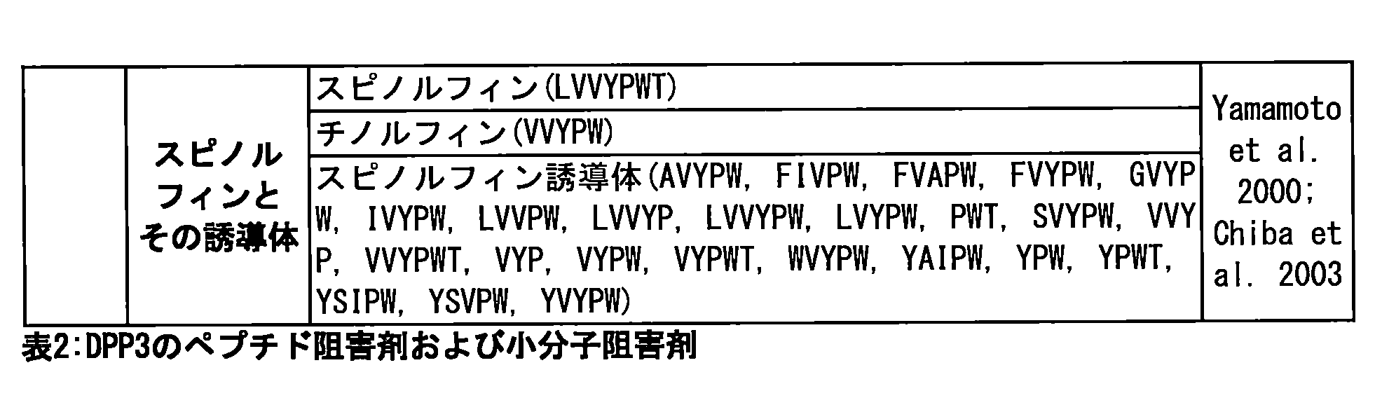
本発明の好ましい態様では、選択された阻害剤は医薬的に許容されるものであり、DPP3に選択的かつ/または特異的であり、かつ細胞膜および/または血液脳関門を通過しない。選択的かつ特異的なDPP3阻害剤は他のタンパク質/酵素に結合したり他のタンパク質/酵素が結合したりせず、DPP3以外のいずれの酵素/プロテアーゼ/ペプチダーゼも阻害しない。小さなペプチドは非特異的なアミノペプチダーゼによって結合かつ切断される可能性があり、小分子阻害剤は細胞膜および血液脳関門を容易に通過する。抗DPP3抗体、抗DPP3抗体断片、または抗DPP3非Ig足場は、特異的かつ選択的にDPP3に結合し、細胞膜および血液脳関門を通過しない。したがって、好ましいDPP3活性阻害剤は特異的な抗DPP3抗体、抗体断片、または非Ig足場である。 In a preferred embodiment of the present invention, the selected inhibitor is pharma- ceutically acceptable, selective and/or specific for DPP3, and does not cross the cell membrane and/or blood-brain barrier. A selective and specific DPP3 inhibitor does not bind or is bound by other proteins/enzymes and does not inhibit any enzymes/proteases/peptidases other than DPP3. Small peptides can be bound and cleaved by non-specific aminopeptidases, and small molecule inhibitors easily cross the cell membrane and blood-brain barrier. An anti-DPP3 antibody, an anti-DPP3 antibody fragment, or an anti-DPP3 non-Ig scaffold specifically and selectively binds to DPP3 and does not cross the cell membrane and blood-brain barrier. Thus, a preferred DPP3 activity inhibitor is a specific anti-DPP3 antibody, an antibody fragment, or a non-Ig scaffold.
本発明の特定の態様では、DPP3活性阻害剤は壊死性の経過を伴うか壊死性の経過に関連する被験者の疾患または状態の予防または治療に使用され、前記阻害剤は、抗DPP3抗体、抗DPP3抗体断片、抗DPP3非Ig足場を含む群から選択される。 In a particular aspect of the invention, an inhibitor of DPP3 activity is used for the prevention or treatment of a disease or condition in a subject that involves or is associated with a necrotic process, said inhibitor being selected from the group comprising an anti-DPP3 antibody, an anti-DPP3 antibody fragment, and an anti-DPP3 non-Ig scaffold.
本発明の特定の態様では、DPP3活性の阻害剤または作動剤は壊死性の経過を伴うか壊死性の経過に関連する被験者の疾患または状態の予防または治療に使用され、前記疾患は、心不全、慢性心不全、急性心不全(AHF)、心筋梗塞(MI)、卒中、肝不全、熱傷、外傷、重症感染症(微生物、ウイルス(たとえばAIDS)、寄生虫(たとえばマラリア))、SIRSまたは敗血症、がん、急性腎障害(AKI)、中枢神経系(CNS)障害(たとえば、発作、神経変性疾患)、自己免疫疾患および血管疾患(たとえば川崎症候群)、ならびに低血圧症を含む群から選択される。 In a particular embodiment of the invention, an inhibitor or agonist of DPP3 activity is used for the prevention or treatment of a disease or condition in a subject involving or associated with a necrotic process, said disease being selected from the group consisting of heart failure, chronic heart failure, acute heart failure (AHF), myocardial infarction (MI), stroke, liver failure, burns, trauma, severe infections (microbial, viral (e.g. AIDS), parasitic (e.g. malaria)), SIRS or sepsis, cancer, acute kidney injury (AKI), central nervous system (CNS) disorders (e.g. stroke, neurodegenerative diseases), autoimmune and vascular diseases (e.g. Kawasaki syndrome), and hypotension.
別の態様では、前記疾患は、急性心不全(AHF)、心筋梗塞(MI)、肝不全、熱傷、重症感染症(微生物、ウイルス(たとえばAIDS)、寄生虫(たとえばマラリア))、SIRSまたは敗血症、がん、急性腎障害(AKI)を含む群から選択される。 In another embodiment, the disease is selected from the group including acute heart failure (AHF), myocardial infarction (MI), liver failure, burns, severe infections (microbial, viral (e.g. AIDS), parasitic (e.g. malaria)), SIRS or sepsis, cancer, acute kidney injury (AKI).
本発明の特定の一態様では、DPP3活性阻害剤は被験者の疾患または状態の予防に使用され、前記疾患または状態は、急性心不全(AHF)、心筋梗塞(MI)、肝不全、がん、急性腎障害(AKI)、および低血圧症である。 In one particular aspect of the present invention, a DPP3 activity inhibitor is used to prevent a disease or condition in a subject, the disease or condition being acute heart failure (AHF), myocardial infarction (MI), liver failure, cancer, acute kidney injury (AKI), and hypotension.
本発明の特定の一態様では、DPP3活性阻害剤は被験者の疾患または状態の治療に使用され、前記疾患または状態は、急性心不全(AHF)、心筋梗塞(MI)、肝不全、がん、急性腎障害(AKI)、および低血圧症である。 In a particular aspect of the present invention, a DPP3 activity inhibitor is used to treat a disease or condition in a subject, the disease or condition being acute heart failure (AHF), myocardial infarction (MI), liver failure, cancer, acute kidney injury (AKI), and hypotension.
本発明のすべての態様について、特定の一態様では、前記疾患はアルツハイマー病ではない。本発明のすべての態様について、特定の一態様では、前記疾患はがんではない。本発明のすべての態様について、特定の一態様では、前記疾患は関節リウマチではない。
特定の一態様では、前記疾患または状態は低血圧症である。
For all aspects of the invention, in one particular embodiment, the disease is not Alzheimer's disease. For all aspects of the invention, in one particular embodiment, the disease is not cancer. For all aspects of the invention, in one particular embodiment, the disease is not rheumatoid arthritis.
In a particular embodiment, the disease or condition is hypotension.
本発明の特定の態様では、DPP3活性阻害剤は壊死性の経過を伴うか壊死性の経過に関連する被験者の疾患または状態の予防または治療に使用され、前記阻害剤は単一結合特異性(mono-binding)または少なくとも二重結合特異性(two-binding)の抗体である。 In a particular embodiment of the invention, an inhibitor of DPP3 activity is used for the prevention or treatment of a disease or condition in a subject that involves or is associated with a necrotic process, said inhibitor being an antibody of mono-binding or at least two-binding specificity.
本発明の特定の態様では、DPP3活性の阻害剤または作動剤は壊死性の経過を伴うか壊死性の経過に関連する被験者の疾患または状態の予防または治療に使用され、前記阻害剤または作動剤は、配列番号1に結合する抗DPP3抗体または抗DPP3抗体断片、あるいは抗DPP3非Ig足場であり、特定の態様では配列番号2に結合する抗DPP3抗体または抗DPP3抗体断片、あるいは抗DPP3非Ig足場である。 In a particular embodiment of the invention, an inhibitor or agonist of DPP3 activity is used for the prevention or treatment of a disease or condition in a subject involving or associated with a necrotic process, said inhibitor or agonist being an anti-DPP3 antibody or an anti-DPP3 antibody fragment or an anti-DPP3 non-Ig scaffold that binds to SEQ ID NO: 1, and in a particular embodiment an anti-DPP3 antibody or an anti-DPP3 antibody fragment or an anti-DPP3 non-Ig scaffold that binds to SEQ ID NO: 2.
本発明の特定の態様では、DPP3活性の阻害剤または作動剤は壊死性の経過を伴うか壊死性の経過に関連する被験者の疾患または状態の予防または治療に使用され、前記阻害剤または作動剤は、DPP3に対し10-7M未満の最低結合親和性を示す抗体または断片あるいは足場である。 In a particular aspect of the invention, an inhibitor or agonist of DPP3 activity is used for the prevention or treatment of a disease or condition in a subject involving or associated with a necrotic process, said inhibitor or agonist being an antibody or fragment or scaffold exhibiting a minimum binding affinity for DPP3 of less than 10-7 M.
壊死性の経過を伴うか壊死性の経過に関連する被験者の疾患または状態を予防または治療する方法の特定の態様では、前記阻害剤または作動剤は、全長DPP3に結合しDPP3の活性を少なくとも10%または少なくとも50%、より好ましくは少なくとも60%、さらに好ましくは70%超、さらに好ましくは80%超、さらに好ましくは90%超、さらに好ましくは95%超、阻害する抗体または断片あるいは足場である。上記のとおり、活性は液相検定で定量されてもよい。 In certain aspects of the method for preventing or treating a disease or condition in a subject involving or associated with a necrotic process, the inhibitor or agonist is an antibody or fragment or scaffold that binds to full-length DPP3 and inhibits DPP3 activity by at least 10% or at least 50%, more preferably at least 60%, even more preferably more than 70%, even more preferably more than 80%, even more preferably more than 90%, even more preferably more than 95%. As described above, activity may be quantified in a liquid phase assay.
本発明の特定の態様では、DPP3活性の阻害剤または作動剤は壊死性の経過を伴うか壊死性の経過に関連する被験者の疾患または状態の予防または治療に使用され、前記阻害剤または作動剤は単一特異性の抗体または断片あるいは足場である。 In a particular aspect of the invention, an inhibitor or agonist of DPP3 activity is used to prevent or treat a disease or condition in a subject that involves or is associated with a necrotic process, said inhibitor or agonist being a monospecific antibody or fragment or scaffold.
単一特異性抗DPP3抗体または単一特異性抗DPP3抗体断片、あるいは単一特異性抗DPP3非Ig足場は、前記抗体または抗体断片あるいは非Ig足場が標的DPP3内の少なくとも5個のアミノ酸を含む1つの特異的な領域に結合することを意味する。単一特異性抗DPP3抗体または単一特異性抗DPP3抗体断片、あるいは単一特異性抗DPP3非Ig足場は、いずれも同じ抗原に対して親和性をもつ抗DPP3抗体または抗DPP3抗体断片、あるいは抗DPP3非Ig足場である。 A monospecific anti-DPP3 antibody or a monospecific anti-DPP3 antibody fragment, or a monospecific anti-DPP3 non-Ig scaffold means that the antibody or antibody fragment or non-Ig scaffold binds to one specific region that contains at least 5 amino acids in the target DPP3. A monospecific anti-DPP3 antibody or a monospecific anti-DPP3 antibody fragment or a monospecific anti-DPP3 non-Ig scaffold is an anti-DPP3 antibody or an anti-DPP3 antibody fragment or an anti-DPP3 non-Ig scaffold that all have affinity for the same antigen.
別の特定の好ましい態様では、DPP3に結合する抗DPP3抗体または抗DPP3抗体断片、あるいは抗DPP3非Ig足場は、それぞれ単一特異性の抗体、抗体断片、または非Ig足場であり、「単一特異性」は前記抗体または抗体断片あるいは非Ig足場が標的DPP3内の少なくとも4個のアミノ酸を含む1つの特異的な領域に結合することを意味する。本発明の単一特異性の抗体または断片あるいは非Ig足場は、いずれも同じ抗原に対して親和性をもつ抗体または断片あるいは非Ig足場である。モノクローナル抗体は単一特異性であるが、単一特異性抗体は一般的な胚細胞からモノクローナル抗体を作製する以外の方法で作製されてもよい。 In another particular preferred embodiment, the anti-DPP3 antibody or anti-DPP3 antibody fragment or anti-DPP3 non-Ig scaffold that binds to DPP3 is a monospecific antibody, antibody fragment, or non-Ig scaffold, respectively, where "monospecific" means that the antibody or antibody fragment or non-Ig scaffold binds to one specific region that contains at least four amino acids in the target DPP3. The monospecific antibodies or fragments or non-Ig scaffolds of the present invention are antibodies or fragments or non-Ig scaffolds that all have affinity for the same antigen. Although monoclonal antibodies are monospecific, monospecific antibodies may be produced by methods other than the production of monoclonal antibodies from common embryonic cells.
本発明の特定の態様では、DPP3活性の阻害剤または作動剤は壊死性の経過を伴うか壊死性の経過に関連する被験者の疾患または状態の予防または治療に使用され、前記被験者はDPP3のレベルが上昇している。「レベルが上昇している」とはレベルが所定の閾値を超えていることである。 In a particular aspect of the invention, an inhibitor or agonist of DPP3 activity is used to prevent or treat a disease or condition involving or associated with a necrotic process in a subject, said subject having elevated levels of DPP3, where "elevated levels" refers to levels above a predefined threshold.
本発明の別の態様は、壊死性の経過を伴うか壊死性の経過に関連する被験者の疾患または状態の予防または治療に使用するための上記DPP3活性阻害剤を含む医薬組成物である。 Another aspect of the present invention is a pharmaceutical composition comprising the above-mentioned DPP3 activity inhibitor for use in the prevention or treatment of a disease or condition in a subject involving or associated with a necrotic process.
本発明の別の態様は、DPP3活性阻害剤を投与する、壊死性の経過を伴うか壊死性の経過に関連する被験者の疾患または状態を予防または治療する方法である。 Another aspect of the invention is a method for preventing or treating a disease or condition in a subject involving or associated with a necrotic process, comprising administering an inhibitor of DPP3 activity.
壊死性の経過を伴うか壊死性の経過に関連する被験者の疾患または状態を予防または治療する方法の特定の態様では、前記阻害剤は、抗DPP3抗体、抗DPP3抗体断片、抗DPP3非Ig足場を含む群から選択される。 In certain aspects of the method for preventing or treating a disease or condition in a subject involving or associated with a necrotic process, the inhibitor is selected from the group including an anti-DPP3 antibody, an anti-DPP3 antibody fragment, and an anti-DPP3 non-Ig scaffold.
壊死性の経過を伴うか壊死性の経過に関連する被験者の疾患または状態を予防または治療する方法の特定の態様では、前記疾患は、心不全、慢性心不全、急性心不全(AHF)、心筋梗塞(MI)、卒中、肝不全、熱傷、外傷、重症感染症(微生物、ウイルス(たとえばAIDS)、寄生虫(たとえばマラリア))、SIRSまたは敗血症、がん、急性腎障害(AKI)、CNS障害(たとえば、発作、神経変性疾患)、自己免疫疾患および血管疾患(たとえば川崎症候群)、ならびに低血圧症を含む群から選択される。 In certain embodiments of the method for preventing or treating a disease or condition in a subject involving or associated with a necrotic process, the disease is selected from the group including heart failure, chronic heart failure, acute heart failure (AHF), myocardial infarction (MI), stroke, liver failure, burns, trauma, severe infections (microbial, viral (e.g. AIDS), parasitic (e.g. malaria)), SIRS or sepsis, cancer, acute kidney injury (AKI), CNS disorders (e.g. stroke, neurodegenerative diseases), autoimmune and vascular diseases (e.g. Kawasaki syndrome), and hypotension.
壊死性の経過を伴うか壊死性の経過に関連する被験者の疾患または状態を予防または治療する方法の特定の態様では、前記阻害剤は単一結合特異性または少なくとも二重結合特異性の抗体である。 In certain embodiments of the method for preventing or treating a disease or condition in a subject involving or associated with a necrotic process, the inhibitor is an antibody of single or at least dual binding specificity.
壊死性の経過を伴うか壊死性の経過に関連する被験者の疾患または状態を予防または治療する方法の特定の態様では、前記阻害剤は、配列番号1に結合する抗DPP3抗体または抗DPP3抗体断片、あるいは抗DPP3非Ig足場であり、特定の態様では配列番号2に結合する抗DPP3抗体または抗DPP3抗体断片、あるいは抗DPP3非Ig足場である。 In certain embodiments of the method for preventing or treating a disease or condition in a subject involving or associated with a necrotic process, the inhibitor is an anti-DPP3 antibody or an anti-DPP3 antibody fragment, or an anti-DPP3 non-Ig scaffold, that binds to SEQ ID NO: 1, and in certain embodiments an anti-DPP3 antibody or an anti-DPP3 antibody fragment, or an anti-DPP3 non-Ig scaffold, that binds to SEQ ID NO: 2.
壊死性の経過を伴うか壊死性の経過に関連する被験者の疾患または状態を予防または治療する方法の特定の態様では、前記阻害剤は、DPP3に対し10-7M未満の最低結合親和性を示す抗体または断片あるいは足場である。 In certain aspects of the method of preventing or treating a disease or condition in a subject involving or associated with a necrotic process, said inhibitor is an antibody or fragment or scaffold exhibiting a minimum binding affinity for DPP3 of less than 10 −7 M.
壊死性の経過を伴うか壊死性の経過に関連する被験者の疾患または状態を予防または治療する方法の特定の態様では、前記阻害剤または作動剤は、単一特異性の抗体または断片あるいは足場である。 In certain embodiments of the method for preventing or treating a disease or condition in a subject involving or associated with a necrotic process, the inhibitor or agonist is a monospecific antibody or fragment or scaffold.
壊死性の経過を伴うか壊死性の経過に関連する被験者の疾患または状態を予防または治療する方法の特定の態様では、前記阻害剤または作動剤は、全長DPP3に結合しDPP3の活性を少なくとも10%または少なくとも50%、より好ましくは少なくとも60%、さらに好ましくは70%超、さらに好ましくは80%超、さらに好ましくは90%超、さらに好ましくは95%超、阻害する抗体または断片あるいは足場である。 In certain aspects of the method for preventing or treating a disease or condition in a subject involving or associated with a necrotic process, the inhibitor or agonist is an antibody or fragment or scaffold that binds to full-length DPP3 and inhibits DPP3 activity by at least 10% or at least 50%, more preferably at least 60%, even more preferably more than 70%, even more preferably more than 80%, even more preferably more than 90%, even more preferably more than 95%.
壊死性の経過を伴うか壊死性の経過に関連する被験者の疾患または状態を予防または治療する方法の特定の態様では、前記被験者はDPP3のレベルが上昇している。「レベルが上昇している」とはレベルが所定の閾値を超えていることである。閾値については先に定義されている。 In certain embodiments of the method for preventing or treating a disease or condition in a subject involving or associated with a necrotic process, the subject has elevated levels of DPP3. "Elevated levels" refers to levels above a predetermined threshold, the threshold being defined above.
本発明の別の態様は患者の血液からのDPP3の排除である。排除はいくつかのアフェレーシス技術および/または親和性クロマトグラフィー工程によって達成できる(Balogun et al. 2010)。このような方法としては、アガロース樹脂に結合された特異的かつ高親和性のDPP3抗体を含有する吸着体を介して患者の血漿を濾過すること(可能性のある吸着体材料に結合したDPP3の分析については実施例12を参照のこと)が挙げられるが、これに限定されるものではない。 Another aspect of the present invention is the clearance of DPP3 from the patient's blood. Clearance can be achieved by several apheresis techniques and/or affinity chromatography steps (Balogun et al. 2010). Such methods include, but are not limited to, filtering the patient's plasma through an adsorbent containing a specific, high affinity DPP3 antibody bound to an agarose resin (see Example 12 for analysis of DPP3 bound to potential adsorbent materials).
本発明の医薬組成物は、目的の投与経路に適合するように製剤される。投与経路は一般に、物質が投与される場所によって分類される。一般的な例としては、経口投与、皮膚投与、皮下投与、皮内投与、舌下投与、筋肉内投与、動脈内投与、静脈内投与、および腹腔内投与が挙げられる。 The pharmaceutical compositions of the present invention are formulated to be compatible with the intended route of administration. Routes of administration are generally classified by where the substance is administered. Common examples include oral, dermal, subcutaneous, intradermal, sublingual, intramuscular, intraarterial, intravenous, and intraperitoneal administration.
医薬組成物は中枢神経系(CNS)を経由して投与されてもよい。このような投与の例としては、硬膜外腔への注射または輸液による硬膜外投与、脳への直接注入による脳内(大脳への)投与、脳室内(脳の脳室系への)投与または髄腔内(脊髄管への)投与などが挙げられる。
以下の本発明の特定の態様について概要を述べる。
(項目1)
壊死性の経過を伴うか壊死性の経過に関連する被験者の疾患または状態を診断する方法であって、
前記被験者の体液試料中のDPP3総量を定量し、かつ/または活性DPP3の量を定量することと、
定量された前記DPP3総量または活性DPP3の量を所定の閾値と比較することとを含み、
定量された前記量が前記所定の閾値を超える場合、前記被験者は壊死性の経過を伴うか壊死性の経過に関連する疾患または状態を有すると診断される、方法。
(項目2)
前記DPP3総量または活性DPP3の量は濃度の単位で定量される、項目1に記載の壊死性の経過を伴うか壊死性の経過に関連する被験者の疾患または状態を診断する方法。
(項目3)
前記試料は全血、血清、血漿を含む群から選択される、項目1または2に記載の壊死性の経過を伴うか壊死性の経過に関連する被験者の疾患または状態を診断する方法。
(項目4)
前記疾患は、心不全、慢性心不全、急性心不全(AHF)、心筋梗塞(MI)、卒中、肝不全、熱傷、外傷、重症感染症(微生物、ウイルス(たとえば後天性免疫不全症候群(AIDS)、寄生虫(たとえばマラリア))、全身性炎症反応症候群(SIRS)または敗血症、がん、急性腎障害(AKI)、中枢神経系(CNS)障害(たとえば発作、神経変性疾患)、自己免疫疾患および血管疾患(たとえば川崎症候群)、ならびに低血圧症を含む群から選択される、項目1~3のいずれか一項に記載の壊死性の経過を伴うか壊死性の経過に関連する被験者の疾患または状態を診断する方法。
(項目5)
前記DPP3総量および/または活性DPP3の量は前記被験者の体液試料中で定量され、かつ
前記試料を全長DPP3に特異的に結合する捕捉結合剤に接触させる工程と、
前記捕捉結合剤に結合したDPP3を分離する工程と、
前記分離済みDPP3にDPP3の基質を添加する工程と、
DPP3の基質の転化率を測定し定量することにより前記活性DPP3を定量する工程と
を含む、項目1~4のいずれか一項に記載の壊死性の経過を伴うか壊死性の経過に関連する被験者の疾患または状態を診断する方法。
(項目6)
前記捕捉結合剤は抗体、抗体断片、または非IgG足場の群から選択されてもよい、項目5に記載の壊死性の経過を伴うか壊死性の経過に関連する被験者の疾患または状態を診断する方法。
(項目7)
前記捕捉結合剤は抗体である、項目5または6に記載の壊死性の経過を伴うか壊死性の経過に関連する被験者の疾患または状態を診断する方法。
(項目8)
前記捕捉結合剤は表面に固定化される、項目5~7のいずれか一項に記載の壊死性の経過を伴うか壊死性の経過に関連する被験者の疾患または状態を診断する方法。
(項目9)
前記分離工程は、前記試料のうち前記捕捉結合剤に結合していない成分を捕捉済みDPP3から除去する洗浄工程である、項目5~8のいずれか一項に記載の壊死性の経過を伴うか壊死性の経過に関連する被験者の疾患または状態を診断する方法。
(項目10)
DPP3基質の転化は、蛍光発生基質(たとえばArg-Arg-βNA、Arg-Arg-AMC)の蛍光、発色基質の色変化、アミノルシフェリンに結合された基質(Promega Protease-Glo(商標)Assay)の発光、質量分析、HPLC/FPLC(逆相クロマトグラフィー、サイズ排除クロマトグラフィー)、薄層クロマトグラフィー、キャピラリーゾーン電気泳動、ゲル電気泳動後に活性染色(固定された活性DPP3)またはウェスタンブロット(切断産物)を行うことを含む群から選択される方法で検出される、項目5~9のいずれか一項に記載の壊死性の経過を伴うか壊死性の経過に関連する被験者の疾患または状態を診断する方法。
(項目11)
前記基質は、アンギオテンシンII、III、IV、Leuエンケファリン、Metエンケファリン、エンドモルフィン1および2、バロルフィン、β-カゾモルフィン、ダイノルフィン、プロクトリン、副腎皮質刺激ホルモン(ACTH)、およびメラノサイト刺激ホルモン(MSH);ならびに蛍光色素分子、発色団、またはアミノルシフェリンに結合されたジペプチドを含む群から選択されてもよく、前記ジペプチドはArg-Argである、項目5~10のいずれか一項に記載の壊死性の経過を伴うか壊死性の経過に関連する被験者の疾患または状態を診断する方法。
(項目12)
前記基質は、蛍光色素分子、発色団、またはアミノルシフェリンに結合されたジペプチドを含む群から選択されてもよく、前記ジペプチドはArg-Argである、項目5~11のいずれか一項に記載の壊死性の経過を伴うか壊死性の経過に関連する被験者の疾患または状態を診断する方法。
(項目13)
項目1~12のいずれか一項に記載の診断方法を少なくとも2回行う、壊死性の経過を伴うか壊死性の経過に関連する被験者の疾患または状態を監視する方法。
(項目14)
壊死性の経過を伴うか壊死性の経過に関連する被験者の疾患または状態の予防または治療に使用するためのDPP3活性阻害剤。
(項目15)
前記阻害剤は抗DPP3抗体および抗DPP3抗体断片、ならびに抗DPP3非Ig足場を含む群から選択される、項目14に記載の壊死性の経過を伴うか壊死性の経過に関連する被験者の疾患または状態の予防または治療に使用するためのDPP3活性阻害剤。
(項目16)
前記疾患は、心不全、慢性心不全、急性心不全(AHF)、心筋梗塞(MI)、卒中、肝不全、熱傷、外傷、重症感染症(微生物、ウイルス(たとえばAIDS)、寄生虫(たとえばマラリア))、SIRSまたは敗血症、がん、急性腎障害(AKI)、CNS障害(たとえば発作、神経変性疾患)、自己免疫疾患および血管疾患(たとえば川崎症候群)、ならびに低血圧症を含む群から選択される、項目14または15に記載の壊死性の経過を伴うか壊死性の経過に関連する被験者の疾患または状態の予防または治療に使用するためのDPP3活性阻害剤。
(項目17)
前記阻害剤は単一結合特異性(mono-binding)または少なくとも二重結合特異性(two-binding)の抗体である、項目14~16のいずれか一項に記載の壊死性の経過を伴うか壊死性の経過に関連する被験者の疾患または状態の予防または治療に使用するためのDPP3活性阻害剤。
(項目18)
前記阻害剤は、配列番号1に結合し特に配列番号2に結合する抗DPP3抗体または抗DPP3抗体断片、あるいは抗DPP3非Ig足場である、項目14~17のいずれか一項に記載の壊死性の経過を伴うか壊死性の経過に関連する被験者の疾患または状態の予防または治療に使用するためのDPP3活性阻害剤。
(項目19)
前記阻害剤はDPP3に対し10-7M以下の最低結合親和性を示す抗体または断片あるいは足場である、項目14~18のいずれか一項に記載の壊死性の経過を伴うか壊死性の経過に関連する被験者の疾患または状態の予防または治療に使用するためのDPP3活性阻害剤。
(項目20)
前記阻害剤は単一特異性の抗体または断片あるいは足場である、項目14~19のいずれか一項に記載の壊死性の経過を伴うか壊死性の経過に関連する被験者の疾患または状態の予防または治療に使用するためのDPP3活性阻害剤。
(項目21)
前記阻害剤は、全長DPP3に結合しDPP3の活性を少なくとも10%または少なくとも50%、より好ましくは少なくとも60%、さらに好ましくは70%超、さらに好ましくは80%超、さらに好ましくは90%超、さらに好ましくは95%超、阻害する抗体または断片あるいは足場である、項目14~20のいずれか一項に記載の壊死性の経過を伴うか壊死性の経過に関連する被験者の疾患または状態の予防または治療に使用するためのDPP3活性阻害剤。
(項目22)
前記阻害剤はDPP3に選択的かつ/または特異的であり、かつ細胞膜および/または血液脳関門を通過しない、項目14~21のいずれか一項に記載の壊死性の経過を伴うか壊死性の経過に関連する被験者の疾患または状態の予防または治療に使用するためのDPP3活性阻害剤。
(項目23)
前記被験者は、体液試料中に所定の閾値を超えるDPP3総量および/または活性DPP3の量を有する、項目14~22のいずれか一項に記載の壊死性の経過を伴うか壊死性の経過に関連する被験者の疾患または状態の予防または治療に使用するためのDPP3活性阻害剤。
(項目24)
項目14~23のいずれか一項に記載の壊死性の経過を伴うか壊死性の経過に関連する被験者の疾患または状態の予防または治療に使用するためのDPP3活性阻害剤を含む、医薬組成物。
(項目25)
アフェレーシスと親和性クロマトグラフィーを含む、血漿からDPP3を体外除去する方法における項目14~23のいずれか一項に記載のDPP3活性阻害剤の使用。
(項目26)
被験者の体液試料中の活性DPP3を定量する方法であって、
前記試料を全長DPP3に特異的に結合する捕捉結合剤に接触させる工程と、
前記捕捉結合剤に結合したDPP3を分離する工程と、
前記分離済みDPP3にDPP3の基質を添加する工程と、
DPP3の基質の転化率を測定し定量することにより前記活性DPP3を定量する工程と
を含む方法。
(項目27)
前記捕捉結合剤は抗体、抗体断片、または非IgG足場の群から選択されてもよい、項目26に記載の被験者の体液試料中の活性DPP3を定量する方法。
(項目28)
前記捕捉結合剤は抗体である、項目26または27に記載の被験者の体液試料中の活性DPP3を定量する方法。
(項目29)
前記捕捉結合剤は表面に固定化される、項目26~28のいずれか一項に記載の被験者の体液試料中の活性DPP3を定量する方法。
(項目30)
前記分離工程は、前記試料のうち前記捕捉結合剤に結合していない成分を捕捉済みDPP3から除去する洗浄工程である、項目26~29のいずれか一項に記載の被験者の体液試料中の活性DPP3を定量する方法。
(項目31)
DPP3基質の転化は、蛍光発生基質(たとえばArg-Arg-βNA、Arg-Arg-AMC)の蛍光、発色基質の色変化、アミノルシフェリンに結合された基質(Promega Protease-Glo(商標)Assay)の発光、質量分析、HPLC/FPLC(逆相クロマトグラフィー、サイズ排除クロマトグラフィー)、薄層クロマトグラフィー、キャピラリーゾーン電気泳動、ゲル電気泳動後に活性染色(固定された活性DPP3)またはウェスタンブロット(切断産物)を行うことを含む群から選択される方法で検出される、項目26~30のいずれか一項に記載の被験者の体液試料中の活性DPP3を定量する方法。
(項目32)
前記基質は、蛍光色素分子、発色団、またはアミノルシフェリンに結合されたジペプチドを含む群から選択されてもよく、前記ジペプチドはArg-Argである、項目26~31のいずれか一項に記載の被験者の体液試料中の活性DPP3を定量する方法。
(項目33)
前記試料は全血、血清、血漿を含む群から選択される血液試料である、項目26~32のいずれか一項に記載の被験者の体液試料中の活性DPP3を定量する方法。
(項目34)
被験者の体液試料中の活性DPP3を定量する検定またはキットであって、
全長DPP3に特異的に結合する捕捉結合剤と、
DPP3の基質
を含む、検定またはキット。
(項目35)
前記捕捉結合剤は抗体、抗体断片、または非IgG足場の群から選択されてもよい、項目34に記載の被験者の体液試料中の活性DPP3を定量する検定またはキット。
(項目36)
前記捕捉結合剤は、DPP3活性を液相検定で50%未満、好ましくは40%未満、より好ましくは30%未満阻害する、項目34または35に記載の被験者の体液試料中の活性DPP3を定量する検定またはキット。
(項目37)
前記捕捉結合剤に対するDPP3の結合領域は配列番号1のアミノ酸316~669の領域内にない、項目34~36のいずれか一項に記載の被験者の体液試料中の活性DPP3を定量する検定またはキット。
(項目38)
前記捕捉結合剤は抗体である、項目34~37のいずれか一項に記載の被験者の体液試料中の活性DPP3を定量する検定またはキット。
(項目39)
前記捕捉結合剤は表面に固定化される、項目34~38のいずれか一項に記載の被験者の体液試料中の活性DPP3を定量する検定またはキット。
(項目40)
前記基質は、アンギオテンシンII、III、IV、Leuエンケファリン、Metエンケファリン、エンドモルフィン1および2、バロルフィン、β-カゾモルフィン、ダイノルフィン、プロクトリン、ACTH、およびMSH;ならびに蛍光色素分子、発色団、またはアミノルシフェリンに結合されたジペプチドを含む群から選択されてもよく、前記ジペプチドはArg-Argである、項目34~39のいずれか一項に記載の被験者の体液試料中の活性DPP3を定量する検定またはキット。
(項目41)
項目1~13のいずれか一項に記載の壊死性の経過を伴うか壊死性の経過に関連する被験者の疾患または状態を診断または監視する方法における、項目26~33のいずれか一項に記載の被験者の体液試料中の活性DPP3を定量する方法または項目34~40のいずれか一項に記載の検定またはキットの使用。
The pharmaceutical composition may be administered via the central nervous system (CNS), including epidural administration by injection or infusion into the epidural space, intracerebral (into the cerebrum) administration by direct injection into the brain, intraventricular (into the ventricular system of the brain) or intrathecal (into the spinal canal) administration.
Certain aspects of the invention are outlined below.
(Item 1)
1. A method for diagnosing a disease or condition involving or associated with a necrotic process in a subject, comprising:
Quantifying the total amount of DPP3 and/or the amount of active DPP3 in the body fluid sample of the subject;
and comparing the quantified amount of total DPP3 or active DPP3 to a predetermined threshold value;
If the quantified amount exceeds the predetermined threshold, the subject is diagnosed as having a disease or condition involving or associated with a necrotic process.
(Item 2)
2. A method for diagnosing a disease or condition in a subject involving or associated with a necrotic process according to item 1, wherein the amount of total DPP3 or active DPP3 is quantified in concentration units.
(Item 3)
3. Method for diagnosing a disease or condition in a subject involving or associated with a necrotic process according to items 1 or 2, wherein said sample is selected from the group comprising whole blood, serum, plasma.
(Item 4)
4. Method for diagnosing a disease or condition in a subject accompanied by or associated with a necrotic process according to any one of items 1 to 3, wherein said disease is selected from the group comprising heart failure, chronic heart failure, acute heart failure (AHF), myocardial infarction (MI), stroke, liver failure, burns, trauma, severe infections (microbial, viral (e.g. acquired immune deficiency syndrome (AIDS), parasitic (e.g. malaria)), systemic inflammatory response syndrome (SIRS) or sepsis, cancer, acute kidney injury (AKI), central nervous system (CNS) disorders (e.g. stroke, neurodegenerative diseases), autoimmune diseases and vascular diseases (e.g. Kawasaki syndrome), and hypotension.
(Item 5)
The amount of total DPP3 and/or active DPP3 is quantified in a body fluid sample of the subject, and the sample is contacted with a capture binding agent that specifically binds full length DPP3;
Separating DPP3 bound to the capture binding agent;
Adding a DPP3 substrate to the separated DPP3;
and quantifying the active DPP3 by measuring and quantifying the conversion rate of a DPP3 substrate.
(Item 6)
6. The method for diagnosing a disease or condition in a subject involving or associated with a necrotic process according to item 5, wherein the capture binding agent may be selected from the group of an antibody, an antibody fragment, or a non-IgG scaffold.
(Item 7)
7. The method for diagnosing a disease or condition in a subject involving or associated with a necrotic process according to items 5 or 6, wherein the capture binding agent is an antibody.
(Item 8)
8. A method for diagnosing a disease or condition in a subject involving or associated with a necrotic process according to any one of items 5 to 7, wherein said capture binding agent is immobilised on a surface.
(Item 9)
9. The method for diagnosing a disease or condition in a subject accompanied by or associated with a necrotic process according to any one of items 5 to 8, wherein the separation step is a washing step for removing components of the sample that are not bound to the capture binding agent from the captured DPP3.
(Item 10)
10. Method for diagnosing a disease or condition in a subject accompanied by or associated with a necrotic process according to any one of items 5 to 9, wherein the conversion of the DPP3 substrate is detected by a method selected from the group comprising: fluorescence of a fluorogenic substrate (e.g. Arg-Arg-βNA, Arg-Arg-AMC), color change of a chromogenic substrate, light emission of a substrate coupled to aminoluciferin (Promega Protease-Glo™ Assay), mass spectrometry, HPLC/FPLC (reverse phase chromatography, size exclusion chromatography), thin layer chromatography, capillary zone electrophoresis, gel electrophoresis followed by activity staining (fixed active DPP3) or Western blot (cleavage products).
(Item 11)
11. Method for diagnosing a disease or condition in a subject involving or associated with a necrotic process according to any one of items 5 to 10, wherein said substrate may be selected from the group comprising angiotensin II, III, IV, Leu-enkephalin, Met-enkephalin, endomorphin 1 and 2, valorphin, β-casomorphin, dynorphin, proctolin, adrenocorticotropic hormone (ACTH), and melanocyte-stimulating hormone (MSH); and a dipeptide linked to a fluorophore, a chromophore, or an aminoluciferin, said dipeptide being Arg-Arg.
(Item 12)
12. Method for diagnosing a disease or condition in a subject involving or associated with a necrotic process according to any one of items 5 to 11, wherein said substrate may be selected from the group comprising a dipeptide linked to a fluorophore, a chromophore or an aminoluciferin, said dipeptide being Arg-Arg.
(Item 13)
13. A method for monitoring a disease or condition in a subject involving or associated with a necrotic process, comprising performing the diagnostic method according to any one of items 1 to 12 at least twice.
(Item 14)
An inhibitor of DPP3 activity for use in the prevention or treatment of a disease or condition in a subject involving or associated with a necrotic process.
(Item 15)
15. The DPP3 activity inhibitor for use in the prevention or treatment of a disease or condition in a subject involving or associated with a necrotic process according to item 14, wherein the inhibitor is selected from the group comprising anti-DPP3 antibodies and anti-DPP3 antibody fragments, and anti-DPP3 non-Ig scaffolds.
(Item 16)
16. A DPP3 activity inhibitor for use in the prevention or treatment of a disease or condition in a subject accompanied by or associated with a necrotic process according to item 14 or 15, wherein said disease is selected from the group comprising heart failure, chronic heart failure, acute heart failure (AHF), myocardial infarction (MI), stroke, liver failure, burns, trauma, severe infections (microbial, viral (e.g. AIDS), parasitic (e.g. malaria)), SIRS or sepsis, cancer, acute kidney injury (AKI), CNS disorders (e.g. stroke, neurodegenerative diseases), autoimmune and vascular diseases (e.g. Kawasaki syndrome), and hypotension.
(Item 17)
17. The DPP3 activity inhibitor for use in the prevention or treatment of a disease or condition in a subject involving or associated with a necrotic process according to any one of items 14 to 16, wherein the inhibitor is an antibody of single binding specificity (mono-binding) or at least dual binding specificity (two-binding).
(Item 18)
18. The DPP3 activity inhibitor for use in the prevention or treatment of a disease or condition in a subject involving or associated with a necrotic process according to any one of items 14 to 17, wherein the inhibitor is an anti-DPP3 antibody or an anti-DPP3 antibody fragment that binds to SEQ ID NO: 1 and in particular to SEQ ID NO: 2, or an anti-DPP3 non-Ig scaffold.
(Item 19)
19. The DPP3 activity inhibitor for use in the prevention or treatment of a disease or condition in a subject involving or associated with a necrotic process according to any one of items 14 to 18, wherein said inhibitor is an antibody or fragment or scaffold that exhibits a minimum binding affinity for DPP3 of 10 −7 M or less.
(Item 20)
20. The DPP3 activity inhibitor for use in the prevention or treatment of a disease or condition in a subject involving or associated with a necrotic process according to any one of items 14 to 19, wherein said inhibitor is a monospecific antibody or fragment or a scaffold.
(Item 21)
21. The DPP3 activity inhibitor for use in the prevention or treatment of a disease or condition in a subject involving or associated with a necrotic process according to any one of items 14 to 20, wherein the inhibitor is an antibody or fragment or scaffold that binds to full-length DPP3 and inhibits the activity of DPP3 by at least 10% or at least 50%, more preferably at least 60%, even more preferably more than 70%, even more preferably more than 80%, even more preferably more than 90%, even more preferably more than 95%.
(Item 22)
22. The DPP3 activity inhibitor for use in the prevention or treatment of a disease or condition in a subject involving or associated with a necrotic process according to any one of items 14 to 21, wherein the inhibitor is selective and/or specific for DPP3 and does not cross the cell membrane and/or the blood-brain barrier.
(Item 23)
23. The DPP3 activity inhibitor for use in the prevention or treatment of a disease or condition in a subject involving or associated with a necrotic process according to any one of items 14 to 22, wherein the subject has an amount of total DPP3 and/or active DPP3 in a body fluid sample that exceeds a predetermined threshold.
(Item 24)
A pharmaceutical composition comprising an inhibitor of DPP3 activity for use in the prophylaxis or treatment of a disease or condition in a subject that is accompanied by or is associated with a necrotic process according to any one of items 14 to 23.
(Item 25)
24. Use of the DPP3 activity inhibitor according to any one of items 14 to 23 in a method for extracorporeal removal of DPP3 from plasma, comprising apheresis and affinity chromatography.
(Item 26)
1. A method for quantifying active DPP3 in a body fluid sample from a subject, comprising:
contacting the sample with a capture binding agent that specifically binds full-length DPP3;
Separating DPP3 bound to the capture binding agent;
Adding a DPP3 substrate to the separated DPP3;
and quantifying the active DPP3 by measuring and quantifying the conversion rate of a DPP3 substrate.
(Item 27)
27. The method of quantifying active DPP3 in a body fluid sample of a subject according to item 26, wherein the capture binding agent may be selected from the group of antibodies, antibody fragments, or non-IgG scaffolds.
(Item 28)
28. The method for quantifying active DPP3 in a body fluid sample of a subject according to item 26 or 27, wherein the capture binding agent is an antibody.
(Item 29)
29. The method for quantifying active DPP3 in a body fluid sample of a subject according to any one of items 26 to 28, wherein the capture binding agent is immobilized on a surface.
(Item 30)
30. The method for quantifying active DPP3 in a body fluid sample of a subject according to any one of Items 26 to 29, wherein the separation step is a washing step for removing components of the sample that are not bound to the capture binding agent from the captured DPP3.
(Item 31)
31. The method for quantifying active DPP3 in a body fluid sample of a subject according to any one of items 26 to 30, wherein the conversion of DPP3 substrate is detected by a method selected from the group comprising: fluorescence of a fluorogenic substrate (e.g. Arg-Arg-βNA, Arg-Arg-AMC), color change of a chromogenic substrate, light emission of a substrate coupled to aminoluciferin (Promega Protease-Glo™ Assay), mass spectrometry, HPLC/FPLC (reverse phase chromatography, size exclusion chromatography), thin layer chromatography, capillary zone electrophoresis, gel electrophoresis followed by activity staining (fixed active DPP3) or Western blot (cleavage products).
(Item 32)
32. The method for quantifying active DPP3 in a body fluid sample of a subject according to any one of items 26 to 31, wherein the substrate may be selected from the group comprising a dipeptide bound to a fluorophore, a chromophore, or an aminoluciferin, and the dipeptide is Arg-Arg.
(Item 33)
33. The method for quantifying active DPP3 in a body fluid sample from a subject according to any one of Items 26 to 32, wherein the sample is a blood sample selected from the group consisting of whole blood, serum, and plasma.
(Item 34)
1. An assay or kit for quantifying active DPP3 in a body fluid sample from a subject, comprising:
a capture binding agent that specifically binds full-length DPP3;
An assay or kit comprising a substrate for DPP3.
(Item 35)
35. The assay or kit for quantifying active DPP3 in a body fluid sample of a subject according to item 34, wherein the capture binding agent may be selected from the group of antibodies, antibody fragments, or non-IgG scaffolds.
(Item 36)
36. The assay or kit for quantifying active DPP3 in a body fluid sample of a subject according to item 34 or 35, wherein the capture binding agent inhibits DPP3 activity by less than 50%, preferably less than 40%, more preferably less than 30% in a liquid phase assay.
(Item 37)
37. The assay or kit for quantifying active DPP3 in a body fluid sample of a subject according to any one of items 34 to 36, wherein the binding region of DPP3 for the capture binding agent is not within the region of amino acids 316 to 669 of SEQ ID NO:1.
(Item 38)
38. The assay or kit for quantifying active DPP3 in a body fluid sample of a subject according to any one of items 34 to 37, wherein the capture binding agent is an antibody.
(Item 39)
39. An assay or kit for quantifying active DPP3 in a body fluid sample of a subject according to any one of items 34 to 38, wherein said capture binding agent is immobilized on a surface.
(Item 40)
40. The assay or kit for quantifying active DPP3 in a body fluid sample of a subject according to any one of items 34 to 39, wherein the substrate may be selected from the group comprising angiotensin II, III, IV, Leu enkephalin, Met enkephalin, endomorphin 1 and 2, valorphin, β-casomorphin, dynorphin, proctolin, ACTH, and MSH; and a dipeptide linked to a fluorophore, a chromophore, or an aminoluciferin, wherein the dipeptide is Arg-Arg.
(Item 41)
Use of the method for quantifying active DPP3 in a body fluid sample of a subject according to any one of items 26 to 33 or the assay or kit according to any one of items 34 to 40 in a method for diagnosing or monitoring a disease or condition in a subject that is accompanied by or associated with a necrotic process according to any one of items 1 to 13.
(1.実施例1)
様々な疾患の患者(急性心筋梗塞(AMI)、心原性ショック、敗血症性ショック、および肝不全の患者)の血漿中のDPP3活性を、特異的なDPP3捕捉活性検定を用いて定量し、健常対照の血漿のDPP3活性と比較した。
(1.1.研究コホート)
1. Example 1
DPP3 activity in plasma from patients with various diseases (patients with acute myocardial infarction (AMI), cardiogenic shock, septic shock, and liver failure) was quantified using a specific DPP3 scavenging activity assay and compared with DPP3 activity in plasma from healthy controls.
1.1 Study cohort
最初の症状で直接救急科に入院した388名の患者から血漿試料を得た。最終診断によって患者を急性心筋梗塞(AMI)、心原性ショック、敗血症性ショック、および肝不全の患者の4つの亜群に分けることができた。対照群は、93名の健常対照の血漿試料群であった。
(1.2.hDPP3捕捉活性検定)
Plasma samples were obtained from 388 patients who were directly admitted to the emergency department with their first symptoms. The final diagnosis allowed the patients to be divided into four subgroups: patients with acute myocardial infarction (AMI), cardiogenic shock, septic shock, and liver failure. The control group consisted of plasma samples from 93 healthy controls.
1.2. hDPP3 Trapping Activity Assay
血漿試料(10μl)のDPP3を最初に親和精製工程によって濃縮し、次に蛍光発生基質Arg-Arg-βNAを添加してその活性を測定した(詳細な説明については実施例4を参照のこと)。様々な試料の蛍光増加の傾き(nmol(βNA)/min/ml(試料)[nmol(βNA)min-1ml-1])の計算値は10μlの試料サイズに関する。
(1.3.結果)
DPP3 from plasma samples (10 μl) was first enriched by an affinity purification step and then its activity was measured by adding the fluorogenic substrate Arg-Arg-βNA (see Example 4 for a detailed description). The calculated slopes of the fluorescence increase for the various samples (nmol(βNA)/min/ml (sample) [nmol(βNA)min −1 ml −1 ]) are for a sample size of 10 μl.
(1.3. Results)
患者の試料を健常対照と比較したところ、重症または臓器不全のあるすべての患者のDPP3活性値は有意に高かった(図10)。
(2.実施例2)
When patient samples were compared with healthy controls, all patients with severe disease or organ failure had significantly higher DPP3 activity levels (Figure 10).
2. Example 2
本実験では、組み換えhDPP3を健常なラットに注射した場合の効果について、血圧を監視することにより研究した。
(2.1.方法)
In this experiment, the effect of injecting recombinant hDPP3 into healthy rats was studied by monitoring blood pressure.
2.1. Methods
2~3ヵ月齢の雄のウィスターラット(Charles River Laboratories(ドイツ))を本研究に使用した。血圧(BP)の測定および記録のため、カテーテル(Introcan-W; 22 G/1''; B.Braun)を右総頚動脈に挿入した。N末端にGSTタグを付けたヒト組み換えジペプチジルペプチダーゼ3(recGST-hDPP3)を尾静脈注射した。 Male Wistar rats (Charles River Laboratories, Germany) aged 2 to 3 months were used in this study. A catheter (Introcan-W; 22 G/1''; B. Braun) was inserted into the right common carotid artery for measuring and recording blood pressure (BP). N-terminally GST-tagged human recombinant dipeptidyl peptidase 3 (recGST-hDPP3) was injected via the tail vein.
最初に動物をイソフルランで麻酔し、体重測定(すべてg)し、長時間の麻酔のために1.2g/kg(体重)のウレタン(濃度=0.4g/mL)を腹腔内(i.p.)注射した。次いで首の腹側領域の毛を刈り、エタノールで拭いた。血管を準備し、カテーテルを挿入した。最後に、両方のカテーテルをヘパリン処置された等張食塩水で洗い流す。次いで血圧測定器(Medex Logical(Medex Medical Ltd.))を患者監視システム(Datex-Ohmeda(GE))に接続した。ラップトップコンピュータをBPモニターに接続し、S/5 Collect SoftwareでBPデータを個別に記録した。 Animals were first anesthetized with isoflurane, weighed (all in g), and injected intraperitoneally (i.p.) with 1.2 g/kg (body weight) of urethane (concentration = 0.4 g/mL) for long-term anesthesia. The ventral area of the neck was then clipped and wiped with ethanol. The vessels were prepared and catheterized. Finally, both catheters were flushed with heparinized isotonic saline. A blood pressure measuring device (Medex Logical (Medex Medical Ltd.)) was then connected to a patient monitoring system (Datex-Ohmeda (GE)). A laptop computer was connected to the BP monitor, and BP data were recorded separately with S/5 Collect Software.
ラットをrecGST-hDPP3のPBS溶液0.2mg/kgで尾静脈注射によって処置した。DPP3の注射の前後の血圧を常に監視した。
(2.2.結果)
Rats were treated with recGST-hDPP3 in PBS at 0.2 mg/kg by tail vein injection. Blood pressure was constantly monitored before and after injection of DPP3.
2.2 Results
健常なラットに組み換えGST-hDPP3を注射すると、血圧はすぐに低下する(図11)。
(3.実施例3)
Injection of recombinant GST-hDPP3 into healthy rats rapidly reduces blood pressure (Figure 11).
3. Example 3
抗体の作製とDPP3結合能力の定量:数種のマウス抗体を作製し、ヒトDPP3への結合能力によってサンドイッチ検定または活性検定でスクリーニングした(表3参照)。
(3.1.方法)
(免疫用のペプチド/結合体)
Generation of antibodies and quantification of DPP3 binding ability: Several mouse antibodies were generated and screened for their ability to bind to human DPP3 in sandwich or activity assays (see Table 3).
3.1. Methods
Peptides/Conjugates for Immunization
免疫用のDPP3ペプチドを合成した(表3参照)(JPT Technologies(ドイツ、ベルリン))。ペプチドをウシ血清アルブミン(BSA)と結合するため、N末端にシステイン残基を追加した(選択されたDPP3配列にシステインが存在しない場合)。SulfoLink-Coupling Gel(Perbio-Science(ドイツ、ボン))を用いてペプチドをBSAに共有結合した。カップリング手順はPerbio社の説明書に従って実行された。組み換えGST-hDPP3はUSBio製であった。
(マウスの免疫化、免疫細胞融合およびスクリーニング)
DPP3 peptides for immunization were synthesized (see Table 3) (JPT Technologies, Berlin, Germany). A cysteine residue was added to the N-terminus to conjugate the peptide to bovine serum albumin (BSA) (if no cysteine was present in the selected DPP3 sequence). The peptide was covalently coupled to BSA using SulfoLink-Coupling Gel (Perbio-Science, Bonn, Germany). The coupling procedure was performed according to the instructions from Perbio. Recombinant GST-hDPP3 was from USBio.
(Mouse Immunization, Immune Cell Fusion and Screening)
Balb/cマウスにGST-hDPP3またはDPP3ペプチド-BSA結合体を、0日目にそれぞれ84μgと100μg(TiterMax Gold Adjuvantで乳化)、14日目にそれぞれ84μgと100μg(フロイント完全アジュバントで乳化)、21日目と28日目にそれぞれ42μgと50μg(フロイント不完全アジュバントに溶解)、腹腔内(i.p.)注射した。49日目、生理食塩水に溶解したGST-hDPP3(42μg)またはDPP3ペプチド-BSA結合体(50μg)を動物に静脈内(i.v.)注射した。3日後、マウスを屠殺し、免疫細胞融合を行った。 Balb/c mice were intraperitoneally (i.p.) injected with 84 μg and 100 μg of GST-hDPP3 or DPP3 peptide-BSA conjugates (emulsified in TiterMax Gold Adjuvant) on day 0, 84 μg and 100 μg of GST-hDPP3 or DPP3 peptide-BSA conjugates (emulsified in Freund's complete adjuvant) on day 14, and 42 μg and 50 μg of GST-hDPP3 or DPP3 peptide-BSA conjugates (dissolved in Freund's incomplete adjuvant) on days 21 and 28. On day 49, animals were intravenously (i.v.) injected with GST-hDPP3 (42 μg) or DPP3 peptide-BSA conjugates (50 μg) dissolved in saline. Three days later, mice were sacrificed and subjected to immune cell fusion.
免疫マウス由来の脾細胞と骨髄腫細胞株SP2/0の細胞を、50%ポリエチレングリコール1mlを用いて37℃で30秒かけて融合させた。洗浄後、細胞を96ウェル細胞培養プレートに播種した。HAT培地[20%ウシ胎仔血清およびHAT-Supplementを補充したRPMI 1640培地]で増殖させることによってハイブリッドクローンを選択した。1週間後、HAT培地をHT培地に替えて3回継代培養した後、通常の細胞培地に戻した。 Spleen cells from immunized mice were fused with cells of the myeloma cell line SP2/0 using 1 ml of 50% polyethylene glycol at 37°C for 30 seconds. After washing, the cells were seeded into 96-well cell culture plates. Hybrid clones were selected by growth in HAT medium [RPMI 1640 medium supplemented with 20% fetal bovine serum and HAT-Supplement]. After one week, the HAT medium was replaced with HT medium and subcultured three times, after which the medium was returned to normal cell culture medium.
融合の2週間後、まずIgG抗体に結合する組換えDPP3について細胞培養液上清をスクリーニングした。したがって、組換えGSTタグ付きDPP3(USBiologicals(米国、セイラム))を96ウェルプレート(100ng/ウェル)に固定化し、50μl/ウェルの細胞培養液上清と共に室温で2時間保温した。プレートを洗浄した後、50μl/ウェルのPOD-ウサギ抗マウスIgGを添加し、室温で1時間保温した。次の洗浄工程の後、50μlの色素原溶液(3.7mMのo-フェニレンジアミンを[クエン酸塩/リン酸水素塩緩衝液、0.012%H2O2]に溶解した液)を各ウェルに添加し、室温で15分間保温し、4Nの硫酸50μlを添加することにより発色反応を停止した。490mmで吸収を検出した。 Two weeks after fusion, cell culture supernatants were first screened for recombinant DPP3 binding to IgG antibodies. Thus, recombinant GST-tagged DPP3 (USBiologicals, Salem, USA) was immobilized in 96-well plates (100 ng/well) and incubated with 50 μl/well of cell culture supernatant at room temperature for 2 h. After washing the plates, 50 μl/well of POD-rabbit anti-mouse IgG was added and incubated at room temperature for 1 h. After a further washing step, 50 μl of chromogen solution (3.7 mM o-phenylenediamine in [citrate/hydrogen phosphate buffer, 0.012% H 2 O 2 ]) was added to each well, incubated at room temperature for 15 min, and the color reaction was stopped by adding 50 μl of 4N sulfuric acid. Absorbance was detected at 490 mm.
陽性の試験微生物培養液を増殖のために24ウェルプレートに移した。再試験の後、選択された培養液を限界希釈法でクローン化および再クローン化し、アイソタイプを決定した。
(マウスモノクローナル抗体の作製)
Positive testing microbial cultures were transferred to 24-well plates for growth. After retesting, selected cultures were cloned and recloned by limiting dilution and isotyped.
(Preparation of mouse monoclonal antibodies)
GSTタグ付きヒトDPP3またはDPP3ペプチドに対する抗体を標準的な抗体作製方法(Marx et al., 1997)で作製し、プロテインAで精製した。抗体純度は、SDSゲル電気泳動分析に基づいて90%以上であった。
(抗体の特性評価-hDPP3阻害の分析)
Antibodies against GST-tagged human DPP3 or DPP3 peptides were generated by standard antibody production methods (Marx et al., 1997) and purified with protein A. Antibody purity was greater than 90% based on SDS gel electrophoresis analysis.
(Antibody Characterization—hDPP3 Inhibition Analysis)
様々な抗体および抗体クローンによるDPP3阻害能を分析するために、既知の手順(Jones et al., 1982)でDPP3活性検定を行った。組換えGSTタグ付きhDPP3を検定緩衝液に希釈(25ng/mlのGST-DPP3を[50mMのトリス-HCl(pH7.5)、100μMのZnCl2]に溶解)し、この溶液200μlを各抗体10μgと共に室温で保温した。1時間の前保温の後、蛍光発生基質Arg-Arg-βNA(20μl、2mM)を溶液に添加し、経時的な遊離βNAの生成を37℃でTwinkle LB 970マイクロプレート蛍光光度計(Berthold Technologies GmbH & Co. KG)を用いて監視した。340nmで励起し410nmで発光を測定することにより、βNAの蛍光を検出する。様々な試料の蛍光増加の勾配(RFU/分)を計算する。緩衝液(対照)とGST-hDPP3の場合の勾配を活性100%とする。可能性のある捕捉結合剤の阻害能は、前記捕捉結合剤と共に保温することによるGST-hDPP3活性の低下(%)で定義される。その結果低下したDPP3活性を図1aおよび表3に示す。
(3.2.結果)
To analyze the DPP3 inhibitory potential of various antibodies and antibody clones, a DPP3 activity assay was performed as previously described (Jones et al., 1982). Recombinant GST-tagged hDPP3 was diluted in assay buffer (25 ng/ml GST-DPP3 in [50 mM Tris-HCl (pH 7.5), 100 μM ZnCl 2 ]) and 200 μl of this solution was incubated with 10 μg of each antibody at room temperature. After 1 h preincubation, the fluorogenic substrate Arg-Arg-βNA (20 μl, 2 mM) was added to the solution and the generation of free βNA over time was monitored at 37°C using a Twinkle LB 970 microplate fluorometer (Berthold Technologies GmbH & Co. KG). Fluorescence of βNA is detected by excitation at 340 nm and emission at 410 nm. The slope of the fluorescence increase (RFU/min) of the various samples is calculated. The slope for buffer (control) and GST-hDPP3 represents 100% activity. The inhibitory capacity of a potential capture binding agent is defined as the % reduction in GST-hDPP3 activity upon incubation with said capture binding agent. The resulting reduced DPP3 activity is shown in Figure 1a and Table 3.
3.2 Results
以下の表は、得られた抗体の選択とその最大阻害率を示す(表3)。以下に示すDPP3領域に対するモノクローナル抗体を天然DPP3への結合能力によって選択した(mAb-FL-DPP3_2555を固相、_2553をトレーサーとして免疫測定に使用した。詳細については実施例2を参照のこと)。 The following table shows a selection of the antibodies obtained and their maximum inhibition rates (Table 3). The monoclonal antibodies against the following DPP3 regions were selected for their ability to bind to native DPP3 (mAb-FL-DPP3_2555 was used as the solid phase and _2553 as the tracer for immunoassays. See Example 2 for details).
固定化DPP3活性検定(実施例4および5参照)のためには、DPP3活性の阻害が強すぎない固相抗体を選択する必要があった。抗体スクリーニングのカットオフ(境界)として、固相抗体はDPP3活性を50%よりも強く阻害するべきではない。mAbDPP3_2555は最も低い阻害率を示した(表3、図1A)。 For the immobilized DPP3 activity assay (see Examples 4 and 5), it was necessary to select solid-phase antibodies that did not inhibit DPP3 activity too strongly. As a cutoff for antibody screening, solid-phase antibodies should not inhibit DPP3 activity more than 50%. mAb DPP3_2555 showed the lowest inhibition rate (Table 3, Figure 1A).
治療に使用できる強いDPP3阻害剤(実施例6~11参照)を作製するには、最も高い阻害率を示すDPP3結合剤を選択する必要があった。DPP3活性を70%阻害する能力をもつモノクローナル抗体mAbDPP3_1967を、可能性のある治療抗体として選択し(図1Aおよび表3を参照のこと)、この抗体を用いて以降のすべての分析を実行した。図1Bは、IC50値が0.2041μg/mlであるmAbDPP3_1967の阻害曲線を示す。
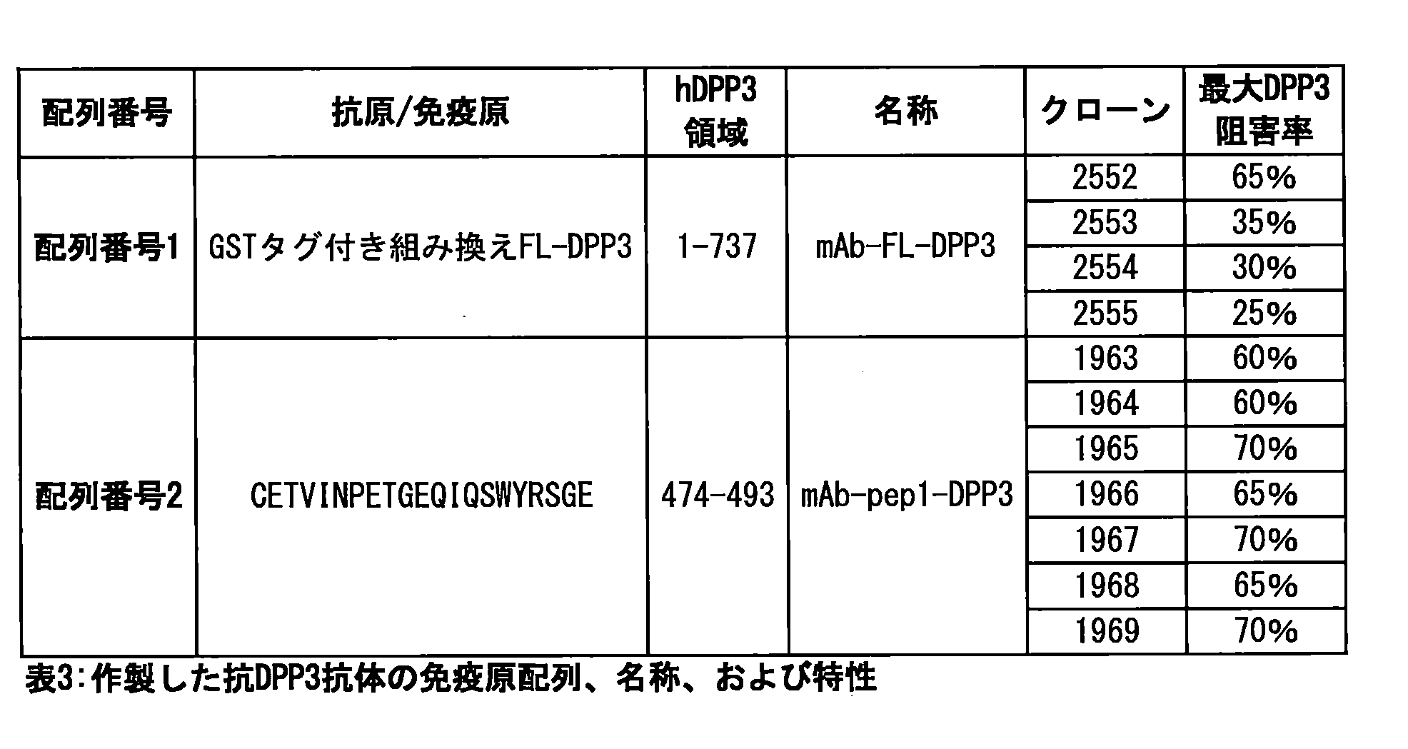
hDPP3免疫測定で高いシグナル/ノイズ(S/N)比を生じた抗体の組み合わせを決定する。
(4.1.方法)
(モノクローナル抗体の作製)
The antibody combination that produced a high signal/noise (S/N) ratio in the hDPP3 immunoassay is determined.
4.1. Method
(Preparation of monoclonal antibodies)
GSTタグ付きヒトDPP3に対する抗体を標準的な抗体作製方法(Marx et al., 1997)で作製し、プロテインAで精製した。SDSゲル電気泳動分析に基づく抗体純度は90%以上であった。異なるクローンのDPP3結合能力を分析した。結果が陽性のクローンを固相またはトレーサー抗体として用いた。
(固相)
Antibodies against GST-tagged human DPP3 were generated by standard antibody production methods (Marx et al., 1997) and purified with protein A. Antibody purity was greater than 90% based on SDS gel electrophoresis analysis. Different clones were analyzed for their DPP3 binding ability. Clones with positive results were used as solid phase or tracer antibodies.
(Solid Phase)
96ウェルのポリスチレンマイクロプレート(Greiner Bio-One International AG(オーストリア))に1つの抗DPP3抗体クローン(捕捉用抗体)を(室温で1時間)塗布した(1.5μg/0.25mLの抗体、100mmol/LのNaCl、50mmol/Lのトリス/HCl;pH7.8)。5%ウシ血清アルブミンでブロックした後、マイクロプレートを真空乾燥した。
(標識の手順(トレーサー))
様々な抗DPP3抗体100μg(100μl)(検出用抗体、1mg/mlをPBSに溶解、pH7.4)を10μlのアクリジニウムNHSエステルと混合し(1mg/mlをアセトニトリルに溶解)(InVent GmbH(ドイツ);EP 0 353 971)、室温で30分間保温した。標識した抗DPP3抗体をShodex Protein 5 μm KW-803(昭和電工(日本))でゲル濾過HPLCによって精製した。精製した標識付き抗体を検定緩衝液(50mmol/lのリン酸カリウム、100mmol/lのNaCl、10mmol/lのNa2-EDTA、5g/lのウシ血清アルブミン、1g/lのマウスIgG、1g/lのウシIgG、50μmol/lのアマスタチン、100μmol/lのロイペプチン;pH7.4)に希釈した。最終濃度は、200μlあたりの標識付き化合物の相対発光量(RLU)で約7×106(標識付き抗体約20ng)であった。アクリジニウムエステルの化学発光をCentro LB 960蛍光光度計(Berthold Technologies GmbH & Co. KG)で測定した。
(検量用試料)
A 96-well polystyrene microplate (Greiner Bio-One International AG, Austria) was coated (1 h at room temperature) with one anti-DPP3 antibody clone (capture antibody) (1.5 μg/0.25 mL antibody, 100 mmol/L NaCl, 50 mmol/L Tris/HCl; pH 7.8). After blocking with 5% bovine serum albumin, the microplate was dried under vacuum.
(Labeling Procedure (Tracer))
100 μg (100 μl) of various anti-DPP3 antibodies (detection antibody, 1 mg/ml dissolved in PBS, pH 7.4) were mixed with 10 μl of acridinium NHS ester (1 mg/ml dissolved in acetonitrile) (InVent GmbH, Germany; EP 0 353 971) and incubated at room temperature for 30 min. The labeled anti-DPP3 antibodies were purified by gel filtration HPLC on Shodex Protein 5 μm KW-803 (Showa Denko, Japan). The purified labeled antibodies were diluted in assay buffer (50 mmol/l potassium phosphate, 100 mmol/l NaCl, 10 mmol/l Na 2 -EDTA, 5 g/l bovine serum albumin, 1 g/l mouse IgG, 1 g/l bovine IgG, 50 μmol/l amastatin, 100 μmol/l leupeptin; pH 7.4). The final concentration was approximately 7×10 6 relative light units (RLU) of labeled compound (approximately 20 ng labeled antibody) per 200 μl. The chemiluminescence of the acridinium ester was measured in a Centro LB 960 fluorometer (Berthold Technologies GmbH & Co. KG).
(Calibration sample)
組み換えヒトGST-DPP3(USBiological(米国))の原液(PBS溶液、pH7.4)を(50mmol/Lのリン酸カリウム、100mmol/LのNaCl、10mmol/LのNa-EDTA、5g/Lのウシ血清アルブミン、1g/LのマウスIgG、1g/LのウシIgG、50μmol/Lのアマスタチン、100μmol/Lのロイペプチン;pH7.4)を用いて線形希釈した。原液を-80℃で保存した。検量用試料を使用前に調製した。
(hDPP3免疫測定)
塗布済み96ウェルマイクロプレートに試料(または検量用試料)10μlをピペットで移し、標識を付け希釈した検出用抗体(200μl)を添加した後、プレートを2~8℃で18~24時間保温した。結合しなかったトレーサーを、350μlの洗浄液(20mMのPBS(pH7.4)、0.1%のTriton X-100)で4回洗浄することによって除去した。十分に結合した化学発光をCentro LB 960発光光度計(Berthold Technologies GmbH & Co. KG)で測定した。
(4.2.結果)
A stock solution of recombinant human GST-DPP3 (USBiological, USA) in PBS, pH 7.4 was linearly diluted with (50 mmol/L potassium phosphate, 100 mmol/L NaCl, 10 mmol/L Na-EDTA, 5 g/L bovine serum albumin, 1 g/L mouse IgG, 1 g/L bovine IgG, 50 μmol/L amastatin, 100 μmol/L leupeptin; pH 7.4). The stock solution was stored at -80°C. Calibration samples were prepared before use.
(hDPP3 Immunoassay)
10 μl of sample (or calibrator) was pipetted into the coated 96-well microplate, labeled and diluted detection antibody (200 μl) was added, and the plate was incubated at 2-8° C. for 18-24 h. Unbound tracer was removed by washing four times with 350 μl washing solution (20 mM PBS pH 7.4, 0.1% Triton X-100). Fully bound chemiluminescence was measured in a Centro LB 960 luminometer (Berthold Technologies GmbH & Co. KG).
(4.2. Results)
すべての抗体を以下のバリエーション(表4および5)で塗布済みマイクロプレートおよび標識付き抗体として組み合わせて、サンドイッチ免疫測定に使用した。hDPP3免疫測定の項に記載されたように保温を行った。組み換えヒトGST-DPP3(100、10、1ng/ml)および血漿試料中の天然hDPP3の結果を特異的シグナル/背景シグナルの比で示す。
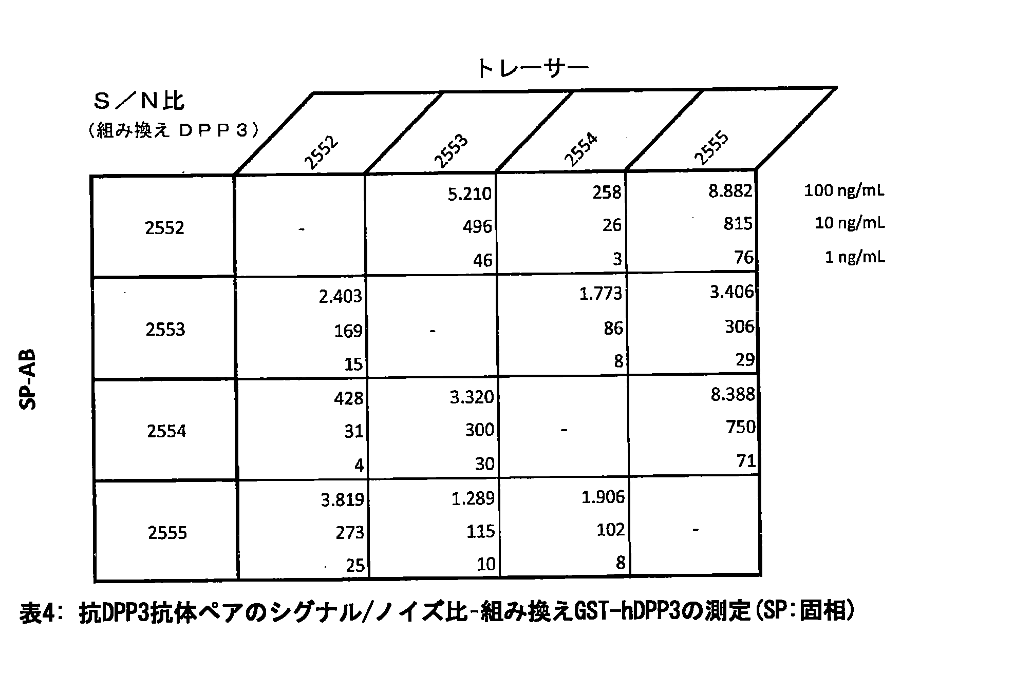

組み換えGST-hDPP3について、すべての組み合わせは良好なシグナル/ノイズ比を示した。さらに、天然試料について、2552と2554を除くすべての組み合わせは良好なシグナル/ノイズ比を示した。したがって、残るすべての組み合わせをさらなる調査に使用できる。RLUシグナルの絶対値が最も高いことに関して、2555を固相抗体、2553を標識付き抗体として用いた。
(5.実施例5)
For recombinant GST-hDPP3, all combinations showed good signal/noise ratios. Moreover, for the native samples, all combinations except 2552 and 2554 showed good signal/noise ratios. Therefore, all remaining combinations can be used for further investigation. With regard to the highest absolute value of RLU signal, 2555 was used as the solid-phase antibody and 2553 was used as the labeled antibody.
5. Example 5
様々な疾患の患者(急性心不全(AHF)、心筋梗塞(MI)、敗血症、がん、急性腎障害(AKI)、および下気道感染症(LRTI)の患者)の血漿DPP3濃度をhDPP3免疫測定で定量し、健常対照の血漿DPP3濃度と比較した。
(5.1.研究コホート)
Plasma DPP3 concentrations in patients with various diseases (acute heart failure (AHF), myocardial infarction (MI), sepsis, cancer, acute kidney injury (AKI), and lower respiratory tract infection (LRTI)) were quantified by hDPP3 immunoassay and compared with plasma DPP3 concentrations in healthy controls.
5.1 Study Cohort
最初の症状で直接救急科または腫瘍科に入院した214名の患者から血漿試料を得た。最終診断によって患者を急性心不全(AHF)、心筋梗塞(MI)、敗血症、がん、急性腎障害(AKI)、下気道感染症(LRTI)の患者の6つの亜群に分けることができた。対照群は、93名の健常対照の血漿試料群であった。
(5.2.hDPP3免疫測定)
Plasma samples were obtained from 214 patients who were directly admitted to the emergency department or oncology department with their first symptoms. The final diagnosis allowed the patients to be divided into six subgroups: patients with acute heart failure (AHF), myocardial infarction (MI), sepsis, cancer, acute kidney injury (AKI), and lower respiratory tract infection (LRTI). The control group consisted of plasma samples from 93 healthy controls.
5.2. hDPP3 Immunoassay
mAbDPP3_2555を固相抗体として、mAbDPP3_2553を標識付きトレーサー抗体として用いた。抗体の固定化、標識、保温は実施例2に記載のとおりに行った。
(5.3.結果)
The mAb DPP3_2555 was used as a solid-phase antibody, and the mAb DPP3_2553 was used as a labeled tracer antibody. Immobilization, labeling, and incubation of the antibodies were performed as described in Example 2.
5.3 Results
対応する診断の詳細と共に、得られたデータを統計的に分析した(図2A)。すべての患者は、健常対照(正常)と比較して有意に高い血漿DPP3濃度を示した。表6は、対照群の75パーセンタイルを超えるDPP3値を有する患者の割合とそれぞれの診断を示す。血漿DPP3レベルの分析結果は、患者の疾患状態を示している。この発見は診断分野でも使用でき、また、(たとえばDPP3の阻害による)治療処置の基礎を築いた。
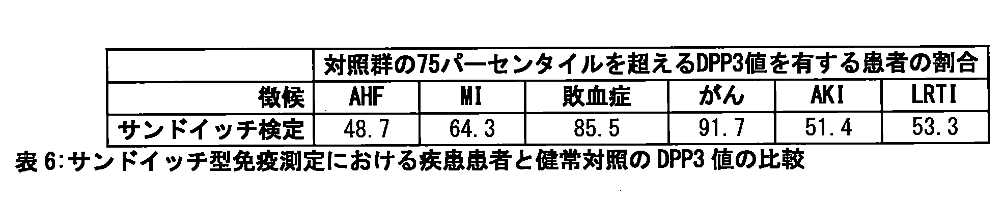
同じ研究コホートを死亡によって分析した。救急科に入院した後に死亡した患者は、病院で生存した救急患者よりも有意に高い血漿DPP3レベルを有していた。したがって、DPP3濃度の上昇は死亡に関して悪い予後を示す(図2B)。
(6.実施例6)
The same study cohort was analyzed by mortality. Patients who died after admission to the emergency department had significantly higher plasma DPP3 levels than emergency patients who survived in the hospital. Thus, elevated DPP3 concentrations indicate a poor prognosis for mortality (Figure 2B).
6. Example 6
ヒト血漿中のDPP3の量は、DPP3濃度だけでなく、活性検定でも定量できる。1つの標準的な手順は、蛍光発生基質としてArg-Arg-βNAを使用する可溶活性検定である。 The amount of DPP3 in human plasma can be quantified not only by DPP3 concentration but also by activity assays. One standard procedure is a soluble activity assay using Arg-Arg-βNA as a fluorogenic substrate.
天然ヒトDPPIIIの活性を、Arg-Arg-β-ナフチルアミド(Bachem Holdig AG(スイス))を加水分解し蛍光性のβ-ナフチルアミドを生成することによって定量した。200μlの緩衝液(50mMのトリス/HCl(pH8.8)、0.04%のNaN3、50μMのアマスタチン、100μMのロイペプチン)と10μlの試料(ヒト血漿)を96ウェルの黒色マイクロプレート(Greiner Bio-One International GmbH(オーストリア))にピペットで移し、37℃で10分間予熱した。基質(20μl、2mM)を添加し、蛍光の増加を37℃で1時間、340nmの励起波長と410nmの発光波長でTwinkle LB 970マイクロプレート蛍光光度計(Berthold Technologies GmbH & Co. KG)で監視した。様々な試料の蛍光増加の傾きを、検量用試料である遊離βNAに関してnmol(βNA)/min/ml(試料)[nmol(βNA)min-1ml-1]で計算する。 Native human DPPIII activity was quantified by hydrolysis of Arg-Arg-β-naphthylamide (Bachem Holdig AG, Switzerland) to produce fluorescent β-naphthylamide. 200 μl of buffer (50 mM Tris/HCl, pH 8.8, 0.04% NaN 3 , 50 μM amastatin, 100 μM leupeptin) and 10 μl of sample (human plasma) were pipetted into a 96-well black microplate (Greiner Bio-One International GmbH, Austria) and pre-warmed for 10 min at 37°C. Substrate (20 μl, 2 mM) was added and the increase in fluorescence was monitored for 1 h at 37°C in a Twinkle LB 970 microplate fluorometer (Berthold Technologies GmbH & Co. KG) at an excitation wavelength of 340 nm and an emission wavelength of 410 nm. The slopes of the fluorescence increase of the various samples are calculated relative to the calibrator free βNA in nmol(βNA)/min/ml sample [nmol(βNA)min −1 ml −1 ].
記載された標準の可溶DPP3活性検定では、DPP3活性を測定しているのか血漿中の他のアミノペプチダーゼの活性を測定しているのか判断できない。DPP3に特異的なシグナルを生成するために、酵素捕捉検定を行った。この検定では、最初の工程でモノクローナル抗体への結合によってDPP3が表面に固定化され、洗浄工程の後、特異的なDPP3活性のみを測定できる。 The standard soluble DPP3 activity assay described does not allow us to determine whether we are measuring DPP3 activity or the activity of other aminopeptidases in the plasma. To generate a signal specific for DPP3, an enzyme capture assay was performed. In this assay, in a first step DPP3 is immobilized on a surface by binding to a monoclonal antibody, and after a washing step only specific DPP3 activity can be measured.
96ウェルの黒色マイクロプレート(Greiner Bio-One International GmbH(オーストリア))を用いて、実施例3に記載されたとおりに固相を調製した。10μlの試料(血漿または標準)と200μlの緩衝液(50mmol/lのリン酸カリウム、100mmol/lのNaCl、5g/lのウシ血清アルブミン、1g/lのマウスIgG、1g/lのウシIgG、50μmol/lのアマスタチン、100μmol/lのロイペプチン;pH7.4)をピペットで前記の塗布済みマイクロプレートに移し、保温した(2~8℃、600rpmで18~24時間)。結合しなかった分析物を、洗浄液で洗浄(3×350μl)することによって除去した。基質を添加(200μl中、100μMを[50mMのトリス/HCl(25℃でのpH8.8)、0.04%NaN3]に溶解)した後、蛍光の増加を37℃で1時間、340nmの励起波長と410nmの発光波長でTwinkle LB 970マイクロプレート蛍光光度計(Berthold Technologies GmbH & Co. KG)で監視した。様々な試料の蛍光増加の傾きを、検量用試料である遊離βNAに関してnmol(βNA)/min/ml(試料)[nmol(βNA)min-1ml-1]で計算する。 Solid phases were prepared as described in Example 3 in 96-well black microplates (Greiner Bio-One International GmbH, Austria). 10 μl of sample (plasma or standard) and 200 μl of buffer (50 mmol/l potassium phosphate, 100 mmol/l NaCl, 5 g/l bovine serum albumin, 1 g/l mouse IgG, 1 g/l bovine IgG, 50 μmol/l amastatin, 100 μmol/l leupeptin; pH 7.4) were pipetted into the coated microplates and incubated (2-8° C., 600 rpm, 18-24 h). Unbound analyte was removed by washing with washing solution (3×350 μl). After addition of the substrate (100 μM in 200 μl [50 mM Tris/HCl (pH 8.8 at 25° C.), 0.04% NaN 3 ]), the increase in fluorescence was monitored for 1 h at 37° C. in a Twinkle LB 970 microplate fluorometer (Berthold Technologies GmbH & Co. KG) at an excitation wavelength of 340 nm and an emission wavelength of 410 nm. The slopes of the fluorescence increase of the various samples are calculated in nmol(βNA)/min/ml (sample) [nmol(βNA)min −1 ml −1 ] with respect to the calibrator free βNA.
それぞれの活性検定の種類で、遊離βNAを検定の検量用試料として用いた。したがって、βNA濃度を増加し(200μl中、50mMのトリス/HCl(25℃でのpH8.8)、0.04%NaN3に0、4、8、16、32、64、125、250μMのβNAをそれぞれ溶解)、37℃で、340nmの励起波長と410nmの発光波長でTwinkle LB 970マイクロプレート蛍光光度計(Berthold Technologies GmbH & Co. KG)で測定した。すべての試料の測定値をこのβNA標準で較正した。
(7.実施例7)
For each activity assay type, free βNA was used as the calibrator for the assay. Therefore, increasing concentrations of βNA (0, 4, 8, 16, 32, 64, 125, 250 μM βNA dissolved in 50 mM Tris/HCl (pH 8.8 at 25° C.), 0.04% NaN 3 in 200 μl) were measured at 37° C. in a Twinkle LB 970 microplate fluorometer (Berthold Technologies GmbH & Co. KG) with an excitation wavelength of 340 nm and an emission wavelength of 410 nm. All sample measurements were calibrated with this βNA standard.
7. Example 7
様々な疾患の患者(急性心不全(AHF)、敗血症、急性腎障害(AKI)、および下気道感染症(LRTI)の患者)の血漿のDPP3活性を特異的なDPP3捕捉活性検定で定量し、健常対照の血漿のDPP3活性と比較した。
(7.1.方法)
DPP3 activity in plasma from patients with various diseases (patients with acute heart failure (AHF), sepsis, acute kidney injury (AKI), and lower respiratory tract infection (LRTI)) was quantified using a specific DPP3 scavenging activity assay and compared with DPP3 activity in plasma from healthy controls.
7.1. Methods
実施例3で分析したコホートの一部をDPP3に特異的な酵素捕捉活性検定に供した。この検定では血漿試料(10μl)のDPP3を最初に親和精製工程によって濃縮し、次に蛍光発生基質Arg-Arg-βNAを添加してその活性を測定した(詳細な説明については実施例4を参照のこと)。様々な試料の蛍光増加の傾き(nmol(βNA)/min/ml(試料)[nmol(βNA)min-1ml-1])の計算値は10μlの試料サイズに関する。
(7.2.結果)
A portion of the cohort analyzed in Example 3 was subjected to a DPP3-specific enzyme capture activity assay in which DPP3 from plasma samples (10 μl) was first enriched by an affinity purification step and then its activity was measured by adding the fluorogenic substrate Arg-Arg-βNA (see Example 4 for a detailed description). The calculated slopes of the fluorescence increase for the various samples (nmol(βNA)/min/ml (sample) [nmol(βNA)min −1 ml −1 ]) are for a sample size of 10 μl.
(7.2. Results)
患者の試料を健常対照と比較したところ、すべての患者(AHF、敗血症、AKI、およびLTRI)のDPP3活性値は有意に高かった(図3)。 When patient samples were compared with healthy controls, DPP3 activity was significantly higher in all patients (AHF, sepsis, AKI, and LTRI) (Figure 3).
表7は、対照群の75パーセンタイルを超えるDPP3値を有する患者の割合とそれぞれの診断を示す。活性はより十分な健常対照と疾患患者の境界を示した。

DPP3活性検定と濃度検定をさらに十分に比較するため、ROC(受信者動作特性)分析を健常対照とAHF患者との境界(図4A)または敗血症患者との境界(図4B)について行った。曲線の下の面積(AUC)および信頼区間(CI)の値を表8に示す。データ分析から、サンドイッチ型免疫測定に比べて活性検定の特異度が高いことがわかった。

本実験では、健常なマウスでmAbDPP3の用量を増やした場合の全般的な安全性を監視した。
(8.1.方法)
In this study, the overall safety of increasing doses of mAb DPP3 was monitored in healthy mice.
8.1. Methods
到着時に4~5週齢、体重約15~18gの雌のBALB/cヌードマウス(CAnN.Cg-Foxn1nu/Crl)(Charles River GmbH(ドイツ、ズルツフェルト))を、最適な衛生条件のもと、空調管理し1時間に10~15回換気を行い、温度の目標範囲22±3℃、相対湿度の目標範囲30~70%、人工の蛍光照明12時間/暗期12時間の持続的に監視された環境で飼育した。個別換気ケージ(IVC)あたり最大4匹を飼育し、M-Zucht(ssniff Spezialdiaten GmbH)からなる餌とオートクレーブした公共水道水を与えた。 Female BALB/c nude mice (CAnN.Cg-Foxn1nu/Crl) (Charles River GmbH, Sulzfeld, Germany), 4-5 weeks old and weighing approximately 15-18 g on arrival, were housed under optimal hygienic conditions in an air-conditioned environment with 10-15 air changes per hour, a target temperature range of 22 ± 3°C, a target relative humidity range of 30-70%, and a continuously monitored 12-h artificial fluorescent light/12-h dark period. They were housed up to 4 per individually ventilated cage (IVC) and fed a diet consisting of M-Zucht (ssniff Spezialdiaten GmbH) and autoclaved municipal tap water.
4日間の順化期間の後、mAbDPP3の投与を開始した。3つの異なるmAbDPP3濃度(0.65mg/kg、1.9mg/kg、および5.75mg/kg)をPBSに溶解し、各群4匹のマウスに注射した。1日目、3日目、5日目、7日目にmAbDPP3を腹腔内(i.p.)投与し、マウスを14日間監視した。
(8.2.結果)
After a 4-day acclimation period, administration of mAbDPP3 was initiated. Three different concentrations of mAbDPP3 (0.65 mg/kg, 1.9 mg/kg, and 5.75 mg/kg) were dissolved in PBS and injected into four mice per group. mAbDPP3 was administered intraperitoneally (i.p.) on days 1, 3, 5, and 7, and the mice were monitored for 14 days.
8.2 Results
マウスはいずれも、14日間の処置の間、副作用なく生存し、最も多い用量の場合も同様であった。mAbDPP3は他の動物実験(常に1.9mg/kgの濃度で実施)に用いても安全である。
(9.実施例9)
All mice survived the 14 days of treatment without side effects, even at the highest dose.mAbDPP3 has been safe for use in other animal studies (always performed at a concentration of 1.9 mg/kg).
9. Example 9
本実験では、健常なラットにおけるmAbDPP3処置の全般的な安全性を、平均血圧を監視することによって研究した。
(9.1.方法)
In this study, the overall safety of mAbDPP3 treatment in healthy rats was studied by monitoring mean blood pressure.
9.1. Method
2~3ヵ月齢の雄のウィスターラット(Charles River Laboratories(ドイツ))を本研究に使用した。血圧(BP)の測定および記録のため、カテーテル(Introcan-W; 22 G/1''; B.Braun)を右総頚動脈に挿入した。投与物およびサンプリングカテーテルは左頚静脈に入れた。 Male Wistar rats (Charles River Laboratories, Germany) aged 2 to 3 months were used in this study. A catheter (Introcan-W; 22 G/1''; B. Braun) was inserted into the right common carotid artery for measuring and recording blood pressure (BP). The dosing and sampling catheters were placed in the left jugular vein.
最初に動物をイソフルランで麻酔し、体重測定(すべてg)し、長時間の麻酔のために1.2g/kg(体重)のウレタン(濃度=0.4g/mL)を腹腔内(i.p.)注射した。次いで首の腹側領域の毛を刈り、エタノールで拭いた。血管を準備し、カテーテルを挿入した。最後に、両方のカテーテルをヘパリン処置された等張食塩水で洗い流す。次いで血圧測定器(Medex Logical(Medex Medical Ltd.))を患者監視システム(Datex-Ohmeda(GE))に接続した。ラップトップコンピュータをBPモニターに接続し、S/5 Collect SoftwareでBPデータを個別に記録した。 Animals were first anesthetized with isoflurane, weighed (all in g), and injected intraperitoneally (i.p.) with 1.2 g/kg (body weight) of urethane (concentration = 0.4 g/mL) for long-term anesthesia. The ventral area of the neck was then clipped and wiped with ethanol. The vessels were prepared and catheterized. Finally, both catheters were flushed with heparinized isotonic saline. A blood pressure measuring device (Medex Logical (Medex Medical Ltd.)) was then connected to a patient monitoring system (Datex-Ohmeda (GE)). A laptop computer was connected to the BP monitor, and BP data were recorded separately with S/5 Collect Software.
ラットを、PBS、mAbDPP3のPBS溶液1.9mg/kgまたは5.75mg/kgでそれぞれ処置した(各群n=3)。化合物は静脈カテーテルから投与した。血圧を化合物の投与の1時間前と化合物の投与後6時間にわたり監視した。
(9.2.結果)
Rats were treated with PBS, 1.9 mg/kg or 5.75 mg/kg of mAbDPP3 in PBS (n=3 per group). Compounds were administered via an intravenous catheter. Blood pressure was monitored 1 hour before and 6 hours after compound administration.
9.2 Results
ラットはmAbDPP3処置によく反応した。高用量のmAbDPP3で処置したラットは平均血圧のわずかな上昇を示した(図5)。一般にmAbDPP3も、高用量の場合でもラットモデルに使用しても安全である。
(10.実施例10)
Rats responded well to mAbDPP3 treatment. Rats treated with high doses of mAbDPP3 showed a slight increase in mean blood pressure (Figure 5). In general, mAbDPP3 is also safe for use in the rat model, even at high doses.
10. Example 10
本研究では、mAbDPP3による処置が敗血症の死亡率にどのような影響を及ぼす可能性があるかをCLPマウスモデルで分析した。
(10.1.方法)
In this study, we analyzed how treatment with mAb DPP3 might affect sepsis mortality in a mouse model of CLP.
10.1. METHODS
12~15週齢の雄のC57Bl/6マウス(Charles River Laboratories(ドイツ))を本研究に用いた。軽度のイソフルラン麻酔下で腹膜炎を外科的に誘発した。腹腔の左上腹部(盲腸の正常な位置)を切開した。盲腸を露出させ、小腸の導入部から遠位側に縫合糸で盲腸の周囲をきつく結紮した。盲腸に24ゲージの針で1つの穿刺創を作り、その創から少量の盲腸内容物を出した。盲腸を腹腔内に戻し、開腹手術部位を閉鎖した。最後に、動物を餌と水に自由に到達できるようにケージに戻した。補液として生理食塩水500μlを皮下(s.c.)投与した。 Male C57Bl/6 mice (Charles River Laboratories, Germany) aged 12-15 weeks were used in this study. Peritonitis was surgically induced under light isoflurane anesthesia. An incision was made in the left upper quadrant of the abdominal cavity (normal location of the cecum). The cecum was exposed and tightly ligated around it with sutures distal to the introduction of the small intestine. A single puncture wound was made in the cecum with a 24-gauge needle, through which a small amount of cecal contents was expelled. The cecum was replaced in the abdominal cavity and the laparotomy site was closed. Finally, the animals were returned to their cages with free access to food and water. 500 μl of saline was administered subcutaneously (s.c.) as fluid replacement.
mAbDPP3(1.9mg/kgをPBSに溶解)を溶媒(PBS)と比較して試験した。化合物または溶媒をCLP(予防処置)の5分前と敗血症が完全に発症した後CLP(治療処置)の2時間後にそれぞれ静脈内(i.v.)注射した。各群には10匹のマウスが含まれ、7日間経過を観察した。
(10.2.結果)
mAbDPP3 (1.9 mg/kg dissolved in PBS) was tested against vehicle (PBS). Compounds or vehicle were injected intravenously (i.v.) 5 min before CLP (prophylactic treatment) and 2 h after CLP (therapeutic treatment) after full sepsis development. Each group contained 10 mice and was followed for 7 days.
10.2. Results
図6から、mAbDPP3抗体はPBS投与に比べて死亡率を大きく低下させたことがわかる。mAbDPP3で処置した場合、4日後にはマウスの75%が生存していた。これに対し、溶媒で処置した場合、4日後にはほぼすべてのマウスが死亡した。
(11.実施例11)
As can be seen from Figure 6, the mAbDPP3 antibody significantly reduced the mortality rate compared to PBS administration. After 4 days, 75% of the mice were alive when treated with mAbDPP3. In contrast, after 4 days, almost all mice were dead when treated with the vehicle.
(11. Example 11)
敗血症性ショックのモデルを用いてラットに心不全を誘発し、心機能に対するmAbDPP3の影響を特性評価した。
(11.1.方法)
(研究設計)
A model of septic shock was used to induce heart failure in rats and characterize the effects of mAb DPP3 on cardiac function.
11.1. METHODS
(Research Design)
研究の流れを以下の図7Aに示す。CLPまたは偽手術の後、動物を自由に水と餌に自由に到達できるようにして20時間休ませた。その後、ラットを麻酔し、気管切開をして動脈・静脈ラインを配置した。CLP手術の24時間後、mAbDPP3または溶媒(生理食塩水)を2mg/kg投与した。循環動態を観血的に継続監視(t=0~3h)した。手術の直後とmAbDPP3または生理食塩水注射の15分後、1時間後、2時間後、および3時間後に心臓の心エコー検査を行った。
(敗血症のCLPモデル)
The study procedure is shown in Figure 7A below. After CLP or sham surgery, animals were allowed to rest for 20 hours with free access to water and food. Rats were then anesthetized, tracheotomized, and arterial and venous lines were placed. 24 hours after CLP surgery, mAbDPP3 or vehicle (saline) was administered at 2 mg/kg. Hemodynamics was continuously monitored invasively (t=0-3 h). Echocardiography of the heart was performed immediately after surgery and 15 minutes, 1 hour, 2 hours, and 3 hours after injection of mAbDPP3 or saline.
CLP Model of Sepsis
Centre d'elevage Janvier(フランス)から入手した雄のウィスターラット(2~3ヶ月齢、300~400g、群の大きさについては表1を参照のこと)を無作為に3つの群のうちの1つに割り当てた。すべての動物を塩酸ケタミン(90mg/kg)およびキシラジン(9mg/kg)で腹腔内(i.p.)麻酔した。多微生物性敗血症を誘発するため、盲腸結紮・穿刺法(CLP)をRittirschのプロトコルを微修正して行った。腹側正中切開(1.5cm)を行い、盲腸を露出させた。次いで盲腸を回盲弁の直下で結紮し、18ゲージの針で1回穿刺した。次いで腹腔を二層で閉じ、補液蘇生(3ml/100g(体重)の生理食塩水を皮下注射)し、動物をケージに戻す。偽処置群の動物には盲腸穿刺をせずに手術を行った。
(観血的血圧)
Male Wistar rats (2-3 months old, 300-400 g, see Table 1 for group sizes) obtained from Centre d'elevage Janvier (France) were randomly assigned to one of three groups. All animals were anesthetized intraperitoneally (i.p.) with ketamine hydrochloride (90 mg/kg) and xylazine (9 mg/kg). To induce polymicrobial sepsis, cecal ligation and puncture (CLP) was performed according to a slight modification of the Rittirsch protocol. A ventral midline incision (1.5 cm) was made to expose the cecum. It was then ligated just below the ileocecal valve and punctured once with an 18-gauge needle. The abdominal cavity was then closed in two layers, fluid resuscitation was performed (3 ml/100 g body weight saline subcutaneously), and the animals were returned to their cages. Animals in the sham group were operated on without cecal puncture.
(Invasive Blood Pressure)
AcqKnowledgeシステム(BIOPAC Systems, Inc.(米国))を用いて循環動態的変数を求めた。これにより完全に自動化された血圧分析システムが得られる。BIOPACシステムに圧力センサーを介してカテーテルを接続する。 Hemodynamic variables were measured using the AcqKnowledge system (BIOPAC Systems, Inc., USA), which provides a fully automated blood pressure analysis system. The catheter was connected to the BIOPAC system via a pressure sensor.
この手順のため、ラットを麻酔した(ケタミンおよびキシラジン)。動物を温熱パッドに移して所望の体温(37~37.5℃まで)にした。温度フィードバックプローブを直腸に挿入した。ラットを仰臥位で手術台に置いた。気管を開き、外部人工呼吸器のために頚動脈および迷走神経を傷つけないようにカテーテル(16G)を挿入した。動脈カテーテルを右頚動脈に挿入した。頚動脈を迷走神経から分離してから結紮する。 For this procedure, rats were anesthetized (ketamine and xylazine). The animals were transferred to a heating pad to the desired body temperature (~37-37.5°C). A temperature feedback probe was inserted into the rectum. The rat was placed on the operating table in a supine position. The trachea was opened and catheters (16G) were inserted into the carotid artery and vagus nerve to avoid injury for external ventilation. An arterial catheter was inserted into the right carotid artery. The carotid artery was separated from the vagus nerve and then ligated.
左頚静脈から中心静脈カテーテルを挿入し、薬物を投与できるようにした。 A central venous catheter was inserted into the left jugular vein to allow for the administration of medication.
手術の後、動物を安定状態になるように休息させてから循環動態的測定を行った。次いで血圧(BP)基準値を記録した。データ採取の間、動脈ラインからの生理食塩水の輸液を停止した。
(心エコー検査)
After surgery, animals were allowed to rest to reach a stable state before hemodynamic measurements were performed. Baseline blood pressure (BP) values were then recorded. Arterial saline infusion was stopped during data collection.
(Echocardiogram)
動物を塩酸ケタミンで麻酔した。胸部の毛を刈り、ラットを臥位で置いた。 The animals were anesthetized with ketamine hydrochloride. The hair on the chest was clipped and the rats were placed in a supine position.
経胸壁エコー検査(TTE)には高周波(14MHz)リニア型プローブと心臓プローブ(10MHz)を備えた市販のGE Healthcare Vivid 7 Ultra-sound Systemを使用した。その後のオフラインでの分析のため、全ての試験をデジタルで記録し保存した。 Transthoracic echocardiography (TTE) was performed using a commercially available GE Healthcare Vivid 7 Ultra-sound System equipped with a high-frequency (14 MHz) linear probe and a cardiac probe (10 MHz). All examinations were digitally recorded and stored for subsequent offline analysis.
グレースケールの画像を2cmの深度で記録した。大動脈弁輪径および肺動脈径を測定するために傍胸骨長軸像で二次元検査を開始した。また、Mモードを用いて左室(LV)の大きさを測定し、短縮率(FS(%))を評価した。LVFSをLV拡張末期径-LV収縮末期径/LV拡張末期径として計算し、%で表した。したがって、拡張末期の時間はLVの最大径で定義された。その結果、収縮末期は、同じ心臓周期における最小径として定義された。全てのパラメータを手動で測定した。各測定について3回の心臓周期を平均化した。 Grey-scale images were recorded at a depth of 2 cm. Two-dimensional examination was started in the parasternal long-axis view to measure the aortic annulus diameter and pulmonary artery diameter. In addition, M-mode was used to measure the left ventricular (LV) dimensions and evaluate the fractional shortening (FS (%)). LVFS was calculated as LV end-diastolic diameter-LV end-systolic diameter/LV end-diastolic diameter and expressed as a percentage. Thus, the time of end-diastole was defined by the maximum diameter of the LV. As a result, end-systole was defined as the minimum diameter in the same cardiac cycle. All parameters were measured manually. Three cardiac cycles were averaged for each measurement.
同じ傍胸骨長軸像からパルス・ドップラー法で肺動脈流を記録した。肺動脈の流出量の速度時間積分を測定した。 Pulmonary artery flow was recorded using the pulsed Doppler technique from the same parasternal long axis view. The velocity time integral of the pulmonary artery outflow was measured.
心尖部五腔像からパルス・ドップラー法で僧帽弁先端部の位置の僧帽弁血流を記録した。
(実験の時点と動物群)
Mitral valve blood flow was recorded at the mitral valve tip level using the pulsed Doppler technique from the apical five-chamber view.
(Experimental time points and animal groups)
手術後、BP基準値と心エコー図を記録した。次いでmAbDPP3(2mg/kg)または溶媒(生理食塩水)を注射し(i.v.;手術の5分後)、生理食塩水の輸液を開始した。循環動態的な項目(BPおよび心エコー図)をmAbDPP3または溶媒を注射した15分後、1時間後、2時間後、3時間後に記録した。下表に1つの対照群と2つのCLP群の概要を示す(表9)。実験の最後に動物を安楽死させ、血液を採取してEDTA-血漿を作製し、臓器を回収してさらに分析した。
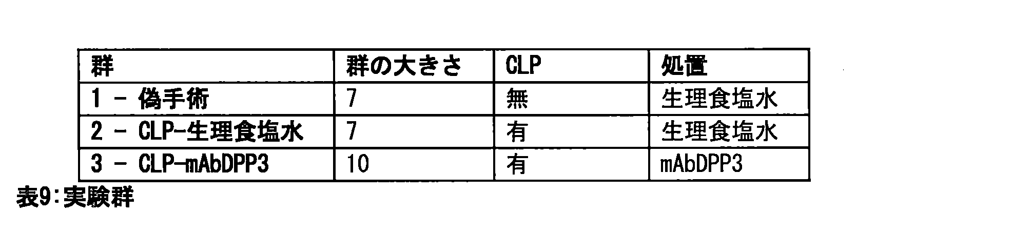
偽手術群の動物に比べて、敗血症ラットは非常に血圧が低く、心臓の短縮率が低下している。mAbDPP3の投与によって短縮率は有意に上昇し(図7B)、平均血圧は上昇し(図7C)、敗血症ラットの健康状態も大いに改善した。
(12.実施例12)
Compared with sham-operated animals, septic rats had very low blood pressure and reduced cardiac shortening fraction. Administration of mAbDPP3 significantly increased the shortening fraction (Fig. 7B), increased mean blood pressure (Fig. 7C), and greatly improved the health status of septic rats.
12. Example 12
ここに記載する研究の目的は、数種のがん細胞株を用いてmAbDPP3の潜在的な抗増殖効果を生体外の細胞培養系で評価することであった。
(12.1.方法)
The aim of the study described here was to evaluate the potential antiproliferative effects of mAb DPP3 in an in vitro cell culture system using several cancer cell lines.
12.1. METHODS
mAbDPP3の原液(1mg/mlをPBSに溶解)を希釈し、最終濃度が0~100μg/mlの範囲になるようにした。PBSを基準化合物として用いた。樹立されているがん細胞株(A549、HCT116、MDA-MB231)に由来するがん細胞を、10%FCSおよびペニシリン/ストレプトアビジンを含むDMEMで培養した。 A stock solution of mAbDPP3 (1 mg/ml in PBS) was diluted to give final concentrations ranging from 0 to 100 μg/ml. PBS was used as a reference compound. Cancer cells derived from established cancer cell lines (A549, HCT116, MDA-MB231) were cultured in DMEM containing 10% FCS and penicillin/streptavidin.
A549細胞はヒト肺胞基底上皮腺がん細胞である。この細胞株は、外植された58歳の男性の腫瘍の肺がん組織を取り出して培養することによって最初に樹立された。本来はこれらの細胞は扁平であり、水や電解質などのいくつかの物質が肺の肺胞を越えて拡散することに関与する。前記A549細胞を生体外で培養すると、単層細胞として成長し、培養フラスコに付着する。さらなる特徴は、これらの細胞がレシチンを合成でき、高濃度の不飽和脂肪酸を含有していることである。A549細胞株は、薬物代謝に関するII型肺上皮細胞モデルの生体外モデルとして、またトランスフェクション宿主として広く使用されている。 A549 cells are human alveolar basal epithelial adenocarcinoma cells. This cell line was first established by culturing explanted lung cancer tissue from a tumor of a 58-year-old male. In nature, these cells are flattened and are involved in the diffusion of several substances, such as water and electrolytes, across the alveoli of the lung. When cultured in vitro, the A549 cells grow as a monolayer of cells and attach to the culture flask. Further characteristics are that these cells can synthesize lecithin and contain high concentrations of unsaturated fatty acids. The A549 cell line is widely used as an in vitro model of type II lung epithelial cell model for drug metabolism and as a transfection host.
HCT116細胞株はヒト大腸がん細胞である。これらの上皮細胞は付着しやすいという培養特性をもち、成人男性に由来する。この細胞株は、適切なトランスフェクション宿主である。この株はrasがん原遺伝子のコドン13に変異を有しており、このコドンの変異に関するPCR検定の陽性対照として使用することができる。 The HCT116 cell line is a human colon cancer cell line. These epithelial cells have adherent culture properties and are derived from an adult male. This cell line is a suitable transfection host. This line contains a mutation at codon 13 of the ras proto-oncogene and can be used as a positive control for PCR assays for mutations at this codon.
MDA-MB231細胞株は、上皮形態をもつヒト乳腺がん細胞である。これらの細胞は、コーカス人の乳がん患者の胸水から単離された。 The MDA-MB231 cell line is a human breast adenocarcinoma cell line with epithelial morphology. These cells were isolated from the pleural effusion of a Caucasian breast cancer patient.
各細胞株について、96ウェルの懸濁細胞培養プレートを作製した。100μLの軟寒天の底層(最終濃度は完全培地中0.6%)を注入し、固化させた。次いで対応する細胞および細胞数を含む軟寒天の最上層(最終濃度0.4%)50μLを上に加えて固化させ、この96ウェルプレートを37℃、10%CO2で一晩保温した。 For each cell line, a 96-well suspension cell culture plate was prepared. A 100 μL bottom layer of soft agar (final concentration 0.6% in complete medium) was poured and allowed to solidify. Then 50 μL of a top layer of soft agar (final concentration 0.4%) containing the corresponding cells and cell number was added on top and allowed to solidify, and the 96-well plate was incubated at 37°C, 10% CO2 overnight.
翌日、化合物をプレートの内側ウェルに添加した。続いて、試験物を細胞培養インキュベータで保温した。最後にAlamar Blueで試験物を発色させ、37℃で3~5時間保温して蛍光強度を測定した(励起:560nm、発光:590nm)。低対照として、細胞を10-5Mスタウロスポリン(6倍値)で処理した。高対照として、細胞を0.1%DMSO(溶媒対照、6倍値)で処理した。 The next day, compounds were added to the inner wells of the plate. The test articles were then incubated in a cell culture incubator. Finally, the test articles were developed with Alamar Blue and incubated at 37°C for 3-5 hours, after which the fluorescence intensity was measured (excitation: 560 nm, emission: 590 nm). As a low control, cells were treated with 10 -5 M staurosporine (6-fold value). As a high control, cells were treated with 0.1% DMSO (solvent control, 6-fold value).
生データをそれぞれ100%と0%に設定された高対照(溶媒0.1%DMSO)および低対照(10-5Mスタウロスポリン)に対する軟寒天の成長(%)に変換した。GraphPad Prism 5ソフトウェアを用いて、傾きが変化するS字型応答に適合するモデルで(軟寒天成長0%を制約範囲の下限とし、また下限を設定せずに、軟寒天成長100%を上限としてIC50を計算した。
(12.2.結果)
Raw data were converted to % soft agar growth relative to high (solvent 0.1% DMSO) and low ( 10-5 M staurosporine) controls, which were set to 100% and 0%, respectively. IC50s were calculated using GraphPad Prism 5 software, fitting a model to a sigmoidal response with variable slope (with 0% soft agar growth as the lower limit of the constrained range, and with no lower limit, with 100% soft agar growth as the upper limit.
12.2. Results
細胞培養系で、使用した様々なmAbDPP3投与量に応じた3つのがん細胞株(A549、HCT116、MDA-MB231)の成長を評価した(図8)。制約範囲の上限(軟寒天成長100%)である溶媒対照のシグナルおよび下限(軟寒天成長0%)であるスタウロスポリン対照のシグナルに基づく標準パラメータを用いてIC50値を求めた。それぞれのIC50値を表10に要約する。
mAbDPP3処置は、試験した3つの細胞株に対して抗増殖効果があった。

mAbDPP3 treatment had an antiproliferative effect on the three cell lines tested.
ここに記載する研究の目的は、mAbDPP3の腫瘍形成を予防する能力を乳がんおよび大腸がんの異種移植片モデルで評価することであった(腫瘍成長阻害研究)。
(13.1.方法)
The aim of the study described here was to evaluate the ability of mAb DPP3 to prevent tumor formation in xenograft models of breast and colon cancer (tumor growth inhibition studies).
13.1. METHODS
単層MDA-MB-231細胞(乳がん)およびHCT-116細胞(大腸がん)をそれぞれ、DMEM+10%FCS中で増殖させた。細胞を空気90%、二酸化炭素10%の加湿雰囲気下、37℃で培養した。培地を規則的に3日毎に交換した。3~4日毎に、密集した培養物をトリプシン/EDTAで1:3~1:3に分割し、密度が約3~4×106細胞/15cm2+25mL培地になるように播種した。 Monolayers of MDA-MB-231 (breast cancer) and HCT-116 (colon cancer) cells, respectively, were grown in DMEM + 10% FCS. Cells were cultured at 37°C in a humidified atmosphere of 90% air and 10% carbon dioxide. Medium was regularly changed every 3 days. Every 3-4 days, confluent cultures were split 1:3 to 1:3 with trypsin/EDTA and seeded to a density of approximately 3-4 x 10⁶ cells/15 cm2 + 25 mL medium.
到着時に4~5週齢、体重約15~18gの雌のBALB/cヌードマウス(CAnN.Cg-Foxn1nu/Crl)(Charles River GmbH(ドイツ、ズルツフェルト))を、最適な衛生条件のもと、空調管理し1時間に10~15回換気を行い、温度の目標範囲22±3℃、相対湿度の目標範囲30~70%、人工の蛍光照明12時間/暗期12時間の持続的に監視された環境で飼育した。個別換気ケージ(IVC)あたり最大4匹を飼育し、M-Zucht(ssniff Spezialdiaten GmbH)からなる餌とオートクレーブした公共水道水を与えた。 Female BALB/c nude mice (CAnN.Cg-Foxn1 nu /Crl) (Charles River GmbH, Sulzfeld, Germany), 4-5 weeks old and weighing approximately 15-18 g on arrival, were housed under optimal hygienic conditions in an air-conditioned environment with 10-15 air changes per hour, a target temperature range of 22 ± 3°C, a target relative humidity range of 30-70%, and a continuously monitored 12 h artificial fluorescent light/12 h dark period. They were housed up to 4 per individually ventilated cage (IVC) and fed a diet consisting of M-Zucht (ssniff Spezialdiaten GmbH) and autoclaved municipal tap water.
ATCCより提供されたヒトの乳がん細胞MDA-MB-231および大腸がん細胞HCT-116を本研究に使用する。5代以内の細胞継代培養物をマウスに接種した。3×10⁶細胞/0.1mLをマウスの右脇腹に皮下注射した。腫瘍体積が約100~200mm3に達すると、適切なサイズの腫瘍を有する20匹のマウスを、腫瘍体積および体重に従って無作為に群に分ける(各群10匹)。動物は腫瘍体積および体重にしたがって投薬され、投与を開始する。以下の表に群について示す(表11)。
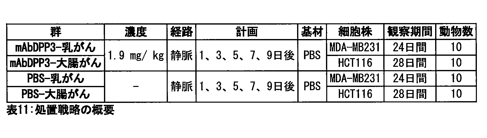
24日間、動物の行動と健康状態を毎日観察し、ノギス測定法で腫瘍成長を2日毎に記録した。原発腫瘍のサイズをノギス測定(OMC Fontanaの手動ノギス)によって測定した。式V=W2×L/2(L=長さ、W=腫瘍の垂直幅、L>W)に従って腫瘍サイズを計算した。個々の相対的腫瘍体積(RTV)を以下のように計算した:RTV=Vt/V0(Vtは各日の体積、V0は処置開始時の体積である)。
(13.2.結果)
Animals were observed daily for behavior and health for 24 days, and tumor growth was recorded every 2 days by caliper measurement. Primary tumor size was measured by caliper measurement (OMC Fontana manual caliper). Tumor size was calculated according to the formula V = W2 x L/2 (L = length, W = vertical width of tumor, L > W). Individual relative tumor volumes (RTV) were calculated as follows: RTV = Vt / V0 ( Vt is the volume on each day, V0 is the volume at the start of treatment).
13.2. Results
異種移植片がんモデルでは、調査したすべての腫瘍の形成がmAbDPP3の投与によって低減された(図9A~D)。mAbDPP3で処置した場合、乳房細胞腫瘍は20倍の大きさに成長するのに2日長くかかり(図9B)、結腸細胞腫瘍は10倍の大きさまで成長するのに2日長くかかる(図9D)。このモデルでは、大腸がん細胞株によって誘発された腫瘍は乳がん細胞株によって誘発された腫瘍よりも成長が非常に遅かった。
(14.実施例14)
In xenograft cancer models, the formation of all tumors examined was reduced by administration of mAbDPP3 (Figures 9A-D). When treated with mAbDPP3, breast cell tumors took 2 days longer to grow to 20-fold larger (Figure 9B), and colon cell tumors took 2 days longer to grow to 10-fold larger (Figure 9D). In this model, tumors induced by colon cancer cell lines grew much slower than tumors induced by breast cancer cell lines.
14. Example 14
DPP3吸着体の使用によって血漿から余剰のDPP3を除去する能力を評価するために、DPP3が抗DPP3カラムに十分に結合するかを分析するべきである。DPP3結合抗体をGlycoLinkカラム(Thermo Fisher)に固定化することによって親和性クロマトグラフィーカラムを作製した。これらのカラムに通液する前後のDPP3濃度を測定することによってカラムとのDPP3の結合を分析した。
(14.1.方法)
To evaluate the ability of the DPP3 adsorbent to remove excess DPP3 from plasma, we should analyze whether DPP3 binds sufficiently to an anti-DPP3 column. Affinity chromatography columns were made by immobilizing DPP3-binding antibodies on GlycoLink columns (Thermo Fisher). The binding of DPP3 to the columns was analyzed by measuring the DPP3 concentration before and after passing the columns.
14.1. METHODS
最初の工程ですべてのDPP3結合抗体(mAbDPP3_2552、2553、2554、2555)を、それぞれ指導説明書に従い1つのGlycoLinkカラムに酸化させ固定化した。次に組み換えGST-hDPP3(USBio)または患者の血漿試料をカラムに注入し、DPP3の結合を監視した。
(抗体の酸化)
In the first step, all DPP3 binding antibodies (mAbDPP3_2552, 2553, 2554, 2555) were oxidized and immobilized on one GlycoLink column according to the manufacturer's instructions. Recombinant GST-hDPP3 (USBio) or patient plasma samples were then injected onto the column and DPP3 binding was monitored.
(Oxidation of Antibodies)
したがって、3mg/mlの各DPP3抗体溶液300μlを700μlのGlycoLink Coupling Bufferに希釈し、最終体積が1ml、pH<6になるようにした。抗体の糖質群を酸化させるため、メタ過ヨウ素酸ナトリウム2.1mgを溶液に添加し、室温で30分間保温した。脱塩カラムを用いて抗体溶液から酸化剤を除去した。
(GlycoLinkカラムの作製)
Therefore, 300 μl of each 3 mg/ml DPP3 antibody solution was diluted in 700 μl of GlycoLink Coupling Buffer to a final volume of 1 ml, pH < 6. To oxidize the carbohydrate groups of the antibody, 2.1 mg of sodium metaperiodate was added to the solution and incubated at room temperature for 30 minutes. The oxidizing agent was removed from the antibody solution using a desalting column.
(Preparation of GlycoLink column)
カップリング反応を触媒するために、酸化した抗体に0.1Mのアニリンを添加した。次いで平衡化したGlycoLinkカラムに溶液を注入し、室温で4時間保温した。結合していない物質をカラムから流した後カラムを洗浄し、平衡化して次に使用するまで保管した。
(DPP3の親和精製)
To catalyze the coupling reaction, 0.1 M aniline was added to the oxidized antibody. The solution was then loaded onto an equilibrated GlycoLink column and incubated at room temperature for 4 hours. Unbound material was flushed from the column, after which the column was washed, equilibrated, and stored until further use.
Affinity Purification of DPP3
組み換えGST-hDPP3または患者の血漿をカップリング緩衝液に希釈(組み換えDPP3の最終濃度=100ng/ml、1ml;血漿1ml+カップリング緩衝液1ml)し、平衡化したmAbDPP3カラムに注入して室温で30分間保温した。結合効率と結合能力を評価できる全体流入量を保った。カラムを洗浄し、結合したタンパク質をGlycoLink Elution Bufferで溶出し、平衡化してから保存した。
(DPP3含量の分析)
Recombinant GST-hDPP3 or patient plasma was diluted in coupling buffer (final concentration of recombinant DPP3 = 100 ng/ml, 1 ml; 1 ml plasma + 1 ml coupling buffer) and injected onto the equilibrated mAbDPP3 column and incubated at room temperature for 30 min. A total flow rate was maintained that allowed assessment of binding efficiency and binding capacity. The column was washed and bound proteins were eluted with GlycoLink Elution Buffer, equilibrated and stored.
(Analysis of DPP3 Content)
親和精製前の試料および組換えGST-hDPP3と流入量のDPP3濃度を、サンドイッチ型発光免疫測定(詳細は実施例2を参照のこと)で測定した。20μlの試料(または検量用試料)を、塗布済み96ウェルマイクロプレートにピペットで入れ、希釈した標識付き検出抗体(200μl)を加えた後、プレートを2~8℃で18~24時間保温した。結合していないトレーサーを、350μlの洗浄溶液(20mMのPBS(pH7.4)、0.1%のTriton X-100)で4回洗浄することによって除去した。十分に結合した化学発光をCentro LB 960蛍光光度計(Berthold Technologies GmbH & Co. KG)で測定した。
(14.2.結果)
DPP3 concentrations in the samples before affinity purification, in the recombinant GST-hDPP3 and in the input were measured by a sandwich-type luminescence immunoassay (see Example 2 for details). 20 μl of sample (or calibrator) was pipetted into a pre-coated 96-well microplate, diluted labeled detection antibody (200 μl) was added and the plate was incubated at 2-8°C for 18-24 h. Unbound tracer was removed by washing four times with 350 μl washing solution (20 mM PBS pH 7.4, 0.1% Triton X-100). Fully bound chemiluminescence was measured in a Centro LB 960 fluorometer (Berthold Technologies GmbH & Co. KG).
14.2. Results
親和性クロマトグラフィーによって血漿および組み換えDPP3の溶液中のほぼすべてのDPP3を試料から除去する(表12参照)。異なるmAbDPP3抗体は異なるDPP3親和性を示す。mAbDPP3_2555は組み換えDPP3への結合速度が最高であるため、この抗体を選択して血漿試料のクロマトグラフィーに使用した。表12は親和性クロマトグラフィーによって血漿DPP3濃度を大きく低減できることを示している。これらの結果から、患者の血漿から余剰のDPP3を除去するためのアフェレーシスにDPP3吸着体を使用できることがわかる。
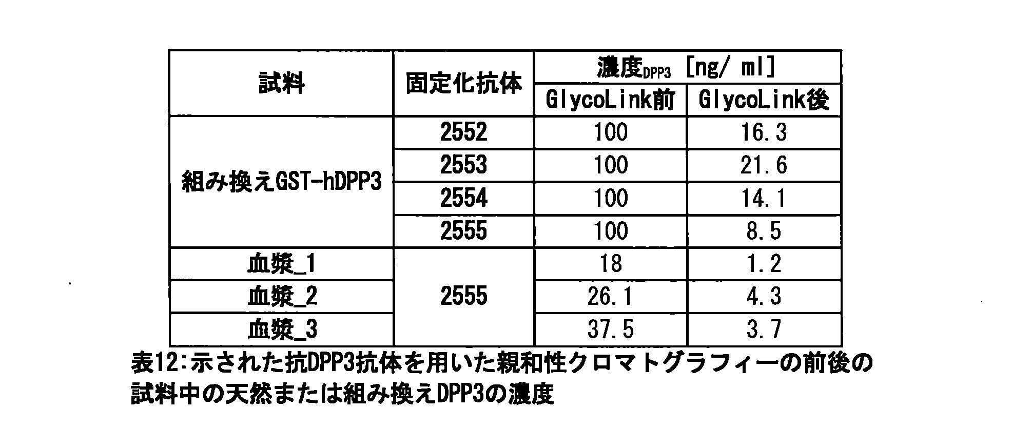
Claims (41)
前記被験者の体液試料中のDPP3総量を定量し、かつ/または活性DPP3の量を定量することと、
定量された前記DPP3総量または活性DPP3の量を所定の閾値と比較することとを含み、
定量された前記量が前記所定の閾値を超える場合、前記被験者は壊死性の経過を伴うか壊死性の経過に関連する疾患または状態を有すると診断される、方法。 1. A method for diagnosing a disease or condition involving or associated with a necrotic process in a subject, comprising:
Quantifying the total amount of DPP3 and/or the amount of active DPP3 in the body fluid sample of the subject;
and comparing the quantified amount of total DPP3 or active DPP3 to a predetermined threshold value;
If the quantified amount exceeds the predetermined threshold, the subject is diagnosed as having a disease or condition involving or associated with a necrotic process.
前記試料を全長DPP3に特異的に結合する捕捉結合剤に接触させる工程と、
前記捕捉結合剤に結合したDPP3を分離する工程と、
前記分離済みDPP3にDPP3の基質を添加する工程と、
DPP3の基質の転化率を測定し定量することにより前記活性DPP3を定量する工程と
を含む、請求項1~4のいずれか一項に記載の壊死性の経過を伴うか壊死性の経過に関連する被験者の疾患または状態を診断する方法。 The amount of total DPP3 and/or active DPP3 is quantified in a body fluid sample of the subject, and the sample is contacted with a capture binding agent that specifically binds full length DPP3;
Separating DPP3 bound to the capture binding agent;
Adding a DPP3 substrate to the separated DPP3;
A method for diagnosing a disease or condition in a subject accompanied by or associated with a necrotic process according to any one of claims 1 to 4, comprising a step of quantifying active DPP3 by measuring and quantifying the conversion rate of a DPP3 substrate.
前記試料を全長DPP3に特異的に結合する捕捉結合剤に接触させる工程と、
前記捕捉結合剤に結合したDPP3を分離する工程と、
前記分離済みDPP3にDPP3の基質を添加する工程と、
DPP3の基質の転化率を測定し定量することにより前記活性DPP3を定量する工程と
を含む方法。 1. A method for quantifying active DPP3 in a body fluid sample from a subject, comprising:
contacting the sample with a capture binding agent that specifically binds full-length DPP3;
Separating DPP3 bound to the capture binding agent;
Adding a DPP3 substrate to the separated DPP3;
and quantifying the active DPP3 by measuring and quantifying the conversion rate of a DPP3 substrate.
全長DPP3に特異的に結合する捕捉結合剤と、
DPP3の基質
を含む、検定またはキット。 1. An assay or kit for quantifying active DPP3 in a body fluid sample from a subject, comprising:
a capture binding agent that specifically binds full-length DPP3;
An assay or kit comprising a substrate for DPP3.
Applications Claiming Priority (5)
| Application Number | Priority Date | Filing Date | Title |
|---|---|---|---|
| EP16166476.8 | 2016-04-21 | ||
| EP16166476 | 2016-04-21 | ||
| PCT/EP2017/059377 WO2017182561A1 (en) | 2016-04-21 | 2017-04-20 | Methods for determining dpp3 and therapeutic methods |
| JP2018555652A JP2019520550A (en) | 2016-04-21 | 2017-04-20 | Method of determination and treatment of DPP3 |
| JP2022110620A JP2022163014A (en) | 2016-04-21 | 2022-07-08 | DPP3 Quantification Method and Treatment Method |
Related Parent Applications (1)
| Application Number | Title | Priority Date | Filing Date |
|---|---|---|---|
| JP2022110620A Division JP2022163014A (en) | 2016-04-21 | 2022-07-08 | DPP3 Quantification Method and Treatment Method |
Publications (2)
| Publication Number | Publication Date |
|---|---|
| JP2025011237A true JP2025011237A (en) | 2025-01-23 |
| JP2025011237A5 JP2025011237A5 (en) | 2025-04-24 |
Family
ID=56014788
Family Applications (4)
| Application Number | Title | Priority Date | Filing Date |
|---|---|---|---|
| JP2018555652A Pending JP2019520550A (en) | 2016-04-21 | 2017-04-20 | Method of determination and treatment of DPP3 |
| JP2020076521A Active JP7219737B2 (en) | 2016-04-21 | 2020-04-23 | DPP3 Quantification Method and Treatment Method |
| JP2022110620A Pending JP2022163014A (en) | 2016-04-21 | 2022-07-08 | DPP3 Quantification Method and Treatment Method |
| JP2024178873A Pending JP2025011237A (en) | 2016-04-21 | 2024-10-11 | Method for quantifying DPP3 and method for treatment |
Family Applications Before (3)
| Application Number | Title | Priority Date | Filing Date |
|---|---|---|---|
| JP2018555652A Pending JP2019520550A (en) | 2016-04-21 | 2017-04-20 | Method of determination and treatment of DPP3 |
| JP2020076521A Active JP7219737B2 (en) | 2016-04-21 | 2020-04-23 | DPP3 Quantification Method and Treatment Method |
| JP2022110620A Pending JP2022163014A (en) | 2016-04-21 | 2022-07-08 | DPP3 Quantification Method and Treatment Method |
Country Status (15)
| Country | Link |
|---|---|
| US (3) | US20190145981A1 (en) |
| EP (5) | EP3854817A1 (en) |
| JP (4) | JP2019520550A (en) |
| KR (3) | KR102565453B1 (en) |
| CN (3) | CN120992960A (en) |
| AU (1) | AU2017252212B2 (en) |
| BR (1) | BR112018071583A2 (en) |
| CA (2) | CA3021252A1 (en) |
| CH (1) | CH713803B1 (en) |
| DE (1) | DE112017002105T5 (en) |
| IL (1) | IL262472B2 (en) |
| MX (3) | MX2018012856A (en) |
| MY (1) | MY200300A (en) |
| SG (1) | SG11201809252YA (en) |
| WO (1) | WO2017182561A1 (en) |
Families Citing this family (18)
| Publication number | Priority date | Publication date | Assignee | Title |
|---|---|---|---|---|
| DE112017002105T5 (en) * | 2016-04-21 | 2019-04-25 | Sphingotec Therapeutics Gmbh | Method for the determination of DPP3 and therapeutic methods |
| KR102835427B1 (en) * | 2017-10-25 | 2025-07-21 | 4틴4 파마슈티컬스 게엠베하 | DPP3 binding agent directed against and binding to a specific DPP3 epitope and its use in the prevention or treatment of diseases/acute conditions associated with oxidative stress |
| CN113557031A (en) * | 2018-12-21 | 2021-10-26 | 4Teen4制药有限公司 | Therapy guidance and/or therapy monitoring for treatment with angiotensin receptor agonists and/or precursors thereof |
| JP2022546061A (en) * | 2019-08-30 | 2022-11-02 | 4ティーン4 ファーマシューティカルズ ゲゼルシャフト ミット ベシュレンクテル ハフツング | Therapy induction and/or therapy monitoring for treatment of shock |
| CN115244080A (en) * | 2020-02-27 | 2022-10-25 | 艾德里诺医药公司 | Anti-adrenomedullin (ADM) antibody or anti-ADM antibody fragment or anti-ADM non-Ig scaffold for the treatment or prevention of shock |
| US20230193348A1 (en) | 2020-02-27 | 2023-06-22 | 4TEEN4 Pharmaceuticals GmbH | Dpp3 for therapy guidance, monitoring and stratification of nt-adm antibodies in patients with shock |
| US20230250166A1 (en) | 2020-02-27 | 2023-08-10 | Adrenomed Ag | Anti-adrenomedullin (adm) binder for use in therapy of patients in shock |
| EP3922993A1 (en) | 2020-06-12 | 2021-12-15 | 4TEEN4 Pharmaceuticals GmbH | Dpp3 in patients infected with coronavirus |
| BR112022017277A2 (en) * | 2020-03-16 | 2022-10-18 | 4TEEN4 Pharmaceuticals GmbH | DPP3 IN PATIENTS INFECTED WITH CORONA VIRUS |
| CA3254142A1 (en) | 2022-03-15 | 2023-09-21 | Adrenomed Ag | Stable aqueous formulation of an anti-adrenomedullin (adm) antibody or anti-adm antibody fragment |
| CN117186177B (en) * | 2022-05-31 | 2024-08-23 | 中国农业大学 | Duck blood cell protein peptide with uric acid reducing activity and preparation method thereof |
| AU2023315168A1 (en) | 2022-07-29 | 2025-02-13 | 4TEEN4 Pharmaceuticals GmbH | Prediction of an increase of dpp3 in a patient with septic shock |
| AU2023313376A1 (en) | 2022-07-29 | 2025-03-06 | Adrenomed Ag | Anti-adrenomedullin (adm) antibody or anti-adm antibody fragment or anti-adm non-ig scaffold for use in therapy or prevention of shock |
| JP2025541138A (en) | 2022-12-15 | 2025-12-18 | 4ティーン4 ファーマシューティカルズ ゲゼルシャフト ミット ベシュレンクテル ハフツング | DPP3 inhibitors for improving pulmonary function in critically ill patients |
| CN115804836A (en) * | 2022-12-27 | 2023-03-17 | 中国人民解放军海军特色医学中心 | Application of a central small molecular protein DPP3 in the preparation of antihypertensive drugs |
| EP4687942A1 (en) | 2023-03-29 | 2026-02-11 | 4TEEN4 Pharmaceuticals GmbH | Dpp3 inhibitor for myocardial protection and prevention of myocardial injury in critically ill patients with blood pressure decline |
| WO2026046968A1 (en) | 2024-08-26 | 2026-03-05 | 4TEEN4 Pharmaceuticals GmbH | Dpp3 binders for the treatment of endothelial dysfunction |
| WO2026046947A1 (en) | 2024-08-26 | 2026-03-05 | 4TEEN4 Pharmaceuticals GmbH | Anti-dpp3 antibody directed to and binding to dpp3 for the treatment of shock in extreme-critically ill patients |
Family Cites Families (38)
| Publication number | Priority date | Publication date | Assignee | Title |
|---|---|---|---|---|
| US5807715A (en) | 1984-08-27 | 1998-09-15 | The Board Of Trustees Of The Leland Stanford Junior University | Methods and transformed mammalian lymphocyte cells for producing functional antigen-binding protein including chimeric immunoglobulin |
| JPS6188884A (en) | 1984-10-04 | 1986-05-07 | Sankyo Co Ltd | Encephalinase b-inhibiting substance and its preparation |
| AU634716B2 (en) | 1988-08-01 | 1993-03-04 | Ciba Corning Diagnostics Corp. | Method for detection of an analyte using acridinium esters and liposomes |
| US5530101A (en) | 1988-12-28 | 1996-06-25 | Protein Design Labs, Inc. | Humanized immunoglobulins |
| DE69120146T2 (en) | 1990-01-12 | 1996-12-12 | Cell Genesys Inc | GENERATION OF XENOGENIC ANTIBODIES |
| US5427908A (en) | 1990-05-01 | 1995-06-27 | Affymax Technologies N.V. | Recombinant library screening methods |
| GB9015198D0 (en) | 1990-07-10 | 1990-08-29 | Brien Caroline J O | Binding substance |
| DK0585287T3 (en) | 1990-07-10 | 2000-04-17 | Cambridge Antibody Tech | Process for producing specific binding pair elements |
| WO1993012227A1 (en) | 1991-12-17 | 1993-06-24 | Genpharm International, Inc. | Transgenic non-human animals capable of producing heterologous antibodies |
| JPH06219989A (en) * | 1993-01-27 | 1994-08-09 | Microbial Chem Res Found | Novel bioactive substances fluostatin A and B, production method thereof and use thereof |
| GB9312324D0 (en) * | 1993-06-15 | 1993-07-28 | Pitman Moore Inc | Vaccine |
| US5612186A (en) | 1994-06-22 | 1997-03-18 | Food Industry Research And Development Institute | Enzyme-capture assay (ECA) for the identification of Escherichia coli in clinical samples |
| US5601986A (en) | 1994-07-14 | 1997-02-11 | Amgen Inc. | Assays and devices for the detection of extrahepatic biliary atresia |
| US6818418B1 (en) | 1998-12-10 | 2004-11-16 | Compound Therapeutics, Inc. | Protein scaffolds for antibody mimics and other binding proteins |
| WO2001016334A2 (en) | 1999-09-01 | 2001-03-08 | Incyte Genomics, Inc. | Human hydrolytic enzymes |
| AU2002225954A1 (en) * | 2000-11-08 | 2002-05-21 | The University Of Georgia Research Foundation, Inc. | Dipeptidylpeptidases and methods of use |
| AU2002323501C1 (en) | 2001-08-30 | 2010-04-29 | Biorexis Technology, Inc | Modified transferrin fusion proteins |
| ES2348230T3 (en) | 2002-06-07 | 2010-12-01 | Dyax Corp. | PREVENTION AND REDUCTION OF THE ISCHEMIA. |
| CN1852974A (en) * | 2003-06-09 | 2006-10-25 | 密歇根大学董事会 | Compositions and methods for treating and diagnosing cancer |
| US20050164301A1 (en) | 2003-10-24 | 2005-07-28 | Avidia Research Institute | LDL receptor class A and EGF domain monomers and multimers |
| US20100028995A1 (en) | 2004-02-23 | 2010-02-04 | Anaphore, Inc. | Tetranectin Trimerizing Polypeptides |
| WO2005106486A2 (en) * | 2004-04-28 | 2005-11-10 | Bayer Healthcare Ag | Diagnostic and therapeutics for diseases associated with dipeptidyl-peptidase 3(dpp3) |
| US20110136131A1 (en) * | 2004-06-01 | 2011-06-09 | Sysmex Corporation | Method for measuring enzyme activity and column for use in measuring enzyme activity |
| AU2005287557B2 (en) | 2004-09-21 | 2011-10-13 | Biontech Ag | Use of microproteins as tryptase inhibitors |
| JP2006230318A (en) | 2005-02-25 | 2006-09-07 | Maruishi Pharmaceutical Co Ltd | Method for diagnosing rheumatoid arthritis |
| CN100420947C (en) * | 2005-05-30 | 2008-09-24 | 孙东旭 | Method for quantitative determination of specific analyte with single trapping agent and reagent kit therefor |
| WO2007055979A2 (en) * | 2005-11-02 | 2007-05-18 | Meyer Pharmaceuticals Llc | Native trre: protein implicated in tnf receptor shedding for treating arthritis and inflammation |
| EP1994415B9 (en) * | 2006-03-13 | 2015-12-16 | Becton Dickinson and Company | Diagnosis and prognosis of dipeptidyl peptidase-associated disease states |
| US20080057590A1 (en) | 2006-06-07 | 2008-03-06 | Mickey Urdea | Markers associated with arteriovascular events and methods of use thereof |
| ES2373832T3 (en) | 2007-12-19 | 2012-02-09 | Affibody Ab | POLYPEPTIDE DERIVED FROM PROTEIN A AND ABLE TO JOIN PDGF. |
| BRPI0921469B1 (en) | 2008-11-03 | 2022-01-18 | Molecular Partners Ag | BINDING PROTEINS THAT INHIBIT INTERACTION WITH THE VEGF-A RECEPTOR, PHARMACEUTICAL COMPOSITION |
| WO2010093872A2 (en) * | 2009-02-13 | 2010-08-19 | The United States Of America, As Represented By The Secretary, Department Of Health And Human Services | Molecular-based method of cancer diagnosis and prognosis |
| MX2012002428A (en) | 2009-08-27 | 2012-09-12 | Covagen Ag | Il-17 binding compounds and medical uses thereof. |
| RU2553333C2 (en) | 2009-12-14 | 2015-06-10 | Сцил Протеинс Гмбх | Method of identifying heteromultimeric modified ubiquitin proteins possessing ability to bind with ligands |
| KR101234352B1 (en) * | 2010-03-18 | 2013-02-15 | 권동락 | Selective Immune Cell Separation Device |
| PL2580236T3 (en) | 2010-06-08 | 2019-09-30 | Pieris Pharmaceuticals Gmbh | Tear lipocalin muteins binding il-4 r alpha |
| CN104946646B (en) * | 2014-03-25 | 2020-03-31 | 中国医学科学院基础医学研究所 | Small nucleic acid molecule, DNA molecule, protein and application for preventing and/or treating Ebola viral hemorrhagic fever |
| DE112017002105T5 (en) * | 2016-04-21 | 2019-04-25 | Sphingotec Therapeutics Gmbh | Method for the determination of DPP3 and therapeutic methods |
-
2017
- 2017-04-20 DE DE112017002105.7T patent/DE112017002105T5/en active Pending
- 2017-04-20 CA CA3021252A patent/CA3021252A1/en active Pending
- 2017-04-20 CH CH001280/2018A patent/CH713803B1/en unknown
- 2017-04-20 KR KR1020187033194A patent/KR102565453B1/en active Active
- 2017-04-20 EP EP21153574.5A patent/EP3854817A1/en active Pending
- 2017-04-20 AU AU2017252212A patent/AU2017252212B2/en active Active
- 2017-04-20 JP JP2018555652A patent/JP2019520550A/en active Pending
- 2017-04-20 CN CN202511191873.XA patent/CN120992960A/en active Pending
- 2017-04-20 SG SG11201809252YA patent/SG11201809252YA/en unknown
- 2017-04-20 IL IL262472A patent/IL262472B2/en unknown
- 2017-04-20 KR KR1020247025458A patent/KR102886863B1/en active Active
- 2017-04-20 EP EP24178565.8A patent/EP4417217A3/en active Pending
- 2017-04-20 EP EP21153560.4A patent/EP3896450A1/en active Pending
- 2017-04-20 MY MYPI2018703855A patent/MY200300A/en unknown
- 2017-04-20 CN CN201780039077.1A patent/CN110168372A/en active Pending
- 2017-04-20 KR KR1020237026743A patent/KR102886883B1/en active Active
- 2017-04-20 BR BR112018071583-2A patent/BR112018071583A2/en unknown
- 2017-04-20 WO PCT/EP2017/059377 patent/WO2017182561A1/en not_active Ceased
- 2017-04-20 US US16/095,563 patent/US20190145981A1/en active Pending
- 2017-04-20 EP EP17720047.4A patent/EP3446125B1/en active Active
- 2017-04-20 CA CA3264486A patent/CA3264486A1/en active Pending
- 2017-04-20 CN CN202510254123.6A patent/CN120093910A/en active Pending
- 2017-04-20 MX MX2018012856A patent/MX2018012856A/en unknown
- 2017-04-20 EP EP24178574.0A patent/EP4417218A3/en active Pending
-
2018
- 2018-10-19 MX MX2020001193A patent/MX2020001193A/en unknown
- 2018-10-19 MX MX2025006655A patent/MX2025006655A/en unknown
-
2020
- 2020-04-23 JP JP2020076521A patent/JP7219737B2/en active Active
- 2020-07-02 US US16/919,449 patent/US11726094B2/en active Active
-
2022
- 2022-07-08 JP JP2022110620A patent/JP2022163014A/en active Pending
-
2023
- 2023-06-21 US US18/338,805 patent/US20240385197A1/en active Pending
-
2024
- 2024-10-11 JP JP2024178873A patent/JP2025011237A/en active Pending
Also Published As
Similar Documents
| Publication | Publication Date | Title |
|---|---|---|
| JP7219737B2 (en) | DPP3 Quantification Method and Treatment Method | |
| JP2023516615A (en) | DPP3 for NT-ADM Antibody Therapy Guidance, Monitoring, and Stratification in Patients with Shock | |
| CN115769076A (en) | DPP3 in patients infected with coronaviruses | |
| US20260028423A1 (en) | Prediction of an increase of dpp3 in a patient with septic shock | |
| CN120359042A (en) | DPP3 inhibitor for improving lung function of critically ill patient | |
| RU2771824C2 (en) | Methods for determining dpp3 and therapeutic methods | |
| HK40108173A (en) | Methods for determining dpp3 and therapeutic methods | |
| HK40108174A (en) | Methods for determining dpp3 and therapeutic methods | |
| AU2023258417B2 (en) | Methods for determining DPP3 and therapeutic methods | |
| HK40121648A (en) | Inhibitor of the activity of dpp3, pharmaceutical composition comprising the same, and use thereof | |
| HK40011980A (en) | Methods for determining dpp3 and therapeutic methods | |
| CN119604526A (en) | Anti-adrenomedullin (ADM) antibodies or anti-ADM antibody fragments or anti-ADM non-Ig scaffolds for treating or preventing shock | |
| CA3207969A1 (en) | Anti-adrenomedullin (adm) antibody or anti-adm antibody fragment or anti-adm non-ig scaffold for use in therapy or prevention of shock and prediction of an increase of dpp3 in a critically ill patient | |
| CN120857945A (en) | DPP3 inhibitors for myocardial protection and prevention of myocardial injury in critically ill patients with hypotension |
Legal Events
| Date | Code | Title | Description |
|---|---|---|---|
| A521 | Request for written amendment filed |
Free format text: JAPANESE INTERMEDIATE CODE: A523 Effective date: 20241011 |
|
| A621 | Written request for application examination |
Free format text: JAPANESE INTERMEDIATE CODE: A621 Effective date: 20241011 |