JP3662948B2 - Modified protein and method for producing the same - Google Patents
Modified protein and method for producing the same Download PDFInfo
- Publication number
- JP3662948B2 JP3662948B2 JP34088593A JP34088593A JP3662948B2 JP 3662948 B2 JP3662948 B2 JP 3662948B2 JP 34088593 A JP34088593 A JP 34088593A JP 34088593 A JP34088593 A JP 34088593A JP 3662948 B2 JP3662948 B2 JP 3662948B2
- Authority
- JP
- Japan
- Prior art keywords
- ivps
- protein
- fusion protein
- fusion
- civps
- Prior art date
- Legal status (The legal status is an assumption and is not a legal conclusion. Google has not performed a legal analysis and makes no representation as to the accuracy of the status listed.)
- Expired - Fee Related
Links
Images








Classifications
-
- C—CHEMISTRY; METALLURGY
- C12—BIOCHEMISTRY; BEER; SPIRITS; WINE; VINEGAR; MICROBIOLOGY; ENZYMOLOGY; MUTATION OR GENETIC ENGINEERING
- C12Y—ENZYMES
- C12Y302/00—Hydrolases acting on glycosyl compounds, i.e. glycosylases (3.2)
- C12Y302/01—Glycosidases, i.e. enzymes hydrolysing O- and S-glycosyl compounds (3.2.1)
- C12Y302/01081—Beta-agarase (3.2.1.81)
-
- C—CHEMISTRY; METALLURGY
- C07—ORGANIC CHEMISTRY
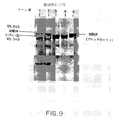
- C07K—PEPTIDES
- C07K14/00—Peptides having more than 20 amino acids; Gastrins; Somatostatins; Melanotropins; Derivatives thereof
- C07K14/435—Peptides having more than 20 amino acids; Gastrins; Somatostatins; Melanotropins; Derivatives thereof from animals; from humans
- C07K14/46—Peptides having more than 20 amino acids; Gastrins; Somatostatins; Melanotropins; Derivatives thereof from animals; from humans from vertebrates
- C07K14/47—Peptides having more than 20 amino acids; Gastrins; Somatostatins; Melanotropins; Derivatives thereof from animals; from humans from vertebrates from mammals
-
- C—CHEMISTRY; METALLURGY
- C07—ORGANIC CHEMISTRY
- C07K—PEPTIDES
- C07K14/00—Peptides having more than 20 amino acids; Gastrins; Somatostatins; Melanotropins; Derivatives thereof
- C07K14/435—Peptides having more than 20 amino acids; Gastrins; Somatostatins; Melanotropins; Derivatives thereof from animals; from humans
- C07K14/46—Peptides having more than 20 amino acids; Gastrins; Somatostatins; Melanotropins; Derivatives thereof from animals; from humans from vertebrates
- C07K14/47—Peptides having more than 20 amino acids; Gastrins; Somatostatins; Melanotropins; Derivatives thereof from animals; from humans from vertebrates from mammals
- C07K14/4701—Peptides having more than 20 amino acids; Gastrins; Somatostatins; Melanotropins; Derivatives thereof from animals; from humans from vertebrates from mammals not used
- C07K14/4716—Muscle proteins, e.g. myosin, actin
-
- C—CHEMISTRY; METALLURGY
- C12—BIOCHEMISTRY; BEER; SPIRITS; WINE; VINEGAR; MICROBIOLOGY; ENZYMOLOGY; MUTATION OR GENETIC ENGINEERING
- C12N—MICROORGANISMS OR ENZYMES; COMPOSITIONS THEREOF; PROPAGATING, PRESERVING, OR MAINTAINING MICROORGANISMS; MUTATION OR GENETIC ENGINEERING; CULTURE MEDIA
- C12N15/00—Mutation or genetic engineering; DNA or RNA concerning genetic engineering, vectors, e.g. plasmids, or their isolation, preparation or purification; Use of hosts therefor
- C12N15/09—Recombinant DNA-technology
- C12N15/11—DNA or RNA fragments; Modified forms thereof; Non-coding nucleic acids having a biological activity
- C12N15/62—DNA sequences coding for fusion proteins
-
- C—CHEMISTRY; METALLURGY
- C12—BIOCHEMISTRY; BEER; SPIRITS; WINE; VINEGAR; MICROBIOLOGY; ENZYMOLOGY; MUTATION OR GENETIC ENGINEERING
- C12N—MICROORGANISMS OR ENZYMES; COMPOSITIONS THEREOF; PROPAGATING, PRESERVING, OR MAINTAINING MICROORGANISMS; MUTATION OR GENETIC ENGINEERING; CULTURE MEDIA
- C12N15/00—Mutation or genetic engineering; DNA or RNA concerning genetic engineering, vectors, e.g. plasmids, or their isolation, preparation or purification; Use of hosts therefor
- C12N15/09—Recombinant DNA-technology
- C12N15/63—Introduction of foreign genetic material using vectors; Vectors; Use of hosts therefor; Regulation of expression
- C12N15/66—General methods for inserting a gene into a vector to form a recombinant vector using cleavage and ligation; Use of non-functional linkers or adaptors, e.g. linkers containing the sequence for a restriction endonuclease
-
- C—CHEMISTRY; METALLURGY
- C12—BIOCHEMISTRY; BEER; SPIRITS; WINE; VINEGAR; MICROBIOLOGY; ENZYMOLOGY; MUTATION OR GENETIC ENGINEERING
- C12N—MICROORGANISMS OR ENZYMES; COMPOSITIONS THEREOF; PROPAGATING, PRESERVING, OR MAINTAINING MICROORGANISMS; MUTATION OR GENETIC ENGINEERING; CULTURE MEDIA
- C12N9/00—Enzymes; Proenzymes; Compositions thereof; Processes for preparing, activating, inhibiting, separating or purifying enzymes
- C12N9/10—Transferases (2.)
- C12N9/12—Transferases (2.) transferring phosphorus containing groups, e.g. kinases (2.7)
- C12N9/1241—Nucleotidyltransferases (2.7.7)
- C12N9/1252—DNA-directed DNA polymerase (2.7.7.7), i.e. DNA replicase
-
- C—CHEMISTRY; METALLURGY
- C12—BIOCHEMISTRY; BEER; SPIRITS; WINE; VINEGAR; MICROBIOLOGY; ENZYMOLOGY; MUTATION OR GENETIC ENGINEERING
- C12N—MICROORGANISMS OR ENZYMES; COMPOSITIONS THEREOF; PROPAGATING, PRESERVING, OR MAINTAINING MICROORGANISMS; MUTATION OR GENETIC ENGINEERING; CULTURE MEDIA
- C12N9/00—Enzymes; Proenzymes; Compositions thereof; Processes for preparing, activating, inhibiting, separating or purifying enzymes
- C12N9/14—Hydrolases (3)
- C12N9/16—Hydrolases (3) acting on ester bonds (3.1)
- C12N9/22—Ribonucleases [RNase]; Deoxyribonucleases [DNase]
-
- C—CHEMISTRY; METALLURGY
- C12—BIOCHEMISTRY; BEER; SPIRITS; WINE; VINEGAR; MICROBIOLOGY; ENZYMOLOGY; MUTATION OR GENETIC ENGINEERING
- C12N—MICROORGANISMS OR ENZYMES; COMPOSITIONS THEREOF; PROPAGATING, PRESERVING, OR MAINTAINING MICROORGANISMS; MUTATION OR GENETIC ENGINEERING; CULTURE MEDIA
- C12N9/00—Enzymes; Proenzymes; Compositions thereof; Processes for preparing, activating, inhibiting, separating or purifying enzymes
- C12N9/14—Hydrolases (3)
- C12N9/24—Hydrolases (3) acting on glycosyl compounds (3.2)
- C12N9/2402—Hydrolases (3) acting on glycosyl compounds (3.2) hydrolysing O- and S- glycosyl compounds (3.2.1)
- C12N9/2468—Hydrolases (3) acting on glycosyl compounds (3.2) hydrolysing O- and S- glycosyl compounds (3.2.1) acting on beta-galactose-glycoside bonds, e.g. carrageenases (3.2.1.83; 3.2.1.157); beta-agarase (3.2.1.81)
-
- C—CHEMISTRY; METALLURGY
- C12—BIOCHEMISTRY; BEER; SPIRITS; WINE; VINEGAR; MICROBIOLOGY; ENZYMOLOGY; MUTATION OR GENETIC ENGINEERING
- C12N—MICROORGANISMS OR ENZYMES; COMPOSITIONS THEREOF; PROPAGATING, PRESERVING, OR MAINTAINING MICROORGANISMS; MUTATION OR GENETIC ENGINEERING; CULTURE MEDIA
- C12N9/00—Enzymes; Proenzymes; Compositions thereof; Processes for preparing, activating, inhibiting, separating or purifying enzymes
- C12N9/14—Hydrolases (3)
- C12N9/24—Hydrolases (3) acting on glycosyl compounds (3.2)
- C12N9/2402—Hydrolases (3) acting on glycosyl compounds (3.2) hydrolysing O- and S- glycosyl compounds (3.2.1)
- C12N9/2468—Hydrolases (3) acting on glycosyl compounds (3.2) hydrolysing O- and S- glycosyl compounds (3.2.1) acting on beta-galactose-glycoside bonds, e.g. carrageenases (3.2.1.83; 3.2.1.157); beta-agarase (3.2.1.81)
- C12N9/2471—Beta-galactosidase (3.2.1.23), i.e. exo-(1-->4)-beta-D-galactanase
-
- C—CHEMISTRY; METALLURGY
- C12—BIOCHEMISTRY; BEER; SPIRITS; WINE; VINEGAR; MICROBIOLOGY; ENZYMOLOGY; MUTATION OR GENETIC ENGINEERING
- C12Y—ENZYMES
- C12Y302/00—Hydrolases acting on glycosyl compounds, i.e. glycosylases (3.2)
- C12Y302/01—Glycosidases, i.e. enzymes hydrolysing O- and S-glycosyl compounds (3.2.1)
- C12Y302/01023—Beta-galactosidase (3.2.1.23), i.e. exo-(1-->4)-beta-D-galactanase
-
- C—CHEMISTRY; METALLURGY
- C07—ORGANIC CHEMISTRY
- C07K—PEPTIDES
- C07K2319/00—Fusion polypeptide
-
- C—CHEMISTRY; METALLURGY
- C07—ORGANIC CHEMISTRY
- C07K—PEPTIDES
- C07K2319/00—Fusion polypeptide
- C07K2319/20—Fusion polypeptide containing a tag with affinity for a non-protein ligand
-
- C—CHEMISTRY; METALLURGY
- C07—ORGANIC CHEMISTRY
- C07K—PEPTIDES
- C07K2319/00—Fusion polypeptide
- C07K2319/20—Fusion polypeptide containing a tag with affinity for a non-protein ligand
- C07K2319/21—Fusion polypeptide containing a tag with affinity for a non-protein ligand containing a His-tag
-
- C—CHEMISTRY; METALLURGY
- C07—ORGANIC CHEMISTRY
- C07K—PEPTIDES
- C07K2319/00—Fusion polypeptide
- C07K2319/90—Fusion polypeptide containing a motif for post-translational modification
- C07K2319/92—Fusion polypeptide containing a motif for post-translational modification containing an intein ("protein splicing")domain
Landscapes
- Health & Medical Sciences (AREA)
- Life Sciences & Earth Sciences (AREA)
- Chemical & Material Sciences (AREA)
- Genetics & Genomics (AREA)
- Organic Chemistry (AREA)
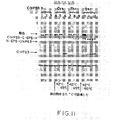
- Engineering & Computer Science (AREA)
- Zoology (AREA)
- Bioinformatics & Cheminformatics (AREA)
- Wood Science & Technology (AREA)
- Molecular Biology (AREA)
- General Engineering & Computer Science (AREA)
- General Health & Medical Sciences (AREA)
- Biochemistry (AREA)
- Biomedical Technology (AREA)
- Biotechnology (AREA)
- Medicinal Chemistry (AREA)
- Microbiology (AREA)
- Biophysics (AREA)
- Gastroenterology & Hepatology (AREA)
- Proteomics, Peptides & Aminoacids (AREA)
- Toxicology (AREA)
- Physics & Mathematics (AREA)
- Plant Pathology (AREA)
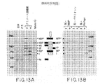
- Peptides Or Proteins (AREA)
- Preparation Of Compounds By Using Micro-Organisms (AREA)
Abstract
Description
【0001】
【産業上の利用分野】
本発明は、改質タンパク質及びその製造法に関する。より具体的には、本発明の改質タンパク質は、ターゲットタンパク質及び、予め決定した条件下で切除または切断し得る制御可能な介在タンパク質配列(CIVPS)を含む。
【0002】
【従来の技術及び発明が解決しようとする課題】
成熟タンパク質の産生には、DNAからRNA、タンパク質への情報の流れが含まれる。その情報を妨げるDNA及びRNA要素を正確に切除することは、既に報告されている[M.Belfort, Annu.Rev.Genet.24:363(1990);T.R.Cech,Annu.Rev.Biochem.59:543(1990);Hunterら,Genes Dev.3:2101(1989)]。近年、介在タンパク質配列の正確な切除についての証拠が、Saccharomyces cerevisiae由来のTFPI対立遺伝子[Hirataら,J.Biol.Chem.265:6726(1990);Kaneら,Science 250:651(1990)]及びMycobacterium tuberculosis由来のrec A遺伝子[Davisら,J.Bact.173:5653(1991);Davisら,Cell 71:1(1992)]に関しても報告された。これらは、各々成熟タンパク質を産生するために除去しなければならない内部フレーム内ペプチドセグメントを含んでいる。各Tfp1及びRecAの発現により、2種類のペプチド、即ち、一方は介在タンパク質配列(IVPS)を表し、他方は外部タンパク質配列(EPS)の連結産生物を表すペプチドが生じる。この翻訳後プロセシング事象(post-translational processhing event)は、「タンパク質スプライシング」と呼称されてきた。同様に、好高熱性の原始Thermococcus litoralis由来のVent DNAポリメラーゼ遺伝子は、2個のフレーム内IVPSを含んでいる[Perlerら,PNAS 89:5577(1992)]。
【0003】
研究への応用とは別にIVPSまたはタンパク質スプライシングを使用する方法の開発に於ける大きな障害は、IVPSの活性及びスプライシング事象を制御することが不可能である点にある。
【0004】
従って、IVPSを用いるターゲットタンパク質を改質するための、特にIVPSの活性が制御可能な、便利な手段を提供する方法が望ましい。その活性が実質的に不活化されるようにターゲットタンパク質を特異的に改質し得る方法を得るのも望ましい。不活化改質タンパク質の活性を回復させるために使用し得る方法を得るのが望ましい。
【0005】
【課題を解決するための手段】
発明の概要
本発明は、予め決定した条件下でスプライシングなしに切断またはタンパク質スプライシングにより切除し得るIVPSとターゲットタンパク質とを含む改質タンパク質に関する。このような予め決定した条件(predetermined condition)は、使用したIVPSに依存し、例えば、温度上昇、pH条件の変化、光への暴露、脱リン酸化、またはアミノ酸残基の脱グリコシル化を包含し得る。IVPSは、ターゲットタンパク質にIVPSを挿入することによってまたは、ターゲットタンパク質のアミノ若しくはカルボキシ末端でターゲットタンパク質とIVPSを融合することによってターゲットタンパク質と結合し得る。従って、制御可能な介在タンパク質配列(CIVPS)と称されるこれらのIVPSは、スプライシングまたは切断反応を制御するのに有用である。本発明はさらに、CIVPSの産生、選択及び試験方法にも関する。
【0006】
好ましい態様の一つでは、CIVPSをコードするDNA配列は、両方のコード配列が連続する読み取り枠を形成するように、ターゲットタンパク質をコードするDNA配列に挿入するか、またはこれと結合させる。その後、この融合DNAの発現を利用して改質ターゲットタンパク質を産生する。もう一つの実施態様では、このようにして産生された改質タンパク質を、CIVPSを切除するか切断する予め決定した条件にかける。特定の実施態様に於いては、ターゲットタンパク質を実質的に不活化させるターゲットタンパク質領域にCIVPSを挿入し、そしてCIVPSの切除によりターゲットタンパク質の活性を回復させる。
【0007】
好ましいCIVPSとしては、T.litoralisから得られるCIVPS1及び2(Vent IVPS1及び2または、IVS1及び2とも称される)並びに、Pyrococcus種から得られるCIVPS3[Deep Vent IVPS1またはIVS1とも称される]が挙げられる。これらのCIVPSは、温度上昇時に改質タンパク質から除去、即ち、タンパク質スプライシングを介して除去し得る。
【0008】
本発明により、特定のCIVPSアミノ酸残基及び少なくとも第1の下流アミノ酸残基はスプライシング反応を調節し、且つこれらの残基の改質によりスプライシング反応が減少するかまたは停止することも知見された。これらの残基は、他のIVPSに保存されることが知見された。このような残基の改質を使用して、IVPSをCIVPSに転換させ得る。
【0009】
本発明により、特定の状況下では、完全スプライシング反応は必要でないか、または望ましくないことが知見された。このような状況下では、CIVPSを改質してスプライシングなしに切断し、ターゲットタンパク質からCIVPSの分離または切断を制御し得る。
【0010】
本発明のCIVPS及び改質タンパク質の潜在的な用途は多様である。これらの例としては、例えば、ターゲットタンパク質の酵素活性の制御、アフィニティークロマトグラフィーによるCIVPSに特異的な抗体を使用する改質タンパク質の精製、及び宿主細胞に毒性であるタンパク質の産生が挙げられる。
【0011】
本発明のCIVPSは、さらに、CIVPSに融合したターゲットタンパク質を含む改質タンパク質を産生するタンパク質精製方法で使用し得る。所望により、3部分融合は、CIVPSがターゲットタンパク質と基質(結合タンパク質)、例えばMBPに対する親和性を有するタンパク質との間にある場合に産生し得る。次いで改質タンパク質は、CIVPSまたは結合タンパク質が特異的親和性を有する基質と、例えばアフィニティークロマトグラフィーを使用して接触させる。高度に精製したターゲットタンパク質は、例えばCIVPSとターゲットタンパク質の間の切断を開始する予め決定した条件にCIVPSをかけることによって、カラムから遊離し得る。あるいは、融合タンパク質は上記の如く精製し得、次いでターゲットタンパク質は、CIVPSを予め決定した条件にかけることによって融合物から放出してもよい。
【0012】
詳細な説明
本発明は、改質タンパク質及びその製造方法に関する。改質タンパク質は、制御可能な介在タンパク質配列(CIVPS)及びターゲットタンパク質を含み、CIVPSは、例えば温度上昇、pH条件の変化、光分解によるアミノ酸残基不ブロック化、脱リン酸化、脱グリコシル化または他の手段などの予め決定した条件下で、タンパク質スプライシングなしに切断またはタンパク質スプライシングにより切除し得る。所望により、改質タンパク質をこれらの条件にかけ得る。CIVPSは実質的にターゲットタンパク質活性を不活化する領域にも挿入し得る。
【0013】
介在タンパク質配列(IVPS)は、前駆体タンパク質内に見出される内部フレーム内ペプチドセグメントであり、ネイティブタンパク質を形成するためにタンパク質スプライシングを介して除去または切り出される。IVPSは、Saccharomyces cerevisiae由来のTFPI対立遺伝子[Hirataら,上掲;Kaneら,上掲]及びMycobacterium tuberculosis由来のrec A遺伝子[Davisら,上掲(1991);Davisら,上掲(1992)]中に存在することが報告されている。これらの文献は、本明細書中参照として含まれる。
【0014】
本発明のCIVPSは、ネイティブのIVPSの固有の特性(例えば、温度の上昇)によりまたは、IVPSを制御すべき反応にかける改質により切除または切断が制御され得る任意の介在タンパク質配列を含む。
【0015】
好高熱性原始Thermococcus litoralis由来のVent DNAポリメラーゼ遺伝子は、発現したポリメラーゼの動力学的及び生化学的特性に影響することなく、DNAレベルで欠失し得る2つのフレーム内IVPS、即ちIVPS1(CIVPS1)及びIVPS2(CIVPS2)[Perlerら,上掲]を含む。両方のIVPSを含むVent DNAポリメラーゼ遺伝子の正しいプロセシングは、ネイティブの原始T.litoralisで起こる。さらに、IVPS1を欠失する発現構築物の正しいプロセシングは、真正細菌のE.coli[Perlerら,上掲]、真核生物のバキュロウイルスに感染した昆虫細胞及びin vitro転写/翻訳系[Hodgesら,Nucleic Acids Research,20:6153(1992)]内で観察されている。さらに、ウサギ網状赤血球及びE.coli in vitro転写/翻訳系は、IVPS2配列を正しく除去して、成熟ポリメラーゼを産生する。理論に結び付けるつもりはないが、Vent及びDeep Vent IVPSは自己スプライシングすると考えられる。
【0016】
Vent DNAポリメラーゼ遺伝子のヌクレオチド配列は、配列表中配列番号:1として記載した。CIVPS1のヌクレオチド配列は、ヌクレオチド1773〜3386である。CIVPS2のヌクレオチド配列は、ヌクレオチド3534〜4703である。CIVPS1及びCIVPS2は、1990年4月24日にAmerican Type Culture Collection(ATCC)に寄託し、ATCC受託番号40795号を受けたファージNEB619から得ることができる。
【0017】
第3のIVPS(CIVPS3、即ち、DV IVPS1)は、好熱性の原始細菌、Pyrococcus種(単離物GB-D)のDNAポリメラーゼ遺伝子中に本発明者によって発見された。Pyrococcus DNAポリメラーゼは、時折Deep Vent DNAポリメラーゼと称される。Deep Vent DNAポリメラーゼのヌクレオチド配列は、配列表中配列番号:2として記載した。CIVPS3のヌクレオチド配列は、1839〜3449である。CIVPS3は、1991年10月1日にATCCに寄託し、受託番号68723を受けたプラスミドpNEB#720から得ることができる。
【0018】
本発明により、上記CIVPS1、CIVPS2及びCIVPS3は、温度上昇時に改質タンパク質から切除し得る。例えば、CIVPSは37℃以下の温度では低減した速度で切除されるが、約42℃〜80℃の温度ではより効率的に切除し得る。好ましい切除温度は、約42℃〜60℃である。予め決定した条件は、ターゲットタンパク質が変性しないか、熱不活化を受けない温度を実験的に考慮すると最も好ましい。改質タンパク質を予め決定した条件に、1分未満〜数時間かけ得る。特定の状態では、ターゲットタンパク質の熱感受性に依存して、温度を下げながらインキュベーション時間を長くするのが好ましい。
【0019】
さらに、異なる改質タンパク質は、種々の温度でスプライシング効率に違いを示し得る。必要により、各改質タンパク質の単離及びスプライシングの最適温度は、実験的に決定し得る。CIVPSが目的温度よりも低すぎる温度でスプライスする場合、CIVPSを改質し得るか、ターゲットタンパク質中のその位置を最適スプライシング温度が上昇するように変化させ得る。改質タンパク質がin vivoでスプライスせず、無傷の改質タンパク質の収量を確実に増加させるために、宿主細胞を増殖して、改質タンパク質をより低い温度(例えば、12℃〜30℃)で精製し得る。これは、スプライシング要素を突然変異させて、スプライシング温度を最適温度例えば30℃〜37℃から42℃〜50℃にシフトさせ、その結果、生理学温度でのスプライシングのレベルを減少させることによっても達成し得る。
【0020】
他のIVPSは、例えば、コード容量(coding capacity)が知見されたタンパク質よりもかなり大きく、成熟タンパク質に存在しないタンパク質配列をコードする遺伝子類を識別することにより単離し得る。IVPSを含むタンパク質は、成熟タンパク質がIVPSを含む前駆体のN-末端及びC-末端配列を依然として有するという点で「プレ-プロ(pre-pro)」前駆体を有するタンパク質と区別し得る。さらに、IVPSは、特定のタンパク質ファミリー(例えばDNAポリメラーゼ)中に保存されるという特質(motifs)が存在しないことにより検出され得る。このような特質が存在しないということは、IVPSがその特質を妨害することを示している[Perlerら,上掲]。挿入がマーカータンパク質活性を減少させるように、疑わしいタンパク質配列をマーカータンパク質、例えばβ-ガラクトシダーゼ中に挿入することにより、目的のIVPSをスクリーニングし得る。得られた改質タンパク質は、マーカータンパク質活性の増加に関して特定の時間間隔で評価し得る。実施例1〜3参照。一度識別できたら、IVPSをコードするDNAを単離し、通常のDNA増幅方法を使用して増幅し得る。
【0021】
IVPSは、以下の特性:
(1)HOエンドヌクレアーゼまたは他のホーミングエンドヌクレアーゼとの類似性、
(2)アミノ酸配列(Ala/Val)HisAsn(Ser/Cys/Thr)(配列番号:45)
をいくらか有する領域の存在及び、成熟タンパク質で観察されるよりも大きな読み取り枠によっても識別し得る。
【0022】
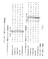
本発明のCIVPSは、スプライシング反応が制御され得るように改質されたIVPSも含む。図1(配列番号:30,配列番号:31,配列番号:32,配列番号:33,配列番号:34,配列番号:35,配列番号:36,配列番号:37,配列番号:38及び配列番号:39)に示されているように、IVPSをスプライシングする公知のタンパク質の並んだスプライス部位から幾つかの類似点が明らかになる。特に、-OH及び-SH側鎖は、下流スプライス部位でジペプチドHis-Asnにより先行した両方のスプライス部位のC-末端側の残基上に見出される。
【0023】
理論に結び付けるつもりはないが、ヒドロキシ/スルフヒドリル基は、スプライシング反応に関係し、これらの残基の改質はスプライシング反応を調節すると考えられる。このような改質は、挿入がマーカータンパク質活性を減少させるように、マーカータンパク質、例えばβ-ガラクトシダーゼに改質CIVPSを挿入することにより評価し得る。得られた改質タンパク質は、次いで、マーカータンパク質活性が増加する制御条件下で特定の時間間隔で評価し得る。実施例1〜3参照。さらに、ウエスタンブロット分析を使用して、スプライシング及び切断産生物を評価し得る。実施例8を参照。同定後、CIVPSをコードするDNAを単離し、標準DNA増幅方法を使用して増幅し得る。
【0024】
本発明により、CIVPS2のセリン1082の単一アミノ酸の変化は、タンパク質スプライシング反応を遅延させるかまたは遮断(block)することが知見された。特に、トレオニン置換変異体は、野生型酵素のポリメラーゼ活性の10%を示したが、システイン及びアラニン置換変異体は検出可能な活性を与えなかった。しかしながら、他のスプライス部位での切断に対応する反応生成物が観察された。この種は、スプライス部位でセリンとシステインを置き換えた変異体中に蓄積したが、セリンがトレオニンまたはアラニンと置き換えた時には変化がなかった。野生型CIVPS2は、スプライシング反応時にカルボキシ末端スプライス部位での切断で予想される大きさの種の蓄積を示したが、セリン1082がトレオニン、システインまたはアラニンに変えられた時もこの産生物の蓄積は減少したけれども依然として観察された。S1082A変異体(variant)はタンパク質スプライシングの証拠を示さなかったが、この産生物を産生した。
【0025】
カルボキシ-末端スプライス部位に於ける突然変異の誘発、即ちトレオニン1472(T1472)残基とセリンとのアミノ酸置換により、野生型と同一のスプライシングパターンが生じた。T1472とアラニン、グリシンまたはイソロイシンとの置換は検出可能なスプライシングを与えなかった。アスパラギン1471をアラニンと置換すると、スプライシングは観察されなかったが、アミノスプライス部位での切断の証拠が観察された。以下の表1は、CIVPS2に於ける切断及びスプライシングでのアミノ酸置換の効果をまとめたものである。
【0026】
【表1】

【0027】
スプライス部位アミノ酸を含むCIVPSアミノ酸の改質は、種々の方法で実施し得る。例えば、改質すべきアミノ酸残基を取り囲む配列を変更して、生物学的リン酸化部位を作り出し、特異的なキナーゼ及びホスファターゼに対する基質とする。プロテインキナーゼの例としては、例えばカゼインキナーゼII、cAMP依存性プロテインキナーゼ、cdc2及びpp60c-src[Pearson及びKemp,Methods in Enzymology 200:62(1991)]が挙げられる。ホスファターゼの例としては、例えばタンパク質ホスファターゼ2A、λホスファターゼ及びエルシニア(Yersinia)由来のyopホスファターゼ[Tonks,Current Opinion in Cell Biology,2:1114(1990)]が挙げられる。
【0028】
実施例6Cに記載の如くCIVPS2を例として使用すると、アルギニン残基を位置1079で置換して、コンセンサスカルモジュリン依存性プロテインキナーゼII部位を作り出す[XRXXS*;Pearsonら,上掲]。タンパク質スプライシング反応は、リン酸化度によって調節され得、ホスホセリンを作り出し、スプライシングをブロックするためにキナーゼを、リン酸を除去するためにホスファターゼを使用し、野生型セリンを回復し、タンパク質スプライシングする。
【0029】
さらに、重要なスプライス部位残基は、改質が逆転するまでスプライシング反応がブロックされるように、化学的に改質され得る。これは、例えば、異常なアミノ酸突然変異誘発[Norenら,Science 244:182(1989);Ellmanら,Methods in Enzymology 202:301(1991)]を使用することにより実施し得る。この方法を使用して、スプライシング反応に含まれるアミノ酸1個を翻訳時に、側鎖の側鎖官能基が化学的または光分解的に除去し得る基によって「マスク」される合成誘導体と置換し得る。例えば、実施例7に記載の如く、CIVPS2のセリン1082を以下の如く本方法により改質した。アンバー停止コドンをセリン1082に対応する位置でVentポリメラーゼ遺伝子に導入した(実施例6D参照)。この遺伝子を、野生型遺伝子のタンパク質スプライシングを指示することが既に示されたin vitro転写/翻訳系[Ellmanら,上掲]に添加した。このコドンを介して読み取るtRNAの非存在下では、切頭産物だけが予想された。化学的にO-(o-ニトロベンジル)セリンでアミノアシル化したアンバーサプレッサーtRNAをこの系に添加したときには、このコドンの先では連続的な翻訳が可能であり、その結果、改質セリンの部位特異性組み込み(incorporation)が得られた。予想通り、完全長前駆体だけが観察され、これは、スプライシング反応がブロックされたことを示している(図9)。o-ニトロベンジル基は350nmでの短時間の照射により除去可能なので[Pillai,Synthesis1(1990)]、ブロックされた前駆体は、照射後には通常スプライスすると考えられる。ブロックされた前駆体は、可視光に暴露されるとセリンを放出し、インキュベートするとスプライシング反応が起きるので、スプライスされた産生物がはっきりと知見される(図10)。
【0030】
この戦略は、CIVPS2の下流スプライス部位に見られるトレオニン1472並びに、側鎖の化学官能基がスプライシングに必要であるか、その位置に嵩高い基を導入することによりスプライシングを立体的に妨害する他の任意の残基に適用し得る。ブロック基は、保護すべき側鎖の化学的性質だけでなく、(化学的または光分解的)脱ブロックの望ましい方法に基づいて選択し得る。例えば、タンパク質スプライシングの他の例(図1)に存在するシステイン基は、例えば、ジスルフィド交換(例えば、ジチオジピリジンを用いる)または遷移金属イオン(例えば、Hg2+)との錯体形成を使用してブロックし得るチオール側鎖を有する。Corey及びSchultz,J.Biological Chemistry 264:3666(1989)参照。得られたブロック化前駆体は、次に、各々金属キレート化剤の添加または温和な還元によりスプライシングするために活性化し得た。
【0031】
IVPS1及びIVPS2は各々、エンドヌクレアーゼ、即ち、I-Tli-II及びI-Tli-Iをコードする。さらにDV IVPS1は、IVPS1がVent DNAポリメラーゼ遺伝子に挿入したのと同位置でDV DNAポリメラーゼに挿入し、Vent IVPS1遺伝子と62%の相同であるエンドヌクレアーゼI-PspIもコードする。Tfp1、M.tuberculosis rec A、Vent及びDeep Vent DNAポリメラーゼ中のIVPS読み取り枠は、ホーミングエンドヌクレアーゼ、イントロンを欠失する対立遺伝子を切断し得るイントロン-コード化タンパク質の種類と似たタンパク質配列を有する[Hirataら,上掲,Kaneら,上掲,Davisら,上掲,Perlerら,上掲]。
【0032】
特定の宿主細胞は、CIVPSの遺伝子産物に対して耐性がないので、場合により、エンドヌクレアーゼ機能を不活化するのが好ましい。本発明により、タンパク質スプライシングは、CIVPSエンドヌクレアーゼ機能が不活化された時に起きることが明らかになった。このような不活化は、例えば、ランダム突然変異誘発、欠失または挿入不活化または特定部位の突然変異誘発を含む種々の方法で達成し得る。エンドヌクレアーゼ機能は、部位特異的突然変異誘発により不活化されるのが好ましい。I-Tli-Iは、特徴的なドデカペプチドモチーフ対中の他の「ホーミングエンドヌクレアーゼ」と似た配列を共有する[Cummingsら,Curr.Gent.16:381(1989)]。実施例6Bに見られるように、エンドヌクレアーゼ活性は、これらのモチーフの一種内の単一残基のオリゴヌクレオチド部位特異的突然変異誘発により不活化された(アスパラギン酸1236からアラニンへ)。別の残基の置換は、タンパク質スプライシングに影響を及ぼすことなくエンドヌクレアーゼ活性を減少または除去し得た。エンドヌクレアーゼ機能の不活化により、改質タンパク質を保持する構築物の安定性を増加させることが明らかになった。
【0033】
本発明で使用し得るターゲットタンパク質としては、例えば、酵素、毒素、細胞毒素、糖タンパク質及び成長因子が挙げられる。このような多くのタンパク質は当業者には公知である。このようなタンパク質のアミノ酸及びヌクレオチド配列は、多くのコンピューターデータベース、例えば、GenBank、EMBL及びSwiss-Protにより容易に得られる。あるいは、ターゲットタンパク質のヌクレオチドまたはアミノ酸配列は、当業界で日常的な方法を使用して決定し得る。
【0034】
実質的にターゲットタンパク質活性を不活化するのが望ましい場合、このような活性を不活化する領域にCIVPSを挿入する。このような領域は当業者には公知であり、例えば、結合部位、酵素活性部位、タンパク質の保存モチーフ、例えばDNAポリメラーゼ及び二量化または多量化部位(multimerization sites)が挙げられる。あるいは、CIVPSをランダムに挿入し、所望の活性レベルが得られるまで各改質タンパク質の活性を測定し得る。このような改質タンパク質は、ネイティブのタンパク質と比較して活性が約50%減少しているのが好ましい。約75%減少しているのがより好ましく、99%以上減少しているのが最も好ましい。
【0035】
CIVPSは、任意の多くの方法によりターゲット遺伝子に挿入し得る。好ましくは、適正なタンパク質スプライシングを確実にするためにCIVPSが切除される場合、CIVPSの切除によりそのアミノ酸がスプライス部位に残るので、適正なスプライス部位残基の直前にCIVPSを挿入することが重要である。これは、好適なスプライス部位アミノ酸の直前にCIVPSを挿入するか、またはCIVPSが好適なアミノ酸を一緒に「持ってくる(bring)」ようにCIVPSを改質することによって達成し得る。
【0036】
例えばCIVPS1、2または3は、好適なスプライス部位アミノ酸、例えばセリン、トレオニンまたはシステイン残基の直前、最も好ましくはセリンまたはトレオニンの直前に挿入し得る。図1を参照。このような部位は、殆どのターゲットタンパク質で便利に利用し得る。
【0037】
ターゲットタンパク質が毒素であるような特定の状態では、第2の制御を加えることによりタンパク質スプライシングをさらに制御することが望ましい。これは、例えばCIVPSが通常先行せず、スプライシング反応を遅延させ得るようなそれほど最適でないアミノ酸の前にCIVPSを挿入することにより達成し得る。
【0038】
上記の如く、CIVPSが好適な下流アミノ酸を一緒に「持ってくる」場合、ターゲットタンパク質内の任意の部位に挿入し得る。これは、所望の下流のアミノ酸のコドンを有するCIVPS DNAの作製により達成し得る。このようなDNAの産生方法を以下に詳述する。このDNAは次いで、ターゲットDNA内の任意の部位に挿入し得る。得られた改質タンパク質のタンパク質スプライシング時には、CIVPSによりもたらされた過剰の残基は後方に残存する。従って、最終産生物の活性が重要である場合、当業者は、過剰の残基が、活性に悪影響を及ぼすようなターゲットタンパク質領域内に絶対残存しないように製造段階を取らねばならない。
【0039】
CIVPSは、標準法[例えば、Hunkapillerら,Nature 310:105(1984)]及び市販のタンパク質合成機を使用して、任意の所望の部位に挿入した、CIVPSを含む、ターゲットタンパク質の第1のアミノ酸配列を化学的に合成することにより、ターゲットタンパク質に挿入またはターゲットタンパク質に融合し得る。
【0040】
あるいは、CIVPSをコードするDNA配列は、両方のコード配列が連続読み取り枠を形成するようにターゲットタンパク質をコードするDNA配列に挿入または融合される。これは、当業者には公知の種々の方法を利用して実施し得、その幾つかを以下に記載する。
【0041】
例えば、CIVPS DNAを、ターゲット遺伝子内でブラント切断し且つ枠内にある任意の制限酵素部位に挿入する。これは、第1に、その3'末端にトレオニンコドン(Vent IVPS2)またはセリンコドン(Deep Vent IVPS1若しくはVent IVPS1)をもつCIVPS DNAフラグメントを合成することにより実施し得る。次いで、このフラグメントを制限エンドヌクレアーゼによりブラント末端に切断した線状プラスミドにフレーム内で結合する。lacZ DNA配列、例えば、EcoRV部位を使用して、残基375(アスパラギン酸)と376(イソロイシン)との間にVent IVPS2またはDeep Vent IVPS1を挿入し得る。図2参照。しかしながら上記の如く、この方法を使用してCIVPSを切除する場合、過剰の残基がスプライス部位に残存し、従って、CIVPSが挿入される場所に応じて、得られたタンパク質はネイティブのタンパク質と同一機能または構造を持たなくてもよい。
【0042】
CIVPS DNAは、ターゲット遺伝子と適合性の制限部位を作り出すためにCIVPSの両方の末端または一方の末端の近くにサイレント突然変異(アミノ酸残基を維持する)を起こすことによっても挿入し得た。例としてCIVPS2を使用すると、サイレント突然変異により、BspEI制限部位は5'末端の近くに作り出され得、SpeI制限部位はその3'末端の近くに作り出され得る。新しい制限部位と重複し、且つasp 594またはthr 595のいずれかに於けるlacZターゲット遺伝子の開始により連続するPCRプライマーを使用して、BspE1及びSpeI制限部位と適合性のlacZフラグメントを作製し得る。次いで、CIVPSをlacZコード領域内のアスパラギン酸コドン(残基 594)とトレオニンコドン(残基 595)の間に挿入する。DNAフラグメントは、CIVPS及びターゲット遺伝子の両方から、CIVPSの末端と重複する挿入部位のその末端とのPCRにより合成され得、従って同一制限部位を含む。好適な制限エンドヌクレアーゼ処理後、適合性末端を有するDNAフラグメントを連結して融合遺伝子を作製し得る。CIVPSの切除後には過剰残基は全く残らないので、スプライシング発生時にネイティブのポリペプチドが形成するだろう。作り出された制限部位がCIVPS内に1個及びターゲット遺伝子内に1個であると、多重フラグメントの連結及び複雑なスクリーニング方法を回避し得るので好ましい。
【0043】
ターゲット遺伝子配列を保持するプラスミドベクターが比較的小さい場合、例えば、約5kb未満である場合、線状型プラスミドをPCRを使用して作製し、次いで線状プラスミドをCIVPS遺伝子に連結する。この方法を使用して、CIVPS遺伝子を以下の如くターゲット遺伝子の任意の位置に挿入し得る。第1に、ターゲット遺伝子を含むプラスミドDNAを、挿入部位(例えば、CIVPS1、2及び3に関してはセリン若しくはトレオニンコドン)で開始するプライマー対または、CIVPSが好適な下流アミノ酸を持ってくる場合には任意のコドンを使用するPCRにより合成し得る。次に、(任意にセリンまたはトレオニンを含む)CIVPS遺伝子を(セリンまたはトレオニンコドンを含まない)線状プラスミドDNAに連結し得る。必要なスプライス部位アミノ酸(セリンまたはトレオニン)を、CIVPSフラグメントまたはターゲット遺伝子のいずれかの上に配置し得る。内在性セリンまたはトレオニンの上流に配置する際に必要なアミノ酸をCIVPSフラグメント上に有する利点は、コード領域にセリンまたはトレオニンが欠失しているので、(CIVPS挿入物を含まない)自己連結ベクターDNAがターゲット遺伝子の欠陥産物だけを発現し得るということである。これは、融合タンパク質がスプライスして機能性産物を産生する場合に、融合構築物に関する表現型の選択を助長し得る。
【0044】
改質タンパク質をコードする融合DNAは、好適な発現ベクター、即ち、挿入したタンパク質-コード配列の転写及び翻訳に必要な要素を含むベクターに挿入し得る。種々の宿主-ベクター系を使用してタンパク質-コード配列を発現し得る。これらの例としては、ウイルス(例えば、ワクシニアウイルス、アデノウイルスなど)に感染した哺乳動物細胞系;ウイルス(例えば、バキュロウイルス)に感染した昆虫細胞系;酵母ベクターを含む酵母のような微生物または、バクテリオファージDNA、プラスミドDNA若しくはコスミドDNAで形質転換したバクテリアが挙げられる。使用した宿主-ベクター系に依存して、多数の好適な転写及び翻訳要素のうちの任意のものを使用し得る。例えば、改質真核生物タンパク質を発現する際、好適な真核生物ベクター及び宿主細胞を使用すると都合がよい。融合DNAの発現により、本発明の改質タンパク質が産生する。
【0045】
一度得られたら、公知方法を好適に組み合わせて、改質タンパク質を分離し且つ精製し得る。これらの方法としては、例えば、塩析及び溶媒沈殿などの溶解性を利用する方法、透析、限外濾過、ゲル濾過及びSDS-ポリアクリルアミドゲル電気泳動などの分子量の違いを利用する方法、イオン交換カラムクロマトグラフィーなどの電荷の違いを利用する方法、アフィニティークロマトグラフィーなどの特異的な親和性を利用する方法、逆相高性能液体クロマトグラフィーなどの疎水性の違いを利用する方法及び等電点電気泳動などの等電点の違いを利用する方法が挙げられる。
【0046】
所望により、改質タンパク質を、CIVPSが除去される予め決定した条件にかけ得る。このような条件は、使用するCIVPSに依存する。例えば、CIVPS1、2及び3は、改質タンパク質を高温、42℃〜80℃、最も好ましくは42℃〜60℃にかけることにより除去し得る。これは、公知方法、例えば、水浴または熱発生レーザーを使用して実施し得る。インキュベーション時間は1分未満から数時間以上を変動し得る。上記の如く、特定の状態では、ターゲットタンパク質の熱感受性に依存して、温度を下げながらインキュベーション時間を延長するのが望ましい。さらに、in vivoスプライシングが望ましい場合、宿主生物の増殖と適合する温度が好ましい。
【0047】
本発明により、改質タンパク質が無毒性であるように毒性ターゲットタンパク質を改質するためにCIVPSを使用することにより宿主細胞に対して非常に毒性のタンパク質を産生し得る。これは、例えば、CIVPSを毒性に関与する領域に挿入することにより実施し得る。単離後、無毒性改質タンパク質を、CIVPSを除去し、得られた毒性を単離し得るような予め決定した条件にかけ得る。
【0048】
タンパク質が宿主細胞に対して非常に毒性が高い場合、「トランスプライシング(transplicing)」と称される方法を使用してそのタンパク質を産生するのが望ましい。この方法を使用すると、毒性タンパク質は、別個の宿主細胞中にCIVPSを挿入することにより改質されている2つ以上の部分(pieces)内で産生する。例えば、ターゲットタンパク質のカルボキシ末端にCIVPSのアミノ末端フラグメントが挿入されるターゲットタンパク質のアミノ部分を含む第1の改質タンパク質を産生し、その後、ターゲットタンパク質のアミノ末端にCIVPSの残りのフラグメントが挿入されるターゲットタンパク質の残りの部分を含む第2の改質タンパク質を産生し得る。あるいは、重複CIVPSフラグメントを使用し得る。次いで、各改質タンパク質を宿主細胞から単離し、CIVPSのスプライシングに好適な条件下、一緒にインキュベートする。これにより連結したターゲットタンパク質が得られる。ターゲットタンパク質を2個の異なる宿主に分割することにより、微小画分でさえも宿主に悪影響を与えるin vivoスプライスする可能性はない。さらに、CIVPS全体を第1の改質タンパク質のスプライス部位の一方の側に挿入し、残りのターゲットタンパク質フラグメントをスプライシング混合物に添加した。
【0049】
本発明のIVPSは、「タンパク質連結」で使用し、非天然アミノ酸残基、構造プローブ、識別エピトープ若しくはタグ(tags)または、他の決定子(determinants)をターゲットタンパク質に添加し得る。例えば、ターゲットタンパク質をIVPSのアミノ末端に融合し得る。停止コドンを、IVPSのカルボキシ末端の直後に配置し得る。次いで融合すべきペプチドを混合物に添加し得る。天然のスプライシング機構を非常に厳密に模倣するためには、このペプチドのアミノ末端はセリン、トレオニンまたはシステインであってもよい。次いで、産生物のスプライシングへ質量作用により増大されたスプライシング反応を進行し得る。
【0050】
上記反応は、ターゲットタンパク質のアミノ末端にカルボキシ末端で融合したIVPSから構成される出発物質を用いて起きるようにもし得る。特定のCIVPS内で開始するセリン残基に先行するように工学的に作製されたメチオニンでの開始が、E.coli内でアミノ末端セリン残基を残すように翻訳を起こさせる。次いで、このターゲットタンパク質のアミノ末端に融合すべきペプチドを添加し、スプライシングを進行させた。添加されるペプチド上のカルボキシ末端残基に関しては公知の要件がないので、このような試みは好ましい。さらに、今回の実験的証拠から、上流スプライス部位の切断は連結反応に先行して起こることが示唆されており、この試みが天然の反応機構に非常に近いことを示している。ターゲットペプチドをペプチドに添加し、融合タンパク質を翻訳し易くすることが可能であった。
【0051】
本発明は、他のタンパク質の誘導または分化などの特定の条件下、または細胞周期の特定の部分の間にターゲットタンパク質の効果を研究するためにも使用し得る。例えば、特定のタンパク質をコードする遺伝子の染色体コピーをCIVPSを含むバージョンと置き換え得る。細胞周期の特定点、分化または他の所望の点で、細胞を加熱すると前駆体がスプライスし、活性ターゲットタンパク質はこの時点でのみ存在する。
【0052】
本発明のCIVPSを使用して、CIVPSに対して特異的な抗体を持つアフィニティクロマトグラフィーを使用することにより改質タンパク質を単離することもできる。例えば、標準法を使用してCIVPSに対して結合親和性を持つモノクローナルまたはポリクローナル抗体を産生し得る。これらの抗体は、アフィニティクロマトグラフィー精製法で使用し、改質タンパク質を単離し得る。精製後、所望により、改質タンパク質をCIVPSを切除する予め決定した条件にかけ得る。
【0053】
上記の如く、CIVPSスプライス部位での切断は、タンパク質スプライシングなしに実施でき、ターゲットタンパク質からCIVPSの分離を制御し得る。従って、このようなCIVPSは融合タンパク質精製系で使用し得る。
【0054】
融合タンパク質精製系は当業者には公知である[欧州特許出願第0 286 239号及びN.M.Sassenfeld、TIBTECH,8:88-93(1990)参照]。通常、このような系では、結合タンパク質とターゲットタンパク質はプロテアーゼ認識部位を持つリンカーで結合されている。次いで、融合物は、結合タンパク質に対して親和性を持つ基質上のアフィニティクロマトグラフィーにより精製する。結合タンパク質とターゲットタンパク質を、次に、プロテアーゼ、例えば第Xa因子と接触させることにより分離する。これらの系では、非常に精製されたターゲットタンパク質を得るために、プロテアーゼはターゲットタンパク質並びに汚染の可能性から分離せねばならず、従って、追加の精製段階にかける。プロテアーゼの代わりにCIVPSを使用することにより、本発明の方法は、現在使用されているタンパク質融合精製系に含まれるこれら及び他の問題を避けることができる。
【0055】
本発明の方法により、基質(結合タンパク質)に対して親和性を持つタンパク質とターゲットタンパク質の間にCIVPSがある融合タンパク質を含む改質タンパク質が形成する。このような融合タンパク質を形成する方法は、当業者には公知である[欧州特許出願第0 286 239号及びJ.Sambrookら,Molecular Cloning:A Laboratory Manual(1989),Cold Spring Harbor Laboratory Press,Cold Spring Harbor,NY,p.17.29-17.33参照]。
【0056】
本発明の方法で使用し得る結合タンパク質としては、例えば、糖結合タンパク質、例えばマルトースまたはアラビノース結合タンパク質、レセプター結合タンパク質、アミノ酸結合タンパク質及び金属結合タンパク質が挙げられる。他の結合タンパク質は当業界で公知である[欧州特許出願第0 286 239号及びN.M.Sassenfeld,TIBTECH,上掲、参照]。
【0057】
次いで改質タンパク質を、結合タンパク質が特異的親和性を有する基質と、例えばアフィニティクロマトグラフィーを用いて接触させる。
【0058】
非常に精製されたターゲットタンパク質を、例えば、CIVPSとターゲットタンパク質の間で切断が開始するような予め決定した条件にCIVPSをかけることにより、カラムから遊離し得る。あるいは、精製融合タンパク質を、上記の如くカラムから溶離且つ遊離し得る。
【0059】
【実施例】
本発明を、以下の実施例によりさらに説明する。これらの実施例は、本発明を理解しやすくするためのものであり、限定するものではない。
【0060】
上記及び下記引用の参照文献は、すべて本明細書中参照として含まれる。
【0061】
実施例1
ターゲット遺伝子コドンの間の平滑部位に挿入するためのIVPSカセットの合成
lacZコード領域(lacZ coding region)又は他の任意のターゲット遺伝子にIVPSをフレーム内挿入(in−frame insertion)するためのDNAフラグメント又はカセットは、第1の下流外部タンパク質配列(external protein sequence=EPS)コドンを用いて又は用いずにポリメラーゼ連鎖反応(PCR)によって製造し得る。天然の(native)下流残基は、Deep Vent IVPS1及びVent IVPS1の場合のセリン、又はVent IVPS2の場合のトレオニンである。IVPS2は、トレオニン又はシステインに先行していれば、程度は低いが、スプライスできることが判明した。理論に拘束されたくはないが、全てのIVPSは、セリン、トレオニン又はシステインのいずれかに先行していれば、ある程度までスプライスできると考えられる。下流のセリン又はトレオニンを含むカセットは、セリン又はトレオニンに先行する位置を含めて、ターゲット遺伝子内の任意の所望位置に挿入することができる。この構築では、セリン又はトレオニンをターゲット遺伝子から欠失させ、これをカセット上の入来残基で置換し得る。下流のセリン、トレオニン又はシステインを有していないカセットは、ターゲット遺伝子内のセリン、トレオニン又はシステインより前に挿入し得る。
【0062】
下記のプロトコルは、第1の下流EPSコドンを含む、Deep Vent IVPS1(CIVP3)及びVent IVPS2(CIVP2)(エンド+及びエンド-バージョン)用カセットの製造を説明するものである。
【0063】
PCR混合物は、Vent DNAポリメラーゼ緩衝液(NEB)に、2mMの硫酸マグネシウムと、400μMの各dNTPと、0.9μMの各プライマーと、40ngのプラスミドDNAと、100μl中2単位のVent DNAポリメラーゼとを加えたものからなる。増幅は、Perkin−Elmer/Cetusサーマルサイクラー(thermal cycler)を用いて、94℃で30秒、48℃で30秒及び72℃で2分のサイクルに30回かけることにより実施した。Deep Vent IVPS1は、pUC19のBamHI部位に挿入したPyrococcus sp.DNAポリメラーゼ遺伝子を含む4.8KbのBamHIフラグメントを有するpNEB#720(ATCC No.68723)から合成した。Vent IVPS2は、ベクターBluescribe SK−(Stratagene)のVent DNAポリメラーゼ遺伝子配列の1.9kb EcoR1フラグメント(2851−4766)を有するpV153−2から合成した。IVPS2にはpNEB671(ATCC No.68447)を使用することもできる。pAMQ29は、Vent IVPS2コード領域内にアミノ酸置換(アスパラギン酸1236をアラニンに)を有する、pV153−2のエンドヌクレアーゼ欠失誘導体である。プライマー5’−AGTGTCTCCGGAGAAAGTGAGAT−3’(配列番号:3)(Vent IVPS2正方向(forward)、3534−3556、A3542のCへの置換)及び5’−AGTATTGTGTACCAGGATGTTG−3’(配列番号:4)(Vent IVPS2/Thr逆方向(reverse)、4685−4706)を用いて、3’末端にトレオニンコドンを有するエンド+又はエンド-Vent IVPS2フラグメント(1173bp)を合成した。プライマー5’−AGCATTTTACCGGAAGAATGGGTT−3’(配列番号:5)(DV IVPS正方向、1839−1862)及び5’−GCTATTATGTGCATAGAGGAATCCA−3’(配列番号:6)(DV IVPS1/Ser逆方向、3428−3452)を用いて、3’末端にセリンコドンを有するPyrococcus sp.(又はDeep Vent)IVPS1フラグメント(1614bp)を合成した。C末端セリン又はトレオニンを欠失したIVPSフラグメントの形成には、最後の3つのヌクレオチドを欠失した逆方向プライマーを使用し得る。
【0064】
PCR試料はフェノール及びクロロホルムで抽出し、0.3M NaAc及び70%エタノール中で−20℃で一晩沈殿させ、ミクロ遠心機(microfuge)で10分間、10Kで遠心分離して回収し、乾燥し、各々を30μlの蒸留水に再懸濁し、1%アガロースゲル上に配置して60ボルトで15時間電気泳動にかけた。PCR増幅フラグメントを含むゲル切片を1%低融点(low melting)アガロースゲル中に配置して80ボルトで2時間電気泳動にかけた。0.5mlのTE緩衝液(10mM トリス−HCl/0.1mM EDTA、pH8.0)中65℃で30分間インキュベートし、フェノール、フェノール−クロロホルム(1:1混合物)及びクロロホルムで抽出し、0.6M NaAc(pH5.2)及び50%イソプロパノール中で−20℃で一晩沈殿させることにより、低融点アガロースゲルからDNAフラグメントを回収した。DNAを遠心分離し、70%エタノールで洗浄し、乾燥し、15.5μlの蒸留水に再懸濁した。
【0065】
IVPS DNAフラグメントのリン酸化を、2μlの10×ポリヌクレオチドキナーゼ緩衝液(NEB)と、15.5μlの精製DNAと、2μlの10mM ATPと、20μl中5単位のT4ポリヌクレオチドキナーゼ(NEB)とを用いて37℃で60分間実施した。試料を65℃の水浴中で10分間加熱した。80μlのTE緩衝液(10mM トリス−HCl/0.1mM EDTA、pH8.0)を加えた後、試料をフェノール、フェノール−クロロホルム(1:1混合物)及びクロロホルムで逐次抽出した。DNAを2.5M NH4Ac及び70%エタノール中で−70℃で3.5時間沈殿させ、ミクロ遠心機で10分間10Kで遠心分離してペレット化し、冷70%エタノールで洗浄し、乾燥し、蒸留水(Vent IVPS2又はDeep Vent IVPS1 DNAの場合は20μl、Vent IVPSエンドDNAの場合は10μl)に再懸濁した。
【0066】
実施例2
β−ガラクトシダーゼをコードするプラスミドのような制限酵素直線化プラスミド(restriction enzyme linearized plas mid)へのIVPSのフレーム内挿入
この実施例では、2つのコドンの間でターゲット遺伝子内に平滑切断(blunt cut)を形成する制限酵素部位にカセットを挿入することによって、IVPSカセットをターゲット遺伝子にクローニングする方法を説明する。カセットは、必要であれば、C末端セリン、システイン又はトレオニンを有し得る。このプロトコルは、制限酵素がターゲット遺伝子ベクターを1回又は2回切断する場合に最も効果的である。具体例として、lacZ遺伝子のEcoRV部位への挿入を説明する(図2)。
【0067】
EcoRV直線化pAHO5の調製
pAHO5は、tacプロモーターの下流のpAGR3のポリリンカー内でBamHI部位とSmaI部位との間に挿入されたpRS415(Simonsら、Gene 53:85−96(1987))からの3.31kb BamHI−DraIフラグメント上に完全なlacZ遺伝子配列を有する。tacプロモーターは、lacIq遺伝子の産物によって抑制され得且つイソプロピルβ−D−チオガラクトシド(IPTG)によって誘導され得る転写制御エレメントである。5.9KbベクターpAHO5(NEB)は更に、tacプロモーター及びポリリンカーの上流の転写終結配列と、大腸菌lacIq遺伝子とを有する。pAHO5は2つのEcoRV認識配列を含んでいる。EcoRVは開裂部位に平滑末端を残す。EcoRV開裂部位の1つは、375番目のコドン(アスパラギン酸)と376番目のコドン(イソロイシン)との間のlacZコード領域内で切断を行い、IVPSフラグメントのフレーム内挿入用部位として予定される。もう1つの部位は大腸菌lacIq遺伝子内で3.2kb下流に位置する。該プラスミドを部分的に切断して、EcoRV部位のうちの1つだけが開裂されている分子を幾つか産生する。これらの直線プラスミドを精製する。IVPSカセットはいずれかのEcoRV部位にランダムにクローニングする。従って、得られた組換え体は、配向(orientation)と固有のEcoRV部位への挿入とのためにスクリーニングする必要がある。DNAは、15μgのpAHO5DNAを100μlの1×NEB緩衝液2中40単位のEcoRV(NEB)と共に37℃で60分間インキュベートすることにより部分的に消化した。EcoRVを失活するために試料を65℃で10分間加熱した後、20μlの染料添加アガロースゲルを試料に加えた。1%低融点アガロースゲルでの電気泳動によりDNAフラグメントを分離した。直線化pAHO5プラスミドDNAを実施例1と同様に低融点アガロースゲルから回収し、44.6μlの蒸留水に再懸濁した。
【0068】
EcoRV直線化pAHO5の脱リン酸化を、2μgのDNAと4単位の子牛腸アルカリホスファターゼ(NEB)との存在下で、50μlの1×NEB緩衝液2中50℃で60分間実施した。0.5μlの0.5M EDTA(pH8.0)を加えた後、試料を65℃の水浴中で30分間加熱し、フェノール、フェノール−クロロホルム(1:1混合物)及びクロロホムで抽出した。DNAを0.75M NH4Ac及び70%エタノール中で2時間沈殿させ、実施例1と同様に回収し、20μlの蒸留水に再懸濁した。
【0069】
IVPS−lacZ融合遺伝子の構築
脱リン酸化pAHO5 DNAとリン酸化IVPSフラグメントとの連結を、20μl倍容で、8.6μlの蒸留水と、2μlの10×T4 DNAリガーゼ緩衝液(NEB)と、4μlの0.1μg/μl脱リン酸化pAHO5 DNAと、前述のように調製した5μlのIVPS DNA(0.25μgのVentIVPS2、0.4μgのDeep Vent IVPS1又は0.25μgのVent IVPS2エンド-)と、160単位のT4 DNAリガーゼ(NEB)とを加えて16℃で15時間実施した。
【0070】
100μlのコンピテントRR1細胞と10μlの連結反応試料とを氷上で30分間混合し、42℃で2分間加熱し、氷上で5分間冷却し、0.8mlのLB培地(10g/リットル トリプトン、5g/リットル酵母抽出物、10g/リットルNaCl、1g/リットル デキストロース、1g/リットルMgCl26H2O、pH7.2、25℃)を加え、30℃で45分間インキュベートして、大腸菌株RR1を形質転換した。100μg/mlのアンピシリンを加えたLBプレートに試料をプレーティングした。30℃で一晩のインキュベーション後に、プレート当たり約150〜300のコロニーが観察された。
【0071】
コロニーのハイブリダイゼーションを使用して、組換えプラスミドを有するクローンをスクリーニングした。実施例1で説明したVent IVPS2正方向プライマー及びDeep Vent IVPS1正方向プライマーを、T4ポリヌクレオチドキナーゼを用いて[Y−32P]で放射性標識し、ハイブリダイゼーションプローブとして使用した。コロニーを採取してニトロセルロース上に配置し、下記の各溶液中で5分間処理した:10%SDS、0.5M NaOH/1.5M NaCl、0.5Mトリス−HCl(pH7.5)/0.5M NaCl(2回)及び2×SSC(2回)。ニトロセルロースフィルターを室温で1時間乾燥し、真空下80℃で2時間焼成し、6×SSCに5分間浸漬し、50mMトリス−Cl(pH8.0)、1M NaCl、1mM EDTA及び0.1%SDSからなる溶液中で42℃で2時間洗浄した。6×NET、5×デンハート(Denhardt’s)、0.5%SDS及び25μg/ml変性サケ精子DNA中で42℃で4時間処理した後、フィルターを放射性標識オリゴマープローブと共に同一条件で16時間インキュベートし、次いで6×SSC中で、室温で15分間の洗浄3回と、42℃で2分間の洗浄2回と、50℃で2分間の洗浄2回とにかけ、その後オートラジオグラムを撮った。36個のクローンが、対応するオリゴマープローブにハイブリダイズしていた。
【0072】
実施例1で説明したVent IVPS2正方向プライマー(又はDeep Vent IVPS1正方向プライマー)と、挿入部位から392nt下流のlacZコード配列(1417−1440、1437にG:T不適合を有する)に対して相補的なlacZ逆方向プライマー(5’−AGGGTCGACAGATTTGATCCAGCG−3’(配列番号:7))とを用いて、ポジティブクローンから抽出したプラスミドDNAのPCR増幅により挿入位置の決定を行うために、ポジティブクローンを更に分析した。14個のクローンからのPCR反応で、対応するDNAフラグメントが産生された。クローンpVT133、138、139、141、142及び144は1.4KbのVent IVPS2挿入物を含んでおり、pVTE 834、836、839及び841はVent IVPS2(エンド-)挿入物を含んでおり、いずれも約1.1kbのDNAフラグメントを産生する。クローンpDVS712、742、745及び746は1.6KbのDeep Vent IVPS1挿入物を有し、約2.0KbのDNAフラグメントを産生する。
【0073】
IVPS−lacZ融合遺伝子の発現
前記クローンを、誘導物質IPTGとの融合(修飾)タンパク質を発現する能力について更に調べた。
【0074】
クローンは、100μg/mlのアンピシリンを加えたLB培地中でOD600nmが0.5になるまで30℃で培養した。非誘導細胞(uninduced cell)から溶解物を調製するために、1.5mlの培養液をペレット化し、100μlの尿素溶解緩衝液(urea lysis buffer)に再懸濁し、10分間沸騰させた。IPTGを最終濃度0.3mMで加えた後、培養物を30℃で更に4時間増殖させた。1.5mlの培養液の細胞をペレット化し、2時間及び4時間の誘導後に250μlの尿素溶解緩衝液で溶解した。クーマシーブルー染色ゲルでタンパク質生成物を分析した。Vent IVPS2−lacZ融合構築物のうちの3つ(pVT139、142及び144)並びに4つのVent IVPS2(エンド-)−lacZ融合構築物の全ては、Vent IVPS2−β−ガラクトシダーゼ融合タンパク質について予想された大きさである約162〜165KDaの主産物を発現した。4つのDeep Vent IVPS1−lacZ融合クローンはいずれも、Deep Vent IVPS1−β−ガラクトシダーゼ融合タンパク質について予想された大きさである173〜178KDaのより大きい産物を発現した。
【0075】
I−Tli−I(NEB)又はβ−ガラクトシダーゼ(Promega)に対する抗体を用いるウェスタンブロットにより、pVT142及び144並びにpVTE836及び839に由来するVent IVPS2融合タンパク質のアイデンティティを更に分析した。試料を、予備染色したマーカー(BRL)を用いて4〜20%SDSゲル(ISS,Daichi,Tokyo,Japan)で電気泳動にかけ、ニトロセルロースに移し、抗血清(マウス由来)でプローブし、アルカリホスフェート結合抗マウス第二抗体を製造業者(Promega)の指示通りに使用して検出した。試験した4つのクローン全部から得られた約160KDaのバンドは両方の血清と反応し、クーマシーブルー染色バンドと同じ位置に移動する。Deep Vent IVPS1融合も調べた。β−ガラクトシダーゼ及びI−PspI(Deep Vent IVPS1のタンパク質生成物)に対する血清を用いてpDVS712及び742のウェスタンブロット分析を行ったところ、クーマシーブルー染色バンドと同じ約168〜175KDaに予想された主バンドが得られた。
【0076】
実施例3
β−ガラクトシダーゼ−IVPS融合におけるタンパク質スプライシングの熱制御
前述の構築物(lacZ EcoRV部位に挿入したIVPS)は誘導後に融合(改質)タンパク質を産生した。IVPSタンパク質は、高温でのインキュベーションによって、連結ターゲットタンパク質(活性β−ガラクトシダーゼ)と遊離IVPSエンドヌクレアーゼとを形成するために、融合タンパク質から切り出すことができる。
【0077】
スプライシングは温度誘導によって制御できる:粗抽出物中のβ−ガラクトシダーゼ活性は温度シフトに応答して増加する。
【0078】
RR1(大腸菌宿主)及びpAHO5含有PR1(実施例2で説明した非融合β−ガラクトシダーゼ親プラスミド)又は融合構築物pVT142(Vent IVPS2もしくはCIVPS2)、pVTE836(Vent IVPS1エンド-)もしくはpDVS712(Deep Vent IVPS1もしくはCIVPS3)の培養物から下記のステップに従って粗抽出物を調製した。単コロニー(single colony)を、100μg/mlのアンピシリンを加えた10mlのLB培地に接種し、30℃で一晩インキュベートし、1リットルのLB培地(100μg/ml アンピシリン)中30℃でOD600nmが約0.5になるまで継代培養し、0.3mMのIPTGで30℃で2時間誘導した。細胞を遠心分離し、100mlのLBに再懸濁し、4℃で3分間超音波処理し、7000rpmで15分間遠心分離した。上清を回収し、−20℃で貯蔵した。
【0079】
粗抽出物の7.5mlアリコートを42℃又は50℃の水浴中でインキュベートした。1mlアリコートを、pVT142及びpVTE836抽出物の場合は1時間後、2時間後及び12時間後に採取し、pDVS712、pAHO5又はRR1抽出物の場合は0.5時間後、1時間後、2時間後、4時間後及び16時間後に採取した。
【0080】
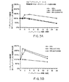
Millerらの方法[Experiments in Molecular Genetics(1972),Cold Sring Harbor,New York,Cold Spring Harbor Laboratory]でβ−ガラクトシダーゼ活性を測定した。アッセイ緩衝液は、Z緩衝液を2.7μl/mlの2−メルカプトエタノールと混合することによって調製した。基質o−ニトロフェニル−β−D−ガラクトピラノシド(ONPG)を4mg/mlでアッセイ緩衝液に溶解した。処理した抽出物又は非処理抽出物0.1mlを、0.9mlのアッセイ緩衝液と1滴の0.1%SDSとが入っている試験管に移し、28℃で5分間インキュベートした。ブランクには0.1mlのLB培地を使用した。4mg/mlのONPGを0.2ml加えてアッセイ反応を開始させた。適当な黄色が発色した時点で、0.5mlの1M Na2CO3を加えて反応を停止させた。インキュベーション時間を記録し、活性をスペクトロホトメーターでOD420nm及びOD550nmで測定した。熱処理した抽出物からの酵素活性は次のように計算した。インキュベーション後の活性をゼロ時点の活性で除算し、比に100を掛けてパーセンテージを出した。酵素活性の比較の結果、熱処理は最初の2時間のインキュベーションではRR1又はRR1/pAHO5抽出物からの活性に作用しないことが判明し、3つのIVPS−LacZ融合構築物pVT142、pVTE836及びpDVS712は全て42℃への温度シフトに応答して非処理試料の143%から221%ヘの酵素活性の増加を示した(図3)。β−ガラクトシダーゼ活性の増加は、IVPSの切り出しと2つのβ−ガラクトシダーゼ半体の連結とに起因するものであり、活性な酵素をより多く形成した。スプライシングはウェスタンブロット分析によって確認した。RR1細胞におけるβ−ガラクトシダーゼ活性は、染色体遺伝子の発現に由来する。一晩のインキュベーションは、すべての試料からの酵素活性を低下させた。これはおそらく、β−ガラクトシダーゼの熱失活に起因する(図3)。
【0081】
スプライシングは温度誘導によって制御できる:クーマシーブルー染色及びウェスタンブロットによるタンパク質の分析
RR1細胞におけるIVPS−lacZ融合タンパク質合成の分析は、β−ガラクトシダーゼの染色体発現によって複雑になる。そこで、分析を容易にするために、すべての構築物を、β−ガラクトシダーゼを合成しない大腸菌宿主に移した。
【0082】
IVPS−lacZ融合クローンからの粗細胞抽出物の調製、及び熱処理した試料のウェスタンブロット分析は下記のように実施した。
【0083】
融合構築物とlacZ発現ベクターpAHO5とを、既述の標準的形質転換手法によってlacZ欠失大腸菌株ER2267(NEB)に導入した。
【0084】
プラスミド含有細胞の場合はアンピシリンを100μg/ml加えたLB培地中で30℃で、ER2267(50ml)、ER2267/pAHO5(50ml)、pVT142又はpDVS712プラスミドの培養物(各々1リットル中)を増殖させた。OD600nmが0.48〜0.55に到達した時点で、誘導物質IPTGを最終濃度0.3mMで培養物に加え、該培養物を23℃で更に3時間インキュベートした。細胞を遠心分離し、LB培地50ml(ER2267又はpAHO5担持ER2267の場合)又は100ml(pVT142又はpDVS712担持ER2267)に再懸濁し、4℃で3分間超音波処理し、7000rpmで10分間遠心分離した。上清を−20℃で貯蔵した。各抽出物の5mlアリコート3つを23℃、42℃又は50℃で16時間インキュベートしサンプリングした。0.9mlのアリコートを1、2、3、4、6時間インキュベートした後1.5mlミクロ遠心管に移した。非処理抽出物又は処理した抽出物5μlを10μlの水及び5μlの5×試料緩衝液(0.31M トリス−Cl、pH6.8/10%SDS/25%2−メルカプトエタノール/50%グリセロール/0.005%ブロモフェノールブルー)と混合し、10分間沸騰させた。
【0085】
各試料5μlを4/20%SDSポリアクリルアミド上に配置し、100ボルトで3〜4時間電気泳動させた。β−ガラクトシダーゼに対する抗体(Promega)とエンドヌクレアーゼI−Tli−IもしくはI−PspIに対する抗体(NEB)とを用いて、Promegaの手順でウェスタンブロットを行った。その結果、構築物pVT142及びpDVS712の両方から23℃でIPTG誘導した後の細胞中には、辛うじて痕跡量のエンドヌクレアーゼしか存在しないことが判明した。これは、切り出し活性があったとしても不十分なものであることを意味する。しかしながら、ER2267/pVT142抽出物をより高い温度42℃又は50℃にシフトした後では、Vent DNAポリメラーゼ前駆体からの切り出しエンドヌクレアーゼと同じ大量のIVPS2生成物(I−Tli−I、約42KDa)が蓄積された(図4)。42℃又は50℃で処理したpDVS712/ER2267抽出物についても類似のパターンが観察され(図4)、Deep Vent IVPS1生成物I−PspIについて予想された約60KDaの生成物が蓄積された。
【0086】
β−ガラクトシダーゼに対する抗体を用いるウェスタンブロット分析では、IVPSドメインの切り出しが、中断されたβ−ガラクトシダーゼのNドメインとCドメインとの連結又は再結合に結びついていることが判明した。どちらの融合構築物の熱処理試料も、完全長のβ−ガラクトシダーゼと同じ大きさの114KDaの生成物を含んでいた(図4)。しかしながら、この生成物はpVT142試料中には少量しか蓄積されなかった。これは、該融合タンパク質からのスプライシングが前述の条件下では効果がないことを意味する。
【0087】
融合タンパク質を、80℃までの高温でスプライスする能力についてさらに調べた。種々の温度での初期反応速度を比較した。抽出物を1.5mlミクロ遠心管内の300μlアリコート中で42℃、50℃、65℃又は80℃でインキュベートした。15分、30分、1時間、2時間及び4時間後に各加熱抽出物試料から20μlを採取し、40μlの水及び20μlの5×試料緩衝液と混合し、10分間沸騰させた。ウェスタンブロット分析の結果、Deep Vent IVPS−β−ガラクトシダーゼ融合タンパク質は65℃及び80℃でスプライスできることが判明したが、114KD生成物の蓄積によって測定したところではスプライシング効率は65℃での方が大きかった。Vent IVPS2の切り出しは65℃では有効であったが、80℃では抑止されると思われる。蓄積が見られなかった理由は、80℃では経時的にβ−ガラクトシダーゼが熱変性し沈殿することにあり得る。
【0088】
実施例4
β−アガラーゼIをコードするプラスミドのようなPCR産生線状プラスミドへのIVPSのフレーム内挿入
実施例2では、実施例1で形成したIVPSカセットを制限酵素直線化プラスミドに挿入する方法を説明した。この方法は、ターゲット遺伝子内の適当な制限酵素部位の使用可能性(availability)によって制限される。環状プラスミド上の互いに反対向きのプライマーを使用するPCR増幅は、任意の位置で任意のプラスミドを直線化させ、PCR反応の能力によってのみ制限される。ターゲットプラスミドが直線状になれば、該プロセスは本質的に、制限酵素によって形成された線状プラスミドについて実施例2で述べたものと同じである。
【0089】
実施例2で述べたように、ターゲット遺伝子へのIVPSカセットの挿入は、IVPSフラグメントと線状プラスミドとの連結によって達成できる。この実施例では、PCRプラマーを用いて、セリン又はトレオニンコドンの直前で直線化したプラスミドを形成する。このようにすると、IVPSを切り出し2つのターゲットタンパク質半体を連結した時に、ターゲットタンパク質中に余計なアミノ酸が残らない。挿入部位のセリン又はトレオニンは、IVPSフラグメント上又はターゲット遺伝子上に配置し得る。セリン又はトレオニンがIVPSカセット上に存在する場合は、下流EPSの第1残基をコードする3つのヌクレオチドを欠失させて、ターゲット遺伝子PCRプライマーを構築し得る。IVPSカセットがセリン又はトレオニンコドンを欠く場合は、互いに反対向きの接し合うPCRプライマーを用いるPCRを用いて、セリン又はトレオニンコドンの部位で直線化されたターゲットプラスミドを合成する。
【0090】
この実施例では、実施例2に記載の手順で、2つのIVPSエレメント、即ちVent IVPS2及びDeep Vent IVPS1をβ−アガラーゼIコード遺伝子内にクローニングする方法を説明する[Yaphe,W.,Can.J.Microbiol.3:987−993(1957)]。Deep Vent IVPS1は290アミノ酸β−アガラーゼI遺伝子の108番目のコドンであるセリンの前に挿入し、Vent IVPS2はβ−アガラーゼI遺伝子の133番目のコドンであるトレオニンの前に挿入する。
【0091】
3’末端にセリンコドン(Deep Vent IVPS1の場合)又はトレオニンコドン(Vent IVPS2の場合)を含むIVPS DNAフラグメントを実施例1と同様に調製した。lacプロモーターの配向でベクターpUC18内のβ−アガラーゼI遺伝子配列を含む3.8Kbの組換えプラスミドであるpAG6a1(NEB)をPCR鋳型として用いて、線状プラスミドDNAフラグメントを合成した。プライマーagaS108.rv(5’−GAGAACTTTGTTCGTACCTG−3’(配列番号:8))及びagaS108.fw(5’−GGTATTATTTCTTCTAAAGCA−3’(配列番号:9))はそれぞれ108番目のコドンのDNA配列5’及び3’に対して相補的である。プライマーagaT133.rv(5’−GTTGTTTGTTGGTTTTACCA−3’(配列番号:10))及びagaT133.fw(5’−ATGGCAAATGCTGTATGGAT−3’(配列番号:11))はそれぞれ133番目のコドンの配列5’及び3’に対して相補的である。各プライマー対を用いて、セリン又はトレオニンコドンを欠失した線状プラスミドDNAフラグメントを合成した。PCR混合物は、Vent DNAポリメラーゼ緩衝液(NEB)に、2mM硫酸マグネシウムと、400μMの各dNTPと、0.5μMの各プライマーと、20ngのプラスミドDNAと、100μl中2単位のVent DNAポリメラーゼとを加えたもので構成した。増幅は、Perkin−Elmer/Cetusサーマルサイクラーを用いて、94℃で30秒、45℃で30秒及び72℃で5分のサイクルに30回かけて実施した。PCR試料はフェノール及びクロロホルムで抽出し、0.3M 酢酸Na及び50%イソプロパノール中で沈殿させ、ミクロ遠心機で10分間10Krpmで遠心分離して回収し、乾燥し、100μlの蒸留水に再懸濁した。次いでDNA試料を1%低融点アガロースゲル上で電気泳動にかけ、PCR合成フラグメントを実施例1と同様に回収した。
【0092】
PCR合成フラグメントとリン酸化IVPSフラグメント(実施例1)との連結を、20μl倍容中で、9.5μlの蒸留水と、2μlの10×T4 DNAリガーゼ緩衝液(NEB)と、4μlの0.01μg/μl PCR合成プラスミドDNAと、4μlのIVPS DNA(0.20μgのVent IVPS2もしくは0.32μgのDeep Vent IVPS1)と、0.5μlの400,000M/mlのT4 DNAリガーゼ(NEB)とを加えて、16℃で12時間実施した。連結試料での大腸菌株RR1の形質転換を実施例2と同様に実施した。アルカリ溶解法(alkaline lysis method)(Sambrookら、Molecular Cloning:A Laboratory Manual(1989),Cold Spring Harbor Laboratory press,Cold Spring Harbor,New York)を用いてプラスミドDNAを抽出するために、100μg/mlのアンピシリンを加えたLB培地で形質転換体を培養した。0.8%アガロースゲル上での電気泳動と、その後の臭化エチジウムでの染色とによって、プラスミドDNAをpAG6a1と比較した。組換えプラスミドpAG108S18はDeep Vent IVPS1挿入物を含み、pAG133T22、26、31及び35はいずれもVent IVPS2挿入物を含んでいる。
【0093】
IVPS−β−アガラーゼI融合遺伝子の発現
クローンを、融合タンパク質を発現する能力について更に調べた。pAG108S18又はpAG133t35を有するRR1細胞を、デキストロースを含まず100μg/mlのアンピシリンを加えた1リットルの改良LB培地で、OD600nmが約0.5になるまで30℃で培養した。誘導物質IPTGを最終濃度0.3mMで加えた後、培養物を冷却し、25℃で更に4時間増殖させた。細胞を遠心分離し、50mlのLB培地に再懸濁した。粗抽出物を実施例3と同様に調製した。これらのクローンから発現された融合(改質)タンパク質を検出するために、I−Tli−I(NEB)、I−PspI(NEB)及びβ−アガラーゼI(NEB)に対する抗体を用いてウェスタンブロットを実施した。試料を、4〜20%SDSゲル(ISS,Daichi,Tokyo,Japan)上で、予備染色したマーカー(BRL)を用いて電気泳動にかけ、ニトロセルロースに移し、抗血清(マウス由来)でプローブし、アルカリホスファターゼ結合抗マウス第二抗体を製造業者(Promega)の指示通りに使用して検出した。抗PspI血清及び抗β−アガラーゼI血清はどちらも、Deep Vent IVPS1(約60KDa)−β−アガラーゼI(約30KDa)融合タンパク質(図5)について予想された大きさの、pAG108S18/RR1から発現された90〜95KDa産物と反応した。抗I−Tli−I血清及び抗β−アガラーゼI血清はどちらも、Vent IVPS2(42KDa)−β−アガラーゼI融合タンパク質(図5)について予想された大きさにほぼ等しい、pAG108S18/RR1からの70〜75KDa産物と反応した(図5)。
【0094】
実施例5
サイレント置換を介する新しい制限酵素部位の形成によるターゲト遺伝子内へのIVPSの挿入
以上の実施例では、第1の下流EPSコドンを有する又は有していない、完全なIVPS配列を含むIVPSカセットを、平滑な直線化プラスミドに挿入した。IVPS又はターゲット遺伝子の末端の近傍でサイレント突然変異(silent mutation)(アミノ酸残基を保持する)によって制限部位を形成することもできる。
【0095】
IVPSの末端の近傍での制限部位の形成
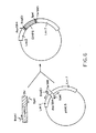
ターゲット遺伝子内の任意の位置へのIVPSの挿入を容易にするために、サイレント突然変異(アミノ酸残基を保持する)によってIVPSの両端に制限部位を形成することができる。新しい制限部位の形成後に、これらの酵素でIVPSを切断する。ターゲット遺伝子プラスミドはPCRで形成する。制限部位はIVPS内にあるため、IVPSを完全にし且つターゲット遺伝子内に適合性クローニング部位を形成するためには、欠失しているIVPS配列をそれぞれのターゲット遺伝子PCRプライマーの5’末端に含ませなければならない(図6)。
【0096】
例えば、Vent IVPS2におけるサイレント突然変異は、プライマーVent IVS2正方向BspEI(5’−AGTGTCTCCGGAGAAAGTGAGAT−3’(配列番号:12))を用いて5’末端にBspEI部位を形成することができ、プライマーVent IVS2逆方向SpeI(5’−ATTGTGTACTAGTATGTTGTTTGCAA−3’(配列番号:13))を用いて3’末端にSpeIを形成することができる。次に、これは例えば、lacZコード領域内のアスパラギン酸コドン(残基594)とトレオニンコドン(残基595)との間に挿入し得る。線状ターゲット遺伝子プラスミドは、実施例4で説明したように、BspEI及びSpeI部位、IVPSの残部、並びにlacZと一致する領域を含むプライマーで、プライマーlacZ1/BspEI逆方向(5’−GCCTCCGGAGACACTATCGCCAAATCACCGCCGTAA−3’(配列番号:14))及びプライマーlacZ2/SpeI正方向(5’−GCCACTAGTACACAATACGCCGAACGATCGCCAGTTCT−3’(配列番号:15))を用いて、PCRにより形成できる。DNAフラグメントはIVPS及びターゲット遺伝子の両方からPCRによって合成する。IVPS及びターゲット遺伝子プライマーは両方とも新しい制限部位を含んでいる。適当な制限エンドヌクレアーゼで切断した後で、融合遺伝子を形成すべく、適合性末端を有するDNAフラグメントを連結することができる。IVPSの切り出し後に余分の残基が残ることはないため、スプライシングが起これば生来のβ−ガラクトシダーゼポリペプチドが形成されると予想される。
【0097】
挿入部位の近傍の制限部位でのIVPSの挿入
別の一般的な方法では(図7)、ターゲット遺伝子(例えばトレオニン又はセリンコドン)内の挿入部位の近傍の制限部位を使用して、ターゲット遺伝子に適合した末端を有するIVPSを挿入することができる。挿入部位又はその近傍のサイレントヌクレオチド置換によって制限部位を形成するか、又は生来の制限部位を使用し得る。線状ターゲット遺伝子プラスミドは、挿入部位の近傍の制限部位で開始しながら実施例4と同様にPCRで形成する。IVPSは、適合性制限部位とターゲット遺伝子配列の残り(制限部位と挿入部位との間の配列)を含むプライマーを用いて合成する。両端が挿入部位で該配列と重なり合うIVPS DNAフラグメントを合成し、適当な酵素で切断し、次いで同じ酵素で切断したベクターに連結し得る。
【0098】
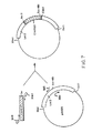
例えば、サイレント突然変異によりSalI部位(残基478〜479)及びXbaI部位(残基481〜482 セリン−アルギニン)を形成することによって、lacZ遺伝子内の残基479(アスパラギン酸)と481(セリン)との間にIVPSエレメントを挿入することができる。これは、プライマーlacZ3 Sal逆方向(5’−AGGGTCGACAGATTTGATCCAGCG−3’(配列番号:7))及びlacZ4 Xba正方向(5’−CCTTCTAGACCGGTGCAGTATGAAGG−3’(配列番号:16))を用いて、実施例2に記載のターゲットプラスミドpAHO5のPCRにより達成し得る。次に、プライマーVent IVS2正方向SalI(5’−GCCGTCGACCCTAGTGTCTCAGGAGAAAGTGAGATC−3’(配列番号:17))及びVent IVS2逆方向XbaI(5’−GCCTCTAGAATTGTGTACCAGGATGTTGTTTGC−3’(配列番号:18))を用いて、PCRによりIVPS2フラグメントを形成する。DNAフラグメントはIVPS及びターゲット遺伝子の両方からPCRによって合成する。IVPS及びターゲット遺伝子プライマーは両方とも新しい制限部位を含む。都合の悪いことに、このベクターは単一XbaI及びSalI部位を含む(図7)。 従って、ターゲット遺伝子ベクターPCR生成物は、部分的消化を生起する条件下で切断しなければならない。次いで、必要な線状プラスミドをアガロースゲルから単離する。適当な制限エンドヌクレアーゼで切断した後、融合遺伝子を形成するために、適合性末端を有するDNAフラグメントを連結することができる。IVPSの切り出し後に余分な残基が残ることはないため、スプライシングが起これば、生来のβ−ガラクトシダーゼポリペプチドが形成されると予想される。通常は、前述のようなクローニングプロセスを容易にするために、ターゲット遺伝子及びベクター内で非反復部位を選択又は形成することが重要である。
【0099】
実施例6
A. Vent DNAポリメラーゼでのIVPS2のスプライシングに関する実験を容易にするために、T7プロモーター構築物pV174−1B1を改質したものを形成した。この改質構築物pANG5(図8)は、pV174−1B1と同じVent DNAポリメラーゼ前駆体をコードする。本明細書で検討しているような突然変異体の形成を簡単にするために、多数のサイレント突然変異を、特に上流及び下流スプライス部位に導入した。変化は下記の通りである:
1.ベクタ−主鎖内のXmaI及びPpuMI部位の破壊。XmaI部位はまずT7発現ベクタ−pAII17をXmaIで切断し、付着末端をDNAポリメラーゼIのクレノウフラグメントで修復し、次いで平滑末端を再連結することによって除去した。プラスミドをXmaIによる開裂に対する耐性についてスクリーニングした。得られたベクタ−からPpuMI部位を同様に除去し、今度はPpuMI開裂に対する耐性についてスクリーニングした。最終ベクタ−をpAML1と命名した。このベクタ−は、ポリメラーゼ遺伝子内の単一のXmaI及びPpuMI部位の使用を可能にした。
【0100】
2.制限部位を形成するためのサイレント塩基変化(silent base change)の導入。この変化は、Kunkelの方法に従い[T.A.Kunkel,J.D.Roberts及びR.A.Zakour,Methodsin Enzymology 154:367−382(1987)]、オリゴヌクレオチド特異的突然変異誘発を用いて導入した。f1ヘルパーファージIR1での重感染により2つのBluescript SK−ファージミド(phagemid)誘導体内に一本鎖鋳型を形成した[Eneaら、Virology 122:22−226(1982)]。第1のフラグメントは、BamHI/EcoRV切断Bluescriptに連結したpV174−1B1からのBsaAI〜BamHIフラグメント(Vent DNAポリメラーゼ配列のヌクレオチド3714〜5837を表す)を含んでいた。第2のフラグメントは、ClaI/EcoRV切断Bluescriptに連結したClaI〜SspIフラグメント(ヌクレオチド816〜4408)を含んでいた。
【0101】
BsaAI/BamHI構築物に、下記の3つのオリゴヌクレオチドで同時に突然変異を与えた:
5’−GCAAAGAACCGGTGCGTCTCTTC−3’(配列番号:19)(AgeI nt4669〜4674)、
5’−AGCAACAGAGTTACCTCTTG−3’(配列番号:20)(アンバー1703オーカー)、
5’−CAGTTTCCAGCTCCTACAATGAGACCTACGAGC−3’(配列番号:21)(D1236A)。
【0102】
改質塩基には下線を引き、変化は括弧内に示した。D1236Aを形成するためのオリゴヌクレオチドは、スクリーニングに使用するためのBsaI部位を形成するためのサイレント塩基変化も含んでいた。得られた単離体をpAMN2と命名した。
【0103】
ClaI/SspI構築物に、下記の4つのオリゴヌクレオチドで同時に突然変異を与えた:
5’−GTAGTGTCGACCCCATGCGG−3’(配列番号:22)(SalI nt3863〜3468)、
5’−CGTTTTGCCTGATTATTATCTCACTTTC−3’(配列番号:23)(BsaBI nt3554〜3563)、
5’−GTCCACCTTCGAAAAAAGATCC−3’(配列番号:24)(BstBI nt3608〜3613)、
5’−CCGCATAAAGGACCTTAAAGC−3’(配列番号:25)(PpuMI nt3517〜3523)。
【0104】
マークは前述の通りである。スクリーニングも前述のように行い、得られた構築物をpAMO22と命名した。
【0105】
BsaAI/BamHI構築物にも下記のオリゴヌクレオチド:
5’−GAGGAAGAGATCATCATCATAGC−3’(配列番号:26)(BsaBIブロッキングnt5641)
で突然変異を与え、damメチル化部位の付加に起因するBsaBI開裂に対する耐性についてスクリーニングした。得られた構築物をpAMW3と命名した。
【0106】
最後に、pV174−1B1の開始コドンにおけるNdeI部位を、部分的NdeI開裂と、クレノウでの末端修復と、T4 DNAリガーゼを用いる再環化とによって失活させた。プラスミドを適当なNdeI部位の消失についてスクリーニングした。この種の構築物の1つをpAKC4と命名した。
【0107】
pANG5構築物は前記部分から組み立てた:
1.pAKC4からのXbaI/ClaI(Vent DNAポリメラーゼの翻訳開始及びアミノ末端)、
2.pAMO22からのClaI/NdeI(よりアミノ末端ポリメラーゼ+IVPS2のアミノ末端領域)、
3.pAMN2からのNdeI/NsiI(IVPS2のカルボキシル末端領域、Vent DNAポリメラーゼのカルボキシル末端領域)、
4.pAMW3からのNsiI/BamHI(ポリメラーゼの最後の5つのアミノ酸+下流領域)、
5.pAML1からのBamHI/XbaI(T7プロモーター、複製起点、アンピシリン耐性)。
【0108】
pANG5と親pV174−1B1とを比較すると、後述のようにpANG5含有株の生存率の方が高いという点を除いて、Vent DNAポリメラーゼ及びI−TliI産生のパターンは同じである。これは、RNA又はDNAレベルでのスプライシングと対照的に、タンパク質レベルでスプライシングが生じた場合に予想されることである。
【0109】
B. 大腸菌におけるVent DNAポリメラーゼ遺伝子の発現の研究過程で、IVPS1及びIVPS2の欠失後に発現及び細胞生存率が大幅に増加することが判明した。この増加は、それぞれIVPS1及びIVPS2の遺伝子産物であるI−TliII及びI−TliIの毒性作用を表すか、又はスプライシング反応自体の毒性作用を表す。エンドヌクレアーゼ及びスプライシング活性は非依存的であり得、スプライシングに影響を与えずにエンドヌクレアーゼを失活させると推論された。pANG5の構築で述べたAへの単一アミノ酸置換を、I−TliIのアミノ近位ドデカペプチドモチーフ内の保存残基内で行った(変更残基D1236)。これらの構築物はVent DNAポリメラーゼを発現したが、I−TliI活性は検出されなかった。pV174−1B1と異なり、BL21(DE3)のようなT7発現株は37℃でもpANG5に良く耐えた。ウェスタンブロットによるタンパク質スプライシングの分析及びパルス−チェイス分析では、pANG5とpV174−1B1との間にタンパク質スプライシングの差は認められなかった。即ち、完全長の前駆体の産生、並びにその後の成熟ポリメラーゼと大きさがI−TliIに対応するタンパク質の形成に差は見られなかった。
【0110】
C. カセット置換突然変異誘発を用いてチロシン1079をアルギニンで置換することにより、共通のカルモジュリン依存性タンパク質キナーゼII部位(XRXXS*;Personら、前出の文献)を構築した。要約すれば、pANG5を非反復部位BsaBI及びPpuMIで切断し、下記の二本鎖(duplex)(配列番号:27)を挿入して、所望の変化を導入した。
【0111】
5’−GTCCTTCGTGCGGACAGTGTCTCAGGAGAAAGTGAGATAA−3’
3’−GAAGCACGCCTGTCACAGAGTCCTCTTTCACTCTATT−5’。
【0112】
正しい構築物をDNA配列決定によって確認した。
【0113】
D. ブロックされたアミノ酸を付加するためのアンバー終止コドンの導入を、
5’−GTCCTTTATGCGGACTAGGTCTCAGGAGAAAGTGAGATAA−3’
3’−GAAATACGCCTGATCCAGAGTCCTCTTTCACTCTATT−5’
内でのカセット置換突然変異誘発によって実施した。
【0114】
同様にして、AgeI及びSmaIで切断したpANG5中に下記の二本鎖(配列番号:29)を配置することにより、チロシン1472をアンバー終止コドンで置換した:
5’−CCGGTTCTTTGCAAACAACATCCTGGTACACAATTAAGACGGCTTTTATGCCACAATACCC−3’
3’− AAGAAACGTTTGTTGTAGGACCATGTGTTAATTCTGCCGAAAATACGGTGTTATGGG−5’
最後に、Vent DNAポリメラーゼ遺伝子がアンバーコドン(TAG)内で終わるため、pAMN2(既述)からの適当な制限フラグメントをpANG5内の対応する部位に挿入して、前記終結コドンをオーカーコドン(TAA)に変える。
【0115】
実施例7
CIVPS2のスプライス部位へのO−(o−ニトロベンジル)セリンの導入によるタンパク質スプライシングの制御
光活性化可能な(photoactivatable)タンパク質スプライシングを明らかにするために、pV174.1B1を用いて2つのベクターを構築した。第1の構築物pANY5(「野生型」とも称する)は下記のようにアミノ酸レベルで記述できる:pV174.1B1 Δ1−1063、Δ1544−1702、V1542M、V1541M、1543オパール(TGA)。この構築物は、in vitro転写/翻訳系で翻訳された時に、45.3kDaのエンドヌクレアーゼ(I−TliI)をスプライスして10.5kDaの連結反応生成物を形成する55.8kDaの前駆体タンパク質を産生するように設計されている。第2の構築物pAOD1(「アンバー変異体」とも称する)は、下記のようにアミノ酸レベルで記述できる:pV174.1B1 Δ−1063、Δ1544−1702、V1542M、V1541M、1543オパール(TGA)、S1082アンバー(TAG)。この構築物は、標準的in vitro転写/翻訳条件で2.2kDaのアンバーフラグメントを生成するように設計されているが、o−ニトロベンジルセリンで化学的にアミノシル化したアンバーサプレッサーtRNAでin vitro反応を補充すると、光活性化可能セリンを取り込むだろう。位置1082のセリンが「ブロック」されていると、前駆体はスプライスすることができない。強い350m光で照射すると、o−ニトロベンジル基が放出され[Pillai、前出の文献]、セリンの求核性ヒドロキシル側鎖が遊離し、タンパク質がスプライスできるようになる。
【0116】
アンバーサプレッサーtRNA(3’末端CA残基を欠く)は、文献[Ellmanら、前出の文献、Norenら、Nucleic Acids Res.18:83(1990)]に記載のように、T7 RNAポリメラーゼでFokI直線化pYPhe2プラスミド鋳型のin vitroランオフ(runoff)転写を行うことによりミリグラムスケールで合成した。αアミンをBPOC、CBZ又はBOCのような官能基で保護したセリン誘導体は市販されている(Bachem、Sigma、Aldrich)。N−ブロックされたセリンは、o−ニトロベンジルブロミドのような試薬を用いて標準的アルキルハロゲン化物置換反応を行うことにより、N−ブロックされたO−(o−ニトロベンジル)セリンに変換できる。次いで、完全にブロックされたセリンを、文献[Ellmanら、前出の文献]に記載のように5’−ホスホデオキシリボシチジリル−(3’−5’)−リボアデノシン(pdCpA)に結合した。次いで、T4 RNAリガーゼ(New England Biolabs,Inc.)を用いてアミノアシル化ダイマーを切頭(truncated)サプレッサーtRNAに連結して、完全長のアミノアシル化サプレッサーtRNAを得た。
【0117】
3μgの塩化セシウム精製プラスミドDNAと、3μlの100mM酢酸マグネシウムと、1μlの100mM酢酸カルシムと、7.5μlの低分子量混合物[Ellmanら、前出の文献](カルシウム又はメチオニン無含有)と、1μlの[35S]−メチオニン(10μCi/μl、1000Ci/mmol)と、1μlの3mg/mlリファムピシンと、総量を30μlにするのに必要な量の水とを氷上で混合することにより、「野生型」構築物のin vitro転写/翻訳を実施した。前記反応物質を37℃で3分間インキュベートし、その間に、大腸菌D10から調製したS−30抽出物[Ellmanら、前出の文]のアリコートを解凍した。8.5μlのS−30抽出物を加えた後、1.5μlのT7RNAポリメラーゼ(300U/μl、New England Biolabs,Inc.)を加え、これらの反応物質を37℃で60分間インキュベートした。試料を10〜20%トリシンSDS−PAGEゲル(NOVEX)上で電気泳動にかけ、オートラジオグラフィーによってタンパク質を可視化した(図9)。
【0118】
「アンバー変異体」のin vitro転写/翻訳を、「野生型」で説明したように、但し反応物質に3.5μlの化学的にアミノアシル化したo−ニトロベンジルセリン−tRNAamberを約3μg/μlの濃度で補充して実施した。サプレッサーtRNAをS−30抽出物の添加の直前に反応に加えた。
【0119】
図9は、「野生型」(pANY5)又は「アンバー変異体」(pAOD1)構築体でプライムしたin vitro転写/翻訳反応の10〜20%トリシンSDS−PAGEゲルを示している。レーン1は、55.8kDa前駆体と、「野生型」構築体からin vitroで発現された、切り出された45.3kDaのI−TliIエンドヌクレアーゼとを示している。レーン2は、添加tRNAに起因する翻訳の阻害が存在しないことを示すために、13.5μgの完全長の無変化アンバーサプレッサーtRNAを加えた「野生型」反応を示している。レーン3及び4は、完全長の非アシル化サプレッサーtRNA(10.5μg)を加えた又は加えていない「アンバー変異体」のin vitro発現の結果を示している。これらの反応はいずれも、予想通りに、完全長の前駆体分子又はスプライス生成物を産生しない。これは、サプレッサーtRNAが細胞抽出物中でいずれの内因性アミノアシル−tRNAシンテターゼによってもアミノアシル化されないことを意味する。分子量約52kDaのバンドは明らかに、アンバー変異体のすぐ下流の二次的翻訳開始部位によるものである。レーン5は、「アンバー変異体」に化学的にアミノアシル化したo−ニトロベンジルセリン−tRNAamberを加えた場合の結果を示している。前駆体タンパク質はin vitroで産生されるが、スプライス生成物(即ちI−TliI)は見えない。
【0120】
前駆体タンパク質の位置1082に部位特異的に挿入したセリンからo−ニトロベンジル基を光化学的に除去して、調節スプライシングを行った。in vitro反応の6μlアリコートを0.5μlのRNase A(10μg/μl)で処理して翻訳を停止させ、GEモデル#RSK6Bタニングランプ(tanning lamp)からの強力(275W)可視光により10cmの距離で10分間照射し、4μlの水で希釈し、その後37℃で60分間インキュベートしてスプライシングを生起させた。得られたスプライス生成物を10〜20%トリシンSDS−PAGEゲルでの電気泳動によって可視化し、次いでオートラジオグラフィーを行った(図10)。
【0121】
図10は、化学的にブロックした前駆体(レーン5、図9)を350nmの光に露光した結果を示している。レーン1〜4は、「野生型」反応(レーン1、図9)を下記のように処理した対照である。レーン1は37℃で60分インキュベートしたもの、レーン2は0.5μlのRNase(10μg/μl)を加えて37℃で60分間インキュベートしたもの、レーン3は350nmの光で10分間照射し37℃で60分間インキュベートしたもの、レーン4はRNaseで前述のように処理し、350nmの光で10分間処理し、37℃で60分間インキュベートしたものである。レーン5〜8は、「ブロックした」前駆体をそれぞれレーン1〜4と同様に処理した結果を示している。「ブロックした」前駆体を照射すると、IーIVPS2でコードされるI−TliI(45.3kDa)エンドヌクレアーゼが切り出される(レーン7〜8及びレーン5〜6参照)。
【0122】
実施例8
ターゲット遺伝子への改質IVPSのフレーム内挿入及びペプチド結合開裂の熱制御
この実施例では、IVPS(CIVPS)カセットの改質方法及びターゲット遺伝子への挿入方法を説明する。具体例として、第1の天然下流残基(セリン)の置換又は欠失によるPyrococcus sp.(又はDeep Vent)IVPS1(CIVPS3)の改質と、大腸菌lacZ遺伝子のEcoRV部位への改質カセットのフレーム内挿入とを取り上げる。
【0123】
IVPSカセットの改質
一般的に、IVPSカセットは残基の置換及び欠失、又はIVPSの一端もしくは両端への残基の付加によって改質できる。このような改質IVPSカセットを用いた改質又は融合タンパク質は、特定のペプチド結合部位におけるスプライシング(ペプチド連結)又は開裂といった種々の触媒活性を示し得る。
【0124】
既述のように、カルボキシルスプライス部位の第1の下流残基は、Deep Vent IVPS1(CIVPS3)及びVent IVPS1の場合のセリン、又はVent IVPS2の場合のトレオニンである。CIVPS1、CIVPS2及びCIVPS3のアミノスプライス部位における第1のIVPS残基はセリンである。システイン残基は、酵母TFP1及びM.tuberculosis RecAのスプライス部位に発見された(Hirataら、前出の文献;Kaneら、前出の文献;Davisら、前出の文献)。スプライス部位のセリン、トレオニン又はシステイン残基は、タンパク質のスプライシング及び開裂に不可欠と考えられている。既述の実施例は、第1の下流残基を有するIVPSがタンパク質のスプライシングに関する情報を含むのに十分であることを明らかにした。しかしながら、これらの残基は種々のIVPSコンテクストで異なる機能を果たし得る。Vent IVPS2(CIVPS2)内で、天然残基、例えばセリンをトレオニン又はシステインで置換すると、スプライシングが低下し、開裂活性が変化した(Hodgesら、前出の文献参照)。
【0125】
ターゲット遺伝子コドンの間の平滑部位にフレーム内挿入するための改質IVPSカセットの合成
lacZコード領域又は他の任意のターゲット遺伝子にフレーム内挿入するためのIVPSカセットは、ポリメラーゼ連鎖反応(PCR)によって調製できる。下記のプロトコルは、それぞれCIVPS3、CIVPS3/Ser、CIVPS3/Thr及びCIVPS3/Cysと称する、カルボキシル末端コドン、セリン、トレオニン又はシステインを有する又は有していない4つのDeep Vent IVPS1カセットの製造を説明するものである。
【0126】
プライマー5’−AGCATTTTACCGGAAGAATGGGTT−3’(配列番号:5)(DV IVPS正、1839〜1862)と、下記の4つの逆プライマーのうちの1つとを用いて、pNEB#720(ATCC No.68723)からカセットを合成した。鋳型として使用したpNEB#720は、pUC19のBamHI部位に挿入したDeep Vent DNAポリメラーゼ遺伝子を含む4.8KbのBamHIフラグメントを有する。逆プライマー5’−GCAATTATGTGCATAGAGGAATCCA−3’(配列番号:40)及び5’−GGTATTATGTGCATAGAGGAATCCA−3’(配列番号:41)(3428〜3452)を用いて、それぞれCIVPS3/Thr及びCIVPS3/Cysフラグメント(1641bp)を形成した。PCR混合物は、Vent DNAポリメラーゼ緩衝液(NEB)に、2mMの硫酸マグネシウムと、400μMの各dNTPと、100μg/mlのBSAと、0.9μMの各プライマーと、40ngのプラスミドDNAと、100μl中2単位のVent DNAポリメラーゼとを加えたものを含む。増幅は、Perkin−Elmer/Cetusサーマルサイクラーを用いて94℃で30秒、48℃で30秒及び72℃で2分のサイクルに20回かけて実施した。プライマー5’−ATTATGTGCATAGAGGAATCCAAAG−3’(配列番号:42)(3425〜3449)を使用して、前述のPCRで、但し増幅を30サイクル実施して、CIVPS3フラグメント(1611bp)を合成した。プライマー5’−GCTATTATGTGCATAGAGGAATCCA−3’(配列番号:6)(3428〜3452)を用いて実施例1と同様にIVPS1/Serフラグメント(1614bp)を合成した。
【0127】
PCR試料をフェノール及びクロロホルムで抽出し、0.3μM NaAc及び50%イソプロパノール中−20℃で6時間沈殿させ、ミクロ遠心機で10分間10Krpmで遠心分離し、乾燥し、各々を20μlの蒸留水に再懸濁し、1%低融点アガロースゲル上に配置して80ボルトで6時間電気泳動にかけた。0.4mlのTE緩衝液(10mMトリス−HCl/0.1mM EDTA、pH8.0)中、65℃で30分間インキュベートし、フェノール及びクロロホルムで抽出し、0.3μM NaAc(pH5.2)及び50%イソプロパノール中−20℃で一晩沈殿させることにより、低融点アガロースゲルからDNAフラグメントを回収した。DNAを遠心分離し、70%エタノールで洗浄し、乾燥し、10μlの蒸留水に再懸濁した。
【0128】
IVPS1 DNAフラグメントのリン酸化を、4μlの10×ポリヌクレオチドキナーゼ緩衝液(NEB)と、3μlの精製DNAと、4μlの10mM ATPと、10単位のT4ポリヌクレオチドキナーゼ(NEB)とを用いて40μL中37℃で60分間実施した。試料を65℃の水浴中で10分間加熱した。80μlのTE緩衝液(10mMトリス−HCl/0.1mM EDTA、pH8.0)を加えた後、試料をフェノール及びクロロホルムで逐次抽出した。DNAを2.4μM NH4Ac及び70%エタノール中で−70℃で一晩沈殿させ、ミクロ遠心機で10分間10Krpmで遠心分離してペレット化し、冷70%エタノールで洗浄し、乾燥し、20μlの蒸留水に再懸濁した。CIVPS3/Serフラグメントのリン酸化を前述のように実施した。
【0129】
ベクターpAHO5中の大腸菌lacZ遺伝子のEcoRV部位へのCIVPS3カセットのフレーム内挿入
PCR合成CIVPSカセットは、ターゲット遺伝子を有する直線化ベクターとの連結によってターゲットコード領域に挿入できる。線状プラスミドベクターは、前述のような制限酵素またはPCR合成によって調製し得る。pAHO5は、tacプロモーターの下流のpAGR3(NEB)のポリリンカー内でBamHI及びSmaI部位の間に挿入されたpRS415(Simonsら、Gene,53:85−96(1987))からの3.1kb BamHI−DraIフラグメント上に完全なlacZ遺伝子配列を有する。tacプロモーターは、lacIq遺伝子の産物によって抑制され得且つイソプロピルβ−D−チオガラクトシド(IPTG)によって誘導され得る転写制御エレメントである。pAHO5は2つのEcoRV認識配列を含む。EcoRVは開裂部位に平滑末端を残す。EcoRV開裂部位の1つはlacZコード領域内で375番目のコドン(アスパラギン酸)と376番目のコドン(イソロイシン)との間を切断する。
【0130】
15μgのpAHO5 DNAを、100μlの1×NEB緩衝液2中で40単位のEcoRV(NEB)と共に37℃で60分間インキュベートすることにより、DNAを部分的に消化した。EcoRVを失活させるべく試料を65℃で10分間加熱した後、20μlの染料添加アガロースゲルを試料に加えた。1%低融点アガロースゲル上での電気泳動によってDNAフラグメントを分離した。実施例8の方法で低融点アガロースゲルから直線化pAHO5プラスミドDNAを回収し、蒸留水に再懸濁した。
【0131】
CIVPS−lacZ融合遺伝子の構築
CIVPS3/Ser−lacZ融合の構築は実施例2で説明した。CIVPS3−lacZ融合は、リン酸化pAHO5 DNAをリン酸化IVPS1フラグメントに連結することによって行った。該反応は、20μl倍容中で、1×T4 DNAリガーゼ緩衝液(NEB)と、0.1μgのpAHO5 DNAと、0.5μgのIVPS1 DNAと、160単位のT4 DNAリガーゼ(NEB)とを加えて16℃で15時間実施した。100μlのコンピテントRR1細胞と10μlの連結試料とを氷上で30分間混合し、42℃で2分間加熱し、氷上で5分加熱冷却し、0.8mlのLB培地(10g/リットル トリプトン、5g/リットル酵母抽出物、10g/リットルNaCl、1g/リットル デキストロース、1g/リットルMgCl26H2O、pH7.2、25℃)を加え、30℃で45分間インキュベートして、大腸菌株RR1を形質転換した。100μg/mlのアンピシリンを加えたLBプレートに試料をプレーティングした。30℃で一晩のインキュベーション後に、プレート当たり約150〜300のコロニーが観察された。
【0132】
CIVPS3/Thr−lacZ及びCIVPS3/Cys−lacZ融合を、0.1μgのEcoRV直線化pAHO5 DNAと0.7gのCIVPS3/Thr又はCIVPS3/Cysフラグメントとの連結によって行った。大腸菌株ER2252(NEB)の形質転換を前述のプロトコルで実施した。
【0133】
コロニーのハイブリダイゼーションを用いて、組換えプラスミドを有するクローンをスクリーニングした。前述のDeep Vent CIVPS3正方向プライマーを、T4ポリヌクレオチドキナーゼで放射性標識し、ハイブリダイゼーションプローブとして使用した。コロニーを採取してニトロセルロースに移し、下記の各溶液中で5分間処理した:10%SDS、0.5M NaOH/1.5M NaCl、0.5Mトリス−HCl(pH7.4)/0.5M NaCl(2回)及び2×SSC(2回)。ニトロセルロースフィルターを室温で1時間乾燥し、真空下80℃で2時間焼成し、6×SSCに5分間浸漬し、50mMトリス−HCl(pH8.0)、1M NaCl、1mM EDTA及び0.1%SDSの溶液中で42℃で2時間洗浄した。6×NET、5×デンハート及び0.5%SDS中で42℃で4時間処理した後、フィルターを放射性標識オリゴマープローブと共に同一条件下で一晩インキュベートし、次いで2×SSC中で、42℃で15分間の洗浄4回と、50℃で2分間の洗浄2回とにかけ、その後オートラジオグラフィーを行った。
【0134】
オリゴマープローブにハイブリダイズしたポジティブクローンを、誘導物質IPTGで融合タンパク質を発現する能力について更に調べた。クローンを、100μg/mlのアンピシリンを加えたLB培地中でOD600nmが0.5に達するまで30℃で培養した。IPTGを最終濃度0.3mMで加えた後、培養物を30℃で更に4時間増殖させた。0.1mlの細胞を0.1mlの尿素溶解緩衝液と共に10分間沸騰させることにより粗溶解物を調製した。前述のポジティブクローンからの融合タンパク質のアイデンティティを、β−ガラクトシターゼ(Promega)又はI−PspI(Deep Vent CIVPS3のタンパク質生成物、NEB)に対する抗体を用いてウェスタンブロットにより分析した。予備染色マーカー(BRL)を用いて4〜20%SDSゲル(ISS,Daichi,Tokyo,Japan)上で試料を電気泳動にかけ、ニトロセルロースに移し、抗血清(マウス由来)でプローブし、アルカリホスフェート結合抗マウス第二抗体を製造業者(Promega)の指示通りに使用して検出した。Deep Vent CIVPS3−lacZ融合クローンは、両抗体と反応する173〜178KDaの産物を発現した。この大きさは、CIVPS3−β−ガラクトシターゼ融合タンパク質(図11)について予想された大きさである。クローンpDV7及びpDV15はCIVPS3挿入物を含む。pDV302、306及び307はCIVPS3/Cysカセットを有し、pDVT319、321及び323はCIVPS3/Thrカセットを含む。CIVPS3/Ser挿入物を含むpDVS712及び742は既に実施例2で説明した。
【0135】
改質CIVPS3カセットを用いるCIVPS3−β−ガラクトシターゼ中での特定ペプチド結合開裂の熱制御
カルボキシル末端でセリンを置換するためのトレオニン又はシステインを有するカセットを含むDVIVPS1(CIVPS3)−β−ガラクトシターゼ融合は、融合タンパク質中の特定ペプチド結合点で熱制御可能な開裂を示す。前述の構築物(lacZ EcoRV部位に挿入したCIVPS3カセット)は、IPTGでの誘導後に融合タンパク質を産生する。25℃で増殖した細胞から調製した細胞抽出物をより高い温度(42℃又は65℃)で処理し、β−ガラクトシターゼ(Promega)又はI−PspI(Deep Vent CIVPS3の産物)に対する抗体を用いてウェスタンブロットで分析した(図11及び図12)。IVPS1/Ser融合タンパク質は、高温でインキュベートすると、タンパク質スプライシングを起こして連結タンパク質と遊離IVPSエンドヌクレアーゼとを生成し得る。連結反応活性は観察されなかったが、42℃ではCIVPS3/Thr又はCIVPS3/Cysカセットを有する融合タンパク質が主にアミノスプライス部位で開裂し、65℃では両方の融合タンパク質がカルボキシルスプライス部位でより大きい開裂活性を示した。
【0136】
CIVPS3−lacZ融合クローンからの細胞抽出物の調製を下記の方法で実施した。種々の大腸菌宿主内で最初に構築した融合構築物の全てを、実施例8で述べた標準的形質転換手順で、β−ガラクトシターゼを合成しないlacZ欠失大腸菌株ER1991(New England Biolabs,Inc.)中に導入した。pDV7、pDVC302、pDVT332又はpDVS712クローンからの単コロニーを、100μg/mlのアンピシリンを加えた1.5mlのLB培地に接種し、OD600nmが約0.5になるまで30℃でインキュベートし、1.5mlの0.6mM IPTG、100μgアンピシリン/mlLを加えることにより0.3mM IPTGで25℃で5時間誘導した。3mlの細胞を遠心分離し、0.5mlのLBに再懸濁し、4℃で1分間超音波処理し、4℃で5分間6,000rpmで遠心分離した。上清を回収し、−20℃で貯蔵した。
【0137】
細胞抽出物を室温で急速解凍した後、42℃又は65℃で熱処理した。48μlの抽出物を12μlの5×試料緩衝液(0.31 トリス−HCl、pH6.8/10%SDS/25%2−メルカプトエタノール/50%グリセロール/0.005%ブロモフェノールブルー)と混合し、10分間沸騰させて、非処理対照試料を調製した。48μlアリコートを1.5mlミクロ遠心管に移し、42℃の水浴中で30、60、120もしくは240分間、又は65℃の水浴中で15、30、60もしくは120分間インキュベートした。各々を12μlの5×試料緩衝液と混合し、10分間沸騰させた。
【0138】
処理した試料を、I−PspI(NEB)(図11及び図12)又はβ−ガラクトシターゼ(Promega)(図12)に対する抗体を用いてウェスタンブロットで分析し、各試料5μlを4/20%SDSポリアクリルアミドゲル(ISS,Daichi,Tokyo,Japan)上に配置して100ボルトで4時間電気泳動にかけた。ウェスタンブロットはPromegaの手順に従って実施した。
【0139】
結果は、融合タンパク質前駆体が主要物質(dominant specie)であり、4つの融合構築物全部から25℃でIPTG誘導した後の細胞中には極めて微量のI−PspIエンドヌクレアーゼしか存在していないことを示した。これは、低温ではスプライシング及び切り出し活性が不十分であることを意味する。しかしながら、pDVS712(CIVPS3/Ser−β−ガラクトシターゼ融合)抽出物をより高い温度42℃又は65℃にシフトすると、CIVPS3生成物I−PspI(約60KDa)が多量に蓄積された(図11及び図12)。IVPSドメインの切り出しは、中断されたβ−ガラクトシターゼのNドメイン及びCドメインの連結に結びついており、完全長のβ−ガラクトシターゼと同じ大きさである116KDaの生成物を産生した(図12)。約130KDa(アミノスプライス部位での開裂に対応する)の別の主産物(IVPS1−C−EPS)も観察された。
【0140】
他の3つの変異体(variant)の融合タンパク質(CIVPS3、CIVPS3/Cys及びCIVPS3/Thrカセットを有する)は低温での安定性がより大きかった。非処理抽出物からは、スプライス部位での開裂に対応するI−PspI又は他の産物が極めて少量しか検出されなかった(図11)。CIVPS3/Ser融合と対照的に、これら3つの融合構築物の熱処理した試料では連結タンパク質は観察されなかった(図12)。pDV7(CIVPS3−β−ガラクトシターゼ融合)試料は、65℃では、I−PspIと単一スプライス部位での開裂に対応する生成物とを微量しか産生しなかった。これは、いずれのスプライス部位でも切り出しが十分でないことを意味する(図11、レーン1〜3)。CIVPS3/Cysカセットを含むpDVC302は、42℃で中庸量のI−PspI及びCIVPS3−C−EPS種の蓄積を示した(図11、レーン5)。I−PspI、C−EPS及びカルボキシルスプライス部位での開裂に対応する約110KDaの生成物(N−EPS−CIVPS3)の収率は65℃で増加したが、CIVPS−C−EPS種は減少した(図11、レーン4〜6;図12)。この結果は、融合タンパク質及び/又はCIVPS−C−EPS生成物からのカルボキシルスプライス部位でのペプチド結合開裂が促進されたことを意味する。pDVT321(CIVPS3/Thrカセットを有する)は、42℃で処理すると、主要生成物CIVPS3−C−EPS以外のI−PspI又はC−EPSを極めて少量しか示さなかった(図11、レーン8;図12)。このデータは、42℃では、アミノスプライス部位でペプチド結合の効果的な開裂が得られるが、カルボキシルスプライス部位では該開裂が得られないことを意味する。65℃で見られた少量のI−PspIの蓄積は、カルボキシルスプライス部位での開裂が促進されたことを意味する(図11、レーン9)。
【0141】
要約すれば、前記データは、Deep Vent IVPS1(CIVPS3)のカルボキシルスプライス部位で単一の天然残基セリンを置換すると、融合タンパク質のプロセシングが変化し、温度によってより良く制御できるようになることを立証している。CIVPS3/Thr−β−ガラクトシターゼ融合タンパク質は(そして程度はより低いがCIVPS3/Cys融合タンパク質も)高温の時にだけ、アミノスプライス部位で特定ペプチド結合を開裂した。
【0142】
実施例9
MIPの構築及び精製
アフィニティクロマトグラフィーによるCIVPS融合体の精製
MBP融合タンパク質へのDeep Vent IVPS1のクローニング
本発明の実施態様の1つでは、CIVPSと、容易に精製できるセグメント、例えば結合タンパク質と、対象となっているタンパク質又はペプチド、即ちターゲットタンパク質とを含む三部型融合体(three−part fusion)を形成することができる。これらの部分の順序は変えることができる。この種の融合体の利点は精製が容易なことにある。前駆体タンパク質を精製すれば、対象となっているペプチドを、改質CIVPSで誘導した単方向タンパク質開裂によって融合体から分離することができる。上述の実施例では、CIVPS部位の1つを改変してその部位のスプライシング又は開裂を低下又は防止すると、別の部位の開裂がスプライシングより促進されることを明らかにした(詳細な説明、並びに実施例8参照)。これは、融合体から対象ペプチドの分離を可能にするものである。
【0143】
この実施例は、結合タンパク質であるマルトース結合タンパク質(MBP)と、CIVPS3と、パラミオシンペプチドとを含む前述のような三部型融合体が、非スプライス前駆体としてアミロース樹脂で簡単に精製できることを明かにする。この実施例では、連結なしで開裂生成物のみを生成するためにスプライシングを妨害すべく開裂をCIVPSの片側に制限する試みは行わなかった。
【0144】
Deep Vent IVPS1挿入物(CIVPS3)の合成
上述の実施例と同様に、但し下記の点を変えて、PCRによりCIVPS3カセットを合成した。PCR混合物は、Vent DNAポリメラーゼ緩衝液(NEB)と、200Mの各dNTPと、10pmoleの各プライマーと、40ngのプラスミドDNAと、100リットル中2単位のVent DNAポリメラーゼとを含んでいた。増幅は、Perken−Elmerサーマルサイクラーを用いて、94℃で30秒、50℃で3秒及び72℃で2分のサイクルに20回かけて実施した。Deep Vent IVPS1はpNEB#720から合成した。
【0145】
正方向プライマーは、2つのフランキングKpnI部位を含む、DV IVPS1の5’末端と同じ3’末端の26/27塩基からなるプライマー96−6、5’−GGTACCCGTCGTGCTAGCATTTTACCGGAAGAATGGGTACCA−3’(配列番号:43)であった。3’KpnI部位は、アミノ酸残基を変えずに制限部位を形成するサイレント置換を含む。Deep Vent IVPS1逆方向プライマーであるプライマー96−7、5’−CCGCTATTATGTGCATAGAGGGATCC−3’(配列番号:44)は、3’末端にBamHI部位を有する。3’末端の23/24塩基はDV IVPS1の3’末端と相同であり、BamHI部位を形成するための単一塩基置換を有する。プライマー96−6及び96−7を使用してDeep Vent IVPS1カセット(1.6kb)を合成した。
【0146】
PCR試料を1:1でクロロホルムと混合し、上方水性層を1%低融点アガロースゲル上に配置して電気泳動にかけた。ゲルから1.6kbバンドを切り取り、65℃でインキュベートした。ゲルの融解後、0.25mlのTE緩衝液((10mMトリス−HCl/0.1mM EDTA、pH7.5)を65℃で加え、試料をフェノール−クロロホルム(1:1混合物)で抽出した。DNAを0.5M NaCl及び2倍容のイソプロパノール中で−20℃で30分間沈殿させた。DNAを遠心分離し、乾燥し、60μlのTE緩衝液に再懸濁した。
【0147】
pPR1002、即ちpMAL−c2−パラミオシンΔSalプラスミドの調製pMAL−c2−パラミオシンΔSal融合プラスミドであるpPR1002は、パラミオシンΔSal欠失と称する、D.immitisパラミオシン遺伝子のEcoRI−SalIフラグメントに結合した、tacプロモーター制御下のmalE遺伝子を含む7.2kbのベクターである(Steelら、J.Immunology,145:3917−3923(1990))。4μgずつの2つのpPR1002試料を、100μg/mlのBSAを含む20μlの1×NEB緩衝液#2中で6単位×XmnI(NEB)により37℃で2時間直線化した。該反応物質を1%低融点アガロースゲルにかけた。7.2kbバンドを切り取り、前述のようにゲルから精製し、40μlのTE緩衝液に再懸濁した。
【0148】
pMIP17の構築
pPR1002とDeep Vent IVPS1との連結を、14.5μlの蒸留水と、2.5μlの10×T4 DNAリガーゼ緩衝液(NEB)と、1μg/μgの開裂pPR1002 DNAと、前述のように調製した5μlの0.2μg/μl Deep Vent IVPS1と、800単位のT4 DNAリガーゼ(NEB)とを加えた25μl容中で16℃で16時間実施した。
【0149】
100μlのコンピテントER2252細胞と5μlの連結試料とを、0.1M CaCl2と1×SSC(0.15M NaCl、15mM クエン酸Na)との1:2混合物100μl中で混合し、42℃で3分間加熱し、氷中で5分間冷却し、0.1mlのLB培地(10g/リットルのトリプトン、5g/リットルの酵母抽出物、10g/リットルのNaCl、1g/リットルのデキストロース、1g/リットルのMgCl2・6H2O、pH7.2、25℃)を加え、30℃で30分間インキュベートすることにより、大腸菌株ER2252を氷上で5分間形質転換した。形質転換細胞300μlをペレット化し、100μlの上清に再懸濁し、LBampプレートにプレーティングした。30℃で一晩のインキュベーション後に、約160のコロニーが観察された。
【0150】
PCR増幅を用いて、組換えプラスミドを有するコロニーのスクリーニングを行った。個々のコロニーを採取してウェル数96のマイクロタイター皿内の100μlの蒸留水に入れ、5分間沸騰させて細胞を溶解した。PCR混合物は、50μlの反応混合物中Vent DNAポリメラセーゼ緩衝液(NEB)と、200μMの各dNTPと、10pmoleの各プライマー(同上)と、2.5μlの細胞溶解物と、2単位のVent Exo DNAポリメラセーゼとを含んでいた。増幅は、Perkin−Elmerサーマルサイクラーを用いて94℃で30秒、50℃で30秒及び72℃で2分のサイクルに30回かけることにより実施した。10μlの各反応混合物を1%アガロースゲル上で電気泳動させた。ポジティブクローンはIVPS1(1.6kb)に対応するバンドを有していた。1つのポジティブプラスミドをpMIP17と命名した。
【0151】
MIPの発現:MBP−Deep Vent IVPS1−パラミオシンΔSal融合
pMIP17を含むポジティブクローンを、100μg/mlのアンピシリンを加えたLB培地中でOD600nmが0.5になるまで30℃で培養した。非誘導細胞から溶解物を調製するために、1.0mlの培養液をペレット化し、50μlのタンパク質試料緩衝液(125mMトリス、700Mm β−メルカプトエタノール、2%SDS、15%グリセロール及び1mg/mlブロモフェノールブルー)に再懸濁した。誘導培養物からの試料は下記のように調製した。IPTGを最終濃度1mMで加えた後、培養物を30℃で更に20時間増殖させた。誘導後5時間目及び20時間目の0.5ml培養物からの細胞をペレット化し、100μlの5×タンパク質試料緩衝液に再懸濁した。予備誘導した5時間目の試料を−20℃で16時間凍結し、20時間目の試料を−70℃で15分凍結した。前駆体の収率を増加させるために、培養物を12℃〜20℃で誘導し、前駆体の量をクーマシーブルー染色ゲルで決定した。全ての試料を5分間沸騰させ、SDS−PAGE電気泳動と、クーマシーブルー染色か、又はI−PspIに対する抗体(NEB)を用いるウェスタンブロットとを順次行ってタンパク質生成物を分析した。試料は4〜20%SDSゲル(ISS,Daichi,Tpkyo,Japan)上で予備染色マーカー(BRL)を用いて電気泳動させ、ニトロセルロースに移し、抗血清(マウス抗−I−PspI)でプローブし、アルカリホスフェート結合抗マウス第二抗体を製造業者(Promega)の指示通りに使用して検出した。クーマシーブルー染色ゲル及びウェスタンブロットの両方で、予想した主要バンドが約132kDaに観察された(データ示さず)。
【0152】
アミロース及びMonoQカラムでのMBP−Deep Vent IVPS1パラミオシンΔSal融合体の大規模精製
単コロニーを使用して、100μg/mlのアンピシリンを加えた4×10mlのLB培地に接種し、OD600nmが0.5になるまで30℃でインキュベートした。これらの培養物を用いて、100μg/mlのアンピシリンを加えた4×1リットルのLB培地に接種し、OD600nmが0.5になるまで30℃でインキュベートした。次いで培養物を12℃にし、1mM IPTGで一晩誘導した。細胞をペレット化し、カラム緩衝液(20mM NaPO4、pH7.4、200mM NaCl及び1mM EDTA)に再懸濁し、超音波処理し、遠心分離し、透明な培養溶解物をアミロース樹脂(NEBタンパク質融合及び精製システム)にかけた。融合タンパク質を(製造業者の指示に従って)マルトースで溶離し、SDS−PAGEゲルで調べた(図13)。アミロース樹脂溶離物を、FPLC MonoQアニオン交換樹脂(Pharmacia)でのクロマトグラフィーにより更に精製した。カラムを0.2M NaCl、10mMトリス−HCl、pH8.5で洗浄し、10mMトリス−HCl、pH8.5中0.2〜1.0MのNaCl直線勾配で溶離した。タンパク質は0.4〜0.6M NaClで溶離した。
【0153】
6つのタンパク質バンドを、MBP、I−PspI及びパラミオシンに対する抗体を用いてウェスタンブロットで同定した。見掛け分子量180kDa及び132kDaの2つのバンドは前記3種類の抗体の全てと反応した。完全長の前駆体は132kDaの筈である。分子量がより大きいバンドはスプライシング中間体と考えられ、類似の高分子量物質が全てのCIVPS構築物について見られた。切り出されたI−PspIは60kDaで泳動し、I−PspI抗体に認識されただけであった。また、スプライスされた生成物(MBP−パラミオシンΔSal、72kDa)は、MBP及びパラミオシン抗体と反応する血清に認識されただけであった。約103kDaのバンドはMBP及びI−PspI抗体とだけ反応した。このバンドは、IVPSのC末端での単一開裂の産物を表す。約89kDaのバンドはI−PspI及びパラミオシン抗血清とだけ反応した。該バンドはIVPSのN末端での単一開裂の産物を表す(図13)。
【0154】
MBP−Deep Vent IVPS1−パラミオシンΔSal融合体の切り出し及び連結
前駆体(132kDa)を含む幾つかのMIP関連ポリペプチドと、泳動速度の遅い物質(見掛け分子量180kDa)と、単一スプライス部位での開裂生成物(130kDa及び89kDa)と、少量のスプライスされた生成物及び切り出された生成物(72kDa及び60kDa)とを含むアミロース樹脂及びMonoQ調製物を、20mMリン酸ナトリウム(pH6.0)及び0.5M NaCl中で37℃で2時間熱処理した。132kDaの前駆体及び180kDaの泳動速度の遅い物質は経時的に減少し、72kDaのスプライスされた生成物及び60kDaの切り出されたI−PspIは増加した(図13)。
【0155】
これらの結果は、三部型CIVPS融合体を精製できるだけでなく、単一開裂生成物を得ることもできることを意味する。CIVPS部位を更に操作すると、連結を伴わないいずれかのスプライス部位の開裂が促進され得る。
【0156】
実施例10
MIP融合におけるCIVPSの改変
置換可能なスプライス部位カセットを有するMIPの構築
この実施例では、両方のスプライス部位に置換可能カセットを有し、CIVPSがカセット置換によって改変された、MIP融合体(実施例9参照)を構築した。また、2つの事例で、改質CIVPSが主に単一スプライス部位で熱誘導的に開裂できるという事実も明らかにする。
【0157】
実施例9では、3つの部分、即ちCIVPS、精製の容易な結合ドメイン(MBP)及び対象となっている遺伝子(パラミオシンΔSal)で形成できる三部型融合体MIPを説明した。精製融合タンパク質のスプライシングでは2種類の主要産物、連結タンパク質ドメイン、MBP−パラミオシンΔSal及び切り出されたCIVPS(又はI−PspI)が得られた。CIVPSにおける改変のなかには、連結反応の抑制と1つのスプライス部位での開裂の増加とを誘起し得るものがあり、その結果、改質CIVPSにより触媒された特定ペプチド結合部位での開裂によって、融合タンパク質から対象ペプチドが分離されるのであろうと推論された。実施例8では、1つのスプライス部位の開裂がCIVPS3の改変(Thr又はCysによるC末端Serの置換)によって促進され得、これらの変化がもう1つの部位のスプライシング又は開裂を低下又は防止することを明らかにした。制御可能なスプライシング又は開裂活性の好ましい特性での改変に関してスクリーニングを行うためには、スプライス部位に種々の変異を導入し分析しなければならない。これは、各改変を有する完全CIVPSカセットの合成によって達成し得る。しかしながらこの操作は、PCR時に余分の変異を導入する可能性がある。
【0158】
本発明者は、スプライス部位の周りで短い範囲にわたるDNAだけを置換することによって前記操作を容易にする手法を開発した。該実施例では、実施例9で形成した元のMIP融合体を、各スプライス部位を挟む2つの非反復制限部位を有するように修飾する方法を説明する。制限消化後のカセット置換では、スプライス部位の1つにおける2つの非反復制限部位の間の短い範囲のDNAを別の短いDNAカセットで置換することができる。この実施例では、実施例9で説明したpMIP17融合体を、各接合部位に2つの非反復制限部位を有するように、即ちアミノスプライス部位にフランキング配置するXhoI部位及びKpnI部位と、カルボキシルスプライス部位にフランキング配置するBamHI部位及びStuI部位とを有するように改変した(図14参照)。
【0159】
スプライス部位カセットを有するMIP融合体は2つのステップで構築した。まず、BamHI及びStuI部位を次のように導入した。4μgのpMIP17(実施例9)を1×EcoRI緩衝液(NEB)中で50μl中0.5単位のEcoRI(NEB)により37℃で10分間消化した。1%アガロースゲルで電気泳動により分離した後、GenecleanIIキット(BIO 101)を用いて直線化pMIP17プラスミドDNA(8.8Kb)を精製した。精製pMIP17 DNAを、100μg/mlのBSAと、40単位のBamHI(NEB)とを加えた1×BamHI緩衝液(NEB)中37℃で3時間消化し、フェノール及びクロロホルムで抽出した。DNAを0.3M 酢酸Na(pH5.2)及び50%2−プロパノール中で−20℃で2時間沈殿させた。DNAをミクロ遠心機で10,000rpmで10分間遠心分離して回収し、乾燥し、20μlの無菌水に再懸濁した。
【0160】
ベクターとの連結の前に、2つの相補オリゴマーMIP301F(5’−GATCCCTCTATGCACATAATTCAGGCCTC−3’(配列番号:46))及びMIP302R(5’−AATTGAGGCCTGAATTATGTGCATAGAGG−3’(配列番号:47))をアニールさせて二本鎖リンカーMIP301F/MIP302Rを形成した。50pmolのオリゴマーMIP301F及びMIP302Rを1×T4 DNAリガーゼ緩衝液(NEB)中で68℃で15分間インキュベートし、20℃〜30℃にゆっくりと冷却した。1μgのEcoRI−BamHI消化pMIP17 DNAを、80単位のT4 DNAリガーゼ(NEB)及び25pmolのリンカーMIP301F/MIP302Rを加えた35μlの1×T4リガーゼ緩衝液(NEB)中で16℃で14時間にわたり連結反応させた。
【0161】
得られた構築物をpMIP18と命名した。上流XhoI及びKpnI部位を次の方法でpMIP18に導入した。2μgのpMIP18を、100μlの1×緩衝液2(NEB)、100μg/mlのBSA及び20単位のKpnI(NEB)中で37℃で4時間消化した。電気泳動で分離した後、線状pMIP18DNAをGenecleanIIキット(BIO101)で精製した。ベクターと連結する前に、2つの相補オリゴマーMIP521F(5’−GCTCGAGGCTAGCATTTTACCGGAAGAATGGGTAC−3’(配列番号:48))及びMIP522R(5’−CCATTCTTCCGGTAAAATGCTAGCCTCGAGCGTAC−3’(配列番号:49))をアニールさせて二本鎖リンカーMIP521F/MIP522Rを形成した。50pmolのオリゴマーMIP301F及びMIP302Rを1×T4 DNAリガーゼ緩衝液(NEB)中で75℃で15分間インキュベートし、20℃〜30℃にゆっくりと冷却した。0.2μgの消化pMIP18を、35μlの1×T4 DNAリガーゼ緩衝液(NEB)、80単位のT4 DNAリガーゼ(NEB)及び25pmolのリンカーMIP521F/MIP522R中で室温で3時間連結反応させた。いずれの場合も、連結DNA試料を用いて大腸菌株ER2252(NEB)を形質転換した。最終構築物pMIP21は、各スプライス部位に2つの非反復制限部位を含む。N末端スプライス部位を包囲するXhoI部位及びKpnI部位と、C末端スプライス部位を包囲するBamHI部位及びStuI部位とが存在する(図14)。
【0162】
改質MIP21融合タンパク質の発現とスプライシング活性とを調べるためにウェスタンブロット分析を行った。pMIP21含有ER2252を、100μg/mlのアンピシリンを加えたLB培地中30℃でOD600nmが0.5になるまで培養した。次いで培養物を1mMのIPTGで30℃で3時間誘導した。4.5mlの培養物をペレット化し、0.5mlのLB培地に再懸濁し、氷上で超音波処理した。透明上清を4/20%のポリアクリルアミドゲル上で100ボルトで4時間電気泳動させた。ウェスタンブロットを抗MBP血清でプローブした。結果は、改質MIP21融合体からのスプライシング活性がMIP17のそれと区別できないものであることを示している。
【0163】
スプライス部位カセット置換によるMIP21の改変
改変MIP融合構築物pMIP21では、アミノスプライス部位カセットがXhoI及びKpnI部位の間に8つのアミノ酸残基を含んでおり、カルボキシルスプライス部位カセットがBamHI及びStuI部位の間の6つのアミノ酸残基をコードする配列を含んでいる。スプライス部位は、N末端XhoI−KpnIカセット又はC末端BamHI−StuIカセットを置換することによって変えることができる。C末端カセット置換の場合は、pMIP21をまずBamHI及びStuIで消化する。元のBamHI−StuIカセットを、所望の変異を含む相補プライマーで置換する。この実施例では、2つの異なる部位カセットでMIP21 BamHI−StuIカセットを置換した。
【0164】
下記のカセット置換実施例では、Ala535をLysで置換するか、又はHis536をLeuで置換した。
【0165】
残基Ala535をLysで置換するためには、相補オリゴマーMIP303F(5’−GATCCCTCTATAAGCATAATTCAGG−3’(配列番号:50))及びMIP304R(5’−CCTGAATTATGCTTATAGAGG−3’(配列番号:51))を使用した。His536をLeuで置換するためには、相補オリゴマーMIP311F(5’−GATCCCTCTATGCACTGAATTCAGG−3’(配列番号:52))及びMIP312R(5’−CCTGAATTCAGTGCATAGAGG−3’(配列番号:53))を使用した。これら2対の相補オリゴマーを前述のように処理して二本鎖リンカーを形成した。どちらのリンカーも、pMIP21のBamHI−StuI開裂後にカルボキシルスプライス部位を置換するための適合性末端を含む。2μgのpMIP21 DNAを、100μg/mlのBSAを加えた1×BamHI緩衝液(NEB)中40単位のBamHI(NEB)で37℃で4時間消化し、クロロホルムで抽出し、0.3M酢酸Na(pH5.2)及び50%2−プロパノール中で−20℃で2時間沈殿させた。ミクロ遠心機で10,000rpmで10分間遠心分離してDNAを回収し、乾燥し、88μlの無菌水に再懸濁した。次いで、BamHI消化pMIP21 DNAを100μlの1×緩衝液2(NEB)中40単位のStuI(NEB)で37℃で3時間消化し、クロロホルムで抽出し、0.3M酢酸Na(pH5.2)及び50%2−プロパノール中−20℃で一晩沈殿させた。ミクロ遠心機で10,000rpmで10分間遠心分離してDNAを回収し、乾燥し、30μlの無菌水に再懸濁した。0.1μgのBamHI−StuI消化DNAを、40単位のT4 DNAリガーゼ(NEB)の存在下で、10μlの1×T4 DNAリガーゼ緩衝液(NEB)中6pmolのリンカーMIP303F/MIP304R又はMIP311F/MIP312Rと23℃で6時間連結させた。連結DNAを用いて大腸菌RR1を形質転換した。pMIP23はAla535からLysへの置換を含み、pMIP28はHis536からLeuへの置換を含む。修飾MIP融合体MIP23及びMIP28の発現を、前述のように抗MBP抗体を用いるウェスタンブロット分析で調べた。その結果、どちらの融合構築物でもスプライシング活性がブロックされていることが判明した。しかしながら各改変は、一方のスプライス部位のみの開裂活性を増加させた。MIP23におけるAla535からLysへの置換はカルボキシルスプライス部位の開裂活性を急増させ、MIP28におけるHis536からLeuへの置換は強度のアミノスプライス部位開裂を示した。
【0166】
改質MIP融合タンパク質の精製及び熱誘導可能な開裂活性
融合タンパク質の発現を低温で誘発し、MIP融合タンパク質をアミロース樹脂カラムで精製した。pMIP23又はpMIP28を含有するRR1を、100μg/mlのアンピシリンを加えた1リットルのLB培地中30℃でOD600nmが0.5になるまで培養した。培養物を氷上で約15℃に冷却した後、IPTGを最終濃度0.3mMで加え、培養物を12℃〜14℃で更に12時間増殖させた。細胞をペレット化し、−70℃で即座に冷凍し、−20℃で貯蔵した。ペレットをカラム緩衝液(10mMトリスpH8.5、500mM NaCl)中で別個に超音波処理し、遠心分離した。各MIP融合体からの透明溶解物をアミロース樹脂(NEBタンパク質融合及び精製システム)にかけ、洗浄し、マルトースで溶離した(実施例9と同様)。
【0167】
MIP23の精製試料を20mM NaPO4(pH6.0)/500mM NaCl中で4℃で透析した。次いで試料を4℃、37℃、50℃及び65℃で1時間インキュベートし、4/20%のSDS−PAGEゲル上で電気泳動させ、その後クーマシーブルーで染色した(図15のA)。ゲルは、温度を上げると、MIP23が元の構築物のように連結生成物(MIP)又は切り出し生成物(I)を形成せずに、C末端開裂生成物(MI、103kD及びP、29kD)を蓄積することを示している。
【0168】
精製MIP28試料を20mM NaPO4(pH6.0)/500mM NaCl中で4℃で1.5時間透析した。次いで試料を4℃、42℃、50℃及び65℃で1時間インキュベートし、1/5倍容の5×タンパク質試料緩衝液(125mMトリス、700mM β−メルカプトエタノール、2%SDS、15%グリセロール及び1mg/mlブロモフェノールブルー)と混合した。タンパク質生成物を4/20%のSDS−PAGEゲルで分析し、次いでクーマシーブルーで染色した(図15)。データは、これらの条件下ではスプライシング活性が完全にブロックされることを示した。アミノスプライス部位の開裂活性は温度の増加に対応して増加し、65℃でより多くのMBP(M、43kD)及びCIVPS3−パラミオシンΔSal(IP、89kD)が得られた。
【0169】
これらの結果は、スプライス部位カセット置換法を使用して融合構築物のスプライス部位を改変することができ、このような改変がスプライシング及び開裂活性に著しい効果を与え得ることを示している。このデータは更に、CIVPSの連結及び完全切り出しの不在下で1つのスプライス部位のみにおける開裂が観察される構築物の一例を示している。
【0170】
実施例11
MICの構築及び精製
CIVPS融合体における外来遺伝子の置換
精製を容易にするための結合ドメインと、スプライシングドメイン(CIVPS3)と、ターゲットタンパク質(パラミオシン)とを含む三部型融合タンパク質(MIP)を、実施例9と同様に構築した。この構築物を精製し、熱活性化によってスプライスできることを明らかにした。この系が種々のターゲットタンパク質を受容する能力を調べるために、MIP構築物中のパラミオシンをSaccharomyces cerevisiaeキチナーゼ遺伝子由来のキチン結合ドメイン(CBD)で置換した[Kuranda及びRobbins,J.Biological Chem.,266(29):19758−19767(1991)]。この第2のタンパク質融合体がスプライスして連結生成物及び切り出し生成物の両方を形成する能力は、該融合方法を別の外来タンパク質に使用できることを立証するものである。更に、キチン結合ドメインはタンパク質精製用の別の結合タンパク質として使用できる。
【0171】
キチン結合ドメイン(CBD)の合成
上述の実施例と同様の手順で、但し下記の点を変えて、PCRによりキチン結合ドメインを合成した。PCR混合物は、Vent DNA ポリメラーゼ緩衝液(NEB)と、200μMの各dNTPと、10pmoleの各プライマーと、20ngのプラスミドDNAと、100μl中1単位のVent DNAポリメラーゼとを含んでいた。増幅は、Perkin−Elmerサーマルサイクラーを用いて95℃で30秒、55℃で30秒及び72℃で30秒のサイクルに20回かけて実施した。キチン結合ドメインは、Saccharomyces cerevisiaeキチナーゼ遺伝子を含むプラスミドpCT30から合成した[Kuranda及びRobbins,J.Biological Chem.,266(29):19758−19767(1991)]。
【0172】
正方向プライマーであるプライマー99−02、5’−GTCAGGCCTCTCAGACAGTACAGCTCGTACAT−3’(配列番号:54)は5’末端にStuI部位(AGGCCT(配列番号:55))を有する。該プライマーの3’末端の22塩基は、キチナーゼ遺伝子のキチン結合ドメインの5’末端と同じである。逆方向プライマーであるプライマー99−03、5’−CCCCTGCAGTTAAAAGTAATTGCTTTCCAAATAAG−3’(配列番号:56)は5’末端にPstI部位(CTGCAG(配列番号:57))を有する。該プライマーの3’末端の26塩基は、キチナーゼ遺伝子のキチン結合ドメインの3’末端のアンチセンス鎖と同じである。プライマー99−02及び99−03を用いてキチン結合ドメインカセット(270bp)を合成した。
【0173】
PCR試料をフェノール−クロロホルム(1:1混合物)で抽出し、DNAを0.5MのNaCl及び2倍容のイソプロパノール中で−20℃で30分間沈殿させた。DNAを遠心分離し、乾燥し、40μlのTE緩衝液(10mMトリス−HCl、0.1mM EDTA、pH7.5)中に最懸濁した。20μlの再懸濁DNAと、21μlの蒸留水と、5μlの10×NEB緩衝液#2と、40単位のPstI(NEB)と、20単位のStuI(NEB)とでの消化を50μl容で37℃で2時間実施した。該反応物質を1.8%の低融点アガロースゲル上に配置して電気泳動にかけた。ゲルから0.25kb PstI/StuI消化生成物を切除し、ゲルが融解するまで65℃でインキュベートした。0.25mlのTE緩衝液を65℃で加え、試料をフェノール−クロロホルム(1:1混合物)で抽出した。DNAを0.5M NaCl及び2倍容のイソプロパノール中で−20℃で30分間沈殿させ、遠心分離し、乾燥し、40μlのTE緩衝液に再懸濁した。
【0174】
pMIP21の調製
PstI/StuI二重消化により、実施例10に記載のpMIP21の残りからパラミオシンコード領域を分離する。5μgずつのpMIP21試料を60単位のPstI(NEB)及び30単位のStuI(NEB)と、5μlのNEB緩衝液#2と、34μlの蒸留水とで、50μl容中37℃で2時間消化した。該反応物質を1%低融点アガロースゲルにかけた。8.1kbバンドを切除して前述のようにゲルから精製し、40μlのTE緩衝液に再懸濁した。
【0175】
MBP−Deep Vent IVPS1−CBD融合体(MIC)の構築
MIP21中のパラミオシンを下記のようにキチン結合ドメインで置換して、MBP−Deep Vent IVPS1−CBD構築体(MIC)を形成した。1μlの8.1kb pMIP21フラグメントと10μlのキチン結合ドメイン(どちらも前述のように調製)とを、9.5μlの蒸留水、2.5μlの10×T4 DNAリガーゼ緩衝液(NEB)及び800単位のT4 DNAリガーゼ(NEB)と混合し、25μl容中16℃で4時間インキュベートした。
【0176】
大腸菌株RR1 tonA(NEB)を、(1)100μlのコンピテントRR1 tonA細胞と、5μlの連結試料と、0.1M CaCl2及び1×SSC(0.15M NaCl、15mM 酢酸Na)の1:2混合物100μlとを氷上で5分間混合し、(2)42℃で3分間加熱し、(3)氷中で5分間冷却し、(4)LBampプレートにプレーティングすることによって形質転換した。30℃で一晩のインキュベーション後に、約200のコロニーが観察された。
【0177】
アルカリ溶解物ミニプレパレーション(mini−prep) DNA(Sambrook、前出の文献)を用いて、キチン結合ドメインを含む組換えプラスミドを有するクローンのスクリーニングを行った。PstI及びStuIで消化すると、ポジティブクローンはキチン結合ドメインに対応するバンドとベクターに対応するバンドとを有していた。10μlのミニプレパレーション DNAと、2.5μlのNEB緩衝液#2と、8.5μlの蒸留水と、40単位のPstI(NEB)と、20単位のStuI(NEB)とを25μl容中で37℃で2時間混合することにより制限酵素消化を実施した。
【0178】
MIC融合体の発現
MIC構築を確認するために、小規模タンパク質調製物をクーマシーブルー染色ゲル及びウェスタンブロットで分析した。ポジティブクローンを、100μg/mlのアンピシリンを加えたLB培地中30℃でOD600が約0.5になるまで培養した。非誘導細胞から溶解物を調製するために、1.5mlの培養液をペレット化し、25μlの5×タンパク質試料緩衝液(125mMトリス、700mM β−メルカプトエタノール、2%SDS、15%グリセロール及び1mg/mlブロモフェノールブルー)に再懸濁した。誘導細胞からのタンパク質試料は下記のように調製した。培養物を12℃に冷却した後、IPTGを最終濃度1mMで加え、培養物を12℃で更に5時間増殖させた。2時間の誘導後に1.5mlの試料を採取し、5時間の誘導後に3mlの試料を採取した。試料をペレット化し、50μlの5×タンパク質試料緩衝液に再懸濁し、−20℃で16時間冷凍し、次いで解凍し、5分間沸騰させた。タンパク質生成物をクーマシーブルー染色ゲルと、抗MBP抗体(NEB)を用いるウェスタンブロットとによって分析した。予備染色マーカー(BRL)を用いて試料を4〜20%SDSゲル(ISS,Daichi,Tokyo,Japan)で電気泳動にかけ、ニトロセルロースに移し、抗MBP抗体でプローブし、アルカリホスフェート結合抗ウサギ第二抗体を製造業者(Promega)の指示通りに使用して検出した。クーマシーブルー染色ゲル及びウェスタンブロットの両方で、MIC融合タンパク質について予想された主バンドが約110kDaに観察された。
【0179】
アミロース及びMonoQカラムでのMICの大規模精製
単コロニーを用いて、100μg/mlのアンピシリンを加えた3×10mlのLB培地に接種し、30℃で一晩インキュベートした。該培養物を用いて、100μg/mlのアンピシリンを加えた3×1リットルのLB培地に接種し、30℃でOD600が0.5になるまでインキュベートした。次いで培養物を12℃にし、1mM IPTGで一晩誘導した。細胞をペレット化し、カラム緩衝液(10mMトリス−HCl、pH8.5、500mM NaCl)に再懸濁し、超音波処理し、遠心分離し、透明培養溶解物をアミロース樹脂(NEBタンパク質融合及び精製システム)にかけた。融合タンパク質を(製造業者の指示通りに)マルトースで溶離し、SDS−PAGEゲルで分析した。アミロース樹脂溶解物を、FPLC MonoQアニオン交換樹脂(Pharmacia)でのクロマトグラフィーで更に精製した。カラムを0.2M NaCl、10mMトリス−HCl、pH8.5で洗浄し、10mMトリス−HCl、pH8.5中0.2〜1.0M NaClの直線NaCl勾配で溶離した。タンパク質は0.4〜0.6M NaClで溶離した。MIC及びMIPタンパク質融合生成物はアミロース樹脂及びMonoQ樹脂の両方で同様に精製された。
【0180】
MBP−Deep Vent IVPS1−CBD融合体の切り出し及び連結
MICのアミロース精製試料を20mM NaPO4、pH6.0、500mM NaClに対して透析した。次いで試料を4℃、37℃、50℃及び65℃で1時間熱処理し、その後SDS−PAGEゲルで調べた(図16)。ゲルは、4℃試料中に約110kDaのMIC前駆体が多量に存在することを示しているが、該前駆体は熱誘導後に減少する。前駆体の減少に伴い、約53kDaの大きさの連結生成物MBP−CBD(MC)、及び約60kDaの大きさの切り出し生成物Deep Vent IVPS1(I=I−PspI)の蓄積が温度上昇に伴って観察された。ゲルは更に、約103kDaの開裂生成物MBP−Deep Vent IVPS1(MI)及び約70kDaのDeep Vent IVPS1−CBD(IC)と同じ大きさのバンドが存在していることを示している。
【0181】
以上、本発明を好ましい実施態様を含めて詳細に説明してきたが、これらの実施態様は当業者により、本明細書の開示に基づいて、「特許請求の範囲」に記載の本発明の思想及び範囲を逸脱することなく様々に変形され得るであろう。
【0182】
【配列表】
配列番号:1
配列の長さ:5837塩基対
配列の型:核酸
鎖の数:二本鎖
トポロジー:
配列:
【0183】
【表2】
【表3】
【表4】
【表5】
配列の長さ:4707塩基対
配列の型:核酸
鎖の数:二本鎖
トポロジー:
ハイポセティカル:なし
アンチセンス:なし
フラグメント型:N-末端
配列の特徴:
特徴を表す記号:CDS
存在位置:363..4298
配列
【0187】
【表6】
【表7】
【表8】
【表9】
【表10】
【表11】
【表12】
配列の長さ:23塩基対
配列の型:核酸
鎖の数:一本鎖
トポロジー:直鎖
配列
AGTGTCTCCG GAGAAAGTGA GAT 23
配列番号:4
配列の長さ:22塩基対
配列の型:核酸
鎖の数:一本鎖
トポロジー:直鎖
配列
AGTATTGTGT ACCAGGATGT TG 22
配列番号:5
配列の長さ:24塩基対
配列の型:核酸
鎖の数:一本鎖
トポロジー:直鎖
配列
AGCATTTTAC CGGAAGAATG GGTT 24
配列番号:6
配列の長さ:25塩基対
配列の型:核酸
鎖の数:一本鎖
トポロジー:直鎖
配列
GCTATTATGT GCATAGAGGA ATCCA 25
配列番号:7
配列の長さ:24塩基対
配列の型:核酸
鎖の数:一本鎖
トポロジー:直鎖
配列
AGGGTCGACA GATTTGATCC AGCG 24
配列番号:8
配列の長さ:20塩基対
配列の型:核酸
鎖の数:一本鎖
トポロジー:直鎖
配列
GAGAACTTTG TTCGTACCTG 20
配列番号:9
配列の長さ:21塩基対
配列の型:核酸
鎖の数:一本鎖
トポロジー:直鎖
配列
GGTATTATTT CTTCTAAAGC A 21
配列番号:10
配列の長さ:20塩基対
配列の型:核酸
鎖の数:一本鎖
トポロジー:直鎖
配列
GTTGTTTGTT GGTTTTACCA 20
配列番号:11
配列の長さ:20塩基対
配列の型:核酸
鎖の数:一本鎖
トポロジー:直鎖
配列
ATGGCAAATG CTGTATGGAT 20
配列番号:12
配列の長さ:23塩基対
配列の型:核酸
鎖の数:一本鎖
トポロジー:直鎖
配列
AGTGTCTCCG GAGAAAGTGA GAT 23
配列番号:13
配列の長さ:26塩基対
配列の型:核酸
鎖の数:一本鎖
トポロジー:直鎖
配列
ATTGTGTACT AGTATGTTGT TTGCAA 26
配列番号:14
配列の長さ:37塩基対
配列の型:核酸
鎖の数:一本鎖
トポロジー:直鎖
配列
GCCTCCGGAG ACACTATCGC CAAAATCACC GCCGTAA 37
配列番号:15
配列の長さ:38塩基対
配列の型:核酸
鎖の数:一本鎖
トポロジー:直鎖
配列
GCCACTAGTA CACAATACGC CGAACGATCG CCAGTTCT 38
配列番号:16
配列の長さ:26塩基対
配列の型:核酸
鎖の数:一本鎖
トポロジー:直鎖
配列
CCTTCTAGAC CGGTGCAGTA TGAAGG 26
配列番号:17
配列の長さ:36塩基対
配列の型:核酸
鎖の数:一本鎖
トポロジー:直鎖
配列
GCCGTCGACC CTAGTGTCTC AGGAGAAAGT GAGATC 36
配列番号:18
配列の長さ:33塩基対
配列の型:核酸
鎖の数:一本鎖
トポロジー:直鎖
配列
GCCTCTAGAA TTGTGTACCA GGATGTTGTT TGC 33
配列番号:19
配列の長さ:23塩基対
配列の型:核酸
鎖の数:一本鎖
トポロジー:直鎖
配列
GCAAAGAACC GGTGCGTCTC TTC 23
配列番号:20
配列の長さ:20塩基対
配列の型:核酸
鎖の数:一本鎖
トポロジー:直鎖
配列
AGCAACAGAG TTACCTCTTG 20
配列番号:21
配列の長さ:33塩基対
配列の型:核酸
鎖の数:一本鎖
トポロジー:直鎖
配列
CAGTTTCCAG CTCCTACAAT GAGACCTACG AGC 33
配列番号:22
配列の長さ:20塩基対
配列の型:核酸
鎖の数:一本鎖
トポロジー:直鎖
配列
GTAGTGTCGA CCCCATGCGG 20
配列番号:23
配列の長さ:28塩基対
配列の型:核酸
鎖の数:一本鎖
トポロジー:直鎖
配列
CGTTTTGCCT GATTATTATC TCACTTTC 28
配列番号:24
配列の長さ:22塩基対
配列の型:核酸
鎖の数:一本鎖
トポロジー:直鎖
配列
GTCCACCTTC GAAAAAAGAT CC 22
配列番号:25
配列の長さ:21塩基対
配列の型:核酸
鎖の数:一本鎖
トポロジー:直鎖
配列
CCGCATAAAG GACCTTAAAG C 21
配列番号:26
配列の長さ:23塩基対
配列の型:核酸
鎖の数:一本鎖
トポロジー:直鎖
配列
GAGGAAGAGA TCATCATCAT AGC 23
配列番号:27
配列の長さ:40塩基対
配列の型:核酸
鎖の数:二本鎖
トポロジー:直鎖
配列
GTCCTTCGTG CGGACAGTGT CTCAGGAGAA AGTGAGATAA 40
配列番号:28
配列の長さ:40塩基対
配列の型:核酸
鎖の数:二本鎖
トポロジー:直鎖
配列
GTCCTTTATG CGGACTAGGT CTCAGGGAGAA AGTGAGATAA 40
配列番号:29
配列の長さ:61塩基対
配列の型:核酸
鎖の数:二本鎖
トポロジー:直鎖
配列
CCGGTTCTTT GCAAACAACA TCCTGGTACA CAATTAAGAC GGCTTTTATG CCACAATACC
C 61
配列番号:30
配列の長さ:22アミノ酸
配列の型:アミノ酸
鎖の数:不明
トポロジー:不明
配列
【0194】
【表13】
配列の長さ:22アミノ酸
配列の型:アミノ酸
鎖の数:不明
トポロジー:不明
配列
【0195】
【表14】
配列の長さ:22アミノ酸
配列の型:アミノ酸
鎖の数:不明
トポロジー:不明
配列
【0196】
【表15】
配列の長さ:22アミノ酸
配列の型:アミノ酸
鎖の数:不明
トポロジー:不明
配列
【0197】
【表16】
配列の長さ:22アミノ酸
配列の型:アミノ酸
鎖の数:不明
トポロジー:不明
配列
【0198】
【表17】
配列の長さ:22アミノ酸
配列の型:アミノ酸
鎖の数:不明
トポロジー:不明
配列
【0199】
【表18】
配列の長さ:22アミノ酸
配列の型:アミノ酸
鎖の数:不明
トポロジー:不明
配列
【0200】
【表19】
配列の長さ:22アミノ酸
配列の型:アミノ酸
鎖の数:不明
トポロジー:不明
配列
【0201】
【表20】
配列の長さ:23アミノ酸
配列の型:アミノ酸
鎖の数:不明
トポロジー:不明
配列
【0202】
【表21】
配列の長さ:23アミノ酸
配列の型:アミノ酸
鎖の数:不明
トポロジー:不明
配列
【0203】
【表22】
配列の長さ:25塩基対
配列の型:核酸
鎖の数:一本鎖
トポロジー:直鎖
配列
GCAATTATGT GCATAGAGGA ATCCA 25
配列番号:41
配列の長さ:25塩基対
配列の型:核酸
鎖の数:一本鎖
トポロジー:直鎖
配列
GGTATTATGT GCATAGAGGA ATCCA 25
配列番号:42
配列の長さ:25塩基対
配列の型:核酸
鎖の数:一本鎖
トポロジー:直鎖
配列
ATTATGTGCA TAGAGGAATC CAAAG 25
配列番号:43
配列の長さ:42塩基対
配列の型:核酸
鎖の数:一本鎖
トポロジー:直鎖
配列
GGTACCCGTC GTGCTAGCAT TTTACCGGAA GAATGGGTAC CA 42
配列番号:44
配列の長さ:27塩基対
配列の型:核酸
鎖の数:一本鎖
トポロジー:直鎖
配列
CCCGCTATTA TGTGCATAGA GGGATCC 27
配列番号:45
配列の長さ:4アミノ酸
配列の型:アミノ酸
鎖の数:不明
トポロジー:不明
配列の特徴
特徴を表す記号:ペプチド
存在位置:1
他の情報:/ノート="位置1のXaa=(Ala/Val)"
配列の特徴
特徴を表す記号:ペプチド
存在位置:4
他の情報:/ノート="位置4のXaa=(Ser/Cys/Thr)"
配列
【0204】
【表23】
配列の長さ:29塩基対
配列の型:核酸
鎖の数:一本鎖
トポロジー:直鎖
配列
GATCCCTCTA TGCACATAAT TCAGGCCTC 29
配列番号:47
配列の長さ:29塩基対
配列の型:核酸
鎖の数:一本鎖
トポロジー:直鎖
配列
AATTGAGGCC TGAATTATGT GCATAGAGG 29
配列番号:48
配列の長さ:35塩基対
配列の型:核酸
鎖の数:一本鎖
トポロジー:直鎖
配列
GCTCGAGGCT AGCATTTTAC CGGAAGAATG GGTAC 35
配列番号:49
配列の長さ:35塩基対
配列の型:核酸
鎖の数:一本鎖
トポロジー:直鎖
配列
CCATTCTTCC GGTAAAATGC TAGCCTCGAG CGTAC 35
配列番号:50
配列の長さ:25塩基対
配列の型:核酸
鎖の数:一本鎖
トポロジー:直鎖
配列
GATCCCTCTA TAAGCATAAT TCAGG 25
配列番号:51
配列の長さ:21塩基対
配列の型:核酸
鎖の数:一本鎖
トポロジー:直鎖
配列
CCTGAATTAT GCTTATAGAG G 21
配列番号:52
配列の長さ:24塩基対
配列の型:核酸
鎖の数:一本鎖
トポロジー:直鎖
配列
GATCCCTCTA TGCACTGAAT TCAGG 24
配列番号:53
配列の長さ:21塩基対
配列の型:核酸
鎖の数:一本鎖
トポロジー:直鎖
配列
CCTGAATTCA GTGCATAGAG G 21
配列番号:54
配列の長さ:32塩基対
配列の型:核酸
鎖の数:一本鎖
トポロジー:直鎖
配列
GTCAGGCCTC TCAGACAGTA CAGCTCGTAC AT 32
配列番号:55
配列の長さ:6塩基対
配列の型:核酸
鎖の数:一本鎖
トポロジー:直鎖
配列
AGGCCT 6
配列番号:56
配列の長さ:35塩基対
配列の型:核酸
鎖の数:一本鎖
トポロジー:直鎖
配列
CCCCTGCAGT TAAAAGTAAT TGCTTTCCAA ATAAG 35
配列番号:57
配列の長さ:6塩基対
配列の型:核酸
鎖の数:一本鎖
トポロジー:直鎖
配列
CTGCAG 6
【図面の簡単な説明】
【図1】目的のタンパク質スプライス部位のアミノ酸配列(配列番号:30,配列番号:31,配列番号:32,配列番号:33,配列番号:34,配列番号:36,配列番号:37,配列番号:38及び配列番号:39)を示す図である。アミノ末端(上)及びカルボキシ末端(下)スプライス部位は矢印で示されたスプライス部位で示され、保存された類似のアミノ酸はボックスで示されている。
【図2】β-ガラクトシダーゼ遺伝子のEcoRV部位へのIVPSの挿入を示す図である。Deep Vent IVPS1(CIVPS3)またはVent IVPS2(CIVPS2)のいずれかのPCR産物を、EcoRV消化pAHO5のAspとβ-ガラクトシダーゼのIIe残基との間に連結して、改質β-ガラクトシダーゼ産物を産生した。
【図3】改質β-ガラクトシダーゼのスプライシングにより活性β-ガラクトシダーゼが産生することを示すグラフである。42℃に於ける表示IVPS-β-ガラクトシダーゼ融合タンパク質を発現する宿主由来の粗な抽出物のインキュベーションにより、経時的に酵素活性が増加するが、宿主(RR1)のみまたは未改質β-ガラクトシダーゼ構築物(pAHO5)との42℃に於けるインキュベーションでは、酵素活性は増加しないことを示す。
【図4】温度制御したタンパク質スプライシング実験結果を示すウエスタンブロットを示す。CIVPS2及びCIVPS3をβ-ガラクトシダーゼのEcoRVにクローン化した。CIVPSタンパク質(各々、I-TliI及びI-PspI)またはβ-ガラクトシダーゼに対する血清を用いる細胞抽出物のウエスタンブロット実験から、改質β-ガラクトシダーゼ融合タンパク質(レーン1、4、7、10)を検出する。42℃(レーン2、5、8、11)または50℃(レーン12)に於ける抽出物の処理(6時間)から、遊離CIVPSタンパク質及び未改質β-ガラクトシダーゼがスプライシング及び産生する(残留セリンまたはトレオニン残基を除く。実施例2&3参照)。pAHO5由来の未改質β-ガラクトシダーゼはレーン6である。レーン3&9は、サイズマーカーを含む。
【図5】β-アガラーゼに対する血清を用いる細胞抽出物のウエスタンブロット実験により、改質β-アガラーゼ融合タンパク質の検出を示す図である。
レーン1&4:サイズマーカー;
レーン2&5:β-アガラーゼ標準;
レーン3:CIVPS2-β-アガラーゼ融合物;
レーン6:CIVPS3-β-アガラーゼ融合物。
【図6】サイレント突然変異によるIVPS内での新しい制限部位(BspEI及びSpeI)の作製によるIVPS2(CIVPS2)のβ-ガラクトシダーゼ遺伝子への挿入を示す図である。
【図7】ターゲット遺伝子内のサイレント突然変異による新しい限部位(XbaI及びSalI)の作製によるβ-ガラクトシダーゼ遺伝子へのDeep Vent IVPS1(CIVPS3)またはVent IVPS2(CIVPS2)のいずれかの挿入を示す図である。
【図8】 pANG5のプラスミドマップを示す図である。
【図9】 CIVPS2の上流スプライシング部位に於ける化学的ブロック化セリンのサプレッサーtRNA-媒介取り込みを示すSDS-PAGEのオートラジオグラムを示す図である。
【図10】化学的ブロック化前駆体タンパク質の可視光照射により開始したCIVPS2のスプライシング反応を示すSDS-PAGEのオートラジオグラムを示す図である。
【図11】温度制御したタンパク質スプライシング及び切断を示すゲルを示す図である。Deep Vent IVPS1(CIVPS3)カセットをβ-ガラクトシダーゼのEcoRV部位にクローン化した。ウエスタンブロット分析を使用して、pDV7(CIVPS3カセット、レーン1-3)、pDVC302(CIVPS3/Cys カセット、レーン4-6)、pDVT321(CIVPS3/Thrカセット、レーン7-9)及びpDVS712(CIVPS3/Serカセット、レーン10-12)の細胞抽出物を試験した。CIVPS3タンパク質(I-PspI)(NEB)に対する抗体で、融合タンパク質並びに、遊離CIVPS3、N-EPS-CIVPS3及びCIVPS3-C-EPS(スプライス部位の一つでの切断由来)の切断産生物を検出する。未処理抽出物は、レーン1、4、7及び10である。42℃(レーン2、5、8及び11)または65℃(レーン3、6、9及び12)に於ける抽出物の処理(2時間)により、種々の効率でスプライシング及び/または切断活性が増加した。
【図12】温度制御したタンパク質スプライシング及び切断を示すウエスタンブロットを示す図である。I-PspI及びβ-ガラクトシダーゼ(C-EPSドメイン)(Promega)に対する抗体を使用するウエスタンブロット分析を使用して、pDVC302(レーン1-3)、pDVT321(レーン4-6)及びpDVS712(レーン7-9)の融合構築物を試験した。42℃(レーン2、5及び8)または65℃(レーン3、6及び9)での抽出物の処理(2時間)により、スプライシング(pDVS712)または切断した。pDVS712抽出物に於けるタンパク質スプライシングにより、遊離CIVPS3タンパク質、I-PspI及び未改質β-ガラクトシダーゼ(残留セリンを除く)が産生した。レーン1は、サイズマーカーを含む。
【図13】クーマシーブルー染色及びイムノブロットにより調べたアミロース及びMonoQカラム上のMIP前駆体の精製を示す図である。部分AとBとの間の図は、分枝状分子MIP*を含む各バンドの提案構物を示す。黒塗りボックスはMBPドメインを示し、白抜きボックスはIVPSドメインを、灰色ボックスはパラミオシン△SaIドメインを示す。プラス(+)は、サンプルが37℃で120分熱処理したことを、マイナス(−)はサン プルを熱処理しなかったことを示す。
部分A:クーマシーブルー染色ゲル。全体:MIP培養物由来の粗な上清。F.T.:アミロース樹脂を素通り。アミロース溶離液(−):アミロース樹脂で精製したMIP調製物。アミロース溶離液(+):レーン4のアミロース溶離液を37℃で120分処理して、スプライシングを誘導した。MonoQ:MonoQ精製サンプル。MonoQ上でのクロマトグラフィー後、MBP-CIVPS3(MI)の回収率は多様であるが、通常低い。略号は以下の通りである。MIP*:見かけの分子量180kDaの分枝状分子(moleculer mass branched molecule);MIP:132kDa前駆体;単一スプライス部位切断産生物(MI,MBP-CIVPS3;IP,CIVPS3-パラミオシン △Sal;M,MBP);及びスプライス化産生物(MP,MBP-パラミオシン △Sal及びI,CIVPS3=PI-PspI)。
部分B:イムノブロット。部分A由来のMonoQサンプルを上記の如く熱処理し、三重に電気泳動させた。MIP-関連タンパク質は、抗-MBP血清、抗-パラミオシン血清及び抗-PI-PspI(抗-CIVPS3)血清との免疫反応により同定した。
【図14】 MIP21融合物中の置換可能なスプライス部位カセットを示す図である。pMIP21は、各スプライス部位とフランキングする2個のユニークな制限部位を含む。スプライス部位は矢印で示されている。スプライス部位の周囲のアミノ酸残基が示されている。スプライス部位は、アミノ末端XhoI−KpnIカセットまたはカルボキシ末端BamHI-StuIカセットのいずれかを別のDNAカセットと置き換えることにより変更し得る。
【図15】改質MIP融合物由来の単一スプライス部位での熱誘導可能な切断を示すゲル電気泳動図を示す。融合タンパク質は、アミロース樹脂カラムにより精製した。
図15のAは、MIP23融合物由来のC-末端スプライス部位での切断を示す図である。精製融合タンパク質サンプルは、4℃、37℃、50℃または65℃で1時間インキュベートした。産生物を4/20% SDS-PAGE、次いでクーマシーブルー染色で分析した。MIP23融合タンパク質(MIP)由来のC-末端スプライス部位の切断から、MBP-CIVPS(MI)及びパラミオシン△SaI(PI)が産生した。
図15のBは、MIP28融合物由来のN-末端スプライス部位での切断を示す図である。精製タンパク質サンプルは、4℃、42℃、50℃または65℃で1時間インキュベートした。産生物を4/20% SDS-PAGE、次いでクーマシーブルー染色で分析した。MIP28融合タンパク質(MIP)由来のN-末端スプライス部位の切断から、MBP(M)及びCIVPS-パラミオシン△SaI(IP)が産生した。サイズ標準(キロダルトン)を左側に示した。
【図16】 MIC融合物の熱誘導可能な切断を示すゲル電気泳動図を示す。精製融合タンパク質サンプルは、4℃、37℃、50℃または65℃で1時間インキュベートした。産生物を4/20% SDS-PAGE、次いでクーマシーブルー染色で分析した。MIC融合タンパク質(MIC)のインキュベーションから、連結産物、MBP-CBD(MC)及び切除産生物、Deep-Vent IVPS1(I=I-PspI)が産生した。切断産物、MBP-Deep-Vent IVPS1(MI)及びDeep-Vent IVPS1-CBD(IC)は、すべてのサンプル中に存在し、この熱処理では変化しない。[0001]
[Industrial application fields]
The present invention relates to a modified protein and a method for producing the same. More specifically, the modified protein of the present invention comprises a target protein and a controllable intervening protein sequence (CIVPS) that can be excised or cleaved under predetermined conditions.
[0002]
[Prior art and problems to be solved by the invention]
The production of mature proteins involves the flow of information from DNA to RNA to proteins. Accurate excision of DNA and RNA elements that interfere with that information has already been reported [M. Belfort, Annu. Rev. Genet. 24: 363 (1990); TRCech, Annu. Rev. Biochem. 59: 543 (1990); Hunter et al., Genes Dev. 3: 2101 (1989)]. Recently, evidence for accurate excision of intervening protein sequences has been provided by TFPI alleles from Saccharomyces cerevisiae [Hirata et al., J. Biol. Chem. 265: 6726 (1990); Kane et al., Science 250: 651 (1990)] and A rec A gene derived from Mycobacterium tuberculosis [Davis et al., J. Bact. 173: 5653 (1991); Davis et al., Cell 71: 1 (1992)] was also reported. These each contain an internal in-frame peptide segment that must be removed to produce the mature protein. Expression of each Tfp1 and RecA yields two peptides, one representing the intervening protein sequence (IVPS) and the other representing the ligation product of the external protein sequence (EPS). This post-translational processhing event has been referred to as “protein splicing”. Similarly, the Vent DNA polymerase gene from the hyperthermophilic primitive Thermococcus litoralis contains two in-frame IVPS [Perler et al., PNAS 89: 5577 (1992)].
[0003]
A major obstacle in developing methods that use IVPS or protein splicing apart from research applications is the inability to control IVPS activity and splicing events.
[0004]
Therefore, a method that provides a convenient means for modifying a target protein using IVPS, particularly with controllable IVPS activity, is desirable. It would also be desirable to have a method that can specifically modify the target protein so that its activity is substantially inactivated. It would be desirable to have a method that can be used to restore the activity of an inactivated modified protein.
[0005]
[Means for Solving the Problems]
Summary of the Invention
The present invention relates to a modified protein comprising an IVPS and a target protein that can be excised by cleavage or protein splicing without splicing under predetermined conditions. Such predetermined conditions depend on the IVPS used and include, for example, temperature increases, changes in pH conditions, exposure to light, dephosphorylation, or deglycosylation of amino acid residues. obtain. An IVPS can bind to a target protein by inserting the IVPS into the target protein or by fusing the target protein and IVPS at the amino or carboxy terminus of the target protein. Thus, these IVPS, referred to as controllable intervening protein sequences (CIVPS), are useful for controlling splicing or cleavage reactions. The invention further relates to methods for producing, selecting and testing CIVPS.
[0006]
In one preferred embodiment, the DNA sequence encoding CIVPS is inserted into or ligated to the DNA sequence encoding the target protein so that both coding sequences form a continuous reading frame. Thereafter, a modified target protein is produced using the expression of the fusion DNA. In another embodiment, the modified protein thus produced is subjected to predetermined conditions that excise or cleave the CIVPS. In certain embodiments, the CIVPS is inserted into a target protein region that substantially inactivates the target protein, and the activity of the target protein is restored by excision of the CIVPS.
[0007]
Preferred CIVPS include CIVPS 1 and 2 obtained from T. litoralis (also referred to as Vent IVPS 1 and 2 or IVS 1 and 2) and CIVPS 3 obtained from Pyrococcus species [also referred to as Deep Vent IVPS 1 or IVS 1]. It is done. These CIVPS can be removed from the modified protein, i.e. via protein splicing, at elevated temperatures.
[0008]
In accordance with the present invention, it has also been found that certain CIVPS amino acid residues and at least the first downstream amino acid residue modulate the splicing reaction and that modification of these residues reduces or stops the splicing reaction. These residues were found to be conserved in other IVPS. Such residue modifications can be used to convert IVPS to CIVPS.
[0009]
In accordance with the present invention, it has been found that under certain circumstances, a complete splicing reaction is not necessary or desirable. Under such circumstances, CIVPS can be modified and cleaved without splicing to control separation or cleavage of CIVPS from the target protein.
[0010]
The potential uses of the CIVPS and modified proteins of the present invention are diverse. Examples of these include, for example, control of enzyme activity of the target protein, purification of modified proteins using antibodies specific for CIVPS by affinity chromatography, and production of proteins that are toxic to host cells.
[0011]
The CIVPS of the present invention may further be used in a protein purification method that produces a modified protein comprising a target protein fused to CIVPS. If desired, a three-part fusion can be produced when CIVPS is between a target protein and a substrate (binding protein), eg, a protein having affinity for MBP. The modified protein is then contacted with a substrate to which the CIVPS or binding protein has specific affinity, for example using affinity chromatography. The highly purified target protein can be released from the column, for example, by subjecting the CIVPS to predetermined conditions that initiate cleavage between the CIVPS and the target protein. Alternatively, the fusion protein can be purified as described above and the target protein can then be released from the fusion by subjecting the CIVPS to predetermined conditions.
[0012]
Detailed description
The present invention relates to a modified protein and a method for producing the same. The modified protein comprises a controllable intervening protein sequence (CIVPS) and a target protein, which can be, for example, increased temperature, altered pH conditions, unblocked amino acid residues by photolysis, dephosphorylation, deglycosylation It can be excised by cleavage or protein splicing without protein splicing under predetermined conditions such as other means. If desired, the modified protein can be subjected to these conditions. CIVPS can also be inserted into regions that substantially inactivate the target protein activity.
[0013]
The intervening protein sequence (IVPS) is an internal in-frame peptide segment found within the precursor protein that is removed or excised via protein splicing to form the native protein. IVPS is a TFPI allele from Saccharomyces cerevisiae [Hirata et al., Supra; Kane et al., Supra] and recA gene from Mycobacterium tuberculosis [Davis et al., Supra (1991); Davis et al., Supra (1992)]. It has been reported to exist in. These references are included herein as references.
[0014]
The CIVPS of the present invention includes any intervening protein sequence whose excision or cleavage can be controlled by the intrinsic properties of native IVPS (eg, increased temperature) or by modification of the IVPS to the reaction to be controlled.
[0015]
The Vent DNA polymerase gene from the thermophilic primordial Thermococcus litoralis has two in-frame IVPS that can be deleted at the DNA level without affecting the kinetic and biochemical properties of the expressed polymerase, namely IVPS1 (CIVPS1) And IVPS2 (CIVPS2) [Perler et al., Supra]. Correct processing of the Vent DNA polymerase gene containing both IVPS occurs in the native primitive T. litoralis. In addition, correct processing of expression constructs lacking IVPS1 has been described by Eubacteria E. coli [Perler et al., Supra], insect cells infected with eukaryotic baculoviruses and in vitro transcription / translation systems [Hodges et al., Nucleic Acids Research, 20: 6153 (1992)]. In addition, rabbit reticulocytes and the E. coli in vitro transcription / translation system correctly remove the IVPS2 sequence to produce mature polymerase. Although not intending to be bound by theory, Vent and Deep Vent IVPS are considered self-splicing.
[0016]
The nucleotide sequence of the Vent DNA polymerase gene is shown as SEQ ID NO: 1 in the sequence listing. The nucleotide sequence of CIVPS1 is nucleotides 1773-3386. The nucleotide sequence of CIVPS2 is nucleotides 3534-4703. CIVPS1 and CIVPS2 can be obtained from phage NEB619 deposited with the American Type Culture Collection (ATCC) on April 24, 1990 and receiving ATCC accession number 40795.
[0017]
A third IVPS (CIVPS3, ie DV IVPS1) was discovered by the inventor in the DNA polymerase gene of a thermophilic protobacterium, Pyrococcus species (isolate GB-D). Pyrococcus DNA polymerase is sometimes referred to as Deep Vent DNA polymerase. The nucleotide sequence of Deep Vent DNA polymerase is shown as SEQ ID NO: 2 in the sequence listing. The nucleotide sequence of CIVPS3 is 1839-3449. CIVPS3 can be obtained from plasmid pNEB # 720 deposited with the ATCC on October 1, 1991 and receiving accession number 68723.
[0018]
According to the present invention, the CIVPS1, CIVPS2 and CIVPS3 can be excised from the modified protein when the temperature is increased. For example, CIVPS is ablated at a reduced rate at temperatures below 37 ° C, but can be more efficiently ablated at temperatures between about 42 ° C and 80 ° C. A preferred ablation temperature is about 42 ° C to 60 ° C. Predetermined conditions are most preferred when experimentally considering the temperature at which the target protein does not denature or undergo thermal inactivation. The modified protein can take less than 1 minute to several hours under predetermined conditions. In certain situations, depending on the heat sensitivity of the target protein, it is preferable to increase the incubation time while lowering the temperature.
[0019]
In addition, different modified proteins can show differences in splicing efficiency at various temperatures. If necessary, the optimal temperature for isolation and splicing of each modified protein can be determined experimentally. If the CIVPS is spliced at a temperature that is too below the target temperature, the CIVPS can be modified or its position in the target protein can be changed to increase the optimal splicing temperature. To ensure that the modified protein does not splice in vivo and increase the yield of intact modified protein, grow the host cell and place the modified protein at a lower temperature (e.g., 12-30 ° C). It can be purified. This is also achieved by mutating the splicing element to shift the splicing temperature from an optimal temperature, e.g. from 30 ° C to 37 ° C to 42 ° C to 50 ° C, thus reducing the level of splicing at physiological temperatures. obtain.
[0020]
Other IVPS can be isolated, for example, by identifying genes that encode a protein sequence that is significantly larger in coding capacity than the found protein and is not present in the mature protein. Proteins containing IVPS can be distinguished from proteins with “pre-pro” precursors in that the mature protein still has the N-terminal and C-terminal sequences of the precursor containing IVPS. Furthermore, IVPS can be detected by the absence of motifs that are conserved in a particular protein family (eg, DNA polymerase). The absence of such a characteristic indicates that IVPS interferes with that characteristic [Perler et al., Supra]. The IVPS of interest can be screened by inserting the suspected protein sequence into a marker protein, such as β-galactosidase, such that the insertion reduces marker protein activity. The resulting modified protein can be evaluated at specific time intervals for increased marker protein activity. See Examples 1-3. Once identified, DNA encoding IVPS can be isolated and amplified using conventional DNA amplification methods.
[0021]
IVPS has the following characteristics:
(1) similarity to HO endonuclease or other homing endonucleases,
(2) Amino acid sequence (Ala / Val) HisAsn (Ser / Cys / Thr) (SEQ ID NO: 45)
Can also be distinguished by the presence of some region with a large open reading frame than observed with the mature protein.
[0022]
The CIVPS of the present invention includes IVPS modified so that the splicing reaction can be controlled. FIG. 1 (SEQ ID NO: 30, SEQ ID NO: 31, SEQ ID NO: 32, SEQ ID NO: 33, SEQ ID NO: 34, SEQ ID NO: 35, SEQ ID NO: 36, SEQ ID NO: 37, SEQ ID NO: 38 and SEQ ID NO: : 39), several similarities emerge from the side-by-side splice sites of known proteins that splice IVPS. In particular, —OH and —SH side chains are found on the C-terminal residues of both splice sites preceded by the dipeptide His-Asn at the downstream splice site.
[0023]
Without intending to be bound by theory, the hydroxy / sulfhydryl group is involved in the splicing reaction, and modification of these residues is thought to modulate the splicing reaction. Such modification can be assessed by inserting modified CIVPS into a marker protein, such as β-galactosidase, such that the insertion reduces marker protein activity. The resulting modified protein can then be evaluated at specific time intervals under controlled conditions where marker protein activity is increased. See Examples 1-3. In addition, Western blot analysis can be used to evaluate splicing and cleavage products. See Example 8. After identification, DNA encoding CIVPS can be isolated and amplified using standard DNA amplification methods.
[0024]
In accordance with the present invention, it has been found that a single amino acid change in serine 1082 of CIVPS2 delays or blocks the protein splicing reaction. In particular, the threonine substitution mutant showed 10% of the polymerase activity of the wild type enzyme, whereas the cysteine and alanine substitution mutants did not provide detectable activity. However, reaction products corresponding to cleavage at other splice sites were observed. This species accumulated in mutants that replaced serine and cysteine at the splice site, but did not change when serine replaced threonine or alanine. Wild-type CIVPS2 showed species accumulation of the expected size upon cleavage at the carboxy-terminal splice site during the splicing reaction, but this product accumulation was also observed when serine 1082 was changed to threonine, cysteine or alanine. Although it decreased, it was still observed. The S1082A variant did not show evidence of protein splicing but produced this product.
[0025]
Induction of mutations at the carboxy-terminal splice site, ie, amino acid substitution of threonine 1472 (T1472) residue with serine resulted in a splicing pattern identical to the wild type. Replacement of T1472 with alanine, glycine or isoleucine did not provide detectable splicing. When asparagine 1471 was replaced with alanine, no splicing was observed, but evidence of cleavage at the amino splice site was observed. Table 1 below summarizes the effects of amino acid substitutions on cleavage and splicing in CIVPS2.
[0026]
[Table 1]
[0027]
Modification of CIVPS amino acids, including splice site amino acids, can be performed in a variety of ways. For example, the sequence surrounding the amino acid residue to be modified is altered to create a biological phosphorylation site, making it a substrate for specific kinases and phosphatases. Examples of protein kinases include, for example, casein kinase II, cAMP-dependent protein kinase, cdc2 and pp60c-src[Pearson and Kemp, Methods in Enzymology 200: 62 (1991)]. Examples of the phosphatase include protein phosphatase 2A, λ phosphatase and yop phosphatase derived from Yersinia [Tonks, Current Opinion in Cell Biology, 2: 1114 (1990)].
[0028]
Using CIVPS2 as an example as described in Example 6C, an arginine residue is substituted at position 1079 to create a consensus calmodulin-dependent protein kinase II site [XRXXS *; Pearson et al., Supra]. The protein splicing reaction can be regulated by the degree of phosphorylation, creating phosphoserine, using kinases to block splicing, phosphatases to remove phosphate, recovering wild-type serine, and protein splicing.
[0029]
In addition, key splice site residues can be chemically modified such that the splicing reaction is blocked until the modification is reversed. This can be done, for example, by using aberrant amino acid mutagenesis [Noren et al., Science 244: 182 (1989); Ellman et al., Methods in Enzymology 202: 301 (1991)]. This method can be used to replace one amino acid involved in the splicing reaction with a synthetic derivative that, during translation, "masks" the side chain functional group of the side chain with a group that can be removed chemically or photolytically. . For example, as described in Example 7, serine 1082 of CIVPS2 was modified by this method as follows. An amber stop codon was introduced into the Vent polymerase gene at a position corresponding to serine 1082 (see Example 6D). This gene was added to an in vitro transcription / translation system [Ellman et al., Supra] already shown to direct protein splicing of the wild type gene. In the absence of tRNA reading through this codon, only truncated products were expected. When amber suppressor tRNA chemically acylated with O- (o-nitrobenzyl) serine is added to this system, continuous translation is possible beyond this codon, resulting in site-specific modification of the modified serine. Incorporation was obtained. As expected, only the full-length precursor was observed, indicating that the splicing reaction was blocked (Figure 9). Since o-nitrobenzyl groups can be removed by brief irradiation at 350 nm [Pillai, Synthesis 1 (1990)], blocked precursors are thought to usually splice after irradiation. The blocked precursor releases serine when exposed to visible light, and a splicing reaction occurs upon incubation, so the spliced product is clearly seen (FIG. 10).
[0030]
This strategy includes threonine 1472 found in the downstream splice site of CIVPS2, as well as other sterically hindered splicing by introducing bulky groups at the position where side chain chemical functional groups are required for splicing. Applicable to any residue. The blocking group may be selected based on the desired method of deblocking (chemically or photolytically) as well as the chemical nature of the side chain to be protected. For example, cysteine groups present in other examples of protein splicing (FIG. 1) can be, for example, disulfide exchange (eg, using dithiodipyridine) or transition metal ions (eg, Hg2+Thiol side chains that can be blocked using complexation with See Corey and Schultz, J. Biological Chemistry 264: 3666 (1989). The resulting blocked precursors could then be activated for splicing by addition of a metal chelator or mild reduction, respectively.
[0031]
IVPS1 and IVPS2 each encode an endonuclease, ie, I-Tli-II and I-Tli-I. Furthermore, DV IVPS1 is inserted into DV DNA polymerase at the same position that IVPS1 was inserted into the Vent DNA polymerase gene, and also encodes endonuclease I-PspI that is 62% homologous to the Vent IVPS1 gene. The IVPS open reading frame in Tfp1, M. tuberculosis rec A, Vent and Deep Vent DNA polymerases has a protein sequence similar to the homing endonuclease, a type of intron-encoded protein that can cleave alleles that lack introns. [Hirata et al., Supra, Kane et al., Supra, Davis et al., Supra, Perler et al., Supra].
[0032]
Since certain host cells are not resistant to the CIVPS gene product, it is sometimes preferable to inactivate endonuclease function. The present invention revealed that protein splicing occurs when CIVPS endonuclease function is inactivated. Such inactivation can be accomplished in a variety of ways including, for example, random mutagenesis, deletion or insertion inactivation or site-directed mutagenesis. The endonuclease function is preferably inactivated by site-directed mutagenesis. I-Tli-I shares a sequence similar to other “homing endonucleases” in the characteristic dodecapeptide motif pair [Cummings et al., Curr. Gent. 16: 381 (1989)]. As seen in Example 6B, endonuclease activity was inactivated by oligonucleotide site-directed mutagenesis of single residues within one of these motifs (from aspartate 1236 to alanine). Substitution of another residue could reduce or eliminate endonuclease activity without affecting protein splicing. Inactivation of endonuclease function has been shown to increase the stability of constructs retaining modified proteins.
[0033]
Examples of target proteins that can be used in the present invention include enzymes, toxins, cytotoxins, glycoproteins, and growth factors. Many such proteins are known to those skilled in the art. The amino acid and nucleotide sequences of such proteins are readily obtained by many computer databases such as GenBank, EMBL and Swiss-Prot. Alternatively, the nucleotide or amino acid sequence of the target protein can be determined using routine methods in the art.
[0034]
When it is desirable to substantially inactivate the target protein activity, CIVPS is inserted into a region that inactivates such activity. Such regions are known to those skilled in the art and include, for example, binding sites, enzyme active sites, protein conserved motifs such as DNA polymerases and dimerization or multimerization sites. Alternatively, CIVPS can be inserted randomly and the activity of each modified protein can be measured until the desired activity level is obtained. Such modified proteins preferably have about 50% reduction in activity compared to the native protein. More preferably, the reduction is about 75%, and most preferably 99% or more.
[0035]
CIVPS can be inserted into the target gene by any of a number of methods. Preferably, when CIVPS is excised to ensure proper protein splicing, it is important to insert the CIVPS immediately before the proper splice site residue, as the excision of CIVPS leaves its amino acid in the splice site. is there. This can be accomplished by inserting the CIVPS immediately before the preferred splice site amino acid or by modifying the CIVPS so that the CIVPS “brings” the preferred amino acid together.
[0036]
For example,
[0037]
In certain situations where the target protein is a toxin, it may be desirable to further control protein splicing by adding a second control. This can be achieved, for example, by inserting the CIVPS in front of a less optimal amino acid that is usually not preceded by a CIVPS and can delay the splicing reaction.
[0038]
As mentioned above, when CIVPS “takes” a suitable downstream amino acid together, it can be inserted at any site within the target protein. This can be accomplished by making CIVPS DNA with the codons of the desired downstream amino acids. A method for producing such DNA will be described in detail below. This DNA can then be inserted at any site within the target DNA. During protein splicing of the resulting modified protein, the excess residues brought about by CIVPS remain behind. Thus, if the activity of the final product is important, one skilled in the art must take a manufacturing step so that excess residues never remain in the target protein region that adversely affects activity.
[0039]
CIVPS is the first amino acid of the target protein, including CIVPS, inserted at any desired site using standard methods [eg, Hunkapiller et al., Nature 310: 105 (1984)] and commercially available protein synthesizers. By chemically synthesizing the sequence, it can be inserted into or fused to the target protein.
[0040]
Alternatively, the DNA sequence encoding CIVPS is inserted or fused to the DNA sequence encoding the target protein such that both coding sequences form a continuous reading frame. This can be done using various methods known to those skilled in the art, some of which are described below.
[0041]
For example, CIVPS DNA is blunt-cut within the target gene and inserted into any restriction enzyme site within the frame. This can be done first by synthesizing a CIVPS DNA fragment having a threonine codon (Vent IVPS2) or a serine codon (Deep Vent IVPS1 or Vent IVPS1) at its 3 ′ end. This fragment is then ligated in frame to a linear plasmid that has been cleaved to the blunt end by a restriction endonuclease. Vent IVPS2 or Deep Vent IVPS1 can be inserted between residues 375 (aspartic acid) and 376 (isoleucine) using lacZ DNA sequences, eg, EcoRV sites. See FIG. However, as noted above, when CIVPS is excised using this method, excess residues remain at the splice site, and thus the resulting protein is identical to the native protein, depending on where the CIVPS is inserted. It may not have a function or structure.
[0042]
CIVPS DNA could also be inserted by making silent mutations (maintaining amino acid residues) near both or one end of CIVPS to create restriction sites compatible with the target gene. Using CIVPS2 as an example, a BspEI restriction site can be created near the 5 ′ end and a SpeI restriction site can be created near its 3 ′ end by silent mutation. PCR primers that overlap with the new restriction sites and that are contiguous with the initiation of the lacZ target gene in either
[0043]
If the plasmid vector carrying the target gene sequence is relatively small, eg, less than about 5 kb, a linear plasmid is created using PCR and then the linear plasmid is ligated to the CIVPS gene. Using this method, the CIVPS gene can be inserted at any position of the target gene as follows. First, a primer pair that starts the plasmid DNA containing the target gene at the insertion site (eg, serine or threonine codon for
[0044]
The fusion DNA encoding the modified protein can be inserted into a suitable expression vector, i.e., a vector containing the elements necessary for transcription and translation of the inserted protein-coding sequence. A variety of host-vector systems may be used to express the protein-coding sequence. Examples of these include mammalian cell lines infected with viruses (eg, vaccinia virus, adenovirus, etc.); insect cell lines infected with viruses (eg, baculovirus); microorganisms such as yeast, including yeast vectors, or Examples include bacteria transformed with bacteriophage DNA, plasmid DNA, or cosmid DNA. Depending on the host-vector system used, any of a number of suitable transcription and translation elements can be used. For example, when expressing modified eukaryotic proteins, it is convenient to use suitable eukaryotic vectors and host cells. The modified protein of the present invention is produced by the expression of the fusion DNA.
[0045]
Once obtained, the modified proteins can be separated and purified using suitable combinations of known methods. These methods include, for example, methods utilizing solubility such as salting out and solvent precipitation, methods utilizing differences in molecular weight such as dialysis, ultrafiltration, gel filtration, and SDS-polyacrylamide gel electrophoresis, ion exchange Method using difference in charge such as column chromatography, method utilizing specific affinity such as affinity chromatography, method utilizing difference in hydrophobicity such as reversed-phase high performance liquid chromatography and isoelectric focusing A method using the difference in isoelectric point such as electrophoresis is exemplified.
[0046]
If desired, the modified protein can be subjected to predetermined conditions from which CIVPS is removed. Such conditions depend on the CIVPS used. For example,
[0047]
According to the present invention, proteins that are highly toxic to host cells can be produced by using CIVPS to modify toxic target proteins such that the modified protein is non-toxic. This can be done, for example, by inserting CIVPS into a region involved in toxicity. After isolation, the non-toxic modified protein can be subjected to predetermined conditions such that CIVPS can be removed and the resulting toxicity can be isolated.
[0048]
If the protein is very toxic to the host cell, it may be desirable to produce the protein using a method referred to as “transplicing”. Using this method, toxic proteins are produced in two or more pieces that have been modified by inserting CIVPS into separate host cells. For example, producing a first modified protein containing the amino portion of the target protein in which the amino terminal fragment of CIVPS is inserted at the carboxy terminus of the target protein, and then the remaining fragment of CIVPS is inserted at the amino terminus of the target protein. A second modified protein containing the remaining portion of the target protein may be produced. Alternatively, overlapping CIVPS fragments can be used. Each modified protein is then isolated from the host cell and incubated together under conditions suitable for CIVPS splicing. As a result, a linked target protein is obtained. By splitting the target protein into two different hosts, there is no possibility of in vivo splicing even the microfractions adversely affect the host. In addition, the entire CIVPS was inserted on one side of the splice site of the first modified protein and the remaining target protein fragment was added to the splicing mixture.
[0049]
The IVPS of the invention may be used in “protein ligation” to add unnatural amino acid residues, structural probes, discriminating epitopes or tags, or other determinants to the target protein. For example, the target protein can be fused to the amino terminus of IVPS. A stop codon can be placed immediately after the carboxy terminus of the IVPS. The peptide to be fused can then be added to the mixture. To very closely mimic the natural splicing mechanism, the amino terminus of this peptide may be serine, threonine or cysteine. The splicing reaction augmented by mass action can then proceed to product splicing.
[0050]
The reaction may also occur using a starting material composed of IVPS fused at the carboxy terminus to the amino terminus of the target protein. Initiation with methionine engineered to precede a serine residue that begins in a particular CIVPS causes translation to leave an amino-terminal serine residue in E. coli. Next, a peptide to be fused to the amino terminus of this target protein was added, and splicing proceeded. Such an attempt is preferred because there are no known requirements for the carboxy terminal residue on the added peptide. Furthermore, this experimental evidence suggests that cleavage of the upstream splice site occurs prior to the ligation reaction, indicating that this attempt is very close to the natural reaction mechanism. It was possible to add the target peptide to the peptide to facilitate translation of the fusion protein.
[0051]
The present invention may also be used to study the effect of a target protein under certain conditions, such as the induction or differentiation of other proteins, or during certain parts of the cell cycle. For example, a chromosomal copy of a gene encoding a particular protein can be replaced with a version containing CIVPS. When the cell is heated at a particular point in the cell cycle, differentiation or other desired point, the precursor splices and the active target protein is present only at this point.
[0052]
The CIVPS of the present invention can also be used to isolate modified proteins by using affinity chromatography with antibodies specific for CIVPS. For example, standard methods can be used to produce monoclonal or polyclonal antibodies with binding affinity for CIVPS. These antibodies can be used in affinity chromatography purification methods to isolate the modified protein. After purification, if desired, the modified protein can be subjected to predetermined conditions that excise the CIVPS.
[0053]
As described above, cleavage at the CIVPS splice site can be performed without protein splicing and can control the separation of CIVPS from the target protein. Therefore, such CIVPS can be used in fusion protein purification systems.
[0054]
Fusion protein purification systems are known to those skilled in the art [see European Patent Application No. 0 286 239 and N.M. Sassenfeld, TIBTECH, 8: 88-93 (1990)]. Usually, in such a system, the binding protein and the target protein are bound by a linker having a protease recognition site. The fusion is then purified by affinity chromatography on a substrate that has affinity for the binding protein. The binding protein and target protein are then separated by contacting with a protease, eg, factor Xa. In these systems, in order to obtain a highly purified target protein, the protease must be separated from the target protein as well as the possibility of contamination and is therefore subject to an additional purification step. By using CIVPS instead of proteases, the method of the present invention avoids these and other problems involved in currently used protein fusion purification systems.
[0055]
By the method of the present invention, a modified protein including a fusion protein having CIVPS between a protein having affinity for a substrate (binding protein) and a target protein is formed. Methods for forming such fusion proteins are known to those skilled in the art [European Patent Application No. 0 286 239 and J. Sambrook et al., Molecular Cloning: A Laboratory Manual (1989), Cold Spring Harbor Laboratory Press, Cold Spring Harbor, NY, p. 17.29-17.33].
[0056]
Binding proteins that can be used in the methods of the present invention include, for example, sugar binding proteins such as maltose or arabinose binding proteins, receptor binding proteins, amino acid binding proteins and metal binding proteins. Other binding proteins are known in the art [see European Patent Application No. 0 286 239 and N.M. Sassenfeld, TIBTECH, supra,].
[0057]
The modified protein is then contacted with a substrate for which the binding protein has specific affinity, for example using affinity chromatography.
[0058]
A highly purified target protein can be released from the column, for example, by subjecting the CIVPS to predetermined conditions such that cleavage begins between the CIVPS and the target protein. Alternatively, the purified fusion protein can be eluted and released from the column as described above.
[0059]
【Example】
The invention is further illustrated by the following examples. These examples are intended to facilitate understanding of the invention and are not intended to be limiting.
[0060]
All references cited above and below are incorporated herein by reference.
[0061]
Example 1
Synthesis of IVPS cassettes for insertion into a smooth site between target gene codons
A DNA fragment or cassette for in-frame insertion of the IVPS into the lacZ coding region or any other target gene is the first downstream external protein sequence (EPS) It can be produced by polymerase chain reaction (PCR) with or without codons. The native downstream residue is serine for Deep Vent IVPS1 and Vent IVPS1, or threonine for Vent IVPS2. It has been found that IVPS2 can be spliced to a lesser extent if preceded by threonine or cysteine. Without wishing to be bound by theory, it is believed that all IVPS can be spliced to some extent if preceded by either serine, threonine or cysteine. A cassette containing downstream serine or threonine can be inserted at any desired location within the target gene, including the position preceding serine or threonine. In this construction, serine or threonine can be deleted from the target gene and replaced with incoming residues on the cassette. A cassette that does not have a downstream serine, threonine, or cysteine can be inserted before the serine, threonine, or cysteine in the target gene.
[0062]
The protocol below includes Deep Vent IVPS1 (CIVP3) and Vent IVPS2 (CIVP2) (End), which contains a first downstream EPS codon.+And end-Version) cassettes will be described.
[0063]
The PCR mixture was prepared by adding 2 mM magnesium sulfate, 400 μM of each dNTP, 0.9 μM of each primer, 40 ng of plasmid DNA, and 2 units of Vent DNA polymerase in 100 μl to Vent DNA polymerase buffer (NEB). It consists of the additions. Amplification was performed using a Perkin-Elmer / Cetus thermal cycler with 30 cycles of 94 ° C. for 30 seconds, 48 ° C. for 30 seconds and 72 ° C. for 2 minutes. Deep Vent IVPS1 is Pyrococcus sp. Inserted into the BamHI site of pUC19. It was synthesized from pNEB # 720 (ATCC No. 68723) having a 4.8 Kb BamHI fragment containing the DNA polymerase gene. Vent IVPS2 was synthesized from pV153-2 with the 1.9 kb EcoR1 fragment (2851-4766) of the Vent DNA polymerase gene sequence of the vector Bluescribe SK- (Stratagene). PNEB671 (ATCC No. 68447) can also be used for IVPS2. pAMQ29 is an endonuclease-deficient derivative of pV153-2 with an amino acid substitution (aspartate 1236 to alanine) within the Vent IVPS2 coding region.
[0064]
PCR samples were extracted with phenol and chloroform, precipitated in 0.3M NaAc and 70% ethanol at -20 ° C overnight, collected by centrifugation at 10K for 10 minutes in a microfuge and dried. Each was resuspended in 30 μl distilled water, placed on a 1% agarose gel and electrophoresed at 60 volts for 15 hours. Gel sections containing PCR amplified fragments were placed in a 1% low melting agarose gel and electrophoresed at 80 volts for 2 hours. Incubate for 30 minutes at 65 ° C. in 0.5 ml TE buffer (10 mM Tris-HCl / 0.1 mM EDTA, pH 8.0), extract with phenol, phenol-chloroform (1: 1 mixture) and chloroform, DNA fragments were recovered from the low melting point agarose gel by precipitation overnight at −20 ° C. in 6M NaAc (pH 5.2) and 50% isopropanol. The DNA was centrifuged, washed with 70% ethanol, dried and resuspended in 15.5 μl distilled water.
[0065]
Phosphorylation of the IVPS DNA fragment was performed with 2 μl of 10 × polynucleotide kinase buffer (NEB), 15.5 μl of purified DNA, 2 μl of 10 mM ATP, and 5 units of T4 polynucleotide kinase (NEB) in 20 μl. And carried out at 37 ° C. for 60 minutes. The sample was heated in a 65 ° C. water bath for 10 minutes. After adding 80 μl TE buffer (10 mM Tris-HCl / 0.1 mM EDTA, pH 8.0), the sample was extracted sequentially with phenol, phenol-chloroform (1: 1 mixture) and chloroform. DNA is 2.5M NHFourPrecipitate in Ac and 70% ethanol at −70 ° C. for 3.5 hours, pellet by centrifugation in a microcentrifuge at 10K for 10 minutes, wash with cold 70% ethanol, dry, distilled water (Vent IVPS2 or In the case of Deep Vent IVPS1 DNA, it was resuspended in 20 μl and in the case of Vent IVPS endo DNA, 10 μl).
[0066]
Example 2
A restriction enzyme linearized plasmid such as a plasmid encoding β-galactosidase mid) Insertion of IVPS into frame
This example describes a method for cloning an IVPS cassette into a target gene by inserting the cassette into a restriction enzyme site that forms a blunt cut in the target gene between two codons. The cassette can have a C-terminal serine, cysteine or threonine if desired. This protocol is most effective when the restriction enzyme cuts the target gene vector once or twice. As a specific example, the insertion of the lacZ gene into the EcoRV site will be described (FIG. 2).
[0067]
Preparation of EcoRV linearized pAHO5
pAHO5 is a 3.31 kb BamHI-DraI fragment from pRS415 (Simons et al., Gene 53: 85-96 (1987)) inserted between the BamHI and SmaI sites within the polylinker of pAGR3 downstream of the tac promoter. Above it has the complete lacZ gene sequence. The tac promoter is lacIqTranscriptional control element that can be repressed by the product of the gene and induced by isopropyl β-D-thiogalactoside (IPTG). The 5.9 Kb vector pAHO5 (NEB) further comprises a transcription termination sequence upstream of the tac promoter and polylinker, and E. coli lacI.qWith genes. pAHO5 contains two EcoRV recognition sequences. EcoRV leaves a blunt end at the cleavage site. One of the EcoRV cleavage sites is cleaved within the lacZ coding region between the 375th codon (aspartic acid) and the 376th codon (isoleucine), and is planned as a site for in-frame insertion of the IVPS fragment. Another site is E. coli lacIqLocated 3.2 kb downstream within the gene. The plasmid is partially cleaved to produce several molecules in which only one of the EcoRV sites is cleaved. These linear plasmids are purified. The IVPS cassette is randomly cloned into any EcoRV site. Thus, the resulting recombinants need to be screened for orientation and insertion into a unique EcoRV site. The DNA was partially digested by incubating 15 μg of pAHO5 DNA with 40 units of EcoRV (NEB) in 100 μl of 1 ×
[0068]
Dephosphorylation of EcoRV linearized pAHO5 was performed in the presence of 2 μg of DNA and 4 units of calf intestinal alkaline phosphatase (NEB) for 60 minutes at 50 ° C. in 50 μl of 1 ×
[0069]
Construction of IVPS-lacZ fusion gene
Ligation of dephosphorylated pAHO5 DNA and phosphorylated IVPS fragment was performed in 20 μl volumes, 8.6 μl distilled water, 2
[0070]
100 μl of competent RR1 cells and 10 μl of the ligation sample were mixed on ice for 30 minutes, heated at 42 ° C. for 2 minutes, cooled on ice for 5 minutes, and 0.8 ml LB medium (10 g / liter tryptone, 5 g / liter). Liter yeast extract, 10 g / liter NaCl, 1 g / liter dextrose, 1 g / liter MgCl26H2O, pH 7.2, 25 ° C.) was added and incubated at 30 ° C. for 45 minutes to transform E. coli strain RR1. Samples were plated on LB plates supplemented with 100 μg / ml ampicillin. Approximately 150-300 colonies per plate were observed after overnight incubation at 30 ° C.
[0071]
Colony hybridization was used to screen clones with recombinant plasmids. The Vent IVPS2 forward primer and Deep Vent IVPS1 forward primer described in Example 1 were [Y-32Radiolabeled with P] and used as a hybridization probe. Colonies were picked and placed on nitrocellulose and treated for 5 minutes in each of the following solutions: 10% SDS, 0.5 M NaOH / 1.5 M NaCl, 0.5 M Tris-HCl (pH 7.5) / 0. .5M NaCl (2 times) and 2 × SSC (2 times). The nitrocellulose filter was dried at room temperature for 1 hour, calcined at 80 ° C. for 2 hours under vacuum, immersed in 6 × SSC for 5 minutes, 50 mM Tris-Cl (pH 8.0), 1M NaCl, 1 mM EDTA and 0.1% Washed for 2 hours at 42 ° C in a solution consisting of SDS. After treatment in 6 × NET, 5 × Denhardt's, 0.5% SDS and 25 μg / ml denatured salmon sperm DNA for 4 hours at 42 ° C., the filter was incubated with the radiolabeled oligomer probe for 16 hours under the same conditions. Then, in 6 × SSC, it was subjected to 3 washes for 15 minutes at room temperature, 2 washes for 2 minutes at 42 ° C., and 2 washes for 2 minutes at 50 ° C., after which an autoradiogram was taken. 36 clones hybridized to the corresponding oligomer probes.
[0072]
Complementary to the Vent IVPS2 forward primer described in Example 1 (or Deep Vent IVPS1 forward primer) and the lacZ coding sequence 392 nt downstream from the insertion site (1417-1440, having a G: T mismatch at 1437) Positive clones were further analyzed for PCR insertion of plasmid DNA extracted from positive clones using a novel lacZ reverse primer (5'-AGGGTCGACAGATTTGATCCAGCG-3 '(SEQ ID NO: 7)) did. PCR reactions from 14 clones produced corresponding DNA fragments. Clones pVT133, 138, 139, 141, 142 and 144 contain the 1.4 Kb Vent IVPS2 insert and pVTE 834, 836, 839 and 841 are Vent IVPS2 (end-) Containing inserts, both of which produce a DNA fragment of about 1.1 kb. Clones pDVS712, 742, 745 and 746 have a 1.6 Kb Deep Vent IVPS1 insert and produce a DNA fragment of about 2.0 Kb.
[0073]
Expression of IVPS-lacZ fusion gene
The clones were further examined for the ability to express a fusion (modified) protein with the inducer IPTG.
[0074]
Clones were OD in LB medium supplemented with 100 μg / ml ampicillin.600nmThe culture was continued at 30 ° C until 0.5 became 0.5. To prepare lysates from uninduced cells, 1.5 ml of culture was pelleted, resuspended in 100 μl urea lysis buffer and boiled for 10 minutes. After IPTG was added at a final concentration of 0.3 mM, the culture was grown at 30 ° C. for an additional 4 hours. Cells of 1.5 ml culture were pelleted and lysed with 250 μl urea lysis buffer after 2 and 4 hours induction. The protein product was analyzed on a Coomassie blue stained gel. Three of the Vent IVPS2-lacZ fusion constructs (pVT139, 142 and 144) and four Vent IVPS2 (End-) -LacZ fusion constructs all expressed a major product of approximately 162-165 KDa, the expected size for the Vent IVPS2-β-galactosidase fusion protein. All four Deep Vent IVPS1-lacZ fusion clones expressed a larger product of 173 to 178 KDa, the expected size for the Deep Vent IVPS1-β-galactosidase fusion protein.
[0075]
The identity of the Vent IVPS2 fusion protein from pVT142 and 144 and pVTE836 and 839 was further analyzed by Western blot using antibodies against I-Tli-I (NEB) or β-galactosidase (Promega). Samples were electrophoresed on 4-20% SDS gels (ISS, Daichi, Tokyo, Japan) using prestained markers (BRL), transferred to nitrocellulose, probed with antiserum (mouse derived), alkaline phosphate Bound anti-mouse secondary antibody was detected using the manufacturer's instructions (Promega). The approximately 160 KDa band from all four clones tested reacts with both sera and migrates to the same position as the Coomassie blue stained band. Deep Vent IVPS1 fusion was also examined. Western blot analysis of pDVS712 and 742 using sera against β-galactosidase and I-PspI (Deep Vent IVPS1 protein product) revealed a major band expected at about 168-175 KDa, the same as the Coomassie blue stained band. was gotten.
[0076]
Example 3
Thermal control of protein splicing in β-galactosidase-IVPS fusion
The aforementioned construct (IVPS inserted into the lacZ EcoRV site) produced a fusion (modified) protein after induction. The IVPS protein can be excised from the fusion protein to form a linked target protein (active β-galactosidase) and free IVPS endonuclease by incubation at elevated temperature.
[0077]
Splicing can be controlled by temperature induction: β-galactosidase activity in the crude extract increases in response to a temperature shift.
[0078]
RR1 (E. coli host) and PR1 containing pAHO5 (unfused β-galactosidase parent plasmid described in Example 2) or fusion construct pVT142 (Vent IVPS2 or CIVPS2), pVTE836 (Vent IVPS1 end-) Or a crude extract from a culture of pDVS712 (Deep Vent IVPS1 or CIVPS3) according to the following steps. A single colony is inoculated into 10 ml LB medium supplemented with 100 μg / ml ampicillin, incubated overnight at 30 ° C., and OD at 30 ° C. in 1 liter LB medium (100 μg / ml ampicillin).600nmWas subcultured until about 0.5 and induced with 0.3 mM IPTG at 30 ° C. for 2 hours. Cells were centrifuged, resuspended in 100 ml LB, sonicated at 4 ° C. for 3 minutes, and centrifuged at 7000 rpm for 15 minutes. The supernatant was collected and stored at -20 ° C.
[0079]
A 7.5 ml aliquot of the crude extract was incubated in a 42 ° C or 50 ° C water bath. 1 ml aliquots were taken after 1 hour, 2 hours and 12 hours for pVT142 and pVTE836 extracts, 0.5 hours, 1 hour and 2 hours after for pDVS712, pAHO5 or RR1 extracts, It was collected after 4 and 16 hours.
[0080]
Miller et al. [Experiments in Molecular Genetics(1972), Cold Spring Harbor, New York, Cold Spring Harbor Laboratory], β-galactosidase activity was measured. The assay buffer was prepared by mixing Z buffer with 2.7 μl / ml 2-mercaptoethanol. The substrate o-nitrophenyl-β-D-galactopyranoside (ONPG) was dissolved in assay buffer at 4 mg / ml. 0.1 ml of treated or untreated extract was transferred to a tube containing 0.9 ml of assay buffer and 1 drop of 0.1% SDS and incubated at 28 ° C. for 5 minutes. As a blank, 0.1 ml of LB medium was used. The assay reaction was initiated by adding 0.2 ml of 4 mg / ml ONPG. When the appropriate yellow color develops, 0.5 ml of 1M Na2COThreeTo stop the reaction. Record the incubation time and measure the activity with a spectrophotometer.420nmAnd OD550nmMeasured with The enzyme activity from the heat-treated extract was calculated as follows. The activity after incubation was divided by the activity at time zero and the ratio was multiplied by 100 to give a percentage. Comparison of enzyme activity revealed that heat treatment did not affect activity from RR1 or RR1 / pAHO5 extracts during the first 2 hours of incubation, and the three IVPS-LacZ fusion constructs pVT142, pVTE836 and pDVS712 were all 42 ° C. The enzyme activity increased from 143% to 221% of the untreated sample in response to a temperature shift to (Figure 3). The increase in β-galactosidase activity was due to IVPS excision and ligation of two β-galactosidase halves, forming more active enzyme. Splicing was confirmed by Western blot analysis. Β-galactosidase activity in RR1 cells is derived from the expression of chromosomal genes. Overnight incubation reduced enzyme activity from all samples. This is probably due to heat inactivation of β-galactosidase (FIG. 3).
[0081]
Splicing can be controlled by temperature induction: protein analysis by Coomassie blue staining and Western blot
Analysis of IVPS-lacZ fusion protein synthesis in RR1 cells is complicated by chromosomal expression of β-galactosidase. Thus, for ease of analysis, all constructs were transferred to E. coli hosts that do not synthesize β-galactosidase.
[0082]
Preparation of crude cell extracts from IVPS-lacZ fusion clones and Western blot analysis of heat treated samples were performed as follows.
[0083]
The fusion construct and the lacZ expression vector pAHO5 were introduced into the lacZ-deficient E. coli strain ER2267 (NEB) by the standard transformation procedure described above.
[0084]
In the case of plasmid-containing cells, cultures of ER2267 (50 ml), ER2267 / pAHO5 (50 ml), pVT142 or pDVS712 plasmid (each in 1 liter) were grown at 30 ° C. in LB medium supplemented with 100 μg / ml ampicillin. . OD600nmWas reached at 0.48-0.55, the inducer IPTG was added to the culture at a final concentration of 0.3 mM, and the culture was further incubated at 23 ° C. for 3 hours. Cells were centrifuged, resuspended in 50 ml LB medium (for ER2267 or ER2267 carrying pAHO5) or 100 ml (ER2267 carrying pVT142 or pDVS712), sonicated at 4 ° C. for 3 minutes, and centrifuged at 7000 rpm for 10 minutes. The supernatant was stored at -20 ° C. Three 5 ml aliquots of each extract were incubated and sampled at 23 ° C, 42 ° C or 50 ° C for 16 hours. 0.9 ml aliquots were incubated for 1, 2, 3, 4, 6 hours and then transferred to 1.5 ml microcentrifuge tubes. 5 μl of untreated or treated extract was mixed with 10 μl of water and 5 μl of 5 × sample buffer (0.31 M Tris-Cl, pH 6.8 / 10% SDS / 25% 2-mercaptoethanol / 50% glycerol / 0 0.005% bromophenol blue) and boiled for 10 minutes.
[0085]
5 μl of each sample was placed on 4/20% SDS polyacrylamide and electrophoresed at 100 volts for 3-4 hours. Western blotting was performed according to the procedure of Promega using an antibody against β-galactosidase (Promega) and an antibody against endonuclease I-Tli-I or I-PspI (NEB). As a result, it was found that only trace amounts of endonuclease were present in the cells after IPTG induction at 23 ° C. from both the constructs pVT142 and pDVS712. This means that even if there is a cutting-out activity, it is insufficient. However, after shifting the ER2267 / pVT142 extract to a higher temperature of 42 ° C. or 50 ° C., the same large amount of IVPS2 product (I-Tli-I, approximately 42 KDa) as the excision endonuclease from the Vent DNA polymerase precursor Accumulated (Figure 4). A similar pattern was observed for the pDVS712 / ER2267 extract treated at 42 ° C. or 50 ° C. (FIG. 4), accumulating the product of about 60 KDa expected for the Deep Vent IVPS1 product I-PspI.
[0086]
Western blot analysis using antibodies against β-galactosidase revealed that excision of the IVPS domain was linked to the disruption of the β-galactosidase N and C domains linking or rebinding. The heat treated samples of both fusion constructs contained a 114 KDa product of the same size as full-length β-galactosidase (FIG. 4). However, this product accumulated only a small amount in the pVT142 sample. This means that splicing from the fusion protein has no effect under the conditions described above.
[0087]
The fusion protein was further investigated for its ability to splice at high temperatures up to 80 ° C. The initial reaction rates at different temperatures were compared. Extracts were incubated at 42 ° C., 50 ° C., 65 ° C. or 80 ° C. in 300 μl aliquots in 1.5 ml microcentrifuge tubes. After 15 minutes, 30 minutes, 1 hour, 2 hours and 4 hours, 20 μl was taken from each heated extract sample, mixed with 40 μl water and 20
[0088]
Example 4
In-frame insertion of IVPS into a PCR-produced linear plasmid, such as a plasmid encoding β-agarase I
In Example 2, the method of inserting the IVPS cassette formed in Example 1 into a restriction enzyme linearized plasmid was described. This method is limited by the availability of appropriate restriction enzyme sites within the target gene. PCR amplification using oppositely oriented primers on a circular plasmid linearizes any plasmid at any position and is limited only by the ability of the PCR reaction. Once the target plasmid is linear, the process is essentially the same as described in Example 2 for the linear plasmid formed by the restriction enzyme.
[0089]
As described in Example 2, insertion of the IVPS cassette into the target gene can be accomplished by ligation of an IVPS fragment and a linear plasmid. In this example, a PCR plumer is used to form a linearized plasmid immediately before the serine or threonine codon. In this way, when IVPS is cut out and the two target protein halves are connected, no extra amino acid remains in the target protein. The insertion site serine or threonine may be located on the IVPS fragment or on the target gene. If serine or threonine is present on the IVPS cassette, the target nucleotide PCR primer can be constructed by deleting the three nucleotides encoding the first residue of the downstream EPS. If the IVPS cassette lacks a serine or threonine codon, a target plasmid linearized at the serine or threonine codon site is synthesized using PCR with PCR primers that are in contact with each other in opposite directions.
[0090]
In this example, the procedure described in Example 2 describes a method for cloning two IVPS elements, namely Vent IVPS2 and Deep Vent IVPS1, into the β-Agarase I coding gene [Yahoo, W. et al. , Can. J. et al. Microbiol. 3: 987-993 (1957)]. Deep Vent IVPS1 is inserted before serine, which is the 108th codon of the 290 amino acid β-agarase I gene, and Vent IVPS2 is inserted before threonine, which is the 133th codon of the β-agarase I gene.
[0091]
An IVPS DNA fragment containing a serine codon (in the case of Deep Vent IVPS1) or a threonine codon (in the case of Vent IVPS2) at the 3 'end was prepared in the same manner as in Example 1. A linear plasmid DNA fragment was synthesized using pAG6a1 (NEB), a 3.8 Kb recombinant plasmid containing the β-agarase I gene sequence in the vector pUC18 in the orientation of the lac promoter, as a PCR template. Primer agaS108. rv (5'-GAGAACTTTGTTCGTACCTG-3 '(SEQ ID NO: 8)) and agaS108. fw (5'-GGTATTATTTCTCTCAAAACA-3 '(SEQ ID NO: 9)) is complementary to the DNA sequences 5' and 3 'of the 108th codon, respectively. Primer agaT133. rv (5'-GTTGTTTGTTGGTTTTACCA-3 '(SEQ ID NO: 10)) and agaT133. fw (5'-ATGGCAAAATGCTGTATGGAT-3 '(SEQ ID NO: 11)) is complementary to the sequences 5' and 3 'of the 133rd codon, respectively. Each primer pair was used to synthesize a linear plasmid DNA fragment lacking a serine or threonine codon. The PCR mixture was prepared by adding 2 mM magnesium sulfate, 400 μM of each dNTP, 0.5 μM of each primer, 20 ng of plasmid DNA, and 2 units of Vent DNA polymerase in 100 μl to Vent DNA polymerase buffer (NEB). Consists of things. Amplification was performed using a Perkin-Elmer / Cetus thermal cycler for 30 cycles of 94 ° C. for 30 seconds, 45 ° C. for 30 seconds and 72 ° C. for 5 minutes. PCR samples were extracted with phenol and chloroform, precipitated in 0.3M Na acetate and 50% isopropanol, collected by centrifugation at 10K rpm for 10 minutes in a microcentrifuge, dried and resuspended in 100 μl distilled water. did. The DNA sample was then electrophoresed on a 1% low melting point agarose gel and the PCR synthesized fragment was recovered as in Example 1.
[0092]
Ligation of the PCR synthesized fragment and the phosphorylated IVPS fragment (Example 1) was performed in 20 μl volumes, 9.5 μl distilled water, 2
[0093]
Expression of IVPS-β-Agarase I fusion gene
Clones were further examined for the ability to express the fusion protein. RR1 cells carrying pAG108S18 or pAG133t35 are treated with 1 liter of modified LB medium without dextrose and supplemented with 100 μg / ml ampicillin at OD600nmThe culture was continued at 30 ° C. until about 0.5. After the inducer IPTG was added at a final concentration of 0.3 mM, the culture was cooled and grown at 25 ° C. for an additional 4 hours. Cells were centrifuged and resuspended in 50 ml LB medium. The crude extract was prepared as in Example 3. To detect fusion (modified) proteins expressed from these clones, Western blots were used with antibodies against I-Tli-I (NEB), I-PspI (NEB) and β-Agarase I (NEB). Carried out. Samples were electrophoresed on 4-20% SDS gels (ISS, Daichi, Tokyo, Japan) using prestained markers (BRL), transferred to nitrocellulose, probed with antiserum (from mice), Alkaline phosphatase-conjugated anti-mouse secondary antibody was detected using manufacturer's (Promega) instructions. Both anti-PspI and anti-β-agarase I sera are expressed from pAG108S18 / RR1 of the expected size for the Deep Vent IVPS1 (about 60 KDa) -β-Agarase I (about 30 KDa) fusion protein (Figure 5). Reacted with 90-95 KDa product. Both anti-I-Tli-I and anti-β-agarase I sera were approximately 70 times from pAG108S18 / RR1, approximately equal to the expected size for the Vent IVPS2 (42KDa) -β-agarase I fusion protein (FIG. 5). Reacted with the ~ 75 KDa product (Figure 5).
[0094]
Example 5
Insertion of IVPS into the target gene by formation of a new restriction enzyme site via silent substitution
In the above examples, an IVPS cassette containing the complete IVPS sequence with or without the first downstream EPS codon was inserted into a smooth linearized plasmid. Restriction sites can also be formed by silent mutation (retaining amino acid residues) near the end of the IVPS or target gene.
[0095]
Restriction site formation near the end of IVPS
To facilitate the insertion of IVPS at any position within the target gene, restriction sites can be created at both ends of the IVPS by silent mutation (retaining amino acid residues). After formation of a new restriction site, IVPS is cleaved with these enzymes. The target gene plasmid is formed by PCR. Since the restriction site is within the IVPS, to complete the IVPS and form a compatible cloning site in the target gene, the missing IVPS sequence must be included at the 5 'end of each target gene PCR primer. Must be (Figure 6).
[0096]
For example, a silent mutation in Vent IVPS2 can form a BspEI site at the 5 ′ end using primer Vent IVS2 forward BspEI (5′-AGGTTCTCCGGAGAAAGTGAGAT-3 ′ (SEQ ID NO: 12)) and primer Vent IVS2 SpeI can be formed at the 3 ′ end using reverse SpeI (5′-ATTGTGTTACTAGTATGTTGTTGCAA-3 ′ (SEQ ID NO: 13)). This can then be inserted, for example, between the aspartate codon (residue 594) and the threonine codon (residue 595) in the lacZ coding region. As described in Example 4, the linear target gene plasmid is a primer containing the BspEI and SpeI sites, the rest of IVPS, and a region corresponding to lacZ, and the primer lacZ1 / BspEI reverse direction (5'-GCCTCCCGGAGACACTATGCCCAAATCCCCCCGTAA-3 ' (SEQ ID NO: 14)) and the primer lacZ2 / SpeI forward direction (5′-GCCCACTAGTACACAATACGCCGAACGATCCGCCAGTTCT-3 ′ (SEQ ID NO: 15)). DNA fragments are synthesized by PCR from both IVPS and target genes. Both IVPS and target gene primers contain new restriction sites. After cleavage with an appropriate restriction endonuclease, DNA fragments with compatible ends can be ligated to form a fusion gene. Since no extra residues remain after excision of IVPS, it is expected that native β-galactosidase polypeptide will be formed if splicing occurs.
[0097]
Insertion of IVPS at a restriction site near the insertion site
In another common method (FIG. 7), a restriction site near the insertion site in the target gene (eg, threonine or serine codon) can be used to insert IVPS with tails compatible with the target gene. Restriction sites can be created by silent nucleotide substitution at or near the insertion site, or native restriction sites can be used. The linear target gene plasmid is formed by PCR in the same manner as in Example 4 while starting at the restriction site near the insertion site. IVPS is synthesized using a primer containing a compatible restriction site and the remainder of the target gene sequence (sequence between the restriction site and the insertion site). An IVPS DNA fragment with both ends overlapping the sequence at the insertion site can be synthesized, cut with the appropriate enzyme, and then ligated into a vector cut with the same enzyme.
[0098]
For example, by forming a SalI site (residues 478-479) and an XbaI site (residues 481-482 serine-arginine) by silent mutation, residues 479 (aspartic acid) and 481 (serine) in the lacZ gene IVPS elements can be inserted between the two. This is the same as in Example 2 using primers lacZ3 Sal reverse direction (5′-AGGGTCGACAGATTTGATCCAGCG-3 ′ (SEQ ID NO: 7)) and lacZ4 Xba forward direction (5′-CCTCTCTAGACCGGGTGCAGTATGAGAG-3 ′ (SEQ ID NO: 16)). Can be achieved by PCR of the target plasmid pAHO5 described in 1. Next, the primers Vent IVS2 forward direction SalI (5′-GCCGTCGACCCTAGTGTCTCAGGAGAAAAGTGGATCTC-3 ′ (SEQ ID NO: 17)) and Vent IVS2 reverse direction XbaI (5′-GCCTCTAGAATTGTGTTACAGGATGTGTTGTTC18 PCR (SEQ ID NO: 17)) Form an IVPS2 fragment. DNA fragments are synthesized by PCR from both IVPS and target genes. Both IVPS and target gene primers contain new restriction sites. Unfortunately, this vector contains a single XbaI and SalI site (FIG. 7). Therefore, the target gene vector PCR product must be cleaved under conditions that cause partial digestion. The required linear plasmid is then isolated from the agarose gel. After cleavage with an appropriate restriction endonuclease, DNA fragments with compatible ends can be ligated to form a fusion gene. Since no extra residues remain after excision of IVPS, it is expected that native β-galactosidase polypeptide will be formed if splicing occurs. Usually, it is important to select or form non-repetitive sites in the target gene and vector to facilitate the cloning process as described above.
[0099]
Example 6
A. To facilitate experiments on IVPS2 splicing with Vent DNA polymerase, a modified version of the T7 promoter construct pV174-1B1 was formed. This modified construct pANG5 (FIG. 8) encodes the same Vent DNA polymerase precursor as pV174-1B1. In order to simplify the formation of mutants as discussed herein, a number of silent mutations were introduced, especially at the upstream and downstream splice sites. The changes are as follows:
1. Vector—Damage of XmaI and PpuMI sites in the backbone. The XmaI site was removed by first cutting the T7 expression vector-pAII17 with XmaI, repairing the sticky ends with the Klenow fragment of DNA polymerase I, and then religating the blunt ends. The plasmid was screened for resistance to cleavage by XmaI. The PpuMI site was similarly removed from the resulting vector and this time screened for resistance to PpuMI cleavage. The final vector was named pAML1. This vector allowed the use of single XmaI and PpuMI sites within the polymerase gene.
[0100]
2. Introduction of silent base change to form a restriction site. This change is in accordance with the method of Kunkel [T. A. Kunkel, J .; D. Roberts and R.A. A. Zakour, Methods Enzymology 154: 367-382 (1987)], introduced using oligonucleotide-specific mutagenesis. Single-stranded templates were formed in two Bluescript SK-phagemid derivatives by superinfection with the f1 helper phage IR1 [Enea et al., Virology 122: 22-226 (1982)]. The first fragment contained the BsaAI to BamHI fragment (representing nucleotides 3714 to 5837 of the Vent DNA polymerase sequence) from pV174-1B1 ligated to BamHI / EcoRV-cleaved Bluescript. The second fragment contained a ClaI to SspI fragment (nucleotides 816 to 4408) linked to ClaI / EcoRV cleaved Bluescript.
[0101]
The BsaAI / BamHI construct was mutated simultaneously with the following three oligonucleotides:
5'-GCAAGAAACCGGTGCGTCCTCTTC-3 '(SEQ ID NO: 19) (Age Int 4669-4694),
5'-AGCAACAGAGTTACCTCTTG-3 '(SEQ ID NO: 20) (Amber 1703 ocher),
5'-CAGTTTCCAGCTCCTACAATGAGACCTACGAGC-3 '(SEQ ID NO: 21) (D1236A).
[0102]
The modified base is underlined and changes are shown in parentheses. The oligonucleotide to form D1236A also contained a silent base change to form a BsaI site for use in screening. The resulting isolate was named pAMN2.
[0103]
The ClaI / SspI construct was mutated simultaneously with the following four oligonucleotides:
5'-GTAGTGTCGACCCCATGCGG-3 '(SEQ ID NO: 22) (SalI nt 3863-3468),
5'-CGTTTTGCCTGATTATTATCTACTACTTC-3 '(SEQ ID NO: 23) (BsaBI nt 3554-3563),
5'-GTCCACCTTCGAAAAAAGATCC-3 '(SEQ ID NO: 24) (BstBI nt 3608-3613),
5'-CCCGCATAAAGGACCTTAAAGC-3 '(SEQ ID NO: 25) (PpuMI nt 3517-3523).
[0104]
The mark is as described above. Screening was also performed as described above, and the resulting construct was named pAMO22.
[0105]
The BsaAI / BamHI construct also has the following oligonucleotides:
5'-GAGGAAGAGATCATCATCATAGC-3 '(SEQ ID NO: 26) (BsaBI blocking nt5641)
And screened for resistance to BsaBI cleavage due to the addition of a dam methylation site. The resulting construct was named pAMW3.
[0106]
Finally, the NdeI site at the start codon of pV174-1B1 was inactivated by partial NdeI cleavage, Klenow end repair and recircularization using T4 DNA ligase. Plasmids were screened for the disappearance of the appropriate NdeI site. One such construct was named pAKC4.
[0107]
The pANG5 construct was assembled from the above parts:
1. XbaI / ClaI from pAKC4 (translation initiation and amino terminus of Vent DNA polymerase),
2. ClaI / NdeI from pAMO22 (more amino terminal polymerase + amino terminal region of IVPS2),
3. NdeI / NsiI from pAMN2 (carboxyl terminal region of IVPS2, carboxyl terminal region of Vent DNA polymerase),
4). NsiI / BamHI from pAMW3 (last 5 amino acids of polymerase + downstream region),
5. BamHI / XbaI (p7 promoter, origin of replication, ampicillin resistance) from pAML1.
[0108]
When pANG5 is compared with parental pV174-1B1, the patterns of Vent DNA polymerase and I-TliI production are the same except that the viability of the pANG5-containing strain is higher as described below. This is to be expected when splicing occurs at the protein level, as opposed to splicing at the RNA or DNA level.
[0109]
B. In the course of studying the expression of the Vent DNA polymerase gene in E. coli, it was found that expression and cell viability increased significantly after deletion of IVPS1 and IVPS2. This increase represents the toxic effect of the gene products of IVPS1 and IVPS2, respectively, I-TliII and I-TliI, or the splicing reaction itself. It was inferred that endonuclease and splicing activity could be independent and inactivated endonuclease without affecting splicing. A single amino acid substitution to A as described in the construction of pANG5 was made within a conserved residue within the amino proximal dodecapeptide motif of I-TliI (modified residue D1236). These constructs expressed Vent DNA polymerase, but no I-TliI activity was detected. Unlike pV174-1B1, T7-expressing strains such as BL21 (DE3) well tolerated pANG5 even at 37 ° C. Analysis of protein splicing by Western blot and pulse-chase analysis showed no difference in protein splicing between pANG5 and pV174-1B1. That is, there was no difference in the production of full-length precursors and the subsequent formation of a protein corresponding in size to I-TliI with mature polymerase.
[0110]
C. A common calmodulin-dependent protein kinase II site (XRXXS *; Person et al., Supra) was constructed by replacing tyrosine 1079 with arginine using cassette displacement mutagenesis. In summary, pANG5 was cut at the non-repetitive sites BsaBI and PpuMI, and the following duplex (SEQ ID NO: 27) was inserted to introduce the desired changes.
[0111]
5'-GTCCTTCGTGCGGACAGTGTCTCAGGAGAAAGTGAGATAA-3 '
3'-GAAGCACGCCTGTCACAGAGCTCCTTTTCACTCTATT-5 '.
[0112]
The correct construct was confirmed by DNA sequencing.
[0113]
D. Introducing an amber stop codon to add a blocked amino acid,
5'-GTCCTTTATGCGGACTAGGTCTCAGGGAGAAGTGAGATAA-3 '
3'-GAAATACGCCCTGATCCAGAGTCCCTCTTCACTCATT-5 '
Performed by in-house cassette displacement mutagenesis.
[0114]
Similarly, tyrosine 1472 was replaced with an amber stop codon by placing the following duplex (SEQ ID NO: 29) in pANG5 cut with AgeI and SmaI:
5'-CCGGTTCTTTGCAAAACAACATCCCTGGTACACAATTAAGACGCGCTTTTAGCCCACAATACCC-3 '
3'- AGAAAACGTTTGTTGTAGGACCATGTGTTAATTCTGCCGAAAATACGGTGTTATGGG-5 '
Finally, since the Vent DNA polymerase gene ends in an amber codon (TAG), an appropriate restriction fragment from pAMN2 (described above) is inserted into the corresponding site in pANG5 and the termination codon is an ocher codon (TAA). Change to
[0115]
Example 7
Control of protein splicing by introducing O- (o-nitrobenzyl) serine into the splice site of CIVPS2
To reveal photoactivatable protein splicing, two vectors were constructed using pV174.1B1. The first construct pANY5 (also referred to as “wild type”) can be described at the amino acid level as follows: pV174.1B1 Δ1-1063, Δ1544-1702, V1542M, V1541M, 1543 opal (TGA). This construct, when translated in an in vitro transcription / translation system, contains a 55.8 kDa precursor protein that splices a 45.3 kDa endonuclease (I-TliI) to form a 10.5 kDa ligation product. Designed to produce. The second construct pAOD1 (also referred to as “amber variant”) can be described at the amino acid level as follows: pV174.1B1 Δ-1063, Δ1544-1702, V1542M, V1541M, 1543 opal (TGA), S1082 amber ( TAG). This construct is designed to generate a 2.2 kDa amber fragment under standard in vitro transcription / translation conditions, but in vitro reaction with an amber suppressor tRNA chemically aminosylated with o-nitrobenzylserine. When supplemented, it will take up photoactivatable serine. If the serine at position 1082 is “blocked”, the precursor cannot be spliced. Irradiation with intense 350 m light releases the o-nitrobenzyl group [Pillai, supra], liberates the nucleophilic hydroxyl side chain of serine and allows the protein to be spliced.
[0116]
Amber suppressor tRNA (which lacks the 3'-terminal CA residue) has been described in the literature [Ellman et al., Supra, Noren et al., Nucleic Acids Res. 18:83 (1990)] was synthesized on a milligram scale by performing in vitro runoff transcription of the FokI linearized pYPhe2 plasmid template with T7 RNA polymerase. Serine derivatives in which the α-amine is protected with a functional group such as BPOC, CBZ or BOC are commercially available (Bachem, Sigma, Aldrich). N-blocked serine can be converted to N-blocked O- (o-nitrobenzyl) serine by performing a standard alkyl halide substitution reaction using a reagent such as o-nitrobenzyl bromide. The fully blocked serine was then conjugated to 5'-phosphodeoxyribocytidylyl- (3'-5 ')-riboadenosine (pdCpA) as described in the literature [Ellman et al., Supra]. . The aminoacylated dimer was then ligated to a truncated suppressor tRNA using T4 RNA ligase (New England Biolabs, Inc.) to obtain a full-length aminoacylated suppressor tRNA.
[0117]
3 μg of purified cesium chloride plasmid DNA, 3 μl of 100 mM magnesium acetate, 1 μl of 100 mM calcium acetate, 7.5 μl of low molecular weight mixture [Ellman et al., Supra] (no calcium or methionine) and 1 μl of [35S] -methionine (10 [mu] Ci / [mu] l, 1000 Ci / mmol), 1 [mu]
[0118]
In vitro transcription / translation of “amber mutant” as described in “wild type” except that 3.5 μl of chemically aminoacylated o-nitrobenzylserine-tRNA in the reactantsamberWas supplemented at a concentration of about 3 μg / μl. Suppressor tRNA was added to the reaction just prior to addition of the S-30 extract.
[0119]
FIG. 9 shows 10-20% tricine SDS-PAGE gels of in vitro transcription / translation reactions primed with “wild type” (pANY5) or “amber mutant” (pAOD1) constructs.
[0120]
Regulatory splicing was performed by photochemically removing the o-nitrobenzyl group from serine inserted site-specifically at position 1082 of the precursor protein. A 6 μl aliquot of the in vitro reaction was treated with 0.5 μl of RNase A (10 μg / μl) to stop translation and a distance of 10 cm with strong (275 W) visible light from the GE model # RSK6B tanning lamp. For 10 minutes, diluted with 4 μl of water and then incubated at 37 ° C. for 60 minutes to cause splicing. The resulting splice product was visualized by electrophoresis on a 10-20% Tricine SDS-PAGE gel followed by autoradiography (Figure 10).
[0121]
FIG. 10 shows the result of exposing a chemically blocked precursor (
[0122]
Example 8
Thermal control of in-frame insertion and peptide bond cleavage of modified IVPS into target genes
In this example, a method for modifying an IVPS (CIVPS) cassette and a method for inserting it into a target gene are described. As a specific example, Pyrococcus sp. By substitution or deletion of the first natural downstream residue (serine). (Or Deep Vent) Take up the modification of IVPS1 (CIVPS3) and the in-frame insertion of the modified cassette into the EcoRV site of the E. coli lacZ gene.
[0123]
Modification of IVPS cassette
In general, IVPS cassettes can be modified by residue substitution and deletion, or addition of residues at one or both ends of the IVPS. Modified or fusion proteins using such modified IVPS cassettes may exhibit various catalytic activities such as splicing (peptide ligation) or cleavage at specific peptide binding sites.
[0124]
As already mentioned, the first downstream residue of the carboxyl splice site is serine for Deep Vent IVPS1 (CIVPS3) and Vent IVPS1, or threonine for Vent IVPS2. The first IVPS residue in the amino splice site of CIVPS1, CIVPS2, and CIVPS3 is serine. Cysteine residues are found in yeast TFP1 and M. pylori. It was found at the splice site of tuberculosis RecA (Hirata et al., supra; Kane et al., supra; Davis et al., supra). Serine, threonine or cysteine residues at the splice site are considered essential for protein splicing and cleavage. The previous examples have revealed that IVPS with a first downstream residue is sufficient to contain information about protein splicing. However, these residues can serve different functions in various IVPS contexts. In Vent IVPS2 (CIVPS2), substitution of natural residues such as serine with threonine or cysteine reduced splicing and altered cleavage activity (see Hodges et al., Supra).
[0125]
Synthesis of modified IVPS cassettes for in-frame insertion at a smooth site between target gene codons
IVPS cassettes for in-frame insertion into the lacZ coding region or any other target gene can be prepared by polymerase chain reaction (PCR). The following protocol describes the production of four Deep Vent IVPS1 cassettes with or without a carboxyl terminal codon, serine, threonine or cysteine, designated CIVPS3, CIVPS3 / Ser, CIVPS3 / Thr and CIVPS3 / Cys, respectively. It is.
[0126]
From pNEB # 720 (ATCC No. 68723) using primer 5'-AGCATTTTACCGGAAGAATGGGTT-3 '(SEQ ID NO: 5) (DV IVPS forward, 1839-1862) and one of the following four reverse primers. A cassette was synthesized. PNEB # 720 used as a template has a 4.8 Kb BamHI fragment containing the Deep Vent DNA polymerase gene inserted into the BamHI site of pUC19. Using
[0127]
PCR samples were extracted with phenol and chloroform, precipitated in 0.3 μM NaAc and 50% isopropanol at −20 ° C. for 6 hours, centrifuged in a microcentrifuge for 10 minutes at 10 K rpm, dried, and each in 20 μl distilled water. Resuspended and placed on a 1% low melting point agarose gel and electrophoresed at 80 volts for 6 hours. Incubate 30 min at 65 ° C. in 0.4 ml TE buffer (10 mM Tris-HCl / 0.1 mM EDTA, pH 8.0), extract with phenol and chloroform, 0.3 μM NaAc (pH 5.2) and 50 DNA fragments were recovered from low melting point agarose gels by precipitation overnight at −20 ° C. in% isopropanol. The DNA was centrifuged, washed with 70% ethanol, dried and resuspended in 10 μl distilled water.
[0128]
Phosphorylation of the IVPS1 DNA fragment was carried out in 40 μL with 4 μl of 10 × polynucleotide kinase buffer (NEB), 3 μl of purified DNA, 4 μl of 10 mM ATP, and 10 units of T4 polynucleotide kinase (NEB). Performed at 37 ° C. for 60 minutes. The sample was heated in a 65 ° C. water bath for 10 minutes. After adding 80 μl of TE buffer (10 mM Tris-HCl / 0.1 mM EDTA, pH 8.0), the sample was sequentially extracted with phenol and chloroform. DNA is 2.4 μM NHFourPrecipitate overnight at -70 ° C in Ac and 70% ethanol, pellet by centrifugation at 10K rpm for 10 minutes in a microcentrifuge, washed with cold 70% ethanol, dried and resuspended in 20 μl distilled water did. Phosphorylation of the CIVPS3 / Ser fragment was performed as described above.
[0129]
In-frame insertion of the CIVPS3 cassette into the EcoRV site of the E. coli lacZ gene in the vector pAHO5
A PCR synthesized CIVPS cassette can be inserted into the target coding region by ligation with a linearized vector carrying the target gene. Linear plasmid vectors can be prepared by restriction enzymes or PCR synthesis as described above. pAHO5 is a 3.1 kb BamHI- from pRS415 (Simons et al., Gene, 53: 85-96 (1987)) inserted between the BamHI and SmaI sites within the polylinker of pAGR3 (NEB) downstream of the tac promoter. Has the complete lacZ gene sequence on the DraI fragment. The tac promoter is lacIqTranscriptional control element that can be repressed by the product of the gene and induced by isopropyl β-D-thiogalactoside (IPTG). pAHO5 contains two EcoRV recognition sequences. EcoRV leaves a blunt end at the cleavage site. One of the EcoRV cleavage sites cleaves between the 375th codon (aspartic acid) and the 376th codon (isoleucine) within the lacZ coding region.
[0130]
The DNA was partially digested by incubating 15 μg of pAHO5 DNA with 40 units of EcoRV (NEB) for 60 minutes at 37 ° C. in 100 μl of 1 ×
[0131]
Construction of CIVPS-lacZ fusion gene
The construction of the CIVPS3 / Ser-lacZ fusion was described in Example 2. CIVPS3-lacZ fusion was performed by ligating phosphorylated pAHO5 DNA to phosphorylated IVPS1 fragment. The reaction was performed by adding 1 × T4 DNA ligase buffer (NEB), 0.1 μg pAHO5 DNA, 0.5 μg IVPS1 DNA, and 160 units of T4 DNA ligase (NEB) in a volume of 20 μl. And carried out at 16 ° C. for 15 hours. 100 μl of competent RR1 cells and 10 μl of ligated sample were mixed on ice for 30 minutes, heated at 42 ° C. for 2 minutes, heated and cooled on ice for 5 minutes, and 0.8 ml of LB medium (10 g / liter tryptone, 5 g / liter). Liter yeast extract, 10 g / liter NaCl, 1 g / liter dextrose, 1 g / liter MgCl26H2O, pH 7.2, 25 ° C.) was added and incubated at 30 ° C. for 45 minutes to transform E. coli strain RR1. Samples were plated on LB plates supplemented with 100 μg / ml ampicillin. Approximately 150-300 colonies per plate were observed after overnight incubation at 30 ° C.
[0132]
CIVPS3 / Thr-lacZ and CIVPS3 / Cys-lacZ fusions were performed by ligation of 0.1 μg EcoRV linearized pAHO5 DNA and 0.7 g CIVPS3 / Thr or CIVPS3 / Cys fragment. Transformation of E. coli strain ER2252 (NEB) was performed with the protocol described above.
[0133]
Colonies with hybridization were screened for clones carrying the recombinant plasmid. The aforementioned Deep Vent CIVPS3 forward primer was radiolabeled with T4 polynucleotide kinase and used as a hybridization probe. Colonies were picked and transferred to nitrocellulose and treated in each of the following solutions for 5 minutes: 10% SDS, 0.5M NaOH / 1.5M NaCl, 0.5M Tris-HCl (pH 7.4) /0.5M. NaCl (2 times) and 2 × SSC (2 times). The nitrocellulose filter was dried at room temperature for 1 hour, calcined at 80 ° C. for 2 hours under vacuum, immersed in 6 × SSC for 5 minutes, 50 mM Tris-HCl (pH 8.0), 1M NaCl, 1 mM EDTA and 0.1% Washed in a solution of SDS at 42 ° C. for 2 hours. After 4 hours treatment at 42 ° C. in 6 × NET, 5 × Denhart and 0.5% SDS, the filter was incubated overnight with the radiolabeled oligomer probe under the same conditions, then in 2 × SSC at 42 ° C. Autoradiography was performed 4 times for 15 minutes and 2 times for 2 minutes at 50 ° C.
[0134]
Positive clones hybridized to the oligomer probe were further examined for the ability to express the fusion protein with the inducer IPTG. Clones were OD in LB medium supplemented with 100 μg / ml ampicillin.600nmIncubated at 30 ° C. until 0.5 reached 0.5. After IPTG was added at a final concentration of 0.3 mM, the culture was grown at 30 ° C. for an additional 4 hours. A crude lysate was prepared by boiling 0.1 ml of cells with 0.1 ml of urea lysis buffer for 10 minutes. The identity of the fusion protein from the aforementioned positive clones was analyzed by Western blot using antibodies against β-galactosidase (Promega) or I-PspI (Deep Vent CIVPS3 protein product, NEB). Samples were electrophoresed on 4-20% SDS gels (ISS, Daichi, Tokyo, Japan) using prestained markers (BRL), transferred to nitrocellulose, probed with antisera (mouse derived), and alkaline phosphate bound Anti-mouse secondary antibody was detected using manufacturer's instructions (Promega). The Deep Vent CIVPS3-lacZ fusion clone expressed a 173-178 KDa product that reacted with both antibodies. This size is that expected for the CIVPS3-β-galactosidase fusion protein (FIG. 11). Clones pDV7 and pDV15 contain the CIVPS3 insert. pDV302, 306 and 307 contain the CIVPS3 / Cys cassette, and pDVT319, 321 and 323 contain the CIVPS3 / Thr cassette. PDVS712 and 742 containing the CIVPS3 / Ser insert have already been described in Example 2.
[0135]
Thermal control of specific peptide bond cleavage in CIVPS3-β-galactosidase using a modified CIVPS3 cassette
A DVIVPS1 (CIVPS3) -β-galactosidase fusion comprising a cassette with threonine or cysteine to replace serine at the carboxyl terminus exhibits thermally controllable cleavage at specific peptide attachment points in the fusion protein. The aforementioned construct (CIVPS3 cassette inserted into the lacZ EcoRV site) produces a fusion protein after induction with IPTG. Cell extracts prepared from cells grown at 25 ° C. are treated at higher temperatures (42 ° C. or 65 ° C.) and used with antibodies against β-galactosidase (Promega) or I-PspI (Deep Vent CIVPS3 product) And analyzed by Western blot (FIGS. 11 and 12). IVPS1 / Ser fusion proteins can undergo protein splicing when incubated at elevated temperatures to produce linking proteins and free IVPS endonucleases. No ligation activity was observed, but at 42 ° C, fusion proteins with CIVPS3 / Thr or CIVPS3 / Cys cassettes were primarily cleaved at the amino splice site, and at 65 ° C both fusion proteins were cleaved at the carboxyl splice site. Showed activity.
[0136]
Preparation of cell extracts from CIVPS3-lacZ fusion clones was performed as follows. All of the fusion constructs initially constructed in the various E. coli hosts were subjected to the lacZ-deficient E. coli strain ER1991 (New England Biolabs, Inc.) which does not synthesize β-galactosidase using the standard transformation procedure described in Example 8. ) Introduced in. Single colonies from pDV7, pDVC302, pDVT332 or pDVS712 clones are inoculated into 1.5 ml LB medium supplemented with 100 μg / ml ampicillin and OD600nmWas incubated at 30 ° C. until approximately 0.5 and induced with 0.3 mM IPTG at 25 ° C. for 5 hours by adding 1.5 ml of 0.6 mM IPTG, 100 μg ampicillin / ml L. 3 ml cells were centrifuged, resuspended in 0.5 ml LB, sonicated at 4 ° C. for 1 minute, and centrifuged at 4 ° C. for 5 minutes at 6,000 rpm. The supernatant was collected and stored at -20 ° C.
[0137]
The cell extract was rapidly thawed at room temperature and then heat treated at 42 ° C or 65 ° C. 48 μl of extract was mixed with 12 μl of 5 × sample buffer (0.31 Tris-HCl, pH 6.8 / 10% SDS / 25% 2-mercaptoethanol / 50% glycerol / 0.005% bromophenol blue). Untreated control samples were prepared by boiling for 10 minutes. A 48 μl aliquot was transferred to a 1.5 ml microcentrifuge tube and incubated in a 42 ° C. water bath for 30, 60, 120 or 240 minutes, or in a 65 ° C. water bath for 15, 30, 60 or 120 minutes. Each was mixed with 12 μl of 5 × sample buffer and boiled for 10 minutes.
[0138]
Treated samples were analyzed by Western blot using antibodies against I-PspI (NEB) (FIGS. 11 and 12) or β-galactosidase (Promega) (FIG. 12) and 5 μl of each sample was 4/20% Placed on SDS polyacrylamide gel (ISS, Daichi, Tokyo, Japan) and electrophoresed at 100 volts for 4 hours. Western blot was performed according to the procedure of Promega.
[0139]
The results show that the fusion protein precursor is the dominant substance and there is very little I-PspI endonuclease present in the cells after IPTG induction at 25 ° C. from all four fusion constructs. Indicated. This means that splicing and excision activity is insufficient at low temperatures. However, when the pDVS712 (CIVPS3 / Ser-β-galactosidase fusion) extract was shifted to a higher temperature of 42 ° C. or 65 ° C., a large amount of CIVPS3 product I-PspI (about 60 KDa) accumulated (FIG. 11 and FIG. 12). The excision of the IVPS domain is linked to the disruption of the β-galactosidase N and C domains, producing a product of 116 KDa that is the same size as full-length β-galactosidase (FIG. 12). Another major product (IVPS1-C-EPS) of about 130 KDa (corresponding to cleavage at the amino splice site) was also observed.
[0140]
The other three variant fusion proteins (with CIVPS3, CIVPS3 / Cys and CIVPS3 / Thr cassettes) were more stable at low temperatures. Only very small amounts of I-PspI or other products corresponding to cleavage at the splice site were detected from the untreated extract (FIG. 11). In contrast to the CIVPS3 / Ser fusion, no linking protein was observed in the heat treated samples of these three fusion constructs (FIG. 12). The pDV7 (CIVPS3-β-galactosidase fusion) sample produced only trace amounts of I-PspI and products corresponding to cleavage at a single splice site at 65 ° C. This means that any splice site is not sufficiently cut out (FIG. 11,
[0141]
In summary, the data demonstrate that substitution of a single natural residue serine at the carboxyl splice site of Deep Vent IVPS1 (CIVPS3) changes the processing of the fusion protein and can be better controlled by temperature. doing. The CIVPS3 / Thr-β-galactosidase fusion protein (and to a lesser extent the CIVPS3 / Cys fusion protein) only cleaves specific peptide bonds at the amino splice site only at elevated temperatures.
[0142]
Example 9
Construction and purification of MIP
Purification of CIVPS fusions by affinity chromatography
Cloning of Deep Vent IVPS1 into MBP fusion protein
In one embodiment of the invention, a three-part fusion comprising a CIVPS, an easily purifiable segment, such as a binding protein, and a protein or peptide of interest, ie a target protein. Can be formed. The order of these parts can be changed. The advantage of this type of fusion is that it is easy to purify. Once the precursor protein is purified, the peptide of interest can be separated from the fusion by unidirectional protein cleavage induced by modified CIVPS. The above examples have shown that modifying one of the CIVPS sites to reduce or prevent splicing or cleavage at that site promotes cleavage at another site rather than splicing (detailed description and implementation). (See Example 8). This allows separation of the peptide of interest from the fusion.
[0143]
This example demonstrates that the above three-part fusion containing the binding protein maltose binding protein (MBP), CIVPS3, and paramyosin peptide can be easily purified with amylose resin as a non-splice precursor. Reveal. In this example, no attempt was made to limit the cleavage to one side of the CIVPS to prevent splicing to produce only the cleavage product without ligation.
[0144]
Synthesis of Deep Vent IVPS1 Insert (CIVPS3)
A CIVPS3 cassette was synthesized by PCR in the same manner as in the above-described example except that the following points were changed. The PCR mixture contained Vent DNA polymerase buffer (NEB), 200 M of each dNTP, 10 pmoles of each primer, 40 ng of plasmid DNA, and 2 units of Vent DNA polymerase in 100 liters. Amplification was performed using a Perken-Elmer thermal cycler over 20 cycles of 94 ° C. for 30 seconds, 50 ° C. for 3 seconds and 72 ° C. for 2 minutes. Deep Vent IVPS1 was synthesized from pNEB # 720.
[0145]
The forward primer is a primer 96-6 consisting of 26/27 bases at the same 3 ′ end as the 5 ′ end of DV IVPS1, containing two flanking KpnI sites, 5′-GGTACCCGTCGTGCTAGCATTTTACCGGAAGAATGGGTACCA-3 ′ (SEQ ID NO: 43) Met. The 3'KpnI site contains a silent substitution that forms a restriction site without changing amino acid residues. The Deep Vent IVPS1 reverse primer, Primer 96-7, 5'-CCGCCTATTATGTGCATAGAGGGATCC-3 '(SEQ ID NO: 44) has a BamHI site at the 3' end. The 23/24 base at the 3 'end is homologous to the 3' end of DV IVPS1 and has a single base substitution to form a BamHI site. A Deep Vent IVPS1 cassette (1.6 kb) was synthesized using primers 96-6 and 96-7.
[0146]
PCR samples were mixed 1: 1 with chloroform and the upper aqueous layer was placed on a 1% low melting point agarose gel and subjected to electrophoresis. A 1.6 kb band was cut from the gel and incubated at 65 ° C. After thawing of the gel, 0.25 ml of TE buffer ((10 mM Tris-HCl / 0.1 mM EDTA, pH 7.5) was added at 65 ° C., and the sample was extracted with phenol-chloroform (1: 1 mixture). Was precipitated in 0.5 M NaCl and 2 volumes of isopropanol for 30 minutes at −20 ° C. The DNA was centrifuged, dried and resuspended in 60 μl TE buffer.
[0147]
Preparation of pPR1002, ie pMAL-c2-paramyosin ΔSal plasmidpPR1002, which is a pMAL-c2-paramyosin ΔSal fusion plasmid, is referred to as a paramyosin ΔSal deletion. This is a 7.2 kb vector containing the malE gene under the control of the tac promoter linked to the EcoRI-SalI fragment of the immitis paramyosin gene (Steel et al., J. Immunology, 145: 3917-3923 (1990)). Two 4 μg samples of pPR1002 were linearized for 2 hours at 37 ° C. with 6 units × XmnI (NEB) in 20 μl of 1 ×
[0148]
Construction of pMIP17
The ligation of pPR1002 and Deep Vent IVPS1 was ligated to 14.5 μl distilled water, 2.5 μl 10 × T4 DNA ligase buffer (NEB), 1 μg / μg cleaved pPR1002 DNA, 5 μl prepared as described above. Of 0.2 μg / μl Deep Vent IVPS1 and 800 units of T4 DNA ligase (NEB) was added at 16 ° C. for 16 hours.
[0149]
100 μl of competent ER2252 cells and 5 μl of ligated sample were added to 0.1 M CaCl2And 1 × SSC (0.15 M NaCl, 15 mM Na citrate) in a 1: 2
[0150]
Screening for colonies with recombinant plasmids was performed using PCR amplification. Individual colonies were picked and placed in 100 μl distilled water in a 96 well microtiter dish and boiled for 5 minutes to lyse the cells. The PCR mixture consists of 50 μl of reaction mixture in Vent DNA polymerase buffer (NEB), 200 μM of each dNTP, 10 pmoles of each primer (same as above), 2.5 μl of cell lysate, and 2 units of Vent Exo DNA polymerase. And included. Amplification was performed using a Perkin-Elmer thermal cycler with 30 cycles of 94 ° C. for 30 seconds, 50 ° C. for 30 seconds and 72 ° C. for 2 minutes. 10 μl of each reaction mixture was electrophoresed on a 1% agarose gel. The positive clone had a band corresponding to IVPS1 (1.6 kb). One positive plasmid was named pMIP17.
[0151]
Expression of MIP: MBP-Deep Vent IVPS1-Paramyosin ΔSal fusion
Positive clones containing pMIP17 were OD in LB medium supplemented with 100 μg / ml ampicillin.600nmThe culture was continued at 30 ° C until 0.5 became 0.5. To prepare lysates from non-induced cells, 1.0 ml culture was pelleted and 50 μl protein sample buffer (125 mM Tris, 700 mM β-mercaptoethanol, 2% SDS, 15% glycerol and 1 mg / ml bromo). Resuspended in phenol blue). Samples from induction cultures were prepared as follows. After IPTG was added at a final concentration of 1 mM, the culture was grown at 30 ° C. for an additional 20 hours. Cells from 0.5 ml cultures at 5 and 20 hours after induction were pelleted and resuspended in 100 μl of 5 × protein sample buffer. The pre-induced 5 hour sample was frozen at -20 ° C for 16 hours and the 20 hour sample was frozen at -70 ° C for 15 minutes. In order to increase the yield of the precursor, the culture was induced at 12 ° C. to 20 ° C. and the amount of precursor was determined on a Coomassie blue stained gel. All samples were boiled for 5 minutes and analyzed for protein products by sequential SDS-PAGE electrophoresis and Coomassie blue staining or Western blot using an antibody against I-PspI (NEB). Samples were electrophoresed on pre-stained markers (BRL) on 4-20% SDS gels (ISS, Daiichi, Tpkyo, Japan), transferred to nitrocellulose, and probed with antiserum (mouse anti-I-PspI). Alkaline phosphate-conjugated anti-mouse secondary antibody was detected using the manufacturer's instructions (Promega). The expected major band was observed at about 132 kDa in both Coomassie blue stained gel and Western blot (data not shown).
[0152]
Large scale purification of MBP-Deep Vent IVPS1 paramyosin ΔSal fusion on amylose and MonoQ columns
Single colonies are used to inoculate 4 × 10 ml LB medium supplemented with 100 μg / ml ampicillin and OD600nmIncubated at 30 ° C. until 0.5 was reached. These cultures are used to inoculate 4 × 1 liter LB medium supplemented with 100 μg / ml ampicillin and OD600nmIncubated at 30 ° C. until 0.5 was reached. The culture was then brought to 12 ° C. and induced overnight with 1 mM IPTG. Cells are pelleted and column buffer (20 mM NaPOFour, PH 7.4, 200 mM NaCl and 1 mM EDTA), sonicated, centrifuged, and the clear culture lysate was applied to amylose resin (NEB protein fusion and purification system). The fusion protein was eluted with maltose (according to the manufacturer's instructions) and examined on an SDS-PAGE gel (Figure 13). The amylose resin eluate was further purified by chromatography on FPLC MonoQ anion exchange resin (Pharmacia). The column was washed with 0.2 M NaCl, 10 mM Tris-HCl, pH 8.5 and eluted with a 0.2-1.0 M NaCl linear gradient in 10 mM Tris-HCl, pH 8.5. The protein was eluted with 0.4-0.6M NaCl.
[0153]
Six protein bands were identified by Western blot using antibodies against MBP, I-PspI and paramyosin. Two bands with apparent molecular weights of 180 kDa and 132 kDa reacted with all three types of antibodies. The full length precursor should be 132 kDa. Higher molecular weight bands were considered splicing intermediates and similar high molecular weight material was found for all CIVPS constructs. The excised I-PspI migrated at 60 kDa and was only recognized by the I-PspI antibody. Also, the spliced product (MBP-paramyosin ΔSal, 72 kDa) was only recognized by sera that react with MBP and paramyosin antibodies. The approximately 103 kDa band reacted only with MBP and I-PspI antibodies. This band represents the product of a single cleavage at the C-terminus of IVPS. The approximately 89 kDa band reacted only with I-PspI and paramyosin antiserum. The band represents the product of a single cleavage at the N-terminus of IVPS (Figure 13).
[0154]
Excision and ligation of MBP-Deep Vent IVPS1-Paramyosin ΔSal fusion
Several MIP-related polypeptides, including a precursor (132 kDa), a slow running substance (apparent molecular weight 180 kDa), cleavage products at a single splice site (130 kDa and 89 kDa), and a small amount of spliced product The amylose resin and MonoQ preparation containing the product and the excised product (72 kDa and 60 kDa) were heat treated in 20 mM sodium phosphate (pH 6.0) and 0.5 M NaCl for 2 hours at 37 ° C. The 132 kDa precursor and the 180 kDa slow migration rate decreased with time, and the 72 kDa spliced product and the 60 kDa excised I-PspI increased (FIG. 13).
[0155]
These results mean that not only can the three-part CIVPS fusion be purified, but a single cleavage product can be obtained. Further manipulation of the CIVPS site can facilitate the cleavage of any splice site without ligation.
[0156]
Example 10
Modification of CIVPS in MIP fusion
Construction of a MIP with a replaceable splice site cassette
In this example, a MIP fusion (see Example 9) was constructed that had a replaceable cassette at both splice sites and the CIVPS was modified by cassette replacement. Two cases also reveal the fact that the modified CIVPS can be cleaved thermally induced primarily at a single splice site.
[0157]
Example 9 described a three-part fusion MIP that can be formed with three parts: CIVPS, an easily purified binding domain (MBP) and a gene of interest (paramyosin ΔSal). Splicing of the purified fusion protein yielded two major products, the linking protein domain, MBP-paramyosin ΔSal and the excised CIVPS (or I-PspI). Some modifications in CIVPS can induce suppression of the ligation reaction and increased cleavage at one splice site, resulting in fusion proteins by cleavage at specific peptide binding sites catalyzed by modified CIVPS. It was inferred that the peptide of interest would be separated from In Example 8, cleavage of one splice site can be facilitated by modification of CIVPS3 (substitution of C-terminal Ser by Thr or Cys), and these changes reduce or prevent splicing or cleavage of another site. Revealed. In order to screen for alterations in favorable properties of controllable splicing or cleavage activity, various mutations at the splice site must be introduced and analyzed. This can be achieved by synthesis of a complete CIVPS cassette with each modification. However, this operation may introduce extra mutations during PCR.
[0158]
The inventor has developed a technique that facilitates the manipulation by replacing only a short range of DNA around the splice site. This example describes how to modify the original MIP fusion formed in Example 9 to have two unique restriction sites flanking each splice site. In cassette replacement after restriction digestion, a short range of DNA between two non-repetitive restriction sites in one of the splice sites can be replaced with another short DNA cassette. In this example, the pMIP17 fusion described in Example 9 has an XhoI and KpnI site flanking at each junction site, ie flanking the amino splice site, and a carboxyl splice site. It was modified to have a BamHI site and a StuI site flanking (see FIG. 14).
[0159]
A MIP fusion with a splice site cassette was constructed in two steps. First, BamHI and StuI sites were introduced as follows. 4 μg of pMIP17 (Example 9) was digested with 0.5 units of EcoRI (NEB) in 50 μl for 10 minutes at 37 ° C. in 1 × EcoRI buffer (NEB). After separation by electrophoresis on a 1% agarose gel, linearized pMIP17 plasmid DNA (8.8 Kb) was purified using the Geneclean II kit (BIO 101). Purified pMIP17 DNA was digested for 3 hours at 37 ° C. in 1 × BamHI buffer (NEB) containing 100 μg / ml BSA and 40 units of BamHI (NEB), and extracted with phenol and chloroform. The DNA was precipitated in 0.3M Na acetate (pH 5.2) and 50% 2-propanol at −20 ° C. for 2 hours. The DNA was collected by centrifugation at 10,000 rpm for 10 minutes in a microcentrifuge, dried and resuspended in 20 μl of sterile water.
[0160]
Prior to ligation with the vector, two complementary oligomers MIP301F (5′-GATCCCTCTATGCACATAATTCAGGCCTC-3 ′ (SEQ ID NO: 46)) and MIP302R (5′-AATTGAGGCCCTGAATTATGTGCATAGAGG-3 ′ (SEQ ID NO: 47)) were annealed and two A double-stranded linker MIP301F / MIP302R was formed. 50 pmoles of oligomers MIP301F and MIP302R were incubated for 15 minutes at 68 ° C. in 1 × T4 DNA ligase buffer (NEB) and slowly cooled to 20-30 ° C. Ligation of 1 μg EcoRI-BamHI digested pMIP17 DNA in 35
[0161]
The resulting construct was named pMIP18. Upstream XhoI and KpnI sites were introduced into pMIP18 in the following manner. 2 μg pMIP18 was digested for 4 hours at 37 ° C. in 100
[0162]
Western blot analysis was performed to examine the expression and splicing activity of the modified MIP21 fusion protein. ER2252 containing pMIP21 was OD at 30 ° C. in LB medium supplemented with 100 μg / ml ampicillin.600nmThe culture was continued until 0.5. The culture was then induced with 1 mM IPTG at 30 ° C. for 3 hours. 4.5 ml culture was pelleted, resuspended in 0.5 ml LB medium and sonicated on ice. The clear supernatant was electrophoresed on a 4/20% polyacrylamide gel at 100 volts for 4 hours. Western blots were probed with anti-MBP serum. The results show that the splicing activity from the modified MIP21 fusion is indistinguishable from that of MIP17.
[0163]
Modification of MIP21 by splice site cassette replacement
In the modified MIP fusion construct pMIP21, the amino splice site cassette contains 8 amino acid residues between the XhoI and KpnI sites, and the carboxyl splice site cassette encodes 6 amino acid residues between the BamHI and StuI sites. Is included. The splice site can be altered by replacing the N-terminal XhoI-KpnI cassette or the C-terminal BamHI-StuI cassette. For C-terminal cassette replacement, pMIP21 is first digested with BamHI and StuI. The original BamHI-StuI cassette is replaced with a complementary primer containing the desired mutation. In this example, the MIP21 BamHI-StuI cassette was replaced with two different site cassettes.
[0164]
In the cassette replacement example below, Ala535Is replaced with Lys, or His536Was replaced with Leu.
[0165]
Residue Ala535To replace Lys with Lys, complementary oligomers MIP303F (5'-GATCCCCTCATAGACATAATTCAGG-3 '(SEQ ID NO: 50)) and MIP304R (5'-CCTGAATTATGCTTATAGAGG-3' (SEQ ID NO: 51)) were used. His536The complementary oligomers MIP311F (5'-GATCCCTCTATGCACTGAATTCAGG-3 '(SEQ ID NO: 52)) and MIP312R (5'-CCTGAATTCAGTGCATAGAGG-3' (SEQ ID NO: 53)) were used to substitute for Leu. These two pairs of complementary oligomers were treated as described above to form a double stranded linker. Both linkers contain compatible ends to replace the carboxyl splice site after BamHI-StuI cleavage of pMIP21. 2 μg of pMIP21 DNA was digested with 40 units of BamHI (NEB) in 1 × BamHI buffer (NEB) supplemented with 100 μg / ml BSA at 37 ° C. for 4 hours, extracted with chloroform, 0.3 M Na acetate ( Precipitation was carried out for 2 hours at -20 ° C in pH 5.2) and 50% 2-propanol. The DNA was collected by centrifugation at 10,000 rpm for 10 minutes in a microcentrifuge, dried and resuspended in 88 μl of sterile water. BamHI digested pMIP21 DNA was then digested with 40 units of StuI (NEB) in 100 μl of 1 × Buffer 2 (NEB) for 3 hours at 37 ° C., extracted with chloroform, 0.3 M Na acetate (pH 5.2) and Precipitation was carried out overnight at −20 ° C. in 50% 2-propanol. The DNA was collected by centrifugation at 10,000 rpm for 10 minutes in a microcentrifuge, dried and resuspended in 30 μl of sterile water. 0.1 μg of BamHI-StuI digested DNA was added in the presence of 40 units of T4 DNA ligase (NEB) with 6 pmol of linker MIP303F / MIP304R or MIP311F / MIP312R and 23 in 10 μl of 1 × T4 DNA ligase buffer (NEB). Ligation was carried out at 0 ° C. for 6 hours. Escherichia coli RR1 was transformed with the ligated DNA. pMIP23 is Ala535PMIP28 contains His to Lys substitution536To Leu substitution. Expression of the modified MIP fusions MIP23 and MIP28 was examined by Western blot analysis using anti-MBP antibody as described above. As a result, it was found that splicing activity was blocked in both fusion constructs. However, each modification increased the cleavage activity of only one splice site. Ala in MIP23535To Lys substitution rapidly increases the cleavage activity of the carboxyl splice site, and His in MIP28536To Leu substitution showed strong amino-splice site cleavage.
[0166]
Purification and heat-inducible cleavage activity of modified MIP fusion proteins
Fusion protein expression was induced at low temperature and the MIP fusion protein was purified on an amylose resin column. RR1 containing pMIP23 or pMIP28 was OD at 30 ° C. in 1 liter LB medium supplemented with 100 μg / ml ampicillin.600nmThe culture was continued until 0.5. After cooling the culture to about 15 ° C. on ice, IPTG was added at a final concentration of 0.3 mM and the culture was grown at 12 ° C. to 14 ° C. for an additional 12 hours. Cells were pelleted, immediately frozen at -70 ° C and stored at -20 ° C. The pellet was sonicated separately in column buffer (10 mM Tris pH 8.5, 500 mM NaCl) and centrifuged. The clear lysate from each MIP fusion was applied to amylose resin (NEB protein fusion and purification system), washed and eluted with maltose (as in Example 9).
[0167]
A purified sample of MIP23 was used as 20 mM NaPO.FourDialyzed at 4 ° C. in (pH 6.0) / 500 mM NaCl. Samples were then incubated for 1 hour at 4 ° C., 37 ° C., 50 ° C. and 65 ° C., electrophoresed on a 4/20% SDS-PAGE gel, and then stained with Coomassie blue (FIG. 15A). As the temperature of the gel increases, the C-terminal cleavage products (MI, 103 kD and P, 29 kD) are not formed when MIP23 does not form a ligation product (MIP) or a cleavage product (I) as in the original construct. It shows that it accumulates.
[0168]
Purified MIP28 sample was treated with 20 mM NaPOFourDialysis was carried out at 4 ° C. for 1.5 hours in (pH 6.0) / 500 mM NaCl. Samples were then incubated for 1 hour at 4 ° C., 42 ° C., 50 ° C. and 65 ° C., and 1/5 volume of 5 × protein sample buffer (125 mM Tris, 700 mM β-mercaptoethanol, 2% SDS, 15% glycerol and 1 mg / ml bromophenol blue). The protein product was analyzed on a 4/20% SDS-PAGE gel and then stained with Coomassie blue (Figure 15). Data showed that splicing activity was completely blocked under these conditions. The cleavage activity of the amino splice site increased with increasing temperature, yielding more MBP (M, 43 kD) and CIVPS3-paramyosin ΔSal (IP, 89 kD) at 65 ° C.
[0169]
These results indicate that the splice site cassette replacement method can be used to modify the splice site of the fusion construct and that such modification can have a significant effect on splicing and cleavage activity. This data further shows an example of a construct in which cleavage at only one splice site is observed in the absence of CIVPS ligation and complete excision.
[0170]
Example 11
Construction and purification of MIC
Replacement of foreign genes in CIVPS fusions
A three-part fusion protein (MIP) comprising a binding domain for facilitating purification, a splicing domain (CIVPS3), and a target protein (paramyosin) was constructed in the same manner as in Example 9. This construct was purified and revealed that it could be spliced by heat activation. To examine the ability of this system to accept various target proteins, paramyosin in the MIP construct was replaced with a chitin binding domain (CBD) derived from the Saccharomyces cerevisiae chitinase gene [Kuranda and Robbins, J. et al. Biological Chem. , 266 (29): 19758-19767 (1991)]. The ability of this second protein fusion to splice to form both a ligation product and a cleaved product demonstrates that the fusion method can be used with other foreign proteins. Furthermore, the chitin binding domain can be used as another binding protein for protein purification.
[0171]
Synthesis of chitin binding domain (CBD)
A chitin-binding domain was synthesized by PCR in the same procedure as in the above Example, except for the following points. The PCR mixture contained Vent DNA polymerase buffer (NEB), 200 μM of each dNTP, 10 pmoles of each primer, 20 ng of plasmid DNA, and 1 unit of Vent DNA polymerase in 100 μl. Amplification was performed using a Perkin-Elmer thermal cycler over 20 cycles of 95 ° C. for 30 seconds, 55 ° C. for 30 seconds and 72 ° C. for 30 seconds. The chitin-binding domain was synthesized from plasmid pCT30 containing the Saccharomyces cerevisiae chitinase gene [Kuranda and Robbins, J. et al. Biological Chem. , 266 (29): 19758-19767 (1991)].
[0172]
Primer 99-02, 5'-GTCAGGCCTCTCGACAGATACACGCTCGTACAT-3 '(SEQ ID NO: 54), which is a forward primer, has a StuI site (AGGCCT (SEQ ID NO: 55)) at the 5' end. The 22 bases at the 3 'end of the primer are the same as the 5' end of the chitin binding domain of the chitinase gene. Primer 99-03, 5'-CCCCCTGCAGTTTAAAAGTAATTGCTTTCCAAATAAG-3 '(SEQ ID NO: 56), which is a reverse primer, has a PstI site (CTGCAG (SEQ ID NO: 57)) at the 5' end. The 26 'base at the 3' end of the primer is the same as the antisense strand at the 3 'end of the chitin binding domain of the chitinase gene. A chitin binding domain cassette (270 bp) was synthesized using primers 99-02 and 99-03.
[0173]
PCR samples were extracted with phenol-chloroform (1: 1 mixture) and DNA was precipitated in 0.5M NaCl and 2 volumes of isopropanol at -20 ° C for 30 minutes. The DNA was centrifuged, dried and resuspended in 40 μl TE buffer (10 mM Tris-HCl, 0.1 mM EDTA, pH 7.5). Digestion with 20 μl of resuspended DNA, 21 μl of distilled water, 5 μl of 10 ×
[0174]
Preparation of pMIP21
The paramyosin coding region is separated from the remainder of pMIP21 described in Example 10 by PstI / StuI double digestion. Each 5 μg pMIP21 sample was digested with 60 units of PstI (NEB) and 30 units of StuI (NEB), 5 μl of
[0175]
Construction of MBP-Deep Vent IVPS1-CBD fusion (MIC)
Paramyosin in MIP21 was replaced with a chitin binding domain as described below to form the MBP-Deep Vent IVPS1-CBD construct (MIC). 1 μl of 8.1 kb pMIP21 fragment and 10 μl of chitin binding domain (both prepared as described above), 9.5 μl of distilled water, 2.5 μl of 10 × T4 DNA ligase buffer (NEB) and 800 units of Mixed with T4 DNA ligase (NEB) and incubated in 25 μl volume at 16 ° C. for 4 hours.
[0176]
Escherichia coli strain RR1 tonA (NEB) was transformed into (1) 100 μl competent RR1 tonA cells, 5 μl of ligated sample, 0.1 M CaCl2And 100 μl of a 1: 2 mixture of 1 × SSC (0.15 M NaCl, 15 mM Na acetate) on ice for 5 minutes, (2) heated at 42 ° C. for 3 minutes, and (3) cooled in ice for 5 minutes. (4) Transformation was performed by plating on LBamp plates. Approximately 200 colonies were observed after overnight incubation at 30 ° C.
[0177]
A clone having a recombinant plasmid containing a chitin-binding domain was screened using alkaline lysate mini-prep DNA (Sambrook, supra). When digested with PstI and StuI, the positive clone had a band corresponding to the chitin binding domain and a band corresponding to the vector. 10 μl of minipreparation DNA, 2.5 μl of
[0178]
Expression of MIC fusion
To confirm MIC construction, small scale protein preparations were analyzed on Coomassie blue stained gels and Western blots. Positive clones were OD at 30 ° C. in LB medium supplemented with 100 μg / ml ampicillin.600Until approximately 0.5. To prepare lysates from non-induced cells, 1.5 ml culture was pelleted and 25 μl of 5 × protein sample buffer (125 mM Tris, 700 mM β-mercaptoethanol, 2% SDS, 15% glycerol and 1 mg / ml (ml bromophenol blue). Protein samples from induced cells were prepared as follows. After the culture was cooled to 12 ° C., IPTG was added at a final concentration of 1 mM and the culture was grown for an additional 5 hours at 12 ° C. A 1.5 ml sample was taken after 2 hours induction and a 3 ml sample was taken after 5 hours induction. Samples were pelleted, resuspended in 50 μl of 5 × protein sample buffer, frozen at −20 ° C. for 16 hours, then thawed and boiled for 5 minutes. The protein product was analyzed by Coomassie blue stained gel and Western blot using anti-MBP antibody (NEB). Samples were electrophoresed on a 4-20% SDS gel (ISS, Daichi, Tokyo, Japan) using prestained markers (BRL), transferred to nitrocellulose, probed with anti-MBP antibody, and alkaline phosphate-conjugated anti-rabbit second The antibodies were detected using the manufacturer's (Promega) instructions. A major band expected for the MIC fusion protein was observed at approximately 110 kDa in both the Coomassie Blue stained gel and Western blot.
[0179]
Large-scale purification of MIC on amylose and MonoQ columns
Single colonies were used to inoculate 3 × 10 ml LB medium supplemented with 100 μg / ml ampicillin and incubated overnight at 30 ° C. The culture is used to inoculate 3 × 1 liter LB medium supplemented with 100 μg / ml ampicillin and OD at 30 ° C.600Incubated until 0.5. The culture was then brought to 12 ° C. and induced overnight with 1 mM IPTG. Cells are pelleted, resuspended in column buffer (10 mM Tris-HCl, pH 8.5, 500 mM NaCl), sonicated, centrifuged and clear culture lysate is amylose resin (NEB protein fusion and purification system) I went to. The fusion protein was eluted with maltose (as per manufacturer's instructions) and analyzed on an SDS-PAGE gel. The amylose resin lysate was further purified by chromatography on FPLC MonoQ anion exchange resin (Pharmacia). The column was washed with 0.2 M NaCl, 10 mM Tris-HCl, pH 8.5 and eluted with a linear NaCl gradient of 0.2-1.0 M NaCl in 10 mM Tris-HCl, pH 8.5. The protein was eluted with 0.4-0.6M NaCl. The MIC and MIP protein fusion products were similarly purified on both amylose and MonoQ resins.
[0180]
Excision and ligation of MBP-Deep Vent IVPS1-CBD fusion
MIC amylose purified sample was used as 20 mM NaPO.FourDialyzed against 500 mM NaCl, pH 6.0. Samples were then heat treated for 1 hour at 4 ° C., 37 ° C., 50 ° C. and 65 ° C., and then examined by SDS-PAGE gel (FIG. 16). The gel shows that there is an abundance of about 110 kDa MIC precursor in the 4 ° C. sample, but the precursor decreases after heat induction. As the precursor decreases, the accumulation of the ligation product MBP-CBD (MC) of about 53 kDa and the excision product Deep Vent IVPS1 (I = I-PspI) of about 60 kDa increases with increasing temperature. Observed. The gel further shows that there are bands of the same size as the cleavage product MBP-Deep Vent IVPS1 (MI) of about 103 kDa and Deep Vent IVPS1-CBD (IC) of about 70 kDa.
[0181]
Although the present invention has been described in detail including preferred embodiments, these embodiments have been described by those skilled in the art based on the disclosure of the present specification and the idea and the present invention described in “Claims”. Various modifications could be made without departing from the scope.
[0182]
[Sequence Listing]
SEQ ID NO: 1
Sequence length: 5837 base pairs
Sequence type: Nucleic acid
Number of strands: double strand
topology:
Array:
[0183]
[Table 2]
[Table 3]
[Table 4]
[Table 5]
Sequence length: 4707 base pairs
Sequence type: Nucleic acid
Number of strands: double strand
topology:
Hypothetical: None
Antisense: None
Fragment type: N-terminal
Sequence features:
Characteristic symbol: CDS
Location: 363..4298
Array
[0187]
[Table 6]
[Table 7]
[Table 8]
[Table 9]
[Table 10]
[Table 11]
[Table 12]
Sequence length: 23 base pairs
Sequence type: Nucleic acid
Number of chains: single chain
Topology: Linear
Array
SEQ ID NO: 4
Sequence length: 22 base pairs
Sequence type: Nucleic acid
Number of chains: single chain
Topology: Linear
Array
AGTATTGTGT ACCAGGATGT TG 22
SEQ ID NO: 5
Sequence length: 24 base pairs
Sequence type: Nucleic acid
Number of chains: single chain
Topology: Linear
Array
AGCATTTTAC CGGAAGAATG GGTT 24
SEQ ID NO: 6
Sequence length: 25 base pairs
Sequence type: Nucleic acid
Number of chains: single chain
Topology: Linear
Array
GCTATTATGT GCATAGAGGA ATCCA 25
SEQ ID NO: 7
Sequence length: 24 base pairs
Sequence type: Nucleic acid
Number of chains: single chain
Topology: Linear
Array
AGGGTCGACA GATTTGATCC AGCG 24
SEQ ID NO: 8
Sequence length: 20 base pairs
Sequence type: Nucleic acid
Number of chains: single chain
Topology: Linear
Array
GAGAACTTTG TTCGTACCTG 20
SEQ ID NO: 9
Sequence length: 21 base pairs
Sequence type: Nucleic acid
Number of chains: single chain
Topology: Linear
Array
GGTATTATTT CTTCTAAAGC A 21
SEQ ID NO: 10
Sequence length: 20 base pairs
Sequence type: Nucleic acid
Number of chains: single chain
Topology: Linear
Array
GTTGTTTGTT GGTTTTACCA 20
SEQ ID NO: 11
Sequence length: 20 base pairs
Sequence type: Nucleic acid
Number of chains: single chain
Topology: Linear
Array
ATGGCAAATG CTGTATGGAT 20
SEQ ID NO: 12
Sequence length: 23 base pairs
Sequence type: Nucleic acid
Number of chains: single chain
Topology: Linear
Array
SEQ ID NO: 13
Sequence length: 26 base pairs
Sequence type: Nucleic acid
Number of chains: single chain
Topology: Linear
Array
ATTGTGTACT AGTATGTTGT TTGCAA 26
SEQ ID NO: 14
Sequence length: 37 base pairs
Sequence type: Nucleic acid
Number of chains: single chain
Topology: Linear
Array
GCCTCCGGAG ACACTATCGC CAAAATCACC GCCGTAA 37
SEQ ID NO: 15
Sequence length: 38 base pairs
Sequence type: Nucleic acid
Number of chains: single chain
Topology: Linear
Array
GCCACTAGTA CACAATACGC CGAACGATCG CCAGTTCT 38
SEQ ID NO: 16
Sequence length: 26 base pairs
Sequence type: Nucleic acid
Number of chains: single chain
Topology: Linear
Array
CCTTCTAGAC CGGTGCAGTA TGAAGG 26
SEQ ID NO: 17
Sequence length: 36 base pairs
Sequence type: Nucleic acid
Number of chains: single chain
Topology: Linear
Array
GCCGTCGACC CTAGTGTCTC AGGAGAAAGT GAGATC 36
SEQ ID NO: 18
Sequence length: 33 base pairs
Sequence type: Nucleic acid
Number of chains: single chain
Topology: Linear
Array
GCCTCTAGAA TTGTGTACCA GGATGTTGTT TGC 33
SEQ ID NO: 19
Sequence length: 23 base pairs
Sequence type: Nucleic acid
Number of chains: single chain
Topology: Linear
Array
SEQ ID NO: 20
Sequence length: 20 base pairs
Sequence type: Nucleic acid
Number of chains: single chain
Topology: Linear
Array
AGCAACAGAG TTACCTCTTG 20
SEQ ID NO: 21
Sequence length: 33 base pairs
Sequence type: Nucleic acid
Number of chains: single chain
Topology: Linear
Array
CAGTTTCCAG CTCCTACAAT GAGACCTACG AGC 33
SEQ ID NO: 22
Sequence length: 20 base pairs
Sequence type: Nucleic acid
Number of chains: single chain
Topology: Linear
Array
GTAGTGTCGA CCCCATGCGG 20
SEQ ID NO: 23
Sequence length: 28 base pairs
Sequence type: Nucleic acid
Number of chains: single chain
Topology: Linear
Array
CGTTTTGCCT GATTATTATC TCACTTTC 28
SEQ ID NO: 24
Sequence length: 22 base pairs
Sequence type: Nucleic acid
Number of chains: single chain
Topology: Linear
Array
GTCCACCTTC GAAAAAAGAT CC 22
SEQ ID NO: 25
Sequence length: 21 base pairs
Sequence type: Nucleic acid
Number of chains: single chain
Topology: Linear
Array
CCGCATAAAG GACCTTAAAG C 21
SEQ ID NO: 26
Sequence length: 23 base pairs
Sequence type: Nucleic acid
Number of chains: single chain
Topology: Linear
Array
SEQ ID NO: 27
Sequence length: 40 base pairs
Sequence type: Nucleic acid
Number of strands: double strand
Topology: Linear
Array
GTCCTTCGTG CGGACAGTGT CTCAGGAGAA AGTGAGATAA 40
SEQ ID NO: 28
Sequence length: 40 base pairs
Sequence type: Nucleic acid
Number of strands: double strand
Topology: Linear
Array
GTCCTTTATG CGGACTAGGT CTCAGGGAGAA AGTGAGATAA 40
SEQ ID NO: 29
Sequence length: 61 base pairs
Sequence type: Nucleic acid
Number of strands: double strand
Topology: Linear
Array
CCGGTTCTTT GCAAACAACA TCCTGGTACA CAATTAAGAC GGCTTTTATG CCACAATACC
C 61
SEQ ID NO: 30
Sequence length: 22 amino acids
Sequence type: amino acid
Number of chains: unknown
Topology: Unknown
Array
[0194]
[Table 13]
Sequence length: 22 amino acids
Sequence type: amino acid
Number of chains: unknown
Topology: Unknown
Array
[0195]
[Table 14]
Sequence length: 22 amino acids
Sequence type: amino acid
Number of chains: unknown
Topology: Unknown
Array
[0196]
[Table 15]
Sequence length: 22 amino acids
Sequence type: amino acid
Number of chains: unknown
Topology: Unknown
Array
[0197]
[Table 16]
Sequence length: 22 amino acids
Sequence type: amino acid
Number of chains: unknown
Topology: Unknown
Array
[0198]
[Table 17]
Sequence length: 22 amino acids
Sequence type: amino acid
Number of chains: unknown
Topology: Unknown
Array
[0199]
[Table 18]
Sequence length: 22 amino acids
Sequence type: amino acid
Number of chains: unknown
Topology: Unknown
Array
[0200]
[Table 19]
Sequence length: 22 amino acids
Sequence type: amino acid
Number of chains: unknown
Topology: Unknown
Array
[0201]
[Table 20]
Sequence length: 23 amino acids
Sequence type: amino acid
Number of chains: unknown
Topology: Unknown
Array
[0202]
[Table 21]
Sequence length: 23 amino acids
Sequence type: amino acid
Number of chains: unknown
Topology: Unknown
Array
[0203]
[Table 22]
Sequence length: 25 base pairs
Sequence type: Nucleic acid
Number of chains: single chain
Topology: Linear
Array
GCAATTATGT GCATAGAGGA ATCCA 25
SEQ ID NO: 41
Sequence length: 25 base pairs
Sequence type: Nucleic acid
Number of chains: single chain
Topology: Linear
Array
GGTATTATGT GCATAGAGGA ATCCA 25
SEQ ID NO: 42
Sequence length: 25 base pairs
Sequence type: Nucleic acid
Number of chains: single chain
Topology: Linear
Array
ATTATGTGCA TAGAGGAATC CAAAG 25
SEQ ID NO: 43
Sequence length: 42 base pairs
Sequence type: Nucleic acid
Number of chains: single chain
Topology: Linear
Array
GGTACCCGTC GTGCTAGCAT TTTACCGGAA GAATGGGTAC CA 42
SEQ ID NO: 44
Sequence length: 27 base pairs
Sequence type: Nucleic acid
Number of chains: single chain
Topology: Linear
Array
CCCGCTATTA TGTGCATAGA GGGATCC 27
SEQ ID NO: 45
Sequence length: 4 amino acids
Sequence type: amino acid
Number of chains: unknown
Topology: Unknown
Sequence features
Characteristic symbol: peptide
Location: 1
Other information: / note = "Xaa at
Sequence features
Characteristic symbol: peptide
Location: 4
Other information: / note = "Xaa at
Array
[0204]
[Table 23]
Sequence length: 29 base pairs
Sequence type: Nucleic acid
Number of chains: single chain
Topology: Linear
Array
GATCCCTCTA TGCACATAAT TCAGGCCTC 29
SEQ ID NO: 47
Sequence length: 29 base pairs
Sequence type: Nucleic acid
Number of chains: single chain
Topology: Linear
Array
AATTGAGGCC TGAATTATGT GCATAGAGG 29
SEQ ID NO: 48
Sequence length: 35 base pairs
Sequence type: Nucleic acid
Number of chains: single chain
Topology: Linear
Array
GCTCGAGGCT AGCATTTTAC CGGAAGAATG GGTAC 35
SEQ ID NO: 49
Sequence length: 35 base pairs
Sequence type: Nucleic acid
Number of chains: single chain
Topology: Linear
Array
CCATTCTTCC GGTAAAATGC TAGCCTCGAG CGTAC 35
SEQ ID NO: 50
Sequence length: 25 base pairs
Sequence type: Nucleic acid
Number of chains: single chain
Topology: Linear
Array
GATCCCTCTA TAAGCATAAT TCAGG 25
SEQ ID NO: 51
Sequence length: 21 base pairs
Sequence type: Nucleic acid
Number of chains: single chain
Topology: Linear
Array
CCTGAATTAT GCTTATAGAG G 21
SEQ ID NO: 52
Sequence length: 24 base pairs
Sequence type: Nucleic acid
Number of chains: single chain
Topology: Linear
Array
GATCCCTCTA TGCACTGAAT TCAGG 24
SEQ ID NO: 53
Sequence length: 21 base pairs
Sequence type: Nucleic acid
Number of chains: single chain
Topology: Linear
Array
CCTGAATTCA GTGCATAGAG G 21
SEQ ID NO: 54
Sequence length: 32 base pairs
Sequence type: Nucleic acid
Number of chains: single chain
Topology: Linear
Array
GTCAGGCCTC TCAGACAGTA CAGCTCGTAC AT 32
SEQ ID NO: 55
Sequence length: 6 base pairs
Sequence type: Nucleic acid
Number of chains: single chain
Topology: Linear
Array
SEQ ID NO: 56
Sequence length: 35 base pairs
Sequence type: Nucleic acid
Number of chains: single chain
Topology: Linear
Array
CCCCTGCAGT TAAAAGTAAT TGCTTTCCAA ATAAG 35
SEQ ID NO: 57
Sequence length: 6 base pairs
Sequence type: Nucleic acid
Number of chains: single chain
Topology: Linear
Array
[Brief description of the drawings]
FIG. 1 Amino acid sequence of a target protein splice site (SEQ ID NO: 30, SEQ ID NO: 31, SEQ ID NO: 32, SEQ ID NO: 33, SEQ ID NO: 34, SEQ ID NO: 36, SEQ ID NO: 37, SEQ ID NO: : 38 and SEQ ID NO: 39). The amino terminal (top) and carboxy terminal (bottom) splice sites are indicated by splice sites indicated by arrows, and conserved similar amino acids are indicated by boxes.
FIG. 2 is a diagram showing insertion of IVPS into the EcoRV site of the β-galactosidase gene. The PCR product of either Deep Vent IVPS1 (CIVPS3) or Vent IVPS2 (CIVPS2) was ligated between the Asp of EcoRV digested pAHO5 and the IIe residue of β-galactosidase to produce a modified β-galactosidase product. .
FIG. 3 is a graph showing that active β-galactosidase is produced by splicing modified β-galactosidase. Incubation of a crude extract from a host expressing the expressed IVPS-β-galactosidase fusion protein at 42 ° C increases enzyme activity over time, but only the host (RR1) or unmodified β-galactosidase construct Incubation with (pAHO5) at 42 ° C shows no increase in enzyme activity.
FIG. 4 shows a Western blot showing the results of temperature-controlled protein splicing experiments. CIVPS2 and CIVPS3 were cloned into the β-galactosidase EcoRV. Detect modified β-galactosidase fusion proteins (
FIG. 5 shows detection of modified β-agarase fusion protein by Western blot experiments of cell extracts using serum against β-agarase.
Lane 3: CIVPS2-β-agarase fusion;
Lane 6: CIVPS3-β-agarase fusion.
FIG. 6 shows insertion of IVPS2 (CIVPS2) into the β-galactosidase gene by creating new restriction sites (BspEI and SpeI) in IVPS by silent mutation.
FIG. 7 shows the insertion of either Deep Vent IVPS1 (CIVPS3) or Vent IVPS2 (CIVPS2) into the β-galactosidase gene by creating new restriction sites (XbaI and SalI) by silent mutation in the target gene. is there.
FIG. 8 is a diagram showing a plasmid map of pANG5.
FIG. 9 shows an SDS-PAGE autoradiogram showing suppressor tRNA-mediated uptake of chemically blocked serine at the upstream splicing site of CIVPS2.
FIG. 10 shows an SDS-PAGE autoradiogram showing the splicing reaction of CIVPS2 initiated by visible light irradiation of a chemically blocked precursor protein.
FIG. 11 shows a gel showing temperature-controlled protein splicing and cleavage. The Deep Vent IVPS1 (CIVPS3) cassette was cloned into the EcoRV site of β-galactosidase. Using Western blot analysis, pDV7 (CIVPS3 cassette, lanes 1-3), pDVC302 (CIVPS3 / Cys cassette, lanes 4-6), pDVT321 (CIVPS3 / Thr cassette, lanes 7-9) and pDVS712 (CIVPS3 / Ser The cell extract of the cassette, lanes 10-12) was tested. Detects fusion proteins and cleavage products of free CIVPS3, N-EPS-CIVPS3 and CIVPS3-C-EPS (derived from cleavage at one of the splice sites) with antibodies against CIVPS3 protein (I-PspI) (NEB) . Untreated extracts are
FIG. 12 shows a Western blot showing temperature-controlled protein splicing and cleavage. Using Western blot analysis using antibodies against I-PspI and β-galactosidase (C-EPS domain) (Promega), pDVC302 (lanes 1-3), pDVT321 (lanes 4-6) and pDVS712 (lanes 7- The fusion construct of 9) was tested. Splicing (pDVS712) or cleavage by treatment of the extract (2 hours) at 42 ° C. (
FIG. 13 shows purification of MIP precursors on amylose and MonoQ columns examined by Coomassie blue staining and immunoblot. The diagram between parts A and B shows the proposed structure for each band containing the branched molecule MIP *. The black box indicates the MBP domain, the white box indicates the IVPS domain, and the gray box indicates the paramyosin ΔSaI domain. A plus (+) indicates that the sample was heat treated at 37 ° C. for 120 minutes, and a minus (−) indicates that the sample was not heat treated.
Part A: Coomassie blue stained gel. Overall: Crude supernatant from MIP culture. F.T .: Through amylose resin. Amylose eluent (-): MIP preparation purified on amylose resin. Amylose eluent (+): The amylose eluent in
Part B: Immunoblot. The MonoQ sample from part A was heat treated as described above and electrophoresed in triplicate. MIP-related proteins were identified by immunoreactivity with anti-MBP serum, anti-paramyosin serum and anti-PI-PspI (anti-CIVPS3) serum.
FIG. 14 shows a substitutable splice site cassette in a MIP21 fusion. pMIP21 contains two unique restriction sites flanking each splice site. Splice sites are indicated by arrows. The amino acid residues surrounding the splice site are indicated. The splice site can be altered by replacing either the amino terminal XhoI-KpnI cassette or the carboxy terminal BamHI-StuI cassette with another DNA cassette.
FIG. 15 shows a gel electropherogram showing heat-inducible cleavage at a single splice site from a modified MIP fusion. The fusion protein was purified with an amylose resin column.
FIG. 15A shows cleavage at the C-terminal splice site from the MIP23 fusion. The purified fusion protein sample was incubated for 1 hour at 4 ° C, 37 ° C, 50 ° C or 65 ° C. Products were analyzed by 4/20% SDS-PAGE followed by Coomassie blue staining. Cleavage of the C-terminal splice site from the MIP23 fusion protein (MIP) produced MBP-CIVPS (MI) and paramyosin ΔSaI (PI).
FIG. 15B shows cleavage at the N-terminal splice site from the MIP28 fusion. Purified protein samples were incubated at 4 ° C, 42 ° C, 50 ° C or 65 ° C for 1 hour. Products were analyzed by 4/20% SDS-PAGE followed by Coomassie blue staining. Cleavage of the N-terminal splice site from the MIP28 fusion protein (MIP) produced MBP (M) and CIVPS-paramyosin ΔSaI (IP). The size standard (kilo dalton) is shown on the left.
FIG. 16 shows a gel electropherogram showing thermally inducible cleavage of the MIC fusion. The purified fusion protein sample was incubated for 1 hour at 4 ° C, 37 ° C, 50 ° C or 65 ° C. Products were analyzed by 4/20% SDS-PAGE followed by Coomassie blue staining. Incubation of the MIC fusion protein (MIC) produced the ligation product, MBP-CBD (MC) and the excision product, Deep-Vent IVPS1 (I = I-PspI). The cleavage products, MBP-Deep-Vent IVPS1 (MI) and Deep-Vent IVPS1-CBD (IC) are present in all samples and do not change with this heat treatment.
Claims (21)
改質されていてもよいIVPSがターゲットタンパク質領域中または近傍に挿入され;
該IVPSは−OHまたは−SH側鎖を持つアミノ酸残基をC末端に有するスプライスジャンクションを伴っており;
予め決定した条件の変化により切除またはスプライスされることにより融合タンパク質を生じさせることを特徴とする前記融合タンパク質。 A fusion protein comprising two components, an intervening protein sequence (IVPS) and a target protein,
IVPS, which may be modified, is inserted in or near the target protein region;
The IVPS is accompanied by a splice junction with an amino acid residue having an -OH or -SH side chain at the C-terminus;
The fusion protein, wherein the fusion protein is generated by being excised or spliced by a change in a predetermined condition.
(b)前記融合DNAを発現させて、改質ターゲットタンパク質を産生する
段階を含むことを特徴とする請求項1に記載の融合タンパク質の製造法。(A) combining a DNA encoding a target protein and a DNA encoding a controllable IVPS to form a fusion DNA;
(B) The method for producing a fusion protein according to claim 1, comprising a step of producing the modified target protein by expressing the fusion DNA.
(b)融合タンパク質と結合タンパク質が結合する基質とを接触させ;
(c)IVPSの切断が起きる条件下に基質に結合した融合タンパク質をおき、次いで結合タンパク質からターゲットタンパク質を分離し;
(d)ターゲットタンパク質を回収する
段階を含むことを特徴とするターゲットタンパク質の精製法。Forming a fusion protein according to claim 1 comprising (a) a target protein , a binding protein with affinity for the substrate and a controllable IVPS ;
(B) contacting the fusion protein with a substrate to which the binding protein binds;
(C) cleavage of the IVPS Place the fusion protein bound to the substrate to conditions under which occurs, then separated target protein from the binding protein;
(D) A method for purifying a target protein comprising the step of recovering the target protein.
(b)融合タンパク質とIVPSが結合する基質とを接触させ;
(c)IVPSの切断が起きる条件下に基質に結合した融合タンパク質をおき、次いでIVPSからターゲットタンパク質を分離し;
(d)ターゲットタンパク質を回収する
段階を含むことを特徴とするターゲットタンパク質の精製法。(A) forming a fusion protein of claim 1 comprising the target protein and the control of the control possible IVPS;
(B) contacting the fusion protein with a substrate to which IVPS binds;
(C) the conditions under which cleavage of the IVPS occurs every fusion protein bound to the substrate, then separated target protein from IVPS;
(D) A method for purifying a target protein comprising the step of recovering the target protein.
(b)場合によってはIVPSを改質し、予め決定した条件を変化させることの結果として切除またはスプライシングが起こるような融合タンパク質を形成するように、IVPSをターゲットタンパク質の領域中または近傍に挿入し;(B) Insert IVPS in or near the target protein region to form a fusion protein that may be modified or spliced as a result of modifying the IVPS, possibly changing the predetermined conditions. ;
(c)融合タンパク質において、そのような制御が提供されているか否かを決定する(C) Determine whether such control is provided in the fusion protein
段階を含むことを特徴とするIVPSを制御可能にする方法。A method for enabling control of an IVPS comprising the steps of:
Applications Claiming Priority (4)
| Application Number | Priority Date | Filing Date | Title |
|---|---|---|---|
| US146885 | 1980-05-05 | ||
| US08/004,139 US5496714A (en) | 1992-12-09 | 1992-12-09 | Modification of protein by use of a controllable interveining protein sequence |
| US14688593A | 1993-11-03 | 1993-11-03 | |
| US004139 | 1993-11-03 |
Publications (2)
| Publication Number | Publication Date |
|---|---|
| JPH0770200A JPH0770200A (en) | 1995-03-14 |
| JP3662948B2 true JP3662948B2 (en) | 2005-06-22 |
Family
ID=26672664
Family Applications (1)
| Application Number | Title | Priority Date | Filing Date |
|---|---|---|---|
| JP34088593A Expired - Fee Related JP3662948B2 (en) | 1992-12-09 | 1993-12-09 | Modified protein and method for producing the same |
Country Status (4)
| Country | Link |
|---|---|
| EP (1) | EP0602899B1 (en) |
| JP (1) | JP3662948B2 (en) |
| AT (1) | ATE280233T1 (en) |
| DE (1) | DE69333667T2 (en) |
Families Citing this family (20)
| Publication number | Priority date | Publication date | Assignee | Title |
|---|---|---|---|---|
| US5834247A (en) * | 1992-12-09 | 1998-11-10 | New England Biolabs, Inc. | Modified proteins comprising controllable intervening protein sequences or their elements methods of producing same and methods for purification of a target protein comprised by a modified protein |
| JP4476360B2 (en) * | 1995-06-28 | 2010-06-09 | ニユー・イングランド・バイオレイブズ・インコーポレイテツド | Modified protein and method for producing the same |
| DE19611759A1 (en) * | 1996-03-25 | 1997-10-02 | Boehringer Mannheim Gmbh | Thermostable DNA polymerase from Thermococcus spec. |
| AU5775798A (en) * | 1996-12-31 | 1998-07-31 | Nexia Biotechnologies, Inc. | Beta-galactosidase with reversibly inactive lactase activity |
| WO1998049274A1 (en) * | 1997-04-29 | 1998-11-05 | Appligene-Oncor | Thermostable dna polymerase and inteins of the thermococcus fumicolans species |
| EP1141250B1 (en) * | 1998-12-18 | 2006-03-01 | The Penn State Research Foundation | Intein-mediated cyclization of peptides |
| AU3391900A (en) | 1999-03-05 | 2000-09-21 | Maxygen, Inc. | Encryption of traits using split gene sequences |
| AU7895700A (en) * | 1999-10-15 | 2001-04-30 | Dalhousie University | Method and vector for producing and transferring trans-spliced peptides |
| JP3772193B2 (en) * | 2000-07-26 | 2006-05-10 | 独立行政法人科学技術振興機構 | Protein-protein interaction analysis probe and method for analyzing protein-protein interaction using the same |
| EP2395084B1 (en) | 2002-01-08 | 2018-03-14 | Agrivida, Inc. | Transgenic plants expressing CIVPS or intein modified proteins and related method |
| EP2167666A2 (en) | 2007-06-29 | 2010-03-31 | Pioneer Hi-Bred International Inc. | Methods for altering the genome of a monocot plant cell |
| WO2009114321A2 (en) | 2008-03-11 | 2009-09-17 | Precision Biosciencs, Inc. | Rationally-designed meganucleases for maize genome engineering |
| CN102088991A (en) | 2008-05-13 | 2011-06-08 | 堪萨斯大学 | Metal abstraction peptide (MAP) tag and associated methods |
| US8420387B2 (en) | 2009-11-06 | 2013-04-16 | Agrivida, Inc. | Intein-modified enzymes, their production and industrial applications |
| US10407742B2 (en) | 2009-11-06 | 2019-09-10 | Agrivida, Inc. | Intein-modified enzymes, their production and industrial applications |
| US9464333B2 (en) | 2009-11-06 | 2016-10-11 | Agrivida, Inc. | Intein-modified enzymes, their production and industrial applications |
| US10443068B2 (en) | 2010-06-25 | 2019-10-15 | Agrivida, Inc. | Plants with engineered endogenous genes |
| US9598700B2 (en) | 2010-06-25 | 2017-03-21 | Agrivida, Inc. | Methods and compositions for processing biomass with elevated levels of starch |
| CA2829207C (en) | 2011-03-07 | 2022-10-11 | Agrivida, Inc. | Consolidated pretreatment and hydrolysis of plant biomass expressing cell wall degrading enzymes |
| US9187735B2 (en) | 2012-06-01 | 2015-11-17 | University Of Kansas | Metal abstraction peptide with superoxide dismutase activity |
Family Cites Families (2)
| Publication number | Priority date | Publication date | Assignee | Title |
|---|---|---|---|---|
| US4851341A (en) * | 1986-12-19 | 1989-07-25 | Immunex Corporation | Immunoaffinity purification system |
| EP0475185A1 (en) * | 1990-08-27 | 1992-03-18 | Nippon Flour Mills Co., Ltd. | DNA's encoding surface antigen of mycoplasma hyopneumoniae, DNA fragments for primer, recombinant antigenic peptides and diagnostic method of mycoplasmal pneumoniae of swine using same |
-
1993
- 1993-12-09 JP JP34088593A patent/JP3662948B2/en not_active Expired - Fee Related
- 1993-12-09 AT AT93309920T patent/ATE280233T1/en not_active IP Right Cessation
- 1993-12-09 DE DE69333667T patent/DE69333667T2/en not_active Expired - Lifetime
- 1993-12-09 EP EP93309920A patent/EP0602899B1/en not_active Expired - Lifetime
Also Published As
| Publication number | Publication date |
|---|---|
| ATE280233T1 (en) | 2004-11-15 |
| EP0602899A3 (en) | 1995-07-05 |
| EP0602899A2 (en) | 1994-06-22 |
| JPH0770200A (en) | 1995-03-14 |
| DE69333667D1 (en) | 2004-11-25 |
| DE69333667T2 (en) | 2006-02-02 |
| EP0602899B1 (en) | 2004-10-20 |
Similar Documents
| Publication | Publication Date | Title |
|---|---|---|
| JP3662948B2 (en) | Modified protein and method for producing the same | |
| US5496714A (en) | Modification of protein by use of a controllable interveining protein sequence | |
| US5834247A (en) | Modified proteins comprising controllable intervening protein sequences or their elements methods of producing same and methods for purification of a target protein comprised by a modified protein | |
| EP0757100B1 (en) | Over-expression and purification of a truncated thermostable DNA polymerase by protein fusion | |
| US7067298B2 (en) | Compositions and methods of using a synthetic Dnase I | |
| Garinot-Schneider et al. | Identification of putative active-site residues in the DNase domain of colicin E9 by random mutagenesis | |
| WO1994002615A1 (en) | Purified thermostable pyrococcus furiosus dna ligase | |
| JP4476360B2 (en) | Modified protein and method for producing the same | |
| US5945326A (en) | Method for cloning and producing the Spel restriction endonuclease | |
| US5804418A (en) | Methods for preparing nucleotide integrases | |
| EP0739983B1 (en) | Gene encoding lacto-n-biosidase | |
| EP1539945B1 (en) | Recombinant type ii restriction endonucleases, mmei and related endonucleases and methods for producing the same | |
| JP2002247991A (en) | Method for improving heat resistance of protein, protein having heat resistance improved by the method and nucleic acid encoding the protein | |
| US5861296A (en) | Purified thermostable inorganic pyprophosphatase obtainable from thermococcus litoralis | |
| JP4105693B2 (en) | Thermostable ribonuclease H | |
| CN114480345A (en) | MazF mutants, recombinant vectors, recombinant engineering bacteria and their applications | |
| WO2008042235A2 (en) | Thermotoga neapolitana dsm 5068 alkaline phosphatase (phoa) gene and recombinant enzyme production | |
| JP3498808B2 (en) | DNA polymerase gene | |
| CA2594178C (en) | Methods of production of modified proteins | |
| JP2000069971A (en) | A new thermostable formate dehydrogenase | |
| US5731185A (en) | Isolated DNA encoding the hphi restriction endonuclease and related methods for producing the same | |
| Tran et al. | Periplasmic Domains Define Holin-Antiholin | |
| Patel | A novel thermostable restriction modification system | |
| KAIFAI | CLONING AND CHARACTERIZATION OF EC0HK31I RESTRICTION AND MODIFICATION SYSTEM FROM | |
| JPH1042877A (en) | Cloning and production method of PshAI restriction endonuclease |
Legal Events
| Date | Code | Title | Description |
|---|---|---|---|
| A521 | Request for written amendment filed |
Free format text: JAPANESE INTERMEDIATE CODE: A523 Effective date: 20040219 |
|
| TRDD | Decision of grant or rejection written | ||
| A01 | Written decision to grant a patent or to grant a registration (utility model) |
Free format text: JAPANESE INTERMEDIATE CODE: A01 Effective date: 20050315 |
|
| A61 | First payment of annual fees (during grant procedure) |
Free format text: JAPANESE INTERMEDIATE CODE: A61 Effective date: 20050325 |
|
| R150 | Certificate of patent or registration of utility model |
Free format text: JAPANESE INTERMEDIATE CODE: R150 |
|
| FPAY | Renewal fee payment (event date is renewal date of database) |
Free format text: PAYMENT UNTIL: 20080401 Year of fee payment: 3 |
|
| FPAY | Renewal fee payment (event date is renewal date of database) |
Free format text: PAYMENT UNTIL: 20090401 Year of fee payment: 4 |
|
| FPAY | Renewal fee payment (event date is renewal date of database) |
Free format text: PAYMENT UNTIL: 20100401 Year of fee payment: 5 |
|
| FPAY | Renewal fee payment (event date is renewal date of database) |
Free format text: PAYMENT UNTIL: 20100401 Year of fee payment: 5 |
|
| FPAY | Renewal fee payment (event date is renewal date of database) |
Free format text: PAYMENT UNTIL: 20110401 Year of fee payment: 6 |
|
| LAPS | Cancellation because of no payment of annual fees |