JP3955352B2 - Osteoclast formation inhibitor - Google Patents
Osteoclast formation inhibitor Download PDFInfo
- Publication number
- JP3955352B2 JP3955352B2 JP05546897A JP5546897A JP3955352B2 JP 3955352 B2 JP3955352 B2 JP 3955352B2 JP 05546897 A JP05546897 A JP 05546897A JP 5546897 A JP5546897 A JP 5546897A JP 3955352 B2 JP3955352 B2 JP 3955352B2
- Authority
- JP
- Japan
- Prior art keywords
- amino acid
- seq
- acid sequence
- osteoclast formation
- cells
- Prior art date
- Legal status (The legal status is an assumption and is not a legal conclusion. Google has not performed a legal analysis and makes no representation as to the accuracy of the status listed.)
- Expired - Fee Related
Links
- 210000002997 osteoclast Anatomy 0.000 title claims description 65
- 230000015572 biosynthetic process Effects 0.000 title claims description 61
- 239000003112 inhibitor Substances 0.000 title claims description 27
- 102000003810 Interleukin-18 Human genes 0.000 claims description 93
- 108090000171 Interleukin-18 Proteins 0.000 claims description 93
- 150000001413 amino acids Chemical class 0.000 claims description 68
- 201000010099 disease Diseases 0.000 claims description 19
- 208000037265 diseases, disorders, signs and symptoms Diseases 0.000 claims description 19
- 239000003814 drug Substances 0.000 claims description 9
- 238000001727 in vivo Methods 0.000 claims description 9
- 229940079593 drug Drugs 0.000 claims description 8
- 230000002401 inhibitory effect Effects 0.000 claims description 8
- 108090000623 proteins and genes Proteins 0.000 claims description 8
- 239000000872 buffer Substances 0.000 claims description 5
- 102000004169 proteins and genes Human genes 0.000 claims description 5
- 239000003381 stabilizer Substances 0.000 claims description 5
- 210000004899 c-terminal region Anatomy 0.000 claims description 4
- 238000012217 deletion Methods 0.000 claims description 2
- 230000037430 deletion Effects 0.000 claims description 2
- 125000003275 alpha amino acid group Chemical group 0.000 claims 7
- 150000001720 carbohydrates Chemical class 0.000 claims 1
- 210000004027 cell Anatomy 0.000 description 57
- 238000000034 method Methods 0.000 description 57
- 101000960954 Homo sapiens Interleukin-18 Proteins 0.000 description 34
- 108020004511 Recombinant DNA Proteins 0.000 description 30
- 102000043959 human IL18 Human genes 0.000 description 30
- 108020004414 DNA Proteins 0.000 description 27
- 108010074328 Interferon-gamma Proteins 0.000 description 23
- 238000004519 manufacturing process Methods 0.000 description 21
- 102100037850 Interferon gamma Human genes 0.000 description 20
- 239000002585 base Substances 0.000 description 20
- 238000007796 conventional method Methods 0.000 description 20
- 108010017213 Granulocyte-Macrophage Colony-Stimulating Factor Proteins 0.000 description 17
- 102100039620 Granulocyte-macrophage colony-stimulating factor Human genes 0.000 description 17
- 235000018417 cysteine Nutrition 0.000 description 16
- XUJNEKJLAYXESH-UHFFFAOYSA-N cysteine Natural products SCC(N)C(O)=O XUJNEKJLAYXESH-UHFFFAOYSA-N 0.000 description 16
- 208000035896 Twin-reversed arterial perfusion sequence Diseases 0.000 description 15
- 230000031018 biological processes and functions Effects 0.000 description 13
- 239000013641 positive control Substances 0.000 description 13
- 208000006386 Bone Resorption Diseases 0.000 description 12
- 230000024279 bone resorption Effects 0.000 description 12
- 239000002609 medium Substances 0.000 description 11
- 108090000765 processed proteins & peptides Proteins 0.000 description 11
- MTCFGRXMJLQNBG-UHFFFAOYSA-N Serine Natural products OCC(N)C(O)=O MTCFGRXMJLQNBG-UHFFFAOYSA-N 0.000 description 10
- 239000003795 chemical substances by application Substances 0.000 description 10
- 239000002299 complementary DNA Substances 0.000 description 10
- 235000001014 amino acid Nutrition 0.000 description 9
- 229940024606 amino acid Drugs 0.000 description 9
- 210000004698 lymphocyte Anatomy 0.000 description 9
- 239000000203 mixture Substances 0.000 description 9
- 229920001184 polypeptide Polymers 0.000 description 9
- 102000004196 processed proteins & peptides Human genes 0.000 description 9
- 241001465754 Metazoa Species 0.000 description 8
- 230000000694 effects Effects 0.000 description 8
- 208000002966 Giant Cell Tumor of Bone Diseases 0.000 description 7
- 208000037147 Hypercalcaemia Diseases 0.000 description 7
- 101000960949 Mus musculus Interleukin-18 Proteins 0.000 description 7
- 208000001132 Osteoporosis Diseases 0.000 description 7
- 238000012258 culturing Methods 0.000 description 7
- 230000000148 hypercalcaemia Effects 0.000 description 7
- 208000030915 hypercalcemia disease Diseases 0.000 description 7
- 230000001939 inductive effect Effects 0.000 description 7
- 230000005764 inhibitory process Effects 0.000 description 7
- 239000008363 phosphate buffer Substances 0.000 description 7
- 238000002360 preparation method Methods 0.000 description 7
- 239000000047 product Substances 0.000 description 7
- 241000282412 Homo Species 0.000 description 6
- FAPWRFPIFSIZLT-UHFFFAOYSA-M Sodium chloride Chemical compound [Na+].[Cl-] FAPWRFPIFSIZLT-UHFFFAOYSA-M 0.000 description 6
- 235000004279 alanine Nutrition 0.000 description 6
- 238000004113 cell culture Methods 0.000 description 6
- 239000013642 negative control Substances 0.000 description 6
- 239000002773 nucleotide Substances 0.000 description 6
- 125000003729 nucleotide group Chemical group 0.000 description 6
- 230000009471 action Effects 0.000 description 5
- 125000003295 alanine group Chemical group N[C@@H](C)C(=O)* 0.000 description 5
- BFNBIHQBYMNNAN-UHFFFAOYSA-N ammonium sulfate Chemical compound N.N.OS(O)(=O)=O BFNBIHQBYMNNAN-UHFFFAOYSA-N 0.000 description 5
- 229910052921 ammonium sulfate Inorganic materials 0.000 description 5
- 235000011130 ammonium sulphate Nutrition 0.000 description 5
- 229960000723 ampicillin Drugs 0.000 description 5
- AVKUERGKIZMTKX-NJBDSQKTSA-N ampicillin Chemical compound C1([C@@H](N)C(=O)N[C@H]2[C@H]3SC([C@@H](N3C2=O)C(O)=O)(C)C)=CC=CC=C1 AVKUERGKIZMTKX-NJBDSQKTSA-N 0.000 description 5
- GMRQFYUYWCNGIN-NKMMMXOESA-N calcitriol Chemical compound C1(/[C@@H]2CC[C@@H]([C@]2(CCC1)C)[C@@H](CCCC(C)(C)O)C)=C\C=C1\C[C@@H](O)C[C@H](O)C1=C GMRQFYUYWCNGIN-NKMMMXOESA-N 0.000 description 5
- 239000003638 chemical reducing agent Substances 0.000 description 5
- 238000004587 chromatography analysis Methods 0.000 description 5
- 239000013611 chromosomal DNA Substances 0.000 description 5
- XEYBRNLFEZDVAW-ARSRFYASSA-N dinoprostone Chemical compound CCCCC[C@H](O)\C=C\[C@H]1[C@H](O)CC(=O)[C@@H]1C\C=C/CCCC(O)=O XEYBRNLFEZDVAW-ARSRFYASSA-N 0.000 description 5
- VHJLVAABSRFDPM-QWWZWVQMSA-N dithiothreitol Chemical compound SC[C@@H](O)[C@H](O)CS VHJLVAABSRFDPM-QWWZWVQMSA-N 0.000 description 5
- 238000000338 in vitro Methods 0.000 description 5
- 210000000963 osteoblast Anatomy 0.000 description 5
- 125000003607 serino group Chemical group [H]N([H])[C@]([H])(C(=O)[*])C(O[H])([H])[H] 0.000 description 5
- 241000588724 Escherichia coli Species 0.000 description 4
- 102000006992 Interferon-alpha Human genes 0.000 description 4
- 108010047761 Interferon-alpha Proteins 0.000 description 4
- 102100039898 Interleukin-18 Human genes 0.000 description 4
- 241000699666 Mus <mouse, genus> Species 0.000 description 4
- 241000699670 Mus sp. Species 0.000 description 4
- 210000004369 blood Anatomy 0.000 description 4
- 239000008280 blood Substances 0.000 description 4
- 238000003501 co-culture Methods 0.000 description 4
- 238000001502 gel electrophoresis Methods 0.000 description 4
- 239000007924 injection Substances 0.000 description 4
- 238000002347 injection Methods 0.000 description 4
- 229920002401 polyacrylamide Polymers 0.000 description 4
- 239000002243 precursor Substances 0.000 description 4
- 235000018102 proteins Nutrition 0.000 description 4
- 239000006228 supernatant Substances 0.000 description 4
- HDTRYLNUVZCQOY-UHFFFAOYSA-N α-D-glucopyranosyl-α-D-glucopyranoside Natural products OC1C(O)C(O)C(CO)OC1OC1C(O)C(O)C(O)C(CO)O1 HDTRYLNUVZCQOY-UHFFFAOYSA-N 0.000 description 3
- SXAMGRAIZSSWIH-UHFFFAOYSA-N 2-[3-[2-(2,3-dihydro-1H-inden-2-ylamino)pyrimidin-5-yl]-1,2,4-oxadiazol-5-yl]-1-(2,4,6,7-tetrahydrotriazolo[4,5-c]pyridin-5-yl)ethanone Chemical compound C1C(CC2=CC=CC=C12)NC1=NC=C(C=N1)C1=NOC(=N1)CC(=O)N1CC2=C(CC1)NN=N2 SXAMGRAIZSSWIH-UHFFFAOYSA-N 0.000 description 3
- 108091003079 Bovine Serum Albumin Proteins 0.000 description 3
- 108020004705 Codon Proteins 0.000 description 3
- 102000004127 Cytokines Human genes 0.000 description 3
- 108090000695 Cytokines Proteins 0.000 description 3
- 102000004190 Enzymes Human genes 0.000 description 3
- 108090000790 Enzymes Proteins 0.000 description 3
- 102000008070 Interferon-gamma Human genes 0.000 description 3
- 102000000589 Interleukin-1 Human genes 0.000 description 3
- 108010002352 Interleukin-1 Proteins 0.000 description 3
- 108010076504 Protein Sorting Signals Proteins 0.000 description 3
- 239000012980 RPMI-1640 medium Substances 0.000 description 3
- HDTRYLNUVZCQOY-WSWWMNSNSA-N Trehalose Natural products O[C@@H]1[C@@H](O)[C@@H](O)[C@@H](CO)O[C@@H]1O[C@@H]1[C@H](O)[C@@H](O)[C@@H](O)[C@@H](CO)O1 HDTRYLNUVZCQOY-WSWWMNSNSA-N 0.000 description 3
- HDTRYLNUVZCQOY-LIZSDCNHSA-N alpha,alpha-trehalose Chemical compound O[C@@H]1[C@@H](O)[C@H](O)[C@@H](CO)O[C@@H]1O[C@@H]1[C@H](O)[C@@H](O)[C@H](O)[C@@H](CO)O1 HDTRYLNUVZCQOY-LIZSDCNHSA-N 0.000 description 3
- 238000004458 analytical method Methods 0.000 description 3
- 210000004102 animal cell Anatomy 0.000 description 3
- 210000000988 bone and bone Anatomy 0.000 description 3
- 210000001185 bone marrow Anatomy 0.000 description 3
- 210000002798 bone marrow cell Anatomy 0.000 description 3
- 238000003776 cleavage reaction Methods 0.000 description 3
- 125000000151 cysteine group Chemical group N[C@@H](CS)C(=O)* 0.000 description 3
- 239000002552 dosage form Substances 0.000 description 3
- 229940088598 enzyme Drugs 0.000 description 3
- 238000002474 experimental method Methods 0.000 description 3
- 229960003130 interferon gamma Drugs 0.000 description 3
- 230000007246 mechanism Effects 0.000 description 3
- 239000012533 medium component Substances 0.000 description 3
- 238000001471 micro-filtration Methods 0.000 description 3
- 239000002504 physiological saline solution Substances 0.000 description 3
- 239000002244 precipitate Substances 0.000 description 3
- 230000007017 scission Effects 0.000 description 3
- 239000011780 sodium chloride Substances 0.000 description 3
- 239000000243 solution Substances 0.000 description 3
- 239000000126 substance Substances 0.000 description 3
- OHVLMTFVQDZYHP-UHFFFAOYSA-N 1-(2,4,6,7-tetrahydrotriazolo[4,5-c]pyridin-5-yl)-2-[4-[2-[[3-(trifluoromethoxy)phenyl]methylamino]pyrimidin-5-yl]piperazin-1-yl]ethanone Chemical compound N1N=NC=2CN(CCC=21)C(CN1CCN(CC1)C=1C=NC(=NC=1)NCC1=CC(=CC=C1)OC(F)(F)F)=O OHVLMTFVQDZYHP-UHFFFAOYSA-N 0.000 description 2
- OWEGMIWEEQEYGQ-UHFFFAOYSA-N 100676-05-9 Natural products OC1C(O)C(O)C(CO)OC1OCC1C(O)C(O)C(O)C(OC2C(OC(O)C(O)C2O)CO)O1 OWEGMIWEEQEYGQ-UHFFFAOYSA-N 0.000 description 2
- 101100007328 Cocos nucifera COS-1 gene Proteins 0.000 description 2
- 108010062580 Concanavalin A Proteins 0.000 description 2
- 108010054576 Deoxyribonuclease EcoRI Proteins 0.000 description 2
- 108010010803 Gelatin Proteins 0.000 description 2
- 108090000723 Insulin-Like Growth Factor I Proteins 0.000 description 2
- 102000003815 Interleukin-11 Human genes 0.000 description 2
- 108090000177 Interleukin-11 Proteins 0.000 description 2
- 102000004889 Interleukin-6 Human genes 0.000 description 2
- 108090001005 Interleukin-6 Proteins 0.000 description 2
- 108091092195 Intron Proteins 0.000 description 2
- FFEARJCKVFRZRR-BYPYZUCNSA-N L-methionine Chemical compound CSCC[C@H](N)C(O)=O FFEARJCKVFRZRR-BYPYZUCNSA-N 0.000 description 2
- TWRXJAOTZQYOKJ-UHFFFAOYSA-L Magnesium chloride Chemical compound [Mg+2].[Cl-].[Cl-] TWRXJAOTZQYOKJ-UHFFFAOYSA-L 0.000 description 2
- GUBGYTABKSRVRQ-PICCSMPSSA-N Maltose Natural products O[C@@H]1[C@@H](O)[C@H](O)[C@@H](CO)O[C@@H]1O[C@@H]1[C@@H](CO)OC(O)[C@H](O)[C@H]1O GUBGYTABKSRVRQ-PICCSMPSSA-N 0.000 description 2
- RJKFOVLPORLFTN-LEKSSAKUSA-N Progesterone Chemical compound C1CC2=CC(=O)CC[C@]2(C)[C@@H]2[C@@H]1[C@@H]1CC[C@H](C(=O)C)[C@@]1(C)CC2 RJKFOVLPORLFTN-LEKSSAKUSA-N 0.000 description 2
- 102000013275 Somatomedins Human genes 0.000 description 2
- 239000013543 active substance Substances 0.000 description 2
- 238000006243 chemical reaction Methods 0.000 description 2
- 239000012228 culture supernatant Substances 0.000 description 2
- 239000002274 desiccant Substances 0.000 description 2
- 239000012153 distilled water Substances 0.000 description 2
- 238000005516 engineering process Methods 0.000 description 2
- 239000003889 eye drop Substances 0.000 description 2
- 239000012894 fetal calf serum Substances 0.000 description 2
- 239000012634 fragment Substances 0.000 description 2
- 239000008273 gelatin Substances 0.000 description 2
- 229920000159 gelatin Polymers 0.000 description 2
- 235000019322 gelatine Nutrition 0.000 description 2
- 235000011852 gelatine desserts Nutrition 0.000 description 2
- 239000001963 growth medium Substances 0.000 description 2
- -1 immune aids Substances 0.000 description 2
- 229940074383 interleukin-11 Drugs 0.000 description 2
- 229940100601 interleukin-6 Drugs 0.000 description 2
- 239000007788 liquid Substances 0.000 description 2
- 239000012092 media component Substances 0.000 description 2
- 229930182817 methionine Natural products 0.000 description 2
- 244000005700 microbiome Species 0.000 description 2
- 239000007923 nasal drop Substances 0.000 description 2
- 239000002674 ointment Substances 0.000 description 2
- 230000011164 ossification Effects 0.000 description 2
- 239000006072 paste Substances 0.000 description 2
- 239000013600 plasmid vector Substances 0.000 description 2
- 239000000843 powder Substances 0.000 description 2
- 230000002265 prevention Effects 0.000 description 2
- 238000001243 protein synthesis Methods 0.000 description 2
- 238000000746 purification Methods 0.000 description 2
- 108091008146 restriction endonucleases Proteins 0.000 description 2
- 238000011218 seed culture Methods 0.000 description 2
- 238000002415 sodium dodecyl sulfate polyacrylamide gel electrophoresis Methods 0.000 description 2
- 210000004989 spleen cell Anatomy 0.000 description 2
- 235000000346 sugar Nutrition 0.000 description 2
- 150000005846 sugar alcohols Chemical class 0.000 description 2
- 208000024891 symptom Diseases 0.000 description 2
- 238000003786 synthesis reaction Methods 0.000 description 2
- 230000014616 translation Effects 0.000 description 2
- XLYOFNOQVPJJNP-UHFFFAOYSA-N water Chemical compound O XLYOFNOQVPJJNP-UHFFFAOYSA-N 0.000 description 2
- DGVVWUTYPXICAM-UHFFFAOYSA-N β‐Mercaptoethanol Chemical compound OCCS DGVVWUTYPXICAM-UHFFFAOYSA-N 0.000 description 2
- FVVCFHXLWDDRHG-UPLOTWCNSA-N (2s,3r,4s,5r,6r)-2-[(2r,3s,4r,5r,6r)-6-[(2s,3s,4s,5r)-3,4-dihydroxy-2,5-bis(hydroxymethyl)oxolan-2-yl]oxy-4,5-dihydroxy-2-(hydroxymethyl)oxan-3-yl]oxy-6-(hydroxymethyl)oxane-3,4,5-triol Chemical compound O[C@H]1[C@H](O)[C@@H](CO)O[C@@]1(CO)O[C@@H]1[C@H](O)[C@@H](O)[C@H](O[C@H]2[C@@H]([C@@H](O)[C@@H](O)[C@@H](CO)O2)O)[C@@H](CO)O1 FVVCFHXLWDDRHG-UPLOTWCNSA-N 0.000 description 1
- MZOFCQQQCNRIBI-VMXHOPILSA-N (3s)-4-[[(2s)-1-[[(2s)-1-[[(1s)-1-carboxy-2-hydroxyethyl]amino]-4-methyl-1-oxopentan-2-yl]amino]-5-(diaminomethylideneamino)-1-oxopentan-2-yl]amino]-3-[[2-[[(2s)-2,6-diaminohexanoyl]amino]acetyl]amino]-4-oxobutanoic acid Chemical compound OC[C@@H](C(O)=O)NC(=O)[C@H](CC(C)C)NC(=O)[C@H](CCCN=C(N)N)NC(=O)[C@H](CC(O)=O)NC(=O)CNC(=O)[C@@H](N)CCCCN MZOFCQQQCNRIBI-VMXHOPILSA-N 0.000 description 1
- 102100025573 1-alkyl-2-acetylglycerophosphocholine esterase Human genes 0.000 description 1
- DBTMGCOVALSLOR-UHFFFAOYSA-N 32-alpha-galactosyl-3-alpha-galactosyl-galactose Natural products OC1C(O)C(O)C(CO)OC1OC1C(O)C(OC2C(C(CO)OC(O)C2O)O)OC(CO)C1O DBTMGCOVALSLOR-UHFFFAOYSA-N 0.000 description 1
- MGYWDJWGVICAEL-UHFFFAOYSA-N 5-fluoro-3-(oxolan-2-yl)-1h-pyrimidine-2,4-dione Chemical compound O=C1C(F)=CNC(=O)N1C1OCCC1 MGYWDJWGVICAEL-UHFFFAOYSA-N 0.000 description 1
- 108020005075 5S Ribosomal RNA Proteins 0.000 description 1
- PVXPPJIGRGXGCY-DJHAAKORSA-N 6-O-alpha-D-glucopyranosyl-alpha-D-fructofuranose Chemical compound O[C@@H]1[C@@H](O)[C@H](O)[C@@H](CO)O[C@@H]1OC[C@@H]1[C@@H](O)[C@H](O)[C@](O)(CO)O1 PVXPPJIGRGXGCY-DJHAAKORSA-N 0.000 description 1
- 206010000871 Acute monocytic leukaemia Diseases 0.000 description 1
- 208000031261 Acute myeloid leukaemia Diseases 0.000 description 1
- GUBGYTABKSRVRQ-XLOQQCSPSA-N Alpha-Lactose Chemical compound O[C@@H]1[C@@H](O)[C@@H](O)[C@@H](CO)O[C@H]1O[C@@H]1[C@@H](CO)O[C@H](O)[C@H](O)[C@H]1O GUBGYTABKSRVRQ-XLOQQCSPSA-N 0.000 description 1
- 208000006820 Arthralgia Diseases 0.000 description 1
- 208000036487 Arthropathies Diseases 0.000 description 1
- 108010024976 Asparaginase Proteins 0.000 description 1
- COVZYZSDYWQREU-UHFFFAOYSA-N Busulfan Chemical compound CS(=O)(=O)OCCCCOS(C)(=O)=O COVZYZSDYWQREU-UHFFFAOYSA-N 0.000 description 1
- 125000001433 C-terminal amino-acid group Chemical group 0.000 description 1
- 0 C[C@](*C=NC)C=CC Chemical compound C[C@](*C=NC)C=CC 0.000 description 1
- 102000055006 Calcitonin Human genes 0.000 description 1
- 108060001064 Calcitonin Proteins 0.000 description 1
- 108010001789 Calcitonin Receptors Proteins 0.000 description 1
- 102100038520 Calcitonin receptor Human genes 0.000 description 1
- OYPRJOBELJOOCE-UHFFFAOYSA-N Calcium Chemical compound [Ca] OYPRJOBELJOOCE-UHFFFAOYSA-N 0.000 description 1
- OPSXJNAGCGVGOG-DKWTVANSSA-L Calcium L-aspartate Chemical compound [Ca+2].[O-]C(=O)[C@@H](N)CC([O-])=O OPSXJNAGCGVGOG-DKWTVANSSA-L 0.000 description 1
- UXVMQQNJUSDDNG-UHFFFAOYSA-L Calcium chloride Chemical compound [Cl-].[Cl-].[Ca+2] UXVMQQNJUSDDNG-UHFFFAOYSA-L 0.000 description 1
- QMBJSIBWORFWQT-DFXBJWIESA-N Chlormadinone acetate Chemical compound C1=C(Cl)C2=CC(=O)CC[C@]2(C)[C@@H]2[C@@H]1[C@@H]1CC[C@@](C(C)=O)(OC(=O)C)[C@@]1(C)CC2 QMBJSIBWORFWQT-DFXBJWIESA-N 0.000 description 1
- 102000029816 Collagenase Human genes 0.000 description 1
- 108060005980 Collagenase Proteins 0.000 description 1
- 241000699800 Cricetinae Species 0.000 description 1
- UHDGCWIWMRVCDJ-CCXZUQQUSA-N Cytarabine Chemical compound O=C1N=C(N)C=CN1[C@H]1[C@@H](O)[C@H](O)[C@@H](CO)O1 UHDGCWIWMRVCDJ-CCXZUQQUSA-N 0.000 description 1
- 241000701022 Cytomegalovirus Species 0.000 description 1
- FBPFZTCFMRRESA-FSIIMWSLSA-N D-Glucitol Natural products OC[C@H](O)[C@H](O)[C@@H](O)[C@H](O)CO FBPFZTCFMRRESA-FSIIMWSLSA-N 0.000 description 1
- FBPFZTCFMRRESA-JGWLITMVSA-N D-glucitol Chemical compound OC[C@H](O)[C@@H](O)[C@H](O)[C@H](O)CO FBPFZTCFMRRESA-JGWLITMVSA-N 0.000 description 1
- RXVWSYJTUUKTEA-UHFFFAOYSA-N D-maltotriose Natural products OC1C(O)C(OC(C(O)CO)C(O)C(O)C=O)OC(CO)C1OC1C(O)C(O)C(O)C(CO)O1 RXVWSYJTUUKTEA-UHFFFAOYSA-N 0.000 description 1
- ZCLAHGAZPPEVDX-UHFFFAOYSA-N D-panose Natural products OC1C(O)C(O)C(OC(C(O)CO)C(O)C(O)C=O)OC1COC1C(O)C(O)C(O)C(CO)O1 ZCLAHGAZPPEVDX-UHFFFAOYSA-N 0.000 description 1
- 102000012410 DNA Ligases Human genes 0.000 description 1
- 108010061982 DNA Ligases Proteins 0.000 description 1
- 102000016928 DNA-directed DNA polymerase Human genes 0.000 description 1
- 108010014303 DNA-directed DNA polymerase Proteins 0.000 description 1
- 229920001353 Dextrin Polymers 0.000 description 1
- 239000004375 Dextrin Substances 0.000 description 1
- 108700024394 Exon Proteins 0.000 description 1
- 229920001917 Ficoll Polymers 0.000 description 1
- GHASVSINZRGABV-UHFFFAOYSA-N Fluorouracil Chemical compound FC1=CNC(=O)NC1=O GHASVSINZRGABV-UHFFFAOYSA-N 0.000 description 1
- WQZGKKKJIJFFOK-GASJEMHNSA-N Glucose Natural products OC[C@H]1OC(O)[C@H](O)[C@@H](O)[C@@H]1O WQZGKKKJIJFFOK-GASJEMHNSA-N 0.000 description 1
- 108010024636 Glutathione Proteins 0.000 description 1
- 108091006905 Human Serum Albumin Proteins 0.000 description 1
- 102000008100 Human Serum Albumin Human genes 0.000 description 1
- 206010020850 Hyperthyroidism Diseases 0.000 description 1
- 102000003996 Interferon-beta Human genes 0.000 description 1
- 108090000467 Interferon-beta Proteins 0.000 description 1
- 102000013462 Interleukin-12 Human genes 0.000 description 1
- 108010065805 Interleukin-12 Proteins 0.000 description 1
- 102000000588 Interleukin-2 Human genes 0.000 description 1
- 108010002350 Interleukin-2 Proteins 0.000 description 1
- 102000000646 Interleukin-3 Human genes 0.000 description 1
- 108010002386 Interleukin-3 Proteins 0.000 description 1
- 102000004388 Interleukin-4 Human genes 0.000 description 1
- 108090000978 Interleukin-4 Proteins 0.000 description 1
- SFBODOKJTYAUCM-UHFFFAOYSA-N Ipriflavone Chemical compound C=1C(OC(C)C)=CC=C(C2=O)C=1OC=C2C1=CC=CC=C1 SFBODOKJTYAUCM-UHFFFAOYSA-N 0.000 description 1
- AYRXSINWFIIFAE-SCLMCMATSA-N Isomaltose Natural products OC[C@H]1O[C@H](OC[C@@H](O)[C@@H](O)[C@H](O)[C@@H](O)C=O)[C@@H](O)[C@@H](O)[C@@H]1O AYRXSINWFIIFAE-SCLMCMATSA-N 0.000 description 1
- 208000012659 Joint disease Diseases 0.000 description 1
- QNAYBMKLOCPYGJ-REOHCLBHSA-N L-alanine Chemical compound C[C@H](N)C(O)=O QNAYBMKLOCPYGJ-REOHCLBHSA-N 0.000 description 1
- FBOZXECLQNJBKD-ZDUSSCGKSA-N L-methotrexate Chemical compound C=1N=C2N=C(N)N=C(N)C2=NC=1CN(C)C1=CC=C(C(=O)N[C@@H](CCC(O)=O)C(O)=O)C=C1 FBOZXECLQNJBKD-ZDUSSCGKSA-N 0.000 description 1
- GUBGYTABKSRVRQ-QKKXKWKRSA-N Lactose Natural products OC[C@H]1O[C@@H](O[C@H]2[C@H](O)[C@@H](O)C(O)O[C@@H]2CO)[C@H](O)[C@@H](O)[C@H]1O GUBGYTABKSRVRQ-QKKXKWKRSA-N 0.000 description 1
- 229920001491 Lentinan Polymers 0.000 description 1
- 102000004083 Lymphotoxin-alpha Human genes 0.000 description 1
- 108090000542 Lymphotoxin-alpha Proteins 0.000 description 1
- XJCCHWKNFMUJFE-CGQAXDJHSA-N Maltotriitol Chemical compound O[C@@H]1[C@@H](O)[C@@H](O[C@@H]([C@H](O)[C@@H](O)CO)[C@H](O)CO)O[C@H](CO)[C@H]1O[C@@H]1[C@H](O)[C@@H](O)[C@H](O)[C@@H](CO)O1 XJCCHWKNFMUJFE-CGQAXDJHSA-N 0.000 description 1
- 208000029725 Metabolic bone disease Diseases 0.000 description 1
- 208000035489 Monocytic Acute Leukemia Diseases 0.000 description 1
- 101000746372 Mus musculus Granulocyte-macrophage colony-stimulating factor Proteins 0.000 description 1
- 208000033776 Myeloid Acute Leukemia Diseases 0.000 description 1
- 125000001429 N-terminal alpha-amino-acid group Chemical group 0.000 description 1
- 206010028980 Neoplasm Diseases 0.000 description 1
- 108091028043 Nucleic acid sequence Proteins 0.000 description 1
- 206010049088 Osteopenia Diseases 0.000 description 1
- 229910019142 PO4 Inorganic materials 0.000 description 1
- 108090000445 Parathyroid hormone Proteins 0.000 description 1
- 102000003982 Parathyroid hormone Human genes 0.000 description 1
- 102000007079 Peptide Fragments Human genes 0.000 description 1
- 108010033276 Peptide Fragments Proteins 0.000 description 1
- 206010036790 Productive cough Diseases 0.000 description 1
- 239000004373 Pullulan Substances 0.000 description 1
- 229920001218 Pullulan Polymers 0.000 description 1
- MUPFEKGTMRGPLJ-RMMQSMQOSA-N Raffinose Natural products O(C[C@H]1[C@@H](O)[C@H](O)[C@@H](O)[C@@H](O[C@@]2(CO)[C@H](O)[C@@H](O)[C@@H](CO)O2)O1)[C@@H]1[C@H](O)[C@@H](O)[C@@H](O)[C@@H](CO)O1 MUPFEKGTMRGPLJ-RMMQSMQOSA-N 0.000 description 1
- 229920002684 Sepharose Polymers 0.000 description 1
- 108010071390 Serum Albumin Proteins 0.000 description 1
- 102000007562 Serum Albumin Human genes 0.000 description 1
- 229920002125 Sokalan® Polymers 0.000 description 1
- 229930006000 Sucrose Natural products 0.000 description 1
- CZMRCDWAGMRECN-UGDNZRGBSA-N Sucrose Chemical compound O[C@H]1[C@H](O)[C@@H](CO)O[C@@]1(CO)O[C@@H]1[C@H](O)[C@@H](O)[C@H](O)[C@@H](CO)O1 CZMRCDWAGMRECN-UGDNZRGBSA-N 0.000 description 1
- 239000012505 Superdex™ Substances 0.000 description 1
- 102000007591 Tartrate-Resistant Acid Phosphatase Human genes 0.000 description 1
- 108010032050 Tartrate-Resistant Acid Phosphatase Proteins 0.000 description 1
- 108020005038 Terminator Codon Proteins 0.000 description 1
- 108060008682 Tumor Necrosis Factor Proteins 0.000 description 1
- 102000000852 Tumor Necrosis Factor-alpha Human genes 0.000 description 1
- MUPFEKGTMRGPLJ-UHFFFAOYSA-N UNPD196149 Natural products OC1C(O)C(CO)OC1(CO)OC1C(O)C(O)C(O)C(COC2C(C(O)C(O)C(CO)O2)O)O1 MUPFEKGTMRGPLJ-UHFFFAOYSA-N 0.000 description 1
- LUEWUZLMQUOBSB-UHFFFAOYSA-N UNPD55895 Natural products OC1C(O)C(O)C(CO)OC1OC1C(CO)OC(OC2C(OC(OC3C(OC(O)C(O)C3O)CO)C(O)C2O)CO)C(O)C1O LUEWUZLMQUOBSB-UHFFFAOYSA-N 0.000 description 1
- 229930003316 Vitamin D Natural products 0.000 description 1
- QYSXJUFSXHHAJI-XFEUOLMDSA-N Vitamin D3 Natural products C1(/[C@@H]2CC[C@@H]([C@]2(CCC1)C)[C@H](C)CCCC(C)C)=C/C=C1\C[C@@H](O)CCC1=C QYSXJUFSXHHAJI-XFEUOLMDSA-N 0.000 description 1
- 239000012190 activator Substances 0.000 description 1
- 231100000215 acute (single dose) toxicity testing Toxicity 0.000 description 1
- 238000011047 acute toxicity test Methods 0.000 description 1
- 239000003513 alkali Substances 0.000 description 1
- GUBGYTABKSRVRQ-ASMJPISFSA-N alpha-maltose Chemical compound O[C@@H]1[C@@H](O)[C@H](O)[C@@H](CO)O[C@@H]1O[C@@H]1[C@@H](CO)O[C@H](O)[C@H](O)[C@H]1O GUBGYTABKSRVRQ-ASMJPISFSA-N 0.000 description 1
- BBDAGFIXKZCXAH-CCXZUQQUSA-N ancitabine Chemical compound N=C1C=CN2[C@@H]3O[C@H](CO)[C@@H](O)[C@@H]3OC2=N1 BBDAGFIXKZCXAH-CCXZUQQUSA-N 0.000 description 1
- 229950000242 ancitabine Drugs 0.000 description 1
- 239000003242 anti bacterial agent Substances 0.000 description 1
- 230000000692 anti-sense effect Effects 0.000 description 1
- 229940088710 antibiotic agent Drugs 0.000 description 1
- WQZGKKKJIJFFOK-VFUOTHLCSA-N beta-D-glucose Chemical compound OC[C@H]1O[C@@H](O)[C@H](O)[C@@H](O)[C@@H]1O WQZGKKKJIJFFOK-VFUOTHLCSA-N 0.000 description 1
- GUBGYTABKSRVRQ-QUYVBRFLSA-N beta-maltose Chemical compound OC[C@H]1O[C@H](O[C@H]2[C@H](O)[C@@H](O)[C@H](O)O[C@@H]2CO)[C@H](O)[C@@H](O)[C@@H]1O GUBGYTABKSRVRQ-QUYVBRFLSA-N 0.000 description 1
- 230000004071 biological effect Effects 0.000 description 1
- 230000037396 body weight Effects 0.000 description 1
- 210000004271 bone marrow stromal cell Anatomy 0.000 description 1
- 229960002092 busulfan Drugs 0.000 description 1
- BBBFJLBPOGFECG-VJVYQDLKSA-N calcitonin Chemical compound N([C@H](C(=O)N[C@@H](CC(C)C)C(=O)NCC(=O)N[C@@H](CCCCN)C(=O)N[C@@H](CC(C)C)C(=O)N[C@@H](CO)C(=O)N[C@@H](CCC(N)=O)C(=O)N[C@@H](CCC(O)=O)C(=O)N[C@@H](CC(C)C)C(=O)N[C@@H](CC=1NC=NC=1)C(=O)N[C@@H](CCCCN)C(=O)N[C@@H](CC(C)C)C(=O)N[C@@H](CCC(N)=O)C(=O)N[C@@H]([C@@H](C)O)C(=O)N[C@@H](CC=1C=CC(O)=CC=1)C(=O)N1[C@@H](CCC1)C(=O)N[C@@H](CCCNC(N)=N)C(=O)N[C@@H]([C@@H](C)O)C(=O)N[C@@H](CC(N)=O)C(=O)N[C@@H]([C@@H](C)O)C(=O)NCC(=O)N[C@@H](CO)C(=O)NCC(=O)N[C@@H]([C@@H](C)O)C(=O)N1[C@@H](CCC1)C(N)=O)C(C)C)C(=O)[C@@H]1CSSC[C@H](N)C(=O)N[C@@H](CO)C(=O)N[C@@H](CC(N)=O)C(=O)N[C@@H](CC(C)C)C(=O)N[C@@H](CO)C(=O)N[C@@H]([C@@H](C)O)C(=O)N1 BBBFJLBPOGFECG-VJVYQDLKSA-N 0.000 description 1
- 229960004015 calcitonin Drugs 0.000 description 1
- 239000011612 calcitriol Substances 0.000 description 1
- 239000011575 calcium Substances 0.000 description 1
- 229960005069 calcium Drugs 0.000 description 1
- 229910052791 calcium Inorganic materials 0.000 description 1
- 239000001110 calcium chloride Substances 0.000 description 1
- 229910001628 calcium chloride Inorganic materials 0.000 description 1
- 229960002713 calcium chloride Drugs 0.000 description 1
- 235000011148 calcium chloride Nutrition 0.000 description 1
- FUFJGUQYACFECW-UHFFFAOYSA-L calcium hydrogenphosphate Chemical compound [Ca+2].OP([O-])([O-])=O FUFJGUQYACFECW-UHFFFAOYSA-L 0.000 description 1
- MKJXYGKVIBWPFZ-UHFFFAOYSA-L calcium lactate Chemical compound [Ca+2].CC(O)C([O-])=O.CC(O)C([O-])=O MKJXYGKVIBWPFZ-UHFFFAOYSA-L 0.000 description 1
- 239000001527 calcium lactate Substances 0.000 description 1
- 229960002401 calcium lactate Drugs 0.000 description 1
- 235000011086 calcium lactate Nutrition 0.000 description 1
- 159000000007 calcium salts Chemical class 0.000 description 1
- 239000002775 capsule Substances 0.000 description 1
- 239000000969 carrier Substances 0.000 description 1
- 230000020411 cell activation Effects 0.000 description 1
- 230000001413 cellular effect Effects 0.000 description 1
- 238000005119 centrifugation Methods 0.000 description 1
- 210000004978 chinese hamster ovary cell Anatomy 0.000 description 1
- 229960001616 chlormadinone acetate Drugs 0.000 description 1
- 238000011098 chromatofocusing Methods 0.000 description 1
- 239000007979 citrate buffer Substances 0.000 description 1
- 108090001092 clostripain Proteins 0.000 description 1
- 229960002424 collagenase Drugs 0.000 description 1
- 239000003246 corticosteroid Substances 0.000 description 1
- 239000013078 crystal Substances 0.000 description 1
- 210000004748 cultured cell Anatomy 0.000 description 1
- 229960000684 cytarabine Drugs 0.000 description 1
- 230000003013 cytotoxicity Effects 0.000 description 1
- 231100000135 cytotoxicity Toxicity 0.000 description 1
- 235000019425 dextrin Nutrition 0.000 description 1
- 235000019700 dicalcium phosphate Nutrition 0.000 description 1
- 230000004069 differentiation Effects 0.000 description 1
- 239000003085 diluting agent Substances 0.000 description 1
- BNIILDVGGAEEIG-UHFFFAOYSA-L disodium hydrogen phosphate Chemical compound [Na+].[Na+].OP([O-])([O-])=O BNIILDVGGAEEIG-UHFFFAOYSA-L 0.000 description 1
- 108010007093 dispase Proteins 0.000 description 1
- 229940011871 estrogen Drugs 0.000 description 1
- 239000000262 estrogen Substances 0.000 description 1
- 229940012356 eye drops Drugs 0.000 description 1
- 239000012091 fetal bovine serum Substances 0.000 description 1
- 238000001914 filtration Methods 0.000 description 1
- 229960002949 fluorouracil Drugs 0.000 description 1
- 230000037406 food intake Effects 0.000 description 1
- 239000012737 fresh medium Substances 0.000 description 1
- 235000021255 galacto-oligosaccharides Nutrition 0.000 description 1
- 150000003271 galactooligosaccharides Chemical class 0.000 description 1
- 239000008103 glucose Substances 0.000 description 1
- RWSXRVCMGQZWBV-WDSKDSINSA-N glutathione Chemical compound OC(=O)[C@@H](N)CCC(=O)N[C@@H](CS)C(=O)NCC(O)=O RWSXRVCMGQZWBV-WDSKDSINSA-N 0.000 description 1
- 239000008187 granular material Substances 0.000 description 1
- 235000013402 health food Nutrition 0.000 description 1
- 210000003958 hematopoietic stem cell Anatomy 0.000 description 1
- 238000004128 high performance liquid chromatography Methods 0.000 description 1
- 230000013632 homeostatic process Effects 0.000 description 1
- 229940088597 hormone Drugs 0.000 description 1
- 239000005556 hormone Substances 0.000 description 1
- 210000005260 human cell Anatomy 0.000 description 1
- 210000000987 immune system Anatomy 0.000 description 1
- 238000003018 immunoassay Methods 0.000 description 1
- 238000011534 incubation Methods 0.000 description 1
- 239000000411 inducer Substances 0.000 description 1
- 230000006698 induction Effects 0.000 description 1
- 230000000977 initiatory effect Effects 0.000 description 1
- 238000003780 insertion Methods 0.000 description 1
- 230000037431 insertion Effects 0.000 description 1
- 229960001388 interferon-beta Drugs 0.000 description 1
- 229940117681 interleukin-12 Drugs 0.000 description 1
- 229940076264 interleukin-3 Drugs 0.000 description 1
- 229940028885 interleukin-4 Drugs 0.000 description 1
- 229960005431 ipriflavone Drugs 0.000 description 1
- DLRVVLDZNNYCBX-RTPHMHGBSA-N isomaltose Chemical compound O[C@@H]1[C@@H](O)[C@H](O)[C@@H](CO)O[C@@H]1OC[C@@H]1[C@@H](O)[C@H](O)[C@@H](O)C(O)O1 DLRVVLDZNNYCBX-RTPHMHGBSA-N 0.000 description 1
- 210000003734 kidney Anatomy 0.000 description 1
- 239000000832 lactitol Substances 0.000 description 1
- VQHSOMBJVWLPSR-JVCRWLNRSA-N lactitol Chemical compound OC[C@H](O)[C@@H](O)[C@@H]([C@H](O)CO)O[C@@H]1O[C@H](CO)[C@H](O)[C@H](O)[C@H]1O VQHSOMBJVWLPSR-JVCRWLNRSA-N 0.000 description 1
- 235000010448 lactitol Nutrition 0.000 description 1
- 229960003451 lactitol Drugs 0.000 description 1
- 239000008101 lactose Substances 0.000 description 1
- 229940115286 lentinan Drugs 0.000 description 1
- 229910001629 magnesium chloride Inorganic materials 0.000 description 1
- 238000012423 maintenance Methods 0.000 description 1
- 239000000845 maltitol Substances 0.000 description 1
- VQHSOMBJVWLPSR-WUJBLJFYSA-N maltitol Chemical compound OC[C@H](O)[C@@H](O)[C@@H]([C@H](O)CO)O[C@H]1O[C@H](CO)[C@@H](O)[C@H](O)[C@H]1O VQHSOMBJVWLPSR-WUJBLJFYSA-N 0.000 description 1
- 235000010449 maltitol Nutrition 0.000 description 1
- 229940035436 maltitol Drugs 0.000 description 1
- UYQJCPNSAVWAFU-UHFFFAOYSA-N malto-tetraose Natural products OC1C(O)C(OC(C(O)CO)C(O)C(O)C=O)OC(CO)C1OC1C(O)C(O)C(OC2C(C(O)C(O)C(CO)O2)O)C(CO)O1 UYQJCPNSAVWAFU-UHFFFAOYSA-N 0.000 description 1
- LUEWUZLMQUOBSB-OUBHKODOSA-N maltotetraose Chemical compound O[C@H]1[C@H](O)[C@@H](O)[C@H](CO)O[C@H]1O[C@@H]1[C@H](CO)O[C@@H](O[C@@H]2[C@@H](O[C@@H](O[C@@H]3[C@@H](O[C@@H](O)[C@H](O)[C@H]3O)CO)[C@H](O)[C@H]2O)CO)[C@H](O)[C@H]1O LUEWUZLMQUOBSB-OUBHKODOSA-N 0.000 description 1
- FYGDTMLNYKFZSV-UHFFFAOYSA-N mannotriose Natural products OC1C(O)C(O)C(CO)OC1OC1C(CO)OC(OC2C(OC(O)C(O)C2O)CO)C(O)C1O FYGDTMLNYKFZSV-UHFFFAOYSA-N 0.000 description 1
- 239000000463 material Substances 0.000 description 1
- 239000012528 membrane Substances 0.000 description 1
- 229960000485 methotrexate Drugs 0.000 description 1
- 229910000403 monosodium phosphate Inorganic materials 0.000 description 1
- 235000019799 monosodium phosphate Nutrition 0.000 description 1
- 229940100662 nasal drops Drugs 0.000 description 1
- 239000006218 nasal suppository Substances 0.000 description 1
- 102000042567 non-coding RNA Human genes 0.000 description 1
- 229940053934 norethindrone Drugs 0.000 description 1
- VIKNJXKGJWUCNN-XGXHKTLJSA-N norethisterone Chemical compound O=C1CC[C@@H]2[C@H]3CC[C@](C)([C@](CC4)(O)C#C)[C@@H]4[C@@H]3CCC2=C1 VIKNJXKGJWUCNN-XGXHKTLJSA-N 0.000 description 1
- 201000008482 osteoarthritis Diseases 0.000 description 1
- 201000008968 osteosarcoma Diseases 0.000 description 1
- ZCLAHGAZPPEVDX-MQHGYYCBSA-N panose Chemical compound O[C@H]1[C@H](O)[C@@H](O)[C@@H](O[C@H]([C@H](O)CO)[C@H](O)[C@@H](O)C=O)O[C@@H]1CO[C@@H]1[C@H](O)[C@@H](O)[C@H](O)[C@@H](CO)O1 ZCLAHGAZPPEVDX-MQHGYYCBSA-N 0.000 description 1
- 239000000199 parathyroid hormone Substances 0.000 description 1
- 229960001319 parathyroid hormone Drugs 0.000 description 1
- 238000010647 peptide synthesis reaction Methods 0.000 description 1
- 239000000546 pharmaceutical excipient Substances 0.000 description 1
- 125000001997 phenyl group Chemical group [H]C1=C([H])C([H])=C(*)C([H])=C1[H] 0.000 description 1
- NBIIXXVUZAFLBC-UHFFFAOYSA-K phosphate Chemical compound [O-]P([O-])([O-])=O NBIIXXVUZAFLBC-UHFFFAOYSA-K 0.000 description 1
- 108010001062 polysaccharide-K Proteins 0.000 description 1
- 230000008569 process Effects 0.000 description 1
- 239000000186 progesterone Substances 0.000 description 1
- 229960003387 progesterone Drugs 0.000 description 1
- 230000035755 proliferation Effects 0.000 description 1
- 230000001737 promoting effect Effects 0.000 description 1
- 235000019423 pullulan Nutrition 0.000 description 1
- MUPFEKGTMRGPLJ-ZQSKZDJDSA-N raffinose Chemical compound O[C@H]1[C@H](O)[C@@H](CO)O[C@@]1(CO)O[C@@H]1[C@H](O)[C@@H](O)[C@H](O)[C@@H](CO[C@@H]2[C@@H]([C@@H](O)[C@@H](O)[C@@H](CO)O2)O)O1 MUPFEKGTMRGPLJ-ZQSKZDJDSA-N 0.000 description 1
- 230000010076 replication Effects 0.000 description 1
- 238000011160 research Methods 0.000 description 1
- 230000004044 response Effects 0.000 description 1
- 238000012552 review Methods 0.000 description 1
- 206010039073 rheumatoid arthritis Diseases 0.000 description 1
- 108020004418 ribosomal RNA Proteins 0.000 description 1
- 229920006395 saturated elastomer Polymers 0.000 description 1
- AJPJDKMHJJGVTQ-UHFFFAOYSA-M sodium dihydrogen phosphate Chemical compound [Na+].OP(O)([O-])=O AJPJDKMHJJGVTQ-UHFFFAOYSA-M 0.000 description 1
- 239000007787 solid Substances 0.000 description 1
- 239000000600 sorbitol Substances 0.000 description 1
- 229960002920 sorbitol Drugs 0.000 description 1
- 235000010356 sorbitol Nutrition 0.000 description 1
- 241000894007 species Species 0.000 description 1
- 208000024794 sputum Diseases 0.000 description 1
- 210000003802 sputum Anatomy 0.000 description 1
- 239000005720 sucrose Substances 0.000 description 1
- 150000008163 sugars Chemical class 0.000 description 1
- 239000003826 tablet Substances 0.000 description 1
- 231100000419 toxicity Toxicity 0.000 description 1
- 230000001988 toxicity Effects 0.000 description 1
- 230000005030 transcription termination Effects 0.000 description 1
- 235000019166 vitamin D Nutrition 0.000 description 1
- 239000011710 vitamin D Substances 0.000 description 1
- 150000003710 vitamin D derivatives Chemical class 0.000 description 1
- MECHNRXZTMCUDQ-RKHKHRCZSA-N vitamin D2 Chemical compound C1(/[C@@H]2CC[C@@H]([C@]2(CCC1)C)[C@H](C)/C=C/[C@H](C)C(C)C)=C\C=C1\C[C@@H](O)CCC1=C MECHNRXZTMCUDQ-RKHKHRCZSA-N 0.000 description 1
- 229940046008 vitamin d Drugs 0.000 description 1
- 238000005406 washing Methods 0.000 description 1
- 229930010764 α-maltose Natural products 0.000 description 1
- FYGDTMLNYKFZSV-BYLHFPJWSA-N β-1,4-galactotrioside Chemical compound O[C@@H]1[C@@H](O)[C@H](O)[C@@H](CO)O[C@H]1O[C@@H]1[C@H](CO)O[C@@H](O[C@@H]2[C@@H](O[C@@H](O)[C@H](O)[C@H]2O)CO)[C@H](O)[C@H]1O FYGDTMLNYKFZSV-BYLHFPJWSA-N 0.000 description 1
Images


Classifications
-
- C—CHEMISTRY; METALLURGY
- C07—ORGANIC CHEMISTRY
- C07K—PEPTIDES
- C07K14/00—Peptides having more than 20 amino acids; Gastrins; Somatostatins; Melanotropins; Derivatives thereof
- C07K14/435—Peptides having more than 20 amino acids; Gastrins; Somatostatins; Melanotropins; Derivatives thereof from animals; from humans
- C07K14/52—Cytokines; Lymphokines; Interferons
- C07K14/54—Interleukins [IL]
-
- A—HUMAN NECESSITIES
- A61—MEDICAL OR VETERINARY SCIENCE; HYGIENE
- A61P—SPECIFIC THERAPEUTIC ACTIVITY OF CHEMICAL COMPOUNDS OR MEDICINAL PREPARATIONS
- A61P19/00—Drugs for skeletal disorders
-
- A—HUMAN NECESSITIES
- A61—MEDICAL OR VETERINARY SCIENCE; HYGIENE
- A61P—SPECIFIC THERAPEUTIC ACTIVITY OF CHEMICAL COMPOUNDS OR MEDICINAL PREPARATIONS
- A61P19/00—Drugs for skeletal disorders
- A61P19/08—Drugs for skeletal disorders for bone diseases, e.g. rachitism, Paget's disease
-
- A—HUMAN NECESSITIES
- A61—MEDICAL OR VETERINARY SCIENCE; HYGIENE
- A61P—SPECIFIC THERAPEUTIC ACTIVITY OF CHEMICAL COMPOUNDS OR MEDICINAL PREPARATIONS
- A61P43/00—Drugs for specific purposes, not provided for in groups A61P1/00-A61P41/00
-
- A—HUMAN NECESSITIES
- A61—MEDICAL OR VETERINARY SCIENCE; HYGIENE
- A61K—PREPARATIONS FOR MEDICAL, DENTAL OR TOILETRY PURPOSES
- A61K38/00—Medicinal preparations containing peptides
Landscapes
- Health & Medical Sciences (AREA)
- Chemical & Material Sciences (AREA)
- Life Sciences & Earth Sciences (AREA)
- Organic Chemistry (AREA)
- Medicinal Chemistry (AREA)
- General Health & Medical Sciences (AREA)
- Engineering & Computer Science (AREA)
- Public Health (AREA)
- Veterinary Medicine (AREA)
- Proteomics, Peptides & Aminoacids (AREA)
- Pharmacology & Pharmacy (AREA)
- Bioinformatics & Cheminformatics (AREA)
- Animal Behavior & Ethology (AREA)
- Molecular Biology (AREA)
- Biophysics (AREA)
- Physical Education & Sports Medicine (AREA)
- Chemical Kinetics & Catalysis (AREA)
- General Chemical & Material Sciences (AREA)
- Toxicology (AREA)
- Zoology (AREA)
- Gastroenterology & Hepatology (AREA)
- Biochemistry (AREA)
- Nuclear Medicine, Radiotherapy & Molecular Imaging (AREA)
- Genetics & Genomics (AREA)
- Rheumatology (AREA)
- Orthopedic Medicine & Surgery (AREA)
- Immunology (AREA)
- Epidemiology (AREA)
- Medicines That Contain Protein Lipid Enzymes And Other Medicines (AREA)
- Preparation Of Compounds By Using Micro-Organisms (AREA)
- Peptides Or Proteins (AREA)
- Medicines Containing Material From Animals Or Micro-Organisms (AREA)
Description
【0001】
【発明の属する技術分野】
この発明は、インターロイキン−18(以下、「IL−18」と略記する。)又はその機能性誘導体を含んでなる破骨細胞形成阻害剤に関する。
【0002】
【従来の技術】
健常な生体においては、骨芽細胞による骨形成と破骨細胞による骨吸収はバランスよく保たれ、その結果、骨組織は基本的形状を変えることなく常に新生骨と置換されて健常な状態が保たれている。また、このバランスは、血中のカルシウム濃度を一定に保つなど生体の恒常性維持のためにも重要な役割を果たしている。一方、このバランスが崩れた場合、特に骨吸収量が骨形成量を上回るような場合には、骨関連の疾患のみならずその他の種々の疾患が惹き起こされることとなる。したがって、生体における一連の骨吸収機構の解明、とりわけ、破骨細胞の形成機構の解明は、科学的に意義があるばかりではなく、臨床的にも極めて意義のあることであり、多大な注目を集めている。
【0003】
しかしながら、破骨細胞の形成の機構は、例えば、促進因子としてインターロイキン−1や阻害因子としてインターロイキン−4等が確認されているにも拘わらず、未だ完全に解明されたと言える状況にはない。これは、生体における破骨細胞の形成も、他の生体内での種々の現象と同様に、数多くの促進因子や阻害因子が複雑に且つ密接に関わり合って制御されているためと考えられている。これらのことから、科学的見地のみならず、臨床的見地からも、効果ある破骨細胞形成阻害剤の確立が希求されている。
【0004】
【発明が解決しようとする課題】
斯かる状況に鑑み、この発明の目的は、効果ある、新規な破骨細胞形成阻害剤を提供することにある。
【0005】
【課題を解決するための手段】
【0006】
IL−18は免疫系における情報伝達物質であるサイトカインの一種である。本サイトカインは、その発見当初には、特開平8−27189号公報、特開平8−193098号公報及びハルキ・オカムラら『ネイチャー』、第378巻、第6,552号、88乃至91頁(1995年)に見られるように、インターフェロン−γ誘導因子として記載されていたが、その後、シンペイ・ウシオら『ザ・ジャーナル・オブ・イムノロジー』、第156巻、4,274乃至4,279頁(1996年)における提案にしたがって、IL−18と呼称されるようになった。IL−18は、免疫担当細胞において生理活性物質として有用なインターフェロン−γ(以下、「IFN−γ」と略記する。)や顆粒球マクロファージコロニー刺激因子(以下、「GM−CSF」と略記する。)の産生を誘導する性質、さらには、キラー細胞の細胞障害性を増強したり、キラー細胞の生成を誘導する性質を兼備している。
【0007】
一方、本発明者らは、上記課題を解決すべく研究を進める過程で、イン・ビトロで前駆破骨細胞からの破骨細胞の形成を阻害する能力のある、マウス骨髄由来のある種のストローマ細胞株において、特定の遺伝子が特異的に多量に発現していることを見出した。そしてさらに詳細に解析したところ、当該遺伝子は、配列表における配列番号7に示すアミノ酸配列を有するIL−18をコードするものであることが判明した。これら知見に基づき、さらに鋭意研究したところ、当該アミノ酸配列を有するIL−18及びその機能性誘導体が破骨細胞の形成を顕著に阻害すること及び、この阻害は主として当該IL−18により誘導され産生されたGM−CSFの作用によっていることが判明した。この発明は、以上の独自の知見に基づき完成されたものである。
【0008】
すなわちこの発明は、上記の課題をIL−18又はその機能性誘導体を含んでなる破骨細胞形成阻害剤により解決するものである。
【0009】
【発明の実施の形態】
この発明の破骨細胞形成阻害剤はIL−18又はその機能性誘導体を含んでなるものである。ここでいうIL−18とは、IL−18としての上述の如き性質を有するポリペプチドすべてを包含するものであり、その出所・由来は問わない。この発明で用いるIL−18としては、例えば、中間部の部分アミノ酸配列として配列表における配列番号1、2及び3と、さらに配列番号4及び5に示すアミノ酸配列を有し、全体としては配列番号6又は7に示すアミノ酸配列を有しているIL−18を挙げることができる。また、本明細書でいう機能性誘導体とは、上述したようなIL−18の性質のうち、破骨細胞の形成を阻害する性質を実質的に失わない範囲で、そのアミノ酸配列におけるアミノ酸の1個又は2個以上を他のアミノ酸で置換したもの、これらのアミノ酸配列におけるN末端及び/又はC末端に1個又は2個以上のアミノ酸を付加したもの、これらアミノ酸配列における中間部に1個又は2個以上のアミノ酸を挿入したもの、これらアミノ酸配列におけるN末端及び/又はC末端のアミノ酸が1個又は2個以上欠失したもの、及び、これらアミノ酸配列における中間部のアミノ酸が1個又は2個以上欠失したものを意味する。斯かる機能性誘導体としては、例えば、同じ特許出願人による特願平9−20906号(特願平9−329715号において優先権主張)の明細書に、IL−18としての性質を実質的に保持しつつその安定性を向上せしめた機能性誘導体のアミノ酸配列について詳述されている。さらにまた、本明細書でいう機能性誘導体とは、以上のようなIL−18又はその機能性誘導体に糖鎖の付加されてなるポリペプチドをも包含する。以上のようなIL−18又はその機能性誘導体(以下、特に断らない限りIL−18とその機能性誘導体の両者を含めて「当該IL−18」と略記する。)は、細胞培養法等により天然の給源から分離したものであっても、組換えDNA技術やペプチド合成法により人工的に合成したものであっても構わない。
【0010】
経済的見地に立てば、組換えDNA技術による方法が有利であり、斯かる方法においては、通常、微生物又は動植物由来の適宜宿主に当該IL−18をコードするDNAを導入して形質転換体となし、これを常法により培養後、培養物をサイトカインを精製するための斯界における慣用の方法により精製して当該IL−18を得る。ここで用いられるDNAは、当該IL−18をコードするDNAを含んでなるものであればいずれでもよく、破骨細胞形成阻害剤の使用の目的や適用する方法に応じて適宜の配列のDNAを選択することができる。例えば、同じ特許出願人による特開平8−193098号公報、特開平8−231598号公報及び特開平8−27189号公報には、マウス及びヒト由来のIL−18をコードするcDNAを含むDNAを導入した形質転換体微生物を培養して得る当該IL−18の製造方法が、また、同じ特許出願人による特願平8−185305号(特願平9−187418号において優先権主張)の明細書には、ヒト由来のIL−18をコードする、染色体DNAを含むDNAを導入した形質転換体動物細胞を培養して得る当該IL−18の製造方法が詳述されている。さらに、同じ特許出願人による特願平9−20906号(特願平9−329715号において優先権主張)の明細書には、ヒトIL−18機能性誘導体をコードするDNAを含むDNAを導入した形質転換体動物細胞を培養して得る当該IL−18の製造方法が詳述されている。
【0011】
以上のような組換えDNA技術は、上述のように経済性において有利である反面、斯かる方法により得られる当該IL−18は、用いる宿主やDNAの配列によっては、本来生体内で産生され機能するIL−18とは理化学的性質に若干の相違の生じる場合もある。しかるに、同じ特許出願人による特願平8−67434号(特願平8−269105号(特開平9−289896号公報)において優先権主張)の明細書には、天然の給源として培養株化されたヒト細胞を用いたIL−18の製造方法が、また、同じ特許出願人による特願平8−213267号(特願平9−213885号において優先権主張)の明細書には、インターロイキン−1β変換酵素を用いたIL−18の製造方法が詳述されており、これらの方法により得られるIL−18は、生体内で産生され機能するIL−18と理化学的性質が実質的に同一か又は、極めて同一に近いと考えられるため、産生量としてはやや低いものの、例えば、ヒトを含む温血動物への投与を前提とする医薬品等として用いる場合には副作用の低さの点で有利である。なお、同じ特許出願人による特開平8−231598号公報に開示された、当該IL−18に対して特異的なモノクローナル抗体を用いた精製方法を適用するときには、高純度の当該IL−18を最小限のコストと労力で得ることができる。
【0012】
この発明の破骨細胞形成阻害剤は、斯くして得られる当該IL−18を含んでなるものであり、イン・ビトロ、イン・ビボを問わず、破骨細胞の形成を阻害するために用いられるすべての形態を包含する。例えば、破骨細胞の形成を阻害して、所望の細胞の維持、増殖及び/又は分化を良好ならしめる、動物細胞等の細胞培養用の培地成分として、骨関連の疾患剤のスクリーニング用キットの構成物として、骨吸収調整剤として、また、破骨細胞関連疾患剤として有利に用いることができる。ここでいう骨吸収調整剤とは、生体内での破骨細胞の形成を阻害することにより骨吸収を正常な域に調整して、比較的軽微な関節痛などの体調不良を改善する薬剤及び健康食品等を包含する。また、ここでいう破骨細胞関連疾患剤とは、生体内での破骨細胞の過剰な形成及び/又は機能に伴うすべての疾患を予防及び/又は治療するための薬剤を包含するものであり、対象疾患としては、例えば、高カルシウム血症、破骨細胞腫、ペーチェット病、骨肉腫、関節症、慢性関節リウマチ、変形性骨炎、原発性甲状腺機能亢進症、骨減少症、骨粗鬆症等を挙げることができる。剤型ならびに使用対象にもよるが、この発明の破骨細胞形成阻害剤は、通常、液状、ペースト状又は固状に調製され、当該IL−18を0.000002乃至100%(w/w)、望ましくは、0.0002乃至0.5%(w/w)含んでなる。
【0013】
この発明の破骨細胞形成阻害剤は当該IL−18単独の形態はもとより、それ以外の、例えば、担体、賦形剤、希釈剤、免疫助成剤、抗生物質、安定剤として血清アルブミンやゼラチンなどの蛋白質のほか、グルコース、マルトース、マルトトリオース、マルトテトラオース、トレハロース、スクロース、イソマルトース、ラクトース、パノース、エルロース、パラチノース、ラクトスクロース、ラフィノース、フラクトオリゴ、ガラクトオリゴ糖、レンチナン、デキストリン及びプルラン等の糖類やソルビトール、マルチトール、ラクチトール及びマルトトリイトール等の糖アルコール類を始めとする糖質、燐酸塩又はクエン酸塩を主体とする緩衝剤、2−メルカプトエタノール、ジチオトレイトール及び還元型グルタチオン等の還元剤、さらには、必要に応じて、インターフェロン−α、インターフェロン−β、インターフェロン−γ、インターロイキン2、インターロイキン3、インターロイキン6、インターロイキン12、TNF−α、TNF−β、GM−CSF、エストロゲン、プロジェステロン、酢酸クロルマジノン、カルシトニン、ソマトカイン、ソマトメジン、インスリン様成長因子、イプリフラボン、パラサイロイドホルモン、ノルエチステロン、ブスルファン、アンシタビン、シタラビン、フルオロウラシル、テトラヒドロフリルフルオロウラシル、メトトレキセート、ビタミンD2 、活性型ビタミンD、クレスチン、L−アスパラギナーゼ及びピシバニールを始めとする他の生理活性物質や、乳酸カルシウム、塩化カルシウム、燐酸水素カルシウム、L−アスパラギン酸カルシウムを始めとするカルシウム塩の1種又は2種以上との組成物としての形態をも包含する。なお、この発明の破骨細胞形成阻害剤を、ヒトを含む温血動物に投与する、例えば、破骨細胞関連疾患剤として用いる場合には、上記のうちから生理的に許容される物質を適宜に選んで組成物とするのが望ましい。
【0014】
さらに、この発明の破骨細胞形成阻害剤は、ヒトを含む温血動物に投与して使用する際の、投薬単位形態の薬剤をも包含し、その投薬単位形態の薬剤とは、当該IL−18を、例えば、1回当りの用量又はその整数倍(4倍まで)若しくはその約数(1/40まで)に相当する量を含んでなり、投薬に適する物理的に分離した一体の剤型にある薬剤を意味する。このような投薬形態の薬剤としては、注射剤、液剤、散剤、顆粒剤、錠剤、カプセル剤、舌下剤、点眼剤、点鼻剤、坐剤などが挙げられる。
【0015】
この発明の、破骨細胞形成阻害剤としての破骨細胞形成阻害剤は、経口的に投与しても非経口的に投与しても、いずれの場合にも、破骨細胞関連疾患の治療・予防に効果を発揮する。疾患の種類や症状にも依るが、具体的には、患者の症状や投与後の経過を観察しながら、成人当たり約0.5μg乃至100mg/回、望ましくは、約2μg乃至10mg/回の当該IL−18を2乃至6回/日又は2乃至10回/週の用量で1日乃至1年間に亙って経口投与するか、皮内、皮下、筋肉内又は静脈内に非経口投与すればよい。
【0016】
次に実験例を示し、当該IL−18の調製及び当該IL−18の理化学的性質並びに生物作用について説明する。
【0017】
【実験例1】
〈ヒトIL−18の調製〉
同じ特許出願人による特開平8−231598号公報に記載された方法にしたがって、ヒトIL−18をコードするcDNAが連結された自律複製可能な組換えDNA『pKGFHH2』を調製した。ジデオキシ法により解析したところ、図1に示すように、この組換えDNAにおいては、配列表における配列番号8に示す塩基配列を含むcDNA『KGFHH2 cDNA』がTacプロモータ『Ptac』の下流に連結されていた。また、この組換えDNA『pKGFHH2』は、配列表における配列番号1、2、3、4及び5に示したアミノ酸配列をコードする塩基配列を全て含むものであった。すなわち、これらアミノ酸配列はそれぞれ、その配列番号8に示した塩基配列における第46乃至63番目、第88乃至105番目、第400乃至420番目、第151乃至165番目及び第214乃至228番目の塩基によりコードされていた。
【0018】
さらに同じく特開平8−231598号公報に記載された方法にしたがって、この組換えDNA『pKGFHH2』を大腸菌Y1090株(ATCC 37197)に導入し、培養し、産生されたポリペプチドをイムノアフィニティークロマトグラフィーにより精製したところ、純度95%以上の精製ヒトIL−18が、培養液1l当たり約25mgの収量で得られた。この精製ヒトIL−18を、同じ特許出願人による特開平8−193098号公報に記載された方法にしたがって、次に示すように分析してその生物作用と理化学的性質を確認した。すなわち、この精製ヒトIL−18の各種濃度の存在下で常法により健常人より採取したヒトリンパ球を培養すると、存在させた精製ヒトIL−18の濃度に依存したIFN−γの産生が認められ、この精製IL−18が、免疫担当細胞であるリンパ球におけるIFN−γの産生を誘導する生物作用を有することが確認された。ユー・ケー・レムリが『ネイチャー』、第227巻、680乃至685頁(1970年)に報告した方法に準じて非還元条件下でSDS−ポリアクリルアミドゲル電気泳動すると、分子量18,500±3,000ダルトンに相当する位置にIFN−γ誘導能ある主たるバンドを示す一方、常法にしたがいクロマトフォーカシングすると、4.9±1.0に等電点を示した。また、そのN末端を常法にしたがいパーキン・エルマー製のプロテイン・シーケンサー『473A型』を用いて分析したところ、配列表の配列番号8に併記したアミノ酸配列におけるN末端にメチオニンが結合した配列番号9に示すアミノ酸配列を有していることが確認された。
【0019】
【実験例2】
〈ヒトIL−18の調製〉
同じ特許出願人による特願平8−67434号(特願平8−269105号(特開平9−289896号公報)において優先権主張)の明細書に記載の方法にしたがって、生後間もないハムスターの新生児の背部皮下にヒト急性単球性白血病由来の骨髄単球系細胞株の一種であるTHP−1細胞(ATCC TIB202)を移植し、3週間飼育した。そして、皮下に生じた腫瘍塊(約15g/匹)を摘出し、分散させた後、細胞を破砕し、産生されたポリペプチドをイムノアフィニティークロマトグラフィーにより精製して、精製ヒトIL−18をハムスター1匹当たり約50ngの収量で得た。
【0020】
この精製ヒトIL−18を、同じく特願平8−67434号の明細書に記載の方法にしたがって、次に示すように分析してその生物作用と理化学的性質を確認した。すなわち、この精製ヒトIL−18の各種濃度の存在下で常法により健常人より採取したヒトリンパ球を培養すると、存在させた精製ヒトIL−18の濃度に依存したIFN−γの産生が認められ、この精製IL−18が、免疫担当細胞であるリンパ球におけるIFN−γの産生を誘導する生物作用を有することが確認された。ユー・ケー・レムリが『ネイチャー』、第272巻、680乃至685頁(1970年)に報告した方法に準じて、還元剤として2%(w/v)ジチオトレイトール存在下でSDS−ポリアクリルアミドゲル電気泳動すると、分子量18,000乃至19,500ダルトンに相当する位置にIFN−γ誘導能ある主たるバンドを示した。さらに同じく特願平8−67434号の明細書にその方法が記載されているペプチドマップにしたがって、この精製ヒトIL−18をシグマ製クロストリパイン剤処理することにより得られるポリペプチド断片を、トーソー製『ODS−120T』のカラムを用いた高速液体クロマトグラフィーに供して分取し、個々のペプチド断片のアミノ酸配列をN末端側から分析したところ、配列表における配列番号10、11、12及び13に示すアミノ酸配列が得られた。これらアミノ酸配列は、それぞれ、配列表における配列番号6に示したアミノ酸配列における第148乃至157番目、第1乃至13番目、第45乃至58番目及び第80乃至96番目の配列と完全に一致した。以上の結果は、この実験例2の方法で得たIL−18が配列表における配列番号6に示すアミノ酸配列を有するものであり、その配列番号1、2、3、4及び5に示した部分アミノ酸配列を全て有していることを示している。
【0021】
【実験例3】
〈機能性誘導体の調製〉
同じ特許出願人による特願平9−20906号(特願平9−329715号において優先権主張)の明細書に記載の方法にしたがって、配列表の配列番号6に示すアミノ酸配列における第38番目のシステインをセリンに、第68番目のシステインをセリンに、第76番目のシステインをアラニンに置換したヒトIL−18機能性誘導体をコードするDNAが連結された、自律複製可能な組換えDNA『pCSHIGIF/MUT35』を調製した。ジデオキシ法により解析したところ、図2に示すように、この組換えDNAにおいては、カーステン・ヘンコらが『ジャーナル・オブ・モレキュラー・バイオロジー』、第185巻、227乃至260頁(1985年)に報告しているヒトインターフェロン−αにおけるサブタイプα2bのシグナルペプチドをコードする塩基配列の下流に、同じ読み枠で、配列表における配列番号14に示した塩基配列を有するDNA『IGIF/MUT35』が連結され、さらにその下流に蛋白質合成の終始コドンが存在していた。このDNAがコードするアミノ酸配列は、配列表における配列番号14に併記したごとく、配列表における配列番号6におけるアミノ酸配列の第38番目のシステインをセリンに、第68番目のシステインをセリンに、第76番目のシステインをアラニンに置換してなるものであった。また、このDNAは、配列表における配列番号1、2、3及び4に示したすべてのアミノ酸配列と、配列番号5に示したアミノ酸配列の第5のアミノ酸のシステインがアラニンに置換されたアミノ酸配列をコードする塩基配列を含むものであった。すなわち、これらアミノ酸配列はそれぞれ、その配列番号14に示した塩基配列における第46乃至63番目、第88乃至105番目、第400乃至420番目、第151乃至165番目及び第214乃至228番目の塩基によりコードされていた。
【0022】
さらに同じ特許出願人による特願平9−20906号(特願平9−329715号において優先権主張)の明細書に記載の方法にしたがって、この組換えDNA『pCSHIGIF/MUT35』を、アフリカミドリザルの腎臓に由来する株化細胞の一種であるCOS−1細胞(ATCC CRL1650)に導入し、培養し、産生されたポリペプチドをイムノアフィニティークロマトグラフィーにより精製して、精製ヒトIL−18機能性誘導体を培養培地1ml当たり約40ngの収量で得た。そしてさらに、同じ特許出願人による特願平9−20906号(特願平9−329715号において優先権主張)の明細書に記載の方法にしたがって、この精製ヒトIL−18機能性誘導体を、次に示すように分析してその生物作用と理化学的性質を確認した。すなわち、この精製ヒトIL−18機能性誘導体の各種濃度の存在下でヒト急性骨髄性白血病に由来する株化細胞の一種であるKG−1細胞(ATCC CCL246)を培養すると、存在させた精製ヒトIL−18機能性誘導体の濃度に依存したIFN−γの産生が認められ、この精製IL−18機能性誘導体が、免疫担当細胞としてのKG−1細胞におけるIFN−γの産生を誘導する生物作用を有することが確認された。ユー・ケー・レムリが『ネイチャー』、第227巻、680乃至685頁(1970年)に報告した方法に準じて、還元剤として2%(w/v)ジチオトレイトール存在下でSDS−ポリアクリルアミドゲル電気泳動すると、分子量18,000乃至19,500ダルトンに相当する位置にIFN−γ誘導能ある主たるバンドを示した。また、そのN末端を常法にしたがいパーキン・エルマー製のプロテイン・シーケンサー『473A型』を用いて分析したところ、配列表の配列番号14に併記したアミノ酸配列におけるN末端部分のアミノ酸配列と完全に一致する、配列表における配列番号15に示すアミノ酸配列を有していることが確認された。
【0023】
【実験例4】
〈機能性誘導体の調製〉
同じ特許出願人による特願平9−20906号(特願平9−329715号において優先権主張)の明細書に記載の方法にしたがって、配列表の配列番号6に示すアミノ酸配列における第38番目のシステインをセリンに、第68番目のシステインをセリンに、第76番目のシステインをアラニンに、第127番目のシステインをセリンに置換したヒトIL−18機能性誘導体をコードするDNAが連結された、自律複製可能な組換えDNA『pCSHIGIF/MUT42』を調製した。ジデオキシ法により解析したところ、図3に示すように、この組換えDNAにおいては、カーステン・ヘンコらが『ジャーナル・オブ・モレキュラー・バイオロジー』、第185巻、227乃至260頁(1985年)に報告しているヒトインターフェロン−αにおけるサブタイプα2bのシグナルペプチドをコードする塩基配列の下流に、同じ読み枠で、配列表における配列番号16に示した塩基配列を有するDNA『IGIF/MUT42』が連結され、さらにその下流に蛋白質合成の終始コドンが存在していた。このDNAがコードするアミノ酸配列は、配列表における配列番号16に併記したごとく、その配列番号6におけるアミノ酸配列の第38番目のシステインをセリンに、第68番目のシステインをセリンに、第76番目のシステインをアラニンに、第127番目のシステインをセリンに置換してなるものであった。また、このDNAは、配列表における配列番号1、2、3及び4に示したすべてのアミノ酸配列と、配列表における配列番号5に示したアミノ酸配列の第5のアミノ酸のシステインがアラニンに置換されたアミノ酸配列をコードする塩基配列を含むものであった。すなわち、これらアミノ酸配列はそれぞれ、配列表における配列番号16に示した塩基配列における第46乃至63番目、第88乃至105番目、第400乃至420番目、第151乃至165番目及び第214乃至228番目の塩基によりコードされていた。
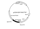
【0024】
さらに同じ特許出願人による特願平9−20906号(特願平9−329715号において優先権主張)の明細書に記載の方法にしたがって、この組換えDNA『pCSHIGIF/MUT42』を、COS−1細胞に導入し、培養し、産生されたポリペプチドをイムノアフィニティークロマトグラフィーにより精製して、精製ヒトIL−18機能性誘導体を培養培地1ml当たり約20ngの収量で得た。そしてさらに、同じ特許出願人による特願平9−20906号(特願平9−329715号において優先権主張)の明細書に記載の方法にしたがって、この精製ヒトIL−18機能性誘導体を、次に示すように分析してその生物作用と理化学的性質を確認した。すなわち、この精製ヒトIL−18機能性誘導体の各種濃度の存在下でKG−1細胞を培養すると、存在させた精製ヒトIL−18機能性誘導体の濃度に依存したIFN−γの産生が認められ、この精製IL−18機能性誘導体が、免疫担当細胞としてのKG−1細胞におけるIFN−γの産生を誘導する生物作用を有することが確認された。ユー・ケー・レムリが『ネイチャー』、第227巻、680乃至685頁(1970年)に報告した方法に準じて、還元剤として2%(w/v)ジチオトレイトール存在下でSDS−ポリアクリルアミドゲル電気泳動すると、分子量18,000乃至19,500ダルトンに相当する位置にIFN−γ誘導能ある主たるバンドを示した。また、そのN末端を常法にしたがいパーキン・エルマー製のプロテイン・シーケンサー『473A型』を用いて分析したところ、配列表の配列番号16に併記したアミノ酸配列におけるN末端部分のアミノ酸配列と完全に一致する、配列表における配列番号15に示すアミノ酸配列を有していることが確認された。
【0025】
【実験例5】
〈ヒトIL−18の調製〉
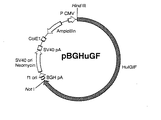
同じ特許出願人による特願平8−185305号(特願平9−187418号において優先権主張)の明細書に記載の方法にしたがって、ヒトIL−18をコードする染色体DNAが連結された、自律複製可能な組換えDNA『pBGHuGF』を調製した。ジデオキシ法により解析したところ、図4に示すようにこの組換えDNAにおいては、ヒトIL−18をコードする染色体DNAである、配列表における配列番号17に示す塩基配列のDNA『HuIGIF』が、制限酵素Hind IIIによる切断部位の下流に連結されていた。配列表における配列番号17に示すように、この染色体DNA『HuIGIF』は11,464bpよりなり、この配列の5′末端より第83乃至1,453番目、第1,466乃至4,848番目、第4,984乃至6,317番目及び第6,452乃至11,224番目に位置する4個のイントロンによりエクソンが分断されている構成であった。これらイントロンを除いた配列のうち、5′末端より第3乃至11443番目の塩基はヒトIL−18前駆体をコードする部分であり、さらにこの内、第4866乃至4983番目の塩基は活性あるヒトIL−18をコードする部分であった。また、このDNAは、配列表における配列番号1、2、3、4及び5に示したアミノ酸配列をコードする塩基配列を全て含むものであった。すなわち、これらアミノ酸配列はそれぞれ、その配列番号17に示す塩基配列における第4,911乃至4,928番目、第4,953乃至4,970番目、第11,372乃至11,392番目、第6,350乃至6,364番目及び第6,413乃至6,427番目の塩基によりコードされていた。
【0026】
さらに同じ特許出願人による特願平8−185305号(特願平9−187418号において優先権主張)の明細書に記載の方法にしたがって、この組換えDNA『pBGHuGF』を、チャイニーズ・ハムスター卵巣細胞由来の株化細胞であるCHO−K1細胞(ATCC CCL61)に導入し、培養し、培養上清にTHP−1細胞を培養して得た細胞破砕物の上清を作用させて、生成したポリペプチドをイムノアフィニティークロマトグラフィーにより精製して、精製ヒトIL−18を培養物1l当たり約15mgの収量で得た。そしてさらに、この精製ヒトIL−18を、同じ特許出願人による特願平8−185305号(特願平9−187418号において優先権主張)の明細書に記載の方法にしたがって、次に示すように分析してその生物作用と理化学的性質を確認した。すなわち、この精製ヒトIL−18の各種濃度の存在下で常法により健常人より採取したヒトリンパ球を培養すると、存在させた精製ヒトIL−18の濃度に依存したIFN−γの産生が認められ、この精製IL−18が、免疫担当細胞であるリンパ球におけるIFN−γの産生を誘導する生物作用を有することが確認された。ユー・ケー・レムリが『ネイチャー』、第227巻、680乃至685頁(1970年)に報告した方法に準じて、還元剤として2%(w/v)ジチオトレイトール存在下でSDS−ポリアクリルアミドゲル電気泳動すると、分子量18,000乃至19,500ダルトンに相当する位置にIFN−γ誘導能ある主たるバンドを示した。また、そのN末端は、配列表の配列番号17に併記したアミノ酸配列のうち、活性あるIL−18のN末端部分のアミノ酸配列と完全に一致する配列表における配列番号15に示すアミノ酸配列を有していた。
【0027】
【実験例6】
〈マウスIL−18の調製〉
0.5ml容反応管に25mM塩化マグネシウムを8μl、10×PCR緩衝液を10μl、25mM dNTPミックスを1μl、2.5単位/μlアンプリタックDNAポリメラーゼを1μl、特開平8−27189号公報に記載された方法にしたがってファージDNAクローンから調製した、配列表における配列番号18に示す塩基配列を有し、配列番号7に示すアミノ酸配列のマウスIL−18をコードするDNAを含む組換えDNAを1ng、配列表の配列番号7におけるN末端及びC末端付近のアミノ酸配列に基づき化学合成した5´−ATAGAATTCAAATGAACTTTGGCCGACTTCACTG−3´及び5´−ATAAAGCTTCTAACTTTGATGTAAGTT−3´で表わされる塩基配列のセンスプライマー及びアンチセンスプライマーの適量を加え、滅菌蒸留水で100μlとした。常法により、この混合物を94℃で1分間、43℃で1分間、72℃で1分間、この順序でインキュベートするサイクルを3回繰返した後、さらに、94℃で1分間、60℃で1分間、72℃で1分間、この順序でインキュベートするサイクルを40回繰返してPCR反応させた。
【0028】
このPCR産物とストラタジーン製プラスミドベクター『pCR−Script SK (+)』を常法にしたがってDNAリガーゼにより連結して組換えDNAとし、これをコンピテントセル法によりストラタジーン製大腸菌株『XL−1 Blue MRF´Kan』に導入して形質転換した。形質転換体を50μg/mlアンピシリンを含むL−ブロス培地(pH7.2)に接種し、37℃で18時間振盪培養した後、培養物を遠心分離して形質転換体を採取し、通常のアルカリ−SDS法を適用して組換えDNAを単離した。この組換えDNAの一部をとり、ジデオキシ法により分析したところ、配列表の配列番号18に示す塩基配列における5´末端及び3´末端にそれぞれEco RI切断部位及びHind III切断部位を含み、さらに、その配列番号18に併記したアミノ酸配列におけるN末端及びC末端のそれぞれ直前及び直後に対応する部位にポリペプチド合成開始のためのメチオニンコドン及びポリペプチド合成終止のためのTAGコドンを有するDNAを含んでいた。また、この組換えDNAは、配列表における配列番号1、2、3、4及び5に示したアミノ酸配列をコードする塩基配列を全て含むものであった。すなわち、これらアミノ酸配列はそれぞれ、その配列番号18に示した塩基配列における第46乃至63番目、第85乃至102番目、第394乃至414番目、第148乃至162番目及び第211乃至225番目の塩基によりコードされていた。
【0029】
そこで、常法にしたがって残りの組換えDNAを制限酵素Eco RI及びHind IIIで切断後、宝酒造製DNAライゲーションキット『DNAライゲーション・キット・バージョン2』を使用して、得られたEco RI−Hind III DNA断片0.1μgと予め同じ制限酵素で切断しておいたファルマシア製プラスミドベクター『pKK223−3』10ngを16℃で30分間反応させて連結して複製可能な組換えDNA『pKGFMH2』を得た。コンピテントセル法により、この組換えDNA pKGFMH2で大腸菌Y1090株(ATCC37197)を形質転換し、得られた形質転換体『KGFMH2』を50μg/mlアンピシリンを含むL−ブロス培地(pH7.2)に接種し、37℃で18時間振盪培養した。培養物を遠心分離して形質転換体を採取し、その一部に通常のSDS−アルカリ法を適用して組換えDNA『pKGFMH2』を抽出した。ジデオキシ法により分析したところ、図5に示すように、組換えDNA『pKGFMH2』においては、配列表における配列番号18に示す塩基配列を含むcDNA『KGFMH2 cDNA』がTacプロモータ『Ptac』の下流に連結されていた。
【0030】
オートクレーブにより滅菌したL−ブロス培地(pH7.2)に、アンピシリンを濃度50μg/mlとなるように添加し、37℃に冷却後、形質転換体KGFMH2を接種し、振盪下、37℃で18時間種培養した。20l容ジャーファーメンタに新鮮な同一培地を18lとり、同様に滅菌し、アンピシリンを添加し、37℃に冷却後、上記で得た種培養物を1%(v/v)接種し、37℃で8時間通気撹拌培養した。培養物を遠心分離して菌体を採取し、150mM塩化ナトリウム、16mM燐酸水素二ナトリウム及び4mM燐酸二水素ナトリウムを含む混液(pH7.3)に浮遊させ、超音波破砕後、遠心分離により菌体破砕物を除去し、上清約2lを採取した。
【0031】
得られた上清約2lに硫酸アンモニウムを含む10mM燐酸緩衝液(pH7.3)を硫酸アンモニウムが40%飽和になるように加え、沈殿物を遠心分離にて除去後、さらに上清に硫酸アンモニウムが85%飽和になるまで加え、4℃で18時間放置後、約8,000rpmで30分間遠心分離して沈澱を採取した。次にこの沈澱を1.5M硫酸アンモニウムを含む10mM燐酸緩衝液(pH6.6)に溶解して約1,300mlとし、濾過後、予め新鮮な同一緩衝液で平衡化させておいたファルマシア製『フェニルセファロース CL−4B』約800mlのカラムに負荷し、カラムを新鮮な同一緩衝液で洗浄後、1.5Mから0Mに下降する硫酸アンモニウムの濃度勾配下、10mM燐酸緩衝液(pH6.6)をSV1.5で通液した。硫酸アンモニウム濃度が1M付近のときに溶出された画分を採取し、膜濃縮し、10mM燐酸緩衝液(pH6.5)に対して4℃で18時間透析後、予め10mM燐酸緩衝液(pH6.5)で平衡化させておいたファルマシア製『DEAE−5PW』約55mlのカラムに負荷した。カラムを新鮮な同一緩衝液で洗浄後、0Mから0.5Mに上昇する塩化ナトリウムの濃度勾配下、カラムに10mM燐酸緩衝液(pH6.5)をSV5.5で通液し、塩化ナトリウム濃度が0.2M付近で溶出された画分を採取した。その後、採取した溶出液を上記と同様に濃縮して約9mlとし、PBSに対して4℃で18時間透析後、予め新鮮なPBSで平衡化させておいたファルマシア製『スーパーデックス 75』のカラムに負荷した。さらにカラムに新鮮なPBSを通液してIFN−γ誘導能ある画分を採取し、膜濃縮して精製マウスIL−18を得た。この精製マウスIL−18の収量は、培養液1l当たり約350μgであった。
【0032】
この精製マウスIL−18を、特開平8−27189号公報に記載の方法にしたがって、次に示すように分析してその生物作用と理化学的性質を確認した。すなわち、この精製マウスIL−18の各種濃度の存在下で常法により採取したマウス脾細胞を培養すると、存在させた精製マウスIL−18の濃度に依存したIFN−γの産生が認められ、この精製IL−18が、免疫担当細胞である牌細胞におけるIFN−γの産生を誘導する生物作用を有することが確認された。ユー・ケー・レムリが『ネイチャー』、第227巻、680乃至685頁(1970年)に報告した方法に準じて、非還元条件下でSDS−ポリアクリルアミドゲル電気泳動すると、分子量19,000±5,000ダルトンに相当する位置にIFN−γ誘導能ある主たるバンドを示した。また、そのN末端は、配列表の配列番号18に併記したアミノ酸配列におけるN末端部に相当する、配列番号19に示すアミノ酸配列を有していた。
【0033】
次に実験例7を示してこの発明で用いるIL−18の生物作用についてさらに詳細に説明し、実験例8を示してその毒性について説明する。
【0034】
【実験例7】
〈生物作用〉
【0035】
【実験例7−1】
〈GM−CSFの産生の誘導〉
ヘパリン加注射器により健常者から血液を採取し、血清無含有のRPMI1640培地(pH7.4)により2倍希釈した。血液をフィコール上に重層し、遠心分離して採取したリンパ球を10%(v/v)ウシ胎児血清を補足したRPMI1640培地(pH7.4)により洗浄した後、新鮮な同一培地に細胞密度1×106 個/mlになるように浮遊させ、12ウェルプレートに2ml/ウェルずつ分注した。
【0036】
別途、実験例1の方法により得たIL−18を10%(v/v)ウシ胎児血清を補足したRPMI1640培地(pH7.4)により濃度1μg/mlに調製して、上記プレートに20乃至200μl/ウェルずつ分注し、500μg/mlのコンカナバリンAを含む新鮮な上記と同一培地を10μl/ウェルずつ加えた後、5%CO2 インキュベータ中、37℃で48時間培養した。培養後、各ウェルから培養上清を0.1mlずつ採取し、通常の酵素免疫測定法によりGM−CSF含量を測定した。同時に、IL−18のみを省略した系を設け、上記と同様に処置して対照とした。結果を表1に示す。
【0037】
【表1】
表1の結果は、補因子としてコンカナバリンAの共存下でIL−18を作用させると、免疫担当細胞としてのリンパ球がIL−18の濃度に依存してGM−CSFを産生したことを示している。なお、実験例2乃至5の方法で得たIL−18乃至その機能性誘導体をそれぞれ別個に同様にこの操作に供した場合にも、いずれも同じくGM−CSFの産生を誘導することが確認された。一方、実験例6の方法で得たIL−18については、実験例7−1で用いたヒトの血液より採取したリンパ球に代えて常法によりマウスより採取した脾細胞を用いたこと以外は実験例7−1に準じて試験したところ、同じくGM−CSFの産生を誘導することが確認された。
【0039】
【実験例7−2】
〈破骨細胞形成の阻害〉
【0040】
【実験例7−2(a)】
ティー・ジェー・マーチンら、『ジャーナル・オブ・セルラー・バイオケミストリー』、第56巻(1994年)、357乃至366頁等にも記載されているように、一般に破骨細胞の前駆細胞が分化し破骨細胞が形成されるためには、骨髄の造血幹細胞に由来する前駆破骨細胞が骨芽細胞や骨髄ストローマ細胞と接触することが必要条件であるとされている。また、ジー・ディー・ルードマン、『エンドクリン・レビュー』、第17巻(1996年)、308乃至332頁等に記載されているように、破骨細胞の特徴は、多核であること、酒石酸耐性酸性ホスファターゼ(以下、「TRAP」と略記する。)活性を有すこと、カルシトニン・レセプターを有すこと等であると一般に認識されている。一方、エヌ・ウダガワら、『ジャーナル・オブ・エキスペリメンタル・メディシン』、第182巻(1995年)、1461乃至1468頁に記載の骨芽細胞と骨髄細胞の共培養系においては、例えば、1α,25−ジヒドロキシビタミンD3、プロスタグランジンE2、副腎皮質ホルモン、インターロイキン1、インターロイキン6又はインターロイキン11のうちのいずれかの因子に応答して破骨細胞様の細胞(以下、「OCL」と略記することもある。)の形成が認められる。ここで形成されるOCLは生体内の破骨細胞の特徴を備えている。したがってこの共培養系は生体内における破骨細胞の形成の過程をイン・ビトロでよく再現するものであり、この系を用いることにより破骨細胞の形成やそれに対する阻害剤についての実験を行うことができる。
【0041】
この共培養系を用いて、当該IL−18の破骨細胞形成の阻害作用を調べた。骨芽細胞は、常法に従い新生マウスの頭蓋冠を0.1%(w/v)コラゲナーゼ(オーストラリア国、ワーシントン・バイオケミカル・カンパニー製)及び0.2%(w/v)ディスパーゼ(合同酒精製)処理して調製した。骨髄細胞は、常法に従い成熟したマウスより調製した。48ウエル・プレートの1ウエル当たり、10%(v/v)ウシ胎児血清を補足したα−MEM培地(以下、この実験例4−2を通して、単に「培地」という。)0.4mlで、2×104 個の初代骨芽細胞と5×10⁵ 個の骨髄細胞を、5%CO2 インキュベーター内で37℃で7日間共培養した。これを陰性対照とした。一方、別のウエルでは、1α,25−ジヒドロキシビタミンD3(和光純薬製)及びプロスタグランジンE2(アメリカ国、シグマ製)を、濃度がそれぞれ10-8M及び10-7Mとなるように添加した培地を用いた以外は全て陰性対照と同一の方法で培養した。これを陽性対照とした。また、さらに別のウエルでは、陽性対照と同濃度の1α,25−ジヒドロキシビタミンD3及びプロスタグランジンE2とともに実験例6の方法で調製したIL−18を0.01乃至10ng/mlのいずれかの濃度となるように添加した培地を用いた以外は全て陽性対照と同一の方法で培養した。いずれの場合も培養3日目に、それぞれのウェルでそれまでに用いていたのと同一組成の新鮮な培地と交換した。6日間培養した後の細胞を、エヌ・ウダガワら、『ジャーナル・オブ・エキスペリメンタル・メディシン』、第182巻(1995年)、1461乃至1468頁に記載の方法に従って、固定し、TRAP活性に基づき染色し、1ウエルあたり染色された細胞(以下、「TRAP陽性細胞」という。)の数を数えた。この実験例4−2を通して、全て同一の培養系を4ウエルずつ設け、1ウエル当たりのTRAP陽性細胞数の平均値を求めた。結果を表2に示す。
【0042】
【表2】
表2に示すように、陰性対照ではTRAP陽性細胞の形成はほとんど認められなかったのに対し、陽性対照ではTRAP陽性細胞の形成は顕著であった。一方、陽性対照にさらにIL−18を添加した系では、その濃度に依存してTRAP陽性細胞の形成が阻害され、IL−18濃度が8ng/ml以上のときその阻害は最大で、TRAP陽性細胞数は陰性対照と同等の値となった。以上のことは、当該IL−18には確かにイン・ビトロにおけるOCL形成を阻害する作用のあることを示しており、さらに当該IL−18が破骨細胞形成を阻害することをも強く示唆している。
【0044】
【実験例7−2(b)】
先にも述べたように、この実験例7−2を通して用いられる共培養系において破骨細胞様の細胞の形成を促す因子には種々のものがあると確認されている。そこでこの実験例7−2(b)では、実験例7−2(a)で示された破骨細胞形成に対する当該IL−18の阻害作用が、ある種の因子に特異的なものか否かを調べた。すなわち、1α,25−ジヒドロキシビタミンD3 、プロスタグランジンE2 、副甲状腺ホルモン、インターロイキン1又はインターロイキン11のいずれかを、それぞれ濃度10-8M、10-7M、200ng/ml、100ng/ml又は20ng/mlとなるように添加した培地を用いた以外は、全て実験例7−2(a)の陰性対照と同一の方法で培養した。これらを陽性対照とした。一方、別のウエルでは、この陽性対照と同濃度のいずれかの因子に加え、さらに実験例6の方法で得たIL−18を10ng/mlとなるように添加した培地を用いた以外は全て陽性対照と同一の方法で培養した。培養後実験例7−2(a)と同じくTRAP陽性細胞の数を比較した。結果を表3に示す。
【0045】
【表3】
表3に示すように、いずれも陽性対照では顕著なTRAP陽性細胞の形成を認めた。これに対し、陽性対照にIL−18を添加した場合にはいずれもTRAP陽性細胞の形成はほぼ完全に阻害された。このことは、当該IL−18が破骨細胞形成の要因によらず、広く一般的に破骨細胞の形成を阻害する作用を有することを強く示唆している。
【0047】
【実験例7−2(c)】
次に、以上実験例7−2(a)及び実験例7−2(b)により確認された、当該IL−18による破骨細胞の形成の阻害が、当該IL−18により産生の誘導されたGM−CSFの作用によるものか否かを調べた。陰性対照及び陽性対照は、それぞれ実験例7−2(a)で示したのと同一の系を用いた。別のウエルでは、この陽性対照と同じ濃度の1α,25−ジヒドロキシビタミンD3及びプロスタグランジンE2を添加するとともに、さらに(i)抗マウスGM−CSFポリクローナル抗体(アメリカ国、アール・アンド・ディー・システムズ製)を濃度10μg/mlとなるように添加するか、(ii)実験例6の方法で得たIL−18を濃度10ng/mlとなるよう添加するか、(iii)iiに加えさらに抗マウスGM−CSFポリクローナル抗体を濃度10μg/mlとなるよう添加するか、(iv)マウスGM−CSF(アメリカ国、アール・アンド・ディー・システムズ製)を濃度0.1ng/mlとなるように添加するか、又は(v)ivに加えさらに抗マウスGM−CSFポリクローナル抗体を濃度10μg/mlとなるように添加した培地のいずれかを用いたこと以外は、全て陽性対照と同じ方法で培養した。培養後実験例7−2(a)と同じくTRAP陽性細胞の数を比較した。結果を表4に示す。なお、表4中に示したi乃至vの符号は、ここで説明した対照系以外の培養系の符号と一致している。
【0048】
【表4】
表4に示すように、TRAP陽性細胞の形成はIL−18によりほぼ完全に阻害された(培養系ii)が、この阻害は、抗マウスGM−CSFポリクローナル抗体の添加によりほぼ完全に解除された(培養系iii)。一方マウスGM−CSFにも、IL−18と同様にTRAP陽性細胞の形成を阻害する作用が認められた(培養系iv)が、この阻害は、抗マウスGM−CSFポリクローナル抗体の添加によりほぼ完全に解除された(培養系v)。また、当該抗体単独ではTRAP陽性細胞の形成には何の影響も与えなかった(培養系i)。以上の結果は、当該IL−18の破骨細胞形成に対する阻害は、主として、当該IL−18により誘導され産生したGM−CSFの作用によるものであることを強く示唆している。
【0050】
【実験例8】
〈急性毒性試験〉
常法にしたがって、8週齢のマウスに実験例1乃至6の方法で得た当該IL−18のいずれかをそれぞれ別個に経皮、経口又は腹腔内に注射投与した。その結果、これら当該IL−18のLD50は、いずれの投与経路によっても約1mg/kgマウス体重以上であった。このことは当該IL−18がヒトを始めとする温血動物への投与を前提とする医薬品に配合して安全であることを裏付けている。
【0051】
また、当該IL−18により誘導され産生されるGM−CSFは、日経BP社発行、『日経バイオ年鑑96』(1995年)、498乃至499頁に記載されているように、日本国内ではまだ臨床応用されるに至ってはいないものの、米国や欧州では既に臨床応用されており、その安全性については問題がないといえる。以上のことは、この発明の破骨細胞形成阻害剤が重篤な副作用を惹起することなくヒトを始めとする温血動物に長期連用でき、破骨細胞の形成及び/又は機能が関与する疾患の治療・予防に効果を発揮することを示している。
【0052】
以下に実施例を示し、この発明の破骨細胞形成阻害剤を説明する。
【0053】
【実施例1】
〈液剤〉
安定剤として1%(w/v)ヒト血清アルブミンを含む生理食塩水に実験例1乃至6の方法により得たいずれかの当該IL−18を2mg/mlになるように溶解し、常法にしたがって精密濾過により滅菌して液剤を得た。
【0054】
本品はいずれも安定性に優れ、細胞培養用の培地成分として有用であり、また、骨吸収調整剤や、高カルシウム血症、破骨細胞腫及び骨粗鬆症等を治療・予防するための破骨細胞関連疾患剤としての注射剤、点眼剤、点鼻剤として有用である。
【0055】
【実施例2】
〈乾燥剤〉
安定剤として1%(w/v)精製ゼラチンを含む生理食塩水100mlに実験例1乃至6の方法により得たいずれかの当該IL−18を50mg溶解し、常法にしたがって精密濾過により滅菌し、バイアル瓶に1mlずつ分注し、凍結乾燥後、密栓した。
【0056】
本品はいずれも安定性に優れ、細胞培養用の培地成分として有用であり、また、骨吸収調整剤や、高カルシウム血症、破骨細胞腫及び骨粗鬆症等を治療・予防するための破骨細胞関連疾患剤としての乾燥注射剤として有用である。
【0057】
【実施例3】
〈乾燥剤〉
安定剤として1%(w/v)トレハロースを含む生理食塩水100mlに実験例1乃至6の方法により得たいずれかの当該IL−18を50mg溶解し、常法にしたがって精密濾過により滅菌し、バイアル瓶に1mlずつ分注し、凍結乾燥後、密栓した。
【0058】
本品はいずれも安定性に優れ、細胞培養用の培地成分として有用であり、また、骨吸収調整剤や、高カルシウム血症、破骨細胞腫及び骨粗鬆症等を治療・予防するための破骨細胞関連疾患剤としての乾燥注射剤として有用である。
【0059】
【実施例4】
〈軟膏剤〉
滅菌蒸留水に和光純薬工業製カルボキシビニルポリマー『ハイビスワコー104』及び高純度トレハロースをそれぞれ濃度1.4%(w/w)及び2.0%(w/w)になるように溶解し、実験例1乃至6の方法により得たいずれかの当該IL−18を均一に混合後、pH7.2に調製して、1g当たり当該IL−18を約1mg含むペースト状物を得た。
【0060】
本品はいずれも延展性と安定性に優れ、骨吸収調整剤や、高カルシウム血症、破骨細胞腫及び骨粗鬆症等を治療・予防するための破骨細胞関連疾患剤としての軟膏剤として有用である。
【0061】
【実施例5】
〈錠剤〉
林原製無水結晶α−マルトース粉末『ファイントース』に実験例1乃至6の方法により得たいずれかの当該IL−18と細胞賦活剤としてのルミンを均一に混合し、得られる混合物を常法により打錠して製品1錠(約200mg)当たり当該IL−18及びルミンをそれぞれ約2mg含む錠剤を得た。
【0062】
本品はいずれも摂取性、安定性に優れ、しかも細胞賦活作用をも有し、骨吸収調整剤や、高カルシウム血症、破骨細胞腫及び骨粗鬆症等を治療・予防するための破骨細胞関連疾患剤としての錠剤として有用である。
【0063】
【発明の効果】
以上説明したように、この発明の破骨細胞形成阻害剤は、イン・ビトロ、イン・ビボを問わず、破骨細胞の形成を顕著に阻害するので、細胞培養用の培地成分として有用であり、また、骨吸収調整剤や、高カルシウム血症、破骨細胞腫及び骨粗鬆症を始めとする破骨細胞関連疾患を治療・予防するための疾患剤としても効果を発揮する。
【0064】
この発明は、斯くも顕著な作用効果を発揮するものであり、斯界に貢献すること誠に多大な意義のある発明といえる。
【0065】
【配列表】
【図面の簡単な説明】
【図1】組換えDNA pKGFHH2の構造を示す図である。
【図2】組換えDNA pCSHIGIF/MUT35の構造を示す図である。
【図3】組換えDNA pCSHIGIF/MUT42の構造を示す図である。
【図4】組換えDNA pBGHuGFの構造を示す図である。
【図5】組換えDNA pKGFMH2の構造を示す図である。
【符号の説明】
KGFHH2 cDNA 当該IL−18をコードするcDNA
IGIF/MUT35 当該IL−18をコードするDNA
IGIF/MUT42 当該IL−18をコードするDNA
HuIGIF 当該IL−18をコードする染色体DNA
KGFMH2 cDNA 当該IL−18をコードするcDNA
5S 5SリボゾームRNA遺伝子
Ptac tacプロモータ
rrnBT1T2 リボゾームRNAオペロンの転写終止領域
AmpR アンピシリン耐性遺伝子
pBR322ori 大腸菌における複製開始点
CMV サイトメガロウイルス・プロモーター
IFNss ヒトインターフェロン−αにおけるサブタイプ
α2bのシグナルペプチドをコードする配列[0001]
BACKGROUND OF THE INVENTION
The present invention relates to an osteoclast formation inhibitor comprising interleukin-18 (hereinafter abbreviated as “IL-18”) or a functional derivative thereof.
[0002]
[Prior art]
In a healthy living body, bone formation by osteoblasts and bone resorption by osteoclasts are maintained in a balanced manner, and as a result, bone tissue is always replaced with new bone without changing its basic shape and remains healthy. I'm leaning. This balance also plays an important role in maintaining the homeostasis of the living body, such as keeping the calcium concentration in the blood constant. On the other hand, when this balance is lost, especially when the amount of bone resorption exceeds the amount of bone formation, not only bone-related diseases but also various other diseases are caused. Therefore, the elucidation of a series of bone resorption mechanisms in the living body, especially the elucidation of the osteoclast formation mechanism, is not only scientifically significant but also clinically significant. Collecting.
[0003]
However, the mechanism of osteoclast formation, for example, has not yet been fully elucidated despite the fact that interleukin-1 as an promoting factor and interleukin-4 as an inhibiting factor have been confirmed. . This is thought to be due to the fact that the formation of osteoclasts in the living body is controlled in a complex and closely related manner by numerous promoters and inhibitors, as well as various other in vivo phenomena. Yes. For these reasons, there is a demand for the establishment of an effective osteoclastogenesis inhibitor not only from a scientific standpoint but also from a clinical standpoint.
[0004]
[Problems to be solved by the invention]
In view of such circumstances, an object of the present invention is to provide an effective novel osteoclast formation inhibitor.
[0005]
[Means for Solving the Problems]
[0006]
IL-18 is a kind of cytokine that is a signal transmitter in the immune system. In the initial discovery, this cytokine is disclosed in JP-A-8-27189, JP-A-8-193098, and Harki Okamura et al., “Nature”, Vol. 378, No. 6,552, pages 88 to 91 (1995). As described in 1993, it was described as an interferon-γ inducer, but later, Shinpei Ushio et al., “The Journal of Immunology”, 156, 4,274-4,279 (1996). According to the proposal in year), it came to be called IL-18. IL-18 is abbreviated as interferon-γ (hereinafter abbreviated as “IFN-γ”) and granulocyte-macrophage colony-stimulating factor (hereinafter, “GM-CSF”) useful as a physiologically active substance in immunocompetent cells. ), And further enhances the cytotoxicity of killer cells and induces the generation of killer cells.
[0007]
On the other hand, in the course of advancing research to solve the above-mentioned problems, the present inventors have found that a certain type of stromal derived from mouse bone marrow has the ability to inhibit the formation of osteoclasts from precursor osteoclasts in vitro. It was found that a specific gene was specifically expressed in large amounts in cell lines. As a result of further detailed analysis, the gene was found to encode IL-18 having the amino acid sequence shown in SEQ ID NO: 7 in the sequence listing. Based on these findings, further earnest studies have revealed that IL-18 having the amino acid sequence and functional derivatives thereof significantly inhibit the formation of osteoclasts, and this inhibition is mainly induced by the production of IL-18. It was found to be due to the action of GM-CSF. The present invention has been completed based on the above unique findings.
[0008]
That is, this invention solves said subject by the osteoclast formation inhibitor containing IL-18 or its functional derivative.
[0009]
DETAILED DESCRIPTION OF THE INVENTION
The osteoclast formation inhibitor of the present invention comprises IL-18 or a functional derivative thereof. IL-18 here includes all polypeptides having the above-mentioned properties as IL-18, and the origin and origin thereof are not limited. IL-18 used in the present invention has, for example, the amino acid sequences shown in SEQ ID NOs: 1, 2, and 3 in the sequence listing as partial amino acid sequences in the middle, and SEQ ID NOs: 4 and 5, and the whole SEQ ID NO: IL-18 having the amino acid sequence shown in 6 or 7 can be mentioned. In addition, the functional derivative referred to in the present specification refers to one of the amino acids in the amino acid sequence within a range that does not substantially lose the property of inhibiting the formation of osteoclasts among the properties of IL-18 as described above. One or two or more substituted with other amino acids, one or two or more amino acids added to the N-terminal and / or C-terminal in these amino acid sequences, one in the middle of these amino acid sequences or Insertion of two or more amino acids, deletion of one or more N-terminal and / or C-terminal amino acids in these amino acid sequences, and one or two intermediate amino acids in these amino acid sequences This means that one or more are deleted. As such a functional derivative, for example, in the specification of Japanese Patent Application No. 9-20906 (claimed priority in Japanese Patent Application No. 9-329715) by the same patent applicant, the property as IL-18 is substantially It details the amino acid sequences of functional derivatives that retain and improve their stability. Furthermore, the functional derivative referred to in the present specification includes a polypeptide obtained by adding a sugar chain to IL-18 or the functional derivative thereof as described above. IL-18 or a functional derivative thereof as described above (hereinafter abbreviated as “IL-18” including both IL-18 and a functional derivative thereof unless otherwise specified) is obtained by a cell culture method or the like. It may be isolated from a natural source or artificially synthesized by recombinant DNA technology or peptide synthesis method.
[0010]
From an economic point of view, a method using a recombinant DNA technique is advantageous. In such a method, a DNA encoding IL-18 is usually introduced into a suitable host derived from a microorganism or an animal or plant, and a transformant is used. None, after culturing by conventional methods, the culture is purified by conventional methods for purifying cytokines to obtain IL-18. The DNA used here may be any DNA as long as it contains DNA encoding IL-18, and DNA having an appropriate sequence may be used depending on the purpose of the osteoclast formation inhibitor and the method to be applied. You can choose. For example, in Japanese Patent Laid-Open Nos. 8-193098, 8-231598, and 8-27189 by the same patent applicant, DNA containing cDNA encoding IL-18 derived from mouse and human is introduced. The method for producing IL-18 obtained by culturing the transformed microorganism is also disclosed in the specification of Japanese Patent Application No. 8-185305 (claimed priority in Japanese Patent Application No. 9-187418) by the same patent applicant. Describes in detail a method for producing IL-18 obtained by culturing transformed animal cells into which DNA containing chromosomal DNA encoding human-derived IL-18 has been introduced. Furthermore, in the specification of Japanese Patent Application No. 9-20906 (claimed priority in Japanese Patent Application No. 9-329715) by the same patent applicant, DNA containing DNA encoding a human IL-18 functional derivative was introduced. The method for producing IL-18 obtained by culturing transformed animal cells is described in detail.
[0011]
While the recombinant DNA technology as described above is advantageous in terms of economy as described above, the IL-18 obtained by such a method is originally produced in vivo depending on the host used and the DNA sequence. Some differences may occur in physicochemical properties from IL-18. However, in the specification of Japanese Patent Application No. 8-67434 (claimed priority in Japanese Patent Application No. 8-269105 (Japanese Patent Application Laid-Open No. 9-289896)) by the same patent applicant, it is cultured as a natural source. A method for producing IL-18 using human cells is also disclosed in Japanese Patent Application No. 8-213267 (priority claim in Japanese Patent Application No. 9-213885) by the same patent applicant. Methods for producing IL-18 using 1β converting enzyme are described in detail. Is IL-18 obtained by these methods substantially identical in physicochemical properties to IL-18 produced and functioning in vivo? Or, because it is considered to be very close to the same, although the production amount is slightly low, for example, in the case of using it as a medicine premised on administration to warm-blooded animals including humans, it has low side effects. Is an interest. When applying the purification method using a monoclonal antibody specific for IL-18 disclosed in JP-A-8-231598 by the same patent applicant, the high-purity IL-18 is minimized. It can be obtained with limited cost and effort.
[0012]
The osteoclast formation inhibitor of the present invention comprises the thus obtained IL-18, and is used for inhibiting the formation of osteoclasts regardless of in vitro or in vivo. Includes all possible forms. For example, as a medium component for cell culture of animal cells or the like that inhibits osteoclast formation and improves the maintenance, proliferation and / or differentiation of desired cells, The composition can be advantageously used as a bone resorption regulator and as an osteoclast-related disease agent. The term “bone resorption regulator” as used herein refers to a drug that adjusts bone resorption to a normal range by inhibiting the formation of osteoclasts in a living body, and improves relatively poor physical condition such as joint pain and Includes health foods. Further, the osteoclast-related disease agent referred to here includes a drug for preventing and / or treating all diseases associated with excessive formation and / or function of osteoclasts in vivo. The target diseases include, for example, hypercalcemia, osteoclastoma, Pechet's disease, osteosarcoma, arthropathy, rheumatoid arthritis, osteoarthritis, primary hyperthyroidism, osteopenia, osteoporosis, etc. Can be mentioned. Although depending on the dosage form and the object of use, the osteoclast formation inhibitor of the present invention is usually prepared in a liquid, paste or solid form, and the IL-18 is 0.000002 to 100% (w / w). Desirably, it comprises 0.0002 to 0.5% (w / w).
[0013]
The osteoclast formation inhibitor of the present invention is not only in the form of the IL-18 alone, but also, for example, carriers, excipients, diluents, immune aids, antibiotics, serum albumin and gelatin as stabilizers, etc. In addition to these proteins, glucose, maltose, maltotriose, maltotetraose, trehalose, sucrose, isomaltose, lactose, panose, erulose, palatinose, lactosucrose, raffinose, fructo-oligo, galactooligosaccharide, lentinan, dextrin, pullulan and other sugars And other sugar alcohols such as sugar alcohols such as sorbitol, maltitol, lactitol and maltotriitol, phosphate or citrate buffer, 2-mercaptoethanol, dithiothreitol and reduced glutathione Reducing agent, In addition, interferon-α, interferon-β, interferon-γ, interleukin 2, interleukin 3, interleukin 6, interleukin 12, TNF-α, TNF-β, GM-CSF, estrogen as necessary. , Progesterone, chlormadinone acetate, calcitonin, somatocaine, somatomedin, insulin-like growth factor, ipriflavone, paracyloid hormone, norethisterone, busulfan, ancitabine, cytarabine, fluorouracil, tetrahydrofurylfluorouracil, methotrexate, vitamin D 2 1 or 2 of other physiologically active substances such as active vitamin D, krestin, L-asparaginase and picibanil, and calcium salts including calcium lactate, calcium chloride, calcium hydrogen phosphate, calcium L-aspartate The form as a composition with a seed | species or more is also included. When the osteoclast formation inhibitor of the present invention is administered to warm-blooded animals including humans, for example, as an osteoclast-related disease agent, a physiologically acceptable substance is appropriately selected from the above. It is desirable to select a composition.
[0014]
Furthermore, the osteoclast formation inhibitor of the present invention also includes a drug in a dosage unit form when administered to a warm-blooded animal including humans. The drug in the dosage unit form includes the IL- 18 is a physically separate unit dosage form suitable for dosing comprising, for example, an amount corresponding to a single dose or an integral multiple (up to 4 times) or a divisor (up to 1/40) thereof Means a drug in Examples of such dosage forms include injections, solutions, powders, granules, tablets, capsules, sublinguals, eye drops, nasal drops, suppositories, and the like.
[0015]
The osteoclast formation inhibitor of the present invention as an osteoclast formation inhibitor can be administered orally or parenterally, in any case, for the treatment of osteoclast-related diseases. Effective for prevention. Although it depends on the type and symptoms of the disease, it is specifically about 0.5 μg to 100 mg / dose, preferably about 2 μg to 10 mg / dose per adult, while observing the patient's symptoms and the course after administration. If IL-18 is orally administered at a dose of 2 to 6 times / day or 2 to 10 times / week for 1 day to 1 year, or administered parenterally intradermally, subcutaneously, intramuscularly or intravenously Good.
[0016]
Next, an experimental example is shown and preparation of the IL-18, physicochemical properties and biological action of the IL-18 are described.
[0017]
[Experiment 1]
<Preparation of human IL-18>
According to the method described in JP-A-8-231598 by the same patent applicant, an autonomously replicable recombinant DNA “pKGFHH2” linked with cDNA encoding human IL-18 was prepared. When analyzed by the dideoxy method, as shown in FIG. 1, in this recombinant DNA, the cDNA “KGFHH2 cDNA” containing the nucleotide sequence shown in SEQ ID NO: 8 in the sequence listing is linked downstream of the Tac promoter “Ptac”. It was. Further, this recombinant DNA “pKGFHH2” contained all the base sequences encoding the amino acid sequences shown in SEQ ID NOs: 1, 2, 3, 4 and 5 in the sequence listing. That is, these amino acid sequences are respectively represented by the 46th to 63rd, 88th to 105th, 400th to 420th, 151th to 165th, and 214th to 228th bases in the base sequence shown in SEQ ID NO: 8. It was coded.
[0018]
Furthermore, according to the method described in JP-A-8-231598, this recombinant DNA “pKGFHH2” is introduced into E. coli Y1090 strain (ATCC 37197), cultured, and the produced polypeptide is subjected to immunoaffinity chromatography. When purified, purified human IL-18 with a purity of 95% or more was obtained in a yield of about 25 mg per liter of culture broth. This purified human IL-18 was analyzed as follows to confirm its biological action and physicochemical properties according to the method described in JP-A-8-193098 by the same patent applicant. That is, when human lymphocytes collected from healthy individuals were cultured by a conventional method in the presence of various concentrations of this purified human IL-18, production of IFN-γ depending on the concentration of the purified human IL-18 present was observed. This purified IL-18 was confirmed to have a biological action that induces the production of IFN-γ in lymphocytes that are immunocompetent cells. When subjected to SDS-polyacrylamide gel electrophoresis under non-reducing conditions according to the method reported by Yu K. Remli in “Nature”, Vol. 227, pages 680 to 685 (1970), the molecular weight is 18,500 ± 3. A main band capable of inducing IFN-γ was shown at a position corresponding to 000 daltons, while an isoelectric point was shown at 4.9 ± 1.0 when chromatofocusing was performed according to a conventional method. The N-terminus was analyzed using a protein sequencer “473A type” manufactured by Perkin Elmer according to a conventional method. As a result, the amino acid sequence shown in SEQ ID NO: 8 in the sequence listing had a methionine bound to the N-terminus. It was confirmed to have the amino acid sequence shown in FIG.
[0019]
[Experimental example 2]
<Preparation of human IL-18>
According to the method described in the specification of Japanese Patent Application No. 8-67434 (claimed priority in Japanese Patent Application No. 8-269105 (JP 9-289896 A)) by the same patent applicant, A THP-1 cell (ATCC TIB202), which is a type of bone marrow monocytic cell line derived from human acute monocytic leukemia, was transplanted subcutaneously in the back of the newborn and reared for 3 weeks. Then, the tumor mass (about 15 g / animal) generated subcutaneously was removed and dispersed, and then the cells were crushed. The produced polypeptide was purified by immunoaffinity chromatography, and purified human IL-18 was purified by hamster. A yield of about 50 ng per animal was obtained.
[0020]
This purified human IL-18 was analyzed according to the method described in the specification of Japanese Patent Application No. 8-67434 as follows to confirm its biological action and physicochemical properties. That is, when human lymphocytes collected from healthy individuals were cultured by a conventional method in the presence of various concentrations of this purified human IL-18, production of IFN-γ depending on the concentration of the purified human IL-18 present was observed. This purified IL-18 was confirmed to have a biological action that induces the production of IFN-γ in lymphocytes that are immunocompetent cells. In accordance with the method reported by YU Remli, Nature, 272, 680-685 (1970), SDS-polyacrylamide in the presence of 2% (w / v) dithiothreitol as a reducing agent Gel electrophoresis showed a main band capable of inducing IFN-γ at a position corresponding to a molecular weight of 18,000 to 19,500 daltons. Furthermore, according to a peptide map whose method is described in the specification of Japanese Patent Application No. 8-67434, a polypeptide fragment obtained by treating this purified human IL-18 with Sigma's clostripain agent is obtained as Tosoh. When the amino acid sequence of each peptide fragment was analyzed from the N-terminal side by subjecting it to high performance liquid chromatography using a column of “ODS-120T” manufactured by ODS-120T, SEQ ID NOs: 10, 11, 12, and 13 in the sequence listing The amino acid sequence shown below was obtained. These amino acid sequences are respectively in the amino acid sequence shown in SEQ ID NO: 6 in the sequence listing. First 148th to 157th, 1st to 13th, 45th to 58th and 80th to 96th sequences were completely matched. The above results indicate that IL-18 obtained by the method of Experimental Example 2 has the amino acid sequence shown in SEQ ID NO: 6 in the sequence listing, and the portions shown in SEQ ID NO: 1, 2, 3, 4 and 5 thereof. It shows having all amino acid sequences.
[0021]
[Experiment 3]
<Preparation of functional derivative>
According to the method described in the specification of Japanese Patent Application No. 9-20906 (claimed priority in Japanese Patent Application No. 9-329715) by the same patent applicant, the 38th amino acid in the amino acid sequence shown in SEQ ID NO: 6 Recombinant DNA “pCSHIGIF / that can replicate autonomously and is linked to a DNA encoding a human IL-18 functional derivative in which cysteine is replaced with serine, 68th cysteine is replaced with serine, and 76th cysteine is replaced with alanine. MUT35 "was prepared. When analyzed by the dideoxy method, as shown in FIG. 2, in this recombinant DNA, Kirsten Henko et al., “Journal of Molecular Biology”, Vol. 185, pages 227 to 260 (1985). A DNA “IGIF / MUT35” having the base sequence shown in SEQ ID NO: 14 in the sequence table is linked downstream of the base sequence encoding the signal peptide of subtype α2b in human interferon-α that has been reported. Furthermore, there was a stop codon for protein synthesis downstream. The amino acid sequence encoded by this DNA is as shown in SEQ ID NO: 14 in the Sequence Listing, wherein the 38th cysteine in the amino acid sequence in SEQ ID NO: 6 in the Sequence Listing is serine, the 68th cysteine in serine, and the 76th sequence. The second cysteine was substituted with alanine. In addition, this DNA has all amino acid sequences shown in SEQ ID NOs: 1, 2, 3, and 4 in the sequence listing, and an amino acid sequence in which the fifth amino acid cysteine in the amino acid sequence shown in SEQ ID NO: 5 is substituted with alanine. It contained the base sequence which codes. That is, these amino acid sequences are represented by the 46th to 63rd, 88th to 105th, 400th to 420th, 151st to 165th, and 214th to 228th bases in the base sequence shown in SEQ ID NO: 14, respectively. It was coded.
[0022]
Further, according to the method described in the specification of Japanese Patent Application No. 9-20906 (claimed priority in Japanese Patent Application No. 9-329715) by the same patent applicant, this recombinant DNA “pCSHIGIF / MUT35” is It is introduced into COS-1 cells (ATCC CRL1650), which is a type of cell line derived from the kidney, cultured, and the produced polypeptide is purified by immunoaffinity chromatography to produce purified human IL-18 functional derivative. A yield of about 40 ng per ml of culture medium was obtained. Furthermore, according to the method described in the specification of Japanese Patent Application No. 9-20906 (claimed priority in Japanese Patent Application No. 9-329715) by the same patent applicant, this purified human IL-18 functional derivative is The biological action and physicochemical properties were confirmed by analysis as shown in. That is, when KG-1 cells (ATCC CCL246), which is a type of cell line derived from human acute myeloid leukemia, are cultured in the presence of various concentrations of this purified human IL-18 functional derivative, the purified humans present. Production of IFN-γ depending on the concentration of the IL-18 functional derivative was observed, and this purified IL-18 functional derivative induces the production of IFN-γ in KG-1 cells as immunocompetent cells. It was confirmed to have In accordance with the method reported by Yu K. Remli in “Nature”, Vol. 227, 680-685 (1970), SDS-polyacrylamide in the presence of 2% (w / v) dithiothreitol as a reducing agent Gel electrophoresis showed a main band capable of inducing IFN-γ at a position corresponding to a molecular weight of 18,000 to 19,500 daltons. In addition, when the N-terminal was analyzed using a protein sequencer “473A type” manufactured by Perkin Elmer according to a conventional method, the amino acid sequence of the N-terminal part in the amino acid sequence shown in SEQ ID NO: 14 in the sequence listing was completely It was confirmed to have a matching amino acid sequence shown in SEQ ID NO: 15 in the sequence listing.
[0023]
[Experimental Example 4]
<Preparation of functional derivative>
According to the method described in the specification of Japanese Patent Application No. 9-20906 (claimed priority in Japanese Patent Application No. 9-329715) by the same patent applicant, the 38th amino acid in the amino acid sequence shown in SEQ ID NO: 6 An autonomously linked DNA encoding a human IL-18 functional derivative in which cysteine is substituted with serine, 68th cysteine with serine, 76th cysteine with alanine, and 127th cysteine with serine. A replicable recombinant DNA “pCSHIGIF / MUT42” was prepared. When analyzed by the dideoxy method, as shown in FIG. 3, in this recombinant DNA, Carsten Henko et al., “Journal of Molecular Biology”, Vol. 185, pages 227 to 260 (1985). The DNA “IGIF / MUT42” having the base sequence shown in SEQ ID NO: 16 in the sequence table is linked downstream of the reported base sequence encoding the signal peptide of subtype α2b in human interferon-α. Furthermore, there was a stop codon for protein synthesis downstream. The amino acid sequence encoded by this DNA is as shown in SEQ ID NO: 16 in the Sequence Listing, wherein the 38th cysteine of the amino acid sequence in SEQ ID NO: 6 is serine, the 68th cysteine is serine, and the 76th The cysteine was replaced with alanine and the 127th cysteine was replaced with serine. In addition, in this DNA, all amino acid sequences shown in SEQ ID NOs: 1, 2, 3, and 4 in the sequence listing, and cysteine of the fifth amino acid in the amino acid sequence shown in SEQ ID NO: 5 in the sequence listing are substituted with alanine. The nucleotide sequence encoding the amino acid sequence was included. That is, these amino acid sequences are the 46th to 63rd, 88th to 105th, 400th to 420th, 151st to 165th, and 214th to 228th positions in the base sequence shown in SEQ ID NO: 16 in the sequence listing, respectively. It was encoded by the base.
[0024]
Further, according to the method described in the specification of Japanese Patent Application No. 9-20906 (claimed priority in Japanese Patent Application No. 9-329715) by the same patent applicant, this recombinant DNA “pCSHIGIF / MUT42” is converted into COS-1 The cells were introduced into cells, cultured, and the produced polypeptide was purified by immunoaffinity chromatography to obtain purified human IL-18 functional derivative in a yield of about 20 ng / ml culture medium. Furthermore, according to the method described in the specification of Japanese Patent Application No. 9-20906 (claimed priority in Japanese Patent Application No. 9-329715) by the same patent applicant, this purified human IL-18 functional derivative is The biological action and physicochemical properties were confirmed by analysis as shown in. That is, when KG-1 cells were cultured in the presence of various concentrations of this purified human IL-18 functional derivative, production of IFN-γ depending on the concentration of the purified human IL-18 functional derivative present was observed. This purified IL-18 functional derivative was confirmed to have a biological action that induces the production of IFN-γ in KG-1 cells as immunocompetent cells. In accordance with the method reported by Yu K. Remli in “Nature”, Vol. 227, 680-685 (1970), SDS-polyacrylamide in the presence of 2% (w / v) dithiothreitol as a reducing agent Gel electrophoresis showed a main band capable of inducing IFN-γ at a position corresponding to a molecular weight of 18,000 to 19,500 daltons. The N-terminal was analyzed using a protein sequencer “473A type” manufactured by Perkin Elmer according to a conventional method. As a result, the amino acid sequence of the N-terminal part in the amino acid sequence shown in SEQ ID NO: 16 in the sequence listing was completely It was confirmed to have a matching amino acid sequence shown in SEQ ID NO: 15 in the sequence listing.
[0025]
[Experimental Example 5]
<Preparation of human IL-18>
In accordance with the method described in the specification of Japanese Patent Application No. 8-185305 (priority claim in Japanese Patent Application No. 9-187418) by the same patent applicant, the chromosomal DNA encoding human IL-18 has been linked. A replicable recombinant DNA “pBGHuGF” was prepared. When analyzed by the dideoxy method, as shown in FIG. 4, in this recombinant DNA, the DNA “HuIGIF” of the nucleotide sequence shown in SEQ ID NO: 17 in the sequence listing, which is a chromosomal DNA encoding human IL-18, is restricted. It was ligated downstream of the cleavage site by the enzyme Hind III. As shown in SEQ ID NO: 17 in the sequence listing, this chromosomal DNA “HuIGIF” consists of 11,464 bp, 83rd to 1453rd, 1st to 466th to 4848th from the 5 ′ end of this sequence, The exons were divided by four introns located at positions 4,984 to 6,317 and 6,452 to 11,224. Of these sequences excluding introns, the 3rd to 11443th bases from the 5 'end are portions encoding human IL-18 precursor, and among these, the 4866th to 4983th bases are active human IL-18s. It was the portion encoding -18. Further, this DNA contained all the base sequences encoding the amino acid sequences shown in SEQ ID NOs: 1, 2, 3, 4 and 5 in the sequence listing. That is, these amino acid sequences are respectively 4,911 to 4,928th, 4,953 to 4,970th, 11,372 to 11,392, 6th in the nucleotide sequence shown in SEQ ID NO: 17. It was encoded by the 350th to 6,364th positions and the 6,413th to 6,427th bases.
[0026]
Further, according to the method described in the specification of Japanese Patent Application No. 8-185305 (priority claimed in Japanese Patent Application No. 9-187418) by the same patent applicant, this recombinant DNA “pBGHuGF” was converted into Chinese hamster ovary cells. Introduced into CHO-K1 cells (ATCC CCL61), which is a cell line derived from the culture, cultured, and the supernatant of cell debris obtained by culturing THP-1 cells on the culture supernatant was allowed to act. The peptide was purified by immunoaffinity chromatography to obtain purified human IL-18 in a yield of approximately 15 mg / l culture. Further, this purified human IL-18 is expressed as follows in accordance with the method described in the specification of Japanese Patent Application No. 8-185305 (priority claimed in Japanese Patent Application No. 9-187418) by the same patent applicant. It was analyzed to confirm its biological action and physicochemical properties. That is, when human lymphocytes collected from healthy individuals were cultured by a conventional method in the presence of various concentrations of this purified human IL-18, production of IFN-γ depending on the concentration of the purified human IL-18 present was observed. This purified IL-18 was confirmed to have a biological action that induces the production of IFN-γ in lymphocytes that are immunocompetent cells. In accordance with the method reported by Yu K. Remli in “Nature”, Vol. 227, 680-685 (1970), SDS-polyacrylamide in the presence of 2% (w / v) dithiothreitol as a reducing agent Gel electrophoresis showed a main band capable of inducing IFN-γ at a position corresponding to a molecular weight of 18,000 to 19,500 daltons. In addition, the N-terminus has the amino acid sequence shown in SEQ ID NO: 15 in the sequence listing that completely matches the amino acid sequence of the N-terminal portion of active IL-18 among the amino acid sequences listed in SEQ ID NO: 17 in the sequence listing. Was.
[0027]
[Experimental example 6]
<Preparation of mouse IL-18>
In a 0.5 ml reaction tube, 8 μl of 25 mM magnesium chloride, 10 μl of 10 × PCR buffer, 1 μl of 25 mM dNTP mix, 1 μl of 2.5 unit / μl amplitac DNA polymerase, described in JP-A-8-27189 1 ng of a recombinant DNA prepared from a phage DNA clone according to the above method and having a nucleotide sequence shown in SEQ ID NO: 18 in the sequence listing and containing mouse IL-18 having the amino acid sequence shown in SEQ ID NO: 7 5'-ATAGAATTCAAATGAACTTTGGCCGAACTTCACTG-3 'and 5'-ATAAAGCTCTCACTACTTTGATGTAAGTT-3' chemically synthesized based on the amino acid sequences near the N-terminus and C-terminus in SEQ ID NO: 7 And an appropriate amount of added antisense primer, was 100μl with sterile distilled water. According to a conventional method, this mixture was incubated at 94 ° C. for 1 minute, 43 ° C. for 1 minute, 72 ° C. for 1 minute in this order three times, and then further, 94 ° C. for 1 minute, 60 ° C. for 1 minute. The PCR reaction was performed by repeating 40 cycles of incubation in this order for 1 minute at 72 ° C. for 1 minute.
[0028]
This PCR product and the Stratagene plasmid vector “pCR-Script SK (+)” are ligated by DNA ligase according to a conventional method to obtain a recombinant DNA, which is transformed into a Stratagene E. coli strain “XL-1” by the competent cell method. It was introduced into Blue MRF'Kan and transformed. The transformant was inoculated into L-broth medium (pH 7.2) containing 50 μg / ml ampicillin, and cultured with shaking at 37 ° C. for 18 hours. The culture was then centrifuged, and the transformant was collected. -Recombinant DNA was isolated by applying the SDS method. When a part of this recombinant DNA was taken and analyzed by the dideoxy method, it contained an Eco RI cleavage site and a Hind III cleavage site at the 5 ′ end and 3 ′ end, respectively, in the base sequence shown in SEQ ID NO: 18 in the sequence listing. A DNA having a methionine codon for initiation of polypeptide synthesis and a TAG codon for termination of polypeptide synthesis at sites corresponding to immediately before and immediately after the N-terminal and C-terminal in the amino acid sequence shown in SEQ ID NO: 18, respectively. It was out. Moreover, this recombinant DNA contained all the base sequences encoding the amino acid sequences shown in SEQ ID NOs: 1, 2, 3, 4 and 5 in the sequence listing. That is, these amino acid sequences are respectively represented by the 46th to 63rd, 85th to 102nd, 394th to 414th, 148th to 162nd, and 211th to 225th bases in the base sequence shown in SEQ ID NO: 18. It was coded.
[0029]
Therefore, the remaining recombinant DNA was cleaved with restriction enzymes Eco RI and Hind III according to a conventional method, and then the obtained Eco RI-Hind III was used using a DNA ligation kit “DNA ligation kit version 2” manufactured by Takara Shuzo. Recombinant recombinant DNA “pKGFMH2” was obtained by ligating 0.1 μg of DNA fragment and 10 ng of Pharmacia plasmid vector “pKK223-3” previously cut with the same restriction enzyme at 16 ° C. for 30 minutes and ligating. . E. coli Y1090 strain (ATCC37197) was transformed with this recombinant DNA pKGFMH2 by competent cell method, and the obtained transformant “KGFMH2” was inoculated into L-broth medium (pH 7.2) containing 50 μg / ml ampicillin. And cultured with shaking at 37 ° C. for 18 hours. The transformant was collected by centrifuging the culture, and a recombinant DNA “pKGFMH2” was extracted by applying a normal SDS-alkali method to a part thereof. When analyzed by the dideoxy method, as shown in FIG. 5, in the recombinant DNA “pKGFMH2”, the cDNA “KGFMH2 cDNA” containing the nucleotide sequence shown in SEQ ID NO: 18 in the sequence listing is linked downstream of the Tac promoter “Ptac”. It had been.
[0030]
Ampicillin was added to an L-broth medium (pH 7.2) sterilized by autoclaving to a concentration of 50 μg / ml, cooled to 37 ° C., inoculated with transformant KGFMH2, and shaken at 37 ° C. for 18 hours. Seed culture. Take 18 l of the same medium in a 20 l jar fermenter, sterilize in the same manner, add ampicillin, cool to 37 ° C., inoculate 1% (v / v) of the seed culture obtained above, And aerated and agitated for 8 hours. The culture is centrifuged to collect the cells, suspended in a mixture (pH 7.3) containing 150 mM sodium chloride, 16 mM disodium hydrogen phosphate and 4 mM sodium dihydrogen phosphate, disrupted by ultrasonic waves, and centrifuged to centrifuge the cells. The crushed material was removed, and about 2 l of the supernatant was collected.
[0031]
To about 2 l of the obtained supernatant, 10 mM phosphate buffer (pH 7.3) containing ammonium sulfate was added so that ammonium sulfate was 40% saturated, and the precipitate was removed by centrifugation, and then 85% ammonium sulfate was further added to the supernatant. The mixture was added to saturation and allowed to stand at 4 ° C. for 18 hours, and then centrifuged at about 8,000 rpm for 30 minutes to collect a precipitate. Next, this precipitate was dissolved in 10 mM phosphate buffer (pH 6.6) containing 1.5 M ammonium sulfate to make about 1,300 ml. After filtration, “Phenyl” made by Pharmacia had been equilibrated with fresh same buffer in advance. Sepharose CL-4B "was loaded onto an approximately 800 ml column, and the column was washed with fresh identical buffer, and then 10 mM phosphate buffer (pH 6.6) was added to SV1. 5 was passed through. The fraction eluted when the ammonium sulfate concentration is around 1 M was collected, concentrated in a membrane, dialyzed against 10 mM phosphate buffer (pH 6.5) at 4 ° C. for 18 hours, and then preliminarily 10 mM phosphate buffer (pH 6.5). The column was loaded onto a column of about 55 ml of “DEAE-5PW” manufactured by Pharmacia that had been equilibrated in step 1). After washing the column with fresh same buffer, 10 mM phosphate buffer (pH 6.5) was passed through the column with SV5.5 under a sodium chloride concentration gradient increasing from 0 M to 0.5 M, and the sodium chloride concentration was The fraction eluted at around 0.2M was collected. Thereafter, the collected eluate was concentrated to about 9 ml in the same manner as described above, dialyzed against PBS at 4 ° C. for 18 hours, and equilibrated with fresh PBS in advance, “Superdex 75” column. Loaded. Further, fresh PBS was passed through the column, and a fraction capable of inducing IFN-γ was collected and concentrated to obtain purified mouse IL-18. The yield of this purified mouse IL-18 was about 350 μg per liter of culture solution.
[0032]
This purified mouse IL-18 was analyzed according to the method described in JP-A-8-27189 as follows to confirm its biological action and physicochemical properties. That is, when mouse spleen cells collected by conventional methods were cultured in the presence of various concentrations of this purified mouse IL-18, IFN-γ production depending on the concentration of purified mouse IL-18 present was observed. It was confirmed that purified IL-18 has a biological effect that induces the production of IFN-γ in sputum cells, which are immunocompetent cells. According to the method reported by Yu K. Remli in “Nature”, Vol. 227, pages 680 to 685 (1970), when subjected to SDS-polyacrylamide gel electrophoresis under non-reducing conditions, a molecular weight of 19,000 ± 5 A main band capable of inducing IFN-γ was shown at a position corresponding to 1,000,000 daltons. Moreover, the N terminal had the amino acid sequence shown to sequence number 19 corresponding to the N terminal part in the amino acid sequence written together to sequence number 18 of the sequence table.
[0033]
Next, Experimental Example 7 is shown to describe the biological action of IL-18 used in the present invention in more detail, and Experimental Example 8 is shown to explain its toxicity.
[0034]
[Experimental Example 7]
<Biological action>
[0035]
[Experimental Example 7-1]
<Induction of GM-CSF production>
Blood was collected from healthy individuals with a heparinized syringe and diluted 2-fold with serum-free RPMI 1640 medium (pH 7.4). Lymphocytes collected by overlaying blood on Ficoll and centrifuging were washed with RPMI 1640 medium (pH 7.4) supplemented with 10% (v / v) fetal calf serum, and then cell density 1 × 10 6 2 ml / well was dispensed to a 12-well plate.
[0036]
Separately, IL-18 obtained by the method of Experimental Example 1 was prepared to a concentration of 1 μg / ml with RPMI 1640 medium (pH 7.4) supplemented with 10% (v / v) fetal bovine serum, and 20 to 200 μl was added to the plate. After adding 10 μl / well of the same medium as above containing 500 μg / ml concanavalin A, add 5% CO 2 The cells were cultured for 48 hours at 37 ° C. in an incubator. After culturing, 0.1 ml of the culture supernatant was collected from each well, and the GM-CSF content was measured by a general enzyme immunoassay. At the same time, a system in which only IL-18 was omitted was provided and treated in the same manner as described above as a control. The results are shown in Table 1.
[0037]
[Table 1]
The results in Table 1 show that when IL-18 was allowed to act in the presence of concanavalin A as a cofactor, lymphocytes as immunocompetent cells produced GM-CSF depending on the concentration of IL-18. Yes. In addition, when IL-18 or a functional derivative thereof obtained by the methods of Experimental Examples 2 to 5 was separately subjected to this operation in the same manner, it was confirmed that all induced the production of GM-CSF. It was. On the other hand, for IL-18 obtained by the method of Experimental Example 6, except that spleen cells collected from mice by a conventional method were used instead of lymphocytes collected from human blood used in Experimental Example 7-1. When tested according to Experimental Example 7-1, it was also confirmed to induce the production of GM-CSF.
[0039]
[Experimental Example 7-2]
<Inhibition of osteoclast formation>
[0040]
[Experimental Example 7-2 (a)]
As described in TJ Martin et al., “Journal of Cellular Biochemistry”, Volume 56 (1994), pages 357 to 366, etc., generally osteoclast precursor cells are differentiated. In order for osteoclasts to be formed, it is said that the precursor osteoclasts derived from bone marrow hematopoietic stem cells are in contact with osteoblasts and bone marrow stromal cells. Also, GE Dee Ludman, "Endclin Review", Volume 17 (1996) 3 As described in pages 08 to 332 and the like, osteoclasts are characterized by being multinucleated, having tartrate-resistant acid phosphatase (hereinafter abbreviated as “TRAP”) activity, calcitonin receptor. It is generally recognized that it has. On the other hand, in the co-culture system of osteoblasts and bone marrow cells described in N. Udagawa et al., “Journal of Experimental Medicine”, Volume 182 (1995), pages 1461 to 1468, for example, 1α , 25-Dihydroxyvitamin D 3 , Prostaglandin E 2 Formation of osteoclast-like cells (hereinafter sometimes abbreviated as “OCL”) in response to any of the factors of corticosteroid, interleukin 1, interleukin 6 and interleukin 11. Is recognized. The OCL formed here has the characteristics of osteoclasts in vivo. Therefore, this co-culture system reproduces the process of osteoclast formation in vivo well in vitro. By using this system, experiments on osteoclast formation and inhibitors against it are conducted. Can do.
[0041]
Using this co-culture system, the inhibitory effect of the IL-18 on osteoclast formation was examined. As for osteoblasts, 0.1% (w / v) collagenase (manufactured by Worthington Biochemical Company, Australia) and 0.2% (w / v) dispase (joint liquor) were applied to the calvaria of newborn mice according to a conventional method. Purification) and prepared. Bone marrow cells were prepared from mature mice according to a conventional method. Α-MEM medium supplemented with 10% (v / v) fetal calf serum per well of a 48-well plate (hereinafter simply referred to as “medium” throughout this Experimental Example 4-2) is 2 in 2 ml. × 10 Four 5 x 10 primary osteoblasts Five Individual bone marrow cells with 5% CO 2 The cells were co-cultured at 37 ° C. for 7 days in an incubator. This was used as a negative control. On the other hand, in another well, 1α, 25-dihydroxyvitamin D Three (Wako Pure Chemical Industries) and prostaglandin E 2 (American, Sigma), each with a concentration of 10 -8 M and 10 -7 All were cultured in the same manner as the negative control except that the medium added to M was used. This was used as a positive control. In still another well, 1α, 25-dihydroxyvitamin D at the same concentration as the positive control. Three And prostaglandin E 2 All were cultured in the same manner as the positive control except that a medium supplemented with IL-18 prepared by the method of Experimental Example 6 at a concentration of 0.01 to 10 ng / ml was used. In each case, on the third day of the culture, each well was replaced with a fresh medium having the same composition as that used so far. After culturing for 6 days, the cells were fixed according to the method described in N. Udagawa et al., “Journal of Experimental Medicine”, Vol. 182 (1995), pages 1461 to 1468, and TRAP activity was observed. The number of cells stained per well (hereinafter referred to as “TRAP positive cells”) was counted. Throughout this Experimental Example 4-2, all the same culture systems were provided in four wells, and the average number of TRAP positive cells per well was determined. The results are shown in Table 2.
[0042]
[Table 2]
As shown in Table 2, formation of TRAP positive cells was hardly observed in the negative control, whereas formation of TRAP positive cells was remarkable in the positive control. On the other hand, in the system in which IL-18 is further added to the positive control, the formation of TRAP positive cells is inhibited depending on the concentration, and when the IL-18 concentration is 8 ng / ml or more, the inhibition is maximum, and the TRAP positive cells Numbers were equivalent to negative controls. The above shows that the IL-18 has an action to inhibit OCL formation in vitro, and strongly suggests that the IL-18 inhibits osteoclast formation. ing.
[0044]
[Experimental Example 7-2 (b)]
As described above, it has been confirmed that there are various factors that promote the formation of osteoclast-like cells in the co-culture system used throughout Experimental Example 7-2. Therefore, in this Experimental Example 7-2 (b), whether or not the inhibitory action of IL-18 on osteoclast formation shown in Experimental Example 7-2 (a) is specific to a certain factor. I investigated. That is, 1α, 25-dihydroxyvitamin D Three , Prostaglandin E 2 , Parathyroid hormone, interleukin 1 or interleukin 11 at a concentration of 10 -8 M, 10 -7 All were cultured in the same manner as the negative control of Experimental Example 7-2 (a) except that the medium added so as to be M, 200 ng / ml, 100 ng / ml or 20 ng / ml was used. These served as positive controls. On the other hand, in another well, in addition to any factor having the same concentration as this positive control, all except for using a medium supplemented with IL-18 obtained by the method of Experimental Example 6 to 10 ng / ml. Incubated in the same manner as the positive control. The number of TRAP positive cells was compared as in Experimental Example 7-2 (a) after culture. The results are shown in Table 3.
[0045]
[Table 3]
As shown in Table 3, formation of significant TRAP-positive cells was observed in all positive controls. On the other hand, when IL-18 was added to the positive control, formation of TRAP-positive cells was almost completely inhibited. This strongly suggests that the IL-18 generally has an action of inhibiting osteoclast formation regardless of the factor of osteoclast formation.
[0047]
[Experimental Example 7-2 (c)]
Next, the inhibition of osteoclast formation by IL-18 confirmed by Experimental Example 7-2 (a) and Experimental Example 7-2 (b) was induced by IL-18. It was investigated whether it was due to the action of GM-CSF. As the negative control and the positive control, the same system as shown in Experimental Example 7-2 (a) was used. Another D 1α, 25-dihydroxyvitamin D at the same concentration as this positive control 3 And prostaglandin E 2 And (i) an anti-mouse GM-CSF polyclonal antibody (manufactured by R & D Systems, USA) to a concentration of 10 μg / ml, or (ii) the method of Experimental Example 6 (Iii) In addition to iii, an anti-mouse GM-CSF polyclonal antibody is added to a concentration of 10 μg / ml, or (iv) Mouse GM- Add CSF (R & D Systems, USA) to a concentration of 0.1 ng / ml, or (v) In addition to iv, add anti-mouse GM-CSF polyclonal antibody at a concentration of 10 μg / ml. All were cultured in the same manner as the positive control except that any of the added media was used. The number of TRAP positive cells was compared as in Experimental Example 7-2 (a) after culture. The results are shown in Table 4. The symbols i to v shown in Table 4 coincide with the symbols of the culture systems other than the control system described here.
[0048]
[Table 4]
As shown in Table 4, the formation of TRAP positive cells was almost completely inhibited by IL-18 (culture system ii), but this inhibition was almost completely eliminated by the addition of anti-mouse GM-CSF polyclonal antibody. (Culture system iii). On the other hand, mouse GM-CSF was also observed to inhibit the formation of TRAP positive cells (culture system iv) in the same manner as IL-18, but this inhibition was almost completely achieved by the addition of anti-mouse GM-CSF polyclonal antibody. (Culture system v). Further, the antibody alone had no effect on the formation of TRAP positive cells (culture system i). The above results strongly suggest that the inhibition of IL-18 on osteoclast formation is mainly due to the action of GM-CSF induced and produced by the IL-18.
[0050]
[Experimental Example 8]
<Acute toxicity test>
According to a conventional method, each of the IL-18s obtained by the methods of Experimental Examples 1 to 6 was separately administered transdermally, orally or intraperitoneally to 8-week-old mice. As a result, the LD50 of the IL-18 was about 1 mg / kg body weight or more by any route of administration. This confirms that the IL-18 is safe when blended with a pharmaceutical premised on administration to humans and other warm-blooded animals.
[0051]
Further, GM-CSF induced and produced by IL-18 is still clinically available in Japan as described in “Nikkei Bio Yearbook 96” (1995), pages 498 to 499, published by Nikkei BP. Although it has not been applied, it has already been clinically applied in the United States and Europe, and it can be said that there is no problem with its safety. As described above, the osteoclast formation inhibitor of the present invention can be used for a long period of time in warm-blooded animals including humans without causing serious side effects, and diseases in which osteoclast formation and / or function are involved. It has been shown to be effective in the treatment and prevention.
[0052]
The following examples illustrate the osteoclast formation inhibitor of this invention.
[0053]
[Example 1]
<Liquid>
Any IL-18 obtained by the methods of Experimental Examples 1 to 6 was dissolved in physiological saline containing 1% (w / v) human serum albumin as a stabilizer so as to be 2 mg / ml. Therefore, it was sterilized by microfiltration to obtain a solution.
[0054]
All of these products have excellent stability and are useful as media components for cell culture. Also, osteoclasts for treating and preventing bone resorption regulators, hypercalcemia, osteoclastoma, osteoporosis, etc. It is useful as an injection, eye drop, and nasal drop as a cell-related disease agent.
[0055]
[Example 2]
<desiccant>
50 mg of any IL-18 obtained by the method of Experimental Examples 1 to 6 was dissolved in 100 ml of physiological saline containing 1% (w / v) purified gelatin as a stabilizer, and sterilized by microfiltration according to a conventional method. 1 ml each was dispensed into a vial, freeze-dried and sealed.
[0056]
All of these products have excellent stability and are useful as medium components for cell culture. Also, osteoclasts for bone resorption regulators and for treating and preventing hypercalcemia, osteoclastoma, osteoporosis, etc. It is useful as a dry injection as a cell-related disease agent.
[0057]
[Example 3]
<desiccant>
50 mg of any IL-18 obtained by the method of Experimental Examples 1 to 6 was dissolved in 100 ml of physiological saline containing 1% (w / v) trehalose as a stabilizer, sterilized by microfiltration according to a conventional method, 1 ml each was dispensed into a vial, freeze-dried and sealed.
[0058]
All of these products have excellent stability and are useful as media components for cell culture. Also, osteoclasts for treating and preventing bone resorption regulators, hypercalcemia, osteoclastoma, osteoporosis, etc. It is useful as a dry injection as a cell-related disease agent.
[0059]
[Example 4]
<Ointment>
In sterilized distilled water, carboxyvinyl polymer “Hibiswako 104” and high-purity trehalose manufactured by Wako Pure Chemical Industries, Ltd. are dissolved to a concentration of 1.4% (w / w) and 2.0% (w / w), respectively. Any of the IL-18s obtained by the methods of Experimental Examples 1 to 6 were uniformly mixed and then adjusted to pH 7.2 to obtain a paste containing about 1 mg of the IL-18 per gram.
[0060]
This product has excellent spreadability and stability, and is useful as a bone resorption regulator and an ointment as an osteoclast-related disease agent for treating and preventing hypercalcemia, osteoclastoma, osteoporosis, etc. It is.
[0061]
[Example 5]
<tablet>
An anhydrous crystal α-maltose powder “Finetose” manufactured by Hayashibara is uniformly mixed with any of the IL-18 obtained by the method of Experimental Examples 1 to 6 and lumine as a cell activator, and the resulting mixture is prepared by a conventional method. Tableting was performed to obtain tablets each containing about 2 mg of IL-18 and lumine per tablet (about 200 mg) of the product.
[0062]
This product is excellent in ingestion and stability, and also has a cell activation effect, and is a bone resorption regulator and osteoclasts for treating and preventing hypercalcemia, osteoclastoma, osteoporosis, etc. It is useful as a tablet as a related disease agent.
[0063]
【The invention's effect】
As described above, the osteoclast formation inhibitor of the present invention remarkably inhibits the formation of osteoclasts regardless of in vitro or in vivo, and thus is useful as a medium component for cell culture. It is also effective as a bone resorption regulator and a disease agent for treating and preventing osteoclast-related diseases including hypercalcemia, osteoclastoma and osteoporosis.
[0064]
The present invention exhibits such a remarkable effect, and it can be said that it is an extremely significant invention to contribute to this field.
[0065]
[Sequence Listing]
[Brief description of the drawings]
FIG. 1 shows the structure of recombinant DNA pKGFHH2.
FIG. 2 shows the structure of recombinant DNA pCSHIGIF / MUT35.
FIG. 3 shows the structure of recombinant DNA pCSHIGIF / MUT42.
FIG. 4 shows the structure of recombinant DNA pBGHuGF.
FIG. 5 shows the structure of recombinant DNA pKGFMH2.
[Explanation of symbols]
KGFHH2 cDNA cDNA encoding the IL-18
IGIF / MUT35 DNA encoding the IL-18
IGIF / MUT42 DNA encoding the IL-18
HuIGIF Chromosomal DNA encoding the IL-18
KGFMH2 cDNA cDNA encoding the IL-18
5S 5S ribosomal RNA gene
Ptac tac promoter
transcription termination region of rrnBT1T2 ribosomal RNA operon
AmpR ampicillin resistance gene
pBR322ori Origin of replication in E. coli
CMV cytomegalovirus promoter
IFNss subtype in human interferon-α
Sequence encoding α2b signal peptide
Claims (8)
Priority Applications (7)
| Application Number | Priority Date | Filing Date | Title |
|---|---|---|---|
| JP05546897A JP3955352B2 (en) | 1997-02-25 | 1997-02-25 | Osteoclast formation inhibitor |
| EP98301352A EP0861663B1 (en) | 1997-02-25 | 1998-02-24 | Osteoclastgenic inhibitory agent comprising interleukin-18 |
| TW087102640A TW570801B (en) | 1997-02-25 | 1998-02-24 | Osteoclastgenic inhibitory agent |
| DE69816502T DE69816502T2 (en) | 1997-02-25 | 1998-02-24 | An agent containing interleukin-18 that inhibits osteoclast cells |
| KR1019980005963A KR100520723B1 (en) | 1997-02-25 | 1998-02-25 | Osteoclast formation inhibitors |
| US09/030,061 US20030095946A1 (en) | 1997-02-25 | 1998-02-25 | Il-18 as an osteoclastgenic inhibitor |
| US10/100,057 US6896880B2 (en) | 1997-02-25 | 2002-03-19 | Method for treating a disease associated with excessive osteoclast formation |
Applications Claiming Priority (1)
| Application Number | Priority Date | Filing Date | Title |
|---|---|---|---|
| JP05546897A JP3955352B2 (en) | 1997-02-25 | 1997-02-25 | Osteoclast formation inhibitor |
Publications (3)
| Publication Number | Publication Date |
|---|---|
| JPH10236974A JPH10236974A (en) | 1998-09-08 |
| JPH10236974A5 JPH10236974A5 (en) | 2004-12-24 |
| JP3955352B2 true JP3955352B2 (en) | 2007-08-08 |
Family
ID=12999446
Family Applications (1)
| Application Number | Title | Priority Date | Filing Date |
|---|---|---|---|
| JP05546897A Expired - Fee Related JP3955352B2 (en) | 1997-02-25 | 1997-02-25 | Osteoclast formation inhibitor |
Country Status (6)
| Country | Link |
|---|---|
| US (2) | US20030095946A1 (en) |
| EP (1) | EP0861663B1 (en) |
| JP (1) | JP3955352B2 (en) |
| KR (1) | KR100520723B1 (en) |
| DE (1) | DE69816502T2 (en) |
| TW (1) | TW570801B (en) |
Families Citing this family (15)
| Publication number | Priority date | Publication date | Assignee | Title |
|---|---|---|---|---|
| US6790442B1 (en) | 1996-06-27 | 2004-09-14 | Kabushiki Kaisha Hayashibara Seibutsu Kagaku Kenkyujo | Genomic DNA encoding a polypeptide capable of inducing the production of interferon-γ |
| EP0816499A3 (en) * | 1996-06-27 | 1999-10-27 | Kabushiki Kaisha Hayashibara Seibutsu Kagaku Kenkyujo | Genomic DNA encoding a polypeptide capable of inducing the production of interferon-gamma |
| JP2001103964A (en) * | 1999-10-05 | 2001-04-17 | Teijin Ltd | Method for inhibiting osteoclast formation or inhibiting bone resorption |
| WO2002066063A1 (en) * | 2001-02-23 | 2002-08-29 | Nippon Organon K.K. | Remedies for metabolic bone diseases |
| EP2295081B1 (en) * | 2001-06-26 | 2018-10-31 | Amgen Inc. | Antibodies to OPGL |
| KR100454508B1 (en) * | 2002-07-05 | 2004-11-03 | 허명준 | Natural water having deodorization ability and sterilization effect against resistent bacteria, and produce method thereof |
| DE60219495T2 (en) * | 2002-12-27 | 2007-12-13 | Universitätsklinikum Münster | Use of Interleukin-18 for the treatment of UV-associated skin diseases |
| US20060286084A1 (en) * | 2003-09-30 | 2006-12-21 | Yoshikazu Morita | Agent for promoting osteogenesis and/or inhibiting bone resorption |
| WO2005075648A1 (en) * | 2004-01-28 | 2005-08-18 | Gifu University | Interleukin-18 mutant proteins |
| US8168165B2 (en) * | 2008-12-23 | 2012-05-01 | Abbott Laboratories | Alkylated interleukin-18 compositions |
| IL272635B2 (en) * | 2017-09-06 | 2024-02-01 | Univ Yale | Interleukin-18 variants and methods of use |
| US12029778B2 (en) | 2019-05-13 | 2024-07-09 | Yale University | Interleukin-18 mimics and methods of use |
| JP2023550545A (en) * | 2020-11-02 | 2023-12-01 | シムハ・アイエル-18・インコーポレイテッド | Interleukin-18 variants and methods of use |
| WO2024102587A2 (en) * | 2022-11-11 | 2024-05-16 | Mural Oncology, Inc. | Interleukin 18 variants |
| AU2024274301A1 (en) * | 2023-05-16 | 2025-12-11 | Fred Hutchinson Cancer Center | Pro-inflammatory cytokine enhanced steap1 recombinant receptors |
Family Cites Families (13)
| Publication number | Priority date | Publication date | Assignee | Title |
|---|---|---|---|---|
| US4588585A (en) | 1982-10-19 | 1986-05-13 | Cetus Corporation | Human recombinant cysteine depleted interferon-β muteins |
| US5714585A (en) | 1987-10-26 | 1998-02-03 | Sterling Winthrop, Inc. | Antibodies that are immunoreactive with interleukin-7 |
| JP3109018B2 (en) | 1994-07-14 | 2000-11-13 | 株式会社林原生物化学研究所 | Protein that induces interferon-γ production |
| DE69519454T2 (en) | 1994-07-14 | 2001-05-03 | Kabushiki Kaisha Hayashibara Seibutsu Kagaku Kenkyujo, Okayama | Protein that induces interferon gamma production and monoclonal antibodies against it |
| JP2724987B2 (en) | 1994-11-15 | 1998-03-09 | 株式会社林原生物化学研究所 | Polypeptide that induces interferon-γ production |
| JP4004088B2 (en) | 1995-09-26 | 2007-11-07 | 株式会社林原生物化学研究所 | Protein that induces production of interferon-γ in immunocompetent cells |
| JP2952750B2 (en) | 1995-02-23 | 1999-09-27 | 株式会社林原生物化学研究所 | Monoclonal antibody |
| TW464656B (en) * | 1994-11-15 | 2001-11-21 | Hayashibara Biochem Lab | Interferon-gamma production inducing polypeptide monoclonal antibody, and agent for interferon-gamma susceptive disease |
| US6207641B1 (en) | 1995-03-10 | 2001-03-27 | Kabushiki Kaisha Hayashibara Seibutsu Kagaku Kenkyujo | Pharmaceutical composition containing IFN-γ inducing polypeptide or factor for treating and/or preventing IFN-γ susceptive diseases |
| EP0721780A3 (en) * | 1994-12-16 | 1997-12-17 | Kabushiki Kaisha Hayashibara Seibutsu Kagaku Kenkyujo | Agent for promoting platelet and/or leukocyte production |
| US5776731A (en) | 1996-02-21 | 1998-07-07 | Immunex Corporation | DNA encoding type-I interleukin-I receptor-like protein designated 2F1 |
| CA2255906A1 (en) | 1996-05-20 | 1997-11-27 | Schering Corporation | Human interleukin-1j and antagonists thereof |
| JP4024366B2 (en) | 1996-11-29 | 2007-12-19 | 株式会社林原生物化学研究所 | Polypeptide |
-
1997
- 1997-02-25 JP JP05546897A patent/JP3955352B2/en not_active Expired - Fee Related
-
1998
- 1998-02-24 TW TW087102640A patent/TW570801B/en not_active IP Right Cessation
- 1998-02-24 DE DE69816502T patent/DE69816502T2/en not_active Expired - Fee Related
- 1998-02-24 EP EP98301352A patent/EP0861663B1/en not_active Expired - Lifetime
- 1998-02-25 US US09/030,061 patent/US20030095946A1/en not_active Abandoned
- 1998-02-25 KR KR1019980005963A patent/KR100520723B1/en not_active Expired - Fee Related
-
2002
- 2002-03-19 US US10/100,057 patent/US6896880B2/en not_active Expired - Fee Related
Also Published As
| Publication number | Publication date |
|---|---|
| DE69816502D1 (en) | 2003-08-28 |
| KR19980071708A (en) | 1998-10-26 |
| TW570801B (en) | 2004-01-11 |
| US6896880B2 (en) | 2005-05-24 |
| EP0861663A3 (en) | 2000-03-29 |
| US20020150555A1 (en) | 2002-10-17 |
| EP0861663A2 (en) | 1998-09-02 |
| US20030095946A1 (en) | 2003-05-22 |
| JPH10236974A (en) | 1998-09-08 |
| DE69816502T2 (en) | 2004-06-09 |
| EP0861663B1 (en) | 2003-07-23 |
| KR100520723B1 (en) | 2007-12-21 |
Similar Documents
| Publication | Publication Date | Title |
|---|---|---|
| JP3955352B2 (en) | Osteoclast formation inhibitor | |
| US5892001A (en) | Epithelium-derived T-cell factor antibodies | |
| US7875709B2 (en) | Nucleic acids encoding interleukin-18 mutants | |
| JP4024366B2 (en) | Polypeptide | |
| US7705140B2 (en) | DNAs encoding TNF receptor family members | |
| JPH09289896A (en) | A protein that induces interferon-γ production in immunocompetent cells | |
| US6207641B1 (en) | Pharmaceutical composition containing IFN-γ inducing polypeptide or factor for treating and/or preventing IFN-γ susceptive diseases | |
| AU680909B2 (en) | Interleukin-15 | |
| US5728548A (en) | Retinoid receptor-1 (RR1) and DNA encoding RR1 | |
| US7135458B1 (en) | Interferon-γ inducing polypeptide, pharmaceutical composition thereof, monoclonal antibody thereto, and methods of use | |
| JP2589094B2 (en) | Antineoplastic agent | |
| JP2697725B2 (en) | Malignant tumor treatment kit | |
| JP4026923B2 (en) | Polypeptide | |
| JP3993652B2 (en) | Sensitive disease agent | |
| JP2007143555A (en) | Polypeptide | |
| JP3056758B2 (en) | Glycosylated interleukin-2 containing drug | |
| JPH06192124A (en) | Blood cell increasing agent | |
| JP2000236884A (en) | peptide | |
| JPH07188293A (en) | Novel hematopoietic factor and blood cell increasing agent containing the hematopoietic factor as an active ingredient |
Legal Events
| Date | Code | Title | Description |
|---|---|---|---|
| A521 | Request for written amendment filed |
Free format text: JAPANESE INTERMEDIATE CODE: A523 Effective date: 20040122 |
|
| A621 | Written request for application examination |
Free format text: JAPANESE INTERMEDIATE CODE: A621 Effective date: 20040122 |
|
| A131 | Notification of reasons for refusal |
Free format text: JAPANESE INTERMEDIATE CODE: A131 Effective date: 20070123 |
|
| A521 | Request for written amendment filed |
Free format text: JAPANESE INTERMEDIATE CODE: A523 Effective date: 20070326 |
|
| TRDD | Decision of grant or rejection written | ||
| A01 | Written decision to grant a patent or to grant a registration (utility model) |
Free format text: JAPANESE INTERMEDIATE CODE: A01 Effective date: 20070424 |
|
| A61 | First payment of annual fees (during grant procedure) |
Free format text: JAPANESE INTERMEDIATE CODE: A61 Effective date: 20070502 |
|
| R150 | Certificate of patent or registration of utility model |
Free format text: JAPANESE INTERMEDIATE CODE: R150 |
|
| LAPS | Cancellation because of no payment of annual fees |