JP4226830B2 - Control of biodegradability of composite biomaterials - Google Patents
Control of biodegradability of composite biomaterials Download PDFInfo
- Publication number
- JP4226830B2 JP4226830B2 JP2002065831A JP2002065831A JP4226830B2 JP 4226830 B2 JP4226830 B2 JP 4226830B2 JP 2002065831 A JP2002065831 A JP 2002065831A JP 2002065831 A JP2002065831 A JP 2002065831A JP 4226830 B2 JP4226830 B2 JP 4226830B2
- Authority
- JP
- Japan
- Prior art keywords
- collagen
- composite
- cross
- glutaraldehyde
- composite biomaterial
- Prior art date
- Legal status (The legal status is an assumption and is not a legal conclusion. Google has not performed a legal analysis and makes no representation as to the accuracy of the status listed.)
- Expired - Fee Related
Links
Images
Classifications
-
- A—HUMAN NECESSITIES
- A61—MEDICAL OR VETERINARY SCIENCE; HYGIENE
- A61L—METHODS OR APPARATUS FOR STERILISING MATERIALS OR OBJECTS IN GENERAL; DISINFECTION, STERILISATION OR DEODORISATION OF AIR; CHEMICAL ASPECTS OF BANDAGES, DRESSINGS, ABSORBENT PADS OR SURGICAL ARTICLES; MATERIALS FOR BANDAGES, DRESSINGS, ABSORBENT PADS OR SURGICAL ARTICLES
- A61L27/00—Materials for grafts or prostheses or for coating grafts or prostheses
- A61L27/50—Materials characterised by their function or physical properties, e.g. injectable or lubricating compositions, shape-memory materials, surface modified materials
- A61L27/58—Materials at least partially resorbable by the body
-
- A—HUMAN NECESSITIES
- A61—MEDICAL OR VETERINARY SCIENCE; HYGIENE
- A61K—PREPARATIONS FOR MEDICAL, DENTAL OR TOILETRY PURPOSES
- A61K33/00—Medicinal preparations containing inorganic active ingredients
- A61K33/06—Aluminium, calcium or magnesium; Compounds thereof, e.g. clay
-
- A—HUMAN NECESSITIES
- A61—MEDICAL OR VETERINARY SCIENCE; HYGIENE
- A61K—PREPARATIONS FOR MEDICAL, DENTAL OR TOILETRY PURPOSES
- A61K33/00—Medicinal preparations containing inorganic active ingredients
- A61K33/42—Phosphorus; Compounds thereof
-
- A—HUMAN NECESSITIES
- A61—MEDICAL OR VETERINARY SCIENCE; HYGIENE
- A61L—METHODS OR APPARATUS FOR STERILISING MATERIALS OR OBJECTS IN GENERAL; DISINFECTION, STERILISATION OR DEODORISATION OF AIR; CHEMICAL ASPECTS OF BANDAGES, DRESSINGS, ABSORBENT PADS OR SURGICAL ARTICLES; MATERIALS FOR BANDAGES, DRESSINGS, ABSORBENT PADS OR SURGICAL ARTICLES
- A61L27/00—Materials for grafts or prostheses or for coating grafts or prostheses
- A61L27/40—Composite materials, i.e. containing one material dispersed in a matrix of the same or different material
- A61L27/44—Composite materials, i.e. containing one material dispersed in a matrix of the same or different material having a macromolecular matrix
- A61L27/46—Composite materials, i.e. containing one material dispersed in a matrix of the same or different material having a macromolecular matrix with phosphorus-containing inorganic fillers
Landscapes
- Health & Medical Sciences (AREA)
- Chemical & Material Sciences (AREA)
- Veterinary Medicine (AREA)
- Medicinal Chemistry (AREA)
- Epidemiology (AREA)
- Life Sciences & Earth Sciences (AREA)
- Animal Behavior & Ethology (AREA)
- General Health & Medical Sciences (AREA)
- Public Health (AREA)
- Inorganic Chemistry (AREA)
- Pharmacology & Pharmacy (AREA)
- Dermatology (AREA)
- Oral & Maxillofacial Surgery (AREA)
- Transplantation (AREA)
- Engineering & Computer Science (AREA)
- Composite Materials (AREA)
- Materials Engineering (AREA)
- Materials For Medical Uses (AREA)
Description
【0001】
【発明の属する技術分野】
本発明は、カルシウム塩(特に、ハイドロキシアパタイト)とコラーゲンを含む複合生体材料の生体内分解性を制御する方法および該方法によって提供される改良された複合生体材料に関する。
【0002】
【従来の技術】
従来、整形外科の領域における骨欠損部の修復には、自己組織の再移植を行うことが多い。しかし、自己組織の使用は患者の負担が大きく、その採取量にも限界があるため、人工インプラントによる補填が不可欠となる。こうした人工骨には生体骨類似の機械的特性に加えて、生体適合性や骨伝導性−すなわち、生体適用後徐々に吸収され、骨再生サイクルに取り込まれて自身の骨に置換していく性質−が求められる。
【0003】
元々脊椎動物の骨は無機物のハイドロキシアパタイト(HAp)と有機物のコラーゲンからなる複合体である。これらは、生体骨中でHApがそのc軸方向にコラーゲン繊維に沿って配向した特有のナノコンポジット構造を形成(自己組織化)し、この構造が骨に特有の機械的性質を与えている。すなわち、単にHApとコラーゲンを組み合わせただけでは、生体骨同様の構造や特性を得ることはできない。
【0004】
そこで、HApとコラーゲンを用いて、より生体骨に近い複合生体材料を開発するための様々な検討がなされてきた。例えば、Mehlischらは、HAp粒子とコラーゲンの混合物を合成し(Mehlisch, A.S et al, Oral Surg Oral Med Oral Pathol, 70(6) (1990), 685-692)、Miyamotoらは、HApセメントで補強したコラーゲンを作製して、その生体適合性を評価した(K.S. TenHuisen, et al, J. Biomed Mater Res, 29(7) (1995), 803-810)。また、TenHuisenらは、リン酸水素カルシウムをHApのプリカーサーとして、コラーゲン(Col)繊維上にHAp結晶を成長させ、HAp/Colナノコンポジットを製造した(Y. Miyamoto et al., Biomaterials, 19 (1998), 707-715)。しかしながら、そのいずれも、生体骨類似のナノ構造を再現することはできなかった。
【0005】
一方、本発明者らは、HApとColの自己組織化を利用して、バイオミメティックな条件下(骨新生が生じる生体内環境に似た条件)で、生体骨類似のナノコンポジット構造を有するHAp/Col複合体を合成することに成功した(特開平7-101708号公報、特開平11-199209号公報、特開平2000-5298号公報等)。この複合体は、優れた生体適合性を有し、破骨細胞に吸収され、骨新生を促すことが確認された。しかしながら、該複合体は移植後すみやかに吸収・分解されてしまうため、人工骨材等としての実用性に欠けるという問題点があった。
【0006】
【発明が解決しようとする課題】
本発明は、生体骨類似の構造をもったカルシウム塩(特に、ハイドロキシアパタイト)とコラーゲンを含む複合体において、その機械的強度を維持しつつ、生体内分解速度を制御し、実用に適した複合生体材料を提供することを目的とする。
【0007】
【課題を解決するための手段】
かかる課題を解決するために本発明者らは鋭意検討した結果、複合体を構成するコラーゲン繊維に架橋を導入すれば、その機械的強度と生体内分解速度が制御できることを見出し、本発明を完成させた。
【0008】
すなわち、本発明は以下の(1)〜(9)を提供するものである。
(1) カルシウム塩とコラーゲンを含む複合生体材料において、コラーゲンに架橋を導入することにより、該複合生体材料の生体内分解速度を制御する方法。
(2) 前記カルシウム塩がハイドロキシアパタイトである、上記(1)記載の方法。
(3) 前記複合生体材料がハイドロキシアパタイトのc軸がコラーゲン繊維に沿うように配向した微小多孔質構造を有するものである、上記(2)記載の方法。
(4) 前記架橋がコラーゲン中の反応可能な官能基の少なくとも1%以上に導入されることを特徴とする、上記(1)〜(3)のいずれか1に記載の方法。
(5) 前記架橋がグルタールアルデヒドを用いた架橋反応によって導入されることを特徴とする、上記(1)〜(4)のいずれか1に記載の方法。
【0009】
(6) 前記グルタールアルデヒドが複合生体材料中のコラーゲン1gに対して10μmol〜10mmol用いられることを特徴とする、上記(5)記載の方法。
(7) ハイドロキシアパタイトとコラーゲンを含む複合生体材料において、該複合生体材料中のコラーゲン1gに対して10μmol〜10mmolのグルタールアルデヒドを用いて架橋を導入したことを特徴とする、複合生体材料。
(8) 前記架橋がコラーゲン中の反応可能なε−アミノ基の少なくとも5%以上に導入されていることを特徴とする、上記(7)記載の複合生体材料。
(9) 前記複合生体材料がハイドロキシアパタイトのc軸がコラーゲン繊維に沿うように配向した微小多孔質構造を有するものである、上記(7)または(8)記載の複合生体材料。
【0010】
【発明の実施の形態】
以下、本発明の内容について詳述する。
1.本発明の複合生体材料
本発明の複合生体材料は、カルシウム塩(特に、ハイドロキシアパタイト)とコラーゲンを含む複合体において、コラーゲンに架橋を導入することにより、生体材料に適した機械的強度と生体内分解速度を実現したことを特徴とする。
【0011】
本発明の複合生体材料に含まれる、カルシウム塩としては、リン酸カルシウムまたは炭酸カルシウムが好ましく、特にハイドロキシアパタイトが最も好ましい。このコラーゲンとハイドロキシアパタイトを含む複合生体材料において、ハイドロキシアパタイトとコラーゲンは自己組織化的に配向し、生体骨類似の複合体を形成することが好ましい。なお、「自己組織化」とは、一般的には「同種あるいは異種の原子、分子、微粒子などが、非共有結合的相互作用によって集合し、特異的な組織を形成すること(東京化学同人「生化学辞典」より)」を意味するが、特に本発明中においては、コラーゲン繊維に沿って、アパタイト構造を有するリン酸カルシウム(ハイドロキシアパタイト:HAp)が生体骨特有の配向、すなわちHApのc軸がコラーゲン繊維に沿うように配向した微小多孔質構造を形成することを意味するものとする。
【0012】
ハイドロキシアパタイトは、一般組成をCa5(PO4)3OH、とする化合物であり、その反応の非化学量論性によって、CaHPO4 、Ca3(PO4)2、Ca4O(PO4)2、Ca10(PO4)6(OH)2、CaP4O11、Ca(PO3)2、Ca2P2O7、Ca(H2PO4)2・H2Oなどリン酸カルシウムと称される1群の化合物を含む。また、ハイドロキシアパタイトは、Ca5(PO4)3OH、またはCa10(PO4)6(OH)2の組成式で示される化合物を基本成分とするもので、Ca成分の一部分は、Sr、Ba、MG、Fe、Al、Y、La、Na、K、Hなどから選ばれる1種以上で置換されてもよい。また、(PO4)成分の一部分が、VO4、BO3、SO4、CO3、SiO4等から選ばれる1種以上で置換されてもよい。更に、(OH)成分の一部分が、F、Cl、O、CO3等から選ばれる1種以上で置換されてもよい。また、これらの各成分の一部が欠陥となっていてもよい。生体骨中のアパタイトのPO4およびOH成分の一部は通常CO3に置換されているため、本複合生体材料の製造中、大気中からのCO3の混入と各成分への一部置換(0〜10質量%程度)があってもよい。
【0013】
なお、ハイドロキシアパタイトは、通常の微結晶・非晶質並びに結晶体の他に、同型固溶体、置換型固溶体、侵入型固溶体であってもよく、非量子論的欠陥を含むものであってもよい。また、この「ハイドロキシアパタイト」中、カルシウム及びリンの原子比(Ca/P)は1.3〜1.8の範囲内にあることが好ましく、特に1.5〜1.7がより好ましい。原子比が1.3〜1.8の範囲内にあると、生成物中のアパタイト(リン酸カルシウム化合物)の組成と結晶構造が、脊椎動物の骨の中に存在するアパタイトと類似の組成と構造をとりうるため、生体親和性・生体吸収性が高くなるからである。
【0014】
コラーゲンは、現在では20種類程度の分子種の異なるものが、哺乳動物に限らず、魚類を含む広範な動物の生体組織中に存在することが知られており、「コラーゲン類」と総称される。本発明で用いられるコラーゲンは、その出発原料とする動物の種、組織部位、年齢等は特に限定されず、任意のものを用いることができる。一般的には、哺乳動物(例えば、ウシ、ブタ、ウマ、ウサギ、ネズミ等)や鳥類(例えば、ニワトリ等)の皮膚、骨、軟骨、腱、臓器などから得られるコラーゲンが用いられる。また、魚類(例えば、タラ、ヒラメ、カレイ、サケ、マス、マグロ、サバ、タイ、イワシ、サメ等)の皮、骨、軟骨、ひれ、うろこ、臓器などから得られるコラーゲン様蛋白を出発原料として用いてもよい。あるいは、動物組織からの抽出ではなく、遺伝子組み替え技術によって得られたコラーゲンを用いてもよい。
【0015】
ここで、コラーゲンの分子種のなかで最も量が多く、よく研究されているのはI型コラーゲンで、通常、単にコラーゲンという場合はI型コラーゲンを指すことも多い。本発明で用いられるコラーゲンの分子種は特に限定されないが、I型コラーゲンを主成分とすることが好ましい。さらに、コラーゲンは、コラーゲンタンパク質のアミノ酸残基を、アセチル化、コハク化、マレイル化、フタル化、ベンゾイル化、エステル化、アミド化、グアニジノ化など、適当に化学修飾して用いてもよい。
【0016】
コラーゲンの調製方法としては、例えば、前記の出発原料(遺伝子組み替え技術は除く)から中性緩衝液や塩酸、酢酸、クエン酸などの希酸で抽出する方法が挙げられる。前者は中性塩可溶性コラーゲン、後者は酸可溶性コラーゲンと呼ばれる。しかし、いずれも抽出されるコラーゲンの量は少なく、大部分は不溶性コラーゲンとして残留する。この不溶性コラーゲンを可溶化させる方法としては、酵素可溶化法とアルカリ可溶化法が知られている。前者は酵素可溶化コラーゲン、後者はアルカリ可溶化コラーゲンと呼ばれるが、ともにほぼ100%の収率で分子状のコラーゲンとして可溶化できる。
【0017】
本発明に用いられるコラーゲンの調製方法(抽出型)は、特に限定されないが、コラーゲンが可溶化しているときの分子量が大きいと、立体障害のために複合体の強度が不十分となるため、モノメリック(単分子)なコラーゲンを用いることが好ましい。特に、酵素可溶化コラーゲンとアルカリ可溶化コラーゲンは、モノメリック分が多量であることに加えて、調製段階でコラーゲンの抗原性の大部分を有する非螺旋部(テロペプチド)が、選択的に分解・除去されるため、本発明の有機無機複合生体材料に好適である。なお、この非螺旋部が分解、除去されたコラーゲンはアテロコラーゲンと呼ばれる。
【0018】
ここで、酵素可溶化コラーゲンとアルカリ可溶化コラーゲンでは、等イオン点に違いがみられる。等イオン点とは、タンパク質分子に固有の解離基に由来する正、負の両荷電がちょうど相殺するpHのことで、コラーゲンの場合は等イオン点のpH領域に近づくと、可溶化していたものが線維化することが知られている。一般的に、酵素可溶化コラーゲンの等イオン点はpH8〜9で、アルカリ可溶化コラーゲンの等イオン点はpH4〜5である。本発明では、pHが7〜11に保たれた反応容器中でコラーゲンの線維化が進み、自己組織化しやすい酵素可溶化コラーゲンを用いることがより好ましい。また、可溶化するための酵素としては、例えば、ペプシン、トリプシン、キモトリプシン、パパイン、プロナーゼなどが例示されるが、酵素反応後の処理の容易性からペプシン、プロナーゼが好適に用いられる。
【0019】
2.ハイドロキシアパタイトとコラーゲンを含む複合体の製造方法
本発明の複合生体材料の母体となる、カルシウム塩とコラーゲンを含む複合体の好適な例として、ハイドロキシアパタイトとコラーゲンを含む複合体の製造方法について説明する。
【0020】
ハイドロキシアパタイトとコラーゲンを含む複合体は、例えば、Kikuchiらの方法(Kikuchi, S. et al, J.,Biomater., 22(13) (2001), 1705-1711, S. Itoh et al, J. Biomed Mater Res, (2001), 445-453)にしたがって製造することができる。該複合体は、少なくとも、コラーゲン、リン酸塩、カルシウム塩の3種の成分を出発物質として製造される。なお、厳密には「塩」に該当するものでないが、本発明において上記リン酸塩にはリン酸も、カルシウム塩には水酸化カルシウムをも含むものとする。
【0021】
用いられるリン酸塩水溶液のリン酸源としては、リン酸水素二ナトリウム、リン酸二水素ナトリウム、リン酸水素二カリウム、リン酸二水素カリウムおよびリン酸等が挙げられる。該リン酸塩水溶液は上述のコラーゲンを溶解して反応に供される。
【0022】
また、用いられるカルシウム塩水溶液のカルシウム源としては、例えば炭酸カルシウム、酢酸カルシウム、水酸化カルシウム等が挙げられる。該カルシウム塩水溶液は均一な状態であれば、懸濁液であってもよく、例えば、炭酸カルシウムを焼成後乳鉢等で粉砕して水酸化カルシウムとし、これに水を加えて得た水酸化カルシウムの懸濁液を好適に用いることができる。
【0023】
前記複合体の製造方法において、前記カルシウム塩水溶液とコラーゲンを含有するリン酸塩水溶液は、反応容器に同時滴下される。ここで「同時」とは厳密に同時に滴下する形態のみをさすものではなく、少量(0.01〜5ml程度)づつ交互に滴下する形態をも含む。なお両溶液は、同時である限り連続的に滴下してもよいし、間欠的に滴下してもよい。
前記反応容器内にはあらかじめ、適当量の純水を入れておく。該純水の量は特に限定されないが、用いるカルシウム塩水溶液の量とほぼ同程度であることが好ましい。
【0024】
前記製造方法において、反応容器内におけるカルシウムイオン濃度は3.75mM以下、リン酸イオン濃度は2.25mM以下に維持されることが重要である。カルシウムイオンやリン酸イオンの濃度が上記範囲内を超えると、複合体の好適な自己組織化が妨げられるからである。これは、反応容器内に対流する上記イオンの濃度が、体液中におけるそれらの濃度を超えると自発的な核形成を起こすためと考えられる。なお、カルシウムイオン濃度が2.5mM、リン酸イオン濃度が1.5mM以下に維持されれば、平均繊維長さ1mm以上の複合体を得ることができ、より好適である。
【0025】
前記製造方法において、反応容器内に生成するハイドロキシアパタイトとコラーゲンは、重量比で3:2〜9:1、好ましくは70:30〜85:15となるように存在することが好ましい。これは理想的な反応が起こったときのハイドロキシアパタイトとコラーゲンの重量比が、生体骨の組成(75:25)により近いことが自己組織化に重要だからである。
【0026】
コラーゲンを含むリン酸水溶液とカルシウム塩水溶液との比率は、3:1〜1:3の範囲とすることが好ましい。コラーゲンを含有するリン酸水溶液の使用量が少ない場合には、カルシウム過剰組成になり強度が低下し、カルシウム塩を含有する水溶液の使用量が少ない場合には、カルシウム欠損が発生して、ヤング率が低下し、強度の低下をまねくこともあるからである(特開平11-199209号公報参照)。
【0027】
本発明において、反応液のpHは7〜11の範囲で、かつ変化の幅を1以内となるように滴下することが望ましい。より好ましくはpH7〜9の範囲で、かつ変化の幅を0.5以内とすることがよい。これは、ネイティブなコラーゲンはpH7〜11の範囲で等電点による沈澱を起こして線維が再生するものであり、またリン酸カルシウムもこのpH範囲において沈澱を起こしやすいため、このpH範囲であればリン酸カルシウムとコラーゲンの自己組織化が促進されるからである。なおpHが11を超えると、コラーゲン分子周辺に水分子が水和して後の加圧成形工程で水分子が離れにくくなるため、複合体の含水率が高くなり、自己組織化が妨げられ、強度も低下するおそれがある。一方、pHが7未満だと、リン酸カルシウム、コラーゲンともに沈澱しにくくなる。また、変化の幅が1を越えると、コラーゲン上でのリン酸カルシウムの核形成に乱れが生じ、自己組織化が悪くなる(Kikuchi et.al., Biomaterials 22, (2000) p1705-1711))。
【0028】
前記製造方法において、好適なpH制御を行うには、pHコントローラーを用いることが簡便である。pHコントローラーは、反応液のpHを測定する手段と、滴下する両溶液の滴下量を調節する手段とを備えたものであり、所期値として設定されたpH(例えば10)に対して一定範囲(例えば±0.3)を保つように、両溶液のpH値に基づいて両溶液の滴下量を調節するものである。pHコントローラーとしては、例えばNISSIN社製のものが挙げられる。なお、反応液のpHが偏ることのないように、両溶液および反応液をたえず攪拌しながら反応を行うことが好ましい。
【0029】
前記製造方法において、反応液の温度は35℃〜40℃に維持されることが好ましい。この範囲の温度であれば、生体内と同様の条件で複合体形成が行われることが期待されるからである。反応液から生じた沈澱物を濾過、乾燥後、加圧成形すれば、ハイドロキシアパタイトとコラーゲンが自己組織化的に配向結合した複合体が得られる。
【0030】
3.架橋の導入
上記のようにして得られたカルシウム塩(特に、ハイドロキシアパタイト)とコラーゲンを含む複合体において、該複合体を構成するコラーゲン繊維に架橋を導入する。架橋は反応液から複合体を単離せずに、直接行うことが好ましい。また、架橋点を増やすために少量(複合体のコラーゲン量に対して、1〜100mol%)のコラーゲンまたは多糖類を添加してもよい。
【0031】
架橋は、架橋剤や縮合剤を用いた化学的架橋、γ線、紫外線、熱脱水、電子線等を用いた物理的架橋など、いずれの方法で行ってもよい。架橋剤としては、例えば、グルタールアルデヒド、ホルムアルデヒド等のアルデヒド系架橋剤;ヘキサメチレンジイソシアネート等のイソシアネート系架橋剤;1−エチル−3−(3−ジメチルアミノプロピル)カルボジイミド塩酸塩等のカルボジド系架橋剤;エチレングリコールジエチルエーテル等のポリエポキシ系架橋剤;トランスグルタミナーゼ等が挙げられる。これらの架橋剤の使用量は、複合体中のコラーゲン1gに対して10μmolから10mmol程度とすることが好ましい。
【0032】
前記架橋は、コラーゲン同士のどの部分を架橋するものであってもよいが、特にカルボキシル基と水酸基、カルボキシル基とε-アミノ基、ε-アミノ基同士を架橋することが好ましい。また、反応可能な官能基のうち、その少なくとも1%以上に架橋が導入されることが好ましく、5%以上に導入されることがより好ましい。架橋が不十分だと生体内での分解が早く、骨欠損部の十分な補填効果が期待できないからである。ただし、過剰な架橋剤の使用は、複合体を形成する各繊維間に架橋を導入する結果、複合体の水分含量を高め、粒子間の結合を阻害して複合体の強度を低下させるので注意が必要である。
【0033】
前記架橋方法のうち、グルタールアルデヒド等の架橋剤を用いた化学的架橋は、架橋度のコントロールしやすさや、得られる複合体の生体適合性という面から、特に好ましい。以下、本発明の好適な態様として、グルタールアルデヒドを用いた架橋方法について説明する。
【0034】
前項で得られた、ハイドロキシアパタイトとコラーゲンを含む複合体の反応液は、複合体合成後直ちに、あるいは3時間までのエージング後、激しく撹拌しながらグルタールアルデヒドを加えて10分間反応させる。架橋反応後、速やかに複合体をろ過し、純水で3回洗浄して過剰なグルタールアルデヒドを除去する。
ここで、グルタールアルデヒドは、複合生体材料中のコラーゲン1gに対して10μmol〜10mmol、特に10μmol〜1mmol添加されることが好ましい。また、反応液の温度は0℃〜40℃に維持されることが好ましい。
【0035】
4.架橋による物性(機械的強度、生体内分解速度)の向上
得られた架橋複合生体材料は、未架橋の複合生体材料に比べて、高い機械的強度を有し、生体内分解速度も遅いため、人工骨材等に必要な生体内滞留性を有する。すなわち、本発明は、ハイドロキシアパタイトとコラーゲン間に架橋を導入することにより、機械的強度を保ちつつ、複合生体材料の生体内分解速度を制御する方法を提供する。
【0036】
前記生体内分解速度は、例えば、マウス、ラット、ウサギ等の骨内に該複合生体材料を移植して、その生体内滞留性をみることによって評価することができる。また、前記機械的強度は、例えば、三点曲げ強度やその値から求められるヤング率によって評価できる。
【0037】
具体的には、コラーゲン1gに対して10μmol〜10mmolのグルタールアルデヒドを添加して架橋を導入した有機無機複合生体材料は、その機械的強度が7MPa(未架橋)から15MPa以上(架橋後)に向上した。そして、未架橋試料が生体骨内において4週間でほぼ(90%以上)吸収されるのに対し、架橋複合生体材料は4週間経っても生体骨内に約50%以上が残存していた。
【0038】
5.架橋複合生体材料の利用方法
前記方法で得られた架橋複合生体材料は、適宜加圧成形して人工骨材等のインプラントとして利用することができる。加圧成形は、0℃以上110℃以下の温度範囲で、かつ10Mpa〜5Gpaの圧力範囲で行うことが好ましい。この温度範囲で加圧成形を行うと、沈澱物に含まれる水のほとんどが急激に放出されるからである。温度は、水の放出量の多い25℃以上60℃以下の範囲とすることが好ましく、35℃以上45℃以下の範囲とすることが特に好ましい。
また、超音波を印加しながら行うと、自己組織化をさらに促進することができるので好ましい。本発明で加圧成形に用いることのできる圧力処理装置としては、例えば神戸製鋼社製のCIP等を挙げることができる。
【0039】
本発明の複合生体材料の形態・形状は特に限定されず、ブロック状、ペースト状、膜状、粒状、スポンジ状など、その用途にあわせて任意の形態・形状に成形することができる。本発明の複合生体材料は、水を吸うとスポンジのような弾性を有し、優れた生体親和性、骨誘導能ないしは骨伝導能を有する。したがって、該複合生体材料をインプラントとして使用する場合、生理食塩水など適当な液体に一旦浸漬してから、使用してもよい。こうして埋入された複合生体材料は、速やかに骨組織と結合し、ドナー側の硬組織と一体化しうる。
【0040】
本発明の複合生体材料には、必須の成分であるカルシウム塩、リン酸塩、コラーゲンに加えて、本発明の目的・効果を損なわない範囲で、さらに他の成分を含有させることもできる。かかる成分としては、例えばSt、MgおよびCO3等の無機塩、クエン酸およびリン脂質等の有機物、骨形成タンパク質、抗ガン剤等の薬剤が挙げられる。
【0041】
本発明の複合生体材料は、生体骨に近い強度と組成をもち、構成成分であるコラーゲンおよびリン酸カルシウムがともに生体溶解性であるため薬剤徐放効果、あるいは骨誘導能ないしは骨伝導能を有する。しかも、架橋によって優れた機械的強度と生体内滞留性(適度な生体内分解速度)を有する。
【0042】
また、本発明の複合生体材料に生理活性の高いサイトカインを含有させ、これを基板として力学・電気などを加えた生体類似環境下あるいは生体内で組織培養を行うことにより、骨髄、肝臓などの組織再建の効果も期待される。たとえば、骨肉腫などの切除骨の再建に、本発明により得られる複合材料に抗癌剤を含浸させたものを用いることで、癌再発の防止とともに生体硬組織の誘導を行うことができる。
【0043】
したがって、本発明によって得られる複合生体材料の用途としては、骨誘導および骨伝導能を有する生体骨置換型骨再建材としての利用法、アミノ酸、糖質、サイトカインを含有する組織工学に用いられる生体活性基材、および抗癌剤等の生体融和型薬剤徐放性基材としての利用法を挙げることができ、具体的には、人工骨、人工関節、腱と骨との接合材、歯科用インプラント材、カテーテル用経皮端子、薬剤徐放性基材、骨髄誘導チャンバー、組織再建用チャンバー・基材等を挙げることができる。
【0044】
【実施例】
以下、実施例により本発明についてさらに詳細に説明するが、本発明はこれに限定されるものではない。
実施例1:架橋 HAp/Col 複合体の作製
(1) HAp/Col複合体の調整
Kikuchiらの方法(M. Kikuchi, et al.,Biomater., 22(13) (2001), 1705-1711)に従い、Hap/Col複合体を調整した。まず、出発物質として炭酸カルシウム(アルカリ分析用、和光純薬)、リン酸(特級、和光純薬)およびブタ皮膚由来のアテロコラーゲン(新田ゼラチン)を用意した。炭酸カルシウムは1050℃で焼成後、加水消化して水酸化カルシウム単相にした。40mMの水酸化カルシウム懸濁液2dm3とコラーゲン2gを含んだ24mMのリン酸水溶液2dm3を、チューブポンプを介して反応容器に導入した。反応容器内のpHはコントローラによってpH9に制御し、温度は湯浴によって40℃に制御した。
【0045】
(2) 架橋反応
反応液を懸濁したまま3時間静置し、激しく撹拌しながら架橋剤:グルタールアルデヒドを加えて10分間反応させた。架橋反応後、速やかに複合体をろ過し、純水で3回洗浄した。比較として、水溶性カルボジイミド、トランスグルタミナーゼ(いずれも縮合剤)を用いて同様に架橋反応を行った。
【0046】
架橋反応は、複合体中のコラーゲン1gに対して、それぞれグルタールアルデヒド:0.0191-13.5 mmol/g、水溶性カルボジイミド:0.0191-8.8 mmol/g 、トランスグルタミナーゼ: 19.1-1910 mg/g の範囲で変化させて行った。なお、グルタールアルデヒドの場合、0.191 mmol/gで理論上コラーゲン分子中の全てのε-amino基が架橋可能となる。
【0047】
(3) 特性値の測定
得られた架橋複合体の特性を以下のようにして測定した。
▲1▼複合体構造(粒子サイズ):
架橋複合体を純水に分散させ、透過型電子顕微鏡Rapid-VueR (Beckman-Colter製) を用いて観察した。
▲2▼三点曲げ強度:
架橋複合体を20MPaで24時間1軸加圧して脱水成形し、universal testing machine (Autograph AGS-1kN, Shimadzu製) により三点曲げ強度を測定した。測定は、5×3×20mmの架橋複合体片を用いて、クロスヘッドスピード500μm、スパン15mmで行った。
【0048】
▲3▼HAp/Col/H2O 比:
上記の加圧成形した架橋複合体のHAp/Col/H2O 比をcarbon determinator (LECO製, RC-412)を用いて測定した。
▲4▼膨潤度:
上記の加圧成形した複合体をリン緩衝液 (pH=7.4, 37°C)中に4週間浸漬し、重量測定を行い膨潤度(下式)を求めた。
【0049】
【数1】
膨潤度(%)=〔(Wx−Wo)/Wo〕×100
Wx:初期重量、Wo:浸漬後の重量
【0050】
▲5▼架橋量:
三点曲げ強度測定に用いた架橋複合体を用いて、sulfo-SDTB 法によりε-amino 基量を測定し、架橋量を求めた。
【0051】
(4) 結果
▲1▼ 透過型電子顕微鏡観察の結果、グルタールアルデヒド架橋複合体の繊維長は平均44.8μmであった。また、架橋されたハイドロキシアパタイトとコラーゲンにはマクロな配向性はみられず、架橋はランダムに生じることがわかった。なお、生体骨類似のナノスコピックな構造(コラーゲン単繊維状のHApの配向)は実質的に維持されていた。グルタールアルデヒド濃度の増加につれて、複合体の色は暗い黄色から茶色に変化した。これは過剰なグルタールアルデヒドが自己組織化繊維間を架橋して複合体繊維長を増大させるためと思われた。
【0052】
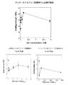
▲2▼ グルタールアルデヒド架橋複合体の場合、三点曲げ強度はグルタールアルデヒド含量にしたがって増加し、1.35mmol/g collagenで最高値に達した(図1)。この結果は、過剰なグルタールアルデヒド架橋剤(1.35mmol/g以上)は、複合体を形成する各繊維間に架橋を導入し、複合体の水分含量が高める結果、粒子間の結合を阻害して複合体強度を低下させることを示唆していた。
水溶性カルボジイミドやトランスグルタミナーゼによる架橋物では、必ずしも濃度的な変化はみられなかった。
【0053】
▲3▼ グルタールアルデヒド架橋複合体のHAp/Col比はほぼ一定だったが、水の含有量はグルタールアルデヒドの量にしたがって増加した。これは、自己組織化繊維内に生じる架橋は複合体の保水性に影響を与えないが、自己組織化繊維間に生じる架橋が複合体の保水性を増加させるためである。水溶性カルボジイミドやトランスグルタミナーゼでも、グルタールアルデヒド同様、反応剤の濃度にしたがってコラーゲンおよび水含量は増加した。
【0054】
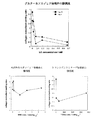
▲4▼ 膨潤度は主としてコラーゲン量に依存する。それゆえ膨潤度は、架橋量を反映するようにコラーゲン量で正規化した(図2)。その結果、膨潤度はグルタールアルデヒド濃度にしたがって減少し、架橋により生体組織中における複合体の生分解性を制御しうることが示唆された。
一方、水溶性カルボジイミドやトランスグルタミナーゼでは膨潤度の増加ははっきりと観察できなかった。これは、水溶性カルボジイミドやトランスグルタミナーゼは縮合剤であるため、架橋は複合体を密にして、膨潤を妨げるためと思われた。
▲5▼ sulfo-SDTB 測定の結果、グルタールアルデヒド1.35mmol/g濃度では、遊離のε-amino基は検出されなかった。この濃度は、コラーゲン中の架橋可能な官能基を架橋するために必要なグルタールアルデヒド量の約70倍にあたる。
【0055】
(5) 結論
グルタールアルデヒド架橋物の場合、グルタールアルデヒドが1.35mmol/g・col濃度を超えると、架橋物の機械的強度は減少し、人工骨材として適当な機械的強度を維持するためには10mmol/g・col以下の濃度で架橋することが好ましいと思われた。一方、生体内での分解が膨潤度に比例するとすれば、架橋量が多いほど分解は抑えられることが予測された。また、架橋によっても、生体骨類似のナノスコピックな構造(コラーゲン単繊維状のHApの配向)は実質的に維持されていた。
【0056】
実施例2:ウサギによる HAp/Col 複合体架橋物の生分解性試験
(1) 試験方法
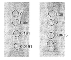
HAp/Col複合体架橋物の生体内分解性を、実施例1で得られた各種グルタールアルデヒド濃度による架橋物(2×2×2mm)をウサギ脛骨内に埋入して調べた。評価は、1,2,4週後に肉眼所見(図3)と組織学的検査(ヘマトキシリンーエオジン染色)を行うことにより評価した。
【0057】
(2) 結果
組織学的検査の結果、炎症反応等、グルタールアルデヒド架橋物による毒性反応は全く見られなかった。また、全ての架橋体において、架橋体周囲に、未架橋の複合体と同程度の骨形成および骨伝導能が認められた。。
【0058】
HAp/Col複合体架橋物の吸収/分解速度は、グルタールアルデヒド濃度にしたがって遅くなり、高密度の架橋(191μmol以上)では、4週間たっても70-80%が骨内に残存していた。コラーゲン1gあたり19.1μmolのグルタールアルデヒドで架橋したものは約50%が、675μmolで架橋したものは約85%以上が残存していた。さらに、コラーゲン1gあたり1.35mmolのグルタールアルデヒドで架橋したもでは表面のみが吸収され、95%以上が残存していた。なお、それぞれの架橋試料におけるε-アミノ基の残存量は、80-95%、0-10%、0%であった。特に1.35mmolのグルタールアルデヒドによる架橋では過剰なグルタールアルデヒド同士が複合体内で架橋のネットワークを形成し、複合体の吸収性をさらに下げていると考えられた。
【0059】
(3) 結論
以上より、コラーゲン1gあたりグルタールアルデヒド19.1μmol〜1.35mmolの濃度で架橋した複合体は、人工骨材に必要とされる機械的強度と、生体内分解速度を有することが確認された。上記結果と実施例1の結果から、ハイドロキシアパタイトとコラーゲンを含む複合体において、コラーゲンの反応可能なε-アミノ基の少なくとも1%以上(好ましくは5%以上)に架橋を導入すれば、機械的強度を維持しつつ、人工骨材に必要な生体内分解速度の達成が可能と考えられた。そしてそのような架橋の導入には、少なくともコラーゲン1gあたりグルタールアルデヒド10μmol〜10mmol程度を用いればよいと考えられた。
【0060】
【発明の効果】
本発明によれば、生体骨類似の構造をもったカルシウム塩(特に、ハイドロキシアパタイト)とコラーゲンを含む複合体において、その機械的強度を維持しつつ、生体内分解速度を制御することができる。
【図面の簡単な説明】
【図1】図1は、架橋剤濃度と架橋複合体の三点曲げ強度の関係を示す。
【図2】図2は、架橋剤濃度と架橋複合体の膨潤度(コラーゲン量で正規化したもの)を示す。
【図3】図3は、ウサギ脛骨内埋入2週間後の各グルタールアルデヒド架橋複合体の写真である。[0001]
BACKGROUND OF THE INVENTION
The present invention relates to a method for controlling biodegradability of a composite biomaterial containing a calcium salt (particularly, hydroxyapatite) and collagen, and an improved composite biomaterial provided by the method.
[0002]
[Prior art]
Conventionally, in order to repair a bone defect in an orthopedic region, self-tissue reimplantation is often performed. However, the use of self-organization is a burden on the patient and the amount of collection is limited, so that it is indispensable to use artificial implants. In addition to mechanical properties similar to living bones, these artificial bones have biocompatibility and osteoconductivity-that is, they are gradually absorbed after being applied to the living body and taken into the bone regeneration cycle and replaced with their own bones. -Is required.
[0003]
Originally, vertebrate bone is a complex composed of inorganic hydroxyapatite (HAp) and organic collagen. These form a unique nanocomposite structure in which HAp is oriented along the collagen fiber in the c-axis direction in the living bone (self-organization), and this structure gives mechanical properties peculiar to bone. In other words, simply combining HAp and collagen cannot provide the same structure and characteristics as living bones.
[0004]
Therefore, various studies have been made to develop a composite biomaterial closer to living bone using HAp and collagen. For example, Mehlisch et al. Synthesized a mixture of HAp particles and collagen (Mehlisch, AS et al, Oral Surg Oral Med Oral Pathol, 70 (6) (1990), 685-692), and Miyamoto et al. Reinforced with HAp cement. Collagen was prepared and its biocompatibility was evaluated (KS TenHuisen, et al, J. Biomed Mater Res, 29 (7) (1995), 803-810). TenHuisen et al. Also produced HAp / Col nanocomposites by growing HAp crystals on collagen (Col) fibers using calcium hydrogen phosphate as a precursor of HAp (Y. Miyamoto et al., Biomaterials, 19 (1998)). ), 707-715). However, none of them could reproduce a nanostructure similar to living bone.
[0005]
On the other hand, the present inventors have used HAp and Col self-organization to produce HAp having a biocomposite-like nanocomposite structure under biomimetic conditions (similar to the in vivo environment in which osteogenesis occurs). / Col complex was successfully synthesized (Japanese Patent Laid-Open Nos. 7-101708, 11-199209, 2000-5298, etc.). This complex was confirmed to have excellent biocompatibility and be absorbed by osteoclasts to promote osteogenesis. However, since the composite is immediately absorbed and decomposed after transplantation, there is a problem in that it is not practical as an artificial aggregate.
[0006]
[Problems to be solved by the invention]
The present invention relates to a composite containing a calcium salt (particularly, hydroxyapatite) and collagen having a structure similar to that of a living bone, and maintaining the mechanical strength while controlling the biodegradation rate and is suitable for practical use. An object is to provide a biomaterial.
[0007]
[Means for Solving the Problems]
As a result of intensive investigations to solve such problems, the present inventors have found that if cross-linking is introduced into the collagen fibers constituting the complex, the mechanical strength and biodegradation rate can be controlled, and the present invention has been completed. I let you.
[0008]
That is, the present invention provides the following (1) to (9).
(1) A method of controlling the biodegradation rate of a composite biomaterial by introducing a crosslink into the collagen in a composite biomaterial containing a calcium salt and collagen.
(2) The method according to (1) above, wherein the calcium salt is hydroxyapatite.
(3) The method according to (2) above, wherein the composite biomaterial has a microporous structure in which the c-axis of hydroxyapatite is oriented along collagen fibers.
(4) The method according to any one of (1) to (3) above, wherein the cross-linking is introduced into at least 1% or more of the reactive functional groups in collagen.
(5) The method according to any one of (1) to (4) above, wherein the crosslinking is introduced by a crosslinking reaction using glutaraldehyde.
[0009]
(6) The method according to (5) above, wherein the glutaraldehyde is used in an amount of 10 μmol to 10 mmol per 1 g of collagen in the composite biomaterial.
(7) A composite biomaterial comprising hydroxyapatite and collagen, wherein cross-linking is introduced to 1 g of collagen in the composite biomaterial using 10 μmol to 10 mmol of glutaraldehyde.
(8) The composite biomaterial as described in (7) above, wherein the cross-link is introduced into at least 5% or more of the reactive ε-amino group in collagen.
(9) The composite biomaterial according to (7) or (8) above, wherein the composite biomaterial has a microporous structure in which the c-axis of hydroxyapatite is oriented along collagen fibers.
[0010]
DETAILED DESCRIPTION OF THE INVENTION
Hereinafter, the contents of the present invention will be described in detail.
1. Composite biomaterial of the present invention
The composite biomaterial of the present invention has achieved mechanical strength and biodegradation rate suitable for biomaterials by introducing cross-links into collagen in a complex containing calcium salt (particularly hydroxyapatite) and collagen. It is characterized by.
[0011]
The calcium salt contained in the composite biomaterial of the present invention is preferably calcium phosphate or calcium carbonate, and particularly preferably hydroxyapatite. In the composite biomaterial containing collagen and hydroxyapatite, it is preferable that hydroxyapatite and collagen are oriented in a self-organizing manner to form a complex similar to living bone. “Self-organization” generally refers to “same or heterogeneous atoms, molecules, fine particles, etc., gather together by non-covalent interaction to form a specific organization (Tokyo Chemical Dojin” In particular, in the present invention, calcium phosphate having an apatite structure (hydroxyapatite: HAp) is oriented in a specific manner in living bones, that is, the c axis of HAp is collagen. It is meant to form a microporous structure oriented along the fiber.
[0012]
Hydroxyapatite has a general composition of CaFive(POFour)ThreeOH, and the reaction is non-stoichiometric, CaHPOFour, CaThree(POFour)2, CaFourO (POFour)2, CaTen(POFour)6(OH)2, CaPFourO11, Ca (POThree)2, Ca2P2O7, Ca (H2POFour)2・ H2Includes a group of compounds called calcium phosphates such as O. Hydroxyapatite is CaFive(POFour)ThreeOH or CaTen(POFour)6(OH)2The basic component is a compound represented by the composition formula: a part of the Ca component is substituted with one or more selected from Sr, Ba, MG, Fe, Al, Y, La, Na, K, H, and the like. May be. Also, (POFour) A part of the ingredient is VOFour, BOThree, SOFour, COThree, SiOFourIt may be substituted with one or more selected from the above. Furthermore, a part of the (OH) component is F, Cl, O, COThreeIt may be substituted with one or more selected from the above. Moreover, some of each of these components may be defective. PO of apatite in living boneFourAnd some OH components are usually COThreeAs the composite biomaterial is being manufactured, CO from the atmosphereThreeAnd partial substitution (about 0 to 10% by mass) for each component may be present.
[0013]
Hydroxyapatite may be the same solid solution, substitutional solid solution, interstitial solid solution, or non-quantitative defects, in addition to the usual microcrystalline / amorphous and crystalline materials. . In the “hydroxyapatite”, the atomic ratio of calcium and phosphorus (Ca / P) is preferably in the range of 1.3 to 1.8, more preferably 1.5 to 1.7. If the atomic ratio is in the range of 1.3 to 1.8, the composition and crystal structure of apatite (calcium phosphate compound) in the product can be similar to that of apatite present in vertebrate bone, This is because biocompatibility and bioabsorbability are increased.
[0014]
Collagen is now known to have about 20 different molecular species in biological tissues of a wide range of animals including fish, not limited to mammals, and is collectively called “collagens”. . Collagen used in the present invention is not particularly limited in terms of animal species, tissue site, age, etc. as its starting material, and any collagen can be used. Generally, collagen obtained from the skin, bone, cartilage, tendon, organ, etc. of mammals (eg, cows, pigs, horses, rabbits, mice, etc.) and birds (eg, chickens, etc.) is used. In addition, collagen-like proteins obtained from the skin, bones, cartilage, fins, scales, organs, etc. of fish (eg cod, flounder, flounder, salmon, trout, tuna, mackerel, Thai, sardine, shark, etc.) are used as starting materials. It may be used. Alternatively, collagen obtained by gene recombination technology may be used instead of extraction from animal tissue.
[0015]
Here, among the molecular species of collagen, the amount is the largest and well-studied is type I collagen. Usually, when it is simply referred to as collagen, it often refers to type I collagen. The molecular species of collagen used in the present invention is not particularly limited, but it is preferable that type I collagen is the main component. Further, collagen may be used by appropriately chemically modifying amino acid residues of collagen protein such as acetylation, succination, maleylation, phthalation, benzoylation, esterification, amidation, guanidinolation and the like.
[0016]
As a method for preparing collagen, for example, a method of extracting from the above starting materials (excluding gene recombination technology) with a neutral buffer solution or dilute acid such as hydrochloric acid, acetic acid, citric acid and the like can be mentioned. The former is called neutral salt-soluble collagen, and the latter is called acid-soluble collagen. However, the amount of extracted collagen is small, and most remains as insoluble collagen. As a method for solubilizing the insoluble collagen, an enzyme solubilization method and an alkali solubilization method are known. The former is called enzyme-solubilized collagen and the latter is called alkali-solubilized collagen, both of which can be solubilized as molecular collagen in a yield of almost 100%.
[0017]
The collagen preparation method (extraction type) used in the present invention is not particularly limited, but if the molecular weight is large when the collagen is solubilized, the strength of the complex becomes insufficient due to steric hindrance, Monomeric (monomolecular) collagen is preferably used. In particular, enzyme-solubilized collagen and alkali-solubilized collagen have a large amount of monomeric components, and non-helical portions (telopeptides) that have most of the antigenicity of collagen at the preparation stage are selectively degraded. -Since it is removed, it is suitable for the organic-inorganic composite biomaterial of the present invention. In addition, the collagen from which the non-spiral portion is decomposed and removed is called atelocollagen.
[0018]
Here, there is a difference in isoionic point between enzyme-solubilized collagen and alkali-solubilized collagen. The isoionic point is the pH at which both positive and negative charges derived from the dissociation group unique to the protein molecule cancel each other. In the case of collagen, it was solubilized when approaching the pH region of the isoionic point. Things are known to become fibrotic. Generally, the isoionic point of enzyme-solubilized collagen is pH 8-9, and the isoionic point of alkali-solubilized collagen is pH 4-5. In the present invention, it is more preferable to use enzyme-solubilized collagen that facilitates self-organization because collagen fibrillation proceeds in a reaction vessel maintained at a pH of 7 to 11. Examples of the enzyme for solubilization include pepsin, trypsin, chymotrypsin, papain, pronase and the like. Pepsin and pronase are preferably used because of the ease of treatment after the enzyme reaction.
[0019]
2. Method for producing a composite containing hydroxyapatite and collagen
As a suitable example of a complex containing calcium salt and collagen, which is a matrix of the composite biomaterial of the present invention, a method for producing a complex containing hydroxyapatite and collagen will be described.
[0020]
A complex containing hydroxyapatite and collagen can be prepared, for example, by the method of Kikuchi et al. (Kikuchi, S. et al, J., Biomater., 22 (13) (2001), 1705-1711, S. Itoh et al, J. Biomed Mater Res, (2001), 445-453). The complex is produced using at least three components of collagen, phosphate, and calcium salt as starting materials. Although not strictly corresponding to a “salt”, in the present invention, the phosphate includes phosphoric acid, and the calcium salt includes calcium hydroxide.
[0021]
Examples of the phosphoric acid source in the aqueous phosphate solution used include disodium hydrogen phosphate, sodium dihydrogen phosphate, dipotassium hydrogen phosphate, potassium dihydrogen phosphate, and phosphoric acid. The aqueous phosphate solution is used for the reaction after dissolving the collagen described above.
[0022]
Moreover, as a calcium source of the calcium salt aqueous solution used, calcium carbonate, calcium acetate, calcium hydroxide etc. are mentioned, for example. The aqueous calcium salt solution may be a suspension as long as it is in a uniform state. For example, calcium carbonate obtained by calcining calcium carbonate in a mortar or the like to obtain calcium hydroxide, and adding water thereto is obtained. Can be suitably used.
[0023]
In the method for producing the complex, the aqueous calcium salt solution and the aqueous phosphate solution containing collagen are simultaneously dropped into a reaction vessel. Here, “simultaneous” does not mean only the form of dripping at the same time, but also includes the form of dripping alternately in small amounts (about 0.01 to 5 ml). Both solutions may be dripped continuously as long as they are simultaneous or may be dropped intermittently.
An appropriate amount of pure water is put in the reaction vessel in advance. The amount of the pure water is not particularly limited, but is preferably about the same as the amount of the calcium salt aqueous solution used.
[0024]
In the production method, it is important that the calcium ion concentration in the reaction vessel is maintained at 3.75 mM or less and the phosphate ion concentration is maintained at 2.25 mM or less. This is because if the concentration of calcium ions or phosphate ions exceeds the above range, suitable self-assembly of the complex is hindered. This is thought to be because spontaneous nucleation occurs when the concentration of the ions convection in the reaction container exceeds those in the body fluid. If the calcium ion concentration is maintained at 2.5 mM and the phosphate ion concentration is maintained at 1.5 mM or less, a composite having an average fiber length of 1 mm or more can be obtained, which is more preferable.
[0025]
In the production method, the hydroxyapatite and collagen produced in the reaction vessel are preferably present in a weight ratio of 3: 2 to 9: 1, preferably 70:30 to 85:15. This is because it is important for self-organization that the weight ratio of hydroxyapatite to collagen when an ideal reaction occurs is closer to the composition of living bone (75:25).
[0026]
The ratio of the phosphoric acid aqueous solution containing collagen and the calcium salt aqueous solution is preferably in the range of 3: 1 to 1: 3. When the amount of the phosphoric acid aqueous solution containing collagen is small, the strength is reduced due to excessive calcium composition, and when the amount of the aqueous solution containing calcium salt is small, calcium deficiency occurs and Young's modulus This is because there is a possibility that the strength is lowered and the strength is lowered (see JP-A-11-199209).
[0027]
In the present invention, it is desirable that the reaction solution is added dropwise so that the pH of the reaction solution is in the range of 7 to 11 and the range of change is within 1. More preferably, the pH is in the range of 7 to 9, and the range of change is 0.5 or less. This is because native collagen causes precipitation at the isoelectric point in the range of
[0028]
In the production method, it is convenient to use a pH controller in order to perform suitable pH control. The pH controller is equipped with a means for measuring the pH of the reaction solution and a means for adjusting the dropping amount of both solutions to be dropped, and is within a certain range with respect to the pH (for example, 10) set as the initial value. The dripping amount of both solutions is adjusted based on the pH value of both solutions so as to maintain (for example, ± 0.3). Examples of the pH controller include those manufactured by NISSIN. It is preferable to carry out the reaction while constantly stirring both solutions and the reaction solution so that the pH of the reaction solution is not biased.
[0029]
In the said manufacturing method, it is preferable that the temperature of a reaction liquid is maintained at 35 to 40 degreeC. This is because the complex formation is expected to be performed under the same conditions as those in the living body if the temperature is within this range. A precipitate formed from the reaction solution is filtered, dried, and then pressure-molded to obtain a complex in which hydroxyapatite and collagen are oriented and bonded in a self-organizing manner.
[0030]
3. Introducing cross-linking
In the composite containing calcium salt (particularly hydroxyapatite) and collagen obtained as described above, cross-linking is introduced into the collagen fibers constituting the composite. Crosslinking is preferably performed directly without isolating the complex from the reaction solution. In order to increase the number of crosslinking points, a small amount (1 to 100 mol% with respect to the amount of collagen in the complex) of collagen or polysaccharide may be added.
[0031]
The cross-linking may be performed by any method such as chemical cross-linking using a cross-linking agent or a condensing agent, physical cross-linking using gamma rays, ultraviolet rays, thermal dehydration, electron beam, or the like. Examples of the crosslinking agent include aldehyde-based crosslinking agents such as glutaraldehyde and formaldehyde; isocyanate-based crosslinking agents such as hexamethylene diisocyanate; carbozide-based crosslinking such as 1-ethyl-3- (3-dimethylaminopropyl) carbodiimide hydrochloride. Agents; polyepoxy crosslinking agents such as ethylene glycol diethyl ether; transglutaminase and the like. The amount of these crosslinking agents used is preferably about 10 μmol to 10 mmol with respect to 1 g of collagen in the complex.
[0032]
The cross-linking may be any cross-linking between collagens, but it is particularly preferable to cross-link carboxyl groups and hydroxyl groups, carboxyl groups and ε-amino groups, or ε-amino groups. In addition, it is preferable that at least 1% or more of the reactive functional groups are introduced with crosslinking, and more preferably 5% or more. This is because if the crosslinking is insufficient, the decomposition in vivo is quick and a sufficient effect of filling the bone defect cannot be expected. However, use of excess cross-linking agent will reduce the strength of the composite by increasing the water content of the composite and inhibiting the bond between particles as a result of introducing cross-links between the fibers forming the composite. is required.
[0033]
Among the crosslinking methods, chemical crosslinking using a crosslinking agent such as glutaraldehyde is particularly preferable from the viewpoint of easy control of the degree of crosslinking and biocompatibility of the resulting composite. Hereinafter, as a preferred embodiment of the present invention, a crosslinking method using glutaraldehyde will be described.
[0034]
The reaction solution of the complex containing hydroxyapatite and collagen obtained in the previous section is reacted for 10 minutes by adding glutaraldehyde with vigorous stirring immediately after synthesis of the complex or after aging for 3 hours. After the cross-linking reaction, the complex is immediately filtered and washed with pure water three times to remove excess glutaraldehyde.
Here, glutaraldehyde is preferably added in an amount of 10 μmol to 10 mmol, particularly 10 μmol to 1 mmol, with respect to 1 g of collagen in the composite biomaterial. Moreover, it is preferable that the temperature of a reaction liquid is maintained at 0 degreeC-40 degreeC.
[0035]
4). Improved physical properties (mechanical strength, biodegradation rate) by crosslinking
The obtained cross-linked composite biomaterial has a high mechanical strength and a slow biodegradation rate compared to an uncrosslinked composite biomaterial, and thus has the in vivo retention required for artificial bones and the like. That is, the present invention provides a method for controlling the biodegradation rate of a composite biomaterial while maintaining mechanical strength by introducing a bridge between hydroxyapatite and collagen.
[0036]
The biodegradation rate can be evaluated by, for example, transplanting the composite biomaterial into bones of mice, rats, rabbits, etc., and checking the in vivo retention. The mechanical strength can be evaluated by, for example, a three-point bending strength or a Young's modulus obtained from the value.
[0037]
Specifically, an organic-inorganic composite biomaterial in which 10 μmol to 10 mmol of glutaraldehyde is added to 1 g of collagen to introduce cross-linking has a mechanical strength of 7 MPa (uncross-linked) to 15 MPa or higher (after cross-linking). Improved. The uncrosslinked sample was absorbed almost (90% or more) in 4 weeks in the living bone, whereas about 50% or more of the crosslinked composite biomaterial remained in the living bone even after 4 weeks.
[0038]
5). Use of cross-linked composite biomaterials
The cross-linked composite biomaterial obtained by the above method can be appropriately pressed and used as an implant such as an artificial bone. The pressure molding is preferably performed in a temperature range of 0 ° C. to 110 ° C. and a pressure range of 10 Mpa to 5 Gpa. This is because most of the water contained in the precipitate is rapidly released when pressure molding is performed in this temperature range. The temperature is preferably in the range of 25 ° C. or more and 60 ° C. or less, and particularly preferably in the range of 35 ° C. or more and 45 ° C. or less where the amount of water released is large.
Moreover, it is preferable to carry out the process while applying ultrasonic waves since self-organization can be further promoted. Examples of the pressure treatment apparatus that can be used for pressure molding in the present invention include CIP manufactured by Kobe Steel.
[0039]
The form / shape of the composite biomaterial of the present invention is not particularly limited, and can be formed into any shape / shape such as a block shape, a paste shape, a film shape, a granular shape, a sponge shape, and the like. The composite biomaterial of the present invention has elasticity like a sponge when it absorbs water, and has excellent biocompatibility, osteoinductive ability or osteoconductivity. Accordingly, when the composite biomaterial is used as an implant, it may be used after it is once immersed in an appropriate liquid such as physiological saline. The composite biomaterial thus embedded can quickly bind to the bone tissue and be integrated with the hard tissue on the donor side.
[0040]
In addition to the essential components calcium salt, phosphate, and collagen, the composite biomaterial of the present invention may further contain other components as long as the objects and effects of the present invention are not impaired. Such components include, for example, St, Mg and COThreeInorganic salts such as citric acid and phospholipid, and agents such as bone morphogenetic proteins and anticancer agents.
[0041]
The composite biomaterial of the present invention has strength and composition close to those of living bones, and both the constituent components collagen and calcium phosphate are biosoluble, and thus have a sustained drug release effect or osteoinductive ability or osteoconductivity. In addition, it has excellent mechanical strength and in-vivo retention (appropriate biodegradation rate) due to crosslinking.
[0042]
In addition, a tissue such as bone marrow and liver can be obtained by containing a highly biologically active cytokine in the composite biomaterial of the present invention and performing tissue culture in a living body-like environment or in vivo with the addition of mechanics and electricity as a substrate. The effect of reconstruction is also expected. For example, by using a composite material obtained by impregnating the composite material obtained by the present invention with an anticancer agent for the reconstruction of a resected bone such as osteosarcoma, it is possible to prevent the recurrence of cancer and induce the hard tissue.
[0043]
Therefore, the use of the composite biomaterial obtained by the present invention includes the use as a living bone replacement type bone reconstruction material having osteoinduction and osteoconductivity, and a living body used for tissue engineering containing amino acids, carbohydrates and cytokines. Examples of active bases and methods for use as sustained release bases for biocompatible drugs such as anticancer agents include artificial bones, artificial joints, joints between tendons and bones, and dental implant materials. , Catheter percutaneous terminals, drug sustained release substrates, bone marrow induction chambers, tissue reconstruction chambers / substrates, and the like.
[0044]
【Example】
EXAMPLES Hereinafter, although an Example demonstrates this invention further in detail, this invention is not limited to this.
Example 1: Crosslinking HAp / Col Fabrication of composite
(1) Preparation of HAp / Col complex
The Hap / Col complex was prepared according to the method of Kikuchi et al. (M. Kikuchi, et al., Biomater., 22 (13) (2001), 1705-1711). First, calcium carbonate (for alkali analysis, Wako Pure Chemical Industries), phosphoric acid (special grade, Wako Pure Chemical Industries), and porcine skin-derived atelocollagen (Nitta Gelatin) were prepared as starting materials. Calcium carbonate was calcined at 1050 ° C. and hydrolyzed to obtain a calcium hydroxide single phase. 40 mM calcium hydroxide suspension 2dmThree2dm of 24mM phosphoric acid solution containing 2g of collagenThreeWas introduced into the reaction vessel via a tube pump. The pH in the reaction vessel was controlled at pH 9 by a controller, and the temperature was controlled at 40 ° C. by a hot water bath.
[0045]
(2) Cross-linking reaction
The reaction solution was suspended and allowed to stand for 3 hours, and the cross-linking agent: glutaraldehyde was added with vigorous stirring to react for 10 minutes. After the cross-linking reaction, the complex was immediately filtered and washed with pure water three times. For comparison, a crosslinking reaction was similarly performed using water-soluble carbodiimide and transglutaminase (both of which are condensing agents).
[0046]
The cross-linking reaction varies within the range of glutaraldehyde: 0.01191-13.5 mmol / g, water-soluble carbodiimide: 0.0191-8.8 mmol / g, and transglutaminase: 19.1-1910 mg / g for 1 g of collagen in the complex. I went. In the case of glutaraldehyde, theoretically, all ε-amino groups in the collagen molecule can be crosslinked at 0.191 mmol / g.
[0047]
(3) Characteristic value measurement
The characteristics of the obtained crosslinked composite were measured as follows.
(1) Composite structure (particle size):
The crosslinked complex was dispersed in pure water and observed using a transmission electron microscope Rapid-VueR (manufactured by Beckman-Colter).
(2) Three-point bending strength:
The crosslinked composite was dehydrated by uniaxial pressing at 20 MPa for 24 hours, and the three-point bending strength was measured with a universal testing machine (Autograph AGS-1kN, manufactured by Shimadzu). The measurement was performed using a cross-linked composite piece of 5 × 3 × 20 mm at a crosshead speed of 500 μm and a span of 15 mm.
[0048]
▲ 3 ▼ HAp / Col / H2O ratio:
HAp / Col / H of the above pressure-molded crosslinked composite2The
(4) Degree of swelling:
The above pressure-molded composite was immersed in a phosphorus buffer solution (pH = 7.4, 37 ° C.) for 4 weeks and weighed to determine the degree of swelling (the following formula).
[0049]
[Expression 1]
Swelling degree (%) = [(Wx−Wo) / Wo] × 100
Wx: initial weight, Wo: weight after immersion
[0050]
(5) Amount of crosslinking:
Using the cross-linked composite used for the three-point bending strength measurement, the amount of ε-amino group was measured by the sulfo-SDTB method to determine the amount of cross-linking.
[0051]
(4) Results
{Circle around (1)} As a result of observation with a transmission electron microscope, the average fiber length of the glutaraldehyde crosslinked composite was 44.8 μm. Moreover, it was found that the crosslinked hydroxyapatite and collagen showed no macro-orientation, and the crosslinking occurred randomly. The nanoscopic structure similar to living bone (the orientation of collagen monofilament HAp) was substantially maintained. As the glutaraldehyde concentration increased, the color of the complex changed from dark yellow to brown. This was thought to be because excess glutaraldehyde increased the composite fiber length by crosslinking between self-assembled fibers.
[0052]
(2) In the case of glutaraldehyde crosslinked complex, the three-point bending strength increased with the glutaraldehyde content and reached the highest value at 1.35 mmol / g collagen (FIG. 1). This result shows that excess glutaraldehyde cross-linking agent (1.35 mmol / g or more) introduces cross-links between the fibers forming the complex and increases the water content of the complex, thereby inhibiting the binding between particles. This suggests that the composite strength is reduced.
The cross-linked product of water-soluble carbodiimide or transglutaminase did not necessarily change in concentration.
[0053]
(3) The HAp / Col ratio of the glutaraldehyde crosslinked complex was almost constant, but the water content increased according to the amount of glutaraldehyde. This is because the cross-linking that occurs within the self-assembled fibers does not affect the water retention of the composite, but the cross-linking that occurs between the self-assembled fibers increases the water retention of the composite. In the case of water-soluble carbodiimide and transglutaminase, the collagen and water contents increased according to the concentration of the reactants, like glutaraldehyde.
[0054]
(4) The degree of swelling mainly depends on the amount of collagen. Therefore, the degree of swelling was normalized with the amount of collagen to reflect the amount of crosslinking (FIG. 2). As a result, the degree of swelling decreased according to the glutaraldehyde concentration, and it was suggested that the biodegradability of the complex in biological tissue can be controlled by crosslinking.
On the other hand, with water-soluble carbodiimide and transglutaminase, an increase in swelling degree could not be clearly observed. This seems to be because water-soluble carbodiimide and transglutaminase are condensing agents, so that the cross-linking makes the complex dense and prevents swelling.
(5) As a result of sulfo-SDTB measurement, free ε-amino groups were not detected at a glutaraldehyde concentration of 1.35 mmol / g. This concentration is about 70 times the amount of glutaraldehyde required to crosslink crosslinkable functional groups in collagen.
[0055]
(5) Conclusion
In the case of glutaraldehyde cross-linked product, when glutaraldehyde exceeds 1.35 mmol / g · col concentration, the mechanical strength of the cross-linked product decreases, and in order to maintain appropriate mechanical strength as an artificial aggregate, 10 mmol / It seemed preferable to crosslink at a concentration below g · col. On the other hand, if the degradation in vivo is proportional to the degree of swelling, it was predicted that the greater the amount of crosslinking, the lower the degradation. In addition, the nanoscopic structure similar to living bone (the orientation of collagen monofilament HAp) was substantially maintained by cross-linking.
[0056]
Example 2: By rabbit HAp / Col Biodegradability test of composite cross-linked product
(1) Test method
The biodegradability of the HAp / Col composite cross-linked product was examined by implanting the cross-linked product (2 × 2 × 2 mm) having various glutaraldehyde concentrations obtained in Example 1 into a rabbit tibia. Evaluation was carried out after 1, 2, and 4 weeks by performing macroscopic findings (FIG. 3) and histological examination (hematoxylin-eosin staining).
[0057]
(2) Results
As a result of histological examination, no toxic reaction such as inflammatory reaction due to the cross-linked glutaraldehyde was observed. Further, in all the crosslinked bodies, bone formation and osteoconductivity similar to those of the uncrosslinked composite were observed around the crosslinked bodies. .
[0058]
The absorption / degradation rate of the HAp / Col composite cross-linked product was decreased according to the glutaraldehyde concentration, and 70-80% of the high-density cross-linked product (191 μmol or more) remained in the bone even after 4 weeks. About 50% of those crosslinked with 19.1 μmol of glutaraldehyde per gram of collagen remained, and about 85% or more of those crosslinked with 675 μmol remained. Furthermore, only the surface was absorbed when cross-linked with 1.35 mmol of glutaraldehyde per gram of collagen, and 95% or more remained. The residual amounts of ε-amino groups in the respective crosslinked samples were 80-95%, 0-10%, and 0%. In particular, in cross-linking with 1.35 mmol glutaraldehyde, it was considered that excess glutaraldehyde formed a cross-linking network in the complex, further reducing the absorbability of the complex.
[0059]
(3) Conclusion
From the above, it was confirmed that the complex cross-linked at a concentration of 19.1 μmol to 1.35 mmol of glutaraldehyde per 1 g of collagen has the mechanical strength and the biodegradation rate required for the artificial aggregate. From the above result and the result of Example 1, in the composite containing hydroxyapatite and collagen, if crosslinking is introduced into at least 1% (preferably 5% or more) of the ε-amino group capable of reacting with collagen, It was considered possible to achieve the biodegradation rate required for artificial bones while maintaining strength. It was considered that at least about 10 μmol to 10 mmol of glutaraldehyde per 1 g of collagen should be used for the introduction of such crosslinking.
[0060]
【The invention's effect】
According to the present invention, in a complex containing a calcium salt (particularly, hydroxyapatite) having a structure similar to that of a living bone and collagen, the biodegradation rate can be controlled while maintaining the mechanical strength.
[Brief description of the drawings]
FIG. 1 shows the relationship between crosslinker concentration and the three-point bending strength of a crosslinked composite.
FIG. 2 shows the crosslinking agent concentration and the degree of swelling of the crosslinked complex (normalized by the amount of collagen).
FIG. 3 is a photograph of each glutaraldehyde crosslinked complex 2 weeks after implantation in a rabbit tibia.
Claims (4)
Priority Applications (3)
| Application Number | Priority Date | Filing Date | Title |
|---|---|---|---|
| JP2002065831A JP4226830B2 (en) | 2002-03-11 | 2002-03-11 | Control of biodegradability of composite biomaterials |
| PCT/JP2002/008335 WO2003075971A1 (en) | 2002-03-11 | 2002-08-19 | Control of biodegradability of composite biomaterial |
| US10/937,732 US7229971B2 (en) | 2002-03-11 | 2004-09-10 | Regulation of biodegradability of composite biomaterials |
Applications Claiming Priority (1)
| Application Number | Priority Date | Filing Date | Title |
|---|---|---|---|
| JP2002065831A JP4226830B2 (en) | 2002-03-11 | 2002-03-11 | Control of biodegradability of composite biomaterials |
Publications (2)
| Publication Number | Publication Date |
|---|---|
| JP2003260124A JP2003260124A (en) | 2003-09-16 |
| JP4226830B2 true JP4226830B2 (en) | 2009-02-18 |
Family
ID=27800237
Family Applications (1)
| Application Number | Title | Priority Date | Filing Date |
|---|---|---|---|
| JP2002065831A Expired - Fee Related JP4226830B2 (en) | 2002-03-11 | 2002-03-11 | Control of biodegradability of composite biomaterials |
Country Status (2)
| Country | Link |
|---|---|
| JP (1) | JP4226830B2 (en) |
| WO (1) | WO2003075971A1 (en) |
Families Citing this family (10)
| Publication number | Priority date | Publication date | Assignee | Title |
|---|---|---|---|---|
| JP2007050084A (en) * | 2005-08-17 | 2007-03-01 | National Institute For Materials Science | Bone reconstruction material manufacturing method, bone reconstruction material, and bone tissue induction method |
| JP5467554B2 (en) * | 2008-04-25 | 2014-04-09 | HOYA Technosurgical株式会社 | Powdery apatite / collagen composite, shape-shaped artificial bone paste, and production method thereof |
| JPWO2013157638A1 (en) * | 2012-04-19 | 2015-12-21 | 国立研究開発法人物質・材料研究機構 | Biomaterials coated with HAp / Col composite |
| JP2015047076A (en) * | 2013-08-29 | 2015-03-16 | 独立行政法人産業技術総合研究所 | Cell culture substrate, osteoblast differentiation induction method and osteoblast production method using the same |
| SG11201704277XA (en) | 2014-11-27 | 2017-06-29 | Toyo Boseki | Porous composite, bone regeneration material, and method for producing porous composite |
| SG11201708129QA (en) * | 2015-04-08 | 2017-11-29 | Toyo Boseki | Porous composite, bone regeneration material, and method for producing porous composite |
| WO2017043498A1 (en) | 2015-09-08 | 2017-03-16 | 東洋紡株式会社 | Porous complex and bone regeneration material |
| US9968660B2 (en) | 2016-04-08 | 2018-05-15 | Toyobo Co., Ltd. | Method of bone regeneration or bone augmentation |
| CN108593489B (en) * | 2018-07-12 | 2020-11-06 | 中北大学 | 3D printing magnesium alloy material degradability testing device and application |
| CN117159804B (en) * | 2023-09-12 | 2026-04-24 | 北京邦塞科技有限公司 | A self-healing hydroxyapatite/collagen filler material, its preparation method and application |
Family Cites Families (6)
| Publication number | Priority date | Publication date | Assignee | Title |
|---|---|---|---|---|
| IL110367A (en) * | 1994-07-19 | 2007-05-15 | Colbar Lifescience Ltd | Collagen-based matrix |
| JPH08276003A (en) * | 1995-04-07 | 1996-10-22 | Terumo Corp | Head tissue restorative dental material and imbedded medical appliance |
| JP3592920B2 (en) * | 1998-01-19 | 2004-11-24 | 独立行政法人物質・材料研究機構 | Method for producing organic-inorganic oriented composite material |
| JP3653626B2 (en) * | 2000-01-13 | 2005-06-02 | 独立行政法人科学技術振興機構 | Organic / inorganic alternating multilayer composite |
| JP2002143291A (en) * | 2000-11-16 | 2002-05-21 | National Institute Of Advanced Industrial & Technology | Composite porous structure and method for producing the same |
| JP4790917B2 (en) * | 2001-02-23 | 2011-10-12 | 独立行政法人科学技術振興機構 | Artificial vertebral body |
-
2002
- 2002-03-11 JP JP2002065831A patent/JP4226830B2/en not_active Expired - Fee Related
- 2002-08-19 WO PCT/JP2002/008335 patent/WO2003075971A1/en not_active Ceased
Also Published As
| Publication number | Publication date |
|---|---|
| JP2003260124A (en) | 2003-09-16 |
| WO2003075971A1 (en) | 2003-09-18 |
Similar Documents
| Publication | Publication Date | Title |
|---|---|---|
| JP4408603B2 (en) | Organic-inorganic composite biomaterial and method for producing the same | |
| JP3727059B2 (en) | Method for producing porous composite material | |
| US20250249147A1 (en) | Biomimetic nano-composite scaffold for enhanced bone healing and fracture repair | |
| EP1566186B1 (en) | Apatite/collagen crosslinked porous material containing self-organized apatite/collagen composite and process for producing the same | |
| WO2006031196A1 (en) | Porous biomaterial-filler composite and a method for making the same | |
| JP4873555B2 (en) | Method for producing porous body containing apatite / collagen composite fiber | |
| CA2467252C (en) | Composite biomaterials | |
| US7229971B2 (en) | Regulation of biodegradability of composite biomaterials | |
| JP4226830B2 (en) | Control of biodegradability of composite biomaterials | |
| US20150132353A1 (en) | BIOMATERIAL COATED WITH HAp/Col COMPOSITE | |
| JPS62268563A (en) | Bone mallow/collagen/inorganic matrix for repairing defect of bone, its preparation and method for repairing defect of bone using the same | |
| TWI687222B (en) | Porous composite body and method for manufacturing porous composite body containing octacalcium phosphate and parathyroid hormone | |
| JPH10151188A (en) | Implant for ossification | |
| Nguyen et al. | Structurally and functionally optimized silk fibroin-alginate-based biomimetic scaffolds reinforced with nanobioceramics for bone tissue engineering applications | |
| CN1157232C (en) | Prepn of osteological material containing nano phase calcium phosphate, collagen and alginate | |
| JP4814477B2 (en) | Bone augmentation and osteoporosis treatment | |
| JP2883213B2 (en) | Biotransplant material and its manufacturing method | |
| KR100362699B1 (en) | Deproteinated bovine bone powder coated by calcium phosphate thin film | |
| Pina et al. | 3.2 Osteoconductive properties of brushite-forming Zn-and ZnSr-substituted β-TCP bone cements |
Legal Events
| Date | Code | Title | Description |
|---|---|---|---|
| RD03 | Notification of appointment of power of attorney |
Free format text: JAPANESE INTERMEDIATE CODE: A7423 Effective date: 20040210 |
|
| A621 | Written request for application examination |
Free format text: JAPANESE INTERMEDIATE CODE: A621 Effective date: 20041118 |
|
| A131 | Notification of reasons for refusal |
Free format text: JAPANESE INTERMEDIATE CODE: A131 Effective date: 20080812 |
|
| A521 | Written amendment |
Free format text: JAPANESE INTERMEDIATE CODE: A523 Effective date: 20081009 |
|
| TRDD | Decision of grant or rejection written | ||
| A01 | Written decision to grant a patent or to grant a registration (utility model) |
Free format text: JAPANESE INTERMEDIATE CODE: A01 Effective date: 20081118 |
|
| A01 | Written decision to grant a patent or to grant a registration (utility model) |
Free format text: JAPANESE INTERMEDIATE CODE: A01 |
|
| A61 | First payment of annual fees (during grant procedure) |
Free format text: JAPANESE INTERMEDIATE CODE: A61 Effective date: 20081127 |
|
| FPAY | Renewal fee payment (event date is renewal date of database) |
Free format text: PAYMENT UNTIL: 20111205 Year of fee payment: 3 |
|
| R150 | Certificate of patent or registration of utility model |
Ref document number: 4226830 Country of ref document: JP Free format text: JAPANESE INTERMEDIATE CODE: R150 Free format text: JAPANESE INTERMEDIATE CODE: R150 |
|
| FPAY | Renewal fee payment (event date is renewal date of database) |
Free format text: PAYMENT UNTIL: 20121205 Year of fee payment: 4 |
|
| R250 | Receipt of annual fees |
Free format text: JAPANESE INTERMEDIATE CODE: R250 |
|
| R250 | Receipt of annual fees |
Free format text: JAPANESE INTERMEDIATE CODE: R250 |
|
| R250 | Receipt of annual fees |
Free format text: JAPANESE INTERMEDIATE CODE: R250 |
|
| S111 | Request for change of ownership or part of ownership |
Free format text: JAPANESE INTERMEDIATE CODE: R313117 |
|
| R350 | Written notification of registration of transfer |
Free format text: JAPANESE INTERMEDIATE CODE: R350 |
|
| R250 | Receipt of annual fees |
Free format text: JAPANESE INTERMEDIATE CODE: R250 |
|
| S533 | Written request for registration of change of name |
Free format text: JAPANESE INTERMEDIATE CODE: R313533 |
|
| R350 | Written notification of registration of transfer |
Free format text: JAPANESE INTERMEDIATE CODE: R350 |
|
| R250 | Receipt of annual fees |
Free format text: JAPANESE INTERMEDIATE CODE: R250 |
|
| R250 | Receipt of annual fees |
Free format text: JAPANESE INTERMEDIATE CODE: R250 |
|
| R250 | Receipt of annual fees |
Free format text: JAPANESE INTERMEDIATE CODE: R250 |
|
| R250 | Receipt of annual fees |
Free format text: JAPANESE INTERMEDIATE CODE: R250 |
|
| R250 | Receipt of annual fees |
Free format text: JAPANESE INTERMEDIATE CODE: R250 |
|
| LAPS | Cancellation because of no payment of annual fees |