JP4922158B2 - Use of viscoelastic compositions to treat increased intraocular pressure - Google Patents
Use of viscoelastic compositions to treat increased intraocular pressure Download PDFInfo
- Publication number
- JP4922158B2 JP4922158B2 JP2007511320A JP2007511320A JP4922158B2 JP 4922158 B2 JP4922158 B2 JP 4922158B2 JP 2007511320 A JP2007511320 A JP 2007511320A JP 2007511320 A JP2007511320 A JP 2007511320A JP 4922158 B2 JP4922158 B2 JP 4922158B2
- Authority
- JP
- Japan
- Prior art keywords
- eye
- linked
- conduit
- cross
- viscoelastic medium
- Prior art date
- Legal status (The legal status is an assumption and is not a legal conclusion. Google has not performed a legal analysis and makes no representation as to the accuracy of the status listed.)
- Expired - Fee Related
Links
- 230000004410 intraocular pressure Effects 0.000 title claims abstract description 34
- 239000000203 mixture Substances 0.000 title description 23
- 238000000034 method Methods 0.000 claims abstract description 57
- 241001465754 Metazoa Species 0.000 claims abstract description 32
- 238000011282 treatment Methods 0.000 claims abstract description 25
- 239000003814 drug Substances 0.000 claims abstract description 15
- 238000004519 manufacturing process Methods 0.000 claims abstract description 4
- 230000035515 penetration Effects 0.000 claims description 39
- 210000003786 sclera Anatomy 0.000 claims description 39
- KIUKXJAPPMFGSW-DNGZLQJQSA-N (2S,3S,4S,5R,6R)-6-[(2S,3R,4R,5S,6R)-3-Acetamido-2-[(2S,3S,4R,5R,6R)-6-[(2R,3R,4R,5S,6R)-3-acetamido-2,5-dihydroxy-6-(hydroxymethyl)oxan-4-yl]oxy-2-carboxy-4,5-dihydroxyoxan-3-yl]oxy-5-hydroxy-6-(hydroxymethyl)oxan-4-yl]oxy-3,4,5-trihydroxyoxane-2-carboxylic acid Chemical compound CC(=O)N[C@H]1[C@H](O)O[C@H](CO)[C@@H](O)[C@@H]1O[C@H]1[C@H](O)[C@@H](O)[C@H](O[C@H]2[C@@H]([C@@H](O[C@H]3[C@@H]([C@@H](O)[C@H](O)[C@H](O3)C(O)=O)O)[C@H](O)[C@@H](CO)O2)NC(C)=O)[C@@H](C(O)=O)O1 KIUKXJAPPMFGSW-DNGZLQJQSA-N 0.000 claims description 37
- 229920002674 hyaluronan Polymers 0.000 claims description 37
- 229960003160 hyaluronic acid Drugs 0.000 claims description 37
- 150000004676 glycans Chemical class 0.000 claims description 29
- 229920001282 polysaccharide Polymers 0.000 claims description 29
- 239000005017 polysaccharide Substances 0.000 claims description 29
- 239000007863 gel particle Substances 0.000 claims description 25
- 208000010412 Glaucoma Diseases 0.000 claims description 19
- 210000002159 anterior chamber Anatomy 0.000 claims description 19
- 239000007924 injection Substances 0.000 claims description 14
- 238000002347 injection Methods 0.000 claims description 14
- 230000015572 biosynthetic process Effects 0.000 claims description 13
- 210000004087 cornea Anatomy 0.000 claims description 10
- 201000002287 Keratoconus Diseases 0.000 claims description 7
- 210000003462 vein Anatomy 0.000 claims description 6
- SQDAZGGFXASXDW-UHFFFAOYSA-N 5-bromo-2-(trifluoromethoxy)pyridine Chemical compound FC(F)(F)OC1=CC=C(Br)C=N1 SQDAZGGFXASXDW-UHFFFAOYSA-N 0.000 claims description 4
- 229920001287 Chondroitin sulfate Polymers 0.000 claims description 4
- 229920002683 Glycosaminoglycan Polymers 0.000 claims description 4
- HTTJABKRGRZYRN-UHFFFAOYSA-N Heparin Chemical compound OC1C(NC(=O)C)C(O)OC(COS(O)(=O)=O)C1OC1C(OS(O)(=O)=O)C(O)C(OC2C(C(OS(O)(=O)=O)C(OC3C(C(O)C(O)C(O3)C(O)=O)OS(O)(=O)=O)C(CO)O2)NS(O)(=O)=O)C(C(O)=O)O1 HTTJABKRGRZYRN-UHFFFAOYSA-N 0.000 claims description 4
- 229940059329 chondroitin sulfate Drugs 0.000 claims description 4
- 238000011049 filling Methods 0.000 claims description 4
- 229920000669 heparin Polymers 0.000 claims description 4
- 229960002897 heparin Drugs 0.000 claims description 4
- 239000003795 chemical substances by application Substances 0.000 claims description 3
- 241000282412 Homo Species 0.000 claims description 2
- 230000003416 augmentation Effects 0.000 claims 1
- 230000002784 sclerotic effect Effects 0.000 claims 1
- 230000000149 penetrating effect Effects 0.000 abstract description 7
- 206010016717 Fistula Diseases 0.000 abstract description 5
- 230000003890 fistula Effects 0.000 abstract description 5
- 210000001508 eye Anatomy 0.000 description 55
- 239000000499 gel Substances 0.000 description 19
- 210000000795 conjunctiva Anatomy 0.000 description 18
- 239000002245 particle Substances 0.000 description 18
- 238000001356 surgical procedure Methods 0.000 description 18
- FAPWRFPIFSIZLT-UHFFFAOYSA-M Sodium chloride Chemical compound [Na+].[Cl-] FAPWRFPIFSIZLT-UHFFFAOYSA-M 0.000 description 13
- 210000001519 tissue Anatomy 0.000 description 11
- 239000011780 sodium chloride Substances 0.000 description 10
- 239000012530 fluid Substances 0.000 description 9
- 210000001742 aqueous humor Anatomy 0.000 description 8
- 241000283973 Oryctolagus cuniculus Species 0.000 description 7
- 239000003431 cross linking reagent Substances 0.000 description 6
- 239000000243 solution Substances 0.000 description 6
- 239000003190 viscoelastic substance Substances 0.000 description 6
- 238000006243 chemical reaction Methods 0.000 description 5
- 238000004132 cross linking Methods 0.000 description 5
- 238000001727 in vivo Methods 0.000 description 5
- 238000003780 insertion Methods 0.000 description 5
- 230000037431 insertion Effects 0.000 description 5
- 230000004962 physiological condition Effects 0.000 description 5
- 239000002002 slurry Substances 0.000 description 5
- WZUVPPKBWHMQCE-UHFFFAOYSA-N Haematoxylin Chemical compound C12=CC(O)=C(O)C=C2CC2(O)C1C1=CC=C(O)C(O)=C1OC2 WZUVPPKBWHMQCE-UHFFFAOYSA-N 0.000 description 4
- 238000001914 filtration Methods 0.000 description 4
- 230000004927 fusion Effects 0.000 description 4
- 238000010561 standard procedure Methods 0.000 description 4
- 102000018866 Hyaluronan Receptors Human genes 0.000 description 3
- 108010013214 Hyaluronan Receptors Proteins 0.000 description 3
- HEMHJVSKTPXQMS-UHFFFAOYSA-M Sodium hydroxide Chemical compound [OH-].[Na+] HEMHJVSKTPXQMS-UHFFFAOYSA-M 0.000 description 3
- 239000007864 aqueous solution Substances 0.000 description 3
- 238000002474 experimental method Methods 0.000 description 3
- 239000007943 implant Substances 0.000 description 3
- 210000001328 optic nerve Anatomy 0.000 description 3
- 238000002360 preparation method Methods 0.000 description 3
- 230000036573 scar formation Effects 0.000 description 3
- 239000000126 substance Substances 0.000 description 3
- 210000001585 trabecular meshwork Anatomy 0.000 description 3
- YBJHBAHKTGYVGT-ZKWXMUAHSA-N (+)-Biotin Chemical compound N1C(=O)N[C@@H]2[C@H](CCCCC(=O)O)SC[C@@H]21 YBJHBAHKTGYVGT-ZKWXMUAHSA-N 0.000 description 2
- SHKUUQIDMUMQQK-UHFFFAOYSA-N 2-[4-(oxiran-2-ylmethoxy)butoxymethyl]oxirane Chemical compound C1OC1COCCCCOCC1CO1 SHKUUQIDMUMQQK-UHFFFAOYSA-N 0.000 description 2
- 241001631457 Cannula Species 0.000 description 2
- 229920001605 Dextranomer Polymers 0.000 description 2
- RTZKZFJDLAIYFH-UHFFFAOYSA-N Diethyl ether Chemical compound CCOCC RTZKZFJDLAIYFH-UHFFFAOYSA-N 0.000 description 2
- GHASVSINZRGABV-UHFFFAOYSA-N Fluorouracil Chemical compound FC1=CNC(=O)NC1=O GHASVSINZRGABV-UHFFFAOYSA-N 0.000 description 2
- NWIBSHFKIJFRCO-WUDYKRTCSA-N Mytomycin Chemical compound C1N2C(C(C(C)=C(N)C3=O)=O)=C3[C@@H](COC(N)=O)[C@@]2(OC)[C@@H]2[C@H]1N2 NWIBSHFKIJFRCO-WUDYKRTCSA-N 0.000 description 2
- 230000015556 catabolic process Effects 0.000 description 2
- 210000003161 choroid Anatomy 0.000 description 2
- 230000006378 damage Effects 0.000 description 2
- 238000006731 degradation reaction Methods 0.000 description 2
- GYZLOYUZLJXAJU-UHFFFAOYSA-N diglycidyl ether Chemical class C1OC1COCC1CO1 GYZLOYUZLJXAJU-UHFFFAOYSA-N 0.000 description 2
- YQGOJNYOYNNSMM-UHFFFAOYSA-N eosin Chemical compound [Na+].OC(=O)C1=CC=CC=C1C1=C2C=C(Br)C(=O)C(Br)=C2OC2=C(Br)C(O)=C(Br)C=C21 YQGOJNYOYNNSMM-UHFFFAOYSA-N 0.000 description 2
- 229960002949 fluorouracil Drugs 0.000 description 2
- 239000011521 glass Substances 0.000 description 2
- 230000007794 irritation Effects 0.000 description 2
- 239000000463 material Substances 0.000 description 2
- 239000002504 physiological saline solution Substances 0.000 description 2
- 230000001603 reducing effect Effects 0.000 description 2
- 210000001525 retina Anatomy 0.000 description 2
- 231100000241 scar Toxicity 0.000 description 2
- 239000002904 solvent Substances 0.000 description 2
- 230000006641 stabilisation Effects 0.000 description 2
- 238000011105 stabilization Methods 0.000 description 2
- 210000004127 vitreous body Anatomy 0.000 description 2
- 201000002862 Angle-Closure Glaucoma Diseases 0.000 description 1
- 208000002177 Cataract Diseases 0.000 description 1
- 208000032544 Cicatrix Diseases 0.000 description 1
- 102000008186 Collagen Human genes 0.000 description 1
- 108010035532 Collagen Proteins 0.000 description 1
- 239000004971 Cross linker Substances 0.000 description 1
- 229920002307 Dextran Polymers 0.000 description 1
- 206010061218 Inflammation Diseases 0.000 description 1
- 206010030348 Open-Angle Glaucoma Diseases 0.000 description 1
- 229920005654 Sephadex Polymers 0.000 description 1
- 239000012507 Sephadex™ Substances 0.000 description 1
- 229920002385 Sodium hyaluronate Polymers 0.000 description 1
- 229920002472 Starch Polymers 0.000 description 1
- 230000002378 acidificating effect Effects 0.000 description 1
- 239000013543 active substance Substances 0.000 description 1
- 230000002411 adverse Effects 0.000 description 1
- 150000001299 aldehydes Chemical class 0.000 description 1
- 239000002260 anti-inflammatory agent Substances 0.000 description 1
- 230000004509 aqueous humor production Effects 0.000 description 1
- 239000007900 aqueous suspension Substances 0.000 description 1
- 239000011324 bead Substances 0.000 description 1
- 230000009286 beneficial effect Effects 0.000 description 1
- 239000013060 biological fluid Substances 0.000 description 1
- 229960002685 biotin Drugs 0.000 description 1
- 235000020958 biotin Nutrition 0.000 description 1
- 239000011616 biotin Substances 0.000 description 1
- 239000008280 blood Substances 0.000 description 1
- 210000004369 blood Anatomy 0.000 description 1
- 210000005252 bulbus oculi Anatomy 0.000 description 1
- 210000004027 cell Anatomy 0.000 description 1
- 230000001886 ciliary effect Effects 0.000 description 1
- 229920001436 collagen Polymers 0.000 description 1
- 150000001875 compounds Chemical class 0.000 description 1
- 238000005520 cutting process Methods 0.000 description 1
- 231100000433 cytotoxic Toxicity 0.000 description 1
- 230000001472 cytotoxic effect Effects 0.000 description 1
- 229960002864 dextranomer Drugs 0.000 description 1
- 239000006185 dispersion Substances 0.000 description 1
- AFOSIXZFDONLBT-UHFFFAOYSA-N divinyl sulfone Chemical class C=CS(=O)(=O)C=C AFOSIXZFDONLBT-UHFFFAOYSA-N 0.000 description 1
- 229940079593 drug Drugs 0.000 description 1
- 230000000694 effects Effects 0.000 description 1
- 210000002889 endothelial cell Anatomy 0.000 description 1
- 150000002118 epoxides Chemical class 0.000 description 1
- 150000002148 esters Chemical class 0.000 description 1
- 208000030533 eye disease Diseases 0.000 description 1
- 238000000855 fermentation Methods 0.000 description 1
- 230000004151 fermentation Effects 0.000 description 1
- 230000035876 healing Effects 0.000 description 1
- 230000001744 histochemical effect Effects 0.000 description 1
- 238000011534 incubation Methods 0.000 description 1
- 230000004968 inflammatory condition Effects 0.000 description 1
- 230000004054 inflammatory process Effects 0.000 description 1
- 238000001802 infusion Methods 0.000 description 1
- 239000000644 isotonic solution Substances 0.000 description 1
- 238000002430 laser surgery Methods 0.000 description 1
- 239000007788 liquid Substances 0.000 description 1
- 238000002690 local anesthesia Methods 0.000 description 1
- 230000005923 long-lasting effect Effects 0.000 description 1
- 230000007774 longterm Effects 0.000 description 1
- 229910052751 metal Inorganic materials 0.000 description 1
- 239000002184 metal Substances 0.000 description 1
- 238000000386 microscopy Methods 0.000 description 1
- 239000004005 microsphere Substances 0.000 description 1
- 229960004857 mitomycin Drugs 0.000 description 1
- 238000002156 mixing Methods 0.000 description 1
- 230000007935 neutral effect Effects 0.000 description 1
- 230000004493 normal intraocular pressure Effects 0.000 description 1
- 239000013307 optical fiber Substances 0.000 description 1
- 230000036407 pain Effects 0.000 description 1
- 229940021222 peritoneal dialysis isotonic solution Drugs 0.000 description 1
- 239000004033 plastic Substances 0.000 description 1
- 230000000069 prophylactic effect Effects 0.000 description 1
- 210000001747 pupil Anatomy 0.000 description 1
- 230000037390 scarring Effects 0.000 description 1
- 230000037387 scars Effects 0.000 description 1
- 210000002966 serum Anatomy 0.000 description 1
- 229910052710 silicon Inorganic materials 0.000 description 1
- 239000010703 silicon Substances 0.000 description 1
- 238000001542 size-exclusion chromatography Methods 0.000 description 1
- 229940010747 sodium hyaluronate Drugs 0.000 description 1
- YWIVKILSMZOHHF-QJZPQSOGSA-N sodium;(2s,3s,4s,5r,6r)-6-[(2s,3r,4r,5s,6r)-3-acetamido-2-[(2s,3s,4r,5r,6r)-6-[(2r,3r,4r,5s,6r)-3-acetamido-2,5-dihydroxy-6-(hydroxymethyl)oxan-4-yl]oxy-2-carboxy-4,5-dihydroxyoxan-3-yl]oxy-5-hydroxy-6-(hydroxymethyl)oxan-4-yl]oxy-3,4,5-trihydroxyoxane-2- Chemical compound [Na+].CC(=O)N[C@H]1[C@H](O)O[C@H](CO)[C@@H](O)[C@@H]1O[C@H]1[C@H](O)[C@@H](O)[C@H](O[C@H]2[C@@H]([C@@H](O[C@H]3[C@@H]([C@@H](O)[C@H](O)[C@H](O3)C(O)=O)O)[C@H](O)[C@@H](CO)O2)NC(C)=O)[C@@H](C(O)=O)O1 YWIVKILSMZOHHF-QJZPQSOGSA-N 0.000 description 1
- 239000012798 spherical particle Substances 0.000 description 1
- 238000010186 staining Methods 0.000 description 1
- 235000019698 starch Nutrition 0.000 description 1
- 239000008107 starch Substances 0.000 description 1
- 239000000725 suspension Substances 0.000 description 1
- 230000001225 therapeutic effect Effects 0.000 description 1
- 230000009772 tissue formation Effects 0.000 description 1
- 230000001256 tonic effect Effects 0.000 description 1
- 239000003981 vehicle Substances 0.000 description 1
- 229940006076 viscoelastic substance Drugs 0.000 description 1
- 230000000007 visual effect Effects 0.000 description 1
- XLYOFNOQVPJJNP-UHFFFAOYSA-N water Substances O XLYOFNOQVPJJNP-UHFFFAOYSA-N 0.000 description 1
Images


Classifications
-
- A—HUMAN NECESSITIES
- A61—MEDICAL OR VETERINARY SCIENCE; HYGIENE
- A61K—PREPARATIONS FOR MEDICAL, DENTAL OR TOILETRY PURPOSES
- A61K9/00—Medicinal preparations characterised by special physical form
- A61K9/0012—Galenical forms characterised by the site of application
- A61K9/0048—Eye, e.g. artificial tears
-
- A—HUMAN NECESSITIES
- A61—MEDICAL OR VETERINARY SCIENCE; HYGIENE
- A61K—PREPARATIONS FOR MEDICAL, DENTAL OR TOILETRY PURPOSES
- A61K47/00—Medicinal preparations characterised by the non-active ingredients used, e.g. carriers or inert additives; Targeting or modifying agents chemically bound to the active ingredient
- A61K47/30—Macromolecular organic or inorganic compounds, e.g. inorganic polyphosphates
- A61K47/36—Polysaccharides; Derivatives thereof, e.g. gums, starch, alginate, dextrin, hyaluronic acid, chitosan, inulin, agar or pectin
-
- A—HUMAN NECESSITIES
- A61—MEDICAL OR VETERINARY SCIENCE; HYGIENE
- A61P—SPECIFIC THERAPEUTIC ACTIVITY OF CHEMICAL COMPOUNDS OR MEDICINAL PREPARATIONS
- A61P27/00—Drugs for disorders of the senses
- A61P27/02—Ophthalmic agents
-
- A—HUMAN NECESSITIES
- A61—MEDICAL OR VETERINARY SCIENCE; HYGIENE
- A61P—SPECIFIC THERAPEUTIC ACTIVITY OF CHEMICAL COMPOUNDS OR MEDICINAL PREPARATIONS
- A61P27/00—Drugs for disorders of the senses
- A61P27/02—Ophthalmic agents
- A61P27/06—Antiglaucoma agents or miotics
Landscapes
- Health & Medical Sciences (AREA)
- Chemical & Material Sciences (AREA)
- Medicinal Chemistry (AREA)
- Pharmacology & Pharmacy (AREA)
- Life Sciences & Earth Sciences (AREA)
- Animal Behavior & Ethology (AREA)
- General Health & Medical Sciences (AREA)
- Public Health (AREA)
- Veterinary Medicine (AREA)
- Ophthalmology & Optometry (AREA)
- Epidemiology (AREA)
- Bioinformatics & Cheminformatics (AREA)
- Engineering & Computer Science (AREA)
- Chemical Kinetics & Catalysis (AREA)
- General Chemical & Material Sciences (AREA)
- Nuclear Medicine, Radiotherapy & Molecular Imaging (AREA)
- Organic Chemistry (AREA)
- Inorganic Chemistry (AREA)
- Pharmaceuticals Containing Other Organic And Inorganic Compounds (AREA)
- Medicinal Preparation (AREA)
- Materials For Medical Uses (AREA)
- Prostheses (AREA)
- Liquid Crystal (AREA)
- Liquid Crystal Display Device Control (AREA)
- Electrochromic Elements, Electrophoresis, Or Variable Reflection Or Absorption Elements (AREA)
Abstract
Description
本発明は、医薬の分野に関する。より特定すると、本発明は、眼科学の分野に関する。本発明は、ヒト又は動物の眼における、増大した眼圧(IOP)を治療する新規な方法、及びその方法において有用である、医療器具のような薬剤を提供する。 The present invention relates to the field of medicine. More particularly, the present invention relates to the field of ophthalmology. The present invention provides a novel method of treating increased intraocular pressure (IOP) in the human or animal eye and agents such as medical devices that are useful in that method.
緑内障は、ほとんどの場合、眼内の増大した圧力を生ずるいくつかの異なる眼の病気により引き起こされる。この高められた圧力は、眼における流体の停滞によりもたらされ、次第に視神経に損傷をもたらす。 Glaucoma is most often caused by a number of different eye diseases that result in increased pressure in the eye. This increased pressure is caused by fluid stagnation in the eye and gradually damages the optic nerve.
緑内障は、眼房水産生の減少作用をもたらすか又は眼房水の流出を増大させるために毎日投与される薬剤により治療され得る。代替として、緑内障は、眼房水の排出をさせ、それによってIOPを低減させるための手術により治療され得る。 Glaucoma can be treated with drugs that are administered daily to cause a reduction in aqueous humor production or to increase the outflow of aqueous humor. Alternatively, glaucoma can be treated with surgery to drain aqueous humor and thereby reduce IOP.
レーザー手術[レーザー線維柱帯形成術(レーザートラベクロプラスティー)]が現在用いられる主な外科的技術である。この非侵襲性操作は10分乃至20分要し、無痛性であり、診察室又は外来患者施設のいずれかで行われ得る。レーザーの強度の熱により、眼の排水管のいくらかの領域を縮小させ、隣接する領域を広げて開かせ、液体をより容易に排出させる。合併症がほとんどなく、そのことが、この操作がますます普及した理由である。 Laser surgery [laser trabeculoplasty] is the main surgical technique currently used. This non-invasive procedure takes 10 to 20 minutes, is painless and can be performed either in the examination room or outpatient facility. The intense heat of the laser shrinks some areas of the eye drain, opens the adjacent areas open, and allows the liquid to drain more easily. There are few complications, which is why this operation has become increasingly popular.
主な侵襲性外科的技術は、線維柱帯切開術(トラベクレクトミー)と呼ばれる緑内障濾過操作である。この操作では、外科医は、小柱網、眼の排水管、の小さい部分を取り除くことにより、隙間を作る。強膜の穿通により、前眼房に達し、水性流体が結膜下の空間に放出され得る。この操作は、通常、局所麻酔下で行われる。ある患者群では、圧力を低下させることにおいて、手術は約80−90%有効である。線維柱帯切開術は比較的安全な外科的操作であるが、約30−50%の患者が、手術して5年以内に白内障を発現する。患者の約10−15%の患者には付加的な手術が必要である。 The main invasive surgical technique is a glaucoma filtration procedure called trabeculectomy. In this operation, the surgeon creates a gap by removing a small portion of the trabecular meshwork, the eye drain. By scleral penetration, the anterior chamber can be reached and aqueous fluid can be released into the subconjunctival space. This operation is usually performed under local anesthesia. In some patient groups, surgery is about 80-90% effective in reducing pressure. Trabeculotomy is a relatively safe surgical procedure, but about 30-50% of patients develop cataract within 5 years of surgery. Approximately 10-15% of patients require additional surgery.
ビスコカナロストミー及び深部強膜切除術のような、より新しい外科的技術は、小柱網の穿通を回避する[DH Johnson及びM JohnsonによるGlaucoma surgery and aqueous outflow:how does non−penetrating glaucoma surgery work?、Arch Ophthalmol(2002)、120(1):67−70]。ビスコカナロストミーでは、その組織内に形成される水路の癒合と手術後の瘢痕形成を防ぐために、高度に粘性のヒアルロン酸組成物が用いられる。その操作は、線維柱帯切開術で見られる合併症を低減させる。ビスコカナロストミーは、強膜の厚さの約三分の一の大きな強膜フラップ(結膜切開の後に)の形成;脈絡膜を覆う強膜薄層に至るまで最初のフラップの内部の第二の強膜の切除を行うこと;シュレムの上壁の中への(上壁を剥離する)及び角膜の中へのこのフラップの調製、従って、「デスメーの窓」の形成;シュレム管の、ヒアルロン酸での膨張;及び最初の強膜のフラップの縫合を含む。その多くの工程は、その操作を難しくし、時間がかかる。 Newer surgical techniques, such as biscocanostomies and deep sclerectomy, avoid trabecular penetration [GLaucoma surgender and aquatic outflows by DH Johnson and M Johnson: how does non-penetrating gums ? Arch Ophthalmol (2002), 120 (1): 67-70]. Biscocanarotomy uses a highly viscous hyaluronic acid composition to prevent the fusion of waterways formed in the tissue and scarring after surgery. The procedure reduces the complications seen with trabeculotomy. Biscocanarotomy is the formation of a large scleral flap (after conjunctival incision) about one third of the sclera thickness; the second inside the first flap, leading to a scleral lamina covering the choroid Performing scleral excision; preparation of this flap into Schlem's upper wall (exfoliating the upper wall) and into the cornea, thus forming a “Desmede window”; hyaluronic acid of Schlemm's canal Inflating; and suturing the first scleral flap. Many of the processes make the operation difficult and time consuming.
少数の患者において、特に金属、プラスチック、珪素又はコラーゲンで作られた種々の種類の排水管インプラントが挿入される。それらは、好結果の水性流体の排出を防いでしまう炎症及び瘢痕形成を回避するのを助け得る。 In a small number of patients, various types of drainage implants are inserted, especially made of metal, plastic, silicon or collagen. They can help avoid inflammation and scar formation that prevent successful drainage of aqueous fluids.
任意に、作成された水路の癒合及び瘢痕形成は、マイトマイシンC及び5−フルオロウラシル(5−FU)のような化学物質の添加により防ぎ得る。 Optionally, the created waterways can coalesce and scar can be prevented by the addition of chemicals such as mitomycin C and 5-fluorouracil (5-FU).
米国特許出願公開2002/0072673 A1及び米国特許5,360,399及び6,375,642 B1はビスコカナロストミーに関する。 US 2002/0072673 A1 and US Pat. Nos. 5,360,399 and 6,375,642 B1 relate to viscocanaltomy.
米国特許6,558,342 B1には、挿入時に、流体又は粘弾性物質を前眼房内に又は結膜下に注入するために用いられ得る眼内管が開示されている。 US Pat. No. 6,558,342 B1 discloses an intraocular tube that can be used to inject fluid or viscoelastic material into the anterior chamber or subconjunctivally upon insertion.
米国特許第6,142,969号には、流体をわきにそらす器具の前眼房への挿入が開示されている。その操作の間に水路が形成され、それは、その器具が挿入される前に眼房水の逆流を防ぐために任意に粘弾性物質で一時的に充填される。 US Pat. No. 6,142,969 discloses the insertion of a device that diverts fluid aside into the anterior chamber. During the operation, a water channel is formed, which is optionally temporarily filled with a viscoelastic material to prevent back flow of the aqueous humor before the device is inserted.
米国特許第5,360,425号には、結膜下空間への針の挿入及びヒアルロン酸ナトリウムのような流体の注入が開示されている。その後に、光ファイバーからのレーザーパルスを用いる強膜切除により導管が形成される。 US Pat. No. 5,360,425 discloses the insertion of a needle into the subconjunctival space and the injection of a fluid such as sodium hyaluronate. Thereafter, a conduit is formed by scleral excision using a laser pulse from an optical fiber.
米国特許第4,955,883号には、隅角穿刺と焼灼の組み合わせを用いて強膜中に導管が永存され得ることが開示されている。この操作の間に、前眼房は粘弾性物質で充填され得る。 U.S. Pat. No. 4,955,883 discloses that a conduit can be persisted in the sclera using a combination of corner puncture and cautery. During this operation, the anterior chamber can be filled with a viscoelastic material.
米国特許第4,716,154号には、架橋されたヒアルロン酸のゲルが硝子液の代替物として用いられ得ることが開示されている。米国特許第5,092,837号には、永久インプラントの挿入の間に崩壊を防ぐために前眼房中に粘弾性物質を滴下し得ることが開示されている。米国特許第5,811,453号には、前眼房中の粘弾性物質の注入が緑内障濾過手術により生じる炎症状態を改善することが開示されている。 US Pat. No. 4,716,154 discloses that cross-linked hyaluronic acid gels can be used as an alternative to vitreous humor. US Pat. No. 5,092,837 discloses that viscoelastic materials can be dripped into the anterior chamber to prevent collapse during insertion of the permanent implant. US Pat. No. 5,811,453 discloses that infusion of viscoelastic material in the anterior chamber improves the inflammatory condition caused by glaucoma filtration surgery.
EP 1 129 683 A1には、人工硝子体として有用であるヒアルロン酸ゲルの注入できる組成物が開示されている。米国特許第5,827,937号には、眼の手術において有用である架橋されたヒアルロン酸を含有する粘弾性ゲルが開示されている。PCT出願公開WO98/26777には、眼の手術中に前眼房に注入される組成物が開示されている。
米国特許第6,383,219号には、緑内障の外科的治療の間に眼房水を排出させるための深部胸膜切除術に有用である、架橋されたヒアルロン酸から製造されたインプラントが開示されている。 US Pat. No. 6,383,219 discloses an implant made from cross-linked hyaluronic acid that is useful for deep pleurectomy to drain aqueous humor during glaucoma surgical treatment. ing.
米国特許出願公開2003/0211166 A1には、架橋されたヒアルロン酸から生成された微小球の組成物が開示されている。その組成物は、シュレム管に注入されることが意図されると記載されている。 US Patent Application Publication 2003/0211166 A1 discloses a composition of microspheres made from crosslinked hyaluronic acid. The composition is described as intended to be injected into Schlemm's canal.
米国特許第6,495,608号及びPCT出願公開WO92/00745には、手術の終わりに除去される、前眼房又は後眼房への粘弾性組成物の注入が開示されている。 US Pat. No. 6,495,608 and PCT application publication WO 92/00745 disclose the injection of a viscoelastic composition into the anterior or posterior chamber that is removed at the end of surgery.
C.RaittaらによるActa Opthalmologica 66:544−551(1988)には、IOPの変化なしでのウサギにおける架橋されたヒアルロン酸の結膜下注入が開示されている。 C. Acta Ophthalmologica 66: 544-551 (1988) by Raita et al. Discloses subconjunctival injection of cross-linked hyaluronic acid in rabbits without changes in IOP.
PCT出願公開WO2004/026347には、前眼房と、強膜における眼静脈との間の水路の外科的形成が開示されている。 PCT application publication WO 2004/026347 discloses the surgical formation of a waterway between the anterior chamber and the ocular vein in the sclera.
公知の侵襲性治療は、それらが複雑であり、時間がかかることにおいていくつかの欠点を有する。さらに、緑内障の侵襲性治療は、形成された水路が癒合しやすく、瘢痕を形成しやすいので、あまり有効でない。 Known invasive treatments have several drawbacks in that they are complex and time consuming. In addition, invasive treatment of glaucoma is not very effective because the formed waterways tend to heal and form scars.
治療を必要としているヒト又は動物の眼における、増大した眼圧を治療する新規な方法を提供することが本発明の目的である。 It is an object of the present invention to provide a novel method of treating increased intraocular pressure in the human or animal eye in need of treatment.
治療を必要としているヒト又は動物の眼における、増大した眼圧を治療する、迅速で費用対効果が高い方法を提供することも本発明の目的である。 It is also an object of the present invention to provide a quick and cost effective method of treating increased intraocular pressure in the human or animal eye in need of treatment.
公知の方法での欠点及び/又は合併症を回避する、穿通強膜造孔術の改良された方法を提供することが本発明の一つの目的である。 It is an object of the present invention to provide an improved method of penetrating scleral foramen that avoids the disadvantages and / or complications of known methods.
増大した眼圧の永続性の低下を与える、穿通強膜造孔術(sclerostomy)の改良された方法を提供することも本発明の一つの目的である。 It is also an object of the present invention to provide an improved method of penetrating sclerostomy that provides an increased reduction in permanence of increased intraocular pressure.
適する媒体の適用により、治療を必要とするヒト又は動物の眼における、増大した眼圧を治療する方法を提供することが他の目的である。 It is another object to provide a method of treating increased intraocular pressure in a human or animal eye in need of treatment by application of a suitable vehicle.
増大した眼圧の治療のための、医療器具のような薬剤の製造のための媒体の使用を提供することが本発明の更に他の目的である。 It is yet another object of the present invention to provide the use of a medium for the manufacture of a medicament, such as a medical device, for the treatment of increased intraocular pressure.
発明の概要
これらの、及び以下の開示から明らかであろう他の目的のために、一つの面により、本発明は、(i)ヒト又は動物の眼における少なくとも一つの強膜穿通導管(sclerally penetrating fistula)を粘弾性媒体で充填するように前記導管に前記媒体を注入する工程
を含む、治療を必要としているヒト又は動物の眼における、増大した眼圧を治療する方法を提供する。
SUMMARY OF THE INVENTION For these and other purposes that will be apparent from the following disclosure, according to one aspect, the present invention provides (i) at least one sclerally penetrating tube in the human or animal eye. A method of treating increased intraocular pressure in a human or animal eye in need of treatment comprising injecting the medium into the conduit so as to fill the fistula) with a viscoelastic medium.
他の面により、本発明は、(i)ヒト又は動物の眼における少なくとも一つの強膜穿通導管を形成する工程及び
(ii)前記の少なくとも一つの強膜穿通導管を粘弾性媒体で充填するように前記導管に前記媒体を注入する工程
を含む、治療を必要としているヒト又は動物の眼における、増大した眼圧を治療する方法を提供する。
According to another aspect, the present invention provides (i) forming at least one scleral penetration conduit in a human or animal eye and (ii) filling the at least one scleral penetration conduit with a viscoelastic medium. A method of treating increased intraocular pressure in a human or animal eye in need of treatment comprising injecting the medium into the conduit.
本方法の好ましい態様では、工程(i)の少なくとも一つの導管の前記形成のすぐ後に、工程(ii)の前記媒体の前記注入が行われる。 In a preferred embodiment of the method, the injection of the medium of step (ii) takes place immediately after the formation of at least one conduit of step (i).
本発明による好ましい方法では、前記導管は、結膜の外科的剥離に続く強膜の穿通により形成される。本発明による他の好ましい方法では、前記導管は、強膜及び結膜の両方の穿通により形成される。すべての方法において、形成される強膜の導管は、本発明による粘弾性媒体で充填される。 In a preferred method according to the invention, the conduit is formed by scleral penetration following surgical removal of the conjunctiva. In another preferred method according to the invention, the conduit is formed by penetration of both the sclera and the conjunctiva. In all methods, the formed scleral conduit is filled with a viscoelastic medium according to the invention.
従って、本発明は、増大した眼圧の治療は、一つ以上の穿通導管、すなわち、全層導管、への粘弾性媒体の適用により有利に行われ得ることを見出したことにある。本発明による導管は強膜を貫いて、すなわち、強角膜縁の遠位に延びており、そして任意に結膜を貫いて延びており、そして粘弾性媒体が前記導管に注入される。この迅速な操作は、強膜穿通導管中に粘弾性媒体を残し、それにより導管の癒合及び随伴する瘢痕形成が防止される。 Accordingly, the present invention has found that treatment of increased intraocular pressure can be advantageously performed by application of a viscoelastic medium to one or more penetration conduits, ie full thickness conduits. A conduit according to the present invention extends through the sclera, i.e., distal to the keratoconus, and optionally extends through the conjunctiva, and a viscoelastic medium is injected into the conduit. This rapid manipulation leaves a viscoelastic medium in the scleral perforation conduit, thereby preventing conduit fusion and concomitant scar formation.
本発明の一つの態様によると、前記の少なくとも一つの導管は、前記眼の強角膜縁の遠位の位置と前眼房との間に延びている。本発明の他の態様によると、前記の少なくとも一つの導管は、前記眼の強角膜縁の遠位の位置と後眼房との間に延びている。本発明の更に他の態様によると、前記の少なくとも一つの導管は、前記眼の強角膜縁の遠位の位置と硝子体との間に延びている。 According to one aspect of the invention, the at least one conduit extends between a location distal to the keratoconus of the eye and the anterior chamber. According to another aspect of the invention, the at least one conduit extends between a location distal to the keratoconus of the eye and the posterior chamber. According to yet another aspect of the invention, the at least one conduit extends between a location distal to the keratoconus of the eye and the vitreous.
本発明の特定の態様では、本発明による前記方法は、ヒト又は動物の眼における、緑内障の治療のための方法である。 In a particular aspect of the invention, the method according to the invention is a method for the treatment of glaucoma in the human or animal eye.
本発明の更に他の面によると、ヒト又は動物の眼の少なくとも一つの強膜穿通導管が、医療器具のような、ヒト又は動物の眼における、増大した眼圧の治療のための薬剤で充填されるように、前記眼の前記導管への前記薬剤の適用による、前記眼における、増大した眼圧の治療のための、前記薬剤の製造のための粘弾性媒体の新規な使用が提供される。 According to yet another aspect of the invention, at least one scleral puncture conduit of the human or animal eye is filled with a medicament for the treatment of increased intraocular pressure in the human or animal eye, such as a medical device. As provided, there is provided a novel use of a viscoelastic medium for the manufacture of the medicament for the treatment of increased intraocular pressure in the eye by application of the medicament to the conduit of the eye .
本発明の一つの態様によると、前記粘弾性媒体は、安定化多糖類及びそれらの誘導体を含む媒体から成る群から選択される。特別な態様では、前記粘弾性媒体は、安定化グリコサミノグリカン類及びそれらの誘導体を含む媒体から選択される。他の特別な態様では、前記粘弾性媒体は、安定化ヒアルロン酸、安定化硫酸コンドロイチン、安定化ヘパリン及びそれらの誘導体を含む媒体から成る群から選択される。特定の態様では、前記粘弾性媒体は、架橋されたヒアルロン酸及びその誘導体を含む媒体から成る群から選択される。 According to one embodiment of the invention, the viscoelastic medium is selected from the group consisting of media comprising stabilized polysaccharides and their derivatives. In a particular embodiment, the viscoelastic medium is selected from media comprising stabilized glycosaminoglycans and their derivatives. In another particular embodiment, the viscoelastic medium is selected from the group consisting of a medium comprising stabilized hyaluronic acid, stabilized chondroitin sulfate, stabilized heparin and derivatives thereof. In a particular embodiment, the viscoelastic medium is selected from the group consisting of media comprising crosslinked hyaluronic acid and its derivatives.
本発明の好ましい態様では、前記粘弾性媒体はゲル粒子として存在する。 In a preferred embodiment of the present invention, the viscoelastic medium is present as gel particles.
好ましくは、前記薬剤は、ヒト又は動物の眼における、緑内障の治療のための薬剤である。 Preferably, the medicament is for the treatment of glaucoma in the human or animal eye.
発明の詳細な記載
本発明は、典型的には緑内障に関連している、増大した眼圧の治療のための改良された方法に関する。本質において、本方法は、強膜穿通導管への粘弾性媒体の適用を含む。他の面によると、本発明は、強膜を穿通することと、結果として生じた導管への粘弾性媒体の適用の画期的な組み合わせにある。
Detailed Description of the Invention The present invention relates to an improved method for the treatment of increased intraocular pressure, typically associated with glaucoma. In essence, the method includes the application of a viscoelastic medium to the scleral penetration conduit. According to another aspect, the present invention is an innovative combination of penetrating the sclera and applying the viscoelastic medium to the resulting conduit.
その最も一般的な形態では、本発明は、(i)ヒト又は動物の眼における少なくとも一つの強膜穿通導管を粘弾性媒体で充填するように前記導管に前記媒体を注入する工程を含む、治療を必要としているヒト又は動物の眼における、増大した眼圧を治療する方法を提供する。 In its most general form, the present invention comprises (i) injecting said medium into said conduit so as to fill at least one scleral penetration conduit in a human or animal eye with a viscoelastic medium. A method of treating increased intraocular pressure in a human or animal eye in need thereof.
他の面により、本発明は、(i)ヒト又は動物の眼における少なくとも一つの強膜穿通導管を形成する工程及び
(ii)前記導管を粘弾性媒体で充填するように前記導管に前記媒体を注入する工程
を含む、治療を必要としているヒト又は動物の眼における、増大した眼圧を治療する方法を提供する。
According to another aspect, the present invention provides (i) forming at least one scleral penetration conduit in a human or animal eye; and (ii) filling the conduit with the medium to fill the conduit with a viscoelastic medium. A method of treating increased intraocular pressure in a human or animal eye in need of treatment comprising an injecting step is provided.
本発明による好ましい方法では、前記導管は、結膜と強膜の両方の穿通により形成される。本発明による他の好ましい方法では、前記導管は、強膜の穿通により形成される。後者の方法では、結膜は、穿通部位において強膜を覆わないで構成されている。実際の目的のために、これは、結膜は、適する外科的操作により、一時的に剥離されていることを意味する。本発明による粘弾性媒体で充填された強膜導管の形成の後に、結膜は、外科的に元に戻されるか又は自然に癒合する。 In a preferred method according to the invention, said conduit is formed by both conjunctival and scleral penetration. In another preferred method according to the invention, the conduit is formed by scleral penetration. In the latter method, the conjunctiva is configured without covering the sclera at the penetration site. For practical purposes, this means that the conjunctiva has been temporarily detached by a suitable surgical procedure. After formation of a scleral conduit filled with a viscoelastic medium according to the present invention, the conjunctiva is surgically replaced or spontaneously healed.
本明細書で用いられているように、「治療する」という用語は、予防的、軽減化又は治療的処置のいずれの種類も含む。 As used herein, the term “treat” includes any type of prophylactic, alleviation or therapeutic treatment.
本明細書において用いられているように、「眼圧」又は“IOP”は、眼の内部の圧力をいう。眼圧は、角膜の所定の面積を平らにするために必要な圧力という仮定を用いて、慣例的に、眼科医により測定される。正常眼圧は約10乃至21mmHgの範囲を有する。従って、「増大した眼圧」という用語は、10−21mmHgの正常範囲を超える眼圧をいう。 As used herein, “intraocular pressure” or “IOP” refers to the pressure inside the eye. Intraocular pressure is routinely measured by an ophthalmologist using the assumption of the pressure required to flatten a given area of the cornea. Normal intraocular pressure has a range of about 10 to 21 mmHg. Thus, the term “increased intraocular pressure” refers to intraocular pressure that exceeds the normal range of 10-21 mmHg.
本発明の一つの態様において、本方法は、緑内障又は緑内障と関連する高眼圧を治療するための方法である。本発明は、開放隅角緑内障、閉塞隅角緑内障、続発性緑内障等を含む、侵襲性治療が選択可能であるすべての種類の緑内障の治療に有用である。 In one embodiment of the invention, the method is a method for treating glaucoma or high intraocular pressure associated with glaucoma. The present invention is useful for the treatment of all types of glaucoma for which invasive treatment can be selected, including open angle glaucoma, closed angle glaucoma, secondary glaucoma and the like.
「形成する」という用語は、昔からの器具の使用を含む、導管の形成をもたらす侵襲性活動のいずれかの種類を意味する。本発明により有用な器具には、針類、カニューレ類、ナイフ類、外科用メス類等が含まれる。前記導管は、眼の外部から、すなわち、結膜又は強膜の外側から、眼の内部まで、すなわち、前眼房又は後眼房又は硝子体まで形成される。 The term “forming” means any type of invasive activity that results in the formation of a conduit, including the use of traditional instruments. Instruments useful in accordance with the present invention include needles, cannulas, knives, scalpels and the like. The conduit is formed from the outside of the eye, i.e. from the outside of the conjunctiva or sclera, to the inside of the eye, i.e. to the anterior or posterior chamber or vitreous.
本明細書において用いられているように、「強膜穿通導管」という用語は、強膜組織において直接形成される、非天然の形成された通路、すなわち、水路又は路を意味する。従って、前記導管は、いずれかの人工的な管等は含まない。人工的な管でなく組織の水路を用いることにより、その水路の刺激及び詰りが低減され得るか又は回避され得る。導管は、強膜を通って眼の内部まで延びている。従って本発明による強膜穿通導管の外部口部は、結膜下、角膜縁の遠位、脈絡膜及び網膜の近位、典型的には強角膜縁から4−7mm、に配置される。前記導管の内部開口部は、眼の前眼房又は後眼房内に配置される。代替として、内部開口部は、眼の硝子体中に配置される。 As used herein, the term “scleral penetration conduit” means a non-naturally formed passage, ie, a waterway or passage, that is formed directly in the scleral tissue. Thus, the conduit does not include any artificial tubes or the like. By using a tissue channel rather than an artificial tube, irritation and clogging of the channel can be reduced or avoided. The conduit extends through the sclera to the interior of the eye. Thus, the external mouth of the scleral penetration conduit according to the present invention is placed subconjunctivally, distal to the corneal border, proximal to the choroid and retina, typically 4-7 mm from the keratoconus. The internal opening of the conduit is located in the anterior or posterior chamber of the eye. Alternatively, the internal opening is placed in the vitreous of the eye.
代替として、結膜は外科的に剥離され得て、強膜穿通導管が形成され、本発明による媒体で充填され、そして結膜は、もとの場所にもどされる。これにより、眼の内部、例えば前眼房、から結膜下の排水路が形成され、本発明による媒体をその導管中に維持し、そして適するIOPを維持する。 Alternatively, the conjunctiva can be removed surgically, a scleral penetration conduit is formed, filled with a medium according to the present invention, and the conjunctiva is returned to its original location. This creates a subconjunctival drainage from the interior of the eye, such as the anterior chamber, maintaining the medium according to the invention in the conduit and maintaining a suitable IOP.
「近位の」及び「前の」という用語は、眼化学の分野における標準的な意味を有し、すなわち、眼の前部(すなわち、角膜)に、より近い物体をいう。一方、「遠位の」及び「後の」という用語は、眼の後部(すなわち、視神経を囲んでいる領域)に近い物体をいう。 The terms “proximal” and “previous” have standard meaning in the field of ophthalmology, ie, an object that is closer to the anterior portion of the eye (ie, the cornea). On the other hand, the terms “distal” and “posterior” refer to objects close to the back of the eye (ie, the area surrounding the optic nerve).
本発明によると、眼科医は、一つ以上の強膜穿通導管を形成し得る。一つより多い導管が形成される場合、その導管は、同じ操作の間に又は異なる機会に形成され得る。さらに、その導管は、同じ房において終わり得るか又は異なる房において終わり得る。任意に、一つの又は複数の導管は、硝子体で終わり得る。導管の数及び配置は、導管の幅及び望ましい眼圧低下効果を含むいくつかの考察により眼科医により決定される。 In accordance with the present invention, an ophthalmologist can form one or more scleral penetration conduits. If more than one conduit is formed, it may be formed during the same operation or at different occasions. Further, the conduits can end in the same tuft or in different tufts. Optionally, one or more conduits can end in the vitreous. The number and placement of the conduits is determined by the ophthalmologist with several considerations including the width of the conduits and the desired intraocular pressure reducing effect.
本発明による特定の方法では、形成された導管は、強膜における眼の可視静脈を穿通しない。刺激及び/又は痛みを低減させるために、その操作中に血液との直接の接触を、可能な限り回避することが有益である。毛様体の穿通も避けるべきである。 In a particular method according to the present invention, the formed conduit does not penetrate the visible vein of the eye in the sclera. In order to reduce irritation and / or pain, it is beneficial to avoid as much as possible direct contact with blood during the operation. Ciliary penetration should also be avoided.
本方法の特定の態様では、小柱網の穿通は回避される。特定の他の態様では、小柱網は穿通され得る。この穿通は、限られた幅の、典型的にはカニューレーのサイズの、例えば32ゲージ乃至18ゲージの、全層の強膜導管の構造を含むことに注意すべきである。本発明による粘弾性媒体の適用なしでは、導管は、迅速に癒合するが、導管への粘弾性媒体の適用は、癒合を防ぎ、導管を永続させる。 In a particular aspect of the method, trabecular meshwork penetration is avoided. In certain other aspects, the trabecular meshwork can be penetrated. It should be noted that this penetration involves a full-thickness scleral conduit structure of limited width, typically cannula size, eg, 32 to 18 gauge. Without the application of the viscoelastic medium according to the present invention, the conduit will coalesce rapidly, but application of the viscoelastic medium to the conduit prevents the fusion and makes the conduit permanent.
一方、線維柱帯切開術は、強膜の一部分を切り取り、永続的に取り去り、強膜と結膜の間に人工的な溜りを形成することを含む。線維柱帯切開術において形成される強膜の空間は、自然に癒合するには大きすぎる。 Trabeculotomy, on the other hand, involves cutting away and permanently removing a portion of the sclera to form an artificial pool between the sclera and conjunctiva. The scleral space formed in trabeculotomy is too large to spontaneously heal.
第一の態様では、本発明による導管は、針のような適する器具を用いて、結膜及び強膜を通って前眼房へと形成される。第二の態様では、本発明による導管は、針のような適する器具を用いて、結膜及び強膜を通って後眼房へと形成される。第三の態様では、本発明による第一の導管は、針のような適する器具を用いて、結膜及び強膜を通って前眼房へと形成され、本発明による第二の導管は、針のような適する器具を用いて、結膜及び強膜を通って後眼房へと形成される。 In a first aspect, a conduit according to the invention is formed through the conjunctiva and sclera into the anterior chamber using a suitable instrument such as a needle. In a second aspect, a conduit according to the present invention is formed through the conjunctiva and sclera into the posterior chamber using a suitable instrument such as a needle. In a third aspect, a first conduit according to the present invention is formed through the conjunctiva and sclera into the anterior chamber using a suitable instrument such as a needle, and the second conduit according to the present invention is a needle. Is formed through the conjunctiva and sclera into the posterior chamber using a suitable device such as
代替的な態様では、結膜は、外科的に剥離され(一時的に)、本発明による導管は、適する器具を用いて強膜を通って形成される。その導管への本発明による媒体の適用の後に、結膜は、元の場所に戻される。 In an alternative embodiment, the conjunctiva is surgically removed (temporarily) and the conduit according to the invention is formed through the sclera using a suitable instrument. After application of the medium according to the invention to the conduit, the conjunctiva is returned to its original location.
本明細書において用いられているように、「粘弾性媒体」という用語は、粘性と弾性の組み合わせを示す媒体を意味する。当業者によく知られているように、粘弾性は、流動計を用いて測定され得る。振動モードにおいて、弾性率(G’)及び粘性率(G”)は、1Hzの周波数において測定され得る。本発明による粘弾性媒体では、下記:

好ましくは
の関係を満たす。
As used herein, the term “viscoelastic medium” means a medium that exhibits a combination of viscosity and elasticity. As is well known to those skilled in the art, viscoelasticity can be measured using a rheometer. In vibration mode, the elastic modulus (G ′) and viscosity (G ″) can be measured at a frequency of 1 Hz. For a viscoelastic medium according to the invention, the following:
Preferably
Satisfy the relationship.
特定すると、本発明による粘弾性媒体は、1N−50Nの圧力の適用により32ゲージ針乃至18ゲージ針で注入できる。特に、前記媒体、又は前記媒体を含有する医療器具のような薬剤は、本発明による強膜穿通導管が前記媒体又は薬剤で充填されるように、前記導管に注入され得る。 In particular, the viscoelastic medium according to the invention can be injected with a 32 to 18 gauge needle by applying a pressure of 1N-50N. In particular, a medicament such as the medium or a medical device containing the medium may be injected into the conduit such that a scleral penetration conduit according to the invention is filled with the medium or medicament.
本発明による粘弾性媒体には、ゲル類、溶液類、懸濁液類、スラリー類及び混合物が含まれる。その媒体は、生理食塩水、及び任意に、細胞毒性物質、抗炎症性物質等のような他の活性物質を含有する。適する粘弾性媒体には、又、安定化デキストラン、及びデキストラノマーのようなそれらの誘導体を含有する媒体も含まれる。デキストラノマーを含有する媒体は、粒子の形態であることができる。 Viscoelastic media according to the present invention include gels, solutions, suspensions, slurries and mixtures. The medium contains physiological saline and optionally other active substances such as cytotoxic substances, anti-inflammatory substances and the like. Suitable viscoelastic media also include media containing stabilized dextran and derivatives thereof such as dextranomers. The medium containing the dextranomer can be in the form of particles.
本発明による粘弾性媒体には、安定化多糖類及びそれらの誘導体を含有する媒体が含まれるが、それらに限定されない。そのような媒体において、多糖又は多糖類の少なくとも一つは、その媒体の粘弾性を提供する。適する粘弾性媒体は、澱粉の安定化誘導体を含有する。適する粘弾性媒体は、又、安定化グリコサミノグリカン類、並びに安定化ヒアルロン酸、硫酸コンドロイチン及びヘパリンのようなそれらの誘導体、並びにそれらの誘導体を含有し得る。粘弾性媒体は、又、2つ以上の適する粘弾性媒体の組み合わせであることができる。 Viscoelastic media according to the present invention include, but are not limited to, media containing stabilized polysaccharides and their derivatives. In such a medium, at least one of the polysaccharide or polysaccharide provides the viscoelastic properties of the medium. Suitable viscoelastic media contain a stabilized derivative of starch. Suitable viscoelastic media may also contain stabilized glycosaminoglycans and their derivatives such as stabilized hyaluronic acid, chondroitin sulfate and heparin, and derivatives thereof. The viscoelastic medium can also be a combination of two or more suitable viscoelastic media.
本明細書において用いられているように、「安定化」という用語は、生理学的条件下で、多糖類を分解に対して母物質よりも安定にする、化学的安定化のいずれかの形態を意味する。安定化多糖類及びそれらの誘導体には、例えば、架橋された及び部分的に架橋された多糖類及びそれらの誘導体が含まれる。 As used herein, the term “stabilization” refers to any form of chemical stabilization that makes the polysaccharide more stable to degradation than the parent material under physiological conditions. means. Stabilized polysaccharides and their derivatives include, for example, crosslinked and partially crosslinked polysaccharides and their derivatives.
本明細書において用いられるように、「誘導体」という用語は、硫酸塩化された多糖類のような、架橋された多糖類及び置換された多糖類を含む、多糖類の誘導体のいずれかの適する形態を意味する。 As used herein, the term “derivative” refers to any suitable form of a derivative of a polysaccharide, including a crosslinked polysaccharide and a substituted polysaccharide, such as a sulfated polysaccharide. Means.
本発明による粘弾性媒体は、生体適合性であり、滅菌してあり、32ゲージ針乃至18ゲージ針のような医学において用いられる標準針により容易に注入できる。任意に、粘弾性媒体の多糖類は非動物由来である。有利には、本発明による粘弾性媒体の多糖類は、生理学的条件下で安定であるが、永続性ではない。本発明の一つの態様によると、その媒体に粘弾性を与える多糖類の少なくとも50%、好ましくは少なくとも70%、より好ましくは少なくとも90%は生体内で少なくとも2週間、より好ましくは2週間乃至2年間、残存する。本発明による媒体に粘弾性を与える一つ又は複数の多糖類は好ましくは生体内で5年以上の後に分解される。「分解される」という用語は、多糖類の20%未満、好ましくは10%未満しか体内に残存しないということを意味する。このように、粘弾性媒体は、組織中に永続的には残存しない。結局は、永続的な強膜導管の形成の後に分解される。 The viscoelastic medium according to the present invention is biocompatible, sterilized and can be easily injected by standard needles used in medicine such as 32 gauge needles or 18 gauge needles. Optionally, the polysaccharide of the viscoelastic medium is of non-animal origin. Advantageously, the polysaccharides of the viscoelastic medium according to the invention are stable under physiological conditions but are not permanent. According to one embodiment of the present invention, at least 50%, preferably at least 70%, more preferably at least 90% of the polysaccharide that imparts viscoelasticity to the medium is at least 2 weeks in vivo, more preferably 2 weeks to 2 Remains for a year. The polysaccharide or polysaccharides that impart viscoelasticity to the medium according to the invention are preferably degraded after more than 5 years in vivo. The term “degraded” means that less than 20%, preferably less than 10% of the polysaccharide remains in the body. In this way, the viscoelastic medium does not persist permanently in the tissue. Eventually it breaks down after the formation of a permanent scleral conduit.
本発明による粘弾性媒体の多糖類は、好ましくは天然のヒアルロン酸よりも生体内で分解に対して抵抗性である。粘弾性を与える安定な多糖類の長期の存在により、形成された水路の癒合が防がれ、それにより治療の成果が改良される。 The polysaccharides of the viscoelastic medium according to the invention are preferably more resistant to degradation in vivo than natural hyaluronic acid. The long-term presence of a stable polysaccharide that imparts viscoelasticity prevents fusion of the formed water channels, thereby improving the outcome of the treatment.
本発明による好ましい粘弾性媒体は、架橋されたヒアルロン酸及びそれらの誘導体を含有する。適する架橋されたヒアルロン酸の一つの種類は、米国特許第5,827,937号の方法を用いて、任意に非動物性であるヒアルロン酸の架橋により得られる。 Preferred viscoelastic media according to the invention contain cross-linked hyaluronic acid and their derivatives. One class of suitable cross-linked hyaluronic acid is obtained by cross-linking hyaluronic acid, optionally non-animal, using the method of US Pat. No. 5,827,937.
簡単に記載すると、前記方法は、水溶性の架橋可能な多糖類の水性溶液又は懸濁液を生成すること;多官能性架橋剤の存在下で多糖類の架橋を開始すること;ゲル化が起こる前に停止することから架橋反応を立体的に妨げ、それによって活性化された多糖類が得られること;及び粘弾性ゲルに至るまで、活性化多糖類の架橋を続けるように活性化多糖類のための立体的に妨げられない条件を再導入することを含む。 Briefly, the method produces an aqueous solution or suspension of a water soluble crosslinkable polysaccharide; initiates the crosslinking of the polysaccharide in the presence of a multifunctional crosslinking agent; Sterically hindering the cross-linking reaction from stopping before it occurs, resulting in an activated polysaccharide; and an activated polysaccharide to continue to cross-link the activated polysaccharide until it reaches a viscoelastic gel Including reintroducing sterically unhindered conditions for.
この特別な方法に関連して用いられる架橋剤は、生体適合性必須要件が満たされることを保証するための考慮がなされれば、多糖類との組み合わせで有用な、いずれかの従来から公知の架橋剤である。しかし、好ましくは、架橋剤は、アルデヒド類、エポキシド類、ポリアジリジル化合物類、グリシジルエーテル類及びジビニルスルホン類から成る群から選ばれる。それらのなかで、グリシジルエーテル類は、特に好ましい群であり、そのうちの1,4−ブタンジオールジグリシジルエーテルは好ましい例として言及され得る。 The cross-linking agent used in connection with this particular method is useful in combination with polysaccharides if any consideration is given to ensure that the biocompatibility requirements are met. It is a crosslinking agent. Preferably, however, the cross-linking agent is selected from the group consisting of aldehydes, epoxides, polyaziridyl compounds, glycidyl ethers and divinyl sulfones. Among them, glycidyl ethers are a particularly preferred group, of which 1,4-butanediol diglycidyl ether can be mentioned as a preferred example.
この特別な方法では、多官能性架橋剤の存在下での初期の架橋反応は、主に、エーテル反応が促進されるべきか又はエステル反応が促進されるべきかに依存して、種々のpH値において行われ得る。 In this particular method, the initial cross-linking reaction in the presence of a polyfunctional cross-linking agent is mainly carried out at various pHs depending on whether the ether reaction or the ester reaction should be promoted. Can be done on values.
本発明の好ましい態様では、粘弾性媒体は、ゲル粒子として又はいずれかの形状のゲル様粒子として存在する。その粒子の主容量、すなわち50%(v/v)より多くが、生理学的食塩水の存在下で、少なくとも10μm、好ましくは、10μm−0.9mmの範囲のような10μm−5mmの範囲の、より好ましくは0.15μm−0.95mmの範囲のサイズを有する。好ましい態様では、粒子の70%(v/v)より多くが、好ましくは90%(v/v)より多くが、生理学的条件下で所定のサイズ限定内である。 In a preferred embodiment of the invention, the viscoelastic medium is present as gel particles or as gel-like particles of any shape. More than 50% (v / v) of the main volume of the particles in the presence of physiological saline is at least 10 μm, preferably in the range of 10 μm-5 mm, such as in the range of 10 μm-0.9 mm. More preferably, it has a size in the range of 0.15 μm-0.95 mm . In preferred embodiments, more than 70% (v / v), preferably more than 90% (v / v) of the particles are within a predetermined size limit under physiological conditions.
当然の結果として、本発明の特定の態様では、形成された導管は、多数の小さいゲル粒子で充填される。特定の他の態様では、導管は、少数のゲル粒子のみで充填される。全体の導管がいずれかの適する形状の一つの大きい粒子で充填される可能性さえある。 As a natural consequence, in certain embodiments of the present invention, the formed conduit is filled with a large number of small gel particles. In certain other embodiments, the conduit is filled with only a small number of gel particles. It is even possible that the entire conduit is filled with one large particle of any suitable shape.
本発明によるゲル粒子のサイズは、例えばゲル粒子中に含まれる及び/又はゲル粒子を取り囲む溶媒、溶液又はキャリヤーのイオン強度及びpHにより変わることは明白である。本明細書を通して、所定の粒子サイズは、生理学的条件、特に等張条件を想定する。ゲル粒子が生理食塩水を含有し、生理食塩水中に分散されることが好ましいが、本発明によるゲル粒子は、そのゲル粒子を他の張性の溶液にさらすことにより一時的に異なるサイズにされ得ることが企図されることに注意すべきである。本発明の範囲内である粒子は、生理的条件下で、例えば生体内の強膜を通して適用される場合、又は生理食塩水もしくは等張食塩水、すなわち関連性がある生物学的液体と同じ張性の、例えば、血清と等浸透圧性の、溶液にさせられる場合、所定の範囲内の粒子サイズを示す。 Obviously, the size of the gel particles according to the invention will depend on, for example, the ionic strength and pH of the solvent, solution or carrier contained in and / or surrounding the gel particles. Throughout this specification, a given particle size assumes physiological conditions, particularly isotonic conditions. Although it is preferred that the gel particles contain saline and are dispersed in the saline, the gel particles according to the present invention are temporarily sized differently by exposing the gel particles to other tonic solutions. Note that it is contemplated to obtain. Particles that are within the scope of the present invention are the same isotonic under physiological conditions, e.g. when applied through the sclera in vivo, or in saline or isotonic saline, i.e. relevant biological fluids. When made into a solution that is, for example, isotonic with serum, exhibits a particle size within a predetermined range.
本発明の特定の態様では、本質的にすべての流体がゲル粒子に組み込まれ得る。そのことは、粘弾性媒体は、遊離の流体が本質的にないゲル粒子から成ることを意味する。 In certain embodiments of the invention, essentially all fluid can be incorporated into the gel particles. That means that the viscoelastic medium consists of gel particles essentially free of free fluid.
本明細書において用いられているように、生理学的又は等張性の溶液は、200ミリオスモル/リットル−400ミリオスモル/リットルの範囲における、好ましくは250ミリオスモル/リットル−350ミリオスモル/リットルの範囲における、より好ましくは約300ミリオスモル/リットルの容量オスモル濃度を有する溶液である。実際的な目的のためには、この容量オスモル濃度は、0.9%(0.154M)NaCl溶液の調製により容易に達成される。 As used herein, physiological or isotonic solutions are more in the range of 200 milliosmol / liter-400 milliosmol / liter, preferably in the range of 250 milliosmol / liter-350 milliosmol / liter. A solution having a volume osmolarity of about 300 milliosmol / liter is preferred. For practical purposes, this osmolarity is readily achieved by the preparation of a 0.9% (0.154 M) NaCl solution.
粘弾性媒体が、架橋されたヒアルロン酸の粒子として存在する場合、粒子の主容量、すなわち50%(v/v)より多く、好ましくは70%(v/v)より多く、より好ましくは90%(v/v)より多く、が5mmより小さい、好ましくは0.9mmより小さい、好ましくは、0.15−0.95mmのような10μm−0.9mmの範囲の、サイズを有する。 When the viscoelastic medium is present as crosslinked hyaluronic acid particles, the main volume of the particles, ie more than 50% (v / v), preferably more than 70% (v / v), more preferably 90% (v / v) greater than, but 5mm smaller, preferably 0.9mm less, preferably, have a 10 [mu] m-0.9mm range, such as 0.15-0.95 m m, the size.
望ましい粒子サイズを得る適する方法は、望ましい濃度において、架橋されたヒアルロン酸から生成されたゲルを製造すること、及びそのゲルを、細かく切り刻む、つきつぶす、又は適する粒子サイズを有するフィルターに通すような物理的破壊に付すことを含む。得られるゲル粒子は、生理食塩水中に分散され、望ましいサイズの粒子を有するゲル分散体又はスラリーを生じる。 A suitable method of obtaining the desired particle size is to produce a gel produced from cross-linked hyaluronic acid at the desired concentration, and to chop the gel, crush or pass it through a filter with a suitable particle size. Includes subjecting to physical destruction. The resulting gel particles are dispersed in saline to yield a gel dispersion or slurry having the desired size particles.
粒子サイズは、レーザー回折、検鏡法、濾過等によるような、いずれかの適する方法において決定され得て、粒子の2つの端の間の最も長い距離により決定される。ゲル粒子の特定の形状は臨界的でない。球状粒子では、直径が、この目的のためのサイズである。サイズ範囲は、望ましい粘弾性媒体の適する濃度のゲルの、切り刻み、つきつぶし、濾過等のような機械的破壊により調節され得る。 The particle size can be determined in any suitable way, such as by laser diffraction, microscopy, filtration, etc., and is determined by the longest distance between the two ends of the particle. The specific shape of the gel particles is not critical. For spherical particles, the diameter is the size for this purpose. The size range can be adjusted by mechanical disruption, such as chopping, crushing, filtration, etc., of a suitable concentration of gel in the desired viscoelastic medium.
本発明の他の面は、ゲルの硬度である。ゲルの硬度は、例えば、存在する場合、架橋剤の濃度及び種類の調整により容易に調節され得る。従って、より硬いゲルは、ゲルにおける、より高度の架橋により得られる。ゲルの硬度に影響を与える他の因子は、例えばpH及び温度である。より硬いゲル及びそのゲルから形成される粒子は、一般的に、より柔らかいゲルよりも粘弾性でなく、生体内で、より長い半減期を有する。本発明における使用のためには、ゲルが、なお注入可能であるように十分な粘弾性を維持することが臨界的である。 Another aspect of the invention is the hardness of the gel. Gel hardness can be easily adjusted, for example, by adjusting the concentration and type of crosslinker, if present. Thus, a harder gel is obtained by a higher degree of crosslinking in the gel. Other factors that affect the hardness of the gel are, for example, pH and temperature. Harder gels and particles formed from the gels are generally less viscoelastic than softer gels and have a longer half-life in vivo. For use in the present invention, it is critical that the gel maintain sufficient viscoelasticity so that it can still be poured.
注入可能な媒体がヒアルロン酸媒体である場合、ヒアルロン酸濃度は5mg/ml以上である。ヒアルロン酸濃度が5mg/ml−100mg/mlの範囲であるのが好ましく、約20mg/mlのような10mg/ml−50mg/mlの範囲であるのがより好ましい。 When the injectable medium is a hyaluronic acid medium, the hyaluronic acid concentration is 5 mg / ml or more. The hyaluronic acid concentration is preferably in the range of 5 mg / ml-100 mg / ml, more preferably in the range of 10 mg / ml-50 mg / ml, such as about 20 mg / ml.
本発明によると、粘弾性媒体は、予め形成された一つ又は複数の導管中に注入される。その粘弾性媒体は、導管の形成の直後に注入され得るか又は後の機会に注入され得る。任意に、導管中の粘弾性媒体は、同じ又は他の粘弾性媒体の後の注入により置換、再充填又は再補充され得る。 According to the invention, the viscoelastic medium is injected into one or more preformed conduits. The viscoelastic medium can be injected immediately after the formation of the conduit or can be injected at a later time. Optionally, the viscoelastic medium in the conduit can be replaced, refilled or refilled by subsequent injection of the same or other viscoelastic medium.
導管は、本発明による媒体で充填される。「充填される」という用語は、その媒体が導管の少なくとも強膜部分全体に適用されることを意味する。任意に、導管の強膜と、存在する場合には結膜部分が媒体で充填される。 The conduit is filled with a medium according to the invention. The term “filled” means that the medium is applied to at least the entire scleral portion of the conduit. Optionally, the sclera of the conduit and, if present, the conjunctival portion are filled with media.
注入される容量は、導管の数及びサイズにより決定される。27ゲージ針を用いて形成される典型的な導管では、典型的には1μl−10μlの範囲における容量が注入される。他の針のサイズでは、その容量は、0.1μl−50μl、典型的には0.1μl−10μlの範囲のように、導管のサイズに合わせられる。それらに限定されることなく、本発明による導管は、典型的には、0.2mm−1.0mmのような0.1mm−2.0mmの範囲の直径、及び3mm−10mmのような2mm−15mmの範囲の長さを有する。 The volume to be injected is determined by the number and size of the conduits. In a typical conduit formed using a 27 gauge needle, volumes in the range of 1 μl-10 μl are typically injected. For other needle sizes, the volume is matched to the size of the conduit, such as in the range of 0.1 μl-50 μl, typically 0.1 μl-10 μl. Without being limited thereto, conduits according to the present invention typically have diameters in the range of 0.1 mm-2.0 mm, such as 0.2 mm-1.0 mm, and 2 mm-, such as 3 mm-10 mm. It has a length in the range of 15 mm.
注入される粘弾性媒体は、眼科医により導管から引き出されず、むしろ、媒体は、導管中に残したままであり、そこで媒体は、癒合及び/又は瘢痕形成を防ぎ、永続性の眼圧低下を可能にする。従って、本発明による好ましい方法は、形成された導管中に前記媒体を残したままにしておく付加的な工程を含む。 The injected viscoelastic medium is not withdrawn from the conduit by the ophthalmologist, but rather the medium remains in the conduit, where the medium prevents healing and / or scar formation and allows a permanent reduction in intraocular pressure To. Accordingly, the preferred method according to the present invention includes the additional step of leaving the medium in the formed conduit.
図面を参照すると、図1aは眼(1)の断面図を示し、眼(1)の前眼房及び後眼房(2,3)は、虹彩(4)及び瞳孔(5)により分離されている。その図において、角膜(6)、強膜(7)、網膜(8)及び視神経(9)も明らかである。図1b−dは、本発明による治療の特定の方法において形成される強膜穿通導管についての種々の可能性を示している。導管は、針(10)を用いて形成され、その針は、本発明による粘弾性媒体を含有する注射器(11)に連結されている。 Referring to the drawings, FIG. 1a shows a cross-sectional view of the eye (1), wherein the anterior chamber and the posterior chamber (2, 3) of the eye (1) are separated by an iris (4) and a pupil (5). Yes. In the figure, the cornea (6), sclera (7), retina (8) and optic nerve (9) are also evident. FIGS. 1b-d show various possibilities for the scleral penetration conduit formed in a particular method of treatment according to the present invention. The conduit is formed using a needle (10), which is connected to a syringe (11) containing a viscoelastic medium according to the invention.
本発明の有利な態様において、前記導管は、標準針(10)を用いて形成され、針(10)に連結された注射器(11)から粘弾性媒体が導管に連続的に注入され、一方、針(10)は引き抜かれ、粘弾性媒体で充填された穿通導管が形成される。 In an advantageous embodiment of the invention, the conduit is formed using a standard needle (10) and viscoelastic medium is continuously injected into the conduit from a syringe (11) connected to the needle (10), The needle (10) is withdrawn to form a penetration conduit filled with a viscoelastic medium.
本方法は、眼(1)の強膜(7)、縁(強膜(7)と角膜(6)の間の接合部)の4−7mm後ろ、への針(10)の挿入を含む。図1bに示されているように、針(10)は、強膜(7)を穿通し、前眼房角(12)に達せられる。針(10)の引き抜きの間、粘弾性媒体は、強膜を通す導管の長さ全体に連続的に放出される。それにより、眼房水の改良された排出が容易にかつ迅速に達成される。又、形成された排水路は、放出された粘弾性物質により、癒合が防がれる。それによって、長期にわたる又は永続的な導管が形成され、その導管は、眼房水の十分な排水を可能にする。 The method involves the insertion of the needle (10) into the sclera (7) of the eye (1), 4-7 mm behind the rim (junction between the sclera (7) and the cornea (6)). As shown in FIG. 1b, the needle (10) penetrates the sclera (7) and reaches the anterior chamber angle (12). During withdrawal of the needle (10), the viscoelastic medium is released continuously over the entire length of the conduit through the sclera. Thereby, improved drainage of aqueous humor is easily and quickly achieved. Further, the formed drainage channel is prevented from being coalesced by the released viscoelastic substance. Thereby, a long-lasting or permanent conduit is formed, which allows sufficient drainage of aqueous humor.
図1cにおいて図解で示されている代替的な態様では、針(10)は、強膜(7)を穿通し、後眼房(3)に達せられる。針(10)の引き抜きの間、粘弾性媒体は、強膜を通す、このように形成された導管中に連続的に放出される。 In an alternative embodiment shown schematically in FIG. 1c, the needle (10) penetrates the sclera (7) and reaches the posterior chamber (3). During withdrawal of the needle (10), the viscoelastic medium is continuously released into the conduit thus formed through the sclera.
図1dにおいて図解で示されている他の態様では、針(10)は、強膜(7)を穿通し、硝子体(13)に達せられる。針(10)の引き抜きの間、粘弾性媒体は、強膜を通す、得られた導管中に連続的に放出される。図1dでは、導管を形成する2つの二者択一のやり方が示されている。 In another embodiment, illustrated schematically in FIG. 1d, the needle (10) penetrates the sclera (7) and reaches the vitreous body (13). During withdrawal of the needle (10), the viscoelastic medium is continuously released into the resulting conduit through the sclera. In FIG. 1d, two alternative ways of forming the conduit are shown.
伝統的な外科的治療は、強膜のフラップの形成及び縫合、組織の外科的除去及び水路の形成を含むが、本発明の方法は、例えば針又はカニューレを用いる強膜の直接穿通を含むことに注目すべきである。本発明の操作は、排水路の形成を簡略化する。 Traditional surgical treatments include formation and suturing of the sclera flap, surgical removal of tissue and formation of water channels, but the method of the present invention involves direct scleral penetration using, for example, a needle or cannula Should be noted. The operation of the present invention simplifies the formation of drainage channels.
それらに限定されずに、本発明は、下記において実施例によりさらに説明されるであろう。 Without being limited thereto, the invention will be further illustrated by the following examples.
実施例1 非動物性安定化ヒアルロン酸の調製
例えば、米国特許第5,827,937号において先に例示されたように、ストレプトコッカスの発酵により調製された10gのヒアルロン酸を100mlの、9より高いpHの1%NaOH中に分散させた。1,4−ブタンジオールジグリシジルエーテルの形態における架橋剤を0.2%の濃度に添加した。得られた組成物を40℃で4時間温置した。
Example 1 Preparation of Non-Animal Stabilized Hyaluronic Acid For example, as exemplified above in US Pat. No. 5,827,937, 10 g of hyaluronic acid prepared by streptococcal fermentation was added to 100 ml, above 9 Dispersed in 1% NaOH at pH. Crosslinking agent in the form of 1,4-butanediol diglycidyl ether was added to a concentration of 0.2%. The resulting composition was incubated at 40 ° C. for 4 hours.
温置した組成物を酸性水溶液で希釈し、混合下で中性のpHにし、20mg/mlの最終的なヒアルロン酸濃度を得、そして再び70℃で12時間温置した。次に、この二番目の温置により得られた粘弾性スラリーを室温に冷却し、その最終粒子サイズ、約0.8mmにつきつぶした。 The incubated composition was diluted with an acidic aqueous solution, brought to neutral pH with mixing, to obtain a final hyaluronic acid concentration of 20 mg / ml and again incubated at 70 ° C. for 12 hours. The viscoelastic slurry obtained by this second incubation was then cooled to room temperature and crushed for its final particle size, about 0.8 mm.
実施例2 ウサギの眼における非動物性安定化ヒアルロン酸の前臨床研究
本研究の目的は、非動物性安定化ヒアルロン酸のような粘弾性組成物の、眼における注入が、緑内障治療のための機能的な排水モデルを提供することを示すためである。
Example 2 Preclinical Study of Non-Animal Stabilized Hyaluronic Acid in Rabbit Eye The purpose of this study is to inject a viscoelastic composition such as non-animal stabilized hyaluronic acid in the eye to treat glaucoma This is to show that a functional drainage model is provided.
本研究では、3群に分けた18匹のウサギを用いた。それらのウサギに標準操作により麻酔をした。組成物、実施例1の方法により得られた非動物性安定化ヒアルロン酸[Q−Med AB(スエーデン、ウプサラ)から市販]、20mg/mlを片方の眼に注入し、反対の眼は、処理しない対照であった。図1b及び図2において示されるように、針を用いて結膜を穿通させ、その針を強膜を通して前眼房角へと動かすことにより強膜中に約5mm長さの導管を形成した。針の引き抜きの間、前記組成物を眼の強膜に注入し、それによって、その組成物で導管を充填した。用いた針は、27ゲージ、23ゲージ及び18ゲージの針であった。 In this study, 18 rabbits divided into 3 groups were used. The rabbits were anesthetized by standard procedures. Composition, non-animal stabilized hyaluronic acid obtained by the method of Example 1 [commercially available from Q-Med AB (Sweden, Uppsala)], 20 mg / ml was injected into one eye and the opposite eye treated There was no control. As shown in FIG. 1b and FIG. 2, a needle was used to penetrate the conjunctiva and a needle about 5 mm long was formed in the sclera by moving the needle through the sclera to the anterior chamber angle. During needle withdrawal, the composition was injected into the sclera of the eye, thereby filling the conduit with the composition. The needles used were 27 gauge, 23 gauge and 18 gauge needles.
その目的は、前眼房から結膜下組織までの排水路を形成することであった。用いた組成物の量、針のサイズ及び種類並びに注入部位を記録した。注入の前及び後に、注入部位を視覚によってチェックした。それらの動物を標準操作により毎日観察した。 The purpose was to form a drainage channel from the anterior chamber to the subconjunctival tissue. The amount of composition used, the size and type of needle and the injection site were recorded. The injection site was visually checked before and after injection. The animals were observed daily by standard procedures.
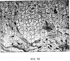
8週及び16週において、それらの動物の9匹を検査し、安楽死させ、注入部位から組織構造試料を取り出した。組織構造試料の写真を図3に示す。図3における断面図に示されている導管を8週間及び16週間維持し、図3Aに示されているように、前記安定化ヒアルロン酸をなお含有していた(ヒアルロン酸結合蛋白質を用いて染色)。細胞は、導管に穿通していなかった。有害な組織反応のエビデンス及び導管内での組織の形成はなかった。 At 8 and 16 weeks, 9 of those animals were examined, euthanized, and histological samples were removed from the injection site. A photograph of the tissue structure sample is shown in FIG. The conduits shown in the cross-sectional view in FIG. 3 were maintained for 8 and 16 weeks and still contained the stabilized hyaluronic acid as shown in FIG. 3A (stained with hyaluronic acid binding protein). ). Cells did not penetrate the conduit. There was no evidence of adverse tissue reactions and no tissue formation in the conduit.
前記組成物への暴露の16週後に回収された試料において、組織化学的染色(図3B、ヘマトキシリン及びエオシンで染色)により、導管の壁が内皮細胞で覆われていたこと、すなわち、導管が永続的であり得る初期サインが示された。 In samples collected 16 weeks after exposure to the composition, histochemical staining (Figure 3B, stained with hematoxylin and eosin) indicated that the wall of the conduit was covered with endothelial cells, i.e. the conduit was permanent. An initial sign that could be relevant was shown.
実施例3 ウサギの眼における安定化ヒアルロン酸の前臨床研究
ウサギに標準操作により麻酔をする。用いた組成物は、それぞれ10mg/ml、30mg/ml及び50mg/mlのヒアルロン酸濃度を有する安定化ヒアルロン酸ゲル粒子を含有するスラリーである。それらの組成物を片方の眼に注入し、反対の眼は、処理しない対照であった。各スラリーにおいて、主容量の粒子は、それぞれ、約0.1mm、0.4mm及び0.8mmである。
Example 3 Preclinical Study of Stabilized Hyaluronic Acid in Rabbit Eyes Rabbits are anesthetized by standard procedures . The composition used is a slurry containing stabilized hyaluronic acid gel particles having hyaluronic acid concentrations of 10 mg / ml, 30 mg / ml and 50 mg / ml, respectively. The compositions were injected into one eye and the opposite eye was an untreated control. In each slurry, the main volume particles are about 0.1 mm, 0.4 mm, and 0.8 mm, respectively.
結膜を穿通させ、針を、強膜を通して前眼房(図1b)又は後眼房(図1c)へと動かすことにより、強膜中に眼当り1−3の導管を針を用いて形成した。針の引き抜きの間、前記組成物を眼の強膜中に注入する。 Through the conjunctiva, the needle is moved through the sclera into the anterior chamber (FIG. 1b) or posterior chamber (FIG. 1c) to form 1-3 conduits per eye in the sclera. . During the withdrawal of the needle, the composition is injected into the sclera of the eye.
用いた組成物の量、針のサイズ及び種類並びに注入部位を記録する。注入の前及び後に、注入部位を視覚によってチェックする。 Record the amount of composition used, the size and type of needle, and the injection site. Visually check the injection site before and after injection.
それらの動物を標準操作により毎日観察した。8週及び16週において、動物達を検査し、安楽死させ、注入部位から組織構造試料を取り出した。 The animals were observed daily by standard procedures. At 8 and 16 weeks, animals were examined, euthanized, and tissue structure samples were removed from the injection site.
実施例4 ウサギの眼における安定化ヒアルロン酸の適用
3つの異なるカニューレサイズ:18G、23G及び27Gを用いて実施例2の一般的操作に従った。形成された水路の直径は、すべてのカニューレでおよそ等しかった。その水路のサイズは、カニューレの直径よりも注入された物質の量に依存しているようである。18Gカニューレでは2/3の眼において、27Gカニューレでは2/3の眼において及び23Gカニューレでは3/3の眼において永続性の水路が見出された。
Example 4 Application of Stabilized Hyaluronic Acid in Rabbit Eyes The general procedure of Example 2 was followed using three different cannula sizes: 18G, 23G and 27G. The diameter of the water channel formed was approximately equal for all cannulas. The size of the channel appears to depend on the amount of material injected rather than the diameter of the cannula. Permanent waterways were found in 2/3 eyes with the 18G cannula, 2/3 eyes with the 27G cannula and 3/3 eyes with the 23G cannula.
実施例5 安定化ヒアルロン酸のゲル粒子を通しての流体流れ
ゲル粒子(平均直径400μm)の形態での、実施例1の方法により得られる非動物性の安定化ヒアルロン酸[Q−Med AB(スエーデン、ウプサラ)から市販]、20mg/mlを含有する水性組成物についての押出力を、30ゲージ針により21N及び23ゲージ針により4Nに決定した。
Example 5 Non-animal stabilized hyaluronic acid obtained by the method of Example 1 in the form of fluid flow gel particles (average diameter 400 μm) through stabilized hyaluronic acid gel particles [Q-Med AB (Sweden, Commercially available from Uppsala), the pushing force for an aqueous composition containing 20 mg / ml was determined to be 21 N with a 30 gauge needle and 4 N with a 23 gauge needle.
実験の第一組において、ポンプを用いて、ゲル粒子を通る生理的食塩水の流れを与えることにより、ゲル粒子を通る流れの可能性を研究した。ガラスカラム(直径5mm)をゲル粒子で30mmの高さまで(約1mlのゲル粒子)充填した。ゲル粒子を通る生理的食塩水の流れをポンプで制御した。生理食塩水は、125μl/分の流量速度でこのカラムを流れることができた。 In the first set of experiments, the possibility of flow through gel particles was studied by using a pump to provide a flow of saline through the gel particles. A glass column (5 mm in diameter) was packed with gel particles to a height of 30 mm (about 1 ml of gel particles). The saline flow through the gel particles was pumped. Saline was able to flow through this column at a flow rate of 125 μl / min.
実験の第二組では、ガラスカラム(直径10mm)を1mlの組成物で充填し、0.9%のNaClの水溶液を、29mmHgの圧力、未治療の緑内障でのIOPに相当する圧力、において注いだ。この圧力により、その組成物を通る160μl/時間(2.7μl/分)の流れがもたらされた。比較のために、健康な眼における眼房水は、1.8μl/分−4.3μl/分の範囲、典型的には2.75μl/分である。[Brubaker RFによる“Flow of aqueous humor in humans(The Friedenwald Lecture)”、Investigative Ophthalmology & Visual Science 32:3145−3166(1991)]。 In the second set of experiments, a glass column (10 mm in diameter) is packed with 1 ml of composition and an aqueous solution of 0.9% NaCl is poured at a pressure of 29 mmHg, corresponding to an IOP in untreated glaucoma. It is. This pressure resulted in a flow of 160 μl / hour (2.7 μl / min) through the composition. For comparison, aqueous humor in healthy eyes is in the range of 1.8 μl / min-4.3 μl / min, typically 2.75 μl / min. ["Flow of aquamous human in humans (The Friedenwald Lecture)" by Brubaker RF, Investigative Ophthalmology & Visual Science 32: 3145-3166 (1915-3166).
行われた実験により、生理食塩水は、29mmHgの圧力の適用の後にゲル粒子を流れることができることが示された。その流量は、正常なヒトの眼における眼房水と同じ大きさの流量である。いずれの特別な理論に縛られることなく、サイズ排除クロマトグラフィーにおけるセファデックスのような、クロマトグラフィーのゲルビーズを溶媒が流れるのと同じように、生理食塩水はゲル粒子間を流れることが予想される。 Experiments performed showed that saline can flow through the gel particles after application of a pressure of 29 mm Hg. The flow rate is as large as that of aqueous humor in a normal human eye. Without being bound by any particular theory, saline is expected to flow between the gel particles just as the solvent flows through chromatographic gel beads, such as Sephadex in size exclusion chromatography. .
Claims (24)
(ii)前記の少なくとも一つの強膜穿通導管を粘弾性媒体で充填するように前記導管に前記媒体を注入する工程を含む、治療を必要としている動物の眼における、増大した眼圧を治療する方法であって、前記強膜穿通導管は眼の可視静脈を穿通しないものであり、粘弾性媒体は架橋されたまたは部分架橋された多糖類から選択される、前記方法。(I) forming at least one scleral penetration conduit in the eye of a non-human animal; and (ii) injecting said medium into said conduit so as to fill said at least one scleral penetration conduit with a viscoelastic medium. A method of treating increased intraocular pressure in an eye of an animal in need of treatment comprising the steps, wherein the scleral penetration conduit does not penetrate the visible vein of the eye and the viscoelastic medium is cross-linked Or said process selected from partially cross-linked polysaccharides.
Applications Claiming Priority (3)
| Application Number | Priority Date | Filing Date | Title |
|---|---|---|---|
| SE0401182-1 | 2004-05-05 | ||
| SE0401182A SE0401182D0 (en) | 2004-05-05 | 2004-05-05 | Novel use of a viscoelastic composition |
| PCT/SE2005/000663 WO2005105037A2 (en) | 2004-05-05 | 2005-05-04 | Use of a viscoelastic composition for treating increased intraocular pressure |
Publications (3)
| Publication Number | Publication Date |
|---|---|
| JP2007536220A JP2007536220A (en) | 2007-12-13 |
| JP2007536220A5 JP2007536220A5 (en) | 2008-04-24 |
| JP4922158B2 true JP4922158B2 (en) | 2012-04-25 |
Family
ID=32466229
Family Applications (1)
| Application Number | Title | Priority Date | Filing Date |
|---|---|---|---|
| JP2007511320A Expired - Fee Related JP4922158B2 (en) | 2004-05-05 | 2005-05-04 | Use of viscoelastic compositions to treat increased intraocular pressure |
Country Status (12)
| Country | Link |
|---|---|
| US (1) | US8721622B2 (en) |
| EP (1) | EP1796624B1 (en) |
| JP (1) | JP4922158B2 (en) |
| AT (1) | ATE506940T1 (en) |
| AU (1) | AU2005237394B2 (en) |
| CA (1) | CA2565382C (en) |
| DE (1) | DE602005027718D1 (en) |
| ES (1) | ES2365621T3 (en) |
| MX (1) | MXPA06012615A (en) |
| RU (1) | RU2398566C2 (en) |
| SE (1) | SE0401182D0 (en) |
| WO (1) | WO2005105037A2 (en) |
Families Citing this family (20)
| Publication number | Priority date | Publication date | Assignee | Title |
|---|---|---|---|---|
| US7909789B2 (en) | 2006-06-26 | 2011-03-22 | Sight Sciences, Inc. | Intraocular implants and methods and kits therefor |
| JP5694664B2 (en) * | 2006-09-29 | 2015-04-01 | サーモディクス,インコーポレイティド | Biodegradable ocular implant and method for treating ocular diseases |
| US8703119B2 (en) | 2007-10-05 | 2014-04-22 | Polygene Ltd. | Injectable biodegradable polymer compositions for soft tissue repair and augmentation |
| US8083720B2 (en) * | 2008-05-13 | 2011-12-27 | Solar Matthew S | Device and method for delivering therapeutic agents to an area of the body |
| US9339514B2 (en) * | 2008-09-12 | 2016-05-17 | Aptissen Sa | Method for treating glaucoma |
| US8715266B2 (en) * | 2008-09-12 | 2014-05-06 | Aptissen Sa | Glaucoma surgery |
| MX2011008683A (en) * | 2009-02-19 | 2011-09-06 | Teijin Ltd | Hydrogel of polysaccharide derivatives. |
| US8529622B2 (en) | 2010-02-05 | 2013-09-10 | Sight Sciences, Inc. | Intraocular implants and related kits and methods |
| US8652118B2 (en) | 2010-04-08 | 2014-02-18 | Kmg Pharma, Llc | Sub-mucosal agent delivery, apparatus, system and method |
| US8430862B2 (en) * | 2010-04-08 | 2013-04-30 | KMG Pharma LLC | Subconjunctival agent delivery apparatus, system and method |
| US8574217B2 (en) | 2010-04-08 | 2013-11-05 | Kmg Pharma, Llc | Sub-mucosal agent delivery method for the eye |
| RU2432141C1 (en) * | 2010-08-30 | 2011-10-27 | Александр Геннадьевич Савенков | Application of irrigation fluid in ophthalmosurgery |
| RU2460517C1 (en) * | 2011-07-14 | 2012-09-10 | Федеральное государственное учреждение "Микрохирургия глаза" имени академика С.Н. Федорова Федерального агентства по высокотехнологичной медицинской помощи" | Pharmaceutical composition for integrated treatment of ocular surface diseases in patients suffering primary open-angle glaucoma |
| AU2012374034B2 (en) | 2012-03-20 | 2017-10-19 | Sight Sciences, Inc. | Ocular delivery systems and methods |
| KR101869988B1 (en) * | 2013-03-27 | 2018-06-21 | 주식회사 엘지화학 | A composition for preparation of viscoelastic crosslinked hyaluronic acid, and crosslinked hyaluronic acid obtained by using the same |
| US9782247B2 (en) * | 2014-02-18 | 2017-10-10 | Cook Medical Technologies, LLC | Flexible embolic double filter |
| US10299958B2 (en) | 2015-03-31 | 2019-05-28 | Sight Sciences, Inc. | Ocular delivery systems and methods |
| US11291684B2 (en) | 2017-05-17 | 2022-04-05 | Tx Medic Ab | Treatment of glaucoma |
| WO2019229719A1 (en) * | 2018-06-01 | 2019-12-05 | Sun Pharma Advanced Research Company Limited | An injection device |
| US11504270B1 (en) | 2019-09-27 | 2022-11-22 | Sight Sciences, Inc. | Ocular delivery systems and methods |
Family Cites Families (29)
| Publication number | Priority date | Publication date | Assignee | Title |
|---|---|---|---|---|
| US2774239A (en) * | 1952-07-26 | 1956-12-18 | Wisconsin Alumni Res Found | Apparatus for determining dynamic mechanical properties of viscoelastic materials |
| SE442820B (en) * | 1984-06-08 | 1986-02-03 | Pharmacia Ab | GEL OF THE CROSS-BOND HYALURONIC ACID FOR USE AS A GLASS BODY SUBSTITUTE |
| US4965253A (en) * | 1987-10-14 | 1990-10-23 | University Of Florida | Viscoelastic material for ophthalmic surgery |
| US5066276A (en) * | 1988-06-21 | 1991-11-19 | Alcon Laboratories, Inc. | Method and apparatus for injecting viscous fluid into the eye to lift pre-retinal and post-retinal membrane with linear pressure control |
| US4955883A (en) * | 1988-08-29 | 1990-09-11 | Diversatronics | Glaucoma needle with a thermal heat band |
| US5092837A (en) * | 1989-12-20 | 1992-03-03 | Robert Ritch | Method for the treatment of glaucoma |
| WO1992000745A1 (en) * | 1990-07-03 | 1992-01-23 | Autogenesis Technologies, Inc. | Collagen-based viscoelastic solution for visco-surgery |
| US5360425A (en) * | 1990-08-17 | 1994-11-01 | Candela Laser Corporation | Sclerostomy method and apparatus |
| US5360399A (en) * | 1992-01-10 | 1994-11-01 | Robert Stegmann | Method and apparatus for maintaining the normal intraocular pressure |
| US5273056A (en) * | 1992-06-12 | 1993-12-28 | Alcon Laboratories, Inc. | Use of combinations of viscoelastics during surgery |
| US5707643A (en) * | 1993-02-26 | 1998-01-13 | Santen Pharmaceutical Co., Ltd. | Biodegradable scleral plug |
| US5811453A (en) * | 1994-12-23 | 1998-09-22 | Alcon Laboratories, Inc. | Viscoelastic compositions and methods of use |
| US5827937A (en) | 1995-07-17 | 1998-10-27 | Q Med Ab | Polysaccharide gel composition |
| US6142969A (en) * | 1996-10-25 | 2000-11-07 | Anamed, Inc. | Sutureless implantable device and method for treatment of glaucoma |
| FR2756493B1 (en) * | 1996-12-02 | 2001-04-13 | Delab | DEVICE FOR LOCAL ADMINISTRATION OF SOLID OR SEMI-SOLID FORMULATIONS |
| US20020111603A1 (en) * | 1996-12-02 | 2002-08-15 | Societe De Conseils De Recherches Et D'application | Device for local administration of solid or semi-solid formulations and delayed-release formulations for proposal parental administration and preparation process |
| FR2759577B1 (en) * | 1997-02-17 | 1999-08-06 | Corneal Ind | DEEP SCLERECTOMY IMPLANT |
| PT999854E (en) * | 1997-07-29 | 2003-12-31 | Alcon Mfg Ltd | COMPATIBLE VISCOELASTIC SYSTEMS CONTAINING GALACTOMANAN AND BORATO POLYMER |
| US6378526B1 (en) * | 1998-08-03 | 2002-04-30 | Insite Vision, Incorporated | Methods of ophthalmic administration |
| WO2000027405A1 (en) | 1998-11-10 | 2000-05-18 | Denki Kagaku Kogyo Kabushiki Kaisha | Hyaluronic acid gel, process for the preparation thereof and medical materials containing the same |
| AU778853B2 (en) * | 1999-04-02 | 2004-12-23 | Fziomed, Inc. | Compositions of polyacids and polyethers and methods for their use in reducing adhesions |
| US6558342B1 (en) * | 1999-06-02 | 2003-05-06 | Optonol Ltd. | Flow control device, introducer and method of implanting |
| US20030211166A1 (en) * | 2001-07-31 | 2003-11-13 | Yamamoto Ronald K | Microparticulate biomaterial composition for medical use |
| DK1239774T3 (en) * | 1999-12-10 | 2006-01-16 | Iscience Corp | Apparatus for the treatment of eye diseases |
| US6375642B1 (en) * | 2000-02-15 | 2002-04-23 | Grieshaber & Co. Ag Schaffhausen | Method of and device for improving a drainage of aqueous humor within the eye |
| US6638239B1 (en) * | 2000-04-14 | 2003-10-28 | Glaukos Corporation | Apparatus and method for treating glaucoma |
| DK1305057T3 (en) * | 2000-07-31 | 2005-10-24 | Iscience Corp | Microparticulate biomaterial composition of hyaluronic acid for medical use |
| FR2819722B1 (en) * | 2001-01-19 | 2006-11-24 | Corneal Ind | USE OF HYALURONIC ACID OR AT LEAST ONE OF ITS SALTS IN THE CONTEXT OF A SURGICAL TREATMENT OF PRESBYOPIA |
| EP1539066B1 (en) * | 2002-09-17 | 2012-11-07 | Iscience Surgical Corporation | Apparatus surgical bypass of aqueous humor |
-
2004
- 2004-05-05 SE SE0401182A patent/SE0401182D0/en unknown
-
2005
- 2005-05-04 DE DE602005027718T patent/DE602005027718D1/en not_active Expired - Lifetime
- 2005-05-04 CA CA2565382A patent/CA2565382C/en not_active Expired - Lifetime
- 2005-05-04 US US11/579,621 patent/US8721622B2/en active Active
- 2005-05-04 JP JP2007511320A patent/JP4922158B2/en not_active Expired - Fee Related
- 2005-05-04 AU AU2005237394A patent/AU2005237394B2/en not_active Ceased
- 2005-05-04 EP EP05740578A patent/EP1796624B1/en not_active Expired - Lifetime
- 2005-05-04 WO PCT/SE2005/000663 patent/WO2005105037A2/en not_active Ceased
- 2005-05-04 ES ES05740578T patent/ES2365621T3/en not_active Expired - Lifetime
- 2005-05-04 RU RU2006142861/15A patent/RU2398566C2/en active
- 2005-05-04 AT AT05740578T patent/ATE506940T1/en not_active IP Right Cessation
- 2005-05-04 MX MXPA06012615A patent/MXPA06012615A/en active IP Right Grant
Also Published As
| Publication number | Publication date |
|---|---|
| AU2005237394A1 (en) | 2005-11-10 |
| US8721622B2 (en) | 2014-05-13 |
| MXPA06012615A (en) | 2007-01-31 |
| ATE506940T1 (en) | 2011-05-15 |
| WO2005105037A8 (en) | 2006-12-21 |
| AU2005237394B2 (en) | 2009-12-10 |
| CA2565382A1 (en) | 2005-11-10 |
| JP2007536220A (en) | 2007-12-13 |
| DE602005027718D1 (en) | 2011-06-09 |
| WO2005105037A3 (en) | 2006-05-18 |
| ES2365621T3 (en) | 2011-10-07 |
| EP1796624A2 (en) | 2007-06-20 |
| US20080058760A1 (en) | 2008-03-06 |
| RU2398566C2 (en) | 2010-09-10 |
| CA2565382C (en) | 2013-12-03 |
| SE0401182D0 (en) | 2004-05-05 |
| WO2005105037A2 (en) | 2005-11-10 |
| RU2006142861A (en) | 2008-06-20 |
| EP1796624B1 (en) | 2011-04-27 |
Similar Documents
| Publication | Publication Date | Title |
|---|---|---|
| JP4922158B2 (en) | Use of viscoelastic compositions to treat increased intraocular pressure | |
| RU2635320C2 (en) | Methods and devices for drug delivery to eye tissue with microneedle | |
| US10307293B2 (en) | Methods for intraocular shunt placement | |
| US9980854B2 (en) | Shunt placement through the sclera | |
| AU2001245522C1 (en) | Apparatus and method for treating glaucoma | |
| AU2004213006B2 (en) | Methods and devices for draining fluids and lowering intraocular pressure | |
| JP2009531298A (en) | Devices and formulations for suprachoroidal drug delivery | |
| US8715266B2 (en) | Glaucoma surgery | |
| US9339514B2 (en) | Method for treating glaucoma | |
| RU2297815C1 (en) | Surgical method for treating refractory glaucoma cases | |
| RU2228160C2 (en) | Method for treating the cases of recurrent ciliochoroidal detachment occurring after glaucoma operations | |
| RU2158569C2 (en) | Method for applying deep subtenon anesthesia | |
| RU2238709C1 (en) | Method for introducing drugs in subscleral way | |
| RU2233146C2 (en) | Method for treating cystic filtration pads | |
| RU2464000C1 (en) | Method of treating unstabilised initial glaucoma | |
| RU2585728C1 (en) | Drain tube for ophthalmic operations |
Legal Events
| Date | Code | Title | Description |
|---|---|---|---|
| A521 | Request for written amendment filed |
Free format text: JAPANESE INTERMEDIATE CODE: A523 Effective date: 20080306 |
|
| A621 | Written request for application examination |
Free format text: JAPANESE INTERMEDIATE CODE: A621 Effective date: 20080306 |
|
| A131 | Notification of reasons for refusal |
Free format text: JAPANESE INTERMEDIATE CODE: A131 Effective date: 20110628 |
|
| A601 | Written request for extension of time |
Free format text: JAPANESE INTERMEDIATE CODE: A601 Effective date: 20110928 |
|
| A602 | Written permission of extension of time |
Free format text: JAPANESE INTERMEDIATE CODE: A602 Effective date: 20111005 |
|
| A601 | Written request for extension of time |
Free format text: JAPANESE INTERMEDIATE CODE: A601 Effective date: 20111028 |
|
| A521 | Request for written amendment filed |
Free format text: JAPANESE INTERMEDIATE CODE: A523 Effective date: 20111107 |
|
| A602 | Written permission of extension of time |
Free format text: JAPANESE INTERMEDIATE CODE: A602 Effective date: 20111107 |
|
| A131 | Notification of reasons for refusal |
Free format text: JAPANESE INTERMEDIATE CODE: A131 Effective date: 20111122 |
|
| A521 | Request for written amendment filed |
Free format text: JAPANESE INTERMEDIATE CODE: A523 Effective date: 20111220 |
|
| TRDD | Decision of grant or rejection written | ||
| A01 | Written decision to grant a patent or to grant a registration (utility model) |
Free format text: JAPANESE INTERMEDIATE CODE: A01 Effective date: 20120127 |
|
| A01 | Written decision to grant a patent or to grant a registration (utility model) |
Free format text: JAPANESE INTERMEDIATE CODE: A01 |
|
| A61 | First payment of annual fees (during grant procedure) |
Free format text: JAPANESE INTERMEDIATE CODE: A61 Effective date: 20120203 |
|
| R150 | Certificate of patent or registration of utility model |
Ref document number: 4922158 Country of ref document: JP Free format text: JAPANESE INTERMEDIATE CODE: R150 Free format text: JAPANESE INTERMEDIATE CODE: R150 |
|
| FPAY | Renewal fee payment (event date is renewal date of database) |
Free format text: PAYMENT UNTIL: 20150210 Year of fee payment: 3 |
|
| R250 | Receipt of annual fees |
Free format text: JAPANESE INTERMEDIATE CODE: R250 |
|
| R250 | Receipt of annual fees |
Free format text: JAPANESE INTERMEDIATE CODE: R250 |
|
| R250 | Receipt of annual fees |
Free format text: JAPANESE INTERMEDIATE CODE: R250 |
|
| R250 | Receipt of annual fees |
Free format text: JAPANESE INTERMEDIATE CODE: R250 |
|
| R250 | Receipt of annual fees |
Free format text: JAPANESE INTERMEDIATE CODE: R250 |
|
| R250 | Receipt of annual fees |
Free format text: JAPANESE INTERMEDIATE CODE: R250 |
|
| R250 | Receipt of annual fees |
Free format text: JAPANESE INTERMEDIATE CODE: R250 |
|
| LAPS | Cancellation because of no payment of annual fees |