JP5123183B2 - 標的療法のための画像ベースの計画方法及び装置 - Google Patents
標的療法のための画像ベースの計画方法及び装置 Download PDFInfo
- Publication number
- JP5123183B2 JP5123183B2 JP2008527545A JP2008527545A JP5123183B2 JP 5123183 B2 JP5123183 B2 JP 5123183B2 JP 2008527545 A JP2008527545 A JP 2008527545A JP 2008527545 A JP2008527545 A JP 2008527545A JP 5123183 B2 JP5123183 B2 JP 5123183B2
- Authority
- JP
- Japan
- Prior art keywords
- map
- patient
- concentration
- imaging
- diagnostic
- Prior art date
- Legal status (The legal status is an assumption and is not a legal conclusion. Google has not performed a legal analysis and makes no representation as to the accuracy of the status listed.)
- Expired - Fee Related
Links
Images

Classifications
-
- G—PHYSICS
- G06—COMPUTING OR CALCULATING; COUNTING
- G06T—IMAGE DATA PROCESSING OR GENERATION, IN GENERAL
- G06T7/00—Image analysis
- G06T7/0002—Inspection of images, e.g. flaw detection
- G06T7/0012—Biomedical image inspection
-
- A—HUMAN NECESSITIES
- A61—MEDICAL OR VETERINARY SCIENCE; HYGIENE
- A61B—DIAGNOSIS; SURGERY; IDENTIFICATION
- A61B5/00—Measuring for diagnostic purposes; Identification of persons
- A61B5/0059—Measuring for diagnostic purposes; Identification of persons using light, e.g. diagnosis by transillumination, diascopy, fluorescence
-
- A—HUMAN NECESSITIES
- A61—MEDICAL OR VETERINARY SCIENCE; HYGIENE
- A61B—DIAGNOSIS; SURGERY; IDENTIFICATION
- A61B5/00—Measuring for diagnostic purposes; Identification of persons
- A61B5/48—Other medical applications
- A61B5/4836—Diagnosis combined with treatment in closed-loop systems or methods
- A61B5/4839—Diagnosis combined with treatment in closed-loop systems or methods combined with drug delivery
-
- A—HUMAN NECESSITIES
- A61—MEDICAL OR VETERINARY SCIENCE; HYGIENE
- A61B—DIAGNOSIS; SURGERY; IDENTIFICATION
- A61B6/00—Apparatus or devices for radiation diagnosis; Apparatus or devices for radiation diagnosis combined with radiation therapy equipment
- A61B6/02—Arrangements for diagnosis sequentially in different planes; Stereoscopic radiation diagnosis
- A61B6/03—Computed tomography [CT]
- A61B6/032—Transmission computed tomography [CT]
-
- A—HUMAN NECESSITIES
- A61—MEDICAL OR VETERINARY SCIENCE; HYGIENE
- A61B—DIAGNOSIS; SURGERY; IDENTIFICATION
- A61B8/00—Diagnosis using ultrasonic, sonic or infrasonic waves
- A61B8/48—Diagnostic techniques
- A61B8/481—Diagnostic techniques involving the use of contrast agents, e.g. microbubbles introduced into the bloodstream
-
- A—HUMAN NECESSITIES
- A61—MEDICAL OR VETERINARY SCIENCE; HYGIENE
- A61B—DIAGNOSIS; SURGERY; IDENTIFICATION
- A61B8/00—Diagnosis using ultrasonic, sonic or infrasonic waves
- A61B8/48—Diagnostic techniques
- A61B8/488—Diagnostic techniques involving Doppler signals
-
- A—HUMAN NECESSITIES
- A61—MEDICAL OR VETERINARY SCIENCE; HYGIENE
- A61P—SPECIFIC THERAPEUTIC ACTIVITY OF CHEMICAL COMPOUNDS OR MEDICINAL PREPARATIONS
- A61P35/00—Antineoplastic agents
-
- A—HUMAN NECESSITIES
- A61—MEDICAL OR VETERINARY SCIENCE; HYGIENE
- A61B—DIAGNOSIS; SURGERY; IDENTIFICATION
- A61B6/00—Apparatus or devices for radiation diagnosis; Apparatus or devices for radiation diagnosis combined with radiation therapy equipment
- A61B6/02—Arrangements for diagnosis sequentially in different planes; Stereoscopic radiation diagnosis
- A61B6/03—Computed tomography [CT]
- A61B6/037—Emission tomography
-
- A—HUMAN NECESSITIES
- A61—MEDICAL OR VETERINARY SCIENCE; HYGIENE
- A61B—DIAGNOSIS; SURGERY; IDENTIFICATION
- A61B6/00—Apparatus or devices for radiation diagnosis; Apparatus or devices for radiation diagnosis combined with radiation therapy equipment
- A61B6/50—Apparatus or devices for radiation diagnosis; Apparatus or devices for radiation diagnosis combined with radiation therapy equipment specially adapted for specific body parts; specially adapted for specific clinical applications
- A61B6/507—Apparatus or devices for radiation diagnosis; Apparatus or devices for radiation diagnosis combined with radiation therapy equipment specially adapted for specific body parts; specially adapted for specific clinical applications for determination of haemodynamic parameters, e.g. perfusion CT
-
- G—PHYSICS
- G06—COMPUTING OR CALCULATING; COUNTING
- G06T—IMAGE DATA PROCESSING OR GENERATION, IN GENERAL
- G06T2207/00—Indexing scheme for image analysis or image enhancement
- G06T2207/10—Image acquisition modality
- G06T2207/10072—Tomographic images
- G06T2207/10076—4D tomography; Time-sequential 3D tomography
-
- G—PHYSICS
- G06—COMPUTING OR CALCULATING; COUNTING
- G06T—IMAGE DATA PROCESSING OR GENERATION, IN GENERAL
- G06T2207/00—Indexing scheme for image analysis or image enhancement
- G06T2207/30—Subject of image; Context of image processing
- G06T2207/30004—Biomedical image processing
-
- G—PHYSICS
- G06—COMPUTING OR CALCULATING; COUNTING
- G06T—IMAGE DATA PROCESSING OR GENERATION, IN GENERAL
- G06T2207/00—Indexing scheme for image analysis or image enhancement
- G06T2207/30—Subject of image; Context of image processing
- G06T2207/30004—Biomedical image processing
- G06T2207/30096—Tumor; Lesion
Landscapes
- Health & Medical Sciences (AREA)
- Life Sciences & Earth Sciences (AREA)
- Engineering & Computer Science (AREA)
- Medical Informatics (AREA)
- General Health & Medical Sciences (AREA)
- Physics & Mathematics (AREA)
- Animal Behavior & Ethology (AREA)
- Veterinary Medicine (AREA)
- Public Health (AREA)
- Molecular Biology (AREA)
- Biophysics (AREA)
- Heart & Thoracic Surgery (AREA)
- Pathology (AREA)
- Surgery (AREA)
- Biomedical Technology (AREA)
- Nuclear Medicine, Radiotherapy & Molecular Imaging (AREA)
- Radiology & Medical Imaging (AREA)
- Theoretical Computer Science (AREA)
- Pharmacology & Pharmacy (AREA)
- Medicinal Chemistry (AREA)
- Chemical & Material Sciences (AREA)
- High Energy & Nuclear Physics (AREA)
- General Physics & Mathematics (AREA)
- Pulmonology (AREA)
- Optics & Photonics (AREA)
- Computer Vision & Pattern Recognition (AREA)
- Quality & Reliability (AREA)
- Bioinformatics & Cheminformatics (AREA)
- Hematology (AREA)
- Chemical Kinetics & Catalysis (AREA)
- General Chemical & Material Sciences (AREA)
- Organic Chemistry (AREA)
- Nuclear Medicine (AREA)
- Medicines That Contain Protein Lipid Enzymes And Other Medicines (AREA)
- Radiation-Therapy Devices (AREA)
- Apparatus For Radiation Diagnosis (AREA)
Description
例1
ガンの放射免疫療法
Claims (10)
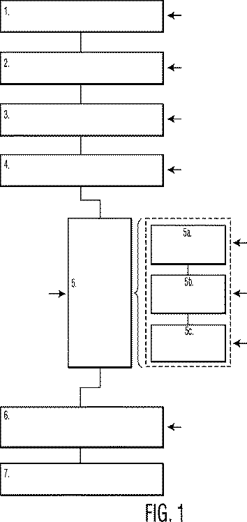
- 連続的又は動的に取得された3D画像データを量的に訂正し、及び基準となる生体構造に空間的にマッピングするため、診断用の標的薬剤が投与された患者から高感度のイメージングモダリティで得られる医療イメージングデータの画像処理をする第1の手段と、
多数の時間点に対して測定されるボクセル又は3D領域あたりの画像強度から、前記診断用の標的薬剤に対する対時間での対応濃度曲線を推定する第2の手段と、
前記診断用の標的薬剤の濃度曲線から、治療用の前記標的薬剤のボクセル又は3D領域あたりの濃度の時間的挙動及び/又は対時間での積分マップを推定する第3の手段と、
前記推定された時間的挙動及び/又は対時間での積分マップに基づき、患者固有の治療投与手順を生成する第4の手段とを有する、4D医療イメージングデバイス。 - 特定の処置の効果、有害度、又はこの両者の推定を計算する第5の手段を更に有する、請求項1に記載のデバイス。
- 前記第4の手段が、前記治療用の薬剤の対時間での推定濃度積分マップから、ボクセル又は3D領域あたりの治療的放射線線量マップを計算する手段を更に有する、請求項1又は2に記載のデバイス。
- 前記治療的放射線線量マップに基づき、腫瘍及び危険な器官に対する吸収放射線線量を計算する手段を更に有する、請求項3に記載のデバイス。
- 前記腫瘍及び危険な器官に対する放射線線量に基づき、患者固有の治療的投与計画を生成する手段を更に有する、請求項4に記載のデバイス。
- 医療イメージングを行う装置における制御部の作動方法において、
標準化された手順に基づき、診断形式の標的薬剤の患者における分布の連続的又は動的なイメージングデータを取得するステップと、
前記診断形式の標的薬剤の濃度の定量的な4Dマップを推定するステップと、
前記推定された診断形式の標的薬剤の濃度の定量的な4Dマップに基づき、治療形式の標的薬剤の濃度及び/又は対時間での積分の定量的な4Dマップを推定するステップと、
前記4D治療濃度マップ及び/又は対時間での積分マップに基づき、患者固有の治療的手順を生成するステップとを前記制御部に実行させる、方法。 - 前記診断形式の標的薬剤の濃度の定量的な4Dマップを推定するステップが、散乱、減衰、空間又は時間分解能の制限、前記標的薬剤の動力学、又はこれらのいずれかの組み合わせを有する統計的不確かさを訂正するステップを更に有する、請求項6に記載の方法。
- 前記治療形式の標的薬剤の濃度の定量的な4Dマップを推定するステップが、前記診断形式及び前記治療形式の標的薬剤間での物理及び/又は動力学的特性における可能性のある差異を考慮するステップを更に有する、請求項6に記載の方法。
- 前記患者固有の治療的手順を生成するステップが、標的となる危険な器官に対する推定線量及び該推定の不確かさを考慮するステップと、医療専門家との対話を提供するステップとを更に有する、請求項6に記載の方法。
- 前記患者固有の治療手順の効果及び/又は有害度の推定を計算するステップを更に有する、請求項6に記載の方法。
Applications Claiming Priority (3)
| Application Number | Priority Date | Filing Date | Title |
|---|---|---|---|
| US71129905P | 2005-08-25 | 2005-08-25 | |
| US60/711,299 | 2005-08-25 | ||
| PCT/IB2006/052758 WO2007023408A2 (en) | 2005-08-25 | 2006-08-09 | 4d image-based planning methods and apparatus for targeted therapy |
Publications (3)
| Publication Number | Publication Date |
|---|---|
| JP2009505709A JP2009505709A (ja) | 2009-02-12 |
| JP2009505709A5 JP2009505709A5 (ja) | 2012-06-07 |
| JP5123183B2 true JP5123183B2 (ja) | 2013-01-16 |
Family
ID=37772001
Family Applications (1)
| Application Number | Title | Priority Date | Filing Date |
|---|---|---|---|
| JP2008527545A Expired - Fee Related JP5123183B2 (ja) | 2005-08-25 | 2006-08-09 | 標的療法のための画像ベースの計画方法及び装置 |
Country Status (5)
| Country | Link |
|---|---|
| US (1) | US20080214933A1 (ja) |
| EP (1) | EP1921982A2 (ja) |
| JP (1) | JP5123183B2 (ja) |
| CN (1) | CN101247755B (ja) |
| WO (1) | WO2007023408A2 (ja) |
Families Citing this family (35)
| Publication number | Priority date | Publication date | Assignee | Title |
|---|---|---|---|---|
| US10201324B2 (en) | 2007-05-04 | 2019-02-12 | Delphinus Medical Technologies, Inc. | Patient interface system |
| AU2008254328A1 (en) | 2007-05-18 | 2008-11-27 | The Johns Hopkins University | A treatment simulator for brain diseases and method of use thereof |
| US8812240B2 (en) * | 2008-03-13 | 2014-08-19 | Siemens Medical Solutions Usa, Inc. | Dose distribution modeling by region from functional imaging |
| US8200466B2 (en) | 2008-07-21 | 2012-06-12 | The Board Of Trustees Of The Leland Stanford Junior University | Method for tuning patient-specific cardiovascular simulations |
| WO2010120534A1 (en) | 2009-03-31 | 2010-10-21 | Whitten Matthew R | System and method for radiation therapy treatment planning using a memetic optimization algorithm |
| US20120230918A1 (en) * | 2009-07-28 | 2012-09-13 | Hoffmann-La-Roche Inc. | Non-invasive in vivo optical imaging method |
| US10007961B2 (en) * | 2009-09-09 | 2018-06-26 | Wisconsin Alumni Research Foundation | Treatment planning system for radiopharmaceuticals |
| JP2011131002A (ja) * | 2009-12-25 | 2011-07-07 | Fujifilm Corp | 蛍光画像撮像装置 |
| WO2011100697A1 (en) | 2010-02-12 | 2011-08-18 | Delphinus Medical Technologies, Inc. | Method of characterizing tissue of a patient |
| WO2011100691A1 (en) | 2010-02-12 | 2011-08-18 | Delphinus Medical Technologies, Inc. | Method of characterizing the pathological response of tissue to a treatmant plan |
| US8315812B2 (en) * | 2010-08-12 | 2012-11-20 | Heartflow, Inc. | Method and system for patient-specific modeling of blood flow |
| GB201014331D0 (en) * | 2010-08-27 | 2010-10-13 | Siemens Medical Solutions | Automatic identification of disruptive events in pets scans |
| DK3281586T3 (da) * | 2010-12-08 | 2019-06-24 | Bayer Healthcare Llc | Generering af et estimat for patientstrålingsdosis som følge af medicinske billeddannelsesscanninger |
| EP2716223A4 (en) * | 2011-05-30 | 2015-04-08 | Fujifilm Corp | DEVICE AND METHOD FOR DETECTING A BEAM EXPOSURE LEVEL AND PROGRAM THEREFOR |
| US9177376B2 (en) | 2012-08-22 | 2015-11-03 | Koninklijke Philips N.V. | Method for determining the distribution of an imaging agent |
| US9763641B2 (en) | 2012-08-30 | 2017-09-19 | Delphinus Medical Technologies, Inc. | Method and system for imaging a volume of tissue with tissue boundary detection |
| JP5996658B2 (ja) * | 2012-08-30 | 2016-09-21 | 株式会社島津製作所 | 脳断層動態画像解析装置 |
| US10123770B2 (en) | 2013-03-13 | 2018-11-13 | Delphinus Medical Technologies, Inc. | Patient support system |
| US11495355B2 (en) * | 2014-05-15 | 2022-11-08 | The Johns Hopkins University | Method, system and computer-readable media for treatment plan risk analysis |
| US10285667B2 (en) | 2014-08-05 | 2019-05-14 | Delphinus Medical Technologies, Inc. | Method for generating an enhanced image of a volume of tissue |
| DE102014221054B4 (de) * | 2014-10-16 | 2026-04-30 | Siemens Healthineers Ag | Verfahren zur Erzeugung eines anzuzeigenden Bilddatensatzes aus Magnetresonanzdaten, Recheneinrichtung und Computerprogramm |
| DE102016100713A1 (de) * | 2016-01-18 | 2017-07-20 | Surgiceye Gmbh | System und Verfahren zur Bestimmung einer Dosis in der Strahlentherapie |
| KR101796868B1 (ko) * | 2016-06-30 | 2017-11-16 | 가톨릭대학교 산학협력단 | 환자의 기하학적 관계를 이용한 호흡 연동 방사선 치료에서의 플래닝 페이즈 최적화 방법 |
| CN107970527B (zh) * | 2016-10-25 | 2020-11-10 | 住友重机械工业株式会社 | 放射线治疗模拟装置 |
| US10426424B2 (en) | 2017-11-21 | 2019-10-01 | General Electric Company | System and method for generating and performing imaging protocol simulations |
| US12602772B2 (en) | 2018-01-08 | 2026-04-14 | Progenics Pharmaceuticals, Inc. | Systems and methods for rapid neural network-based image segmentation and radiopharmaceutical uptake determination |
| EP3413317B1 (de) * | 2018-01-29 | 2020-06-17 | Siemens Healthcare GmbH | Bereitstellen eines medizinischen bildes |
| JP7568628B2 (ja) | 2019-01-07 | 2024-10-16 | エクシーニ ディアグノスティクス アーべー | プラットホーム非依存の全身画像セグメント化のためのシステムおよび方法 |
| KR102392778B1 (ko) * | 2020-01-02 | 2022-05-04 | 한국원자력의학원 | 영상 기반의 치료용 방사성의약품 치료선량 결정 시스템 및 방법 |
| KR102490645B1 (ko) * | 2020-07-16 | 2023-01-25 | 고려대학교 산학협력단 | 흡수에너지 기반 전기장 암치료 계획 시스템 및 방법 |
| CN112221023B (zh) * | 2020-09-27 | 2023-12-15 | 中国辐射防护研究院 | 一种放射性药物的剂量评估系统及方法 |
| US12364394B2 (en) * | 2020-11-06 | 2025-07-22 | BAMF Health LLC | System and method for radiopharmaceutical therapy analysis using machine learning |
| WO2022175062A1 (en) * | 2021-02-22 | 2022-08-25 | Koninklijke Philips N.V. | Method for estimating radiation exposure in a 3d environment |
| US12597127B2 (en) | 2021-10-08 | 2026-04-07 | Exini Diagnostics Ab | Systems and methods for automated identification and classification of lesions in local lymph and distant metastases |
| AU2023282844A1 (en) * | 2022-06-08 | 2024-11-28 | Exini Diagnostics Ab | Systems and methods for assessing disease burden and progression |
Family Cites Families (23)
| Publication number | Priority date | Publication date | Assignee | Title |
|---|---|---|---|---|
| US5396418A (en) * | 1988-10-20 | 1995-03-07 | Picker International, Inc. | Four dimensional spiral volume imaging using fast retrace |
| DE69325485T2 (de) * | 1992-09-09 | 1999-10-28 | Picker International, Inc. | Verfahren und Vorrichtung zur Bilderzeugung |
| JPH07271997A (ja) * | 1994-03-29 | 1995-10-20 | Toshiba Corp | 画像処理装置 |
| US5684889A (en) | 1995-04-28 | 1997-11-04 | Sloan-Kettering Institute For Cancer Research | Parametric image representation of imaging agent kinetics |
| US5602397A (en) | 1995-11-01 | 1997-02-11 | University Of Louisville Research Foundation, Inc. | Optical imaging system utilizing a charge amplification device |
| US5844241A (en) * | 1996-07-19 | 1998-12-01 | City Of Hope | System and method for determining internal radioactivity and absorbed dose estimates |
| US5919135A (en) * | 1997-02-28 | 1999-07-06 | Lemelson; Jerome | System and method for treating cellular disorders in a living being |
| WO1999062565A2 (en) * | 1998-06-04 | 1999-12-09 | Coulter Pharmaceutical, Inc. | Patient-specific dosimetry |
| US20020102208A1 (en) * | 1999-03-01 | 2002-08-01 | Paul Chinn | Radiolabeling kit and binding assay |
| US6907280B2 (en) * | 1999-12-02 | 2005-06-14 | The General Hospital Corporation | Method and apparatus for objectively measuring pain, pain treatment and other related techniques |
| EP1299031A4 (en) * | 2000-03-21 | 2006-02-08 | Bechtel Bwxt Idaho Llc | METHOD AND COMPUTER MEDIUM FOR IMPROVED RADIO THERAPY DOSE PLANTING |
| AU2001249935A1 (en) * | 2000-04-05 | 2001-10-23 | Therics, Inc. | System and method for rapidly customizing a design and remotely manufacturing biomedical devices using a computer system |
| AU2002212273B2 (en) * | 2000-09-15 | 2007-06-14 | Aaron Diamond Aids Research Centre | System and method for optimizing drug therapy for the treatment of diseases |
| US7630750B2 (en) * | 2001-02-05 | 2009-12-08 | The Research Foundation For The State University Of New York | Computer aided treatment planning |
| US7011814B2 (en) * | 2001-04-23 | 2006-03-14 | Sicel Technologies, Inc. | Systems, methods and devices for in vivo monitoring of a localized response via a radiolabeled analyte in a subject |
| DE60208009T2 (de) * | 2002-06-17 | 2006-08-24 | Nucletron B.V. | System zur Echtzeitplanung von Strahlentherapie |
| JP2005531643A (ja) * | 2002-07-02 | 2005-10-20 | ユニベルシテア、メディッシュ、セントラム、ユトレヒト | 直径が少なくとも1マイクロメートルの粒子を含んでなる走査懸濁液 |
| EP3067070A1 (en) * | 2002-09-24 | 2016-09-14 | The Government of the United States of America as represented by the Secretary of the Department of Health and Human Services | Method for convection enhanced delivery of therapeutic agents |
| US20040193446A1 (en) * | 2003-03-27 | 2004-09-30 | Mayer Steven Lloyd | System and method for managing a patient treatment program including a prescribed drug regimen |
| DK1644049T3 (da) * | 2003-04-30 | 2008-02-04 | Univ Ramot | Fremgangsmåde og anordning til radioterapi |
| US20040225174A1 (en) * | 2003-05-06 | 2004-11-11 | Fuller Donald B. | Method for computed tomography-ultrasound interactive prostate brachytherapy |
| US8249813B2 (en) * | 2003-05-22 | 2012-08-21 | Brainlab Ag | System and process for metabolic guidance of introduced cellular material |
| MXPA06008588A (es) * | 2004-01-27 | 2006-08-28 | Aspect Medical Systems Inc | Sistema para la distribucion adaptable de farmaco. |
-
2006
- 2006-08-09 JP JP2008527545A patent/JP5123183B2/ja not_active Expired - Fee Related
- 2006-08-09 US US12/064,372 patent/US20080214933A1/en not_active Abandoned
- 2006-08-09 CN CN200680031102.3A patent/CN101247755B/zh not_active Expired - Fee Related
- 2006-08-09 WO PCT/IB2006/052758 patent/WO2007023408A2/en not_active Ceased
- 2006-08-09 EP EP06795626A patent/EP1921982A2/en not_active Ceased
Also Published As
| Publication number | Publication date |
|---|---|
| EP1921982A2 (en) | 2008-05-21 |
| US20080214933A1 (en) | 2008-09-04 |
| WO2007023408A2 (en) | 2007-03-01 |
| CN101247755B (zh) | 2018-04-24 |
| CN101247755A (zh) | 2008-08-20 |
| WO2007023408A3 (en) | 2007-11-01 |
| JP2009505709A (ja) | 2009-02-12 |
Similar Documents
| Publication | Publication Date | Title |
|---|---|---|
| JP5123183B2 (ja) | 標的療法のための画像ベースの計画方法及び装置 | |
| Wolbarst et al. | Evolving and experimental technologies in medical imaging | |
| Abadi et al. | Virtual clinical trials in medical imaging: a review | |
| Cherry | Multimodality in vivo imaging systems: twice the power or double the trouble? | |
| Bercovich et al. | Medical imaging: from roentgen to the digital revolution, and beyond | |
| Feng | Biomedical information technology | |
| Kasban et al. | A comparative study of medical imaging techniques | |
| Astolfo et al. | In vivo visualization of gold-loaded cells in mice using x-ray computed tomography | |
| US20160282432A1 (en) | Omni-Tomographic Imaging for Interior Reconstruction using Simultaneous Data Acquisition from Multiple Imaging Modalities | |
| Stout et al. | Guidance for methods descriptions used in preclinical imaging papers | |
| Beyer et al. | Medical physics and imaging–A timely perspective | |
| Cherry et al. | Essentials of in vivo biomedical imaging | |
| Forssell-Aronsson et al. | Medical imaging for improved tumour characterization, delineation and treatment verification | |
| Iftimia et al. | Advances in Optical Imaging for Clinical Medicine | |
| Cobo et al. | Physical foundations for trustworthy medical imaging: a review for artificial intelligence researchers | |
| Ilyas et al. | Advances in biomedical imaging techniques: a compre-hensive review | |
| Mara et al. | Medical Imaging for Use Condition Measurement | |
| Moran et al. | The physics of medical imaging | |
| Landini et al. | Fundamentals in cardiovascular imaging technologies | |
| Fatima et al. | Integrating CT and MRI in Cancer Staging and Treatment Monitoring: Challenges and Innovations | |
| Erturk et al. | Imaging tools in human research | |
| Haq et al. | Advancements in Imaging Technologies for Precise Disease Diagnosis | |
| Kory | Tomographic Medical Imaging Modalities: a Short Conceptual Introduction | |
| Wang et al. | Omni-tomography/Multi-tomography--Integrating Multiple Modalities for Simultaneous Imaging | |
| Baril et al. | In Vivo Imaging in Mice |
Legal Events
| Date | Code | Title | Description |
|---|---|---|---|
| A621 | Written request for application examination |
Free format text: JAPANESE INTERMEDIATE CODE: A621 Effective date: 20090810 |
|
| A977 | Report on retrieval |
Free format text: JAPANESE INTERMEDIATE CODE: A971007 Effective date: 20111228 |
|
| A131 | Notification of reasons for refusal |
Free format text: JAPANESE INTERMEDIATE CODE: A131 Effective date: 20120105 |
|
| A524 | Written submission of copy of amendment under article 19 pct |
Free format text: JAPANESE INTERMEDIATE CODE: A524 Effective date: 20120405 |
|
| A02 | Decision of refusal |
Free format text: JAPANESE INTERMEDIATE CODE: A02 Effective date: 20120605 |
|
| A521 | Request for written amendment filed |
Free format text: JAPANESE INTERMEDIATE CODE: A523 Effective date: 20120810 |
|
| A911 | Transfer to examiner for re-examination before appeal (zenchi) |
Free format text: JAPANESE INTERMEDIATE CODE: A911 Effective date: 20120904 |
|
| TRDD | Decision of grant or rejection written | ||
| A01 | Written decision to grant a patent or to grant a registration (utility model) |
Free format text: JAPANESE INTERMEDIATE CODE: A01 Effective date: 20120927 |
|
| A01 | Written decision to grant a patent or to grant a registration (utility model) |
Free format text: JAPANESE INTERMEDIATE CODE: A01 |
|
| A61 | First payment of annual fees (during grant procedure) |
Free format text: JAPANESE INTERMEDIATE CODE: A61 Effective date: 20121025 |
|
| FPAY | Renewal fee payment (event date is renewal date of database) |
Free format text: PAYMENT UNTIL: 20151102 Year of fee payment: 3 |
|
| R150 | Certificate of patent or registration of utility model |
Free format text: JAPANESE INTERMEDIATE CODE: R150 |
|
| R250 | Receipt of annual fees |
Free format text: JAPANESE INTERMEDIATE CODE: R250 |
|
| R250 | Receipt of annual fees |
Free format text: JAPANESE INTERMEDIATE CODE: R250 |
|
| R250 | Receipt of annual fees |
Free format text: JAPANESE INTERMEDIATE CODE: R250 |
|
| LAPS | Cancellation because of no payment of annual fees |