JP5389337B2 - Swivel anchor for tissue nodule fixation - Google Patents
Swivel anchor for tissue nodule fixation Download PDFInfo
- Publication number
- JP5389337B2 JP5389337B2 JP2007132157A JP2007132157A JP5389337B2 JP 5389337 B2 JP5389337 B2 JP 5389337B2 JP 2007132157 A JP2007132157 A JP 2007132157A JP 2007132157 A JP2007132157 A JP 2007132157A JP 5389337 B2 JP5389337 B2 JP 5389337B2
- Authority
- JP
- Japan
- Prior art keywords
- anchor
- suture
- swivel
- tip
- fixation
- Prior art date
- Legal status (The legal status is an assumption and is not a legal conclusion. Google has not performed a legal analysis and makes no representation as to the accuracy of the status listed.)
- Active
Links
Images


Classifications
-
- A—HUMAN NECESSITIES
- A61—MEDICAL OR VETERINARY SCIENCE; HYGIENE
- A61B—DIAGNOSIS; SURGERY; IDENTIFICATION
- A61B17/00—Surgical instruments, devices or methods
- A61B17/068—Surgical staplers, e.g. containing multiple staples or clamps
-
- A—HUMAN NECESSITIES
- A61—MEDICAL OR VETERINARY SCIENCE; HYGIENE
- A61B—DIAGNOSIS; SURGERY; IDENTIFICATION
- A61B17/00—Surgical instruments, devices or methods
- A61B17/04—Surgical instruments, devices or methods for suturing wounds; Holders or packages for needles or suture materials
- A61B17/0401—Suture anchors, buttons or pledgets, i.e. means for attaching sutures to bone, cartilage or soft tissue; Instruments for applying or removing suture anchors
-
- A—HUMAN NECESSITIES
- A61—MEDICAL OR VETERINARY SCIENCE; HYGIENE
- A61B—DIAGNOSIS; SURGERY; IDENTIFICATION
- A61B17/00—Surgical instruments, devices or methods
- A61B17/064—Surgical staples, i.e. penetrating the tissue
-
- A—HUMAN NECESSITIES
- A61—MEDICAL OR VETERINARY SCIENCE; HYGIENE
- A61B—DIAGNOSIS; SURGERY; IDENTIFICATION
- A61B17/00—Surgical instruments, devices or methods
- A61B17/04—Surgical instruments, devices or methods for suturing wounds; Holders or packages for needles or suture materials
- A61B17/0401—Suture anchors, buttons or pledgets, i.e. means for attaching sutures to bone, cartilage or soft tissue; Instruments for applying or removing suture anchors
- A61B2017/0408—Rivets
-
- A—HUMAN NECESSITIES
- A61—MEDICAL OR VETERINARY SCIENCE; HYGIENE
- A61B—DIAGNOSIS; SURGERY; IDENTIFICATION
- A61B17/00—Surgical instruments, devices or methods
- A61B17/04—Surgical instruments, devices or methods for suturing wounds; Holders or packages for needles or suture materials
- A61B17/0401—Suture anchors, buttons or pledgets, i.e. means for attaching sutures to bone, cartilage or soft tissue; Instruments for applying or removing suture anchors
- A61B2017/0409—Instruments for applying suture anchors
-
- A—HUMAN NECESSITIES
- A61—MEDICAL OR VETERINARY SCIENCE; HYGIENE
- A61B—DIAGNOSIS; SURGERY; IDENTIFICATION
- A61B17/00—Surgical instruments, devices or methods
- A61B17/04—Surgical instruments, devices or methods for suturing wounds; Holders or packages for needles or suture materials
- A61B17/0401—Suture anchors, buttons or pledgets, i.e. means for attaching sutures to bone, cartilage or soft tissue; Instruments for applying or removing suture anchors
- A61B2017/0412—Suture anchors, buttons or pledgets, i.e. means for attaching sutures to bone, cartilage or soft tissue; Instruments for applying or removing suture anchors having anchoring barbs or pins extending outwardly from suture anchor body
-
- A—HUMAN NECESSITIES
- A61—MEDICAL OR VETERINARY SCIENCE; HYGIENE
- A61B—DIAGNOSIS; SURGERY; IDENTIFICATION
- A61B17/00—Surgical instruments, devices or methods
- A61B17/04—Surgical instruments, devices or methods for suturing wounds; Holders or packages for needles or suture materials
- A61B17/0401—Suture anchors, buttons or pledgets, i.e. means for attaching sutures to bone, cartilage or soft tissue; Instruments for applying or removing suture anchors
- A61B2017/0438—Suture anchors, buttons or pledgets, i.e. means for attaching sutures to bone, cartilage or soft tissue; Instruments for applying or removing suture anchors slotted, i.e. having a longitudinal slot for enhancing their elasticity
-
- A—HUMAN NECESSITIES
- A61—MEDICAL OR VETERINARY SCIENCE; HYGIENE
- A61B—DIAGNOSIS; SURGERY; IDENTIFICATION
- A61B17/00—Surgical instruments, devices or methods
- A61B17/04—Surgical instruments, devices or methods for suturing wounds; Holders or packages for needles or suture materials
- A61B17/0401—Suture anchors, buttons or pledgets, i.e. means for attaching sutures to bone, cartilage or soft tissue; Instruments for applying or removing suture anchors
- A61B2017/044—Suture anchors, buttons or pledgets, i.e. means for attaching sutures to bone, cartilage or soft tissue; Instruments for applying or removing suture anchors with a threaded shaft, e.g. screws
-
- A—HUMAN NECESSITIES
- A61—MEDICAL OR VETERINARY SCIENCE; HYGIENE
- A61B—DIAGNOSIS; SURGERY; IDENTIFICATION
- A61B17/00—Surgical instruments, devices or methods
- A61B17/04—Surgical instruments, devices or methods for suturing wounds; Holders or packages for needles or suture materials
- A61B17/0401—Suture anchors, buttons or pledgets, i.e. means for attaching sutures to bone, cartilage or soft tissue; Instruments for applying or removing suture anchors
- A61B2017/0446—Means for attaching and blocking the suture in the suture anchor
- A61B2017/0448—Additional elements on or within the anchor
- A61B2017/045—Additional elements on or within the anchor snug fit within the anchor
-
- A—HUMAN NECESSITIES
- A61—MEDICAL OR VETERINARY SCIENCE; HYGIENE
- A61B—DIAGNOSIS; SURGERY; IDENTIFICATION
- A61B17/00—Surgical instruments, devices or methods
- A61B17/04—Surgical instruments, devices or methods for suturing wounds; Holders or packages for needles or suture materials
- A61B17/0401—Suture anchors, buttons or pledgets, i.e. means for attaching sutures to bone, cartilage or soft tissue; Instruments for applying or removing suture anchors
- A61B2017/0446—Means for attaching and blocking the suture in the suture anchor
- A61B2017/0454—Means for attaching and blocking the suture in the suture anchor the anchor being crimped or clamped on the suture
Landscapes
- Health & Medical Sciences (AREA)
- Surgery (AREA)
- Life Sciences & Earth Sciences (AREA)
- Biomedical Technology (AREA)
- Nuclear Medicine, Radiotherapy & Molecular Imaging (AREA)
- Engineering & Computer Science (AREA)
- Heart & Thoracic Surgery (AREA)
- Medical Informatics (AREA)
- Molecular Biology (AREA)
- Animal Behavior & Ethology (AREA)
- General Health & Medical Sciences (AREA)
- Public Health (AREA)
- Veterinary Medicine (AREA)
- Rheumatology (AREA)
- Surgical Instruments (AREA)
Description
本発明は、外科的固定術に係り、より特には、無結節(ノットレス)組織固定に対するスウィベル縫合アンカーを使用する、靱帯修復及び再建等である解剖学的組織修復を行う方法に係る。 The present invention relates to surgical fixation, and more particularly to a method for performing anatomical tissue repair, such as ligament repair and reconstruction, using a swivel suture anchor for knotless tissue fixation.
外科処置中の固定縫合は、困難且つ要求の多いものであり得る。多種の縫合構造は、縫合において、特には関節鏡視下手術中、縫合において結節(ノット)を作る必要を避けるよう、開発されてきている。例えば、ThalのU.S. Pat. No. 6,143,017(特許文献1)は、アンカー装置によってとらえられる(snagged)自立している連続的な縫合ループを使用する組織固定を開示する。組織がThalの教示に従って骨に対して結合され得るようになる一方、修復される組織上のループの長さ又は張力の調整等である外科的調整を原位置で(in situ)いかに達成するかは、明らかにされていない。
したがって、無結節組織固定に対する向上された技術は、技術的に必要とされる。 Therefore, improved techniques for nodular tissue fixation are technically needed.
本発明は、無結節組織固定に対するスウィベルアンカーを与える。スウィベルアンカーは、アンカー本体、及びアンカー本体に対して回転可能に取り付けられるフォーク状先端を有して形成される。縫合アンカーは、フォーク状端部において捕捉される縫合の過剰な捻れ及び結節を引き起こすことなく回転可能な挿入を可能にするよう構成される。キャニュレーテッド干渉スクリュ(cannulated interference screw)等である固定装置は、骨における穴においてアンカーを固定するようスウィベルアンカーの本体にわたって挿入可能である。 The present invention provides a swivel anchor for nodule tissue fixation. The swivel anchor is formed having an anchor body and a fork-like tip that is rotatably attached to the anchor body. The suture anchor is configured to allow rotatable insertion without causing excessive twisting and knotting of the suture that is captured at the forked end. An anchoring device, such as a cannulated interference screw, can be inserted across the body of the swivel anchor to anchor the anchor in a hole in the bone.
本発明の他の特性及び利点は、添付の図面を参照して説明される本発明の典型的な実施例に関する以下の説明から明らかとなる。 Other features and advantages of the present invention will become apparent from the following description of exemplary embodiments of the invention which are described with reference to the accompanying drawings.
本発明は、スウィベルアンカー器具を使用する無結節組織固定に対する装置及び方法を与える。望ましい方法において、スウィベルアンカーは、望ましくは高強度縫合である、少なくとも2つのループの縫合を有して形成される縫合鎖(suture chain)を有して使用される。鎖のループは、スウィベルアンカーを固定装置(例えばアンカー、干渉プラグ又はスクリュ、あるいはインプラント)のキャニュレーテッド本体へと更に係合及びロックするよう適合されるドライバ上に、スウィベルアンカーを取り付けることによって捕捉される(captured)。 The present invention provides an apparatus and method for nodular tissue fixation using a swivel anchor instrument. In the preferred method, the swivel anchor is used with a suture chain formed with at least two loop sutures, preferably high strength sutures. The loop of the chain is obtained by mounting the swivel anchor on a driver adapted to further engage and lock the swivel anchor to the cannulated body of the anchoring device (eg anchor, interference plug or screw, or implant). Captured.
本発明はまた、スウィベルアンカー及び縫合鎖を用いることによる外科的適用中の解剖学的組織の無結節固定に対する方法を与える。該方法は、(i)縫合を有して形成され且つ接続される少なくとも2つのループを有して形成される縫合鎖を与える段階; (ii)固定装置に対して縫合鎖の一端を固定する段階; (iii)スウィベルロックを固定装置の少なくとも一部分と安全に係合する段階; (iv)結合される軟組織及び縫合鎖に張力を与えるよう(to tension)縫合鎖の他端を引く段階、を有する。 The present invention also provides a method for nodule fixation of anatomical tissue during surgical application by using swivel anchors and sutures. The method includes (i) providing a suture formed with at least two loops formed and connected with a suture; (ii) securing one end of the suture to a fixation device; (Iii) securely engaging the swivel lock with at least a portion of the fixation device; (iv) pulling the other end of the suture to tension the soft tissue and suture to be joined. Have.
本発明に従って使用される縫合鎖は、米国特許出願番号2006/0259076中に詳細に記載される。「鎖」という語は、本発明の典型的な実施例を参照するよう明細書及び請求項中で使用される。本願中の「鎖」は、一連の縫合ループを有して形成される縫合構造を広く示す。ループは、必ずしもではないが、連結され得る。このようにして、本願中で使用される「鎖」という語は、制限的ではないが、リンク又はリングが互いに対して適合されるという一般的に理解される定義を有する。更には、本発明の鎖は、共に接続される2つ又はそれより多いループを有し得る。各ループは、望ましく一定の周(fixed perimeter)を有する。縫合は、ループ形状を確立及び保持するよう、以下に更に説明される通り、結節されるのではなく組み合わされ得る(interlaced)。望ましくは、全てのループは同様の寸法である。 The suture used in accordance with the present invention is described in detail in US Patent Application No. 2006/0259076. The term “chain” is used in the specification and claims to refer to exemplary embodiments of the invention. “Chain” in this application broadly refers to a suture structure formed with a series of suture loops. The loops can be connected, but not necessarily. Thus, as used herein, the term “chain” has a generally understood definition that links or rings are adapted to each other, but are not limiting. Furthermore, the chains of the present invention can have two or more loops connected together. Each loop desirably has a fixed perimeter. The stitches can be interlaced rather than knotted, as further described below, to establish and maintain a loop shape. Desirably, all loops are of similar dimensions.
同様の要素が同様の参照符号によって示される図面を参照すると、図1−16は、本発明の典型的な実施例に従って骨に対して組織(例えば、損傷した腱)を接近させる2つの外科的処置を示す。単純化するために、本発明は、回旋筋腱板(ローテーターカフ)の固定を参照して以下に記載される。しかしながら、本発明は、この典型的な実施例に対して制限されず、例えば膝のACL再建における損傷した腱の固定、又は骨に対する腱の固定における、全般的な組織の固定に対する適用性を有する。 Referring to the drawings in which like elements are indicated by like reference numerals, FIGS. 1-16 illustrate two surgical approaches for accessing tissue (eg, a damaged tendon) against a bone in accordance with an exemplary embodiment of the present invention. Indicates treatment. For simplicity, the present invention is described below with reference to the fixation of the rotator cuff (rotator cuff). However, the present invention is not limited to this exemplary embodiment and has applicability to general tissue fixation, for example, fixation of damaged tendons in knee ACL reconstruction, or fixation of tendons to bone. .
本発明の典型的な実施例に従う組織固定の方法段階は、図1乃至16において概略的に示される。以下により詳細に記載される通り、図1−8は、無結節単列技術を使用する回旋筋腱板修復中の一連の段階を示し、図9−16は、無結節複列技術を使用する回旋筋腱板修復中の一連の段階を示す。両技術は、ドライバ組立体100(図17−20)、スウィベルロックアンカー200(図21)、及び固定装置300(図22)を参照して説明される。 The method steps of tissue fixation according to an exemplary embodiment of the present invention are schematically illustrated in FIGS. As described in more detail below, FIGS. 1-8 illustrate a series of stages during rotator cuff repair using a no-nodal single row technique, and FIGS. 9-16 use a no-nodal double-row technique. Figure 2 shows a series of stages during rotator cuff repair. Both techniques are described with reference to driver assembly 100 (FIGS. 17-20), swivel lock anchor 200 (FIG. 21), and securing device 300 (FIG. 22).
単列及び複列技術は本願の譲受人であるArthrex,Inc.社によって販売される特定の器具を参照して、また、外科的組織固定中に使用される一連の特定の段階及び装置を参照して、以下に記載されるが、本発明はかかる実施例の特定な開示に制限されることなく、当業者にとって明らかである修正及び変形を予期するものである、ことは理解されるべきである。したがって、本発明は、以下に記載される特定の実施例に制限されてはならず、事実上典型的であるのみである。 Single-row and double-row technologies are described in Arthurx, Inc. Reference is made to specific instruments sold by the company, and with reference to a series of specific steps and devices used during surgical tissue fixation, the present invention is described in such an embodiment. It should be understood that modifications and variations that would be apparent to those skilled in the art are anticipated without limitation to the specific disclosure. Accordingly, the present invention should not be limited to the specific examples described below, but is merely exemplary in nature.
<無結節単列修復技術 − 図1−8>
図1−8は、本発明に従ったスウィベルロック装置を使用する単列修復技術に対する肩修復における一連の段階を示す。
<Nodule single row repair technology-Fig. 1-8>
1-8 illustrate a series of steps in shoulder repair for a single row repair technique using a swivel lock device according to the present invention.
裂傷の可動性(mobility)は、図1中に示される通り、Arthrex KingFisherTM Suture Retriever/Tissue Grasper等である縫合回収器(retriever)/組織捕捉器(grasper)105を使用して判断される。大きなU字型の裂傷の場合は、辺縁集中縫合(margin convergence suturing)は、接近(approximation)の前に求められ得る。続いて、シェーバ(shaver)、高速バー(high−speed burr)、又は軟骨ピック(chondro pick)は、出血骨層を準備するよう(to prepare a bleeding bone bed)使用される。
The mobility of the laceration is determined using a suture /
図2中に示される通り、リンクされていない、Arthrex FiberChain
等である縫合鎖10の自由端10aは、Arthrex 5.75mm Crystal Cannula等であるキャニューレ(cannula)を介して、Athrex ScorpionTM Suture Passer等である縫合パッサー110を使用して、回旋筋腱板150を通される。次に、縫合は、同一のキャニューレを介して回収される。
As shown in FIG. 2, Arthrex FiberChain, not linked
The
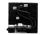
続いて、縫合鎖10の自由端10aは、図3中に示される通り、末端リンク(terminal link)10bをその対向する端部において通される。ループ10bは、縫合鎖10の自由端10aを引くことによって締められる。キャニューレの先端は、回旋筋腱板150に対して安全に縫合鎖ループを位置付けることを支援するよう使用され得る。縫合回収器及び/又は組織捕捉器105は、ループが完全にきつく締められていることを確実にするよう有用であり得る。第2の縫合鎖11は、回旋筋腱板150を介して通され、自由端は、末端ループを通され、ループは第1の複合鎖10と同様にして締められる。
Subsequently, the
図4中に示される通り、縫合鎖10,11は、回旋筋腱板辺縁に対して近接する所望のアンカー位置を確定するよう牽引縫合(traction suture)として使用され、骨ソケット155は、Arthrex 5.5mm Bio−Corkscrew FT Punch等であるパンチ115を有して上外側経皮門脈を通ってパンチされる。第2の骨ソケット155はまた、第2の繊維鎖(fiber chain)11と同様に形成される。
As shown in FIG. 4, the
図5中に示される通り、縫合鎖端部10a,11aは、外側門脈(lateral portal)を通って回収される。本発明のスウィベルアンカー200は、経皮上外側門脈を介してもたらされ、回旋筋腱板150の自由辺縁から第3のリンク10cを捕捉する。リンク10cは、アンカー200のフォーク状アンカー先端250によって捕捉される(図21参照)。望ましい一実施例では、縫合鎖10における各リンクは、長さ約6mmであり、従って、スウィベルアンカー200の全長が18mmであるため、腱板端部から第3のリンク10cを捕捉することは、通常、腱板を直接骨ソケット155の端部において位置付け、アンカー先端250が縫合鎖10を骨ソケット155の下部まで押す際、かかる(spans)回旋筋腱板150及び縫合鎖10に完全に張力を与える。
As shown in FIG. 5, the suture chain ends 10a, 11a are withdrawn through the lateral portal. The
続いて組織間張力は、図6中に示される通り、評価される。ドライバ組立体100は、骨ソケット155へと進まされ、縫合鎖10は、アンカー本体225が骨に接触するまでソケット155の下部に向かって押される。張力が適切ではない場合、ドライバ100は、ドライバ組立体100が引き込まれると同時に、(スウィベル先端250の割り込み(wedging)を解放するよう)縫合鎖10の自由端を引っ張ることによって、骨ソケット155から取り外される。続いて、近接するより近位のリンクは、その代わりに捕捉される。張力が大きすぎて骨ソケット155の下部に対してドライバ100を完全に挿入できない場合は、ドライバ100は取り除かれ、近接するより遠位のリンクが捕捉される。続いて、ドライバ100は、骨ソケットの基部まで再度挿入される。インプラント200のフォーク状先端250は、ドライバの近位端部において滑り止めをつけられるゼロ保持力縫合を有してドライバに対して保持される。
Subsequently, the intertissue tension is evaluated as shown in FIG. The
スクリュ等である固定装置300は、図7中に示される通り、挿入ハンドル22が時計回りに回転される際、親指パッド50を保持することによってドライバ組立体100によって先に進められる。インプラント400が完全に位置付けられる際、アンカー200のシャフト225は、スウィベルアンカー構造400の安定性を最適にするよう固定装置300によって完全に係合される。スウィベルアンカー構造400は、アンカー200及び固定装置300を有する。先端保持縫合は、ドライバハンドル22の後方における滑り止めからほどかれ、ドライバ組立体100は取り除かれる。続いて保持縫合の一肢(one limb)は、インプラントから完全にそれを取り除くよう引っ張られる。
The securing
挿入段階は、図8中に示される通り最終的な縫合/アンカー構造を得るよう第2のスウィベルアンカー構造400に対して反復される。自由縫合端10a,11aは、ArthrexオープンエンドFiberWire Suture Cutter等である縫合カッターを有して切断され、骨ソケット155の端部と重なる(flush with)。
The insertion step is repeated for the second
<無結節複列修復技術 −図9−16>
図9−16は、本発明に従ったスウィベルアンカーを使用する複列修復技術に対する肩修復における一連の段階を示す。
<Nodule double row repair technology-Fig. 9-16>
FIGS. 9-16 illustrate a series of steps in shoulder repair for a double row repair technique using a swivel anchor according to the present invention.
裂傷の可動性は、縫合回収器/組織捕捉器を使用して判断される。大きなU字型裂傷の場合、辺縁集中縫合は、腱の近接の前に求められ得る。シェーバ、高速バー、又は軟骨ピックは、出血骨層を準備するよう使用される。続いて、図9中に示される通り、パイロットホール160は、経皮上外側門脈を通る内側列(medial row)を有する、Arthrex Bio−Corkscrew FT縫合アンカー等である、2つの縫合アンカー120に対して準備される。Arthrex 5.5mm Bio−Corkscrew FT Punch等であるパンチ115は、パイロットホール160を形成するよう上腕の関節縁に近接して、45°の「デッドマン」角度においてレーザ線に対して進められる。タッピングは、頻繁ではないが必要であり得る。
The mobility of the laceration is determined using a suture collector / tissue capturer. For large U-shaped lacerations, marginal sutures can be sought before tendon proximity. A shaver, high speed bar, or cartilage pick is used to prepare the bleeding bone layer. Subsequently, as shown in FIG. 9, the
図10中に示される通り、縫合アンカー120はいずれも置かれる。望ましい一実施例では、縫合アンカー120は、縫合鎖10を事前に有している。
As shown in FIG. 10, any
続いて、図11中に示される通り、縫合始端部(leader)は、側方門脈を介して縫合鎖10の構造ハロゲン化ランタノイドの1つから回収され、Arthrex Scorpion Suture Passer等である縫合パッサー110上へと取り付けられる。縫合鎖10縫合始端部は、回旋筋腱板150の自由辺縁から約15mmを回旋筋腱板150を介して通される。この段階は続いて、第2の縫合鎖11に対して反復される。
Subsequently, as shown in FIG. 11, the suture leader is recovered from one of the structural halogenated lanthanoids of the
図12中に示される通り、縫合鎖縫合端部10a,11aはいずれも、外側門脈を介して回収され、腱板をフットプリントの内側部分と接触させるよう張力をかけられる。キャニューレの先端は、フットプリントに対して腱を押すよう使用され得る。続いて、2つの骨ソケット155は、パンチ115を使用して外側列スウィベルアンカー200に対して形成される。かかる2つの骨ソケット155は、腱板150が2つの前に置かれた縫合鎖10,11によって適切に張力をかけられる際、腱板150の外側辺縁に対して近接されるべきである。
As shown in FIG. 12, both suture suture ends 10a, 11a are retrieved via the outer portal vein and tensioned to bring the rotator cuff into contact with the inner portion of the footprint. The cannula tip can be used to push the tendon against the footprint. Subsequently, two
図13中に示される通り、スウィベルアンカー200は、経皮上外側門脈を介してもたらされ、回旋筋腱板150の自由辺縁から第3のリンク10cを捕捉する。リンク10cは、アンカー200のフォーク状アンカー先端240によって捕捉される(図21参照)。望ましい一実施例では、縫合鎖10,11における各リンクは、長さ約6mmであり、従って、スウィベルアンカー200の全長が18mmであるため、腱板端部から第3のリンクを捕捉することは、通常、骨ソケット155の端部において直接腱板を位置付け、アンカー先端250が縫合鎖10を骨ソケット155の下部まで押す際、かかる回旋筋腱板150及び縫合鎖10に完全に張力を与える。
As shown in FIG. 13, the
続いて組織間張力は、図14中に示される通り、評価される。ドライバ組立体100は、骨ソケット155へと進まされ、縫合鎖10は、アンカー本体225が骨に接触するまでソケット155の下部に向かって押される。張力が適切ではない場合、ドライバ100は、ドライバ組立体100引き込まれると同時に、(スウィベル先端250の割り込み(wedging)を解放するよう)縫合鎖10の自由端を引っ張ることによって、骨ソケット155から取り外され、近接するより近位のリンクは、その代わりに捕捉される。張力が大きすぎて骨ソケット155の下部に対してドライバ100を完全に挿入できない場合は、ドライバ100は取り除かれ、近接するより遠位のリンクが捕捉される。続いて、ドライバ100は、骨ソケット155の基部まで再度挿入される。
Subsequently, the intertissue tension is evaluated as shown in FIG. The
スクリュ等である固定装置300は、図15中に示される通り、挿入ハンドル22が時計回りに回転される際、親指パッド50を保持することによって先に進められる。インプラント400が完全に位置付けられる際、アンカー200のシャフト225は、スウィベルアンカー構造(construct)400の安定性を最適にするよう固定装置300によって完全に係合される。前述された通り、スウィベルアンカー構造400は、アンカー200及び固定装置300を有する。先端保持縫合は、ドライバハンドル22の後方における滑り止めからほどかれ、ドライバ組立体100は取り除かれる。保持縫合の一肢(one limb)は、インプラントから完全にそれを取り除くよう引っ張られる。
As shown in FIG. 15, the fixing
続いて図13−15の段階は、図16中に示される通り、最終構造を得るよう第2のスウィベルアンカー構造400に対して反復される。自由縫合端部は、縫合カッターを有して切断され、骨ソケット155の端部と重なる。
Subsequently, the steps of FIGS. 13-15 are repeated for the second
上述された典型的な実施例に加えて、本発明はまた、縫合鎖10を使用する軟組織の側々閉鎖に対して使用され得る。この方法は、アンカー200等であるアンカーを使用する骨に対する修復の外側面において鎖リンクを固定する最終段階を有して、軟組織の側々閉鎖を実行するよう縫合鎖10を使用することを有する。固定段階は、最初の2つの実施例において示されるものと同一である。更には、修復されるべき軟組織分裂が全長にわたって骨上にある場合、縫合アンカー120と同様である縫合アンカーは、修復の内側面において骨へと挿入され得、側々辺縁集中が「靴紐型」にある縫合鎖を使用して実行され、縫合鎖は、アンカー200等であるアンカーを使用して骨に対して修復の外側面において固定される。
In addition to the exemplary embodiment described above, the present invention can also be used for side-by-side closure of soft tissue using the
図17−22を参照すると、ドライバ組立体100(図17−20)、スウィベル縫合200(図21)、及び固定装置300(図22)の詳細が示される。図17−20中に示される通り、ドライバ組立体100は、シャフト25及び挿入ハンドル22を有するキャニュレーテッドドライバ20; 摺動可能且つ回転可能にキャニュレーテッドドライバ20を介して通る管又はロッド30; キャニュレーテッド本体42及び親指パッド50を有するねじ山付きゲージ40; 及び先端44を有する。図17は、組立体の形状(A)及び非組立体の形状(B)の両方においてドライバ組立体100を示す。図18は、ドライバ組立体100のキャニュレーテッドドライバ20のより詳細な図を与える。図19は、ドライバ組立体100のねじ山付きゲージ40のより詳細な図を与える。図20は、ドライバ組立体100のロッド30のより詳細な図を与える。
Referring to FIGS. 17-22, details of driver assembly 100 (FIGS. 17-20), swivel suture 200 (FIG. 21), and fixation device 300 (FIG. 22) are shown. As shown in FIGS. 17-20, the
図21は、スウィベルアンカー200の多種の図を示す。スウィベルアンカー200の取付け中、アンカー本体225は、ドライバ100の作動端部上へと組み立てられる。アンカー先端250は、薄いロッド又は管30の先端上へと取り付けられるか、あるいはねじ込まれる。上述された通り、フォーク状アンカー先端250は、骨において事前にドリル開けされたホール155への取付けに対して、(上述された2つの典型的な実施例において示される通り、肩の靱帯を介して通されている(laced))、縫合鎖10,11を捕捉するよう使用される。縫合アンカー200は、ねじ山付き本体255及び取外し可能なフォーク状先端250を有する。フォーク状先端250は、アンカー本体255に対して回転可能に取り付けられ得る。これによって、フォーク状先端250によって縫合鎖10,11の過剰な捩れ及び結節を引き起こすことなく、縫合アンカー200の回転可能な挿入を可能にする。
FIG. 21 shows various views of the
図22は、ドライバ組立体100及びスウィベルアンカー200と連動して用いられる(例えばキャニュレーテッド干渉スクリュである)固定装置300の多種の図である。望ましくは、固定装置300は、ドライバ組立体100上に事前に搭載される。2つの典型的な実施例を参照して上述された通り、固定装置300は、挿入ハンドル22が時計回りに開店される際に、親指パッド50を保持することによって骨ソケット155へと進められる。固定装置300が完全に位置付けられる際、フォーク状スウィベルアンカー200のシャフト225は、スウィベルアンカー構造400の安定性を最適化するよう、固定装置300によって完全に係合される。前述された通り、スウィベルアンカー構造400は、アンカー200及び固定装置300を有する。
FIG. 22 shows various views of a fixation device 300 (eg, a cannulated interference screw) used in conjunction with the
上述された2つの典型的な実施例において、縫合アンカー200は、Arthrex Swivel−LockTM(スウィベル縫合アンカー)である。しかしながら、縫合アンカー200はまた、所定の縫合鎖のリンクを捕捉するよう、骨アンカー、又は、フォーク状船体を有するArthrex Push−LockTM型アンカーであり得る。あるいは、組織を通る縫合鎖10,11は、単一アンカー又は複数のアンカーのいずれかを使用して固定され得る。加えて、上述された及び他の多種のアンカーは、上述された工程においてごく僅かな変形を有して交互に使用され得る。例えば、縫合鎖10,11は、アンカーの挿入に先立ちフォーク状の歯において2つの鎖ループを捕捉することによって固定され得る。
In the two exemplary embodiments described above, the
更には、通常の縫合は、本発明の縫合鎖に加えて使用され得る。この場合、複列の実施例の第1の縫合アンカー120は、通常の縫合を事前に有される(例えば米国出願番号2007/0060922中に開示される、Arthrex BioCorkscrewTM、又はBioCorkscrew−FTTM)。通常の縫合を使用する典型的な一実施例では、技術は、外側固定が(鎖リンクではなく)縫合肢(suture limbs)を捕捉すること、及びアンカー200が置かれる際に縫合に張力を与えることによって達成される上述されたものと同様である。これは、アンカー200と骨との間の縫合の干渉固定に依存する。
Furthermore, normal sutures can be used in addition to the sutures of the present invention. In this case, the
上述された通り、本発明のスウィベルアンカー及び縫合鎖組立体は、例えば1つ又はそれより多い骨アンカーと連動して、外科的組織修復における適用性を有する。修復構造上の張力は、骨アンカーによってとらえられる鎖リンクの選択を介して調整可能である。 As described above, the swivel anchor and suture assembly of the present invention has applicability in surgical tissue repair, for example, in conjunction with one or more bone anchors. The tension on the repair structure can be adjusted through the choice of chain links captured by the bone anchor.
本発明は、その実施例に特に関連して記載されてきたが、他の多くの変形及び修正、並びに他の使用は、当業者にとって明らかとなっている。したがって、本発明が本願中の特定の開示によって制限されない、ことは望ましい。 Although the present invention has been described with particular reference to its embodiments, many other variations and modifications and other uses will become apparent to those skilled in the art. Accordingly, it is desirable that the present invention not be limited by the specific disclosure herein.
10 縫合鎖
10a 縫合鎖10の自由端
10b ループ
11 縫合鎖
20 キャニュレーテッドドライバ
22 挿入ハンドル
30 ロッド
42 キャニュレーテッド本体
44 先端
50 親指パッド
105 組織捕捉器
100 ドライバ組立体
110 縫合パッサー
115 パンチ
120 縫合アンカー
150 回旋筋腱板
155 骨ソケット
160 パイロットホール
200 スウィベルアンカー
225 シャフト
250 フォーク状アンカー先端
300 固定装置
400 スウィベルアンカー構造
10 Suture chain
10a Free end of
10b loop
11 Suture chain
20 cannulated driver
22 Insertion handle
30 rods
42 cannulated body
44 Tip
50 thumb pad
105 Tissue trap
100 Driver assembly
110 Suture Passer
115 punch
120 suture anchor
150 rotator cuff
155 bone socket
300 Fixing device
400 swivel anchor structure
Claims (2)
固定装置と、
シャフトを備えるアンカー先端とを含み、
該アンカー先端は、その遠位端部で縫合を捕捉するよう構成され、
前記アンカー先端が前記固定装置に対してスウィベル可能であるよう、並びに、前記固定装置を前記アンカー先端の前記シャフトの上で回転することによって、前記アンカー先端内に捕捉される前記縫合の過剰な捩れ及び結節を引き起こさずに、当該縫合アンカーを骨内に回転可能に固定し得るよう、前記アンカー先端の前記シャフトは、前記固定装置によって回転可能に係合され、
前記アンカー先端は、前記シャフトに回転可能に取り付けられる、
縫合アンカー。 A suture anchor,
A fixing device;
An anchor tip with a shaft,
The anchor tip is configured to capture a suture at its distal end;
Excessive twisting of the suture captured within the anchor tip such that the anchor tip is swivelable relative to the anchoring device and by rotating the anchoring device over the shaft of the anchor tip And the shaft at the tip of the anchor is rotatably engaged by the fixation device so that the suture anchor can be rotatably fixed in the bone without causing nodules ,
The anchor tip is rotatably attached to the shaft ;
Suture anchor.
Applications Claiming Priority (2)
| Application Number | Priority Date | Filing Date | Title |
|---|---|---|---|
| US80109706P | 2006-05-18 | 2006-05-18 | |
| US60/801,097 | 2006-05-18 |
Publications (3)
| Publication Number | Publication Date |
|---|---|
| JP2007307379A JP2007307379A (en) | 2007-11-29 |
| JP2007307379A5 JP2007307379A5 (en) | 2010-07-01 |
| JP5389337B2 true JP5389337B2 (en) | 2014-01-15 |
Family
ID=38473017
Family Applications (1)
| Application Number | Title | Priority Date | Filing Date |
|---|---|---|---|
| JP2007132157A Active JP5389337B2 (en) | 2006-05-18 | 2007-05-17 | Swivel anchor for tissue nodule fixation |
Country Status (5)
| Country | Link |
|---|---|
| US (4) | US9005246B2 (en) |
| EP (1) | EP1857054A3 (en) |
| JP (1) | JP5389337B2 (en) |
| KR (1) | KR101363114B1 (en) |
| AU (1) | AU2007202269B2 (en) |
Families Citing this family (142)
| Publication number | Priority date | Publication date | Assignee | Title |
|---|---|---|---|---|
| US8343186B2 (en) | 2004-04-06 | 2013-01-01 | Arthrex, Inc. | Fully threaded suture anchor with transverse anchor pin |
| US9521999B2 (en) | 2005-09-13 | 2016-12-20 | Arthrex, Inc. | Fully-threaded bioabsorbable suture anchor |
| US8821541B2 (en) | 1999-02-02 | 2014-09-02 | Arthrex, Inc. | Suture anchor with insert-molded rigid member |
| US8177841B2 (en) | 2000-05-01 | 2012-05-15 | Arthrosurface Inc. | System and method for joint resurface repair |
| US7896883B2 (en) | 2000-05-01 | 2011-03-01 | Arthrosurface, Inc. | Bone resurfacing system and method |
| US7163541B2 (en) | 2002-12-03 | 2007-01-16 | Arthrosurface Incorporated | Tibial resurfacing system |
| US7678151B2 (en) * | 2000-05-01 | 2010-03-16 | Ek Steven W | System and method for joint resurface repair |
| US7713305B2 (en) * | 2000-05-01 | 2010-05-11 | Arthrosurface, Inc. | Articular surface implant |
| US7896885B2 (en) | 2002-12-03 | 2011-03-01 | Arthrosurface Inc. | Retrograde delivery of resurfacing devices |
| US7618462B2 (en) | 2000-05-01 | 2009-11-17 | Arthrosurface Incorporated | System and method for joint resurface repair |
| US6610067B2 (en) | 2000-05-01 | 2003-08-26 | Arthrosurface, Incorporated | System and method for joint resurface repair |
| US6520964B2 (en) | 2000-05-01 | 2003-02-18 | Std Manufacturing, Inc. | System and method for joint resurface repair |
| US7993369B2 (en) | 2000-06-22 | 2011-08-09 | Arthrex, Inc. | Graft fixation using a plug against suture |
| US7901408B2 (en) | 2002-12-03 | 2011-03-08 | Arthrosurface, Inc. | System and method for retrograde procedure |
| US8388624B2 (en) | 2003-02-24 | 2013-03-05 | Arthrosurface Incorporated | Trochlear resurfacing system and method |
| US7951163B2 (en) * | 2003-11-20 | 2011-05-31 | Arthrosurface, Inc. | Retrograde excision system and apparatus |
| EP1845890A4 (en) * | 2003-11-20 | 2010-06-09 | Arthrosurface Inc | System and method for retrograde procedure |
| WO2006004885A2 (en) | 2004-06-28 | 2006-01-12 | Arthrosurface, Inc. | System for articular surface replacement |
| US7828853B2 (en) | 2004-11-22 | 2010-11-09 | Arthrosurface, Inc. | Articular surface implant and delivery system |
| US20090192546A1 (en) | 2005-03-30 | 2009-07-30 | Reinhold Schmieding | Fenestrated suture anchor and method for knotless fixation of tissue |
| US11844512B2 (en) | 2005-03-30 | 2023-12-19 | Arthrex, Inc. | Suture anchor for knotless fixation of tissue |
| US20090187216A1 (en) | 2006-05-18 | 2009-07-23 | Arthrex, Inc. | Fenestrated swivel anchor for knotless fixation of tissue |
| US8465522B2 (en) * | 2005-03-30 | 2013-06-18 | Arthrex, Inc. | Self-reinforcing tissue fixation |
| WO2007016540A2 (en) * | 2005-07-29 | 2007-02-08 | Arthrosurface, Inc. | System and method for articular surface repair |
| US20090318960A1 (en) * | 2006-02-01 | 2009-12-24 | Burkhart Stephen S | Method of knotless tissue fixation with criss-cross suture pattern |
| JP5389337B2 (en) | 2006-05-18 | 2014-01-15 | オーソレクス,インコーポレイテッド | Swivel anchor for tissue nodule fixation |
| US8167906B2 (en) * | 2006-11-01 | 2012-05-01 | Depuy Mitek, Inc. | Suture anchor with pulley |
| AU2007332787A1 (en) | 2006-12-11 | 2008-06-19 | Arthrosurface Incorporated | Retrograde resection apparatus and method |
| CA2677726A1 (en) * | 2007-02-14 | 2008-08-21 | Arthrosurface Inc. | Bone cement delivery device |
| US8702754B2 (en) | 2007-09-14 | 2014-04-22 | Depuy Mitek, Llc | Methods for anchoring suture to bone |
| US8882801B2 (en) * | 2007-09-14 | 2014-11-11 | Depuy Mitek, Llc | Dual thread cannulated suture anchor |
| EP2214565A4 (en) * | 2007-10-27 | 2015-08-26 | Parcus Medical Llc | Suture anchor |
| FR2925287B1 (en) * | 2007-12-21 | 2010-12-17 | B & G | DEVICE FOR ANCHORING A FABRIC INTO A BONE |
| US9295460B2 (en) * | 2007-12-31 | 2016-03-29 | Cayenne Medical, Inc. | Anchors and method for securing suture to bone |
| EP2299915B1 (en) | 2008-06-16 | 2018-07-18 | Cayenne Medical, Inc. | Anchor for securing suture to bone |
| US8202297B2 (en) * | 2008-06-19 | 2012-06-19 | Arthrex, Inc. | Technique for tissue fixation by reeling in and anchoring suture attached to tissue |
| US8202296B2 (en) * | 2008-06-19 | 2012-06-19 | Arthrex, Inc. | Technique for tissue fixation by capturing and anchoring a link of suture chain attached to tissue |
| US8790368B2 (en) * | 2008-09-18 | 2014-07-29 | Smith & Nephew, Inc. | Tenodesis implant |
| JP5904794B2 (en) * | 2008-12-09 | 2016-04-20 | スミス アンド ネフュー インコーポレーテッドSmith & Nephew,Inc. | Tissue repair assembly |
| US8834521B2 (en) | 2008-12-23 | 2014-09-16 | Arthrex, Inc. | Suturing construct with spliced tails |
| US20100262184A1 (en) * | 2009-04-08 | 2010-10-14 | Dreyfuss Peter J | Bio-active construct created between fixation device and suture fixed in bone |
| EP2238914B1 (en) * | 2009-04-10 | 2015-05-20 | Arthrex, Inc. | Twist-in suture anchor |
| EP2429429B1 (en) | 2009-04-17 | 2018-07-25 | Arthrosurface Incorporated | Glenoid resurfacing system |
| WO2010121250A1 (en) | 2009-04-17 | 2010-10-21 | Arthrosurface Incorporated | Glenoid resurfacing system and method |
| US10945743B2 (en) | 2009-04-17 | 2021-03-16 | Arthrosurface Incorporated | Glenoid repair system and methods of use thereof |
| CA2713309C (en) | 2009-08-20 | 2013-07-02 | Howmedica Osteonics Corp. | Flexible acl instrumentation, kit and method |
| US8613756B2 (en) | 2009-10-30 | 2013-12-24 | Depuy Mitek, Llc | Knotless suture anchor |
| CA2792048A1 (en) | 2010-03-05 | 2011-09-09 | Arthrosurface Incorporated | Tibial resurfacing system and method |
| US8679159B2 (en) | 2010-08-30 | 2014-03-25 | Depuy Mitek, Llc | Anchor driver with suture clutch |
| US8435264B2 (en) | 2010-08-30 | 2013-05-07 | Depuy Mitek, Llc | Knotless suture anchor and driver |
| US8460340B2 (en) | 2010-08-30 | 2013-06-11 | Depuy Mitek, Llc | Knotless suture anchor |
| US8469998B2 (en) | 2010-08-30 | 2013-06-25 | Depuy Mitek, Llc | Knotless suture anchor |
| US8591545B2 (en) | 2011-03-25 | 2013-11-26 | Smith & Nephew, Inc. | Flat suture anchor |
| US9066716B2 (en) | 2011-03-30 | 2015-06-30 | Arthrosurface Incorporated | Suture coil and suture sheath for tissue repair |
| WO2012177386A1 (en) | 2011-06-23 | 2012-12-27 | Synthes Usa, Llc | Suture anchor system and method |
| US9301745B2 (en) | 2011-07-21 | 2016-04-05 | Arthrex, Inc. | Knotless suture constructs |
| US9107653B2 (en) | 2011-09-22 | 2015-08-18 | Arthrex, Inc. | Tensionable knotless anchors with splice and methods of tissue repair |
| US20130123809A1 (en) | 2011-11-11 | 2013-05-16 | VentureMD Innovations, LLC | Transosseous attachment instruments |
| US10548585B2 (en) | 2011-11-16 | 2020-02-04 | VentureMD Innovations, LLC | Soft tissue attachment |
| US10675014B2 (en) | 2011-11-16 | 2020-06-09 | Crossroads Extremity Systems, Llc | Knotless soft tissue attachment |
| US10470756B2 (en) | 2011-11-16 | 2019-11-12 | VentureMD Innovations, LLC | Suture anchor and method |
| US9131937B2 (en) | 2011-11-16 | 2015-09-15 | VentureMD Innovations, LLC | Suture anchor |
| US9445803B2 (en) | 2011-11-23 | 2016-09-20 | Howmedica Osteonics Corp. | Filamentary suture anchor |
| EP2601894B1 (en) | 2011-12-09 | 2018-08-29 | Arthrex, Inc. | Tensionable knotless anchor systems |
| US20130165982A1 (en) | 2011-12-22 | 2013-06-27 | Arthrosurface Incorporated | System and Method for Bone Fixation |
| US9808242B2 (en) * | 2012-04-06 | 2017-11-07 | Howmedica Osteonics Corp. | Knotless filament anchor for soft tissue repair |
| US9345471B2 (en) | 2012-06-22 | 2016-05-24 | Arthrex, Inc. | Tensionable knotless labral anchor and methods of tissue repair |
| WO2014008126A1 (en) | 2012-07-03 | 2014-01-09 | Arthrosurface Incorporated | System and method for joint resurfacing and repair |
| US8821494B2 (en) | 2012-08-03 | 2014-09-02 | Howmedica Osteonics Corp. | Surgical instruments and methods of use |
| US10258320B2 (en) | 2012-10-26 | 2019-04-16 | Arthrex, Inc. | Systems for locking a cinch loop in tissue repair |
| US9198650B2 (en) | 2012-12-27 | 2015-12-01 | Medos International Sarl | Multi-piece anchor inserter |
| US9687221B2 (en) | 2013-02-13 | 2017-06-27 | Venture MD Innovations, LLC | Method of anchoring a suture |
| EP2767241A1 (en) | 2013-02-19 | 2014-08-20 | Arthrex, Inc. | Knotless swivel anchor with anchor body advanced over anchor tip |
| EP2769685B1 (en) * | 2013-02-25 | 2018-06-06 | Arthrex, Inc. | Tissue protector suture constructs |
| US9402620B2 (en) | 2013-03-04 | 2016-08-02 | Howmedica Osteonics Corp. | Knotless filamentary fixation devices, assemblies and systems and methods of assembly and use |
| EP2774546B1 (en) | 2013-03-05 | 2017-08-02 | Arthrex, Inc. | Knotless tape/suture construct for fixation of tissue to bone |
| US9687222B2 (en) | 2013-03-05 | 2017-06-27 | Arthrex, Inc. | Knotless repair technique using tape/suture hybrid |
| US9788826B2 (en) | 2013-03-11 | 2017-10-17 | Howmedica Osteonics Corp. | Filamentary fixation device and assembly and method of assembly, manufacture and use |
| US9463013B2 (en) | 2013-03-13 | 2016-10-11 | Stryker Corporation | Adjustable continuous filament structure and method of manufacture and use |
| CA2906888A1 (en) | 2013-03-15 | 2014-11-27 | Mark Brunsvold | Suture anchor |
| US9492200B2 (en) | 2013-04-16 | 2016-11-15 | Arthrosurface Incorporated | Suture system and method |
| WO2014176270A1 (en) | 2013-04-22 | 2014-10-30 | Pivot Medical, Inc. | Method and apparatus for attaching tissue to bone |
| US9655714B1 (en) * | 2013-05-17 | 2017-05-23 | W. David Hovis | Arthroscopic tenodesis tool |
| US9277951B1 (en) * | 2013-05-17 | 2016-03-08 | W. David Hovis | Arthroscopic tenodesis tool |
| US10363126B1 (en) * | 2013-05-17 | 2019-07-30 | W. David Hovis | Arthroscopic tenodesis tool |
| US10610211B2 (en) | 2013-12-12 | 2020-04-07 | Howmedica Osteonics Corp. | Filament engagement system and methods of use |
| US9801621B2 (en) | 2014-01-23 | 2017-10-31 | Arthrex, Inc. | Arthroscopic biceps tenodesis in continuity |
| US9693765B2 (en) | 2014-01-27 | 2017-07-04 | Arthrex, Inc. | Surgical assembly for rotator cuff repair |
| WO2015117022A1 (en) * | 2014-01-31 | 2015-08-06 | Ticker Jonathan | Suture anchor |
| US10624748B2 (en) | 2014-03-07 | 2020-04-21 | Arthrosurface Incorporated | System and method for repairing articular surfaces |
| US11607319B2 (en) | 2014-03-07 | 2023-03-21 | Arthrosurface Incorporated | System and method for repairing articular surfaces |
| US20150250472A1 (en) | 2014-03-07 | 2015-09-10 | Arthrosurface Incorporated | Delivery System for Articular Surface Implant |
| US9901335B2 (en) | 2014-07-10 | 2018-02-27 | Maxwell Choongwon Park | Method and apparatus for securing soft tissue to bone |
| US9986992B2 (en) | 2014-10-28 | 2018-06-05 | Stryker Corporation | Suture anchor and associated methods of use |
| US20160270903A1 (en) * | 2015-03-18 | 2016-09-22 | Arthrex, Inc. | System and method for spine ligament reconstruction |
| US10154868B2 (en) | 2015-07-17 | 2018-12-18 | Kator, Llc | Transosseous method |
| US10820918B2 (en) | 2015-07-17 | 2020-11-03 | Crossroads Extremity Systems, Llc | Transosseous guide and method |
| US9962174B2 (en) | 2015-07-17 | 2018-05-08 | Kator, Llc | Transosseous method |
| US9814455B2 (en) | 2015-07-23 | 2017-11-14 | Arthrex, Inc. | Measuring tool using suture and suture anchor |
| US10226243B2 (en) | 2015-08-04 | 2019-03-12 | Kator, Llc | Transosseous suture anchor |
| US12383253B2 (en) | 2015-08-04 | 2025-08-12 | Crossroads Extremity Systems, Llc | Suture anchor |
| US10064724B2 (en) | 2015-08-11 | 2018-09-04 | Richard J Hansen | External bone anchor system for joint replacement implants |
| US10368855B2 (en) | 2015-10-20 | 2019-08-06 | Arthrex, Inc. | Surgical constructs for tissue repair |
| EP4631440A3 (en) | 2015-12-16 | 2025-12-17 | ConMed Corporation | Knotless suture anchor and deployment device |
| US10398426B2 (en) | 2016-01-06 | 2019-09-03 | Arthrex, Inc. | Point-loading knotless fixation devices |
| US10631845B2 (en) | 2016-01-22 | 2020-04-28 | Arthrex, Inc. | Tensionable knotless anchors and methods of tissue repair |
| US10172606B2 (en) | 2016-01-22 | 2019-01-08 | Arthrex, Inc. | Tensionable knotless anchors and methods of tissue repair |
| US11642120B2 (en) | 2016-01-22 | 2023-05-09 | Arthrex, Inc. | Tensionable knotless anchors and methods of tissue repair |
| WO2017201216A1 (en) * | 2016-05-18 | 2017-11-23 | Orthonoble Inc. | Disposable instrument nosepieces for repairing soft tissue to bone coupling |
| WO2017210620A1 (en) | 2016-06-02 | 2017-12-07 | Bracy Barton | Suture tool and method of use |
| US10722343B2 (en) | 2017-02-22 | 2020-07-28 | Stryker Corporation | Fixation member with separate eyelet and methods of use thereof |
| US10893933B2 (en) | 2017-02-24 | 2021-01-19 | James Jastifer | Tissue anchors, kits, and associated methods |
| US10463357B2 (en) | 2017-03-13 | 2019-11-05 | Medos International Sarl | Methods and devices for knotless suture anchoring |
| US10383618B2 (en) | 2017-03-13 | 2019-08-20 | Medos International Sarl | Methods and devices for knotless suture anchoring |
| US10786234B2 (en) * | 2017-03-27 | 2020-09-29 | Arthrex, Inc. | Suture anchor assembly with universal inserter device |
| US10639026B2 (en) | 2017-07-27 | 2020-05-05 | Medos International Sarl | Knotless suture anchoring using two awl shafts |
| WO2019028344A1 (en) | 2017-08-04 | 2019-02-07 | Arthrosurface Incorporated | Multicomponent articular surface implant |
| US10820915B2 (en) | 2018-03-06 | 2020-11-03 | Medos International Sarl | Methods, systems, and devices for instability repair |
| US11272924B2 (en) | 2018-07-18 | 2022-03-15 | Arthrex, Inc. | Knotless closure sutures and methods of tissue fixation |
| US10512483B1 (en) * | 2018-11-13 | 2019-12-24 | T & J Enterprises, Llc | Cervical tenaculum device |
| US11497526B2 (en) * | 2018-11-13 | 2022-11-15 | T & J Enterprises, Llc | Cervical tenaculum device |
| USD921479S1 (en) | 2018-11-27 | 2021-06-08 | International Life Sciences LLC | Eyelet interference screw |
| WO2020117763A1 (en) | 2018-12-03 | 2020-06-11 | International Life Sciences Llc D/B/A Artelon | Eyelet interference screw and methods of use |
| GB2609338B (en) | 2019-03-12 | 2023-06-14 | Arthrosurface Inc | Humeral and glenoid articular surface implant systems and methods |
| US11737744B2 (en) | 2019-07-25 | 2023-08-29 | Arthrex, Inc. | Slack reducing suture anchor assembly and method of tissue repair |
| WO2021076245A1 (en) | 2019-10-18 | 2021-04-22 | Sparta Biopharma LLC | Connective tissue to bone interface scaffolds |
| WO2022046701A1 (en) | 2020-08-24 | 2022-03-03 | Sparta Biopharma LLC | Methods of forming bone interface scaffolds |
| US12357448B2 (en) | 2020-12-07 | 2025-07-15 | Arthrex, Inc. | Tensionable knotless anchors and methods of tissue repair |
| WO2022154535A1 (en) * | 2021-01-13 | 2022-07-21 | (주)오스테오닉 | Tissue-suturing device |
| KR20220102537A (en) | 2021-01-13 | 2022-07-20 | (주)오스테오닉 | Suture equipment for rotator cuff |
| FR3120306B1 (en) | 2021-03-02 | 2025-06-20 | Rs4 | Suture anchor for anchoring soft tissue to bone |
| US20220338857A1 (en) * | 2021-04-27 | 2022-10-27 | Medos International Sarl | Knotless suture anchor system with suture management structure |
| US12440202B2 (en) | 2022-02-24 | 2025-10-14 | Biomet Manufacturing, Llc | Knotless bone anchor system and method |
| WO2025019184A1 (en) | 2023-07-20 | 2025-01-23 | Arthrex, Inc. | Surgical constructs for tissue fixation and methods of tissue repairs |
| US20250025187A1 (en) | 2023-07-21 | 2025-01-23 | Arthrex, Inc. | Center of Rotation Guide |
| US20250049429A1 (en) | 2023-08-07 | 2025-02-13 | Arthrex, Inc. | Variable Density Soft Anchors and Methods of Tissue Repairs |
| US20250312028A1 (en) | 2024-04-08 | 2025-10-09 | Arthrex, Inc. | Inserter with integrated cutter and method of use |
| US20250331885A1 (en) | 2024-04-25 | 2025-10-30 | Arthrex, Inc. | Punch Obturator |
| US20250375284A1 (en) | 2024-06-11 | 2025-12-11 | Arthrex, Inc. | Single Step Tissue Augmentation |
| WO2026010898A1 (en) | 2024-07-05 | 2026-01-08 | Arthrex, Inc. | Soft anchors and methods of tissue repairs |
| US20260060667A1 (en) | 2024-08-28 | 2026-03-05 | Arthrex, Inc. | Soft Anchor Constructs and Methods of Tissue Repairs |
| US12544208B1 (en) | 2025-06-09 | 2026-02-10 | Theramicro Inc. | Implant for reinforcement of soft tissues |
Family Cites Families (89)
| Publication number | Priority date | Publication date | Assignee | Title |
|---|---|---|---|---|
| CH648197A5 (en) | 1980-05-28 | 1985-03-15 | Synthes Ag | IMPLANT AND SCREW FASTENING ON ITS BONE. |
| DE3445738A1 (en) | 1984-12-14 | 1986-06-19 | Draenert Klaus | IMPLANT FOR BONE REINFORCEMENT AND ANCHORING OF BONE SCREWS, IMPLANTS OR IMPLANT PARTS |
| CH672058A5 (en) | 1986-08-05 | 1989-10-31 | Synthes Ag | |
| US4870957A (en) * | 1988-12-27 | 1989-10-03 | Marlowe Goble E | Ligament anchor system |
| US5152790A (en) * | 1991-03-21 | 1992-10-06 | American Cyanamid Company | Ligament reconstruction graft anchor apparatus |
| US5249899A (en) | 1992-10-28 | 1993-10-05 | Wilson Robert L | Head bolt and driver therefore |
| US5423860A (en) * | 1993-05-28 | 1995-06-13 | American Cyanamid Company | Protective carrier for suture anchor |
| US5500000A (en) * | 1993-07-01 | 1996-03-19 | United States Surgical Corporation | Soft tissue repair system and method |
| US5540718A (en) | 1993-09-20 | 1996-07-30 | Bartlett; Edwin C. | Apparatus and method for anchoring sutures |
| US5545180A (en) * | 1993-12-13 | 1996-08-13 | Ethicon, Inc. | Umbrella-shaped suture anchor device with actuating ring member |
| US5458601A (en) * | 1994-03-28 | 1995-10-17 | Medical University Of South Carolina | Adjustable ligament anchor |
| US5411523A (en) * | 1994-04-11 | 1995-05-02 | Mitek Surgical Products, Inc. | Suture anchor and driver combination |
| US5649963A (en) * | 1994-11-10 | 1997-07-22 | Innovasive Devices, Inc. | Suture anchor assembly and methods |
| US5860973A (en) | 1995-02-27 | 1999-01-19 | Michelson; Gary Karlin | Translateral spinal implant |
| US6086608A (en) * | 1996-02-22 | 2000-07-11 | Smith & Nephew, Inc. | Suture collet |
| US5957953A (en) * | 1996-02-16 | 1999-09-28 | Smith & Nephew, Inc. | Expandable suture anchor |
| US5868749A (en) | 1996-04-05 | 1999-02-09 | Reed; Thomas M. | Fixation devices |
| US7611521B2 (en) | 1996-09-13 | 2009-11-03 | Tendon Technology, Ltd. | Apparatus and methods for tendon or ligament repair |
| US5766250A (en) * | 1996-10-28 | 1998-06-16 | Medicinelodge, Inc. | Ligament fixator for a ligament anchor system |
| US6533816B2 (en) * | 1999-02-09 | 2003-03-18 | Joseph H. Sklar | Graft ligament anchor and method for attaching a graft ligament to a bone |
| US7083647B1 (en) | 1996-11-27 | 2006-08-01 | Sklar Joseph H | Fixation screw, graft ligament anchor assembly, and method for securing a graft ligament in a bone tunnel |
| US6083522A (en) * | 1997-01-09 | 2000-07-04 | Neucoll, Inc. | Devices for tissue repair and methods for preparation and use thereof |
| US5707395A (en) * | 1997-01-16 | 1998-01-13 | Li Medical Technologies, Inc. | Surgical fastener and method and apparatus for ligament repair |
| US5702398A (en) * | 1997-02-21 | 1997-12-30 | Tarabishy; Sam | Tension screw |
| US6027523A (en) * | 1997-10-06 | 2000-02-22 | Arthrex, Inc. | Suture anchor with attached disk |
| US6368326B1 (en) * | 1998-09-28 | 2002-04-09 | Daos Limited | Internal cord fixation device |
| US9521999B2 (en) | 2005-09-13 | 2016-12-20 | Arthrex, Inc. | Fully-threaded bioabsorbable suture anchor |
| US6355043B1 (en) | 1999-03-01 | 2002-03-12 | Sulzer Orthopedics Ltd. | Bone screw for anchoring a marrow nail |
| US6143017A (en) | 1999-03-17 | 2000-11-07 | Thal; Raymond | Free loop knotless suture anchor assembly |
| US6045574A (en) * | 1999-04-01 | 2000-04-04 | Thal; Raymond | Sleeve and loop knotless suture anchor assembly |
| US6267766B1 (en) * | 1999-05-28 | 2001-07-31 | Stephen S. Burkhart | Suture anchor reel device kit and method |
| US6214007B1 (en) * | 1999-06-01 | 2001-04-10 | David G. Anderson | Surgical fastener for fixation of a soft tissue graft to a bone tunnel |
| US6048343A (en) | 1999-06-02 | 2000-04-11 | Mathis; John M. | Bone screw system |
| US6464706B1 (en) * | 1999-06-10 | 2002-10-15 | Thomas F. Winters | Tissue fixation device and method |
| JP2001025478A (en) * | 1999-07-14 | 2001-01-30 | Ryosuke Sasaki | Tool and method for reconstructing anterior crucial ligament of knee |
| US6517542B1 (en) * | 1999-08-04 | 2003-02-11 | The Cleveland Clinic Foundation | Bone anchoring system |
| US6527794B1 (en) * | 1999-08-10 | 2003-03-04 | Ethicon, Inc. | Self-locking suture anchor |
| DE19949285C2 (en) | 1999-10-12 | 2002-08-14 | Impag Gmbh Medizintechnik | bone screw |
| DE59901812D1 (en) | 1999-10-21 | 2002-07-25 | Storz Karl Gmbh & Co Kg | interference screw |
| EP1093773B1 (en) | 1999-10-21 | 2001-10-04 | Karl Storz GmbH & Co. KG | Biodegradable fixation device |
| AUPQ366099A0 (en) | 1999-10-26 | 1999-11-18 | Queensland University Of Technology | Ortho paedic screw |
| DE59901090D1 (en) | 1999-12-23 | 2002-05-02 | Storz Karl Gmbh & Co Kg | Decentralized drive screw |
| AU2001269942B2 (en) * | 2000-06-22 | 2005-05-26 | Arthrex, Inc. | Graft fixation using a screw or plug against suture or tissue |
| US7993369B2 (en) | 2000-06-22 | 2011-08-09 | Arthrex, Inc. | Graft fixation using a plug against suture |
| US7329272B2 (en) * | 2000-06-22 | 2008-02-12 | Arthrex, Inc. | Graft fixation using a plug against suture |
| WO2002021997A2 (en) * | 2000-09-12 | 2002-03-21 | Axya Medical, Inc. | Apparatus and method for securing suture to bone |
| US6923824B2 (en) * | 2000-09-12 | 2005-08-02 | Axya Medical, Inc. | Apparatus and method for securing suture to bone |
| ES2325612T3 (en) * | 2000-12-22 | 2009-09-10 | Tyco Healthcare Group Lp | SCREW SCREW. |
| US7144413B2 (en) * | 2001-04-20 | 2006-12-05 | Synthes (U.S.A.) | Graft fixation system and method |
| US7491217B1 (en) * | 2001-10-16 | 2009-02-17 | Hendren Ronald D | Graft anchoring device |
| US20030153947A1 (en) * | 2002-02-14 | 2003-08-14 | Tomoaki Koseki | Sternum suture material and its manufacturing method |
| US6939135B2 (en) | 2002-06-03 | 2005-09-06 | Schubert L. Sapian | Growth factor releasing biofunctional dental implant |
| US20040093030A1 (en) * | 2002-10-28 | 2004-05-13 | Cox James E. | Bone anchor and assembly |
| US7090690B2 (en) * | 2002-11-19 | 2006-08-15 | Arthrocare Corporation | Devices and methods for repairing soft tissue |
| US7572298B2 (en) * | 2003-03-28 | 2009-08-11 | Ethicon, Inc. | Implantable medical devices and methods for making same |
| US7354442B2 (en) | 2003-05-05 | 2008-04-08 | Warsaw Orthopedic, Inc. | Bone anchor and methods of using the same |
| US7300439B2 (en) | 2003-06-24 | 2007-11-27 | Depuy Mitek, Inc. | Porous resorbable graft fixation pin |
| US20050245932A1 (en) * | 2004-04-16 | 2005-11-03 | Fanton Gary S | Apparatus and methods for securing tissue to bone |
| US7645293B2 (en) * | 2004-04-21 | 2010-01-12 | United States Surgical Corporation | Suture anchor installation system and method |
| CA2568562A1 (en) | 2004-06-02 | 2006-06-08 | Michael L. Green | System and method for attaching soft tissue to bone |
| US8702752B2 (en) * | 2004-06-18 | 2014-04-22 | Arthrex, Inc. | Knotless anchor for surgical repair |
| US8114127B2 (en) * | 2004-06-22 | 2012-02-14 | Hs West Investments, Llc | Bone anchors for use in attaching soft tissue to bone |
| US20060079904A1 (en) * | 2004-10-13 | 2006-04-13 | Raymond Thal | Multirow knotless suture anchor assembly |
| US8034090B2 (en) | 2004-11-09 | 2011-10-11 | Biomet Sports Medicine, Llc | Tissue fixation device |
| US7976565B1 (en) * | 2004-12-07 | 2011-07-12 | Biomet Sports Medicine, Llc | Expanding suture anchor having an actuator pin |
| US8986345B2 (en) * | 2004-12-07 | 2015-03-24 | Biomet Sports Medicine, Llc | Expanding suture anchor having an actuator pin |
| NL1028292C2 (en) * | 2005-02-16 | 2006-08-17 | Kokbing Lo | Securing element for connecting a ligament to bone part, comprises ligament coupling element, clamping element having two parts with corresponding surfaces, stressing element and actuating element |
| EP1855616A4 (en) | 2005-03-10 | 2013-07-03 | Covidien Lp | Suture anchors |
| EP1707127B1 (en) | 2005-03-30 | 2010-12-22 | Arthrex Inc | Looped high strength suture chain for knotless fixation |
| US20090192546A1 (en) | 2005-03-30 | 2009-07-30 | Reinhold Schmieding | Fenestrated suture anchor and method for knotless fixation of tissue |
| US7981140B2 (en) * | 2005-03-30 | 2011-07-19 | Arthrex, Inc. | Knotless fixation of tissue to bone with suture chain |
| US20080208253A1 (en) | 2006-05-18 | 2008-08-28 | Dreyfuss Peter J | Self-punching swivel anchor and method for knotless fixation of tissue |
| US8012174B2 (en) | 2006-02-01 | 2011-09-06 | Arthrex, Inc. | Method for double row fixation of tendon to bone |
| US20080009904A1 (en) * | 2006-03-17 | 2008-01-10 | Bourque Barnard J | Soft Tissue Fixation |
| US7828820B2 (en) * | 2006-03-21 | 2010-11-09 | Biomet Sports Medicine, Llc | Method and apparatuses for securing suture |
| JP5389337B2 (en) | 2006-05-18 | 2014-01-15 | オーソレクス,インコーポレイテッド | Swivel anchor for tissue nodule fixation |
| US20080154314A1 (en) | 2006-08-16 | 2008-06-26 | Mcdevitt Dennis M | Composite interference screw for attaching a graft ligament to a bone, and other apparatus for making attachments to bone |
| DE102006045007A1 (en) | 2006-08-17 | 2008-02-21 | Daimler Ag | Tandem axle with two drivable axles and a partially disconnectable drive train |
| US8845685B2 (en) | 2007-05-03 | 2014-09-30 | Biomet Sports Medicine, Llc | Anchor assembly and method of use |
| US7794484B2 (en) | 2007-05-07 | 2010-09-14 | Biomet Sports Medicine, Llc | Fixation device for delivery of biological material between soft tissue and bone |
| FR2925287B1 (en) | 2007-12-21 | 2010-12-17 | B & G | DEVICE FOR ANCHORING A FABRIC INTO A BONE |
| US20090281581A1 (en) | 2008-05-06 | 2009-11-12 | Berg Jeffery H | Method and device for securing sutures to bones |
| WO2009140034A1 (en) | 2008-05-14 | 2009-11-19 | Smith & Nephew, Inc. | Arthroscopic biceps tenodesis repair |
| US20100249854A1 (en) | 2009-03-31 | 2010-09-30 | Thomas Jonathan D | Multizone Implants |
| WO2010121271A1 (en) | 2009-04-17 | 2010-10-21 | Shane Barwood | Tenodesis system |
| US20130035721A1 (en) | 2011-08-04 | 2013-02-07 | John Eric Brunelle | Surgical anchor |
| US9138220B2 (en) | 2011-12-19 | 2015-09-22 | Medos International Sarl | Knotless suture anchor |
| US9345471B2 (en) | 2012-06-22 | 2016-05-24 | Arthrex, Inc. | Tensionable knotless labral anchor and methods of tissue repair |
| US9936940B2 (en) | 2013-06-07 | 2018-04-10 | Biomet Sports Medicine, Llc | Method and apparatus for coupling soft tissue to bone |
-
2007
- 2007-05-17 JP JP2007132157A patent/JP5389337B2/en active Active
- 2007-05-17 AU AU2007202269A patent/AU2007202269B2/en active Active
- 2007-05-18 US US11/802,057 patent/US9005246B2/en active Active
- 2007-05-18 EP EP07252043A patent/EP1857054A3/en not_active Withdrawn
- 2007-05-18 KR KR1020070048819A patent/KR101363114B1/en active Active
-
2015
- 2015-02-05 US US14/615,044 patent/US20150157312A1/en not_active Abandoned
-
2016
- 2016-11-18 US US15/355,600 patent/US10537318B2/en active Active
-
2020
- 2020-01-17 US US16/745,667 patent/US11523814B2/en active Active
Also Published As
| Publication number | Publication date |
|---|---|
| EP1857054A2 (en) | 2007-11-21 |
| AU2007202269B2 (en) | 2013-01-24 |
| US11523814B2 (en) | 2022-12-13 |
| US20200146673A1 (en) | 2020-05-14 |
| US9005246B2 (en) | 2015-04-14 |
| US10537318B2 (en) | 2020-01-21 |
| EP1857054A3 (en) | 2009-08-05 |
| KR20070112063A (en) | 2007-11-22 |
| US20150157312A1 (en) | 2015-06-11 |
| US20170135689A1 (en) | 2017-05-18 |
| JP2007307379A (en) | 2007-11-29 |
| AU2007202269A1 (en) | 2007-12-06 |
| US20080004659A1 (en) | 2008-01-03 |
| KR101363114B1 (en) | 2014-02-13 |
Similar Documents
| Publication | Publication Date | Title |
|---|---|---|
| JP5389337B2 (en) | Swivel anchor for tissue nodule fixation | |
| US8202297B2 (en) | Technique for tissue fixation by reeling in and anchoring suture attached to tissue | |
| US8202296B2 (en) | Technique for tissue fixation by capturing and anchoring a link of suture chain attached to tissue | |
| EP1834592B1 (en) | Apparatus for arthroscopic surgery using suture anchors | |
| JP6319778B2 (en) | Medical device and medical kit for attaching sutures to bone | |
| JP5576861B2 (en) | Anchor and method for securing a suture to bone | |
| US20090318960A1 (en) | Method of knotless tissue fixation with criss-cross suture pattern | |
| US10045770B2 (en) | Method for knotless fixation of tissue with swivel anchor | |
| AU2012261743B2 (en) | Methods and systems for attaching tissue to bone | |
| US20080208253A1 (en) | Self-punching swivel anchor and method for knotless fixation of tissue | |
| US8348975B2 (en) | Method for creating knotless double row construct with medial row closure | |
| US20080009900A1 (en) | Surgical grasping device | |
| US20100004683A1 (en) | Anchors and method for securing suture to bone | |
| US20090204127A1 (en) | System and method for tying surgical knots | |
| AU2015224390A1 (en) | Method and apparatus for arthroscopic surgery using suture anchors | |
| AU2013267056A1 (en) | Method and apparatus for arthroscopic surgery using suture anchors |
Legal Events
| Date | Code | Title | Description |
|---|---|---|---|
| A521 | Request for written amendment filed |
Free format text: JAPANESE INTERMEDIATE CODE: A523 Effective date: 20100513 |
|
| A621 | Written request for application examination |
Free format text: JAPANESE INTERMEDIATE CODE: A621 Effective date: 20100513 |
|
| A977 | Report on retrieval |
Free format text: JAPANESE INTERMEDIATE CODE: A971007 Effective date: 20120328 |
|
| A131 | Notification of reasons for refusal |
Free format text: JAPANESE INTERMEDIATE CODE: A131 Effective date: 20120417 |
|
| A521 | Request for written amendment filed |
Free format text: JAPANESE INTERMEDIATE CODE: A523 Effective date: 20120713 |
|
| A02 | Decision of refusal |
Free format text: JAPANESE INTERMEDIATE CODE: A02 Effective date: 20121016 |
|
| A521 | Request for written amendment filed |
Free format text: JAPANESE INTERMEDIATE CODE: A523 Effective date: 20130213 |
|
| A911 | Transfer to examiner for re-examination before appeal (zenchi) |
Free format text: JAPANESE INTERMEDIATE CODE: A911 Effective date: 20130220 |
|
| A912 | Re-examination (zenchi) completed and case transferred to appeal board |
Free format text: JAPANESE INTERMEDIATE CODE: A912 Effective date: 20130329 |
|
| A521 | Request for written amendment filed |
Free format text: JAPANESE INTERMEDIATE CODE: A523 Effective date: 20130822 |
|
| A61 | First payment of annual fees (during grant procedure) |
Free format text: JAPANESE INTERMEDIATE CODE: A61 Effective date: 20131009 |
|
| R150 | Certificate of patent or registration of utility model |
Ref document number: 5389337 Country of ref document: JP Free format text: JAPANESE INTERMEDIATE CODE: R150 Free format text: JAPANESE INTERMEDIATE CODE: R150 |
|
| R250 | Receipt of annual fees |
Free format text: JAPANESE INTERMEDIATE CODE: R250 |
|
| R250 | Receipt of annual fees |
Free format text: JAPANESE INTERMEDIATE CODE: R250 |
|
| R250 | Receipt of annual fees |
Free format text: JAPANESE INTERMEDIATE CODE: R250 |
|
| R250 | Receipt of annual fees |
Free format text: JAPANESE INTERMEDIATE CODE: R250 |
|
| R250 | Receipt of annual fees |
Free format text: JAPANESE INTERMEDIATE CODE: R250 |
|
| R250 | Receipt of annual fees |
Free format text: JAPANESE INTERMEDIATE CODE: R250 |
|
| R250 | Receipt of annual fees |
Free format text: JAPANESE INTERMEDIATE CODE: R250 |
|
| R250 | Receipt of annual fees |
Free format text: JAPANESE INTERMEDIATE CODE: R250 |
|
| R250 | Receipt of annual fees |
Free format text: JAPANESE INTERMEDIATE CODE: R250 |
|
| R250 | Receipt of annual fees |
Free format text: JAPANESE INTERMEDIATE CODE: R250 |